EP2175262A1 - Method of measuring physical property value of cell and system for measuring physical property - Google Patents
Method of measuring physical property value of cell and system for measuring physical property Download PDFInfo
- Publication number
- EP2175262A1 EP2175262A1 EP08791735A EP08791735A EP2175262A1 EP 2175262 A1 EP2175262 A1 EP 2175262A1 EP 08791735 A EP08791735 A EP 08791735A EP 08791735 A EP08791735 A EP 08791735A EP 2175262 A1 EP2175262 A1 EP 2175262A1
- Authority
- EP
- European Patent Office
- Prior art keywords
- cell
- dielectric
- physical property
- shape
- expression
- Prior art date
- Legal status (The legal status is an assumption and is not a legal conclusion. Google has not performed a legal analysis and makes no representation as to the accuracy of the status listed.)
- Ceased
Links
Images




Classifications
-
- G—PHYSICS
- G01—MEASURING; TESTING
- G01N—INVESTIGATING OR ANALYSING MATERIALS BY DETERMINING THEIR CHEMICAL OR PHYSICAL PROPERTIES
- G01N33/00—Investigating or analysing materials by specific methods not covered by groups G01N1/00 - G01N31/00
- G01N33/48—Biological material, e.g. blood, urine; Haemocytometers
- G01N33/50—Chemical analysis of biological material, e.g. blood, urine; Testing involving biospecific ligand binding methods; Immunological testing
- G01N33/5005—Chemical analysis of biological material, e.g. blood, urine; Testing involving biospecific ligand binding methods; Immunological testing involving human or animal cells
- G01N33/5008—Chemical analysis of biological material, e.g. blood, urine; Testing involving biospecific ligand binding methods; Immunological testing involving human or animal cells for testing or evaluating the effect of chemical or biological compounds, e.g. drugs, cosmetics
- G01N33/5044—Chemical analysis of biological material, e.g. blood, urine; Testing involving biospecific ligand binding methods; Immunological testing involving human or animal cells for testing or evaluating the effect of chemical or biological compounds, e.g. drugs, cosmetics involving specific cell types
-
- G—PHYSICS
- G01—MEASURING; TESTING
- G01N—INVESTIGATING OR ANALYSING MATERIALS BY DETERMINING THEIR CHEMICAL OR PHYSICAL PROPERTIES
- G01N33/00—Investigating or analysing materials by specific methods not covered by groups G01N1/00 - G01N31/00
- G01N33/48—Biological material, e.g. blood, urine; Haemocytometers
- G01N33/483—Physical analysis of biological material
- G01N33/487—Physical analysis of biological material of liquid biological material
- G01N33/48707—Physical analysis of biological material of liquid biological material by electrical means
- G01N33/48728—Investigating individual cells, e.g. by patch clamp, voltage clamp
-
- G—PHYSICS
- G01—MEASURING; TESTING
- G01N—INVESTIGATING OR ANALYSING MATERIALS BY DETERMINING THEIR CHEMICAL OR PHYSICAL PROPERTIES
- G01N27/00—Investigating or analysing materials by the use of electric, electrochemical, or magnetic means
- G01N27/02—Investigating or analysing materials by the use of electric, electrochemical, or magnetic means by investigating impedance
- G01N27/026—Dielectric impedance spectroscopy
Definitions
- the present invention relates to a method and an apparatus for measuring physical property values of a cell. More specifically, the invention relates to a technique for measuring electrical characteristics of a cell by utilizing a dielectric spectroscopy.
- a muscle cell and a nerve cell show high values of electric conductivities (hereinafter referred to as a cell cytoplasm conductivities) of an extracellular fluid and an intracellular fluid as compared with the case of a skin cell containing therein less water.
- a cell cytoplasm conductivities electric conductivities
- dielectric relaxation characteristics change depending on forms of the cell.
- FIG. 10 is a Scanning Electron Microscope (SEM: 1500-fold magnification) picture showing Discocytes
- FIG. 11 is a SEM picture (1500-fold magnification) showing an Echinocyte.
- a red blood cell having a normal shape is of a disc-like shape having a dimple at the center thereof.
- the shape of the blood cell sensitively changes depending on a state of the body, a disease or the like, there is also the red blood cell having the echin-shape as shown in FIG. 11 .
- the method utilizing the dielectric spectroscopy can not be applied to any of the cells having the respective shapes other than the spherical shape and the ellipsoidal shape.
- the physical property values exhibiting the electrical characteristics such as the cell cytoplasm electric conductivity ⁇ i and the membrane capacitance C m have been unable to be known in the nondisruptive manner.
- a method of measuring physical property values of a cell according to the present invention has: a process for calculating a dielectric spectrum about a cell having a specific shape by setting an arbitrary membrane capacitance C m and an arbitrary cell cytoplasm electric conductivity ⁇ i , thereby carrying out a numeral analysis; a process for obtaining a rise ⁇ in relative electric permittivity and relaxation time ⁇ of the cell by revolving the dielectric spectrum to a dielectric relaxation expression; and a process for creating a regression expression corresponding to a shape of the cell based on the rise ⁇ in relative electric permittivity and the relaxation time ⁇ .
- the method of measuring physical property values of a cell may further have a process for measuring the dielectric spectrum of the cell, and comparing the measurement result and the regression expression with each other, thereby obtaining a membrane capacitance C m, exp and a cell cytoplasm electric conductivity ⁇ i, exp of the cell.
- the regression expression is created every shape of the cell, and a membrane capacitance C m, exp and a cell cytoplasm conductivity K i, exp are obtained by application of the regression expression corresponding to the shape of the cell.
- the cell for example, may also have a nonisotropic shape.
- a physical property measuring apparatus has: means for calculating a dielectric spectrum about a cell having a specific shape by setting an arbitrary membrane capacitance C m and an arbitrary cell cytoplasm electric conductivity ⁇ i , thereby carrying out a numeral analysis; means for obtaining a rise ⁇ in relative electric permittivity and relaxation time ⁇ of the cell by revolving the dielectric spectrum to a dielectric relaxation expression; and means for creating a regression expression corresponding to a shape of the cell based on the rise ⁇ in relative electric permittivity and the relaxation time ⁇ .
- the dielectric spectrum actually measured, and the regression expression created by the simulation are compared with each other, thereby determining the membrane capacitance C m, exp and the cell cytoplasm electric conductivity ⁇ i, exp of the cell as an object of the measurement. Therefore, with respect to the cell as well, having the nonisotropic shape, which is not adapted to the conventional relaxation expression, the physical property values exhibiting the electrical characteristics such as the cell cytoplasm electric conductivity ⁇ i and the membrane capacitance C m of that cell can be obtained in the nondisruptive manner.
- FIG. 1 is a flow chart showing a method of measuring physical property values of a cell according to an embodiment of the present invention in the order of processes thereof. As shown in FIG. 1
- a measurement method in the method of measuring physical property values of a cell of this embodiment (hereinafter simply referred to as a measurement method as well), firstly, with regard to cells, each having a nonisotropic shape, such as a normal red blood cell and Echinocyte, complex electric permittivity responses when an AC electric field is applied to specific isotropic three-dimensional shapes in each of which an outer phase and an inner phase are partitioned by a thin membrane are obtained in a manner of a numerical analysis by using models of shapes of the cells each having the nonisotropic shape (Step S1).
- a calculation method at this time is especially by no means limited, for example, a three-dimensional finite difference method or the like can be utilized.
- Step S2 the numerical calculations are carried out based on the complex electric permittivity responses obtained in Step S1 while the membrane capacitance C m and the cell cytoplasm electric conductivity ⁇ i are changed independently of each other, thereby calculating dielectric spectra of the cells having the respective shapes (Step S2).
- Step S3 two parameters of a rise ⁇ in relative electric permittivity and relaxation time ⁇ are calculated from each of the dielectric spectra obtained in Step S2 (Step S3).
- the dielectric spectrum of each of the cells can not be perfectly expressed by a simple relaxation expression, by revolving the dielectric spectrum of each of the cells to a Cole-Cole type relaxation expression shown in Expression 1 which will be shown below, the dielectric spectrum of each of the cells can be converted into three values exhibiting dielectric relaxation characteristics, that is, the rise ⁇ in relative electric permittivity, the relaxation time ⁇ , and a Cole-Cole parameter ⁇ exhibiting the spreading of the relaxation.
- Step S4 dependency of the rise ⁇ in relative electric permittivity, and the relaxation time ⁇ on (C m , ⁇ i ) is obtained every shape of the cell based on the rise ⁇ in relative electric permittivity, and the relaxation time ⁇ calculated in Step S3, thereby creating a regression expression (Step S4).
- coefficients obtained from each of the dielectric spectra are largely influenced by the shape of the cell, it is thought that since the essence of the polarization of the cell is the interface polarization, the dependency of the rise ⁇ in relative electric permittivity, and the relaxation time ⁇ on (C m , ⁇ i ) form the same function form as that of the Maxwell-Wagner equation.
- the frequency dispersion of the complex electric permittivity is obtained every shape of the cell in advance in the manner of the numerical analysis and the results of the numerical analysis are made to revolve to the dielectric relaxation expression, thereby creating the regression with respect to the dependency of the rise ⁇ in relative electric permittivity, and the relaxation time ⁇ on (C m , ⁇ i ). Therefore, with regard to the cell as well, having the nonisotropic shape, which is not adapted to the conventional relaxation expression, the physical property values exhibiting the electrical characteristics such as the cell cytoplasm electric conductivity ⁇ i and membrane capacitance C m thereof can be speedily and easily measured in the nondisruptive manner.
- the cell cytoplasm electric conductivity ⁇ i and the membrane capacitance C m of the cell are obtained by using the measurement method of this embodiment, firstly, with respect to the cell as the object of the measurement which is dispersed into a normal saline solution or the like, a complex electric permittivity thereof is measured by sweeping a frequency by using an impedance analyzer or the like. Next, a real part ( ⁇ ') of the resulting dielectric spectrum is fitted by using the Cole-Cole type relaxation expression shown in Expression 1 described above, thereby obtaining a rise ⁇ exp in relative electric permittivity, and relaxation time ⁇ exp .
- Step S4 a regression expression (f(C m , ⁇ i ), g(C m , ⁇ i )) created in Step S4 are compared with each other, thereby determining a membrane capacitance C m , exp and the cell cytoplasm electric conductivity ⁇ i, exp of the cell as the object of the measurement (Step S5).
- the dielectric spectrum actually measured and the regression expression created by the simulation are compared with each other, thereby determining the membrane capacitance C m, exp and the cell cytoplasm electric conductivity ⁇ i, exp of the cell as the object of the measurement. Therefore, with regard to the cell as well, having the nonisotropic shape, which is not adapted to the conventional relaxation expression, the physical property values exhibiting the electrical characteristics thereof can be measured in the nondisruptive manner.
- the dielectric spectrum which most coincides with the dielectric spectrum actually measured whereby the labor and time required for the measurement can be greatly shortened as compared with the conventional method of deriving the suppression number of measurement specimens.
- the physical property values of the cell which are determined by using the measurement method of this embodiment can be converted into information such as desorption of a membrane protein and abnormality of a cell cytoplasm to be used in diagnosis of a disease or the like.
- the physical property values of the cell which are determined by using the measurement method of this embodiment can be converted into information such as desorption of a membrane protein and abnormality of a cell cytoplasm to be used in diagnosis of a disease or the like.
- a blood cell counter not only the counting, but also up to the state of the blood cell can be simultaneously measured at a high speed, and also a statistical study about a rate of abnormal cells, and the like becomes possible.
- the present invention is by no means limited thereto.
- a membrane electric conductivity, a cell cytoplasm electric permittivity, and the like can also be measured.
- the measurement method of this embodiment can be carried out by using an apparatus for carrying out the processes described above. All it takes for the apparatus for measuring physical property values of a cell used in this embodiment is to include at least means for calculating a dielectric spectrum about a cell having a specific shape by setting an arbitrary membrane capacitance C m and an arbitrary cell cytoplasm electric conductivity ⁇ i , thereby carrying out a numeral analysis; means for obtaining a rise ⁇ in relative electric permittivity and relaxation time ⁇ of the cell by revolving the dielectric spectrum to a dielectric relaxation expression; and means for creating a regression expression corresponding to a shape of the cell based on the rise ⁇ in relative electric permittivity and the relaxation time ⁇ .
- a rabbit preserved blood (made by KOHJINBIO Co., Ltd.) was subjected to centrifugal processing, thereby extracting only red blood cells. Also, the red blood cells thus extracted were dispersed into a normal saline solution a concentration of which was adjusted so as to have pH5.3, thereby preparing a specimen (No. 1) of the Spherocytes. The red blood cells thus extracted were dispersed into a normal saline solution a concentration of which was adjusted so as to have pH8.5, thereby preparing a specimen (No. 2) of the Discocytes.
- the red blood cells thus extracted were dispersed into a normal saline solution a concentration of which was adjusted so as to have pH7.7, thereby preparing a specimen (No. 3) of the Echinocytes.
- the specimen was prepared by utilizing a difference in osmotic pressure, in this example, in order to unify the shapes of the blood cells, the specimens were prepared by using the technique described above.
- each of the specimens was observed by using a microscope, and as a result, a ratio of the red blood cells each having the shape as the object of the measurement to the total number of red blood cells was 90% or more in a fraction of a quantity.
- a specimen (No. 4) was also prepared by dispersing Normocytes into a phosphate buffered saline (PBS).
- PBS phosphate buffered saline
- the electric permittivity was measured while the electrode polarization was suppressed in the range of 10 kHz to 110 MHz by using an impedance analyzer (4294A made by Agilent Technologies, Inc.). Also, with respect to the specimen (No.
- the volume fraction P was calculated by using an ellipsoidal model and a spherical model, and a complex electric permittivity was obtained through a simulation using a three-dimensional finite difference method. It is noted that a length of a long axis of a Discocyte was measured by using a light microscope, and as a result, it was proved to be about 7.9 ⁇ m which was approximately equal to that of the shape of the red blood cell (RBC) of the human. Then, a simulation of the Discocyte was carried out by reference to the expression of the RBC of the human.
- RBC red blood cell
- FIG. 2 is a view showing a simulation model of the Discocyte
- FIG. 3 is a view showing a simulation model of the Echinocyte.
- the numerical calculation about the shapes were carried out under the 25 conditions in total with respect to the Discocyte and the Echinocyte while the membrane capacitance C m and the cell cytoplasm electric conductivity ⁇ i were changed independently of each other, thereby calculating the dielectric spectra with respect to the Discocyte and the Echinocyte, respectively.
- the respective time particles were fitted by using the Cole-Cole type relaxation expression shown in Expression 1, thereby calculating the rise ⁇ in relative electric permittivity and the relaxation time ⁇ .
- the multiple regression analysis was carried out for the calculation results about the 25 conditions, thereby determining the constants a, b and c in Expression 2 and Expression 3 described above.
- FIGS. 4(a) and (b) are respectively diagrams showing regression curved surfaces of the Discocyte.
- FIG. 4(a) and (b) are respectively diagrams showing regression curved surfaces of the Discocyte.
- FIG. 4(a) is a 3D curved surface about the rise ⁇ in relative electric permittivity
- FIG. 4(b) is a 3D curved surface about the relaxation time ⁇
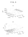
- FIGS. 5(a) and (b) are respectively diagrams showing regression curved surfaces of the Echinocyte.
- FIG. 5(a) is a 3D curved surface about the rise ⁇ in relative electric permittivity
- FIG. 5(b) is a 3D curved surface about the relaxation time ⁇ .
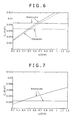
- FIG. 6 is a graphical representation showing a relationship between values obtained through calculations about the Discocyte, and values actually measured with the cell cytoplasm electric conductivity ⁇ i being plotted on an axis of abscissa and the membrane capacitance C m being platted on an axis of ordinate
- FIG. 7 is a graphical representation showing a relationship between values obtained through calculations about the Echinocyte, and values actually measured with the cell cytoplasm electric conductivity ⁇ i being plotted on an axis of abscissa and the membrane capacitance C m being platted on an axis of ordinate. It is noted that FIG. 6 shows the relationship in the case of the Discocyte together with values as well of the Normocyte.
- the membrane capacitance C m, exp and the cell cytoplasm electric conductivity ⁇ i, exp of each of the measurement specimens were determined from an intersection point between each two curve lines shown in FIG. 6 and FIG. 7 .
- the values of the membrane capacitances C m, exp and the cell cytoplasm electric conductivities ⁇ i, exp of the specimens of No. 1 to No. 4 which have been determined from the processes described above are collectively shown in Table 1.
- FIG. 8 is a graphical representation showing the dielectric dispersion of the Discocyte with the frequency being plotted on an axis of abscissa and the complex electric permittivity ( ⁇ ', ⁇ ") being plotted on an axis of ordinate, and FIG.
- FIG. 9 is a graphical representation showing the dielectric dispersion of the Echinocyte with the frequency being plotted on an axis of abscissa and the complex electric permittivity ( ⁇ ', ⁇ ") being plotted on an axis of ordinate.
- the calculation results reproduced the values actually measured.
- the values actually measured exhibited the "awkward" spectrum unable to be expressed by the Cole-Cole expression which is generally applied to such a case, the calculation result about the Discocyte properly reproduced the values actually measured. From this, it was confirmed that the simple estimation processes described above for the phase constants were valid.
Landscapes
- Health & Medical Sciences (AREA)
- Life Sciences & Earth Sciences (AREA)
- Engineering & Computer Science (AREA)
- Biomedical Technology (AREA)
- Chemical & Material Sciences (AREA)
- Immunology (AREA)
- Hematology (AREA)
- Physics & Mathematics (AREA)
- Urology & Nephrology (AREA)
- Molecular Biology (AREA)
- Analytical Chemistry (AREA)
- Biochemistry (AREA)
- Pathology (AREA)
- General Physics & Mathematics (AREA)
- General Health & Medical Sciences (AREA)
- Food Science & Technology (AREA)
- Medicinal Chemistry (AREA)
- Cell Biology (AREA)
- Bioinformatics & Cheminformatics (AREA)
- Microbiology (AREA)
- Toxicology (AREA)
- Tropical Medicine & Parasitology (AREA)
- Biotechnology (AREA)
- Biophysics (AREA)
- Investigating Or Analyzing Materials By The Use Of Electric Means (AREA)
- Investigating Or Analysing Biological Materials (AREA)
- Measurement Of Resistance Or Impedance (AREA)
Abstract
Description
- The present invention relates to a method and an apparatus for measuring physical property values of a cell. More specifically, the invention relates to a technique for measuring electrical characteristics of a cell by utilizing a dielectric spectroscopy.
- In general, it is known that in a cell, physical property values exhibiting electrical characteristics such as an electric conductivity, an electric permittivity, and a dielectric constant change depending on kinds, statuses or the like of the cell. For example, a muscle cell and a nerve cell show high values of electric conductivities (hereinafter referred to as a cell cytoplasm conductivities) of an extracellular fluid and an intracellular fluid as compared with the case of a skin cell containing therein less water. In addition, when the electric permittivity of the cell is measured by sweeping a frequency, dielectric relaxation characteristics change depending on forms of the cell. Thus, by utilizing such characteristics, qualitative and quantitative analyses, and identification of the cell, and moreover confirmation of presence or absence of a disease, and the like become possible. Also, heretofore, a technique for utilizing a difference in dielectric characteristics between the cells has also been proposed as an identification method in separating the cells or the like from each other by a flow cytometry (for example, refer to
Parent Documents 1 and 2). - On the other hand, since the cell causes interfere polarization (Maxwell-Wagner polarization) under an electric field, when a dielectric spectroscopy measurement is carried out with respect to a solution containing therein cells, a frequency dispersion of a complex electric permittivity is obtained. Also, when a relaxation expression such as a Maxwell-Wagner equation is applied to the resulting dielectric spectrum, physical property values exhibiting the electrical characteristics, of the cell, such as a cell cytoplasm electric conductivity κi and a membrane capacitance Cm are obtained. The electrical characteristics of the cell such as a blood cell are measured by utilizing the dielectric spectroscopy in such a manner, thereby making it possible to know the state of the cell in a nondisruptive manner. Patent Document 1: Publication of the Translation of
JP-T-2003-507739
Patent Document 2: Publication of the Translation ofJP-T-2005-512042 - However, the prior art described above involves the problems shown below. That is to say, there is encountered such a problem that although the interface polarization is largely influenced by the shape of the cell, the Maxwell-Wagner equation which has been conventionally used in the analysis is established only in the cell having either a spherical shape or an ellipsoidal shape.
FIG. 10 is a Scanning Electron Microscope (SEM: 1500-fold magnification) picture showing Discocytes, andFIG. 11 is a SEM picture (1500-fold magnification) showing an Echinocyte. As shown inFIG. 10 , a red blood cell having a normal shape is of a disc-like shape having a dimple at the center thereof. In addition, since the shape of the blood cell sensitively changes depending on a state of the body, a disease or the like, there is also the red blood cell having the echin-shape as shown inFIG. 11 . Although as has been described, the cells having the various kinds of shapes exist, the method utilizing the dielectric spectroscopy can not be applied to any of the cells having the respective shapes other than the spherical shape and the ellipsoidal shape. Thus, heretofore, with regard to these cells having the nonisotropic shapes, the physical property values exhibiting the electrical characteristics such as the cell cytoplasm electric conductivity κi and the membrane capacitance Cm have been unable to be known in the nondisruptive manner. - In the light of the foregoing, it is therefore a principal object of the present invention to provide a method of measuring physical values of a cell, and a physical property measuring apparatus with each of which with respect to a cell as well having a nonisotropic shape, physical property values exhibiting electrical characteristics thereof can be measured in a nondisruptive manner.
- A method of measuring physical property values of a cell according to the present invention has: a process for calculating a dielectric spectrum about a cell having a specific shape by setting an arbitrary membrane capacitance Cm and an arbitrary cell cytoplasm electric conductivity κi, thereby carrying out a numeral analysis; a process for obtaining a rise Δε in relative electric permittivity and relaxation time τ of the cell by revolving the dielectric spectrum to a dielectric relaxation expression; and a process for creating a regression expression corresponding to a shape of the cell based on the rise Δε in relative electric permittivity and the relaxation time τ.
The method of measuring physical property values of a cell, in addition to the processes described above, may further have a process for measuring the dielectric spectrum of the cell, and comparing the measurement result and the regression expression with each other, thereby obtaining a membrane capacitance Cm, exp and a cell cytoplasm electric conductivity κi, exp of the cell.
In addition, it is also possible that the regression expression is created every shape of the cell, and a membrane capacitance Cm, exp and a cell cytoplasm conductivity Ki, exp are obtained by application of the regression expression corresponding to the shape of the cell.
Moreover, the cell, for example, may also have a nonisotropic shape.
On the other hand, a physical property measuring apparatus according to the present invention has: means for calculating a dielectric spectrum about a cell having a specific shape by setting an arbitrary membrane capacitance Cm and an arbitrary cell cytoplasm electric conductivity κi, thereby carrying out a numeral analysis; means for obtaining a rise Δε in relative electric permittivity and relaxation time τ of the cell by revolving the dielectric spectrum to a dielectric relaxation expression; and means for creating a regression expression corresponding to a shape of the cell based on the rise Δε in relative electric permittivity and the relaxation time τ. - According to the present invention, the dielectric spectrum actually measured, and the regression expression created by the simulation are compared with each other, thereby determining the membrane capacitance Cm, exp and the cell cytoplasm electric conductivity κi, exp of the cell as an object of the measurement. Therefore, with respect to the cell as well, having the nonisotropic shape, which is not adapted to the conventional relaxation expression, the physical property values exhibiting the electrical characteristics such as the cell cytoplasm electric conductivity κi and the membrane capacitance Cm of that cell can be obtained in the nondisruptive manner.
-
- [
FIG. 1 ]
FIG. 1 is a flow chart showing a method of measuring physical property values of a cell according to an embodiment of the present invention in the order of processes thereof. - [
FIG. 2 ]
FIG. 2 is a view showing a simulation model of a Discocyte. - [
FIG. 3 ]
FIG. 3 is a view showing a simulation model of an Echinocyte. - [
FIG. 4 ]
FIGS. 4(a) and (b) are respectively diagrams each showing a regression curved surface of the Discocyte obtained through a calculation, andFIG. 4(a) is a 3D curved surface about a rise Δε in relative electric permittivity andFIG. 4(b) is a 3D curved surface about relaxation time τ. - [
FIG. 5 ]
FIGS. 5(a) and (b) are respectively diagrams each showing a regression curved surface of the Echinocyte obtained through a calculation, andFIG. 5(a) is a 3D curved surface about a rise Δε in relative electric permittivity andFIG. 5(b) is a 3D curved surface about relaxation time τ. - [
FIG. 6 ]
FIG. 6 is a graph showing a relationship between values obtained through a calculation and values actually measured with respect to the Discocyte with a cell cytoplasm electric conductivity κi being platted on an axis of abscissa and a membrane capacitance Cm being plotted on an axis of ordinate. - [
FIG. 7 ]
FIG. 7 is a graph showing a relationship between values obtained through a calculation and values actually measured with respect to the Echinocyte with a cell cytoplasm electric conductivity κi being platted on an axis of abscissa and a membrane capacitance Cm being plotted on an axis of ordinate. - [
FIG. 8 ]
FIG. 8 is a graphical representation showing a dielectric dispersion of the Discocyte with a frequency being plotted on an axis of abscissa and a complex electric permittivity (ε', ε") being plotted on an axis of ordinate. - [
FIG. 9 ]
FIG. 9 is a graphical representation showing a dielectric dispersion of the Echinocyte with a frequency being plotted on an axis of abscissa and a complex electric permittivity (ε', ε") being plotted on an axis of ordinate. - [
FIG. 10 ]
FIG. 10 is a picture (SEM picture at 15000-fold magnification) used as a substitute for a drawing showing the Discocytes. - [
FIG. 11 ]
FIG. 11 is a picture (SEM picture at 15000-fold magnification) used as a substitute for a drawing showing the Echinocyte. - Hereinafter, Best Mode for Carrying Out the Invention will be described in detail with reference to the accompanying drawings.
FIG. 1 is a flow chart showing a method of measuring physical property values of a cell according to an embodiment of the present invention in the order of processes thereof. As shown inFIG. 1 , in the method of measuring physical property values of a cell of this embodiment (hereinafter simply referred to as a measurement method as well), firstly, with regard to cells, each having a nonisotropic shape, such as a normal red blood cell and Echinocyte, complex electric permittivity responses when an AC electric field is applied to specific isotropic three-dimensional shapes in each of which an outer phase and an inner phase are partitioned by a thin membrane are obtained in a manner of a numerical analysis by using models of shapes of the cells each having the nonisotropic shape (Step S1). Although a calculation method at this time is especially by no means limited, for example, a three-dimensional finite difference method or the like can be utilized. - Next, the numerical calculations are carried out based on the complex electric permittivity responses obtained in Step S1 while the membrane capacitance Cm and the cell cytoplasm electric conductivity κi are changed independently of each other, thereby calculating dielectric spectra of the cells having the respective shapes (Step S2).
- Next, two parameters of a rise Δε in relative electric permittivity and relaxation time τ are calculated from each of the dielectric spectra obtained in Step S2 (Step S3). Although the dielectric spectrum of each of the cells can not be perfectly expressed by a simple relaxation expression, by revolving the dielectric spectrum of each of the cells to a Cole-Cole type relaxation expression shown in
Expression 1 which will be shown below, the dielectric spectrum of each of the cells can be converted into three values exhibiting dielectric relaxation characteristics, that is, the rise Δε in relative electric permittivity, the relaxation time τ, and a Cole-Cole parameter β exhibiting the spreading of the relaxation. Then, in this embodiment, a real part (ε') of each of the dielectric spectra obtained from the calculations is fitted by using the Cole-Cole type relaxation expression shown inExpression 1 which will be shown below, thereby obtaining the rise Δε in relative electric permittivity, and the relaxation time τ. It is noted that inExpression 1 which will be shown below, ω is an angular frequency, ε0 is an electric permittivity of a vacuum, κi is an electric conductivity of a solution in a low frequency limit, ε∞ is a relative electric permittivity in a high frequency limit, and i2 = -1. -

- Next, dependency of the rise Δε in relative electric permittivity, and the relaxation time τ on (Cm, κi) is obtained every shape of the cell based on the rise Δε in relative electric permittivity, and the relaxation time τ calculated in Step S3, thereby creating a regression expression (Step S4). Although coefficients obtained from each of the dielectric spectra are largely influenced by the shape of the cell, it is thought that since the essence of the polarization of the cell is the interface polarization, the dependency of the rise Δε in relative electric permittivity, and the relaxation time τ on (Cm, κi) form the same function form as that of the Maxwell-Wagner equation. Thus, in the measurement method of the present invention, a relationship shown in
Expression 2 andExpression 3 which will be shown below was set, and a multiple regression analysis of constants a, b, c and d was carried out for the results obtained in Step S3, thereby obtaining the dependency of the rise Δε in relative electric permittivity, and the relaxation time τ on (Cm, κi). It is noted that P inExpression 2 shown below is a volume fraction of the cell which is obtained from the model of each of the shapes used in Step S1. -

-

- In such a manner, the frequency dispersion of the complex electric permittivity is obtained every shape of the cell in advance in the manner of the numerical analysis and the results of the numerical analysis are made to revolve to the dielectric relaxation expression, thereby creating the regression with respect to the dependency of the rise Δε in relative electric permittivity, and the relaxation time τ on (Cm, κi). Therefore, with regard to the cell as well, having the nonisotropic shape, which is not adapted to the conventional relaxation expression, the physical property values exhibiting the electrical characteristics such as the cell cytoplasm electric conductivity κi and membrane capacitance Cm thereof can be speedily and easily measured in the nondisruptive manner.
- In addition, when the cell cytoplasm electric conductivity κi and the membrane capacitance Cm of the cell are obtained by using the measurement method of this embodiment, firstly, with respect to the cell as the object of the measurement which is dispersed into a normal saline solution or the like, a complex electric permittivity thereof is measured by sweeping a frequency by using an impedance analyzer or the like. Next, a real part (ε') of the resulting dielectric spectrum is fitted by using the Cole-Cole type relaxation expression shown in
Expression 1 described above, thereby obtaining a rise Δεexp in relative electric permittivity, and relaxation time τexp. Also, (Δεexp, τexp) and a regression expression (f(Cm, κi), g(Cm, κi)) created in Step S4 are compared with each other, thereby determining a membrane capacitance Cm, exp and the cell cytoplasm electric conductivity κi, exp of the cell as the object of the measurement (Step S5). - As has been described, in the measurement method of this embodiment, the dielectric spectrum actually measured and the regression expression created by the simulation are compared with each other, thereby determining the membrane capacitance Cm, exp and the cell cytoplasm electric conductivity κi, exp of the cell as the object of the measurement. Therefore, with regard to the cell as well, having the nonisotropic shape, which is not adapted to the conventional relaxation expression, the physical property values exhibiting the electrical characteristics thereof can be measured in the nondisruptive manner. In addition, of the dielectric spectra which are obtained by estimating unknown amounts about the cell cytoplasm electric conductivity and the like, thereby carrying out numerous calculations, the dielectric spectrum which most coincides with the dielectric spectrum actually measured, whereby the labor and time required for the measurement can be greatly shortened as compared with the conventional method of deriving the suppression number of measurement specimens.
- In addition, the physical property values of the cell which are determined by using the measurement method of this embodiment can be converted into information such as desorption of a membrane protein and abnormality of a cell cytoplasm to be used in diagnosis of a disease or the like. Moreover, for example, in a blood cell counter, not only the counting, but also up to the state of the blood cell can be simultaneously measured at a high speed, and also a statistical study about a rate of abnormal cells, and the like becomes possible.
- It should be noted that in the embodiment described above, the case where the cell cytoplasm electric conductivity κi and the membrane capacitance Cm are obtained has been described as an example, the present invention is by no means limited thereto. Thus, with regard to the physical property values of the cell, in addition to the cell cytoplasm electric conductivity κi and the membrane capacitance Cm, for example, a membrane electric conductivity, a cell cytoplasm electric permittivity, and the like can also be measured.
- In addition, the measurement method of this embodiment can be carried out by using an apparatus for carrying out the processes described above. All it takes for the apparatus for measuring physical property values of a cell used in this embodiment is to include at least means for calculating a dielectric spectrum about a cell having a specific shape by setting an arbitrary membrane capacitance Cm and an arbitrary cell cytoplasm electric conductivity κi, thereby carrying out a numeral analysis; means for obtaining a rise Δε in relative electric permittivity and relaxation time τ of the cell by revolving the dielectric spectrum to a dielectric relaxation expression; and means for creating a regression expression corresponding to a shape of the cell based on the rise Δε in relative electric permittivity and the relaxation time τ.
- Hereinafter, an example of the present invention will be compared with a comparative example departing from the scope of the present invention to concretely describe the effects thereof. It should be noted that the present invention is by no means limited to the example which will be described below. In this example, cell cytoplasm electric conductivities κi and the membrane capacitances Cm of cells having various kinds of shapes were measured by using the method of measuring physical property values of a cell shown in
FIG. 1 . - Firstly, a rabbit preserved blood (made by KOHJINBIO Co., Ltd.) was subjected to centrifugal processing, thereby extracting only red blood cells. Also, the red blood cells thus extracted were dispersed into a normal saline solution a concentration of which was adjusted so as to have pH5.3, thereby preparing a specimen (No. 1) of the Spherocytes. The red blood cells thus extracted were dispersed into a normal saline solution a concentration of which was adjusted so as to have pH8.5, thereby preparing a specimen (No. 2) of the Discocytes. Also, the red blood cells thus extracted were dispersed into a normal saline solution a concentration of which was adjusted so as to have pH7.7, thereby preparing a specimen (No. 3) of the Echinocytes. It should be noted that although for the purpose of obtaining the Spherocytes, in general, the specimen was prepared by utilizing a difference in osmotic pressure, in this example, in order to unify the shapes of the blood cells, the specimens were prepared by using the technique described above. In addition, each of the specimens was observed by using a microscope, and as a result, a ratio of the red blood cells each having the shape as the object of the measurement to the total number of red blood cells was 90% or more in a fraction of a quantity. Moreover, in this example, for the purpose of reference, a specimen (No. 4) was also prepared by dispersing Normocytes into a phosphate buffered saline (PBS).
- Next, with respect to each of the specimens prepared by using the method described above, the electric permittivity was measured while the electrode polarization was suppressed in the range of 10 kHz to 110 MHz by using an impedance analyzer (4294A made by Agilent Technologies, Inc.). Also, with respect to the specimen (No. 1) of the Spherocytes, known values, that is, a relative electric permittivity εa = 78.3, an electric conductivity κa = 1.67 S/m, and cell membrane electric conductivity κm = 1 × 10⁻⁷ S/m of an outer aqueous phase were used, and the Maxwell-Wagner equation for a thin shell was applied to a dielectric spectrum obtained through the measurement, thereby obtaining a volume fraction P, a membrane capacitance Cm (= εm·ε0/dm where dm is a thickness of a membrane, εm is a relative electric permittivity of the membrane, and ε0 is an electric permittivity of the vacuum) per unit area of the cell membrane, and an electric permittivity εi and an electric conductivity κi of an inner aqueous phase of the red blood cell.
- On the other hand, with respect to the Discocytes and the Echinocytes, the volume fraction P was calculated by using an ellipsoidal model and a spherical model, and a complex electric permittivity was obtained through a simulation using a three-dimensional finite difference method. It is noted that a length of a long axis of a Discocyte was measured by using a light microscope, and as a result, it was proved to be about 7.9 µm which was approximately equal to that of the shape of the red blood cell (RBC) of the human. Then, a simulation of the Discocyte was carried out by reference to the expression of the RBC of the human. In addition, although the Echinocytes also have a variety in the numbers of protrusions, and thus the Echinocytes having various kinds of shapes exist, in this example, the simulation was carried out based on the result of the observation made by using the light microscope.
FIG. 2 is a view showing a simulation model of the Discocyte, andFIG. 3 is a view showing a simulation model of the Echinocyte. - When the assumption of the diluteness is established, in the normal shape, the resolutions are obtained in the case where the direction of the principal axis and the electric field either intersect perpendicularly with each other or are parallel with each other, and they are weighted with the probability density to be averaged, thereby making it possible to obtain the complex electric permittivity. Then, the calculation was carried out with a small volume fraction P (P ≤ 0.04%) so that the assumption of the diluteness is established, and the calculation results (ε', ε") were normalized by P as shown in
Expression 4 and Expression 5 which will be shown below. It is noted that κl in Expression 5 shown below is an electric conductivity in a low frequency limit. -

-

- Next, the numerical calculation about the shapes were carried out under the 25 conditions in total with respect to the Discocyte and the Echinocyte while the membrane capacitance Cm and the cell cytoplasm electric conductivity κi were changed independently of each other, thereby calculating the dielectric spectra with respect to the Discocyte and the Echinocyte, respectively. After that, for the purpose of easily characterizing the dielectric spectra obtained through the calculations, the respective time particles were fitted by using the Cole-Cole type relaxation expression shown in
Expression 1, thereby calculating the rise Δε in relative electric permittivity and the relaxation time τ. Also, the multiple regression analysis was carried out for the calculation results about the 25 conditions, thereby determining the constants a, b and c inExpression 2 andExpression 3 described above. As a result, in the case of the Discocyte, the values of a = 1.10 × 10⁶ m2/F, b = 2.86 × 10⁻² m/s, c = 4.52 × 10⁻⁶ m, and d = 1.19 × 10⁻⁶ m2/S were obtained. In addition, in the case of the Echinocyte, the values of a = 2.08 × 10⁶ m2/F, b = 5.26 × 10⁻³ m/S, c = 1.02 × 10⁻⁵ m, and d = 2.16 × 10⁻⁶ m2/S were obtained.
FIGS. 4(a) and (b) are respectively diagrams showing regression curved surfaces of the Discocyte.FIG. 4(a) is a 3D curved surface about the rise Δε in relative electric permittivity, andFIG. 4(b) is a 3D curved surface about the relaxation time τ. In addition,FIGS. 5(a) and (b) are respectively diagrams showing regression curved surfaces of the Echinocyte.FIG. 5(a) is a 3D curved surface about the rise Δε in relative electric permittivity, andFIG. 5(b) is a 3D curved surface about the relaxation time τ. - Next, with regard to the dielectric spectra (values actually measured) of the specimens of No. 2 and No. 3 in each of which the cell cytoplasm electric conductivity κi and the membrane capacitance Cm were unknown, similarly, the real part (ε') of the complex electric permittivity thereof was fitted by using the Cole-Cole type relaxation expression shown in
Expression 1 described above, thereby calculating the relaxation parameters (Δεexp, τemp). Also, curved surfaces which the regression curved surfaces shown inFIG. 4 andFIG. 5 , and Δε = Δεexp and τ = τexp have in common were respectively obtained.FIG. 6 is a graphical representation showing a relationship between values obtained through calculations about the Discocyte, and values actually measured with the cell cytoplasm electric conductivity κi being plotted on an axis of abscissa and the membrane capacitance Cm being platted on an axis of ordinate, andFIG. 7 is a graphical representation showing a relationship between values obtained through calculations about the Echinocyte, and values actually measured with the cell cytoplasm electric conductivity κi being plotted on an axis of abscissa and the membrane capacitance Cm being platted on an axis of ordinate. It is noted thatFIG. 6 shows the relationship in the case of the Discocyte together with values as well of the Normocyte. Also, the membrane capacitance Cm, exp and the cell cytoplasm electric conductivity κi, exp of each of the measurement specimens (No. 2 and No. 3) were determined from an intersection point between each two curve lines shown inFIG. 6 and FIG. 7 . The values of the membrane capacitances Cm, exp and the cell cytoplasm electric conductivities κi, exp of the specimens of No. 1 to No. 4 which have been determined from the processes described above are collectively shown in Table 1. -
[Table 1] Specimen Volume fraction P (%) Membrane capacitance Cm (F/m2) Cell cytoplasm electric conductivity Ki (S/m) No. Kind of cell 1 Spherocyte 5.80 6.33 × 10⁻³ 0.713 2 Discocyte 7.18 6.68 × 10⁻³ 0.539 3 Echinocyte 8.73 2.54 × 10⁻³ 0.373 4 Normocyte 9.80 8.79 × 10⁻³ 0.666 - In addition, for the purpose of confirming whether or not the values thus determined reproduced the dielectric dispersions actually measured, the dielectric dispersions of the Discocyte and the Echinocyte were calculated again by the finite difference method using the values obtained from
FIG.6 and FIG. 7 .FIG. 8 is a graphical representation showing the dielectric dispersion of the Discocyte with the frequency being plotted on an axis of abscissa and the complex electric permittivity (ε', ε") being plotted on an axis of ordinate, andFIG. 9 is a graphical representation showing the dielectric dispersion of the Echinocyte with the frequency being plotted on an axis of abscissa and the complex electric permittivity (ε', ε") being plotted on an axis of ordinate. As shown inFIG. 8 and FIG. 9 , in any of the Discocyte and the Echinocyte, the calculation results reproduced the values actually measured. In particular, although the values actually measured exhibited the "awkward" spectrum unable to be expressed by the Cole-Cole expression which is generally applied to such a case, the calculation result about the Discocyte properly reproduced the values actually measured. From this, it was confirmed that the simple estimation processes described above for the phase constants were valid.
Claims (5)
- A method of measuring physical property values of a cell having:a process for calculating a dielectric spectrum about a cell having a specific shape by setting an arbitrary membrane capacitance Cm and an arbitrary cell cytoplasm electric conductivity κi, thereby carrying out a numeral analysis;a process for obtaining a rise Δε in relative electric permittivity and relaxation time τ of said cell by revolving the dielectric spectrum to a dielectric relaxation expression; anda process for creating a regression expression corresponding to a shape of said cell based on the rise Δε in relative electric permittivity and the relaxation time τ.
- The method of measuring physical property values of a cell according to claim 1, characterized by further having a process for measuring the dielectric spectrum of said cell, and comparing the measurement result and the regression expression with each other, thereby obtaining a membrane capacitance Cm, exp and a cell cytoplasm electric conductivity κi, exp of said cell.
- The method of measuring physical property values of a cell according to claim 1, characterized in that:the regression expression is created every shape of the cell, and a membrane capacitance Cm, exp and a cell cytoplasm electric conductivity κi, exp are obtained by application of the regression expression corresponding to the shape of said cell.
- The method of measuring physical property values of a cell according to claim 1, characterized in that:said cell has a nonisotropic shape.
- A physical property measuring apparatus having:means for calculating a dielectric spectrum about a cell having a specific shape by setting an arbitrary membrane capacitance Cm and an arbitrary cell cytoplasm electric conductivity κi, thereby carrying out a numeral analysis;means for obtaining a rise Δε in relative electric permittivity and relaxation time τ of said cell by revolving the dielectric spectrum to a dielectric relaxation expression; andmeans for creating a regression expression corresponding to a shape of said cell based on the rise Δε in relative electric permittivity and the relaxation time τ.
Applications Claiming Priority (2)
| Application Number | Priority Date | Filing Date | Title |
|---|---|---|---|
| JP2007209108A JP4893526B2 (en) | 2007-08-10 | 2007-08-10 | Cell physical property value measuring method and physical property measuring apparatus |
| PCT/JP2008/063500 WO2009022536A1 (en) | 2007-08-10 | 2008-07-28 | Method of measuring physical property value of cell and system for measuring physical property |
Publications (2)
| Publication Number | Publication Date |
|---|---|
| EP2175262A1 true EP2175262A1 (en) | 2010-04-14 |
| EP2175262A4 EP2175262A4 (en) | 2014-12-17 |
Family
ID=40350591
Family Applications (1)
| Application Number | Title | Priority Date | Filing Date |
|---|---|---|---|
| EP08791735.7A Ceased EP2175262A4 (en) | 2007-08-10 | 2008-07-28 | METHOD FOR MEASURING A PHYSICAL PROPERTY VALUE OF A CELL AND SYSTEM FOR MEASURING PHYSICAL PROPERTY |
Country Status (5)
| Country | Link |
|---|---|
| US (1) | US8478546B2 (en) |
| EP (1) | EP2175262A4 (en) |
| JP (1) | JP4893526B2 (en) |
| CN (1) | CN101772700B (en) |
| WO (1) | WO2009022536A1 (en) |
Families Citing this family (27)
| Publication number | Priority date | Publication date | Assignee | Title |
|---|---|---|---|---|
| JP5098817B2 (en) | 2008-05-29 | 2012-12-12 | ソニー株式会社 | Physical property measuring apparatus and physical property measuring method |
| JP5604862B2 (en) * | 2009-01-09 | 2014-10-15 | ソニー株式会社 | Channel device, complex permittivity measuring apparatus and dielectric cytometry apparatus |
| KR101119381B1 (en) * | 2009-06-18 | 2012-03-07 | 한국과학기술원 | Device and system for measuring properties of cells and method of measuring properties of cells using the same |
| JP5556144B2 (en) * | 2009-11-26 | 2014-07-23 | ソニー株式会社 | Blood cell analyzer, blood cell analysis method, and blood cell analysis system |
| JP5549484B2 (en) | 2010-09-01 | 2014-07-16 | ソニー株式会社 | Sample cartridge and apparatus for measuring electrical properties of liquid samples |
| CN101975898B (en) * | 2010-09-17 | 2012-07-04 | 西安交通大学 | Device for dynamically and electrically researching dielectric property of cells by using ultrasonic pulse |
| JP5617532B2 (en) | 2010-10-29 | 2014-11-05 | ソニー株式会社 | Dielectric cytometry apparatus and cell sorting method using dielectric cytometry |
| JP5824950B2 (en) * | 2011-08-08 | 2015-12-02 | ソニー株式会社 | Blood analyzer and blood analysis method |
| US10001451B2 (en) | 2012-09-04 | 2018-06-19 | Sony Corporation | Cell determination device, cell determination system, and cell determination method |
| JP2014115256A (en) * | 2012-12-12 | 2014-06-26 | Sony Corp | Container for electrical measurement, apparatus for electrical measurement, and method for electrical measurement |
| CN104937400B (en) | 2013-01-28 | 2018-07-03 | 索尼公司 | Impedance measuring device for biological sample and impedance measuring system for biological sample |
| EP2975387B1 (en) | 2013-03-13 | 2021-03-31 | Sony Corporation | Device for analyzing condition of blood, system for analyzing condition of blood, and program for analyzing condition of blood |
| WO2014141844A1 (en) | 2013-03-15 | 2014-09-18 | ソニー株式会社 | Blood condition analyzing device, blood condition analyzing system, blood condition analyzing method, and blood condition analyzing program for realizing method on computer |
| JPWO2014156370A1 (en) | 2013-03-29 | 2017-02-16 | ソニー株式会社 | Blood state evaluation apparatus, blood state evaluation system, blood state evaluation method and program |
| EP2980571A4 (en) | 2013-03-29 | 2016-10-26 | Sony Corp | BLOOD STATUS ANALYSIS DEVICE, BLOOD STATUS ANALYSIS SYSTEM, BLOOD CONDITION ANALYSIS METHOD, AND PROGRAM |
| JP6569209B2 (en) | 2014-01-07 | 2019-09-04 | ソニー株式会社 | Electrical measurement cartridge, electrical measurement apparatus, and electrical measurement method |
| JP2015169432A (en) | 2014-03-04 | 2015-09-28 | ソニー株式会社 | Electrical measurement cartridge, electrical measurement apparatus and electrical measuring method |
| JP6515677B2 (en) | 2014-07-24 | 2019-05-22 | ソニー株式会社 | Contact structure and electrical measurement apparatus for biological sample using the contact structure |
| JP2016024161A (en) | 2014-07-24 | 2016-02-08 | ソニー株式会社 | Electrical measurement cartridge, biological sample electrical measurement device, biological sample electrical measurement system, and biological sample electrical measurement method |
| WO2016132779A1 (en) | 2015-02-20 | 2016-08-25 | ソニー株式会社 | Electrical characteristic measurement device, electrical characteristic measurement system, electrical characteristic measurement method, and electrical characteristic measurement program for causing computer to implement said method |
| ES2925018T3 (en) | 2016-05-11 | 2022-10-13 | Nova Biomedical Corp | Whole blood SO2 sensor |
| US10758151B2 (en) * | 2017-01-23 | 2020-09-01 | NovaScan, Inc. | Techniques for detecting cancerous cells in excised tissue samples using impedance detection |
| JP6876911B2 (en) | 2017-02-21 | 2021-05-26 | ソニーグループ株式会社 | Cartridge for electrical measurement, electrical measuring device and electrical measuring method |
| KR102221953B1 (en) * | 2018-08-31 | 2021-03-04 | 연세대학교 원주산학협력단 | Analysis of Electro-polarization Characteristics of Cells by Regulation of Dielectrophoretic Force |
| JP6705550B1 (en) * | 2019-03-22 | 2020-06-03 | Toto株式会社 | Electrostatic chuck |
| US12019040B2 (en) | 2019-09-30 | 2024-06-25 | Cornell University | System and devices for monitoring cell-containing materials and methods of their use |
| KR102925308B1 (en) * | 2024-01-31 | 2026-02-09 | 원광대학교산학협력단 | Method for analyzing of proteins by dielectric spectroscopy and biosensor using the same |
Family Cites Families (5)
| Publication number | Priority date | Publication date | Assignee | Title |
|---|---|---|---|---|
| WO1996026438A1 (en) * | 1995-02-18 | 1996-08-29 | University College Of Wales Aberystwyth | Biomass measurement apparatus and method |
| US6641708B1 (en) | 1996-01-31 | 2003-11-04 | Board Of Regents, The University Of Texas System | Method and apparatus for fractionation using conventional dielectrophoresis and field flow fractionation |
| CN1152253C (en) * | 1999-02-23 | 2004-06-02 | 王劲松 | Detecting method and apparatus for yeast concentration (measuring bioconcentration) |
| US6703819B2 (en) | 2001-12-03 | 2004-03-09 | Board Of Regents, The University Of Texas System | Particle impedance sensor |
| JP5098817B2 (en) | 2008-05-29 | 2012-12-12 | ソニー株式会社 | Physical property measuring apparatus and physical property measuring method |
-
2007
- 2007-08-10 JP JP2007209108A patent/JP4893526B2/en not_active Expired - Fee Related
-
2008
- 2008-07-28 WO PCT/JP2008/063500 patent/WO2009022536A1/en not_active Ceased
- 2008-07-28 US US12/733,031 patent/US8478546B2/en not_active Expired - Fee Related
- 2008-07-28 CN CN200880101833.XA patent/CN101772700B/en not_active Expired - Fee Related
- 2008-07-28 EP EP08791735.7A patent/EP2175262A4/en not_active Ceased
Also Published As
| Publication number | Publication date |
|---|---|
| JP4893526B2 (en) | 2012-03-07 |
| WO2009022536A1 (en) | 2009-02-19 |
| CN101772700B (en) | 2012-05-02 |
| JP2009042141A (en) | 2009-02-26 |
| EP2175262A4 (en) | 2014-12-17 |
| US20100136606A1 (en) | 2010-06-03 |
| US8478546B2 (en) | 2013-07-02 |
| CN101772700A (en) | 2010-07-07 |
Similar Documents
| Publication | Publication Date | Title |
|---|---|---|
| EP2175262A1 (en) | Method of measuring physical property value of cell and system for measuring physical property | |
| EP2955512B1 (en) | Microparticle analyzing device and microparticle analyzing system | |
| Coster et al. | Characterisation of ultrafiltration membranes by impedance spectroscopy. I. Determination of the separate electrical parameters and porosity of the skin and sublayers | |
| CN110056348A (en) | A kind of method and system of measurement formation fluid composition and property | |
| US20100268109A1 (en) | Method for analyzing the structure of an electrically conductive object | |
| WO1996024837A2 (en) | Spectrophotometric method and apparatus for the characterization of blood, blood types, and other bodily fluids | |
| CN107003296A (en) | Hemolysis test method and system | |
| Hayashi et al. | Dielectric inspection of erythrocyte morphology | |
| JPWO2014141844A1 (en) | Blood state analysis apparatus, blood state analysis system, blood state analysis method, and blood state analysis program for causing a computer to realize the method | |
| Kawashima et al. | Low-frequency impedance-based cell discrimination considering ion transport model in cell suspension | |
| Mondal et al. | Estimation of humidity sensor equivalent circuit parameters utilizing frequency response measurements combined with subspace identification and similarity transformations | |
| Zhbanov et al. | Electrochemical impedance spectroscopy of blood. Part 2: numerical analysis of experimental dielectric spectra using the biconcave shape of human erythrocytes | |
| CN113030007A (en) | Method for rapidly testing quality stability of tobacco essence based on similarity learning algorithm | |
| KR101613587B1 (en) | Method and apparatus for measuring hematocrit of blood | |
| Ferrara et al. | Optimal frequency range for the measurement of AC conductivity in aqueous solutions | |
| CN106908485A (en) | A kind of method that Non-Destructive Testing separates membrane flux | |
| Kawashima et al. | Multiple Regression Fitting Electrical Impedance Spectro-Tomography for Quantitative Image Reconstruction of Dead Cell Fraction and Cell Concentration | |
| Ballerini et al. | Fractal analysis of microscopic images of breast tissue | |
| CN114627058A (en) | Vision and quality fused cut tobacco component detection method | |
| Jiménez et al. | Broadband dielectric spectra of spheroidal hematite particles | |
| Szwacki et al. | Polymer membranes loaded with lipids for taste sensing: electrochemical impedance spectroscopy studies | |
| de Araujo et al. | Detection and characterization of biological cells by impedance spectroscopy | |
| Canas et al. | Electrochemical and structural characterizations of an experimental track-etched membrane in KCl solutions | |
| DE69624135T2 (en) | METHOD FOR TREATING A CELL SUSPENSION | |
| Valladares Pérez et al. | Measurement of resistivity of biofluids in the presence of an electrical double layer |
Legal Events
| Date | Code | Title | Description |
|---|---|---|---|
| PUAI | Public reference made under article 153(3) epc to a published international application that has entered the european phase |
Free format text: ORIGINAL CODE: 0009012 |
|
| 17P | Request for examination filed |
Effective date: 20100203 |
|
| AK | Designated contracting states |
Kind code of ref document: A1 Designated state(s): AT BE BG CH CY CZ DE DK EE ES FI FR GB GR HR HU IE IS IT LI LT LU LV MC MT NL NO PL PT RO SE SI SK TR |
|
| AX | Request for extension of the european patent |
Extension state: AL BA MK RS |
|
| DAX | Request for extension of the european patent (deleted) | ||
| RIC1 | Information provided on ipc code assigned before grant |
Ipc: G01N 22/00 20060101AFI20141105BHEP Ipc: G01N 33/48 20060101ALI20141105BHEP Ipc: G01N 27/22 20060101ALI20141105BHEP |
|
| A4 | Supplementary search report drawn up and despatched |
Effective date: 20141113 |
|
| 17Q | First examination report despatched |
Effective date: 20141209 |
|
| REG | Reference to a national code |
Ref country code: DE Ref legal event code: R003 |
|
| STAA | Information on the status of an ep patent application or granted ep patent |
Free format text: STATUS: THE APPLICATION HAS BEEN REFUSED |
|
| 18R | Application refused |
Effective date: 20180726 |