EP2172147A1 - Medical image processing device and medical image processing method - Google Patents
Medical image processing device and medical image processing method Download PDFInfo
- Publication number
- EP2172147A1 EP2172147A1 EP08740050A EP08740050A EP2172147A1 EP 2172147 A1 EP2172147 A1 EP 2172147A1 EP 08740050 A EP08740050 A EP 08740050A EP 08740050 A EP08740050 A EP 08740050A EP 2172147 A1 EP2172147 A1 EP 2172147A1
- Authority
- EP
- European Patent Office
- Prior art keywords
- feature value
- shape
- value
- section
- image processing
- Prior art date
- Legal status (The legal status is an assumption and is not a legal conclusion. Google has not performed a legal analysis and makes no representation as to the accuracy of the status listed.)
- Withdrawn
Links
Images




Classifications
-
- G—PHYSICS
- G06—COMPUTING OR CALCULATING; COUNTING
- G06T—IMAGE DATA PROCESSING OR GENERATION, IN GENERAL
- G06T7/00—Image analysis
- G06T7/0002—Inspection of images, e.g. flaw detection
- G06T7/0012—Biomedical image inspection
-
- G—PHYSICS
- G06—COMPUTING OR CALCULATING; COUNTING
- G06T—IMAGE DATA PROCESSING OR GENERATION, IN GENERAL
- G06T7/00—Image analysis
- G06T7/60—Analysis of geometric attributes
- G06T7/64—Analysis of geometric attributes of convexity or concavity
-
- A—HUMAN NECESSITIES
- A61—MEDICAL OR VETERINARY SCIENCE; HYGIENE
- A61B—DIAGNOSIS; SURGERY; IDENTIFICATION
- A61B1/00—Instruments for performing medical examinations of the interior of cavities or tubes of the body by visual or photographical inspection, e.g. endoscopes; Illuminating arrangements therefor
- A61B1/04—Instruments for performing medical examinations of the interior of cavities or tubes of the body by visual or photographical inspection, e.g. endoscopes; Illuminating arrangements therefor combined with photographic or television appliances
- A61B1/05—Instruments for performing medical examinations of the interior of cavities or tubes of the body by visual or photographical inspection, e.g. endoscopes; Illuminating arrangements therefor combined with photographic or television appliances characterised by the image sensor, e.g. camera, being in the distal end portion
-
- G—PHYSICS
- G06—COMPUTING OR CALCULATING; COUNTING
- G06T—IMAGE DATA PROCESSING OR GENERATION, IN GENERAL
- G06T2207/00—Indexing scheme for image analysis or image enhancement
- G06T2207/10—Image acquisition modality
- G06T2207/10068—Endoscopic image
-
- G—PHYSICS
- G06—COMPUTING OR CALCULATING; COUNTING
- G06T—IMAGE DATA PROCESSING OR GENERATION, IN GENERAL
- G06T2207/00—Indexing scheme for image analysis or image enhancement
- G06T2207/30—Subject of image; Context of image processing
- G06T2207/30004—Biomedical image processing
- G06T2207/30028—Colon; Small intestine
-
- G—PHYSICS
- G06—COMPUTING OR CALCULATING; COUNTING
- G06T—IMAGE DATA PROCESSING OR GENERATION, IN GENERAL
- G06T2207/00—Indexing scheme for image analysis or image enhancement
- G06T2207/30—Subject of image; Context of image processing
- G06T2207/30004—Biomedical image processing
- G06T2207/30092—Stomach; Gastric
Definitions
- the present invention relates to a medical image processing apparatus and a medical image processing method, and more particularly to a medical image processing apparatus and a medical image processing method that detects a lesion having an elevated shape existing in a two-dimensional image of an image of living tissue based on the two-dimensional image.
- the endoscope system includes, for example, the endoscope including an insertion portion inserted into a body cavity as a living body, an objective optical system placed in a distal end portion of the insertion portion, and an image pickup portion that picks up an image of the inside of the body cavity formed by the objective optical system and outputs the image as an image pickup signal, and the medical image processing apparatus that performs a process for displaying the image of the inside of the body cavity on a monitor or the like as a display portion based on the image pickup signal. Then, based on the image of the inside of the body cavity displayed on the monitor or the like as the display portion, a user observes, for example, an organ or the like as a subject in the body cavity.
- the endoscope system including the above-described configuration can also pick up an image of, for example, mucosa of a digestive tract such as a large intestine as a subject in the body cavity.
- the user can comprehensively observe, for example, various comprehensive findings such as a color tone of the mucosa, a shape of a lesion, and a microstructure of a mucosa surface.
- an endoscope apparatus has been proposed that can produce a three-dimensional model of a subject based on data of a two-dimensional image according to an image pickup signal of an image of the subject picked up by an endoscope.
- a method for identifying a lesion having an elevated shape such as a polyp in a three-dimensional model produced by, for example, CT (Computed Tomography) a method is proposed that can identify the lesion by evaluating a shape of the three-dimensional model based on a shape feature value such as ShapeIndex or Curvedness as described in US Patent Application Publication No. 20030223627 .
- the shape feature value used in the method proposed in US Patent Application Publication No. 20030223627 is a feature value whose calculation accuracy depends on a size of the lesion having the elevated shape.
- the method proposed in Patent Document 1 is applied to an endoscope image from which specific information of a size of a lesion having an elevated shape is hard to obtain unlike the three-dimensional model produced by the CT, calculation of a shape feature value according to the size of the elevated shape is difficult, which reduces detection accuracy of the lesion.
- the present invention is achieved in view of the above-described points, and has an object to provide a medical image processing apparatus and a medical image processing method that can increase detection accuracy in detecting a lesion having an elevated shape in an endoscope image as compared with conventional apparatuses and methods.
- the medical image processing apparatus of the present invention includes: a three-dimensional model estimating section that estimates a three-dimensional model of living tissue based on a two-dimensional image of an image of the living tissue inputted from a medical image pickup apparatus; a local region setting section that sets a plurality of local regions around a target pixel in the two-dimensional image; a shape feature value calculating section that uses three-dimensional coordinate data corresponding to each of the plurality of local regions and calculates shape feature values of respective voxels corresponding to the target pixel based on the three-dimensional model of the living tissue estimated by the three-dimensional model estimating section; a shape feature value selecting section that selects, as an optimum shape feature value of a voxel corresponding to the target pixel, a shape feature value calculated according to one local region including an optimum three-dimensional coordinate data amount among the plurality of shape feature values calculated according to the plurality of local regions; and an elevated shape detecting section that detects an elevated shape existing in the two-dimensional image based on a selection result of
- a medical image processing method of the present invention includes steps of: estimating a three-dimensional model of living tissue based on a two-dimensional image of an image of the living tissue inputted from a medical image pickup apparatus; setting a plurality of local regions around a target pixel in the two-dimensional image; using three-dimensional coordinate data corresponding to each of the plurality of local regions and calculating shape feature values of respective voxels corresponding to the target pixel based on the three-dimensional model of the living tissue; selecting, as an optimum shape feature value of a voxel corresponding to the target pixel, a shape feature value calculated according to one local region including an optimum three-dimensional coordinate data amount among the plurality of shape feature values calculated according to the plurality of local regions; and detecting an elevated shape existing in the two-dimensional image based on a result of the selection.
- Figs. 1 to 6 relate to an embodiment of the present invention.
- Fig. 1 is a diagram of an example of a configuration of essential parts in an endoscope system in which a medical image processing apparatus according to an embodiment of the present invention is used.
- Fig. 2 is a flowchart of an example of a process performed in the medical image processing apparatus in Fig. 1 .
- Fig. 3 is a diagram of an example of a two-dimensional image subjected to the process shown in Fig. 2 .
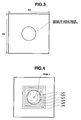
- Fig. 4 is a diagram of an example of each local region applied for calculating a shape feature value of one pixel in the process shown in Fig. 2 .
- Fig. 5 is a scatter diagram showing distribution of shape feature values of one pixel calculated by the process shown in Fig. 2 .
- Fig. 6 is a flowchart of an example of a process performed after the process shown in Fig. 2 in the medical image processing apparatus in Fig. 1 .
- an endoscope system 1 includes, as essential parts, a medical observation device 2 that picks up an image of a subject and outputs a two-dimensional image of the image of the subject, a medical image processing apparatus 3 that is constituted by a personal computer or the like, performs image processing of a video signal of the two-dimensional image outputted from the medical observation device 2 and outputs the video signal having subjected to the image processing as an image signal, and a monitor 4 that displays an image based on the image signal outputted from the medical image processing apparatus 3.
- a medical observation device 2 that picks up an image of a subject and outputs a two-dimensional image of the image of the subject
- a medical image processing apparatus 3 that is constituted by a personal computer or the like, performs image processing of a video signal of the two-dimensional image outputted from the medical observation device 2 and outputs the video signal having subjected to the image processing as an image signal
- a monitor 4 that displays an image based on the image signal outputted from the medical image processing apparatus 3.
- the medical observation device 2 includes, as essential parts, an endoscope 6 that is inserted into a body cavity, picks up an image of a subject in the body cavity, and outputs the image as an image pickup signal, a light source device 7 that supplies an illumination light for illuminating the subject whose image is picked up by the endoscope 6, a camera control unit (hereinafter abbreviated as CCU) 8 that performs various controls of the endoscope 6, performs signal processing of the image pickup signal outputted from the endoscope 6, and outputs the signal as a video signal of the two-dimensional image, and a monitor 9 that displays the image of the subject picked up by the endoscope 6 based on the video signal of the two-dimensional image outputted from the CCU 8.
- CCU camera control unit
- the endoscope 6 includes an insertion portion 11 inserted into the body cavity, and an operation portion 12 provided on a proximal end side of the insertion portion 11.
- a light guide 13 for transmitting the illumination light supplied by the light source device 7 is inserted through a portion from the proximal end side in the insertion portion 11 to a distal end portion 14 on a distal end side in the insertion portion 11.
- a distal end side of the light guide 13 is placed in the distal end portion 14 of the endoscope 6, and a rear end side thereof is connected to the light source device 7. Since the light guide 13 has such a configuration, the illumination light supplied by the light source device 7 is transmitted by the light guide 13 and then emitted from an unshown illumination window provided on a distal end surface of the distal end portion 14 of the insertion portion 11. Then, the illumination light is outputted from the unshown illumination window to illuminate living tissue or the like as the subject.
- an objective optical system 15 mounted to an unshown observation window adjacent to the unshown illumination window, and an image pickup portion 17 that is placed in an image forming position of the objective optical system 15 and has an image pickup device 16 constituted by, for example, a CCD (charge-coupled device) are provided.
- an image pickup device 16 constituted by, for example, a CCD (charge-coupled device)
- the image of the subject formed by the objective optical system 15 is picked up by the image pickup device 16 and then outputted as the image pickup signal.
- the image pickup device 16 is connected to the CCU 8 via a signal wire. Then, the image pickup device 16 is driven based on a drive signal outputted from the CCU 8, and also outputs the image pickup signal to the CCU 8.
- the image pickup signal inputted to the CCU 8 is subjected to the signal processing in an unshown signal processing circuit provided in the CCU 8, and thus converted and outputted as the video signal of the two-dimensional image.
- the video signal of the two-dimensional image outputted from the CCU 8 is outputted to the monitor 9 and the medical image processing apparatus 3.
- the image of the subject based on the video signal outputted from the CCU 8 is displayed as the two-dimensional image on the monitor 9.
- the medical image processing apparatus 3 includes an image inputting section 21 that performs A/D conversion of the video signal of the two-dimensional image outputted from the medical observation device 2 and outputs the video signal, a CPU 22 as a central processing unit that performs the image processing of the video signal outputted from the image inputting section 21, a processing program storing section 23 in which a processing program related to the image processing has been written, an image storing section 24 that stores the video signal outputted from the image inputting section 21 or the like, and an analysis information storing section 25 that stores image data or the like as an image processing result of the CPU 22.
- the medical image processing apparatus 3 includes a storage device interface 26, a hard disk 27 as a storage device that stores image data or the like as the image processing result of the CPU 22 via the storage device interface 26, a display processing section 28 that performs display processing for displaying the image data on the monitor 4 based on the image data as the image processing result of the CPU 22, and also outputs the image data having been subjected to the display processing as an image signal, and an input operation section 29 constituted by a keyboard or the like with which a user can input parameters in the image processing performed by the CPU 22 and operation instructions to the medical image processing apparatus 3.
- the monitor 4 displays the image based on the image signal outputted from the display processing section 28.
- the image inputting section 21, the CPU 22, the processing program storing section 23, the image storing section 24, the analysis information storing section 25, the storage device interface 26, the display processing section 28, and the input operation section 29 in the medical image processing apparatus 3 are connected to one another via a data bus 30.
- the user inserts the insertion portion 11 of the endoscope 6 into a tubular organ such as a large intestine, for example. Then, when the insertion portion 11 is inserted into the tubular organ by the user, an image of living tissue existing on an inner wall of the tubular organ and having an elevated shape is picked up by the image pickup portion 17 provided in the distal end portion 14. Then, the image of the living tissue having the elevated shape is outputted as the image pickup signal from the image pickup portion 17 to the CCU 8.
- the CCU 8 performs the signal processing of the image pickup signal outputted from the image pickup device 16 of the image pickup portion 17 in the unshown signal processing circuit, and thus converts the image pickup signal into the video signal of the two-dimensional image and outputs the video signal. Then, the monitor 9 displays, as the two-dimensional image, the image of the living tissue having the elevated shape based on the video signal outputted from the CCU 8. Also, the CCU 8 outputs the video signal of the two-dimensional image obtained by performing the signal processing of the image pickup signal outputted from the image pickup device 16 of the image pickup portion 17, to the medical image processing apparatus 3.
- the video signal of the two-dimensional image outputted to the medical image processing apparatus 3 is A/D converted in the image inputting section 21, and then inputted to the CPU 22.
- the CPU 22 having a function as a three-dimensional model estimating section performs a process such as geometric conversion, for example, based on luminance information or the like of the video signal of the two-dimensional image outputted from the image inputting section 21, and estimates a three-dimensional model of the living tissue having the elevated shape (obtains three-dimensional coordinate data).
- the CPU 22 corrects distortion of the video signal of the two-dimensional image outputted from the image inputting section 21, caused by image surface curving of the objective optical system 15, and then applies, for example, an image processing method described in Japanese Patent Application Laid-Open Publication No. 11-337845 to an R (red) component contained in the video signal to estimate a three-dimensional model of the living tissue having the elevated shape (obtains three-dimensional coordinate data).
- the CPU 22 obtains a two-dimensional image having a size of ISX ⁇ ISY (for example, 45 ⁇ 45) as a two-dimensional image including an image of living tissue having an elevated shape as shown in Fig. 3 (Step S1 in Fig. 2 ).
- the image of the living tissue having the elevated shape included in the image shown in Fig. 3 is an image picked up from an upper side of the elevated shape.
- the CPU 22 having a function as a local region setting section sets a value of a pixel k (1 ⁇ k ⁇ ISX ⁇ ISY) as a target pixel to 1 (Step S2 in Fig. 2 ), sets a value of a variable x (1 ⁇ x ⁇ N) to 1, and sets a value of a variable max to 0 (Step S3 in Fig. 2 ), and then sets a local region L k (x) around the pixel k in the two-dimensional image obtained by the process in Step S 1 in Fig. 2 (Step S4 in Fig. 2 ).
- the local region L k (x) is a region having a length of one side of A k (x) (having a size of A k (x) ⁇ A k (x)).
- the CPU 22 calculates an approximate curved surface equation in the local region L k (x) based on three-dimensional coordinate data corresponding to each two-dimensional coordinate in the local region L k (x) among the previously obtained three-dimensional coordinate data (Step S5 in Fig. 2 ). Then, the CPU 22 having a function as a shape feature value calculating section calculates a ShapeIndex value SI k (x) and a Curvedness value CV k (x) as a shape feature value of a voxel corresponding to the pixel k based on the calculated approximate curved surface equation (Step S6 in Fig. 2 ).
- the above-described ShapeIndex value is a value for indicating a concave or convex state of each voxel in a three-dimensional model, and indicated as a numerical value within a range of 0 to 1. Specifically, in one voxel (or one voxel group) existing in the three-dimensional model, when the ShapeIndex value is close to 0, the existence of a concave shape is indicated, and when the ShapeIndex value is close to 1, the existence of a convex shape is indicated.
- the above-described Curvedness value is a value corresponding to a curvature of each voxel in the three-dimensional model.
- the ShapeIndex value and the Curvedness value can be calculated by the same method as described in, for example, US Patent Application No. 20030223627 .
- a description of a specific calculating method of the ShapeIndex value and the Curvedness value in one voxel (or one voxel group) will be omitted.
- the CPU 22 determines whether values of the variable max, the ShapeIndex value SI k (x), and the Curvedness value CV k (x) satisfy the relationship in the following Expression (1) (Step S7 in Fig. 2 ).
- max 2 ⁇ SI k x 2 + CV k x 2
- the CPU 22 having a function as a shape feature value selecting section continues a process in Step S10 in Fig. 2 described later. Also, when the values of the variable max, the ShapeIndex value SI k (x), and the Curvedness value CV k (x) satisfy the relationship in the Expression (1), the CPU 22 having the function as the shape feature value selecting section updates an optimum value SIp(k) of the Shapelndex value to the value of SI k (x) calculated in Step S6 in Fig. 2 , and updates an optimum value CVp(k) of the Curvedness value to the value of CV k (x) calculated in Step S6 in Fig. 2 (Step S8 in Fig. 2 ).
- the CPU 22 substitutes the optimum value SIp(k) of the Shapelndex value and the optimum value CVp(k) of the Curvedness value into the following Expression(2) to calculate the value of the variable max (Step S9 in Fig. 2 ).
- Max SIp k 2 + CVIp k 2 1 / 2
- the CPU 22 determines whether the variable x is N or not (Step S10 in Fig. 2 ). When the variable x is not N, the CPU 22 adds 1 to the variable x (Step S11 in Fig. 2 ), and then repeats the processes from Steps S4 to S10 in Fig. 2 described above. When the variable x is N, the CPU 22 sets the optimum value SIp(k) of the ShapeIndex value and the optimum value CVp(k) of the Curvedness value as shape feature values of the voxel corresponding to the pixel k (Step S12 in Fig. 2 ), and then continues a process in Step S13 in Fig. 2 described later.
- Step S4 in Fig. 2 is repeated, and thus as shown in Fig. 4 , six local regions L k (1) to L k (6) around the pixel k are set in the image in Fig. 3 .
- Steps S5 and S6 in Fig. 2 are repeated, and thus ShapeIndex values SI k (1) to SI k (6) corresponding to the sizes of the local regions L k (1) to L k (6), and the Curvedness values CV k (1) to CV k (6) corresponding to the sizes of the local regions L k (1) to L k (6) are calculated.
- the six sets of ShapeIndex values and Curvedness values calculated by the processes in Steps 5 and S6 in Fig. 2 exist in coordinate positions as shown in Fig. 5 on a coordinate plane with ShapeIndex value on the abscissa and Curvedness value on the ordinate.
- a set of ShapeIndex value and Curvedness value existing in a coordinate position most distant from an original point on the coordinate plane are selected as a shape feature value calculated by an optimum three-dimensional coordinate data amount, specifically, an optimum value SIp(k) of the ShapeIndex value and an optimum values CVp(k) of the Curvedness value.
- the ShapeIndex value SI k (3) is selected as the optimum value SIp(k) of the ShapeIndex value
- the Curvedness value CV k (3) is selected as the optimum value CVp(k) of the Curvedness value.
- the processes from Steps S4 to S9 in Fig. 2 are repeated N times, and thus one local region having an optimum size for calculating a shape feature value used in identifying an elevated shape, that is, an optimum three-dimensional coordinate data amount for calculating the shape feature value is selected from the N local regions. Also, the processes from Steps S4 to S9 in Fig. 2 are repeated N times, and thus a set of ShapeIndex value and Curvedness value as the shape feature value calculated by the optimum three-dimensional coordinate data amount are selected from the N sets of Shapelndex values and Curvedness values calculated according to the respective N local regions.
- the CPU 22 determines whether the processes up to Step S12 in Fig. 2 have been performed for all pixels k (Step S13 in Fig. 2 ). Then, when the CPU 22 determines that the processes up to Step S12 in Fig. 2 have not been performed for all the pixels k, the CPU 22 sets a next pixel (k+1) (Step S 14 in Fig. 2 ) and then repeats the above-described processes from Steps S3 to S 13 in Fig. 2 for the pixel (k+1). When the CPU 22 determines that the processes up to the Step S12 in Fig. 2 have been performed for all the pixels k, the series of processes shown in Fig. 2 performed for one two-dimensional image obtained in Step S 1 in Fig. 2 is finished.
- the CPU 22 performs a threshold process of each of the optimum value SIp(k) of the Shapelndex value and the optimum value CVp(k) of the Curvedness value calculated by the series of processes in Fig. 2 , and detects a position where a lesion having an elevated shape such as a polyp exists in the two-dimensional image obtained by the process in Step S1 in Fig. 2 .
- the CPU 22 having a function as an elevated shape detecting section detects each pixel from which a shape feature value that satisfies SIp(k) > SIth and CVp(k) > CVth is calculated as a pixel including an image of a lesion having an elevated shape (such as a polyp) in the two-dimensional image obtained by the process in Step S 1 in Fig. 2 .
- the medical image processing apparatus 3 of the present embodiment can calculate a shape feature value used in identifying an elevated shape according to the size of the elevated shape.
- the medical image processing apparatus 3 of the present embodiment can increase detection accuracy in detecting a lesion having an elevated shape in an endoscope image as compared with a conventional apparatus.
- the medical image processing apparatus 3 of the present embodiment may perform the series of processes shown in the flowchart in Fig. 2 for one two-dimensional image obtained in Step S 1 in Fig. 2 , and then further perform a series of processes shown in a flowchart in Fig. 6 .
- the CPU 22 having a function as a pixel extracting section extracts each pixel corresponding to a voxel where CVp(k) is a maximum value from a two-dimensional image having subjected to the process (in Fig. 2 ), as a pixel corresponding to a vertex of an elevated shape in a three-dimensional model according to the two-dimensional image based on a result of the series of processes in Fig. 2 (Step S21 in Fig. 6 ).
- Step S21 in Fig. 6 the case where Q pixels where the CVp(k) is a maximum value are extracted by the process in Step S21 in Fig. 6 will be described.
- the series of processes described hereinafter is not performed for pixels other than the pixel where the CVp(k) is the maximum value.
- the CPU 22 sets a value of one pixel t (1 ⁇ t ⁇ Q) among the extracted Q pixels to 1 (Step S22 in Fig. 6 ), and then detects a length A t (x) of one side in a local region L t (x) used for calculating SIp(t) and CVp(t) (Step S23 in Fig. 6 ).
- the CPU 22 estimates a size of the local region L t (x) in a three-dimensional model based on the value of A t (x) (Step S24 in Fig. 6 ). Then, the CPU 22 calculates a radius r t of the elevated shape included in the three-dimensional model of the local region L t (x) (Step S25 in Fig. 6 ).
- the CPU 22 regards the elevated shape included in the three-dimensional model of the local region L t (x) as a semispherical shape based on the value of the radius r t , and calculates a Curvedness value CV t in the pixel t using the following Expression(3) (Step S26 in Fig. 6 ).
- CV t 2 ⁇ 1 / r t 2 1 / 2
- the CPU 22 calculates a correction coefficient B t for correcting the Curvedness value CV t based on the value of the radius r t and a reference radius r using the following Expression (4) (Step S27 in Fig. 6 ).
- B t r t / r
- the CPU 22 having a function as a feature value correcting section calculates a correction value CV t (r) of the Curvedness value at the pixel t based on the Curvedness value CV t and the value of the correction coefficient B t using the following Expression(5) (Step S28 in Fig. 6 ).
- the reference radius r in the Expressions (4) and (5) is a predetermined reference value previously set as a value of a radius of a semispherical three-dimensional model having an ideal shape.
- the CPU 22 performs the above-described process, and thus calculates Curvedness values at vertexes of elevated shapes having various sizes (various radii) as an identical value using one scale of the reference radius r without depending on the sizes (radii).
- the CPU 22 updates an optimum value CVp(t) of the Curvedness value of the voxel corresponding to the pixel t to the value of CV t (r) calculated in Step S28 in Fig. 6 (from CV t (x) calculated by the process in Fig. 2 ) (Step S29 in Fig. 6 ).
- the CPU 22 determines whether the value of the pixel t is Q or not (Step S30 in Fig. 6 ). When the CPU 22 detects that the value of the pixel t is not Q, the CPU 22 adds 1 to the value of the pixel t (Step S31 in Fig. 6 ), and then repeats the above-described processes from Steps S23 to S30 in Fig. 6 . When the CPU 22 detects that the value of the pixel t is Q, the CPU 22 finishes the series of processes shown in Figs. 2 and 6 performed for one two-dimensional image obtained in Step S1 in Fig. 2 .
- the CPU 22 performs a threshold process of each of the optimum value SIp(k) of the ShapeIndex value and the optimum value CVp(k) of the Curvedness value calculated by the series of processes shown in Figs. 2 and 6 , and detects a position where a lesion having an elevated shape such as a polyp exists in the two-dimensional image obtained by the process in Step S1 in Fig. 2 .
- the CPU 22 having a function as an elevated shape detecting section detects each pixel from which a shape feature value that satisfies one of SIp(k) > SIth and CVp(t) > CVth, or SIp(k) > SIth and CVp(k) > CVth as a pixel including an image of a lesion having an elevated shape (such as a polyp) in the two-dimensional image obtained by the process in Step S 1 in Fig. 2 .
- the medical image processing apparatus 3 of the present embodiment performs the series of processes shown in the flowchart in Fig. 2 for one two-dimensional image obtained in Step S 1 in Fig. 2 , and then performs the series of processes shown in the flowchart in Fig. 6 .
- the medical image processing apparatus 3 can calculate Curvedness values at vertexes of elevated shapes having various sizes (various radii) as an identical value, and can thereby increase detection accuracy in detecting a lesion having an elevated shape in an endoscope image as compared with a conventional apparatus.
Landscapes
- Engineering & Computer Science (AREA)
- Physics & Mathematics (AREA)
- Computer Vision & Pattern Recognition (AREA)
- Theoretical Computer Science (AREA)
- General Physics & Mathematics (AREA)
- Nuclear Medicine, Radiotherapy & Molecular Imaging (AREA)
- Quality & Reliability (AREA)
- Radiology & Medical Imaging (AREA)
- Health & Medical Sciences (AREA)
- Medical Informatics (AREA)
- General Health & Medical Sciences (AREA)
- Geometry (AREA)
- Image Analysis (AREA)
- Endoscopes (AREA)
- Image Processing (AREA)
- Apparatus For Radiation Diagnosis (AREA)
Abstract
Description
- The present invention relates to a medical image processing apparatus and a medical image processing method, and more particularly to a medical image processing apparatus and a medical image processing method that detects a lesion having an elevated shape existing in a two-dimensional image of an image of living tissue based on the two-dimensional image.
- An endoscope system including an endoscope and a medical image processing apparatus or the like is widely used in the medical field. Specifically, the endoscope system includes, for example, the endoscope including an insertion portion inserted into a body cavity as a living body, an objective optical system placed in a distal end portion of the insertion portion, and an image pickup portion that picks up an image of the inside of the body cavity formed by the objective optical system and outputs the image as an image pickup signal, and the medical image processing apparatus that performs a process for displaying the image of the inside of the body cavity on a monitor or the like as a display portion based on the image pickup signal. Then, based on the image of the inside of the body cavity displayed on the monitor or the like as the display portion, a user observes, for example, an organ or the like as a subject in the body cavity.
- Also, the endoscope system including the above-described configuration can also pick up an image of, for example, mucosa of a digestive tract such as a large intestine as a subject in the body cavity. Thus, the user can comprehensively observe, for example, various comprehensive findings such as a color tone of the mucosa, a shape of a lesion, and a microstructure of a mucosa surface.
- Further, in recent years, an endoscope apparatus has been proposed that can produce a three-dimensional model of a subject based on data of a two-dimensional image according to an image pickup signal of an image of the subject picked up by an endoscope.
- Meanwhile, as a method for identifying a lesion having an elevated shape such as a polyp in a three-dimensional model produced by, for example, CT (Computed Tomography), a method is proposed that can identify the lesion by evaluating a shape of the three-dimensional model based on a shape feature value such as ShapeIndex or Curvedness as described in
US Patent Application Publication No. 20030223627 . - The shape feature value used in the method proposed in
US Patent Application Publication No. 20030223627 is a feature value whose calculation accuracy depends on a size of the lesion having the elevated shape. Thus, for example, when the method proposed inPatent Document 1 is applied to an endoscope image from which specific information of a size of a lesion having an elevated shape is hard to obtain unlike the three-dimensional model produced by the CT, calculation of a shape feature value according to the size of the elevated shape is difficult, which reduces detection accuracy of the lesion. - The present invention is achieved in view of the above-described points, and has an object to provide a medical image processing apparatus and a medical image processing method that can increase detection accuracy in detecting a lesion having an elevated shape in an endoscope image as compared with conventional apparatuses and methods.
- The medical image processing apparatus of the present invention includes: a three-dimensional model estimating section that estimates a three-dimensional model of living tissue based on a two-dimensional image of an image of the living tissue inputted from a medical image pickup apparatus; a local region setting section that sets a plurality of local regions around a target pixel in the two-dimensional image; a shape feature value calculating section that uses three-dimensional coordinate data corresponding to each of the plurality of local regions and calculates shape feature values of respective voxels corresponding to the target pixel based on the three-dimensional model of the living tissue estimated by the three-dimensional model estimating section; a shape feature value selecting section that selects, as an optimum shape feature value of a voxel corresponding to the target pixel, a shape feature value calculated according to one local region including an optimum three-dimensional coordinate data amount among the plurality of shape feature values calculated according to the plurality of local regions; and an elevated shape detecting section that detects an elevated shape existing in the two-dimensional image based on a selection result of the shape feature value selecting section.
- A medical image processing method of the present invention includes steps of: estimating a three-dimensional model of living tissue based on a two-dimensional image of an image of the living tissue inputted from a medical image pickup apparatus; setting a plurality of local regions around a target pixel in the two-dimensional image; using three-dimensional coordinate data corresponding to each of the plurality of local regions and calculating shape feature values of respective voxels corresponding to the target pixel based on the three-dimensional model of the living tissue; selecting, as an optimum shape feature value of a voxel corresponding to the target pixel, a shape feature value calculated according to one local region including an optimum three-dimensional coordinate data amount among the plurality of shape feature values calculated according to the plurality of local regions; and detecting an elevated shape existing in the two-dimensional image based on a result of the selection.
-
-
Fig. 1 is a diagram of an example of a configuration of essential parts in an endoscope system in which a medical image processing apparatus according to an embodiment of the present invention is used; -
Fig. 2 is a flowchart of an example of a process performed in the medical image processing apparatus inFig. 1 ; -
Fig. 3 is a diagram of an example of a two-dimensional image subjected to the process shown inFig. 2 ; -
Fig. 4 is a diagram of an example of each local region applied for calculating a shape feature value of one pixel in the process shown inFig. 2 ; -
Fig. 5 is a scatter diagram showing distribution of shape feature values of one pixel calculated by the process shown inFig. 2 ; and -
Fig. 6 is a flowchart of an example of a process performed after the process shown inFig. 2 in the medical image processing apparatus inFig. 1 . - Now, an embodiment of the present invention will be described with reference to the drawings.
-
Figs. 1 to 6 relate to an embodiment of the present invention.Fig. 1 is a diagram of an example of a configuration of essential parts in an endoscope system in which a medical image processing apparatus according to an embodiment of the present invention is used.Fig. 2 is a flowchart of an example of a process performed in the medical image processing apparatus inFig. 1 .Fig. 3 is a diagram of an example of a two-dimensional image subjected to the process shown inFig. 2 .Fig. 4 is a diagram of an example of each local region applied for calculating a shape feature value of one pixel in the process shown inFig. 2 .Fig. 5 is a scatter diagram showing distribution of shape feature values of one pixel calculated by the process shown inFig. 2 .Fig. 6 is a flowchart of an example of a process performed after the process shown inFig. 2 in the medical image processing apparatus inFig. 1 . - As shown in
Fig. 1 , anendoscope system 1 includes, as essential parts, amedical observation device 2 that picks up an image of a subject and outputs a two-dimensional image of the image of the subject, a medicalimage processing apparatus 3 that is constituted by a personal computer or the like, performs image processing of a video signal of the two-dimensional image outputted from themedical observation device 2 and outputs the video signal having subjected to the image processing as an image signal, and amonitor 4 that displays an image based on the image signal outputted from the medicalimage processing apparatus 3. - The
medical observation device 2 includes, as essential parts, anendoscope 6 that is inserted into a body cavity, picks up an image of a subject in the body cavity, and outputs the image as an image pickup signal, alight source device 7 that supplies an illumination light for illuminating the subject whose image is picked up by theendoscope 6, a camera control unit (hereinafter abbreviated as CCU) 8 that performs various controls of theendoscope 6, performs signal processing of the image pickup signal outputted from theendoscope 6, and outputs the signal as a video signal of the two-dimensional image, and amonitor 9 that displays the image of the subject picked up by theendoscope 6 based on the video signal of the two-dimensional image outputted from theCCU 8. - The
endoscope 6 includes aninsertion portion 11 inserted into the body cavity, and anoperation portion 12 provided on a proximal end side of theinsertion portion 11. Alight guide 13 for transmitting the illumination light supplied by thelight source device 7 is inserted through a portion from the proximal end side in theinsertion portion 11 to adistal end portion 14 on a distal end side in theinsertion portion 11. - A distal end side of the
light guide 13 is placed in thedistal end portion 14 of theendoscope 6, and a rear end side thereof is connected to thelight source device 7. Since thelight guide 13 has such a configuration, the illumination light supplied by thelight source device 7 is transmitted by thelight guide 13 and then emitted from an unshown illumination window provided on a distal end surface of thedistal end portion 14 of theinsertion portion 11. Then, the illumination light is outputted from the unshown illumination window to illuminate living tissue or the like as the subject. - In the
distal end portion 14 of theendoscope 6, an objectiveoptical system 15 mounted to an unshown observation window adjacent to the unshown illumination window, and animage pickup portion 17 that is placed in an image forming position of the objectiveoptical system 15 and has animage pickup device 16 constituted by, for example, a CCD (charge-coupled device) are provided. With such a configuration, the image of the subject formed by the objectiveoptical system 15 is picked up by theimage pickup device 16 and then outputted as the image pickup signal. - The
image pickup device 16 is connected to the CCU 8 via a signal wire. Then, theimage pickup device 16 is driven based on a drive signal outputted from theCCU 8, and also outputs the image pickup signal to theCCU 8. - Also, the image pickup signal inputted to the
CCU 8 is subjected to the signal processing in an unshown signal processing circuit provided in theCCU 8, and thus converted and outputted as the video signal of the two-dimensional image. The video signal of the two-dimensional image outputted from the CCU 8 is outputted to themonitor 9 and the medicalimage processing apparatus 3. Thus, the image of the subject based on the video signal outputted from theCCU 8 is displayed as the two-dimensional image on themonitor 9. - The medical
image processing apparatus 3 includes animage inputting section 21 that performs A/D conversion of the video signal of the two-dimensional image outputted from themedical observation device 2 and outputs the video signal, aCPU 22 as a central processing unit that performs the image processing of the video signal outputted from theimage inputting section 21, a processingprogram storing section 23 in which a processing program related to the image processing has been written, animage storing section 24 that stores the video signal outputted from theimage inputting section 21 or the like, and an analysisinformation storing section 25 that stores image data or the like as an image processing result of theCPU 22. - Also, the medical
image processing apparatus 3 includes astorage device interface 26, ahard disk 27 as a storage device that stores image data or the like as the image processing result of theCPU 22 via thestorage device interface 26, adisplay processing section 28 that performs display processing for displaying the image data on themonitor 4 based on the image data as the image processing result of theCPU 22, and also outputs the image data having been subjected to the display processing as an image signal, and aninput operation section 29 constituted by a keyboard or the like with which a user can input parameters in the image processing performed by theCPU 22 and operation instructions to the medicalimage processing apparatus 3. Themonitor 4 displays the image based on the image signal outputted from thedisplay processing section 28. - The
image inputting section 21, theCPU 22, the processingprogram storing section 23, theimage storing section 24, the analysisinformation storing section 25, thestorage device interface 26, thedisplay processing section 28, and theinput operation section 29 in the medicalimage processing apparatus 3 are connected to one another via adata bus 30. - Next, an operation of the
endoscope system 1 will be described. - First, the user inserts the
insertion portion 11 of theendoscope 6 into a tubular organ such as a large intestine, for example. Then, when theinsertion portion 11 is inserted into the tubular organ by the user, an image of living tissue existing on an inner wall of the tubular organ and having an elevated shape is picked up by theimage pickup portion 17 provided in thedistal end portion 14. Then, the image of the living tissue having the elevated shape is outputted as the image pickup signal from theimage pickup portion 17 to theCCU 8. - The CCU 8 performs the signal processing of the image pickup signal outputted from the
image pickup device 16 of theimage pickup portion 17 in the unshown signal processing circuit, and thus converts the image pickup signal into the video signal of the two-dimensional image and outputs the video signal. Then, themonitor 9 displays, as the two-dimensional image, the image of the living tissue having the elevated shape based on the video signal outputted from theCCU 8. Also, theCCU 8 outputs the video signal of the two-dimensional image obtained by performing the signal processing of the image pickup signal outputted from theimage pickup device 16 of theimage pickup portion 17, to the medicalimage processing apparatus 3. - The video signal of the two-dimensional image outputted to the medical
image processing apparatus 3 is A/D converted in theimage inputting section 21, and then inputted to theCPU 22. - Then, the
CPU 22 having a function as a three-dimensional model estimating section performs a process such as geometric conversion, for example, based on luminance information or the like of the video signal of the two-dimensional image outputted from theimage inputting section 21, and estimates a three-dimensional model of the living tissue having the elevated shape (obtains three-dimensional coordinate data). Specifically, theCPU 22 corrects distortion of the video signal of the two-dimensional image outputted from theimage inputting section 21, caused by image surface curving of the objectiveoptical system 15, and then applies, for example, an image processing method described in Japanese Patent Application Laid-Open Publication No.11-337845 - Next, a process for calculating a shape feature value based on the three-dimensional model (three-dimensional coordinate data) of the living tissue having the elevated shape will be described.
- First, the
CPU 22 obtains a two-dimensional image having a size of ISX × ISY (for example, 45 × 45) as a two-dimensional image including an image of living tissue having an elevated shape as shown inFig. 3 (Step S1 inFig. 2 ). The image of the living tissue having the elevated shape included in the image shown inFig. 3 is an image picked up from an upper side of the elevated shape. - Then, the
CPU 22 having a function as a local region setting section sets a value of a pixel k (1 ≤ k ≤ ISX × ISY) as a target pixel to 1 (Step S2 inFig. 2 ), sets a value of a variable x (1 ≤ x ≤ N) to 1, and sets a value of a variable max to 0 (Step S3 inFig. 2 ), and then sets a local region Lk(x) around the pixel k in the two-dimensional image obtained by the process inStep S 1 inFig. 2 (Step S4 inFig. 2 ). In the present embodiment, the local region Lk(x) is a region having a length of one side of Ak(x) (having a size of Ak(x) × Ak(x)). - Then, the
CPU 22 calculates an approximate curved surface equation in the local region Lk(x) based on three-dimensional coordinate data corresponding to each two-dimensional coordinate in the local region Lk(x) among the previously obtained three-dimensional coordinate data (Step S5 inFig. 2 ). Then, theCPU 22 having a function as a shape feature value calculating section calculates a ShapeIndex value SIk(x) and a Curvedness value CVk(x) as a shape feature value of a voxel corresponding to the pixel k based on the calculated approximate curved surface equation (Step S6 inFig. 2 ). - The above-described ShapeIndex value is a value for indicating a concave or convex state of each voxel in a three-dimensional model, and indicated as a numerical value within a range of 0 to 1. Specifically, in one voxel (or one voxel group) existing in the three-dimensional model, when the ShapeIndex value is close to 0, the existence of a concave shape is indicated, and when the ShapeIndex value is close to 1, the existence of a convex shape is indicated. The above-described Curvedness value is a value corresponding to a curvature of each voxel in the three-dimensional model. Specifically, in one voxel (or one voxel group) existing in the three-dimensional model, when the Curvedness value is smaller, the existence of a more gently curved surface is indicated, and when the Curvedness value is larger, the existence of a more sharply curved surface is indicated. Further, the ShapeIndex value and the Curvedness value can be calculated by the same method as described in, for example,
US Patent Application No. 20030223627 . Thus, in the present embodiment, a description of a specific calculating method of the ShapeIndex value and the Curvedness value in one voxel (or one voxel group) will be omitted. - The
CPU 22 determines whether values of the variable max, the ShapeIndex value SIk(x), and the Curvedness value CVk(x) satisfy the relationship in the following Expression (1) (Step S7 inFig. 2 ).
- Then, when the values of the variable max, the ShapeIndex value SIk(x), and the Curvedness value CVk(x) do not satisfy the relationship in the Expression (1), the
CPU 22 having a function as a shape feature value selecting section continues a process in Step S10 inFig. 2 described later. Also, when the values of the variable max, the ShapeIndex value SIk(x), and the Curvedness value CVk(x) satisfy the relationship in the Expression (1), theCPU 22 having the function as the shape feature value selecting section updates an optimum value SIp(k) of the Shapelndex value to the value of SIk(x) calculated in Step S6 inFig. 2 , and updates an optimum value CVp(k) of the Curvedness value to the value of CVk (x) calculated in Step S6 inFig. 2 (Step S8 inFig. 2 ). - Further, the
CPU 22 substitutes the optimum value SIp(k) of the Shapelndex value and the optimum value CVp(k) of the Curvedness value into the following Expression(2) to calculate the value of the variable max (Step S9 inFig. 2 ).
- Then, the
CPU 22 determines whether the variable x is N or not (Step S10 inFig. 2 ). When the variable x is not N, theCPU 22 adds 1 to the variable x (Step S11 inFig. 2 ), and then repeats the processes from Steps S4 to S10 inFig. 2 described above. When the variable x is N, theCPU 22 sets the optimum value SIp(k) of the ShapeIndex value and the optimum value CVp(k) of the Curvedness value as shape feature values of the voxel corresponding to the pixel k (Step S12 inFig. 2 ), and then continues a process in Step S13 inFig. 2 described later. - The processes from Steps S4 to S9 in
Fig. 2 repeated N times in total will be described with reference toFigs. 3, 4 and5 . The case of N = 6 will be hereinafter described by way of example. - The process in Step S4 in
Fig. 2 is repeated, and thus as shown inFig. 4 , six local regions Lk(1) to Lk(6) around the pixel k are set in the image inFig. 3 . - In the present embodiment, the N local regions Lk(x) are previously set having different sizes according to the value of the variable x. Specifically, in the case of N = 6, for example, the local regions Lk(x) are previously set as regions having sizes of Lk(1) = Ak(1) × Ak(1) = 5 × 5, Lk(2) = Ak(2) × Ak(2) = 7 × 7, Lk (3) = Ak(3) × Ak(3) = 9 × 9, Lk(4) = Ak(4) × Ak(4) = 11 × 11, Lk(5) = Ak(5) × Ak(5) = 13 × 13, and Lk(6) = Ak(6) × Ak(6) = 15 × 15.
- Then, the processes in Steps S5 and S6 in
Fig. 2 are repeated, and thus ShapeIndex values SIk(1) to SIk(6) corresponding to the sizes of the local regions Lk(1) to Lk(6), and the Curvedness values CVk(1) to CVk(6) corresponding to the sizes of the local regions Lk(1) to Lk(6) are calculated. - Then, the processes from Steps S7 to S9 in
Fig. 2 are repeated, and a set of ShapeIndex value and Curvedness value as optimum values are selected from the six sets of ShapeIndex values and Curvedness values. - Specifically, for example, the six sets of ShapeIndex values and Curvedness values calculated by the processes in
Steps 5 and S6 inFig. 2 exist in coordinate positions as shown inFig. 5 on a coordinate plane with ShapeIndex value on the abscissa and Curvedness value on the ordinate. At this time, a set of ShapeIndex value and Curvedness value existing in a coordinate position most distant from an original point on the coordinate plane are selected as a shape feature value calculated by an optimum three-dimensional coordinate data amount, specifically, an optimum value SIp(k) of the ShapeIndex value and an optimum values CVp(k) of the Curvedness value. Then, in the case shown inFig. 5 , the ShapeIndex value SIk(3) is selected as the optimum value SIp(k) of the ShapeIndex value and the Curvedness value CVk(3) is selected as the optimum value CVp(k) of the Curvedness value. - As described above, the processes from Steps S4 to S9 in
Fig. 2 are repeated N times, and thus one local region having an optimum size for calculating a shape feature value used in identifying an elevated shape, that is, an optimum three-dimensional coordinate data amount for calculating the shape feature value is selected from the N local regions. Also, the processes from Steps S4 to S9 inFig. 2 are repeated N times, and thus a set of ShapeIndex value and Curvedness value as the shape feature value calculated by the optimum three-dimensional coordinate data amount are selected from the N sets of Shapelndex values and Curvedness values calculated according to the respective N local regions. - Meanwhile, when the
CPU 22 determines that the variable x is N in the process in Step S10 inFig. 2 , theCPU 22 further determines whether the processes up to Step S12 inFig. 2 have been performed for all pixels k (Step S13 inFig. 2 ). Then, when theCPU 22 determines that the processes up to Step S12 inFig. 2 have not been performed for all the pixels k, theCPU 22 sets a next pixel (k+1) (Step S 14 inFig. 2 ) and then repeats the above-described processes from Steps S3 toS 13 inFig. 2 for the pixel (k+1). When theCPU 22 determines that the processes up to the Step S12 inFig. 2 have been performed for all the pixels k, the series of processes shown inFig. 2 performed for one two-dimensional image obtained inStep S 1 inFig. 2 is finished. - Then, the
CPU 22 performs a threshold process of each of the optimum value SIp(k) of the Shapelndex value and the optimum value CVp(k) of the Curvedness value calculated by the series of processes inFig. 2 , and detects a position where a lesion having an elevated shape such as a polyp exists in the two-dimensional image obtained by the process in Step S1 inFig. 2 . Specifically, for example, when a threshold of the ShapeIndex value is SIth and a threshold of the Curvedness value is CVth, theCPU 22 having a function as an elevated shape detecting section detects each pixel from which a shape feature value that satisfies SIp(k) > SIth and CVp(k) > CVth is calculated as a pixel including an image of a lesion having an elevated shape (such as a polyp) in the two-dimensional image obtained by the process inStep S 1 inFig. 2 . - As described above, the medical
image processing apparatus 3 of the present embodiment can calculate a shape feature value used in identifying an elevated shape according to the size of the elevated shape. Thus, the medicalimage processing apparatus 3 of the present embodiment can increase detection accuracy in detecting a lesion having an elevated shape in an endoscope image as compared with a conventional apparatus. - The medical
image processing apparatus 3 of the present embodiment may perform the series of processes shown in the flowchart inFig. 2 for one two-dimensional image obtained inStep S 1 inFig. 2 , and then further perform a series of processes shown in a flowchart inFig. 6 . - First, the
CPU 22 having a function as a pixel extracting section extracts each pixel corresponding to a voxel where CVp(k) is a maximum value from a two-dimensional image having subjected to the process (inFig. 2 ), as a pixel corresponding to a vertex of an elevated shape in a three-dimensional model according to the two-dimensional image based on a result of the series of processes inFig. 2 (Step S21 inFig. 6 ). Hereinafter, the case where Q pixels where the CVp(k) is a maximum value are extracted by the process in Step S21 inFig. 6 will be described. The series of processes described hereinafter is not performed for pixels other than the pixel where the CVp(k) is the maximum value. - Next, the
CPU 22 sets a value of one pixel t (1 ≤ t ≤ Q) among the extracted Q pixels to 1 (Step S22 inFig. 6 ), and then detects a length At(x) of one side in a local region Lt(x) used for calculating SIp(t) and CVp(t) (Step S23 inFig. 6 ). - The
CPU 22 estimates a size of the local region Lt(x) in a three-dimensional model based on the value of At(x) (Step S24 inFig. 6 ). Then, theCPU 22 calculates a radius rt of the elevated shape included in the three-dimensional model of the local region Lt(x) (Step S25 inFig. 6 ). - The
CPU 22 regards the elevated shape included in the three-dimensional model of the local region Lt(x) as a semispherical shape based on the value of the radius rt, and calculates a Curvedness value CVt in the pixel t using the following Expression(3) (Step S26 inFig. 6 ).
- The
CPU 22 calculates a correction coefficient Bt for correcting the Curvedness value CVt based on the value of the radius rt and a reference radius r using the following Expression (4) (Step S27 inFig. 6 ).
- Then, the
CPU 22 having a function as a feature value correcting section calculates a correction value CVt(r) of the Curvedness value at the pixel t based on the Curvedness value CVt and the value of the correction coefficient Bt using the following Expression(5) (Step S28 inFig. 6 ).
- The reference radius r in the Expressions (4) and (5) is a predetermined reference value previously set as a value of a radius of a semispherical three-dimensional model having an ideal shape.
- Specifically, the
CPU 22 performs the above-described process, and thus calculates Curvedness values at vertexes of elevated shapes having various sizes (various radii) as an identical value using one scale of the reference radius r without depending on the sizes (radii). - Then, the
CPU 22 updates an optimum value CVp(t) of the Curvedness value of the voxel corresponding to the pixel t to the value of CVt(r) calculated in Step S28 inFig. 6 (from CVt(x) calculated by the process inFig. 2 ) (Step S29 inFig. 6 ). - Further, the
CPU 22 determines whether the value of the pixel t is Q or not (Step S30 inFig. 6 ). When theCPU 22 detects that the value of the pixel t is not Q, theCPU 22 adds 1 to the value of the pixel t (Step S31 inFig. 6 ), and then repeats the above-described processes from Steps S23 to S30 inFig. 6 . When theCPU 22 detects that the value of the pixel t is Q, theCPU 22 finishes the series of processes shown inFigs. 2 and6 performed for one two-dimensional image obtained in Step S1 inFig. 2 . - Then, the
CPU 22 performs a threshold process of each of the optimum value SIp(k) of the ShapeIndex value and the optimum value CVp(k) of the Curvedness value calculated by the series of processes shown inFigs. 2 and6 , and detects a position where a lesion having an elevated shape such as a polyp exists in the two-dimensional image obtained by the process in Step S1 inFig. 2 . Specifically, for example, when a threshold of the ShapeIndex value is SIth and a threshold of the Curvedness value is CVth, theCPU 22 having a function as an elevated shape detecting section detects each pixel from which a shape feature value that satisfies one of SIp(k) > SIth and CVp(t) > CVth, or SIp(k) > SIth and CVp(k) > CVth as a pixel including an image of a lesion having an elevated shape (such as a polyp) in the two-dimensional image obtained by the process inStep S 1 inFig. 2 . - As described above, the medical
image processing apparatus 3 of the present embodiment performs the series of processes shown in the flowchart inFig. 2 for one two-dimensional image obtained inStep S 1 inFig. 2 , and then performs the series of processes shown in the flowchart inFig. 6 . Thus, the medicalimage processing apparatus 3 can calculate Curvedness values at vertexes of elevated shapes having various sizes (various radii) as an identical value, and can thereby increase detection accuracy in detecting a lesion having an elevated shape in an endoscope image as compared with a conventional apparatus. - It should be understood that the present invention is not limited to the above-described embodiment, and various changes and applications may be made without departing from the gist of the invention.
- This application is filed claiming the priority of Japanese Patent Application No.
2007-194987 filed on July 26, 2007
Claims (14)
- A medical image processing apparatus, comprising:a three-dimensional model estimating section that estimates a three-dimensional model of living tissue based on a two-dimensional image of an image of the living tissue inputted from a medical image pickup apparatus;a local region setting section that sets a plurality of local regions around a target pixel in the two-dimensional image;a shape feature value calculating section that uses three-dimensional coordinate data corresponding to each of the plurality of local regions and calculates shape feature values of respective voxels corresponding to the target pixel based on the three-dimensional model of the living tissue estimated by the three-dimensional model estimating section;a shape feature value selecting section that selects, as an optimum shape feature value of a voxel corresponding to the target pixel, a shape feature value calculated according to one local region including an optimum three-dimensional coordinate data amount among the plurality of shape feature values calculated according to the plurality of local regions; andan elevated shape detecting section that detects an elevated shape existing in the two-dimensional image based on a selection result of the shape feature value selecting section.
- The medical image processing apparatus according to claim 1, wherein the shape feature value calculating section calculates at least a first feature value indicating a curvature of a voxel corresponding to the target pixel as the shape feature value.
- The medical image processing apparatus according to claim 2, wherein the shape feature value calculating section further calculates a second feature value indicating a concave or convex state of the voxel corresponding to the target pixel as the shape feature value.
- The medical image processing apparatus according to claim 3, wherein the shape feature value selecting section calculates a sum of squares of the first feature value and the second feature value for each of the plurality of shape feature values calculated according to each of the plurality of local regions, and selects a shape feature value at which the sum of squares is maximum as an optimum shape feature value of the voxel corresponding to the target pixel.
- The medical image processing apparatus according to claim 2, wherein the medical image processing apparatus further comprises:a pixel extracting section that extracts each pixel corresponding to a voxel group at which the first feature value is a maximum value in the two-dimensional image; anda feature value correcting section that corrects the first feature value in the voxel group corresponding to each pixel using a predetermined reference value in a semispherical three-dimensional model having an ideal shape, andthe elevated shape detecting section detects an elevated shape existing in the two-dimensional image based on the selection result of the shape feature value selecting section and a correction result of the feature value correcting section.
- The medical image processing apparatus according to claim 3, wherein the medical image processing apparatus further comprises:a pixel extracting section that extracts each pixel corresponding to a voxel group at which the first feature value is a maximum value in the two-dimensional image; anda feature value correcting section that corrects the first feature value in the voxel group corresponding to each pixel using a predetermined reference value in a semispherical three-dimensional model having an ideal shape, andthe elevated shape detecting section detects an elevated shape existing in the two-dimensional image based on the selection result of the shape feature value selecting section and a correction result of the feature value correcting section.
- The medical image processing apparatus according to claim 4, wherein the medical image processing apparatus further comprises:a pixel extracting section that extracts each pixel corresponding to a voxel group at which the first feature value is a maximum value in the two-dimensional image; anda feature value correcting section that corrects the first feature value in the voxel group corresponding to each pixel using a predetermined reference value in a semispherical three-dimensional model having an ideal shape, andthe elevated shape detecting section detects an elevated shape existing in the two-dimensional image based on the selection result of the shape feature value selecting section and a correction result of the feature value correcting section.
- A medical image processing method, comprising steps of:estimating a three-dimensional model of living tissue based on a two-dimensional image of an image of the living tissue inputted from a medical image pickup apparatus;setting a plurality of local regions around a target pixel in the two-dimensional image;using three-dimensional coordinate data corresponding to each of the plurality of local regions and calculating shape feature values of respective voxels corresponding to the target pixel based on the three-dimensional model of the living tissue;selecting, as an optimum shape feature value of a voxel corresponding to the target pixel, a shape feature value calculated according to one local region including an optimum three-dimensional coordinate data amount among the plurality of shape feature values calculated according to the plurality of local regions; anddetecting an elevated shape existing in the two-dimensional image based on a result of the selection.
- The medical image processing method according to claim 8, further comprising a step of calculating at least a first feature value indicating a curvature of a voxel corresponding to the target pixel as the shape feature value of the voxel corresponding to the target pixel.
- The medical image processing method according to claim 9, further comprising a step of calculating a second feature value indicating a concave or convex state of the voxel corresponding to the target pixel as the shape feature value of the voxel corresponding to the target pixel.
- The medical image processing method according to claim 10, further comprising a step of calculating a sum of squares of the first feature value and the second feature value for each of the plurality of shape feature values calculated according to each of the plurality of local regions, and selecting a shape feature value at which the sum of squares is maximum as an optimum shape feature value of the voxel corresponding to the target pixel.
- The medical image processing method according to claim 9, further comprising steps of:extracting each pixel corresponding to a voxel group at which the first feature value is a maximum value in the two-dimensional image;correcting the first feature value in the voxel group corresponding to each pixel using a predetermined reference value in a semispherical three-dimensional model having an ideal shape; anddetecting an elevated shape existing in the two-dimensional image based on the result of the selection and a result of the correction.
- The medical image processing method according to claim 10, further comprising steps of:extracting each pixel corresponding to a voxel group at which the first feature value is a maximum value in the two-dimensional image; andcorrecting the first feature value in the voxel group corresponding to each pixel using a predetermined reference value in a semispherical three-dimensional model having an ideal shape; anddetecting an elevated shape existing in the two-dimensional image based on the result of the selection and a result of the correction.
- The medical image processing method according to claim 11, further comprising steps of:extracting each pixel corresponding to a voxel group at which the first feature value is a maximum value in the two-dimensional image; andcorrecting the first feature value in the voxel group corresponding to each pixel using a predetermined reference value in a semispherical three-dimensional model having an ideal shape, anddetecting an elevated shape existing in the two-dimensional image based on the result of the selection and a result of the correction.
Applications Claiming Priority (2)
| Application Number | Priority Date | Filing Date | Title |
|---|---|---|---|
| JP2007194987A JP5078486B2 (en) | 2007-07-26 | 2007-07-26 | Medical image processing apparatus and method of operating medical image processing apparatus |
| PCT/JP2008/056947 WO2009013922A1 (en) | 2007-07-26 | 2008-04-08 | Medical image processing device and medical image processing method |
Publications (2)
| Publication Number | Publication Date |
|---|---|
| EP2172147A1 true EP2172147A1 (en) | 2010-04-07 |
| EP2172147A4 EP2172147A4 (en) | 2017-01-25 |
Family
ID=40281182
Family Applications (1)
| Application Number | Title | Priority Date | Filing Date |
|---|---|---|---|
| EP08740050.3A Withdrawn EP2172147A4 (en) | 2007-07-26 | 2008-04-08 | Medical image processing device and medical image processing method |
Country Status (4)
| Country | Link |
|---|---|
| US (1) | US8121369B2 (en) |
| EP (1) | EP2172147A4 (en) |
| JP (1) | JP5078486B2 (en) |
| WO (1) | WO2009013922A1 (en) |
Cited By (1)
| Publication number | Priority date | Publication date | Assignee | Title |
|---|---|---|---|---|
| US10568695B2 (en) | 2016-09-26 | 2020-02-25 | International Business Machines Corporation | Surgical skin lesion removal |
Families Citing this family (8)
| Publication number | Priority date | Publication date | Assignee | Title |
|---|---|---|---|---|
| EP2502547B1 (en) * | 2010-06-30 | 2015-03-04 | Olympus Medical Systems Corp. | Image processing apparatus and image processing method |
| WO2012114600A1 (en) * | 2011-02-22 | 2012-08-30 | オリンパスメディカルシステムズ株式会社 | Medical image processing device and medical image processing method |
| US8953865B2 (en) * | 2011-03-03 | 2015-02-10 | Hitachi Medical Corporation | Medical image processing device and medical image processing method |
| JP6128989B2 (en) * | 2013-06-27 | 2017-05-17 | オリンパス株式会社 | Image processing apparatus, endoscope apparatus, and operation method of image processing apparatus |
| CN104814712A (en) * | 2013-11-07 | 2015-08-05 | 南京三维视嘉科技发展有限公司 | Three-dimensional endoscope and three-dimensional imaging method |
| CN103824306B (en) * | 2014-03-25 | 2016-09-14 | 武汉大学 | A kind of ultrasonic image division method based on kinetics statistical shape model |
| CN105574927A (en) * | 2015-10-14 | 2016-05-11 | 深圳市艾科赛龙科技有限公司 | Method for making living tissues and organs |
| WO2021156926A1 (en) * | 2020-02-03 | 2021-08-12 | オリンパス株式会社 | Endoscope control device, endoscope control method, and endoscope control program |
Family Cites Families (13)
| Publication number | Priority date | Publication date | Assignee | Title |
|---|---|---|---|---|
| US5920319A (en) * | 1994-10-27 | 1999-07-06 | Wake Forest University | Automatic analysis in virtual endoscopy |
| JPH11337845A (en) * | 1998-05-25 | 1999-12-10 | Mitsubishi Electric Corp | Endoscope device |
| US20030048931A1 (en) * | 2001-03-23 | 2003-03-13 | Peter Johnson | Quantification and differentiation of tissue based upon quantitative image analysis |
| US7379572B2 (en) | 2001-10-16 | 2008-05-27 | University Of Chicago | Method for computer-aided detection of three-dimensional lesions |
| US7260250B2 (en) * | 2002-09-30 | 2007-08-21 | The United States Of America As Represented By The Secretary Of The Department Of Health And Human Services | Computer-aided classification of anomalies in anatomical structures |
| US20050053270A1 (en) * | 2003-09-05 | 2005-03-10 | Konica Minolta Medical & Graphic, Inc. | Image processing apparatus and signal processing apparatus |
| US20050152588A1 (en) * | 2003-10-28 | 2005-07-14 | University Of Chicago | Method for virtual endoscopic visualization of the colon by shape-scale signatures, centerlining, and computerized detection of masses |
| EP1652465B1 (en) * | 2004-03-04 | 2014-06-25 | Olympus Corporation | Endoscope image pickup device |
| JP2006061274A (en) * | 2004-08-25 | 2006-03-09 | Konica Minolta Medical & Graphic Inc | Program and endoscope system |
| TWI404060B (en) * | 2004-12-01 | 2013-08-01 | Hoya股份有限公司 | Optical component design method, optical system, optical device of optical disk drive, objective lens and optical disk drive |
| JP2006230910A (en) * | 2005-02-28 | 2006-09-07 | Konica Minolta Medical & Graphic Inc | Image processor and image processing method |
| EP1870020B1 (en) * | 2005-04-13 | 2015-08-05 | Olympus Medical Systems Corp. | Image processing apparatus and image processing method |
| JP2007260144A (en) * | 2006-03-28 | 2007-10-11 | Olympus Medical Systems Corp | Medical image processing apparatus and medical image processing method |
-
2007
- 2007-07-26 JP JP2007194987A patent/JP5078486B2/en not_active Expired - Fee Related
-
2008
- 2008-04-08 WO PCT/JP2008/056947 patent/WO2009013922A1/en not_active Ceased
- 2008-04-08 EP EP08740050.3A patent/EP2172147A4/en not_active Withdrawn
-
2010
- 2010-01-05 US US12/652,321 patent/US8121369B2/en active Active
Non-Patent Citations (1)
| Title |
|---|
| See references of WO2009013922A1 * |
Cited By (1)
| Publication number | Priority date | Publication date | Assignee | Title |
|---|---|---|---|---|
| US10568695B2 (en) | 2016-09-26 | 2020-02-25 | International Business Machines Corporation | Surgical skin lesion removal |
Also Published As
| Publication number | Publication date |
|---|---|
| JP5078486B2 (en) | 2012-11-21 |
| WO2009013922A1 (en) | 2009-01-29 |
| JP2009028248A (en) | 2009-02-12 |
| EP2172147A4 (en) | 2017-01-25 |
| US20100104156A1 (en) | 2010-04-29 |
| US8121369B2 (en) | 2012-02-21 |
Similar Documents
| Publication | Publication Date | Title |
|---|---|---|
| US8121369B2 (en) | Medical image processing apparatus and medical image processing method | |
| EP2047791B1 (en) | Medical image processing apparatus and medical image processing method | |
| US7830378B2 (en) | Medical image processing apparatus and medical image processing method | |
| US8639002B2 (en) | Medical image processing apparatus and method for controlling medical image processing apparatus | |
| US8515141B2 (en) | Medical image processing apparatus and method for detecting locally protruding lesion | |
| US8165367B2 (en) | Medical image processing apparatus and medical image processing method having three-dimensional model estimating | |
| CN101594817B (en) | Medical image processing device and medical image processing method | |
| EP1994876A1 (en) | Medical video image processing device and medical video image processing method | |
| US8666135B2 (en) | Image processing apparatus | |
| EP1992273A1 (en) | Medical image processing device and medical image processing method | |
| EP1992274B1 (en) | Medical image processing device and medical image processing method | |
| WO2008044365A1 (en) | Medical image processing device and medical image processing method | |
| JP2008023266A (en) | Medical image processing apparatus and medical image processing method |
Legal Events
| Date | Code | Title | Description |
|---|---|---|---|
| PUAI | Public reference made under article 153(3) epc to a published international application that has entered the european phase |
Free format text: ORIGINAL CODE: 0009012 |
|
| 17P | Request for examination filed |
Effective date: 20100105 |
|
| AK | Designated contracting states |
Kind code of ref document: A1 Designated state(s): AT BE BG CH CY CZ DE DK EE ES FI FR GB GR HR HU IE IS IT LI LT LU LV MC MT NL NO PL PT RO SE SI SK TR |
|
| AX | Request for extension of the european patent |
Extension state: AL BA MK RS |
|
| DAX | Request for extension of the european patent (deleted) | ||
| RAP1 | Party data changed (applicant data changed or rights of an application transferred) |
Owner name: OLYMPUS CORPORATION |
|
| RAP1 | Party data changed (applicant data changed or rights of an application transferred) |
Owner name: OLYMPUS CORPORATION |
|
| RAP1 | Party data changed (applicant data changed or rights of an application transferred) |
Owner name: OLYMPUS CORPORATION |
|
| RIN1 | Information on inventor provided before grant (corrected) |
Inventor name: YOSHIDA, RYOKO |
|
| RA4 | Supplementary search report drawn up and despatched (corrected) |
Effective date: 20161223 |
|
| RIC1 | Information provided on ipc code assigned before grant |
Ipc: A61B 1/00 20060101ALI20161219BHEP Ipc: A61B 1/04 20060101ALI20161219BHEP Ipc: G06T 7/00 20170101AFI20161219BHEP Ipc: G06T 7/60 20170101ALI20161219BHEP |
|
| STAA | Information on the status of an ep patent application or granted ep patent |
Free format text: STATUS: REQUEST FOR EXAMINATION WAS MADE |
|
| STAA | Information on the status of an ep patent application or granted ep patent |
Free format text: STATUS: THE APPLICATION IS DEEMED TO BE WITHDRAWN |
|
| 18D | Application deemed to be withdrawn |
Effective date: 20170722 |