EP2163268B1 - A method for fixing antibody on the surface of medical instrument - Google Patents
A method for fixing antibody on the surface of medical instrument Download PDFInfo
- Publication number
- EP2163268B1 EP2163268B1 EP07816530.5A EP07816530A EP2163268B1 EP 2163268 B1 EP2163268 B1 EP 2163268B1 EP 07816530 A EP07816530 A EP 07816530A EP 2163268 B1 EP2163268 B1 EP 2163268B1
- Authority
- EP
- European Patent Office
- Prior art keywords
- antibody
- scaffold
- instrument
- buffer solution
- body material
- Prior art date
- Legal status (The legal status is an assumption and is not a legal conclusion. Google has not performed a legal analysis and makes no representation as to the accuracy of the status listed.)
- Active
Links
Images


Classifications
-
- A—HUMAN NECESSITIES
- A61—MEDICAL OR VETERINARY SCIENCE; HYGIENE
- A61L—METHODS OR APPARATUS FOR STERILISING MATERIALS OR OBJECTS IN GENERAL; DISINFECTION, STERILISATION OR DEODORISATION OF AIR; CHEMICAL ASPECTS OF BANDAGES, DRESSINGS, ABSORBENT PADS OR SURGICAL ARTICLES; MATERIALS FOR BANDAGES, DRESSINGS, ABSORBENT PADS OR SURGICAL ARTICLES
- A61L31/00—Materials for other surgical articles, e.g. stents, stent-grafts, shunts, surgical drapes, guide wires, materials for adhesion prevention, occluding devices, surgical gloves, tissue fixation devices
- A61L31/04—Macromolecular materials
- A61L31/043—Proteins; Polypeptides; Degradation products thereof
- A61L31/047—Other specific proteins or polypeptides not covered by A61L31/044 - A61L31/046
-
- A—HUMAN NECESSITIES
- A61—MEDICAL OR VETERINARY SCIENCE; HYGIENE
- A61L—METHODS OR APPARATUS FOR STERILISING MATERIALS OR OBJECTS IN GENERAL; DISINFECTION, STERILISATION OR DEODORISATION OF AIR; CHEMICAL ASPECTS OF BANDAGES, DRESSINGS, ABSORBENT PADS OR SURGICAL ARTICLES; MATERIALS FOR BANDAGES, DRESSINGS, ABSORBENT PADS OR SURGICAL ARTICLES
- A61L27/00—Materials for grafts or prostheses or for coating grafts or prostheses
- A61L27/14—Macromolecular materials
- A61L27/22—Polypeptides or derivatives thereof, e.g. degradation products
- A61L27/227—Other specific proteins or polypeptides not covered by A61L27/222, A61L27/225 or A61L27/24
-
- A—HUMAN NECESSITIES
- A61—MEDICAL OR VETERINARY SCIENCE; HYGIENE
- A61L—METHODS OR APPARATUS FOR STERILISING MATERIALS OR OBJECTS IN GENERAL; DISINFECTION, STERILISATION OR DEODORISATION OF AIR; CHEMICAL ASPECTS OF BANDAGES, DRESSINGS, ABSORBENT PADS OR SURGICAL ARTICLES; MATERIALS FOR BANDAGES, DRESSINGS, ABSORBENT PADS OR SURGICAL ARTICLES
- A61L27/00—Materials for grafts or prostheses or for coating grafts or prostheses
- A61L27/50—Materials characterised by their function or physical properties, e.g. injectable or lubricating compositions, shape-memory materials, surface modified materials
- A61L27/54—Biologically active materials, e.g. therapeutic substances
-
- A—HUMAN NECESSITIES
- A61—MEDICAL OR VETERINARY SCIENCE; HYGIENE
- A61L—METHODS OR APPARATUS FOR STERILISING MATERIALS OR OBJECTS IN GENERAL; DISINFECTION, STERILISATION OR DEODORISATION OF AIR; CHEMICAL ASPECTS OF BANDAGES, DRESSINGS, ABSORBENT PADS OR SURGICAL ARTICLES; MATERIALS FOR BANDAGES, DRESSINGS, ABSORBENT PADS OR SURGICAL ARTICLES
- A61L29/00—Materials for catheters, medical tubing, cannulae, or endoscopes or for coating catheters
- A61L29/04—Macromolecular materials
- A61L29/044—Proteins; Polypeptides; Degradation products thereof
- A61L29/048—Other specific proteins or polypeptides not covered by A61L29/045 - A61L29/047
-
- A—HUMAN NECESSITIES
- A61—MEDICAL OR VETERINARY SCIENCE; HYGIENE
- A61L—METHODS OR APPARATUS FOR STERILISING MATERIALS OR OBJECTS IN GENERAL; DISINFECTION, STERILISATION OR DEODORISATION OF AIR; CHEMICAL ASPECTS OF BANDAGES, DRESSINGS, ABSORBENT PADS OR SURGICAL ARTICLES; MATERIALS FOR BANDAGES, DRESSINGS, ABSORBENT PADS OR SURGICAL ARTICLES
- A61L29/00—Materials for catheters, medical tubing, cannulae, or endoscopes or for coating catheters
- A61L29/14—Materials characterised by their function or physical properties, e.g. lubricating compositions
- A61L29/16—Biologically active materials, e.g. therapeutic substances
-
- A—HUMAN NECESSITIES
- A61—MEDICAL OR VETERINARY SCIENCE; HYGIENE
- A61L—METHODS OR APPARATUS FOR STERILISING MATERIALS OR OBJECTS IN GENERAL; DISINFECTION, STERILISATION OR DEODORISATION OF AIR; CHEMICAL ASPECTS OF BANDAGES, DRESSINGS, ABSORBENT PADS OR SURGICAL ARTICLES; MATERIALS FOR BANDAGES, DRESSINGS, ABSORBENT PADS OR SURGICAL ARTICLES
- A61L31/00—Materials for other surgical articles, e.g. stents, stent-grafts, shunts, surgical drapes, guide wires, materials for adhesion prevention, occluding devices, surgical gloves, tissue fixation devices
- A61L31/14—Materials characterised by their function or physical properties, e.g. injectable or lubricating compositions, shape-memory materials, surface modified materials
- A61L31/16—Biologically active materials, e.g. therapeutic substances
-
- A—HUMAN NECESSITIES
- A61—MEDICAL OR VETERINARY SCIENCE; HYGIENE
- A61L—METHODS OR APPARATUS FOR STERILISING MATERIALS OR OBJECTS IN GENERAL; DISINFECTION, STERILISATION OR DEODORISATION OF AIR; CHEMICAL ASPECTS OF BANDAGES, DRESSINGS, ABSORBENT PADS OR SURGICAL ARTICLES; MATERIALS FOR BANDAGES, DRESSINGS, ABSORBENT PADS OR SURGICAL ARTICLES
- A61L2300/00—Biologically active materials used in bandages, wound dressings, absorbent pads or medical devices
- A61L2300/20—Biologically active materials used in bandages, wound dressings, absorbent pads or medical devices containing or releasing organic materials
- A61L2300/252—Polypeptides, proteins, e.g. glycoproteins, lipoproteins, cytokines
- A61L2300/256—Antibodies, e.g. immunoglobulins, vaccines
Definitions
- the present invention belongs to the field of medical instruments, and relates to a method for fixing antibody on the surface of a medical instrument.
- the method for fixing the antibody on the solid surface generally includes chemical bonding and physical adsorption.
- the method of chemical bonding generally includes the steps of forming an aldehyde group and a carboxy group on the solid surface firstly and fixing the antibody by chemical grafting.
- Two key points call for solution when fixing the antibody on the solid surface: 1) degree to which the antibody is firmly fixed on the solid surface; 2) the maintenance of the activity of the antibody on the solid surface.
- the most commonly used method is to form an aldehyde group and a carboxy group on the solid surface, such as the surface of glass, metal and plastic firstly and then fix the antibody on the solid surface by chemical grafting.
- This method has advantages of high degree for firmly fixing the antibody well, but can reduce the activity of the antibody to a certain extent.
- the method of physical adsorption is simple and able to keep good activity of the antibody on the solid surface, but is hard to maintain the degree to which the antibody is firmly fixed.
- how to fix these biological products on the surface of the medical instrument remains a key technical problem left to be solved.
- WO2008/061431 discloses a method for fixing monoclonal antibody on the surface of the medical stent.
- the stent surface comprises holes with multicrystal phase structure prepared by chemical corrosion, electrochemical corrosion, anodic oxidation, micro-arc oxidation or micro-arc nitridation.
- US5728420 discloses a method for making a medical device having a glycoprotein immobilized on the substrate surface.
- the method includes oxidizing 1,2 dihydroxy moieties with a periodate to form an aldehyde-functional material.
- CN1961978 discloses a method for preparing a drug carrier or gene carrier support comprising the steps of preparing holes in the surface of the instrument and detecting after fixing.
- the object of the present invention is to provide a method for fixing antibody on the surface of a medical instrument, which may improve the degree to which the antibody is firmly fixed on the surface of a medical instrument and keep high activity of antibody on the surface of the medical instrument.
- the technical solution of the present invention is as mentioned in the claims. It concerns a method for fixing antibody on the surface of a medical instrument, mainly includes 1 pre-treating the instrument surface, 2 preparing holes, 3 post-treating the instrument surface and 4 fixing the antibody; first, preparing multicrystal phase structure which has holes of the same size in the surface of the instrument by chemical corrosion, electrochemical corrosion, anodic oxidation, micro-arc oxidation, or micro-arc nitridation; and then immersing the bare metal scaffold which has holes in its surface into a buffer solution containing antibody, adjusting the pH value of the antibody buffer solution, fixing the antibody on the surface of the instrument by the attraction between positive and negative charge and physical adsorption effect of holes; finally, confirming the effectiveness of the fixed antibody by artificial simulation hemodynamics and detecting method of antibody activity in the scaffold surface.
- the step 1 of pre-treating the instrument surface includes: washing the instrument surface by ultrasonic to remove impurities, using 99.5% concentration of analytical reagent acetone solution or 75% concentration of medical ethanol solvent to wash the scaffold body material for 5 - 15min by 28 - 100kHz ultrasonic to remove the impurities on the body material, putting the washed body material into a dryer to dry for 30 - 60min at 30 - 40°C, and then taking out the body material left to be used.
- the step 2 of preparing holes includes: directly preparing holes of the same size in the instrument by acid solution corrosion; immersing the instrument material into the 0 - 100 °C corrosion solution; and the corrosion solution is preferably hydrochloric acid at concentration of 1 - 38% or the hydrochloric acid mixed acid solution with 1 - 38% hydrochloric acid mixed with 1 - 98% vitriol; and then forming single nanometer-size holes by corrosion for 1min ⁇ 480h; and the corrosion time varies according to the concentration and temperature.
- the step 3 of post-treating the instrument surface includes: using 99.5% concentration of analytical reagent acetone solution and distilled water in sequence to wash the body material for 5 - 15min using 28 ⁇ 100kHz ultrasonic; and finally putting the washed body material into a dryer to dry for 30 - 60min at 30 - 40°C, and then taking out the body material left to be used, or preparing 1 - 38% concentration of hydrochloric acid solution with distilled water and then immersing the body material into the prepared solution and putting the solution containing the body material into an incubator at 20°C and then taking out the body material after 30min - 48h.
- the instrument includes scaffold, catheter, guide wire, heart pacemakers, heart valves, surgical implant materials, implant hard tissue, and non-metal medical instruments whose substrate is ceramics, organic polymer, inorganic substance or metal oxide; the scaffold is balloon-expandable, self-expanding, vessel or non-vessel stent, stent whose substrate is stainless steel, nickel-titanium memory alloy, cobalt-based alloy, titanium or titanium alloy; and the scaffold made by wire weaving, tube laser cutting, die casting or welding.
- the biological product antibodies include one or more of the following substances: heparin, anti-platelet membrane glycoprotein (GPIIb/IIIa) receptor antagonists, antibody-treating cancer drug, abciximab, biological peptides, CD34 antibody, CD31 antibody and CD133 antibody.
- the step 4 of fixing the antibody includes:
- Conducting step 5 of directly detecting the antibody activity after fixing the antibody includes: performing fluorescence coloring through the specific reaction of CD34 antigen-antibody using fluorescein isothiocyanate (FITC) labeled mouse anti-human CD34 antibody and detecting the antibody activity on the scaffold surface by photo-detection of fluorescence using fluorescence microscope .
- FITC fluorescein isothiocyanate
- Step 5 of directly detecting the antibody activity includes:
- step 6 of indirectly detecting the antibody activity is conducted: detecting the antibody activity on the scaffold surface with 3-amino-9-ethyl-carbazole (AEC) staining kit after cascade amplification of goat anti-mouse IgG secondary antibody labeled by horseradish peroxidase (HRP) .
- AEC 3-amino-9-ethyl-carbazole
- step 6 of indirectly detecting the antibody activity includes:
- step 7 of detecting the content of the antibody on surface is conducted: indirectly detecting the content of the antibody on the scaffold surface by tetramethylbenzidine (TMB) chromogenic after cascade amplification of horseradish peroxidase (HRP) labeled goat anti-mouse IgG secondary antibody.
- TMB tetramethylbenzidine
- step 7 of detecting the content of the antibody on surface includes:
- step 8 of detecting the firm degree of the antibody is conducted: putting the scaffold with the fixed mouse anti-human CD34 antibody on its surface into fluid artificial simulated body fluid, such as a phosphate buffer solution, whose pressure is 100mmHg and flow rate is 91 cm / s, performing a scouring experiment, when the scaffold is in a normal state or in a state after the balloon dilation; finally, using the method step 6 described to detect the antibody activity on the scaffold surface and using the method of step to quantify the antibody on the scaffold.
- fluid artificial simulated body fluid such as a phosphate buffer solution, whose pressure is 100mmHg and flow rate is 91 cm / s
- a method for fixing antibody on the surface of medical instrument mainly includes 1 pre-treating the instrument surface, 2 preparing holes, 3 post-treating the instrument surface, 4 fixing the antibody, and 5 detecting the antibody activity, etc; first, preparing the surface which has holes of the same size 100 ⁇ 1000 nm and multicrystal phase structure in the surface of the medical instrument, such as the 316L stainless steel bare metal scaffold, by chemical corrosion, electrochemical corrosion, anodic oxidation, micro-arc oxidation, or micro-arc nitridation. As shown in Fig. 1 , it is the scanning electron microscope photo of the processed surface of 316L stainless steel stent, wherein:
- the instrument includes scaffold, catheter, guide wire, heart pacemakers, heart valves, surgical implant materials, implant hard tissue, and non-metal medical instruments whose substrate is ceramics, organic polymer, inorganic substance or metal oxide; the scaffold is balloon-expandable, self-expanding, vessel or non-vessel stent whose substrate is stainless steel, nickel-titanium memory alloy, cobalt-based alloy, titanium or titanium alloy; and the scaffold is made by wire weaving, tube laser cutting, die casting or welding.
- the biological product includes one or more of the following substances: heparin, anti-platelet membrane glycoprotein (GPIIb / IIIa) receptor antagonists, antibody-treating cancer drug, abciximab, biological peptides, CD34 antibody, CD31 antibody and CD133 antibody.
- heparin anti-platelet membrane glycoprotein (GPIIb / IIIa) receptor antagonists
- antibody-treating cancer drug abciximab
- biological peptides CD34 antibody
- CD31 antibody CD31 antibody
- CD133 antibody CD133 antibody
- the method of fixing antibody CD34 on the scaffold surface based on the antigen-antibody binding reaction and the fluorescence detection of the antibody activity have the steps as follows:
- the method of fixing antibody CD34 on the scaffold surface based on the antigen-antibody binding reaction and the immunohistochemistry detection of the antibody activity, and the method of fixing antibody CD34 on the scaffold surface is the same as the steps 1 and 2 disclosed in example 1;
- example 5 makes the control of the relative contents of antibodies fixed with the holes under different craft conditions, and the steps thereof is as follows:
- the present invention provides a method for fixing antibody on the surface of medical instruments.
- the method can promote the degree to which the antibody is firmly fixed on the surface of the instrument and keep high activity of the antibody on the surface of the medical instrument.
- the practicality in industry thereof is shown as follows:
Landscapes
- Health & Medical Sciences (AREA)
- Life Sciences & Earth Sciences (AREA)
- General Health & Medical Sciences (AREA)
- Veterinary Medicine (AREA)
- Public Health (AREA)
- Epidemiology (AREA)
- Animal Behavior & Ethology (AREA)
- Chemical & Material Sciences (AREA)
- Medicinal Chemistry (AREA)
- Engineering & Computer Science (AREA)
- Biomedical Technology (AREA)
- Molecular Biology (AREA)
- Vascular Medicine (AREA)
- Surgery (AREA)
- Heart & Thoracic Surgery (AREA)
- Dermatology (AREA)
- Oral & Maxillofacial Surgery (AREA)
- Transplantation (AREA)
- Materials For Medical Uses (AREA)
- Peptides Or Proteins (AREA)
Description
- The present invention belongs to the field of medical instruments, and relates to a method for fixing antibody on the surface of a medical instrument.
- It has been widely applied to biochip to fix biological products, such as protein, antibody, growth factor and gene plasmid, on the solid surfaces, such as the surface of metal, ceramics and polymeric material. As the development of medical instruments, especially the emergence of drug-containing medical instruments, the fixing of biological products on the surface of the medical instrument has become an extensive and in-depth issue. Comparing with common protein, the antibody contained in biological products has advantages such as high stability, hard inactivation and specificity of antigen-antibody reaction and has been widely used as in vitro diagnostic reagent and has been applied to medical diagnosis. Thus, the method for firmly fixing the antibody on the surface of the medical instrument including the surface of metal and various surfaces of polymeric coatings without damaging the activity of the antibody has become a key scientific and technological technique.
- Nowadays, the method for fixing the antibody on the solid surface generally includes chemical bonding and physical adsorption. The method of chemical bonding generally includes the steps of forming an aldehyde group and a carboxy group on the solid surface firstly and fixing the antibody by chemical grafting. Two key points call for solution when fixing the antibody on the solid surface: 1) degree to which the antibody is firmly fixed on the solid surface; 2) the maintenance of the activity of the antibody on the solid surface. For the biochip, the most commonly used method is to form an aldehyde group and a carboxy group on the solid surface, such as the surface of glass, metal and plastic firstly and then fix the antibody on the solid surface by chemical grafting. This method has advantages of high degree for firmly fixing the antibody well, but can reduce the activity of the antibody to a certain extent. The method of physical adsorption is simple and able to keep good activity of the antibody on the solid surface, but is hard to maintain the degree to which the antibody is firmly fixed. Thus, how to fix these biological products on the surface of the medical instrument remains a key technical problem left to be solved.
-
WO2008/061431 discloses a method for fixing monoclonal antibody on the surface of the medical stent. The stent surface comprises holes with multicrystal phase structure prepared by chemical corrosion, electrochemical corrosion, anodic oxidation, micro-arc oxidation or micro-arc nitridation. -
US5728420 discloses a method for making a medical device having a glycoprotein immobilized on the substrate surface. The method includes oxidizing 1,2 dihydroxy moieties with a periodate to form an aldehyde-functional material. -
CN1961978 discloses a method for preparing a drug carrier or gene carrier support comprising the steps of preparing holes in the surface of the instrument and detecting after fixing. - The object of the present invention is to provide a method for fixing antibody on the surface of a medical instrument, which may improve the degree to which the antibody is firmly fixed on the surface of a medical instrument and keep high activity of antibody on the surface of the medical instrument.
- The technical solution of the present invention is as mentioned in the claims. It concerns a method for fixing antibody on the surface of a medical instrument, mainly includes ① pre-treating the instrument surface, ② preparing holes, ③ post-treating the instrument surface and ④ fixing the antibody; first, preparing multicrystal phase structure which has holes of the same size in the surface of the instrument by chemical corrosion, electrochemical corrosion, anodic oxidation, micro-arc oxidation, or micro-arc nitridation; and then immersing the bare metal scaffold which has holes in its surface into a buffer solution containing antibody, adjusting the pH value of the antibody buffer solution, fixing the antibody on the surface of the instrument by the attraction between positive and negative charge and physical adsorption effect of holes; finally, confirming the effectiveness of the fixed antibody by artificial simulation hemodynamics and detecting method of antibody activity in the scaffold surface.
- The
step ① of pre-treating the instrument surface includes: washing the instrument surface by ultrasonic to remove impurities, using 99.5% concentration of analytical reagent acetone solution or 75% concentration of medical ethanol solvent to wash the scaffold body material for 5 - 15min by 28 - 100kHz ultrasonic to remove the impurities on the body material, putting the washed body material into a dryer to dry for 30 - 60min at 30 - 40°C, and then taking out the body material left to be used. - The step ② of preparing holes includes: directly preparing holes of the same size in the instrument by acid solution corrosion; immersing the instrument material into the 0 - 100 °C corrosion solution; and the corrosion solution is preferably hydrochloric acid at concentration of 1 - 38% or the hydrochloric acid mixed acid solution with 1 - 38% hydrochloric acid mixed with 1 - 98% vitriol; and then forming single nanometer-size holes by corrosion for 1min ∼ 480h; and the corrosion time varies according to the concentration and temperature.
- The step ③ of post-treating the instrument surface includes: using 99.5% concentration of analytical reagent acetone solution and distilled water in sequence to wash the body material for 5 - 15min using 28 ∼ 100kHz ultrasonic; and finally putting the washed body material into a dryer to dry for 30 - 60min at 30 - 40°C, and then taking out the body material left to be used, or preparing 1 - 38% concentration of hydrochloric acid solution with distilled water and then immersing the body material into the prepared solution and putting the solution containing the body material into an incubator at 20°C and then taking out the body material after 30min - 48h.
- The instrument includes scaffold, catheter, guide wire, heart pacemakers, heart valves, surgical implant materials, implant hard tissue, and non-metal medical instruments whose substrate is ceramics, organic polymer, inorganic substance or metal oxide; the scaffold is balloon-expandable, self-expanding, vessel or non-vessel stent, stent whose substrate is stainless steel, nickel-titanium memory alloy, cobalt-based alloy, titanium or titanium alloy; and the scaffold made by wire weaving, tube laser cutting, die casting or welding.
- The biological product antibodies include one or more of the following substances: heparin, anti-platelet membrane glycoprotein (GPIIb/IIIa) receptor antagonists, antibody-treating cancer drug, abciximab, biological peptides, CD34 antibody, CD31 antibody and CD133 antibody.
- The step ④ of fixing the antibody includes:
- (1) immersing the scaffold which has holes in its surface into a phosphate buffer solution or a carbonate buffer solution containing 0.1 ∼ 100µg/mL CD34 antibody to incubate for 20 - 60min at 25 - 37°C and then taking out the scaffold;
- (2) washing the scaffold 3 times for 5min per time using the phosphate buffer solution and blowing the scaffold for 15min at room temperature and then preserving the scaffold at 4°C after it becomes dry.
- Conducting step ⑤ of directly detecting the antibody activity after fixing the antibody includes: performing fluorescence coloring through the specific reaction of CD34 antigen-antibody using fluorescein isothiocyanate (FITC) labeled mouse anti-human CD34 antibody and detecting the antibody activity on the scaffold surface by photo-detection of fluorescence using fluorescence microscope .
- Step ⑤ of directly detecting the antibody activity includes:
- (1) immersing the scaffold which has antibody on its surface into a phosphate buffer solution containing 500µL 5∼10% bovine serum albumin ( BSA ) to incubate for 30min ∼ 2h at 25 ∼ 37°C and then taking out the scaffold to dry in the air;
- (2) immersing the scaffold which has antibody on its surface into a phosphate buffer solution containing 500µL 0.01∼10ng/µL human recombinant CD34 antigen for 30min - 2h at 25 - 37°C and then taking out the scaffold to dry in the air;
- (3) immersing the scaffold into a phosphate buffer solution containing 500µL 1µg/mL fluorescein isothiocyanate (FITC) labeled CD34 antibody to incubate for 20 - 30min at 25 - 37°C and then taking out the scaffold;
- (4) washing the scaffold 3 times for 5min per time with the phosphate buffer solution and then drying the scaffold;
- (5) observing the scaffold surface for fluorescence under the fluorescence microscope (the bare metal scaffold as a control at the same time).
- After the step ④ of fixing the antibody, step ⑥ of indirectly detecting the antibody activity is conducted: detecting the antibody activity on the scaffold surface with 3-amino-9-ethyl-carbazole (AEC) staining kit after cascade amplification of goat anti-mouse IgG secondary antibody labeled by horseradish peroxidase (HRP) .
- After the step ④ of fixing the antibody, step ⑥ of indirectly detecting the antibody activity includes:
- (1) immersing the scaffold which has antibody on its surface into a phosphate buffer solution containing 500µL 5∼10% bovine serum albumin ( BSA ) to incubate for 30min ∼ 2h at 25 - 37°C and then taking out the scaffold to dry in the air;
- (2) immersing the scaffold into a phosphate buffer solution containing horseradish peroxidase (HRP) labeled goat anti-mouse IgG to incubate for 20 - 30min at 25 - 37°C and then taking out the scaffold;
- (3) washing the scaffold 3 times for 5min per time by the phosphate buffer solution;
- (4) using 3-amino-9-ethyl-carbazole (AEC) staining kit produced by China Huamei Bio-engineering Corporation , after immunohistochemical staining taking a photograph of the scaffold, and using the 316L stainless steel stent with holes as a control.
- After the step ④ of fixing the antibody, step ⑦ of detecting the content of the antibody on surface is conducted: indirectly detecting the content of the antibody on the scaffold surface by tetramethylbenzidine (TMB) chromogenic after cascade amplification of horseradish peroxidase (HRP) labeled goat anti-mouse IgG secondary antibody.
- After the step ④ of fixing the antibody, step ⑦ of detecting the content of the antibody on surface includes:
- (1) immersing the scaffold which has antibody on its surface into a phosphate buffer solution containing 500µL 5∼10% bovine serum albumin ( BSA ) to incubate for 30min ∼ 2h at 25 - 37°C and then taking out the scaffold to dry in the air;
- (2) immersing the scaffold into a phosphate buffer solution containing horseradish peroxidase (HRP) labeled goat anti-mouse IgG to incubate for 20 - 30min at 25 - 37°C and then taking out the scaffold;
- (3) washing the scaffold 3 times for 5min per time by the phosphate buffer solution; putting the scaffold into 500µL TMB substrate buffer solution; the 500µL TMB substrate buffer solution is made of, for e. g., 0.5 mL anhydrous ethanol of 10 mg/5 mL mixed with 5 mL substrate buffer solution; the substrate buffer solution is made of, for e. g., 1.42 g Na2HPO4 mixed with 0.96 g sodium citrate which is added distilled water to 50 mL; after washing, adding 32 µL hydrogen peroxide of 0.75% into the phosphate buffer solution to incubate for 15 minutes and then adding 250µL 2 M sulfuric acid solution to stop the reaction;
- (4) using the microplate reader to measure the absorbance of the solution at 492nm;
- (5) coating the mouse anti-human CD34 antibody having a known concentration on an ELISA plate according to the different concentration gradients of 1:10, 1:20, 1:50, 1:100, 1:500, 1:1000, 1:10000 and 1:50000, and conducting steps (1) to (4) at the same time, and then drawing a standard curve by simulation of the concentration of CD34 antibody with the absorbance of the solution at 492nm.
- (6) determining the amount of the antibody on the scaffold surface according to the measured absorbance of and the standard curve; the result showed that 316L stainless steel stent with holes can fix 1 ∼ 20 ng CD34 antibody each 1mm in length.
- After the step ④ of fixing the antibody, step ⑧ of detecting the firm degree of the antibody is conducted: putting the scaffold with the fixed mouse anti-human CD34 antibody on its surface into fluid artificial simulated body fluid, such as a phosphate buffer solution, whose pressure is 100mmHg and flow rate is 91 cm / s, performing a scouring experiment, when the scaffold is in a normal state or in a state after the balloon dilation; finally, using the method step ⑥ described to detect the antibody activity on the scaffold surface and using the method of step to quantify the antibody on the scaffold.
- The beneficial effects of the present invention include:
- 1. It forms the surface with holes of the same size and the multicrystal phase structure in the surface of the medical instrument by chemical corrosion, electrochemical corrosion; the biological product antibody is fixed on the surface of the medical instrument by the effect of physical adsorption with a high degree of firmness for keeping high activity of the antibody on the surface of the medical instrument.
- 2. The formation of the holes makes it possible to fix the biological product antibody on the surface of the medical instrument well and thus gives full play to the role of the biological products in the prevention of in-stent restenosis and antithrombotic and overcomes the negative impacts brought about by the medical instrument during use, such as in-stent restenosis and formation of antithrombotic, to bring gospel for patients with coronary atherosclerosis.
- 3. The present invention has simple processess and accurate and reliable detection results, which can be widely applied to biochip and the surface of the medical instrument, such as vessel stents and orthopedic implant devices, for fixing the biological antibody.
-
-
Fig. 1 illustrates the scanning electron microscope photo of the treated surface of 316L stainless steel stent of the invention; -
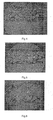
Fig. 2 illustrates the fluorescent photo of the scaffold with antibody of the invention; -
Fig. 3 illustrates 3-amino-9-ethyl-carbazole (AEC) staining photo of the 316L stainless steel stent with holes of the invention; -
Fig. 4 illustrates 3-amino-9-ethyl-carbazole (AEC) staining photo of the scaffold with antibody of the invention; -
Fig. 5 illustrates the staining result of the scaffold with antibody before it is preinstalled after being scoured for 24h of the invention; -
Fig. 6 illustrates staining result of the scaffold with antibody after scoured for 24h after the balloon dilation of the invention; -
Fig. 7 illustrates the control of the relative contents of antibodies fixed with the holes under different craft conditions of the invention. - A method for fixing antibody on the surface of medical instrument, mainly includes ① pre-treating the instrument surface, ② preparing holes, ③ post-treating the instrument surface, ④ fixing the antibody, and ⑤ detecting the antibody activity, etc; first, preparing the surface which has holes of the same size 100 ∼ 1000 nm and multicrystal phase structure in the surface of the medical instrument, such as the 316L stainless steel bare metal scaffold, by chemical corrosion, electrochemical corrosion, anodic oxidation, micro-arc oxidation, or micro-arc nitridation. As shown in
Fig. 1 , it is the scanning electron microscope photo of the processed surface of 316L stainless steel stent, wherein: - ① pre-treating the instrument surface: washing the surface of the instrument such as stainless steel bare scaffold by ultrasonic to remove impurities, using analytical reagent acetone solution at a concentration of 99.5% or medical ethanol solvent at a concentration of 75% to wash the scaffold body material for 5 ∼ 15min using 28 ∼ 100kHz ultrasonic to remove the impurities on the body material, putting the washed body material into a dryer to dry for 30 - 60min at 30 ∼ 40°C, and then taking out the body material left to be used.
- ② preparing holes: directly preparing holes of the same size in the instrument by acid solution corrosion; immersing the instrument material into the 0 ~ 100 °C, corrosion solution; and the corrosion solution is preferably hydrochloric acid at concentration of 1 - 38% or the hydrochloric acid mixed acid solution containing 1 ∼ 38% hydrochloric acid mixed with 1 - 98% vitriol; and then forming single nanometer-size holes after corrosion for 1 min ∼ 480h; and the corrosion time varies according to the concentration and temperature.
- ③ post-treating the instrument surface: using 99.5% concentration of analytical reagent acetone solution and distilled water to wash the body material for 5 ∼ 15min in succession by 28 ∼ 100kHz ultrasonic; and finally putting the washed body material into a dryer to dry for 30 60min at 30 ∼ 40°C, and then taking out the body material left to be used, or preparing 1 ∼ 38% concentration of hydrochloric acid solution with distilled water and then immersing the body material into the prepared solution and putting the solution containing the body material into an incubator at 20°C and then taking out the body material after 30min ~ 48h.
- ④ fixing the antibody: immersing the bare metal scaffold which has holes in its surface into a buffer solution containing antibody, adjusting the pH value of the antibody buffer solution, fixing the antibody on the surface of the instrument by the attraction between positive and negative charge and hole effect, and promoting the degree to which the antibody is firmly fixed on the surface of the instrument by the depth and size of the holes in the surface of the bare metal scaffold. The method can fix the antibody firmly and keep high activity of the antibody on the scaffold surface;
- ⑤ detecting the antibody activity: confirming the effectiveness of the fixed antibody by artificial simulation hemodynamics and detecting method of antibody activity in the scaffold surface.
- The instrument includes scaffold, catheter, guide wire, heart pacemakers, heart valves, surgical implant materials, implant hard tissue, and non-metal medical instruments whose substrate is ceramics, organic polymer, inorganic substance or metal oxide; the scaffold is balloon-expandable, self-expanding, vessel or non-vessel stent whose substrate is stainless steel, nickel-titanium memory alloy, cobalt-based alloy, titanium or titanium alloy; and the scaffold is made by wire weaving, tube laser cutting, die casting or welding.
- The biological product includes one or more of the following substances: heparin, anti-platelet membrane glycoprotein (GPIIb / IIIa) receptor antagonists, antibody-treating cancer drug, abciximab, biological peptides, CD34 antibody, CD31 antibody and CD133 antibody.
- Hereafter the preferable examples of the present invention will be given:
- The method of fixing antibody CD34 on the scaffold surface based on the antigen-antibody binding reaction and the fluorescence detection of the antibody activity have the steps as follows:
- (1) immersing the 316L stainless steel stent which has holes in its surface into a phosphate buffer solution or a carbonate buffer solution, whose pH is 7.2 - 7.4, containing 0.1 ∼ 100µg/mL CD34 antibody such as type II mouse anti-human CD34 antibody produced by U.S. Stem Cell Technology Co., Ltd., to incubate for 30min ∼ 2h at 25 - 37°C and then taking out the scaffold;
- (2) washing the scaffold 3 times for 5min per time using a phosphate buffer solution, whose pH is 7.2 ∼ 7.4, and blowing the scaffold for 15min at room temperature 25°C and then preserving the scaffold at 4°C after it becomes dry.
- (3) immersing the scaffold which has antibody on its surface into a phosphate buffer solution, whose pH is 7.2 ∼ 7.4, containing 500µL 5∼10 % bovine serum albumin ( BSA ) to incubate for 20 ∼ 30min at 25 ∼ 37 °C and then taking out the scaffold to dry in the air;
- (4) immersing the scaffold which has antibody on its surface into a phosphate buffer solution, whose pH is 7.2 - 7.4, produced by U.S. biology company containing 500µL 0.01∼10ng/µL human recombinant CD34 antigen for 20 - 30min at 25 - 37°C and then taking out the scaffold to dry in the air;
- (5) immersing the scaffold into a phosphate buffer solution, whose pH is 7.2 ∼ 7.4, containing 500µL 1µg/mL fluorescein isothiocyanate (FITC) labeled CD34 antibody such as type II mouse anti-human CD34 antibody produced by U.S. Stem Cell Technology Co., Ltd. to incubate for 20 - 30min at 25 - 37°C and then taking out the scaffold;
- (6) washing the scaffold 3 times for 5min per time by a phosphate buffer solution, whose pH is 7.2 ∼ 7.4, and then drying the scaffold;
- (7) taking fluorescence photography of the scaffold using fluorescence microscope, such as Japan CKX41-A32PH/FL Olympus fluorescence microscope and using the 316L stainless steel stent with holes as a control stent with the result shown in
Fig. 2 . - The method of fixing antibody CD34 on the scaffold surface based on the antigen-antibody binding reaction and the immunohistochemistry detection of the antibody activity, and the method of fixing antibody CD34 on the scaffold surface is the same as the
steps 1 and 2 disclosed in example 1; - (3) immersing the scaffold which has antibody on surface into the phosphate buffer solution containing 500µL 5∼10% bovine serum albumin ( BSA ) to incubate for 30min ∼ 2h at 25 - 37°C and then taking out the scaffold to dry in the air;
- (4) immersing the scaffold into a phosphate buffer solution containing horseradish peroxidase (HRP) labeled goat anti-mouse IgG to incubate for 20 - 30min at 25 - 37°C and then taking out the scaffold;
- (5) washing the scaffold 3 times for 5min per time by a phosphate buffer solution;
- (6) after using 3-amino-9-ethyl-carbazole (AEC) staining kit produced by China Huamei Bio-engineering Corporation for immunohistochemical staining, taking a photograph of the scaffold, and using the 316L stainless steel stent with holes as a control with the result shown in
Fig. 3 andFig. 4 . - The steps of hemodynamics simulation experiment of scaffold with antibody in artificial body flow, the method of fixing the antibody on the scaffold surface and the firm degree detection are as follows:
- (1) putting the scaffold with the fixed mouse anti-human CD34 antibody on its surface into fluid artificial simulated body flow, such as a phosphate buffer solution, whose pressure is 100mmHg, flow rate is 9 1 cm / s and pH is 7.2 ∼ 7.4, to make a scouring experiment, when the scaffold is at a normal state or at a state after the balloon dilation;
- (2) after the scaffold surface is scoured for 24 hours, detecting the activity of the antibody on the scaffold surface with the method disclosed in Example 2; the experimental results are shown in
Fig. 5 and Fig. 6 , which indicate that the method of adsorbing the antibody using the holes in the surface of the scaffold has the advantage of a high degree of firmness, and enables the antibody to be fixed on the scaffold surface even in the arterial blood flow in high-speed and to keep high activity. - It is the method of fixing the antibody on the scaffold surface and the quantitative detection of the antibody after being cascade amplified, and the steps thereof are as follows:
- (1) immersing the 316L stainless steel stent which has holes in its surface into a phosphate buffer solution, whose pH is 7.2 - 7.4, containing 0.1 ∼ 10µg/mL CD34 antibody such as type II mouse anti-human CD34 antibody produced by U.S. Stem Cell Technology Co., Ltd. to incubate for 30min ∼ 2h at 25 ∼ 37°C and then taking out the scaffold;
- (2) washing the scaffold 3 times for 5min per time using a phosphate buffer solution, whose pH is 7.2 - 7.4, and blowing the scaffold for 15min at room temperature 25°C and then preserving the scaffold at 4°C after it becomes dry.
- (3) immersing the scaffold which has antibody on its surface into a phosphate buffer solution, whose pH is 7.2 ∼ 7.4, containing 500µL 5∼10 % bovine serum albumin ( BSA ) to incubate for 20 - 30min at 25 - 37 °C and then taking out the scaffold to dry in the air;
- (4) immersing the scaffold into a phosphate buffer solution containing horseradish peroxidase (HRP) labeled goat anti-mouse IgG produced by China Huamei Bio-engineering Corporation to incubate for 20 - 30min at 25 - 37°C and then taking out the scaffold;
- (5) washing the scaffold 3 times for 5min per time by a phosphate buffer solution; the step of washing is putting the scaffold into 500µL TMB substrate buffer solution; the 500µ TMB substrate buffer solution is made of, for e. g., 0.5 mL anhydrous ethanol of 10 mg/5 mL mixed with 5mL substrate buffer solution; the substrate buffer solution is made of, for e. g., 1.42 g Na2HPO4 mixed with 0.96 g sodium citrate which is added distilled water to 50 mL; after washing, adding 32 µL hydrogen peroxide of 0.75% into the phosphate buffer solution to incubate for 15 minutes and then adding 250%L 2 M sulfuric acid solution to stop the reaction;
- (6) using the microplate reader to measure the absorbance of the solution (A492), taking the 316L stainless steel stent with holes as a control, coating CD34 antibody having a known concentration on an ELISA plate according to the different concentration gradients of 1:10, 1:20, 1:50, 1:100, 1:500, 1:1000, 1:10000 and 1:50000 at the same time, and detecting A492nm according to the above mentioned methods, and then making a simulation of the concentration of CD34 antibody with A492nm to draw a standard curve.
- (7) determining the quantities of the antibody on the scaffold surface according to the absorbance of the measured solution and the standard curve; the result showed that 316L stainless steel stent with holes can fix 1 ∼ 20 ng CD34 antibody for each 1mm in length.
- In order to detect the impact of the relative contents of different buffer solution on the antibody fixed on the surface of the vessel stent, example 5 makes the control of the relative contents of antibodies fixed with the holes under different craft conditions, and the steps thereof is as follows:
- (1) immersing the vessel stent of 12mm length which has holes into a phosphate buffer solution, whose pH is 7.2 - 7.4, or a carbonate buffer solution, whose pH is 9.6, containing 0.1 ∼ 10µg/mL CD34 antibody such as type II mouse anti-human CD34 antibody produced by U.S. Stem Cell Technology Co., Ltd. to incubate for 30min - 2h at 25 - 37°C and then taking out the scaffold to dry in the air;
- (2) determining the impact of different pH values on the relative contents of antibody fixed on the surface of the vessel stent with holes, and the result shown in
Fig. 7 indicates that the relative contents of the antibody fixed on the vessel stent in the carbonate solution are more than that in other solutions under the same conditions. - The present invention provides a method for fixing antibody on the surface of medical instruments. The method can promote the degree to which the antibody is firmly fixed on the surface of the instrument and keep high activity of the antibody on the surface of the medical instrument. The practicality in industry thereof is shown as follows:
- (1) It forms the surface with holes of the same size and the multicrystal phase structure in the surface of the medical instrument by chemical corrosion, electrochemical corrosion; the biological product antibody is fixed on the surface of the medical instrument by the effect of physical adsorption with a high degree of firmness for keeping high activity of the antibody on the surface of the medical instrument.
- (2) The formation of the holes makes it possible to fix the biological product antibody on the surface of the medical instrument well and thus gives full play to the role of the biological products in the prevention of in-stent restenosis and antithrombotic and overcomes the negative impacts brought about by the medical instrument in the process of using, such as in-stent restenosis and formation of antithrombotic, to bring gospel for patients with coronary atherosclerosis.
- (3) The present invention has simple craft and accurate and reliable detection results and can be widely applied to biochip and the surface of the medical instruments, such as vessel stents and orthopedic implant devices, for fixing the biological antibody.
Claims (9)
- A method for fixing antibody on the surface of a medical instrument, characterized in that the method includes① pre-treating the instrument surface by washing the instrument surface by ultrasonic to remove impurities, wherein analytical reagent acetone solution at a concentration of 99.5% or medical ethanol solvent at a concentration of 75% is used to wash the scaffold body material for 5∼15min using 28∼100kHz ultrasonic to remove the impurities on the body material, drying the washed body material in a dryer at 30~40°C, for 30∼60min, and then removing the body material from the dryer;② preparing multicrystal phase structure which has holes of the same size in the surface of the instrument by chemical corrosion, electrochemical corrosion, anodic oxidation, micro-arc oxidation, or micro-arc nitridation;③ post-treating the instrument surface by:using 99.5% analytical reagent acetone solution and distilled water in sequence to wash the body material for 5∼15min using 28∼100kHz ultrasonic; drying the washed body material in a dryer at 30~40°C, for 30∼60min, and then removing the body material from the dryer, orpreparing 1∼38% hydrochloric acid solution with distilled water, immersing the body material into the prepared solution, placing the solution, containing the body material into an incubator at 20°C for 30min∼48h, and then removing the body material from the incubator;④ fixing the antibody by immersing the bare metal scaffold which has holes in its surface into a buffer solution containing antibody for incubation, adjusting the pH value of the antibody buffer solution, whereby the antibody is fixed on the surface of the instrument by the attraction between positive and negative charge and the physical adsorption effect of the holes; and⑤ finally, confirming the effectiveness of the fixed antibody by artificial simulation hemodynamics and detecting the antibody activity on the instrument surface.
- The method according to claim 1, wherein the step ② of preparing holesof the same size in the instrument is undertaken by acid solution corrosion, wherein the instrument is immersed into the corrosion solution at 0~100°C, wherein the corrosion solution is preferably hydrochloric acid at concentration of 1∼38% or hydrochloric acid mixed acid solution containing 1~38% hydrochloric acid mixed with 1~98% vitriol; and then single nanometer-size holes are formed by corrosion for 1min∼480h, the corrosion time varying according to the concentration and temperature.
- The method according to claim 1, wherein the instrument includes scaffold, catheter, guide wire, heart pacemakers, heart valves, surgical implant materials, implant hard tissue, and non-metal medical instruments whose substrate is ceramics, organic polymer, inorganic substance or metal oxide; the scaffold is balloon-expandable, self-expanding, vessel or non-vessel stent, stent whose substrate is stainless steel, nickel-titanium memory alloy, cobalt-based alloy, titanium or titanium alloy; and the scaffold made by wire weaving, tube laser cutting, die casting or welding.
- The method according to claim 1, wherein the biological product antibodies include one or more of the following substances: heparin, anti-platelet membrane glycoprotein (GPIIb/IIIa) receptor antagonists, antibody-treating cancer drug, abciximab, biological peptides, CD34 antibody, CD31 antibody and CD133 antibody.
- The method according to claim 1, wherein the step ④ of fixing the antibody includes:(1) immersing the scaffold which has holes in its surface into a phosphate buffer solution or a carbonate buffer solution containing 0.1∼100µ g/mL CD34 antibody to incubate for 20∼60min at 25∼37°C and then taking out the instrument;(2) washing the scaffold 3 times for 5min per time using a phosphate buffer solution and blowing the instrument for 15min at room temperature and then preserving the instrument at 4°C after it becomes dry.
- The method according to claim 1, wherein the antibody activity after fixing of the antibody is ⑤ directly detected by performing fluorescence coloring through the specific reaction of CD34 antigen-antibody using fluorescein isothiocyanate (FITC) labeled mouse anti-human CD34 antibody and detecting the antibody activity on the instrument surface by photo-detection of fluorescence using fluorescence microscope.
- The method according to claim 1, wherein after the step ④ of fixing the antibody, the antibody activity is ⑥ indirectly detected by detecting the antibody activity on the scaffold surface using 3-amino-9-ethyl-carbazole (AEC) staining kit after the antibody is cascade amplified by horseradish peroxidase (HRP) labeled goat anti-mouse IgG secondary antibody.
- The method according to claim 1, wherein the step ④ of fixing the antibody, the content of the antibody on the surface is ⑦ detected by indirectly detecting the content of the antibody on the instrument surface by tetramethylbenzidine (TMB) chromogenic after cascade amplification of goat anti-mouse IgG secondary antibody labeled by horseradish peroxidase (HRP); the steps are as follows:(1) immersing the scaffold with antibody on its surface into a phosphate buffer solution containing 500 µL 5∼10% bovine serum albumin (BSA) to incubate at 25∼37°C for 30min∼2h and then taking out the scaffold to dry in the air;(2) immersing the scaffold into a phosphate buffer solution containing horseradish peroxidase (HRP) labeled goat anti-mouse IgG to incubate at 25∼37°C for 20∼30min and then taking out the scaffold;(3) washing the scaffold 3 times for 5min per time with a phosphate buffer solution; putting the scaffold into 500µL TMB substrate buffer solution; the 500 µ L TMB substrate buffer solution is made of, for e. g., 0.5 mL anhydrous ethanol of 10 mg/5 mL mixed with 5 mL substrate buffer solution; the substrate buffer solution is made of, for e. g., 1.42 g Na2HPO4 mixed with 0.96 g sodium citrate which is added distilled water to 50 mL; after washing, adding 32 µL hydrogen peroxide of 0.75% into a phosphate buffer solution to incubate for 15 minutes and then adding 250 µ L 2 M sulfuric acid solution to terminate the reaction;(4) using the microplate reader to measure the absorbance of the solution at 492nm; coating CD34 antibody having a known concentration on an ELISA plate according to the different concentration gradients of 1:10, 1:20, 1:50, 1:100, 1:500, 1:1000, and 1:10000, and then drawing a standard curve by simulation of the concentration of CD34 antibody with A492nm;(5) determining the amount of the antibody on the scaffold surface according to the measured absorbance and the standard curve.
- The method according to claim 1, wherein after the step ④ of fixing the antibody, which antibody is mouse anti-human CD34 antibody, the firmness of the antibody is ⑧ detected by: putting the scaffold that has the fixed mouse anti-human CD34 antibody on its surface into fluid artificial simulated body fluid, such as a phosphate buffer solution whose pressure is 100mmHg and flow rate is 91cm / s, performing a scouring experiment, when the scaffold is in a normal state or in a state after balloon dilation; finally, using the method of step ⑥ as defined in claim 7 to detect the antibody activity on the scaffold surface, and using the method of step ⑦ as defined in claim 8 to quantify the antibody on the scaffold.
Applications Claiming Priority (2)
| Application Number | Priority Date | Filing Date | Title |
|---|---|---|---|
| CN2007101076436A CN101310778B (en) | 2007-05-23 | 2007-05-23 | Method for fixing antibody on medical appliance |
| PCT/CN2007/002916 WO2008141495A1 (en) | 2007-05-23 | 2007-10-10 | A method for fixing antibody on the surface of medical instrument |
Publications (3)
| Publication Number | Publication Date |
|---|---|
| EP2163268A1 EP2163268A1 (en) | 2010-03-17 |
| EP2163268A4 EP2163268A4 (en) | 2012-10-24 |
| EP2163268B1 true EP2163268B1 (en) | 2014-01-22 |
Family
ID=40031390
Family Applications (1)
| Application Number | Title | Priority Date | Filing Date |
|---|---|---|---|
| EP07816530.5A Active EP2163268B1 (en) | 2007-05-23 | 2007-10-10 | A method for fixing antibody on the surface of medical instrument |
Country Status (4)
| Country | Link |
|---|---|
| US (1) | US8361536B2 (en) |
| EP (1) | EP2163268B1 (en) |
| CN (1) | CN101310778B (en) |
| WO (1) | WO2008141495A1 (en) |
Cited By (2)
| Publication number | Priority date | Publication date | Assignee | Title |
|---|---|---|---|---|
| US12150850B2 (en) | 2018-03-01 | 2024-11-26 | Titanium Textiles Ag | Tension-free titanium metal knitted fabric for surgically shaping soft tissues |
| US12186193B2 (en) | 2018-03-01 | 2025-01-07 | Titanium Textiles Ag | Titanium matrix based on a tension-free metal warp knit fabric for guided tissue regeneration |
Families Citing this family (14)
| Publication number | Priority date | Publication date | Assignee | Title |
|---|---|---|---|---|
| CN101310778B (en) | 2007-05-23 | 2012-06-06 | 乐普(北京)医疗器械股份有限公司 | Method for fixing antibody on medical appliance |
| US10642934B2 (en) | 2011-03-31 | 2020-05-05 | Microsoft Technology Licensing, Llc | Augmented conversational understanding architecture |
| US9842168B2 (en) | 2011-03-31 | 2017-12-12 | Microsoft Technology Licensing, Llc | Task driven user intents |
| US9760566B2 (en) | 2011-03-31 | 2017-09-12 | Microsoft Technology Licensing, Llc | Augmented conversational understanding agent to identify conversation context between two humans and taking an agent action thereof |
| CN102727991B (en) * | 2011-04-08 | 2014-10-01 | 乐普(北京)医疗器械股份有限公司 | Method for carrying out loading, drug fixing and controlled release on medical device surface |
| US9064006B2 (en) | 2012-08-23 | 2015-06-23 | Microsoft Technology Licensing, Llc | Translating natural language utterances to keyword search queries |
| CN104826177B (en) * | 2015-06-01 | 2017-11-28 | 巩晓东 | A kind of method for fixing hbv antibody in medical apparatus surface |
| CN106338301A (en) * | 2016-04-10 | 2017-01-18 | 天津海河标测技术检测有限公司 | Test method for confirming cleaning mode of reusable medical instrument |
| JP2021534878A (en) * | 2018-08-24 | 2021-12-16 | クヴァンテック アーゲー | Vascular instruments and methods for manufacturing vascular instruments |
| CN111249601A (en) * | 2018-11-30 | 2020-06-09 | 上海微创医疗器械(集团)有限公司 | Porous balloon and preparation method thereof |
| CN111304427B (en) * | 2020-02-26 | 2022-03-08 | 江苏康瑞新材料科技股份有限公司 | Processing technology of martensitic stainless steel medical instrument parts |
| CN111304415B (en) * | 2020-02-26 | 2022-03-04 | 江苏康瑞新材料科技股份有限公司 | Processing technology of austenitic stainless steel medical device parts |
| CN111647045B (en) * | 2020-06-17 | 2022-05-27 | 清华大学深圳国际研究生院 | CD133 antagonistic polypeptide, derivative and application thereof |
| CN114910542B (en) * | 2022-04-28 | 2023-09-22 | 中国民航大学 | Electrochemical anti-fouling sensor based on platelet membrane and its construction method and its application in detecting CD44 |
Family Cites Families (12)
| Publication number | Priority date | Publication date | Assignee | Title |
|---|---|---|---|---|
| US5728420A (en) * | 1996-08-09 | 1998-03-17 | Medtronic, Inc. | Oxidative method for attachment of glycoproteins to surfaces of medical devices |
| US20060194008A1 (en) * | 1999-09-22 | 2006-08-31 | Princeton University | Devices with multiple surface functionality |
| US20100254900A1 (en) * | 2002-03-18 | 2010-10-07 | Campbell Phil G | Biocompatible polymers and Methods of use |
| US20040078090A1 (en) * | 2002-10-18 | 2004-04-22 | Francois Binette | Biocompatible scaffolds with tissue fragments |
| WO2004112863A1 (en) * | 2003-06-20 | 2004-12-29 | Mcgill University | Biodegradable membrane-covered implant comprising chitosan |
| US20060034884A1 (en) * | 2004-08-10 | 2006-02-16 | Stenzel Eric B | Coated medical device having an increased coating surface area |
| CN1278743C (en) * | 2004-08-13 | 2006-10-11 | 重庆大学 | Preparation method of drug-eluting vascular stent |
| CN200980747Y (en) | 2006-06-14 | 2007-11-28 | 北京乐普医疗器械有限公司 | Drug Release Structures for Drug Eluting Devices |
| CN200980748Y (en) | 2006-08-01 | 2007-11-28 | 北京乐普医疗器械有限公司 | Porous drug release structure of a drug eluting device |
| WO2008061431A1 (en) * | 2006-11-20 | 2008-05-29 | Lepu Medical Technology (Beijing) Co., Ltd. | Vessel stent with multi drug-coatings |
| CN100435861C (en) * | 2006-11-27 | 2008-11-26 | 中国医学科学院生物医学工程研究所 | Preparation method of drug-loaded or gene-loaded scaffold |
| CN101310778B (en) | 2007-05-23 | 2012-06-06 | 乐普(北京)医疗器械股份有限公司 | Method for fixing antibody on medical appliance |
-
2007
- 2007-05-23 CN CN2007101076436A patent/CN101310778B/en active Active
- 2007-10-10 US US12/593,110 patent/US8361536B2/en active Active
- 2007-10-10 EP EP07816530.5A patent/EP2163268B1/en active Active
- 2007-10-10 WO PCT/CN2007/002916 patent/WO2008141495A1/en not_active Ceased
Cited By (2)
| Publication number | Priority date | Publication date | Assignee | Title |
|---|---|---|---|---|
| US12150850B2 (en) | 2018-03-01 | 2024-11-26 | Titanium Textiles Ag | Tension-free titanium metal knitted fabric for surgically shaping soft tissues |
| US12186193B2 (en) | 2018-03-01 | 2025-01-07 | Titanium Textiles Ag | Titanium matrix based on a tension-free metal warp knit fabric for guided tissue regeneration |
Also Published As
| Publication number | Publication date |
|---|---|
| CN101310778B (en) | 2012-06-06 |
| US8361536B2 (en) | 2013-01-29 |
| WO2008141495A1 (en) | 2008-11-27 |
| EP2163268A1 (en) | 2010-03-17 |
| EP2163268A4 (en) | 2012-10-24 |
| CN101310778A (en) | 2008-11-26 |
| US20100112189A1 (en) | 2010-05-06 |
Similar Documents
| Publication | Publication Date | Title |
|---|---|---|
| EP2163268A1 (en) | A method for fixing antibody on the surface of medical instrument | |
| Wang et al. | A novel Ca-Mg-P/PDA composite coating of Mg alloys to improve corrosion resistance for orthopedic implant materials | |
| Liao et al. | A single nanowire sensor for intracellular glucose detection | |
| Chen et al. | Improving the in vitro cell differentiation and in vivo osseointegration of titanium dental implant through oxygen plasma immersion ion implantation treatment | |
| Huang et al. | Nitrogen plasma immersion ion implantation treatment to enhance corrosion resistance, bone cell growth, and antibacterial adhesion of Ti-6Al-4V alloy in dental applications | |
| Zhen et al. | A review on in vitro corrosion performance test of biodegradable metallic materials | |
| Shin et al. | Enhanced osteogenic differentiation of human mesenchymal stem cells on Ti surfaces with electrochemical nanopattern formation | |
| Fan et al. | Adhesion of neural cells on silicon wafer with nano-topographic surface | |
| Middleton et al. | Fibronectin silanized titanium alloy: a bioinductive and durable coating to enhance fibroblast attachment in vitro | |
| Säuberlich et al. | Cell culture tests for assessing the tolerance of soft tissue to variously modified titanium surfaces | |
| CN106404915A (en) | Real-time quantitative detection method of cell traction force | |
| Nagaraja et al. | Oxide layer formation, corrosion, and biocompatibility of nitinol cardiovascular devices | |
| Zhu et al. | Biocompatibility of Fe–O films synthesized by plasma immersion ion implantation and deposition | |
| Martin et al. | Characterization and analysis of osteopontin‐immobilized poly (2‐hydroxyethyl methacrylate) surfaces | |
| Mishra et al. | Surface functionalization of gallium nitride for biomedical implant applications | |
| Zhou et al. | Iron surface functionalization system-Iron oxide nanostructured arrays with polycaprolactone coatings: Biodegradation, cytocompatibility, and drug release behavior | |
| Alvarez-Lopez et al. | Improved cell adhesion to activated vapor silanization-biofunctionalized Ti-6Al-4V surfaces with ECM-derived oligopeptides | |
| Yue et al. | Dealloyed nano-porous TiCu coatings with controlled copper release for cardiovascular devices | |
| Du et al. | Amperometric alcohol sensors based on protein multilayers composed of avidin and biotin‐labeled alcohol oxidase | |
| CN102698323B (en) | Preparation method of anticlotting materials | |
| Itakura et al. | Osteocompatibility of platinum-plated titanium assessed in vitro | |
| EP2307594B1 (en) | Silicon-based biomimetic treatment for the osteointegration of metal substrates | |
| Erdogan et al. | Nanostructured 316L Stainless Steel Stent Surfaces Improve Corrosion Resistance, and Enhance Endothelization and Hemocompatibility | |
| Tarnowski et al. | Oxygen transport characteristics of refunctionalized fluoropolymeric membranes and their application in the design of biosensors based upon the Clark-type oxygen probe | |
| Deguchi et al. | Heterogeneous role of integrins in fibroblast response to small cyclic mechanical stimulus generated by a nanoporous gold actuator |
Legal Events
| Date | Code | Title | Description |
|---|---|---|---|
| PUAI | Public reference made under article 153(3) epc to a published international application that has entered the european phase |
Free format text: ORIGINAL CODE: 0009012 |
|
| 17P | Request for examination filed |
Effective date: 20090807 |
|
| AK | Designated contracting states |
Kind code of ref document: A1 Designated state(s): AT BE BG CH CY CZ DE DK EE ES FI FR GB GR HU IE IS IT LI LT LU LV MC MT NL PL PT RO SE SI SK TR |
|
| AX | Request for extension of the european patent |
Extension state: AL BA HR MK RS |
|
| DAX | Request for extension of the european patent (deleted) | ||
| REG | Reference to a national code |
Ref country code: DE Ref legal event code: R079 Ref document number: 602007034939 Country of ref document: DE Free format text: PREVIOUS MAIN CLASS: A61L0027000000 Ipc: A61L0027220000 |
|
| A4 | Supplementary search report drawn up and despatched |
Effective date: 20120920 |
|
| RIC1 | Information provided on ipc code assigned before grant |
Ipc: A61L 27/54 20060101ALI20120914BHEP Ipc: A61L 27/22 20060101AFI20120914BHEP Ipc: A61L 31/16 20060101ALI20120914BHEP Ipc: A61L 29/16 20060101ALI20120914BHEP Ipc: A61L 31/04 20060101ALI20120914BHEP Ipc: A61L 29/04 20060101ALI20120914BHEP Ipc: A61F 2/02 20060101ALI20120914BHEP |
|
| GRAP | Despatch of communication of intention to grant a patent |
Free format text: ORIGINAL CODE: EPIDOSNIGR1 |
|
| INTG | Intention to grant announced |
Effective date: 20130729 |
|
| GRAS | Grant fee paid |
Free format text: ORIGINAL CODE: EPIDOSNIGR3 |
|
| GRAA | (expected) grant |
Free format text: ORIGINAL CODE: 0009210 |
|
| AK | Designated contracting states |
Kind code of ref document: B1 Designated state(s): AT BE BG CH CY CZ DE DK EE ES FI FR GB GR HU IE IS IT LI LT LU LV MC MT NL PL PT RO SE SI SK TR |
|
| REG | Reference to a national code |
Ref country code: GB Ref legal event code: FG4D |
|
| REG | Reference to a national code |
Ref country code: CH Ref legal event code: EP |
|
| REG | Reference to a national code |
Ref country code: AT Ref legal event code: REF Ref document number: 650453 Country of ref document: AT Kind code of ref document: T Effective date: 20140215 |
|
| REG | Reference to a national code |
Ref country code: IE Ref legal event code: FG4D |
|
| REG | Reference to a national code |
Ref country code: DE Ref legal event code: R096 Ref document number: 602007034939 Country of ref document: DE Effective date: 20140227 |
|
| REG | Reference to a national code |
Ref country code: NL Ref legal event code: VDEP Effective date: 20140122 |
|
| REG | Reference to a national code |
Ref country code: AT Ref legal event code: MK05 Ref document number: 650453 Country of ref document: AT Kind code of ref document: T Effective date: 20140122 |
|
| REG | Reference to a national code |
Ref country code: LT Ref legal event code: MG4D |
|
| PG25 | Lapsed in a contracting state [announced via postgrant information from national office to epo] |
Ref country code: IS Free format text: LAPSE BECAUSE OF FAILURE TO SUBMIT A TRANSLATION OF THE DESCRIPTION OR TO PAY THE FEE WITHIN THE PRESCRIBED TIME-LIMIT Effective date: 20140522 Ref country code: LT Free format text: LAPSE BECAUSE OF FAILURE TO SUBMIT A TRANSLATION OF THE DESCRIPTION OR TO PAY THE FEE WITHIN THE PRESCRIBED TIME-LIMIT Effective date: 20140122 |
|
| PG25 | Lapsed in a contracting state [announced via postgrant information from national office to epo] |
Ref country code: AT Free format text: LAPSE BECAUSE OF FAILURE TO SUBMIT A TRANSLATION OF THE DESCRIPTION OR TO PAY THE FEE WITHIN THE PRESCRIBED TIME-LIMIT Effective date: 20140122 Ref country code: SE Free format text: LAPSE BECAUSE OF FAILURE TO SUBMIT A TRANSLATION OF THE DESCRIPTION OR TO PAY THE FEE WITHIN THE PRESCRIBED TIME-LIMIT Effective date: 20140122 Ref country code: NL Free format text: LAPSE BECAUSE OF FAILURE TO SUBMIT A TRANSLATION OF THE DESCRIPTION OR TO PAY THE FEE WITHIN THE PRESCRIBED TIME-LIMIT Effective date: 20140122 Ref country code: PT Free format text: LAPSE BECAUSE OF FAILURE TO SUBMIT A TRANSLATION OF THE DESCRIPTION OR TO PAY THE FEE WITHIN THE PRESCRIBED TIME-LIMIT Effective date: 20140522 Ref country code: ES Free format text: LAPSE BECAUSE OF FAILURE TO SUBMIT A TRANSLATION OF THE DESCRIPTION OR TO PAY THE FEE WITHIN THE PRESCRIBED TIME-LIMIT Effective date: 20140122 Ref country code: CY Free format text: LAPSE BECAUSE OF FAILURE TO SUBMIT A TRANSLATION OF THE DESCRIPTION OR TO PAY THE FEE WITHIN THE PRESCRIBED TIME-LIMIT Effective date: 20140122 Ref country code: FI Free format text: LAPSE BECAUSE OF FAILURE TO SUBMIT A TRANSLATION OF THE DESCRIPTION OR TO PAY THE FEE WITHIN THE PRESCRIBED TIME-LIMIT Effective date: 20140122 |
|
| PG25 | Lapsed in a contracting state [announced via postgrant information from national office to epo] |
Ref country code: BE Free format text: LAPSE BECAUSE OF FAILURE TO SUBMIT A TRANSLATION OF THE DESCRIPTION OR TO PAY THE FEE WITHIN THE PRESCRIBED TIME-LIMIT Effective date: 20140122 Ref country code: LV Free format text: LAPSE BECAUSE OF FAILURE TO SUBMIT A TRANSLATION OF THE DESCRIPTION OR TO PAY THE FEE WITHIN THE PRESCRIBED TIME-LIMIT Effective date: 20140122 |
|
| REG | Reference to a national code |
Ref country code: DE Ref legal event code: R097 Ref document number: 602007034939 Country of ref document: DE |
|
| PG25 | Lapsed in a contracting state [announced via postgrant information from national office to epo] |
Ref country code: DK Free format text: LAPSE BECAUSE OF FAILURE TO SUBMIT A TRANSLATION OF THE DESCRIPTION OR TO PAY THE FEE WITHIN THE PRESCRIBED TIME-LIMIT Effective date: 20140122 Ref country code: EE Free format text: LAPSE BECAUSE OF FAILURE TO SUBMIT A TRANSLATION OF THE DESCRIPTION OR TO PAY THE FEE WITHIN THE PRESCRIBED TIME-LIMIT Effective date: 20140122 Ref country code: RO Free format text: LAPSE BECAUSE OF FAILURE TO SUBMIT A TRANSLATION OF THE DESCRIPTION OR TO PAY THE FEE WITHIN THE PRESCRIBED TIME-LIMIT Effective date: 20140122 Ref country code: CZ Free format text: LAPSE BECAUSE OF FAILURE TO SUBMIT A TRANSLATION OF THE DESCRIPTION OR TO PAY THE FEE WITHIN THE PRESCRIBED TIME-LIMIT Effective date: 20140122 |
|
| PG25 | Lapsed in a contracting state [announced via postgrant information from national office to epo] |
Ref country code: PL Free format text: LAPSE BECAUSE OF FAILURE TO SUBMIT A TRANSLATION OF THE DESCRIPTION OR TO PAY THE FEE WITHIN THE PRESCRIBED TIME-LIMIT Effective date: 20140122 Ref country code: SK Free format text: LAPSE BECAUSE OF FAILURE TO SUBMIT A TRANSLATION OF THE DESCRIPTION OR TO PAY THE FEE WITHIN THE PRESCRIBED TIME-LIMIT Effective date: 20140122 |
|
| PLBE | No opposition filed within time limit |
Free format text: ORIGINAL CODE: 0009261 |
|
| STAA | Information on the status of an ep patent application or granted ep patent |
Free format text: STATUS: NO OPPOSITION FILED WITHIN TIME LIMIT |
|
| 26N | No opposition filed |
Effective date: 20141023 |
|
| REG | Reference to a national code |
Ref country code: DE Ref legal event code: R097 Ref document number: 602007034939 Country of ref document: DE Effective date: 20141023 |
|
| REG | Reference to a national code |
Ref country code: DE Ref legal event code: R119 Ref document number: 602007034939 Country of ref document: DE |
|
| PG25 | Lapsed in a contracting state [announced via postgrant information from national office to epo] |
Ref country code: LU Free format text: LAPSE BECAUSE OF FAILURE TO SUBMIT A TRANSLATION OF THE DESCRIPTION OR TO PAY THE FEE WITHIN THE PRESCRIBED TIME-LIMIT Effective date: 20141010 Ref country code: SI Free format text: LAPSE BECAUSE OF FAILURE TO SUBMIT A TRANSLATION OF THE DESCRIPTION OR TO PAY THE FEE WITHIN THE PRESCRIBED TIME-LIMIT Effective date: 20140122 Ref country code: MC Free format text: LAPSE BECAUSE OF FAILURE TO SUBMIT A TRANSLATION OF THE DESCRIPTION OR TO PAY THE FEE WITHIN THE PRESCRIBED TIME-LIMIT Effective date: 20140122 |
|
| REG | Reference to a national code |
Ref country code: CH Ref legal event code: PL |
|
| REG | Reference to a national code |
Ref country code: IE Ref legal event code: MM4A |
|
| PG25 | Lapsed in a contracting state [announced via postgrant information from national office to epo] |
Ref country code: LI Free format text: LAPSE BECAUSE OF NON-PAYMENT OF DUE FEES Effective date: 20141031 Ref country code: DE Free format text: LAPSE BECAUSE OF NON-PAYMENT OF DUE FEES Effective date: 20150501 Ref country code: CH Free format text: LAPSE BECAUSE OF NON-PAYMENT OF DUE FEES Effective date: 20141031 |
|
| REG | Reference to a national code |
Ref country code: FR Ref legal event code: ST Effective date: 20150630 |
|
| PG25 | Lapsed in a contracting state [announced via postgrant information from national office to epo] |
Ref country code: FR Free format text: LAPSE BECAUSE OF NON-PAYMENT OF DUE FEES Effective date: 20141031 |
|
| PG25 | Lapsed in a contracting state [announced via postgrant information from national office to epo] |
Ref country code: IE Free format text: LAPSE BECAUSE OF NON-PAYMENT OF DUE FEES Effective date: 20141010 |
|
| PG25 | Lapsed in a contracting state [announced via postgrant information from national office to epo] |
Ref country code: BG Free format text: LAPSE BECAUSE OF FAILURE TO SUBMIT A TRANSLATION OF THE DESCRIPTION OR TO PAY THE FEE WITHIN THE PRESCRIBED TIME-LIMIT Effective date: 20140122 |
|
| PG25 | Lapsed in a contracting state [announced via postgrant information from national office to epo] |
Ref country code: GR Free format text: LAPSE BECAUSE OF FAILURE TO SUBMIT A TRANSLATION OF THE DESCRIPTION OR TO PAY THE FEE WITHIN THE PRESCRIBED TIME-LIMIT Effective date: 20140423 Ref country code: IT Free format text: LAPSE BECAUSE OF FAILURE TO SUBMIT A TRANSLATION OF THE DESCRIPTION OR TO PAY THE FEE WITHIN THE PRESCRIBED TIME-LIMIT Effective date: 20140122 |
|
| PG25 | Lapsed in a contracting state [announced via postgrant information from national office to epo] |
Ref country code: MT Free format text: LAPSE BECAUSE OF FAILURE TO SUBMIT A TRANSLATION OF THE DESCRIPTION OR TO PAY THE FEE WITHIN THE PRESCRIBED TIME-LIMIT Effective date: 20140122 Ref country code: HU Free format text: LAPSE BECAUSE OF FAILURE TO SUBMIT A TRANSLATION OF THE DESCRIPTION OR TO PAY THE FEE WITHIN THE PRESCRIBED TIME-LIMIT; INVALID AB INITIO Effective date: 20071010 Ref country code: TR Free format text: LAPSE BECAUSE OF FAILURE TO SUBMIT A TRANSLATION OF THE DESCRIPTION OR TO PAY THE FEE WITHIN THE PRESCRIBED TIME-LIMIT Effective date: 20140122 |
|
| PGFP | Annual fee paid to national office [announced via postgrant information from national office to epo] |
Ref country code: GB Payment date: 20251022 Year of fee payment: 19 |