EP2162742B1 - Assay for detection of transient intracellular ca2+ - Google Patents
Assay for detection of transient intracellular ca2+ Download PDFInfo
- Publication number
- EP2162742B1 EP2162742B1 EP08789892.0A EP08789892A EP2162742B1 EP 2162742 B1 EP2162742 B1 EP 2162742B1 EP 08789892 A EP08789892 A EP 08789892A EP 2162742 B1 EP2162742 B1 EP 2162742B1
- Authority
- EP
- European Patent Office
- Prior art keywords
- camkii
- nr2b
- nr2a
- mls
- cells
- Prior art date
- Legal status (The legal status is an assumption and is not a legal conclusion. Google has not performed a legal analysis and makes no representation as to the accuracy of the status listed.)
- Not-in-force
Links
- 230000003834 intracellular effect Effects 0.000 title claims description 39
- 238000003556 assay Methods 0.000 title claims description 21
- 238000001514 detection method Methods 0.000 title claims description 10
- 230000001052 transient effect Effects 0.000 title claims description 8
- 238000000034 method Methods 0.000 claims description 32
- 230000000694 effects Effects 0.000 claims description 31
- 108090001041 N-Methyl-D-Aspartate Receptors Proteins 0.000 claims description 26
- 102000004868 N-Methyl-D-Aspartate Receptors Human genes 0.000 claims description 24
- 239000011575 calcium Substances 0.000 claims description 24
- 108010043121 Green Fluorescent Proteins Proteins 0.000 claims description 21
- 102000004657 Calcium-Calmodulin-Dependent Protein Kinase Type 2 Human genes 0.000 claims description 19
- 108010003721 Calcium-Calmodulin-Dependent Protein Kinase Type 2 Proteins 0.000 claims description 19
- 235000018102 proteins Nutrition 0.000 claims description 16
- 102000004169 proteins and genes Human genes 0.000 claims description 16
- 108090000623 proteins and genes Proteins 0.000 claims description 16
- 101001047093 Homo sapiens Potassium voltage-gated channel subfamily H member 1 Proteins 0.000 claims description 13
- 102100022810 Potassium voltage-gated channel subfamily H member 1 Human genes 0.000 claims description 13
- 102000003566 TRPV1 Human genes 0.000 claims description 13
- 101150016206 Trpv1 gene Proteins 0.000 claims description 13
- 239000005090 green fluorescent protein Substances 0.000 claims description 13
- 108010005456 AMPA 4 glutamate receptor ionotropic Proteins 0.000 claims description 12
- 102100030668 Glutamate receptor 4 Human genes 0.000 claims description 12
- 102000004144 Green Fluorescent Proteins Human genes 0.000 claims description 12
- 238000012217 deletion Methods 0.000 claims description 12
- 230000037430 deletion Effects 0.000 claims description 12
- 230000001419 dependent effect Effects 0.000 claims description 11
- 238000001890 transfection Methods 0.000 claims description 10
- 125000000539 amino acid group Chemical group 0.000 claims description 9
- 230000002438 mitochondrial effect Effects 0.000 claims description 9
- 239000013612 plasmid Substances 0.000 claims description 9
- OYPRJOBELJOOCE-UHFFFAOYSA-N Calcium Chemical compound [Ca] OYPRJOBELJOOCE-UHFFFAOYSA-N 0.000 claims description 7
- 229910052791 calcium Inorganic materials 0.000 claims description 7
- 102000005962 receptors Human genes 0.000 claims description 7
- 108020003175 receptors Proteins 0.000 claims description 7
- 239000013598 vector Substances 0.000 claims description 7
- 108091006146 Channels Proteins 0.000 claims description 6
- 108010076504 Protein Sorting Signals Proteins 0.000 claims description 6
- 102000037865 fusion proteins Human genes 0.000 claims description 6
- 108020001507 fusion proteins Proteins 0.000 claims description 6
- 230000026731 phosphorylation Effects 0.000 claims description 6
- 238000006366 phosphorylation reaction Methods 0.000 claims description 6
- 238000011002 quantification Methods 0.000 claims description 6
- 102000034573 Channels Human genes 0.000 claims description 5
- 230000004807 localization Effects 0.000 claims description 5
- 230000004927 fusion Effects 0.000 claims description 4
- 238000003384 imaging method Methods 0.000 claims description 4
- 239000002773 nucleotide Substances 0.000 claims description 4
- 125000003729 nucleotide group Chemical group 0.000 claims description 4
- 230000035945 sensitivity Effects 0.000 claims description 4
- 230000008685 targeting Effects 0.000 claims description 4
- 108020004635 Complementary DNA Proteins 0.000 claims description 3
- 241000255581 Drosophila <fruit fly, genus> Species 0.000 claims description 3
- 108090000013 Voltage-Gated Potassium Channels Proteins 0.000 claims description 3
- 102000003734 Voltage-Gated Potassium Channels Human genes 0.000 claims description 3
- 102200126591 c.125A>G Human genes 0.000 claims description 3
- 238000003366 endpoint assay Methods 0.000 claims description 3
- 230000000638 stimulation Effects 0.000 claims description 3
- 210000003734 kidney Anatomy 0.000 claims description 2
- 235000004252 protein component Nutrition 0.000 claims description 2
- 230000002441 reversible effect Effects 0.000 claims description 2
- 102100029458 Glutamate receptor ionotropic, NMDA 2A Human genes 0.000 claims 10
- 108091008634 hepatocyte nuclear factors 4 Proteins 0.000 claims 10
- 102100022630 Glutamate receptor ionotropic, NMDA 2B Human genes 0.000 claims 5
- 108010038912 Retinoid X Receptors Proteins 0.000 claims 5
- 102000014649 NMDA glutamate receptor activity proteins Human genes 0.000 claims 2
- 238000012544 monitoring process Methods 0.000 claims 1
- 210000004027 cell Anatomy 0.000 description 66
- 230000009460 calcium influx Effects 0.000 description 12
- 239000012981 Hank's balanced salt solution Substances 0.000 description 11
- 230000004913 activation Effects 0.000 description 8
- YKPUWZUDDOIDPM-SOFGYWHQSA-N capsaicin Chemical compound COC1=CC(CNC(=O)CCCC\C=C\C(C)C)=CC=C1O YKPUWZUDDOIDPM-SOFGYWHQSA-N 0.000 description 8
- 239000003112 inhibitor Substances 0.000 description 7
- 238000005259 measurement Methods 0.000 description 7
- 229930195712 glutamate Natural products 0.000 description 6
- 230000005945 translocation Effects 0.000 description 6
- WHUUTDBJXJRKMK-VKHMYHEASA-N L-glutamic acid Chemical compound OC(=O)[C@@H](N)CCC(O)=O WHUUTDBJXJRKMK-VKHMYHEASA-N 0.000 description 5
- 210000000170 cell membrane Anatomy 0.000 description 5
- 238000002474 experimental method Methods 0.000 description 5
- 230000007274 generation of a signal involved in cell-cell signaling Effects 0.000 description 5
- PGHMRUGBZOYCAA-UHFFFAOYSA-N ionomycin Natural products O1C(CC(O)C(C)C(O)C(C)C=CCC(C)CC(C)C(O)=CC(=O)C(C)CC(C)CC(CCC(O)=O)C)CCC1(C)C1OC(C)(C(C)O)CC1 PGHMRUGBZOYCAA-UHFFFAOYSA-N 0.000 description 5
- PGHMRUGBZOYCAA-ADZNBVRBSA-N ionomycin Chemical compound O1[C@H](C[C@H](O)[C@H](C)[C@H](O)[C@H](C)/C=C/C[C@@H](C)C[C@@H](C)C(/O)=C/C(=O)[C@@H](C)C[C@@H](C)C[C@@H](CCC(O)=O)C)CC[C@@]1(C)[C@@H]1O[C@](C)([C@@H](C)O)CC1 PGHMRUGBZOYCAA-ADZNBVRBSA-N 0.000 description 5
- 108090000765 processed proteins & peptides Proteins 0.000 description 5
- VOROEQBFPPIACJ-UHFFFAOYSA-N 5-Phosphononorvaline Chemical compound OC(=O)C(N)CCCP(O)(O)=O VOROEQBFPPIACJ-UHFFFAOYSA-N 0.000 description 4
- RPXVIAFEQBNEAX-UHFFFAOYSA-N 6-Cyano-7-nitroquinoxaline-2,3-dione Chemical compound N1C(=O)C(=O)NC2=C1C=C([N+](=O)[O-])C(C#N)=C2 RPXVIAFEQBNEAX-UHFFFAOYSA-N 0.000 description 4
- -1 Ca2+ ions Chemical class 0.000 description 4
- 102000018899 Glutamate Receptors Human genes 0.000 description 4
- 108010027915 Glutamate Receptors Proteins 0.000 description 4
- DHMQDGOQFOQNFH-UHFFFAOYSA-N Glycine Chemical compound NCC(O)=O DHMQDGOQFOQNFH-UHFFFAOYSA-N 0.000 description 4
- 108010025083 TRPV1 receptor Proteins 0.000 description 4
- 229960002504 capsaicin Drugs 0.000 description 4
- 235000017663 capsaicin Nutrition 0.000 description 4
- 239000001963 growth medium Substances 0.000 description 4
- 239000002609 medium Substances 0.000 description 4
- 210000003470 mitochondria Anatomy 0.000 description 4
- 239000002953 phosphate buffered saline Substances 0.000 description 4
- 108010062740 TRPV Cation Channels Proteins 0.000 description 3
- 102000011040 TRPV Cation Channels Human genes 0.000 description 3
- DRCMAZOSEIMCHM-UHFFFAOYSA-N capsazepine Chemical compound C1C=2C=C(O)C(O)=CC=2CCCN1C(=S)NCCC1=CC=C(Cl)C=C1 DRCMAZOSEIMCHM-UHFFFAOYSA-N 0.000 description 3
- 239000003153 chemical reaction reagent Substances 0.000 description 3
- DEFVIWRASFVYLL-UHFFFAOYSA-N ethylene glycol bis(2-aminoethyl)tetraacetic acid Chemical compound OC(=O)CN(CC(O)=O)CCOCCOCCN(CC(O)=O)CC(O)=O DEFVIWRASFVYLL-UHFFFAOYSA-N 0.000 description 3
- 239000011521 glass Substances 0.000 description 3
- 230000004941 influx Effects 0.000 description 3
- 230000001537 neural effect Effects 0.000 description 3
- 230000004044 response Effects 0.000 description 3
- 238000002741 site-directed mutagenesis Methods 0.000 description 3
- JKMHFZQWWAIEOD-UHFFFAOYSA-N 2-[4-(2-hydroxyethyl)piperazin-1-yl]ethanesulfonic acid Chemical compound OCC[NH+]1CCN(CCS([O-])(=O)=O)CC1 JKMHFZQWWAIEOD-UHFFFAOYSA-N 0.000 description 2
- FWMNVWWHGCHHJJ-SKKKGAJSSA-N 4-amino-1-[(2r)-6-amino-2-[[(2r)-2-[[(2r)-2-[[(2r)-2-amino-3-phenylpropanoyl]amino]-3-phenylpropanoyl]amino]-4-methylpentanoyl]amino]hexanoyl]piperidine-4-carboxylic acid Chemical compound C([C@H](C(=O)N[C@H](CC(C)C)C(=O)N[C@H](CCCCN)C(=O)N1CCC(N)(CC1)C(O)=O)NC(=O)[C@H](N)CC=1C=CC=CC=1)C1=CC=CC=C1 FWMNVWWHGCHHJJ-SKKKGAJSSA-N 0.000 description 2
- UXVMQQNJUSDDNG-UHFFFAOYSA-L Calcium chloride Chemical compound [Cl-].[Cl-].[Ca+2] UXVMQQNJUSDDNG-UHFFFAOYSA-L 0.000 description 2
- OZLGRUXZXMRXGP-UHFFFAOYSA-N Fluo-3 Chemical compound CC1=CC=C(N(CC(O)=O)CC(O)=O)C(OCCOC=2C(=CC=C(C=2)C2=C3C=C(Cl)C(=O)C=C3OC3=CC(O)=C(Cl)C=C32)N(CC(O)=O)CC(O)=O)=C1 OZLGRUXZXMRXGP-UHFFFAOYSA-N 0.000 description 2
- 239000004471 Glycine Substances 0.000 description 2
- 239000007995 HEPES buffer Substances 0.000 description 2
- 102000004310 Ion Channels Human genes 0.000 description 2
- 108090000862 Ion Channels Proteins 0.000 description 2
- HOKKHZGPKSLGJE-GSVOUGTGSA-N N-Methyl-D-aspartic acid Chemical compound CN[C@@H](C(O)=O)CC(O)=O HOKKHZGPKSLGJE-GSVOUGTGSA-N 0.000 description 2
- 229930040373 Paraformaldehyde Natural products 0.000 description 2
- WCUXLLCKKVVCTQ-UHFFFAOYSA-M Potassium chloride Chemical compound [Cl-].[K+] WCUXLLCKKVVCTQ-UHFFFAOYSA-M 0.000 description 2
- FAPWRFPIFSIZLT-UHFFFAOYSA-M Sodium chloride Chemical compound [Na+].[Cl-] FAPWRFPIFSIZLT-UHFFFAOYSA-M 0.000 description 2
- 239000000872 buffer Substances 0.000 description 2
- 239000001110 calcium chloride Substances 0.000 description 2
- 229910001628 calcium chloride Inorganic materials 0.000 description 2
- 239000002738 chelating agent Substances 0.000 description 2
- 239000002299 complementary DNA Substances 0.000 description 2
- QLTXKCWMEZIHBJ-PJGJYSAQSA-N dizocilpine maleate Chemical compound OC(=O)\C=C/C(O)=O.C12=CC=CC=C2[C@]2(C)C3=CC=CC=C3C[C@H]1N2 QLTXKCWMEZIHBJ-PJGJYSAQSA-N 0.000 description 2
- 239000003596 drug target Substances 0.000 description 2
- 210000002472 endoplasmic reticulum Anatomy 0.000 description 2
- 239000013604 expression vector Substances 0.000 description 2
- 238000002866 fluorescence resonance energy transfer Methods 0.000 description 2
- YFHXZQPUBCBNIP-UHFFFAOYSA-N fura-2 Chemical compound CC1=CC=C(N(CC(O)=O)CC(O)=O)C(OCCOC=2C(=CC=3OC(=CC=3C=2)C=2OC(=CN=2)C(O)=O)N(CC(O)=O)CC(O)=O)=C1 YFHXZQPUBCBNIP-UHFFFAOYSA-N 0.000 description 2
- 239000008187 granular material Substances 0.000 description 2
- PNDZEEPOYCVIIY-UHFFFAOYSA-N indo-1 Chemical compound CC1=CC=C(N(CC(O)=O)CC(O)=O)C(OCCOC=2C(=CC=C(C=2)C=2N=C3[CH]C(=CC=C3C=2)C(O)=O)N(CC(O)=O)CC(O)=O)=C1 PNDZEEPOYCVIIY-UHFFFAOYSA-N 0.000 description 2
- 230000001939 inductive effect Effects 0.000 description 2
- 230000002427 irreversible effect Effects 0.000 description 2
- 239000007788 liquid Substances 0.000 description 2
- 229920002521 macromolecule Polymers 0.000 description 2
- 230000025608 mitochondrion localization Effects 0.000 description 2
- 210000000633 nuclear envelope Anatomy 0.000 description 2
- 229920002866 paraformaldehyde Polymers 0.000 description 2
- 230000004962 physiological condition Effects 0.000 description 2
- 230000002035 prolonged effect Effects 0.000 description 2
- 230000004850 protein–protein interaction Effects 0.000 description 2
- 230000007727 signaling mechanism Effects 0.000 description 2
- 239000000243 solution Substances 0.000 description 2
- UCSJYZPVAKXKNQ-HZYVHMACSA-N streptomycin Chemical compound CN[C@H]1[C@H](O)[C@@H](O)[C@H](CO)O[C@H]1O[C@@H]1[C@](C=O)(O)[C@H](C)O[C@H]1O[C@@H]1[C@@H](NC(N)=N)[C@H](O)[C@@H](NC(N)=N)[C@H](O)[C@H]1O UCSJYZPVAKXKNQ-HZYVHMACSA-N 0.000 description 2
- 210000000225 synapse Anatomy 0.000 description 2
- KZDCMKVLEYCGQX-UDPGNSCCSA-N 2-(diethylamino)ethyl 4-aminobenzoate;(2s,5r,6r)-3,3-dimethyl-7-oxo-6-[(2-phenylacetyl)amino]-4-thia-1-azabicyclo[3.2.0]heptane-2-carboxylic acid;hydrate Chemical group O.CCN(CC)CCOC(=O)C1=CC=C(N)C=C1.N([C@H]1[C@H]2SC([C@@H](N2C1=O)C(O)=O)(C)C)C(=O)CC1=CC=CC=C1 KZDCMKVLEYCGQX-UDPGNSCCSA-N 0.000 description 1
- FWBHETKCLVMNFS-UHFFFAOYSA-N 4',6-Diamino-2-phenylindol Chemical compound C1=CC(C(=N)N)=CC=C1C1=CC2=CC=C(C(N)=N)C=C2N1 FWBHETKCLVMNFS-UHFFFAOYSA-N 0.000 description 1
- 108090000078 AMPA Receptors Proteins 0.000 description 1
- 102000003678 AMPA Receptors Human genes 0.000 description 1
- 102000019025 Calcium-Calmodulin-Dependent Protein Kinases Human genes 0.000 description 1
- 108010026870 Calcium-Calmodulin-Dependent Protein Kinases Proteins 0.000 description 1
- 235000008534 Capsicum annuum var annuum Nutrition 0.000 description 1
- 240000008574 Capsicum frutescens Species 0.000 description 1
- 238000001712 DNA sequencing Methods 0.000 description 1
- 239000006144 Dulbecco’s modified Eagle's medium Substances 0.000 description 1
- 102000003688 G-Protein-Coupled Receptors Human genes 0.000 description 1
- 108090000045 G-Protein-Coupled Receptors Proteins 0.000 description 1
- 238000012218 Kunkel's method Methods 0.000 description 1
- 108050006807 Nicotinic acetylcholine receptors Proteins 0.000 description 1
- 102100029613 Transient receptor potential cation channel subfamily V member 1 Human genes 0.000 description 1
- 108050004388 Transient receptor potential cation channel subfamily V member 1 Proteins 0.000 description 1
- 238000009825 accumulation Methods 0.000 description 1
- 230000002776 aggregation Effects 0.000 description 1
- 238000004220 aggregation Methods 0.000 description 1
- APKFDSVGJQXUKY-INPOYWNPSA-N amphotericin B Chemical compound O[C@H]1[C@@H](N)[C@H](O)[C@@H](C)O[C@H]1O[C@H]1/C=C/C=C/C=C/C=C/C=C/C=C/C=C/[C@H](C)[C@@H](O)[C@@H](C)[C@H](C)OC(=O)C[C@H](O)C[C@H](O)CC[C@@H](O)[C@H](O)C[C@H](O)C[C@](O)(C[C@H](O)[C@H]2C(O)=O)O[C@H]2C1 APKFDSVGJQXUKY-INPOYWNPSA-N 0.000 description 1
- 239000003242 anti bacterial agent Substances 0.000 description 1
- 229940088710 antibiotic agent Drugs 0.000 description 1
- 210000003050 axon Anatomy 0.000 description 1
- 230000015572 biosynthetic process Effects 0.000 description 1
- 238000006555 catalytic reaction Methods 0.000 description 1
- 238000004113 cell culture Methods 0.000 description 1
- 230000001413 cellular effect Effects 0.000 description 1
- 238000010367 cloning Methods 0.000 description 1
- 238000012761 co-transfection Methods 0.000 description 1
- 210000000172 cytosol Anatomy 0.000 description 1
- 238000011161 development Methods 0.000 description 1
- 239000012973 diazabicyclooctane Substances 0.000 description 1
- LOKCTEFSRHRXRJ-UHFFFAOYSA-I dipotassium trisodium dihydrogen phosphate hydrogen phosphate dichloride Chemical compound P(=O)(O)(O)[O-].[K+].P(=O)(O)([O-])[O-].[Na+].[Na+].[Cl-].[K+].[Cl-].[Na+] LOKCTEFSRHRXRJ-UHFFFAOYSA-I 0.000 description 1
- BNIILDVGGAEEIG-UHFFFAOYSA-L disodium hydrogen phosphate Chemical compound [Na+].[Na+].OP([O-])([O-])=O BNIILDVGGAEEIG-UHFFFAOYSA-L 0.000 description 1
- WHUUTDBJXJRKMK-VKHMYHEASA-L glutamate group Chemical group N[C@@H](CCC(=O)[O-])C(=O)[O-] WHUUTDBJXJRKMK-VKHMYHEASA-L 0.000 description 1
- 238000011534 incubation Methods 0.000 description 1
- 230000003993 interaction Effects 0.000 description 1
- 210000004020 intracellular membrane Anatomy 0.000 description 1
- 239000002555 ionophore Substances 0.000 description 1
- 230000000236 ionophoric effect Effects 0.000 description 1
- 230000005923 long-lasting effect Effects 0.000 description 1
- 230000001404 mediated effect Effects 0.000 description 1
- 239000012528 membrane Substances 0.000 description 1
- 239000002207 metabolite Substances 0.000 description 1
- 230000035772 mutation Effects 0.000 description 1
- 210000003463 organelle Anatomy 0.000 description 1
- 239000011148 porous material Substances 0.000 description 1
- 239000001103 potassium chloride Substances 0.000 description 1
- 235000011164 potassium chloride Nutrition 0.000 description 1
- GNSKLFRGEWLPPA-UHFFFAOYSA-M potassium dihydrogen phosphate Chemical compound [K+].OP(O)([O-])=O GNSKLFRGEWLPPA-UHFFFAOYSA-M 0.000 description 1
- 230000003578 releasing effect Effects 0.000 description 1
- 239000002356 single layer Substances 0.000 description 1
- 239000011780 sodium chloride Substances 0.000 description 1
- 229960005322 streptomycin Drugs 0.000 description 1
- 230000002123 temporal effect Effects 0.000 description 1
- IMNIMPAHZVJRPE-UHFFFAOYSA-N triethylenediamine Chemical compound C1CN2CCN1CC2 IMNIMPAHZVJRPE-UHFFFAOYSA-N 0.000 description 1
- 238000009827 uniform distribution Methods 0.000 description 1
Images





Classifications
-
- C—CHEMISTRY; METALLURGY
- C12—BIOCHEMISTRY; BEER; SPIRITS; WINE; VINEGAR; MICROBIOLOGY; ENZYMOLOGY; MUTATION OR GENETIC ENGINEERING
- C12N—MICROORGANISMS OR ENZYMES; COMPOSITIONS THEREOF; PROPAGATING, PRESERVING, OR MAINTAINING MICROORGANISMS; MUTATION OR GENETIC ENGINEERING; CULTURE MEDIA
- C12N9/00—Enzymes; Proenzymes; Compositions thereof; Processes for preparing, activating, inhibiting, separating or purifying enzymes
- C12N9/10—Transferases (2.)
- C12N9/12—Transferases (2.) transferring phosphorus containing groups, e.g. kinases (2.7)
-
- C—CHEMISTRY; METALLURGY
- C07—ORGANIC CHEMISTRY
- C07K—PEPTIDES
- C07K14/00—Peptides having more than 20 amino acids; Gastrins; Somatostatins; Melanotropins; Derivatives thereof
- C07K14/435—Peptides having more than 20 amino acids; Gastrins; Somatostatins; Melanotropins; Derivatives thereof from animals; from humans
- C07K14/43504—Peptides having more than 20 amino acids; Gastrins; Somatostatins; Melanotropins; Derivatives thereof from animals; from humans from invertebrates
- C07K14/43595—Peptides having more than 20 amino acids; Gastrins; Somatostatins; Melanotropins; Derivatives thereof from animals; from humans from invertebrates from coelenteratae, e.g. medusae
-
- C—CHEMISTRY; METALLURGY
- C12—BIOCHEMISTRY; BEER; SPIRITS; WINE; VINEGAR; MICROBIOLOGY; ENZYMOLOGY; MUTATION OR GENETIC ENGINEERING
- C12Y—ENZYMES
- C12Y207/00—Transferases transferring phosphorus-containing groups (2.7)
- C12Y207/11—Protein-serine/threonine kinases (2.7.11)
- C12Y207/11001—Non-specific serine/threonine protein kinase (2.7.11.1), i.e. casein kinase or checkpoint kinase
-
- G—PHYSICS
- G01—MEASURING; TESTING
- G01N—INVESTIGATING OR ANALYSING MATERIALS BY DETERMINING THEIR CHEMICAL OR PHYSICAL PROPERTIES
- G01N33/00—Investigating or analysing materials by specific methods not covered by groups G01N1/00 - G01N31/00
- G01N33/48—Biological material, e.g. blood, urine; Haemocytometers
- G01N33/50—Chemical analysis of biological material, e.g. blood, urine; Testing involving biospecific ligand binding methods; Immunological testing
- G01N33/5005—Chemical analysis of biological material, e.g. blood, urine; Testing involving biospecific ligand binding methods; Immunological testing involving human or animal cells
-
- G—PHYSICS
- G01—MEASURING; TESTING
- G01N—INVESTIGATING OR ANALYSING MATERIALS BY DETERMINING THEIR CHEMICAL OR PHYSICAL PROPERTIES
- G01N33/00—Investigating or analysing materials by specific methods not covered by groups G01N1/00 - G01N31/00
- G01N33/48—Biological material, e.g. blood, urine; Haemocytometers
- G01N33/50—Chemical analysis of biological material, e.g. blood, urine; Testing involving biospecific ligand binding methods; Immunological testing
- G01N33/68—Chemical analysis of biological material, e.g. blood, urine; Testing involving biospecific ligand binding methods; Immunological testing involving proteins, peptides or amino acids
- G01N33/6872—Intracellular protein regulatory factors and their receptors, e.g. including ion channels
-
- C—CHEMISTRY; METALLURGY
- C07—ORGANIC CHEMISTRY
- C07K—PEPTIDES
- C07K2319/00—Fusion polypeptide
- C07K2319/60—Fusion polypeptide containing spectroscopic/fluorescent detection, e.g. green fluorescent protein [GFP]
-
- G—PHYSICS
- G01—MEASURING; TESTING
- G01N—INVESTIGATING OR ANALYSING MATERIALS BY DETERMINING THEIR CHEMICAL OR PHYSICAL PROPERTIES
- G01N2333/00—Assays involving biological materials from specific organisms or of a specific nature
- G01N2333/90—Enzymes; Proenzymes
- G01N2333/91—Transferases (2.)
- G01N2333/912—Transferases (2.) transferring phosphorus containing groups, e.g. kinases (2.7)
-
- G—PHYSICS
- G01—MEASURING; TESTING
- G01N—INVESTIGATING OR ANALYSING MATERIALS BY DETERMINING THEIR CHEMICAL OR PHYSICAL PROPERTIES
- G01N33/00—Investigating or analysing materials by specific methods not covered by groups G01N1/00 - G01N31/00
- G01N33/48—Biological material, e.g. blood, urine; Haemocytometers
- G01N33/50—Chemical analysis of biological material, e.g. blood, urine; Testing involving biospecific ligand binding methods; Immunological testing
- G01N33/84—Chemical analysis of biological material, e.g. blood, urine; Testing involving biospecific ligand binding methods; Immunological testing involving inorganic compounds or pH
Definitions
- the invention relates to detection and quantification of transient changes in intracellular metabolites by making use of a resulting protein-protein interaction that is irreversible and longlasting.
- Increase in concentration of intracellular free Ca 2+ ions is a widely used signaling mechanism under various physiological conditions. Release of free Ca 2+ ions inside the cell happens either through plasma membrane channel proteins that allow Ca 2+ influx or by release from intracellular Ca 2+ stores. Since Ca 2+ release thus becomes the activity of a wide variety of proteins, methods for detection and measurement of intracellular free Ca 2+ are used as assays for many proteins that are potential drug targets. These methods should be capable of sensing the transient rise in intracellular [Ca 2+ ] and also of generating a detectable signal.
- Most of the commonly used methods for Ca 2+ sensing use molecules that exhibit changes in their fluorescence characteristics upon exposure to increase in [Ca 2+ ]. These include small molecular weight macromolecules such as fura-2, indo-1, fluo-3, and Calcium- Green ( U.S. Pat. Nos. 4,603,209 and 5,049,673 ) as well as proteins such as GFP (green fluorescent protein) family of proteins with a sensor peptide inserted within them ( U.S. Pat. No. 6,469,154 ) or the cameleon molecules of tandem GFP constructs with an intervening Ca 2+ sensing peptide sequence, that generate a FRET signal upon sensing free Ca 2+ ( U.S. Pat. No. 5,998,204 ).
- GFP green fluorescent protein family of proteins with a sensor peptide inserted within them
- U.S. Pat. No. 6,469,154 cameleon molecules of tandem GFP constructs with an intervening Ca 2+ sensing peptide sequence, that generate a FRET signal upon
- the main object of this invention is to detect intracellular Ca 2+ transients using simple end point assay.
- the other object is to develop a Ca 2+ sensing assay with broad applicability to many Ca 2+ channel proteins.
- Another object is to detect intracellular Ca 2+ transients using a simple method wherein the need for realtime measurements on live cells is avoided.
- This invention relates to a method using an end point assay for the quantitation of transient intracellular calcium with a set of pairs of protein components having different levels of sensitivity to the concentration of calcium, with broad applicability to many Ca 2+ channel proteins comprising:
- the present invention relates to a method using an assay for detecting intracellular binding between two proteins.
- the assay is useful for detecting intracellular release of free Ca 2+ .
- the transient Ca 2+ signal is captured by an irreversible protein-protein interaction so that the signal is converted into a form that can be preserved for days to even months. There is absolutely no requirement of any real time signal acquisition or live cell signal acquisition.
- Increase in concentration of intracellular free Ca 2+ ions is a widely used signaling mechanism under various physiological conditions. Release of free Ca 2+ ions inside the cell happens either through plasma membrane channel proteins that allow Ca 2+ influx or by release from intracellular Ca 2+ stores. Since Ca 2+ release thus becomes the activity of a wide variety of proteins, methods for detection and measurement of intracellular free Ca 2+ are used as assays for many proteins that are potential drug targets. These methods should be capable of sensing the transient rise in intracellular [Ca 2+ ] and also of generating a detectable signal.
- Most of the commonly used methods for Ca 2+ sensing use molecules that exhibit changes in their fluorescence characteristics upon exposure to increase in [Ca 2+ ]. These include small molecular weight macromolecules such as fura-2, indo-1, fluo-3, and Calcium-Green ( U.S. Pat. Nos. 4,603,209 and 5,049,673 ) as well as proteins such as GFP (green fluorescent protein) family of proteins with a sensor peptide inserted within them ( U.S: Pat. No. 6,469,154 ) or the cameleon molecules of tandem GFP constructs with an intervening Ca 2+ sensing peptide sequence, that generate a FRET signal upon sensing free Ca 2+ ( U.S. Pat. No. 5,998,204 ).
- GFP green fluorescent protein family of proteins with a sensor peptide inserted within them
- U.S: Pat. No. 6,469,154 cameleon molecules of tandem GFP constructs with an intervening Ca 2+ sensing peptide sequence, that generate a FRET signal upon
- Ca 2+ /calmodulin dependent protein kinase type II present at neuronal synapses, upon activation by increase in intracellular Ca 2+ , gets translocated to the NMDA-type glutamate receptor and binds irreversibly by its T-site (the Thr 286 -binding site) to the NR2B subunit of the receptor.
- a GFP-fusion of CaMKII (GFP-CaMKII) and a fusion of the binding sequence on NR2B with a mitochondrial targeting sequence (MLS-NR2B) upon co-transfection in HEK-293 cells, bind to each other in response to increase in intracellular Ca 2+ .
- FIG. 1 depicts schematically the appearance of punctae of green fluorescence in cells which had a uniform distribution of green fluorescence before stimulation with Ca 2+ .
- intracellular Ca 2+ influx was generated by adding the ionophore, ionomycin, which binds to the cell membrane and forms pores through which extracellular Ca 2+ flows in. This is a combination of two published methods ( Strack, S., McNeill, R. B., and Col bran, R. J., 2000, JBiol Chem 275, 23798-23806 ; Bayer, K.
- NR2A encode amino acid residues 1265-1301
- NR2B encoding amino acid residues 1271-1311
- NR2A and NR2B form parts of fusion proteins with a mitochondrial targeting signal sequence, a myc-epitope and GST at the N-terminal end (MLS-NR2A or MLS-NR2B).
- the sequences of the fusion constructs were confirmed by DNA sequencing of the NR2A or NR2B segments.
- the cDNA of a-CaMKII was amplified by PCR from pGEM2a ( Praseeda M., Pradeep K. K., Krupa A., Krishna S. S., Leena S., Kumar R. R., Cheriyan J., Mayadevi M., Srinivasan N. and Omkumar R. V., 2004, Biochem J, 378, 391-397 ) using forward primer having nucleotide sequence as set forth in SEQ ID NO: 1 and reverse primer having nucleotide sequence as set for the in SEQ ID NO: 2 to yield a PCR product with Kpn I and Bam HI sites at 5' and 3' ends respectively.
- the PCR product was cloned into pEGFP-C1 vector (Clontech) using the corresponding restriction sites in the multiple cloning site of the vector, which resulted in an N-terminal GFP-fusion of ⁇ -CaMKII (GFP- ⁇ -CaMKII).
- Site-directed mutagenesis was carried out using the QuikChange Site-Directed Mutagenesis Kit from Stratagene based on Kunkel's method ( Kunkel, T. A., 1985, Proc. Natl. Acad. Sci. USA 82, 488-492 ).
- the cDNA of ⁇ -CaMKII present in the expression vector pEGFP-C1- ⁇ -CaMKII served as the template to generate mutants.
- the NR2A or NR2B sequences present in the pSGH-NR2A or pSGH-NR2B expression vectors were also used as templates to generate mutants.
- HEK-293 (Human Embryonic Kidney) cells were cultured as monolayer in DMEM containing 10% FBS and antibiotics (penicillin 100 units/ml, streptomycin 100 ⁇ g/ml, fungizone 2.5 ⁇ g/ml) at 37°C in a humid atmosphere having 5% CO 2 .
- HEK-293 cells 1.5 x 10 4 HEK-293 cells were seeded on sterile 12 mm cover slips in 24 well plates (Nunc). After 24 hrs, the cells were cotransfected with plasmid constructs encoding GFP- ⁇ -CaMKII (0.1 ⁇ g/well) and either of MLS-NR2A or MLS-NR2B (0.25 ⁇ g/well), or pRK-NR2A or pRK-NR2B (along with pRc-NR1 for expression of functional NMDA receptor) using Lipofectamine reagent.
- HBSS Hank's balanced salt solution
- Appearance of perinuclear granules due to mitochondrial localization of GFP- ⁇ -CaMKII is taken as the signal for binding between GFP- ⁇ -CaMKII and MLS-NR2B.
- GFP- ⁇ -CaMKII and full-length NR2B accumulation of GFP-fluorescence in the membraneous structures such as nuclear membrane, endoplasmic reticulum membrane, plasma membrane, etc is taken as signal for binding.
- Data presented are for cells fixed immediately after incubation with 15 ⁇ M ionomycin (-Ca 2+ influx) or 2-5 minutes after Ca 2+ stimulus (+Ca 2+ influx).
- the total number of green fluorescent cells and cells which develop the signal for binding were counted manually for each microscopic field. Number of cells that develop signal for binding is represented as percentage of total number of green fluorescent cells (number of green fluorescent cells which develop signal for binding + number of green fluorescent cells which do not develop signal) in a field. Average for at least three separate fields was obtained for each experiment. Mean ⁇ standard deviation of data obtained from three such experiments is presented as values in inset in Figure 2 . Wherever images for lack of binding are presented, no cell was found to develop the perinuclear granules in repeated experiments and hence no quantitation data is presented.
- NMDA receptor For expression of NMDA receptor (NMDAR), the cells were cotransfected with plasmid constructs encoding GFP- ⁇ -CaMKII (0.1 ⁇ g/well) and NR1 (pRc-NR1 plasmid) and NR2B (pRK-NR2B plasmid) subunits of NMDAR (0.33 ⁇ g/well of pRc-NR1 and pRK-NR2B) using Lipofectamine (Invitrogen). When growth medium was added to the cells, it was supplemented with an inhibitor of NMDA receptor, either 1 mM of 2-amino-5-phosphovaleric acid (AP-5) or 10 ⁇ M MK-801.
- AP-5 2-amino-5-phosphovaleric acid
- HBSS Free Hank's balanced salt solution
- NMDAR containing NR2A subunit cells were co-transfected with plasmid constructs encoding GFP- ⁇ -CaMKII (0.1 ⁇ g/well) and NR1 (pRc-NR1 plasmid) and NR2A (pRK-NR2A plasmid) subunits of NMDAR (0.33 ⁇ g/well of pRc-NR1 and pRK-NR2A) along with MLS-NR2B (0.33 ⁇ g/well of pSGH-NR2B) and growth medium was not supplemented with AP-5. NMDAR activity was detected as described in the previous section.
- TRPV1 HEK-293. cells were cotransfected with TRPV1 (pCDNA3-TRPV1, about 0.2 ⁇ g/well), MLS-NR2B (pSGH-MLS-NR2B, about 0.33 ⁇ g/well) and GFP-CaMKII (pEGFP-CaMKII, about 0.1 ⁇ g/well) using lipofectamine reagent. After 48 hours, the transfected cells were subjected to TRPV1 activity assay. The growth medium was removed and the cells were washed with Ca 2+ and Mg free HBSS containing 400 ⁇ M EGTA.
- the receptor was activated with 3 ⁇ M or 10 nM of capsaicin with 2 mM Ca 2+ in HBSS without Mg 2+ for 5 minutes. Control without Ca 2+ during activation was also performed.
- HEK-293 cells were cotransfected with GluR4 (pRK5-GluR4, about 0.33 ⁇ g/well), MLS-NR2B (pSGH-MLS-NR2B, about 0.33 ⁇ g/well) and GFP-CaMKII (pEGFP-CaMKII, about 0.1 ⁇ g/well) using lipofectamine reagent.
- GluR4 pRK5-GluR4, about 0.33 ⁇ g/well
- MLS-NR2B pSGH-MLS-NR2B, about 0.33 ⁇ g/well
- GFP-CaMKII pEGFP-CaMKII, about 0.1 ⁇ g/well
- the GluR4 receptor was activated with HBSS solution containing 10 ⁇ M glutamate with or without 2 mM Ca 2+ for about 5 minutes.
- the inhibitor, CNQX (50 ⁇ M) when used, was preincubated for 3 minutes with cells in Ca 2+ and Mg 2+ -free HBSS followed by activation using the components as described above with 50 ⁇ M CNQX. Cells were then fixed and were mounted onto glass slides.
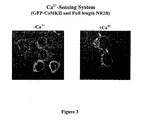
- the data in Figure 3 is obtained by transfecting cells with full-length NR2B instead of MLS-NR2B. Since full length NR2B is localized to nuclear and other intracellular membranes, GFP-CaMKII localizes to those membraneous structures instead of mitochondria, upon sensing increase in intracellular [Ca 2+ ].
- the inventors have found that the number of cells that develop the GFP-clustering signal is dependent on the concentration of extracellular Ca 2+ and hence the intracellular Ca 2+ ( Figure 6 ). It also depends on the efficiency of the binding between CaMKII and NR2B ( Figure 5 ). The inventors developed a method to express quantitatively the strength of the signal as mentioned in Methods section (Methodology for Transfection, Ca 2+ -stimulation and imaging). Data in Figure 4 is used to demonstrate this method.
- the cell count determined as mentioned in methods section as well as in the previous section, for the wild-type and T286D mutant forms of GFP- ⁇ -CaMKII are presented in Figure 5 .
- the T286D mutation which is known to enhance the Ca 2+ /calmoduin affinity of ⁇ -CaMKII also improved its efficiency of binding in the intracellular binding system as seen by the increased (more than two fold) cell count ( Figure 5 ).
- the voltage gated potassium channel of Drosophila (Eag) that is localised in synapses as well as in axons also interacts at the T-site of CaMKII ( Sun, X. X., Hodge, J. J., Zhou, Y., Nguyen, M., and Griffith, L. C. (2004) J Biol Chem 279, 10206-10214 ).
- the Eag amino acid residues 731-803 (or longer or shorter sequences) which comprise the CaMKII binding motif was cloned into the pSGH vector to obtain an MLS-Eag fusion that could be targeted to mitochondria.
- FIG. 7 shows the Ca 2+ -depednent localization of GFP-(WT)-CaMKII to MLS-Eag.
- This system could also be a Ca 2+ -sensor similar to the CaMKII-NR2B system.
- Any peptide sequence which binds to the T-site of CaMKII similar to NR2B could be used as a binding partner for CaMKII in a Ca 2+ -sensing system.
- the neuronal N-methyl-D-aspartate (NMDA)-type glutamate receptor is a glutamate activated Ca 2+ -channel that has NR1 and NR2B as subunits.
- Functional NMDAR can be formed by NR1 and NR2A subunit also instead of the NR1/NR2B combination.
- the NR2A subunit does not bind CaMKII, a translocation signal consequent to influx of Ca 2+ will not be obtained.
- By additionally transfecting the mitochondrially localized MLS-NR2B sequence it is possible to detect the activity of the NR2A-containing NMDAR also in a Ca 2+ -dependent manner.
- the arrows in Figure 9 show mitochondrially translocated GFP- ⁇ -CaMKII.
- Ca 2+ -channel proteins include Ca 2+ -channel proteins (nicotinic acetyl choline receptor channel ( Grando SA, Horton RM, Mauro TM, Kist DA, Lee TX, Dahl MV., J Invest. Dermatol., 1996, 107, p412-418 ), the vanilloid receptor 1 (TRPV1) ( Witte DG, Cassar SC, Masters JN, Esbenshade T, and Hancock AA. 2002, J Biomol Screen.
- Ca 2+ -channel proteins including vanilloid receptor 1 (TRPV1) ( Witte DG, Cassar SC, Masters JN, Esbenshade T, and Hancock AA. 2002, J Biomol Screen.
- G-protein coupled receptors Fujii, R., Hosoya, M., Fukusumi, S., Kawamata, Y., Habata, Y., Hinuma, S., Onda, H., Nishimura, O., and Fujino, M. 2000, J. Biol., Chem., 275, p21068-21074 ), etc. could be expressed in a heterologous cell culture system to generate specific Ca 2+ -channel activity.
- Vanilloid receptor is a Ca 2+ -channel that is activated by capsaicin, one of the active principle of hot chilli peppers ( Caterina et al., 1997, Nature, Vol. 389, p816-824 ).
- TRPV1 vanilloid receptor
- the signal generation was specific for transfection by TRPV1, activation by capsaicin and also for the presence of Ca 2+ in the extracellular medium. Signal generation due to activation of TRPV1 could be inhibited by the TRPV1-specific inhibitor capsazepine ( Fig. 10 ). All these results show that the activity of TRPV1 receptor is being detected by our invention.
- the homomeric AMPA-type glutamate receptor formed of the GluR4 subunit causes Ca 2+ -influx upon activation by glutamate ( Cushing, et al., 1999, J. Neurosci. Methods. Vol. 90, p33-36 ).
- glutamate Upon transfection of HEK-293 cells with the GluR4 subunit along with GFP-CaMKII and MLS-NR2B, it was possible to detect the Ca 2+ -channel activity of the receptor.
- the receptor was activated by treatment with glutamate which caused the formation of green punctae due to translocation of GFP-CaMKII to mitochondrially localized MLS-NR2B. Generation of the signal could be blocked by the specific inhibitor of the receptor, CNQX ( Fig. 11 ).
Landscapes
- Health & Medical Sciences (AREA)
- Life Sciences & Earth Sciences (AREA)
- Chemical & Material Sciences (AREA)
- Engineering & Computer Science (AREA)
- Molecular Biology (AREA)
- Biomedical Technology (AREA)
- Immunology (AREA)
- Organic Chemistry (AREA)
- General Health & Medical Sciences (AREA)
- Hematology (AREA)
- Biochemistry (AREA)
- Urology & Nephrology (AREA)
- Medicinal Chemistry (AREA)
- Zoology (AREA)
- Cell Biology (AREA)
- Genetics & Genomics (AREA)
- Biotechnology (AREA)
- Microbiology (AREA)
- Bioinformatics & Cheminformatics (AREA)
- Wood Science & Technology (AREA)
- Physics & Mathematics (AREA)
- Pathology (AREA)
- General Physics & Mathematics (AREA)
- Food Science & Technology (AREA)
- Analytical Chemistry (AREA)
- General Engineering & Computer Science (AREA)
- Tropical Medicine & Parasitology (AREA)
- Proteomics, Peptides & Aminoacids (AREA)
- Biophysics (AREA)
- Gastroenterology & Hepatology (AREA)
- Inorganic Chemistry (AREA)
- Toxicology (AREA)
- Measuring Or Testing Involving Enzymes Or Micro-Organisms (AREA)
- Peptides Or Proteins (AREA)
- Micro-Organisms Or Cultivation Processes Thereof (AREA)
Description
- The invention relates to detection and quantification of transient changes in intracellular metabolites by making use of a resulting protein-protein interaction that is irreversible and longlasting.
- Increase in concentration of intracellular free Ca2+ ions is a widely used signaling mechanism under various physiological conditions. Release of free Ca2+ ions inside the cell happens either through plasma membrane channel proteins that allow Ca2+ influx or by release from intracellular Ca2+ stores. Since Ca2+ release thus becomes the activity of a wide variety of proteins, methods for detection and measurement of intracellular free Ca2+ are used as assays for many proteins that are potential drug targets. These methods should be capable of sensing the transient rise in intracellular [Ca2+] and also of generating a detectable signal.
- Most of the commonly used methods for Ca2+ sensing use molecules that exhibit changes in their fluorescence characteristics upon exposure to increase in [Ca2+]. These include small molecular weight macromolecules such as fura-2, indo-1, fluo-3, and Calcium-Green (
U.S. Pat. Nos. 4,603,209 and5,049,673 ) as well as proteins such as GFP (green fluorescent protein) family of proteins with a sensor peptide inserted within them (U.S. Pat. No. 6,469,154 ) or the cameleon molecules of tandem GFP constructs with an intervening Ca2+ sensing peptide sequence, that generate a FRET signal upon sensing free Ca2+ (U.S. Pat. No. 5,998,204 ). In all these methods, the fluorescence signal is also as short-lived as the Ca2+-transient, and hence signal acquisition in real-time is mandatory. Moreover this also imposes rapid liquid dispensing. These factors not only necessitates the use of more expensive equipments but also limits the choice of equipments that can be used in addition to imposing severe constraints for carrying out these assays at high throughput scale.U.S. Pat. No. 6,514,709 disclosed a method that prolongs the duration of the Ca2+-transients by incorporating an intracellular chelator that alters the kinetics of signal generation, so that the signal is prolonged. However in all these methods, measurements will have to be done on cells in the live condition. - Pradeep KK et al Biochem J 2009, 419, 123 - 132 discusses the regulation of Ca2+/calmodulin-dependent protein kinase II catalysis by N-methyl-D-aspartate receptor subunit 2B.
- The main object of this invention is to detect intracellular Ca2+ transients using simple end point assay.
- The other object is to develop a Ca2+ sensing assay with broad applicability to many Ca2+ channel proteins.
- Another object is to detect intracellular Ca2+ transients using a simple method wherein the need for realtime measurements on live cells is avoided.
- This invention relates to a method using an end point assay for the quantitation of transient intracellular calcium with a set of pairs of protein components having different levels of sensitivity to the concentration of calcium, with broad applicability to many Ca2+ channel proteins comprising:
- i) the generation of expression constructs for the fusion proteins having the Ca2+/calmodulin dependent protein kinase II (CaMKII) phosphorylation sites of NR2A or NR2B subunits of N-methyl-D-aspartate receptor (NMDAR) or the voltage gated potassium channel of Drosophila (Eag) or any protein sequence which binds to the T-site of CaMKII, conjugated to a mitochondrial localizing signal sequence, or mutants of these sequences;
- ii) the generation of mammalian expression constructs of α-CaMKII as a chimera with green fluorescent protein (GFP-α-CaMKII) or its mutants; and
- iii) subjecting to the step of transfection, Ca2+ stimulation, imaging and quantification of the number of cells with a Ca2+-dependent signal;
wherein an NMDA receptor activity assay, TRPVI receptor activity assay or GluR4 receptor activity assay is performed to detect the activity of Ca2+ channel proteins; and
wherein the cDNAs of NR2A encoding amino acid residues 1265-1301, or NR2B encoding amino acid residues 1271-1311, or Eag encoding amino acid residues 731-803, harboring the phosphorylation site for CaMKII (Ser1291, Ser1303, Thr787) of NR2A, NR2B and Eag respectively are subcloned into PSGH vector; and
wherein the mutants of α-CaMKII and NR2B or NR2A are selected from:- T286D-α-CaMKII and mitochondrial localisation sequence (MLS)-WT-NR2B,
- F293E/N294D-α-CaMKII and MLS-WT-NR2B,
- MLS-(S1303A)-NR2B and WT-α-CaMKII,
- S1303A-NR2B and WT-α-CaMKII,
- K42R/T286D-α-CaMKII and MLS-WT-NR2B,
- MLS-DeltaINNR2A (deletion of Ile1286-Asn1287 motif of NR2A) and WT-α-CaMKII,
- MLS-(S1291A)/DeltaIN (deletion of Ile1286-Asn1287 motif of NR2A) NR2A and WT-α-CaMKII,
- MLS-DeltaINNR2A (deletion of Ile1286-Asn1287 motif of NR2A) and T286D-α-CaMKII,
- DeltaINNR2A (deletion of Ile1286-Asn1287 motif of NR2A) and T286D-α-CaMKII, and
- MLS-K1292A-NR2B and WT-GaMKII;
wherein a specific method of quantification is used by taking the total number of green fluorescent cells which develop punctae as a signal for intracellular calcium release and representing it as percentage of the total number of green fluorescent cells. -
-
Figure: 1 is a schematic representation of the assay system for detection of release of intracellular free Ca2+. -
Figure: 2 shows Ca2+-dependent generation of signal due to GFP-α-CaMKII translocation towards mitochondrially localized NR2B sequence (MLS-NR2B) observed as the appearance of punctae. -
Figure: 3 shows Ca2+-dependent generation of signal due to GFP-α-CaMKII translocation towards full-length NR2B localized on intracellular membranous organelles. -
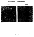
Figure: 4 shows two images of cells, one obtained after inducing Ca2+-influx to cells (Image 1) and the other obtained without inducing any Ca2+-influx (Image 2). -
Figure: 5 shows the qunatitation data obtained as described inFigure 4 , for the wild-type and T286D mutant forms of GFP-α-CaMKII. -
Figure: 6 shows the quantitation data obtained as described inFigure 4 , for the wild-type and T286D mutant forms of GFP-α-CaMKII, at different concentrations of Ca2+ in the extracellular medium. -
Figure: 7 shows Ca2+-dependent generation of signal due to GFP-α-CaMKII translocation towards mitochondrially localized Eag sequence (MLS-Eag) -
Figure: 8 shows signal generated due to Ca2+-influx through NMDA receptor formed of NR1 and NR2B subunits. -
Figure: 9 shows signal generated due to Ca2+-influx through NMDA receptor formed of NR1 and NR2A subunits. -
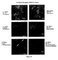
Figure: 10 shows signal generated due to Ca2+-influx through vanilloid Receptor (TRPV1) -
Figure: 11 shows signal generated due to Ca2+-influx through the AMPA-type glutamate receptor, GluR4. - The present invention relates to a method using an assay for detecting intracellular binding between two proteins. The assay is useful for detecting intracellular release of free Ca2+. In this assay method the transient Ca2+ signal is captured by an irreversible protein-protein interaction so that the signal is converted into a form that can be preserved for days to even months. There is absolutely no requirement of any real time signal acquisition or live cell signal acquisition.
- Increase in concentration of intracellular free Ca2+ ions is a widely used signaling mechanism under various physiological conditions. Release of free Ca2+ ions inside the cell happens either through plasma membrane channel proteins that allow Ca2+ influx or by release from intracellular Ca2+ stores. Since Ca2+ release thus becomes the activity of a wide variety of proteins, methods for detection and measurement of intracellular free Ca2+ are used as assays for many proteins that are potential drug targets. These methods should be capable of sensing the transient rise in intracellular [Ca2+] and also of generating a detectable signal.
- Most of the commonly used methods for Ca2+ sensing use molecules that exhibit changes in their fluorescence characteristics upon exposure to increase in [Ca2+]. These include small molecular weight macromolecules such as fura-2, indo-1, fluo-3, and Calcium-Green (
U.S. Pat. Nos. 4,603,209 and5,049,673 ) as well as proteins such as GFP (green fluorescent protein) family of proteins with a sensor peptide inserted within them (U.S: Pat. No. 6,469,154 ) or the cameleon molecules of tandem GFP constructs with an intervening Ca2+ sensing peptide sequence, that generate a FRET signal upon sensing free Ca2+ (U.S. Pat. No. 5,998,204 ). In all these existing methods, the fluorescence signal is also as short-lived as the Ca2+-transient, and hence signal acquisition in real-time is mandatory. This requires sophisticated equipments with increased temporal resolution and capability for real time measurements from live cells. Moreover this also imposes rapid liquid dispensing. Consequently the cost of instrumentation becomes significantly high as well as the choice of equipments that can be used becomes limited. In addition severe constraints are imposed for carrying out these assays at high throughput scale.U.S. Pat. No. 6,514,709 disclosed a method that prolongs the duration of the Ca2+-transients by incorporating an intracellular chelator that alters the kinetics of signal generation, so that the signal is prolonged. Even then, measurements will have to be done on cells in the live condition. - Ca2+/calmodulin dependent protein kinase type II (CaMKII) present at neuronal synapses, upon activation by increase in intracellular Ca2+, gets translocated to the NMDA-type glutamate receptor and binds irreversibly by its T-site (the Thr286-binding site) to the NR2B subunit of the receptor. A GFP-fusion of CaMKII (GFP-CaMKII) and a fusion of the binding sequence on NR2B with a mitochondrial targeting sequence (MLS-NR2B) upon co-transfection in HEK-293 cells, bind to each other in response to increase in intracellular Ca2+. GFP-CaMKII present in the cytosol translocates to mitochondria, where it binds to MLS-NR2B.
Figure 1 depicts schematically the appearance of punctae of green fluorescence in cells which had a uniform distribution of green fluorescence before stimulation with Ca2+. To demonstrate this phenomenon, intracellular Ca2+ influx was generated by adding the ionophore, ionomycin, which binds to the cell membrane and forms pores through which extracellular Ca2+ flows in. This is a combination of two published methods (Strack, S., McNeill, R. B., and Col bran, R. J., 2000, JBiol Chem 275, 23798-23806; Bayer, K. U., LeBel, E., McDonald, G. L., O'Leary, H., Schulman, H. and De Koninck, P., 2006, J. Neurosci. Vol. 26, pi 164-1174) but is not identical to either. - Surprisingly it has been found that the percentage of green cells that formed green punctae increases with increase in concentration of extracellular Ca2+ provided. At higher concentrations of extracellular Ca2+ more cells formed green punctae.
- The cDNAs of NR2A (encoding amino acid residues 1265-1301) or NR2B (encoding amino acid residues 1271-1311) harboring the phosphorylation site for CaMKII (Ser1291 and Ser1303 of NR2A and NR2B respectively) present between the BamHl and EcoRI sites in pGEX-2T-NR2A or pGEX- 2T-NR2B vectors (Praseeda, M., Mayadevi, M., and Omkumar, R. V., 2004, Biochem.Biophys.Res.Cowimun. 313, 845-849) were subcloned into the pSGH vector. The sequences of NR2A and NR2B form parts of fusion proteins with a mitochondrial targeting signal sequence, a myc-epitope and GST at the N-terminal end (MLS-NR2A or MLS-NR2B). The sequences of the fusion constructs were confirmed by DNA sequencing of the NR2A or NR2B segments.
- The cDNA of a-CaMKII was amplified by PCR from pGEM2a (Praseeda M., Pradeep K. K., Krupa A., Krishna S. S., Leena S., Kumar R. R., Cheriyan J., Mayadevi M., Srinivasan N. and Omkumar R. V., 2004, Biochem J, 378, 391-397) using forward primer having nucleotide sequence as set forth in SEQ ID NO: 1 and reverse primer having nucleotide sequence as set for the in SEQ ID NO: 2 to yield a PCR product with KpnI and BamHI sites at 5' and 3' ends respectively. The PCR product was cloned into pEGFP-C1 vector (Clontech) using the corresponding restriction sites in the multiple cloning site of the vector, which resulted in an N-terminal GFP-fusion of α-CaMKII (GFP-α-CaMKII).
- 5'-CGGGGTACCATGGCTACCATCACCTGC-3' SEQ ID NO: 1
- 5'-CGCGGATCCCTATCAATGGGGCAGGACGG-3' SEQ ID NO: 2
- Site-directed mutagenesis was carried out using the QuikChange Site-Directed Mutagenesis Kit from Stratagene based on Kunkel's method (Kunkel, T. A., 1985, Proc. Natl. Acad. Sci. USA 82, 488-492). The cDNA of α-CaMKII present in the expression vector pEGFP-C1-α-CaMKII served as the template to generate mutants. The NR2A or NR2B sequences present in the pSGH-NR2A or pSGH-NR2B expression vectors were also used as templates to generate mutants.
- HEK-293 (Human Embryonic Kidney) cells were cultured as monolayer in DMEM containing 10% FBS and antibiotics (
penicillin 100 units/ml,streptomycin 100 µg/ml, fungizone 2.5 µg/ml) at 37°C in a humid atmosphere having 5% CO2. - For transfection, 1.5 x 10⁴ HEK-293 cells were seeded on sterile 12 mm cover slips in 24 well plates (Nunc). After 24 hrs, the cells were cotransfected with plasmid constructs encoding GFP-α-CaMKII (0.1 µg/well) and either of MLS-NR2A or MLS-NR2B (0.25 µg/well), or pRK-NR2A or pRK-NR2B (along with pRc-NR1 for expression of functional NMDA receptor) using Lipofectamine reagent. About 48 hrs following transfection, cells were washed with Hank's balanced salt solution (HBSS) containing 20 mM HEPES and 1.4 mM EGTA. The cells were then incubated with 15 µM ionomycin in the same buffer for 10 min. Subsequently the cells were incubated in the same buffer containing 2 mM CaCl2 and 3 µM ionomycin. Cells were fixed with 3.7% paraformaldehyde in PBS (Phosphate-buffered saline, 10 mM disodium hydrogen phosphate, 1.8 mM potassium dihydrogen orthophosphate, 140 mM sodium chloride, 2.7 mM potassium chloride, pH adjusted to 7.4) at different time points after addition of calcium, mounted with mountant containing DABCO (antifade) and were observed through either a simple fluorescence microscope or a confocal fluorescence microscope (TCS-SP2-AOBS from Leica). Appearance of perinuclear granules due to mitochondrial localization of GFP-α-CaMKII is taken as the signal for binding between GFP-α-CaMKII and MLS-NR2B. In case of binding between GFP-α-CaMKII and full-length NR2B, accumulation of GFP-fluorescence in the membraneous structures such as nuclear membrane, endoplasmic reticulum membrane, plasma membrane, etc is taken as signal for binding. Data presented are for cells fixed immediately after incubation with 15 µM ionomycin (-Ca2+ influx) or 2-5 minutes after Ca2+ stimulus (+Ca2+ influx). The total number of green fluorescent cells and cells which develop the signal for binding were counted manually for each microscopic field. Number of cells that develop signal for binding is represented as percentage of total number of green fluorescent cells (number of green fluorescent cells which develop signal for binding + number of green fluorescent cells which do not develop signal) in a field. Average for at least three separate fields was obtained for each experiment. Mean±standard deviation of data obtained from three such experiments is presented as values in inset in
Figure 2 . Wherever images for lack of binding are presented, no cell was found to develop the perinuclear granules in repeated experiments and hence no quantitation data is presented. - For expression of NMDA receptor (NMDAR), the cells were cotransfected with plasmid constructs encoding GFP-α-CaMKII (0.1 µg/well) and NR1 (pRc-NR1 plasmid) and NR2B (pRK-NR2B plasmid) subunits of NMDAR (0.33 µg/well of pRc-NR1 and pRK-NR2B) using Lipofectamine (Invitrogen). When growth medium was added to the cells, it was supplemented with an inhibitor of NMDA receptor, either 1 mM of 2-amino-5-phosphovaleric acid (AP-5) or 10 µM MK-801. For detecting activity of NMDAR, about 48 hrs following transfection, cells were washed twice with Ca2+ and Mg2+ free Hank's balanced salt solution (HBSS). The cells were then incubated with HBSS solution containing glutamate (10 µM) and glycine (2 µM), with or without CaCl2 (2 mM) for about 5 minutes. Cells were fixed with 3.7% paraformaldehyde in PBS for about 10 to 15 minutes and were washed thrice with PBS. The fixed cells were mounted onto glass slides.
- In experiments with NMDAR containing NR2A subunit, cells were co-transfected with plasmid constructs encoding GFP-α-CaMKII (0.1 µg/well) and NR1 (pRc-NR1 plasmid) and NR2A (pRK-NR2A plasmid) subunits of NMDAR (0.33 µg/well of pRc-NR1 and pRK-NR2A) along with MLS-NR2B (0.33 µg/well of pSGH-NR2B) and growth medium was not supplemented with AP-5. NMDAR activity was detected as described in the previous section.
- For the expression of TRPV1, HEK-293. cells were cotransfected with TRPV1 (pCDNA3-TRPV1, about 0.2 µg/well), MLS-NR2B (pSGH-MLS-NR2B, about 0.33 µg/well) and GFP-CaMKII (pEGFP-CaMKII, about 0.1 µg/well) using lipofectamine reagent. After 48 hours, the transfected cells were subjected to TRPV1 activity assay. The growth medium was removed and the cells were washed with Ca2+ and Mg free HBSS containing 400 µM EGTA. The receptor was activated with 3 µM or 10 nM of capsaicin with 2 mM Ca2+ in HBSS without Mg2+ for 5 minutes. Control without Ca2+ during activation was also performed. The inhibitor of TRPV1 receptor, capsazepine (50 □M), when used, was preincubated for 3 minutes with cells in Ca2+ and Mg2+-free HBSS followed by activation using the components as described above with 50 µM capsazepine. Cells were then fixed and were mounted onto glass slides.
- HEK-293 cells were cotransfected with GluR4 (pRK5-GluR4, about 0.33 µg/well), MLS-NR2B (pSGH-MLS-NR2B, about 0.33 µg/well) and GFP-CaMKII (pEGFP-CaMKII, about 0.1 µg/well) using lipofectamine reagent. During growth, the medium was supplemented with 50 µM CNQX (inhibitor of AMPA receptor). After 48 hours, the transfected cells were subjected to GluR4 activity assay. The growth medium was removed and the cells were washed twice with Ca2+ and Mg free HBSS containing 2 mM HEPES and 400 µM EGTA. The GluR4 receptor was activated with HBSS solution containing 10 µM glutamate with or without 2 mM Ca2+ for about 5 minutes. The inhibitor, CNQX (50 µM), when used, was preincubated for 3 minutes with cells in Ca2+ and Mg2+-free HBSS followed by activation using the components as described above with 50 µM CNQX. Cells were then fixed and were mounted onto glass slides.
- The system described in
Figure 1 was imaged by confocal and non-confocal fluorescence microscopes and the images are presened inFigure 2 . The arrows in the Plus-Ca2+ images indicate the cells which develop punctate appearance due to mitochondrial localization of GFP-α-CaMKII, consequent to intracellular rise in [Ca2+]. Quantification of cell count carried out as described inFigure 4 is shown in the confocal Plus-Ca2+ image. - The data in
Figure 3 is obtained by transfecting cells with full-length NR2B instead of MLS-NR2B. Since full length NR2B is localized to nuclear and other intracellular membranes, GFP-CaMKII localizes to those membraneous structures instead of mitochondria, upon sensing increase in intracellular [Ca2+]. - The inventors have found that the number of cells that develop the GFP-clustering signal is dependent on the concentration of extracellular Ca2+ and hence the intracellular Ca2+ (
Figure 6 ). It also depends on the efficiency of the binding between CaMKII and NR2B (Figure 5 ). The inventors developed a method to express quantitatively the strength of the signal as mentioned in Methods section (Methodology for Transfection, Ca2+-stimulation and imaging). Data inFigure 4 is used to demonstrate this method. - In image1 of
Figure 4 , there are totally 11 cells expressing GFP of which two cells (numbered 3 and 7) show clustering/aggregation of GFP as seen by regions of intense fluorescence. Percentage of cells showing clustering is obtained as 2*100/11 = 27%. - In
image 2, no cells are found to have the GFP clustering, out of the 6 cells expressing GFP. - The cell count determined as mentioned in methods section as well as in the previous section, for the wild-type and T286D mutant forms of GFP-α-CaMKII are presented in
Figure 5 . The T286D mutation which is known to enhance the Ca2+/calmoduin affinity of α-CaMKII also improved its efficiency of binding in the intracellular binding system as seen by the increased (more than two fold) cell count (Figure 5 ). - Different concentrations of extracellular Ca2+ are expected to produce proportional increases in intracellular [Ca2+] when ionomycin-mediated Ca2+-influx was generated. This was reflected in the cell count obtained for different extracellular [Ca2+] (
Figure 6 ). As [Ca2+] increased, the cell count also increased. Moreover the T286D mutant of GFP-α-CaMKII which showed enhanced signal compared to GFP-(WT)- α-CaMKII (Figure 5 ) also showed improved sensitivity to [Ca2+] as seen by response at lower [Ca2+] upto 2 nM. This observation of dependence of the signal on [Ca2+] is the key advancement in our invention making this method suitable for sensing intracellular [Ca2+] in a semiquantitative manner. - The following pairs of mutants of GFP-α-CaMKII and either of NR2A or NR2B sequences when expressed in HEK-293 cells exhibited. Ca2+-dependent binding and generation of localization signals. Some of these pairs. generated signals more than the wild-type pair of GFP-α-CaMKII and MLS-NR2B. These could be used either as detection systems with improved sensitivity or under cellular environments where the wild-type pair encounters technical problems such as faulty expression.
- 1. T286D-α-CaMKII and MLS-WT-NR2B
- 2. F293E/N294D-α-CaMKII and MLS-WT-NR2B
- 3. MLS-(S1303A)-NR2B and WT-α-CaMKII
- 4. S1303A-NR2B and WT-α-CaMKII
- 5. K42R/T286D-α-CaMKII and MLS-WT-NR2B
- 6. MLS-DeltaINNR2A (deletion of Ile1286-Asn1287 motif of NR2A) and WT-α-CaMKII
- 7. MLS-(S1291A)/DeltaIN (deletion of Ile1286-Asn1287 motif of NR2A) NR2A and WT-α-CaMKII
- 8. MLS-DeltaINNR2A (deletion of Ile1286-Asn1287 motif of NR2A) and T286D-α-CaMKII
- 9. DeltaINNR2A (deletion of Ile1286-Asn1287 motif of NR2A) and T286D-α-CaMKII
- 10. MLS-K1292A-NR2B and WT-α-CaMKII
- The voltage gated potassium channel of Drosophila (Eag) that is localised in synapses as well as in axons also interacts at the T-site of CaMKII (Sun, X. X., Hodge, J. J., Zhou, Y., Nguyen, M., and Griffith, L. C. (2004) J Biol Chem 279, 10206-10214). The Eag amino acid residues 731-803 (or longer or shorter sequences) which comprise the CaMKII binding motif was cloned into the pSGH vector to obtain an MLS-Eag fusion that could be targeted to mitochondria. When wild-type or suitable mutant forms of GFP-CaMKII coexpressed with wild-type or suitable mutant forms of MLS-Eag were activated with increase in intracellular Ca2+, the GFP-CaMKII should translocate to mitochondria giving rise to stable green fluorescent punctae as observed in case of MLS-NR2B.
Figure 7 shows the Ca2+-depednent localization of GFP-(WT)-CaMKII to MLS-Eag. This system could also be a Ca2+-sensor similar to the CaMKII-NR2B system. - Any peptide sequence which binds to the T-site of CaMKII similar to NR2B could be used as a binding partner for CaMKII in a Ca2+-sensing system.
- The neuronal N-methyl-D-aspartate (NMDA)-type glutamate receptor is a glutamate activated Ca2+-channel that has NR1 and NR2B as subunits.
- When activated with glutamate and glycine it caused influx of Ca2+ from the extracellular medium leading to translocation of GFP-□-CaMKII to NR2B that is localized in the nuclear membrane, endoplasmic reticulum and plasma membrane. This signal, shown by arrows in
Figure 8 , was not formed in the absence of extracellular Ca2+. The signal generation was blocked by the NMDA receptor specific inhibitors, AP-5 and MK-801, further establishing that the signal was specific for the activity of NMDA receptor. - Functional NMDAR can be formed by NR1 and NR2A subunit also instead of the NR1/NR2B combination. However, since the NR2A subunit does not bind CaMKII, a translocation signal consequent to influx of Ca2+ will not be obtained. By additionally transfecting the mitochondrially localized MLS-NR2B sequence, it is possible to detect the activity of the NR2A-containing NMDAR also in a Ca2+-dependent manner. The arrows in
Figure 9 show mitochondrially translocated GFP-α-CaMKII. - Many other proteins with intracellular Ca2+-releasing activity such as Ca2+-channel proteins (nicotinic acetyl choline receptor channel (Grando SA, Horton RM, Mauro TM, Kist DA, Lee TX, Dahl MV., J Invest. Dermatol., 1996, 107, p412-418), the vanilloid receptor 1 (TRPV1) (Witte DG, Cassar SC, Masters JN, Esbenshade T, and Hancock AA. 2002, J Biomol Screen. 7, p466-475), G-protein coupled receptors (Fujii, R., Hosoya, M., Fukusumi, S., Kawamata, Y., Habata, Y., Hinuma, S., Onda, H., Nishimura, O., and Fujino, M. 2000, J. Biol., Chem., 275, p21068-21074), etc. could be expressed in a heterologous cell culture system to generate specific Ca2+-channel activity. Activity of all such proteins could also be detected if they are co-expressed with a suitable pair of wild-type or mutant forms of GFP-CaMKII and NR2B, NR2A, Eag or any other protein which binds to the T-site of CaMKII, as described in earlier sections.
- Vanilloid receptor is a Ca2+-channel that is activated by capsaicin, one of the active principle of hot chilli peppers (Caterina et al., 1997, Nature, Vol. 389, p816-824). We have found that the activity of vanilloid receptor (TRPV1) could be detected by our invention for Ca2+-sensing. HEK-293 cells cotransfected with TRPV1, GFP-CaMKII and MLS-NR2B upon activation with capsaicin (3 µM or 10 nM), developed green punctae in a Ca2+-dependent manner as signal for the Ca2+-channel activity of TRPV1 receptor. The signal generation was specific for transfection by TRPV1, activation by capsaicin and also for the presence of Ca2+ in the extracellular medium. Signal generation due to activation of TRPV1 could be inhibited by the TRPV1-specific inhibitor capsazepine (
Fig. 10 ). All these results show that the activity of TRPV1 receptor is being detected by our invention. - The homomeric AMPA-type glutamate receptor formed of the GluR4 subunit causes Ca2+-influx upon activation by glutamate (Cushing, et al., 1999, J. Neurosci. Methods. Vol. 90, p33-36). Upon transfection of HEK-293 cells with the GluR4 subunit along with GFP-CaMKII and MLS-NR2B, it was possible to detect the Ca2+-channel activity of the receptor. The receptor was activated by treatment with glutamate which caused the formation of green punctae due to translocation of GFP-CaMKII to mitochondrially localized MLS-NR2B. Generation of the signal could be blocked by the specific inhibitor of the receptor, CNQX (
Fig. 11 ).
Claims (8)
- A method using an end point assay for the quantitation of transient intracellular calcium with a set of pairs of protein components having different levels of sensitivity to the concentration of calcium, with broad applicability to many Ca2+ channel proteins comprising:i) the generation of expression constructs for the fusion proteins having the Ca2+/calmodulin dependent protein kinase II (CaMKII) phosphorylation sites of NR2A or NR2B subunits of N-methyl-D-aspartate receptor (NMDAR) or the voltage gated potassium channel of Drosophila (Eag) or any protein sequence which binds to the T-site of CaMKII, conjugated to a mitochondrial localizing signal sequence, or mutants of these sequences;ii) the generation of mammalian expression constructs of α-CaMKII as a chimera with green fluorescent protein (GFP-α-CaMKII) or its mutants; andiii) subjecting to the step of transfection, Ca2+ stimulation, imaging and quantification of the number of cells with a Ca2+-dependent signal;
wherein an NMDA receptor activity assay, TRPVI receptor activity assay or GluR4 receptor activity assay is performed to detect the activity of Ca2+ channel proteins; and
wherein the cDNAs of NR2A encoding amino acid residues 1265-1301, or NR2B encoding amino acid residues 1271-1311, or Eag encoding amino acid residues 731-803, harboring the phosphorylation site for CaMKII (Ser1291, Ser1303, Thr787) of NR2A, NR2B and Eag respectively are subcloned into PSGH vector; and
wherein the mutants of α-CaMKII and NR2B or NR2A are selected from:T286D-α-CaMKII and mitochondrial localisation sequence (MLS)-WT-NR2B,F293E/N294D-α-CaMKII and MLS-WT-NR2B,S1303A-NR2B and WT-α-CaMKII,K42R/T286D-α-CaMKII and MLS-WT-NR2B,MLS-DeltaINNR2A (deletion of Ile1286-Asn1287 motif of NR2A) andWT-α-CaMKII,MLS-(S1291A)/DeltaIN (deletion of Ile1286-Asn1287 motif of NR2A) NR2A and WT-α-CaMKII,MLS-DeltaINNR2A (deletion of Ile1286-Asn1287 motif of NR2A) anda286D-α-CaMKII, andDeltaINNR2A (deletion of Ile1286-Asn1287 motif of NR2A) and T286D-α-CaMKII,wherein the sequences of NR2A, NR2B and Eag form parts of fusion proteins with a mitochondrial targeting signal sequence, a myc-epitope and GST at the N-terminal end, MLS-NR2A, MLS-NR2B or MLS-Eag; and
wherein a specific method of quantification is used by taking the total number of green fluorescent cells which develop punctae as a signal for intracellular calcium release and representing it as percentage of the total number of green fluorescent cells. - The method according to claim 1, wherein the N terminal GFP fusion of α-CaMKII is generated by using forward primer having nucleotide sequence as in SEQ ID No.1 and reverse primer having nucleotide sequence as in SEQ ID No. 2.
- The method according to claim 1, wherein for transfection, human embryonic kidney (HEK-293) cells were seeded and after about 24 hours co-transfected with plasmid constructs.
- The method according to claim 1, wherein for expression of the NMDA receptor, the cells are co-transfected with plasmid constructs.
- The method according to claim 1, wherein for the expression of TRPV1, HEK-293 cells are co-transfected with TRPV1 along with GFP-α-CaMKII and MLS-NR2B.
- The method according to claim 1, wherein for the expression of GluR4 receptor, HEK-293 cells are co-transfected with GluR4 along with GFP-α-CaMKII and MLS-NR2B.
- The method according to claim 1, wherein the activity of any protein with Ca2+ channel activity or activity of intracellular Ca2+ release is detected by co-expressing them with GFP-α-CaMKII and MLS-NR2B.
- The method according to claim 1, wherein real time monitoring of signals is not required for detection of transient intracellular Ca2+.
Applications Claiming Priority (2)
| Application Number | Priority Date | Filing Date | Title |
|---|---|---|---|
| IN1276CH2007 | 2007-06-19 | ||
| PCT/IN2008/000370 WO2008155778A2 (en) | 2007-06-19 | 2008-06-12 | Assay for detection of transient intracellular ca2+ |
Publications (3)
| Publication Number | Publication Date |
|---|---|
| EP2162742A2 EP2162742A2 (en) | 2010-03-17 |
| EP2162742A4 EP2162742A4 (en) | 2010-08-18 |
| EP2162742B1 true EP2162742B1 (en) | 2015-09-30 |
Family
ID=40156779
Family Applications (1)
| Application Number | Title | Priority Date | Filing Date |
|---|---|---|---|
| EP08789892.0A Not-in-force EP2162742B1 (en) | 2007-06-19 | 2008-06-12 | Assay for detection of transient intracellular ca2+ |
Country Status (3)
| Country | Link |
|---|---|
| US (3) | US8304198B2 (en) |
| EP (1) | EP2162742B1 (en) |
| WO (1) | WO2008155778A2 (en) |
Families Citing this family (4)
| Publication number | Priority date | Publication date | Assignee | Title |
|---|---|---|---|---|
| GB0909064D0 (en) | 2009-05-27 | 2009-07-01 | Univ Cardiff | Method and system of analysing dynamic phenomena |
| ES2627541T3 (en) | 2012-04-20 | 2017-07-28 | Ucb Pharma, S.A. | Methods to treat Parkinson's disease |
| WO2019207604A1 (en) | 2018-04-25 | 2019-10-31 | Rajiv Gandhi Centre For Biotechnology | Tacrine derivatives targeting nmda receptor, acetylcholine esterase, butyryl choline and beta secretase activity |
| CN114957445B (en) * | 2022-05-31 | 2023-09-19 | 陕西脉元生物科技有限公司 | NMDAR NR1 subunit, mutant of NMDAR, construction method and application thereof |
Family Cites Families (5)
| Publication number | Priority date | Publication date | Assignee | Title |
|---|---|---|---|---|
| US4603209A (en) * | 1984-09-07 | 1986-07-29 | The Regents Of The University Of California | Fluorescent indicator dyes for calcium ions |
| US5049673A (en) * | 1987-10-30 | 1991-09-17 | The Regents Of The University Of California | Fluorescent indicator dyes for calcium working at long wavelengths |
| US5998204A (en) | 1997-03-14 | 1999-12-07 | The Regents Of The University Of California | Fluorescent protein sensors for detection of analytes |
| US6469154B1 (en) * | 1999-05-21 | 2002-10-22 | The Regents Of The University Of California | Fluorescent protein indicators |
| EP1266037B1 (en) * | 2000-03-13 | 2006-06-14 | Merck & Co., Inc. | High throughput screening by measurement of intracellular calcium levels |
-
2008
- 2008-06-12 US US12/665,599 patent/US8304198B2/en not_active Expired - Fee Related
- 2008-06-12 WO PCT/IN2008/000370 patent/WO2008155778A2/en not_active Ceased
- 2008-06-12 EP EP08789892.0A patent/EP2162742B1/en not_active Not-in-force
-
2012
- 2012-10-26 US US13/661,787 patent/US9958462B2/en not_active Expired - Fee Related
-
2016
- 2016-05-05 US US15/147,326 patent/US20160320414A1/en not_active Abandoned
Also Published As
| Publication number | Publication date |
|---|---|
| WO2008155778A3 (en) | 2009-02-19 |
| WO2008155778A4 (en) | 2009-04-09 |
| US9958462B2 (en) | 2018-05-01 |
| US20130089868A1 (en) | 2013-04-11 |
| US8304198B2 (en) | 2012-11-06 |
| EP2162742A2 (en) | 2010-03-17 |
| US20160320414A1 (en) | 2016-11-03 |
| WO2008155778A2 (en) | 2008-12-24 |
| EP2162742A4 (en) | 2010-08-18 |
| US20100240071A1 (en) | 2010-09-23 |
Similar Documents
| Publication | Publication Date | Title |
|---|---|---|
| Ecke et al. | Hetero-oligomerization of the P2Y11 receptor with the P2Y1 receptor controls the internalization and ligand selectivity of the P2Y11 receptor | |
| Sardini et al. | Cell volume regulation and swelling-activated chloride channels | |
| Piao et al. | Combinatorial mutagenesis of the voltage-sensing domain enables the optical resolution of action potentials firing at 60 Hz by a genetically encoded fluorescent sensor of membrane potential | |
| Han et al. | TULP3 is required for localization of membrane-associated proteins ARL13B and INPP5E to primary cilia | |
| Orthmann-Murphy et al. | Two distinct heterotypic channels mediate gap junction coupling between astrocyte and oligodendrocyte connexins | |
| Schwenk et al. | Modular composition and dynamics of native GABAB receptors identified by high-resolution proteomics | |
| Bruzzone et al. | Loss-of-function and residual channel activity of connexin26 mutations associated with non-syndromic deafness | |
| Koval et al. | Mix and match: investigating heteromeric and heterotypic gap junction channels in model systems and native tissues | |
| Drenan et al. | R7BP augments the function of RGS7· Gβ5 complexes by a plasma membrane-targeting mechanism | |
| US20030220479A1 (en) | Chimeric Galpha15 variants and their use in the analysis and discovery of modulators of G-protein coupled receptors | |
| Yang et al. | A three amino acid tail following the TM4 region of the N-methyl-D-aspartate receptor (NR) 2 subunits is sufficient to overcome endoplasmic reticulum retention of NR1-1a subunit | |
| US20160320414A1 (en) | Assay for Detection of Transient Intracellular CA2+ | |
| Bondar et al. | The G protein Gi1 exhibits basal coupling but not preassembly with G protein-coupled receptors | |
| US10126303B2 (en) | Methods for identifying compounds as modulators of SLC38A9 interactions | |
| Sengupta et al. | Caveolin-1 alters Ca2+ signal duration through specific interaction with the Gαq family of G proteins | |
| Fischer et al. | Evidence of G-protein-coupled receptor and substrate transporter heteromerization at a single molecule level | |
| Bedner et al. | A method to determine the relative cAMP permeability of connexin channels | |
| Bonanomi et al. | Synaptophysin I selectively specifies the exocytic pathway of synaptobrevin 2/VAMP2 | |
| Archana et al. | Assays for L-type voltage gated calcium channels | |
| Prochnow et al. | Replacement of a single cysteine in the fourth transmembrane region of zebrafish pannexin1 alters hemichannel gating behavior | |
| Langlhofer et al. | Length of the TM3-4 loop of the glycine receptor modulates receptor desensitization | |
| Li et al. | Homeostatic scaling of dynorphin signaling by a non-canonical opioid receptor | |
| Collet et al. | Effects of extracellular ATP on freshly isolated mouse skeletal muscle cells during pre-natal and post-natal development | |
| Tetenborg et al. | Regulation of Cx36 trafficking through the early secretory pathway by COPII cargo receptors and Grasp55 | |
| Borsdorf et al. | The cell adhesion molecule CD44 acts as a modulator of 5-HT7 receptor functions |
Legal Events
| Date | Code | Title | Description |
|---|---|---|---|
| PUAI | Public reference made under article 153(3) epc to a published international application that has entered the european phase |
Free format text: ORIGINAL CODE: 0009012 |
|
| 17P | Request for examination filed |
Effective date: 20091221 |
|
| AK | Designated contracting states |
Kind code of ref document: A2 Designated state(s): AT BE BG CH CY CZ DE DK EE ES FI FR GB GR HR HU IE IS IT LI LT LU LV MC MT NL NO PL PT RO SE SI SK TR |
|
| AX | Request for extension of the european patent |
Extension state: AL BA MK RS |
|
| A4 | Supplementary search report drawn up and despatched |
Effective date: 20100715 |
|
| 17Q | First examination report despatched |
Effective date: 20110526 |
|
| DAX | Request for extension of the european patent (deleted) | ||
| REG | Reference to a national code |
Ref country code: DE Ref legal event code: R079 Ref document number: 602008040431 Country of ref document: DE Free format text: PREVIOUS MAIN CLASS: G01N0033500000 Ipc: G01N0033680000 |
|
| GRAP | Despatch of communication of intention to grant a patent |
Free format text: ORIGINAL CODE: EPIDOSNIGR1 |
|
| RIC1 | Information provided on ipc code assigned before grant |
Ipc: G01N 33/68 20060101AFI20150317BHEP |
|
| INTG | Intention to grant announced |
Effective date: 20150417 |
|
| GRAS | Grant fee paid |
Free format text: ORIGINAL CODE: EPIDOSNIGR3 |
|
| GRAA | (expected) grant |
Free format text: ORIGINAL CODE: 0009210 |
|
| AK | Designated contracting states |
Kind code of ref document: B1 Designated state(s): AT BE BG CH CY CZ DE DK EE ES FI FR GB GR HR HU IE IS IT LI LT LU LV MC MT NL NO PL PT RO SE SI SK TR |
|
| REG | Reference to a national code |
Ref country code: CH Ref legal event code: EP Ref country code: GB Ref legal event code: FG4D |
|
| REG | Reference to a national code |
Ref country code: AT Ref legal event code: REF Ref document number: 752727 Country of ref document: AT Kind code of ref document: T Effective date: 20151015 |
|
| REG | Reference to a national code |
Ref country code: IE Ref legal event code: FG4D |
|
| REG | Reference to a national code |
Ref country code: DE Ref legal event code: R096 Ref document number: 602008040431 Country of ref document: DE |
|
| PG25 | Lapsed in a contracting state [announced via postgrant information from national office to epo] |
Ref country code: LT Free format text: LAPSE BECAUSE OF FAILURE TO SUBMIT A TRANSLATION OF THE DESCRIPTION OR TO PAY THE FEE WITHIN THE PRESCRIBED TIME-LIMIT Effective date: 20150930 Ref country code: NO Free format text: LAPSE BECAUSE OF FAILURE TO SUBMIT A TRANSLATION OF THE DESCRIPTION OR TO PAY THE FEE WITHIN THE PRESCRIBED TIME-LIMIT Effective date: 20151230 Ref country code: LV Free format text: LAPSE BECAUSE OF FAILURE TO SUBMIT A TRANSLATION OF THE DESCRIPTION OR TO PAY THE FEE WITHIN THE PRESCRIBED TIME-LIMIT Effective date: 20150930 Ref country code: GR Free format text: LAPSE BECAUSE OF FAILURE TO SUBMIT A TRANSLATION OF THE DESCRIPTION OR TO PAY THE FEE WITHIN THE PRESCRIBED TIME-LIMIT Effective date: 20151231 Ref country code: FI Free format text: LAPSE BECAUSE OF FAILURE TO SUBMIT A TRANSLATION OF THE DESCRIPTION OR TO PAY THE FEE WITHIN THE PRESCRIBED TIME-LIMIT Effective date: 20150930 |
|
| REG | Reference to a national code |
Ref country code: NL Ref legal event code: MP Effective date: 20150930 |
|
| REG | Reference to a national code |
Ref country code: LT Ref legal event code: MG4D |
|
| REG | Reference to a national code |
Ref country code: AT Ref legal event code: MK05 Ref document number: 752727 Country of ref document: AT Kind code of ref document: T Effective date: 20150930 |
|
| PG25 | Lapsed in a contracting state [announced via postgrant information from national office to epo] |
Ref country code: HR Free format text: LAPSE BECAUSE OF FAILURE TO SUBMIT A TRANSLATION OF THE DESCRIPTION OR TO PAY THE FEE WITHIN THE PRESCRIBED TIME-LIMIT Effective date: 20150930 Ref country code: SE Free format text: LAPSE BECAUSE OF FAILURE TO SUBMIT A TRANSLATION OF THE DESCRIPTION OR TO PAY THE FEE WITHIN THE PRESCRIBED TIME-LIMIT Effective date: 20150930 |
|
| PG25 | Lapsed in a contracting state [announced via postgrant information from national office to epo] |
Ref country code: IT Free format text: LAPSE BECAUSE OF FAILURE TO SUBMIT A TRANSLATION OF THE DESCRIPTION OR TO PAY THE FEE WITHIN THE PRESCRIBED TIME-LIMIT Effective date: 20150930 Ref country code: EE Free format text: LAPSE BECAUSE OF FAILURE TO SUBMIT A TRANSLATION OF THE DESCRIPTION OR TO PAY THE FEE WITHIN THE PRESCRIBED TIME-LIMIT Effective date: 20150930 Ref country code: NL Free format text: LAPSE BECAUSE OF FAILURE TO SUBMIT A TRANSLATION OF THE DESCRIPTION OR TO PAY THE FEE WITHIN THE PRESCRIBED TIME-LIMIT Effective date: 20150930 Ref country code: IS Free format text: LAPSE BECAUSE OF FAILURE TO SUBMIT A TRANSLATION OF THE DESCRIPTION OR TO PAY THE FEE WITHIN THE PRESCRIBED TIME-LIMIT Effective date: 20160130 Ref country code: ES Free format text: LAPSE BECAUSE OF FAILURE TO SUBMIT A TRANSLATION OF THE DESCRIPTION OR TO PAY THE FEE WITHIN THE PRESCRIBED TIME-LIMIT Effective date: 20150930 Ref country code: SK Free format text: LAPSE BECAUSE OF FAILURE TO SUBMIT A TRANSLATION OF THE DESCRIPTION OR TO PAY THE FEE WITHIN THE PRESCRIBED TIME-LIMIT Effective date: 20150930 Ref country code: CZ Free format text: LAPSE BECAUSE OF FAILURE TO SUBMIT A TRANSLATION OF THE DESCRIPTION OR TO PAY THE FEE WITHIN THE PRESCRIBED TIME-LIMIT Effective date: 20150930 |
|
| PG25 | Lapsed in a contracting state [announced via postgrant information from national office to epo] |
Ref country code: RO Free format text: LAPSE BECAUSE OF FAILURE TO SUBMIT A TRANSLATION OF THE DESCRIPTION OR TO PAY THE FEE WITHIN THE PRESCRIBED TIME-LIMIT Effective date: 20150930 Ref country code: AT Free format text: LAPSE BECAUSE OF FAILURE TO SUBMIT A TRANSLATION OF THE DESCRIPTION OR TO PAY THE FEE WITHIN THE PRESCRIBED TIME-LIMIT Effective date: 20150930 Ref country code: PL Free format text: LAPSE BECAUSE OF FAILURE TO SUBMIT A TRANSLATION OF THE DESCRIPTION OR TO PAY THE FEE WITHIN THE PRESCRIBED TIME-LIMIT Effective date: 20150930 Ref country code: PT Free format text: LAPSE BECAUSE OF FAILURE TO SUBMIT A TRANSLATION OF THE DESCRIPTION OR TO PAY THE FEE WITHIN THE PRESCRIBED TIME-LIMIT Effective date: 20160201 |
|
| REG | Reference to a national code |
Ref country code: FR Ref legal event code: PLFP Year of fee payment: 9 |
|
| REG | Reference to a national code |
Ref country code: DE Ref legal event code: R097 Ref document number: 602008040431 Country of ref document: DE |
|
| PLBE | No opposition filed within time limit |
Free format text: ORIGINAL CODE: 0009261 |
|
| STAA | Information on the status of an ep patent application or granted ep patent |
Free format text: STATUS: NO OPPOSITION FILED WITHIN TIME LIMIT |
|
| PG25 | Lapsed in a contracting state [announced via postgrant information from national office to epo] |
Ref country code: DK Free format text: LAPSE BECAUSE OF FAILURE TO SUBMIT A TRANSLATION OF THE DESCRIPTION OR TO PAY THE FEE WITHIN THE PRESCRIBED TIME-LIMIT Effective date: 20150930 |
|
| 26N | No opposition filed |
Effective date: 20160701 |
|
| PG25 | Lapsed in a contracting state [announced via postgrant information from national office to epo] |
Ref country code: SI Free format text: LAPSE BECAUSE OF FAILURE TO SUBMIT A TRANSLATION OF THE DESCRIPTION OR TO PAY THE FEE WITHIN THE PRESCRIBED TIME-LIMIT Effective date: 20150930 |
|
| PG25 | Lapsed in a contracting state [announced via postgrant information from national office to epo] |
Ref country code: BE Free format text: LAPSE BECAUSE OF FAILURE TO SUBMIT A TRANSLATION OF THE DESCRIPTION OR TO PAY THE FEE WITHIN THE PRESCRIBED TIME-LIMIT Effective date: 20150930 |
|
| PG25 | Lapsed in a contracting state [announced via postgrant information from national office to epo] |
Ref country code: MC Free format text: LAPSE BECAUSE OF FAILURE TO SUBMIT A TRANSLATION OF THE DESCRIPTION OR TO PAY THE FEE WITHIN THE PRESCRIBED TIME-LIMIT Effective date: 20150930 |
|
| REG | Reference to a national code |
Ref country code: CH Ref legal event code: PL |
|
| REG | Reference to a national code |
Ref country code: IE Ref legal event code: MM4A |
|
| PG25 | Lapsed in a contracting state [announced via postgrant information from national office to epo] |
Ref country code: LI Free format text: LAPSE BECAUSE OF NON-PAYMENT OF DUE FEES Effective date: 20160630 Ref country code: CH Free format text: LAPSE BECAUSE OF NON-PAYMENT OF DUE FEES Effective date: 20160630 |
|
| PG25 | Lapsed in a contracting state [announced via postgrant information from national office to epo] |
Ref country code: IE Free format text: LAPSE BECAUSE OF NON-PAYMENT OF DUE FEES Effective date: 20160612 |
|
| REG | Reference to a national code |
Ref country code: FR Ref legal event code: PLFP Year of fee payment: 10 |
|
| PGFP | Annual fee paid to national office [announced via postgrant information from national office to epo] |
Ref country code: DE Payment date: 20171229 Year of fee payment: 10 Ref country code: FR Payment date: 20171229 Year of fee payment: 10 |
|
| PGFP | Annual fee paid to national office [announced via postgrant information from national office to epo] |
Ref country code: GB Payment date: 20171229 Year of fee payment: 10 |
|
| PG25 | Lapsed in a contracting state [announced via postgrant information from national office to epo] |
Ref country code: HU Free format text: LAPSE BECAUSE OF FAILURE TO SUBMIT A TRANSLATION OF THE DESCRIPTION OR TO PAY THE FEE WITHIN THE PRESCRIBED TIME-LIMIT; INVALID AB INITIO Effective date: 20080612 Ref country code: CY Free format text: LAPSE BECAUSE OF FAILURE TO SUBMIT A TRANSLATION OF THE DESCRIPTION OR TO PAY THE FEE WITHIN THE PRESCRIBED TIME-LIMIT Effective date: 20150930 |
|
| PG25 | Lapsed in a contracting state [announced via postgrant information from national office to epo] |
Ref country code: TR Free format text: LAPSE BECAUSE OF FAILURE TO SUBMIT A TRANSLATION OF THE DESCRIPTION OR TO PAY THE FEE WITHIN THE PRESCRIBED TIME-LIMIT Effective date: 20150930 Ref country code: MT Free format text: LAPSE BECAUSE OF NON-PAYMENT OF DUE FEES Effective date: 20160630 Ref country code: LU Free format text: LAPSE BECAUSE OF NON-PAYMENT OF DUE FEES Effective date: 20160612 |
|
| PG25 | Lapsed in a contracting state [announced via postgrant information from national office to epo] |
Ref country code: BG Free format text: LAPSE BECAUSE OF FAILURE TO SUBMIT A TRANSLATION OF THE DESCRIPTION OR TO PAY THE FEE WITHIN THE PRESCRIBED TIME-LIMIT Effective date: 20150930 |
|
| REG | Reference to a national code |
Ref country code: DE Ref legal event code: R119 Ref document number: 602008040431 Country of ref document: DE |
|
| GBPC | Gb: european patent ceased through non-payment of renewal fee |
Effective date: 20180612 |
|
| PG25 | Lapsed in a contracting state [announced via postgrant information from national office to epo] |
Ref country code: FR Free format text: LAPSE BECAUSE OF NON-PAYMENT OF DUE FEES Effective date: 20180630 Ref country code: GB Free format text: LAPSE BECAUSE OF NON-PAYMENT OF DUE FEES Effective date: 20180612 Ref country code: DE Free format text: LAPSE BECAUSE OF NON-PAYMENT OF DUE FEES Effective date: 20190101 |