EP2150586B1 - Dyes having ratiometric fluorescence response for detecting metabolites - Google Patents
Dyes having ratiometric fluorescence response for detecting metabolites Download PDFInfo
- Publication number
- EP2150586B1 EP2150586B1 EP08756041.3A EP08756041A EP2150586B1 EP 2150586 B1 EP2150586 B1 EP 2150586B1 EP 08756041 A EP08756041 A EP 08756041A EP 2150586 B1 EP2150586 B1 EP 2150586B1
- Authority
- EP
- European Patent Office
- Prior art keywords
- binding
- analytes
- binding protein
- protein
- sample
- Prior art date
- Legal status (The legal status is an assumption and is not a legal conclusion. Google has not performed a legal analysis and makes no representation as to the accuracy of the status listed.)
- Not-in-force
Links
- 0 CCC*1c(C=CC(C(C)c(ccc(*(CN)CN=IC)c2)c2O2)C2=O)cccc1 Chemical compound CCC*1c(C=CC(C(C)c(ccc(*(CN)CN=IC)c2)c2O2)C2=O)cccc1 0.000 description 2
Images


Classifications
-
- G—PHYSICS
- G01—MEASURING; TESTING
- G01N—INVESTIGATING OR ANALYSING MATERIALS BY DETERMINING THEIR CHEMICAL OR PHYSICAL PROPERTIES
- G01N33/00—Investigating or analysing materials by specific methods not covered by groups G01N1/00 - G01N31/00
- G01N33/48—Biological material, e.g. blood, urine; Haemocytometers
- G01N33/50—Chemical analysis of biological material, e.g. blood, urine; Testing involving biospecific ligand binding methods; Immunological testing
- G01N33/58—Chemical analysis of biological material, e.g. blood, urine; Testing involving biospecific ligand binding methods; Immunological testing involving labelled substances
- G01N33/582—Chemical analysis of biological material, e.g. blood, urine; Testing involving biospecific ligand binding methods; Immunological testing involving labelled substances with fluorescent label
-
- C—CHEMISTRY; METALLURGY
- C09—DYES; PAINTS; POLISHES; NATURAL RESINS; ADHESIVES; COMPOSITIONS NOT OTHERWISE PROVIDED FOR; APPLICATIONS OF MATERIALS NOT OTHERWISE PROVIDED FOR
- C09B—ORGANIC DYES OR CLOSELY-RELATED COMPOUNDS FOR PRODUCING DYES, e.g. PIGMENTS; MORDANTS; LAKES
- C09B23/00—Methine or polymethine dyes, e.g. cyanine dyes
- C09B23/10—The polymethine chain containing an even number of >CH- groups
- C09B23/105—The polymethine chain containing an even number of >CH- groups two >CH- groups
-
- C—CHEMISTRY; METALLURGY
- C09—DYES; PAINTS; POLISHES; NATURAL RESINS; ADHESIVES; COMPOSITIONS NOT OTHERWISE PROVIDED FOR; APPLICATIONS OF MATERIALS NOT OTHERWISE PROVIDED FOR
- C09B—ORGANIC DYES OR CLOSELY-RELATED COMPOUNDS FOR PRODUCING DYES, e.g. PIGMENTS; MORDANTS; LAKES
- C09B57/00—Other synthetic dyes of known constitution
- C09B57/02—Coumarine dyes
-
- G—PHYSICS
- G01—MEASURING; TESTING
- G01N—INVESTIGATING OR ANALYSING MATERIALS BY DETERMINING THEIR CHEMICAL OR PHYSICAL PROPERTIES
- G01N33/00—Investigating or analysing materials by specific methods not covered by groups G01N1/00 - G01N31/00
- G01N33/48—Biological material, e.g. blood, urine; Haemocytometers
- G01N33/50—Chemical analysis of biological material, e.g. blood, urine; Testing involving biospecific ligand binding methods; Immunological testing
- G01N33/53—Immunoassay; Biospecific binding assay; Materials therefor
- G01N33/531—Production of immunochemical test materials
- G01N33/532—Production of labelled immunochemicals
- G01N33/533—Production of labelled immunochemicals with fluorescent label
-
- G—PHYSICS
- G01—MEASURING; TESTING
- G01N—INVESTIGATING OR ANALYSING MATERIALS BY DETERMINING THEIR CHEMICAL OR PHYSICAL PROPERTIES
- G01N33/00—Investigating or analysing materials by specific methods not covered by groups G01N1/00 - G01N31/00
- G01N33/48—Biological material, e.g. blood, urine; Haemocytometers
- G01N33/50—Chemical analysis of biological material, e.g. blood, urine; Testing involving biospecific ligand binding methods; Immunological testing
- G01N33/66—Chemical analysis of biological material, e.g. blood, urine; Testing involving biospecific ligand binding methods; Immunological testing involving blood sugars, e.g. galactose
-
- A—HUMAN NECESSITIES
- A61—MEDICAL OR VETERINARY SCIENCE; HYGIENE
- A61K—PREPARATIONS FOR MEDICAL, DENTAL OR TOILETRY PURPOSES
- A61K8/00—Cosmetics or similar toiletry preparations
- A61K8/18—Cosmetics or similar toiletry preparations characterised by the composition
- A61K8/30—Cosmetics or similar toiletry preparations characterised by the composition containing organic compounds
- A61K8/49—Cosmetics or similar toiletry preparations characterised by the composition containing organic compounds containing heterocyclic compounds
- A61K8/4973—Cosmetics or similar toiletry preparations characterised by the composition containing organic compounds containing heterocyclic compounds with oxygen as the only hetero atom
- A61K8/498—Cosmetics or similar toiletry preparations characterised by the composition containing organic compounds containing heterocyclic compounds with oxygen as the only hetero atom having 6-membered rings or their condensed derivatives, e.g. coumarin
-
- C—CHEMISTRY; METALLURGY
- C12—BIOCHEMISTRY; BEER; SPIRITS; WINE; VINEGAR; MICROBIOLOGY; ENZYMOLOGY; MUTATION OR GENETIC ENGINEERING
- C12M—APPARATUS FOR ENZYMOLOGY OR MICROBIOLOGY; APPARATUS FOR CULTURING MICROORGANISMS FOR PRODUCING BIOMASS, FOR GROWING CELLS OR FOR OBTAINING FERMENTATION OR METABOLIC PRODUCTS, i.e. BIOREACTORS OR FERMENTERS
- C12M1/00—Apparatus for enzymology or microbiology
- C12M1/34—Measuring or testing with condition measuring or sensing means, e.g. colony counters
-
- G—PHYSICS
- G01—MEASURING; TESTING
- G01N—INVESTIGATING OR ANALYSING MATERIALS BY DETERMINING THEIR CHEMICAL OR PHYSICAL PROPERTIES
- G01N33/00—Investigating or analysing materials by specific methods not covered by groups G01N1/00 - G01N31/00
- G01N33/48—Biological material, e.g. blood, urine; Haemocytometers
- G01N33/50—Chemical analysis of biological material, e.g. blood, urine; Testing involving biospecific ligand binding methods; Immunological testing
- G01N33/53—Immunoassay; Biospecific binding assay; Materials therefor
Definitions
- the presently disclosed subject matter relates to fluorescent dyes, conjugates thereof, and their use for detecting one or more metabolites in a biological sample.
- the detection of physiologically-important molecules, such as metabolites, in a biological sample can be critical for monitoring a condition or a disease state of a subject.
- the monitoring of glucose can be used to diagnose and manage diabetes, wherein maintaining blood glucose within a normal range of 70 to 120 mg/dL with insulin therapy and increased glucose monitoring can improve the long-term prognosis of subjects suffering from diabetes.
- Glucose levels alone do not provide sufficient information for understanding the metabolic processes underlying diabetes and its development.
- the monitoring of fatty acids can provide an additional understanding of the events leading to the development of a pre-diabetic state or insulin resistance in a subject suffering from diabetes, particularly Type 2 diabetes.
- energy metabolites such as lactate, a byproduct of moderate exercise, can be monitored to assess the energy expenditure, exercise burden, or fatigue level of a subject.
- One method for detecting metabolites includes conjugating a fluorophore, such as a fluorescent dye, with a binding member, i.e., a binding member of a specific binding pair, that has an affinity and specificity for a ligand, e.g., a metabolite of interest, and measuring a change in a fluorescent property of the fluorophore upon ligand binding.
- a fluorophore such as a fluorescent dye
- WO2006/025887 describes long wavelength fluorophores and their respective biosensors for use in the detection of an analyte.
- the fluorophores have a squaraine, Nile Red, benzodiaxazole, coumarin or aza coumarin nucleus and contain a thiol-reactive site that can be covalently attached to a thiol group of a binding molecule.
- US Patent Publication No. 2006/166368 describes protein dyes whose molecular structure is that of a coumarin moiety coupled to a quaternary ammonium heterocycle through a vinyl or polyvinyl linkage.
- the presently disclosed subject matter provides thiol-reactive, environmentally sensitive fluorescent dyes, or fluorophores, which have an emission wavelength in the visible spectral region and exhibit a ratiometric response to one or more ligands or target analytes.
- the presently disclosed fluorescent dyes can be conjugated with another molecule, such as a member of a specific binding pair, which has an affinity for a specific ligand or analyte.
- conjugates can be used as a biosensor compound to detect a metabolite of interest.
- the biosensor compound exhibits a detectable change in a fluorescence property as a result of binding to the ligand or analyte or as a result of a change in concentration of the ligand or analyte in a sample under test.
- the presently disclosed subject matter provides a biosensor device comprising a biosensor compound.
- the presently disclosed subject matter provides a reagent for determining the presence or amount of one or more analytes in a sample, the reagent comprising a biosensor compound.
- the presently disclosed subject matter provides a kit for determining the presence or amount of one or more analytes in a sample, the kit comprising a biosensor compound.
- the present invention relates to the following embodiments:
- the term "about,” when referring to a value is meant to encompass variations of, in some embodiments ⁇ 50%, in some embodiments ⁇ 20%, in some embodiments ⁇ 10%, in some embodiments ⁇ 5%, in some embodiments ⁇ 1%, in some embodiments ⁇ 0.5%, and in some embodiments ⁇ 0.1% from the specified amount, as such variations are appropriate to perform the disclosed methods or employ the disclosed compositions.
- fluorophores thiol-reactive, environmentally sensitive fluorescent dyes, also referred to herein as fluorophores, which have an emission wavelength in the visible spectral region and exhibit a ratiometric response to one or more ligands or target analytes.
- fluorophore is meant to include a moiety of a larger molecule or conjugate that can be induced to emit fluorescence when irradiated, i.e., excited, by electromagnetic radiation of an appropriate wavelength. More particularly, a fluorophore can be a functional group of a molecule or conjugate that absorbs light of a certain wavelength and emits light at different wavelength.
- a fluorophore can include a fluorescent molecule, such as the presently disclosed fluorescent dyes.
- the presently disclosed subject matter provides a fluorophore having the formulate described above.
- the presently disclosed fluorophores include a coumarin nucleus or a derivative of a coumarin nucleus. Suitable nuclei are described in U.S. Patent Application Publication No. 2006/0280652, filed May 18, 2005 .
- a "derivative of a coumarin nucleus" refers to a chemical compound that is derived from or obtained from a parent compound, e.g., coumarin, and contains essential elements of the parent compound but typically has one or more different functional groups.
- Such functional groups can be added to a parent compound, for example, to improve the molecule's solubility, absorption, biological half life, fluorescent properties, and the like, or to decrease the toxicity of the molecule, eliminate or attenuate any undesirable side effect of the molecule, and the like.
- a derivative of a coumarin nucleus is meant to include any chemical modification, addition, deletion, or substitution to a coumarin nucleus.
- a derivative of a coumarin nucleus can include any reaction product of the derivative, for example, the reaction product of the derivative with an amino acid residue.
- the presently disclosed fluorophore can include a coumarin nucleus having a reactive group that can be conjugated, e.g., covalently attached, to an amino acid, for example, an amino acid residue of a protein.
- a derivative is an ester or amide of a parent compound having a carboxylic acid functional group.
- the coumarin nucleus can include a thiol-reactive group that can be conjugated to the thiol moiety of a cysteine amino acid residue in a natural or an engineered or mutated protein.
- thiol-reactive group refers to a substituent group that can react with a thiol moiety to form a carbon-sulfur bond.
- suitable thiol-reactive groups that can be introduced into the presently disclosed fluorophores include a halo-acetyl group and a halo-acetamide group.
- the halo-acetyl group includes an iodoacetyl group
- the halo-acetamide group can include an iodoacetamide or bromoacetamide group.
- thiol-reactive groups known in the art, such as maleimide groups, are suitable for use with the presently disclosed subject matter.
- the fluorophore of the invention is selected from the group consisting of: and
- the presently disclosed fluorophores can be conjugated with a binding member of a specific binding pair, such as a binding protein, to form a biosensor compound that can be used to detect one or more analytes in a sample under test.
- a binding member of a specific binding pair such as a binding protein
- the presently disclosed fluorophores can be used in combination with binding protein assays to detect physiologically important molecules, including metabolites, such as glucose, fatty acids, and lactates, in biological samples.
- the presently disclosed fluorophores include a reactive group that can be used to couple or conjugate the fluorophore with another molecule, including a member of a specific binding pair, such as a binding protein or a receptor, which has an affinity for a specific ligand or analyte.
- conjugate refers to a molecule comprising two or more subunits bound together, optionally through a linking group, to form a single molecular structure.
- the binding can be made either by a direct chemical bond between the subunits or through a linking group.
- affinity refers to the strength of the attraction between one binding member of a specific binding pair to another binding member of a specific binding pair at a particular binding site.
- specificity and derivations thereof, refer to the likelihood that a binding member will bind to another member of a specific binding pair.
- binding between one binding member e.g., a binding protein
- another binding member of a specific binding pair e.g., a ligand or analyte
- the presently disclosed conjugates When bound to a target analyte or ligand, the presently disclosed conjugates can exhibit a detectable change in a fluorescence property in response to changes in the environment of the binding protein or receptor of the fluorophore conjugate.
- the detectable change in the fluorescent property can include a shift in the fluorescence emission wavelength, a change in fluorescence intensity, a change in fluorescence lifetime, a change in anisotropy, a change in polarization, and combinations thereof.
- the binding protein or receptor can adopt two conformations: a ligand-free open form and a closed form when bound to a ligand. These two conformations interconvert, for example, via a global hinge-binding mechanism upon ligand binding or changes in ligand concentration.
- environmentally-sensitive fluorophores By positioning environmentally-sensitive fluorophores in locations that undergo local conformational changes in concert with these global conformational changes, such ligand-mediated conformational changes can be exploited to couple ligand binding to changes in one or more fluorescence properties. Accordingly, these engineered conformational coupling mechanisms enable reagentless optical biosensors to be formed from selected binding proteins and environmentally-sensitive fluorophores.
- biosensor and “biosensor compound” generally refer to a compound that undergoes a detectable change in specific response to a ligand or target analyte. More particularly, the presently disclosed biosensors combine the molecular recognition properties of biological macromolecules, such as a binding protein, with environmentally-sensitive fluorophores that produce a detectable change in a fluorescent property upon ligand binding. Accordingly, a biosensor translates a binding event into a directly measurable fluorescent property.
- the presently disclosed subject matter provides a biosensor compound.
- the biosensor compound is selected from the group consisting of: and
- the binding member B is a binding protein.
- binding protein refers to a protein, that when conjugated with a fluorophore, interacts with a specific analyte or ligand in a manner capable of producing a detectable florescence signal differentiable from when a target analyte or ligand is present or absent, or when a target analyte or ligand is present in varying concentrations over time.
- producing a detectable signal refers to the ability to recognize a change in a property of a reporter group, e.g., a fluorophore, in a manner that enables the detection of ligand-protein binding.
- the producing of a detectable signal can be reversible or non-reversible.
- the signal-producing event includes continuous, programmed, and episodic means, including one-time or reusable applications.
- the reversible signal-producing event can be instantaneous or can be time-dependent, so long as a correlation with the presence or concentration of analyte is established.
- the binding member conjugated with the presently disclosed fluorophores comprises a polypeptide or protein.
- the binding protein is selected from the group consisting of W183C, SM4, and Y10C.
- Representative binding proteins suitable for use with the presently disclosed fluorophores include, but are not limited to periplasmic binding proteins (PBPs).
- PBPs periplasmic binding proteins
- a PBP is a protein characterized by its three-dimensional configuration (tertiary structure), rather than its amino acid sequence (primary structure) and is characterized by a lobe-hinge-lobe region. The PBP normally binds an analyte specifically in a cleft region between the lobes of the PBP.
- Periplasmic binding proteins suitable for use with the presently disclosed fluorophores include any protein that possesses the structural characteristics described herein. Analyzing the three-dimensional structure of a protein to determine the characteristic lobe-hinge-lobe structure of the PBPs is well within the capabilities of one of ordinary skill in the art.
- PBPs include, but are not limited to, glucose-galactose binding protein (GGBP), maltose binding protein (MBP), ribose binding protein (RBP), arabinose binding protein (ABP), dipeptide binding protein (DPBP), glutamate binding protein (GluBP), iron binding protein (FeBP), histidine binding protein (HBP), phosphate binding protein (PhosBP), glutamine binding protein (QBP), oligopeptide binding protein (OppA), or derivatives thereof, as well as other proteins that belong to the families of proteins known as periplasmic binding protein like I (PBP-like I) and periplasmic binding protein like II (PBP-like II).
- GGBP glucose-galactose binding protein
- MBP maltose binding protein
- RBP ribose binding protein
- ABCP arabinose binding protein
- DPBP dipeptide binding protein
- GluBP glutamate binding protein
- FeBP iron binding protein
- HBP histidine binding protein
- PhosBP
- the PBP-like I and PBP-like II proteins have two similar lobe domains comprised of parallel ⁇ -sheets and adjacent ⁇ helices.
- the glucose-galactose binding protein (GGBP) belongs to the PBP-like I family of proteins
- the maltose binding protein (MBP) belongs to the PBP-like II family of proteins.
- the ribose binding protein (RBP) is also a member of the PBP family of proteins.
- Other non-limiting examples of periplasmic binding proteins are listed in Table I. TABLE I. Genes Encoding Common Periplasmic Binding Proteins. Gene name Substrate Species alsB Allose E. coli araF Arabinose E.
- solfataricus glnH protein: GLNBP
- Gluconate E. coli gntX
- E. coli hemT Haemin Y. enterocolitica HisJ protein: HBP
- HBP Histidine
- E. coli hitA Iron Haemophilus influenzae livJ Leucine/valine/isoleucine
- E. coli livK protein: L-BP
- Leucine Leucine
- E. coli malE protein: MBP
- maltodextrin/maltose E. coli mglB glucose/galactose
- proteins that can comprise the binding members include, but are not limited to intestinal fatty acid binding proteins (FAPBs).
- the FABPs are a family of proteins that are expressed at least in the liver, intestine, kidney, lungs, heart, skeletal muscle, adipose tissue, abnormal skin, adipose, endothelial cells, mammary gland, brain, stomach, tongue, placenta, testis, retina.
- the family of FABPs is, generally speaking, a family of small intracellular proteins (about 14 kDa) that bind fatty acids and other hydrophobic ligands through non-covalent interactions. See Smith, E. R. and Storch, J., J. Biol.
- FABP family of proteins include, but are not limited to, proteins encoded by the genes FABP1, FABP2, FABP3, FABP4, FABP5, FABP6, FABP7, FABP(9) and MP2. Proteins belonging to the FABP include I-FABP, L-FABP, H-FABP, A-FABP, KLBP, mal-1, E-FABP, PA-FABP, C-FABP, S-FABP, LE-LBP, DA11, LP2, Melanogenic Inhibitor, and the like.
- the binding member is selected from the group consisting of a GGBP, a FABP and a GGBP derivative.
- a mutated glucose/galactose binding protein comprises a detectable reporter group, e.g., a coumarin-based fluorophore, whose detectable characteristics alter upon glucose binding. The change in the detectable characteristics can be due to an alteration in the environment of the fluorophore attached to the mutated GGBP or to a conformational change of the protein resulting from binding.
- the FABP is I-FABP.
- a "derivative of a protein” or “a derivative of a polypeptide” is a protein or polypeptide that shares substantial sequence identity with the wild-type protein.
- Derivative proteins or polypeptides of the presently disclosed subject matter can be made or prepared by techniques well known to those of skill in the art. Examples of such techniques include, but are not limited to, mutagenesis and direct synthesis. Derivative proteins also can be modified, either by natural processes, such as post-translational processing, or by chemical modification techniques which are well known in the art. Such modifications are well described in basic texts and in more detailed monographs, as well as in voluminous research literature.
- Modifications can occur anywhere in the polypeptide chain, including the peptide backbone, the amino acid side-chains and the amino or carboxyl termini. It will be appreciated that the same type of modification can be present in the same or varying degrees at several sites in a given polypeptide or protein. Also, a given polypeptide or protein can contain more than one modification.
- modifications include, but are not limited to, glycosylation, acetylation, acylation, ADP-ribosylation, amidation, covalent attachment of flavin, covalent attachment of a heme moiety, covalent attachment of a nucleotide or nucleotide derivative, covalent attachment of a lipid or lipid derivative, covalent attachment of phosphotidylinositol, cross-linking, cyclization, disulfide bond formation, demethylation, formation of covalent crosslinks, formation of cystine, formation of pyroglutamate, formylation, gamma-carboxylation, glycosylation, GPI anchor formation, hydroxylation, iodination, methylation, myristoylation, oxidation, proteolytic processing, phosphorylation, prenylation, racemization, selenoylation, sulfation, transfer-RNA mediated addition of amino acids to proteins such as arginylation, and ubiquitination.
- Polypeptides or proteins can even be branched as a result of ubiquitination, and they can be cyclic, with or without branching.
- branching See, e.g., T. E. Creighton, Proteins--Structure And Molecular Properties, 2nd ed., W. H. Freeman and Company, New York (1993 ); Wold, F., "Posttranslational Protein Modifications: Perspectives and Prospects," in Posttranslational Covalent Modification Of Proteins, B. C.
- derivative proteins include, but are not limited to, mutant and fusion proteins.
- mutant protein is used herein as it is known in the art.
- a mutant protein can be created by addition, deletion or substitution of the wild-type primary structure of the protein or polypeptide. Mutations include, for example, the addition or substitution of cysteine groups, non-naturally occurring amino acids, and replacement of substantially non-reactive amino acids with reactive amino acids.
- a mutant protein can be mutated to bind more than one analyte in a specific manner. Indeed, the mutant proteins can possess specificity towards its wild-type analyte and another target ligand. Likewise, a mutant protein can be able to only bind an analyte or analytes that the wild-type binding protein does not bind.
- Looger, L. L., et al. Nature 423 (6936): 185-190 (2003 ) disclose methods for redesigning binding sites within periplasmic binding proteins that provide new analyte-binding properties for the proteins. These mutant binding proteins retain the ability to undergo conformational change, which can produce a directly generated signal upon analyte-binding.
- Looger, et al. constructed several mutant proteins, each with new selectivities for TNT (trinitrotoluene), L-lactate, or serotonin.
- Looger et al. generated L-lactate binding proteins from ABP, GGBP, RBP, HBP and QBP.
- Table II lists other mutations to GGBP and is taken from Looger L. L. et al., Nature 423: 185-190, (2003 ). TABLE II Mutations of GGBP residues providing L-lactate selectivity. Residue: 10 14 16 91 92 152 154 158 183 236 256 Wildtype GGBP Y D F N K H D R W D N Lactate-specific mutant G1 K K F K L M H K K A D Lactate-specific mutant G2 K M K K L K K M K A S
- GGBP protein for example the GGBP protein of GenBank Accession No. P02927 without the 23 amino acid leader sequence (i.e., the mature chain)
- mutations of a GGBP protein include, but are not limited to, having a cysteine substituted for lysine at position 11 (K11C), a cysteine substituted for aspartic acid at position 14 (D 14C), a cysteine substituted for valine at position 19 (V19C), a cysteine substituted for asparagine at position 43 (N43C), a cysteine substituted for glycine at position 74 (G74C), a cysteine substituted for tyrosine at position 107 (Y107C), a cysteine substituted for threonine at position 110 (T110C), a cysteine substituted for serine at position 112 (S 112C), a double mutant including a cysteine substituted for serine at position 112 and serine substituted for leucine at position 238 (
- the mutation can serve one or more of several purposes.
- a naturally occurring protein can be mutated to change the long-term stability, including thermal stability, of the protein, to conjugate the protein to a particular encapsulation matrix or polymer, to provide binding sites for detectable reporter groups, to adjust its binding constant with respect to a particular analyte, or combinations thereof.
- the analyte and mutated protein can act as binding partners.
- the term "associates” or “binds” as used herein refers to binding partners having a relative binding constant (Kd) sufficiently strong to allow detection of binding to the protein by a detection means.
- Kd can be calculated as the concentration of free analyte at which half the protein is bound, or vice versa.
- the Kd values for the binding partners are between about 0.0001 mM and about 50 mM.
- derivative polypeptides or proteins also can be used to incorporate a presently disclosed fluorophore onto or within the binding member.
- the fluorophores can be used to indicate a change in the binding member, including, but not limited to, three-dimensional conformational changes, changes in orientation of the amino acid side chains of proteinaceous binding domains, and redox states of the binding member.
- some of the labeling moieties used in the current methods and compositions can be attached through chemical means, such as reduction, oxidation, conjugation, and condensation reactions. Examples of residues commonly used to label proteins include, but not are limited to, lysine and cysteine.
- any thiol-reactive group can be used to attach a fluorophore to a naturally occurring or engineered cysteine in the primary structure of the polypeptide.
- lysine residues can be labeled using succinimide ester derivatives of fluorophores. See Richieri, G. V. et al., J. Biol. Chem., 267: 23495-501 (1992 ).
- the fluorophore can be attached to the mutated protein, for example a GGBP, by any conventional means known in the art.
- the reporter group can be attached via amines or carboxyl residues on the protein.
- Exemplary embodiments include covalent coupling via thiol groups on cysteine residues of the mutated or native protein.
- cysteines can be located at position 10, at position 11, position 14, at position 15, position 19, at position 26, at position 43, at position 74, at position 92, at position 93, position 107, position 110, position 112, at position 113, at position 137, at position 149, at position 152, at position 154, at position 182, at position 183, at position 186, at position 211, at position 213, at position 216, at position 238, at position 240, at position 242, at position 255, at position 257, at position 287, at position 292, at position 294, and at position 296.
- the presently disclosed fluorescent dyes can be covalently attached to a binding protein in a site-specific manner to obtain the desired change in fluorescence.
- the fluorescent dye can be attached at a site on the binding protein so that the conformational change maximizes the change in fluorescence properties.
- Conjugates containing fluorophores attached at various sites, for example, cysteine mutant sites constructed in mutated GGBPs, can be screened to identify which sites result in the largest change in fluorescence upon glucose binding.
- the binding member is at least one fragment of a receptor, wherein the receptor is a protein in nature and is capable of binding to a suitable ligand via an active site, and wherein at least one amino acid residue of the fragment is located in the proximity of the active site and is a cysteine residue or is substituted with a cysteine residue, as described in U.S. Patent Application Publication No. 2003/0153012 to Renard et al.
- receptor refers to a protein macromolecule having an active site capable of binding a ligand. More particularly, a “receptor” refers to an antibody, a hormone or bacterial receptor, an affinity protein, a transport protein, a viral receptor, or any polypeptide having a specific affinity for a ligand.
- the receptor can have one or more disulfide bridges.
- the receptor can be an antibody or an antibody fragment, including, but not limited to, an Fv, scFv, or Fab fragment, or a miniantibody.
- the antibody can be a natural or artificial monoclonal antibody.
- the term "ligand” refers to any molecule capable of binding to the receptor via an active site.
- the ligand is a protein, a peptide or hapten antigen, such as a bacterial antigen, or a hormone, a cytokine, an interleukin, a tumor necrosis factor (TNF) a growth factor, a viral protein, or a peptide or nucleotide sequence.
- active site when referring to, for example, "an active site of the receptor or of the receptor fragment” refers to amino acid residues of the receptor or receptor fragment that contribute to the binding of the ligand. This active site also can be referred to as a "binding site” or "paratope.”
- proximity refers to the position of amino acid residues of the receptor that are in direct contact with the ligand, those that are in contact by water molecules, and those for which the solvent accessible surface area (ASA), as that term in known in the art, see, e.g., Creighton, T. E., "Proteins: Structure and molecular properties," 2nd ed., (W.H. Freeman & Co., New York) 227-229 (1993 ), is modified by the binding of the ligand.
- ASA solvent accessible surface area
- the presently disclosed subject matter provides a method for determining the presence or amount of one or more analytes in a sample, the method comprising:
- the biosensor compound is selected from the group consisting of: and B is a binding protein as described in more detail hereinabove.
- the binding protein is selected from the group consisting of W183C, SM4, and Y10C.
- Embodiments that exhibit a shift in emission wavelength upon ligand binding enable a biosensor comprising a presently disclosed fluorophore to be self-referencing.
- the fluorophore exhibits an increase in a first emission wavelength in the presence of a metabolite, such as glucose, and a decrease in a second emission wavelength.
- a ratio between the first emission wavelength and the second wavelength can be calculated to determine the concentration of glucose in the sample under test.
- Such self referencing can correct for variations in excitation source intensity and other sources of noise and instability in the biosensor without requiring a reference dye.
- a single excitation wavelength can be used to observe a ratiometric response in the fluorescence output of the biosensor.
- ratio response means that the intensities of the first emission wavelength and the second emission wavelength vary independently such that the ratio of the two emission wavelengths (the “ratiometric quotient” or “QR”) can be used to indicate the presence and/or amount, e.g., concentration, of the ligand or analyte in a sample.
- the presently disclosed method further comprises: (a) measuring a fluorescence intensity at a first emission wavelength before contacting the biosensor compound with a sample suspected of containing one or more analytes; (b) measuring a fluorescence intensity at a second emission wavelength after contacting the biosensor compound with the sample suspected of containing one or more analytes; and (c) determining the ratio of the second emission wavelength to the first emission wavelength to determine the presence or amount of one or more analytes in the sample.
- the binding of the one or more target analytes to a fluorophore-binding member conjugate creates or alters a discernable fluorescence property.
- Discernable changes in the fluorescence property include, but are not limited to, an emission wavelength shift and change, e.g., an increase or decrease, in signal intensity.
- the presently disclosed fluorophore-binding member conjugates do not generate a signal when not bound to the target analytes.
- the fluorophore-binding member conjugates generate a signal, for example, a first emission wavelength, even when not bound to a target analyte.
- the binding of the target analyte still can change the fluorescence property, such that binding is discernable.
- the binding of the fluorophore-binding member to a target analyte also can cause a decrease in signal intensity.
- a detected fluorescence property or a change in the detected fluorescence property due to either a conformational change in the binding protein, subsequent changes in the microenvironment of the fluorescent dye, or both, can be correlated to the presence and/or amount, i.e., analyte concentration, of one or more target analytes.
- the mutated binding protein undergoes a conformation change as a result of changes in analyte concentration of the sample suspected of containing one or more analytes. Accordingly, the method detects changes in the fluorescence property as a result of changes in the analyte concentration.
- concentration has its ordinary meaning in the art.
- concentration can be expressed as a qualitative value, such as negative- or positive-type results, for example, as a "YES” or “NO” response indicating the presence or absence of a target analyte, or as a quantitative value, for example in units of mg/dL. Further, the concentration of a given analyte can be reported as a relative quantity or an absolute quantity. As used herein, “quantitative results" refer to results that provide absolute or relative values.
- the quantity (concentration) of an analyte can be equal to zero, indicating the absence of the particular analyte sought or that the concentration of the particular analyte is below the detection limits of the assay.
- the quantity measured can be the measured signal, e.g., fluorescence, without any additional measurements or manipulations.
- the quantity measured can be expressed as a difference, percentage or ratio of the measured value of the particular analyte to a measured value of another compound including, but not limited to, a standard or another analyte. The difference can be negative, indicating a decrease in the amount of measured analyte(s).
- the quantities also can be expressed as a difference or ratio of the analyte(s) to itself, measured at a different point in time.
- the quantities of analytes can be determined directly from a generated signal, or the generated signal can be used in an algorithm, with the algorithm designed to correlate the value of the generated signals to the quantity of analyte(s) in the sample.
- the presently disclosed fluorophores having a reactive group typically exhibit an emission wavelength of about 575 nm or greater. In some embodiments, the presently disclosed fluorophores have an emission wavelength of about 615 nm. Fluorophores having such emission characteristics typically avoid or minimize background interference from the biological system or other components in the analyte, e.g., glucose, source. Further, fluorophores that operate at long emission wavelengths (for example, about 575 nm or greater) can be used as biosensors in vivo. For example, such fluorophores can be incorporated into an implantable biosensor device (with human skin being opaque below about 575 nm).
- the sample suspected of containing one or more target analytes is irradiated with an energy source, such as a laser or light emitting diode (LED), to excite the fluorescent dye.
- an energy source such as a laser or light emitting diode (LED)
- a fluorescence property or a change in a fluorescence property can then be detected.
- a fluorometer can be used to measure the wavelength and/or intensity of fluorescent light.
- An example of a fluorometer suitable for use with the presently disclosed fluorophores is described in U.S. Published Patent Application No. 2005/0113657, filed Nov. 26, 2003 .
- the fluorescence and detection can be carried out continuously or intermittently at predetermined times allowing episodic or continuous sensing of an analyte, for example, glucose, to be performed.
- an analyte for example, glucose
- the presently disclosed fluorescent dyes are amenable for use with devices capable of continuously measuring the concentrations of one or more analytes.
- the method further comprises continuously: (a) contacting the binding protein with the sample suspected of containing one or more analytes; (b) irradiating the sample with electromagnetic radiation; and (c) detecting the fluorescence property.
- the term "continuously,” in conjunction with the measuring of an analyte, is used to mean the device either generates or is capable of generating a detectable signal at any time during the life span of the device.
- the detectable signal can be constant, in that the device is always generating a signal, even if a signal is not detected.
- the device can be used episodically, such that a detectable signal can be generated, and detected, at any desired time.
- a specific binding partner or member is a member of a specific binding pair.
- a "specific binding pair” refers to two different molecules where one of the molecules through chemical or physical means specifically binds the second molecule. In this sense, an analyte is a reciprocal member of a specific binding pair.
- specific binding pairs can include members that are analogs of the original specific binding partners, for example, an analyte-analog having a similar structure to the analyte.
- an analyte-analog has an amino acid sequence that has at least about 60% or 65% sequence identity, about 70% or 75% sequence identity, about 80% or 85% sequence identity, about 90%, 91 %, 92%, 93%, 94%, 95%, 96%, 97%, 98%, 99% or greater amino acid sequence identity compared to an analyte amino acid sequence using alignment programs and standard parameters well known in the art.
- An analog of an analyte also can have the same function as an analyte.
- an “analyte,” as used herein, generally refers to a substance to be detected, which can be present or suspected of being present in a test sample. More particularly, an “analyte” can be any substance for which there exists a naturally occurring specific binder partner, such as a binding protein or receptor, or for which a specific binding partner can be prepared. Accordingly, an “analyte” is a substance that can bind one or more specific binding partners in an assay. In some embodiments, the analyte can be any compound, such as a metabolite, to be detected or measured and which has at least one binding site.
- the target analytes can be any molecule or compound, of which the presence or amount is to be determined in a sample under test.
- classes of analytes that can be measured by the presently disclosed methods include, but are not limited to amino acids, peptides, polypeptides, proteins, carbohydrates, fatty acid, lipids, nucleotides, oligonucleotides, polynucleotides, glycoproteins or proteoglycans, lipoproteins, lipopolysaccharides, drugs, drug metabolites, small organic molecules, inorganic molecules and natural or synthetic polymers.
- target analytes include, but are not limited to, glucose, free fatty acids, lactic acid, C-reactive protein and anti-inflammatory mediators, such as cytokines, eicosanoids, or leukotrienes.
- the target analytes are selected from the group consisting of fatty acids, C-reactive protein, and leukotrienes.
- the target analytes are selected from the group consisting of glucose, lactic acid and fatty acids.
- Carbohydrate includes, but is not limited to monosaccharides, disaccharides, oligosaccharides and polysaccharides.
- Carbohydrate also includes, but is not limited to, molecules comprising carbon, hydrogen and oxygen that do not fall within the traditional definition of a saccharide-i.e., an aldehyde or ketone derivative of a straight chain polyhydroxyl alcohol, containing at least three carbon atoms.
- a carbohydrate as used herein can contain fewer than three carbon atoms.
- fatty acids include all fatty acids, including free fatty acids (FFA) and fatty acids esterified to other molecules. Examples of specific fatty acids include, but are not limited to, palmitate, stearate, oleate, linoleate, linolenate, and arachidonate.
- free fatty acid is used herein as it is known in the art in that FFA are not part of other molecules, such as triglycerides or phospholipids. Free fatty acids also include non-esterified fatty acids that are bound to or adsorbed onto albumin.
- unbound free fatty acid (unbound FFA) is used to denote a free fatty acid or free fatty acids that are not bound or adsorbed onto albumin or other serum proteins.
- lipid is used as it is in the art, i.e., a substance of biological origin that is made up primarily or exclusively of nonpolar chemical groups such that it is readily soluble in most organic solvents, but only sparingly soluble in aqueous solvents.
- lipids include, but are not limited to, fatty acids, triacylglycerols, glycerophospholipids, sphingolipids, cholesterol, steroids and derivatives thereof.
- lipids include but are not limited to, the ceramides, which are derivatives of sphingolipids and derivatives of ceramides, such as sphingomyelins, cerebrosides and gangliosides.
- Lipids also include, but are not limited to, the common classes of glycerophospholipds (or phospholipids), such as phosphatidic acid, phosphatidylethanolamine, phosphatidylcholine, phosphatidylserine, phosphatidylinositol, phosphatidylglycerol, and the like.
- glycerophospholipds or phospholipids
- a “drug” can be a known drug or a drug candidate, whose activity or effects on a particular cell type are not yet known.
- a “drug metabolite” is any of the by-products or the breakdown products of a drug that is changed chemically into another compound or compounds.
- small organic molecule includes, but is not limited to, an organic molecule or compound that does not fit precisely into other classifications highlighted herein. More particularly, the term “small organic molecule” as used herein, refers to organic compounds, whether naturally-occurring or artificially created (e.g., via chemical synthesis) that have relatively low molecular weight and that are not proteins, polypeptides, or nucleic acids. Typically, small molecules have a molecular weight of less than about 1500 g/mol. Also, small molecules typically have multiple carbon-carbon bonds.
- sample includes any liquid or fluid sample, including a sample derived from a biological source, such as a physiological fluid, including whole blood or whole blood components, such as red blood cells, white blood cells, platelets, serum and plasma; ascites; urine; saliva; sweat; milk; synovial fluid; peritoneal fluid; amniotic fluid; percerebrospinal fluid; lymph fluid; lung embolism; cerebrospinal fluid; pericardial fluid; cervicovaginal samples; tissue extracts; cell extracts; and other constituents of the body that are suspected of containing the analyte of interest.
- a biological source such as a physiological fluid, including whole blood or whole blood components, such as red blood cells, white blood cells, platelets, serum and plasma; ascites; urine; saliva; sweat; milk; synovial fluid; peritoneal fluid; amniotic fluid; percerebrospinal fluid; lymph fluid; lung embolism; cerebrospinal fluid; pericardial fluid; cervicovaginal samples;
- test sample In addition to physiological fluids, other liquid samples, such as water, food products and the like, for the performance of environmental or food production assays are suitable for use with the presently disclosed subject matter.
- a solid material suspected of containing the analyte also can be used as the test sample. In some instances it might be beneficial to modify a solid test sample to form a liquid medium or to release the analyte.
- the sample can be pre-treated prior to use, such as preparing plasma from blood, diluting viscous fluids, or the like.
- Such methods of treatment can involve filtration, distillation, concentration, inactivation of interfering compounds, and the addition of reagents.
- the sample can be any sample obtained from a subject.
- subject refers to an organism, tissue, or cell from which a sample can be obtained.
- a subject can include a human subject for medical purposes, such as diagnosis and/or treatment of a condition or disease, or an animal subject for medical, veterinary purposes, or developmental purposes.
- a subject also can include sample material from tissue culture, cell culture, organ replication, stem cell production and the like. Suitable animal subjects include mammals and avians.
- avian as used herein includes, but is not limited to, chickens, ducks, geese, quail, turkeys, and pheasants.
- mammal includes, but is not limited to, primates, e.g, humans, monkeys, apes, and the like; bovines, e.g., cattle, oxen, and the like; ovines, e.g., sheep and the like; caprines, e.g., goats and the like; porcines, e.g., pigs, hogs, and the like; equines, e.g., horses, donkeys, zebras, and the like; felines, including wild and domestic cats; canines, including dogs; lagomorphs, including rabbits, hares, and the like; and rodents, including mice, rats, and the like.
- primates e.g, humans, monkeys, apes, and the like
- bovines e.g., cattle, oxen, and the like
- ovines e.g., sheep and the like
- caprines e.g., goats and the
- the subject is a mammal or a mammalian cell. More preferably, the subject is a human or a human cell.
- Human subjects include, but are not limited to, fetal, neonatal, infant, juvenile, and adult subjects.
- a "subject" can include a patient afflicted with or suspected of being afflicted with a condition or disease.
- the terms “subject” and “patient” are used interchangeably herein.
- a subject also can refer to cells or collections of cells in laboratory or bioprocessing culture in tests for viability, differentiation, marker production, expression, and the like.
- diagnosis refers to a predictive process in which the presence, absence, severity or course of treatment of a disease, disorder or other medical condition is assessed. For purposes herein, diagnosis also includes predictive processes for determining the outcome resulting from a treatment.
- diagnosis refers to the determination of whether a sample specimen exhibits one or more characteristics of a condition or disease.
- diagnosis includes establishing the presence or absence of, for example, a target antigen or reagent bound targets, or establishing, or otherwise determining one or more characteristics of a condition or disease, including type, grade, stage, or similar conditions.
- diagnosis can include distinguishing one form of a disease from another.
- the term “diagnosing” encompasses the initial diagnosis or detection, prognosis, and monitoring of a condition or disease.
- prognosis refers to the determination or prediction of the course of a disease or condition.
- the course of a disease or condition can be determined, for example, based on life expectancy or quality of life.
- Prognosis includes the determination of the time course of a disease or condition, with or without a treatment or treatments. In the instance where treatment(s) are contemplated, the prognosis includes determining the efficacy of a treatment for a disease or condition.
- risk refers to a predictive process in which the probability of a particular outcome is assessed.
- monitoring refers to the ongoing diagnosis of samples obtained from a subject having or suspected of having a disease or condition.
- marker refers to a molecule, such as a protein, including an antigen, that when detected in a sample is characteristic of or indicates the presence of a disease or condition.
- the presently disclosed subject matter also provides methods for monitoring disease states in a subject, including chronic diseases, such as, but not limited to, heart disease, coronary artery disease, diabetes, metabolic disorders, inflammatory diseases, such as rheumatoid arthritis, and cancer.
- chronic diseases such as, but not limited to, heart disease, coronary artery disease, diabetes, metabolic disorders, inflammatory diseases, such as rheumatoid arthritis, and cancer.
- the metabolic disorders can include, but are not limited to, hyperlipidemia, hypolipidemia, hyperthyroidism, and hypothyroidism.
- markers for heart disease that could be monitored in vivo using the presently disclosed biosensors include, but are not limited to, total fatty acids, lactate, glucose, free fatty acids and various cardiotonic agents, such as, but not limited to cardioglycosides and sympathomimetics.
- Markers of diabetes include, but are not limited to, glucose, lactate and fatty acids.
- markers for coronary artery disease include, but are not limited to, C-reactive peptide and free fatty acids.
- markers of various metabolic disorders include, but are not limited to, specific fatty acids.
- the presently disclosed fluorescent dyes also are suitable for use in devices for monitoring drug treatment.
- the biosensor could be designed to specifically bind a drug, drug candidate or a drug metabolite.
- the plasma concentration of the drug could be monitored and dosages could be adjusted or maintained based on the concentration measurements provided by the sensor.
- a pharmaceutical regimen could be individualized for a particular subject, including the use of a biosensor that can specifically and reversibly bind the drug or drug metabolite to determine plasma concentrations of the drug.
- the concentrations provided by the sensor can then be used to determine the bioavailability of the drug in the subject.
- the dose of the drug administered to the subject can then be altered to increase or decrease the bioavailability of the drug to the subject to provide maximum therapeutic benefits and avoiding toxicity.
- Biosensor devices comprising the presently disclosed fluorophores can be used to simultaneously monitor a variety of metabolites, the measurements of which could be used to profile the subject's metabolic or physical state. For example, during extended periods of strenuous exercise, glucose is broken down in anaerobic processes to lactic acid.
- the presently disclosed biosensors can be used to determine lactate thresholds of athletes, to maximize the benefits of training and decrease recovery time. Similarly, the biosensors can be used to determine lactate thresholds in soldiers to prevent fatigue and exhaustion and to decrease recovery time. To that end, the presently disclosed biosensors can be used to monitor glucose levels, lactic acids levels and other metabolites during exercise or physical stress.
- the presently disclosed fluorophores can be used with a biosensor device that is capable of conveying a signal, e.g., emitted light, to a detector that is capable of detecting the signal.
- the device comprises the detector, whereas in other embodiments, the device does not comprise the detector, i.e., the detector is external to the device.
- the signal generated by the device can be a direct indicator of the binding of the one or more target analytes to the binding member, e.g., a binding protein or receptor, associated with the fluorophore.
- the presently disclosed fluorophores can be used in biosensor devices suitable for use in various settings, including in vivo, in vitro and in situ.
- Such devices include medical devices or implants for monitoring metabolic substrate levels in a subject.
- the implants should be biocompatible such that they produce little or no detectable inflammation/rejection reaction.
- One approach for rendering the implants more biocompatible comprises coating the implants with biocompatible polymers, such as poly(urethane) elastomers, poly(urea) and poly(vinylchloride).
- biocompatible polymers such as poly(urethane) elastomers, poly(urea) and poly(vinylchloride).
- Exemplary biosensor devices are described in U.S. Patent Application Publication No. 2006/0078908, filed June 7, 2005 , and U.S. Patent Application Publication No. 2005/0113657 , each of which is incorporated herein by reference in its entirety.
- Biosensor devices comprising the presently disclosed fluorophores can be used to monitor a condition or disease state in a patient in an acute care facility, such as an emergency room or a post-operative recovery room or a hospital.
- an acute care facility such as an emergency room or a post-operative recovery room or a hospital.
- mortality can be decreased by as much as 30% in post-operative patients when glucose levels are monitored and kept normal.
- the presently disclosed biosensor can be used in situations where monitoring glucose or other metabolites is essential to recovery or the overall health of the subject.
- the presently disclosed biosensor can be used or adapted for use in strips, implants, micro- and nano-particles, and the like.
- the presently disclosed ICOPIC dye was designed so that the quaternary nitrogen, i.e., the nitrogen atom having a positive charge, is part of an aromatic ring and is farther away from the bridging double bond between the coumarin or aza-coumarin nucleus and the haloacetyl moiety.
- the IOPIC dye was synthesized as shown in Scheme 1.
- compound 1 was synthesized by reacting 4-picoline and 2-bromoethanol in toluene at 120 °C. The reaction was stopped after 45 min, cooled to room temperature, and the toluene layer was decanted out. The obtained light yellow oil was washed with hexane two times and evaporated on a rotary evaporator. The residue obtained was refrigerated to afford a low melting light yellow solid. The product was characterized by 1 H and 13 C NMR spectroscopy and FAB-mass spectroscopy.
- compound 2 was synthesized as follows. Compound 1 was dissolved in anhydrous methanol and catalytic amount of piperidine was added. The resulting solution was heated to 70 °C for 30 min. During this time the reaction mixture turned to light brown. To this reaction mixture, coumarin aldehyde was added and heated to reflux. After 2 h, the reaction was stopped and then cooled in the refrigerator to afford a red solid, which was filtered and dried. The crude dye was purified by silica gel column chromatography to afford the pure dye. FAB-mass analysis confirmed the product.
- the presently disclosed ICOPIC dye was synthesized as follows. To a solution of compound 2 in a mixture of chloroform and acetonitrile was added 3 drops of pyridine and the obtained solution was stirred at room temperature under argon atmosphere for 10 min. To this reaction mixture iodoacetic anhydride was added and stirring was continued for 2 h at room temperature. After 2 h the reaction was stopped and the solvent was evaporated to obtain a red solid. The obtained solid was washed with MTBE ether and dried under vacuum to afford the ICOPIC as a red solid. The compound was confirmed by FAB-mass spectroscopy and NMR.
- the ICOPIC (ester version) was labeled on glucose binding protein W183C. Referring now to Figure 1 , the ratiometric response to glucose was studied. Referring once again to Figure 1 , the presently disclosed ICOPIC dye responds to glucose ratiometrically and the QR obtained is about 2.0. The QR could be significantly improved by increasing the labeling ratio of the dye to the protein.
- the o -ICOPIC ester dye has been conjugated to SM4 (GBP) protein and the fluorescence response was studied in the absence and presence of glucose. Referring now to Figure 3 , the obtained response is shown. Referring once again to Figure 3 , upon addition of glucose, the spectrum undergoes a 1.5-fold enhancement in fluorescence intensity and a shift in wavelength from about 600 nm to about 625 nm. The QR was calculated and the optimized value was found to be about 7.0. This QR value is the highest QR value observed for any known ratiometric dye with SM4. Accordingly, this dye has significant utility in bio-sensor chemistry.
- the iodoacetamide linker is more stable toward hydrolysis when compared to the iodoacetyl linker group.
- compound 2 was synthesized by reacting 4-picoline with 2-bromoethylaminehydrobromide in methanol under reflux for 48 h. The reaction mixture was cooled to room temperature and the product was precipitated as a white solid. The precipitated product was filtered and washed with cold methanol followed by methyl- tert -butyl ether and dried under vacuum. NMR spectroscopy ( 1 H, 13 C) and FAB-mass spectrum confirmed the structure.
- the final dye 6 containing the iodoacetamide moiety, was synthesized by reacting compound 5 with iodoacetic anhydride in the presence of pyridine in a mixture of methanol and chloroform. The final dye was purified by washing off the excess of iodoacetic anhydride and pyridine. FAB-mass spectrum confirmed the final dye.
- a presently disclosed p -ICOPIC dye can be prepared as follows.
- Compound 9 Compound 8 (4-picoline, 1.0 g) was dissolved in anhydrous methanol (5 mL). Bromoethylamine HBr (2.20 g) was added to the solution. The reaction was heated to reflux and allowed to react overnight. Upon cooling the next day, the product had precipitated. The precipitate was collected by filtration, washed with a small amount methanol and allowed to air dry. The product (1.2 g) was obtained as a colorless solid. The product was confirmed by 1 H, 13 C NMR and FAB-MS.
- Compound 12 Compound 11 (72 mg) was dissolved in a 1:1 solution of dichloromethane:trifluoroacetic acid and allowed to stir for 1 hr. The red color was quenched instantly after adding the TFA solution. After 1 hr, the solvent was evaporated leaving an orange residue. The residue was dissolved in methanol and evaporated multiple times to remove most of the TFA, at which point the deep red color was present once again. The red residue product was dissolved in a small volume of water, followed by freezing and lyophilization. The product was confirmed by 1 H, 13 C NMR and HRMS.
- Compound 13 Compound 12 was dissolved in dichloromethane (3 mL) with a few drops of methanol added. Chloroacetic anhydride (10 eq) and pyridine (10 eq) was added to the solution. The reaction proceeded for 2 hrs at which point another 10 eq of chloroacetic anhydride was added. After another 4 hrs, the solvent was evaporated and the reaction mixture was purified by silica chromatography using a methanol/dichloromethane solvent mixture. The desired product was eluted with a 10% methanol/dichloromethane mixture. The product was confirmed by 1 H and 13 C NMR and HRMS.
- Compound 2 Compound 1 (2-picoline, 1.0 g) was dissolved in anhydrous methanol (5 mL). Bromoethylamine HBr (2.20 g) was added to the solution. The reaction was heated to reflux and allowed to react overnight. Upon cooling the next day, the product had precipitated. The precipitate was collected by filtration, washed with a small amount of methanol and allowed to air dry. The product (1.05 g) was obtained as a colorless solid. The product was confirmed by 1 H NMR.
- Compound 5 Compound 5 (65 mg) was dissolved in a 1:1 solution of dichloromethane:trifluoroacetic acid (TFA) and allowed to stir for 1 hr. The red color was quenched instantly after adding the TFA solution. After 1 hr, the solvent was evaporated leaving an orange residue. The residue was dissolved in methanol and evaporated multiple times to remove most of the TFA, at which point the deep red color was present once again. The red residue product was dissolved in a small volume of water, followed by freezing and lyophilization. The product was confirmed by 1 H, 13 C NMR and HRMS.
- TFA dichloromethane:trifluoroacetic acid
- Compound 6 Compound 5 was dissolved in dichloromethane (3 mL) with a few drops of methanol added. Chloroacetic anhydride (10 eq) and pyridine (10 eq) were added to the solution. The reaction stirred for 2 hrs at which point another 10 eq of chloroacetic anhydride was added. After another 4 hrs, the solvent was evaporated and the reaction mixture was purified by silica chromatography using a methanol/dichloromethane solvent mixture. The desired product was eluted with a 10% methanol/dichloromethane mixture. The product was confirmed by 1 H and 13 C NMR and HRMS.
- the red residue was dissolved in a small amount of 20% MeOH/DCM and added to a large volume diethyl ether with swirling to precipitate the dye.
- the precipitate was filtered as a red solid.
- the purified product (7) was confirmed by NMR and HRMS.
- the stability of ortho and para-ICOPIC amide was monitored by both NMR and HPLC, initially at 37 °C for 7 days, followed by incubation at 55 °C for 7 days. Approximately 2 mg of each ICOPIC ( 1,2 ) was dissolved in 0.7 mL of CD 3 OD. The initial NMR spectrum was taken after the sample was prepared and the proton chemical shifts were assigned (color coded below). The prepared samples were then incubated in the NMR tube at 37 and 55 °C for 7 days each. Any change in molecular structure would be evident by loss of a proton peak and formation of a new peak.
- the dyes were also analyzed by HPLC at two time points - once when freshly dissolved and second after incubation for 14 days (7 days at 37 °C plus 7 days at 55 °C).
- the HPLC chromatogram for p -ICOPIC is illustrated in Figures 6A and 6B and the HPLC chromatogram for o -ICOPIC is shown in Figures 7A and 7B .
- the concentrations of the dye samples were not matched before and after incubation at the described temperatures.
- the Y-axis scales in Figures 6A, 6B , 7A, and 7B are arbitrary and only the position and number of peaks are relevant.
- the fluorescence of the SM4- o -ICOPIC system was measured in the presence of glucose ranging from 2.5 mM to 200 mM.
- the resulting spectra show a 25-nm red shift (see Figure 9A ) and they were analyzed ratiometrically using the wavelength ranges of 550-570 nm and 650-670 nm for each spectrum.
- the ratio points were plotted against glucose concentration to give the hyperbolic binding curve shown in Figure 9B .
- SM4 (2.0 mg) was mixed with approximately 50 eq. of o -ICOPIC amide dissolved in 20 ⁇ L of DMSO. The solution was allowed to react for 4 hrs followed by purification by gel filtration (NAP-10). The UV/Vis spectrum after the labeling is shown in Figure 10 .
- the fluorescence of the W183C- o -ICOPIC system was measured in the presence of glucose ranging from 2.5 mM to 200 mM.
- the resulting spectra show a 25-nm red shift (see Figure 11A ) and they were analyzed ratiometrically using the wavelength ranges of 550-570 nm and 650-670 nm for each spectrum.
- the ratio points were plotted against glucose concentration to give the hyperbolic binding curve shown in Figure 11B .
- the fluorescence of the Y10C- p -ICOPIC system was measured in the presence of glucose ranging from 1 mM to 100 mM.
- the resulting spectra show a 43-nm red shift (see Figure 13A ) with a 5-fold decrease in fluorescence intensity.
- the spectra were analyzed ratiometrically using the wavelength ranges of 550-570 nm and 700-720 nm for each spectrum. The ratio points were plotted against glucose concentration to give the hyperbolic binding curve shown in Figure 13B .
- the fluorescence intensity of W183C-o-ICOPIC is compared to that of W183C-acrylodan in Figure 14 .
- the increase in fluorescence intensity can add to increased sensitivity of a sensor.
- the excitation wavelength for W183C- o -ICOPIC was 485 nm and the excitation wavelength for W183C-acrylodan was 380 nm. All other conditions were the same.
Landscapes
- Health & Medical Sciences (AREA)
- Life Sciences & Earth Sciences (AREA)
- Engineering & Computer Science (AREA)
- Immunology (AREA)
- Chemical & Material Sciences (AREA)
- Hematology (AREA)
- Molecular Biology (AREA)
- Biomedical Technology (AREA)
- Urology & Nephrology (AREA)
- Food Science & Technology (AREA)
- Biochemistry (AREA)
- Cell Biology (AREA)
- Biotechnology (AREA)
- Medicinal Chemistry (AREA)
- Physics & Mathematics (AREA)
- Analytical Chemistry (AREA)
- Microbiology (AREA)
- General Health & Medical Sciences (AREA)
- General Physics & Mathematics (AREA)
- Pathology (AREA)
- Organic Chemistry (AREA)
- Diabetes (AREA)
- Investigating Or Analysing Materials By The Use Of Chemical Reactions (AREA)
- Investigating, Analyzing Materials By Fluorescence Or Luminescence (AREA)
- Investigating Or Analysing Biological Materials (AREA)
Description
- The presently disclosed subject matter relates to fluorescent dyes, conjugates thereof, and their use for detecting one or more metabolites in a biological sample.
- The detection of physiologically-important molecules, such as metabolites, in a biological sample can be critical for monitoring a condition or a disease state of a subject. For example, the monitoring of glucose can be used to diagnose and manage diabetes, wherein maintaining blood glucose within a normal range of 70 to 120 mg/dL with insulin therapy and increased glucose monitoring can improve the long-term prognosis of subjects suffering from diabetes. Glucose levels alone, however, do not provide sufficient information for understanding the metabolic processes underlying diabetes and its development. The monitoring of fatty acids can provide an additional understanding of the events leading to the development of a pre-diabetic state or insulin resistance in a subject suffering from diabetes, particularly
Type 2 diabetes. Similarly, energy metabolites, such as lactate, a byproduct of moderate exercise, can be monitored to assess the energy expenditure, exercise burden, or fatigue level of a subject. - One method for detecting metabolites, such as glucose, fatty acids, and lactate, includes conjugating a fluorophore, such as a fluorescent dye, with a binding member, i.e., a binding member of a specific binding pair, that has an affinity and specificity for a ligand, e.g., a metabolite of interest, and measuring a change in a fluorescent property of the fluorophore upon ligand binding. There is a need in the art, however, for improved fluorescent dyes for use as fluorophores in such systems. The presently disclosed subject matter addresses, in whole or in part, these and other needs in the art.
-
WO2006/025887 describes long wavelength fluorophores and their respective biosensors for use in the detection of an analyte. The fluorophores have a squaraine, Nile Red, benzodiaxazole, coumarin or aza coumarin nucleus and contain a thiol-reactive site that can be covalently attached to a thiol group of a binding molecule. -
US Patent Publication No. 2006/166368 describes protein dyes whose molecular structure is that of a coumarin moiety coupled to a quaternary ammonium heterocycle through a vinyl or polyvinyl linkage. - The presently disclosed subject matter provides thiol-reactive, environmentally sensitive fluorescent dyes, or fluorophores, which have an emission wavelength in the visible spectral region and exhibit a ratiometric response to one or more ligands or target analytes.
- The presently disclosed fluorescent dyes can be conjugated with another molecule, such as a member of a specific binding pair, which has an affinity for a specific ligand or analyte. Such conjugates can be used as a biosensor compound to detect a metabolite of interest.
- The biosensor compound exhibits a detectable change in a fluorescence property as a result of binding to the ligand or analyte or as a result of a change in concentration of the ligand or analyte in a sample under test.
- The presently disclosed subject matter provides a biosensor device comprising a biosensor compound. The presently disclosed subject matter provides a reagent for determining the presence or amount of one or more analytes in a sample, the reagent comprising a biosensor compound. The presently disclosed subject matter provides a kit for determining the presence or amount of one or more analytes in a sample, the kit comprising a biosensor compound.
- In particular, the present invention relates to the following embodiments:
- 1. A fluorophore selected from the group consisting of:


- 2. A biosensor compound selected from the group consisting of:


- 3. The biosensor compound of
embodiment 2, wherein the binding member is a binding protein, or wherein the binding protein is a periplasmic binding protein, or wherein the binding protein is selected from the group consisting of a glucose binding protein and a glucose/galactose binding protein (GGBP). - 4. The biosensor compound of embodiment 3, wherein the binding member is at least one fragment of a receptor, wherein the receptor is a protein in nature and is capable of binding to a suitable ligand via an active site, and wherein at least one amino acid residue of the fragment is located in a proximity of the active site and is a cysteine residue or is substituted with a cysteine residue.
- 5. A method for determining the presence or amount of one or more analytes in a sample, the method comprising:
- (a) providing a biosensor compound having at least one mutated binding protein with a fluorophore covalently attached thereto through a thiol group of the mutated binding protein, wherein the biosensor compound is defined as in
embodiments 2 or 3; - (b) contacting the biosensor compound with a sample suspected of containing one or more analytes to bind the one or more analytes, if present, to the binding protein;
- (c) irradiating the sample suspected of containing one or more analytes with electromagnetic radiation to induce the fluorophore to fluoresce; and
- (d) detecting a fluorescence property to determine the presence or amount of one or more analytes in the sample.
- (a) providing a biosensor compound having at least one mutated binding protein with a fluorophore covalently attached thereto through a thiol group of the mutated binding protein, wherein the biosensor compound is defined as in
- 6. The method of
embodiment 5, further comprising:- (a) measuring a fluorescence intensity at a first emission wavelength before contacting the biosensor compound with a sample suspected of containing one or more analytes;
- (b) measuring a fluorescence intensity at a second emission wavelength after contacting the biosensor compound with a sample suspected of containing one or more analytes; and
- (c) determining the ratio of the second emission wavelength to the first emission wavelength to determine the presence or amount of one or more analytes in the sample.
- 7. The method of
embodiment 5, further comprising continuously:- (a) contacting the binding protein with the sample suspected of containing one or more analytes;
- (b) irradiating the sample with electromagnetic radiation; and
- (c) detecting the fluorescence property.
- 8. The method of
embodiment 5, wherein the mutated binding protein undergoes a conformation change as a result of changes in analyte concentration of the sample suspected of containing one or more analytes and wherein the method detects changes in the fluorescence property as a result of changes in the analyte concentration. - 9. The method of
embodiment 5, wherein the one or more analytes is selected from the group consisting of glucose, a fatty acid, and lactate. - 10. A biosensor device comprising the biosensor compound of
embodiment 2. - 11. A reagent for determining the presence or amount of one or more analytes in a sample, the reagent comprising a biosensor compound of
embodiment 2. - 12. A kit for determining the presence or amount of one or more analytes in a sample, the kit comprising the biosensor device of embodiment 10.
- Having thus described the presently disclosed subject matter in general terms, reference will now be made to the accompanying Drawings, which are not necessarily drawn to scale, and wherein:
-
Figures 1A and 1B show the ratiometric response of a presently disclosed fluorophore-binding protein conjugate, e.g., W183-labled ICOPIC conjugate, to the presence of glucose.Figure 1A (upper curve) shows the fluorescence intensity of W183-labled ICOPIC conjugate in the absence of glucose.Figure 1B (lower curve) shows the fluorescence intensity of W183C-labeled ICOPIC conjugate in the presence of 100 mM glucose; -
Figures 2A and 2B show representative absorption spectra of a presently disclosed fluorophore-binding protein conjugate, e.g., W183C-labeled ICOPIC conjugate.Figure 2A shows an absorption spectrum where the conjugates were kept at 4 °C for 3 days.Figure 2B shows an absorption spectrum where the conjugates were kept at 37 °C for 3 days; -
Figures 3A and 3B show fluorescence spectra of a presently disclosed ortho-substituted fluorophore-binding pair conjugate, e.g., a SM4-o-ICOPIC ester dye.Figure 3A shows the fluorescence spectrum of a SM4-o-ICOPIC ester dye in the absence of glucose.Figure 3B shows the fluorescence spectrum of a SM4-o-ICOPIC ester dye in the presence of glucose; -
Figures 4A-4C show the stability of a presently disclosed p-ICOPIC dye as monitored by NMR spectrometry.Figure 4A shows an NMR spectrum of the presently disclosed p-ICOPIC dye atday 1.Figure 4B shows an NMR spectrum of the presently disclosed p-ICOPIC dye after 7 days at 37 °C.Figure 4C shows an NMR spectrum of the presently disclosed p-ICOPIC dye after 7 days at 55 °C; -
Figures 5A-5C show the stability of a presently disclosed o-ICOPIC dye as monitored by NMR spectrometry.Figure 5A shows an NMR spectrum of the presently disclosed o-ICOPIC dye atday 1.Figure 5B shows an NMR spectrum of the presently disclosed o-ICOPIC dye after 7 days at 37 °C.Figure 5C shows an NMR spectrum of the presently disclosed o-ICOPIC dye after 7 days at 55 °C; -
Figure 6A and 6B show the stability of a presently disclosed p-ICOPIC dye as monitored by HPLC.Figure 6A shows an HPLC trace (λ = 500 nm) of the presently disclosed p-ICOPIC dye freshly prepared.Figure 6B shows an HPLC trace (λ = 500 nm) of the presently disclosed p-ICOPIC dye at day 14 after incubation; -
Figures 7A and 7B show the stability of a presently disclosed o-ICOPIC dye as monitored by HPLC.Figure 7A shows an HPLC trace (λ = 500 nm) of the presently disclosed o-ICOPIC dye freshly prepared.Figure 7B shows an HPLC trace (λ = 500 nm) of the presently disclosed o-ICOPIC dye at day 14 after incubation; -
Figure 8 shows the UV-VIS spectrum of an SM4-o-ICOPIC conjugate; -
Figures 9A and9B show fluorescence spectra and a binding curve for a SM4-o-ICOPIC conjugate.Figure 9A shows fluorescence spectra of a SM4-o-ICOPIC conjugate at increasing concentrations of glucose from about 0 mM (upper most curve) to about 200 mM (bottom curve).Figure 9B shows a binding curve of SM4-o-ICOPIC with glucose; -
Figure 10 shows a UV-VIS spectrum of a presently disclosed W183C-o-ICOPIC conjugate; -
Figures 11A and11B show fluorescence spectra and a binding curve for a W183C-o-ICOPIC conjugate.Figure 11A shows fluorescence spectra of a W183C-o-ICOPIC conjugate at increasing concentrations of glucose from about 0 mM (upper most curve) to about 200 mM (bottom curve).Figure 11B shows a binding curve of W183C-o-ICOPIC with glucose; -
Figure 12 shows a UV-VIS spectrum of a presently disclosed Y10C-p-ICOPIC conjugate; -
Figures 13A and13B show fluorescence spectra and a binding curve for a Y10C-o-ICOPIC conjugate.Figure 13A shows fluorescence spectra of a Y10C-o-ICOPIC conjugate at increasing concentrations of glucose from about 0 mM (upper most curve) to about 200 mM (bottom curve).Figure 13B shows a binding curve of Y10-o-ICOPIC with glucose; and -
Figure 14 shows the fluorescence spectra of W183C-o-ICOPIC (dashed line) and W183C-acrylodan (solid line). - The presently disclosed subject matter now will be described more fully hereinafter with reference to the accompanying Drawings, in which some, but not all embodiments of the presently disclosed subject matter are shown. Many modifications and other embodiments of the presently disclosed subject matter set forth herein will come to mind to one skilled in the art to which the presently disclosed subject matter pertains having the benefit of the teachings presented in the foregoing descriptions and the associated Drawings.
- Following long-standing patent law convention, the terms "a," "an," and "the" refer to "one or more" when used in this application, including the claims. Thus, for example, reference to "a sample" includes a plurality of samples, unless the context clearly is to the contrary (e.g., a plurality of samples), and so forth.
- Throughout this specification and the claims, the words "comprise," "comprises," and "comprising" are used in a non-exclusive sense, except where the context requires otherwise.
- As used herein, the term "about," when referring to a value is meant to encompass variations of, in some embodiments ± 50%, in some embodiments ± 20%, in some embodiments ± 10%, in some embodiments ± 5%, in some embodiments ± 1%, in some embodiments ± 0.5%, and in some embodiments ± 0.1% from the specified amount, as such variations are appropriate to perform the disclosed methods or employ the disclosed compositions.
- The presently disclosed subject matter provides thiol-reactive, environmentally sensitive fluorescent dyes, also referred to herein as fluorophores, which have an emission wavelength in the visible spectral region and exhibit a ratiometric response to one or more ligands or target analytes. As used herein, the term "fluorophore" is meant to include a moiety of a larger molecule or conjugate that can be induced to emit fluorescence when irradiated, i.e., excited, by electromagnetic radiation of an appropriate wavelength. More particularly, a fluorophore can be a functional group of a molecule or conjugate that absorbs light of a certain wavelength and emits light at different wavelength. The intensity and the wavelength of the light emitted, as well as other fluorescence properties including, but not limited to, fluorescence lifetime, anisotropy, polarization, and combinations thereof, depend on the identity of the fluorophore and its chemical environment. A fluorophore can include a fluorescent molecule, such as the presently disclosed fluorescent dyes.
- The presently disclosed subject matter provides a fluorophore having the formulate described above.
- The presently disclosed fluorophores include a coumarin nucleus or a derivative of a coumarin nucleus. Suitable nuclei are described in
U.S. Patent Application Publication No. 2006/0280652, filed May 18, 2005 . As used herein, a "derivative of a coumarin nucleus" refers to a chemical compound that is derived from or obtained from a parent compound, e.g., coumarin, and contains essential elements of the parent compound but typically has one or more different functional groups. Such functional groups can be added to a parent compound, for example, to improve the molecule's solubility, absorption, biological half life, fluorescent properties, and the like, or to decrease the toxicity of the molecule, eliminate or attenuate any undesirable side effect of the molecule, and the like. A derivative of a coumarin nucleus is meant to include any chemical modification, addition, deletion, or substitution to a coumarin nucleus. Further, a derivative of a coumarin nucleus can include any reaction product of the derivative, for example, the reaction product of the derivative with an amino acid residue. Accordingly, in some embodiments, the presently disclosed fluorophore can include a coumarin nucleus having a reactive group that can be conjugated, e.g., covalently attached, to an amino acid, for example, an amino acid residue of a protein. A non-limiting example of a derivative is an ester or amide of a parent compound having a carboxylic acid functional group. - More particularly, in some embodiments, the coumarin nucleus can include a thiol-reactive group that can be conjugated to the thiol moiety of a cysteine amino acid residue in a natural or an engineered or mutated protein. As used herein, the term "thiol-reactive group" refers to a substituent group that can react with a thiol moiety to form a carbon-sulfur bond. Examples of suitable thiol-reactive groups that can be introduced into the presently disclosed fluorophores include a halo-acetyl group and a halo-acetamide group. In some embodiments, the halo-acetyl group includes an iodoacetyl group, whereas the halo-acetamide group can include an iodoacetamide or bromoacetamide group. One of ordinary skill in the art upon review of the presently disclosed subject matter would recognize that other thiol-reactive groups known in the art, such as maleimide groups, are suitable for use with the presently disclosed subject matter.
- The fluorophore of the invention is selected from the group consisting of:

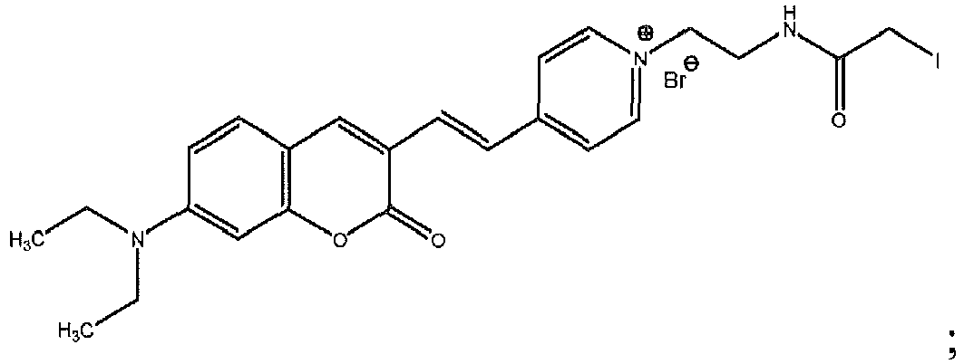


- As described immediately herein below, the presently disclosed fluorophores can be conjugated with a binding member of a specific binding pair, such as a binding protein, to form a biosensor compound that can be used to detect one or more analytes in a sample under test.
- The presently disclosed fluorophores can be used in combination with binding protein assays to detect physiologically important molecules, including metabolites, such as glucose, fatty acids, and lactates, in biological samples. In some embodiments, the presently disclosed fluorophores include a reactive group that can be used to couple or conjugate the fluorophore with another molecule, including a member of a specific binding pair, such as a binding protein or a receptor, which has an affinity for a specific ligand or analyte.
- As used herein, the term "conjugate" refers to a molecule comprising two or more subunits bound together, optionally through a linking group, to form a single molecular structure. The binding can be made either by a direct chemical bond between the subunits or through a linking group. Such binding in a conjugate typically is irreversible. As used herein, the term "affinity" refers to the strength of the attraction between one binding member of a specific binding pair to another binding member of a specific binding pair at a particular binding site. The term "specificity" and derivations thereof, refer to the likelihood that a binding member will bind to another member of a specific binding pair. Such binding between one binding member, e.g., a binding protein, to another binding member of a specific binding pair, e.g., a ligand or analyte, can be reversible.
- When bound to a target analyte or ligand, the presently disclosed conjugates can exhibit a detectable change in a fluorescence property in response to changes in the environment of the binding protein or receptor of the fluorophore conjugate. The detectable change in the fluorescent property can include a shift in the fluorescence emission wavelength, a change in fluorescence intensity, a change in fluorescence lifetime, a change in anisotropy, a change in polarization, and combinations thereof.
- Without wishing to be bound to any one particular theory, the binding protein or receptor can adopt two conformations: a ligand-free open form and a closed form when bound to a ligand. These two conformations interconvert, for example, via a global hinge-binding mechanism upon ligand binding or changes in ligand concentration. By positioning environmentally-sensitive fluorophores in locations that undergo local conformational changes in concert with these global conformational changes, such ligand-mediated conformational changes can be exploited to couple ligand binding to changes in one or more fluorescence properties. Accordingly, these engineered conformational coupling mechanisms enable reagentless optical biosensors to be formed from selected binding proteins and environmentally-sensitive fluorophores.
- As used herein, the terms "biosensor" and "biosensor compound" generally refer to a compound that undergoes a detectable change in specific response to a ligand or target analyte. More particularly, the presently disclosed biosensors combine the molecular recognition properties of biological macromolecules, such as a binding protein, with environmentally-sensitive fluorophores that produce a detectable change in a fluorescent property upon ligand binding. Accordingly, a biosensor translates a binding event into a directly measurable fluorescent property.
- The presently disclosed subject matter provides a biosensor compound.
- The biosensor compound is selected from the group consisting of:


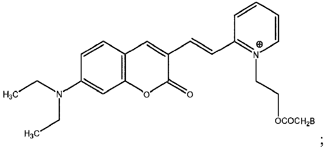

- In some embodiments, the binding member B is a binding protein. As used herein, the term "binding protein" refers to a protein, that when conjugated with a fluorophore, interacts with a specific analyte or ligand in a manner capable of producing a detectable florescence signal differentiable from when a target analyte or ligand is present or absent, or when a target analyte or ligand is present in varying concentrations over time. The term "producing a detectable signal" refers to the ability to recognize a change in a property of a reporter group, e.g., a fluorophore, in a manner that enables the detection of ligand-protein binding. Further, the producing of a detectable signal can be reversible or non-reversible. The signal-producing event includes continuous, programmed, and episodic means, including one-time or reusable applications. The reversible signal-producing event can be instantaneous or can be time-dependent, so long as a correlation with the presence or concentration of analyte is established.
- In some embodiments, the binding member conjugated with the presently disclosed fluorophores comprises a polypeptide or protein. In some embodiments, the binding protein is selected from the group consisting of W183C, SM4, and Y10C. Representative binding proteins suitable for use with the presently disclosed fluorophores, include, but are not limited to periplasmic binding proteins (PBPs). As used herein a PBP is a protein characterized by its three-dimensional configuration (tertiary structure), rather than its amino acid sequence (primary structure) and is characterized by a lobe-hinge-lobe region. The PBP normally binds an analyte specifically in a cleft region between the lobes of the PBP. Further, the binding of an analyte in the cleft region can cause a conformational change to the PBP that makes detection of the analyte possible. Periplasmic binding proteins suitable for use with the presently disclosed fluorophores include any protein that possesses the structural characteristics described herein. Analyzing the three-dimensional structure of a protein to determine the characteristic lobe-hinge-lobe structure of the PBPs is well within the capabilities of one of ordinary skill in the art. Examples of PBPs include, but are not limited to, glucose-galactose binding protein (GGBP), maltose binding protein (MBP), ribose binding protein (RBP), arabinose binding protein (ABP), dipeptide binding protein (DPBP), glutamate binding protein (GluBP), iron binding protein (FeBP), histidine binding protein (HBP), phosphate binding protein (PhosBP), glutamine binding protein (QBP), oligopeptide binding protein (OppA), or derivatives thereof, as well as other proteins that belong to the families of proteins known as periplasmic binding protein like I (PBP-like I) and periplasmic binding protein like II (PBP-like II). The PBP-like I and PBP-like II proteins have two similar lobe domains comprised of parallel β-sheets and adjacent α helices. The glucose-galactose binding protein (GGBP) belongs to the PBP-like I family of proteins, whereas the maltose binding protein (MBP) belongs to the PBP-like II family of proteins. The ribose binding protein (RBP) is also a member of the PBP family of proteins. Other non-limiting examples of periplasmic binding proteins are listed in Table I.
TABLE I. Genes Encoding Common Periplasmic Binding Proteins. Gene name Substrate Species alsB Allose E. coli araF Arabinose E. coli AraS Arabinose/fructose/xylose S. solfataricus argT Lysine/arginine/ornithine Salmonella typhimurium Artl Arginine E. coli artJ Arginine E. coli b1310 Unknown (putative, multiple sugar) E. coli b1487 Unknown (putative, oligopeptide binding) E. coli b1516 Unknown (sugar binding protein homolog) E. coli butE vitamin B12 E. coli CAC1474 Proline/glycine/betaine Clostridium acetobutylicum Cbt Dicarboxylate (Succinate, malate, fumarate) E. coli CbtA Cellobiose S. solfataricus chvE Sugar A. tumefaciens CysP Thiosulfate E. coli dctP C4-dicarboxylate Rhodobacter capsulatus dppA Dipeptide E. coli FbpA Iron Neisseria gonorrhoeae fecB Fe(III)-dicitrate E. coli fepB enterobactin-Fe E. coli fhuD Ferrichydroxamate E. coli FliY Cystine E. coli GlcS glucose/galactose/mannose S. solfataricus glnH (protein: GLNBP) Gluconate E. coli gntX Gluconate E. coli hemT Haemin Y. enterocolitica HisJ (protein: HBP) Histidine E. coli hitA Iron Haemophilus influenzae livJ Leucine/valine/isoleucine E. coli livK (protein: L-BP) Leucine E. coli malE (protein: MBP) maltodextrin/maltose E. coli mglB glucose/galactose (protein: GGBP) E. coli modA Molybdate E. coli MppA L-alanyl-gamma-D-glutamyl-meso-diaminopimelate E. coli nasF nitrate/nitrite Klebsiella oxytoca nikA Nickel E. coli opBC Choline B. subtilis OppA Oligopeptide Salmonella typhimurium PhnD Alkylphosphonate E. coli PhoS (Psts) Phosphate E. coli potD putrescine/spermidine E. coli potF Polyamines E. coli proX Betaine E. coli rbsB Ribose E. coli SapA Peptides S. typhimurium Sbp Sulfate Salmonella typhimurium TauA Taurin E. coli TbpA Thiamin E. coli tctC Tricarboxylate Salmonella typhimurium TreS Trehalose S. solfataricus tTroA Zinc Treponema pallidum UgpB sn-glycerol-3-phosphate E. coli XylF Xylose E. coli YaeC Unknown (putative) E. coli YbeJ(Gltl) glutamate/aspartate (putative, superfamily: lysine- arginine-ornithine-binding protein) E. coli YdcS(b1440) Unknown (putative, spermidine) E. coli YehZ Unknown (putative) E. coli YejA Unknown (putative, homology to periplasmic oligopeptide-binding protein - Helicobactr pylori) E. coli YgiS(b3020) Oligopeptides (putative) E. coli YhbN Unknown E. coli YhdW Unknown (putative, amino acids) E. coli YliB (b0830) Unknown E. coli (putative peptides) YphF Unknown (putative sugars) E. coli Ytrf Acetoin B. subtilis - Other examples of proteins that can comprise the binding members include, but are not limited to intestinal fatty acid binding proteins (FAPBs). The FABPs are a family of proteins that are expressed at least in the liver, intestine, kidney, lungs, heart, skeletal muscle, adipose tissue, abnormal skin, adipose, endothelial cells, mammary gland, brain, stomach, tongue, placenta, testis, retina. The family of FABPs is, generally speaking, a family of small intracellular proteins (about 14 kDa) that bind fatty acids and other hydrophobic ligands through non-covalent interactions. See Smith, E. R. and Storch, J., J. Biol. Chem., 274 (50):35325-35330 (1999). Members of the FABP family of proteins include, but are not limited to, proteins encoded by the genes FABP1, FABP2, FABP3, FABP4, FABP5, FABP6, FABP7, FABP(9) and MP2. Proteins belonging to the FABP include I-FABP, L-FABP, H-FABP, A-FABP, KLBP, mal-1, E-FABP, PA-FABP, C-FABP, S-FABP, LE-LBP, DA11, LP2, Melanogenic Inhibitor, and the like.
- In some embodiments, the binding member is selected from the group consisting of a GGBP, a FABP and a GGBP derivative. In one embodiment of the presently disclosed subject matter, a mutated glucose/galactose binding protein (GGBP) comprises a detectable reporter group, e.g., a coumarin-based fluorophore, whose detectable characteristics alter upon glucose binding. The change in the detectable characteristics can be due to an alteration in the environment of the fluorophore attached to the mutated GGBP or to a conformational change of the protein resulting from binding. In some embodiments, the FABP is I-FABP.
- As used herein, a "derivative of a protein" or "a derivative of a polypeptide" is a protein or polypeptide that shares substantial sequence identity with the wild-type protein. Derivative proteins or polypeptides of the presently disclosed subject matter can be made or prepared by techniques well known to those of skill in the art. Examples of such techniques include, but are not limited to, mutagenesis and direct synthesis. Derivative proteins also can be modified, either by natural processes, such as post-translational processing, or by chemical modification techniques which are well known in the art. Such modifications are well described in basic texts and in more detailed monographs, as well as in voluminous research literature. Modifications can occur anywhere in the polypeptide chain, including the peptide backbone, the amino acid side-chains and the amino or carboxyl termini. It will be appreciated that the same type of modification can be present in the same or varying degrees at several sites in a given polypeptide or protein. Also, a given polypeptide or protein can contain more than one modification. Examples of modifications include, but are not limited to, glycosylation, acetylation, acylation, ADP-ribosylation, amidation, covalent attachment of flavin, covalent attachment of a heme moiety, covalent attachment of a nucleotide or nucleotide derivative, covalent attachment of a lipid or lipid derivative, covalent attachment of phosphotidylinositol, cross-linking, cyclization, disulfide bond formation, demethylation, formation of covalent crosslinks, formation of cystine, formation of pyroglutamate, formylation, gamma-carboxylation, glycosylation, GPI anchor formation, hydroxylation, iodination, methylation, myristoylation, oxidation, proteolytic processing, phosphorylation, prenylation, racemization, selenoylation, sulfation, transfer-RNA mediated addition of amino acids to proteins such as arginylation, and ubiquitination. Polypeptides or proteins can even be branched as a result of ubiquitination, and they can be cyclic, with or without branching. (See, e.g., T. E. Creighton, Proteins--Structure And Molecular Properties, 2nd ed., W. H. Freeman and Company, New York (1993); Wold, F., "Posttranslational Protein Modifications: Perspectives and Prospects," in Posttranslational Covalent Modification Of Proteins, B. C. Johnson, Ed., Academic Press, New York (1983); Seifter et al., Methods in Enzymol, 182:626-646 (1990) and Rattan et al., Ann NY Acad Sci., 663:48-62 (1992). Examples of derivative proteins include, but are not limited to, mutant and fusion proteins.
- A "mutant protein" is used herein as it is known in the art. In general, a mutant protein can be created by addition, deletion or substitution of the wild-type primary structure of the protein or polypeptide. Mutations include, for example, the addition or substitution of cysteine groups, non-naturally occurring amino acids, and replacement of substantially non-reactive amino acids with reactive amino acids. A mutant protein can be mutated to bind more than one analyte in a specific manner. Indeed, the mutant proteins can possess specificity towards its wild-type analyte and another target ligand. Likewise, a mutant protein can be able to only bind an analyte or analytes that the wild-type binding protein does not bind. Methods of generating mutant proteins are well-known in the art. For example, Looger, L. L., et al., Nature 423 (6936): 185-190 (2003), disclose methods for redesigning binding sites within periplasmic binding proteins that provide new analyte-binding properties for the proteins. These mutant binding proteins retain the ability to undergo conformational change, which can produce a directly generated signal upon analyte-binding. By introducing between 5 and 17 amino acid changes, Looger, et al. constructed several mutant proteins, each with new selectivities for TNT (trinitrotoluene), L-lactate, or serotonin. For example, Looger et al. generated L-lactate binding proteins from ABP, GGBP, RBP, HBP and QBP.
- Table II lists other mutations to GGBP and is taken from Looger L. L. et al., Nature 423: 185-190, (2003).
TABLE II Mutations of GGBP residues providing L-lactate selectivity. Residue: 10 14 16 91 92 152 154 158 183 236 256 Wildtype GGBP Y D F N K H D R W D N Lactate-specific mutant G1 K K F K L M H K K A D Lactate-specific mutant G2 K M K K L K K M K A S - Examples of mutations of a GGBP protein, for example the GGBP protein of GenBank Accession No. P02927 without the 23 amino acid leader sequence (i.e., the mature chain), include, but are not limited to, having a cysteine substituted for lysine at position 11 (K11C), a cysteine substituted for aspartic acid at position 14 (D 14C), a cysteine substituted for valine at position 19 (V19C), a cysteine substituted for asparagine at position 43 (N43C), a cysteine substituted for glycine at position 74 (G74C), a cysteine substituted for tyrosine at position 107 (Y107C), a cysteine substituted for threonine at position 110 (T110C), a cysteine substituted for serine at position 112 (S 112C), a double mutant including a cysteine substituted for serine at position 112 and serine substituted for leucine at position 238 (S112C/L238S), a cysteine substituted for lysine at position 113 (K113C), a cysteine substituted for lysine at position 137 (K137C), a cysteine substituted for glutamic acid at position 149 (E149C), a double mutant including a cysteine substituted for glutamic acid at position 149 and an arginine substituted for alanine at position 213 (E149C/A213R), a double mutant including a cysteine substituted for glutamic acid at position 149 and a serine substituted for leucine at position 238 (E149C/L238S), a double mutant including a serine substituted for alanine at position 213 and a cysteine substituted for histidine at position 152 (H152C/A213S), a cysteine substituted for methionine at position 182 (M182C), a cysteine substituted for alanine at position 213 (A213C), a double mutant including a cysteine substituted for alanine at position 213 and a cysteine substituted for leucine at position 238 (A213C/L238C), a cysteine substituted for methionine at position 216 (M216C), a cysteine substituted for aspartic acid at position 236 (D236C), a cysteine substituted for leucine at position 238 (L238C) a cysteine substituted for aspartic acid at position 287 (D287C), a cysteine substituted for arginine at position 292 (R292C), a cysteine substituted for valine at position 296 (V296C), a triple mutant including a cysteine substituted for glutamic acid at position 149 and a serine substituted for alanine at position 213 and a serine substituted for leucine at position 238 (E149C/A213S/L238S), a triple mutant including a cysteine substituted for glutamic acid at position 149 and an arginine substituted for alanine at position 213 and a serine substituted for leucine at position 238 (E149C/A213R/L238S), a cysteine substituted for glutamic acid at position 149 and a cysteine substituted for alanine at position 213 and a serine substituted for leucine at position 238 (E149C/A213C/L238S).
- The mutation can serve one or more of several purposes. For example, a naturally occurring protein can be mutated to change the long-term stability, including thermal stability, of the protein, to conjugate the protein to a particular encapsulation matrix or polymer, to provide binding sites for detectable reporter groups, to adjust its binding constant with respect to a particular analyte, or combinations thereof.
- The analyte and mutated protein can act as binding partners. The term "associates" or "binds" as used herein refers to binding partners having a relative binding constant (Kd) sufficiently strong to allow detection of binding to the protein by a detection means. The Kd can be calculated as the concentration of free analyte at which half the protein is bound, or vice versa. When the analyte of interest is glucose, the Kd values for the binding partners are between about 0.0001 mM and about 50 mM.
- In addition to changing binding characteristics, derivative polypeptides or proteins also can be used to incorporate a presently disclosed fluorophore onto or within the binding member. The fluorophores can be used to indicate a change in the binding member, including, but not limited to, three-dimensional conformational changes, changes in orientation of the amino acid side chains of proteinaceous binding domains, and redox states of the binding member. With the addition/substitution of one or more residues into the primary structure of a protein, some of the labeling moieties used in the current methods and compositions can be attached through chemical means, such as reduction, oxidation, conjugation, and condensation reactions. Examples of residues commonly used to label proteins include, but not are limited to, lysine and cysteine. For example, any thiol-reactive group can be used to attach a fluorophore to a naturally occurring or engineered cysteine in the primary structure of the polypeptide. Also, for example, lysine residues can be labeled using succinimide ester derivatives of fluorophores. See Richieri, G. V. et al., J. Biol. Chem., 267: 23495-501 (1992).
- The fluorophore can be attached to the mutated protein, for example a GGBP, by any conventional means known in the art. For example, the reporter group can be attached via amines or carboxyl residues on the protein. Exemplary embodiments include covalent coupling via thiol groups on cysteine residues of the mutated or native protein. For example, for mutated GGBP, cysteines can be located at position 10, at position 11, position 14, at position 15, position 19, at position 26, at position 43, at position 74, at position 92, at position 93, position 107, position 110, position 112, at position 113, at position 137, at position 149, at position 152, at position 154, at position 182, at position 183, at position 186, at position 211, at position 213, at position 216, at position 238, at
position 240, at position 242, at position 255, at position 257, at position 287, at position 292, at position 294, and at position 296. - The presently disclosed fluorescent dyes can be covalently attached to a binding protein in a site-specific manner to obtain the desired change in fluorescence. The fluorescent dye can be attached at a site on the binding protein so that the conformational change maximizes the change in fluorescence properties. Conjugates containing fluorophores attached at various sites, for example, cysteine mutant sites constructed in mutated GGBPs, can be screened to identify which sites result in the largest change in fluorescence upon glucose binding.
- In some embodiments, the binding member is at least one fragment of a receptor, wherein the receptor is a protein in nature and is capable of binding to a suitable ligand via an active site, and wherein at least one amino acid residue of the fragment is located in the proximity of the active site and is a cysteine residue or is substituted with a cysteine residue, as described in
U.S. Patent Application Publication No. 2003/0153012 to Renard et al. - As used herein, the term "receptor" refers to a protein macromolecule having an active site capable of binding a ligand. More particularly, a "receptor" refers to an antibody, a hormone or bacterial receptor, an affinity protein, a transport protein, a viral receptor, or any polypeptide having a specific affinity for a ligand.
- In some embodiments, the receptor can have one or more disulfide bridges. In some embodiments, the receptor can be an antibody or an antibody fragment, including, but not limited to, an Fv, scFv, or Fab fragment, or a miniantibody. In some embodiments, the antibody can be a natural or artificial monoclonal antibody.
- As used herein, the term "ligand" refers to any molecule capable of binding to the receptor via an active site. In some embodiments, the ligand is a protein, a peptide or hapten antigen, such as a bacterial antigen, or a hormone, a cytokine, an interleukin, a tumor necrosis factor (TNF) a growth factor, a viral protein, or a peptide or nucleotide sequence.
- As used herein, the term "active site" when referring to, for example, "an active site of the receptor or of the receptor fragment" refers to amino acid residues of the receptor or receptor fragment that contribute to the binding of the ligand. This active site also can be referred to as a "binding site" or "paratope."
- The term "proximity" as used herein refers to the position of amino acid residues of the receptor that are in direct contact with the ligand, those that are in contact by water molecules, and those for which the solvent accessible surface area (ASA), as that term in known in the art, see, e.g., Creighton, T. E., "Proteins: Structure and molecular properties," 2nd ed., (W.H. Freeman & Co., New York) 227-229 (1993), is modified by the binding of the ligand.
- The presently disclosed subject matter provides a method for determining the presence or amount of one or more analytes in a sample, the method comprising:
- (a) providing a biosensor compound having at least one mutated binding protein with a fluorophore covalently attached thereto through a thiol group of the mutated binding protein, wherein the biosensor is described above;
wherein the biosensor compound exhibits a detectable change in a fluorescence property as a result of binding to the ligand or analyte or as a result of a change in concentration of the ligand or analyte in a sample under test; - (b) contacting the biosensor compound with a sample suspected of containing one or more analytes to bind the one or more analytes, if present, to the binding protein;
- (c) irradiating the sample suspected of containing one or more analytes with electromagnetic radiation to induce the fluorophore to fluoresce; and
- (d) detecting a fluorescence property to determine the presence or amount of one or more analytes in the sample.
B is a binding member having a binding affinity for a ligand or analyte to be detected. - The biosensor compound is selected from the group consisting of:


- Embodiments that exhibit a shift in emission wavelength upon ligand binding enable a biosensor comprising a presently disclosed fluorophore to be self-referencing. In such embodiments, the fluorophore exhibits an increase in a first emission wavelength in the presence of a metabolite, such as glucose, and a decrease in a second emission wavelength. A ratio between the first emission wavelength and the second wavelength can be calculated to determine the concentration of glucose in the sample under test. Such self referencing can correct for variations in excitation source intensity and other sources of noise and instability in the biosensor without requiring a reference dye. Thus, a single excitation wavelength can be used to observe a ratiometric response in the fluorescence output of the biosensor. As used herein, the term "ratiometric response" means that the intensities of the first emission wavelength and the second emission wavelength vary independently such that the ratio of the two emission wavelengths (the "ratiometric quotient" or "QR") can be used to indicate the presence and/or amount, e.g., concentration, of the ligand or analyte in a sample.
- Accordingly, in some embodiments, the presently disclosed method further comprises: (a) measuring a fluorescence intensity at a first emission wavelength before contacting the biosensor compound with a sample suspected of containing one or more analytes; (b) measuring a fluorescence intensity at a second emission wavelength after contacting the biosensor compound with the sample suspected of containing one or more analytes; and (c) determining the ratio of the second emission wavelength to the first emission wavelength to determine the presence or amount of one or more analytes in the sample.
- The binding of the one or more target analytes to a fluorophore-binding member conjugate creates or alters a discernable fluorescence property. Discernable changes in the fluorescence property include, but are not limited to, an emission wavelength shift and change, e.g., an increase or decrease, in signal intensity. In some embodiments, the presently disclosed fluorophore-binding member conjugates do not generate a signal when not bound to the target analytes. In other embodiments, the fluorophore-binding member conjugates generate a signal, for example, a first emission wavelength, even when not bound to a target analyte. In such embodiments, the binding of the target analyte, however, still can change the fluorescence property, such that binding is discernable. The binding of the fluorophore-binding member to a target analyte also can cause a decrease in signal intensity.
- A detected fluorescence property or a change in the detected fluorescence property due to either a conformational change in the binding protein, subsequent changes in the microenvironment of the fluorescent dye, or both, can be correlated to the presence and/or amount, i.e., analyte concentration, of one or more target analytes. In some embodiments of the presently disclosed method, the mutated binding protein undergoes a conformation change as a result of changes in analyte concentration of the sample suspected of containing one or more analytes. Accordingly, the method detects changes in the fluorescence property as a result of changes in the analyte concentration.
- The amount of one or more analytes present in a sample under test can be represented as a concentration. As used herein, the term "concentration" has its ordinary meaning in the art. The concentration can be expressed as a qualitative value, such as negative- or positive-type results, for example, as a "YES" or "NO" response indicating the presence or absence of a target analyte, or as a quantitative value, for example in units of mg/dL. Further, the concentration of a given analyte can be reported as a relative quantity or an absolute quantity. As used herein, "quantitative results" refer to results that provide absolute or relative values.
- The quantity (concentration) of an analyte can be equal to zero, indicating the absence of the particular analyte sought or that the concentration of the particular analyte is below the detection limits of the assay. The quantity measured can be the measured signal, e.g., fluorescence, without any additional measurements or manipulations. Alternatively, the quantity measured can be expressed as a difference, percentage or ratio of the measured value of the particular analyte to a measured value of another compound including, but not limited to, a standard or another analyte. The difference can be negative, indicating a decrease in the amount of measured analyte(s). The quantities also can be expressed as a difference or ratio of the analyte(s) to itself, measured at a different point in time. The quantities of analytes can be determined directly from a generated signal, or the generated signal can be used in an algorithm, with the algorithm designed to correlate the value of the generated signals to the quantity of analyte(s) in the sample.
- The presently disclosed fluorophores having a reactive group, e.g., a thiol-reactive group, typically exhibit an emission wavelength of about 575 nm or greater. In some embodiments, the presently disclosed fluorophores have an emission wavelength of about 615 nm. Fluorophores having such emission characteristics typically avoid or minimize background interference from the biological system or other components in the analyte, e.g., glucose, source. Further, fluorophores that operate at long emission wavelengths (for example, about 575 nm or greater) can be used as biosensors in vivo. For example, such fluorophores can be incorporated into an implantable biosensor device (with human skin being opaque below about 575 nm).
- In the presently disclosed methods for detecting the presence and/or amount of one or more target analytes, the sample suspected of containing one or more target analytes is irradiated with an energy source, such as a laser or light emitting diode (LED), to excite the fluorescent dye. A fluorescence property or a change in a fluorescence property can then be detected.
- In some embodiments, a fluorometer can be used to measure the wavelength and/or intensity of fluorescent light. An example of a fluorometer suitable for use with the presently disclosed fluorophores is described in
U.S. Published Patent Application No. 2005/0113657, filed Nov. 26, 2003 . - The fluorescence and detection can be carried out continuously or intermittently at predetermined times allowing episodic or continuous sensing of an analyte, for example, glucose, to be performed. Thus, the presently disclosed fluorescent dyes are amenable for use with devices capable of continuously measuring the concentrations of one or more analytes. Accordingly, in some embodiments, the method further comprises continuously: (a) contacting the binding protein with the sample suspected of containing one or more analytes; (b) irradiating the sample with electromagnetic radiation; and (c) detecting the fluorescence property. As used herein, the term "continuously," in conjunction with the measuring of an analyte, is used to mean the device either generates or is capable of generating a detectable signal at any time during the life span of the device. The detectable signal can be constant, in that the device is always generating a signal, even if a signal is not detected. Alternatively, the device can be used episodically, such that a detectable signal can be generated, and detected, at any desired time.
- The presently disclosed subject matter provides methods which can use specific binding partners for a particular analyte or analytes of interest. A specific binding partner or member, as used herein, is a member of a specific binding pair. A "specific binding pair" refers to two different molecules where one of the molecules through chemical or physical means specifically binds the second molecule. In this sense, an analyte is a reciprocal member of a specific binding pair. Further, specific binding pairs can include members that are analogs of the original specific binding partners, for example, an analyte-analog having a similar structure to the analyte. By "similar" it is intended that, for example, an analyte-analog has an amino acid sequence that has at least about 60% or 65% sequence identity, about 70% or 75% sequence identity, about 80% or 85% sequence identity, about 90%, 91 %, 92%, 93%, 94%, 95%, 96%, 97%, 98%, 99% or greater amino acid sequence identity compared to an analyte amino acid sequence using alignment programs and standard parameters well known in the art. An analog of an analyte also can have the same function as an analyte.
- The term "analyte," as used herein, generally refers to a substance to be detected, which can be present or suspected of being present in a test sample. More particularly, an "analyte" can be any substance for which there exists a naturally occurring specific binder partner, such as a binding protein or receptor, or for which a specific binding partner can be prepared. Accordingly, an "analyte" is a substance that can bind one or more specific binding partners in an assay. In some embodiments, the analyte can be any compound, such as a metabolite, to be detected or measured and which has at least one binding site.
- The target analytes can be any molecule or compound, of which the presence or amount is to be determined in a sample under test. Examples of classes of analytes that can be measured by the presently disclosed methods include, but are not limited to amino acids, peptides, polypeptides, proteins, carbohydrates, fatty acid, lipids, nucleotides, oligonucleotides, polynucleotides, glycoproteins or proteoglycans, lipoproteins, lipopolysaccharides, drugs, drug metabolites, small organic molecules, inorganic molecules and natural or synthetic polymers. Examples of target analytes include, but are not limited to, glucose, free fatty acids, lactic acid, C-reactive protein and anti-inflammatory mediators, such as cytokines, eicosanoids, or leukotrienes. In some embodiments, the target analytes are selected from the group consisting of fatty acids, C-reactive protein, and leukotrienes. In another embodiment, the target analytes are selected from the group consisting of glucose, lactic acid and fatty acids.
- As used herein, the term "carbohydrate" includes, but is not limited to monosaccharides, disaccharides, oligosaccharides and polysaccharides. "Carbohydrate" also includes, but is not limited to, molecules comprising carbon, hydrogen and oxygen that do not fall within the traditional definition of a saccharide-i.e., an aldehyde or ketone derivative of a straight chain polyhydroxyl alcohol, containing at least three carbon atoms. Thus, for example, a carbohydrate as used herein can contain fewer than three carbon atoms.
- The term "fatty acids," as used herein include all fatty acids, including free fatty acids (FFA) and fatty acids esterified to other molecules. Examples of specific fatty acids include, but are not limited to, palmitate, stearate, oleate, linoleate, linolenate, and arachidonate. The term "free fatty acid" is used herein as it is known in the art in that FFA are not part of other molecules, such as triglycerides or phospholipids. Free fatty acids also include non-esterified fatty acids that are bound to or adsorbed onto albumin. As used herein, the term "unbound free fatty acid" (unbound FFA) is used to denote a free fatty acid or free fatty acids that are not bound or adsorbed onto albumin or other serum proteins.
- As used herein, the term "lipid" is used as it is in the art, i.e., a substance of biological origin that is made up primarily or exclusively of nonpolar chemical groups such that it is readily soluble in most organic solvents, but only sparingly soluble in aqueous solvents. Examples of lipids include, but are not limited to, fatty acids, triacylglycerols, glycerophospholipids, sphingolipids, cholesterol, steroids and derivatives thereof. For example, "lipids" include but are not limited to, the ceramides, which are derivatives of sphingolipids and derivatives of ceramides, such as sphingomyelins, cerebrosides and gangliosides. "Lipids" also include, but are not limited to, the common classes of glycerophospholipds (or phospholipids), such as phosphatidic acid, phosphatidylethanolamine, phosphatidylcholine, phosphatidylserine, phosphatidylinositol, phosphatidylglycerol, and the like.
- As used herein, a "drug" can be a known drug or a drug candidate, whose activity or effects on a particular cell type are not yet known. A "drug metabolite" is any of the by-products or the breakdown products of a drug that is changed chemically into another compound or compounds. As used herein, "small organic molecule" includes, but is not limited to, an organic molecule or compound that does not fit precisely into other classifications highlighted herein. More particularly, the term "small organic molecule" as used herein, refers to organic compounds, whether naturally-occurring or artificially created (e.g., via chemical synthesis) that have relatively low molecular weight and that are not proteins, polypeptides, or nucleic acids. Typically, small molecules have a molecular weight of less than about 1500 g/mol. Also, small molecules typically have multiple carbon-carbon bonds.
- As used herein, the term "sample" includes any liquid or fluid sample, including a sample derived from a biological source, such as a physiological fluid, including whole blood or whole blood components, such as red blood cells, white blood cells, platelets, serum and plasma; ascites; urine; saliva; sweat; milk; synovial fluid; peritoneal fluid; amniotic fluid; percerebrospinal fluid; lymph fluid; lung embolism; cerebrospinal fluid; pericardial fluid; cervicovaginal samples; tissue extracts; cell extracts; and other constituents of the body that are suspected of containing the analyte of interest. In addition to physiological fluids, other liquid samples, such as water, food products and the like, for the performance of environmental or food production assays are suitable for use with the presently disclosed subject matter. A solid material suspected of containing the analyte also can be used as the test sample. In some instances it might be beneficial to modify a solid test sample to form a liquid medium or to release the analyte.
- In some embodiments, the sample can be pre-treated prior to use, such as preparing plasma from blood, diluting viscous fluids, or the like. Such methods of treatment can involve filtration, distillation, concentration, inactivation of interfering compounds, and the addition of reagents.
- The sample can be any sample obtained from a subject. The term "subject" refers to an organism, tissue, or cell from which a sample can be obtained. A subject can include a human subject for medical purposes, such as diagnosis and/or treatment of a condition or disease, or an animal subject for medical, veterinary purposes, or developmental purposes. A subject also can include sample material from tissue culture, cell culture, organ replication, stem cell production and the like. Suitable animal subjects include mammals and avians. The term "avian" as used herein includes, but is not limited to, chickens, ducks, geese, quail, turkeys, and pheasants. The term "mammal" as used herein includes, but is not limited to, primates, e.g, humans, monkeys, apes, and the like; bovines, e.g., cattle, oxen, and the like; ovines, e.g., sheep and the like; caprines, e.g., goats and the like; porcines, e.g., pigs, hogs, and the like; equines, e.g., horses, donkeys, zebras, and the like; felines, including wild and domestic cats; canines, including dogs; lagomorphs, including rabbits, hares, and the like; and rodents, including mice, rats, and the like. Preferably, the subject is a mammal or a mammalian cell. More preferably, the subject is a human or a human cell. Human subjects include, but are not limited to, fetal, neonatal, infant, juvenile, and adult subjects. Further, a "subject" can include a patient afflicted with or suspected of being afflicted with a condition or disease. Thus, the terms "subject" and "patient" are used interchangeably herein. A subject also can refer to cells or collections of cells in laboratory or bioprocessing culture in tests for viability, differentiation, marker production, expression, and the like.
- The presently disclosed methods can be used to diagnose, for the prognosis, or the monitoring of a disease state or condition. As used herein, the term "diagnosis" refers to a predictive process in which the presence, absence, severity or course of treatment of a disease, disorder or other medical condition is assessed. For purposes herein, diagnosis also includes predictive processes for determining the outcome resulting from a treatment. Likewise, the term "diagnosing," refers to the determination of whether a sample specimen exhibits one or more characteristics of a condition or disease. The term "diagnosing" includes establishing the presence or absence of, for example, a target antigen or reagent bound targets, or establishing, or otherwise determining one or more characteristics of a condition or disease, including type, grade, stage, or similar conditions. As used herein, the term "diagnosing" can include distinguishing one form of a disease from another. The term "diagnosing" encompasses the initial diagnosis or detection, prognosis, and monitoring of a condition or disease.
- The term "prognosis," and derivations thereof, refers to the determination or prediction of the course of a disease or condition. The course of a disease or condition can be determined, for example, based on life expectancy or quality of life. "Prognosis" includes the determination of the time course of a disease or condition, with or without a treatment or treatments. In the instance where treatment(s) are contemplated, the prognosis includes determining the efficacy of a treatment for a disease or condition.
- As used herein, the term "risk" refers to a predictive process in which the probability of a particular outcome is assessed.
- The term "monitoring," such as in "monitoring the course of a disease or condition," refers to the ongoing diagnosis of samples obtained from a subject having or suspected of having a disease or condition.
- The term "marker" refers to a molecule, such as a protein, including an antigen, that when detected in a sample is characteristic of or indicates the presence of a disease or condition.
- The presently disclosed subject matter also provides methods for monitoring disease states in a subject, including chronic diseases, such as, but not limited to, heart disease, coronary artery disease, diabetes, metabolic disorders, inflammatory diseases, such as rheumatoid arthritis, and cancer. The metabolic disorders can include, but are not limited to, hyperlipidemia, hypolipidemia, hyperthyroidism, and hypothyroidism.
- Further, the presently disclosed methods can be used to monitor specific markers of a chronic disease. By monitoring the concentrations of molecular artifacts, metabolites, and deleterious and/or beneficial molecules of a disease state, the subject's progression, regression or stability can be assessed, and treatments can, in turn be adjusted or revised accordingly. For example, markers for heart disease that could be monitored in vivo using the presently disclosed biosensors include, but are not limited to, total fatty acids, lactate, glucose, free fatty acids and various cardiotonic agents, such as, but not limited to cardioglycosides and sympathomimetics. Markers of diabetes include, but are not limited to, glucose, lactate and fatty acids. Likewise, markers for coronary artery disease include, but are not limited to, C-reactive peptide and free fatty acids. Generally, markers of various metabolic disorders include, but are not limited to, specific fatty acids.
- The presently disclosed fluorescent dyes also are suitable for use in devices for monitoring drug treatment. Indeed, the biosensor could be designed to specifically bind a drug, drug candidate or a drug metabolite. In this manner, the plasma concentration of the drug could be monitored and dosages could be adjusted or maintained based on the concentration measurements provided by the sensor. Accordingly, a pharmaceutical regimen could be individualized for a particular subject, including the use of a biosensor that can specifically and reversibly bind the drug or drug metabolite to determine plasma concentrations of the drug. The concentrations provided by the sensor can then be used to determine the bioavailability of the drug in the subject. The dose of the drug administered to the subject can then be altered to increase or decrease the bioavailability of the drug to the subject to provide maximum therapeutic benefits and avoiding toxicity.
- Biosensor devices comprising the presently disclosed fluorophores can be used to simultaneously monitor a variety of metabolites, the measurements of which could be used to profile the subject's metabolic or physical state. For example, during extended periods of strenuous exercise, glucose is broken down in anaerobic processes to lactic acid. The presently disclosed biosensors can be used to determine lactate thresholds of athletes, to maximize the benefits of training and decrease recovery time. Similarly, the biosensors can be used to determine lactate thresholds in soldiers to prevent fatigue and exhaustion and to decrease recovery time. To that end, the presently disclosed biosensors can be used to monitor glucose levels, lactic acids levels and other metabolites during exercise or physical stress.
- In some embodiments, the presently disclosed fluorophores can be used with a biosensor device that is capable of conveying a signal, e.g., emitted light, to a detector that is capable of detecting the signal. In some embodiments, the device comprises the detector, whereas in other embodiments, the device does not comprise the detector, i.e., the detector is external to the device. The signal generated by the device can be a direct indicator of the binding of the one or more target analytes to the binding member, e.g., a binding protein or receptor, associated with the fluorophore.
- The presently disclosed fluorophores can be used in biosensor devices suitable for use in various settings, including in vivo, in vitro and in situ. Such devices include medical devices or implants for monitoring metabolic substrate levels in a subject. When such devices are implanted in a subject, the implants should be biocompatible such that they produce little or no detectable inflammation/rejection reaction. One approach for rendering the implants more biocompatible comprises coating the implants with biocompatible polymers, such as poly(urethane) elastomers, poly(urea) and poly(vinylchloride). Exemplary biosensor devices are described in
U.S. Patent Application Publication No. 2006/0078908, filed June 7, 2005 , andU.S. Patent Application Publication No. 2005/0113657 , each of which is incorporated herein by reference in its entirety. - Biosensor devices comprising the presently disclosed fluorophores can be used to monitor a condition or disease state in a patient in an acute care facility, such as an emergency room or a post-operative recovery room or a hospital. For example, in embodiments providing a method for monitoring glucose levels in a subject, studies have shown that mortality can be decreased by as much as 30% in post-operative patients when glucose levels are monitored and kept normal. Thus, the presently disclosed biosensor can be used in situations where monitoring glucose or other metabolites is essential to recovery or the overall health of the subject.
- The presently disclosed biosensor can be used or adapted for use in strips, implants, micro- and nano-particles, and the like.
- While the following terms are believed to be well understood by one of ordinary skill in the art, the following definitions are set forth to facilitate explanation of the presently disclosed subject matter. Unless otherwise defined, all technical and scientific terms used herein have the same meaning as commonly understood by one of ordinary skill in the art to which this presently described subject matter belongs.
- Throughout the specification and claims, a given chemical formula or name shall encompass all optical and stereoisomers, as well as racemic mixtures where such isomers and mixtures exist.
- The following Examples have been included to provide guidance to one of ordinary skill in the art for practicing representative embodiments of the presently disclosed subject matter. In light of the present disclosure and the general level of skill in the art, those of skill can appreciate that the following Examples are intended to be exemplary only and that numerous changes, modifications, and alterations can be employed. The following Examples are offered by way of illustration.
- The presently disclosed ICOPIC dye was designed so that the quaternary nitrogen, i.e., the nitrogen atom having a positive charge, is part of an aromatic ring and is farther away from the bridging double bond between the coumarin or aza-coumarin nucleus and the haloacetyl moiety. The IOPIC dye was synthesized as shown in
Scheme 1.
- Referring now to
Scheme 1,compound 1 was synthesized by reacting 4-picoline and 2-bromoethanol in toluene at 120 °C. The reaction was stopped after 45 min, cooled to room temperature, and the toluene layer was decanted out. The obtained light yellow oil was washed with hexane two times and evaporated on a rotary evaporator. The residue obtained was refrigerated to afford a low melting light yellow solid. The product was characterized by 1H and 13C NMR spectroscopy and FAB-mass spectroscopy. - Referring once again to
Scheme 1,compound 2 was synthesized as follows.Compound 1 was dissolved in anhydrous methanol and catalytic amount of piperidine was added. The resulting solution was heated to 70 °C for 30 min. During this time the reaction mixture turned to light brown. To this reaction mixture, coumarin aldehyde was added and heated to reflux. After 2 h, the reaction was stopped and then cooled in the refrigerator to afford a red solid, which was filtered and dried. The crude dye was purified by silica gel column chromatography to afford the pure dye. FAB-mass analysis confirmed the product. - Referring once again to
Scheme 1, the presently disclosed ICOPIC dye was synthesized as follows. To a solution ofcompound 2 in a mixture of chloroform and acetonitrile was added 3 drops of pyridine and the obtained solution was stirred at room temperature under argon atmosphere for 10 min. To this reaction mixture iodoacetic anhydride was added and stirring was continued for 2 h at room temperature. After 2 h the reaction was stopped and the solvent was evaporated to obtain a red solid. The obtained solid was washed with MTBE ether and dried under vacuum to afford the ICOPIC as a red solid. The compound was confirmed by FAB-mass spectroscopy and NMR. - The ICOPIC (ester version) was labeled on glucose binding protein W183C. Referring now to
Figure 1 , the ratiometric response to glucose was studied. Referring once again toFigure 1 , the presently disclosed ICOPIC dye responds to glucose ratiometrically and the QR obtained is about 2.0. The QR could be significantly improved by increasing the labeling ratio of the dye to the protein. - Temperature stability studies of the W183C-ICOPIC conjugate were carried out at 37 °C over 3 days. Referring now to
Figure 2 , the dye absorption spectrum remains almost unchanged after 3 days at 37 °C. The fluorescence response to glucose, however, was lower (data not shown). Without wishing to be bound to any one particular theory, this observation is likely due to the cleavage of the ester bond and release of the dye from the cysteine residue. The potential release of the dye from the cysteine residue is thought to be minimized in the iodoacetamide version of the dye, as provided in Example 5. - The o-ICOPIC ester dye has been conjugated to SM4 (GBP) protein and the fluorescence response was studied in the absence and presence of glucose. Referring now to
Figure 3 , the obtained response is shown. Referring once again toFigure 3 , upon addition of glucose, the spectrum undergoes a 1.5-fold enhancement in fluorescence intensity and a shift in wavelength from about 600 nm to about 625 nm. The QR was calculated and the optimized value was found to be about 7.0. This QR value is the highest QR value observed for any known ratiometric dye with SM4. Accordingly, this dye has significant utility in bio-sensor chemistry. - To improve the stability of the dye's linker group of ICOPIC, synthesis of an iodoacetamide version of the dye was conducted. The iodoacetamide linker is more stable toward hydrolysis when compared to the iodoacetyl linker group.
- One method for synthesizing an iodoacetamide version of the presently disclosed ICOPIC dye is shown in
Scheme 2.
- Referring now to
Scheme 2,compound 2 was synthesized by reacting 4-picoline with 2-bromoethylaminehydrobromide in methanol under reflux for 48 h. The reaction mixture was cooled to room temperature and the product was precipitated as a white solid. The precipitated product was filtered and washed with cold methanol followed by methyl-tert-butyl ether and dried under vacuum. NMR spectroscopy (1H, 13C) and FAB-mass spectrum confirmed the structure. - Referring once again to
Scheme 2,compounds 4 and 5 were prepared as follows.Compound 2 was dissolved in methanol and catalytic amount of piperidine was added and allowed to reflux for 30 min. The coumarin aldehyde (3) was added and refluxing was continued for an additional 4 h. The progress of the reaction was monitored by TLC. After completion of the reaction the solvent was evaporated to afford a residue. The obtained residue was chromatographed over alumina, eluted with CHCl3/MeOH (6:4) containing a few drops of ammonia solution. The pure product was eluted as a red band and the solvent was evaporated to afford a red dye. Analysis by FAB-mass spectrum confirmed the product. - Referring once again to
Scheme 2, the final dye 6, containing the iodoacetamide moiety, was synthesized by reactingcompound 5 with iodoacetic anhydride in the presence of pyridine in a mixture of methanol and chloroform. The final dye was purified by washing off the excess of iodoacetic anhydride and pyridine. FAB-mass spectrum confirmed the final dye. - An alternative method for synthesizing p-ICOPIC amide is provided in Scheme 3.

- Referring now to Scheme 3, a presently disclosed p-ICOPIC dye can be prepared as follows.
- Compound 9. Compound 8 (4-picoline, 1.0 g) was dissolved in anhydrous methanol (5 mL). Bromoethylamine HBr (2.20 g) was added to the solution. The reaction was heated to reflux and allowed to react overnight. Upon cooling the next day, the product had precipitated. The precipitate was collected by filtration, washed with a small amount methanol and allowed to air dry. The product (1.2 g) was obtained as a colorless solid. The product was confirmed by 1H, 13C NMR and FAB-MS. 1H NMR of 9 (400 MHz, CD3OD) δ = 8.88 (d, 2H), 8.01 (d, 2H), 4.96 (t, 2H), 3.69 (t, 2H), 2.71 (s, 3H). 13C NMR of 9 (400 MHz, CD3OD), δ = 162.9, 145.3, 130.5, 58.4, 40.5, 22.4. FAB-MS obs = 137.1 calc = 137.11.
- Compound 10. Compound 9 (200 mg) and sodium carbonate (166 mg, 2eq) were dissolved in 5 mL of H2O. Boc2O (161 mg, 1.1 eq) was dissolved in 5 mL of dioxane and added dropwise to the aqueous solution. The reaction was allowed to stir overnight at room temperature. The next day the solvent was evaporated to dryness leaving a solid white residue. Methanol (10 mL) was added and the resulting solution was filtered to remove the sodium carbonate. The filtrated solution was evaporated to yield 210 mg of product (10) which was used without further purification. The product was confirmed by 1H NMR. 1H NMR of 10 (400 MHz, CD3OD), δ = 8.77 (d, 2H), 7.94 (d, 2H), 4.64 (t, 2H), 3.62 (t, 2H), 2.66 (s, 3H), 1.31 (s, 9H).
- Compound 11. Compound 10 (190 mg) and 7-diethylamino-coumarin-3-aldehyde (140 mg) were dissolved in 6 mL of methanol followed by the addition of 50 µL of piperidine. The reaction was heated to 50 °C and allowed to react overnight with stirring. The solvent was then evaporated and the remaining red residue was purified by column chromatography. The product was eluted with a 10% methanol in dichloromethane solution. Pure fractions were combined and evaporated to yield 72 mg of the desired product (11). The product was confirmed by 1H, 13C NMR and HRMS. 1H NMR of 11 (400 MHz, CDCl3), δ = 8.87 (d, 1H) 8.06 (s, 1H), 7.93 (d, 1H), 7.68 (d, 1H), 7.63 (d, 1H), 7.39 (d, 1H), 6.55 (d, 1H), 6.36 (s, 1H), 4.90 (t, 2H), 3.75 (q, 2H), 3.40 (q, 4H), 1.23 (s, 9H), 1.18 (t, 6H). 13C NMR of 11 (400 MHz, CDCl3), δ = 160.6, 156.9, 156.7, 154.7, 152.5, 146.4, 144.4, 138.1, 131.1, 123.7, 122.9, 114.1, 110.2, 109.4,96.9,80.0,60.0,45.4,41.3,28.5, 12.8. HRMS observed = 464.2551, calculated = 464.25
- Compound 12. Compound 11 (72 mg) was dissolved in a 1:1 solution of dichloromethane:trifluoroacetic acid and allowed to stir for 1 hr. The red color was quenched instantly after adding the TFA solution. After 1 hr, the solvent was evaporated leaving an orange residue. The residue was dissolved in methanol and evaporated multiple times to remove most of the TFA, at which point the deep red color was present once again. The red residue product was dissolved in a small volume of water, followed by freezing and lyophilization. The product was confirmed by 1H, 13C NMR and HRMS. 1H NMR of 12 (400 MHz, CD3OD), δ = 8.75 (d, 2H), 8.13 (s, 1H), 8.11 (d, 2H), 7.81 (d, 1H), 7.73 (d, 1H), 7.47 (d, 1H), 6.77 (dd, 1H), 6.53 (d, 1H), 4.84 (t, 2H), 3.66 (t, 2H), 3.51 (q, 4H), 1.23 (t, 6H). 13C NMR of 12 (400 MHz, CD3OD), δ = 163.0, 159.1, 158.0, 155.0, 148.3, 146.2, 140.5, 132.9, 125.9, 124.5, 115.9, 112.4, 111.3, 98.4, 58.9, 46.9, 41.3, 13.6. HRMS observed = 364.2025, calculated = 364.20
- Compound 13. Compound 12 was dissolved in dichloromethane (3 mL) with a few drops of methanol added. Chloroacetic anhydride (10 eq) and pyridine (10 eq) was added to the solution. The reaction proceeded for 2 hrs at which point another 10 eq of chloroacetic anhydride was added. After another 4 hrs, the solvent was evaporated and the reaction mixture was purified by silica chromatography using a methanol/dichloromethane solvent mixture. The desired product was eluted with a 10% methanol/dichloromethane mixture. The product was confirmed by 1H and 13C NMR and HRMS. 1H NMR of 13 (400 MHz, CD3OD), δ = 8.70 (d, 1H) 8.16 (s, 1H), 8.08 (d, 1H), 7.81 (d, 1H), 7.74 (d, 1H), 7.50 (d, 1H), 6.81 (dd, 1H), 6.57 (d, 1H), 4.64 (t, 2H), 4.06 (s, 2H), 3.84 (t, 2H), 3.55 (q, 4H), 1.26 (t, 6H). 13C NMR of 13 (400 MHz, CD3OD), δ = 169.0, 161.1, 157.0, 155.2, 152.9, 146.0, 144.2, 137.7, 130.7, 123.5, 122.7, 114.1, 110.3, 109.4, 96.4, 59.7, 44.8, 41.8, 40.0, 11.6. HRMS observed = 440.1743, calculated = 440.17.
- Compound 14. The o-ICOPIC chloroacetamide (13) was then converted to the iodoacetamide by simple substitution using sodium iodide (NaI). o-ICOPIC chloroacetamide (compound 9) was dissolved in 3 mL of 1:2 MeOH:Acetone solution. Sodium iodide (5 eq) was added to the mixture. This solution was covered in aluminum foil and allowed to reflux overnight. The following day, another 3 equivalents of NaI was added and allowed to reflux another 24 hrs. The next day, the solvent was evaporated under reduced pressure. The red solid was taken up in dichloromethane with a small amount of MeOH. The insoluble salts were filtered and the filtrate was evaporated once again. The red residue was dissolved in a small amount of 20% MeOH/DCM and added to a large volume diethyl ether with swirling to precipitate the dye. The precipitate was filtered as a red solid. The purified product (compound 7) was confirmed by NMR and HRMS. 1H NMR of 14 (400 MHz, CD3OD), δ = 8.81 (d, 1H) 8.18 (s, 1H), 8.02 (d, 1H), 7.76 (d, 1H), 7.68 (d, 1H), 7.45 (d, 1H), 6.66 (dd, 1H), 6.47 (d, 1H), 4.77 (t, 2H), 3.85 (q, 2H), 3.70 (s, 2H), 3.46 (q, 4H), 1.23 (t, 6H). 13C NMR of 14 (400 MHz, CD3OD), δ = 170.8, 161.5, 156.9, 155.0, 152.8, 146.1, 144.0, 137.9, 130.9, 123.9, 122.6, 113.9, 110.4, 109.4, 96.8, 58.8, 45.2, 40.0, 12.4, -1.2 ppm. HRMS observed = 532.1097, calculated = 532.11.
- Referring now to Scheme 4, an ester version of the presently disclosed o-ICOPIC dye was prepared as follows:

- Referring now to
Scheme 5, an amide derivative of the presently disclosed o-ICOPIC dye is synthesized as follows:
- An alternative method of synthesizing an amide version of the presently disclosed o-ICOPIC dye is provided in Scheme 6.
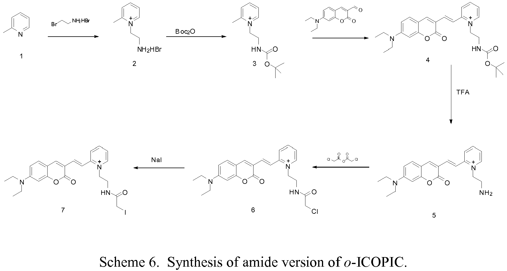
- Referring now to Scheme 6, a method of synthesizing an amide version of the presently disclosed o-ICOPIC dye is provided.
-
Compound 2. Compound 1 (2-picoline, 1.0 g) was dissolved in anhydrous methanol (5 mL). Bromoethylamine HBr (2.20 g) was added to the solution. The reaction was heated to reflux and allowed to react overnight. Upon cooling the next day, the product had precipitated. The precipitate was collected by filtration, washed with a small amount of methanol and allowed to air dry. The product (1.05 g) was obtained as a colorless solid. The product was confirmed by 1H NMR. 1H NMR of 2, (400 MHz, CD3OD) δ 9.03 (d, 1H), 8.50 (d, 1H), 8.07 (d, 1H), 8.00 (t, 1H), 4.99 (t, 2H), 3.64 (t, 2H), 2.98 (s, 3H). - Compound 3. Compound 2 (200 mg) and sodium carbonate (166 mg, 2eq) were dissolved in 5 mL of H2O. Boc2O (161 mg, 1.1 eq) was dissolved in 5 mL of dioxane and added dropwise to the aqueous solution. The reaction was allowed to stir overnight at room temperature. The next day the solvent was evaporated to dryness leaving a solid white residue. Methanol (10 mL) was added and the resulting solution was filtered to remove the sodium carbonate. The filtrate was evaporated to yield 190 mg of product (3), which was used without further purification. The product was confirmed by 1H NMR. 1H NMR of 3 (400 MHz, CD3OD), δ = 8.77 (d, 1H), 8.43 (t, 1H), 8.00 (d, 1H), 7.92 (t, 1H), 4.68 (t, 2H), 3.62 (t, 2H), 2.94 (s. 3H), 1.31 (s, 9H).
- Compound 4. Compound 3 (190 mg) and 7-diethylamino-coumarin-3-aldehyde (140 mg) were dissolved in 6 mL of methanol followed by the addition of 50 µL of piperidine. The reaction was heated to 50 °C and allowed to react overnight with stirring. The solvent was then evaporated and the remaining red residue was purified by column chromatography. The product was eluted with a 10% methanol in dichloromethane solution. Pure fractions were combined and evaporated to yield 65 mg of the desired product (4). The product was confirmed by 1H, 13C NMR and HRMS. 1H NMR of 4, (400 MHz, CDCl3) δ = 9.16 (d, 1H), 8.73 (s, 1H), 8.30 (d, 1H), 8.21 (t, 1H), 8.02 (d, 1H), 7.86 (d, 1H), 7.62 (m, 1H), 7.54 (d, 1H), 6.58 (dd, 1H), 6.42 (s, 1H), 5.10 (t, 2H), 3.76 (q, 2H), 3.42 (q, 4H), 1.25 (s, 9H), 1.21 (t, 6H). 13C NMR of 4, (400 MHz, CDCl3) δ = 161.4, 157.4, 156.7, 154.0, 152.8, 146.0, 145.8, 143.7, 140.2, 131.8, 125.0, 124.1, 115.0, 113.8, 109.6, 110.4, 97.1, 79.9, 57.7, 45.5, 40.1, 28.6, 12.8. HRMS of 4 observed = 464.2546. calculated = 464.25
-
Compound 5. Compound 4 (65 mg) was dissolved in a 1:1 solution of dichloromethane:trifluoroacetic acid (TFA) and allowed to stir for 1 hr. The red color was quenched instantly after adding the TFA solution. After 1 hr, the solvent was evaporated leaving an orange residue. The residue was dissolved in methanol and evaporated multiple times to remove most of the TFA, at which point the deep red color was present once again. The red residue product was dissolved in a small volume of water, followed by freezing and lyophilization. The product was confirmed by 1H, 13C NMR and HRMS. 1H NMR of 5 (400 MHz, CD3OD), δ = 8.80 (d, 1H), 8.43 (m, 2H), 8.30 (s, 1H), 7.93 (d, 1H), 7.82 (t, 1H), 7.81 (d, 1H), 7.51 (m, 1H), 6.80 (dd, 1H), 6.53 (dd, 1H), 5.07 (t, 2H), 3.65 (t, 2H), 3.52 (q, 4H), 1.23 (t, 6H). 13C NMR of 5 (400 MHz, CD3OD), δ = 163.3, 159.2, 156.2, 155.2, 149.2, 147.4, 147.1, 143.0, 133.2, 127.7, 126.9, 117.0, 115.3, 112.6, 111.2, 98.4, 56.7, 46.9, 40.0, 13.6. HRMS of 5: observed = 364.2022, calculated = 364.20. - Compound 6.
Compound 5 was dissolved in dichloromethane (3 mL) with a few drops of methanol added. Chloroacetic anhydride (10 eq) and pyridine (10 eq) were added to the solution. The reaction stirred for 2 hrs at which point another 10 eq of chloroacetic anhydride was added. After another 4 hrs, the solvent was evaporated and the reaction mixture was purified by silica chromatography using a methanol/dichloromethane solvent mixture. The desired product was eluted with a 10% methanol/dichloromethane mixture. The product was confirmed by 1H and 13C NMR and HRMS. 1H NMR of 6 (400 MHz, CD3OD), δ = 8.66 (d, 1H), 8.40 (m, 2H), 8.28 (s, 1H), 8.07 (d, 1H), 7.77 (m, 2H), 7.52 (d, 1H), 6.81 (dd, 1H), 6.57 (s, 1H), 4.82 (t, 2H), 3.99 (s, 2H), 3.84 (t, 2H), 3.54 (q, 4H), 1.24 (t, 6H). 13C NMR of 6 (400 MHz, CD3OD), δ = 169.0, 161.2, 157.1, 154.2, 153.1, 146.7, 145.4, 144.5, 140.0, 131.0, 125.2, 124.5, 115.9, 113.5, 110.4, 109.1, 96.4, 57.3, 44.9, 41.7, 38.9, 11.6. HRMS of 6: observed = 440.1743, calculated = 440.17. - Compound 7. The o-ICOPIC chloroacetamide (6) was then converted to the iodoacetamide by simple substitution using sodium iodide (NaI). o-ICOPIC chloroacetamide (9) was dissolved in 3 mL of 1:2 MeOH:Acetone solution. Sodium iodide (5 eq) was added to the mixture. This solution was covered in aluminum foil and allowed to reflux overnight. The following day, another 3 equivalents of NaI was added and allowed to reflux another 24 hrs. The next day, the solvent was evaporated under reduced pressure. The red solid was taken up in dichloromethane with a small amount of MeOH. The insoluble salts were filtered and the filtrate was evaporated once again. The red residue was dissolved in a small amount of 20% MeOH/DCM and added to a large volume diethyl ether with swirling to precipitate the dye. The precipitate was filtered as a red solid. The purified product (7) was confirmed by NMR and HRMS. 1H NMR of 7 (400 MHz, CD3OD), δ = 8.74 (d, 1H), 8.46 (m, 2H), 8.42 (t, 1H), 7.98 (d, 1H), 7.85 (d, 1H), 7.83 (m, 1H), 7.57 (d, 1H), 6.79 (d, 1H), 6.50 (s, 1H), 4.87 (t, 2H), 3.82 (t, 2H), 3.64 (s, 2H), 3.53 (q, 4H), 1.23 (t, 6H). 13C NMR of 7 (400 MHz, CD3OD), δ = 171.0, 161.4, 157.1, 153.8, 153.0, 146.7, 145.5, 144.4, 139.9, 131.1, 125.5, 124.9, 115.7, 113.4, 110.5, 109.1, 96.3, 57.3, 44.9, 41.7, 38.9, 11.8, -3.0. HRMS of 7: observed = 532.1097, calculated = 532.11.
- The stability of ortho and para-ICOPIC amide was monitored by both NMR and HPLC, initially at 37 °C for 7 days, followed by incubation at 55 °C for 7 days. Approximately 2 mg of each ICOPIC (1,2) was dissolved in 0.7 mL of CD3OD. The initial NMR spectrum was taken after the sample was prepared and the proton chemical shifts were assigned (color coded below). The prepared samples were then incubated in the NMR tube at 37 and 55 °C for 7 days each. Any change in molecular structure would be evident by loss of a proton peak and formation of a new peak. The NMR spectra in
Figures 4A-4C (for the para-ICOPIC amide) and 5A-5C (for the ortho-ICOPIC amide) are zoomed on the aromatic region since this is the most susceptible area for reactivity. The spectra show that no significant changes occur in either dye after incubation for 7 days. The NMR experiments also were done with D2O as solvent, which also showed no significant changes in structure (data not shown). - In addition to NMR analysis, the dyes were also analyzed by HPLC at two time points - once when freshly dissolved and second after incubation for 14 days (7 days at 37 °C plus 7 days at 55 °C). The HPLC chromatogram for p-ICOPIC is illustrated in
Figures 6A and 6B and the HPLC chromatogram for o-ICOPIC is shown inFigures 7A and 7B . Once again no new peaks are observed in the chromatogram. Note that the concentrations of the dye samples were not matched before and after incubation at the described temperatures. Thus, the Y-axis scales inFigures 6A, 6B ,7A, and 7B are arbitrary and only the position and number of peaks are relevant. - SM4 (1.2 mg) was mixed with approximately 50 eq. of o-ICOPIC amide. The solution was allowed to react for 4 hrs followed by purification by gel filtration (NAP-10). The UV/Vis spectrum after the labeling is shown in
Figure 8 . - The fluorescence of the SM4-o-ICOPIC system was measured in the presence of glucose ranging from 2.5 mM to 200 mM. The resulting spectra show a 25-nm red shift (see
Figure 9A ) and they were analyzed ratiometrically using the wavelength ranges of 550-570 nm and 650-670 nm for each spectrum. The ratio points were plotted against glucose concentration to give the hyperbolic binding curve shown inFigure 9B . - The following tables of values were determined by the hyperbolic fit:
R0 0.25 Kd 71.0 Qr* 6.86 Rinf 1.71 overall below 200mg% above 200mg% MPE (%) = 2.2 2.4 2.1 - SM4 (2.0 mg) was mixed with approximately 50 eq. of o-ICOPIC amide dissolved in 20 µL of DMSO. The solution was allowed to react for 4 hrs followed by purification by gel filtration (NAP-10). The UV/Vis spectrum after the labeling is shown in
Figure 10 . - The fluorescence of the W183C-o-ICOPIC system was measured in the presence of glucose ranging from 2.5 mM to 200 mM. The resulting spectra show a 25-nm red shift (see
Figure 11A ) and they were analyzed ratiometrically using the wavelength ranges of 550-570 nm and 650-670 nm for each spectrum. The ratio points were plotted against glucose concentration to give the hyperbolic binding curve shown inFigure 11B . - The following tables of values were determined by the hyperbolic fit:
Ro 0.24 Kd 54.4 Qr* 5.2 Rinf 1.25 overall below 200mg% above 200mg% MPE (%) = 2.0 1.6 2.2 - A fresh batch of Y10C GGBP (25 mg) was expressed and purified. Y10C (0.6 mg) was mixed with approximately 40 eq. of p-ICOPIC (0.28 mg) amide dissolved in 20 µL of DMSO. The solution was allowed to react for 4 hrs, at which point the progress of the reaction was checked by HPLC. After 4 hrs, only approximately 20% of the protein labeled. Another approximately 40 eq of p-ICOPIC amide was added and allowed to react overnight. The next day, the progress was checked once again by HPLC and the dye:protein ratio was approximately 0.45. The protein was purified by gel filtration (NAP-5). The UV/Vis spectrum after the labeling is shown in
Figure 12 . - The fluorescence of the Y10C-p-ICOPIC system was measured in the presence of glucose ranging from 1 mM to 100 mM. The resulting spectra show a 43-nm red shift (see
Figure 13A ) with a 5-fold decrease in fluorescence intensity. The spectra were analyzed ratiometrically using the wavelength ranges of 550-570 nm and 700-720 nm for each spectrum. The ratio points were plotted against glucose concentration to give the hyperbolic binding curve shown inFigure 13B . - The following tables of values were determined by the hyperbolic fit:
R0 0.04 Kd 12.0 Qr* 6.7 Rinf 0.29 overall below 200mg% above 200mg% MPE (%) = 8.8 14.2 4.5 - The fluorescence intensity of W183C-o-ICOPIC is compared to that of W183C-acrylodan in
Figure 14 . As described herein below, the fluorescence intensity of W183C-o-ICOPIC (dye:protein ratio = 0.8) is 1.5 times greater than that of W183C-acrylodan (dye:protein ratio = 1.2) at a protein concentration of 2 µM. If the dye:protein ratio is factored in, the increase in fluorescence intensity will likely be greater than 2 times for W183C-o-ICOPIC over W183C-acrylodan. The increase in fluorescence intensity can add to increased sensitivity of a sensor. The excitation wavelength for W183C-o-ICOPIC was 485 nm and the excitation wavelength for W183C-acrylodan was 380 nm. All other conditions were the same.
Claims (12)
- A fluorophore selected from the group consisting of:


- A biosensor compound selected from the group consisting of:


- The biosensor compound of claim 2, wherein the binding member is a binding protein, or wherein the binding protein is a periplasmic binding protein, or wherein the binding protein is selected from the group consisting of a glucose binding protein and a glucose/galactose binding protein (GGBP).
- The biosensor compound of claim 3, wherein the binding member is at least one fragment of a receptor, wherein the receptor is a protein in nature and is capable of binding to a suitable ligand via an active site, and wherein at least one amino acid residue of the fragment is located in a proximity of the active site and is a cysteine residue or is substituted with a cysteine residue.
- A method for determining the presence or amount of one or more analytes in a sample, the method comprising:(a) providing a biosensor compound having at least one mutated binding protein with a fluorophore covalently attached thereto through a thiol group of the mutated binding protein, wherein the biosensor compound is defined as in claims 2 or 3;(b) contacting the biosensor compound with a sample suspected of containing one or more analytes to bind the one or more analytes, if present, to the binding protein;(c) irradiating the sample suspected of containing one or more analytes with electromagnetic radiation to induce the fluorophore to fluoresce; and(d) detecting a fluorescence property to determine the presence or amount of one or more analytes in the sample.
- The method of claim 5, further comprising:(a) measuring a fluorescence intensity at a first emission wavelength before contacting the biosensor compound with a sample suspected of containing one or more analytes;(b) measuring a fluorescence intensity at a second emission wavelength after contacting the biosensor compound with a sample suspected of containing one or more analytes; and(c) determining the ratio of the second emission wavelength to the first emission wavelength to determine the presence or amount of one or more analytes in the sample.
- The method of claim 5, further comprising continuously:(a) contacting the binding protein with the sample suspected of containing one or more analytes;(b) irradiating the sample with electromagnetic radiation; and(c) detecting the fluorescence property.
- The method of claim 5, wherein the mutated binding protein undergoes a conformation change as a result of changes in analyte concentration of the sample suspected of containing one or more analytes and wherein the method detects changes in the fluorescence property as a result of changes in the analyte concentration.
- The method of claim 5, wherein the one or more analytes is selected from the group consisting of glucose, a fatty acid, and lactate.
- A biosensor device comprising the biosensor compound of claim 2.
- A reagent for determining the presence or amount of one or more analytes in a sample, the reagent comprising a biosensor compound of claim 2.
- A kit for determining the presence or amount of one or more analytes in a sample, the kit comprising the biosensor device of claim 10.
Applications Claiming Priority (2)
| Application Number | Priority Date | Filing Date | Title |
|---|---|---|---|
| US93950707P | 2007-05-22 | 2007-05-22 | |
| PCT/US2008/064346 WO2008147805A2 (en) | 2007-05-22 | 2008-05-21 | Dyes having ratiometric fluorescence response for detecting metabolites |
Publications (2)
| Publication Number | Publication Date |
|---|---|
| EP2150586A2 EP2150586A2 (en) | 2010-02-10 |
| EP2150586B1 true EP2150586B1 (en) | 2018-03-14 |
Family
ID=40075727
Family Applications (1)
| Application Number | Title | Priority Date | Filing Date |
|---|---|---|---|
| EP08756041.3A Not-in-force EP2150586B1 (en) | 2007-05-22 | 2008-05-21 | Dyes having ratiometric fluorescence response for detecting metabolites |
Country Status (4)
| Country | Link |
|---|---|
| US (1) | US8772047B2 (en) |
| EP (1) | EP2150586B1 (en) |
| JP (1) | JP5453248B2 (en) |
| WO (1) | WO2008147805A2 (en) |
Families Citing this family (18)
| Publication number | Priority date | Publication date | Assignee | Title |
|---|---|---|---|---|
| US9625458B2 (en) * | 2002-10-16 | 2017-04-18 | Duke University | Biosensor |
| AU2008213677A1 (en) | 2007-02-06 | 2008-08-14 | Glumetrics, Inc. | Optical systems and methods for rationmetric measurement of blood glucose concentration |
| JP5517919B2 (en) * | 2007-05-10 | 2014-06-11 | グルメトリクス、 インク. | Balanced non-consumable fluorescent sensor for immediate intravascular glucose measurement |
| US9023661B2 (en) * | 2007-10-18 | 2015-05-05 | Becton, Dickinson And Company | Visual glucose sensor and methods of use thereof |
| WO2009067626A1 (en) | 2007-11-21 | 2009-05-28 | Glumetrics, Inc. | Use of an equilibrium intravascular sensor to achieve tight glycemic control |
| EP2483679A4 (en) | 2009-09-30 | 2013-04-24 | Glumetrics Inc | Sensors with thromboresistant coating |
| US8741591B2 (en) | 2009-10-09 | 2014-06-03 | The Research Foundation For The State University Of New York | pH-insensitive glucose indicator protein |
| EP2545373B1 (en) * | 2010-03-11 | 2022-08-24 | Medtronic Minimed, Inc. | Measuring analyte concentration incorporating temperature and ph correction |
| US9574057B2 (en) | 2012-03-28 | 2017-02-21 | Becton, Dickinson And Company | Hydrogel adhesion to molded polymers |
| US10466247B2 (en) | 2012-11-20 | 2019-11-05 | Becton, Dickinson And Company | System and method for diagnosing sensor performance using analyte-independent ratiometric signals |
| US10379125B2 (en) | 2013-12-27 | 2019-08-13 | Becton, Dickinson And Company | System and method for dynamically calibrating and measuring analyte concentration in diabetes management monitors |
| EP3317278B1 (en) | 2015-07-01 | 2021-04-14 | Crinetics Pharmaceuticals, Inc. | Somatostatin modulators and uses thereof |
| CA3005831A1 (en) * | 2015-11-20 | 2017-05-26 | Duke University | Glucose biosensors and uses thereof |
| EP3658560A4 (en) | 2017-07-25 | 2021-01-06 | Crinetics Pharmaceuticals, Inc. | SOMATOSTATIN MODULATORS AND USES OF THESE LATEST |
| US20230134948A1 (en) * | 2020-04-02 | 2023-05-04 | Public University Corporation Nagoya City University | Staining method, microscopic observation method, staining agent and staining kit |
| CN113717164B (en) * | 2021-08-30 | 2022-05-10 | 安徽大学 | A red fluorescent probe and its preparation and its application in cell imaging |
| US12433962B2 (en) | 2021-11-03 | 2025-10-07 | The Hong Kong University Of Science And Technology | Probes for imaging b-amyloid and inhibition of β-amyloid aggregation |
| CN117777114A (en) * | 2023-12-22 | 2024-03-29 | 西藏自治区人民政府驻成都办事处医院 | A near-infrared cell membrane targeting dye based on the large Stokes shift of the coumarin skeleton and its preparation method and application |
Family Cites Families (30)
| Publication number | Priority date | Publication date | Assignee | Title |
|---|---|---|---|---|
| US3964871A (en) * | 1974-12-18 | 1976-06-22 | Becton, Dickinson And Company | Method and device for detecting glucose |
| US5001054A (en) * | 1986-06-26 | 1991-03-19 | Becton, Dickinson And Company | Method for monitoring glucose |
| US4981779A (en) * | 1986-06-26 | 1991-01-01 | Becton, Dickinson And Company | Apparatus for monitoring glucose |
| JPH0743379B2 (en) * | 1990-11-30 | 1995-05-15 | 株式会社島津製作所 | Immune reaction automatic analyzer |
| US5246867A (en) * | 1992-01-17 | 1993-09-21 | University Of Maryland At Baltimore | Determination and quantification of saccharides by luminescence lifetimes and energy transfer |
| US5630924A (en) * | 1995-04-20 | 1997-05-20 | Perseptive Biosystems, Inc. | Compositions, methods and apparatus for ultrafast electroseparation analysis |
| US5712223A (en) * | 1996-09-16 | 1998-01-27 | Eastman Kodak Company | Chromeme dyes for thermal imaging |
| EP1044374B1 (en) * | 1997-12-31 | 2008-10-08 | Duke University | Glucose biosensor |
| US6922576B2 (en) * | 1998-06-19 | 2005-07-26 | Becton, Dickinson And Company | Micro optical sensor device |
| AU759888B2 (en) * | 1998-07-17 | 2003-05-01 | University Of Maryland At Baltimore | Engineered proteins for analyte sensing |
| US6192891B1 (en) * | 1999-04-26 | 2001-02-27 | Becton Dickinson And Company | Integrated system including medication delivery pen, blood monitoring device, and lancer |
| FR2805820B1 (en) * | 2000-03-01 | 2009-09-11 | Pasteur Institut | BIOSENSORS, PROCESS FOR OBTAINING THEM AND THEIR APPLICATIONS |
| DE10160524A1 (en) * | 2001-12-05 | 2003-06-26 | Dyomics Gmbh | New fluorescent markers with a large Stokes shift |
| US7064103B2 (en) * | 2002-01-04 | 2006-06-20 | Becton, Dickinson And Company | Binding protein as biosensors |
| US20030153026A1 (en) * | 2002-01-04 | 2003-08-14 | Javier Alarcon | Entrapped binding protein as biosensors |
| US7851593B2 (en) * | 2002-01-04 | 2010-12-14 | Becton, Dickinson And Company | Binding proteins as biosensors |
| US6855556B2 (en) * | 2002-01-04 | 2005-02-15 | Becton, Dickinson And Company | Binding protein as biosensors |
| US9625458B2 (en) * | 2002-10-16 | 2017-04-18 | Duke University | Biosensor |
| DE10356130A1 (en) * | 2003-11-28 | 2005-06-23 | Dyomics Gmbh | New coumarin-based polymethine dyes with adjustable Stokes shift |
| US7563907B2 (en) * | 2002-12-05 | 2009-07-21 | Dyomics Gmbh | Compounds based on polymethines |
| USD491275S1 (en) * | 2003-01-15 | 2004-06-08 | Becton, Dickinson And Company | Blood glucose test strip vial |
| US7496392B2 (en) * | 2003-11-26 | 2009-02-24 | Becton, Dickinson And Company | Fiber optic device for sensing analytes |
| US7787923B2 (en) * | 2003-11-26 | 2010-08-31 | Becton, Dickinson And Company | Fiber optic device for sensing analytes and method of making same |
| WO2005102154A2 (en) * | 2004-04-16 | 2005-11-03 | Becton, Dickinson And Company | Blood glucose meter having integral lancet device and test strip storage vial for single handed use and methods for using same |
| US7563891B2 (en) * | 2004-05-21 | 2009-07-21 | Becton, Dickinson & Company | Long wavelength thiol-reactive fluorophores |
| EP1754059B1 (en) * | 2004-06-09 | 2010-08-04 | Becton, Dickinson and Company | Multianalyte sensor |
| US7625758B2 (en) * | 2005-01-26 | 2009-12-01 | Berkelman Thomas R | Coumarin-based cyanine dyes for non-specific protein binding |
| USD545439S1 (en) * | 2005-04-15 | 2007-06-26 | Becton, Dickinson And Company | Blood glucose meter |
| JP2007055956A (en) * | 2005-08-26 | 2007-03-08 | Tokyo Univ Of Pharmacy & Life Science | Novel quinoline compounds as fluorescent labeling reagents and hydrophobic field detection reagents |
| JP5337691B2 (en) * | 2006-04-20 | 2013-11-06 | ベクトン・ディキンソン・アンド・カンパニー | Thermostable protein and method for producing and using the same |
-
2008
- 2008-05-21 JP JP2010509518A patent/JP5453248B2/en not_active Expired - Fee Related
- 2008-05-21 US US12/124,553 patent/US8772047B2/en not_active Expired - Fee Related
- 2008-05-21 WO PCT/US2008/064346 patent/WO2008147805A2/en not_active Ceased
- 2008-05-21 EP EP08756041.3A patent/EP2150586B1/en not_active Not-in-force
Also Published As
| Publication number | Publication date |
|---|---|
| US8772047B2 (en) | 2014-07-08 |
| EP2150586A2 (en) | 2010-02-10 |
| US20080311675A1 (en) | 2008-12-18 |
| JP2010528292A (en) | 2010-08-19 |
| WO2008147805A2 (en) | 2008-12-04 |
| WO2008147805A3 (en) | 2009-06-04 |
| JP5453248B2 (en) | 2014-03-26 |
Similar Documents
| Publication | Publication Date | Title |
|---|---|---|
| US8772047B2 (en) | Dyes having ratiometric fluorescence response for detecting metabolites | |
| US9023661B2 (en) | Visual glucose sensor and methods of use thereof | |
| Ke et al. | A near-infrared naphthalimide fluorescent probe for targeting the lysosomes of liver cancer cells and specifically selecting HSA | |
| US11029320B2 (en) | Development and use of fluorescent probes of unbound bilirubin | |
| Zhu et al. | Constructing a NIR fluorescent probe for ratiometric imaging viscosity in mice and detecting blood viscosity in folliculitis mice and peritonitis mice | |
| Chen et al. | Wide-acidity-range pH fluorescence probes for evaluation of acidification in mitochondria and digestive tract mucosa | |
| Dey et al. | Renal clearable new NIR probe: precise quantification of albumin in biofluids and fatty liver disease state identification through tissue specific high contrast imaging in vivo | |
| Zheng et al. | A small, steady, rapid and selective TICT based fluorescent HSA sensor for pre-clinical diagnosis | |
| Pei et al. | TICT-based microenvironment-sensitive probe with turn-on red emission for human serum albumin detection and for targeting lipid droplet imaging | |
| Liu et al. | Near-infrared optical transducer for dynamic imaging of cerebrospinal fluid glucose in brain tumor | |
| Niu et al. | An endoplasmic reticulum-targeted fluorescent probe for detecting polarity and viscosity and its bioimaging in vitro and vivo | |
| JP2025143450A (en) | Single-step method, kit, and system for determining the concentration of unconjugated bilirubin in biological fluids | |
| JP5224536B2 (en) | Determination system for urinary tract tumor | |
| SIMONYAN | Development of Genetically Encoded Fluorescent Indicators for Quantitative Measurements of Intracellular Parameters | |
| Sayyed et al. | Development and In Vitro Evaluation of a Sensitive Multiplex Lateral Flow Automated Immunoassay for GFAP, H-FABP, and UCH-L1 in Mild Traumatic Brain Injury |
Legal Events
| Date | Code | Title | Description |
|---|---|---|---|
| PUAI | Public reference made under article 153(3) epc to a published international application that has entered the european phase |
Free format text: ORIGINAL CODE: 0009012 |
|
| 17P | Request for examination filed |
Effective date: 20091125 |
|
| AK | Designated contracting states |
Kind code of ref document: A2 Designated state(s): AT BE BG CH CY CZ DE DK EE ES FI FR GB GR HR HU IE IS IT LI LT LU LV MC MT NL NO PL PT RO SE SI SK TR |
|
| AX | Request for extension of the european patent |
Extension state: AL BA MK RS |
|
| 17Q | First examination report despatched |
Effective date: 20110124 |
|
| DAX | Request for extension of the european patent (deleted) | ||
| RAP1 | Party data changed (applicant data changed or rights of an application transferred) |
Owner name: BECTON, DICKINSON AND COMPANY |
|
| STAA | Information on the status of an ep patent application or granted ep patent |
Free format text: STATUS: EXAMINATION IS IN PROGRESS |
|
| GRAP | Despatch of communication of intention to grant a patent |
Free format text: ORIGINAL CODE: EPIDOSNIGR1 |
|
| STAA | Information on the status of an ep patent application or granted ep patent |
Free format text: STATUS: GRANT OF PATENT IS INTENDED |
|
| INTG | Intention to grant announced |
Effective date: 20170928 |
|
| GRAS | Grant fee paid |
Free format text: ORIGINAL CODE: EPIDOSNIGR3 |
|
| GRAA | (expected) grant |
Free format text: ORIGINAL CODE: 0009210 |
|
| STAA | Information on the status of an ep patent application or granted ep patent |
Free format text: STATUS: THE PATENT HAS BEEN GRANTED |
|
| AK | Designated contracting states |
Kind code of ref document: B1 Designated state(s): AT BE BG CH CY CZ DE DK EE ES FI FR GB GR HR HU IE IS IT LI LT LU LV MC MT NL NO PL PT RO SE SI SK TR |
|
| REG | Reference to a national code |
Ref country code: GB Ref legal event code: FG4D |
|
| REG | Reference to a national code |
Ref country code: CH Ref legal event code: EP Ref country code: AT Ref legal event code: REF Ref document number: 978840 Country of ref document: AT Kind code of ref document: T Effective date: 20180315 |
|
| REG | Reference to a national code |
Ref country code: IE Ref legal event code: FG4D |
|
| REG | Reference to a national code |
Ref country code: DE Ref legal event code: R096 Ref document number: 602008054438 Country of ref document: DE |
|
| REG | Reference to a national code |
Ref country code: FR Ref legal event code: PLFP Year of fee payment: 11 |
|
| REG | Reference to a national code |
Ref country code: NL Ref legal event code: MP Effective date: 20180314 |
|
| REG | Reference to a national code |
Ref country code: LT Ref legal event code: MG4D |
|
| PG25 | Lapsed in a contracting state [announced via postgrant information from national office to epo] |
Ref country code: ES Free format text: LAPSE BECAUSE OF FAILURE TO SUBMIT A TRANSLATION OF THE DESCRIPTION OR TO PAY THE FEE WITHIN THE PRESCRIBED TIME-LIMIT Effective date: 20180314 Ref country code: CY Free format text: LAPSE BECAUSE OF FAILURE TO SUBMIT A TRANSLATION OF THE DESCRIPTION OR TO PAY THE FEE WITHIN THE PRESCRIBED TIME-LIMIT Effective date: 20180314 Ref country code: NO Free format text: LAPSE BECAUSE OF FAILURE TO SUBMIT A TRANSLATION OF THE DESCRIPTION OR TO PAY THE FEE WITHIN THE PRESCRIBED TIME-LIMIT Effective date: 20180614 Ref country code: FI Free format text: LAPSE BECAUSE OF FAILURE TO SUBMIT A TRANSLATION OF THE DESCRIPTION OR TO PAY THE FEE WITHIN THE PRESCRIBED TIME-LIMIT Effective date: 20180314 Ref country code: LT Free format text: LAPSE BECAUSE OF FAILURE TO SUBMIT A TRANSLATION OF THE DESCRIPTION OR TO PAY THE FEE WITHIN THE PRESCRIBED TIME-LIMIT Effective date: 20180314 Ref country code: HR Free format text: LAPSE BECAUSE OF FAILURE TO SUBMIT A TRANSLATION OF THE DESCRIPTION OR TO PAY THE FEE WITHIN THE PRESCRIBED TIME-LIMIT Effective date: 20180314 |
|
| PGFP | Annual fee paid to national office [announced via postgrant information from national office to epo] |
Ref country code: DE Payment date: 20180430 Year of fee payment: 11 |
|
| REG | Reference to a national code |
Ref country code: AT Ref legal event code: MK05 Ref document number: 978840 Country of ref document: AT Kind code of ref document: T Effective date: 20180314 |
|
| PG25 | Lapsed in a contracting state [announced via postgrant information from national office to epo] |
Ref country code: BG Free format text: LAPSE BECAUSE OF FAILURE TO SUBMIT A TRANSLATION OF THE DESCRIPTION OR TO PAY THE FEE WITHIN THE PRESCRIBED TIME-LIMIT Effective date: 20180614 Ref country code: LV Free format text: LAPSE BECAUSE OF FAILURE TO SUBMIT A TRANSLATION OF THE DESCRIPTION OR TO PAY THE FEE WITHIN THE PRESCRIBED TIME-LIMIT Effective date: 20180314 Ref country code: GR Free format text: LAPSE BECAUSE OF FAILURE TO SUBMIT A TRANSLATION OF THE DESCRIPTION OR TO PAY THE FEE WITHIN THE PRESCRIBED TIME-LIMIT Effective date: 20180615 Ref country code: SE Free format text: LAPSE BECAUSE OF FAILURE TO SUBMIT A TRANSLATION OF THE DESCRIPTION OR TO PAY THE FEE WITHIN THE PRESCRIBED TIME-LIMIT Effective date: 20180314 |
|
| PGFP | Annual fee paid to national office [announced via postgrant information from national office to epo] |
Ref country code: FR Payment date: 20180502 Year of fee payment: 11 |
|
| PG25 | Lapsed in a contracting state [announced via postgrant information from national office to epo] |
Ref country code: IT Free format text: LAPSE BECAUSE OF FAILURE TO SUBMIT A TRANSLATION OF THE DESCRIPTION OR TO PAY THE FEE WITHIN THE PRESCRIBED TIME-LIMIT Effective date: 20180314 Ref country code: RO Free format text: LAPSE BECAUSE OF FAILURE TO SUBMIT A TRANSLATION OF THE DESCRIPTION OR TO PAY THE FEE WITHIN THE PRESCRIBED TIME-LIMIT Effective date: 20180314 Ref country code: PL Free format text: LAPSE BECAUSE OF FAILURE TO SUBMIT A TRANSLATION OF THE DESCRIPTION OR TO PAY THE FEE WITHIN THE PRESCRIBED TIME-LIMIT Effective date: 20180314 Ref country code: NL Free format text: LAPSE BECAUSE OF FAILURE TO SUBMIT A TRANSLATION OF THE DESCRIPTION OR TO PAY THE FEE WITHIN THE PRESCRIBED TIME-LIMIT Effective date: 20180314 Ref country code: EE Free format text: LAPSE BECAUSE OF FAILURE TO SUBMIT A TRANSLATION OF THE DESCRIPTION OR TO PAY THE FEE WITHIN THE PRESCRIBED TIME-LIMIT Effective date: 20180314 |
|
| PGFP | Annual fee paid to national office [announced via postgrant information from national office to epo] |
Ref country code: GB Payment date: 20180427 Year of fee payment: 11 |
|
| PG25 | Lapsed in a contracting state [announced via postgrant information from national office to epo] |
Ref country code: AT Free format text: LAPSE BECAUSE OF FAILURE TO SUBMIT A TRANSLATION OF THE DESCRIPTION OR TO PAY THE FEE WITHIN THE PRESCRIBED TIME-LIMIT Effective date: 20180314 Ref country code: CZ Free format text: LAPSE BECAUSE OF FAILURE TO SUBMIT A TRANSLATION OF THE DESCRIPTION OR TO PAY THE FEE WITHIN THE PRESCRIBED TIME-LIMIT Effective date: 20180314 Ref country code: SK Free format text: LAPSE BECAUSE OF FAILURE TO SUBMIT A TRANSLATION OF THE DESCRIPTION OR TO PAY THE FEE WITHIN THE PRESCRIBED TIME-LIMIT Effective date: 20180314 |
|
| REG | Reference to a national code |
Ref country code: CH Ref legal event code: PL |
|
| REG | Reference to a national code |
Ref country code: DE Ref legal event code: R097 Ref document number: 602008054438 Country of ref document: DE |
|
| PG25 | Lapsed in a contracting state [announced via postgrant information from national office to epo] |
Ref country code: PT Free format text: LAPSE BECAUSE OF FAILURE TO SUBMIT A TRANSLATION OF THE DESCRIPTION OR TO PAY THE FEE WITHIN THE PRESCRIBED TIME-LIMIT Effective date: 20180716 |
|
| PLBE | No opposition filed within time limit |
Free format text: ORIGINAL CODE: 0009261 |
|
| STAA | Information on the status of an ep patent application or granted ep patent |
Free format text: STATUS: NO OPPOSITION FILED WITHIN TIME LIMIT |
|
| REG | Reference to a national code |
Ref country code: BE Ref legal event code: MM Effective date: 20180531 |
|
| PG25 | Lapsed in a contracting state [announced via postgrant information from national office to epo] |
Ref country code: DK Free format text: LAPSE BECAUSE OF FAILURE TO SUBMIT A TRANSLATION OF THE DESCRIPTION OR TO PAY THE FEE WITHIN THE PRESCRIBED TIME-LIMIT Effective date: 20180314 Ref country code: MC Free format text: LAPSE BECAUSE OF FAILURE TO SUBMIT A TRANSLATION OF THE DESCRIPTION OR TO PAY THE FEE WITHIN THE PRESCRIBED TIME-LIMIT Effective date: 20180314 |
|
| 26N | No opposition filed |
Effective date: 20181217 |
|
| REG | Reference to a national code |
Ref country code: IE Ref legal event code: MM4A |
|
| PG25 | Lapsed in a contracting state [announced via postgrant information from national office to epo] |
Ref country code: SI Free format text: LAPSE BECAUSE OF FAILURE TO SUBMIT A TRANSLATION OF THE DESCRIPTION OR TO PAY THE FEE WITHIN THE PRESCRIBED TIME-LIMIT Effective date: 20180314 Ref country code: LI Free format text: LAPSE BECAUSE OF NON-PAYMENT OF DUE FEES Effective date: 20180531 Ref country code: CH Free format text: LAPSE BECAUSE OF NON-PAYMENT OF DUE FEES Effective date: 20180531 |
|
| PG25 | Lapsed in a contracting state [announced via postgrant information from national office to epo] |
Ref country code: LU Free format text: LAPSE BECAUSE OF NON-PAYMENT OF DUE FEES Effective date: 20180521 |
|
| PG25 | Lapsed in a contracting state [announced via postgrant information from national office to epo] |
Ref country code: IE Free format text: LAPSE BECAUSE OF NON-PAYMENT OF DUE FEES Effective date: 20180521 |
|
| PG25 | Lapsed in a contracting state [announced via postgrant information from national office to epo] |
Ref country code: BE Free format text: LAPSE BECAUSE OF NON-PAYMENT OF DUE FEES Effective date: 20180531 |
|
| REG | Reference to a national code |
Ref country code: DE Ref legal event code: R119 Ref document number: 602008054438 Country of ref document: DE |
|
| GBPC | Gb: european patent ceased through non-payment of renewal fee |
Effective date: 20190521 |
|
| PG25 | Lapsed in a contracting state [announced via postgrant information from national office to epo] |
Ref country code: MT Free format text: LAPSE BECAUSE OF NON-PAYMENT OF DUE FEES Effective date: 20180521 |
|
| PG25 | Lapsed in a contracting state [announced via postgrant information from national office to epo] |
Ref country code: TR Free format text: LAPSE BECAUSE OF FAILURE TO SUBMIT A TRANSLATION OF THE DESCRIPTION OR TO PAY THE FEE WITHIN THE PRESCRIBED TIME-LIMIT Effective date: 20180314 |
|
| PG25 | Lapsed in a contracting state [announced via postgrant information from national office to epo] |
Ref country code: DE Free format text: LAPSE BECAUSE OF NON-PAYMENT OF DUE FEES Effective date: 20191203 Ref country code: GB Free format text: LAPSE BECAUSE OF NON-PAYMENT OF DUE FEES Effective date: 20190521 |
|
| PG25 | Lapsed in a contracting state [announced via postgrant information from national office to epo] |
Ref country code: HU Free format text: LAPSE BECAUSE OF FAILURE TO SUBMIT A TRANSLATION OF THE DESCRIPTION OR TO PAY THE FEE WITHIN THE PRESCRIBED TIME-LIMIT; INVALID AB INITIO Effective date: 20080521 |
|
| PG25 | Lapsed in a contracting state [announced via postgrant information from national office to epo] |
Ref country code: FR Free format text: LAPSE BECAUSE OF NON-PAYMENT OF DUE FEES Effective date: 20190531 |
|
| PG25 | Lapsed in a contracting state [announced via postgrant information from national office to epo] |
Ref country code: IS Free format text: LAPSE BECAUSE OF FAILURE TO SUBMIT A TRANSLATION OF THE DESCRIPTION OR TO PAY THE FEE WITHIN THE PRESCRIBED TIME-LIMIT Effective date: 20180714 |