EP2149358B1 - Use of oxygen for the treatment of wounds - Google Patents
Use of oxygen for the treatment of wounds Download PDFInfo
- Publication number
- EP2149358B1 EP2149358B1 EP09014450.2A EP09014450A EP2149358B1 EP 2149358 B1 EP2149358 B1 EP 2149358B1 EP 09014450 A EP09014450 A EP 09014450A EP 2149358 B1 EP2149358 B1 EP 2149358B1
- Authority
- EP
- European Patent Office
- Prior art keywords
- oxygen
- wound
- microcatheter
- containing fluid
- tissue
- Prior art date
- Legal status (The legal status is an assumption and is not a legal conclusion. Google has not performed a legal analysis and makes no representation as to the accuracy of the status listed.)
- Ceased
Links
- QVGXLLKOCUKJST-UHFFFAOYSA-N atomic oxygen Chemical compound [O] QVGXLLKOCUKJST-UHFFFAOYSA-N 0.000 title claims description 91
- 229910052760 oxygen Inorganic materials 0.000 title claims description 91
- 239000001301 oxygen Substances 0.000 title claims description 91
- 208000027418 Wounds and injury Diseases 0.000 title description 88
- 206010052428 Wound Diseases 0.000 title description 87
- 238000011282 treatment Methods 0.000 title description 8
- 239000012530 fluid Substances 0.000 claims description 45
- XLYOFNOQVPJJNP-UHFFFAOYSA-N water Substances O XLYOFNOQVPJJNP-UHFFFAOYSA-N 0.000 claims description 20
- 239000010410 layer Substances 0.000 claims description 7
- AEMRFAOFKBGASW-UHFFFAOYSA-N Glycolic acid Polymers OCC(O)=O AEMRFAOFKBGASW-UHFFFAOYSA-N 0.000 claims description 2
- 229920000954 Polyglycolide Polymers 0.000 claims description 2
- 238000004891 communication Methods 0.000 claims description 2
- 229920000747 poly(lactic acid) Polymers 0.000 claims description 2
- 229920001610 polycaprolactone Polymers 0.000 claims description 2
- 229920000642 polymer Polymers 0.000 claims description 2
- 239000002344 surface layer Substances 0.000 claims description 2
- 210000001519 tissue Anatomy 0.000 description 30
- 239000007789 gas Substances 0.000 description 14
- 239000007788 liquid Substances 0.000 description 11
- MYMOFIZGZYHOMD-UHFFFAOYSA-N Dioxygen Chemical compound O=O MYMOFIZGZYHOMD-UHFFFAOYSA-N 0.000 description 10
- 206010063560 Excessive granulation tissue Diseases 0.000 description 10
- 210000001126 granulation tissue Anatomy 0.000 description 10
- 229910001882 dioxygen Inorganic materials 0.000 description 8
- 230000029663 wound healing Effects 0.000 description 8
- 238000002347 injection Methods 0.000 description 7
- 239000007924 injection Substances 0.000 description 7
- 238000000034 method Methods 0.000 description 6
- 238000001802 infusion Methods 0.000 description 5
- 230000000699 topical effect Effects 0.000 description 5
- 230000003179 granulation Effects 0.000 description 4
- 238000005469 granulation Methods 0.000 description 4
- 231100000397 ulcer Toxicity 0.000 description 4
- 108010010803 Gelatin Proteins 0.000 description 3
- 229920005830 Polyurethane Foam Polymers 0.000 description 3
- 208000025865 Ulcer Diseases 0.000 description 3
- 239000000853 adhesive Substances 0.000 description 3
- 230000001070 adhesive effect Effects 0.000 description 3
- 230000015572 biosynthetic process Effects 0.000 description 3
- 230000001684 chronic effect Effects 0.000 description 3
- 229920000159 gelatin Polymers 0.000 description 3
- 239000008273 gelatin Substances 0.000 description 3
- 235000019322 gelatine Nutrition 0.000 description 3
- 235000011852 gelatine desserts Nutrition 0.000 description 3
- 230000001338 necrotic effect Effects 0.000 description 3
- 239000011496 polyurethane foam Substances 0.000 description 3
- 239000000243 solution Substances 0.000 description 3
- WSFSSNUMVMOOMR-UHFFFAOYSA-N Formaldehyde Chemical compound O=C WSFSSNUMVMOOMR-UHFFFAOYSA-N 0.000 description 2
- 206010040943 Skin Ulcer Diseases 0.000 description 2
- 239000002390 adhesive tape Substances 0.000 description 2
- 239000007864 aqueous solution Substances 0.000 description 2
- 239000008280 blood Substances 0.000 description 2
- 210000004369 blood Anatomy 0.000 description 2
- 230000008878 coupling Effects 0.000 description 2
- 238000010168 coupling process Methods 0.000 description 2
- 238000005859 coupling reaction Methods 0.000 description 2
- 230000006378 damage Effects 0.000 description 2
- 230000008021 deposition Effects 0.000 description 2
- 230000000694 effects Effects 0.000 description 2
- 210000000416 exudates and transudate Anatomy 0.000 description 2
- 230000003902 lesion Effects 0.000 description 2
- 239000000203 mixture Substances 0.000 description 2
- 239000004810 polytetrafluoroethylene Substances 0.000 description 2
- 229920001343 polytetrafluoroethylene Polymers 0.000 description 2
- 230000001737 promoting effect Effects 0.000 description 2
- 239000003642 reactive oxygen metabolite Substances 0.000 description 2
- 239000002904 solvent Substances 0.000 description 2
- WZUVPPKBWHMQCE-XJKSGUPXSA-N (+)-haematoxylin Chemical compound C12=CC(O)=C(O)C=C2C[C@]2(O)[C@H]1C1=CC=C(O)C(O)=C1OC2 WZUVPPKBWHMQCE-XJKSGUPXSA-N 0.000 description 1
- 206010002091 Anaesthesia Diseases 0.000 description 1
- GHXZTYHSJHQHIJ-UHFFFAOYSA-N Chlorhexidine Chemical compound C=1C=C(Cl)C=CC=1NC(N)=NC(N)=NCCCCCCN=C(N)N=C(N)NC1=CC=C(Cl)C=C1 GHXZTYHSJHQHIJ-UHFFFAOYSA-N 0.000 description 1
- 102000008186 Collagen Human genes 0.000 description 1
- 108010035532 Collagen Proteins 0.000 description 1
- 206010056340 Diabetic ulcer Diseases 0.000 description 1
- 206010015150 Erythema Diseases 0.000 description 1
- WZUVPPKBWHMQCE-UHFFFAOYSA-N Haematoxylin Natural products C12=CC(O)=C(O)C=C2CC2(O)C1C1=CC=C(O)C(O)=C1OC2 WZUVPPKBWHMQCE-UHFFFAOYSA-N 0.000 description 1
- 206010021143 Hypoxia Diseases 0.000 description 1
- 206010061218 Inflammation Diseases 0.000 description 1
- 208000031074 Reinjury Diseases 0.000 description 1
- 206010072170 Skin wound Diseases 0.000 description 1
- FAPWRFPIFSIZLT-UHFFFAOYSA-M Sodium chloride Chemical compound [Na+].[Cl-] FAPWRFPIFSIZLT-UHFFFAOYSA-M 0.000 description 1
- OUUQCZGPVNCOIJ-UHFFFAOYSA-M Superoxide Chemical compound [O-][O] OUUQCZGPVNCOIJ-UHFFFAOYSA-M 0.000 description 1
- 208000000558 Varicose Ulcer Diseases 0.000 description 1
- 230000002745 absorbent Effects 0.000 description 1
- 239000002250 absorbent Substances 0.000 description 1
- 230000001154 acute effect Effects 0.000 description 1
- 125000003545 alkoxy group Chemical group 0.000 description 1
- 238000001949 anaesthesia Methods 0.000 description 1
- 230000037005 anaesthesia Effects 0.000 description 1
- 238000004458 analytical method Methods 0.000 description 1
- 230000033115 angiogenesis Effects 0.000 description 1
- 230000009286 beneficial effect Effects 0.000 description 1
- 210000004027 cell Anatomy 0.000 description 1
- 229960003260 chlorhexidine Drugs 0.000 description 1
- 229920001436 collagen Polymers 0.000 description 1
- 230000000295 complement effect Effects 0.000 description 1
- 238000010276 construction Methods 0.000 description 1
- 230000008602 contraction Effects 0.000 description 1
- 238000001804 debridement Methods 0.000 description 1
- 230000002939 deleterious effect Effects 0.000 description 1
- 230000001419 dependent effect Effects 0.000 description 1
- 230000006866 deterioration Effects 0.000 description 1
- 208000037265 diseases, disorders, signs and symptoms Diseases 0.000 description 1
- 208000035475 disorder Diseases 0.000 description 1
- 238000004090 dissolution Methods 0.000 description 1
- 239000003814 drug Substances 0.000 description 1
- 230000002500 effect on skin Effects 0.000 description 1
- 238000005868 electrolysis reaction Methods 0.000 description 1
- YQGOJNYOYNNSMM-UHFFFAOYSA-N eosin Chemical compound [Na+].OC(=O)C1=CC=CC=C1C1=C2C=C(Br)C(=O)C(Br)=C2OC2=C(Br)C(O)=C(Br)C=C21 YQGOJNYOYNNSMM-UHFFFAOYSA-N 0.000 description 1
- 231100000321 erythema Toxicity 0.000 description 1
- 230000005284 excitation Effects 0.000 description 1
- 210000002950 fibroblast Anatomy 0.000 description 1
- 239000003349 gelling agent Substances 0.000 description 1
- 150000004820 halides Chemical class 0.000 description 1
- 229910052736 halogen Inorganic materials 0.000 description 1
- -1 halogen ions Chemical class 0.000 description 1
- 230000009931 harmful effect Effects 0.000 description 1
- 230000035876 healing Effects 0.000 description 1
- 150000002432 hydroperoxides Chemical class 0.000 description 1
- 238000002639 hyperbaric oxygen therapy Methods 0.000 description 1
- 230000007954 hypoxia Effects 0.000 description 1
- 238000010191 image analysis Methods 0.000 description 1
- 238000001727 in vivo Methods 0.000 description 1
- 230000004054 inflammatory process Effects 0.000 description 1
- 208000014674 injury Diseases 0.000 description 1
- 150000002500 ions Chemical class 0.000 description 1
- 239000002502 liposome Substances 0.000 description 1
- 230000004089 microcirculation Effects 0.000 description 1
- 238000000520 microinjection Methods 0.000 description 1
- 239000012188 paraffin wax Substances 0.000 description 1
- 229950011087 perflunafene Drugs 0.000 description 1
- UWEYRJFJVCLAGH-IJWZVTFUSA-N perfluorodecalin Chemical compound FC1(F)C(F)(F)C(F)(F)C(F)(F)[C@@]2(F)C(F)(F)C(F)(F)C(F)(F)C(F)(F)[C@@]21F UWEYRJFJVCLAGH-IJWZVTFUSA-N 0.000 description 1
- 230000002093 peripheral effect Effects 0.000 description 1
- 230000001766 physiological effect Effects 0.000 description 1
- 239000002504 physiological saline solution Substances 0.000 description 1
- 239000011148 porous material Substances 0.000 description 1
- 230000008092 positive effect Effects 0.000 description 1
- 230000035755 proliferation Effects 0.000 description 1
- 230000002035 prolonged effect Effects 0.000 description 1
- 229920006395 saturated elastomer Polymers 0.000 description 1
- 230000003068 static effect Effects 0.000 description 1
- 238000003786 synthesis reaction Methods 0.000 description 1
- TXEYQDLBPFQVAA-UHFFFAOYSA-N tetrafluoromethane Chemical compound FC(F)(F)F TXEYQDLBPFQVAA-UHFFFAOYSA-N 0.000 description 1
- 230000001225 therapeutic effect Effects 0.000 description 1
Images

Classifications
-
- A—HUMAN NECESSITIES
- A61—MEDICAL OR VETERINARY SCIENCE; HYGIENE
- A61M—DEVICES FOR INTRODUCING MEDIA INTO, OR ONTO, THE BODY; DEVICES FOR TRANSDUCING BODY MEDIA OR FOR TAKING MEDIA FROM THE BODY; DEVICES FOR PRODUCING OR ENDING SLEEP OR STUPOR
- A61M35/00—Devices for applying media, e.g. remedies, on the human body
-
- A—HUMAN NECESSITIES
- A61—MEDICAL OR VETERINARY SCIENCE; HYGIENE
- A61P—SPECIFIC THERAPEUTIC ACTIVITY OF CHEMICAL COMPOUNDS OR MEDICINAL PREPARATIONS
- A61P17/00—Drugs for dermatological disorders
- A61P17/02—Drugs for dermatological disorders for treating wounds, ulcers, burns, scars, keloids, or the like
-
- A—HUMAN NECESSITIES
- A61—MEDICAL OR VETERINARY SCIENCE; HYGIENE
- A61H—PHYSICAL THERAPY APPARATUS, e.g. DEVICES FOR LOCATING OR STIMULATING REFLEX POINTS IN THE BODY; ARTIFICIAL RESPIRATION; MASSAGE; BATHING DEVICES FOR SPECIAL THERAPEUTIC OR HYGIENIC PURPOSES OR SPECIFIC PARTS OF THE BODY
- A61H33/00—Bathing devices for special therapeutic or hygienic purposes
- A61H33/14—Devices for gas baths with ozone, hydrogen, or the like
Definitions
- the present invention relates to an apparatus for use in for treating wounds and lesions on a patient's body by introducing an oxygen-containing fluid into a tissue beneath the surface of the wound or lesion.
- Treatments based on inhalation of oxygen while considered effective, are complex and entail a risk from the potential harmful effects of prolonged hypoxia.
- Numerous wound treatment devices are known that administer oxygen gas topically to wound surfaces. Examples can be found in GB-A-2409412 , WO-A-9853778 , US-B-6179804 , US-A-4624656 , US-A-4224941 , US-A-5662625 , US-A-5810795 , FR-A-2656218 and WO-A-9741816 .
- These devices generally comprise an impermeable sheet having a peripheral region for attachment in gas-tight fashion around the wound, for example with adhesive, to form an enclosed chamber over the wound.
- the devices further comprise a gas inlet for introducing oxygen gas into the chamber above the wound, and optionally an outlet for removal of gas from the chamber.
- the devices can be operated with a static charge of gaseous oxygen, or with a continuous flow of gaseous oxygen over the wound. It will be appreciated that such devices are relatively bulky and prone to leakage, and can interfere with dressing changes and removal of wound exudates.
- US-A-6139876 describes a gelatin with increased oxygen content for use as a topical wound dressing.
- the gelatin comprises a gelling agent, a solvent, and microbubbles of oxygen in a substantially even distribution with a partial pressure exceeding normal atmospheric pressure, wherein the surface tension of the gelatin is sufficiently high to retain at least a portion of the overpressure of the oxygen throughout a predetermined period of time after having been exposed to an atmospheric environment.
- US20020160053 describes an aqueous solution for promoting the growth of tissue cells at wound sites.
- This solution comprises water containing at least 1 to 500 ppm of active oxygen and 10 to 10000 ppm of halogen ions.
- the solution is produced by electrolysis of aqueous halide solutions.
- the active oxygen can include singlet oxygen formed by excitation of triplet oxygen, superoxide formed by reduction of oxygen by a single electron, and hydroxy radical, as well as hypochlorous ions, organic peroxy radicals (ROO.), alkoxy radioals (RO.), and organic hydroperoxides (ROOH).
- ROO. organic peroxy radicals
- RO. alkoxy radioals
- ROOH organic hydroperoxides
- US-A-20050125035 describes an active suture having an internal passageway for post-surgical delivery of active fluids.
- the active fluids may comprise a wide range of medicaments, including oxygen-rich liquids to assist wound healing.
- the present invention provides an apparatus comprising: a source of an oxygen-containing fluid selected from gaseous oxygen or water containing at least 10mg per liter of dissolved oxygen; and a microcatheter having an inlet aperture in fluid communication with the source of oxygen-containing fluid and having one or more outlet apertures, wherein at least a region of the microcatheter has an effective internal diameter of less than 75 micrometers characterised in that said outlet apertures are located in a tip region of the microcatheter for injecting an effective amount of an oxygen-containing fluid selectively and directly into a layer of wound tissue located below a surface layer of a wound, for use in a method of treatment of a wound.
- the inventive apparatus is used in a method comprising the step of injecting an effective amount of an oxygen-containing fluid into said wound at a location below a surface of said wound, said step of injecting being carried out through a conduit having one or more outlets for said fluid, wherein substantially all of said outlets are positioned below the surface of the wound during said step of injecting.
- the apparatus of the present invention differs from the use of active sutures to deliver oxygen rich liquids, because the apparatus of the present invention delivers the oxygen rich liquid directly and selectively to the wound tissue below the surface of the wound, without any of the damage to surrounding healthy tissue that is caused by suturing.
- the apparatus of the present invention delivers the oxygen directly to the granulation tissue layer of the wound, where it is most effective.
- the present invention is especially useful for the treatment of chronic wounds such as ulcers that are not normally sutured.
- the apertured sutures of US-A-20050125035 deliver oxygen rich fluid nonselectively along the length of the suture. It follows also that the apertured sutures cannot be used to deliver an oxygen enriched fluid under pressure to a predetermined tissue layer under the surface of the wound, since the fluid under pressure will leak out of exposed parts of the apertured suture.
- microcatheter refers to any tubular body having a small internal and external diameter, including micro-injection needles, syringes, hollow sutures, and the like. Suitable microcatheters are described, for example, in US-B-6626885 .
- the conduit is flexible.
- the conduit has a substantially cylindrical lumen with one or more outlet apertures in a tip region thereof, but no apertures intermediate the inlet and the tip apertures, whereby all of the fluid injected into the inlet of the catheter flows out near the tip, i.e. below the surface of the wound in use.
- multiple conduits, or a single conduit with multiple outlets may be used to deliver the oxygen-containing fluid to the wound.
- the conduit is branched to permit oxygen injection at a plurality of locations beneath the surface of the wound.
- At least a tip region of the conduit is bioabsorbable, whereby the tip region can be left in the wound for resorption in vivo if necessary.
- at least the tip region of the may be made from a bioabsorbable polymer such as polylactide/polyglycolide, or poly-epsilon-caprolactone.
- the conduit can function as a flow regulator for the oxygen-containing fluid under pressure, whereby the fluid is delivered to the tip of the conduit at high pressure and low flow rate.
- the high pressure achieves a high dissolution of oxygen and resulting high concentration of the oxygen in the physiological fluid proximate to the conduit tip.
- the low flow rate inhibits liquid or gas bolus formation inside the wound.
- at least a tip region of the conduit suitably has an effective internal diameter of less than about 75 micrometers.
- at least a region of the conduit has an effective internal diameter of less than about 50 micrometers, for example from about 5 to about 40 micrometers.
- the term "effective internal diameter" refers to the equivalent open circular internal diameter.
- the said region of the conduit is located proximate to the tip (outlet) of the conduit.
- the conduit may comprise a length of tubing having internal diameter greater than about 100 micrometers, which may be branched, the end or ends of which are joined by a suitable coupling to respective microtubes (microcatheters) having internal diameter less than about 75 micrometers for flow control, the open end or ends of the microtubes being inserted below the surface of the wound.
- the microtubes are suitably bioabsorbable.
- the oxygen-containing fluid may be a liquid or a gas.
- the liquid is preferably aqueous, for example it may be water or a physiologically acceptable saline solution.
- the oxygen may be dissolved, preferably under pressure, to give a concentration of at least about 10 mgO 2 per liter, preferably at least about 20 mgO 2 per liter, for example from about 20 mgO 2 per liter to about 30 mgO 2 per liter. Oxygen concentrations in this range can be achieved by dissolving oxygen at a gas pressure of about 3 bar in water.
- the source of the fluid may be a portable pressurized reservoir containing a mixture of both fluid and oxygen bearing gas.
- the oxygen may be trapped in gaseous or dissolved form inside liposomes in the aqueous solution.
- the liquid may be a medically acceptable perfluorocarbon solvent such as perfluorodecalin containing dissolved oxygen.
- the oxygen-containing fluid may be any oxygen-containing gas.
- the gas comprises more than about 22mol.% of oxygen, more suitably at least about 40mol% oxygen, for example at least about 60mol.% oxygen, and in some embodiments it is substantially pure oxygen.
- the oxygen gas may be humidified before injection into the wound.
- the source of gaseous oxygen may comprise a pressurized oxygen tank, which may be portable.
- the source of oxygen-enriched fluid supplies the fluid at a pressure of from about 0.1 bar to about 5 bar (gauge), more suitably from about 0.5 bar to about 2 bar, for example about 1 bar.
- Suitable sources comprise an infusion pump, for example a spring-driven infusion pump. Pumps of this type are described, for example, in WO-A-0038767 , the entire content of which is incorporated herein by reference.
- the step of injecting the oxygen-containing fluid into the wound tissue is performed in substantially continuous fashion for a period of from about 1 hour to about 30 days, preferably from about 1 day to about 14 days.
- the pressure and delivery rate of the injection are adjusted to prevent the formation of a bolus of liquid or gas inside the wound.
- the effective internal diameter of the conduit, D, length of the conduit, L, the gauge pressure of the fluid used in the system, ⁇ P, and viscosity of the fluid, ⁇ all impact the delivery rate, V, according to the Hagen-Poiseuille equation.
- V ⁇ ⁇ ⁇ ⁇ PD 4 128 ⁇ ⁇ L
- Typical delivery rates per wound for gaseous oxygen are suitably about 0.5ml/hr to about 4ml/hr, typically about 1ml/hr to about 3ml/hr, for example about 2ml/hr.
- Typical delivery rates per wound for oxygen-enriched liquids are suitably about 0.05ml/hr to about 1ml/hr, typically about 0.1ml/hr to about 0.4ml/hr, for example about 0.15ml/hr.
- the conduit is arranged to deliver the oxygen below any surface necrotic tissue of the wound.
- the conduit is arranged to deliver the oxygen into or onto a granulation tissue in the wound.
- the oxygen injection is performed at a depth of from about 0.5mm to about 10mm below the surface of the wound, preferably from about 1mm to about 5 mm.
- the invention thereby provides an apparatus which provides a good method of promoting granulation tissue beneath necrotic tissue in instances were debridement would be difficult or painful for the patient.
- by injecting at this depth it ensures that the oxygenated fluid is delivered directly to healthy tissue where it may be effective.
- the wound is a chronic wound.
- Suitable chronic wounds include dermal ulcers such as a venous ulcers, diabetic ulcers or decubitis ulcers.
- the invention may also be useful for the treatment of acute wounds and burns.
- the apparatus comprises a spring driven infusion pump 1 having a valve outlet 2.
- a branched extension line 3 of PTFE tubing having an internal diameter of about 1mm has an inlet connector 4 that can be inserted in pressure-tight fashion into the pump outlet 2.
- the outlets at the ends of the branches of the extension line 3 are equipped with valves 5 and Luer-lock fittings for attachment with complementary fittings 7 on flow restrictor tubing 6.
- the flow restrictor tubing 6 is made of PVC with internal diameter about 150 micrometers.
- the length of the flow restrictor tubing is selected for the fluid being injected. For example, in this embodiment the length of the flow restrictor tubing is about 15cm for liquid injection and about 40cm for gas injection.
- the distal end 8 of the flow restrictor tubing 6 is joined in fluid-tight fashion to the inlet end of a microtube 9 formed of PTFE and having outside diameter about 150 micrometers, internal diameter about 49 micrometers, and length about 10cm.
- the tip 10 of the microtube 9 is inserted into the granulation tissue of a wound, below the necrotic surface tissue of the wound and oxygen-enriched fluid is delivered through the microtube, for example under a pump pressure of about 1 bar gauge.
- the apparatus in this embodiment is being used to inject humidified oxygen into the wound.
- the apparatus is generally as described in relation to Fig. 1 , in particular it comprises an infusion pump 20 that delivers oxygen gas at about 1bar gauge pressure to an extension line 21.
- a humidifier 22 is provided in the extension line.
- the humidifier comprises a moist porous material that saturates the oxygen with water vapor.
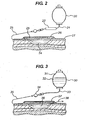
- the apparatus is shown injecting humidified oxygen through microtube 23 into wound 24 below the surface of the wound.
- the wound 24 is covered by a conventional dressing comprising an absorbent layer 25 covered by an adhesive-coated semipermeable backing layer 26 that is bonded to the skin 27 around the wound in substantially liquid-tight fashion.
- the apparatus in this embodiment is being used to inject oxygen-enriched water into the wound.
- the apparatus is generally as described in relation to Fig. 1 , in particular it comprises an infusion pump 30 that is filled with a mixture of oxygen gas 31 and water 32.
- the pump 30 delivers the water saturated with oxygen at about 1 bar gauge pressure to an extension line 33.
- the extension line 33 is joined by a suitable coupling 34 to microtube 35, which injects the oxygenated water into wound 36 below the surface of the wound.
- the wound 36 is covered by a conventional dressing 37 similar to that described in relation to Fig. 2 .
- a drain 38 is provided to remove excess fluid from the wound.
- Standard full thickness wounds were made in a porcine model. Under anaesthesia, the back and flank skin of the pig was clipped and washed using an aseptic cleanser (Chlorhexidine - 5%). The area was shaved using a razor and mopped clean using sterile swabs. Twelve full thickness wounds were created on anatomically similar sites (six on each flank). At each site a single 2.0 x 2.0cm full-thickness excisional wound was created using a scalpel.
- Oxygen bearing water was delivered using microtubing as above fed into the dorsal aspect of the wound (i.e. the edge parallel to the spine) and positioned in such a way as to allow delivery to the centre of the wound.
- the microtubing was then fixed into position using adhesive tape (SLEEK, Registered Trade Mark of Smith & Nephew Ltd). Wounds were then dressed with 6 x 5 cm polyurethane foam (TIELLE, Registered Trade Mark of Johnson & Johnson) dressings in such a way as to minimise leakage.
- Dressings and tubing were further secured by layering topper gauze over the polyurethane foam dressings on all wounds; this was further secured with adhesive tape and Elastic netting (SURGIFIX, Registered Trade Mark of Cistema d'Asti, Italy).
- Oxygen enriched water was delivered to the microcatheter at about 1 bar gauge.
- the resulting oxygen-enriched water is estimated to contain 25-30mg/l of oxygen.
- Five wounds were treated as reference examples, and received purely topical delivery (i.e. the microcatheter remained on top of the newly formed tissue). In one wound the microcatheter was embedded in the newly formed tissue (wound 19.2.10 in Table 1) resulting in oxygen being delivered from inside the wound.
- an island-type adhesive dressing comprising a sheet of hydrophilic polyurethane foam (TIELLE®, available from Johnson & Johnson Medical) was applied over the wounds.
Landscapes
- Health & Medical Sciences (AREA)
- Life Sciences & Earth Sciences (AREA)
- Engineering & Computer Science (AREA)
- Veterinary Medicine (AREA)
- Public Health (AREA)
- General Health & Medical Sciences (AREA)
- Animal Behavior & Ethology (AREA)
- Hematology (AREA)
- Heart & Thoracic Surgery (AREA)
- Biomedical Technology (AREA)
- Anesthesiology (AREA)
- Chemical Kinetics & Catalysis (AREA)
- Pharmacology & Pharmacy (AREA)
- Organic Chemistry (AREA)
- Nuclear Medicine, Radiotherapy & Molecular Imaging (AREA)
- Medicinal Chemistry (AREA)
- General Chemical & Material Sciences (AREA)
- Chemical & Material Sciences (AREA)
- Dermatology (AREA)
- Bioinformatics & Cheminformatics (AREA)
- Pharmaceuticals Containing Other Organic And Inorganic Compounds (AREA)
- Medicinal Preparation (AREA)
Description
- The present invention relates to an apparatus for use in for treating wounds and lesions on a patient's body by introducing an oxygen-containing fluid into a tissue beneath the surface of the wound or lesion.
- The effect of oxygen on wound healing has been extensively studied. Wound healing is dependent upon several processes including proliferation of fibroblasts, collagen synthesis, angiogenesis and re-epithelialization. Many skin sores, ulcers, wounds and bums do not heal properly because there is a severe depletion of oxygen reaching these affected areas due to deterioration of the associated blood microcirculation. Tissue oxygen levels have previously been measured in non-healing wounds to be (5-20mmHg) (6,7-27 mbar) as compared to control tissue values of 30-50 mmHg (40-66,7 mbar).
- Hitherto, many of these wound healing disorders have been treated by various methods of administration of oxygen gas, known as, either through inhalation of the gas in hyperbaric oxygen therapy, or by topical treatment with the gas.
- Treatments based on inhalation of oxygen, while considered effective, are complex and entail a risk from the potential harmful effects of prolonged hypoxia.
- Numerous wound treatment devices are known that administer oxygen gas topically to wound surfaces. Examples can be found in
GB-A-2409412 WO-A-9853778 US-B-6179804 ,US-A-4624656 ,US-A-4224941 ,US-A-5662625 ,US-A-5810795 ,FR-A-2656218 WO-A-9741816 -
US-A-6139876 describes a gelatin with increased oxygen content for use as a topical wound dressing. The gelatin comprises a gelling agent, a solvent, and microbubbles of oxygen in a substantially even distribution with a partial pressure exceeding normal atmospheric pressure, wherein the surface tension of the gelatin is sufficiently high to retain at least a portion of the overpressure of the oxygen throughout a predetermined period of time after having been exposed to an atmospheric environment. -
US20020160053 describes an aqueous solution for promoting the growth of tissue cells at wound sites. This solution comprises water containing at least 1 to 500 ppm of active oxygen and 10 to 10000 ppm of halogen ions. The solution is produced by electrolysis of aqueous halide solutions. The active oxygen can include singlet oxygen formed by excitation of triplet oxygen, superoxide formed by reduction of oxygen by a single electron, and hydroxy radical, as well as hypochlorous ions, organic peroxy radicals (ROO.), alkoxy radioals (RO.), and organic hydroperoxides (ROOH). However, the physiological effects of these reactive oxygen species differ from the effect of oxygen gas, and indeed the reactive oxygen species can be deleterious to wound healing. -
US-A-20050125035 describes an active suture having an internal passageway for post-surgical delivery of active fluids. The active fluids may comprise a wide range of medicaments, including oxygen-rich liquids to assist wound healing. - The present invention provides an apparatus comprising: a source of an oxygen-containing fluid selected from gaseous oxygen or water containing at least 10mg per liter of dissolved oxygen; and a microcatheter having an inlet aperture in fluid communication with the source of oxygen-containing fluid and having one or more outlet apertures, wherein at least a region of the microcatheter has an effective internal diameter of less than 75 micrometers characterised in that said outlet apertures are located in a tip region of the microcatheter for injecting an effective amount of an oxygen-containing fluid selectively and directly into a layer of wound tissue located below a surface layer of a wound, for use in a method of treatment of a wound. The inventive apparatus is used in a method comprising the step of injecting an effective amount of an oxygen-containing fluid into said wound at a location below a surface of said wound, said step of injecting being carried out through a conduit having one or more outlets for said fluid, wherein substantially all of said outlets are positioned below the surface of the wound during said step of injecting.
-
-
Fig. 1 shows a schematic expanded view of an apparatus for use in a method according to the present invention; -
Fig. 2 shows a schematic view of a wound being treated with moist oxygen gas in accordance with the present invention; and -
Fig. 3 shows a schematic view of a wound being treated with oxygen-enriched water in accordance with the present invention. - It has been found that water containing oxygen dissolved under pressure can be injected directly into wound tissue at a rate sufficient to have a positive effect on wound healing, without forming a gas bolus in the wound tissue. It has further been found that injection of oxygen-containing fluid into the wound tissue in this way is even more beneficial to wound healing than topical application of the oxygen-containing fluid to the wound. In particular, the amount of granulation tissue deposited (as determined both visually and by histological analysis) was significantly greater when oxygen-containing fluid was delivered into the tissue rather than onto the surface of the tissue.
- The apparatus of the present invention differs from the use of active sutures to deliver oxygen rich liquids, because the apparatus of the present invention delivers the oxygen rich liquid directly and selectively to the wound tissue below the surface of the wound, without any of the damage to surrounding healthy tissue that is caused by suturing. The apparatus of the present invention delivers the oxygen directly to the granulation tissue layer of the wound, where it is most effective. The present invention is especially useful for the treatment of chronic wounds such as ulcers that are not normally sutured. Furthermore, the apertured sutures of
US-A-20050125035 deliver oxygen rich fluid nonselectively along the length of the suture. It follows also that the apertured sutures cannot be used to deliver an oxygen enriched fluid under pressure to a predetermined tissue layer under the surface of the wound, since the fluid under pressure will leak out of exposed parts of the apertured suture. - The conduit through which the oxygen-containing liquid is delivered to the wound tissue is a microcatheter. The term "microcatheter" refers to any tubular body having a small internal and external diameter, including micro-injection needles, syringes, hollow sutures, and the like. Suitable microcatheters are described, for example, in
US-B-6626885 . - Preferably, the conduit is flexible. Suitably, the conduit has a substantially cylindrical lumen with one or more outlet apertures in a tip region thereof, but no apertures intermediate the inlet and the tip apertures, whereby all of the fluid injected into the inlet of the catheter flows out near the tip, i.e. below the surface of the wound in use. It will be appreciated that multiple conduits, or a single conduit with multiple outlets, may be used to deliver the oxygen-containing fluid to the wound. In certain embodiments, the conduit is branched to permit oxygen injection at a plurality of locations beneath the surface of the wound.
- Suitably, at least a tip region of the conduit is bioabsorbable, whereby the tip region can be left in the wound for resorption in vivo if necessary. For example, at least the tip region of the may be made from a bioabsorbable polymer such as polylactide/polyglycolide, or poly-epsilon-caprolactone.
- It has been found that the conduit can function as a flow regulator for the oxygen-containing fluid under pressure, whereby the fluid is delivered to the tip of the conduit at high pressure and low flow rate. The high pressure achieves a high dissolution of oxygen and resulting high concentration of the oxygen in the physiological fluid proximate to the conduit tip. The low flow rate inhibits liquid or gas bolus formation inside the wound. In order to provide this combination of properties, at least a tip region of the conduit suitably has an effective internal diameter of less than about 75 micrometers. Preferably at least a region of the conduit has an effective internal diameter of less than about 50 micrometers, for example from about 5 to about 40 micrometers. The term "effective internal diameter" refers to the equivalent open circular internal diameter. Suitably, the said region of the conduit is located proximate to the tip (outlet) of the conduit. For example, the conduit may comprise a length of tubing having internal diameter greater than about 100 micrometers, which may be branched, the end or ends of which are joined by a suitable coupling to respective microtubes (microcatheters) having internal diameter less than about 75 micrometers for flow control, the open end or ends of the microtubes being inserted below the surface of the wound. The microtubes are suitably bioabsorbable.
- It will be appreciated that considerable variation in the construction and internal geometry of the conduit is possible, provided that it delivers the oxygen-containing gas at high pressure and low flow rate.
- The oxygen-containing fluid may be a liquid or a gas. The liquid is preferably aqueous, for example it may be water or a physiologically acceptable saline solution. In these embodiments the oxygen may be dissolved, preferably under pressure, to give a concentration of at least about 10 mgO2 per liter, preferably at least about 20 mgO2 per liter, for example from about 20 mgO2 per liter to about 30 mgO2 per liter. Oxygen concentrations in this range can be achieved by dissolving oxygen at a gas pressure of about 3 bar in water. The source of the fluid may be a portable pressurized reservoir containing a mixture of both fluid and oxygen bearing gas.
- In other embodiments, the oxygen may be trapped in gaseous or dissolved form inside liposomes in the aqueous solution. In yet other embodiments, the liquid may be a medically acceptable perfluorocarbon solvent such as perfluorodecalin containing dissolved oxygen.
- In other embodiments, the oxygen-containing fluid may be any oxygen-containing gas. Suitably, the gas comprises more than about 22mol.% of oxygen, more suitably at least about 40mol% oxygen, for example at least about 60mol.% oxygen, and in some embodiments it is substantially pure oxygen. The oxygen gas may be humidified before injection into the wound. The source of gaseous oxygen may comprise a pressurized oxygen tank, which may be portable.
- Suitably, the source of oxygen-enriched fluid supplies the fluid at a pressure of from about 0.1 bar to about 5 bar (gauge), more suitably from about 0.5 bar to about 2 bar, for example about 1 bar. Suitable sources comprise an infusion pump, for example a spring-driven infusion pump. Pumps of this type are described, for example, in
WO-A-0038767 - Suitably, the step of injecting the oxygen-containing fluid into the wound tissue is performed in substantially continuous fashion for a period of from about 1 hour to about 30 days, preferably from about 1 day to about 14 days.
- Suitably, the pressure and delivery rate of the injection are adjusted to prevent the formation of a bolus of liquid or gas inside the wound. It will be appreciated that considerable variation in the pressure of the fluid and the internal geometry of the microcatheter is possible, provided that it results in the oxygen-containing fluid being delivered at a flow rate that is effective to promote wound healing without forming a bolus inside the wound. The effective internal diameter of the conduit, D, length of the conduit, L, the gauge pressure of the fluid used in the system, ΔP, and viscosity of the fluid, η, all impact the delivery rate, V, according to the Hagen-Poiseuille equation.

- Typical delivery rates per wound for gaseous oxygen are suitably about 0.5ml/hr to about 4ml/hr, typically about 1ml/hr to about 3ml/hr, for example about 2ml/hr. Typical delivery rates per wound for oxygen-enriched liquids are suitably about 0.05ml/hr to about 1ml/hr, typically about 0.1ml/hr to about 0.4ml/hr, for example about 0.15ml/hr.
- Suitably, the conduit is arranged to deliver the oxygen below any surface necrotic tissue of the wound. Suitably, the conduit is arranged to deliver the oxygen into or onto a granulation tissue in the wound. Typically, the oxygen injection is performed at a depth of from about 0.5mm to about 10mm below the surface of the wound, preferably from about 1mm to about 5 mm. The invention thereby provides an apparatus which provides a good method of promoting granulation tissue beneath necrotic tissue in instances were debridement would be difficult or painful for the patient. Furthermore, by injecting at this depth it ensures that the oxygenated fluid is delivered directly to healthy tissue where it may be effective. In contrast to topical delivery, it is also more difficult for the oxygen to escape from the wound but would rather tend to remain in the tissue of the wound where it may offer therapeutic benefit to the patient.
- Suitably, the wound is a chronic wound. Suitable chronic wounds include dermal ulcers such as a venous ulcers, diabetic ulcers or decubitis ulcers. The invention may also be useful for the treatment of acute wounds and burns.
- An embodiment of the present invention will now be described further, by way of example, with reference to the accompanying drawings, in which:
- Referring to
Fig. 1 , the apparatus according to the invention comprises a spring driven infusion pump 1 having a valve outlet 2. A branched extension line 3 of PTFE tubing having an internal diameter of about 1mm has an inlet connector 4 that can be inserted in pressure-tight fashion into the pump outlet 2. The outlets at the ends of the branches of the extension line 3 are equipped with valves 5 and Luer-lock fittings for attachment with complementary fittings 7 on flow restrictor tubing 6. The flow restrictor tubing 6 is made of PVC with internal diameter about 150 micrometers. The length of the flow restrictor tubing is selected for the fluid being injected. For example, in this embodiment the length of the flow restrictor tubing is about 15cm for liquid injection and about 40cm for gas injection. The distal end 8 of the flow restrictor tubing 6 is joined in fluid-tight fashion to the inlet end of a microtube 9 formed of PTFE and having outside diameter about 150 micrometers, internal diameter about 49 micrometers, and length about 10cm. - In use, the
tip 10 of the microtube 9 is inserted into the granulation tissue of a wound, below the necrotic surface tissue of the wound and oxygen-enriched fluid is delivered through the microtube, for example under a pump pressure of about 1 bar gauge. - Referring to
Fig. 2 , the apparatus in this embodiment is being used to inject humidified oxygen into the wound. The apparatus is generally as described in relation toFig. 1 , in particular it comprises aninfusion pump 20 that delivers oxygen gas at about 1bar gauge pressure to anextension line 21. Ahumidifier 22 is provided in the extension line. The humidifier comprises a moist porous material that saturates the oxygen with water vapor. The apparatus is shown injecting humidified oxygen throughmicrotube 23 intowound 24 below the surface of the wound. Thewound 24 is covered by a conventional dressing comprising anabsorbent layer 25 covered by an adhesive-coatedsemipermeable backing layer 26 that is bonded to theskin 27 around the wound in substantially liquid-tight fashion. - Referring to
Fig. 3 , the apparatus in this embodiment is being used to inject oxygen-enriched water into the wound. The apparatus is generally as described in relation toFig. 1 , in particular it comprises aninfusion pump 30 that is filled with a mixture ofoxygen gas 31 andwater 32. Thepump 30 delivers the water saturated with oxygen at about 1 bar gauge pressure to anextension line 33. Theextension line 33 is joined by asuitable coupling 34 to microtube 35, which injects the oxygenated water intowound 36 below the surface of the wound. Thewound 36 is covered by aconventional dressing 37 similar to that described in relation toFig. 2 . In addition, adrain 38 is provided to remove excess fluid from the wound. - Standard full thickness wounds were made in a porcine model. Under anaesthesia, the back and flank skin of the pig was clipped and washed using an aseptic cleanser (Chlorhexidine - 5%). The area was shaved using a razor and mopped clean using sterile swabs. Twelve full thickness wounds were created on anatomically similar sites (six on each flank). At each site a single 2.0 x 2.0cm full-thickness excisional wound was created using a scalpel.
- After injury, wounds were washed with sterile physiological saline and carefully swabbed dry with sterile gauze to remove any tissue debris and blood. Oxygen bearing water was delivered using microtubing as above fed into the dorsal aspect of the wound (i.e. the edge parallel to the spine) and positioned in such a way as to allow delivery to the centre of the wound. The microtubing was then fixed into position using adhesive tape (SLEEK, Registered Trade Mark of Smith & Nephew Ltd). Wounds were then dressed with 6 x 5 cm polyurethane foam (TIELLE, Registered Trade Mark of Johnson & Johnson) dressings in such a way as to minimise leakage. Dressings and tubing were further secured by layering topper gauze over the polyurethane foam dressings on all wounds; this was further secured with adhesive tape and Elastic netting (SURGIFIX, Registered Trade Mark of Cistema d'Asti, Italy).
- Wound assessment was performed on post-wounding days 2, 4 and 7. The outer surface of each dressed wound was photographed (both prior to and post cleansing). Macroscopic assessment of wound and marginal tissues was undertaken on days 2, 4 and 7. These assessments included consideration of presence and degree of inflammation, erythema, exudate, re-injury. All findings were documented and supported with photographic images.
- On post wounding wound at day 7 the wound and surrounding normal tissue were excised and subsequently fixed (in 10% formalin) for routine histological assessment. Tissue slices were embedded in paraffin wax, sectioned (7µm) and representative sections stained with Haematoxylin and Eosin (H&E). Image analysis based quantitative assessment of wound width (contraction), granulation tissue deposition (granulation tissue depth and granulation tissue area) was then performed.
- Oxygen enriched water was delivered to the microcatheter at about 1 bar gauge. The resulting oxygen-enriched water is estimated to contain 25-30mg/l of oxygen. Five wounds were treated as reference examples, and received purely topical delivery (i.e. the microcatheter remained on top of the newly formed tissue). In one wound the microcatheter was embedded in the newly formed tissue (wound 19.2.10 in Table 1) resulting in oxygen being delivered from inside the wound. During treatment, an island-type adhesive dressing comprising a sheet of hydrophilic polyurethane foam (TIELLE®, available from Johnson & Johnson Medical) was applied over the wounds.
- The appearance of the wounds following delivery of oxygen for 7 days was higher in the wound that received oxygen-enriched water injected below the tissue surface
- The results were confirmed by histological determination of the amount of granulation tissue. The results are shown in Table 1.
Table 1 Animal Granulation
Mean depth (mm)Granulation
Tissue Area (mm2)Total granulation
Volume (mm3)19.02.01 4.01 63.38 254 19.01.03 4.57 82.95 379 19.02.05 7.28 102.72 748 19.02.08 4.39 81.04 599 19.02.10 6.52 128.66 839 19.02.12 6.12 101.50 621 - Only animal 19.02.10 had oxygen-enriched water delivered into tissue. All others had the fluid delivered from above. These results show that the total granulation deposition volume (depth x area) was 839 mm3 for the wound to which oxygen was delivered into the tissue compared to an average granulation volume of 520.2 ± 199 mm3 for the wound to which oxygen-enriched water was delivered onto (not into) the tissue.
- The above embodiment has been described by way of example only. Many other embodiments falling within the scope of the accompanying claims will be apparent to the skilled reader.
Claims (11)
- An apparatus comprising:a source (1,20,30) of an oxygen-containing fluid selected from gaseous oxygen or water containing at least 10mg per liter of dissolved oxygen; anda microcatheter (9,23,35) having an inlet aperture in fluid communication with the source of oxygen-containing fluid and having one or more outlet apertures, wherein at least a region of the microcatheter (9,23,35) has an effective internal diameter of less than 75 micrometerscharacterised in that said outlet apertures are located in a tip region of the microcatheter for injecting an effective amount of an oxygen-containing fluid selectively and directly into a layer of wound tissue located below a surface layer of a wound.
- An apparatus according to claim 1, wherein said oxygen containing fluid is gaseous oxygen and said apparatus further comprises a humidifier (22) located intermediate the source and the microcatheter (9,23,35) for humidifying the gaseous oxygen.
- An apparatus according to claim 1 or 2, wherein at least the tip region of the microcatheter (9,23,35) is bioabsorbable.
- An apparatus according to claim 3, wherein at least the tip region of the microcatheter (9,23,35) is made from a bioabsorbable polymer selected from polylactide/polyglycolide, or poly-epsilon-caprolactone.
- An apparatus according to any of claims 1 to 3, wherein said source supplies said oxygen-containing fluid at a pressure of from 0.1 bar gauge to 5 bar gauge.
- An apparatus according to any of claims 1 to 3, wherein said source supplies said oxygen-containing fluid at a pressure of from 0.5bar gauge to 2 bar gauge.
- An apparatus according to any of the preceding claims, wherein the microcatheter (9,23,35) has no apertures intermediate the inlet and the tip apertures.
- An apparatus according to any of the preceding claims, wherein the microcatheter (9,23,35) is branched.
- An apparatus according to any preceding claim, wherein the microcatheter (9,23,35) has an effective internal diameter of less than about 50 micrometers.
- An apparatus according to any preceding claim, wherein the oxygen-containing fluid is water containing at least 20mg per liter of dissolved oxygen.
- An apparatus according to any preceding claims, wherein said outlet apertures are located for delivering said oxygen-containing fluid selectively and directly to a layer of wound tissue about 0.5 mm to about 10 mm below the surface of the wound.
Priority Applications (1)
| Application Number | Priority Date | Filing Date | Title |
|---|---|---|---|
| EP15150995.7A EP2905008B2 (en) | 2006-06-29 | 2007-06-25 | Apparatus for the treatment of wounds with oxygen |
Applications Claiming Priority (2)
| Application Number | Priority Date | Filing Date | Title |
|---|---|---|---|
| US11/477,307 US8808754B2 (en) | 2006-06-29 | 2006-06-29 | Methods for the treatment of wounds |
| EP07798976A EP2037860A2 (en) | 2006-06-29 | 2007-06-25 | Methods for the treatment of wounds |
Related Parent Applications (1)
| Application Number | Title | Priority Date | Filing Date |
|---|---|---|---|
| EP07798976A Division EP2037860A2 (en) | 2006-06-29 | 2007-06-25 | Methods for the treatment of wounds |
Related Child Applications (1)
| Application Number | Title | Priority Date | Filing Date |
|---|---|---|---|
| EP15150995.7A Division EP2905008B2 (en) | 2006-06-29 | 2007-06-25 | Apparatus for the treatment of wounds with oxygen |
Publications (2)
| Publication Number | Publication Date |
|---|---|
| EP2149358A1 EP2149358A1 (en) | 2010-02-03 |
| EP2149358B1 true EP2149358B1 (en) | 2015-01-14 |
Family
ID=38662689
Family Applications (3)
| Application Number | Title | Priority Date | Filing Date |
|---|---|---|---|
| EP09014450.2A Ceased EP2149358B1 (en) | 2006-06-29 | 2007-06-25 | Use of oxygen for the treatment of wounds |
| EP07798976A Withdrawn EP2037860A2 (en) | 2006-06-29 | 2007-06-25 | Methods for the treatment of wounds |
| EP15150995.7A Ceased EP2905008B2 (en) | 2006-06-29 | 2007-06-25 | Apparatus for the treatment of wounds with oxygen |
Family Applications After (2)
| Application Number | Title | Priority Date | Filing Date |
|---|---|---|---|
| EP07798976A Withdrawn EP2037860A2 (en) | 2006-06-29 | 2007-06-25 | Methods for the treatment of wounds |
| EP15150995.7A Ceased EP2905008B2 (en) | 2006-06-29 | 2007-06-25 | Apparatus for the treatment of wounds with oxygen |
Country Status (3)
| Country | Link |
|---|---|
| US (1) | US8808754B2 (en) |
| EP (3) | EP2149358B1 (en) |
| WO (1) | WO2008002855A2 (en) |
Families Citing this family (10)
| Publication number | Priority date | Publication date | Assignee | Title |
|---|---|---|---|---|
| US7763769B2 (en) * | 2001-02-16 | 2010-07-27 | Kci Licensing, Inc. | Biocompatible wound dressing |
| US7700819B2 (en) | 2001-02-16 | 2010-04-20 | Kci Licensing, Inc. | Biocompatible wound dressing |
| EP1884223A1 (en) † | 2006-08-04 | 2008-02-06 | IP Randwyck B.V. | Wound-stimulating unit |
| US8287506B2 (en) | 2007-10-26 | 2012-10-16 | Electrochemical Oxygen Concepts, Inc. | Apparatus and methods for controlling tissue oxygenation for wound healing and promoting tissue viability |
| US10226610B2 (en) | 2007-10-26 | 2019-03-12 | Electrochemical Oxygen Concepts, Inc. | Apparatus and methods for controlling tissue oxygenation for wound healing and promoting tissue viability |
| WO2011049633A2 (en) | 2009-10-23 | 2011-04-28 | Amir Belson | Portable topical oxygen therapy system |
| US20150343190A1 (en) * | 2014-03-05 | 2015-12-03 | Prosper Brands LLC | Method of oxygen saturation for wound treatment |
| WO2017100320A1 (en) | 2015-12-07 | 2017-06-15 | Electrochemical Oxygen Concepts, Inc. | Apparatus and methods for controlling tissue oxygenation for wound healing and promoting tissue viability |
| US10702686B2 (en) | 2017-06-30 | 2020-07-07 | Electrochemical Oxygen Concepts, Inc. | Wound oxygen supply system |
| CA3176016A1 (en) * | 2020-04-24 | 2021-10-28 | Michael Beattie | Uses of super-oxygenated water and gel |
Family Cites Families (23)
| Publication number | Priority date | Publication date | Assignee | Title |
|---|---|---|---|---|
| US4224941A (en) | 1978-11-15 | 1980-09-30 | Stivala Oscar G | Hyperbaric treatment apparatus |
| US4624656A (en) | 1983-07-25 | 1986-11-25 | Hospitak, Inc. | Hyperbaric gas treatment device |
| FR2656218A1 (en) | 1989-12-21 | 1991-06-28 | France Prod Oxygenes Co | Device for local treatment of the human or animal body using gas |
| HU213450B (en) | 1995-04-26 | 1997-06-30 | Ladanyi Jozsef | Gel contains gelatin and process for producing it |
| US5662625A (en) | 1996-05-06 | 1997-09-02 | Gwr Medical, L.L.P. | Pressure controllable hyperbaric device |
| US5810795A (en) | 1996-05-13 | 1998-09-22 | Westwood; Joseph R. | Hyperbaric device with secondary pressure zone |
| IL120910A (en) | 1997-05-26 | 2004-01-04 | Lrr & D Ltd | Multipurpose dynamic occlusive dressing |
| WO2000004943A1 (en) * | 1998-07-24 | 2000-02-03 | Therox, Inc. | Method and apparatus for the preparation and delivery of gas-enriched fluids |
| US6458109B1 (en) * | 1998-08-07 | 2002-10-01 | Hill-Rom Services, Inc. | Wound treatment apparatus |
| AU2214600A (en) | 1998-12-29 | 2000-07-31 | Mckinley Medical, Lllp | Spring-powered infusion pump |
| US6350253B1 (en) | 1999-07-19 | 2002-02-26 | I-Flow Corporation | Catheter for uniform delivery of medication |
| US6179804B1 (en) | 1999-08-18 | 2001-01-30 | Oxypatch, Llc | Treatment apparatus for wounds |
| JP2001139477A (en) | 1999-11-17 | 2001-05-22 | Coherent Technology:Kk | Tissue cell growth promoting solution at the wound site |
| WO2001061600A1 (en) * | 2000-02-17 | 2001-08-23 | Matsushita Electric Industrial Co., Ltd. | Semiconductor memory card that records contents for trial and purchase, recording apparatus, reproducing apparatus, and sales method |
| US20020098246A1 (en) * | 2001-01-22 | 2002-07-25 | Howes Randolph M. | Compositions, methods, apparatuses, and systems for singlet oxygen delivery |
| US7288574B2 (en) * | 2001-07-18 | 2007-10-30 | Eckert C Edward | Two-phase oxygenated solution and method of use |
| WO2003074100A1 (en) * | 2002-03-04 | 2003-09-12 | New X-National Technology K.K. | Closed cell culture system |
| GB0325120D0 (en) † | 2003-10-28 | 2003-12-03 | Smith & Nephew | Apparatus with actives |
| GB0325130D0 (en) † | 2003-10-28 | 2003-12-03 | Smith & Nephew | Apparatus with scaffold |
| US8257393B2 (en) | 2003-12-04 | 2012-09-04 | Ethicon, Inc. | Active suture for the delivery of therapeutic fluids |
| AU2004296810B2 (en) * | 2003-12-04 | 2009-11-19 | Ethicon, Inc. | Active suture for the delivery of therapeutic fluids |
| GB2409412A (en) | 2003-12-22 | 2005-06-29 | Johnson & Johnson Medical Ltd | Hyperbaric wound treatment device |
| GB0424046D0 (en) † | 2004-10-29 | 2004-12-01 | Smith & Nephew | Apparatus |
-
2006
- 2006-06-29 US US11/477,307 patent/US8808754B2/en active Active
-
2007
- 2007-06-25 EP EP09014450.2A patent/EP2149358B1/en not_active Ceased
- 2007-06-25 EP EP07798976A patent/EP2037860A2/en not_active Withdrawn
- 2007-06-25 EP EP15150995.7A patent/EP2905008B2/en not_active Ceased
- 2007-06-25 WO PCT/US2007/071981 patent/WO2008002855A2/en not_active Ceased
Also Published As
| Publication number | Publication date |
|---|---|
| EP2905008A1 (en) | 2015-08-12 |
| EP2905008B1 (en) | 2018-04-11 |
| US8808754B2 (en) | 2014-08-19 |
| US20080003299A1 (en) | 2008-01-03 |
| EP2037860A2 (en) | 2009-03-25 |
| EP2905008B2 (en) | 2022-05-18 |
| WO2008002855A2 (en) | 2008-01-03 |
| WO2008002855A8 (en) | 2008-03-27 |
| EP2149358A1 (en) | 2010-02-03 |
Similar Documents
| Publication | Publication Date | Title |
|---|---|---|
| EP2905008B2 (en) | Apparatus for the treatment of wounds with oxygen | |
| US11806215B2 (en) | Apparatus for wound therapy | |
| US8357130B2 (en) | Wound care apparatus | |
| Hess et al. | A review of mechanical adjuncts in wound healing: hydrotherapy, ultrasound, negative pressure therapy, hyperbaric oxygen, and electrostimulation | |
| CN110996866B (en) | Device for wound pressure therapy | |
| US20080262413A1 (en) | Method For Supplying Oxygenated Water To Promote Internal Healing | |
| US20120046603A1 (en) | Hyperbaric dressing and method | |
| US20080140026A1 (en) | Acoustically-Aided and/or gel-aided delivery of beneficial gaseous, ionic, unstable, metastable, short-lived or reactive species for medical purposes | |
| JP5016032B2 (en) | Oxygen therapy using ultrasound | |
| US20240245871A1 (en) | Portable medical treatment system and method of use | |
| US20150164829A1 (en) | Materials and methods for controlling infections | |
| US20070060834A1 (en) | Catheter control console | |
| US20220192888A1 (en) | Bandage system for wound visualization, microcurrent therapy and topical and transdermal drug delivery | |
| RU2068263C1 (en) | Method for treating purulent wounds | |
| US20240189499A1 (en) | Dynamic negative pressure wound therapy system | |
| CN221357400U (en) | Antibacterial transparent dressing for indwelling needle | |
| CN111298267A (en) | Sustained-release system for wound analgesia | |
| KR102258962B1 (en) | Nasal packing device | |
| US20220241322A1 (en) | Wound-treating gas composition, and wound-treating apparatus | |
| CN115671519A (en) | A device of changing dressings for mixed haemorrhoids postoperative | |
| Congress et al. | CAWC Events | |
| HK1234686A1 (en) | Portable medical treatment system and method of use | |
| HK1234686B (en) | Portable medical treatment system and method of use |
Legal Events
| Date | Code | Title | Description |
|---|---|---|---|
| PUAI | Public reference made under article 153(3) epc to a published international application that has entered the european phase |
Free format text: ORIGINAL CODE: 0009012 |
|
| AC | Divisional application: reference to earlier application |
Ref document number: 2037860 Country of ref document: EP Kind code of ref document: P |
|
| AK | Designated contracting states |
Kind code of ref document: A1 Designated state(s): DE FR GB IT |
|
| 17P | Request for examination filed |
Effective date: 20100706 |
|
| 17Q | First examination report despatched |
Effective date: 20100802 |
|
| GRAP | Despatch of communication of intention to grant a patent |
Free format text: ORIGINAL CODE: EPIDOSNIGR1 |
|
| INTG | Intention to grant announced |
Effective date: 20140819 |
|
| GRAS | Grant fee paid |
Free format text: ORIGINAL CODE: EPIDOSNIGR3 |
|
| GRAP | Despatch of communication of intention to grant a patent |
Free format text: ORIGINAL CODE: EPIDOSNIGR1 |
|
| INTG | Intention to grant announced |
Effective date: 20141118 |
|
| GRAA | (expected) grant |
Free format text: ORIGINAL CODE: 0009210 |
|
| AC | Divisional application: reference to earlier application |
Ref document number: 2037860 Country of ref document: EP Kind code of ref document: P |
|
| AK | Designated contracting states |
Kind code of ref document: B1 Designated state(s): DE FR GB IT |
|
| REG | Reference to a national code |
Ref country code: GB Ref legal event code: FG4D |
|
| REG | Reference to a national code |
Ref country code: DE Ref legal event code: R096 Ref document number: 602007040082 Country of ref document: DE Effective date: 20150305 |
|
| REG | Reference to a national code |
Ref country code: DE Ref legal event code: R097 Ref document number: 602007040082 Country of ref document: DE |
|
| PLBE | No opposition filed within time limit |
Free format text: ORIGINAL CODE: 0009261 |
|
| STAA | Information on the status of an ep patent application or granted ep patent |
Free format text: STATUS: NO OPPOSITION FILED WITHIN TIME LIMIT |
|
| 26N | No opposition filed |
Effective date: 20151015 |
|
| REG | Reference to a national code |
Ref country code: FR Ref legal event code: PLFP Year of fee payment: 10 |
|
| REG | Reference to a national code |
Ref country code: FR Ref legal event code: PLFP Year of fee payment: 11 |
|
| REG | Reference to a national code |
Ref country code: DE Ref legal event code: R082 Ref document number: 602007040082 Country of ref document: DE Representative=s name: OLSWANG GERMANY LLP, DE Ref country code: DE Ref legal event code: R081 Ref document number: 602007040082 Country of ref document: DE Owner name: SYSTAGENIX WOUND MANAGEMENT, LTD., GATWICK AIR, GB Free format text: FORMER OWNER: SYSTAGENIX WOUND MANAGEMENT IP CO. BV., AMSTERDAM, NL Ref country code: DE Ref legal event code: R082 Ref document number: 602007040082 Country of ref document: DE Representative=s name: SIMMONS & SIMMONS LLP, DE |
|
| REG | Reference to a national code |
Ref country code: GB Ref legal event code: 732E Free format text: REGISTERED BETWEEN 20180215 AND 20180221 |
|
| REG | Reference to a national code |
Ref country code: FR Ref legal event code: PLFP Year of fee payment: 12 |
|
| REG | Reference to a national code |
Ref country code: DE Ref legal event code: R082 Ref document number: 602007040082 Country of ref document: DE Representative=s name: SIMMONS & SIMMONS LLP, DE |
|
| PGFP | Annual fee paid to national office [announced via postgrant information from national office to epo] |
Ref country code: IT Payment date: 20190521 Year of fee payment: 13 |
|
| PGFP | Annual fee paid to national office [announced via postgrant information from national office to epo] |
Ref country code: FR Payment date: 20190522 Year of fee payment: 13 |
|
| PG25 | Lapsed in a contracting state [announced via postgrant information from national office to epo] |
Ref country code: FR Free format text: LAPSE BECAUSE OF NON-PAYMENT OF DUE FEES Effective date: 20200630 |
|
| PG25 | Lapsed in a contracting state [announced via postgrant information from national office to epo] |
Ref country code: IT Free format text: LAPSE BECAUSE OF NON-PAYMENT OF DUE FEES Effective date: 20200625 |
|
| PGFP | Annual fee paid to national office [announced via postgrant information from national office to epo] |
Ref country code: GB Payment date: 20220519 Year of fee payment: 16 Ref country code: DE Payment date: 20220518 Year of fee payment: 16 |
|
| REG | Reference to a national code |
Ref country code: DE Ref legal event code: R119 Ref document number: 602007040082 Country of ref document: DE |
|
| GBPC | Gb: european patent ceased through non-payment of renewal fee |
Effective date: 20230625 |
|
| PG25 | Lapsed in a contracting state [announced via postgrant information from national office to epo] |
Ref country code: DE Free format text: LAPSE BECAUSE OF NON-PAYMENT OF DUE FEES Effective date: 20240103 Ref country code: GB Free format text: LAPSE BECAUSE OF NON-PAYMENT OF DUE FEES Effective date: 20230625 |