EP2137532B1 - Devices and methods for detecting analytes - Google Patents
Devices and methods for detecting analytes Download PDFInfo
- Publication number
- EP2137532B1 EP2137532B1 EP08776297.7A EP08776297A EP2137532B1 EP 2137532 B1 EP2137532 B1 EP 2137532B1 EP 08776297 A EP08776297 A EP 08776297A EP 2137532 B1 EP2137532 B1 EP 2137532B1
- Authority
- EP
- European Patent Office
- Prior art keywords
- zone
- mobilizable
- antibody
- analyte
- ligand
- Prior art date
- Legal status (The legal status is an assumption and is not a legal conclusion. Google has not performed a legal analysis and makes no representation as to the accuracy of the status listed.)
- Not-in-force
Links
- 238000000034 method Methods 0.000 title claims description 25
- 239000012491 analyte Substances 0.000 claims description 154
- 239000003446 ligand Substances 0.000 claims description 120
- 230000027455 binding Effects 0.000 claims description 110
- 238000001514 detection method Methods 0.000 claims description 72
- 239000000203 mixture Substances 0.000 claims description 67
- 238000012360 testing method Methods 0.000 claims description 59
- 239000007788 liquid Substances 0.000 claims description 58
- 230000000903 blocking effect Effects 0.000 claims description 45
- YBJHBAHKTGYVGT-ZKWXMUAHSA-N (+)-Biotin Chemical compound N1C(=O)N[C@@H]2[C@H](CCCCC(=O)O)SC[C@@H]21 YBJHBAHKTGYVGT-ZKWXMUAHSA-N 0.000 claims description 38
- 229960002685 biotin Drugs 0.000 claims description 23
- 239000011616 biotin Substances 0.000 claims description 23
- 235000020958 biotin Nutrition 0.000 claims description 19
- 108090001008 Avidin Proteins 0.000 claims description 8
- PYWVYCXTNDRMGF-UHFFFAOYSA-N rhodamine B Chemical compound [Cl-].C=12C=CC(=[N+](CC)CC)C=C2OC2=CC(N(CC)CC)=CC=C2C=1C1=CC=CC=C1C(O)=O PYWVYCXTNDRMGF-UHFFFAOYSA-N 0.000 claims description 4
- 108010090804 Streptavidin Proteins 0.000 claims description 2
- 239000000243 solution Substances 0.000 description 33
- 230000033228 biological regulation Effects 0.000 description 26
- 239000012528 membrane Substances 0.000 description 21
- ZPUCINDJVBIVPJ-LJISPDSOSA-N cocaine Chemical compound O([C@H]1C[C@@H]2CC[C@@H](N2C)[C@H]1C(=O)OC)C(=O)C1=CC=CC=C1 ZPUCINDJVBIVPJ-LJISPDSOSA-N 0.000 description 20
- 210000002700 urine Anatomy 0.000 description 19
- 239000000463 material Substances 0.000 description 16
- 239000000020 Nitrocellulose Substances 0.000 description 14
- 229920001220 nitrocellulos Polymers 0.000 description 14
- JTJMJGYZQZDUJJ-UHFFFAOYSA-N phencyclidine Chemical compound C1CCCCN1C1(C=2C=CC=CC=2)CCCCC1 JTJMJGYZQZDUJJ-UHFFFAOYSA-N 0.000 description 11
- 229960003920 cocaine Drugs 0.000 description 10
- PCHJSUWPFVWCPO-UHFFFAOYSA-N gold Chemical compound [Au] PCHJSUWPFVWCPO-UHFFFAOYSA-N 0.000 description 10
- 239000002245 particle Substances 0.000 description 10
- 239000012530 fluid Substances 0.000 description 9
- 239000007790 solid phase Substances 0.000 description 9
- KWTSXDURSIMDCE-QMMMGPOBSA-N (S)-amphetamine Chemical compound C[C@H](N)CC1=CC=CC=C1 KWTSXDURSIMDCE-QMMMGPOBSA-N 0.000 description 8
- CYQFCXCEBYINGO-UHFFFAOYSA-N THC Natural products C1=C(C)CCC2C(C)(C)OC3=CC(CCCCC)=CC(O)=C3C21 CYQFCXCEBYINGO-UHFFFAOYSA-N 0.000 description 8
- 229940025084 amphetamine Drugs 0.000 description 8
- 239000003153 chemical reaction reagent Substances 0.000 description 8
- CYQFCXCEBYINGO-IAGOWNOFSA-N delta1-THC Chemical compound C1=C(C)CC[C@H]2C(C)(C)OC3=CC(CCCCC)=CC(O)=C3[C@@H]21 CYQFCXCEBYINGO-IAGOWNOFSA-N 0.000 description 8
- 229960004242 dronabinol Drugs 0.000 description 8
- 238000003018 immunoassay Methods 0.000 description 8
- 229960001252 methamphetamine Drugs 0.000 description 8
- MYWUZJCMWCOHBA-VIFPVBQESA-N methamphetamine Chemical compound CN[C@@H](C)CC1=CC=CC=C1 MYWUZJCMWCOHBA-VIFPVBQESA-N 0.000 description 8
- 239000002250 absorbent Substances 0.000 description 6
- 230000002745 absorbent Effects 0.000 description 6
- 239000000427 antigen Substances 0.000 description 6
- 102000036639 antigens Human genes 0.000 description 6
- 108091007433 antigens Proteins 0.000 description 6
- 239000008366 buffered solution Substances 0.000 description 6
- 238000010790 dilution Methods 0.000 description 6
- 239000012895 dilution Substances 0.000 description 6
- BQJCRHHNABKAKU-KBQPJGBKSA-N morphine Chemical compound O([C@H]1[C@H](C=C[C@H]23)O)C4=C5[C@@]12CCN(C)[C@@H]3CC5=CC=C4O BQJCRHHNABKAKU-KBQPJGBKSA-N 0.000 description 6
- 238000007865 diluting Methods 0.000 description 5
- 238000002360 preparation method Methods 0.000 description 5
- 239000007921 spray Substances 0.000 description 5
- 239000007791 liquid phase Substances 0.000 description 4
- 230000009870 specific binding Effects 0.000 description 4
- 238000011144 upstream manufacturing Methods 0.000 description 4
- 238000004458 analytical method Methods 0.000 description 3
- 210000004369 blood Anatomy 0.000 description 3
- 239000008280 blood Substances 0.000 description 3
- 238000006243 chemical reaction Methods 0.000 description 3
- 238000004891 communication Methods 0.000 description 3
- 229940079593 drug Drugs 0.000 description 3
- 239000003814 drug Substances 0.000 description 3
- 239000011152 fibreglass Substances 0.000 description 3
- 229960005181 morphine Drugs 0.000 description 3
- 229920000728 polyester Polymers 0.000 description 3
- 108090000623 proteins and genes Proteins 0.000 description 3
- 102000004169 proteins and genes Human genes 0.000 description 3
- 239000000126 substance Substances 0.000 description 3
- 241000283707 Capra Species 0.000 description 2
- 206010013654 Drug abuse Diseases 0.000 description 2
- 229910019142 PO4 Inorganic materials 0.000 description 2
- PXIPVTKHYLBLMZ-UHFFFAOYSA-N Sodium azide Chemical compound [Na+].[N-]=[N+]=[N-] PXIPVTKHYLBLMZ-UHFFFAOYSA-N 0.000 description 2
- UIIMBOGNXHQVGW-UHFFFAOYSA-M Sodium bicarbonate Chemical compound [Na+].OC([O-])=O UIIMBOGNXHQVGW-UHFFFAOYSA-M 0.000 description 2
- 239000007983 Tris buffer Substances 0.000 description 2
- 229920004890 Triton X-100 Polymers 0.000 description 2
- 239000013504 Triton X-100 Substances 0.000 description 2
- 238000010521 absorption reaction Methods 0.000 description 2
- 230000009471 action Effects 0.000 description 2
- 239000012042 active reagent Substances 0.000 description 2
- 230000003466 anti-cipated effect Effects 0.000 description 2
- 238000003556 assay Methods 0.000 description 2
- 239000007853 buffer solution Substances 0.000 description 2
- 229920002678 cellulose Polymers 0.000 description 2
- 239000001913 cellulose Substances 0.000 description 2
- 230000008859 change Effects 0.000 description 2
- 230000002860 competitive effect Effects 0.000 description 2
- 230000008021 deposition Effects 0.000 description 2
- BNIILDVGGAEEIG-UHFFFAOYSA-L disodium hydrogen phosphate Chemical compound [Na+].[Na+].OP([O-])([O-])=O BNIILDVGGAEEIG-UHFFFAOYSA-L 0.000 description 2
- 229910000397 disodium phosphate Inorganic materials 0.000 description 2
- 239000011521 glass Substances 0.000 description 2
- 239000004816 latex Substances 0.000 description 2
- 229920000126 latex Polymers 0.000 description 2
- 238000002156 mixing Methods 0.000 description 2
- 230000037361 pathway Effects 0.000 description 2
- NBIIXXVUZAFLBC-UHFFFAOYSA-K phosphate Chemical compound [O-]P([O-])([O-])=O NBIIXXVUZAFLBC-UHFFFAOYSA-K 0.000 description 2
- 239000010452 phosphate Substances 0.000 description 2
- 230000035945 sensitivity Effects 0.000 description 2
- 208000011117 substance-related disease Diseases 0.000 description 2
- 239000006228 supernatant Substances 0.000 description 2
- XLYOFNOQVPJJNP-UHFFFAOYSA-N water Substances O XLYOFNOQVPJJNP-UHFFFAOYSA-N 0.000 description 2
- 208000035473 Communicable disease Diseases 0.000 description 1
- 102000004190 Enzymes Human genes 0.000 description 1
- 108090000790 Enzymes Proteins 0.000 description 1
- 102000008394 Immunoglobulin Fragments Human genes 0.000 description 1
- 108010021625 Immunoglobulin Fragments Proteins 0.000 description 1
- 239000004677 Nylon Substances 0.000 description 1
- 241000283973 Oryctolagus cuniculus Species 0.000 description 1
- 230000001745 anti-biotin effect Effects 0.000 description 1
- 230000015572 biosynthetic process Effects 0.000 description 1
- 210000001124 body fluid Anatomy 0.000 description 1
- 239000010839 body fluid Substances 0.000 description 1
- 229910021538 borax Inorganic materials 0.000 description 1
- 238000004587 chromatography analysis Methods 0.000 description 1
- 238000000576 coating method Methods 0.000 description 1
- 238000012875 competitive assay Methods 0.000 description 1
- 238000002967 competitive immunoassay Methods 0.000 description 1
- 230000021615 conjugation Effects 0.000 description 1
- 238000010276 construction Methods 0.000 description 1
- 230000007423 decrease Effects 0.000 description 1
- 238000003745 diagnosis Methods 0.000 description 1
- 201000010099 disease Diseases 0.000 description 1
- 208000037265 diseases, disorders, signs and symptoms Diseases 0.000 description 1
- 239000012153 distilled water Substances 0.000 description 1
- 230000000694 effects Effects 0.000 description 1
- 238000002372 labelling Methods 0.000 description 1
- 238000012125 lateral flow test Methods 0.000 description 1
- 238000012544 monitoring process Methods 0.000 description 1
- 229920001778 nylon Polymers 0.000 description 1
- 230000003287 optical effect Effects 0.000 description 1
- 239000012071 phase Substances 0.000 description 1
- 229950010883 phencyclidine Drugs 0.000 description 1
- 239000008055 phosphate buffer solution Substances 0.000 description 1
- 239000004033 plastic Substances 0.000 description 1
- 229920003023 plastic Polymers 0.000 description 1
- 230000035935 pregnancy Effects 0.000 description 1
- 238000004393 prognosis Methods 0.000 description 1
- 230000001105 regulatory effect Effects 0.000 description 1
- 230000004044 response Effects 0.000 description 1
- 210000003296 saliva Anatomy 0.000 description 1
- 150000003839 salts Chemical class 0.000 description 1
- 210000002966 serum Anatomy 0.000 description 1
- 150000003384 small molecules Chemical class 0.000 description 1
- 229910000030 sodium bicarbonate Inorganic materials 0.000 description 1
- 239000004328 sodium tetraborate Substances 0.000 description 1
- 235000010339 sodium tetraborate Nutrition 0.000 description 1
- 239000011343 solid material Substances 0.000 description 1
- 238000006467 substitution reaction Methods 0.000 description 1
- 210000004243 sweat Anatomy 0.000 description 1
- 239000011534 wash buffer Substances 0.000 description 1
Images



Classifications
-
- G—PHYSICS
- G01—MEASURING; TESTING
- G01N—INVESTIGATING OR ANALYSING MATERIALS BY DETERMINING THEIR CHEMICAL OR PHYSICAL PROPERTIES
- G01N33/00—Investigating or analysing materials by specific methods not covered by groups G01N1/00 - G01N31/00
- G01N33/48—Biological material, e.g. blood, urine; Haemocytometers
- G01N33/50—Chemical analysis of biological material, e.g. blood, urine; Testing involving biospecific ligand binding methods; Immunological testing
- G01N33/53—Immunoassay; Biospecific binding assay; Materials therefor
- G01N33/543—Immunoassay; Biospecific binding assay; Materials therefor with an insoluble carrier for immobilising immunochemicals
- G01N33/54366—Apparatus specially adapted for solid-phase testing
- G01N33/54386—Analytical elements
- G01N33/54387—Immunochromatographic test strips
- G01N33/54388—Immunochromatographic test strips based on lateral flow
Definitions
- Immunoassays for detecting an analyte in a sample are used extensively in various fields. Immunoassays can be used to detect analytes in various samples (such as saliva, blood, urine, serum, and sweat) to aid in diagnosis, prognosis and/or treatment of diseases. For instance, immunoassays are routinely performed for monitoring signs of early pregnancy, the presence of rum, infectious diseases and drug abuse. Immunoassay is effected by the specific binding sites contained between immune mo1lecules, such as antigen/antibody, hapten/antibody, and biotin/antibiotin. Most immunoassays are performed on a solid phase, such as on lateral-flow test strips, glass or plastic multi-porous discs, or immune chromatography devices.
- immunoassays There are two common formats of immunoassays, namely the sandwich assay and the competitive assay.
- sandwich assay the analyte competes with a labeled analyte to bind with antibodies.
- This format has been commonly used to detect small analytes such as hapten in a sample.
- the label used can be one that indicates its presence as a color band in the detection or result reading zone.
- Reagents and devices utilizing competitive principle have been described in detail in U.S. Patent Nos. 4,235,601 . 4,442,204 , 5,229,073 .
- NC membrane nitrocellulose
- a test strip comprising three specific zones, a mobilization zone; a trap zone and a detection zone.
- An antibody bound to a color label in the mobilization zone, can bind with an analyte to form a complex (a color label-antibody-analyte complex).
- the complex can be moved by a liquid to a trap zone where a ligand is immobilized.
- the immobilized ligand is not able to bind to an antibody that has already formed a complex with an analyte.
- Such a complex is stable and cannot be affected by the immobilized ligand.
- the ligand can only bind an antibody that is freely floating and not already bound to an analyte.
- the analyte-antibody complex can then be captured by an immobilized detection receptor as to show a direct positive result.
- the device described in this patent requires that the affinity between the antibody and the analyte be higher than the affinity between the antibody and the ligand. In practice, it is very difficult to select the antibody with these limited features to apply to this invention.
- a test strip consisting of a sample application zone, a mobilization zone, a first capture zone and a second capture zone.
- these zones are arranged along the pathway of liquid flowing, so that the liquid flows through each zone sequentially.
- the first capture zone is coated with a first capture antibody and the second capture zone is coated with a second capture antibody.
- the molecular weight of a labeled analyte analog is heavier than the weight of the analyte in the sample. If an analyte is present in the sample, the labeled analyte analog and analyte will move along following the pathway of the flowing liquid with the analyte traveling faster than the analyte analog.
- the analyte will reach the first capture zone earlier than labeled analyte analog.
- the analyte is captured and fixed by the first capture antibody before the labeled analyte analog reaches the capture zone.
- The, labeled analyte analog will continue to flow past the first capture zone and come in contact with the second capture zone coated with a second capture antibody.
- the color in second capture zone will then change in response to the binding of the labeled analyte analogs further indicates a positive result.
- such a test strip is inconvenient and the results from such a test strip are not accurate.
- WO 97/31269 discloses a chromatographic strip comprising a) a labelled anti-first binding partner; b) an unlabelled first binding partner; c) an immobilised second binding partner which binds the first binding partner in a trapping zone; and d) an immobilised binding moiety in a detection zone, specific for the first binding partner.
- the present invention provides a test device for detecting the presence or absence of an analyte in a liquid sample comprising a mobilization zone comprising a mobilizeable ligand and a mobilizeable antibody, wherein the mobilizable antibody can specifically bind either the analyte or the mobilizeable ligand but not both simultaneously; a blocking zone located downstream from the mobilization zone, wherein the blocking zone comprises an immobilized trapping receptor that binds the mobilizeable ligand; and a detection zone located downstream from the blocking zone, wherein the detection zone comprises an immobilized detecting receptor that binds the mobilizeable antibody.
- the mobilizeable antibody is adapted to be contacted by the sample before the mobilizeable ligand is contacted by the sample.
- the mobilizeable ligand is adapted to be spatially separated from the mobilizeable antibody so that the sample contacts the mobilizeable antibody prior to contacting the mobilizeable ligands.
- the device of claim can be configured to adopt a lateral flow strip format that supports a flow of said liquid sample across the mobilization zone, the blocking zone, and the detection zone.
- the mobilizeable ligand and the mobilizeable antibody are adapted to be separated by a gap.
- the mobilizeable ligand and the mobilizeable antibody are adapted to be separated from each other by a vertical or horizontal distance.
- the mobilizeable ligand comprises a member of a binding pair that is distinct from the analyte, and wherein the immobilized trapping receptor comprises other member of the binding pair.
- the binding pair can be selected from the group of biotin and avidin, biotin and streptoavidin, and rhodamine and anti-rhodamine.
- the mobilizeable antibody exists in a dry state and is contained in a first bibulous strip
- the mobilizeable ligand exists in a dry state and is contained in a second bibulous strip; and wherein the first strip is physically separated from the second strip.
- the blocking zone and detection zone are located on the second bibulous strip, downstream from the mobilizeable ligand.
- the mobilizeable antibody and the mobilizeable ligand exhibit a first binding affinity
- the analyte and the mobilizeable antibody exhibit a second binding affinity, wherein the first binding affinity is higher than the second affinity constant.
- the mobilizeable antibody can further comprise a color particle.
- the mobilizeable ligand and mobilizeable antibody are configured on the mobilization zone so that contact time during which the sample contacts the mobilizeable antibody is longer than contact time during which the sample contacts the mobilizeable ligand.
- the mobilizeable antibody comprises a detectable label.
- the present invention also provides methods of using the subject device.
- the present invention provides a method for detecting the presence or absence of an analyte in a liquid sample by contacting the liquid sample with a subject test device (any one of which described herein) to effect detecting the presence or absence of said analyte.
- the subject method proceeds with contacting the liquid sample with mobilizeable antibody on the test device to form a first mixture, followed by contacting the first mixture with the moblizeable ligand to form a second mixture, wherein the second mixture further makes contact with the blocking zone.
- the mobilizable ligand and mobilizable antibody may be configured on the mobilization zone so that contact time during which the sample contacts the mobilizable antibody is longer than contact time during which the sample contacts the mobilizable ligand.
- a test device is a test strip comprising a mobilization zone K , blocking zone D and detection zone F , e.g,, as shown in Fig. 1 .
- the mobilization zone comprises a mobilizeable ligand and a mobilizeable antibody for specifically binding either a target analyte or the mobilizeable ligand.
- a trapping receptor for capturing the mobilizeable ligand is present in the blocking zone D .
- the trapping receptor can capture the ligands but not directly capture any analyte-binding antibody.
- the detection zone of the device further comprises an immobilized detecting receptor which can bind to the mobilizeable antibody, for example after the antibody has been dissolved into solution.
- the test strip comprises a bibulous material for supporting a fluid sample flowing thereon.
- a bibulous material refers to those materials that can stably absorb a fluid and make the fluid pass through the material via capillary action.
- a bibulous material can be a material that supports the movement and transport of fluids along its length.
- the bibulous material is a material for driving fluid movement through the device via capillary action.
- the test strip may comprise a single type of bibulous material on which the antibodies and ligands are disposed.
- the test strip can be made up of multiple kinds of bibulous materials which can act reciprocally together with a fluid sample.
- Bibulous materials include, but are not limited to, nitrocellulose, filter paper, fiberglass, polyester.
- the bibulous solid phase in some embodiments, further contains other suitable reagents for conducting the test on the device.
- the solid phase may further contain a buffer solution to adjust pH value.
- the test strip of the test device further comprises a sample application zone A for receiving the sample.
- the application zone A is located upstream of a blocking zone D , an absorbent zone H for absorbing the sample after it has passed through the test strip, and a test control zone G .
- the test control zone comprises an amount of reagent for determining if the sample has had enough time to pass over the detection zone, or if enough time to complete the test has passed.
- an amount of antibodies specifically binding to either a target analyte or an amount of ligand are contained on the analyte binding zone of the mobilization zone.
- the antibodies can be in a dry state.
- the mobilization zone further has an amount of the ligands located in the regulation zone of the mobilization zone.
- the ligands are located downstream of the analyte binding antibodies.
- the antibodies and ligands can be dissolved by the sample. In their dissolved state, the antibodies and ligands move through the zones of the test device with the sample, as the sample passes through the corresponding zones.
- a sample in the test device, a sample first contacts the antibodies located in the antibody binding zone of the mobilization zone. The antibodies are dissolved in the sample and come in contact with the analyte in the sample. The sample remains in contact with the antibodies in the antibody binding zone for a period of time. The time period is sufficient to form a first liquid mixture consisting of the analyte bound to antibodies, or an analyte-antibody complex. In some embodiments, all of the analyte-binding antibodies are dissolved in the sample.
- analyte-binding antibodies is dissolved in the sample.
- the first liquid mixture then passes to the regulation zone, located subsequent to the analyte binding zone, where the first liquid mixture comes into contact with the ligands located in the regulation zone.
- the ligands are then dissolved by the first liquid mixture further forming a second liquid mixture.
- the sample does not contain any target analyte.
- the mobilized antibodies carried in the first mixture can be bound by the mobile ligands dissolved in the second liquid mixture, so as to form antibody-ligand complexes. These antibody-ligand complexes can then be captured by an amount of trapping receptors located in the blocking zone.
- the trapping receptors are ligands located in the blocking zone have specific binding sites for directly binding the mobilized ligands.
- the trapping receptors are immobilized on the blocking zone located downstream of the mobilization zone. Because the antibody-ligand complexes are bound by the trapping receptors, the mobilized antibodies generally do not pass through the blocking zone. Typically, any unbound ligands dissolved in the second liquid mixture will subsequently be bound by the trapping receptors on the blocking zone as well.
- an analyte or an amount of analyte is present in the sample, at least a portion of the analyte can be bound by the mobilizeable antibodies on the analyte binding zone. If present the analytes typically form a first complex, an antibody-analyte complex, to create a first liquid mixture. In some embodiments, only a portion of the analyte binding antibodies are bound by the target analyte. In such an embodiment, the antibodies dissolved in the first liquid mixture, but which remain unbound to an analyte, can then be bound by the ligands located in the regulation zone. When the first liquid mixture contacts the ligands located downstream of the antibody binding zone, a second mixture is formed. The second mixture typically includes the first antibody-analyte complexes and the ligands dissolved by the first liquid mixture.
- the first liquid mixture can contain both analyte-antibody complexes and antibody-ligand complexes.
- the second mixture then comes in contact with the blocking zone.
- the blocking zone typically contains an amount of immobilized trapping receptors.
- the trapping receptors can bind to either the ligand-antibody complex, if a portion of the antibodies are unbound by the analytes, or to unbound ligands, if only a portion of the ligands are bound to the mobilized antibodies.
- the trapping receptors typically do not capture the antibody-analyte complexes.
- the sample then passes to the detection zone, located downstream from the blocking zone.
- the analyte-antibody complexes can, in some embodiments, be captured by an amount of detection molecules immobilized on the detection zone of the test device when the second liquid mixture arrives at the detection zone.
- the detection zone has an amount of immobilized receptors for specifically binding the analyte binding antibodies.
- the receptors are typically antibodies.
- the analyte-binding antibodies are conjugated with a detectable label. Determining the amount of the detectable label correlates to the amount of the anlayte in the test sample.
- the detectable labels are color particles. The amount of the analyte can then be directly determined through detecting the color particles captured on the detection zone by naked eyes.
- the second liquid mixture contains the ligands dissolved in the sample.
- the ligands dissolved in the sample compete with the analyte for the antibodies already complexed with the analyte.
- the ligands may bind to any unbound analyte-binding antibodies directly, since the binding sites of the ligands are also exposed when the ligands are dissolved in the first mixture.
- the ligand is directly or indirectly connected to, linked with, or conjugated with one member of a binding pair of specific molecules that are not related to the analyte-binding antibody. In such an embodiment, some of the binding sites of the ligands are covered by one member of the pair. This configuration reduces the competition between the ligand and the analyte for the binding site on the antibodies in the antibody-analyte complex.
- the ligand comprises a portion of a conjugate that includes biotin.
- the ligand further forms a biotin/avidin conjugate, wherein the avidin is immobilized on the blocking zone for capturing the second complexes (antibody-ligand-biotin) when the second mixture passes through the blocking zone.
- the ligands are a portion of a conjugate with one partner of a pair of specific binding molecules conjugated with a protein.
- the lowest concentration, or the cut-off value should be changed in order to distinguish drug abusers from patients undergoing normal medical treatment.
- the patient blood may contain a concentration of an analyse higher than the standard cut-off value.
- concentration of the target analyte in a sample is higher than the standard cut-off value, that may not necessarily mean that the patient is a drug user.
- the patient may simply have a higher concentration of analyte simply due to the fact that he/she consumes the analyte as part of treatment.
- the concentration of the analyte can not be detected by the device and compared with the standard level of the analyse in the general population. It would be more accurate to use a device with an increased cut-off value to identify a potential drug user.
- the device comprises an analyte binding antibody located in the mobilization zone, wherein the antibody has a lower affinity, K1 , for binding a target analyte than its affinity, K2 , for binding to the ligand. If K1 is less than K2 , a higher concentration of analyte is required to compete with the mobilizeable ligand for the binding sites located on the mobilizeable antibody in the second liquid mixture.
- the antibody for specifically binding an analyte and the ligand may both be in a dry phase prior to being contacted by the liquid sample.
- the antibody can be in the liquid phase prior to being contacted by a sample.
- the antibody and ligand are typically contained on the single strip of the test device in the dry state. When a liquid sample moves through the mobilization zone, the antibodies and the ligands can be dissolved in, and carried by, the liquid sample.
- the mobilizeable antibody specifically capable of binding to an analyte can be conjugated with a detectable label.
- the label can be selected from, but not limited to, an enzyme, a non-water soluble particle, a colloidal gold particle, a latex particle, or any combination thereof.
- the label is a water-soluble substance or a fluorescent substance.
- the label can be any label suitable for labeling the antibody.
- the test device can be used to detect an analyte using the competition principle employed in immunoassays to indicate the presence or absence of a target analyte in a sample by displaying a direct positive or negative result.
- the subject device can comprise electronic readers that detects optical signals generated in an immunoassay.
- Fig.1 depicts one embodiment of the test device.
- the test device comprises a test strip 100 having an sample reception zone A , an analyte binding zone B typically containing an amount of antibodies and color labels, a regulation zone C , with an amount of ligands as a portion of conjugates conjugated with biotin, and a nitrocellulose membrane E .
- the nitrocellulose membrane further comprises a blocking zone D , with an amount of immobilized avidin, a detection zone F , and a result control zone G .
- an absorption zone H for absorbing the liquid passed from the mobilization zone K to the control zone G , is present.
- the mobilization zone K encompasses both the analyte-binding zone B and the regulation zone C .
- the analytes and ligands on the analyte binding zone and regulation zone, respectively, can be dissolved and carried along the test strip by the sample.
- the sample reception zone A is typically located upstream from the mobilization zone.
- the sample reception zone A can, additionally, be treated with chemical reagents which can improve or regulate the reactive conditions between the analyte and the antibody.
- Chemical reagents used include, but are not limited to, reactive PH values, ionic concentrations, removal of some interferential substances in the samples, or any other suitable chemical reagent, or any combination thereof known in the prior art.
- the absorbent zone H is typically located downstream from the detection zone so as to facilitate and enhance the flow of liquid. Suitable materials for absorbent zone include, but may not be limited to, filter paper, or any other absorbent materials.
- the mobilization zone K comprises an amount of antibodies for binding to either a target analyte or an amount of ligand.
- the antibodies are contained in the mobilization zone in a dry state.
- the mobilization zone further has an amount of the ligands thereon downstream from the analyte binding antibodies.
- the antibodies and ligands can be moved by and dissolved in the sample when the sample passes the corresponding zones.
- An amount of ligand can be contained on the regulation zone C located downstream from the analyte binding zone B .
- an amount of antibodies for specially binding the analyte can be contained in a dry state on the analyte binding zone B .
- a liquid sample is typically applied to the strip 100 .
- the sample then comes in contact with the analyte binding antibody located on the analyte binding zone B for a period of time.
- the sample then comes into contact with the ligands on the regulation zone C for a time period shorter than the time period in which the sample was in contact with the analyte binding zone B .
- the analyte and analyte binding antibodies form a first mixture in the antibody binding zone, which can be maintained in the antibody binding zone for a period of time prior to contacting the ligands located downstream from the analyte binding antibodies.
- the time in which the first mixture typically contacts the analyte binding antibody is longer than the time in which the second mixture contacts the mobilizeable ligand.
- the time in which the first mixture contacts the analyte binding antibody is between about one second to about sixty seconds. In some embodiments, the time is between about 10 seconds to about 30 seconds. In some embodiments, the time period is longer than 60 seconds.
- the test strip typically further comprises an analyte binding zone B and a regulation zone C , with a gap 102 separating the analyte binding zone B and the regulation zone C .
- the gap 102 typically comprises a bibulous material. In some embodiments of the device described herein, more than one gap is present. A gap may be located between the regulation zone and the blocking zone on the test strip. In such an embodiment, the second gap is shorter than the first gap. Therefore. the time period in which the liquid sample contacts the analyte binding zone for reacting with the analyte binding antibodies is longer than the time period in which the sample contacts the ligands. The binding between the analyte binding antibody and the analyte in the liquid sample is more complete than the binding between the ligands and the analyte antibodies in the second liquid mixture due to the different lengths in times.
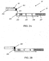
- a vertical distance is present between the antibody and the ligand as shown in Fig. 2A .
- the analyte binding zone 201 is located on a first piece of bibulous strip 20 and the regulation zone 203 is located on a second bibulous strip 21 .
- a gap 202 exists between the two strips 20, 21 .
- the first strip 20 is typically positioned above, and is physically separated from, the second strip 21 .
- the first strip can be located in any suitable position relative to the second strip, so long as the first and second strips are physically separated.
- a sample is applied to the device at the sample application zone 200 located on the first strip 20 .
- the sample contacts the first strip 20 and dissolves the analyte binding antibodies in the analyte binding zone 201 into a liquid phase to form a first analyte-antibody mixture.
- the mixture can be maintained in the analyte binding zone for a period of time.
- the first strip 20 is then positioned adjacent to, and in fluid communication with, the second strip 21 , as shown in Fig. 2B .
- the sample flows from the first strip 20 to the second strip 21 through the analyte binding zone 201 to the regulation zone 203 when the first strip 20 is in contact with the second strip 21 .
- the analyte-antibody mixture comes into contact with the regulation zone 203 located on the second strip 21.
- the antibody-analyte mixture further dissolves the ligands contained on the regulation zone 203 so as to form a second mixture.
- the second mixture is typically formed in a time period shorter than the time period in which the first mixture was formed.
- a blocking zone 204 is typically located downstream from the regulation zone 203 in the detection device.
- the second liquid mixture in the regulation zone then flows along the length of the test strip to the blocking zone.
- the first strip is already in fluid communication with the second strip.
- the liquid mixture can automatically drop down to arrive in the regulation zone 201 due to the gravitation of the liquid sample.
- an amount of trapping receptors are immobilized on the blocking zone.
- the immobilized trapping receptors have specific sites for binding the ligand but do not directly binding any of the analyte-binding antibodies in the second liquid mixture.
- the trapping receptors can be selected from an antibody, an antibody fragment, one partner of some specific molecule pairs irrelevant to the mobilizeable ligands, or any other suitable trapping receptor. When one partner of a pair of specific molecules is immobilized on the blocking zone, another partner of the pair of specific molecules can be conjugated with the ligand and contained on the regulation zone.
- the specific molecule pairs can be selected from biotin and avidin, biotin and streptavidin, antibody and an antigen (antibody for binding a target analyte and analyte itself are excluded), rhodamine and anti-rhodamine, mouse IgG and anti-mouse IgG, or any other suitable molecular pair. More particularly, if the mobilizable ligand are attached or labeled or bound with biotin in the mobilization zone, the blocking zone immobilizes the ligands by an amount of avidin for specifically binding the biotin, as a portion of a conjugate conjugated with the ligands.
- the mobilizeable ligand is conjugated with a protein.
- one protein molecule can be conjugated with several small antigens so that monomolecular antigen ligand can bind to several receptors for the analyte.
- the antigen ligand can also link with biotin.
- the antigen ligand reacts with the analyte antibody to form a complex (analyte-antibody-biotin complex).
- one monomolecular streptavidin-IgG complex can bind to four molecular biotin.
- the blocking zone in some embodiments is disposed on a solid material including, but not limited to, filter paper, cellulose membranes, nylon membranes, or any other suitable material. In some embodiments, the blocking zone is disposed on a cellulose membrane.
- FIG. 3A A device in which a horizontal gap is present between the analyte binding zone 301 and the regulation zone 303 is shown in Fig. 3A .
- the analyte binding zone 301 is located on a first strip 30
- the regulation zone 301 is contained on a second strip 31 .
- the first strip 30 is separated from the second strip 31 along the horizontal axis by a gap 302 between the two strips.
- the sample is applied to the first strip 30 at the sample application zone.
- the sample comes into contact with the analyte binding zone 301 on the first strip 30 .
- the antibodies located in the analyte binding zone 301 are then dissolved into the sample and a first sample mixture is formed.
- the first sample mixture can be maintained in the analyte binding zone for a period of time.
- the first strip 30 can then be positioned adjacent to the second strip 31 , as shown in Fig. 3B .
- the analyte binding zone 301 is placed in fluid communication with the regulation zone 303 , the sample flows from the analyte binding zone 301 to the regulation zone, wherein a second liquid mixture is formed.
- the gap between the analyte binding zone 301 and the regulation zone 303 can be designed to meet various requirements, including, but not limited to, the particular sensitivity and specificity requirements for testing one target analyte using the present device.
- the width of the gap 302 is between about 1 and 100 millimeters. In some embodiments, the width of the gap is at least 1, 1, 2, 3, 4, 5, 6, 7, 8, or 10 millimeters, In some embodiments, the gap is more than 100 millimeters.
- the test device has a housing 400 for housing the test strip 100 , as depicted in Fig. 4 .
- the housing 400 comprises a sample well 41 , through which a sample is introduced to the strip 100 .
- the housing 400 has a sample well 41 aligned with the sample application zone A , and a window 42 aligned with the detection zone F and the control zone G .
- the result shown on the detection zone F can be read through the transparent viewing window 42 .
- the blocking zone D and mobilizeable zone K are typically hidden by the opaque wall of the housing. In some embodiments, the blocking zone and the mobilization zone are viewable through the device wherein the walls of the device are transparent.
- One method for using the test device comprises detecting the presence or absence of a target analyte in a liquid sample with a direct positive or negative result.
- the method includes contacting a sample with an amount of analyte binding antibodies for specifically binding target analytes or an amount of ligand so as to form a first liquid mixture.
- the first mixture contains antibody-analyte complexes if the target analyte is present.
- the first liquid mixture further contains an amount of antibodies if the amount of analyte is less than the amount of the antibodies or an amount of analyte if the amount of analyte is more than the amount of the antibodies.
- the first mixture is typically incubated or maintained for a first time period prior to coming into contact with an amount of ligand, so as to form a second liquid mixture.
- the ligands in the second liquid mixture compete with the analyte for binding the antibodies of the antibody-analyte complexes in a substantially liquid phase.
- the second liquid mixture is typically incubated or maintained a second time period, the second time period being shorter than the first time period.
- the second liquid mixture contacts the solid phase with a blocking zone having an amount of trapping receptors for extracting the ligands, including the unbound ligands or the ligand-antibody complexes, from the second liquid mixture.
- the analyte binding antibodies are not directly bound in this second liquid mixture.
- the test result correlates to the amount of antibody-analyte complexes.
- the method includes applying a liquid sample to a solid phase comprising an analyte binding zone, a regulating zone separated from and downstream from the analyte binding zone, a blocking zone and a detection zone.
- An amount of analyte binding antibodies is contained on the analyte binding zone in a dry state.
- An amount of ligands is contained on the regulation zone in a dry state.
- the analyte binding antibodies and the ligands can be dissolved in a liquid phase and carried by a liquid sample.
- the blocking zone contains an amount of immobilized trapping receptors for binding the ligands but not binding any analyte-binding antibody directly.
- One method for detecting the presence or absence of an analyte in a sample comprises contacting the sample with an amount of antibody for specifically binding an amount of either a target analyte or a ligand as to form a first mixture; contacting the first mixture with an amount of ligand to form a second mixture; contacting the second mixture to a solid phase comprising a blocking zone, wherein the blocking zone comprises an amount of immobilized trapping receptors specifically binding the ligands but not directly binding the antibodies of the first mixture; and determining the amount of the antibodies in the second mixture, wherein the amount of antibodies detected corresponds to an amount of the analyte in the liquid sample.
- the solid phase comprises a lateral flow strip.
- both the ligands and the antibodies for binding the analytes are dried on the lateral flow strip and disposed upstream of the blocking zone.
- the method can further provide for the formation of a molecular conjugate comprising a ligand unrelated to the analyte and the trapping receptor is formed in the second mixture.
- the molecular conjugate can be selected from biotin and avidin, biotin and streptoavidin, or rhodamine and anti-rhodamine.
- the molecular conjugate further comprises a detectable label.
- the detectable label can be selected from the group consisting of colloidal gold, a latex particle, or a chromophore.
- an affinity constant between the mobilizeable antibody and the mobilizeable ligand is higher than an affinity constant between the analyte and the mobilizeable antibody is present.
- the length of time in which the sample contacts the analyte binding antibodies is longer than the length of time in which the first liquid mixture contacts the ligands.
- 3L of a colloidal gold solution having symmetrical and suitable particles (20-40nm in diameter), was prepared in a clean glass container.
- the colloidal gold solution was first mixed.
- 25mg of COC monoclonal antibody was then added to the mixture.
- the solution was continually mixing for approximately 1 hour. After mixing the solution, 35ml of 10% BSA was added to block the reaction. The solution was then mixed again for an additional hour. In succession, the block solution was then centrifuged at 11000rpm for 35 minutes. The supernatant was then removed.
- the deposition was dissolved in 0.003mol/L 0.02%BSA/phosphate solution (washing buffer), and the solution centrifuged again. The supernatant was decanted again and the deposition collected. Thereafter, the concentration of the colloidal gold solution can then be adjusted as needed. The OD value of the solution under 540 nm wavelength was then determined.
- a test strip as shown in Fig. 1 was prepared.
- sample application pad M made from fiberglass was used. The fiberglass was then treated with buffered solution (Borax 0.05M, pH9.3, surface active reagent: 2% S-17: (RHODASURF®ON-870)) and dried in oven at 37°C.
- buffered solution Borax 0.05M, pH9.3, surface active reagent: 2% S-17: (RHODASURF®ON-870)
- label pad B The polyester membrane was then treated with a buffered solution (PVA 5g/L, Na 2 HPO 4 , 7.1g/L, S-14 (Triton-X-100) 0.1%, BSA 5g/L, NaN 3 0.2g/L). The membrane was dried in the oven at 37 °C. The membrane was additionally treated with anti-COC labeled with colloidal gold particles under the concentration of 10 OD and in the condition of 1ul/cm and automatic spray microprocessor, to form label pad 1. Label pad 1 was then dried in oven at 37°C.
- label pad C The polyester membrane was treated with buffered solution (PVA 5g/L, Na 2 HPO 4 , 7.lg/L, S-14 (Triton-X-100) 0.1%, BSA 5g/L, NaN, 0.2g/L) and was dried in oven at 37°C. The membrane was then treated with 0.5mg/ml Biotin-BSA-COC solution 4 under the criterion of 2.5ul/cm and automatic spray microprocessor to form label pad 2. Label pad 2 was then dried in oven at 37°C.
- buffered solution PVA 5g/L, Na 2 HPO 4 , 7.lg/L, S-14 (Triton-X-100) 0.1%, BSA 5g/L, NaN, 0.2g/L
- the membrane was then treated with 0.5mg/ml Biotin-BSA-COC solution 4 under the criterion of 2.5ul/cm and automatic spray microprocessor to form label pad 2. Label pad 2 was then dried in oven at 37°C.
- nitrocellulose membrane E The blocking zone D of the nitrocellulose (NC) membrane E was treated with Streptavidin-IgG (Yingkelong Biotechnology (Hangzhou) Co., Lid.) at a concentration of 0.6 mg/ml and in the condition of 2.5ul/cm and 3 bands automatic spray microspray-membrane processor.
- the detection zone F was treated with goat anti-mouse antibody IgG (purchased from Fitzgerald Inc) at a concentration of 0.6 mg/ml and in the criterion condition of 1.0ul/cm and 1 bands automatic spray microspray-membrane processor. Thereafter, the NC membrane E was dried in an oven at 37°C.
- test strip with sample pad, labeled pads, and NC membrane The prepared parts were cut into a test strip of approximately 0.4 ⁇ 10cm. The sample application zone A , label pad 1 B , label pad 2 C , NC membrane E and absorbent filter paper H were sequentially assembled. Label pad 1 B and label pad 2 C remained isolated from the sample application zone A , label pad 1 B , label pad 2 C , and the NC membrane E by a distance 102 . All these parts were adhibited onto a bibulous material.
- the result for the cocaine test strip is shown in Fig. 5A.
- the test shows the results in the detection zone J for different concentrations of cocaine in the urine samples.
- the cocaine concentrations of the sample are 0ng/ml, 150ng/ml, 450ng/ml and 900ng/ml from left to right.
- I absorption zone
- J detection zone
- K blocking zone
- L labeled zone
- M sample reception zone.
- the detection result shows that detection sensitivity can be improved up to 50 ng/ml degree following the method and the device provided by present invention. False-negatives were not found.
- Dilution of PCP solution Dilute PCP with negative urine into detection solution under the concentration of 0ng/ml, 12.5ng/ml, 37.5ng/ml and 75ng/ml.
- Label pad 1 was treated with rabbit IgG and colloidal gold particles under the concentration of 0.6mg/ml and automatic spray microprocessor. Thereafter, the label pad 1 was dried in oven at 37°C. The concentration and method of other reagents are the same as in Example 1.
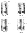
- Fig. 5B illustrates how when PCP is not present in the urine sample, no band appears in the detection zone 5, which indicates a negative result directly (first and second from left to right).
- a band appears in the detection zone, and the band color darkens as the concentration increases.
- test strip procedure used is the same as that described in Example 1.
- the protocol followed is similar to that described in Example 2 with the exception that PCP is replaced with methyl-amphetamine (MET), where the MET dilution is achieved by diluting MET with the negative urine into the detection solution with concentration 0ng/ml, 150ng/ml, 450ng/ml, and 900ng/ml.
- MET methyl-amphetamine
- Fig. 5C illustrates how when MET is not present in the urine sample, no band appears in the detection zone 15 , which indicates a negative result directly (first and second from left to right).
- a band appears in the detection zone, and the band color darkens as the concentration increases.
- test strip procedure used is the same as that described in Example 1.
- the protocol followed is similar to that described in Example 2 with the exception that PCP is replaced with morphine.
- the morphine dilution solution is created by diluting MOP with the negative urine into a detection solution with concentration 0 ng/ml, 105 ng/ml, 450 ng/ml, and 900 ng/ml.
- Fig. 5D illustrates how when MOP is not present in the urine sample, no band appears in the detection zone 25 , which indicates a negative result directly (first and second from left to right).
- MOP is present, a band appears in the detection zone, and the band color darkens as the concentration increases.
- test strip procedure used is the same as that described in Example 1.
- the protocol followed is similar to that described in Example 2 with the exception that PCP is replaced with amphetamine (AMP).
- AMP amphetamine
- the AMP dilution is created by diluting AMP with the negative urine into a detection solution with concentration 0 ng/ml, 150. ng/ml, 1500 ng/ml, and 3000 ng/ml.
- Fig. 5E illustrates how when AMP is not present in the urine sample, no band appears in the detection zone 35, which indicates a negative result directly (first and second from left to right).
- AMP is present, a band appears in the detection zone, and the band color darkens as the concentration increases.
- Example 6 The detection of tetrahydrocannabinol (THC) in urine
- test strip procedure used is the same as that described in Example 1.
- the protocol followed is similar to that described in Example 2 with the exception that PCP is replaced with tetrahydrocannabinol (THC).
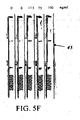
- THC dilution is created by diluting THC with the negative urine into detection solution with concentration 0 ng/ml, 17.5 ng/ml, 75 ng/ml, and 150 ng/ml.
- Fig. 5F illustrates how when THC is not present in the urine sample, no band appears in detection zone 45 , which indicates a negative result directly (first and second from left to right).
- a band appears in the detection zone, and band color darkens as the concentration increases.
Landscapes
- Health & Medical Sciences (AREA)
- Immunology (AREA)
- Life Sciences & Earth Sciences (AREA)
- Engineering & Computer Science (AREA)
- Molecular Biology (AREA)
- Biomedical Technology (AREA)
- Chemical & Material Sciences (AREA)
- Hematology (AREA)
- Urology & Nephrology (AREA)
- Biotechnology (AREA)
- Microbiology (AREA)
- Cell Biology (AREA)
- Food Science & Technology (AREA)
- Medicinal Chemistry (AREA)
- Physics & Mathematics (AREA)
- Analytical Chemistry (AREA)
- Biochemistry (AREA)
- General Health & Medical Sciences (AREA)
- General Physics & Mathematics (AREA)
- Pathology (AREA)
- Investigating Or Analysing Biological Materials (AREA)
- Investigating Or Analysing Materials By The Use Of Chemical Reactions (AREA)
Description
- Immunoassays for detecting an analyte in a sample are used extensively in various fields. Immunoassays can be used to detect analytes in various samples (such as saliva, blood, urine, serum, and sweat) to aid in diagnosis, prognosis and/or treatment of diseases. For instance, immunoassays are routinely performed for monitoring signs of early pregnancy, the presence of rumors, infectious diseases and drug abuse. Immunoassay is effected by the specific binding sites contained between immune mo1lecules, such as antigen/antibody, hapten/antibody, and biotin/antibiotin. Most immunoassays are performed on a solid phase, such as on lateral-flow test strips, glass or plastic multi-porous discs, or immune chromatography devices.
- There are two common formats of immunoassays, namely the sandwich assay and the competitive assay. In a competitive immunoassay, the analyte competes with a labeled analyte to bind with antibodies. This format has been commonly used to detect small analytes such as hapten in a sample. The label used can be one that indicates its presence as a color band in the detection or result reading zone. Reagents and devices utilizing competitive principle have been described in detail in
U.S. Patent Nos. 4,235,601 .4,442,204 ,5,229,073 . For devices utilizing competitive principles, if the color of the detection zone does not change or if no color band appears in detection zone or result reading zone, this indicates a positive result, and thus suggests that an analyte may be present in a sample. Conversely, if color is already present in the detection zone or result reading zone changes or if a color band appears, this can be interpreted as a negative result, which further suggests that the analyte may not be present in a sample. - Other devices in which a positive result is directly indicated have been described in, for example,
U.S. Patent Nos. 5,028,535 ,5,089,391 ,5,627,526 ,5,143,852 .5,480,792 , and5,985,579 , When such a device is used to detect, for example, hapten, if the analyte is present or if its concentration is higher than an anticipated threshold, the color appearing in the test zone suggests a positive result. Conversely, a negative result suggests analyte may not be present or that the analyte concentration is lower than an anticipated threshold. Although the above-mentioned devices and methods can detect several analytes at one time, there are still many disadvantages to the current devices available. For instance, samples and multiple antibodies for binding analytes have to be put in a sample well for about 10-15 minutes to react prior to be applied to the nitrocellulose (NC) membrane. Furthermore, the NC membrane has to be washed repetitively prior to reading the results. Thus such devices are time-consuming, inconvenient to operate, and require multi-steps reactions. - In
U.S. Patent No. 5,451,504 a test strip is disclosed comprising three specific zones, a mobilization zone; a trap zone and a detection zone. An antibody, bound to a color label in the mobilization zone, can bind with an analyte to form a complex (a color label-antibody-analyte complex). The complex can be moved by a liquid to a trap zone where a ligand is immobilized. The immobilized ligand is not able to bind to an antibody that has already formed a complex with an analyte. Such a complex is stable and cannot be affected by the immobilized ligand. The ligand can only bind an antibody that is freely floating and not already bound to an analyte. The analyte-antibody complex can then be captured by an immobilized detection receptor as to show a direct positive result. However, the device described in this patent requires that the affinity between the antibody and the analyte be higher than the affinity between the antibody and the ligand. In practice, it is very difficult to select the antibody with these limited features to apply to this invention. - In
U.S. 6,699,722 a test strip is described consisting of a sample application zone, a mobilization zone, a first capture zone and a second capture zone. In this device, these zones are arranged along the pathway of liquid flowing, so that the liquid flows through each zone sequentially. The first capture zone is coated with a first capture antibody and the second capture zone is coated with a second capture antibody. The molecular weight of a labeled analyte analog is heavier than the weight of the analyte in the sample. If an analyte is present in the sample, the labeled analyte analog and analyte will move along following the pathway of the flowing liquid with the analyte traveling faster than the analyte analog. As result, the analyte will reach the first capture zone earlier than labeled analyte analog. The analyte is captured and fixed by the first capture antibody before the labeled analyte analog reaches the capture zone. The, labeled analyte analog will continue to flow past the first capture zone and come in contact with the second capture zone coated with a second capture antibody. The color in second capture zone will then change in response to the binding of the labeled analyte analogs further indicates a positive result. In practice, such a test strip is inconvenient and the results from such a test strip are not accurate. -
WO 97/31269 - The present invention provides a test device for detecting the presence or absence of an analyte in a liquid sample comprising a mobilization zone comprising a mobilizeable ligand and a mobilizeable antibody, wherein the mobilizable antibody can specifically bind either the analyte or the mobilizeable ligand but not both simultaneously; a blocking zone located downstream from the mobilization zone, wherein the blocking zone comprises an immobilized trapping receptor that binds the mobilizeable ligand; and a detection zone located downstream from the blocking zone, wherein the detection zone comprises an immobilized detecting receptor that binds the mobilizeable antibody. In one embodiment, the mobilizeable antibody is adapted to be contacted by the sample before the mobilizeable ligand is contacted by the sample. In another embodiment, the mobilizeable ligand is adapted to be spatially separated from the mobilizeable antibody so that the sample contacts the mobilizeable antibody prior to contacting the mobilizeable ligands. Where desired, the device of claim can be configured to adopt a lateral flow strip format that supports a flow of said liquid sample across the mobilization zone, the blocking zone, and the detection zone. In some embodiments, the mobilizeable ligand and the mobilizeable antibody are adapted to be separated by a gap. In some embodiments, the mobilizeable ligand and the mobilizeable antibody are adapted to be separated from each other by a vertical or horizontal distance. In other embodiments, the mobilizeable ligand comprises a member of a binding pair that is distinct from the analyte, and wherein the immobilized trapping receptor comprises other member of the binding pair. The binding pair can be selected from the group of biotin and avidin, biotin and streptoavidin, and rhodamine and anti-rhodamine. In yet other embodiments, the mobilizeable antibody exists in a dry state and is contained in a first bibulous strip, and the mobilizeable ligand exists in a dry state and is contained in a second bibulous strip; and wherein the first strip is physically separated from the second strip. In still yet other embodiments, the blocking zone and detection zone are located on the second bibulous strip, downstream from the mobilizeable ligand.
- In some embodiments, the mobilizeable antibody and the mobilizeable ligand exhibit a first binding affinity, and the analyte and the mobilizeable antibody exhibit a second binding affinity, wherein the first binding affinity is higher than the second affinity constant. The mobilizeable antibody can further comprise a color particle. In some other embodiments, the mobilizeable ligand and mobilizeable antibody are configured on the mobilization zone so that contact time during which the sample contacts the mobilizeable antibody is longer than contact time during which the sample contacts the mobilizeable ligand. Where desired, the mobilizeable antibody comprises a detectable label.
- The present invention also provides methods of using the subject device. In one embodiment, the present invention provides a method for detecting the presence or absence of an analyte in a liquid sample by contacting the liquid sample with a subject test device (any one of which described herein) to effect detecting the presence or absence of said analyte. In one embodiment, the subject method proceeds with contacting the liquid sample with mobilizeable antibody on the test device to form a first mixture, followed by contacting the first mixture with the moblizeable ligand to form a second mixture, wherein the second mixture further makes contact with the blocking zone. The mobilizable ligand and mobilizable antibody may be configured on the mobilization zone so that contact time during which the sample contacts the mobilizable antibody is longer than contact time during which the sample contacts the mobilizable ligand.
- The novel features of the invention are set forth with particularity in the appended claims. A better understanding of the features and advantages of the present invention will be obtained by reference to the following detailed description that sets forth illustrative embodiments, in which the principles of the invention are utilized, and the accompanying drawings of which:
-
FIG. 1 is an illustration of a test strip of the device. -
FIGS. 2A & 2B illustrate a test strip of the devices comprising two strips separated by a vertical distance. -
FIGS. 3A & 3B illustrates an alternative embodiment of a test strip having two portions separated by a horizontal distance. -
FIG. 4 illustrates a device having a housing for the test strip. -
FIGS. 5A-5F illustrate the results displayed by the device in detecting different analytes. - The devices and methods described are adapted and configured to detect the presence or absence of an analyte by directly providing a positive or negative result. Typically, a test device is a test strip comprising a mobilization zone K , blocking zone D and detection zone F , e.g,, as shown in
Fig. 1 . The mobilization zone comprises a mobilizeable ligand and a mobilizeable antibody for specifically binding either a target analyte or the mobilizeable ligand. A trapping receptor for capturing the mobilizeable ligand, is present in the blocking zone D . Typically, the trapping receptor can capture the ligands but not directly capture any analyte-binding antibody. The detection zone of the device further comprises an immobilized detecting receptor which can bind to the mobilizeable antibody, for example after the antibody has been dissolved into solution. In some embodiments, the test strip comprises a bibulous material for supporting a fluid sample flowing thereon. A bibulous material refers to those materials that can stably absorb a fluid and make the fluid pass through the material via capillary action. In some embodiments, a bibulous material can be a material that supports the movement and transport of fluids along its length. In another embodiment, the bibulous material is a material for driving fluid movement through the device via capillary action. The test strip may comprise a single type of bibulous material on which the antibodies and ligands are disposed. Alternatively, the test strip can be made up of multiple kinds of bibulous materials which can act reciprocally together with a fluid sample. Bibulous materials include, but are not limited to, nitrocellulose, filter paper, fiberglass, polyester. In addition, the bibulous solid phase, in some embodiments, further contains other suitable reagents for conducting the test on the device. By way of example only, the solid phase may further contain a buffer solution to adjust pH value. - The test strip of the test device further comprises a sample application zone A for receiving the sample. The application zone A is located upstream of a blocking zone D , an absorbent zone H for absorbing the sample after it has passed through the test strip, and a test control zone G . The test control zone comprises an amount of reagent for determining if the sample has had enough time to pass over the detection zone, or if enough time to complete the test has passed. When the test device is used, an amount of antibodies specifically binding to either a target analyte or an amount of ligand are contained on the analyte binding zone of the mobilization zone. The antibodies can be in a dry state. The mobilization zone further has an amount of the ligands located in the regulation zone of the mobilization zone. The ligands are located downstream of the analyte binding antibodies.
- The antibodies and ligands can be dissolved by the sample. In their dissolved state, the antibodies and ligands move through the zones of the test device with the sample, as the sample passes through the corresponding zones. In some embodiments, in the test device, a sample first contacts the antibodies located in the antibody binding zone of the mobilization zone. The antibodies are dissolved in the sample and come in contact with the analyte in the sample. The sample remains in contact with the antibodies in the antibody binding zone for a period of time. The time period is sufficient to form a first liquid mixture consisting of the analyte bound to antibodies, or an analyte-antibody complex. In some embodiments, all of the analyte-binding antibodies are dissolved in the sample. In some embodiments, at least a portion of analyte-binding antibodies is dissolved in the sample. The first liquid mixture then passes to the regulation zone, located subsequent to the analyte binding zone, where the first liquid mixture comes into contact with the ligands located in the regulation zone. The ligands are then dissolved by the first liquid mixture further forming a second liquid mixture. In some embodiments, the sample does not contain any target analyte. In such an embodiment, the mobilized antibodies carried in the first mixture can be bound by the mobile ligands dissolved in the second liquid mixture, so as to form antibody-ligand complexes. These antibody-ligand complexes can then be captured by an amount of trapping receptors located in the blocking zone. The trapping receptors are ligands located in the blocking zone have specific binding sites for directly binding the mobilized ligands. The trapping receptors are immobilized on the blocking zone located downstream of the mobilization zone. Because the antibody-ligand complexes are bound by the trapping receptors, the mobilized antibodies generally do not pass through the blocking zone. Typically, any unbound ligands dissolved in the second liquid mixture will subsequently be bound by the trapping receptors on the blocking zone as well.
- If an analyte or an amount of analyte is present in the sample, at least a portion of the analyte can be bound by the mobilizeable antibodies on the analyte binding zone. If present the analytes typically form a first complex, an antibody-analyte complex, to create a first liquid mixture. In some embodiments, only a portion of the analyte binding antibodies are bound by the target analyte. In such an embodiment, the antibodies dissolved in the first liquid mixture, but which remain unbound to an analyte, can then be bound by the ligands located in the regulation zone. When the first liquid mixture contacts the ligands located downstream of the antibody binding zone, a second mixture is formed. The second mixture typically includes the first antibody-analyte complexes and the ligands dissolved by the first liquid mixture.
- In some embodiments, all of the antibody-analyte complexes remain undisturbed by the dissolved ligands. In some embodiments, some of the dissolved ligands will compete with the analyte to bind to the binding sites on the antibodies of the first complexes. In such a case, the first liquid mixture can contain both analyte-antibody complexes and antibody-ligand complexes. The second mixture then comes in contact with the blocking zone. The blocking zone typically contains an amount of immobilized trapping receptors. The trapping receptors can bind to either the ligand-antibody complex, if a portion of the antibodies are unbound by the analytes, or to unbound ligands, if only a portion of the ligands are bound to the mobilized antibodies. The trapping receptors typically do not capture the antibody-analyte complexes.
- The sample then passes to the detection zone, located downstream from the blocking zone. The analyte-antibody complexes can, in some embodiments, be captured by an amount of detection molecules immobilized on the detection zone of the test device when the second liquid mixture arrives at the detection zone. The detection zone has an amount of immobilized receptors for specifically binding the analyte binding antibodies. The receptors are typically antibodies. In some embodiments, the analyte-binding antibodies are conjugated with a detectable label. Determining the amount of the detectable label correlates to the amount of the anlayte in the test sample. Typically, the detectable labels are color particles. The amount of the analyte can then be directly determined through detecting the color particles captured on the detection zone by naked eyes.
- When the second liquid mixture comes in contact with the detection zone, specific binding sites on the antibody are typically exposed when dissolved in the sample. The exposed binding sites of the antibodies are typically recognizable by the analyte, so as to enable a reaction in a substantially short period of time. The second liquid mixture contains the ligands dissolved in the sample. In some embodiments, the ligands dissolved in the sample compete with the analyte for the antibodies already complexed with the analyte. Alternatively the ligands may bind to any unbound analyte-binding antibodies directly, since the binding sites of the ligands are also exposed when the ligands are dissolved in the first mixture. Further, in some other embodiments, the ligand is directly or indirectly connected to, linked with, or conjugated with one member of a binding pair of specific molecules that are not related to the analyte-binding antibody. In such an embodiment, some of the binding sites of the ligands are covered by one member of the pair. This configuration reduces the competition between the ligand and the analyte for the binding site on the antibodies in the antibody-analyte complex. In some embodiments, the ligand comprises a portion of a conjugate that includes biotin. The ligand further forms a biotin/avidin conjugate, wherein the avidin is immobilized on the blocking zone for capturing the second complexes (antibody-ligand-biotin) when the second mixture passes through the blocking zone. In yet another embodiment, the ligands are a portion of a conjugate with one partner of a pair of specific binding molecules conjugated with a protein.
- In modem assays for small molecules, especially for drug abuse detection, the lowest concentration, or the cut-off value, should be changed in order to distinguish drug abusers from patients undergoing normal medical treatment. For example, if a patient's blood is analyzed for presence of an analyte, the patient blood may contain a concentration of an analyse higher than the standard cut-off value. When the concentration of the target analyte in a sample is higher than the standard cut-off value, that may not necessarily mean that the patient is a drug user. The patient may simply have a higher concentration of analyte simply due to the fact that he/she consumes the analyte as part of treatment. In such a situation, the concentration of the analyte can not be detected by the device and compared with the standard level of the analyse in the general population. It would be more accurate to use a device with an increased cut-off value to identify a potential drug user. In some embodiments, the device comprises an analyte binding antibody located in the mobilization zone, wherein the antibody has a lower affinity, K1, for binding a target analyte than its affinity, K2, for binding to the ligand. If K1 is less than K2, a higher concentration of analyte is required to compete with the mobilizeable ligand for the binding sites located on the mobilizeable antibody in the second liquid mixture. Thus, a proper negative result, not a false positive result, can result on the detection zone even if the analyte concentration is higher than a standard level in patient's body fluid. An antibody with an affinity of K1 less than or equal to K2 is typically used.
- The antibody for specifically binding an analyte and the ligand may both be in a dry phase prior to being contacted by the liquid sample. Alternatively the antibody can be in the liquid phase prior to being contacted by a sample. The antibody and ligand are typically contained on the single strip of the test device in the dry state. When a liquid sample moves through the mobilization zone, the antibodies and the ligands can be dissolved in, and carried by, the liquid sample.
- The mobilizeable antibody specifically capable of binding to an analyte can be conjugated with a detectable label. The label can be selected from, but not limited to, an enzyme, a non-water soluble particle, a colloidal gold particle, a latex particle, or any combination thereof. In some embodiments, the label is a water-soluble substance or a fluorescent substance. The label can be any label suitable for labeling the antibody.
- The test device can be used to detect an analyte using the competition principle employed in immunoassays to indicate the presence or absence of a target analyte in a sample by displaying a direct positive or negative result. In particular, the subject device can comprise electronic readers that detects optical signals generated in an immunoassay.
-
Fig.1 depicts one embodiment of the test device. The test device, as shown, comprises atest strip 100 having an sample reception zone A , an analyte binding zone B typically containing an amount of antibodies and color labels, a regulation zone C , with an amount of ligands as a portion of conjugates conjugated with biotin, and a nitrocellulose membrane E . The nitrocellulose membrane further comprises a blocking zone D , with an amount of immobilized avidin, a detection zone F , and a result control zone G . Typically, an absorption zone H , for absorbing the liquid passed from the mobilization zone K to the control zone G , is present. The mobilization zone K encompasses both the analyte-binding zone B and the regulation zone C . The analytes and ligands on the analyte binding zone and regulation zone, respectively, can be dissolved and carried along the test strip by the sample. - The sample reception zone A is typically located upstream from the mobilization zone. The sample reception zone A can, additionally, be treated with chemical reagents which can improve or regulate the reactive conditions between the analyte and the antibody. Chemical reagents used include, but are not limited to, reactive PH values, ionic concentrations, removal of some interferential substances in the samples, or any other suitable chemical reagent, or any combination thereof known in the prior art. The absorbent zone H is typically located downstream from the detection zone so as to facilitate and enhance the flow of liquid. Suitable materials for absorbent zone include, but may not be limited to, filter paper, or any other absorbent materials.
- The mobilization zone K comprises an amount of antibodies for binding to either a target analyte or an amount of ligand. The antibodies are contained in the mobilization zone in a dry state. The mobilization zone further has an amount of the ligands thereon downstream from the analyte binding antibodies. The antibodies and ligands can be moved by and dissolved in the sample when the sample passes the corresponding zones. An amount of ligand can be contained on the regulation zone C located downstream from the analyte binding zone B . In addition, an amount of antibodies for specially binding the analyte can be contained in a dry state on the analyte binding zone B . A liquid sample is typically applied to the
strip 100 . The sample then comes in contact with the analyte binding antibody located on the analyte binding zone B for a period of time. The sample then comes into contact with the ligands on the regulation zone C for a time period shorter than the time period in which the sample was in contact with the analyte binding zone B . The analyte and analyte binding antibodies form a first mixture in the antibody binding zone, which can be maintained in the antibody binding zone for a period of time prior to contacting the ligands located downstream from the analyte binding antibodies. The time in which the first mixture typically contacts the analyte binding antibody is longer than the time in which the second mixture contacts the mobilizeable ligand. In some embodiments, the time in which the first mixture contacts the analyte binding antibody is between about one second to about sixty seconds. In some embodiments, the time is between about 10 seconds to about 30 seconds. In some embodiments, the time period is longer than 60 seconds. - The test strip typically further comprises an analyte binding zone B and a regulation zone C , with a
gap 102 separating the analyte binding zone B and the regulation zone C . Thegap 102 typically comprises a bibulous material. In some embodiments of the device described herein, more than one gap is present. A gap may be located between the regulation zone and the blocking zone on the test strip. In such an embodiment, the second gap is shorter than the first gap. Therefore. the time period in which the liquid sample contacts the analyte binding zone for reacting with the analyte binding antibodies is longer than the time period in which the sample contacts the ligands. The binding between the analyte binding antibody and the analyte in the liquid sample is more complete than the binding between the ligands and the analyte antibodies in the second liquid mixture due to the different lengths in times. - In another aspect of the device described herein, a vertical distance is present between the antibody and the ligand as shown in
Fig. 2A . In such an embodiment, theanalyte binding zone 201 is located on a first piece ofbibulous strip 20 and theregulation zone 203 is located on a secondbibulous strip 21 . Agap 202 exists between the twostrips first strip 20 is typically positioned above, and is physically separated from, thesecond strip 21 . The first strip can be located in any suitable position relative to the second strip, so long as the first and second strips are physically separated. A sample is applied to the device at thesample application zone 200 located on thefirst strip 20 . The sample contacts thefirst strip 20 and dissolves the analyte binding antibodies in theanalyte binding zone 201 into a liquid phase to form a first analyte-antibody mixture. The mixture can be maintained in the analyte binding zone for a period of time. Thefirst strip 20 is then positioned adjacent to, and in fluid communication with, thesecond strip 21 , as shown inFig. 2B . The sample flows from thefirst strip 20 to thesecond strip 21 through theanalyte binding zone 201 to theregulation zone 203 when thefirst strip 20 is in contact with thesecond strip 21 . After contact occurs between the first andsecond strips regulation zone 203 located on thesecond strip 21. The antibody-analyte mixture further dissolves the ligands contained on theregulation zone 203 so as to form a second mixture. The second mixture is typically formed in a time period shorter than the time period in which the first mixture was formed. - A blocking
zone 204 is typically located downstream from theregulation zone 203 in the detection device. The second liquid mixture in the regulation zone then flows along the length of the test strip to the blocking zone. In an alternative embodiment the first strip is already in fluid communication with the second strip. In such an embodiment, the liquid mixture can automatically drop down to arrive in theregulation zone 201 due to the gravitation of the liquid sample. - In some embodiments, an amount of trapping receptors are immobilized on the blocking zone. The immobilized trapping receptors have specific sites for binding the ligand but do not directly binding any of the analyte-binding antibodies in the second liquid mixture. The trapping receptors can be selected from an antibody, an antibody fragment, one partner of some specific molecule pairs irrelevant to the mobilizeable ligands, or any other suitable trapping receptor. When one partner of a pair of specific molecules is immobilized on the blocking zone, another partner of the pair of specific molecules can be conjugated with the ligand and contained on the regulation zone. The specific molecule pairs can be selected from biotin and avidin, biotin and streptavidin, antibody and an antigen (antibody for binding a target analyte and analyte itself are excluded), rhodamine and anti-rhodamine, mouse IgG and anti-mouse IgG, or any other suitable molecular pair. More particularly, if the mobilizable ligand are attached or labeled or bound with biotin in the mobilization zone, the blocking zone immobilizes the ligands by an amount of avidin for specifically binding the biotin, as a portion of a conjugate conjugated with the ligands.
- In another embodiment, the mobilizeable ligand (antigen-ligand) is conjugated with a protein. Generally, one protein molecule can be conjugated with several small antigens so that monomolecular antigen ligand can bind to several receptors for the analyte. The antigen ligand can also link with biotin. In negative condition, the antigen ligand reacts with the analyte antibody to form a complex (analyte-antibody-biotin complex). When the complex moves to the solid phase with liquid flowing and binds to streptavidin-IgG complex, one monomolecular streptavidin-IgG complex can bind to four molecular biotin. This multi-site conjugation prevents the analyte and antibody from moving past the solid phase. The ability to prevent as few as possible antibodies or analytes accordingly decreases the possibility of false positive. Other reagents for improving the test can also be disposed on or upstream of the blocking zone. In addition, the blocking zone in some embodiments is disposed on a solid material including, but not limited to, filter paper, cellulose membranes, nylon membranes, or any other suitable material. In some embodiments, the blocking zone is disposed on a cellulose membrane.
- A device in which a horizontal gap is present between the
analyte binding zone 301 and theregulation zone 303 is shown inFig. 3A . Theanalyte binding zone 301 is located on afirst strip 30 , and theregulation zone 301 is contained on asecond strip 31 . Thefirst strip 30 is separated from thesecond strip 31 along the horizontal axis by agap 302 between the two strips. The sample is applied to thefirst strip 30 at the sample application zone. The sample comes into contact with theanalyte binding zone 301 on thefirst strip 30 . The antibodies located in theanalyte binding zone 301 are then dissolved into the sample and a first sample mixture is formed. The first sample mixture can be maintained in the analyte binding zone for a period of time. Thefirst strip 30 can then be positioned adjacent to thesecond strip 31 , as shown inFig. 3B . When theanalyte binding zone 301 is placed in fluid communication with theregulation zone 303 , the sample flows from theanalyte binding zone 301 to the regulation zone, wherein a second liquid mixture is formed. The gap between theanalyte binding zone 301 and theregulation zone 303 can be designed to meet various requirements, including, but not limited to, the particular sensitivity and specificity requirements for testing one target analyte using the present device. In some embodiments, the width of thegap 302 is between about 1 and 100 millimeters. In some embodiments, the width of the gap is at least 1, 1, 2, 3, 4, 5, 6, 7, 8, or 10 millimeters, In some embodiments, the gap is more than 100 millimeters. - In some embodiments, the test device has a
housing 400 for housing thetest strip 100 , as depicted inFig. 4 . Thehousing 400 comprises a sample well 41 , through which a sample is introduced to thestrip 100 . As illustratedFig. 4 , thehousing 400 has a sample well 41 aligned with the sample application zone A , and awindow 42 aligned with the detection zone F and the control zone G . The result shown on the detection zone F can be read through thetransparent viewing window 42. The blocking zone D and mobilizeable zone K (as seen inFig. 1 ) are typically hidden by the opaque wall of the housing. In some embodiments, the blocking zone and the mobilization zone are viewable through the device wherein the walls of the device are transparent. - Also provided herein are methods for using the test device. One method for using the test device comprises detecting the presence or absence of a target analyte in a liquid sample with a direct positive or negative result. The method includes contacting a sample with an amount of analyte binding antibodies for specifically binding target analytes or an amount of ligand so as to form a first liquid mixture. The first mixture contains antibody-analyte complexes if the target analyte is present. The first liquid mixture further contains an amount of antibodies if the amount of analyte is less than the amount of the antibodies or an amount of analyte if the amount of analyte is more than the amount of the antibodies. The first mixture is typically incubated or maintained for a first time period prior to coming into contact with an amount of ligand, so as to form a second liquid mixture. The ligands in the second liquid mixture compete with the analyte for binding the antibodies of the antibody-analyte complexes in a substantially liquid phase. The second liquid mixture is typically incubated or maintained a second time period, the second time period being shorter than the first time period. During the second time period the second liquid mixture contacts the solid phase with a blocking zone having an amount of trapping receptors for extracting the ligands, including the unbound ligands or the ligand-antibody complexes, from the second liquid mixture. The analyte binding antibodies are not directly bound in this second liquid mixture. The test result correlates to the amount of antibody-analyte complexes.
- In other embodiments, the method includes applying a liquid sample to a solid phase comprising an analyte binding zone, a regulating zone separated from and downstream from the analyte binding zone, a blocking zone and a detection zone. An amount of analyte binding antibodies is contained on the analyte binding zone in a dry state. An amount of ligands is contained on the regulation zone in a dry state. The analyte binding antibodies and the ligands can be dissolved in a liquid phase and carried by a liquid sample. The blocking zone contains an amount of immobilized trapping receptors for binding the ligands but not binding any analyte-binding antibody directly.
- Also provided herein are methods for detecting the presence or absence of an analyte. One method for detecting the presence or absence of an analyte in a sample comprises contacting the sample with an amount of antibody for specifically binding an amount of either a target analyte or a ligand as to form a first mixture; contacting the first mixture with an amount of ligand to form a second mixture; contacting the second mixture to a solid phase comprising a blocking zone, wherein the blocking zone comprises an amount of immobilized trapping receptors specifically binding the ligands but not directly binding the antibodies of the first mixture; and determining the amount of the antibodies in the second mixture, wherein the amount of antibodies detected corresponds to an amount of the analyte in the liquid sample. In some embodiments, the solid phase comprises a lateral flow strip. In some embodiments, both the ligands and the antibodies for binding the analytes are dried on the lateral flow strip and disposed upstream of the blocking zone. The method can further provide for the formation of a molecular conjugate comprising a ligand unrelated to the analyte and the trapping receptor is formed in the second mixture. The molecular conjugate can be selected from biotin and avidin, biotin and streptoavidin, or rhodamine and anti-rhodamine. Additionally, the molecular conjugate further comprises a detectable label. The detectable label can be selected from the group consisting of colloidal gold, a latex particle, or a chromophore. In some embodiments an affinity constant between the mobilizeable antibody and the mobilizeable ligand is higher than an affinity constant between the analyte and the mobilizeable antibody is present. In some embodiments, the length of time in which the sample contacts the analyte binding antibodies is longer than the length of time in which the first liquid mixture contacts the ligands.
- The invention will be further illustrated by the following examples, which are intended to be purely examples of the invention and not as limitations to the invention.
- 3L of a colloidal gold solution, having symmetrical and suitable particles (20-40nm in diameter), was prepared in a clean glass container. The colloidal gold solution was first mixed. A phosphate buffer solution with a pH=7.0 was then added to the colloidal gold solution to adjust the pH of the colloidal gold solution to about 6.8. 25mg of COC monoclonal antibody was then added to the mixture. The solution was continually mixing for approximately 1 hour. After mixing the solution, 35ml of 10% BSA was added to block the reaction. The solution was then mixed again for an additional hour. In succession, the block solution was then centrifuged at 11000rpm for 35 minutes. The supernatant was then removed. Afterward, the deposition was dissolved in 0.003mol/L 0.02%BSA/phosphate solution (washing buffer), and the solution centrifuged again. The supernatant was decanted again and the deposition collected. Thereafter, the concentration of the colloidal gold solution can then be adjusted as needed. The OD value of the solution under 540 nm wavelength was then determined.
- 2.5mg/ml of BSA-Cocaine conjugate (purchased from Immunetic, Inc) was dialyzed in 1000ml NaHCO3 buffer solution (pH=8.0) for 4 hours to form solution (1). After diluting biotin with distilled water to a concentration of 10 mM, 47µl biotin solution and solution (1) were mixed together and stirred for 1 hour at room temperature to form the solution (2). Determining that the solution (2) was dialyze in 1000 ml phosphate buffered solution (pH=7.4) for 12 hours to and from solution (3). The concentration of cocaine-BSA-Biotin under 655 nm wavelengths in a spectrophotometer, was then diluted into solution (4) to form the terminal solution in concentration of 0.5mg/ml.
- A test strip as shown in
Fig. 1 was prepared. - Preparation of the sample application pad: A sample application pad M made from fiberglass was used. The fiberglass was then treated with buffered solution (Borax 0.05M, pH9.3, surface active reagent: 2% S-17: (RHODASURF®ON-870)) and dried in oven at 37°C.
- Preparation of label pad B: The polyester membrane was then treated with a buffered solution (PVA 5g/L, Na2HPO4, 7.1g/L, S-14 (Triton-X-100) 0.1%, BSA 5g/L, NaN3 0.2g/L). The membrane was dried in the oven at 37 °C. The membrane was additionally treated with anti-COC labeled with colloidal gold particles under the concentration of 10 OD and in the condition of 1ul/cm and automatic spray microprocessor, to form label pad 1. Label pad 1 was then dried in oven at 37°C.
- Preparation of label pad C: The polyester membrane was treated with buffered solution (PVA 5g/L, Na2HPO4, 7.lg/L, S-14 (Triton-X-100) 0.1%, BSA 5g/L, NaN, 0.2g/L) and was dried in oven at 37°C. The membrane was then treated with 0.5mg/ml Biotin-BSA-COC solution 4 under the criterion of 2.5ul/cm and automatic spray microprocessor to form label pad 2. Label pad 2 was then dried in oven at 37°C.
- Preparation of the nitrocellulose membrane: The blocking zone D of the nitrocellulose (NC) membrane E was treated with Streptavidin-IgG (Yingkelong Biotechnology (Hangzhou) Co., Lid.) at a concentration of 0.6 mg/ml and in the condition of 2.5ul/cm and 3 bands automatic spray microspray-membrane processor. The detection zone F was treated with goat anti-mouse antibody IgG (purchased from Fitzgerald Inc) at a concentration of 0.6 mg/ml and in the criterion condition of 1.0ul/cm and 1 bands automatic spray microspray-membrane processor. Thereafter, the NC membrane E was dried in an oven at 37°C.
- Construction of the test strip with sample pad, labeled pads, and NC membrane: The prepared parts were cut into a test strip of approximately 0.4× 10cm. The sample application zone A, label pad 1 B , label pad 2 C , NC membrane E and absorbent filter paper H were sequentially assembled. Label pad 1 B and label pad 2 C remained isolated from the sample application zone A , label pad 1 B , label pad 2 C , and the NC membrane E by a
distance 102 . All these parts were adhibited onto a bibulous material. - Dilute cocaine with the negative urine into detection solution under the concentration of 0ng/ml, 150ng/ml, 50ng/ml, 100ng/ml, 150ng/ml, 450ng/ml and 900ng/ml.
- The detection results are summarized in Table 1.
Table 1. Cocaine detection solution quantity Result Negative positive exact quantity exact incident negative detection solution 10 10 0 10 100% 25ng/ml 10 5 5 5 50% 50ng/ml 10 0 10 10 100% 100ng/ml 10 0 10 10 100% 150ng/ml 10 0 10 10 100% 450ng/ml 10 0 10 10 100% 900ng/ml 10 0 10 10 100% - The result for the cocaine test strip is shown in
Fig. 5A. The test shows the results in the detection zone J for different concentrations of cocaine in the urine samples. The cocaine concentrations of the sample are 0ng/ml, 150ng/ml, 450ng/ml and 900ng/ml from left to right. I : absorption zone; J : detection zone; K : blocking zone; L : labeled zone; M : sample reception zone. - The detection result shows that detection sensitivity can be improved up to 50 ng/ml degree following the method and the device provided by present invention. False-negatives were not found.
- A test strip was prepared as described in Example 1. The protocol followed was the same as Example 1 with several differences: (1) COC was replaced with PCP; (2) buffered solution for treating sample application pad A was TRIS buffered solution, the concrete prescription including 0.1M TRIS salt (PH=8.0), surface active reagent comprising 1% S-7 (RHODASURF®ON-870), 1% S-9 (RHODASURF®ON-870) and 1%BSA; (3) the concentration of Biotin-BSA-PCP for treating label pad C was 1 mg/ml, the treating criterion is 2.5ul/cm, and the label pad C was dried in oven at 37°C; (3) when preparing the NC membrane E , the detection result operation zone G , which is downstream from detection zone F and near absorbent pad H , is coated with goat anti-rabbit IgG. The coating method is the same as that for detection zone F in Example 1.
- Dilution of PCP solution: Dilute PCP with negative urine into detection solution under the concentration of 0ng/ml, 12.5ng/ml, 37.5ng/ml and 75ng/ml.
- Label pad 1 was treated with rabbit IgG and colloidal gold particles under the concentration of 0.6mg/ml and automatic spray microprocessor. Thereafter, the label pad 1 was dried in oven at 37°C. The concentration and method of other reagents are the same as in Example 1.
-
Fig. 5B illustrates how when PCP is not present in the urine sample, no band appears in thedetection zone 5, which indicates a negative result directly (first and second from left to right). When PCP is present, a band appears in the detection zone, and the band color darkens as the concentration increases. - The test strip procedure used is the same as that described in Example 1. The protocol followed is similar to that described in Example 2 with the exception that PCP is replaced with methyl-amphetamine (MET), where the MET dilution is achieved by diluting MET with the negative urine into the detection solution with concentration 0ng/ml, 150ng/ml, 450ng/ml, and 900ng/ml.
-
Fig. 5C illustrates how when MET is not present in the urine sample, no band appears in thedetection zone 15, which indicates a negative result directly (first and second from left to right). When MET is present, a band appears in the detection zone, and the band color darkens as the concentration increases. - The test strip procedure used is the same as that described in Example 1. The protocol followed is similar to that described in Example 2 with the exception that PCP is replaced with morphine. The morphine dilution solution is created by diluting MOP with the negative urine into a detection solution with
concentration 0 ng/ml, 105 ng/ml, 450 ng/ml, and 900 ng/ml. -
Fig. 5D illustrates how when MOP is not present in the urine sample, no band appears in thedetection zone 25, which indicates a negative result directly (first and second from left to right). When MOP is present, a band appears in the detection zone, and the band color darkens as the concentration increases. - The test strip procedure used is the same as that described in Example 1. The protocol followed is similar to that described in Example 2 with the exception that PCP is replaced with amphetamine (AMP). The AMP dilution is created by diluting AMP with the negative urine into a detection solution with
concentration 0 ng/ml, 150. ng/ml, 1500 ng/ml, and 3000 ng/ml. -
Fig. 5E illustrates how when AMP is not present in the urine sample, no band appears in thedetection zone 35, which indicates a negative result directly (first and second from left to right). When AMP is present, a band appears in the detection zone, and the band color darkens as the concentration increases. - The test strip procedure used is the same as that described in Example 1. The protocol followed is similar to that described in Example 2 with the exception that PCP is replaced with tetrahydrocannabinol (THC). The THC dilution is created by diluting THC with the negative urine into detection solution with
concentration 0 ng/ml, 17.5 ng/ml, 75 ng/ml, and 150 ng/ml. -
Fig. 5F illustrates how when THC is not present in the urine sample, no band appears indetection zone 45 , which indicates a negative result directly (first and second from left to right). When THC is present, a band appears in the detection zone, and band color darkens as the concentration increases. - While preferred embodiments of the present invention have been shown and described herein, it will be obvious to those skilled in the art that such embodiments are provided by way of example only. Numerous variations, changes, and substitutions will now occur to those skilled in the art without departing from the invention. It should be understood that various alternatives to the embodiments of the invention described herein may be employed in practicing the invention. It is intended that the following claims define the scope of the invention and that methods and structures within the scope of these claims and their equivalents be covered thereby.
Claims (15)
- A test device for detecting the presence or absence of an analyte in a liquid sample comprising:a mobilization zone comprising a mobilizeable ligand and a mobilizable antibody, wherein the mobilizable antibody can specifically bind either the analyte or the mobilizable ligand but not both simultaneously;a blocking zone located downstream from the mobilization zone, wherein the blocking zone comprises an immobilized trapping receptor that binds the mobilizable ligand; anda detection zone located downstream from the blocking zone, wherein the detection zone comprises an immobilized detecting receptor that binds the mobilizable antibody.
- The device of claim 1, wherein the mobilizable antibody is adapted to be contacted by the sample before the mobilizable ligand is contacted by the sample.
- The device of claim 1, wherein the mobilizable ligand is adapted to be spatially separated from the mobilizable antibody so that the sample contacts the mobilizable antibody prior to contacting the mobilizable ligand.
- The device of any preceding claim, that is configured to adopt a lateral flow strip format that supports a flow of said liquid sample across the mobilization zone, the blocking zone, and the detection zone.
- The device of any preceding claim, wherein the mobilizable ligand and the mobilizable antibody are adapted to be separated by a gap.
- The device of any preceding claim, wherein the mobilizable ligand comprises a member of a binding pair that is distinct from the analyte, and wherein the immobilized trapping receptor comprises the other member of the binding pair.
- The device of claim 6, wherein the binding pair is selected from the group of biotin and avidin, biotin and streptavidin, and rhodamine and anti-rhodamine.
- The device of any preceding claim, wherein the mobilizable antibody exists in a dry state and is contained in a first bibulous strip, and the mobilizable ligand exists in a dry state and is contained in a second bibulous strip, and wherein the first strip is physically separated from the second strip.
- The device of claim 8, wherein the blocking zone and detection zone are located on the second bibulous strip, downstream from the mobilizable ligand.
- The device of any preceding claim, wherein the mobilizable antibody and the mobilizable ligand exhibit a first binding affinity, and the analyte and the mobilizable antibody exhibit a second binding affinity, wherein the first binding affinity is higher than the second affinity constant.
- The device of any preceding claim, wherein the inobilizable ligand and mobilizable antibody are configured on the mobilization zone so that the contact time during which the sample contacts the mobilizable antibody is longer than the contact time during which the sample contacts the mobilizable ligand.
- The device of any preceding claim, wherein the mobilizable antibody comprises a detectable label.
- A method for detecting the presence or absence of an analyte in a liquid sample, the method comprising:contacting the liquid sample with the test device of any one of claims 1-12, thereby detecting the presence or absence of said analyte.
- The method of claim 13, wherein the liquid sample is permitted to first contact the mobilizable antibody on the test device to form a first mixture, followed by contacting the first mixture with the moblizable ligand to form a second mixture, wherein the second mixture further makes contact with the blocking zone.
- The method of claim 13 or claim 14, wherein the mobilizable ligand and mobilizable antibody are configured on the mobilization zone so that contact time during which the sample contacts the mobilizable antibody is longer than contact time during which the sample contacts the mobilizable ligand.
Applications Claiming Priority (2)
| Application Number | Priority Date | Filing Date | Title |
|---|---|---|---|
| CN2007100676071A CN101021526B (en) | 2007-03-16 | 2007-03-16 | Apparatus and method for visual display of detecting result |
| PCT/IB2008/001677 WO2008135862A2 (en) | 2007-03-16 | 2008-03-17 | Devices and methods for detecting analytes |
Publications (2)
| Publication Number | Publication Date |
|---|---|
| EP2137532A2 EP2137532A2 (en) | 2009-12-30 |
| EP2137532B1 true EP2137532B1 (en) | 2015-08-26 |
Family
ID=38709362
Family Applications (1)
| Application Number | Title | Priority Date | Filing Date |
|---|---|---|---|
| EP08776297.7A Not-in-force EP2137532B1 (en) | 2007-03-16 | 2008-03-17 | Devices and methods for detecting analytes |
Country Status (7)
| Country | Link |
|---|---|
| US (1) | US8313955B2 (en) |
| EP (1) | EP2137532B1 (en) |
| CN (1) | CN101021526B (en) |
| AU (1) | AU2008247060A1 (en) |
| CA (1) | CA2679105A1 (en) |
| DE (1) | DE212008000023U1 (en) |
| WO (1) | WO2008135862A2 (en) |
Cited By (1)
| Publication number | Priority date | Publication date | Assignee | Title |
|---|---|---|---|---|
| US12405272B2 (en) | 2019-04-10 | 2025-09-02 | Spd Swiss Precision Diagnostics Gmbh | Assay device |
Families Citing this family (28)
| Publication number | Priority date | Publication date | Assignee | Title |
|---|---|---|---|---|
| US9134303B1 (en) | 1998-08-25 | 2015-09-15 | Alere Scarborough, Inc. | ICT immunoassay for Legionella pneumophila serogroup 1 antigen employing affinity purified antibodies thereto |
| US6824997B1 (en) | 1998-09-18 | 2004-11-30 | Binax, Inc. | Process and materials for the rapid detection of streptococcus pneumoniae employing purified antigen-specific antibodies |
| US8669052B2 (en) | 2008-06-10 | 2014-03-11 | Rapid Pathogen Screening, Inc. | Lateral flow nucleic acid detector |
| US8815609B2 (en) | 2008-05-20 | 2014-08-26 | Rapid Pathogen Screening, Inc. | Multiplanar lateral flow assay with diverting zone |
| US20130196310A1 (en) | 2008-05-20 | 2013-08-01 | Rapid Pathogen Screening, Inc. | Method and Device for Combined Detection of Viral and Bacterial Infections |
| US9068981B2 (en) | 2009-12-04 | 2015-06-30 | Rapid Pathogen Screening, Inc. | Lateral flow assays with time delayed components |
| US8962260B2 (en) | 2008-05-20 | 2015-02-24 | Rapid Pathogen Screening, Inc. | Method and device for combined detection of viral and bacterial infections |
| US8609433B2 (en) | 2009-12-04 | 2013-12-17 | Rapid Pathogen Screening, Inc. | Multiplanar lateral flow assay with sample compressor |
| US20110086359A1 (en) | 2008-06-10 | 2011-04-14 | Rapid Pathogen Screening, Inc. | Lateral flow assays |
| JP5997418B2 (en) * | 2008-12-30 | 2016-09-28 | 株式会社テクノメデイカ | Oxidative stress state system |
| DE102009010563A1 (en) | 2009-02-16 | 2010-08-26 | Matthias W. Engel | Device for the detection of analytes in body fluids |
| CN101943698A (en) * | 2010-09-03 | 2011-01-12 | 山西大学 | Dot immuno-gold filtration assay (DIGFA) quick detection card for leather hydrolyzate in milk and preparation method thereof |
| US8486717B2 (en) | 2011-01-18 | 2013-07-16 | Symbolics, Llc | Lateral flow assays using two dimensional features |
| CN103293134A (en) * | 2012-03-01 | 2013-09-11 | 庞磊 | Fluorescence analysis method and device |
| CN102928594B (en) * | 2011-12-27 | 2015-11-25 | 北京宝瑞源科技孵化有限公司 | phencyclidine detection kit and preparation method thereof |
| CN102707047A (en) * | 2012-06-05 | 2012-10-03 | 南通市产品质量监督检验所 | Colloidal gold test card for quickly testing ergot alkaloid and preparation method thereof |
| US9874556B2 (en) | 2012-07-18 | 2018-01-23 | Symbolics, Llc | Lateral flow assays using two dimensional features |
| US20140072959A1 (en) | 2012-09-12 | 2014-03-13 | Force Diagnostics, Inc. | Rapid tests for insurance underwriting |
| CA2902484C (en) | 2013-02-26 | 2021-05-18 | Astute Medical, Inc. | Lateral flow assay with test strip retainer |
| WO2015038978A1 (en) | 2013-09-13 | 2015-03-19 | Symbolics, Llc | Lateral flow assays using two dimensional test and control signal readout patterns |
| US20160025752A1 (en) * | 2014-06-24 | 2016-01-28 | Seed Research And Development, Llc | Devices and methods for detecting and/or quantifying analytes in fluids |
| CN104090109A (en) * | 2014-07-25 | 2014-10-08 | 胡晓武 | Colloidal gold immunochromatography test paper and colloidal gold immunochromatography test method for quickly detecting human blood procalcitonin |
| US20180106799A1 (en) | 2015-04-06 | 2018-04-19 | Bludiagnostics, Inc. | A test device for detecting an analyte in a saliva sample and method of use |
| US10808287B2 (en) | 2015-10-23 | 2020-10-20 | Rapid Pathogen Screening, Inc. | Methods and devices for accurate diagnosis of infections |
| WO2018039047A1 (en) | 2016-08-23 | 2018-03-01 | Qoolabs, Inc. | Lateral flow assay for assessing recombinant protein expression or reporter gene expression |
| EP3574326B1 (en) | 2017-05-19 | 2025-03-19 | Philip Morris Products S.A. | Diagnostic test for distinguishing the smoking status of a subject |
| CN108931644B (en) * | 2018-07-19 | 2021-09-10 | 河南省农业科学院 | Foot-and-mouth disease virus immune antibody evaluation and infection and immune differential diagnosis dual test strip |
| CN108982845B (en) * | 2018-07-19 | 2021-07-30 | 河南省农业科学院 | A differential diagnosis test strip between a foot-and-mouth disease virus-infected animal and a vaccine-immunized animal |
Family Cites Families (16)
| Publication number | Priority date | Publication date | Assignee | Title |
|---|---|---|---|---|
| US4235601A (en) | 1979-01-12 | 1980-11-25 | Thyroid Diagnostics, Inc. | Test device and method for its use |
| US4442204A (en) | 1981-04-10 | 1984-04-10 | Miles Laboratories, Inc. | Homogeneous specific binding assay device and preformed complex method |
| US4806312A (en) * | 1985-08-28 | 1989-02-21 | Miles Inc. | Multizone analytical element having detectable signal concentrating zone |
| US5028535A (en) | 1989-01-10 | 1991-07-02 | Biosite Diagnostics, Inc. | Threshold ligand-receptor assay |
| CA2072758A1 (en) | 1990-09-14 | 1992-03-15 | Kenneth Francis Buechler | Antibodies to complexes of ligand receptors and ligands and their utility in ligand-receptor assays |
| US5143852A (en) | 1990-09-14 | 1992-09-01 | Biosite Diagnostics, Inc. | Antibodies to ligand analogues and their utility in ligand-receptor assays |
| US5451504A (en) | 1991-07-29 | 1995-09-19 | Serex, Inc. | Method and device for detecting the presence of analyte in a sample |
| US5229073A (en) | 1992-02-27 | 1993-07-20 | Abbott Laboratories | One-step competitive immunoassay for the semiquantitative determination of plasma lipoprotein(a) |
| US5627526A (en) | 1994-09-30 | 1997-05-06 | Harris Corp. | Proximity detection using DPSK waveform |
| US5874216A (en) | 1996-02-23 | 1999-02-23 | Ensys Environmental Products, Inc. | Indirect label assay device for detecting small molecules and method of use thereof |
| US6306642B1 (en) * | 1997-11-24 | 2001-10-23 | Quidel Corporation | Enzyme substrate delivery and product registration in one step enzyme immunoassays |
| US6306665B1 (en) * | 1999-10-13 | 2001-10-23 | A-Fem Medical Corporation | Covalent bonding of molecules to an activated solid phase material |
| US6699722B2 (en) | 2000-04-14 | 2004-03-02 | A-Fem Medical Corporation | Positive detection lateral-flow apparatus and method for small and large analytes |
| EP1706743B1 (en) | 2003-12-12 | 2009-11-18 | Inverness Medical Switzerland GmbH | Assay |
| EP1787121B1 (en) * | 2004-07-29 | 2010-08-25 | Relia Diagnostic Systems, LLC | Lateral flow system and assay |
| US20060240569A1 (en) * | 2005-04-20 | 2006-10-26 | Becton, Dickinson And Company | Semi-quantitative immunochromatographic device |
-
2007
- 2007-03-16 CN CN2007100676071A patent/CN101021526B/en not_active Expired - Fee Related
-
2008
- 2008-03-17 WO PCT/IB2008/001677 patent/WO2008135862A2/en not_active Ceased
- 2008-03-17 CA CA002679105A patent/CA2679105A1/en not_active Abandoned
- 2008-03-17 AU AU2008247060A patent/AU2008247060A1/en not_active Abandoned
- 2008-03-17 US US12/531,532 patent/US8313955B2/en not_active Expired - Fee Related
- 2008-03-17 EP EP08776297.7A patent/EP2137532B1/en not_active Not-in-force
- 2008-03-17 DE DE212008000023U patent/DE212008000023U1/en not_active Expired - Lifetime
Cited By (1)
| Publication number | Priority date | Publication date | Assignee | Title |
|---|---|---|---|---|
| US12405272B2 (en) | 2019-04-10 | 2025-09-02 | Spd Swiss Precision Diagnostics Gmbh | Assay device |
Also Published As
| Publication number | Publication date |
|---|---|
| CA2679105A1 (en) | 2008-11-13 |
| AU2008247060A1 (en) | 2008-11-13 |
| DE212008000023U1 (en) | 2009-12-31 |
| CN101021526A (en) | 2007-08-22 |
| CN101021526B (en) | 2012-01-25 |
| WO2008135862A2 (en) | 2008-11-13 |
| US8313955B2 (en) | 2012-11-20 |
| HK1140262A1 (en) | 2010-10-08 |
| WO2008135862A3 (en) | 2008-12-31 |
| US20100143941A1 (en) | 2010-06-10 |
| EP2137532A2 (en) | 2009-12-30 |
Similar Documents
| Publication | Publication Date | Title |
|---|---|---|
| US8313955B2 (en) | Devices and methods for detecting analytes | |
| CA2252912C (en) | Assay method, kit and assay device | |
| US7393697B2 (en) | Diagnostic test for analytes in a sample | |
| JP4846573B2 (en) | Lateral flow assay device and method with natural analyte as reference | |
| JP2930426B2 (en) | Apparatus for performing one or more competitive immunoassays | |
| US6699722B2 (en) | Positive detection lateral-flow apparatus and method for small and large analytes | |
| US20110300531A1 (en) | Method and device to detect the presence of analytes in a sample | |
| US20130102063A1 (en) | Small molecules and protein analysis devices based on molecular imprinted polymers | |
| EP0447154A2 (en) | Device for ligand-receptor methods | |
| US6689317B1 (en) | Immunoassay apparatus for diagnosis | |
| CN101750244B (en) | A method for separating red blood cells in a blood sample and its application | |
| JP2010512537A (en) | Indirect lateral flow sandwich assay | |
| JPH0566547B2 (en) | ||
| JP2002516393A (en) | Kit with ligand binding assay and inhibitory analyte separation region | |
| JPS62501989A (en) | Measuring method of clinical parameters by enzyme immunoassay | |
| US4837145A (en) | Layered immunoassay using antibodies bound to transport particles to determine the presence of an antigen | |
| EP0586574A1 (en) | Methods and reagents for performing ion-capture digoxin assays | |
| EP1540343B1 (en) | Method for the elimination of interferences in immunochromatographic assays | |
| CN107209179B (en) | Test strip for immunochromatography for detecting object in sample containing red blood cells, and immunochromatography using same | |
| US8232045B2 (en) | Colloidal metal conjugates | |
| US20080311680A1 (en) | Method for detecting high antigen concentration and device therefor | |
| JP3644780B2 (en) | Immunological quantification equipment | |
| WO1999064863A9 (en) | Colloidal colorimetric flow through and lateral flow assays utilizing soluble submicron particles | |
| HK1140262B (en) | Devices and methods for detecting analytes | |
| JP3158764U (en) | Device and method for detecting an analyte |
Legal Events
| Date | Code | Title | Description |
|---|---|---|---|
| PUAI | Public reference made under article 153(3) epc to a published international application that has entered the european phase |
Free format text: ORIGINAL CODE: 0009012 |
|
| 17P | Request for examination filed |
Effective date: 20091013 |
|
| AK | Designated contracting states |
Kind code of ref document: A2 Designated state(s): AT BE BG CH CY CZ DE DK EE ES FI FR GB GR HR HU IE IS IT LI LT LU LV MC MT NL NO PL PT RO SE SI SK TR |
|
| RAP1 | Party data changed (applicant data changed or rights of an application transferred) |
Owner name: ALERIS SWITZERLAND GMBH |
|
| RAP1 | Party data changed (applicant data changed or rights of an application transferred) |
Owner name: ALERE SWITZERLAND GMBH |
|
| REG | Reference to a national code |
Ref country code: HK Ref legal event code: DE Ref document number: 1140262 Country of ref document: HK |
|
| 17Q | First examination report despatched |
Effective date: 20101102 |
|
| DAX | Request for extension of the european patent (deleted) | ||
| GRAP | Despatch of communication of intention to grant a patent |
Free format text: ORIGINAL CODE: EPIDOSNIGR1 |
|
| INTG | Intention to grant announced |
Effective date: 20141219 |
|
| GRAP | Despatch of communication of intention to grant a patent |
Free format text: ORIGINAL CODE: EPIDOSNIGR1 |
|
| INTG | Intention to grant announced |
Effective date: 20150410 |
|
| GRAS | Grant fee paid |
Free format text: ORIGINAL CODE: EPIDOSNIGR3 |
|
| GRAA | (expected) grant |
Free format text: ORIGINAL CODE: 0009210 |
|
| AK | Designated contracting states |
Kind code of ref document: B1 Designated state(s): AT BE BG CH CY CZ DE DK EE ES FI FR GB GR HR HU IE IS IT LI LT LU LV MC MT NL NO PL PT RO SE SI SK TR |
|
| REG | Reference to a national code |
Ref country code: GB Ref legal event code: FG4D |
|
| REG | Reference to a national code |
Ref country code: CH Ref legal event code: EP |
|
| REG | Reference to a national code |
Ref country code: AT Ref legal event code: REF Ref document number: 745490 Country of ref document: AT Kind code of ref document: T Effective date: 20150915 |
|
| REG | Reference to a national code |
Ref country code: IE Ref legal event code: FG4D |
|
| REG | Reference to a national code |
Ref country code: DE Ref legal event code: R096 Ref document number: 602008039816 Country of ref document: DE |
|
| REG | Reference to a national code |
Ref country code: NL Ref legal event code: FP |
|
| REG | Reference to a national code |
Ref country code: AT Ref legal event code: MK05 Ref document number: 745490 Country of ref document: AT Kind code of ref document: T Effective date: 20150826 |
|
| REG | Reference to a national code |
Ref country code: LT Ref legal event code: MG4D |
|
| PG25 | Lapsed in a contracting state [announced via postgrant information from national office to epo] |
Ref country code: LT Free format text: LAPSE BECAUSE OF FAILURE TO SUBMIT A TRANSLATION OF THE DESCRIPTION OR TO PAY THE FEE WITHIN THE PRESCRIBED TIME-LIMIT Effective date: 20150826 Ref country code: LV Free format text: LAPSE BECAUSE OF FAILURE TO SUBMIT A TRANSLATION OF THE DESCRIPTION OR TO PAY THE FEE WITHIN THE PRESCRIBED TIME-LIMIT Effective date: 20150826 Ref country code: GR Free format text: LAPSE BECAUSE OF FAILURE TO SUBMIT A TRANSLATION OF THE DESCRIPTION OR TO PAY THE FEE WITHIN THE PRESCRIBED TIME-LIMIT Effective date: 20151127 Ref country code: FI Free format text: LAPSE BECAUSE OF FAILURE TO SUBMIT A TRANSLATION OF THE DESCRIPTION OR TO PAY THE FEE WITHIN THE PRESCRIBED TIME-LIMIT Effective date: 20150826 Ref country code: NO Free format text: LAPSE BECAUSE OF FAILURE TO SUBMIT A TRANSLATION OF THE DESCRIPTION OR TO PAY THE FEE WITHIN THE PRESCRIBED TIME-LIMIT Effective date: 20151126 |
|
| REG | Reference to a national code |
Ref country code: FR Ref legal event code: PLFP Year of fee payment: 9 |
|
| PG25 | Lapsed in a contracting state [announced via postgrant information from national office to epo] |
Ref country code: IS Free format text: LAPSE BECAUSE OF FAILURE TO SUBMIT A TRANSLATION OF THE DESCRIPTION OR TO PAY THE FEE WITHIN THE PRESCRIBED TIME-LIMIT Effective date: 20151226 Ref country code: PT Free format text: LAPSE BECAUSE OF FAILURE TO SUBMIT A TRANSLATION OF THE DESCRIPTION OR TO PAY THE FEE WITHIN THE PRESCRIBED TIME-LIMIT Effective date: 20151228 Ref country code: AT Free format text: LAPSE BECAUSE OF FAILURE TO SUBMIT A TRANSLATION OF THE DESCRIPTION OR TO PAY THE FEE WITHIN THE PRESCRIBED TIME-LIMIT Effective date: 20150826 Ref country code: SE Free format text: LAPSE BECAUSE OF FAILURE TO SUBMIT A TRANSLATION OF THE DESCRIPTION OR TO PAY THE FEE WITHIN THE PRESCRIBED TIME-LIMIT Effective date: 20150826 Ref country code: HR Free format text: LAPSE BECAUSE OF FAILURE TO SUBMIT A TRANSLATION OF THE DESCRIPTION OR TO PAY THE FEE WITHIN THE PRESCRIBED TIME-LIMIT Effective date: 20150826 Ref country code: ES Free format text: LAPSE BECAUSE OF FAILURE TO SUBMIT A TRANSLATION OF THE DESCRIPTION OR TO PAY THE FEE WITHIN THE PRESCRIBED TIME-LIMIT Effective date: 20150826 Ref country code: PL Free format text: LAPSE BECAUSE OF FAILURE TO SUBMIT A TRANSLATION OF THE DESCRIPTION OR TO PAY THE FEE WITHIN THE PRESCRIBED TIME-LIMIT Effective date: 20150826 |
|
| PG25 | Lapsed in a contracting state [announced via postgrant information from national office to epo] |
Ref country code: IT Free format text: LAPSE BECAUSE OF FAILURE TO SUBMIT A TRANSLATION OF THE DESCRIPTION OR TO PAY THE FEE WITHIN THE PRESCRIBED TIME-LIMIT Effective date: 20150826 Ref country code: SK Free format text: LAPSE BECAUSE OF FAILURE TO SUBMIT A TRANSLATION OF THE DESCRIPTION OR TO PAY THE FEE WITHIN THE PRESCRIBED TIME-LIMIT Effective date: 20150826 Ref country code: DK Free format text: LAPSE BECAUSE OF FAILURE TO SUBMIT A TRANSLATION OF THE DESCRIPTION OR TO PAY THE FEE WITHIN THE PRESCRIBED TIME-LIMIT Effective date: 20150826 Ref country code: EE Free format text: LAPSE BECAUSE OF FAILURE TO SUBMIT A TRANSLATION OF THE DESCRIPTION OR TO PAY THE FEE WITHIN THE PRESCRIBED TIME-LIMIT Effective date: 20150826 Ref country code: CZ Free format text: LAPSE BECAUSE OF FAILURE TO SUBMIT A TRANSLATION OF THE DESCRIPTION OR TO PAY THE FEE WITHIN THE PRESCRIBED TIME-LIMIT Effective date: 20150826 |
|
| REG | Reference to a national code |
Ref country code: DE Ref legal event code: R097 Ref document number: 602008039816 Country of ref document: DE |
|
| PG25 | Lapsed in a contracting state [announced via postgrant information from national office to epo] |
Ref country code: RO Free format text: LAPSE BECAUSE OF FAILURE TO SUBMIT A TRANSLATION OF THE DESCRIPTION OR TO PAY THE FEE WITHIN THE PRESCRIBED TIME-LIMIT Effective date: 20150826 |
|
| PLBE | No opposition filed within time limit |
Free format text: ORIGINAL CODE: 0009261 |
|
| STAA | Information on the status of an ep patent application or granted ep patent |
Free format text: STATUS: NO OPPOSITION FILED WITHIN TIME LIMIT |
|
| 26N | No opposition filed |
Effective date: 20160530 |
|
| PG25 | Lapsed in a contracting state [announced via postgrant information from national office to epo] |
Ref country code: SI Free format text: LAPSE BECAUSE OF FAILURE TO SUBMIT A TRANSLATION OF THE DESCRIPTION OR TO PAY THE FEE WITHIN THE PRESCRIBED TIME-LIMIT Effective date: 20150826 Ref country code: BE Free format text: LAPSE BECAUSE OF NON-PAYMENT OF DUE FEES Effective date: 20160331 |
|
| PG25 | Lapsed in a contracting state [announced via postgrant information from national office to epo] |
Ref country code: MC Free format text: LAPSE BECAUSE OF FAILURE TO SUBMIT A TRANSLATION OF THE DESCRIPTION OR TO PAY THE FEE WITHIN THE PRESCRIBED TIME-LIMIT Effective date: 20150826 Ref country code: LU Free format text: LAPSE BECAUSE OF FAILURE TO SUBMIT A TRANSLATION OF THE DESCRIPTION OR TO PAY THE FEE WITHIN THE PRESCRIBED TIME-LIMIT Effective date: 20160317 |
|
| REG | Reference to a national code |
Ref country code: CH Ref legal event code: PL |
|
| REG | Reference to a national code |
Ref country code: IE Ref legal event code: MM4A |
|
| PG25 | Lapsed in a contracting state [announced via postgrant information from national office to epo] |
Ref country code: BE Free format text: LAPSE BECAUSE OF FAILURE TO SUBMIT A TRANSLATION OF THE DESCRIPTION OR TO PAY THE FEE WITHIN THE PRESCRIBED TIME-LIMIT Effective date: 20150826 |
|
| PG25 | Lapsed in a contracting state [announced via postgrant information from national office to epo] |
Ref country code: LI Free format text: LAPSE BECAUSE OF NON-PAYMENT OF DUE FEES Effective date: 20160331 Ref country code: CH Free format text: LAPSE BECAUSE OF NON-PAYMENT OF DUE FEES Effective date: 20160331 Ref country code: IE Free format text: LAPSE BECAUSE OF NON-PAYMENT OF DUE FEES Effective date: 20160317 |
|
| REG | Reference to a national code |
Ref country code: FR Ref legal event code: PLFP Year of fee payment: 10 |
|
| PG25 | Lapsed in a contracting state [announced via postgrant information from national office to epo] |
Ref country code: MT Free format text: LAPSE BECAUSE OF FAILURE TO SUBMIT A TRANSLATION OF THE DESCRIPTION OR TO PAY THE FEE WITHIN THE PRESCRIBED TIME-LIMIT Effective date: 20150826 |
|
| REG | Reference to a national code |
Ref country code: DE Ref legal event code: R082 Ref document number: 602008039816 Country of ref document: DE Representative=s name: RAUSCH WANISCHECK-BERGMANN BRINKMANN PARTNERSC, DE |
|
| REG | Reference to a national code |
Ref country code: FR Ref legal event code: PLFP Year of fee payment: 11 |
|
| PG25 | Lapsed in a contracting state [announced via postgrant information from national office to epo] |
Ref country code: CY Free format text: LAPSE BECAUSE OF FAILURE TO SUBMIT A TRANSLATION OF THE DESCRIPTION OR TO PAY THE FEE WITHIN THE PRESCRIBED TIME-LIMIT Effective date: 20150826 Ref country code: HU Free format text: LAPSE BECAUSE OF FAILURE TO SUBMIT A TRANSLATION OF THE DESCRIPTION OR TO PAY THE FEE WITHIN THE PRESCRIBED TIME-LIMIT; INVALID AB INITIO Effective date: 20080317 |
|
| PG25 | Lapsed in a contracting state [announced via postgrant information from national office to epo] |
Ref country code: MT Free format text: LAPSE BECAUSE OF FAILURE TO SUBMIT A TRANSLATION OF THE DESCRIPTION OR TO PAY THE FEE WITHIN THE PRESCRIBED TIME-LIMIT Effective date: 20160331 Ref country code: TR Free format text: LAPSE BECAUSE OF FAILURE TO SUBMIT A TRANSLATION OF THE DESCRIPTION OR TO PAY THE FEE WITHIN THE PRESCRIBED TIME-LIMIT Effective date: 20150826 |
|
| PG25 | Lapsed in a contracting state [announced via postgrant information from national office to epo] |
Ref country code: BG Free format text: LAPSE BECAUSE OF FAILURE TO SUBMIT A TRANSLATION OF THE DESCRIPTION OR TO PAY THE FEE WITHIN THE PRESCRIBED TIME-LIMIT Effective date: 20150826 |
|
| PGFP | Annual fee paid to national office [announced via postgrant information from national office to epo] |
Ref country code: DE Payment date: 20190215 Year of fee payment: 12 Ref country code: GB Payment date: 20190227 Year of fee payment: 12 |
|
| PGFP | Annual fee paid to national office [announced via postgrant information from national office to epo] |
Ref country code: FR Payment date: 20190220 Year of fee payment: 12 Ref country code: NL Payment date: 20190301 Year of fee payment: 12 |
|
| REG | Reference to a national code |
Ref country code: DE Ref legal event code: R119 Ref document number: 602008039816 Country of ref document: DE |
|
| REG | Reference to a national code |
Ref country code: NL Ref legal event code: MM Effective date: 20200401 |
|
| PG25 | Lapsed in a contracting state [announced via postgrant information from national office to epo] |
Ref country code: NL Free format text: LAPSE BECAUSE OF NON-PAYMENT OF DUE FEES Effective date: 20200401 |
|
| PG25 | Lapsed in a contracting state [announced via postgrant information from national office to epo] |
Ref country code: DE Free format text: LAPSE BECAUSE OF NON-PAYMENT OF DUE FEES Effective date: 20201001 Ref country code: FR Free format text: LAPSE BECAUSE OF NON-PAYMENT OF DUE FEES Effective date: 20200331 |
|
| GBPC | Gb: european patent ceased through non-payment of renewal fee |
Effective date: 20200317 |
|
| PG25 | Lapsed in a contracting state [announced via postgrant information from national office to epo] |
Ref country code: GB Free format text: LAPSE BECAUSE OF NON-PAYMENT OF DUE FEES Effective date: 20200317 |