EP2133433A1 - A method for predicting responsiveness to a treatment with an anti-HER2 antibody - Google Patents
A method for predicting responsiveness to a treatment with an anti-HER2 antibody Download PDFInfo
- Publication number
- EP2133433A1 EP2133433A1 EP08305241A EP08305241A EP2133433A1 EP 2133433 A1 EP2133433 A1 EP 2133433A1 EP 08305241 A EP08305241 A EP 08305241A EP 08305241 A EP08305241 A EP 08305241A EP 2133433 A1 EP2133433 A1 EP 2133433A1
- Authority
- EP
- European Patent Office
- Prior art keywords
- her2
- genes
- patient
- pcr
- expression level
- Prior art date
- Legal status (The legal status is an assumption and is not a legal conclusion. Google has not performed a legal analysis and makes no representation as to the accuracy of the status listed.)
- Ceased
Links
- 238000000034 method Methods 0.000 title claims abstract description 58
- 238000011282 treatment Methods 0.000 title claims abstract description 34
- 230000004043 responsiveness Effects 0.000 title description 5
- 108090000623 proteins and genes Proteins 0.000 claims abstract description 72
- 230000014509 gene expression Effects 0.000 claims abstract description 60
- 229960000575 trastuzumab Drugs 0.000 claims abstract description 36
- 239000002981 blocking agent Substances 0.000 claims abstract description 25
- 102100036940 G-protein coupled receptor 22 Human genes 0.000 claims abstract description 16
- 102100034227 Grainyhead-like protein 2 homolog Human genes 0.000 claims abstract description 16
- 101001069929 Homo sapiens Grainyhead-like protein 2 homolog Proteins 0.000 claims abstract description 16
- 102100038883 Peroxisomal biogenesis factor 19 Human genes 0.000 claims abstract description 16
- 101000842611 Homo sapiens Derlin-1 Proteins 0.000 claims abstract description 14
- 101001099381 Homo sapiens Peroxisomal biogenesis factor 19 Proteins 0.000 claims abstract description 14
- 239000012472 biological sample Substances 0.000 claims abstract description 14
- 102100030438 Derlin-1 Human genes 0.000 claims abstract description 13
- 101001071360 Homo sapiens G-protein coupled receptor 22 Proteins 0.000 claims abstract description 13
- 238000000018 DNA microarray Methods 0.000 claims abstract description 10
- 238000000338 in vitro Methods 0.000 claims abstract description 5
- 101001012157 Homo sapiens Receptor tyrosine-protein kinase erbB-2 Proteins 0.000 claims description 57
- 102100030086 Receptor tyrosine-protein kinase erbB-2 Human genes 0.000 claims description 57
- 206010028980 Neoplasm Diseases 0.000 claims description 34
- 150000007523 nucleic acids Chemical class 0.000 claims description 28
- 102000039446 nucleic acids Human genes 0.000 claims description 27
- 108020004707 nucleic acids Proteins 0.000 claims description 27
- 239000000523 sample Substances 0.000 claims description 25
- 208000037265 diseases, disorders, signs and symptoms Diseases 0.000 claims description 17
- 201000011510 cancer Diseases 0.000 claims description 16
- 201000010099 disease Diseases 0.000 claims description 15
- 229960003668 docetaxel Drugs 0.000 claims description 15
- 208000026310 Breast neoplasm Diseases 0.000 claims description 13
- 206010006187 Breast cancer Diseases 0.000 claims description 12
- ZDZOTLJHXYCWBA-VCVYQWHSSA-N N-debenzoyl-N-(tert-butoxycarbonyl)-10-deacetyltaxol Chemical compound O([C@H]1[C@H]2[C@@](C([C@H](O)C3=C(C)[C@@H](OC(=O)[C@H](O)[C@@H](NC(=O)OC(C)(C)C)C=4C=CC=CC=4)C[C@]1(O)C3(C)C)=O)(C)[C@@H](O)C[C@H]1OC[C@]12OC(=O)C)C(=O)C1=CC=CC=C1 ZDZOTLJHXYCWBA-VCVYQWHSSA-N 0.000 claims description 10
- 108020004999 messenger RNA Proteins 0.000 claims description 6
- 239000007787 solid Substances 0.000 claims description 6
- 229940123237 Taxane Drugs 0.000 claims description 3
- DKPFODGZWDEEBT-QFIAKTPHSA-N taxane Chemical class C([C@]1(C)CCC[C@@H](C)[C@H]1C1)C[C@H]2[C@H](C)CC[C@@H]1C2(C)C DKPFODGZWDEEBT-QFIAKTPHSA-N 0.000 claims description 2
- 238000003757 reverse transcription PCR Methods 0.000 claims 1
- 230000004044 response Effects 0.000 description 25
- 210000004027 cell Anatomy 0.000 description 16
- 239000013615 primer Substances 0.000 description 15
- 239000002987 primer (paints) Substances 0.000 description 15
- 230000001575 pathological effect Effects 0.000 description 14
- 238000012360 testing method Methods 0.000 description 13
- 102000004169 proteins and genes Human genes 0.000 description 12
- 108091032973 (ribonucleotides)n+m Proteins 0.000 description 11
- 238000003556 assay Methods 0.000 description 11
- 238000002493 microarray Methods 0.000 description 11
- 210000001519 tissue Anatomy 0.000 description 11
- 241000282414 Homo sapiens Species 0.000 description 10
- 230000003321 amplification Effects 0.000 description 10
- 238000003199 nucleic acid amplification method Methods 0.000 description 10
- 108020004635 Complementary DNA Proteins 0.000 description 9
- 238000010240 RT-PCR analysis Methods 0.000 description 9
- 238000004458 analytical method Methods 0.000 description 9
- 238000010804 cDNA synthesis Methods 0.000 description 7
- 239000002299 complementary DNA Substances 0.000 description 7
- 101150106899 28 gene Proteins 0.000 description 6
- 238000009396 hybridization Methods 0.000 description 6
- 238000010208 microarray analysis Methods 0.000 description 6
- 230000002018 overexpression Effects 0.000 description 6
- 239000000758 substrate Substances 0.000 description 6
- 102100034794 Centrosomal protein of 89 kDa Human genes 0.000 description 5
- 102100031089 Cystinosin Human genes 0.000 description 5
- 208000017891 HER2 positive breast carcinoma Diseases 0.000 description 5
- 102100028896 Heterogeneous nuclear ribonucleoprotein Q Human genes 0.000 description 5
- 101000945825 Homo sapiens Centrosomal protein of 89 kDa Proteins 0.000 description 5
- 101000922034 Homo sapiens Cystinosin Proteins 0.000 description 5
- 102100031406 Sentrin-specific protease 7 Human genes 0.000 description 5
- 102100026642 Zinc transporter 6 Human genes 0.000 description 5
- 239000011324 bead Substances 0.000 description 5
- 239000003446 ligand Substances 0.000 description 5
- YBJHBAHKTGYVGT-ZKWXMUAHSA-N (+)-Biotin Chemical compound N1C(=O)N[C@@H]2[C@H](CCCCC(=O)O)SC[C@@H]21 YBJHBAHKTGYVGT-ZKWXMUAHSA-N 0.000 description 4
- 206010055113 Breast cancer metastatic Diseases 0.000 description 4
- 230000033616 DNA repair Effects 0.000 description 4
- 101000839069 Homo sapiens Heterogeneous nuclear ribonucleoprotein Q Proteins 0.000 description 4
- 101000708013 Homo sapiens Sentrin-specific protease 7 Proteins 0.000 description 4
- 101710029140 KIAA1549 Proteins 0.000 description 4
- VYPSYNLAJGMNEJ-UHFFFAOYSA-N Silicium dioxide Chemical compound O=[Si]=O VYPSYNLAJGMNEJ-UHFFFAOYSA-N 0.000 description 4
- 102100022865 UPF0606 protein KIAA1549 Human genes 0.000 description 4
- 230000015572 biosynthetic process Effects 0.000 description 4
- 239000011521 glass Substances 0.000 description 4
- 239000003550 marker Substances 0.000 description 4
- 230000035945 sensitivity Effects 0.000 description 4
- 102100040964 26S proteasome non-ATPase regulatory subunit 11 Human genes 0.000 description 3
- 108090000397 Caspase 3 Proteins 0.000 description 3
- 102100029855 Caspase-3 Human genes 0.000 description 3
- 102100025734 Dual specificity protein phosphatase CDC14A Human genes 0.000 description 3
- 102000001301 EGF receptor Human genes 0.000 description 3
- 108060006698 EGF receptor Proteins 0.000 description 3
- 102100031702 Endoplasmic reticulum membrane sensor NFE2L1 Human genes 0.000 description 3
- 102000004190 Enzymes Human genes 0.000 description 3
- 108090000790 Enzymes Proteins 0.000 description 3
- 101000612519 Homo sapiens 26S proteasome non-ATPase regulatory subunit 11 Proteins 0.000 description 3
- 101000932600 Homo sapiens Dual specificity protein phosphatase CDC14A Proteins 0.000 description 3
- 101000575450 Homo sapiens Major facilitator superfamily domain-containing protein 6 Proteins 0.000 description 3
- 101000614405 Homo sapiens P2X purinoceptor 1 Proteins 0.000 description 3
- 101000595859 Homo sapiens Phosphatidylinositol transfer protein alpha isoform Proteins 0.000 description 3
- 101000875501 Homo sapiens Protein FAM114A2 Proteins 0.000 description 3
- 101000708009 Homo sapiens Sentrin-specific protease 8 Proteins 0.000 description 3
- 101001068027 Homo sapiens Serine/threonine-protein phosphatase 2A catalytic subunit alpha isoform Proteins 0.000 description 3
- 101000585070 Homo sapiens Syntaxin-1A Proteins 0.000 description 3
- 101000621390 Homo sapiens Wee1-like protein kinase Proteins 0.000 description 3
- 101000721407 Homo sapiens Zinc finger protein OZF Proteins 0.000 description 3
- 101000994496 Homo sapiens cAMP-dependent protein kinase catalytic subunit alpha Proteins 0.000 description 3
- 102100022744 Laminin subunit alpha-3 Human genes 0.000 description 3
- 102100025608 Major facilitator superfamily domain-containing protein 6 Human genes 0.000 description 3
- 108010071380 NF-E2-Related Factor 1 Proteins 0.000 description 3
- 102100040444 P2X purinoceptor 1 Human genes 0.000 description 3
- 102000038030 PI3Ks Human genes 0.000 description 3
- 108091007960 PI3Ks Proteins 0.000 description 3
- 102100036062 Phosphatidylinositol transfer protein alpha isoform Human genes 0.000 description 3
- 102100035993 Protein FAM114A2 Human genes 0.000 description 3
- 108091006554 SLC30A6 Proteins 0.000 description 3
- 102100036913 SLC35A4 upstream open reading frame protein Human genes 0.000 description 3
- 102100031407 Sentrin-specific protease 8 Human genes 0.000 description 3
- 102100034464 Serine/threonine-protein phosphatase 2A catalytic subunit alpha isoform Human genes 0.000 description 3
- 108010002687 Survivin Proteins 0.000 description 3
- 102000000763 Survivin Human genes 0.000 description 3
- 102100029932 Syntaxin-1A Human genes 0.000 description 3
- 102100023037 Wee1-like protein kinase Human genes 0.000 description 3
- 102100025229 Zinc finger protein OZF Human genes 0.000 description 3
- 239000000427 antigen Substances 0.000 description 3
- 230000006907 apoptotic process Effects 0.000 description 3
- 102100032791 cAMP-dependent protein kinase catalytic subunit alpha Human genes 0.000 description 3
- 230000006369 cell cycle progression Effects 0.000 description 3
- 238000006243 chemical reaction Methods 0.000 description 3
- 239000003153 chemical reaction reagent Substances 0.000 description 3
- 238000002512 chemotherapy Methods 0.000 description 3
- 238000001514 detection method Methods 0.000 description 3
- 230000002055 immunohistochemical effect Effects 0.000 description 3
- 239000000463 material Substances 0.000 description 3
- 230000007246 mechanism Effects 0.000 description 3
- 230000001404 mediated effect Effects 0.000 description 3
- 239000012528 membrane Substances 0.000 description 3
- 239000002773 nucleotide Substances 0.000 description 3
- 125000003729 nucleotide group Chemical group 0.000 description 3
- 210000000056 organ Anatomy 0.000 description 3
- 230000037361 pathway Effects 0.000 description 3
- 230000002285 radioactive effect Effects 0.000 description 3
- 238000002560 therapeutic procedure Methods 0.000 description 3
- IJGRMHOSHXDMSA-UHFFFAOYSA-N Atomic nitrogen Chemical compound N#N IJGRMHOSHXDMSA-UHFFFAOYSA-N 0.000 description 2
- 108090001008 Avidin Proteins 0.000 description 2
- 201000009030 Carcinoma Diseases 0.000 description 2
- 108091006146 Channels Proteins 0.000 description 2
- 206010009944 Colon cancer Diseases 0.000 description 2
- 208000001333 Colorectal Neoplasms Diseases 0.000 description 2
- 108090000266 Cyclin-dependent kinases Proteins 0.000 description 2
- 102000003903 Cyclin-dependent kinases Human genes 0.000 description 2
- 101710178882 Derlin-1 Proteins 0.000 description 2
- 238000002965 ELISA Methods 0.000 description 2
- ZHNUHDYFZUAESO-UHFFFAOYSA-N Formamide Chemical compound NC=O ZHNUHDYFZUAESO-UHFFFAOYSA-N 0.000 description 2
- 101710108874 G-protein coupled receptor 22 Proteins 0.000 description 2
- 101001046916 Homo sapiens UPF0606 protein KIAA1549 Proteins 0.000 description 2
- 241001529936 Murinae Species 0.000 description 2
- 239000000020 Nitrocellulose Substances 0.000 description 2
- 108091028043 Nucleic acid sequence Proteins 0.000 description 2
- 239000004677 Nylon Substances 0.000 description 2
- 238000012408 PCR amplification Methods 0.000 description 2
- 101710142874 Peroxisomal biogenesis factor 19 Proteins 0.000 description 2
- 238000002123 RNA extraction Methods 0.000 description 2
- 238000011529 RT qPCR Methods 0.000 description 2
- 108091006541 SLC35A4 Proteins 0.000 description 2
- FAPWRFPIFSIZLT-UHFFFAOYSA-M Sodium chloride Chemical compound [Na+].[Cl-] FAPWRFPIFSIZLT-UHFFFAOYSA-M 0.000 description 2
- 230000009471 action Effects 0.000 description 2
- 230000000259 anti-tumor effect Effects 0.000 description 2
- 230000008901 benefit Effects 0.000 description 2
- 238000001574 biopsy Methods 0.000 description 2
- 229960002685 biotin Drugs 0.000 description 2
- 235000020958 biotin Nutrition 0.000 description 2
- 239000011616 biotin Substances 0.000 description 2
- 210000000481 breast Anatomy 0.000 description 2
- 229960004562 carboplatin Drugs 0.000 description 2
- 190000008236 carboplatin Chemical compound 0.000 description 2
- 230000022131 cell cycle Effects 0.000 description 2
- 230000005889 cellular cytotoxicity Effects 0.000 description 2
- 230000000694 effects Effects 0.000 description 2
- 230000002255 enzymatic effect Effects 0.000 description 2
- 238000001914 filtration Methods 0.000 description 2
- 208000005017 glioblastoma Diseases 0.000 description 2
- 239000003102 growth factor Substances 0.000 description 2
- 229940022353 herceptin Drugs 0.000 description 2
- 238000003018 immunoassay Methods 0.000 description 2
- 230000006698 induction Effects 0.000 description 2
- 108010008094 laminin alpha 3 Proteins 0.000 description 2
- 208000032839 leukemia Diseases 0.000 description 2
- 238000007834 ligase chain reaction Methods 0.000 description 2
- 201000007270 liver cancer Diseases 0.000 description 2
- 208000014018 liver neoplasm Diseases 0.000 description 2
- 239000002184 metal Substances 0.000 description 2
- 229910052751 metal Inorganic materials 0.000 description 2
- 206010061289 metastatic neoplasm Diseases 0.000 description 2
- 238000011227 neoadjuvant chemotherapy Methods 0.000 description 2
- 229920001220 nitrocellulos Polymers 0.000 description 2
- 229920001778 nylon Polymers 0.000 description 2
- 229920003023 plastic Polymers 0.000 description 2
- 239000004033 plastic Substances 0.000 description 2
- 229920000642 polymer Polymers 0.000 description 2
- 229920001184 polypeptide Polymers 0.000 description 2
- 102000004196 processed proteins & peptides Human genes 0.000 description 2
- 108090000765 processed proteins & peptides Proteins 0.000 description 2
- 229920005989 resin Polymers 0.000 description 2
- 239000011347 resin Substances 0.000 description 2
- 230000019491 signal transduction Effects 0.000 description 2
- 239000000377 silicon dioxide Substances 0.000 description 2
- 238000010186 staining Methods 0.000 description 2
- 230000004083 survival effect Effects 0.000 description 2
- 238000012549 training Methods 0.000 description 2
- 210000004881 tumor cell Anatomy 0.000 description 2
- 230000003442 weekly effect Effects 0.000 description 2
- 108020004463 18S ribosomal RNA Proteins 0.000 description 1
- 102100026596 Bcl-2-like protein 1 Human genes 0.000 description 1
- 206010005003 Bladder cancer Diseases 0.000 description 1
- 101150013553 CD40 gene Proteins 0.000 description 1
- OKTJSMMVPCPJKN-UHFFFAOYSA-N Carbon Chemical compound [C] OKTJSMMVPCPJKN-UHFFFAOYSA-N 0.000 description 1
- 102100026550 Caspase-9 Human genes 0.000 description 1
- 108090000566 Caspase-9 Proteins 0.000 description 1
- 206010008342 Cervix carcinoma Diseases 0.000 description 1
- 102000008179 Cyclin B2 Human genes 0.000 description 1
- 108010060387 Cyclin B2 Proteins 0.000 description 1
- 108010024986 Cyclin-Dependent Kinase 2 Proteins 0.000 description 1
- 102100036239 Cyclin-dependent kinase 2 Human genes 0.000 description 1
- 108020005124 DNA Adducts Proteins 0.000 description 1
- 102100027830 DNA repair protein XRCC2 Human genes 0.000 description 1
- 230000006820 DNA synthesis Effects 0.000 description 1
- 101100216227 Dictyostelium discoideum anapc3 gene Proteins 0.000 description 1
- 101100226017 Dictyostelium discoideum repD gene Proteins 0.000 description 1
- 102100023877 E3 ubiquitin-protein ligase RBX1 Human genes 0.000 description 1
- 101710095156 E3 ubiquitin-protein ligase RBX1 Proteins 0.000 description 1
- 101150105460 ERCC2 gene Proteins 0.000 description 1
- 206010014733 Endometrial cancer Diseases 0.000 description 1
- 206010014759 Endometrial neoplasm Diseases 0.000 description 1
- 102100038595 Estrogen receptor Human genes 0.000 description 1
- KRHYYFGTRYWZRS-UHFFFAOYSA-M Fluoride anion Chemical compound [F-] KRHYYFGTRYWZRS-UHFFFAOYSA-M 0.000 description 1
- 230000037057 G1 phase arrest Effects 0.000 description 1
- 206010017993 Gastrointestinal neoplasms Diseases 0.000 description 1
- 102100035184 General transcription and DNA repair factor IIH helicase subunit XPD Human genes 0.000 description 1
- 108090000288 Glycoproteins Proteins 0.000 description 1
- 102000003886 Glycoproteins Human genes 0.000 description 1
- 102000009465 Growth Factor Receptors Human genes 0.000 description 1
- 108010009202 Growth Factor Receptors Proteins 0.000 description 1
- 101150054472 HER2 gene Proteins 0.000 description 1
- 101710113864 Heat shock protein 90 Proteins 0.000 description 1
- 102100034051 Heat shock protein HSP 90-alpha Human genes 0.000 description 1
- 102100028008 Heme oxygenase 2 Human genes 0.000 description 1
- 101710141313 Heterogeneous nuclear ribonucleoprotein Q Proteins 0.000 description 1
- 101000649306 Homo sapiens DNA repair protein XRCC2 Proteins 0.000 description 1
- 101001079615 Homo sapiens Heme oxygenase 2 Proteins 0.000 description 1
- 101000968663 Homo sapiens MutS protein homolog 5 Proteins 0.000 description 1
- 101000679857 Homo sapiens Tumor necrosis factor receptor superfamily member 3 Proteins 0.000 description 1
- 101000785546 Homo sapiens Zinc transporter 6 Proteins 0.000 description 1
- 102000005755 Intercellular Signaling Peptides and Proteins Human genes 0.000 description 1
- 108010070716 Intercellular Signaling Peptides and Proteins Proteins 0.000 description 1
- 208000008839 Kidney Neoplasms Diseases 0.000 description 1
- 206010025323 Lymphomas Diseases 0.000 description 1
- 241000124008 Mammalia Species 0.000 description 1
- 238000000585 Mann–Whitney U test Methods 0.000 description 1
- 108010020004 Microtubule-Associated Proteins Proteins 0.000 description 1
- 102000009664 Microtubule-Associated Proteins Human genes 0.000 description 1
- 102100021156 MutS protein homolog 5 Human genes 0.000 description 1
- 101710135898 Myc proto-oncogene protein Proteins 0.000 description 1
- 102100038895 Myc proto-oncogene protein Human genes 0.000 description 1
- 206010061309 Neoplasm progression Diseases 0.000 description 1
- 238000000636 Northern blotting Methods 0.000 description 1
- 108020004711 Nucleic Acid Probes Proteins 0.000 description 1
- 108091034117 Oligonucleotide Proteins 0.000 description 1
- 206010033128 Ovarian cancer Diseases 0.000 description 1
- 206010061535 Ovarian neoplasm Diseases 0.000 description 1
- 102000014160 PTEN Phosphohydrolase Human genes 0.000 description 1
- 108010011536 PTEN Phosphohydrolase Proteins 0.000 description 1
- 206010061902 Pancreatic neoplasm Diseases 0.000 description 1
- 108090000216 Phospholipid Transfer Proteins Proteins 0.000 description 1
- 102000003867 Phospholipid Transfer Proteins Human genes 0.000 description 1
- 239000004793 Polystyrene Substances 0.000 description 1
- 102100025803 Progesterone receptor Human genes 0.000 description 1
- 206010060862 Prostate cancer Diseases 0.000 description 1
- 208000000236 Prostatic Neoplasms Diseases 0.000 description 1
- 102000052575 Proto-Oncogene Human genes 0.000 description 1
- 108700020978 Proto-Oncogene Proteins 0.000 description 1
- 101710178916 RING-box protein 1 Proteins 0.000 description 1
- 206010038389 Renal cancer Diseases 0.000 description 1
- 108010055623 S-Phase Kinase-Associated Proteins Proteins 0.000 description 1
- 102000000341 S-Phase Kinase-Associated Proteins Human genes 0.000 description 1
- 206010061934 Salivary gland cancer Diseases 0.000 description 1
- 206010039491 Sarcoma Diseases 0.000 description 1
- 101710147718 Sentrin-specific protease 7 Proteins 0.000 description 1
- 206010041067 Small cell lung cancer Diseases 0.000 description 1
- 102000003718 TNF receptor-associated factor 5 Human genes 0.000 description 1
- 108090000001 TNF receptor-associated factor 5 Proteins 0.000 description 1
- 208000024770 Thyroid neoplasm Diseases 0.000 description 1
- 101710150448 Transcriptional regulator Myc Proteins 0.000 description 1
- 102100022156 Tumor necrosis factor receptor superfamily member 3 Human genes 0.000 description 1
- 102100040245 Tumor necrosis factor receptor superfamily member 5 Human genes 0.000 description 1
- 208000007097 Urinary Bladder Neoplasms Diseases 0.000 description 1
- 208000006105 Uterine Cervical Neoplasms Diseases 0.000 description 1
- 108010073929 Vascular Endothelial Growth Factor A Proteins 0.000 description 1
- 102000005789 Vascular Endothelial Growth Factors Human genes 0.000 description 1
- 108010019530 Vascular Endothelial Growth Factors Proteins 0.000 description 1
- 206010047741 Vulval cancer Diseases 0.000 description 1
- 108700031763 Xeroderma Pigmentosum Group D Proteins 0.000 description 1
- JLCPHMBAVCMARE-UHFFFAOYSA-N [3-[[3-[[3-[[3-[[3-[[3-[[3-[[3-[[3-[[3-[[3-[[5-(2-amino-6-oxo-1H-purin-9-yl)-3-[[3-[[3-[[3-[[3-[[3-[[5-(2-amino-6-oxo-1H-purin-9-yl)-3-[[5-(2-amino-6-oxo-1H-purin-9-yl)-3-hydroxyoxolan-2-yl]methoxy-hydroxyphosphoryl]oxyoxolan-2-yl]methoxy-hydroxyphosphoryl]oxy-5-(5-methyl-2,4-dioxopyrimidin-1-yl)oxolan-2-yl]methoxy-hydroxyphosphoryl]oxy-5-(6-aminopurin-9-yl)oxolan-2-yl]methoxy-hydroxyphosphoryl]oxy-5-(6-aminopurin-9-yl)oxolan-2-yl]methoxy-hydroxyphosphoryl]oxy-5-(6-aminopurin-9-yl)oxolan-2-yl]methoxy-hydroxyphosphoryl]oxy-5-(6-aminopurin-9-yl)oxolan-2-yl]methoxy-hydroxyphosphoryl]oxyoxolan-2-yl]methoxy-hydroxyphosphoryl]oxy-5-(5-methyl-2,4-dioxopyrimidin-1-yl)oxolan-2-yl]methoxy-hydroxyphosphoryl]oxy-5-(4-amino-2-oxopyrimidin-1-yl)oxolan-2-yl]methoxy-hydroxyphosphoryl]oxy-5-(5-methyl-2,4-dioxopyrimidin-1-yl)oxolan-2-yl]methoxy-hydroxyphosphoryl]oxy-5-(5-methyl-2,4-dioxopyrimidin-1-yl)oxolan-2-yl]methoxy-hydroxyphosphoryl]oxy-5-(6-aminopurin-9-yl)oxolan-2-yl]methoxy-hydroxyphosphoryl]oxy-5-(6-aminopurin-9-yl)oxolan-2-yl]methoxy-hydroxyphosphoryl]oxy-5-(4-amino-2-oxopyrimidin-1-yl)oxolan-2-yl]methoxy-hydroxyphosphoryl]oxy-5-(4-amino-2-oxopyrimidin-1-yl)oxolan-2-yl]methoxy-hydroxyphosphoryl]oxy-5-(4-amino-2-oxopyrimidin-1-yl)oxolan-2-yl]methoxy-hydroxyphosphoryl]oxy-5-(6-aminopurin-9-yl)oxolan-2-yl]methoxy-hydroxyphosphoryl]oxy-5-(4-amino-2-oxopyrimidin-1-yl)oxolan-2-yl]methyl [5-(6-aminopurin-9-yl)-2-(hydroxymethyl)oxolan-3-yl] hydrogen phosphate Polymers Cc1cn(C2CC(OP(O)(=O)OCC3OC(CC3OP(O)(=O)OCC3OC(CC3O)n3cnc4c3nc(N)[nH]c4=O)n3cnc4c3nc(N)[nH]c4=O)C(COP(O)(=O)OC3CC(OC3COP(O)(=O)OC3CC(OC3COP(O)(=O)OC3CC(OC3COP(O)(=O)OC3CC(OC3COP(O)(=O)OC3CC(OC3COP(O)(=O)OC3CC(OC3COP(O)(=O)OC3CC(OC3COP(O)(=O)OC3CC(OC3COP(O)(=O)OC3CC(OC3COP(O)(=O)OC3CC(OC3COP(O)(=O)OC3CC(OC3COP(O)(=O)OC3CC(OC3COP(O)(=O)OC3CC(OC3COP(O)(=O)OC3CC(OC3COP(O)(=O)OC3CC(OC3COP(O)(=O)OC3CC(OC3COP(O)(=O)OC3CC(OC3CO)n3cnc4c(N)ncnc34)n3ccc(N)nc3=O)n3cnc4c(N)ncnc34)n3ccc(N)nc3=O)n3ccc(N)nc3=O)n3ccc(N)nc3=O)n3cnc4c(N)ncnc34)n3cnc4c(N)ncnc34)n3cc(C)c(=O)[nH]c3=O)n3cc(C)c(=O)[nH]c3=O)n3ccc(N)nc3=O)n3cc(C)c(=O)[nH]c3=O)n3cnc4c3nc(N)[nH]c4=O)n3cnc4c(N)ncnc34)n3cnc4c(N)ncnc34)n3cnc4c(N)ncnc34)n3cnc4c(N)ncnc34)O2)c(=O)[nH]c1=O JLCPHMBAVCMARE-UHFFFAOYSA-N 0.000 description 1
- 230000002159 abnormal effect Effects 0.000 description 1
- 230000004913 activation Effects 0.000 description 1
- 239000004480 active ingredient Substances 0.000 description 1
- 239000002671 adjuvant Substances 0.000 description 1
- 230000004520 agglutination Effects 0.000 description 1
- 230000033115 angiogenesis Effects 0.000 description 1
- 230000002491 angiogenic effect Effects 0.000 description 1
- 238000000137 annealing Methods 0.000 description 1
- 238000011319 anticancer therapy Methods 0.000 description 1
- 108091007433 antigens Proteins 0.000 description 1
- 102000036639 antigens Human genes 0.000 description 1
- 239000002246 antineoplastic agent Substances 0.000 description 1
- 230000001640 apoptogenic effect Effects 0.000 description 1
- 230000034720 apoptotic signaling pathway Effects 0.000 description 1
- 238000013459 approach Methods 0.000 description 1
- 201000000053 blastoma Diseases 0.000 description 1
- 210000004369 blood Anatomy 0.000 description 1
- 239000008280 blood Substances 0.000 description 1
- 201000008275 breast carcinoma Diseases 0.000 description 1
- 239000000872 buffer Substances 0.000 description 1
- 229910052799 carbon Inorganic materials 0.000 description 1
- 230000010261 cell growth Effects 0.000 description 1
- 230000001413 cellular effect Effects 0.000 description 1
- 201000010881 cervical cancer Diseases 0.000 description 1
- 229960005395 cetuximab Drugs 0.000 description 1
- 239000003795 chemical substances by application Substances 0.000 description 1
- 238000009104 chemotherapy regimen Methods 0.000 description 1
- 229960004316 cisplatin Drugs 0.000 description 1
- DQLATGHUWYMOKM-UHFFFAOYSA-L cisplatin Chemical compound N[Pt](N)(Cl)Cl DQLATGHUWYMOKM-UHFFFAOYSA-L 0.000 description 1
- 208000029742 colonic neoplasm Diseases 0.000 description 1
- 230000000295 complement effect Effects 0.000 description 1
- 238000011443 conventional therapy Methods 0.000 description 1
- 230000009089 cytolysis Effects 0.000 description 1
- 229940127089 cytotoxic agent Drugs 0.000 description 1
- 238000004925 denaturation Methods 0.000 description 1
- 230000036425 denaturation Effects 0.000 description 1
- 238000013461 design Methods 0.000 description 1
- 238000011161 development Methods 0.000 description 1
- 238000003745 diagnosis Methods 0.000 description 1
- 238000010790 dilution Methods 0.000 description 1
- 239000012895 dilution Substances 0.000 description 1
- 238000006073 displacement reaction Methods 0.000 description 1
- 229940079593 drug Drugs 0.000 description 1
- 239000003814 drug Substances 0.000 description 1
- 239000000975 dye Substances 0.000 description 1
- 201000008184 embryoma Diseases 0.000 description 1
- 201000003914 endometrial carcinoma Diseases 0.000 description 1
- 230000002357 endometrial effect Effects 0.000 description 1
- 108700020302 erbB-2 Genes Proteins 0.000 description 1
- 108010038795 estrogen receptors Proteins 0.000 description 1
- 230000006846 excision repair Effects 0.000 description 1
- 230000001747 exhibiting effect Effects 0.000 description 1
- 238000002474 experimental method Methods 0.000 description 1
- 238000009093 first-line therapy Methods 0.000 description 1
- 239000012530 fluid Substances 0.000 description 1
- 239000007850 fluorescent dye Substances 0.000 description 1
- 238000001215 fluorescent labelling Methods 0.000 description 1
- 230000002496 gastric effect Effects 0.000 description 1
- 108091008053 gene clusters Proteins 0.000 description 1
- 230000000762 glandular Effects 0.000 description 1
- 230000002518 glial effect Effects 0.000 description 1
- 150000004676 glycans Chemical class 0.000 description 1
- 201000010536 head and neck cancer Diseases 0.000 description 1
- 208000014829 head and neck neoplasm Diseases 0.000 description 1
- 230000002440 hepatic effect Effects 0.000 description 1
- 206010073071 hepatocellular carcinoma Diseases 0.000 description 1
- 108091008039 hormone receptors Proteins 0.000 description 1
- 230000002267 hypothalamic effect Effects 0.000 description 1
- 230000001900 immune effect Effects 0.000 description 1
- 238000000760 immunoelectrophoresis Methods 0.000 description 1
- 238000003364 immunohistochemistry Methods 0.000 description 1
- 238000001114 immunoprecipitation Methods 0.000 description 1
- 201000004933 in situ carcinoma Diseases 0.000 description 1
- 238000007901 in situ hybridization Methods 0.000 description 1
- 238000001727 in vivo Methods 0.000 description 1
- 238000011534 incubation Methods 0.000 description 1
- 230000002757 inflammatory effect Effects 0.000 description 1
- 239000003112 inhibitor Substances 0.000 description 1
- 230000005764 inhibitory process Effects 0.000 description 1
- 208000030776 invasive breast carcinoma Diseases 0.000 description 1
- 201000010982 kidney cancer Diseases 0.000 description 1
- 238000002372 labelling Methods 0.000 description 1
- 239000004816 latex Substances 0.000 description 1
- 229920000126 latex Polymers 0.000 description 1
- 239000007788 liquid Substances 0.000 description 1
- 239000007791 liquid phase Substances 0.000 description 1
- 238000011068 loading method Methods 0.000 description 1
- 230000004807 localization Effects 0.000 description 1
- 210000004072 lung Anatomy 0.000 description 1
- 210000001165 lymph node Anatomy 0.000 description 1
- 230000002101 lytic effect Effects 0.000 description 1
- 230000036210 malignancy Effects 0.000 description 1
- 208000015486 malignant pancreatic neoplasm Diseases 0.000 description 1
- 238000002844 melting Methods 0.000 description 1
- 230000008018 melting Effects 0.000 description 1
- 150000002739 metals Chemical class 0.000 description 1
- 230000001394 metastastic effect Effects 0.000 description 1
- 208000037819 metastatic cancer Diseases 0.000 description 1
- 208000011575 metastatic malignant neoplasm Diseases 0.000 description 1
- 210000004088 microvessel Anatomy 0.000 description 1
- 230000002297 mitogenic effect Effects 0.000 description 1
- 239000000203 mixture Substances 0.000 description 1
- 230000003990 molecular pathway Effects 0.000 description 1
- 239000003068 molecular probe Substances 0.000 description 1
- 239000013642 negative control Substances 0.000 description 1
- 230000032147 negative regulation of DNA repair Effects 0.000 description 1
- 238000009099 neoadjuvant therapy Methods 0.000 description 1
- 230000001537 neural effect Effects 0.000 description 1
- 229910052757 nitrogen Inorganic materials 0.000 description 1
- 208000002154 non-small cell lung carcinoma Diseases 0.000 description 1
- 238000010606 normalization Methods 0.000 description 1
- 238000003499 nucleic acid array Methods 0.000 description 1
- 239000002853 nucleic acid probe Substances 0.000 description 1
- 230000002611 ovarian Effects 0.000 description 1
- 201000002528 pancreatic cancer Diseases 0.000 description 1
- 208000008443 pancreatic carcinoma Diseases 0.000 description 1
- 239000000123 paper Substances 0.000 description 1
- 229960002087 pertuzumab Drugs 0.000 description 1
- 238000009521 phase II clinical trial Methods 0.000 description 1
- 230000004962 physiological condition Effects 0.000 description 1
- 210000002381 plasma Anatomy 0.000 description 1
- 229920001282 polysaccharide Polymers 0.000 description 1
- 239000005017 polysaccharide Substances 0.000 description 1
- 229920002223 polystyrene Polymers 0.000 description 1
- 239000004800 polyvinyl chloride Substances 0.000 description 1
- 229920000915 polyvinyl chloride Polymers 0.000 description 1
- 238000010837 poor prognosis Methods 0.000 description 1
- 239000013641 positive control Substances 0.000 description 1
- 238000002360 preparation method Methods 0.000 description 1
- 230000037452 priming Effects 0.000 description 1
- 108090000468 progesterone receptors Proteins 0.000 description 1
- 230000001737 promoting effect Effects 0.000 description 1
- 210000002307 prostate Anatomy 0.000 description 1
- 230000005855 radiation Effects 0.000 description 1
- 238000000163 radioactive labelling Methods 0.000 description 1
- 238000003127 radioimmunoassay Methods 0.000 description 1
- 238000001959 radiotherapy Methods 0.000 description 1
- 102000027426 receptor tyrosine kinases Human genes 0.000 description 1
- 108091008598 receptor tyrosine kinases Proteins 0.000 description 1
- 102000005962 receptors Human genes 0.000 description 1
- 108020003175 receptors Proteins 0.000 description 1
- 230000009467 reduction Effects 0.000 description 1
- 239000013643 reference control Substances 0.000 description 1
- 230000001105 regulatory effect Effects 0.000 description 1
- 230000008439 repair process Effects 0.000 description 1
- 238000011160 research Methods 0.000 description 1
- 238000010839 reverse transcription Methods 0.000 description 1
- 238000012552 review Methods 0.000 description 1
- 210000003296 saliva Anatomy 0.000 description 1
- 210000003079 salivary gland Anatomy 0.000 description 1
- 201000003804 salivary gland carcinoma Diseases 0.000 description 1
- 208000011581 secondary neoplasm Diseases 0.000 description 1
- 210000000582 semen Anatomy 0.000 description 1
- 238000000926 separation method Methods 0.000 description 1
- 208000000587 small cell lung carcinoma Diseases 0.000 description 1
- 150000003384 small molecules Chemical class 0.000 description 1
- 239000011780 sodium chloride Substances 0.000 description 1
- 239000007790 solid phase Substances 0.000 description 1
- 239000000243 solution Substances 0.000 description 1
- 206010041823 squamous cell carcinoma Diseases 0.000 description 1
- 208000017572 squamous cell neoplasm Diseases 0.000 description 1
- 238000012289 standard assay Methods 0.000 description 1
- 238000011255 standard chemotherapy Methods 0.000 description 1
- 238000010561 standard procedure Methods 0.000 description 1
- 238000007619 statistical method Methods 0.000 description 1
- 239000000126 substance Substances 0.000 description 1
- 238000003786 synthesis reaction Methods 0.000 description 1
- 238000009121 systemic therapy Methods 0.000 description 1
- 229940063683 taxotere Drugs 0.000 description 1
- ABZLKHKQJHEPAX-UHFFFAOYSA-N tetramethylrhodamine Chemical compound C=12C=CC(N(C)C)=CC2=[O+]C2=CC(N(C)C)=CC=C2C=1C1=CC=CC=C1C([O-])=O ABZLKHKQJHEPAX-UHFFFAOYSA-N 0.000 description 1
- 230000001225 therapeutic effect Effects 0.000 description 1
- 201000002510 thyroid cancer Diseases 0.000 description 1
- 238000013518 transcription Methods 0.000 description 1
- 230000035897 transcription Effects 0.000 description 1
- 230000001131 transforming effect Effects 0.000 description 1
- 230000005740 tumor formation Effects 0.000 description 1
- 230000005751 tumor progression Effects 0.000 description 1
- 208000029729 tumor suppressor gene on chromosome 11 Diseases 0.000 description 1
- 201000005112 urinary bladder cancer Diseases 0.000 description 1
- 210000002700 urine Anatomy 0.000 description 1
- 229960002066 vinorelbine Drugs 0.000 description 1
- 201000005102 vulva cancer Diseases 0.000 description 1
- XLYOFNOQVPJJNP-UHFFFAOYSA-N water Substances O XLYOFNOQVPJJNP-UHFFFAOYSA-N 0.000 description 1
- 238000001262 western blot Methods 0.000 description 1
Classifications
-
- C—CHEMISTRY; METALLURGY
- C12—BIOCHEMISTRY; BEER; SPIRITS; WINE; VINEGAR; MICROBIOLOGY; ENZYMOLOGY; MUTATION OR GENETIC ENGINEERING
- C12Q—MEASURING OR TESTING PROCESSES INVOLVING ENZYMES, NUCLEIC ACIDS OR MICROORGANISMS; COMPOSITIONS OR TEST PAPERS THEREFOR; PROCESSES OF PREPARING SUCH COMPOSITIONS; CONDITION-RESPONSIVE CONTROL IN MICROBIOLOGICAL OR ENZYMOLOGICAL PROCESSES
- C12Q1/00—Measuring or testing processes involving enzymes, nucleic acids or microorganisms; Compositions therefor; Processes of preparing such compositions
- C12Q1/68—Measuring or testing processes involving enzymes, nucleic acids or microorganisms; Compositions therefor; Processes of preparing such compositions involving nucleic acids
- C12Q1/6876—Nucleic acid products used in the analysis of nucleic acids, e.g. primers or probes
- C12Q1/6883—Nucleic acid products used in the analysis of nucleic acids, e.g. primers or probes for diseases caused by alterations of genetic material
- C12Q1/6886—Nucleic acid products used in the analysis of nucleic acids, e.g. primers or probes for diseases caused by alterations of genetic material for cancer
-
- A—HUMAN NECESSITIES
- A61—MEDICAL OR VETERINARY SCIENCE; HYGIENE
- A61P—SPECIFIC THERAPEUTIC ACTIVITY OF CHEMICAL COMPOUNDS OR MEDICINAL PREPARATIONS
- A61P35/00—Antineoplastic agents
-
- C—CHEMISTRY; METALLURGY
- C12—BIOCHEMISTRY; BEER; SPIRITS; WINE; VINEGAR; MICROBIOLOGY; ENZYMOLOGY; MUTATION OR GENETIC ENGINEERING
- C12Q—MEASURING OR TESTING PROCESSES INVOLVING ENZYMES, NUCLEIC ACIDS OR MICROORGANISMS; COMPOSITIONS OR TEST PAPERS THEREFOR; PROCESSES OF PREPARING SUCH COMPOSITIONS; CONDITION-RESPONSIVE CONTROL IN MICROBIOLOGICAL OR ENZYMOLOGICAL PROCESSES
- C12Q2600/00—Oligonucleotides characterized by their use
- C12Q2600/106—Pharmacogenomics, i.e. genetic variability in individual responses to drugs and drug metabolism
-
- C—CHEMISTRY; METALLURGY
- C12—BIOCHEMISTRY; BEER; SPIRITS; WINE; VINEGAR; MICROBIOLOGY; ENZYMOLOGY; MUTATION OR GENETIC ENGINEERING
- C12Q—MEASURING OR TESTING PROCESSES INVOLVING ENZYMES, NUCLEIC ACIDS OR MICROORGANISMS; COMPOSITIONS OR TEST PAPERS THEREFOR; PROCESSES OF PREPARING SUCH COMPOSITIONS; CONDITION-RESPONSIVE CONTROL IN MICROBIOLOGICAL OR ENZYMOLOGICAL PROCESSES
- C12Q2600/00—Oligonucleotides characterized by their use
- C12Q2600/158—Expression markers
Definitions
- the present invention relates to a method for predicting the response to a treatment with a HER2 blocking agent, such as an anti-HER2 antibody.
- the human epidermal growth factor receptor 2 (HER2) proto-oncogene encodes a 185 kDa glycoprotein receptor tyrosine kinase, which is a member of the growth factor receptor family that includes epidermal growth factor receptor (EGFR) 1, 3, and 4.
- EGFR epidermal growth factor receptor
- Amplification and overexpression of HER2 is observed in 20-30% of invasive breast cancer and correlates with tumor progression and poor prognosis.
- the EGFR family stimulates mitogenesis through ligand-induced pathways, there is no known ligand for HER2.
- Increased HER2 expression induces a signaling pathway that involves Ras and Src as well as PI3K/Akt and is associated with tumor formation.
- the transforming ability of HER2 has been linked to cell survival and through mitogenic signaling pathways.
- trastuzumab (Herceptin®; F. Hoffmann-La Roche, Basel, Switzerland) is a humanized monoclonal antibody directed against the HER2 protein. It produces significant (>50%) tumor regression in ⁇ 15% of patients with HER2-positive metastatic breast cancer that is refractory to conventional therapy, and in ⁇ 23% of patients when used as first-line therapy (Cobleigh et al, 1999).
- trastuzumab to standard chemotherapy significantly improves response rate, response duration, and survival.
- the clinical benefits of trastuzumab-based therapies have been well documented in both the adjuvant and the metastatic settings.
- trastuzumab is often observed in women with HER2-positive breast cancer and has been shown to involve multiple potential mechanisms.
- trastuzumab exerts its antitumor effects in breast cancer cells, or through which a patient shows resistance to these antitumor effects, are not yet fully understood.
- trastuzumab action involves multiple mechanisms including the induction of apoptotic signaling pathways, cell cycle perturbation, cellular cytotoxicity, and inhibition of nuclear excision repair mechanisms.
- Treatment with trastuzumab dephosphorylates and down-regulates HER2, leading to significant clinical efficacy against HER2-positive breast cancer. It also sensitizes breast cancer cells to chemotherapeutic agents, especially tubulin-polymerizing agents and radiation therapy. It was demonstrated that anti-HER2 monoclonal antibodies inhibit HER2-overexpressing breast cancer cells through G1 cell cycle arrest that was associated with the induction of the cyclin-dependent kinase (CDK) inhibitor p27kip1 and reduction of CDK2.
- CDK cyclin-dependent kinase
- trastuzumab may also inhibit the PI3K/Akt pathway by promoting PTEN activation.
- Overexpression of HER2 in human tumor cells has also been shown to be associated with increased angiogenesis and expression of vascular endothelial growth factor, and trastuzumab has been shown to reduce tumor volume and microvessel density in HER2-positive breast cancer models in vivo. Synergy with DNA-damaging drugs is thought to be due to trastuzumab-mediated inhibition of DNA repair.
- Trastuzumab partially inhibits repair of DNA adducts in vitro after treatment with cisplatin and blocks unscheduled DNA synthesis after radiation.
- trastuzumab has also been shown to be associated with immunoreactive actions via antibody-directed cellular cytotoxicity (ADCC).
- ADCC antibody-directed cellular cytotoxicity
- trastuzumab-based neoadjuvant chemotherapy has been shown to achieve promising efficacy, with a good pathological complete response (pCR) rate, while being well tolerated in women with stage II or III HER2-positive breast cancer (Buzdar et al, 2005; Coudert et al, 2006; Coudert et al, 2007).
- pCR pathological complete response
- docetaxel associated with trastuzumab shows evidence of improved efficacy in obtaining pCR rates.
- RNA profiling to predict responses to trastuzumab-vinorelbine-based treatments in patients with early HER2-positive breast cancer (Harris et al, 2007).
- resistant tumors exhibited a higher expression of several growth factors, growth factor receptors, the PI3K regulatory subunit p85, microtubule-associated protein 2, and some basal genes.
- RNA profiling Using microarray analysis, the inventors performed a RNA profiling and found a signature of pathological complete response (pCR).
- pCR pathological complete response
- the invention provides an in vitro method for predicting whether a patient would be responsive to a treatment with a HER2 blocking agent, preferably an anti-HER2 antibody such as trastuzumab, which method comprises determining the expression level of at least 4 genes in a biological sample of said patient, wherein said genes are GPR22, PEX19, GRHL2 and DERL1.
- the invention provides an in vitro method for predicting whether a patient would be responsive to a treatment with a combination of trastuzumab and docetaxel, which method comprises determining the expression level of at least 4 genes in a biological sample of said patient, wherein said genes are GPR22, PEX19, GRHL2 and DERL1.
- the combined expression profile of these genes is informative of the status of the patient who, before any treatment with a HER2 blocking agent, can be classified as responder or non-responder, and be given the appropriate treatment.
- the method usually comprises the further step of comparing the expression level of said genes with reference values obtained from responder and non-responder groups of patients.
- the patient is preferably affected with a HER2-positive cancer.
- the patient is preferably with breast cancer.
- the expression level is advantageously determined by quantifying the level of mRNA of said genes in the biological sample.
- Using a DNA chip is particularly useful in that respect.
- the assay using such a chip is indeed reliable, fast, and cheap.
- a further subject of the invention is the DNA chip that allows performing such method.
- the inventors examined the expression of a panel of genes involved in cell cycle progression, DNA repair, and apoptosis that may have a putative role in trastuzumab resistance, in a series of breast carcinomas that had been treated with trastuzumab-based neoadjuvant chemotherapy. In parallel, they used microarray analysis on the same tumor samples in order to identify a potential marker of pCR that may have prognostic value in the identifying patients who are more likely to respond to trastuzumab therapy.
- the inventors identified a set of genes whose combined expression profiles allow to distinguish patients between responder and non-responder to a treatment with a HER2 blocking agent, preferably an anti-HER2 antibody such as trastuzumab.
- the rapid determination of the expression level of said genes offers a powerful tool for predicting responsiveness to a HER2 blocking agent, preferably an anti-HER2 antibody such as trastuzumab,.
- HER2 blocking agent preferably an anti-HER2 antibody such as trastuzumab
- sensitivity 100%
- specificity 78%
- positive predictive value 67%
- negative predictive value 100%
- patient refers to any subject (preferably human) afflicted with a disease likely to benefit from a treatment with a HER2 blocking agent, in particular a HER2-related disease.
- the patient may be a man or a woman, preferably a woman, especially a woman with breast cancer.
- Such diseases include benign and malignant tumors; leukemias and lymphoid malignancies; neuronal, glial, astrocytal, hypothalamic and other glandular, macrophagal, epithelial, stromal and blastocoelic disorders; and inflammatory, angiogenic and immunologic disorders. Cancers are more preferably targeted.
- cancer and “cancerous” refer to or describe the physiological condition in mammals that is typically characterized by unregulated cell growth.
- cancer include, but are not limited to, carcinoma, lymphoma, blastoma, sarcoma, and leukemia. More particular examples of such cancers include squamous cell cancer, small-cell lung cancer, non-small cell lung cancer, gastrointestinal cancer, pancreatic cancer, glioblastoma, cervical cancer, ovarian cancer, liver cancer, bladder cancer, hepatoma, breast cancer, colon cancer, colorectal cancer, endometrial carcinoma, salivary gland carcinoma, kidney cancer, liver cancer, prostate cancer, vulval cancer, thyroid cancer, hepatic carcinoma and various types of head and neck cancer.
- the patient is preferably affected with a HER2-positive disease, in particular a HER2-positive cancer.
- the HER2 positive cancer is primary tumor.
- the HER2 positive cancer is a secondary tumor such as e . g . a locally advanced cancer or a metastatic cancer.
- HER2-positive means that the HER2 protein is overexpressed, i . e . shows an abnormal level of expression in a cell from a disease within a specific tissue or organ of the patient relative to the level of expression in a normal cell from that tissue or organ.
- Patients having a cancer characterized by overexpression of the HER2 receptor can be determined by standard assays known in the art. Preferably overexpression is measured in fixed cells of frozen or paraffin-embedded tissue sections using immunohistochemical (IHC) detection. When coupled with histological staining, localization of the targeted protein can be determined and extent of its expression within a tumor can be measured both qualitatively and semi-quantitatively.
- IHC immunohistochemical
- IHC detection assays are known in the art and include the Clinical Trial Assay (CTA), the commercially available LabCorp® 4D5 test, and the commercially available DAKO HercepTest® (DAKO, Carpinteria, Calif.).
- CTA Clinical Trial Assay
- DAKO DAKO HercepTest®
- the latter assay uses a specific range of 0 to 3+ cell staining (0 being normal expression, 3+ indicating the strongest positive expression) to identify cancers having overexpression of the HER2 protein.
- CTA Clinical Trial Assay
- DAKO DAKO HercepTest®
- cancers include, but are not limited to, breast, gastric, endometrial, salivary gland, non-small cell lung, pancreatic, renal, ovarian, peritoneal, prostate, bladder, colorectal cancers, and glioblastomas.
- Methods of the invention are useful in the treatment/management of any such cancer whose cells overexpress the HER2 protein. Of particular interest is breast cancer.
- a "responder" patient, or group of patients refers to a patient, or group of patients, who shows or will show a clinically significant relief in the disease when treated with a HER2 blocking agent.
- the expression level of the genes or transcripts is determined and compared between a responder group and a non-responder group of patients. Said "expression level of genes or transcripts" corresponds to the combined expression profile of said genes or transcripts in either group.
- the comparison between groups can be performed by computer tools, using the Mann and Whitney test performed with 1000 permutations (Zoe software, IGBM France). These tools take into account the differential expression of the gene clusters, i . e . of the combination of the genes between groups, and generate an algorithm. The latter next allows for a prediction of the non-responder or responder status of any further patient, provided the same transcript level has been determined in said patient.
- the patients After being tested for responsiveness to a treatment with a HER2 blocking agent, the patients may be prescribed with a HER2 blocking agent with or without the same basic treatment.
- the methods of the invention are directed to treatment of an existing disease, such as cancer, it is recognized that the methods may be useful in preventing further disease development, including tumor outgrowths arising during therapy.
- the methods of the invention are particularly useful in the treatment of subjects having breast cancer, more particularly subjects having metastatic breast cancer and experiencing a relapse following one or more chemotherapy regimens for their metastatic disease.
- the method of the invention makes it possible to discriminate between "responder” and “non-responder” patients to a treatment with a HER2 blocking agent
- HER2 blocking agents refer to molecules, such as proteins or small molecules, that can significantly reduce HER2 properties.
- Such blocking agents include anti-HER2 antibodies, e . g . trastuzumab, pertuzumab, or cetuximab.
- anti-HER2 antibody is trastuzumab.
- Trastuzumab (sold under the tradename Herceptin®) is a recombinant humanized anti-HER2 monoclonal antibody used for the treatment of HER2 over- expressed/HER2 gene amplified metastatic breast cancer.
- Trastuzumab binds specifically to the same epitope of HER2 as the murine anti-HER2 antibody 4D5.
- Trastuzumab is a recombinant humanized version of the murine anti-HER2 antibody 4D5, referred to as rhuMAb 4D5 or trastuzumab) and has been clinically active in patients with HER2-overexpressing metastatic breast cancers that had received extensive prior anticancer therapy.
- Trastuzumab and its method of preparation are described in U.S. patent 5,821,337 .
- the method of the invention is useful for predicting whether a patient would be responsive to a treatment with a HER2 blocking agent combined with a taxane, such as docetaxel.
- Docetaxel refers to the active ingredient of Taxotere®.
- Table A presents the set of four genes whose combined expression profile has been shown to be informative with regard to responsiveness to a treatment with HER2 blocking agent. These are the GPR22, PEX19, GRHL2 and DERL1 transcripts. TABLE A: subset of 4 genes (transcripts) Gene GENBANK access number Name Seq ID NO : GPR22 NM_005295 G protein-coupled receptor 22 1 PEX19 NM_002857 Peroxisomal biogenesis factor 19 3 GRHL2 NM_024915 Grainyhead-like 2 (Drosophila) 5 DERL1 NM_024295 Derlin 1 7
- the method of the invention further comprises determining the expression level of the genes or transcripts of Table B, or of a subcombination thereof (combined with the set of four genes or transcripts as defined in Table A): TABLE B : Other transcripts of interest for the predictive method AgilentSpotID GenbankID UGCluster Name Symbol p value as00595 AK095652 Hs.494822 Hypothetical protein LOC158402 LOC158402 0 as02991 NM_003390 Hs.249441 WEE1 homolog (S.
- CDC14A 0,001 as16749 NM_024915 Hs.661088 Grainyhead-like 2 (Drosophila) GRHL2 0 as18913 NM_145204 Hs.513002 SUMO/sentrin specific peptidase family member 8 SENP8 0,001 as19535 NM_002815 Hs.655396 Proteasome (prosome, macropain) 26S subunit, non-ATPase, 11 PSMD11 0,001 as20561 NM_004937 Hs.187667 Cystinosis, nephropathic CTNS 0 NM_024295 as21050 KIAA1549 Derlin 1 DERL1 0 as21549 NM_032816 Hs.599703 Coiled-coil domain containing 123 CCDC123 0 as21885 NM_002730 Hs.631630 Protein kinase, cAMP-dependent, catalytic, alpha PRKACA 0,001 as227
- Biodetermination of the expression level of a gene or transcript can be performed by a variety of techniques, from a biological sample.
- biological sample means any biological sample derived from a patient, preferably a sample which contains nucleic acids. Examples of such samples include fluids, tissues, cell samples, organs, biopsies, etc. Most preferred samples are disease tissue samples. Blood, plasma, saliva, urine, seminal fluid, etc, may also be used. The biological sample may be treated prior to its use, e . g . in order to render nucleic acids available. Techniques of cell or protein lysis, concentration or dilution of nucleic acids, are known by the skilled person.
- the expression level as determined is a relative expression level.
- the determination comprises contacting the sample with selective reagents such as probes, primers or ligands, and thereby detecting the presence, or measuring the amount, of polypeptide or nucleic acids of interest originally in the sample.
- Contacting may be performed in any suitable device, such as a plate, microtiter dish, test tube, well, glass, column, and so forth
- the contacting is performed on a substrate coated with the reagent, such as a nucleic acid array or a specific ligand array.
- the substrate may be a solid or semi-solid substrate such as any suitable support comprising glass, plastic, nylon, paper, metal, polymers and the like.
- the substrate may be of various forms and sizes, such as a slide, a membrane, a bead, a column, a gel, etc.
- the contacting may be made under any condition suitable for a detectable complex, such as a nucleic acid hybrid or an antibody-antigen complex, to be formed between the reagent and the nucleic acids or polypeptides of the sample.
- the expression level may be determined by determining the quantity of mRNA.
- nucleic acid contained in the samples e . g ., cell or tissue prepared from the patient
- the extracted mRNA is then detected by hybridization (e. g., Northern blot analysis) and/or amplification (e . g ., RT-PCR).
- hybridization e. g., Northern blot analysis
- amplification e. g ., RT-PCR
- quantitative or semi-quantitative RT-PCR is preferred.
- Real-time quantitative or semi-quantitative RT-PCR is particularly advantageous.
- LCR ligase chain reaction
- TMA transcription-mediated amplification
- SDA strand displacement amplification
- NASBA nucleic acid sequence based amplification
- Nucleic acids having at least 10 nucleotides and exhibiting sequence complementarity or homology to the mRNA of interest herein find utility as hybridization probes or amplification primers. It is understood that such nucleic acids need not be identical, but are typically at least about 80% identical to the homologous region of comparable size, more preferably 85% identical and even more preferably 90-95% identical. In certain embodiments, it will be advantageous to use nucleic acids in combination with appropriate means, such as a detectable label, for detecting hybridization. A wide variety of appropriate indicators are known in the art including, fluorescent, radioactive, enzymatic or other ligands (e. g. avidin/biotin).
- Probes typically comprise single-stranded nucleic acids of between 10 to 1000 nucleotides in length, for instance of between 10 and 800, more preferably of between 15 and 700, typically of between 20 and 500.
- Primers typically are shorter single-stranded nucleic acids, of between 10 to 25 nucleotides in length, designed to perfectly or almost perfectly match a nucleic acid of interest, to be amplified.
- the probes and primers are "specific" to the nucleic acids they hybridize to, i.e. they preferably hybridize under high stringency hybridization conditions (corresponding to the highest melting temperature Tm, e . g ., 50 % formamide, 5x or 6x SCC.
- SCC is a 0.15 M NaCl, 0.015 M Na-citrate).
- the nucleic acid primers or probes used herein may be assembled as a kit.
- a kit includes consensus primers and molecular probes.
- a preferred kit also includes the components necessary to determine if amplification has occurred.
- the kit may also include, for example, PCR buffers and enzymes; positive control sequences, reaction control primers; and instructions for amplifying and detecting the specific sequences.
- the expression level is determined by DNA chip analysis.
- DNA chip or nucleic acid microarray consists of different nucleic acid probes that are chemically attached to a substrate, which can be a microchip, a glass slide or a microsphere-sized bead.
- a microchip may be constituted of polymers, plastics, resins, polysaccharides, silica or silica-based materials, carbon, metals, inorganic glasses, or nitrocellulose.
- Probes comprise nucleic acids such as cDNAs or oligonucleotides that may be about 10 to about 60 base pairs.
- a sample from a test subject optionally first subjected to a reverse transcription, is labelled and contacted with the microarray in hybridization conditions, leading to the formation of complexes between target nucleic acids that are complementary to probe sequences attached to the microarray surface.
- the labelled hybridized complexes are then detected and can be quantified or semi-quantified. Labelling may be achieved by various methods, e . g . by using radioactive or fluorescent labelling.
- Many variants of the microarray hybridization technology are available to the man skilled in the art (see e.g. the review by Hoheisel, et 2006)
- the invention further provides a DNA chip comprising a solid support which carries nucleic acids that are specific to GPR22, PEX19, GRHL2 and DERL1 genes.
- the invention provides a DNA chip which further carries nucleic acids that are specific to any or all of the transcripts listed in Table B, or a subcombination thereof.
- the invention more particularly provides a DNA chip comprising a solid support which carries nucleic acids that are specific to GPR22, PEX19, GRHL2 and DERL1 genes, and which further carries nucleic acids that are specific to any or all of the transcripts listed in Table C.
- Other methods for determining the expression level of said genes include the determination of the quantity of proteins encoded by said genes.
- Such methods comprise contacting a biological sample with a binding partner capable of selectively interacting with a marker protein present in the sample.
- the binding partner is generally an antibody, that may be polyclonal or monoclonal, preferably monoclonal.
- the presence of the protein can be detected using standard electrophoretic and immunodiagnostic techniques, including immunoassays such as competition, direct reaction, or sandwich type assays.
- immunoassays include, but are not limited to, Western blots; agglutination tests; enzyme-labeled and mediated immunoassays, such as ELISAs; biotin/avidin type assays; radioimmunoassays; immunoelectrophoresis; immunoprecipitation, etc.
- the reactions generally include revealing labels such as fluorescent, chemiluminescent, radioactive, enzymatic labels or dye molecules, or other methods for detecting the formation of a complex between the antigen and the antibody or antibodies reacted therewith.
- the aforementioned assays generally involve separation of unbound protein in a liquid phase from a solid phase support to which antigen-antibody complexes are bound.
- Solid supports which can be used in the practice of the invention include substrates such as nitrocellulose (e . g ., in membrane or microtiter well form); polyvinylchloride ( e . g ., sheets or microtiter wells); polystyrene latex ( e . g ., beads or microtiter plates); polyvinylidine fluoride; diazotized paper; nylon membranes; activated beads, magnetically responsive beads, and the like.
- substrates such as nitrocellulose (e . g ., in membrane or microtiter well form); polyvinylchloride ( e . g ., sheets or microtiter wells); polystyrene latex ( e . g ., beads or microtiter plates); polyvinylidine fluoride; diazo
- an ELISA method can be used, wherein the wells of a microtiter plate are coated with an antibody against the protein to be tested. A biological sample containing or suspected of containing the marker protein is then added to the coated wells. After a period of incubation sufficient to allow the formation of antibody-antigen complexes, the plate(s) can be washed to remove unbound moieties and a detectably labeled secondary binding molecule added. The secondary binding molecule is allowed to react with any captured sample marker protein, the plate washed and the presence of the secondary binding molecule detected using methods well known in the art.
- a method for treating a HER2 -related disease is contemplated, which method comprises
- the classification of the patient -as responder or non-responder- is made according to the combined expression level of said four genes. It allows to define a subgroup of patients who will be responsive to a treatment with a HER2 blocking agent.
- Another particular subject of the invention is thus a HER2 blocking agent, such as an anti-HER2 antibody, for treating a patient with a HER2-related disease, such as a cancer, and classified as responder to a treatment with a HER2 blocking agent, by the method described above.
- a HER2 blocking agent such as an anti-HER2 antibody
- EXAMPLE 1 GENE EXPRESSION PROFILE AND RESPONSE TO TRASTUZUMAB-DOCETAXEL-BASED TREATMENT IN BREAST CARCINOMA
- neoadjuvant trastuzumab 4 mg/kg loading dose followed by 2 mg/kg once weekly
- docetaxel alone 100 mg/m2 every 3 weeks for six cycles
- docetaxel 75 mg/m2 every 3 weeks for six cycles
- carboplatin AUC 6
- pCR rates were assessed using Chevallier's classification (Chevallier et al, 1993) 3 weeks after the last course of trastuzumab-containing neoadjuvant treatment.
- HER2 status was determined using both immunohistochemistry and fluorescence in situ hybridization (Arnould et al, 2007).
- Needle core biopsies were taken at baseline, with one used for the initial diagnosis and two used for RNA extraction. All tissue samples were snap frozen and stored in liquid nitrogen, and only samples containing ⁇ 30% tumor cells were analyzed further.
- RT-PCR real-time quantitative and reverse transcriptase polymerase chain reaction
- RNA was reverse transcribed in 20 ⁇ l of RT-PCR (Arnal et al, 2000).
- the real-time quantitative PCR was performed on ABI PRISM® 7300 (Applied Biosystems, Foster City, CA, USA) using the TaqMan® method.
- Analysis of 18S ribosomal RNA was used to assess complementary DNA (cDNA) quality and as a reference control. Results were analyzed at the Ct level and references for the genes analyzed are summarized in Table 2.
- Table 2 References and nucleotide sequences of primers and probes used in this study Function Gene or transcript Reference NCBI Reference or sequences Cell cycle cdc27 NM_001256 Hs01559427_m1 SKP2 NM_032637 Hs01021867_m1 p27 NM_004064 Hs00153277_m1 p53 NM_000546 Hs00153340_m1 c-Myc NM_002467 Hs00153408_m1 Cyclin B2 NM_004701 Hs00270424_m1 RBX1 NM_014248 Hs00360274_m1 CCL4 NM_002984 Hs99999148_m1 CDC45l NM_003504 Hs00907337_m1 XRCC2 NM_005431 Hs00538799_m1 ERCC2 NM_000400 Hs00361161_m1 DNA repair MREIIA NM_005591 H
- caspase-3 and their splice variant expressions were determined by design primers and probes labeled at the 5' end with FAM and at the 3' end with TAMRA. Assays on Demand (Applied Biosystems) were used for the other studied genes.
- Amplifications were performed in a total volume of 25 ⁇ l in the presence of 12.5 ⁇ l Universal Master Mix (Applied Biosystems), 150 nM of each primer and 200 nM probe for survivin, 300 nM primers and 150 nM probes for survivin-DEx3, 300 nM primers and 200 nM probe for survivin-2B, 300 nM primers and 150 nM probe for survivin-3B, 600 nM primers and 200 nM probe for survivin-2a, caspase-3, and their splice variants caspase-3s, or 1.25 ⁇ l of Assays on Demand and 12 ng cDNA (or water as a negative control).
- the PCR program consisted of a 10-min initial denaturation step at 95°C, followed by 40 cycles of 15 sec at 95°C, and 1 min at 60°C.
- cDNA from MCF-7 cells was analyzed simultaneously as a control and all samples were amplified in duplicate, with results analyzed using either the 2-DCt method for expression comparison or the 2-DDCt method (Livak et al, 2001) for statistical analysis.
- Microarray analyses were performed using the Affymetrix-Microarray Platform of the Institute of Genetics and Molecular and Cellular Biology (IGBMC) and Génopole Alsace-Lorraine (Dr Philippe Kastner). The analysis used samples from 25 patients (11 with pCR and 14 with non-pCR) and the resulting profile was validated using an independent and blinded group of 13 patients (four with pCR and nine with non-pCR).
- IGBMC Affymetrix-Microarray Platform of the Institute of Genetics and Molecular and Cellular Biology
- Dr Philippe Kastner Génopole Alsace-Lorraine
- the fluorescent nucleic acids hybridized onto the microarrays were prepared from total RNA.
- One microgram of total RNA was reverse transcribed into cDNA using a poly-dT with an extended region as a 3' end primer.
- All the different double-strand cDNAs had a common 3' end extension, which was used as a specific annealing site during PCR amplification.
- This unidirectional PCR amplification produced single-strand linear PCR products, which were labeled by random priming with dUTP-Cy5 (red) for the test samples or with dUTP-Cy3 (green) for the reference samples.
- Test and reference samples were co-hybridized onto microarrays.
- the inventors determined the lowest median expression level of the population and excluded every gene with an A value lower than this. Using this heuristic filtering, we identified 14,829 genes to analyze further. From this subset of genes, statistical filtering was performed on the M values using IGBMC in-house statistical 'Zoe' software. The Mann-Whitney U test was then used with 1000 permutations to compare pCR and non-pCR rates, with p ⁇ 0.002 considered significant.
- the discriminatory 28-gene profile was then validated using the independent cohort of 13 patients.
- the analysis of the profile was performed without prior knowledge of the patients' pathological response.
- Each patient's tumor sample was classified using a correlation coefficient based on the mean expression value of each selected gene for the pCR and non-pCR subsets.
- a patient was classified as being in the pCR group when their correlation coefficient was higher, with mean values above the non-pCR values, and vice versa.
- the present 28-gene profile correctly classified the four pCR patients as having the pCR expression profile, and 8/9 non-pCR patients into the non-pCR profile.
- the present 28-gene profile for a trastuzumab-docetaxel-based regimen exhibited 100% sensibility, 89% specificity, and 92% accuracy (Table 4).
- Table 4 Performance of the 28-gene (Table B) expression profile for the independent cohort response prediction Predicted pCR Non-pCR Total Observed pCR 4 0 4 Non-pCR 1 8 9 Total 5 8 13 Cases Percentage Sensitivity 4/4 100 Specificity 8/9 89 Positive prediction value 4/5 80 Negative prediction value 8/8 100 Accuracy 12/13 92 Non-pCR, non-pathological complete response; pCR, pathological complete response
- the inventors Using microarray analysis, the inventors generated a 28-gene profile that can discriminate between tumor samples that would attain a pCR and those that would not in response to treatment with a trastuzumab-docetaxel-based regimen, with 92% accuracy. This profile was not affected by treatment effect (TAXHER01 or GETNA01), and the results confirm previous analyses from these two studies that have commented on the association between pCR and HER2 amplification (Arnould et al, 2007).
- the present results also teach that genes not involved in classical cancer pathways such as apoptosis, cell cycle progression, or DNA repair are involved in determining responses to a trastuzumab-docetaxel-based regimen.
- Table 5 Performance of the expression profile of genes of Table C-subgroup for the independent cohort response prediction Predicted pCR Non-pCR Total Observed pCR 4 0 4 Non-pCR 2 7 9 Total 6 7 13 Cases Percentage Sensitivity 4/4 100 Specificity 7/9 78 Positive prediction value 4/6 67 Negative prediction value 7/7 100 Accuracy 11/13 85
- Table 6 Performance of the expression profile of genes of Table A-subgroup for the independent cohort response prediction Predicted pCR Non-pCR Total Observed pCR 4 0 4 Non-pCR 2 7 9 Total 6 7 13 Cases Percentage Sensitivity 4/4 100 Specificity 7/9 78 Positive prediction value 4/6 67 Negative prediction value 7/7 100 Accuracy 11/13 85
Landscapes
- Chemical & Material Sciences (AREA)
- Health & Medical Sciences (AREA)
- Life Sciences & Earth Sciences (AREA)
- Organic Chemistry (AREA)
- Proteomics, Peptides & Aminoacids (AREA)
- Pathology (AREA)
- Immunology (AREA)
- Zoology (AREA)
- Wood Science & Technology (AREA)
- Engineering & Computer Science (AREA)
- Analytical Chemistry (AREA)
- Genetics & Genomics (AREA)
- General Health & Medical Sciences (AREA)
- Microbiology (AREA)
- Biotechnology (AREA)
- Oncology (AREA)
- General Engineering & Computer Science (AREA)
- Bioinformatics & Cheminformatics (AREA)
- Biochemistry (AREA)
- Molecular Biology (AREA)
- Physics & Mathematics (AREA)
- Biophysics (AREA)
- Hospice & Palliative Care (AREA)
- Animal Behavior & Ethology (AREA)
- Nuclear Medicine, Radiotherapy & Molecular Imaging (AREA)
- Chemical Kinetics & Catalysis (AREA)
- Medicinal Chemistry (AREA)
- Pharmacology & Pharmacy (AREA)
- Public Health (AREA)
- Veterinary Medicine (AREA)
- General Chemical & Material Sciences (AREA)
- Measuring Or Testing Involving Enzymes Or Micro-Organisms (AREA)
Abstract
The invention provides an in vitro method for predicting whether a patient would be responsive to a treatment with an anti-HER2 blocking agent, such as trastuzumab, which method comprises determining the expression level of at least 4 genes in a biological sample of said patient, wherein said genes are GPR22, PEX19, GRHL2 and DERL1. The invention further provides a DNA chip for performing such method.
Description
- The present invention relates to a method for predicting the response to a treatment with a HER2 blocking agent, such as an anti-HER2 antibody.
- The human epidermal growth factor receptor 2 (HER2) proto-oncogene encodes a 185 kDa glycoprotein receptor tyrosine kinase, which is a member of the growth factor receptor family that includes epidermal growth factor receptor (EGFR) 1, 3, and 4. Amplification and overexpression of HER2 is observed in 20-30% of invasive breast cancer and correlates with tumor progression and poor prognosis. Although the EGFR family stimulates mitogenesis through ligand-induced pathways, there is no known ligand for HER2. Increased HER2 expression induces a signaling pathway that involves Ras and Src as well as PI3K/Akt and is associated with tumor formation. The transforming ability of HER2 has been linked to cell survival and through mitogenic signaling pathways.
- Trastuzumab (Herceptin®; F. Hoffmann-La Roche, Basel, Switzerland) is a humanized monoclonal antibody directed against the HER2 protein. It produces significant (>50%) tumor regression in ∼15% of patients with HER2-positive metastatic breast cancer that is refractory to conventional therapy, and in ∼23% of patients when used as first-line therapy (Cobleigh et al, 1999). The addition of trastuzumab to standard chemotherapy significantly improves response rate, response duration, and survival. The clinical benefits of trastuzumab-based therapies have been well documented in both the adjuvant and the metastatic settings.
- However resistance to trastuzumab is often observed in women with HER2-positive breast cancer and has been shown to involve multiple potential mechanisms.
- The precise molecular pathways through which trastuzumab exerts its antitumor effects in breast cancer cells, or through which a patient shows resistance to these antitumor effects, are not yet fully understood.
- Trastuzumab action involves multiple mechanisms including the induction of apoptotic signaling pathways, cell cycle perturbation, cellular cytotoxicity, and inhibition of nuclear excision repair mechanisms. Treatment with trastuzumab dephosphorylates and down-regulates HER2, leading to significant clinical efficacy against HER2-positive breast cancer. It also sensitizes breast cancer cells to chemotherapeutic agents, especially tubulin-polymerizing agents and radiation therapy. It was demonstrated that anti-HER2 monoclonal antibodies inhibit HER2-overexpressing breast cancer cells through G1 cell cycle arrest that was associated with the induction of the cyclin-dependent kinase (CDK) inhibitor p27kip1 and reduction of CDK2. Trastuzumab may also inhibit the PI3K/Akt pathway by promoting PTEN activation. Overexpression of HER2 in human tumor cells has also been shown to be associated with increased angiogenesis and expression of vascular endothelial growth factor, and trastuzumab has been shown to reduce tumor volume and microvessel density in HER2-positive breast cancer models in vivo. Synergy with DNA-damaging drugs is thought to be due to trastuzumab-mediated inhibition of DNA repair. Trastuzumab partially inhibits repair of DNA adducts in vitro after treatment with cisplatin and blocks unscheduled DNA synthesis after radiation. Finally, trastuzumab has also been shown to be associated with immunoreactive actions via antibody-directed cellular cytotoxicity (ADCC).
- Recently, trastuzumab-based neoadjuvant chemotherapy has been shown to achieve promising efficacy, with a good pathological complete response (pCR) rate, while being well tolerated in women with stage II or III HER2-positive breast cancer (Buzdar et al, 2005; Coudert et al, 2006; Coudert et al, 2007). Among taxanes, docetaxel associated with trastuzumab shows evidence of improved efficacy in obtaining pCR rates.
- To date, only one study has used RNA profiling to predict responses to trastuzumab-vinorelbine-based treatments in patients with early HER2-positive breast cancer (Harris et al, 2007). In this study, resistant tumors exhibited a higher expression of several growth factors, growth factor receptors, the PI3K regulatory subunit p85, microtubule-associated protein 2, and some basal genes.
- There is still a need for a complete molecular signature which would be useful for predicting responsiveness to an anti-HER2 antibody treatment, especially a trastuzumab-docetaxel-based chemotherapy.
- Using microarray analysis, the inventors performed a RNA profiling and found a signature of pathological complete response (pCR).
- On this basis the invention provides an in vitro method for predicting whether a patient would be responsive to a treatment with a HER2 blocking agent, preferably an anti-HER2 antibody such as trastuzumab, which method comprises determining the expression level of at least 4 genes in a biological sample of said patient, wherein said genes are GPR22, PEX19, GRHL2 and DERL1.
- In a particular embodiment, the invention provides an in vitro method for predicting whether a patient would be responsive to a treatment with a combination of trastuzumab and docetaxel, which method comprises determining the expression level of at least 4 genes in a biological sample of said patient, wherein said genes are GPR22, PEX19, GRHL2 and DERL1.
- The combined expression profile of these genes is informative of the status of the patient who, before any treatment with a HER2 blocking agent, can be classified as responder or non-responder, and be given the appropriate treatment.
- The method usually comprises the further step of comparing the expression level of said genes with reference values obtained from responder and non-responder groups of patients.
- Generally speaking, the patient is preferably affected with a HER2-positive cancer. The patient is preferably with breast cancer.
- The expression level is advantageously determined by quantifying the level of mRNA of said genes in the biological sample. Using a DNA chip is particularly useful in that respect. The assay using such a chip is indeed reliable, fast, and cheap.
- A further subject of the invention is the DNA chip that allows performing such method.
- The inventors examined the expression of a panel of genes involved in cell cycle progression, DNA repair, and apoptosis that may have a putative role in trastuzumab resistance, in a series of breast carcinomas that had been treated with trastuzumab-based neoadjuvant chemotherapy. In parallel, they used microarray analysis on the same tumor samples in order to identify a potential marker of pCR that may have prognostic value in the identifying patients who are more likely to respond to trastuzumab therapy.
- On this basis, the inventors identified a set of genes whose combined expression profiles allow to distinguish patients between responder and non-responder to a treatment with a HER2 blocking agent, preferably an anti-HER2 antibody such as trastuzumab.
- In practice, the rapid determination of the expression level of said genes, e.g. by a quantitative RT-PCR, offers a powerful tool for predicting responsiveness to a HER2 blocking agent, preferably an anti-HER2 antibody such as trastuzumab,.
- The study presented in the Examples shows that such analysis allows to predict efficacy of a HER2 blocking agent, preferably an anti-HER2 antibody such as trastuzumab, with a sensitivity of 100%, a specificity of 78%, and a positive predictive value of 67%, and a negative predictive value of 100%.
- The term "patient" refers to any subject (preferably human) afflicted with a disease likely to benefit from a treatment with a HER2 blocking agent, in particular a HER2-related disease. The patient may be a man or a woman, preferably a woman, especially a woman with breast cancer.
- Such diseases include benign and malignant tumors; leukemias and lymphoid malignancies; neuronal, glial, astrocytal, hypothalamic and other glandular, macrophagal, epithelial, stromal and blastocoelic disorders; and inflammatory, angiogenic and immunologic disorders. Cancers are more preferably targeted.
- The terms "cancer" and "cancerous" refer to or describe the physiological condition in mammals that is typically characterized by unregulated cell growth. Examples of cancer include, but are not limited to, carcinoma, lymphoma, blastoma, sarcoma, and leukemia. More particular examples of such cancers include squamous cell cancer, small-cell lung cancer, non-small cell lung cancer, gastrointestinal cancer, pancreatic cancer, glioblastoma, cervical cancer, ovarian cancer, liver cancer, bladder cancer, hepatoma, breast cancer, colon cancer, colorectal cancer, endometrial carcinoma, salivary gland carcinoma, kidney cancer, liver cancer, prostate cancer, vulval cancer, thyroid cancer, hepatic carcinoma and various types of head and neck cancer.
- The patient is preferably affected with a HER2-positive disease, in particular a HER2-positive cancer.
- Preferably the HER2 positive cancer is primary tumor. Alternatively the HER2 positive cancer is a secondary tumor such as e.g. a locally advanced cancer or a metastatic cancer.
- The term "HER2-positive" means that the HER2 protein is overexpressed, i.e. shows an abnormal level of expression in a cell from a disease within a specific tissue or organ of the patient relative to the level of expression in a normal cell from that tissue or organ. Patients having a cancer characterized by overexpression of the HER2 receptor can be determined by standard assays known in the art. Preferably overexpression is measured in fixed cells of frozen or paraffin-embedded tissue sections using immunohistochemical (IHC) detection. When coupled with histological staining, localization of the targeted protein can be determined and extent of its expression within a tumor can be measured both qualitatively and semi-quantitatively. Such IHC detection assays are known in the art and include the Clinical Trial Assay (CTA), the commercially available LabCorp® 4D5 test, and the commercially available DAKO HercepTest® (DAKO, Carpinteria, Calif.). The latter assay uses a specific range of 0 to 3+ cell staining (0 being normal expression, 3+ indicating the strongest positive expression) to identify cancers having overexpression of the HER2 protein. Thus, patients having a cancer characterized by overexpression of the HER2 protein in the range of 1+, 2+, or 3+, preferably 2+ or 3+, more preferably 3+ are particularly encompassed.
- Using standard detection assays, several types of cancers have been characterized as having cells that overexpress the HER2 protein. Such cancers include, but are not limited to, breast, gastric, endometrial, salivary gland, non-small cell lung, pancreatic, renal, ovarian, peritoneal, prostate, bladder, colorectal cancers, and glioblastomas. Methods of the invention are useful in the treatment/management of any such cancer whose cells overexpress the HER2 protein. Of particular interest is breast cancer.
- A "responder" patient, or group of patients, refers to a patient, or group of patients, who shows or will show a clinically significant relief in the disease when treated with a HER2 blocking agent.
- The expression level of the genes or transcripts is determined and compared between a responder group and a non-responder group of patients. Said "expression level of genes or transcripts" corresponds to the combined expression profile of said genes or transcripts in either group.
- The comparison between groups can be performed by computer tools, using the Mann and Whitney test performed with 1000 permutations (Zoe software, IGBM Strasbourg). These tools take into account the differential expression of the gene clusters, i.e. of the combination of the genes between groups, and generate an algorithm. The latter next allows for a prediction of the non-responder or responder status of any further patient, provided the same transcript level has been determined in said patient.
- After being tested for responsiveness to a treatment with a HER2 blocking agent, the patients may be prescribed with a HER2 blocking agent with or without the same basic treatment.
- While the methods of the invention are directed to treatment of an existing disease, such as cancer, it is recognized that the methods may be useful in preventing further disease development, including tumor outgrowths arising during therapy. The methods of the invention are particularly useful in the treatment of subjects having breast cancer, more particularly subjects having metastatic breast cancer and experiencing a relapse following one or more chemotherapy regimens for their metastatic disease.
- The method of the invention makes it possible to discriminate between "responder" and "non-responder" patients to a treatment with a HER2 blocking agent
- "HER2 blocking agents" refer to molecules, such as proteins or small molecules, that can significantly reduce HER2 properties.
- Such blocking agents include anti-HER2 antibodies, e.g. trastuzumab, pertuzumab, or cetuximab. Preferably the anti-HER2 antibody is trastuzumab.
- Trastuzumab (sold under the tradename Herceptin®) is a recombinant humanized anti-HER2 monoclonal antibody used for the treatment of HER2 over- expressed/HER2 gene amplified metastatic breast cancer. Trastuzumab binds specifically to the same epitope of HER2 as the murine anti-HER2 antibody 4D5. Trastuzumab is a recombinant humanized version of the murine anti-HER2 antibody 4D5, referred to as rhuMAb 4D5 or trastuzumab) and has been clinically active in patients with HER2-overexpressing metastatic breast cancers that had received extensive prior anticancer therapy. Trastuzumab and its method of preparation are described in
U.S. patent 5,821,337 . - In a preferred embodiment, the method of the invention is useful for predicting whether a patient would be responsive to a treatment with a HER2 blocking agent combined with a taxane, such as docetaxel.
- "Docetaxel" refers to the active ingredient of Taxotere®.
- All the genes or transcripts identified are known per se, and listed in the below tables A, B and C.
- Table A presents the set of four genes whose combined expression profile has been shown to be informative with regard to responsiveness to a treatment with HER2 blocking agent. These are the GPR22, PEX19, GRHL2 and DERL1 transcripts.
TABLE A: subset of 4 genes (transcripts) Gene GENBANK access number Name Seq ID NO : GPR22 NM_005295 G protein-coupled receptor 22 1 PEX19 NM_002857 Peroxisomal biogenesis factor 19 3 GRHL2 NM_024915 Grainyhead-like 2 (Drosophila) 5 DERL1 NM_024295 Derlin 1 7 - In a particular embodiment, the method of the invention further comprises determining the expression level of the genes or transcripts of Table B, or of a subcombination thereof (combined with the set of four genes or transcripts as defined in Table A):
TABLE B : Other transcripts of interest for the predictive method AgilentSpotID GenbankID UGCluster Name Symbol p value as00595 AK095652 Hs.494822 Hypothetical protein LOC158402 LOC158402 0 as02991 NM_003390 Hs.249441 WEE1 homolog (S. pombe) WEE1 0,001 as03022 AL117644 Hs.429819 Phosphatidylinositol transfer protein, alpha PITPNA 0,001 as04760 AK022035 Hs.659665 CDNA FLJ11973 fis, clone HEMBB1001221 0 as05104 NM_002715 Hs.483408 Protein phosphatase 2 (formerly 2A), catalytic subunit, alpha isoform PPP2CA 0,001 as06024 NM_007145 Hs.643436 Zinc finger protein 146 ZNF146 0,001 as06108 NM_020654 Hs.529551 SUMO1/sentrin specific peptidase 7 SENP7 0 as06408 NM_080670 Hs.406840 Solute carrier family 35, member A4 SLC35A4 0,001 as06448 XM_045127 Hs.605380 Homo sapiens KIAA1549 protein KIAA1549 0 as08524 NM_003204 Hs.514284 Nuclear factor (erythroid-derived 2)-like 1 NFE2L1 0,001 as09257 NM_005295 Hs.657277 G protein-coupled receptor 22 GPR22 0 as10291 NM_006372 Hs.571177 Synaptotagmin binding, cytoplasmic RNA interacting protein SYNCRIP 0 as11424 NM_018691 Hs.166551 Chromosome 5 open reading frame 3 C5orf3 0,001 as11638 NM_002857 Hs.517232 Peroxisomal biogenesis factor 19 PEX19 0 as12494 NM_002558 Hs.41735 Purinergic receptor P2X, ligand-gated ion channel, 1 P2RX1 0,001 as15670 NM_017964 Hs.23248 Solute carrier family 30 (zinc transporter), member 6 SLC30A6 0 as15820 NM_003672 Hs.127411 CDC14 cell division cycle 14 homolog A (S. cerevisiae) CDC14A 0,001 as16749 NM_024915 Hs.661088 Grainyhead-like 2 (Drosophila) GRHL2 0 as18913 NM_145204 Hs.513002 SUMO/sentrin specific peptidase family member 8 SENP8 0,001 as19535 NM_002815 Hs.655396 Proteasome (prosome, macropain) 26S subunit, non-ATPase, 11 PSMD11 0,001 as20561 NM_004937 Hs.187667 Cystinosis, nephropathic CTNS 0 NM_024295 as21050 KIAA1549 Derlin 1 DERL1 0 as21549 NM_032816 Hs.599703 Coiled-coil domain containing 123 CCDC123 0 as21885 NM_002730 Hs.631630 Protein kinase, cAMP-dependent, catalytic, alpha PRKACA 0,001 as22719 NM_000227 Hs.436367 Laminin, alpha 3 LAMA3 0,001 as22767 NM_017694 Hs.644886 FLJ20160 protein FLJ20160 0,001 as24280 XM_295178 Not available Homo sapiens hypothetical LOC340171 LOC340171 0 as25304 NM_004603 Hs.647024 Syntaxin 1A (brain) STX1A 0,001 TABLE C : Subgroup of transcripts of interest for the predictive method Gene Genbank ID Description LOC158402 AK095652 Hypothetical protein LOC158402 AK022035 CDNA FLJ11973 fis, clone HEMBB1001221 SENP7 NM_020654 SUMO1/sentrin specific peptidase 7 KIAA1549 XM_045127 Homo sapiens KIAA1549 protein GPR22 NM_005295 G protein-coupled receptor 22 SYNCRIP NM_006372 Synaptotagmin binding, cytoplasmic RNA interacting protein PEX19 NM_002857 Peroxisomal biogenesis factor 19 SLC30A6 NM_017964 Solute carrier family 30 (zinc transporter), member 6 GRHL2 NM_024915 Grainyhead-like 2 (Drosophila) CTNS NM_004937 Cystinosis, nephropathic DERL1 NM_024295 Derlin 1 CCDC123 NM_032816 Coiled-coil domain containing 123 LOC340171 XM_295178 Homo sapiens hypothetical LOC340171 - Determination of the expression level of a gene or transcript can be performed by a variety of techniques, from a biological sample. The term "biological sample" means any biological sample derived from a patient, preferably a sample which contains nucleic acids. Examples of such samples include fluids, tissues, cell samples, organs, biopsies, etc. Most preferred samples are disease tissue samples. Blood, plasma, saliva, urine, seminal fluid, etc, may also be used. The biological sample may be treated prior to its use, e.g. in order to render nucleic acids available. Techniques of cell or protein lysis, concentration or dilution of nucleic acids, are known by the skilled person.
- Generally, the expression level as determined is a relative expression level.
- More preferably, the determination comprises contacting the sample with selective reagents such as probes, primers or ligands, and thereby detecting the presence, or measuring the amount, of polypeptide or nucleic acids of interest originally in the sample. Contacting may be performed in any suitable device, such as a plate, microtiter dish, test tube, well, glass, column, and so forth In specific embodiments, the contacting is performed on a substrate coated with the reagent, such as a nucleic acid array or a specific ligand array. The substrate may be a solid or semi-solid substrate such as any suitable support comprising glass, plastic, nylon, paper, metal, polymers and the like. The substrate may be of various forms and sizes, such as a slide, a membrane, a bead, a column, a gel, etc. The contacting may be made under any condition suitable for a detectable complex, such as a nucleic acid hybrid or an antibody-antigen complex, to be formed between the reagent and the nucleic acids or polypeptides of the sample.
- In a preferred embodiment, the expression level may be determined by determining the quantity of mRNA.
- Methods for determining the quantity of mRNA are well known in the art. For example the nucleic acid contained in the samples (e.g., cell or tissue prepared from the patient) is first extracted according to standard methods, for example using lytic enzymes or chemical solutions or extracted by nucleic-acid-binding resins following the manufacturer's instructions. The extracted mRNA is then detected by hybridization (e. g., Northern blot analysis) and/or amplification (e.g., RT-PCR). Preferably quantitative or semi-quantitative RT-PCR is preferred. Real-time quantitative or semi-quantitative RT-PCR is particularly advantageous.
- Other methods of Amplification include ligase chain reaction (LCR), transcription-mediated amplification (TMA), strand displacement amplification (SDA) and nucleic acid sequence based amplification (NASBA).
- Nucleic acids having at least 10 nucleotides and exhibiting sequence complementarity or homology to the mRNA of interest herein find utility as hybridization probes or amplification primers. It is understood that such nucleic acids need not be identical, but are typically at least about 80% identical to the homologous region of comparable size, more preferably 85% identical and even more preferably 90-95% identical. In certain embodiments, it will be advantageous to use nucleic acids in combination with appropriate means, such as a detectable label, for detecting hybridization. A wide variety of appropriate indicators are known in the art including, fluorescent, radioactive, enzymatic or other ligands (e. g. avidin/biotin).
- Probes typically comprise single-stranded nucleic acids of between 10 to 1000 nucleotides in length, for instance of between 10 and 800, more preferably of between 15 and 700, typically of between 20 and 500. Primers typically are shorter single-stranded nucleic acids, of between 10 to 25 nucleotides in length, designed to perfectly or almost perfectly match a nucleic acid of interest, to be amplified. The probes and primers are "specific" to the nucleic acids they hybridize to, i.e. they preferably hybridize under high stringency hybridization conditions (corresponding to the highest melting temperature Tm, e.g., 50 % formamide, 5x or 6x SCC. SCC is a 0.15 M NaCl, 0.015 M Na-citrate).
- The nucleic acid primers or probes used herein may be assembled as a kit. Such a kit includes consensus primers and molecular probes. A preferred kit also includes the components necessary to determine if amplification has occurred. The kit may also include, for example, PCR buffers and enzymes; positive control sequences, reaction control primers; and instructions for amplifying and detecting the specific sequences.
- In another preferred embodiment, the expression level is determined by DNA chip analysis. Such DNA chip or nucleic acid microarray consists of different nucleic acid probes that are chemically attached to a substrate, which can be a microchip, a glass slide or a microsphere-sized bead. A microchip may be constituted of polymers, plastics, resins, polysaccharides, silica or silica-based materials, carbon, metals, inorganic glasses, or nitrocellulose. Probes comprise nucleic acids such as cDNAs or oligonucleotides that may be about 10 to about 60 base pairs. To determine the expression level, a sample from a test subject, optionally first subjected to a reverse transcription, is labelled and contacted with the microarray in hybridization conditions, leading to the formation of complexes between target nucleic acids that are complementary to probe sequences attached to the microarray surface. The labelled hybridized complexes are then detected and can be quantified or semi-quantified. Labelling may be achieved by various methods, e.g. by using radioactive or fluorescent labelling. Many variants of the microarray hybridization technology are available to the man skilled in the art (see e.g. the review by Hoheisel, et 2006)
- In this context, the invention further provides a DNA chip comprising a solid support which carries nucleic acids that are specific to GPR22, PEX19, GRHL2 and DERL1 genes.
- In a particular embodiment, the invention provides a DNA chip which further carries nucleic acids that are specific to any or all of the transcripts listed in Table B, or a subcombination thereof.
- In a preferred embodiment, the invention more particularly provides a DNA chip comprising a solid support which carries nucleic acids that are specific to GPR22, PEX19, GRHL2 and DERL1 genes, and which further carries nucleic acids that are specific to any or all of the transcripts listed in Table C.
- Other methods for determining the expression level of said genes include the determination of the quantity of proteins encoded by said genes.
- Such methods comprise contacting a biological sample with a binding partner capable of selectively interacting with a marker protein present in the sample. The binding partner is generally an antibody, that may be polyclonal or monoclonal, preferably monoclonal.
- The presence of the protein can be detected using standard electrophoretic and immunodiagnostic techniques, including immunoassays such as competition, direct reaction, or sandwich type assays. Such assays include, but are not limited to, Western blots; agglutination tests; enzyme-labeled and mediated immunoassays, such as ELISAs; biotin/avidin type assays; radioimmunoassays; immunoelectrophoresis; immunoprecipitation, etc. The reactions generally include revealing labels such as fluorescent, chemiluminescent, radioactive, enzymatic labels or dye molecules, or other methods for detecting the formation of a complex between the antigen and the antibody or antibodies reacted therewith.
- The aforementioned assays generally involve separation of unbound protein in a liquid phase from a solid phase support to which antigen-antibody complexes are bound. Solid supports which can be used in the practice of the invention include substrates such as nitrocellulose (e. g., in membrane or microtiter well form); polyvinylchloride (e. g., sheets or microtiter wells); polystyrene latex (e.g., beads or microtiter plates); polyvinylidine fluoride; diazotized paper; nylon membranes; activated beads, magnetically responsive beads, and the like.
- More particularly, an ELISA method can be used, wherein the wells of a microtiter plate are coated with an antibody against the protein to be tested. A biological sample containing or suspected of containing the marker protein is then added to the coated wells. After a period of incubation sufficient to allow the formation of antibody-antigen complexes, the plate(s) can be washed to remove unbound moieties and a detectably labeled secondary binding molecule added. The secondary binding molecule is allowed to react with any captured sample marker protein, the plate washed and the presence of the secondary binding molecule detected using methods well known in the art.
- A method for treating a HER2 -related disease is contemplated, which method comprises
- a) a preliminary step of testing whether a patient with a HER2 - related disease would be responsive to a treatment with a HER2 blocking agent, by determining the expression level of four genes in a biological sample of said patient, wherein said genes are GPR22, PEX19, GRHL2 and DERL1; hereby classifying the patient as responder or non responder;
- b) a step of administering a HER2 blocking agent to a patient classified as responder.
- The classification of the patient -as responder or non-responder- is made according to the combined expression level of said four genes. It allows to define a subgroup of patients who will be responsive to a treatment with a HER2 blocking agent.
- Another particular subject of the invention is thus a HER2 blocking agent, such as an anti-HER2 antibody, for treating a patient with a HER2-related disease, such as a cancer, and classified as responder to a treatment with a HER2 blocking agent, by the method described above.
- The example illustrates the invention without limiting its scope.
- The inventors retrospectively studied a population of 38 patients who had received trastuzumab in combination with chemotherapy as primary systemic therapy for their operable, HER2-positive, stage II/III breast cancer (Table 1).
Table 1: Demographic data Training set Independent set Total (n=25) (n=13) (n=38) Age (years) ≤50 13 9 22 >50 12 4 16 SBR grade I 1 0 1 II 14 8 22 III 10 4 14 Unknown 0 1 1 Hormone receptors ER-negative 13 3 16 ER-positive 12 10 22 PR-negative 16 3 19 PR-positive 9 10 19 Tumor size (cm) <2 1 0 1 2-4 17 12 29 >4 6 1 7 ND 1 0 1 Treatment TH 18 11 29 TCH 7 2 9 Pathological response pCR 11 4 15 non-pCR 14 9 23 ER, estrogen receptor; non-pCR, absence of pathological complete response; ND, not determined; pCR, pathological complete response; PR, progesterone receptor SBR, Scarff-Bloom-Richardson; TCH, trastuzumab + carboplatin + docetaxel; TH, trastuzumab + docetaxel - All patients provided written, informed consent for their tissue material and clinical data to be used for research purposes. Patients were treated in two open-label phase II clinical trials: TAXHER01 (n=29) and GETNA01 (n=10) (Coudert et al, 2006; Coudert et al, 2007).
- All patients received weekly neoadjuvant trastuzumab (4 mg/kg loading dose followed by 2 mg/kg once weekly) in combination with either docetaxel alone (100 mg/m2 every 3 weeks for six cycles) or docetaxel (75 mg/m2 every 3 weeks for six cycles) combined with carboplatin (AUC 6) every 3 weeks for six cycles. pCR rates were assessed using Chevallier's classification (Chevallier et al, 1993) 3 weeks after the last course of trastuzumab-containing neoadjuvant treatment. An absence of disease in the breast or in the lymph nodes, with or without in situ carcinoma, was considered to be a pCR. HER2 status was determined using both immunohistochemistry and fluorescence in situ hybridization (Arnould et al, 2007).
- Needle core biopsies were taken at baseline, with one used for the initial diagnosis and two used for RNA extraction. All tissue samples were snap frozen and stored in liquid nitrogen, and only samples containing ≥30% tumor cells were analyzed further.
- Total RNA was extracted from tissue samples by using the TRlzol® method as recommended by the manufacturer (Invitrogen Corporation, Carlsbad, CA, USA). The quantity and purity of the extracted RNA were assessed using a NanoDrop® 1000 spectrophotometer (NanoDrop, Wilmington, DE, USA) at 260 and 280 nm (the A260/280 ratio of pure RNA is higher than 1.8). The quality of the extracted RNA was determined using an Agilent 2100 bioanalyzer (Agilent, Santa Clara, CA, USA). Total RNA from a pool of four normal mammary tissues was used as a normal sample, and RNA extracted from the MCF-7 human breast cancer cell line was used to calibrate real-time quantitative and reverse transcriptase polymerase chain reaction (RT-PCR).
- One microgram of total RNA was reverse transcribed in 20 µl of RT-PCR (Arnal et al, 2000). The real-time quantitative PCR was performed on ABI PRISM® 7300 (Applied Biosystems, Foster City, CA, USA) using the TaqMan® method. Analysis of 18S ribosomal RNA was used to assess complementary DNA (cDNA) quality and as a reference control. Results were analyzed at the Ct level and references for the genes analyzed are summarized in Table 2.
Table 2: References and nucleotide sequences of primers and probes used in this study Function Gene or transcript Reference NCBI Reference or sequences Cell cycle cdc27 NM_001256 Hs01559427_m1 SKP2 NM_032637 Hs01021867_m1 p27 NM_004064 Hs00153277_m1 p53 NM_000546 Hs00153340_m1 c-Myc NM_002467 Hs00153408_m1 Cyclin B2 NM_004701 Hs00270424_m1 RBX1 NM_014248 Hs00360274_m1 CCL4 NM_002984 Hs99999148_m1 CDC45l NM_003504 Hs00907337_m1 XRCC2 NM_005431 Hs00538799_m1 ERCC2 NM_000400 Hs00361161_m1 DNA repair MREIIA NM_005591 Hs00967442_m1 HMOX2 NM_002134 Hs01558390_m1 MSH5 NM_002441 Hs00159268_m1 F: 5'-ccagatgacgaccccatagag-3' (SEQ ID NO:9) Apoptosis Survivin NM_001168 R: 5'-ttgttggtttcctttgcaatttt-3'(SEQ ID NO:10) P: 5'-cattcgtccggttgcgctttcc-3' (SEQ ID NO:11) F: 5'-aagaactggcccttcttgga-3'(SEQ ID NO:12) Survivin-2B NM_001012271 R: 5'-ccaagtgctggtattacaggcgta-3' (SEQ ID NO:13) P: 5'-actgccccactgagaacgagcca-3'(SEQ ID NO:14) F:5'-cccagtgtttcttctgcttcaa-3' (SEQ ID NO:15) Survivin-ΔEx3 NM_001012270 R: 5'-ttcttcgcagtttcctcaaattct-3' (SEQ ID NO:16) P: 5'-acgaccccatgcaaaggaaaccaaca-3' (SEQ ID NO:17) F: 5'-ccagatgacgaccccatagag-3' (SEQ ID NO:18) Survivin-3B AB154416 R: 5'-aagaactggcccttcttgga-3' (SEQ ID NO:19) P: 5'-cattcgtccggttgcgctttcc-3' (SEQ ID NO:20) F: 5'-gctttgttttgaactgagttgtcaa-3'(SEQ ID NO:21) Survivin-2α R: 5'-gcaatgagggtggaaagca-3' (SEQ ID NO:22) P: 5'-agatttgagttgcaaagacacttagtatgggaggg-3' (SEQ ID NO:23) F: 5'-ctggactgtggcattgagaca-3' (SEQ ID NO:24) Caspase-3 NM_032991 R: 5'-agtcggcctccatggtattt-3' (SEQ ID NO:25) P: 5'-tggtgttgatgatgacatggcgtgtc-3' (SEQ ID NO:26) F:5'-agaagtctaactggaaaacccaaact-3' (SEQ ID NO:27) Caspase-3s R: 5'-caaagcgactggatgaacca-3' (SEQ ID NO:28) P: 5'-attattcaggttattattcttggcg-3' (SEQ ID NO:29) Casp8AP2 NM_012115 Hs00201640_m1 Caspase-9 NM_032996 Hs00154261_m1 ASC NM_013258 Hs0154724_gH Fasl NM_000639 Hs00899442_m1 LTBR NM_002342 Hs00158922_m1 HSP90 NM_001040141 Hs00743767_sH TRAF5 NM_004619 Hs01072220_m1 BCL-x NM_001191 Hs00236329_m1 CD40 NM_000074 Hs99999100_s1 Housekeeping 18S x03205.1 Hs99999901_s1 F, forward; NCBI, National Center for Biotechnology Information; P, probe; R, reverse - Survivin, caspase-3, and their splice variant expressions were determined by design primers and probes labeled at the 5' end with FAM and at the 3' end with TAMRA. Assays on Demand (Applied Biosystems) were used for the other studied genes. Amplifications were performed in a total volume of 25 µl in the presence of 12.5 µl Universal Master Mix (Applied Biosystems), 150 nM of each primer and 200 nM probe for survivin, 300 nM primers and 150 nM probes for survivin-DEx3, 300 nM primers and 200 nM probe for survivin-2B, 300 nM primers and 150 nM probe for survivin-3B, 600 nM primers and 200 nM probe for survivin-2a, caspase-3, and their splice variants caspase-3s, or 1.25 µl of Assays on Demand and 12 ng cDNA (or water as a negative control). The PCR program consisted of a 10-min initial denaturation step at 95°C, followed by 40 cycles of 15 sec at 95°C, and 1 min at 60°C. cDNA from MCF-7 cells was analyzed simultaneously as a control and all samples were amplified in duplicate, with results analyzed using either the 2-DCt method for expression comparison or the 2-DDCt method (Livak et al, 2001) for statistical analysis.
- The Mann-Whitney U and Chi2 tests were used to compare gene expression with pathological response, with a threshold corresponding to normal mammary tissue relative expression value used to separate the population into two groups. Statistical significance was considered when p<0.05.
- Microarray analyses were performed using the Affymetrix-Microarray Platform of the Institute of Genetics and Molecular and Cellular Biology (IGBMC) and Génopole Alsace-Lorraine (Dr Philippe Kastner). The analysis used samples from 25 patients (11 with pCR and 14 with non-pCR) and the resulting profile was validated using an independent and blinded group of 13 patients (four with pCR and nine with non-pCR).
- The fluorescent nucleic acids hybridized onto the microarrays were prepared from total RNA. One microgram of total RNA was reverse transcribed into cDNA using a poly-dT with an extended region as a 3' end primer. After second-strand synthesis, all the different double-strand cDNAs had a common 3' end extension, which was used as a specific annealing site during PCR amplification. This unidirectional PCR amplification produced single-strand linear PCR products, which were labeled by random priming with dUTP-Cy5 (red) for the test samples or with dUTP-Cy3 (green) for the reference samples. Test and reference samples were co-hybridized onto microarrays. Human microarrays from the Affymetrix-Microarray Platform of the IGBMC and Génopole Alsace-Lorraine were used, onto which 25,000 genes were spotted. Reference genes were eliminated. Hybridized slides were scanned to detect fluorescence signals at high resolution. Fluorescent intensities were normalized and standardized by using the IGBMC in-house 'Elea' software followed by a LOWESS (LOcal Weighted Estimates of Smooth Scatterplots) fitting-based method. Briefly, genes were selected as invariants from ranks of values in the Cy3 and Cy5 channels, and were then used in the LOWESS algorithm to compute the normalization factor between the two channel values. This generated two values: the signal value A = Log2 (test value * reference value)/2; and the log ratio M = Log2 (test value / reference value).
- Using the A values, the inventors determined the lowest median expression level of the population and excluded every gene with an A value lower than this. Using this heuristic filtering, we identified 14,829 genes to analyze further. From this subset of genes, statistical filtering was performed on the M values using IGBMC in-house statistical 'Zoe' software. The Mann-Whitney U test was then used with 1000 permutations to compare pCR and non-pCR rates, with p<0.002 considered significant.
- When the relative expression of genes associated with cell cycle progression was compared with pathological response, it was found that the expression of these genes did not correlate with the observed pathological response, as shown by the Chi2 test. The inventors next compared the relative expression of DNA repair genes with pathological response, and the results similarly showed that the expression of these genes did not correlate with pathological response to a trastuzumab-docetaxel-based regimen. No relationship was also found between the relative expression of apoptotic genes and pathological responses. However, the Chi2 test did show a significant (p<0.0001) inverse relationship between expression of bcl-xL and response to trastuzumab-docetaxel-based treatment.
- Of the 25 patients in the training set, 11 (44%) patients showed pCR and 14 (56%) had non-pCR. Microarray analysis of tumor samples from these patients indicated that expression significantly differed between pCR and non-pCR tumor samples for 28 genes. Among these 28 genes, 12 were more highly expressed in pCR tumor samples (WEE1, ZNF146, SENP7, GPR22, SYNCRIP, SLC30A6, GRHL2, CCDC123, STX1A, cDNA FLJ11973 fis, clone HEMBB1001221, KIAA1549 and Homo sapiens hypothetical LOC340171), and 16 genes were highly expressed in non-pCR samples (LOC158402, PITPNA, PPP2CA, SLC35A4, NFE2L1, C5orf3, PEX19, P2RX1, CDC14A, SENP8, PSMD11, CTNS, PRKACA, LAMA3, FLJ20160, and DERL1 (see Table B). In addition, there was no difference observed for treatment effect (TAXHER01 or GETNA01) on this 28-gene expression profile.
- The discriminatory 28-gene profile was then validated using the independent cohort of 13 patients. The analysis of the profile was performed without prior knowledge of the patients' pathological response. Each patient's tumor sample was classified using a correlation coefficient based on the mean expression value of each selected gene for the pCR and non-pCR subsets. A patient was classified as being in the pCR group when their correlation coefficient was higher, with mean values above the non-pCR values, and vice versa. Using this approach, the present 28-gene profile correctly classified the four pCR patients as having the pCR expression profile, and 8/9 non-pCR patients into the non-pCR profile. Thus, the present 28-gene profile for a trastuzumab-docetaxel-based regimen exhibited 100% sensibility, 89% specificity, and 92% accuracy (Table 4).
Table 4: Performance of the 28-gene (Table B) expression profile for the independent cohort response prediction Predicted pCR Non-pCR Total Observed pCR 4 0 4 Non-pCR 1 8 9 Total 5 8 13 Cases Percentage Sensitivity 4/4 100 Specificity 8/9 89 Positive prediction value 4/5 80 Negative prediction value 8/8 100 Accuracy 12/13 92 Non-pCR, non-pathological complete response; pCR, pathological complete response - Using microarray analysis, the inventors generated a 28-gene profile that can discriminate between tumor samples that would attain a pCR and those that would not in response to treatment with a trastuzumab-docetaxel-based regimen, with 92% accuracy. This profile was not affected by treatment effect (TAXHER01 or GETNA01), and the results confirm previous analyses from these two studies that have commented on the association between pCR and HER2 amplification (Arnould et al, 2007).
- Importantly, the present results also teach that genes not involved in classical cancer pathways such as apoptosis, cell cycle progression, or DNA repair are involved in determining responses to a trastuzumab-docetaxel-based regimen.
- Using the same method as the one described in Example 1, the inventors showed that subgroups of genes were discriminative too.
Table 5: Performance of the expression profile of genes of Table C-subgroup for the independent cohort response prediction Predicted pCR Non-pCR Total Observed pCR 4 0 4 Non-pCR 2 7 9 Total 6 7 13 Cases Percentage Sensitivity 4/4 100 Specificity 7/9 78 Positive prediction value 4/6 67 Negative prediction value 7/7 100 Accuracy 11/13 85 Table 6: Performance of the expression profile of genes of Table A-subgroup for the independent cohort response prediction Predicted pCR Non-pCR Total Observed pCR 4 0 4 Non-pCR 2 7 9 Total 6 7 13 Cases Percentage Sensitivity 4/4 100 Specificity 7/9 78 Positive prediction value 4/6 67 Negative prediction value 7/7 100 Accuracy 11/13 85 -
- Arnal M, Franco N , Fargeot P, Riedinger JM, Brunet-Lecomte P, Lizard-Nacol S. Enhancement of mdr1 gene expression in normal tissue adjacent to advanced breast cancer. Breast Cancer Res Treat 2000;61:13-20.
- Arnould L, Arveux P, Couturier J, Gelly-Marty M, Loustalot C, Ettore F et al. Pathologic complete response to trastuzumab-based neoadjuvant therapy is related to the level of HER-2 amplification. Clin Cancer Res 2007;13:6404-9.
- Buzdar AU, Ibrahim NK, Francis D, Booser DJ, Thomas ES, Theriault RL et al. Significantly higher pathologic complete remission rate after neoadjuvant therapy with trastuzumab, paclitaxel, and epirubicin chemotherapy: results of a randomized trial in human epidermal growth factor receptor 2-positive operable breast cancer. J Clin Oncol 2005;23:3676-85.
- Chevallier B, Roche H, Olivier JP, Chollet P, Hurteloup P. Inflammatory breast cancer. Pilot study of intensive induction chemotherapy (FEC-HD) results in a high histologic response rate. Am J Clin Oncol 1993;16:223-8.
- Cobleigh MA, Vogel CL, Tripathy D, Robert NJ, Scholl S, Fehrenbacher L et al. Multinational study of the efficacy and safety of humanized anti-HER2 monoclonal antibody in women who have HER2-overexpressing metastatic breast cancer that has progressed after chemotherapy for metastatic disease. J Clin Oncol 1999;17:2639-48.
- Coudert BP, Arnould L, Moreau L, Chollet P, Weber B, Vanlemmens L et al. Pre-operative systemic (neo-adjuvant) therapy with trastuzumab and docetaxel for HER2-overexpressing stage II or III breast cancer: results of a multicenter phase II trial. Ann Oncol 2006;17:409-14.
- Coudert BP, Largillier R, Arnould L, Chollet P, Campone M, Coeffic D et al. Multicenter phase II trial of neoadjuvant therapy with trastuzumab, docetaxel, and carboplatin for human epidermal growth factor receptor-2-overexpressing stage II or III breast cancer: results of the GETN(A)-1 trial. J Clin Oncol 2007;25:2678-84.
- Harris LN, You F, Schnitt SJ, Witkiewicz A, Lu X, Sgroi D et al. Predictors of resistance to preoperative trastuzumab and vinorelbine for HER2-positive early breast cancer. Clin Cancer Res 2007;13:1198-207.
- Hoheisel, Nat Rev Genet. 2006 Mar;7(3):200-10
- Livak KJ, Schmittgen TD. Analysis of relative gene expression data using real-time quantitative PCR and the 2(-Delta Delta C(T)) Method. Methods 2001;25:402-8.


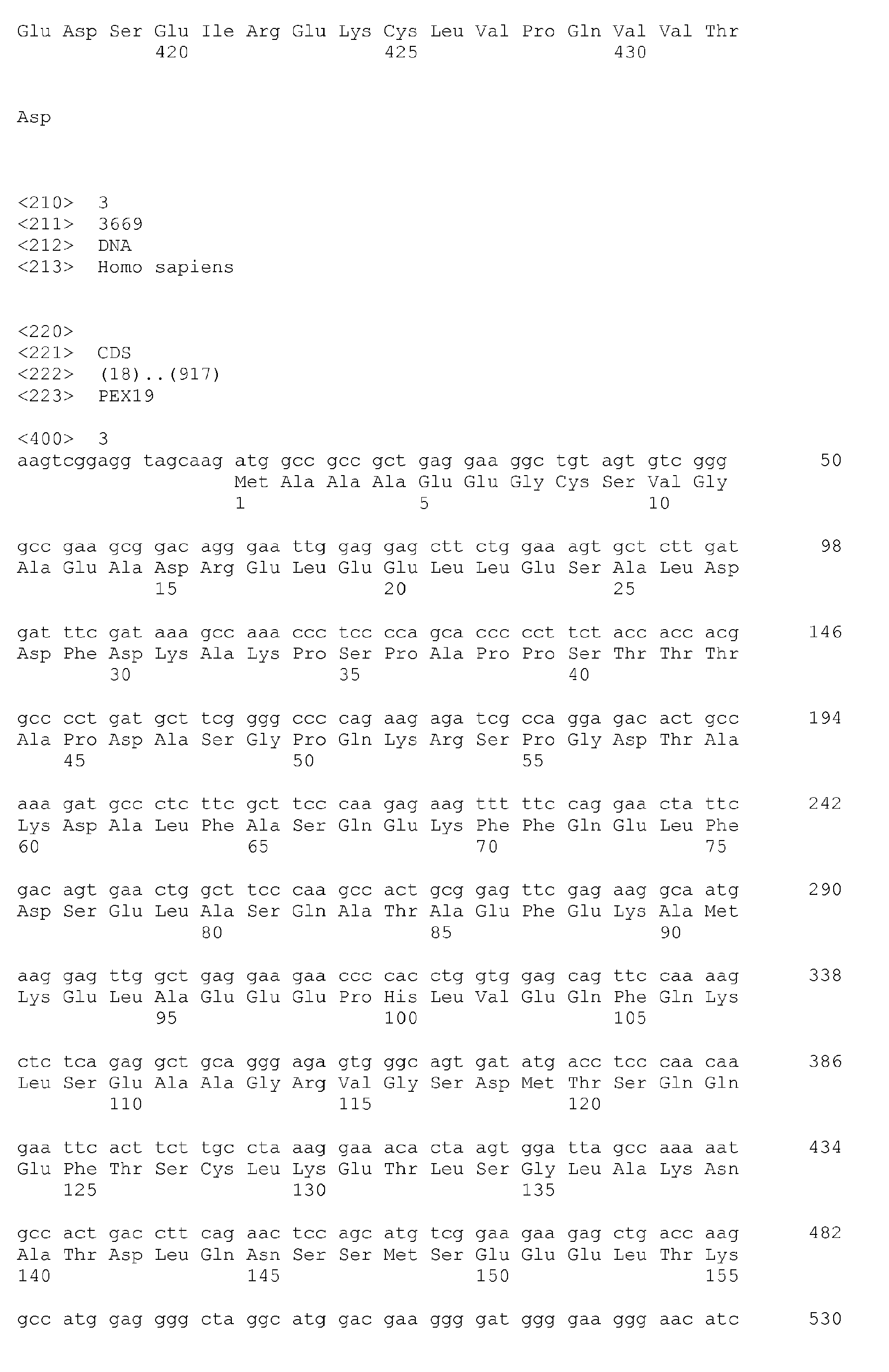


Claims (17)
- An in vitro method for predicting whether a patient would be responsive to a treatment with an anti-HER2 antibody, which method comprises determining the expression level of at least 4 genes in a biological sample of said patient, wherein said genes are GPR22, PEX19, GRHL2 and DERL1.
- The method of claim 1, wherein the patient is affected with a HER2-positive cancer.
- The method of claim 1 or 2, wherein the patient is with breast cancer.
- The method of any of claims 1 to 3, wherein the HER2 blocking agent is an anti-HER2 antibody
- The method of claim 4, wherein the anti-HER2 antibody is trastuzumab.
- The method of any of claims 1 to 5, for predicting whether a patient would be responsive to a treatment with a HER2 blocking agent, combined with a taxane, such as docetaxel.
- The method of any of claims 1 to 6, further comprising the step of comparing the combined expression level of said genes with reference values obtained from responder and non-responder groups of patients.
- The method of any of claims 1 to 7, wherein the biological sample is a disease tissue sample.
- The method of any of claims 1 to 8, wherein the expression level is determined by quantifying the level of mRNA of said genes in the biological sample.
- The method of claim 9, wherein the expression level is determined by real-time quantitative or semi-quantitative RT-PCR.
- The method of claim 9 or 10, wherein the expression level is determined by using a DNA chip.
- The method of any of claims 1 to 11, further comprising determining the expression level of the transcripts listed in Table B, or of a subcombination thereof.
- The method of claim 12, further comprising determining the expression level of any or all of the transcripts listed in Table C.
- A DNA chip comprising a solid support which carries nucleic acids that are specific to GPR22, PEX19, GRHL2 and DERL1 genes.
- The chip of claim 14, which further carries nucleic acids that are specific to any or all of the transcripts listed in Table B, or a subcombination thereof.
- The chip of claim 15, which further carries nucleic acids that are specific to any or all of the transcripts listed in Table C.
- A HER2 blocking agent, such as an anti-HER2 antibody, for treating a patient with a HER2-related disease, such as a cancer, and classified as responder to a treatment with a HER2 blocking agent, by the method of any of claims 1 to 13.
Priority Applications (4)
| Application Number | Priority Date | Filing Date | Title |
|---|---|---|---|
| EP08305241A EP2133433A1 (en) | 2008-06-09 | 2008-06-09 | A method for predicting responsiveness to a treatment with an anti-HER2 antibody |
| US12/994,989 US20110189185A1 (en) | 2008-06-09 | 2009-06-08 | Method for Predicting Responsiveness to a Treatment With an Anti-HER2 Antibody |
| PCT/EP2009/057036 WO2009150127A1 (en) | 2008-06-09 | 2009-06-08 | A method for predicting responsiveness to a treatment with an anti-her2 antibody |
| EP09761690A EP2297350A1 (en) | 2008-06-09 | 2009-06-08 | A method for predicting responsiveness to a treatment with an anti-her2 antibody |
Applications Claiming Priority (1)
| Application Number | Priority Date | Filing Date | Title |
|---|---|---|---|
| EP08305241A EP2133433A1 (en) | 2008-06-09 | 2008-06-09 | A method for predicting responsiveness to a treatment with an anti-HER2 antibody |
Publications (1)
| Publication Number | Publication Date |
|---|---|
| EP2133433A1 true EP2133433A1 (en) | 2009-12-16 |
Family
ID=39710998
Family Applications (2)
| Application Number | Title | Priority Date | Filing Date |
|---|---|---|---|
| EP08305241A Ceased EP2133433A1 (en) | 2008-06-09 | 2008-06-09 | A method for predicting responsiveness to a treatment with an anti-HER2 antibody |
| EP09761690A Withdrawn EP2297350A1 (en) | 2008-06-09 | 2009-06-08 | A method for predicting responsiveness to a treatment with an anti-her2 antibody |
Family Applications After (1)
| Application Number | Title | Priority Date | Filing Date |
|---|---|---|---|
| EP09761690A Withdrawn EP2297350A1 (en) | 2008-06-09 | 2009-06-08 | A method for predicting responsiveness to a treatment with an anti-her2 antibody |
Country Status (3)
| Country | Link |
|---|---|
| US (1) | US20110189185A1 (en) |
| EP (2) | EP2133433A1 (en) |
| WO (1) | WO2009150127A1 (en) |
Cited By (4)
| Publication number | Priority date | Publication date | Assignee | Title |
|---|---|---|---|---|
| WO2013118073A1 (en) * | 2012-02-07 | 2013-08-15 | Bioalternatives Sas | Serpinb4, as a marker for an early evaluation of the response to anti-egfr therapies by a non-invasive method |
| EP3134549A4 (en) * | 2014-04-21 | 2017-11-22 | Mayo Foundation for Medical Education and Research | Methods and materials for identifying and treating mammals having her2-positive breast cancer |
| EP3387443A4 (en) * | 2015-12-10 | 2019-11-13 | Nantomics, LLC | INTEGRATED ANALYSIS TO DETERMINE PROGNOSIS FOLLOWING TREATMENT AGAINST BREAST CANCER |
| EP3945136A1 (en) * | 2020-07-28 | 2022-02-02 | Hospital Clínic de Barcelona | In vitro method for the prognosis of patients suffering from her2-positive breast cancer |
Families Citing this family (3)
| Publication number | Priority date | Publication date | Assignee | Title |
|---|---|---|---|---|
| FR2976294B1 (en) | 2011-06-10 | 2018-02-16 | Universite De Nice Sophia Antipolis | METHOD FOR PREDICTING THE RESPONSE TO TREATMENT WITH HER2 BLOCKING AGENT |
| JP5692887B1 (en) * | 2013-05-17 | 2015-04-01 | ナノキャリア株式会社 | Polymer micelle pharmaceutical composition |
| US10769242B2 (en) | 2015-01-05 | 2020-09-08 | Children's Hospital Medical Center | System and method for data mining very large drugs and clinical effects databases |
Citations (8)
| Publication number | Priority date | Publication date | Assignee | Title |
|---|---|---|---|---|
| US5821337A (en) | 1991-06-14 | 1998-10-13 | Genentech, Inc. | Immunoglobulin variants |
| WO2000061185A1 (en) * | 1999-04-09 | 2000-10-19 | Aventis Pharma S.A | Docetaxel in combination with rhumab her2 for the treatment of cancers |
| WO2002061432A2 (en) * | 2001-02-01 | 2002-08-08 | Lifespan Biosciences, Inc. | Gpr22, a g protein-coupled receptor (gpcr) and compositions and methods related thereto |
| WO2002068579A2 (en) * | 2001-01-10 | 2002-09-06 | Pe Corporation (Ny) | Kits, such as nucleic acid arrays, comprising a majority of human exons or transcripts, for detecting expression and other uses thereof |
| WO2003000012A2 (en) * | 2001-06-21 | 2003-01-03 | Millennium Pharmaceuticals, Inc. | Compositions, kits, and methods for identification, assessment, prevention, and therapy of breast and ovarian cancer |
| WO2004065545A2 (en) * | 2003-01-15 | 2004-08-05 | Rosetta Inpharmatics Llc. | Diagnosis and prognosis of breast cancer patients |
| WO2005070020A2 (en) * | 2004-01-23 | 2005-08-04 | The Regents Of The University Of Colorado | Gefitinib sensitivity-related gene expression and products and methods related thereto |
| WO2006041959A2 (en) * | 2004-10-06 | 2006-04-20 | Wellstat Biologics Corporation | Detection of elevated levels of her-2/neu protein on circulating cancer cells and treatment |
-
2008
- 2008-06-09 EP EP08305241A patent/EP2133433A1/en not_active Ceased
-
2009
- 2009-06-08 WO PCT/EP2009/057036 patent/WO2009150127A1/en not_active Ceased
- 2009-06-08 EP EP09761690A patent/EP2297350A1/en not_active Withdrawn
- 2009-06-08 US US12/994,989 patent/US20110189185A1/en not_active Abandoned
Patent Citations (8)
| Publication number | Priority date | Publication date | Assignee | Title |
|---|---|---|---|---|
| US5821337A (en) | 1991-06-14 | 1998-10-13 | Genentech, Inc. | Immunoglobulin variants |
| WO2000061185A1 (en) * | 1999-04-09 | 2000-10-19 | Aventis Pharma S.A | Docetaxel in combination with rhumab her2 for the treatment of cancers |
| WO2002068579A2 (en) * | 2001-01-10 | 2002-09-06 | Pe Corporation (Ny) | Kits, such as nucleic acid arrays, comprising a majority of human exons or transcripts, for detecting expression and other uses thereof |
| WO2002061432A2 (en) * | 2001-02-01 | 2002-08-08 | Lifespan Biosciences, Inc. | Gpr22, a g protein-coupled receptor (gpcr) and compositions and methods related thereto |
| WO2003000012A2 (en) * | 2001-06-21 | 2003-01-03 | Millennium Pharmaceuticals, Inc. | Compositions, kits, and methods for identification, assessment, prevention, and therapy of breast and ovarian cancer |
| WO2004065545A2 (en) * | 2003-01-15 | 2004-08-05 | Rosetta Inpharmatics Llc. | Diagnosis and prognosis of breast cancer patients |
| WO2005070020A2 (en) * | 2004-01-23 | 2005-08-04 | The Regents Of The University Of Colorado | Gefitinib sensitivity-related gene expression and products and methods related thereto |
| WO2006041959A2 (en) * | 2004-10-06 | 2006-04-20 | Wellstat Biologics Corporation | Detection of elevated levels of her-2/neu protein on circulating cancer cells and treatment |
Non-Patent Citations (10)
Cited By (5)
| Publication number | Priority date | Publication date | Assignee | Title |
|---|---|---|---|---|
| WO2013118073A1 (en) * | 2012-02-07 | 2013-08-15 | Bioalternatives Sas | Serpinb4, as a marker for an early evaluation of the response to anti-egfr therapies by a non-invasive method |
| EP3134549A4 (en) * | 2014-04-21 | 2017-11-22 | Mayo Foundation for Medical Education and Research | Methods and materials for identifying and treating mammals having her2-positive breast cancer |
| EP3387443A4 (en) * | 2015-12-10 | 2019-11-13 | Nantomics, LLC | INTEGRATED ANALYSIS TO DETERMINE PROGNOSIS FOLLOWING TREATMENT AGAINST BREAST CANCER |
| EP3945136A1 (en) * | 2020-07-28 | 2022-02-02 | Hospital Clínic de Barcelona | In vitro method for the prognosis of patients suffering from her2-positive breast cancer |
| WO2022023235A1 (en) * | 2020-07-28 | 2022-02-03 | Hospital Clinic De Barcelona | In vitro method for the prognosis of patients suffering from her2-positive breast cancer |
Also Published As
| Publication number | Publication date |
|---|---|
| US20110189185A1 (en) | 2011-08-04 |
| WO2009150127A1 (en) | 2009-12-17 |
| EP2297350A1 (en) | 2011-03-23 |
Similar Documents
| Publication | Publication Date | Title |
|---|---|---|
| US10689711B2 (en) | Test kits and methods for their use to detect genetic markers for urothelial carcinoma of the bladder and treatment thereof | |
| US20110217297A1 (en) | Methods for classifying and treating breast cancers | |
| CA3147181A1 (en) | Urine markers for detection of bladder cancer | |
| WO2008089577A1 (en) | Breast cancer gene array | |
| US20110189185A1 (en) | Method for Predicting Responsiveness to a Treatment With an Anti-HER2 Antibody | |
| WO2015033172A1 (en) | Molecular diagnostic test for oesophageal cancer | |
| KR101801980B1 (en) | A composition for diagnosis of primary central nervous system lymphoma and a diagnosing kit comprising the same | |
| EP1611890B1 (en) | Methods for assessing and treating cancer | |
| AU2005201935A1 (en) | Prognostic for hematological malignancy | |
| EP3953713A1 (en) | Method of classifying a sample based on determination of fgfr | |
| EP3714070B1 (en) | A novel cip2a variant and uses thereof | |
| KR20240157611A (en) | Novel Biomarker for Predicting Therapeutic Response and Prognosis of Metastatic Breast Cancer To Chemotherapeutic Agents and Uses Thereof | |
| KR20210133200A (en) | Composition And Kit For Diagnosing Prognosis Of Bladder Cancer According To Gender | |
| CN113817829A (en) | Use of biomarkers in the preparation of products for predicting colorectal cancer sensitivity to oxaliplatin therapy | |
| CN113846165A (en) | Application of biomarkers in predicting colorectal cancer sensitivity to oxaliplatin therapy | |
| US20090130678A1 (en) | Methods and Kits for Breast Cancer Prognosis | |
| WO2014173986A2 (en) | Methods for diagnosing and monitoring the response to treatment of hepatocellular carcinoma | |
| KR101540052B1 (en) | KDM4D, a marker for predicting the response to anti-cancer drug in a patient with ovarian cancer | |
| KR102849117B1 (en) | TRIM51 biomarker for predicting melanoma treatment resistance and use thereof | |
| EP2596126B1 (en) | Methods for determining a prognosis for survival for a patient with leukaemia | |
| EP2686439B1 (en) | Novel tumor marker determination | |
| CA3082650C (en) | A novel cip2a variant and uses thereof | |
| Araki et al. | Biomarkers for predicting the sensitivity of cancer cells to TRAIL-R1 agonistic monoclonal antibody | |
| HK40095386A (en) | Diagnostic test | |
| WO2024252154A1 (en) | Lung cancer biomarkers |
Legal Events
| Date | Code | Title | Description |
|---|---|---|---|
| PUAI | Public reference made under article 153(3) epc to a published international application that has entered the european phase |
Free format text: ORIGINAL CODE: 0009012 |
|
| AK | Designated contracting states |
Kind code of ref document: A1 Designated state(s): AT BE BG CH CY CZ DE DK EE ES FI FR GB GR HR HU IE IS IT LI LT LU LV MC MT NL NO PL PT RO SE SI SK TR |
|
| AX | Request for extension of the european patent |
Extension state: AL BA MK RS |
|
| STAA | Information on the status of an ep patent application or granted ep patent |
Free format text: STATUS: THE APPLICATION HAS BEEN REFUSED |
|
| 18R | Application refused |
Effective date: 20100108 |