EP2131611A2 - Method and apparatus for mathematically characterizing ear canal geometry - Google Patents
Method and apparatus for mathematically characterizing ear canal geometry Download PDFInfo
- Publication number
- EP2131611A2 EP2131611A2 EP20090162091 EP09162091A EP2131611A2 EP 2131611 A2 EP2131611 A2 EP 2131611A2 EP 20090162091 EP20090162091 EP 20090162091 EP 09162091 A EP09162091 A EP 09162091A EP 2131611 A2 EP2131611 A2 EP 2131611A2
- Authority
- EP
- European Patent Office
- Prior art keywords
- ear canal
- sensor
- balloon
- ultrasonic transducer
- information
- Prior art date
- Legal status (The legal status is an assumption and is not a legal conclusion. Google has not performed a legal analysis and makes no representation as to the accuracy of the status listed.)
- Withdrawn
Links
Images
Classifications
-
- H—ELECTRICITY
- H04—ELECTRIC COMMUNICATION TECHNIQUE
- H04R—LOUDSPEAKERS, MICROPHONES, GRAMOPHONE PICK-UPS OR LIKE ACOUSTIC ELECTROMECHANICAL TRANSDUCERS; ELECTRIC HEARING AIDS; PUBLIC ADDRESS SYSTEMS
- H04R25/00—Electric hearing aids
- H04R25/65—Housing parts, e.g. shells, tips or moulds, or their manufacture
- H04R25/652—Ear tips; Ear moulds
-
- A—HUMAN NECESSITIES
- A61—MEDICAL OR VETERINARY SCIENCE; HYGIENE
- A61B—DIAGNOSIS; SURGERY; IDENTIFICATION
- A61B5/00—Measuring for diagnostic purposes; Identification of persons
- A61B5/103—Measuring devices for testing the shape, pattern, colour, size or movement of the body or parts thereof, for diagnostic purposes
- A61B5/107—Measuring physical dimensions, e.g. size of the entire body or parts thereof
- A61B5/1076—Measuring physical dimensions, e.g. size of the entire body or parts thereof for measuring dimensions inside body cavities, e.g. using catheters
-
- A—HUMAN NECESSITIES
- A61—MEDICAL OR VETERINARY SCIENCE; HYGIENE
- A61B—DIAGNOSIS; SURGERY; IDENTIFICATION
- A61B5/00—Measuring for diagnostic purposes; Identification of persons
- A61B5/68—Arrangements of detecting, measuring or recording means, e.g. sensors, in relation to patient
- A61B5/6801—Arrangements of detecting, measuring or recording means, e.g. sensors, in relation to patient specially adapted to be attached to or worn on the body surface
- A61B5/6813—Specially adapted to be attached to a specific body part
- A61B5/6814—Head
- A61B5/6815—Ear
- A61B5/6817—Ear canal
-
- A—HUMAN NECESSITIES
- A61—MEDICAL OR VETERINARY SCIENCE; HYGIENE
- A61B—DIAGNOSIS; SURGERY; IDENTIFICATION
- A61B8/00—Diagnosis using ultrasonic, sonic or infrasonic waves
- A61B8/42—Details of probe positioning or probe attachment to the patient
- A61B8/4245—Details of probe positioning or probe attachment to the patient involving determining the position of the probe, e.g. with respect to an external reference frame or to the patient
- A61B8/4254—Details of probe positioning or probe attachment to the patient involving determining the position of the probe, e.g. with respect to an external reference frame or to the patient using sensors mounted on the probe
-
- H—ELECTRICITY
- H04—ELECTRIC COMMUNICATION TECHNIQUE
- H04R—LOUDSPEAKERS, MICROPHONES, GRAMOPHONE PICK-UPS OR LIKE ACOUSTIC ELECTROMECHANICAL TRANSDUCERS; ELECTRIC HEARING AIDS; PUBLIC ADDRESS SYSTEMS
- H04R25/00—Electric hearing aids
- H04R25/65—Housing parts, e.g. shells, tips or moulds, or their manufacture
- H04R25/652—Ear tips; Ear moulds
- H04R25/656—Non-customized, universal ear tips, i.e. ear tips which are not specifically adapted to the size or shape of the ear or ear canal
-
- H—ELECTRICITY
- H04—ELECTRIC COMMUNICATION TECHNIQUE
- H04R—LOUDSPEAKERS, MICROPHONES, GRAMOPHONE PICK-UPS OR LIKE ACOUSTIC ELECTROMECHANICAL TRANSDUCERS; ELECTRIC HEARING AIDS; PUBLIC ADDRESS SYSTEMS
- H04R2225/00—Details of deaf aids covered by H04R25/00, not provided for in any of its subgroups
- H04R2225/77—Design aspects, e.g. CAD, of hearing aid tips, moulds or housings
Definitions
- the present application relates generally to physically fitting hearing assistance devices and in particular to mathematically characterizing ear canal geometry for physically fitting hearing assistance devices.
- Hearing instrument manufacturers have adopted laser scanning of traditional impressions as the method of obtaining mathematical representations of the ear canal geometry.
- a dispenser will acquire the impression from the patient and mail the impression directly to the manufacturer.
- the impression can become distorted during the mailing process due to uncured material and excessive temperatures in the delivery trucks; the result is an inaccurate impression of the patient's physiology thereby leading to a hearing instrument that does not fit properly.
- the hearing instrument it is common for the hearing instrument to pop out of the patient's ear during chewing or other jaw motion. Since the impression is obtained in a "static" condition within the ear canal, geometry changes of the ear canal during chewing can cause the hearing instrument to jostle and pop out. In order to mitigate these occurrences, it may be advantageous to acquire two different impressions: one with the jaw closed and one with the jaw open. The final earmold for the hearing instrument can be extrapolated as an average from the two impressions, thereby reducing the potential for "popout".
- the process can typically take from ten to fifteen minutes per pair of impressions, not to mention administrative time in preparing, shipping, and receiving the impressions. It would be advantageous to have a process that acquired the mathematical representation at the dispenser's office in less time and forwarded the data directly to the manufacturer electronically to save time and expense.
- the present disclosure may provide apparatus and methods for physically fitting a hearing assistance device, including mathematically characterizing a person's ear canal.
- the apparatus may comprise an expandable balloon for positioning in the ear canal, the balloon including a hollow interior channel.
- a flexible catheter may extend through the interior portion of the balloon, and the flexible catheter includes a track.
- a platform is movably coupled to the track.
- An ultrasonic transducer and a microelectromechanical systems (MEMS) sensor may be coupled to the platform.
- the ultrasonic transducer may scan the ear canal and the MEMS sensor senses motion and position of the transducer.
- a mathematical representation of the ear canal may be adapted to be computed using information from the transducer and sensor.
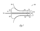
- FIG. 1 illustrates a cross-section of an ear with a characterizing apparatus positioned for mathematically characterizing the geometry of the ear canal according to one embodiment of the present subject matter.
- Various embodiments of the present subject matter utilize 1) an ultrasonic transducer system for the purpose of imaging the physiology of the ear canal, 2) a replaceable, fluid-filled medical balloon catheter for the purpose of expanding against the concha and ear canal walls so as to mechanically couple the transducer system to the skin, and 3) a mechanical system to traverse the transducer system within the ear canal in order to image and mathematically compute its shape.
- Modern techniques for obtaining a person's ear canal geometry utilize highly-compliant polymeric materials that are injected into a person's ear canal with a syringe. After the material cures, it is pulled from the ear thereby giving a three dimensional impression of the ear canal geometry. Traditionally, the impression is cast in a silicone investment and removed, thereby leaving a representation of the original ear canal. In a procedure that is similar to the traditional casting of a bell, a custom earmold is created by pouring resins into the investment and allowing them to cure into plastic. Although this technique gives a reasonably accurate representation of the ear canal geometry, it does not yield any mathematical information about its shape. A mathematical representation would be valuable for an automated, digital shell making procedure such as stereo lithography, etc.
- a removable, thin-walled, highly-compliant, elastomeric balloon is pre-formed to fit the physiology of a nominal ear canal and concha.
- the physiology of the general population can be characterized with three different pre-formed balloons.
- the removable balloon is pre-formed with a central, cylindrical, hollow channel, and can be expanded to fit the patient's physiology snugly.
- the channel contains a conduit for air-pressure relief, a conduit for expanding the balloon with saline or gel, a flexible track and mechanism to position a movable platform, and an ultrasonic transducer probe fixed to the platform.
- the probe operates in pulse/echo mode and scans the ear canal as the platform is translated and/or rotated down the flexible track.
- the transducer system also contains a three-axis microelectromechanical systems (MEMS) gyrator or MEMS accelerator sensor that is used to compute the rate at which the platform is translated and/or rotated. Since the rate at which the platform is translated and/or rotated is known, signal processing can be performed on the acquired ultrasonic and MEMS signal to reconstruct a mathematical representation of the ear canal, much the same way ultrasound is used to image human features inside the human body.
- MEMS microelectromechanical systems
- the present subject matter includes a method for scanning and obtaining a mathematical characterization of a person's ear canal geometry, in various embodiments.
- Various embodiments include a medical balloon that expands with saline within a person's ear canal while relieving internal air pressure to protect the tympanic membrane.
- a transducer probe transmits and receives ultrasonic acoustical energy within the expanded medical catheter and saline-filled balloon, in an embodiment.
- Various embodiments include a mechanism that transverses and/or rotates the ultrasonic probe at various locations within the medical catheter and balloon.
- a method is provided for processing the echoes from the ultrasonic probe and yielding a point cloud mathematical representation of the ear canal geometry, in various embodiments.
- FIG. 1 illustrates a cross-section of an ear with a characterizing apparatus positioned for mathematically characterizing the geometry of the ear canal according to one embodiment of the present subject matter.
- a cross section is depicted of a person's ear canal 100, including pinna 102 and eardrum 104.
- the apparatus includes an expandable balloon 110 for positioning in the ear canal, the balloon including a hollow interior channel 111.
- a flexible catheter 112 extends through the interior portion of the balloon, the flexible catheter including a track 118 and conduit 116 for air-pressure relief.
- a platform 114 is movably coupled to the track.
- An ultrasonic transducer 120 and a microelectromechanical systems (MEMS) sensor 122 are coupled to the platform.
- MEMS microelectromechanical systems
- the ultrasonic transducer 120 scans the ear canal and the MEMS sensor 122 senses motion and position of the transducer.
- a mathematical representation of the ear canal is adapted to be computed using information from the transducer and sensor.
Landscapes
- Health & Medical Sciences (AREA)
- Life Sciences & Earth Sciences (AREA)
- Engineering & Computer Science (AREA)
- Physics & Mathematics (AREA)
- General Health & Medical Sciences (AREA)
- Otolaryngology (AREA)
- Biomedical Technology (AREA)
- Molecular Biology (AREA)
- Veterinary Medicine (AREA)
- Public Health (AREA)
- Biophysics (AREA)
- Pathology (AREA)
- Animal Behavior & Ethology (AREA)
- Heart & Thoracic Surgery (AREA)
- Medical Informatics (AREA)
- Surgery (AREA)
- Acoustics & Sound (AREA)
- Manufacturing & Machinery (AREA)
- Neurosurgery (AREA)
- Signal Processing (AREA)
- Dentistry (AREA)
- Oral & Maxillofacial Surgery (AREA)
- Nuclear Medicine, Radiotherapy & Molecular Imaging (AREA)
- Radiology & Medical Imaging (AREA)
- Ultra Sonic Daignosis Equipment (AREA)
Abstract
Description
- The present application claims the benefit under 35 U.S.C. 119(e) of
U.S. Provisional Patent Application Serial Number 61/059,025, filed June 5, 2008 - The present application relates generally to physically fitting hearing assistance devices and in particular to mathematically characterizing ear canal geometry for physically fitting hearing assistance devices.
- Hearing instrument manufacturers have adopted laser scanning of traditional impressions as the method of obtaining mathematical representations of the ear canal geometry. A dispenser will acquire the impression from the patient and mail the impression directly to the manufacturer. Unfortunately, the impression can become distorted during the mailing process due to uncured material and excessive temperatures in the delivery trucks; the result is an inaccurate impression of the patient's physiology thereby leading to a hearing instrument that does not fit properly. It is common practice for the dispenser to acquire another impression from the patient and return the hearing instrument for rework.
- In some instances, it is common for the hearing instrument to pop out of the patient's ear during chewing or other jaw motion. Since the impression is obtained in a "static" condition within the ear canal, geometry changes of the ear canal during chewing can cause the hearing instrument to jostle and pop out. In order to mitigate these occurrences, it may be advantageous to acquire two different impressions: one with the jaw closed and one with the jaw open. The final earmold for the hearing instrument can be extrapolated as an average from the two impressions, thereby reducing the potential for "popout".
- Depending on the cure rate of the polymeric material used for ear impressions, the process can typically take from ten to fifteen minutes per pair of impressions, not to mention administrative time in preparing, shipping, and receiving the impressions. It would be advantageous to have a process that acquired the mathematical representation at the dispenser's office in less time and forwarded the data directly to the manufacturer electronically to save time and expense.
- The present disclosure may provide apparatus and methods for physically fitting a hearing assistance device, including mathematically characterizing a person's ear canal. The apparatus may comprise an expandable balloon for positioning in the ear canal, the balloon including a hollow interior channel. A flexible catheter may extend through the interior portion of the balloon, and the flexible catheter includes a track. A platform is movably coupled to the track. An ultrasonic transducer and a microelectromechanical systems (MEMS) sensor may be coupled to the platform. The ultrasonic transducer may scan the ear canal and the MEMS sensor senses motion and position of the transducer. According to various embodiments, a mathematical representation of the ear canal may be adapted to be computed using information from the transducer and sensor.
- This summary is an overview of some of the teachings of the present application and is not intended to be an exclusive or exhaustive treatment of the present subject matter. Further details about the present subject matter are found in the detailed description. The scope of the present invention is defined by the appended claims.
-
FIG. 1 illustrates a cross-section of an ear with a characterizing apparatus positioned for mathematically characterizing the geometry of the ear canal according to one embodiment of the present subject matter. - The following detailed description of the present subject matter refers to subject matter in the accompanying drawings which show specific aspects and embodiments in which the present subject matter may be practiced. These embodiments are described in sufficient detail to enable those skilled in the art to practice the present subject matter. References to "an", "one", or "various" embodiments in this disclosure are not necessarily to the same embodiment, and such references contemplate more than one embodiment. The following detailed description is demonstrative and not to be taken in a limiting sense. The scope of the present subject matter is defined by the appended claims.
- Various embodiments of the present subject matter utilize 1) an ultrasonic transducer system for the purpose of imaging the physiology of the ear canal, 2) a replaceable, fluid-filled medical balloon catheter for the purpose of expanding against the concha and ear canal walls so as to mechanically couple the transducer system to the skin, and 3) a mechanical system to traverse the transducer system within the ear canal in order to image and mathematically compute its shape.
- Modern techniques for obtaining a person's ear canal geometry utilize highly-compliant polymeric materials that are injected into a person's ear canal with a syringe. After the material cures, it is pulled from the ear thereby giving a three dimensional impression of the ear canal geometry. Traditionally, the impression is cast in a silicone investment and removed, thereby leaving a representation of the original ear canal. In a procedure that is similar to the traditional casting of a bell, a custom earmold is created by pouring resins into the investment and allowing them to cure into plastic. Although this technique gives a reasonably accurate representation of the ear canal geometry, it does not yield any mathematical information about its shape. A mathematical representation would be valuable for an automated, digital shell making procedure such as stereo lithography, etc.
- Recently, laser technology has been used to scan the features of the impression into a mathematical format such as "point cloud" data. These data are used to create a custom shell with automated, digital shell making processes. Unfortunately, the inconvenience of taking a person's ear impression remains the first step in this procedure.
- In an effort to preclude the need to "shoot" a person's impression with a syringe of polymeric material, various patents have been filed which use ultrasonic and/or optical devices located on the end of a straight wand. The wand is inserted into the ear canal, and the device scans the geometry. If the device is based on an ultrasonic scanner, the ear canal must be filled with a saline-like fluid thereby making the process inconvenient, messy, and impractical. If the device is based on an optical scanner, complicated mirrors and positioning devices are needed to rotate and control the optical light beam. If the beam is scattered by hair within the ear canal, imaging precision is degraded. In addition, getting useful information beyond the first bend of the ear canal is difficult with a straight wand.
- A removable, thin-walled, highly-compliant, elastomeric balloon is pre-formed to fit the physiology of a nominal ear canal and concha. In various embodiments, the physiology of the general population can be characterized with three different pre-formed balloons. The removable balloon is pre-formed with a central, cylindrical, hollow channel, and can be expanded to fit the patient's physiology snugly. The channel contains a conduit for air-pressure relief, a conduit for expanding the balloon with saline or gel, a flexible track and mechanism to position a movable platform, and an ultrasonic transducer probe fixed to the platform. The probe operates in pulse/echo mode and scans the ear canal as the platform is translated and/or rotated down the flexible track. In various embodiments, the transducer system also contains a three-axis microelectromechanical systems (MEMS) gyrator or MEMS accelerator sensor that is used to compute the rate at which the platform is translated and/or rotated. Since the rate at which the platform is translated and/or rotated is known, signal processing can be performed on the acquired ultrasonic and MEMS signal to reconstruct a mathematical representation of the ear canal, much the same way ultrasound is used to image human features inside the human body.
- The present subject matter includes a method for scanning and obtaining a mathematical characterization of a person's ear canal geometry, in various embodiments. Various embodiments include a medical balloon that expands with saline within a person's ear canal while relieving internal air pressure to protect the tympanic membrane. A transducer probe transmits and receives ultrasonic acoustical energy within the expanded medical catheter and saline-filled balloon, in an embodiment. Various embodiments include a mechanism that transverses and/or rotates the ultrasonic probe at various locations within the medical catheter and balloon. A method is provided for processing the echoes from the ultrasonic probe and yielding a point cloud mathematical representation of the ear canal geometry, in various embodiments.
-
FIG. 1 illustrates a cross-section of an ear with a characterizing apparatus positioned for mathematically characterizing the geometry of the ear canal according to one embodiment of the present subject matter. A cross section is depicted of a person'sear canal 100, includingpinna 102 andeardrum 104. The apparatus includes anexpandable balloon 110 for positioning in the ear canal, the balloon including a hollowinterior channel 111. Aflexible catheter 112 extends through the interior portion of the balloon, the flexible catheter including atrack 118 andconduit 116 for air-pressure relief. Aplatform 114 is movably coupled to the track. Anultrasonic transducer 120 and a microelectromechanical systems (MEMS)sensor 122 are coupled to the platform. Theultrasonic transducer 120 scans the ear canal and theMEMS sensor 122 senses motion and position of the transducer. According to various embodiments, a mathematical representation of the ear canal is adapted to be computed using information from the transducer and sensor. - This application is intended to cover adaptations or variations of the present subject matter. It is to be understood that the above description is intended to be illustrative, and not restrictive. Thus, the scope of the present subject matter is determined by the appended claims.
Claims (15)
- An apparatus for mathematically characterizing a person's ear canal, the apparatus comprising:an expandable balloon for positioning in the ear canal, the balloon including a hollow interior channel;a flexible catheter extending through the interior portion of the balloon, the flexible catheter including a track;a platform movably coupled to the track;an ultrasonic transducer coupled to the platform; anda processor adapted to characterize geometry of the ear canal from information from the ultrasonic transducer obtained from scans of the ear canal.
- The apparatus of claim 1, further comprising a microelectromechanical systems (MEMS) sensor coupled to the platform, the MEMS sensor adapted to sense motion and position of the ultrasonic transducer.
- The apparatus of claim 2, wherein the processor is further adapted to characterize geometry of the ear canal from information from the ultrasonic transducer and the MEMS sensor.
- The apparatus of any of the preceding claims, further comprising a mechanism adapted to transverse and rotate the ultrasonic transducer within the balloon.
- The apparatus of any of the preceding claims, wherein the ultrasonic transducer includes a probe adapted to transmit and receive acoustical energy within the balloon after expanding the balloon to snugly fit the person's ear canal.
- The apparatus of any of the preceding claims, further comprising a memory for storing information from the transducer and sensor.
- The apparatus of any of the preceding claims, further comprising a communication link for transmitting information from the transducer and sensor.
- The apparatus of any of the preceding claims, wherein the track includes a flexible track.
- The apparatus of any of the preceding claims, wherein the hollow interior channel of the balloon includes a conduit for air-pressure relief to protect the person's tympanic membrane.
- A method of mathematically characterizing a person's ear canal, the method comprising:positioning a pre-formed expandable balloon in the ear canal, the balloon including a hollow central channel;expanding the balloon to fit the person's ear canal snugly;scanning the ear canal using an ultrasonic transducer probe, the probe fixed to a movable platform along a track contained within the channel;sensing motion and position of the transducer probe using a sensor, the sensor fixed to the movable platform; andusing information from the ultrasonic transducer probe and sensor to compute a mathematical representation of the ear canal.
- The method of claim 10, further comprising computing a point cloud mathematical representation of the person's ear canal geometry using the information from the ultrasonic transducer probe and sensor.
- The method of claim 10 or claim 11, wherein scanning the ear canal using an ultrasonic transducer probe includes scanning the ear canal as the platform is translated and/or rotated down the track.
- The method of any of claims 10 to 12, wherein sensing motion and position of the transducer probe includes using a microelectromechanical systems (MEMS) sensor.
- The method of any of claims 10 to 13, further comprising storing the information from the ultrasonic transducer probe and sensor.
- The method of any of claims 10 to 14, further comprising transmitting the information from the ultrasonic transducer probe and sensor.
Applications Claiming Priority (1)
| Application Number | Priority Date | Filing Date | Title |
|---|---|---|---|
| US5902508P | 2008-06-05 | 2008-06-05 |
Publications (2)
| Publication Number | Publication Date |
|---|---|
| EP2131611A2 true EP2131611A2 (en) | 2009-12-09 |
| EP2131611A3 EP2131611A3 (en) | 2013-04-03 |
Family
ID=41056903
Family Applications (1)
| Application Number | Title | Priority Date | Filing Date |
|---|---|---|---|
| EP20090162091 Withdrawn EP2131611A3 (en) | 2008-06-05 | 2009-06-05 | Method and apparatus for mathematically characterizing ear canal geometry |
Country Status (2)
| Country | Link |
|---|---|
| US (1) | US8840558B2 (en) |
| EP (1) | EP2131611A3 (en) |
Cited By (1)
| Publication number | Priority date | Publication date | Assignee | Title |
|---|---|---|---|---|
| US20220196833A1 (en) * | 2020-12-23 | 2022-06-23 | Sonova Ag | Method for Determining a Geometry of an Ear Canal or a Portion of an Ear of a Person |
Families Citing this family (3)
| Publication number | Priority date | Publication date | Assignee | Title |
|---|---|---|---|---|
| ATE549972T1 (en) | 2009-06-05 | 2012-04-15 | Starkey Lab Inc | METHOD AND DEVICE FOR THE MATHEMATICAL CHARACTERIZATION OF THE AUDIO CANAL GEOMETRY |
| US10716672B2 (en) * | 2015-04-07 | 2020-07-21 | St. Jude Medical, Cardiology Division, Inc. | System and method for intraprocedural assessment of geometry and compliance of valve annulus for trans-catheter valve implantation |
| AU2019355553B2 (en) * | 2018-10-03 | 2025-07-03 | Otonexus Medical Technologies, Inc. | Methods and devices for membrane characterization with ultrasound and optical illumination |
Family Cites Families (34)
| Publication number | Priority date | Publication date | Assignee | Title |
|---|---|---|---|---|
| US4190033A (en) * | 1977-02-23 | 1980-02-26 | Foti Thomas M | Closed flow caloric test method |
| DE4135286C1 (en) | 1991-10-25 | 1993-01-14 | Siemens Ag, 8000 Muenchen, De | Outer ear hearing passage and=or lug measurer - uses insertable vessel fillable with liq. to register individual shape for insertable housing of hearing aid |
| US5190046A (en) * | 1992-05-01 | 1993-03-02 | Shturman Cardiology Systems, Inc. | Ultrasound imaging balloon catheter |
| US5735282A (en) * | 1996-05-30 | 1998-04-07 | Acuson Corporation | Flexible ultrasonic transducers and related systems |
| US5904651A (en) * | 1996-10-28 | 1999-05-18 | Ep Technologies, Inc. | Systems and methods for visualizing tissue during diagnostic or therapeutic procedures |
| US6292433B1 (en) * | 1997-02-03 | 2001-09-18 | Teratech Corporation | Multi-dimensional beamforming device |
| US20020050518A1 (en) * | 1997-12-08 | 2002-05-02 | Roustaei Alexander R. | Sensor array |
| US6093150A (en) * | 1997-12-31 | 2000-07-25 | Acuson Corporation | Ultrasound otoscope |
| EP1058594A2 (en) | 1998-12-10 | 2000-12-13 | William Forrest Fagan | Method for the manufacture of hearing aid shells |
| GB2344555A (en) * | 1998-12-10 | 2000-06-14 | William Forrest Fagan | Method for the manufacture of hearing aid shells |
| US7037267B1 (en) * | 1999-11-10 | 2006-05-02 | David Lipson | Medical diagnostic methods, systems, and related equipment |
| US6631197B1 (en) * | 2000-07-24 | 2003-10-07 | Gn Resound North America Corporation | Wide audio bandwidth transduction method and device |
| US6868594B2 (en) * | 2001-01-05 | 2005-03-22 | Koninklijke Philips Electronics, N.V. | Method for making a transducer |
| ES2378060T3 (en) * | 2001-03-02 | 2012-04-04 | 3Shape A/S | Procedure for modeling custom ear pieces |
| US6709397B2 (en) * | 2001-10-16 | 2004-03-23 | Envisioneering, L.L.C. | Scanning probe |
| WO2002021894A2 (en) * | 2002-01-21 | 2002-03-21 | Phonak Ag | Method for the reconstruction of the geometry of the inner surface of a cavity |
| US20030220556A1 (en) * | 2002-05-20 | 2003-11-27 | Vespro Ltd. | Method, system and device for tissue characterization |
| US7147604B1 (en) * | 2002-08-07 | 2006-12-12 | Cardiomems, Inc. | High Q factor sensor |
| US7311667B2 (en) * | 2002-09-09 | 2007-12-25 | Siemens Medical Solutions Usa, Inc. | Multiple pattern transducer array and method of use |
| US20040181128A1 (en) * | 2003-03-11 | 2004-09-16 | Masters Martin W. | Determining the geometry and dimensions of a three-dimensional object |
| US7304644B2 (en) * | 2003-03-12 | 2007-12-04 | Siemens Medical Solutions Usa, Inc. | System and method for performing a virtual endoscopy |
| US7175599B2 (en) * | 2003-04-17 | 2007-02-13 | Brigham And Women's Hospital, Inc. | Shear mode diagnostic ultrasound |
| US20040221853A1 (en) * | 2003-05-08 | 2004-11-11 | Plasiatek, Llc | Ultrasonic placement and monitoring of a tube within the body |
| US7998073B2 (en) * | 2003-08-04 | 2011-08-16 | Imacor Inc. | Ultrasound imaging with reduced noise |
| EP1695113A2 (en) * | 2003-11-26 | 2006-08-30 | Teratech Corporation | Modular portable ultrasound systems |
| WO2007004083A1 (en) | 2005-06-30 | 2007-01-11 | Koninklijke Philips Electronics N.V. | Sizing and positioning technology for an in-the-ear multi-measurement sensor to enable nibp calculation |
| US7625343B2 (en) * | 2005-07-01 | 2009-12-01 | Scimed Life Systems, Inc. | Concave phased array imaging catheter |
| EP2439697A3 (en) * | 2005-09-22 | 2012-07-04 | 3M Innovative Properties Company | Method for refining the calibration of an imaging system |
| US7878977B2 (en) * | 2005-09-30 | 2011-02-01 | Siemens Medical Solutions Usa, Inc. | Flexible ultrasound transducer array |
| US7840042B2 (en) * | 2006-01-20 | 2010-11-23 | 3M Innovative Properties Company | Superposition for visualization of three-dimensional data acquisition |
| US8038622B2 (en) * | 2007-08-03 | 2011-10-18 | Innoscion, Llc | Wired and wireless remotely controlled ultrasonic transducer and imaging apparatus |
| US20080229832A1 (en) * | 2007-02-16 | 2008-09-25 | Los Alamos National Security | Automatic time-of-flight selection for ultrasound tomography |
| WO2010097710A1 (en) * | 2009-02-27 | 2010-09-02 | Dalhousie University | High-frequency ultrasound imaging system |
| ATE549972T1 (en) * | 2009-06-05 | 2012-04-15 | Starkey Lab Inc | METHOD AND DEVICE FOR THE MATHEMATICAL CHARACTERIZATION OF THE AUDIO CANAL GEOMETRY |
-
2009
- 2009-06-04 US US12/478,200 patent/US8840558B2/en active Active
- 2009-06-05 EP EP20090162091 patent/EP2131611A3/en not_active Withdrawn
Cited By (2)
| Publication number | Priority date | Publication date | Assignee | Title |
|---|---|---|---|---|
| US20220196833A1 (en) * | 2020-12-23 | 2022-06-23 | Sonova Ag | Method for Determining a Geometry of an Ear Canal or a Portion of an Ear of a Person |
| EP4018935A1 (en) * | 2020-12-23 | 2022-06-29 | Sonova AG | Method for determining a geometry of an ear canal or a portion of an ear of a person |
Also Published As
| Publication number | Publication date |
|---|---|
| US20090306517A1 (en) | 2009-12-10 |
| EP2131611A3 (en) | 2013-04-03 |
| US8840558B2 (en) | 2014-09-23 |
Similar Documents
| Publication | Publication Date | Title |
|---|---|---|
| US7092543B1 (en) | One-size-fits-all uni-ear hearing instrument | |
| US6751494B2 (en) | Method for the reconstruction of the geometry of the inner surface of a cavity | |
| DK3198891T3 (en) | DEVICE FOR PRINTING THE EAR | |
| US20090296980A1 (en) | System and Method for Producing a Geometric Model of the Auditory Canal | |
| US8840558B2 (en) | Method and apparatus for mathematically characterizing ear canal geometry | |
| US20210014619A1 (en) | Hearing device including a sensor and a method of forming same | |
| US9924276B2 (en) | Adjustable venting for hearing instruments | |
| EP1603620B1 (en) | Determining the geometry and dimensions of a three-dimensional object | |
| Khanna et al. | Specification of the acoustical input to the ear at high frequencies | |
| WO2003003899A3 (en) | Methods and apparatus for objective fetal diagnosis | |
| EP0381608A3 (en) | Method and apparatus for the fitting of a hearing aid, and associated probe | |
| US10652677B2 (en) | Hearing assistance device and method of forming same | |
| WO2017187608A1 (en) | Ultrasonic imaging device, and ultrasonic transmission/reception method | |
| WO2000034739A2 (en) | Method for the manufacture of hearing aid shells | |
| US8357084B2 (en) | Structure for probe insertion | |
| EP2258266B1 (en) | Method and apparatus for mathematically characterizing ear canal geometry | |
| US20220240893A1 (en) | Acoustic window with compound shape for ultrasound probe | |
| JP2019523063A (en) | Imaging device | |
| US20220196833A1 (en) | Method for Determining a Geometry of an Ear Canal or a Portion of an Ear of a Person | |
| Cavar et al. | The position of the tongue root in the articulation of posterior sibilants in Polish | |
| EP4404589A1 (en) | Housing for an earpiece and method for producing a housing for an earpiece | |
| DE4300804A1 (en) | Balloon ear plug tampon | |
| US20060182296A1 (en) | Ultrasonic ear wax cleaning system | |
| EP1477102B1 (en) | Structure for probe insertion | |
| EP1468246A2 (en) | Method for the reconstruction of the geometry of the inner surface of a cavity |
Legal Events
| Date | Code | Title | Description |
|---|---|---|---|
| PUAI | Public reference made under article 153(3) epc to a published international application that has entered the european phase |
Free format text: ORIGINAL CODE: 0009012 |
|
| 17P | Request for examination filed |
Effective date: 20090605 |
|
| AK | Designated contracting states |
Kind code of ref document: A2 Designated state(s): AT BE BG CH CY CZ DE DK EE ES FI FR GB GR HR HU IE IS IT LI LT LU LV MC MK MT NL NO PL PT RO SE SI SK TR |
|
| PUAL | Search report despatched |
Free format text: ORIGINAL CODE: 0009013 |
|
| AK | Designated contracting states |
Kind code of ref document: A3 Designated state(s): AT BE BG CH CY CZ DE DK EE ES FI FR GB GR HR HU IE IS IT LI LT LU LV MC MK MT NL NO PL PT RO SE SI SK TR |
|
| AX | Request for extension of the european patent |
Extension state: AL BA RS |
|
| RIC1 | Information provided on ipc code assigned before grant |
Ipc: H04R 25/00 20060101AFI20130227BHEP Ipc: A61B 5/00 20060101ALI20130227BHEP |
|
| GRAP | Despatch of communication of intention to grant a patent |
Free format text: ORIGINAL CODE: EPIDOSNIGR1 |
|
| INTG | Intention to grant announced |
Effective date: 20140520 |
|
| STAA | Information on the status of an ep patent application or granted ep patent |
Free format text: STATUS: THE APPLICATION IS DEEMED TO BE WITHDRAWN |
|
| 18D | Application deemed to be withdrawn |
Effective date: 20141001 |