EP2128619A1 - Detection of peptides - Google Patents
Detection of peptides Download PDFInfo
- Publication number
- EP2128619A1 EP2128619A1 EP09009355A EP09009355A EP2128619A1 EP 2128619 A1 EP2128619 A1 EP 2128619A1 EP 09009355 A EP09009355 A EP 09009355A EP 09009355 A EP09009355 A EP 09009355A EP 2128619 A1 EP2128619 A1 EP 2128619A1
- Authority
- EP
- European Patent Office
- Prior art keywords
- protein
- target peptide
- capture agents
- proteins
- peptide fragments
- Prior art date
- Legal status (The legal status is an assumption and is not a legal conclusion. Google has not performed a legal analysis and makes no representation as to the accuracy of the status listed.)
- Withdrawn
Links
- 108090000765 processed proteins & peptides Proteins 0.000 title claims description 160
- 102000004196 processed proteins & peptides Human genes 0.000 title description 65
- 238000001514 detection method Methods 0.000 title description 28
- 108090000623 proteins and genes Proteins 0.000 claims abstract description 288
- 102000004169 proteins and genes Human genes 0.000 claims abstract description 280
- 239000003795 chemical substances by application Substances 0.000 claims abstract description 184
- 102000007079 Peptide Fragments Human genes 0.000 claims abstract description 183
- 108010033276 Peptide Fragments Proteins 0.000 claims abstract description 183
- 238000000034 method Methods 0.000 claims abstract description 144
- 239000007787 solid Substances 0.000 claims abstract description 31
- 238000004519 manufacturing process Methods 0.000 claims abstract description 12
- 238000013467 fragmentation Methods 0.000 claims abstract description 10
- 238000006062 fragmentation reaction Methods 0.000 claims abstract description 10
- 150000001875 compounds Chemical class 0.000 claims description 58
- 238000003776 cleavage reaction Methods 0.000 claims description 32
- 230000007017 scission Effects 0.000 claims description 32
- 230000004481 post-translational protein modification Effects 0.000 claims description 31
- 238000004458 analytical method Methods 0.000 claims description 28
- 230000014509 gene expression Effects 0.000 claims description 21
- 230000002255 enzymatic effect Effects 0.000 claims description 19
- 230000004044 response Effects 0.000 claims description 13
- 230000001413 cellular effect Effects 0.000 claims description 12
- 239000000126 substance Substances 0.000 claims description 10
- 238000000491 multivariate analysis Methods 0.000 claims description 7
- 238000012163 sequencing technique Methods 0.000 claims description 6
- 230000001640 apoptogenic effect Effects 0.000 claims description 4
- 206010020718 hyperplasia Diseases 0.000 claims description 4
- 230000002390 hyperplastic effect Effects 0.000 claims description 4
- 230000003100 immobilizing effect Effects 0.000 claims description 4
- 230000001394 metastastic effect Effects 0.000 claims description 4
- 206010061289 metastatic neoplasm Diseases 0.000 claims description 4
- 125000000524 functional group Chemical group 0.000 claims description 3
- 230000027455 binding Effects 0.000 abstract description 59
- 239000012634 fragment Substances 0.000 abstract description 15
- 235000018102 proteins Nutrition 0.000 description 246
- 239000000523 sample Substances 0.000 description 40
- XLYOFNOQVPJJNP-UHFFFAOYSA-N water Chemical compound O XLYOFNOQVPJJNP-UHFFFAOYSA-N 0.000 description 34
- 238000003491 array Methods 0.000 description 33
- 208000037265 diseases, disorders, signs and symptoms Diseases 0.000 description 33
- 210000004027 cell Anatomy 0.000 description 32
- 201000010099 disease Diseases 0.000 description 32
- 238000002493 microarray Methods 0.000 description 30
- 229910001868 water Inorganic materials 0.000 description 26
- XUIMIQQOPSSXEZ-UHFFFAOYSA-N Silicon Chemical compound [Si] XUIMIQQOPSSXEZ-UHFFFAOYSA-N 0.000 description 25
- 239000000017 hydrogel Substances 0.000 description 25
- 239000010703 silicon Substances 0.000 description 25
- 229910052710 silicon Inorganic materials 0.000 description 25
- 238000011534 incubation Methods 0.000 description 24
- 229940024606 amino acid Drugs 0.000 description 23
- 239000004201 L-cysteine Substances 0.000 description 21
- 235000001014 amino acid Nutrition 0.000 description 21
- 238000004949 mass spectrometry Methods 0.000 description 21
- 239000000243 solution Substances 0.000 description 20
- 150000001413 amino acids Chemical group 0.000 description 19
- -1 aldehyde activated acrylamide Chemical class 0.000 description 17
- 239000011521 glass Substances 0.000 description 17
- 229920002401 polyacrylamide Polymers 0.000 description 17
- 108091006905 Human Serum Albumin Proteins 0.000 description 16
- 102000008100 Human Serum Albumin Human genes 0.000 description 16
- DHMQDGOQFOQNFH-UHFFFAOYSA-N Glycine Chemical compound NCC(O)=O DHMQDGOQFOQNFH-UHFFFAOYSA-N 0.000 description 14
- 239000000499 gel Substances 0.000 description 14
- 230000003993 interaction Effects 0.000 description 14
- 239000004472 Lysine Substances 0.000 description 13
- 210000004899 c-terminal region Anatomy 0.000 description 13
- WEVYAHXRMPXWCK-UHFFFAOYSA-N Acetonitrile Chemical compound CC#N WEVYAHXRMPXWCK-UHFFFAOYSA-N 0.000 description 12
- 238000003745 diagnosis Methods 0.000 description 12
- 102000039446 nucleic acids Human genes 0.000 description 12
- 108020004707 nucleic acids Proteins 0.000 description 12
- 150000007523 nucleic acids Chemical class 0.000 description 12
- 238000002360 preparation method Methods 0.000 description 12
- 229960004441 tyrosine Drugs 0.000 description 12
- 230000008901 benefit Effects 0.000 description 11
- 229920001778 nylon Polymers 0.000 description 11
- 210000001519 tissue Anatomy 0.000 description 11
- 239000012588 trypsin Substances 0.000 description 11
- SXRSQZLOMIGNAQ-UHFFFAOYSA-N Glutaraldehyde Chemical compound O=CCCCC=O SXRSQZLOMIGNAQ-UHFFFAOYSA-N 0.000 description 10
- FAPWRFPIFSIZLT-UHFFFAOYSA-M Sodium chloride Chemical compound [Na+].[Cl-] FAPWRFPIFSIZLT-UHFFFAOYSA-M 0.000 description 10
- 108090000631 Trypsin Proteins 0.000 description 10
- 102000004142 Trypsin Human genes 0.000 description 10
- 238000003556 assay Methods 0.000 description 10
- 229960002885 histidine Drugs 0.000 description 10
- 238000001840 matrix-assisted laser desorption--ionisation time-of-flight mass spectrometry Methods 0.000 description 10
- 239000012528 membrane Substances 0.000 description 10
- 239000003656 tris buffered saline Substances 0.000 description 10
- 239000000020 Nitrocellulose Substances 0.000 description 9
- 239000000427 antigen Substances 0.000 description 9
- 238000013459 approach Methods 0.000 description 9
- 239000003153 chemical reaction reagent Substances 0.000 description 9
- 239000011159 matrix material Substances 0.000 description 9
- 238000000816 matrix-assisted laser desorption--ionisation Methods 0.000 description 9
- 229920001220 nitrocellulos Polymers 0.000 description 9
- 238000012360 testing method Methods 0.000 description 9
- 102000004190 Enzymes Human genes 0.000 description 8
- 108090000790 Enzymes Proteins 0.000 description 8
- 241001465754 Metazoa Species 0.000 description 8
- 239000004677 Nylon Substances 0.000 description 8
- 102000035195 Peptidases Human genes 0.000 description 8
- 108091005804 Peptidases Proteins 0.000 description 8
- 108091007433 antigens Proteins 0.000 description 8
- 102000036639 antigens Human genes 0.000 description 8
- 238000003795 desorption Methods 0.000 description 8
- 229940088598 enzyme Drugs 0.000 description 8
- 238000002372 labelling Methods 0.000 description 8
- 238000001254 matrix assisted laser desorption--ionisation time-of-flight mass spectrum Methods 0.000 description 8
- 239000000203 mixture Substances 0.000 description 8
- 239000000758 substrate Substances 0.000 description 8
- 229920000936 Agarose Polymers 0.000 description 7
- 102000005593 Endopeptidases Human genes 0.000 description 7
- 108010059378 Endopeptidases Proteins 0.000 description 7
- 238000001906 matrix-assisted laser desorption--ionisation mass spectrometry Methods 0.000 description 7
- 108020004999 messenger RNA Proteins 0.000 description 7
- 238000012544 monitoring process Methods 0.000 description 7
- 238000001228 spectrum Methods 0.000 description 7
- 238000004885 tandem mass spectrometry Methods 0.000 description 7
- 238000005406 washing Methods 0.000 description 7
- WHUUTDBJXJRKMK-VKHMYHEASA-N L-glutamic acid Chemical compound OC(=O)[C@@H](N)CCC(O)=O WHUUTDBJXJRKMK-VKHMYHEASA-N 0.000 description 6
- KDXKERNSBIXSRK-YFKPBYRVSA-N L-lysine Chemical compound NCCCC[C@H](N)C(O)=O KDXKERNSBIXSRK-YFKPBYRVSA-N 0.000 description 6
- KDXKERNSBIXSRK-UHFFFAOYSA-N Lysine Natural products NCCCCC(N)C(O)=O KDXKERNSBIXSRK-UHFFFAOYSA-N 0.000 description 6
- 206010028980 Neoplasm Diseases 0.000 description 6
- 239000011230 binding agent Substances 0.000 description 6
- 239000012472 biological sample Substances 0.000 description 6
- 239000000872 buffer Substances 0.000 description 6
- 230000002860 competitive effect Effects 0.000 description 6
- 125000000151 cysteine group Chemical group N[C@@H](CS)C(=O)* 0.000 description 6
- 229920003023 plastic Polymers 0.000 description 6
- 239000004033 plastic Substances 0.000 description 6
- 230000017854 proteolysis Effects 0.000 description 6
- 102000005962 receptors Human genes 0.000 description 6
- 108020003175 receptors Proteins 0.000 description 6
- 238000011282 treatment Methods 0.000 description 6
- 206010006187 Breast cancer Diseases 0.000 description 5
- 208000026310 Breast neoplasm Diseases 0.000 description 5
- 239000004471 Glycine Substances 0.000 description 5
- ODKSFYDXXFIFQN-BYPYZUCNSA-N L-arginine Chemical compound OC(=O)[C@@H](N)CCCN=C(N)N ODKSFYDXXFIFQN-BYPYZUCNSA-N 0.000 description 5
- 102100033237 Pro-epidermal growth factor Human genes 0.000 description 5
- 239000011543 agarose gel Substances 0.000 description 5
- 125000003172 aldehyde group Chemical group 0.000 description 5
- 210000001124 body fluid Anatomy 0.000 description 5
- 239000010839 body fluid Substances 0.000 description 5
- 201000011510 cancer Diseases 0.000 description 5
- 239000008367 deionised water Substances 0.000 description 5
- 229910021641 deionized water Inorganic materials 0.000 description 5
- 238000013461 design Methods 0.000 description 5
- UQLDLKMNUJERMK-UHFFFAOYSA-L di(octadecanoyloxy)lead Chemical compound [Pb+2].CCCCCCCCCCCCCCCCCC([O-])=O.CCCCCCCCCCCCCCCCCC([O-])=O UQLDLKMNUJERMK-UHFFFAOYSA-L 0.000 description 5
- VHJLVAABSRFDPM-QWWZWVQMSA-N dithiothreitol Chemical compound SC[C@@H](O)[C@H](O)CS VHJLVAABSRFDPM-QWWZWVQMSA-N 0.000 description 5
- 235000018977 lysine Nutrition 0.000 description 5
- 238000001819 mass spectrum Methods 0.000 description 5
- 229920002643 polyglutamic acid Polymers 0.000 description 5
- 210000002966 serum Anatomy 0.000 description 5
- 239000011780 sodium chloride Substances 0.000 description 5
- 239000007790 solid phase Substances 0.000 description 5
- 230000001225 therapeutic effect Effects 0.000 description 5
- 238000000539 two dimensional gel electrophoresis Methods 0.000 description 5
- HRPVXLWXLXDGHG-UHFFFAOYSA-N Acrylamide Chemical compound NC(=O)C=C HRPVXLWXLXDGHG-UHFFFAOYSA-N 0.000 description 4
- 102000009027 Albumins Human genes 0.000 description 4
- 108010088751 Albumins Proteins 0.000 description 4
- BWGNESOTFCXPMA-UHFFFAOYSA-N Dihydrogen disulfide Chemical compound SS BWGNESOTFCXPMA-UHFFFAOYSA-N 0.000 description 4
- 241000588724 Escherichia coli Species 0.000 description 4
- WHUUTDBJXJRKMK-UHFFFAOYSA-N Glutamic acid Natural products OC(=O)C(N)CCC(O)=O WHUUTDBJXJRKMK-UHFFFAOYSA-N 0.000 description 4
- HNDVDQJCIGZPNO-YFKPBYRVSA-N L-histidine Chemical compound OC(=O)[C@@H](N)CC1=CN=CN1 HNDVDQJCIGZPNO-YFKPBYRVSA-N 0.000 description 4
- CDBYLPFSWZWCQE-UHFFFAOYSA-L Sodium Carbonate Chemical compound [Na+].[Na+].[O-]C([O-])=O CDBYLPFSWZWCQE-UHFFFAOYSA-L 0.000 description 4
- 239000007983 Tris buffer Substances 0.000 description 4
- 150000001299 aldehydes Chemical group 0.000 description 4
- 125000003277 amino group Chemical group 0.000 description 4
- 229960001230 asparagine Drugs 0.000 description 4
- ATDGTVJJHBUTRL-UHFFFAOYSA-N cyanogen bromide Chemical compound BrC#N ATDGTVJJHBUTRL-UHFFFAOYSA-N 0.000 description 4
- 235000018417 cysteine Nutrition 0.000 description 4
- 229940079593 drug Drugs 0.000 description 4
- 239000003814 drug Substances 0.000 description 4
- 230000004064 dysfunction Effects 0.000 description 4
- 230000000694 effects Effects 0.000 description 4
- 239000000284 extract Substances 0.000 description 4
- HNDVDQJCIGZPNO-UHFFFAOYSA-N histidine Natural products OC(=O)C(N)CC1=CN=CN1 HNDVDQJCIGZPNO-UHFFFAOYSA-N 0.000 description 4
- 210000004408 hybridoma Anatomy 0.000 description 4
- 238000000126 in silico method Methods 0.000 description 4
- 230000001965 increasing effect Effects 0.000 description 4
- 230000001939 inductive effect Effects 0.000 description 4
- 150000002669 lysines Chemical class 0.000 description 4
- 239000003068 molecular probe Substances 0.000 description 4
- 239000002773 nucleotide Substances 0.000 description 4
- 125000003729 nucleotide group Chemical group 0.000 description 4
- 239000000816 peptidomimetic Substances 0.000 description 4
- 238000002823 phage display Methods 0.000 description 4
- 238000003498 protein array Methods 0.000 description 4
- 229940107700 pyruvic acid Drugs 0.000 description 4
- 230000009467 reduction Effects 0.000 description 4
- 238000003786 synthesis reaction Methods 0.000 description 4
- 125000003396 thiol group Chemical group [H]S* 0.000 description 4
- LENZDBCJOHFCAS-UHFFFAOYSA-N tris Chemical compound OCC(N)(CO)CO LENZDBCJOHFCAS-UHFFFAOYSA-N 0.000 description 4
- DGVVWUTYPXICAM-UHFFFAOYSA-N β‐Mercaptoethanol Chemical compound OCCS DGVVWUTYPXICAM-UHFFFAOYSA-N 0.000 description 4
- ATRRKUHOCOJYRX-UHFFFAOYSA-N Ammonium bicarbonate Chemical compound [NH4+].OC([O-])=O ATRRKUHOCOJYRX-UHFFFAOYSA-N 0.000 description 3
- 229910000013 Ammonium bicarbonate Inorganic materials 0.000 description 3
- 239000002126 C01EB10 - Adenosine Substances 0.000 description 3
- PEDCQBHIVMGVHV-UHFFFAOYSA-N Glycerine Chemical compound OCC(O)CO PEDCQBHIVMGVHV-UHFFFAOYSA-N 0.000 description 3
- 108010001336 Horseradish Peroxidase Proteins 0.000 description 3
- XUJNEKJLAYXESH-REOHCLBHSA-N L-Cysteine Chemical compound SC[C@H](N)C(O)=O XUJNEKJLAYXESH-REOHCLBHSA-N 0.000 description 3
- CKLJMWTZIZZHCS-REOHCLBHSA-N L-aspartic acid Chemical compound OC(=O)[C@@H](N)CC(O)=O CKLJMWTZIZZHCS-REOHCLBHSA-N 0.000 description 3
- 108010052285 Membrane Proteins Proteins 0.000 description 3
- 108010020346 Polyglutamic Acid Proteins 0.000 description 3
- 108010039918 Polylysine Proteins 0.000 description 3
- 239000002253 acid Substances 0.000 description 3
- 230000004913 activation Effects 0.000 description 3
- 229960005305 adenosine Drugs 0.000 description 3
- 235000012538 ammonium bicarbonate Nutrition 0.000 description 3
- 239000001099 ammonium carbonate Substances 0.000 description 3
- 230000000890 antigenic effect Effects 0.000 description 3
- 230000015572 biosynthetic process Effects 0.000 description 3
- 230000036755 cellular response Effects 0.000 description 3
- 210000003169 central nervous system Anatomy 0.000 description 3
- 230000000295 complement effect Effects 0.000 description 3
- 239000002299 complementary DNA Substances 0.000 description 3
- 230000008878 coupling Effects 0.000 description 3
- 238000010168 coupling process Methods 0.000 description 3
- 238000005859 coupling reaction Methods 0.000 description 3
- 239000013078 crystal Substances 0.000 description 3
- 150000001945 cysteines Chemical class 0.000 description 3
- 230000029087 digestion Effects 0.000 description 3
- 239000012153 distilled water Substances 0.000 description 3
- 238000005516 engineering process Methods 0.000 description 3
- 150000002148 esters Chemical class 0.000 description 3
- 238000002474 experimental method Methods 0.000 description 3
- 230000013595 glycosylation Effects 0.000 description 3
- 238000006206 glycosylation reaction Methods 0.000 description 3
- 206010073071 hepatocellular carcinoma Diseases 0.000 description 3
- 230000003834 intracellular effect Effects 0.000 description 3
- PGLTVOMIXTUURA-UHFFFAOYSA-N iodoacetamide Chemical compound NC(=O)CI PGLTVOMIXTUURA-UHFFFAOYSA-N 0.000 description 3
- 239000000463 material Substances 0.000 description 3
- 230000004048 modification Effects 0.000 description 3
- 238000012986 modification Methods 0.000 description 3
- 210000001672 ovary Anatomy 0.000 description 3
- 238000010647 peptide synthesis reaction Methods 0.000 description 3
- 230000026731 phosphorylation Effects 0.000 description 3
- 238000006366 phosphorylation reaction Methods 0.000 description 3
- 229920000656 polylysine Polymers 0.000 description 3
- 230000008569 process Effects 0.000 description 3
- 238000004445 quantitative analysis Methods 0.000 description 3
- 230000002829 reductive effect Effects 0.000 description 3
- 238000011160 research Methods 0.000 description 3
- PCMORTLOPMLEFB-ONEGZZNKSA-N sinapic acid Chemical compound COC1=CC(\C=C\C(O)=O)=CC(OC)=C1O PCMORTLOPMLEFB-ONEGZZNKSA-N 0.000 description 3
- PCMORTLOPMLEFB-UHFFFAOYSA-N sinapinic acid Natural products COC1=CC(C=CC(O)=O)=CC(OC)=C1O PCMORTLOPMLEFB-UHFFFAOYSA-N 0.000 description 3
- 229910000033 sodium borohydride Inorganic materials 0.000 description 3
- 239000012279 sodium borohydride Substances 0.000 description 3
- 241000894007 species Species 0.000 description 3
- 238000002560 therapeutic procedure Methods 0.000 description 3
- 238000013519 translation Methods 0.000 description 3
- 230000014616 translation Effects 0.000 description 3
- 229960004799 tryptophan Drugs 0.000 description 3
- HZAXFHJVJLSVMW-UHFFFAOYSA-N 2-Aminoethan-1-ol Chemical compound NCCO HZAXFHJVJLSVMW-UHFFFAOYSA-N 0.000 description 2
- QKNYBSVHEMOAJP-UHFFFAOYSA-N 2-amino-2-(hydroxymethyl)propane-1,3-diol;hydron;chloride Chemical compound Cl.OCC(N)(CO)CO QKNYBSVHEMOAJP-UHFFFAOYSA-N 0.000 description 2
- TYEYBOSBBBHJIV-UHFFFAOYSA-N 2-oxobutanoic acid Chemical compound CCC(=O)C(O)=O TYEYBOSBBBHJIV-UHFFFAOYSA-N 0.000 description 2
- XDLMVUHYZWKMMD-UHFFFAOYSA-N 3-trimethoxysilylpropyl 2-methylprop-2-enoate Chemical compound CO[Si](OC)(OC)CCCOC(=O)C(C)=C XDLMVUHYZWKMMD-UHFFFAOYSA-N 0.000 description 2
- 208000024827 Alzheimer disease Diseases 0.000 description 2
- 102000016289 Cell Adhesion Molecules Human genes 0.000 description 2
- 108010067225 Cell Adhesion Molecules Proteins 0.000 description 2
- 108090000317 Chymotrypsin Proteins 0.000 description 2
- 108020004414 DNA Proteins 0.000 description 2
- 108091008102 DNA aptamers Proteins 0.000 description 2
- 238000000018 DNA microarray Methods 0.000 description 2
- RTZKZFJDLAIYFH-UHFFFAOYSA-N Diethyl ether Chemical compound CCOCC RTZKZFJDLAIYFH-UHFFFAOYSA-N 0.000 description 2
- 108010051815 Glutamyl endopeptidase Proteins 0.000 description 2
- 108060003951 Immunoglobulin Proteins 0.000 description 2
- 102000018071 Immunoglobulin Fc Fragments Human genes 0.000 description 2
- 108010091135 Immunoglobulin Fc Fragments Proteins 0.000 description 2
- RHGKLRLOHDJJDR-BYPYZUCNSA-N L-citrulline Chemical compound NC(=O)NCCC[C@H]([NH3+])C([O-])=O RHGKLRLOHDJJDR-BYPYZUCNSA-N 0.000 description 2
- OUYCCCASQSFEME-QMMMGPOBSA-N L-tyrosine Chemical compound OC(=O)[C@@H](N)CC1=CC=C(O)C=C1 OUYCCCASQSFEME-QMMMGPOBSA-N 0.000 description 2
- PEEHTFAAVSWFBL-UHFFFAOYSA-N Maleimide Chemical compound O=C1NC(=O)C=C1 PEEHTFAAVSWFBL-UHFFFAOYSA-N 0.000 description 2
- 238000000585 Mann–Whitney U test Methods 0.000 description 2
- 102000018697 Membrane Proteins Human genes 0.000 description 2
- 241001529936 Murinae Species 0.000 description 2
- RFMMMVDNIPUKGG-YFKPBYRVSA-N N-acetyl-L-glutamic acid Chemical compound CC(=O)N[C@H](C(O)=O)CCC(O)=O RFMMMVDNIPUKGG-YFKPBYRVSA-N 0.000 description 2
- OKJIRPAQVSHGFK-UHFFFAOYSA-N N-acetylglycine Chemical compound CC(=O)NCC(O)=O OKJIRPAQVSHGFK-UHFFFAOYSA-N 0.000 description 2
- 108091034117 Oligonucleotide Proteins 0.000 description 2
- 241000283973 Oryctolagus cuniculus Species 0.000 description 2
- 239000002033 PVDF binder Substances 0.000 description 2
- 108010004729 Phycoerythrin Proteins 0.000 description 2
- 229920000805 Polyaspartic acid Polymers 0.000 description 2
- 206010060862 Prostate cancer Diseases 0.000 description 2
- 208000000236 Prostatic Neoplasms Diseases 0.000 description 2
- 239000004365 Protease Substances 0.000 description 2
- LCTONWCANYUPML-UHFFFAOYSA-N Pyruvic acid Chemical compound CC(=O)C(O)=O LCTONWCANYUPML-UHFFFAOYSA-N 0.000 description 2
- 108091008103 RNA aptamers Proteins 0.000 description 2
- PXIPVTKHYLBLMZ-UHFFFAOYSA-N Sodium azide Chemical compound [Na+].[N-]=[N+]=[N-] PXIPVTKHYLBLMZ-UHFFFAOYSA-N 0.000 description 2
- 229910000831 Steel Inorganic materials 0.000 description 2
- 108010090804 Streptavidin Proteins 0.000 description 2
- 101710120037 Toxin CcdB Proteins 0.000 description 2
- XSQUKJJJFZCRTK-UHFFFAOYSA-N Urea Chemical compound NC(N)=O XSQUKJJJFZCRTK-UHFFFAOYSA-N 0.000 description 2
- 201000004810 Vascular dementia Diseases 0.000 description 2
- JLCPHMBAVCMARE-UHFFFAOYSA-N [3-[[3-[[3-[[3-[[3-[[3-[[3-[[3-[[3-[[3-[[3-[[5-(2-amino-6-oxo-1H-purin-9-yl)-3-[[3-[[3-[[3-[[3-[[3-[[5-(2-amino-6-oxo-1H-purin-9-yl)-3-[[5-(2-amino-6-oxo-1H-purin-9-yl)-3-hydroxyoxolan-2-yl]methoxy-hydroxyphosphoryl]oxyoxolan-2-yl]methoxy-hydroxyphosphoryl]oxy-5-(5-methyl-2,4-dioxopyrimidin-1-yl)oxolan-2-yl]methoxy-hydroxyphosphoryl]oxy-5-(6-aminopurin-9-yl)oxolan-2-yl]methoxy-hydroxyphosphoryl]oxy-5-(6-aminopurin-9-yl)oxolan-2-yl]methoxy-hydroxyphosphoryl]oxy-5-(6-aminopurin-9-yl)oxolan-2-yl]methoxy-hydroxyphosphoryl]oxy-5-(6-aminopurin-9-yl)oxolan-2-yl]methoxy-hydroxyphosphoryl]oxyoxolan-2-yl]methoxy-hydroxyphosphoryl]oxy-5-(5-methyl-2,4-dioxopyrimidin-1-yl)oxolan-2-yl]methoxy-hydroxyphosphoryl]oxy-5-(4-amino-2-oxopyrimidin-1-yl)oxolan-2-yl]methoxy-hydroxyphosphoryl]oxy-5-(5-methyl-2,4-dioxopyrimidin-1-yl)oxolan-2-yl]methoxy-hydroxyphosphoryl]oxy-5-(5-methyl-2,4-dioxopyrimidin-1-yl)oxolan-2-yl]methoxy-hydroxyphosphoryl]oxy-5-(6-aminopurin-9-yl)oxolan-2-yl]methoxy-hydroxyphosphoryl]oxy-5-(6-aminopurin-9-yl)oxolan-2-yl]methoxy-hydroxyphosphoryl]oxy-5-(4-amino-2-oxopyrimidin-1-yl)oxolan-2-yl]methoxy-hydroxyphosphoryl]oxy-5-(4-amino-2-oxopyrimidin-1-yl)oxolan-2-yl]methoxy-hydroxyphosphoryl]oxy-5-(4-amino-2-oxopyrimidin-1-yl)oxolan-2-yl]methoxy-hydroxyphosphoryl]oxy-5-(6-aminopurin-9-yl)oxolan-2-yl]methoxy-hydroxyphosphoryl]oxy-5-(4-amino-2-oxopyrimidin-1-yl)oxolan-2-yl]methyl [5-(6-aminopurin-9-yl)-2-(hydroxymethyl)oxolan-3-yl] hydrogen phosphate Polymers Cc1cn(C2CC(OP(O)(=O)OCC3OC(CC3OP(O)(=O)OCC3OC(CC3O)n3cnc4c3nc(N)[nH]c4=O)n3cnc4c3nc(N)[nH]c4=O)C(COP(O)(=O)OC3CC(OC3COP(O)(=O)OC3CC(OC3COP(O)(=O)OC3CC(OC3COP(O)(=O)OC3CC(OC3COP(O)(=O)OC3CC(OC3COP(O)(=O)OC3CC(OC3COP(O)(=O)OC3CC(OC3COP(O)(=O)OC3CC(OC3COP(O)(=O)OC3CC(OC3COP(O)(=O)OC3CC(OC3COP(O)(=O)OC3CC(OC3COP(O)(=O)OC3CC(OC3COP(O)(=O)OC3CC(OC3COP(O)(=O)OC3CC(OC3COP(O)(=O)OC3CC(OC3COP(O)(=O)OC3CC(OC3COP(O)(=O)OC3CC(OC3CO)n3cnc4c(N)ncnc34)n3ccc(N)nc3=O)n3cnc4c(N)ncnc34)n3ccc(N)nc3=O)n3ccc(N)nc3=O)n3ccc(N)nc3=O)n3cnc4c(N)ncnc34)n3cnc4c(N)ncnc34)n3cc(C)c(=O)[nH]c3=O)n3cc(C)c(=O)[nH]c3=O)n3ccc(N)nc3=O)n3cc(C)c(=O)[nH]c3=O)n3cnc4c3nc(N)[nH]c4=O)n3cnc4c(N)ncnc34)n3cnc4c(N)ncnc34)n3cnc4c(N)ncnc34)n3cnc4c(N)ncnc34)O2)c(=O)[nH]c1=O JLCPHMBAVCMARE-UHFFFAOYSA-N 0.000 description 2
- 230000006978 adaptation Effects 0.000 description 2
- 230000029936 alkylation Effects 0.000 description 2
- 238000005804 alkylation reaction Methods 0.000 description 2
- 150000001412 amines Chemical class 0.000 description 2
- ROOXNKNUYICQNP-UHFFFAOYSA-N ammonium persulfate Chemical compound [NH4+].[NH4+].[O-]S(=O)(=O)OOS([O-])(=O)=O ROOXNKNUYICQNP-UHFFFAOYSA-N 0.000 description 2
- 239000012491 analyte Substances 0.000 description 2
- 150000008064 anhydrides Chemical class 0.000 description 2
- 239000012148 binding buffer Substances 0.000 description 2
- 238000000225 bioluminescence resonance energy transfer Methods 0.000 description 2
- 238000005266 casting Methods 0.000 description 2
- 239000013522 chelant Substances 0.000 description 2
- 229960002376 chymotrypsin Drugs 0.000 description 2
- 238000003759 clinical diagnosis Methods 0.000 description 2
- 238000003271 compound fluorescence assay Methods 0.000 description 2
- 238000004132 cross linking Methods 0.000 description 2
- 239000003431 cross linking reagent Substances 0.000 description 2
- 230000037029 cross reaction Effects 0.000 description 2
- XUJNEKJLAYXESH-UHFFFAOYSA-N cysteine Natural products SCC(N)C(O)=O XUJNEKJLAYXESH-UHFFFAOYSA-N 0.000 description 2
- 230000001086 cytosolic effect Effects 0.000 description 2
- 210000004443 dendritic cell Anatomy 0.000 description 2
- 238000009795 derivation Methods 0.000 description 2
- 239000003599 detergent Substances 0.000 description 2
- 238000011161 development Methods 0.000 description 2
- 239000003596 drug target Substances 0.000 description 2
- MHMNJMPURVTYEJ-UHFFFAOYSA-N fluorescein-5-isothiocyanate Chemical compound O1C(=O)C2=CC(N=C=S)=CC=C2C21C1=CC=C(O)C=C1OC1=CC(O)=CC=C21 MHMNJMPURVTYEJ-UHFFFAOYSA-N 0.000 description 2
- 238000002866 fluorescence resonance energy transfer Methods 0.000 description 2
- 229960002989 glutamic acid Drugs 0.000 description 2
- 150000003278 haem Chemical class 0.000 description 2
- 230000002209 hydrophobic effect Effects 0.000 description 2
- 102000018358 immunoglobulin Human genes 0.000 description 2
- 150000002500 ions Chemical class 0.000 description 2
- JVTAAEKCZFNVCJ-UHFFFAOYSA-N lactic acid Chemical compound CC(O)C(O)=O JVTAAEKCZFNVCJ-UHFFFAOYSA-N 0.000 description 2
- 125000005439 maleimidyl group Chemical group C1(C=CC(N1*)=O)=O 0.000 description 2
- 210000004962 mammalian cell Anatomy 0.000 description 2
- 239000003550 marker Substances 0.000 description 2
- 238000000074 matrix-assisted laser desorption--ionisation tandem time-of-flight detection Methods 0.000 description 2
- 238000005259 measurement Methods 0.000 description 2
- DWPCPZJAHOETAG-UHFFFAOYSA-N meso-lanthionine Natural products OC(=O)C(N)CSCC(N)C(O)=O DWPCPZJAHOETAG-UHFFFAOYSA-N 0.000 description 2
- BDAGIHXWWSANSR-UHFFFAOYSA-N methanoic acid Natural products OC=O BDAGIHXWWSANSR-UHFFFAOYSA-N 0.000 description 2
- 210000003463 organelle Anatomy 0.000 description 2
- AZQWKYJCGOJGHM-UHFFFAOYSA-N para-benzoquinone Natural products O=C1C=CC(=O)C=C1 AZQWKYJCGOJGHM-UHFFFAOYSA-N 0.000 description 2
- 239000002245 particle Substances 0.000 description 2
- 108010064470 polyaspartate Proteins 0.000 description 2
- 229920002981 polyvinylidene fluoride Polymers 0.000 description 2
- 230000002797 proteolythic effect Effects 0.000 description 2
- 229940024999 proteolytic enzymes for treatment of wounds and ulcers Drugs 0.000 description 2
- MMXZSJMASHPLLR-UHFFFAOYSA-N pyrroloquinoline quinone Chemical compound C12=C(C(O)=O)C=C(C(O)=O)N=C2C(=O)C(=O)C2=C1NC(C(=O)O)=C2 MMXZSJMASHPLLR-UHFFFAOYSA-N 0.000 description 2
- 150000003839 salts Chemical class 0.000 description 2
- FSYKKLYZXJSNPZ-UHFFFAOYSA-N sarcosine Chemical compound C[NH2+]CC([O-])=O FSYKKLYZXJSNPZ-UHFFFAOYSA-N 0.000 description 2
- 201000000980 schizophrenia Diseases 0.000 description 2
- 229910000077 silane Inorganic materials 0.000 description 2
- 150000003384 small molecules Chemical class 0.000 description 2
- 229910000029 sodium carbonate Inorganic materials 0.000 description 2
- 239000001509 sodium citrate Substances 0.000 description 2
- NLJMYIDDQXHKNR-UHFFFAOYSA-K sodium citrate Chemical compound O.O.[Na+].[Na+].[Na+].[O-]C(=O)CC(O)(CC([O-])=O)C([O-])=O NLJMYIDDQXHKNR-UHFFFAOYSA-K 0.000 description 2
- 238000010532 solid phase synthesis reaction Methods 0.000 description 2
- 238000010561 standard procedure Methods 0.000 description 2
- 239000010959 steel Substances 0.000 description 2
- MPLHNVLQVRSVEE-UHFFFAOYSA-N texas red Chemical compound [O-]S(=O)(=O)C1=CC(S(Cl)(=O)=O)=CC=C1C(C1=CC=2CCCN3CCCC(C=23)=C1O1)=C2C1=C(CCC1)C3=[N+]1CCCC3=C2 MPLHNVLQVRSVEE-UHFFFAOYSA-N 0.000 description 2
- 150000003573 thiols Chemical class 0.000 description 2
- 229940056345 tums Drugs 0.000 description 2
- AIFRHYZBTHREPW-UHFFFAOYSA-N β-carboline Chemical compound N1=CC=C2C3=CC=CC=C3NC2=C1 AIFRHYZBTHREPW-UHFFFAOYSA-N 0.000 description 2
- KJUMGRKYMUFWBX-HNNXBMFYSA-N (2R)-2-amino-3-[[4-(2-carboxyethyl)-5-[[4-(2-carboxyethyl)-3-(carboxymethyl)-1H-pyrrol-2-yl]methyl]-3-(carboxymethyl)-1H-pyrrol-2-yl]methylsulfanyl]propanoic acid Chemical compound N[C@@H](CSCc1[nH]c(Cc2[nH]cc(CCC(O)=O)c2CC(O)=O)c(CCC(O)=O)c1CC(O)=O)C(O)=O KJUMGRKYMUFWBX-HNNXBMFYSA-N 0.000 description 1
- YEDNBEGNKOANMB-REOHCLBHSA-N (2r)-2-amino-3-sulfanylpropanamide Chemical compound SC[C@H](N)C(N)=O YEDNBEGNKOANMB-REOHCLBHSA-N 0.000 description 1
- GOVXIURHMKJFQX-DKWTVANSSA-N (2r)-2-amino-3-sulfanylpropanoic acid;(methyldisulfanyl)methane Chemical compound CSSC.SC[C@H](N)C(O)=O GOVXIURHMKJFQX-DKWTVANSSA-N 0.000 description 1
- GKRMRGCDDQDKAB-WNQIDUERSA-N (2r)-2-amino-3-sulfanylpropanoic acid;1,3-oxazole-2-carboxylic acid Chemical compound SC[C@H](N)C(O)=O.OC(=O)C1=NC=CO1 GKRMRGCDDQDKAB-WNQIDUERSA-N 0.000 description 1
- NXABXESIRNEKSM-WNQIDUERSA-N (2r)-2-amino-3-sulfanylpropanoic acid;1,3-thiazole-2-carboxylic acid Chemical compound SC[C@H](N)C(O)=O.OC(=O)C1=NC=CS1 NXABXESIRNEKSM-WNQIDUERSA-N 0.000 description 1
- XOGSUJOFUQHWSE-WNQIDUERSA-N (2r)-2-amino-3-sulfanylpropanoic acid;4,5-dihydro-1,3-oxazole-2-carboxylic acid Chemical compound SC[C@H](N)C(O)=O.OC(=O)C1=NCCO1 XOGSUJOFUQHWSE-WNQIDUERSA-N 0.000 description 1
- XCIRMLHOFVDUDP-BYPYZUCNSA-N (2r)-3-acetylsulfanyl-2-aminopropanoic acid Chemical compound CC(=O)SC[C@H](N)C(O)=O XCIRMLHOFVDUDP-BYPYZUCNSA-N 0.000 description 1
- DZPNAHMBNCBROI-JEDNCBNOSA-N (2s)-2,6-diaminohexanoic acid;1,3-thiazole-2-carboxylic acid Chemical compound OC(=O)C1=NC=CS1.NCCCC[C@H](N)C(O)=O DZPNAHMBNCBROI-JEDNCBNOSA-N 0.000 description 1
- JDTWZSUNGHMMJM-MSZQBOFLSA-N (2s)-2-acetamido-3-methylpentanoic acid Chemical compound CCC(C)[C@@H](C(O)=O)NC(C)=O JDTWZSUNGHMMJM-MSZQBOFLSA-N 0.000 description 1
- UMMQVDUMUMBTAV-YFKPBYRVSA-N (2s)-2-amino-3-(1h-imidazol-5-yl)propanamide Chemical compound NC(=O)[C@@H](N)CC1=CN=CN1 UMMQVDUMUMBTAV-YFKPBYRVSA-N 0.000 description 1
- FOOBQHKMWYGHCE-UWVGGRQHSA-N (2s)-2-amino-3-[2-[(3s)-4-amino-4-oxo-3-(trimethylazaniumyl)butyl]-1h-imidazol-5-yl]propanoate Chemical compound C[N+](C)(C)[C@H](C(N)=O)CCC1=NC=C(C[C@H](N)C([O-])=O)N1 FOOBQHKMWYGHCE-UWVGGRQHSA-N 0.000 description 1
- MGOGKPMIZGEGOZ-REOHCLBHSA-N (2s)-2-amino-3-hydroxypropanamide Chemical compound OC[C@H](N)C(N)=O MGOGKPMIZGEGOZ-REOHCLBHSA-N 0.000 description 1
- CAZZXKZNCFREEU-WNQIDUERSA-N (2s)-2-amino-3-hydroxypropanoic acid;1,3-thiazole-2-carboxylic acid Chemical compound OC[C@H](N)C(O)=O.OC(=O)C1=NC=CS1 CAZZXKZNCFREEU-WNQIDUERSA-N 0.000 description 1
- XDEHMKQLKPZERH-BYPYZUCNSA-N (2s)-2-amino-3-methylbutanamide Chemical compound CC(C)[C@H](N)C(N)=O XDEHMKQLKPZERH-BYPYZUCNSA-N 0.000 description 1
- IWKZTTDNVPAHNP-FQEVSTJZSA-N (2s)-2-amino-6-(hexadecanoylamino)hexanoic acid Chemical compound CCCCCCCCCCCCCCCC(=O)NCCCC[C@H](N)C(O)=O IWKZTTDNVPAHNP-FQEVSTJZSA-N 0.000 description 1
- DSLBDPPHINVUID-REOHCLBHSA-N (2s)-2-aminobutanediamide Chemical compound NC(=O)[C@@H](N)CC(N)=O DSLBDPPHINVUID-REOHCLBHSA-N 0.000 description 1
- GZUNDMVLILFNOP-BQBZGAKWSA-N (2s)-6-amino-2-[2-[(1s)-1-carboxyethyl]hydrazinyl]hexanoic acid Chemical compound OC(=O)[C@H](C)NN[C@H](C(O)=O)CCCCN GZUNDMVLILFNOP-BQBZGAKWSA-N 0.000 description 1
- PZUOEYPTQJILHP-GBXIJSLDSA-N (2s,3r)-2-amino-3-hydroxybutanamide Chemical compound C[C@@H](O)[C@H](N)C(N)=O PZUOEYPTQJILHP-GBXIJSLDSA-N 0.000 description 1
- YYLQUHNPNCGKJQ-NHYDCYSISA-N (3R)-3-hydroxy-L-aspartic acid Chemical compound OC(=O)[C@@H](N)[C@@H](O)C(O)=O YYLQUHNPNCGKJQ-NHYDCYSISA-N 0.000 description 1
- HWNGLKPRXKKTPK-YVZJFKFKSA-N (3S,4S)-3,4-dihydroxy-L-proline Chemical compound O[C@H]1CN[C@H](C(O)=O)[C@@H]1O HWNGLKPRXKKTPK-YVZJFKFKSA-N 0.000 description 1
- WEJNJKVEBWKQIV-NFJMKROFSA-N (3s)-3-amino-2-methyl-4-oxo-4-sulfanylbutanoic acid Chemical compound OC(=O)C(C)[C@H](N)C(S)=O WEJNJKVEBWKQIV-NFJMKROFSA-N 0.000 description 1
- VOXXWSYKYCBWHO-QMMMGPOBSA-N (S)-3-phenyllactic acid Chemical compound OC(=O)[C@@H](O)CC1=CC=CC=C1 VOXXWSYKYCBWHO-QMMMGPOBSA-N 0.000 description 1
- PAWSVPVNIXFKOS-IHWYPQMZSA-N (Z)-2-aminobutenoic acid Chemical compound C\C=C(/N)C(O)=O PAWSVPVNIXFKOS-IHWYPQMZSA-N 0.000 description 1
- VWVGLOXKDVGAFA-YVMONPNESA-N (Z)-dehydrotyrosine Chemical compound OC(=O)C(/N)=C/C1=CC=C(O)C=C1 VWVGLOXKDVGAFA-YVMONPNESA-N 0.000 description 1
- 125000003088 (fluoren-9-ylmethoxy)carbonyl group Chemical group 0.000 description 1
- BWKMGYQJPOAASG-UHFFFAOYSA-M 1,2,3,4-tetrahydroisoquinoline-3-carboxylate Chemical compound C1=CC=C2CNC(C(=O)[O-])CC2=C1 BWKMGYQJPOAASG-UHFFFAOYSA-M 0.000 description 1
- IJVLVRYLIMQVDD-UHFFFAOYSA-N 1,3-thiazole-2-carboxylic acid Chemical compound OC(=O)C1=NC=CS1 IJVLVRYLIMQVDD-UHFFFAOYSA-N 0.000 description 1
- MPBMJFQAGBVIDC-UHFFFAOYSA-N 1-hydroxy-1,2,3,4-tetrahydroisoquinoline-3-carboxylic acid Chemical compound C1=CC=C2C(O)NC(C(O)=O)CC2=C1 MPBMJFQAGBVIDC-UHFFFAOYSA-N 0.000 description 1
- CPXSBHKDEPPWIX-RAYCSJGISA-N 2'-alpha-mannosyl-L-tryptophan Chemical compound N1C2=CC=CC=C2C(C[C@H](N)C(O)=O)=C1[C@H]1O[C@H](CO)[C@@H](O)[C@H](O)[C@@H]1O CPXSBHKDEPPWIX-RAYCSJGISA-N 0.000 description 1
- JPHGTWWUDOEBRJ-UHFFFAOYSA-N 2-amino-3,4,4a,5-tetrahydro-1h-naphthalene-2-carboxylic acid Chemical compound C1C=CC=C2CC(N)(C(O)=O)CCC21 JPHGTWWUDOEBRJ-UHFFFAOYSA-N 0.000 description 1
- AEVTVBJMRCZGOO-UHFFFAOYSA-N 2-aminoacetic acid;1,3-oxazole-2-carboxylic acid Chemical compound NCC(O)=O.OC(=O)C1=NC=CO1 AEVTVBJMRCZGOO-UHFFFAOYSA-N 0.000 description 1
- FATXACPEVYPFKR-UHFFFAOYSA-N 2-aminoacetic acid;1,3-thiazole-2-carboxylic acid Chemical compound NCC(O)=O.OC(=O)C1=NC=CS1 FATXACPEVYPFKR-UHFFFAOYSA-N 0.000 description 1
- ADVPTQAUNPRNPO-UHFFFAOYSA-N 2-azaniumyl-3-sulfinopropanoate Chemical compound OC(=O)C(N)CS(O)=O ADVPTQAUNPRNPO-UHFFFAOYSA-N 0.000 description 1
- YLTNWAQTQJRBKR-LURJTMIESA-N 2-methyl-L-glutamine Chemical compound OC(=O)[C@](N)(C)CCC(N)=O YLTNWAQTQJRBKR-LURJTMIESA-N 0.000 description 1
- NQUNIMFHIWQQGJ-UHFFFAOYSA-N 2-nitro-5-thiocyanatobenzoic acid Chemical compound OC(=O)C1=CC(SC#N)=CC=C1[N+]([O-])=O NQUNIMFHIWQQGJ-UHFFFAOYSA-N 0.000 description 1
- BVVBXLXIPSOPGI-UHFFFAOYSA-N 3-(2,5-dioxopyrrol-1-yl)propanoic acid;1-hydroxypyrrolidine-2,5-dione Chemical compound ON1C(=O)CCC1=O.OC(=O)CCN1C(=O)C=CC1=O BVVBXLXIPSOPGI-UHFFFAOYSA-N 0.000 description 1
- ARKLUIZKQMHJPQ-BYPYZUCNSA-N 3-[(2r)-2-amino-3-sulfanylpropanoyl]-4h-imidazol-2-one Chemical compound SC[C@H](N)C(=O)N1CC=NC1=O ARKLUIZKQMHJPQ-BYPYZUCNSA-N 0.000 description 1
- LSXXICABPUICTN-BYPYZUCNSA-N 3-[(2s)-2-amino-3-hydroxypropanoyl]-4h-imidazol-2-one Chemical compound OC[C@H](N)C(=O)N1CC=NC1=O LSXXICABPUICTN-BYPYZUCNSA-N 0.000 description 1
- ZPZDIFSPRVHGIF-UHFFFAOYSA-N 3-aminopropylsilicon Chemical compound NCCC[Si] ZPZDIFSPRVHGIF-UHFFFAOYSA-N 0.000 description 1
- XBKONSCREBSMCS-REOHCLBHSA-N 3-disulfanyl-L-alanine Chemical compound OC(=O)[C@@H](N)CSS XBKONSCREBSMCS-REOHCLBHSA-N 0.000 description 1
- BJBUEDPLEOHJGE-BKLSDQPFSA-N 3-hydroxy-L-proline Chemical compound OC1CCN[C@@H]1C(O)=O BJBUEDPLEOHJGE-BKLSDQPFSA-N 0.000 description 1
- OSWFIVFLDKOXQC-UHFFFAOYSA-N 4-(3-methoxyphenyl)aniline Chemical compound COC1=CC=CC(C=2C=CC(N)=CC=2)=C1 OSWFIVFLDKOXQC-UHFFFAOYSA-N 0.000 description 1
- ASYBZHICIMVQII-AKGZTFGVSA-N 4-hydroxy-L-lysine Chemical compound NCCC(O)C[C@H](N)C(O)=O ASYBZHICIMVQII-AKGZTFGVSA-N 0.000 description 1
- ZMGMDXCADSRNCX-UHFFFAOYSA-N 5,6-dihydroxy-1,3-diazepan-2-one Chemical compound OC1CNC(=O)NCC1O ZMGMDXCADSRNCX-UHFFFAOYSA-N 0.000 description 1
- FMPYWEZCXOUZPM-UFLZEWODSA-N 5-[(3as,4s,6ar)-2-oxo-1,3,3a,4,6,6a-hexahydrothieno[3,4-d]imidazol-4-yl]pentanoic acid;2-iodoacetamide Chemical compound NC(=O)CI.N1C(=O)N[C@@H]2[C@H](CCCCC(=O)O)SC[C@@H]21 FMPYWEZCXOUZPM-UFLZEWODSA-N 0.000 description 1
- SQDAZGGFXASXDW-UHFFFAOYSA-N 5-bromo-2-(trifluoromethoxy)pyridine Chemical compound FC(F)(F)OC1=CC=C(Br)C=N1 SQDAZGGFXASXDW-UHFFFAOYSA-N 0.000 description 1
- 102000040125 5-hydroxytryptamine receptor family Human genes 0.000 description 1
- 108091032151 5-hydroxytryptamine receptor family Proteins 0.000 description 1
- AATIXZODJZMQQA-AKGZTFGVSA-N 5-methyl-L-arginine Chemical compound NC(=N)NC(C)CC[C@H](N)C(O)=O AATIXZODJZMQQA-AKGZTFGVSA-N 0.000 description 1
- ODHCTXKNWHHXJC-UHFFFAOYSA-N 5-oxoproline Chemical compound OC(=O)C1CCC(=O)N1 ODHCTXKNWHHXJC-UHFFFAOYSA-N 0.000 description 1
- 230000005730 ADP ribosylation Effects 0.000 description 1
- 208000017194 Affective disease Diseases 0.000 description 1
- 239000012099 Alexa Fluor family Substances 0.000 description 1
- 102000002260 Alkaline Phosphatase Human genes 0.000 description 1
- 108020004774 Alkaline Phosphatase Proteins 0.000 description 1
- 108010001779 Ancrod Proteins 0.000 description 1
- 102000008873 Angiotensin II receptor Human genes 0.000 description 1
- 108050000824 Angiotensin II receptor Proteins 0.000 description 1
- XUUXCWCKKCZEAW-YFKPBYRVSA-N Arg-Gly Chemical compound OC(=O)CNC(=O)[C@@H](N)CCCN=C(N)N XUUXCWCKKCZEAW-YFKPBYRVSA-N 0.000 description 1
- KLKHFFMNGWULBN-VKHMYHEASA-N Asn-Gly Chemical compound NC(=O)C[C@H](N)C(=O)NCC(O)=O KLKHFFMNGWULBN-VKHMYHEASA-N 0.000 description 1
- UKGGPJNBONZZCM-WDSKDSINSA-N Asp-Pro Chemical compound OC(=O)C[C@H](N)C(=O)N1CCC[C@H]1C(O)=O UKGGPJNBONZZCM-WDSKDSINSA-N 0.000 description 1
- 241000894006 Bacteria Species 0.000 description 1
- 102100039705 Beta-2 adrenergic receptor Human genes 0.000 description 1
- 108091003079 Bovine Serum Albumin Proteins 0.000 description 1
- 108010004032 Bromelains Proteins 0.000 description 1
- 102000000584 Calmodulin Human genes 0.000 description 1
- 108010041952 Calmodulin Proteins 0.000 description 1
- 101710132601 Capsid protein Proteins 0.000 description 1
- 102000004859 Cholecystokinin Receptors Human genes 0.000 description 1
- 108090001085 Cholecystokinin Receptors Proteins 0.000 description 1
- 229920002567 Chondroitin Polymers 0.000 description 1
- 229920001287 Chondroitin sulfate Polymers 0.000 description 1
- 101710094648 Coat protein Proteins 0.000 description 1
- 108091026890 Coding region Proteins 0.000 description 1
- 108010035532 Collagen Proteins 0.000 description 1
- 102000008186 Collagen Human genes 0.000 description 1
- 102000029816 Collagenase Human genes 0.000 description 1
- 108060005980 Collagenase Proteins 0.000 description 1
- 108020004635 Complementary DNA Proteins 0.000 description 1
- 239000004971 Cross linker Substances 0.000 description 1
- 108010069514 Cyclic Peptides Proteins 0.000 description 1
- 102000001189 Cyclic Peptides Human genes 0.000 description 1
- 102000012192 Cystatin C Human genes 0.000 description 1
- 108010061642 Cystatin C Proteins 0.000 description 1
- 102000004127 Cytokines Human genes 0.000 description 1
- 108090000695 Cytokines Proteins 0.000 description 1
- DCXYFEDJOCDNAF-UWTATZPHSA-N D-Asparagine Chemical compound OC(=O)[C@H](N)CC(N)=O DCXYFEDJOCDNAF-UWTATZPHSA-N 0.000 description 1
- MTCFGRXMJLQNBG-UWTATZPHSA-N D-Serine Chemical compound OC[C@@H](N)C(O)=O MTCFGRXMJLQNBG-UWTATZPHSA-N 0.000 description 1
- 229930195711 D-Serine Natural products 0.000 description 1
- QNAYBMKLOCPYGJ-UWTATZPHSA-N D-alanine Chemical compound C[C@@H](N)C(O)=O QNAYBMKLOCPYGJ-UWTATZPHSA-N 0.000 description 1
- AGPKZVBTJJNPAG-CRCLSJGQSA-N D-allo-isoleucine Chemical compound CC[C@H](C)[C@@H](N)C(O)=O AGPKZVBTJJNPAG-CRCLSJGQSA-N 0.000 description 1
- QNAYBMKLOCPYGJ-UHFFFAOYSA-N D-alpha-Ala Natural products CC([NH3+])C([O-])=O QNAYBMKLOCPYGJ-UHFFFAOYSA-N 0.000 description 1
- 150000008574 D-amino acids Chemical class 0.000 description 1
- 229930182846 D-asparagine Natural products 0.000 description 1
- ROHFNLRQFUQHCH-RXMQYKEDSA-N D-leucine Chemical compound CC(C)C[C@@H](N)C(O)=O ROHFNLRQFUQHCH-RXMQYKEDSA-N 0.000 description 1
- 229930182819 D-leucine Natural products 0.000 description 1
- FFEARJCKVFRZRR-SCSAIBSYSA-N D-methionine Chemical compound CSCC[C@@H](N)C(O)=O FFEARJCKVFRZRR-SCSAIBSYSA-N 0.000 description 1
- 229930182818 D-methionine Natural products 0.000 description 1
- COLNVLDHVKWLRT-MRVPVSSYSA-N D-phenylalanine Chemical compound OC(=O)[C@H](N)CC1=CC=CC=C1 COLNVLDHVKWLRT-MRVPVSSYSA-N 0.000 description 1
- 229930182832 D-phenylalanine Natural products 0.000 description 1
- 229930182827 D-tryptophan Natural products 0.000 description 1
- QIVBCDIJIAJPQS-SECBINFHSA-N D-tryptophane Chemical compound C1=CC=C2C(C[C@@H](N)C(O)=O)=CNC2=C1 QIVBCDIJIAJPQS-SECBINFHSA-N 0.000 description 1
- 230000003682 DNA packaging effect Effects 0.000 description 1
- UQBOJOOOTLPNST-UHFFFAOYSA-N Dehydroalanine Chemical compound NC(=C)C(O)=O UQBOJOOOTLPNST-UHFFFAOYSA-N 0.000 description 1
- 208000020401 Depressive disease Diseases 0.000 description 1
- 108010016626 Dipeptides Proteins 0.000 description 1
- 102000001301 EGF receptor Human genes 0.000 description 1
- 108060006698 EGF receptor Proteins 0.000 description 1
- 238000002965 ELISA Methods 0.000 description 1
- 206010063045 Effusion Diseases 0.000 description 1
- 241000196324 Embryophyta Species 0.000 description 1
- 102000010180 Endothelin receptor Human genes 0.000 description 1
- 108050001739 Endothelin receptor Proteins 0.000 description 1
- 241001524679 Escherichia virus M13 Species 0.000 description 1
- 108700024394 Exon Proteins 0.000 description 1
- 108010074860 Factor Xa Proteins 0.000 description 1
- 108090000270 Ficain Proteins 0.000 description 1
- 241000724791 Filamentous phage Species 0.000 description 1
- 102000005744 Glycoside Hydrolases Human genes 0.000 description 1
- 108010031186 Glycoside Hydrolases Proteins 0.000 description 1
- VPZXBVLAVMBEQI-VKHMYHEASA-N Glycyl-alanine Chemical compound OC(=O)[C@H](C)NC(=O)CN VPZXBVLAVMBEQI-VKHMYHEASA-N 0.000 description 1
- 102100021181 Golgi phosphoprotein 3 Human genes 0.000 description 1
- VOXXWSYKYCBWHO-UHFFFAOYSA-N HO-Phe-OH Natural products OC(=O)C(O)CC1=CC=CC=C1 VOXXWSYKYCBWHO-UHFFFAOYSA-N 0.000 description 1
- 108050005077 Haptoglobin Proteins 0.000 description 1
- 102100025255 Haptoglobin Human genes 0.000 description 1
- 206010019280 Heart failures Diseases 0.000 description 1
- 229920002971 Heparan sulfate Polymers 0.000 description 1
- 206010019851 Hepatotoxicity Diseases 0.000 description 1
- 241000238631 Hexapoda Species 0.000 description 1
- 102000006947 Histones Human genes 0.000 description 1
- 108010033040 Histones Proteins 0.000 description 1
- 241000282412 Homo Species 0.000 description 1
- 102000003839 Human Proteins Human genes 0.000 description 1
- 108090000144 Human Proteins Proteins 0.000 description 1
- AVXURJPOCDRRFD-UHFFFAOYSA-N Hydroxylamine Chemical compound ON AVXURJPOCDRRFD-UHFFFAOYSA-N 0.000 description 1
- PMMYEEVYMWASQN-DMTCNVIQSA-N Hydroxyproline Chemical compound O[C@H]1CN[C@H](C(O)=O)C1 PMMYEEVYMWASQN-DMTCNVIQSA-N 0.000 description 1
- 108010031792 IGF Type 2 Receptor Proteins 0.000 description 1
- 102000009786 Immunoglobulin Constant Regions Human genes 0.000 description 1
- 108010009817 Immunoglobulin Constant Regions Proteins 0.000 description 1
- 108060005987 Kallikrein Proteins 0.000 description 1
- 102000001399 Kallikrein Human genes 0.000 description 1
- 229920000288 Keratan sulfate Polymers 0.000 description 1
- XMTCKNXTTXDPJX-REOHCLBHSA-N L-3-oxoalanine Chemical compound O=C[C@H](N)C(O)=O XMTCKNXTTXDPJX-REOHCLBHSA-N 0.000 description 1
- OAORYCZPERQARS-VIFPVBQESA-N L-6'-bromotryptophan Chemical compound BrC1=CC=C2C(C[C@H]([NH3+])C([O-])=O)=CNC2=C1 OAORYCZPERQARS-VIFPVBQESA-N 0.000 description 1
- GDFAOVXKHJXLEI-UHFFFAOYSA-N L-N-Boc-N-methylalanine Natural products CNC(C)C(O)=O GDFAOVXKHJXLEI-UHFFFAOYSA-N 0.000 description 1
- PWKSKIMOESPYIA-BYPYZUCNSA-N L-N-acetyl-Cysteine Chemical compound CC(=O)N[C@@H](CS)C(O)=O PWKSKIMOESPYIA-BYPYZUCNSA-N 0.000 description 1
- QIVBCDIJIAJPQS-VIFPVBQESA-N L-Tryptophan Natural products C1=CC=C2C(C[C@H](N)C(O)=O)=CNC2=C1 QIVBCDIJIAJPQS-VIFPVBQESA-N 0.000 description 1
- HQMLIDZJXVVKCW-REOHCLBHSA-N L-alaninamide Chemical compound C[C@H](N)C(N)=O HQMLIDZJXVVKCW-REOHCLBHSA-N 0.000 description 1
- 125000000998 L-alanino group Chemical group [H]N([*])[C@](C([H])([H])[H])([H])C(=O)O[H] 0.000 description 1
- GFXYTQPNNXGICT-YFKPBYRVSA-N L-allysine Chemical compound OC(=O)[C@@H](N)CCCC=O GFXYTQPNNXGICT-YFKPBYRVSA-N 0.000 description 1
- 150000008575 L-amino acids Chemical class 0.000 description 1
- ULEBESPCVWBNIF-BYPYZUCNSA-N L-arginine amide Chemical compound NC(=O)[C@@H](N)CCCNC(N)=N ULEBESPCVWBNIF-BYPYZUCNSA-N 0.000 description 1
- PMLJIHNCYNOQEQ-REOHCLBHSA-N L-aspartic 1-amide Chemical compound NC(=O)[C@@H](N)CC(O)=O PMLJIHNCYNOQEQ-REOHCLBHSA-N 0.000 description 1
- PMLJIHNCYNOQEQ-UHFFFAOYSA-N L-aspartic acid alpha-amide Natural products NC(=O)C(N)CC(O)=O PMLJIHNCYNOQEQ-UHFFFAOYSA-N 0.000 description 1
- 125000000415 L-cysteinyl group Chemical group O=C([*])[C@@](N([H])[H])([H])C([H])([H])S[H] 0.000 description 1
- LEVWYRKDKASIDU-IMJSIDKUSA-N L-cystine Chemical compound [O-]C(=O)[C@@H]([NH3+])CSSC[C@H]([NH3+])C([O-])=O LEVWYRKDKASIDU-IMJSIDKUSA-N 0.000 description 1
- 239000004158 L-cystine Substances 0.000 description 1
- 235000019393 L-cystine Nutrition 0.000 description 1
- ZGEYCCHDTIDZAE-BYPYZUCNSA-N L-glutamic acid 5-methyl ester Chemical compound COC(=O)CC[C@H](N)C(O)=O ZGEYCCHDTIDZAE-BYPYZUCNSA-N 0.000 description 1
- LCGISIDBXHGCDW-VKHMYHEASA-N L-glutamine amide Chemical compound NC(=O)[C@@H](N)CCC(N)=O LCGISIDBXHGCDW-VKHMYHEASA-N 0.000 description 1
- 125000003338 L-glutaminyl group Chemical group O=C([*])[C@](N([H])[H])([H])C([H])([H])C([H])([H])C(=O)N([H])[H] 0.000 description 1
- JDAMFKGXSUOWBV-WHFBIAKZSA-N L-isoleucinamide Chemical compound CC[C@H](C)[C@H](N)C(N)=O JDAMFKGXSUOWBV-WHFBIAKZSA-N 0.000 description 1
- DWPCPZJAHOETAG-IMJSIDKUSA-N L-lanthionine Chemical compound OC(=O)[C@@H](N)CSC[C@H](N)C(O)=O DWPCPZJAHOETAG-IMJSIDKUSA-N 0.000 description 1
- FORGMRSGVSYZQR-YFKPBYRVSA-N L-leucinamide Chemical compound CC(C)C[C@H](N)C(N)=O FORGMRSGVSYZQR-YFKPBYRVSA-N 0.000 description 1
- HKXLAGBDJVHRQG-YFKPBYRVSA-N L-lysinamide Chemical compound NCCCC[C@H](N)C(N)=O HKXLAGBDJVHRQG-YFKPBYRVSA-N 0.000 description 1
- GSYTVXOARWSQSV-BYPYZUCNSA-N L-methioninamide Chemical compound CSCC[C@H](N)C(N)=O GSYTVXOARWSQSV-BYPYZUCNSA-N 0.000 description 1
- FFEARJCKVFRZRR-BYPYZUCNSA-N L-methionine Chemical compound CSCC[C@H](N)C(O)=O FFEARJCKVFRZRR-BYPYZUCNSA-N 0.000 description 1
- UCUNFLYVYCGDHP-BYPYZUCNSA-N L-methionine sulfone Chemical compound CS(=O)(=O)CC[C@H](N)C(O)=O UCUNFLYVYCGDHP-BYPYZUCNSA-N 0.000 description 1
- UCUNFLYVYCGDHP-UHFFFAOYSA-N L-methionine sulfone Natural products CS(=O)(=O)CCC(N)C(O)=O UCUNFLYVYCGDHP-UHFFFAOYSA-N 0.000 description 1
- OBSIQMZKFXFYLV-QMMMGPOBSA-N L-phenylalanine amide Chemical compound NC(=O)[C@@H](N)CC1=CC=CC=C1 OBSIQMZKFXFYLV-QMMMGPOBSA-N 0.000 description 1
- 125000002068 L-phenylalanino group Chemical group [H]OC(=O)[C@@]([H])(N([H])[*])C([H])([H])C1=C([H])C([H])=C([H])C([H])=C1[H] 0.000 description 1
- VLJNHYLEOZPXFW-BYPYZUCNSA-N L-prolinamide Chemical compound NC(=O)[C@@H]1CCCN1 VLJNHYLEOZPXFW-BYPYZUCNSA-N 0.000 description 1
- CMUNUTVVOOHQPW-LURJTMIESA-N L-proline betaine Chemical compound C[N+]1(C)CCC[C@H]1C([O-])=O CMUNUTVVOOHQPW-LURJTMIESA-N 0.000 description 1
- ZKZBPNGNEQAJSX-REOHCLBHSA-N L-selenocysteine Chemical compound [SeH]C[C@H](N)C(O)=O ZKZBPNGNEQAJSX-REOHCLBHSA-N 0.000 description 1
- 229930182853 L-selenocysteine Natural products 0.000 description 1
- XUIIKFGFIJCVMT-LBPRGKRZSA-N L-thyroxine Chemical compound IC1=CC(C[C@H]([NH3+])C([O-])=O)=CC(I)=C1OC1=CC(I)=C(O)C(I)=C1 XUIIKFGFIJCVMT-LBPRGKRZSA-N 0.000 description 1
- JLSKPBDKNIXMBS-VIFPVBQESA-N L-tryptophanamide Chemical compound C1=CC=C2C(C[C@H](N)C(N)=O)=CNC2=C1 JLSKPBDKNIXMBS-VIFPVBQESA-N 0.000 description 1
- 125000002707 L-tryptophyl group Chemical group [H]C1=C([H])C([H])=C2C(C([C@](N([H])[H])(C(=O)[*])[H])([H])[H])=C([H])N([H])C2=C1[H] 0.000 description 1
- PQFMNVGMJJMLAE-QMMMGPOBSA-N L-tyrosinamide Chemical compound NC(=O)[C@@H](N)CC1=CC=C(O)C=C1 PQFMNVGMJJMLAE-QMMMGPOBSA-N 0.000 description 1
- 108090001090 Lectins Proteins 0.000 description 1
- 102000004856 Lectins Human genes 0.000 description 1
- 101710125418 Major capsid protein Proteins 0.000 description 1
- 102000019218 Mannose-6-phosphate receptors Human genes 0.000 description 1
- 108010058682 Mitochondrial Proteins Proteins 0.000 description 1
- 102000006404 Mitochondrial Proteins Human genes 0.000 description 1
- 208000019022 Mood disease Diseases 0.000 description 1
- 241000699670 Mus sp. Species 0.000 description 1
- VEYYWZRYIYDQJM-ZETCQYMHSA-N N(2)-acetyl-L-lysine Chemical compound CC(=O)N[C@H](C([O-])=O)CCCC[NH3+] VEYYWZRYIYDQJM-ZETCQYMHSA-N 0.000 description 1
- ONXPDKGXOOORHB-BYPYZUCNSA-N N(5)-methyl-L-glutamine Chemical compound CNC(=O)CC[C@H](N)C(O)=O ONXPDKGXOOORHB-BYPYZUCNSA-N 0.000 description 1
- MXNRLFUSFKVQSK-QMMMGPOBSA-O N(6),N(6),N(6)-trimethyl-L-lysine Chemical compound C[N+](C)(C)CCCC[C@H]([NH3+])C([O-])=O MXNRLFUSFKVQSK-QMMMGPOBSA-O 0.000 description 1
- XXEWFEBMSGLYBY-ZETCQYMHSA-N N(6),N(6)-dimethyl-L-lysine Chemical compound CN(C)CCCC[C@H](N)C(O)=O XXEWFEBMSGLYBY-ZETCQYMHSA-N 0.000 description 1
- DTERQYGMUDWYAZ-ZETCQYMHSA-N N(6)-acetyl-L-lysine Chemical compound CC(=O)NCCCC[C@H]([NH3+])C([O-])=O DTERQYGMUDWYAZ-ZETCQYMHSA-N 0.000 description 1
- PWIKLEYMFKCERQ-YFKPBYRVSA-N N(6)-carboxy-L-lysine Chemical compound OC(=O)[C@@H](N)CCCCNC(O)=O PWIKLEYMFKCERQ-YFKPBYRVSA-N 0.000 description 1
- COTIXRRJLCSLLS-KIYNQFGBSA-N N(6)-lipoyl-L-lysine Chemical compound OC(=O)[C@@H](N)CCCCNC(=O)CCCCC1CCSS1 COTIXRRJLCSLLS-KIYNQFGBSA-N 0.000 description 1
- YDGMGEXADBMOMJ-LURJTMIESA-N N(g)-dimethylarginine Chemical compound CN(C)C(\N)=N\CCC[C@H](N)C(O)=O YDGMGEXADBMOMJ-LURJTMIESA-N 0.000 description 1
- HVPFXCBJHIIJGS-LURJTMIESA-N N(omega),N'(omega)-dimethyl-L-arginine Chemical compound CN\C(=N/C)NCCC[C@H](N)C(O)=O HVPFXCBJHIIJGS-LURJTMIESA-N 0.000 description 1
- 125000001697 N(pros)-methyl-L-histidine group Chemical group 0.000 description 1
- KWYHDKDOAIKMQN-UHFFFAOYSA-N N,N,N',N'-tetramethylethylenediamine Chemical compound CN(C)CCN(C)C KWYHDKDOAIKMQN-UHFFFAOYSA-N 0.000 description 1
- JRNVZBWKYDBUCA-UHFFFAOYSA-N N-Chlorosuccinimide Substances ClN1C(=O)CCC1=O JRNVZBWKYDBUCA-UHFFFAOYSA-N 0.000 description 1
- JJIHLJJYMXLCOY-UHFFFAOYSA-N N-acetyl-DL-serine Natural products CC(=O)NC(CO)C(O)=O JJIHLJJYMXLCOY-UHFFFAOYSA-N 0.000 description 1
- KTHDTJVBEPMMGL-VKHMYHEASA-N N-acetyl-L-alanine Chemical compound OC(=O)[C@H](C)NC(C)=O KTHDTJVBEPMMGL-VKHMYHEASA-N 0.000 description 1
- KTHDTJVBEPMMGL-UHFFFAOYSA-N N-acetyl-L-alanine Natural products OC(=O)C(C)NC(C)=O KTHDTJVBEPMMGL-UHFFFAOYSA-N 0.000 description 1
- OTCCIMWXFLJLIA-BYPYZUCNSA-N N-acetyl-L-aspartic acid Chemical compound CC(=O)N[C@H](C(O)=O)CC(O)=O OTCCIMWXFLJLIA-BYPYZUCNSA-N 0.000 description 1
- KSMRODHGGIIXDV-YFKPBYRVSA-N N-acetyl-L-glutamine Chemical compound CC(=O)N[C@H](C(O)=O)CCC(N)=O KSMRODHGGIIXDV-YFKPBYRVSA-N 0.000 description 1
- XUYPXLNMDZIRQH-LURJTMIESA-N N-acetyl-L-methionine Chemical compound CSCC[C@@H](C(O)=O)NC(C)=O XUYPXLNMDZIRQH-LURJTMIESA-N 0.000 description 1
- GNMSLDIYJOSUSW-LURJTMIESA-N N-acetyl-L-proline Chemical compound CC(=O)N1CCC[C@H]1C(O)=O GNMSLDIYJOSUSW-LURJTMIESA-N 0.000 description 1
- JJIHLJJYMXLCOY-BYPYZUCNSA-N N-acetyl-L-serine Chemical compound CC(=O)N[C@@H](CO)C(O)=O JJIHLJJYMXLCOY-BYPYZUCNSA-N 0.000 description 1
- PEDXUVCGOLSNLQ-WUJLRWPWSA-N N-acetyl-L-threonine Chemical compound C[C@@H](O)[C@@H](C(O)=O)NC(C)=O PEDXUVCGOLSNLQ-WUJLRWPWSA-N 0.000 description 1
- CAHKINHBCWCHCF-JTQLQIEISA-N N-acetyl-L-tyrosine Chemical compound CC(=O)N[C@H](C(O)=O)CC1=CC=C(O)C=C1 CAHKINHBCWCHCF-JTQLQIEISA-N 0.000 description 1
- IHYJTAOFMMMOPX-LURJTMIESA-N N-acetyl-L-valine Chemical compound CC(C)[C@@H](C(O)=O)NC(C)=O IHYJTAOFMMMOPX-LURJTMIESA-N 0.000 description 1
- PCLIMKBDDGJMGD-UHFFFAOYSA-N N-bromosuccinimide Chemical compound BrN1C(=O)CCC1=O PCLIMKBDDGJMGD-UHFFFAOYSA-N 0.000 description 1
- PYUSHNKNPOHWEZ-YFKPBYRVSA-N N-formyl-L-methionine Chemical compound CSCC[C@@H](C(O)=O)NC=O PYUSHNKNPOHWEZ-YFKPBYRVSA-N 0.000 description 1
- UGJBHEZMOKVTIM-UHFFFAOYSA-N N-formylglycine Chemical compound OC(=O)CNC=O UGJBHEZMOKVTIM-UHFFFAOYSA-N 0.000 description 1
- 108010079364 N-glycylalanine Proteins 0.000 description 1
- GDFAOVXKHJXLEI-VKHMYHEASA-N N-methyl-L-alanine Chemical compound C[NH2+][C@@H](C)C([O-])=O GDFAOVXKHJXLEI-VKHMYHEASA-N 0.000 description 1
- YAXAFCHJCYILRU-YFKPBYRVSA-N N-methyl-L-methionine Chemical compound C[NH2+][C@H](C([O-])=O)CCSC YAXAFCHJCYILRU-YFKPBYRVSA-N 0.000 description 1
- DYUGTPXLDJQBRB-UHFFFAOYSA-N N-myristoylglycine Chemical compound CCCCCCCCCCCCCC(=O)NCC(O)=O DYUGTPXLDJQBRB-UHFFFAOYSA-N 0.000 description 1
- YYHOQQKJSHXDEN-KRWDZBQOSA-N N-palmitoyl-L-cysteine Chemical compound CCCCCCCCCCCCCCCC(=O)N[C@@H](CS)C(O)=O YYHOQQKJSHXDEN-KRWDZBQOSA-N 0.000 description 1
- XTLFOGNEQSPWGW-SFHVURJKSA-N N6-myristoyl lysine Chemical compound CCCCCCCCCCCCCC(=O)NCCCC[C@H](N)C(O)=O XTLFOGNEQSPWGW-SFHVURJKSA-N 0.000 description 1
- VYRYVUMGXBAPCM-SCSAIBSYSA-N NC=CSC[C@@H](N)C(O)=O Chemical compound NC=CSC[C@@H](N)C(O)=O VYRYVUMGXBAPCM-SCSAIBSYSA-N 0.000 description 1
- 102000007999 Nuclear Proteins Human genes 0.000 description 1
- 108010089610 Nuclear Proteins Proteins 0.000 description 1
- 108091028043 Nucleic acid sequence Proteins 0.000 description 1
- 108091007494 Nucleic acid- binding domains Proteins 0.000 description 1
- 101710141454 Nucleoprotein Proteins 0.000 description 1
- 108010038807 Oligopeptides Proteins 0.000 description 1
- 102000015636 Oligopeptides Human genes 0.000 description 1
- 108010067372 Pancreatic elastase Proteins 0.000 description 1
- 102000016387 Pancreatic elastase Human genes 0.000 description 1
- 102000057297 Pepsin A Human genes 0.000 description 1
- 108090000284 Pepsin A Proteins 0.000 description 1
- 108010030544 Peptidyl-Lys metalloendopeptidase Proteins 0.000 description 1
- 102000045595 Phosphoprotein Phosphatases Human genes 0.000 description 1
- 108700019535 Phosphoprotein Phosphatases Proteins 0.000 description 1
- 241000276498 Pollachius virens Species 0.000 description 1
- 239000002202 Polyethylene glycol Substances 0.000 description 1
- 239000004743 Polypropylene Substances 0.000 description 1
- 101710083689 Probable capsid protein Proteins 0.000 description 1
- 108010072866 Prostate-Specific Antigen Proteins 0.000 description 1
- 102100038358 Prostate-specific antigen Human genes 0.000 description 1
- 229940124158 Protease/peptidase inhibitor Drugs 0.000 description 1
- 102000001253 Protein Kinase Human genes 0.000 description 1
- 108010076504 Protein Sorting Signals Proteins 0.000 description 1
- 108010026552 Proteome Proteins 0.000 description 1
- 239000012083 RIPA buffer Substances 0.000 description 1
- 241000700159 Rattus Species 0.000 description 1
- 102000002278 Ribosomal Proteins Human genes 0.000 description 1
- 108010000605 Ribosomal Proteins Proteins 0.000 description 1
- BNRXZEPOHPEEAS-FXQIFTODSA-N S-Glutathionyl-L-cysteine Chemical compound OC(=O)[C@@H](N)CCC(=O)N[C@H](C(=O)NCC(O)=O)CSSC[C@H](N)C(O)=O BNRXZEPOHPEEAS-FXQIFTODSA-N 0.000 description 1
- ZVHYBHXYDZELLD-REPDADMKSA-N S-geranylgeranyl-L-cysteine Chemical compound CC(C)=CCC\C(C)=C\CC\C(C)=C\CC\C(C)=C\CSC[C@H](N)C(O)=O ZVHYBHXYDZELLD-REPDADMKSA-N 0.000 description 1
- WMPKHEXZYFWEEW-VKHMYHEASA-N S-glycyl-L-cysteine Chemical compound NCC(=O)SC[C@H](N)C(O)=O WMPKHEXZYFWEEW-VKHMYHEASA-N 0.000 description 1
- FXIRVRPOOYSARH-REOHCLBHSA-N S-hydroxy-L-cysteine Chemical compound OC(=O)[C@@H](N)CSO FXIRVRPOOYSARH-REOHCLBHSA-N 0.000 description 1
- IDIDJDIHTAOVLG-UHFFFAOYSA-N S-methyl-L-cysteine Natural products CSCC(N)C(O)=O IDIDJDIHTAOVLG-UHFFFAOYSA-N 0.000 description 1
- IDIDJDIHTAOVLG-VKHMYHEASA-N S-methylcysteine Chemical compound CSC[C@H](N)C(O)=O IDIDJDIHTAOVLG-VKHMYHEASA-N 0.000 description 1
- FRAMWPHPFIXRCP-KRWDZBQOSA-N S-palmitoyl-L-cysteine Chemical compound CCCCCCCCCCCCCCCC(=O)SC[C@H](N)C(O)=O FRAMWPHPFIXRCP-KRWDZBQOSA-N 0.000 description 1
- NOKPBJYHPHHWAN-REOHCLBHSA-N S-sulfo-L-cysteine Chemical compound OC(=O)[C@@H](N)CSS(O)(=O)=O NOKPBJYHPHHWAN-REOHCLBHSA-N 0.000 description 1
- 102000014400 SH2 domains Human genes 0.000 description 1
- 108050003452 SH2 domains Proteins 0.000 description 1
- 102000000395 SH3 domains Human genes 0.000 description 1
- 108050008861 SH3 domains Proteins 0.000 description 1
- 240000004808 Saccharomyces cerevisiae Species 0.000 description 1
- 108010077895 Sarcosine Proteins 0.000 description 1
- 239000002262 Schiff base Substances 0.000 description 1
- 150000004753 Schiff bases Chemical class 0.000 description 1
- BLRPTPMANUNPDV-UHFFFAOYSA-N Silane Chemical compound [SiH4] BLRPTPMANUNPDV-UHFFFAOYSA-N 0.000 description 1
- DBMJMQXJHONAFJ-UHFFFAOYSA-M Sodium laurylsulphate Chemical compound [Na+].CCCCCCCCCCCCOS([O-])(=O)=O DBMJMQXJHONAFJ-UHFFFAOYSA-M 0.000 description 1
- 238000000692 Student's t-test Methods 0.000 description 1
- MNEMQJJMDDZXRO-REOHCLBHSA-N S‐phosphocysteine Chemical compound OC(=O)[C@@H](N)CSP(O)(O)=O MNEMQJJMDDZXRO-REOHCLBHSA-N 0.000 description 1
- 108090001109 Thermolysin Proteins 0.000 description 1
- 108090000190 Thrombin Proteins 0.000 description 1
- YZCKVEUIGOORGS-NJFSPNSNSA-N Tritium Chemical group [3H] YZCKVEUIGOORGS-NJFSPNSNSA-N 0.000 description 1
- 229920004890 Triton X-100 Polymers 0.000 description 1
- 239000013504 Triton X-100 Substances 0.000 description 1
- HCHKCACWOHOZIP-UHFFFAOYSA-N Zinc Chemical compound [Zn] HCHKCACWOHOZIP-UHFFFAOYSA-N 0.000 description 1
- 239000002250 absorbent Substances 0.000 description 1
- 230000002745 absorbent Effects 0.000 description 1
- 150000007513 acids Chemical class 0.000 description 1
- 239000012082 adaptor molecule Substances 0.000 description 1
- 238000013019 agitation Methods 0.000 description 1
- 229960003767 alanine Drugs 0.000 description 1
- VREFGVBLTWBCJP-UHFFFAOYSA-N alprazolam Chemical compound C12=CC(Cl)=CC=C2N2C(C)=NN=C2CN=C1C1=CC=CC=C1 VREFGVBLTWBCJP-UHFFFAOYSA-N 0.000 description 1
- 125000000539 amino acid group Chemical group 0.000 description 1
- 229910001870 ammonium persulfate Inorganic materials 0.000 description 1
- 229960004233 ancrod Drugs 0.000 description 1
- 230000002547 anomalous effect Effects 0.000 description 1
- 230000001093 anti-cancer Effects 0.000 description 1
- 230000003466 anti-cipated effect Effects 0.000 description 1
- 108010030518 arginine endopeptidase Proteins 0.000 description 1
- 229960005261 aspartic acid Drugs 0.000 description 1
- 108010093581 aspartyl-proline Proteins 0.000 description 1
- 210000003719 b-lymphocyte Anatomy 0.000 description 1
- 239000011324 bead Substances 0.000 description 1
- 108010014499 beta-2 Adrenergic Receptors Proteins 0.000 description 1
- 238000002306 biochemical method Methods 0.000 description 1
- 230000008236 biological pathway Effects 0.000 description 1
- 238000001574 biopsy Methods 0.000 description 1
- 230000001851 biosynthetic effect Effects 0.000 description 1
- 150000001615 biotins Chemical class 0.000 description 1
- 230000000903 blocking effect Effects 0.000 description 1
- 210000004369 blood Anatomy 0.000 description 1
- 239000008280 blood Substances 0.000 description 1
- 210000000601 blood cell Anatomy 0.000 description 1
- 238000009835 boiling Methods 0.000 description 1
- 210000001185 bone marrow Anatomy 0.000 description 1
- 229940098773 bovine serum albumin Drugs 0.000 description 1
- 210000004556 brain Anatomy 0.000 description 1
- 235000019835 bromelain Nutrition 0.000 description 1
- 125000004744 butyloxycarbonyl group Chemical group 0.000 description 1
- 239000004202 carbamide Substances 0.000 description 1
- 235000014633 carbohydrates Nutrition 0.000 description 1
- 150000001720 carbohydrates Chemical class 0.000 description 1
- 150000001732 carboxylic acid derivatives Chemical class 0.000 description 1
- 238000004113 cell culture Methods 0.000 description 1
- 210000001175 cerebrospinal fluid Anatomy 0.000 description 1
- 230000008859 change Effects 0.000 description 1
- 238000012512 characterization method Methods 0.000 description 1
- 239000013043 chemical agent Substances 0.000 description 1
- 238000004182 chemical digestion Methods 0.000 description 1
- 238000006243 chemical reaction Methods 0.000 description 1
- 150000001805 chlorine compounds Chemical class 0.000 description 1
- 229940059329 chondroitin sulfate Drugs 0.000 description 1
- 229960002173 citrulline Drugs 0.000 description 1
- 238000010367 cloning Methods 0.000 description 1
- 108090001092 clostripain Proteins 0.000 description 1
- 229920001436 collagen Polymers 0.000 description 1
- 229960002424 collagenase Drugs 0.000 description 1
- 239000000356 contaminant Substances 0.000 description 1
- 239000013068 control sample Substances 0.000 description 1
- 239000006059 cover glass Substances 0.000 description 1
- 230000009260 cross reactivity Effects 0.000 description 1
- 239000012531 culture fluid Substances 0.000 description 1
- 229960003067 cystine Drugs 0.000 description 1
- 210000000805 cytoplasm Anatomy 0.000 description 1
- 230000003247 decreasing effect Effects 0.000 description 1
- 230000007812 deficiency Effects 0.000 description 1
- 230000007850 degeneration Effects 0.000 description 1
- 230000022811 deglycosylation Effects 0.000 description 1
- 230000018044 dehydration Effects 0.000 description 1
- 238000006297 dehydration reaction Methods 0.000 description 1
- 229940009976 deoxycholate Drugs 0.000 description 1
- KXGVEGMKQFWNSR-LLQZFEROSA-N deoxycholic acid Chemical compound C([C@H]1CC2)[C@H](O)CC[C@]1(C)[C@@H]1[C@@H]2[C@@H]2CC[C@H]([C@@H](CCC(O)=O)C)[C@@]2(C)[C@@H](O)C1 KXGVEGMKQFWNSR-LLQZFEROSA-N 0.000 description 1
- KDTSHFARGAKYJN-UHFFFAOYSA-N dephosphocoenzyme A Natural products OC1C(O)C(COP(O)(=O)OP(O)(=O)OCC(C)(C)C(O)C(=O)NCCC(=O)NCCS)OC1N1C2=NC=NC(N)=C2N=C1 KDTSHFARGAKYJN-UHFFFAOYSA-N 0.000 description 1
- 238000001212 derivatisation Methods 0.000 description 1
- AVJBPWGFOQAPRH-FWMKGIEWSA-L dermatan sulfate Chemical compound CC(=O)N[C@H]1[C@H](O)O[C@H](CO)[C@H](OS([O-])(=O)=O)[C@@H]1O[C@H]1[C@H](O)[C@@H](O)[C@H](O)[C@H](C([O-])=O)O1 AVJBPWGFOQAPRH-FWMKGIEWSA-L 0.000 description 1
- 238000009792 diffusion process Methods 0.000 description 1
- 239000000539 dimer Substances 0.000 description 1
- 208000035475 disorder Diseases 0.000 description 1
- 238000006073 displacement reaction Methods 0.000 description 1
- 238000009509 drug development Methods 0.000 description 1
- 238000001035 drying Methods 0.000 description 1
- 239000000975 dye Substances 0.000 description 1
- 201000003104 endogenous depression Diseases 0.000 description 1
- 229940066758 endopeptidases Drugs 0.000 description 1
- 210000002472 endoplasmic reticulum Anatomy 0.000 description 1
- 108010003914 endoproteinase Asp-N Proteins 0.000 description 1
- 239000003623 enhancer Substances 0.000 description 1
- 230000007613 environmental effect Effects 0.000 description 1
- YSMODUONRAFBET-UHNVWZDZSA-N erythro-5-hydroxy-L-lysine Chemical compound NC[C@H](O)CC[C@H](N)C(O)=O YSMODUONRAFBET-UHNVWZDZSA-N 0.000 description 1
- YYLQUHNPNCGKJQ-UHFFFAOYSA-N erythro-beta-hydroxy-L-aspartic acid Natural products OC(=O)C(N)C(O)C(O)=O YYLQUHNPNCGKJQ-UHFFFAOYSA-N 0.000 description 1
- 230000001747 exhibiting effect Effects 0.000 description 1
- 238000010195 expression analysis Methods 0.000 description 1
- 238000000605 extraction Methods 0.000 description 1
- 210000000416 exudates and transudate Anatomy 0.000 description 1
- 235000019836 ficin Nutrition 0.000 description 1
- POTUGHMKJGOKRI-UHFFFAOYSA-N ficin Chemical compound FI=CI=N POTUGHMKJGOKRI-UHFFFAOYSA-N 0.000 description 1
- 238000012921 fluorescence analysis Methods 0.000 description 1
- 238000000198 fluorescence anisotropy Methods 0.000 description 1
- 238000002060 fluorescence correlation spectroscopy Methods 0.000 description 1
- 238000001917 fluorescence detection Methods 0.000 description 1
- 239000007850 fluorescent dye Substances 0.000 description 1
- 238000001215 fluorescent labelling Methods 0.000 description 1
- 108010063318 follipsin Proteins 0.000 description 1
- 235000019253 formic acid Nutrition 0.000 description 1
- 238000005194 fractionation Methods 0.000 description 1
- 230000006870 function Effects 0.000 description 1
- 230000004927 fusion Effects 0.000 description 1
- UHBYWPGGCSDKFX-VKHMYHEASA-N gamma-carboxy-L-glutamic acid Chemical compound OC(=O)[C@@H](N)CC(C(O)=O)C(O)=O UHBYWPGGCSDKFX-VKHMYHEASA-N 0.000 description 1
- OPCBKDJCJYBGTQ-BKLSDQPFSA-N gamma-hydroxy-L-arginine Chemical compound OC(=O)[C@@H](N)CC(O)CNC(N)=N OPCBKDJCJYBGTQ-BKLSDQPFSA-N 0.000 description 1
- 238000002523 gelfiltration Methods 0.000 description 1
- 101150017583 gluC gene Proteins 0.000 description 1
- 235000013922 glutamic acid Nutrition 0.000 description 1
- 239000004220 glutamic acid Substances 0.000 description 1
- BEBCJVAWIBVWNZ-UHFFFAOYSA-N glycinamide Chemical compound NCC(N)=O BEBCJVAWIBVWNZ-UHFFFAOYSA-N 0.000 description 1
- VPZXBVLAVMBEQI-UHFFFAOYSA-N glycyl-DL-alpha-alanine Natural products OC(=O)C(C)NC(=O)CN VPZXBVLAVMBEQI-UHFFFAOYSA-N 0.000 description 1
- PCHJSUWPFVWCPO-UHFFFAOYSA-N gold Chemical compound [Au] PCHJSUWPFVWCPO-UHFFFAOYSA-N 0.000 description 1
- 239000010931 gold Substances 0.000 description 1
- 229910052737 gold Inorganic materials 0.000 description 1
- 229960004198 guanidine Drugs 0.000 description 1
- PJJJBBJSCAKJQF-UHFFFAOYSA-N guanidinium chloride Chemical compound [Cl-].NC(N)=[NH2+] PJJJBBJSCAKJQF-UHFFFAOYSA-N 0.000 description 1
- 229940029575 guanosine Drugs 0.000 description 1
- 230000007686 hepatotoxicity Effects 0.000 description 1
- 231100000304 hepatotoxicity Toxicity 0.000 description 1
- 229940088597 hormone Drugs 0.000 description 1
- 239000005556 hormone Substances 0.000 description 1
- 229960000027 human factor ix Drugs 0.000 description 1
- 125000001165 hydrophobic group Chemical group 0.000 description 1
- 230000005661 hydrophobic surface Effects 0.000 description 1
- 230000033444 hydroxylation Effects 0.000 description 1
- 238000005805 hydroxylation reaction Methods 0.000 description 1
- 229960002591 hydroxyproline Drugs 0.000 description 1
- 238000002649 immunization Methods 0.000 description 1
- 230000003053 immunization Effects 0.000 description 1
- 229940072221 immunoglobulins Drugs 0.000 description 1
- 238000001114 immunoprecipitation Methods 0.000 description 1
- 230000006872 improvement Effects 0.000 description 1
- 238000012405 in silico analysis Methods 0.000 description 1
- 238000000338 in vitro Methods 0.000 description 1
- 238000001727 in vivo Methods 0.000 description 1
- 238000010952 in-situ formation Methods 0.000 description 1
- 238000011065 in-situ storage Methods 0.000 description 1
- 238000002347 injection Methods 0.000 description 1
- 239000007924 injection Substances 0.000 description 1
- 238000007641 inkjet printing Methods 0.000 description 1
- XEEYBQQBJWHFJM-UHFFFAOYSA-N iron Substances [Fe] XEEYBQQBJWHFJM-UHFFFAOYSA-N 0.000 description 1
- 229910052742 iron Inorganic materials 0.000 description 1
- 238000002955 isolation Methods 0.000 description 1
- XAAKCCMYRKZRAK-UHFFFAOYSA-N isoquinoline-1-carboxylic acid Chemical compound C1=CC=C2C(C(=O)O)=NC=CC2=C1 XAAKCCMYRKZRAK-UHFFFAOYSA-N 0.000 description 1
- KXCLCNHUUKTANI-RBIYJLQWSA-N keratan Chemical compound CC(=O)N[C@@H]1[C@@H](O)C[C@@H](COS(O)(=O)=O)O[C@H]1O[C@@H]1[C@@H](O)[C@H](O[C@@H]2[C@H](O[C@@H](O[C@H]3[C@H]([C@@H](COS(O)(=O)=O)O[C@@H](O)[C@@H]3O)O)[C@H](NC(C)=O)[C@H]2O)COS(O)(=O)=O)O[C@H](COS(O)(=O)=O)[C@@H]1O KXCLCNHUUKTANI-RBIYJLQWSA-N 0.000 description 1
- 210000003734 kidney Anatomy 0.000 description 1
- 208000017169 kidney disease Diseases 0.000 description 1
- 239000004310 lactic acid Substances 0.000 description 1
- 235000014655 lactic acid Nutrition 0.000 description 1
- 229910052747 lanthanoid Inorganic materials 0.000 description 1
- 150000002602 lanthanoids Chemical class 0.000 description 1
- 239000002523 lectin Substances 0.000 description 1
- 229950008325 levothyroxine Drugs 0.000 description 1
- 239000003446 ligand Substances 0.000 description 1
- 230000000670 limiting effect Effects 0.000 description 1
- 108700041430 link Proteins 0.000 description 1
- 125000005647 linker group Chemical group 0.000 description 1
- 238000004020 luminiscence type Methods 0.000 description 1
- 101150035025 lysC gene Proteins 0.000 description 1
- 230000002132 lysosomal effect Effects 0.000 description 1
- 108010045758 lysosomal proteins Proteins 0.000 description 1
- 238000002595 magnetic resonance imaging Methods 0.000 description 1
- 208000024714 major depressive disease Diseases 0.000 description 1
- 230000000873 masking effect Effects 0.000 description 1
- 230000001404 mediated effect Effects 0.000 description 1
- 238000002844 melting Methods 0.000 description 1
- 230000008018 melting Effects 0.000 description 1
- 210000004379 membrane Anatomy 0.000 description 1
- DWPCPZJAHOETAG-ZXZARUISSA-N meso-lanthionine Chemical compound OC(=O)[C@@H](N)CSC[C@@H](N)C(O)=O DWPCPZJAHOETAG-ZXZARUISSA-N 0.000 description 1
- 230000002503 metabolic effect Effects 0.000 description 1
- 230000004060 metabolic process Effects 0.000 description 1
- 229910052751 metal Inorganic materials 0.000 description 1
- 239000002184 metal Substances 0.000 description 1
- 229960004452 methionine Drugs 0.000 description 1
- MCYHPZGUONZRGO-VKHMYHEASA-N methyl L-cysteinate Chemical compound COC(=O)[C@@H](N)CS MCYHPZGUONZRGO-VKHMYHEASA-N 0.000 description 1
- 230000000813 microbial effect Effects 0.000 description 1
- 238000010369 molecular cloning Methods 0.000 description 1
- 108010046778 molybdenum cofactor Proteins 0.000 description 1
- 229960001682 n-acetyltyrosine Drugs 0.000 description 1
- 230000004770 neurodegeneration Effects 0.000 description 1
- 208000015122 neurodegenerative disease Diseases 0.000 description 1
- 230000000626 neurodegenerative effect Effects 0.000 description 1
- 238000006386 neutralization reaction Methods 0.000 description 1
- 230000009871 nonspecific binding Effects 0.000 description 1
- 238000003499 nucleic acid array Methods 0.000 description 1
- 102000044158 nucleic acid binding protein Human genes 0.000 description 1
- 108700020942 nucleic acid binding protein Proteins 0.000 description 1
- 238000007899 nucleic acid hybridization Methods 0.000 description 1
- 238000011275 oncology therapy Methods 0.000 description 1
- 230000003287 optical effect Effects 0.000 description 1
- 210000000056 organ Anatomy 0.000 description 1
- 230000003647 oxidation Effects 0.000 description 1
- 238000007254 oxidation reaction Methods 0.000 description 1
- 210000000496 pancreas Anatomy 0.000 description 1
- 230000036961 partial effect Effects 0.000 description 1
- 230000037361 pathway Effects 0.000 description 1
- 229940111202 pepsin Drugs 0.000 description 1
- 239000000137 peptide hydrolase inhibitor Substances 0.000 description 1
- 230000000144 pharmacologic effect Effects 0.000 description 1
- 239000012071 phase Substances 0.000 description 1
- 239000008363 phosphate buffer Substances 0.000 description 1
- CCTIOCVIZPCTGO-BYPYZUCNSA-N phosphoarginine Chemical compound OC(=O)[C@@H](N)CCCNC(=N)NP(O)(O)=O CCTIOCVIZPCTGO-BYPYZUCNSA-N 0.000 description 1
- DLYUQMMRRRQYAE-UHFFFAOYSA-N phosphorus pentoxide Inorganic materials O1P(O2)(=O)OP3(=O)OP1(=O)OP2(=O)O3 DLYUQMMRRRQYAE-UHFFFAOYSA-N 0.000 description 1
- BZQFBWGGLXLEPQ-REOHCLBHSA-N phosphoserine Chemical compound OC(=O)[C@@H](N)COP(O)(O)=O BZQFBWGGLXLEPQ-REOHCLBHSA-N 0.000 description 1
- USRGIUJOYOXOQJ-GBXIJSLDSA-N phosphothreonine Chemical compound OP(=O)(O)O[C@H](C)[C@H](N)C(O)=O USRGIUJOYOXOQJ-GBXIJSLDSA-N 0.000 description 1
- 230000035790 physiological processes and functions Effects 0.000 description 1
- 210000002381 plasma Anatomy 0.000 description 1
- 230000010287 polarization Effects 0.000 description 1
- 108010011110 polyarginine Proteins 0.000 description 1
- 229920001223 polyethylene glycol Polymers 0.000 description 1
- 229920000232 polyglycine polymer Polymers 0.000 description 1
- 108091033319 polynucleotide Proteins 0.000 description 1
- 102000040430 polynucleotide Human genes 0.000 description 1
- 239000002157 polynucleotide Substances 0.000 description 1
- 229920001184 polypeptide Polymers 0.000 description 1
- 229920001155 polypropylene Polymers 0.000 description 1
- 229920000136 polysorbate Polymers 0.000 description 1
- 239000011148 porous material Substances 0.000 description 1
- 238000010149 post-hoc-test Methods 0.000 description 1
- 102000035123 post-translationally modified proteins Human genes 0.000 description 1
- 108091005626 post-translationally modified proteins Proteins 0.000 description 1
- 238000002203 pretreatment Methods 0.000 description 1
- 238000007639 printing Methods 0.000 description 1
- 238000012545 processing Methods 0.000 description 1
- 238000004393 prognosis Methods 0.000 description 1
- OCQZXMCGTAWGEQ-UHFFFAOYSA-N prop-2-enamide;n-[(prop-2-enoylamino)methyl]prop-2-enamide Chemical compound NC(=O)C=C.C=CC(=O)NCNC(=O)C=C OCQZXMCGTAWGEQ-UHFFFAOYSA-N 0.000 description 1
- 238000000159 protein binding assay Methods 0.000 description 1
- 238000002331 protein detection Methods 0.000 description 1
- 108060006633 protein kinase Proteins 0.000 description 1
- 230000007026 protein scission Effects 0.000 description 1
- 230000004850 protein–protein interaction Effects 0.000 description 1
- 238000000746 purification Methods 0.000 description 1
- 229960003581 pyridoxal Drugs 0.000 description 1
- 239000011674 pyridoxal Substances 0.000 description 1
- IFIJJMKUTOBNME-UHFFFAOYSA-N pyrrole-2,5-dione;pyrrolidine-2,5-dione Chemical compound O=C1CCC(=O)N1.O=C1NC(=O)C=C1 IFIJJMKUTOBNME-UHFFFAOYSA-N 0.000 description 1
- 230000008085 renal dysfunction Effects 0.000 description 1
- 230000002940 repellent Effects 0.000 description 1
- 239000005871 repellent Substances 0.000 description 1
- 239000011347 resin Substances 0.000 description 1
- 229920005989 resin Polymers 0.000 description 1
- 238000003757 reverse transcription PCR Methods 0.000 description 1
- 206010039073 rheumatoid arthritis Diseases 0.000 description 1
- PYWVYCXTNDRMGF-UHFFFAOYSA-N rhodamine B Chemical compound [Cl-].C=12C=CC(=[N+](CC)CC)C=C2OC2=CC(N(CC)CC)=CC=C2C=1C1=CC=CC=C1C(O)=O PYWVYCXTNDRMGF-UHFFFAOYSA-N 0.000 description 1
- 210000003296 saliva Anatomy 0.000 description 1
- 238000012216 screening Methods 0.000 description 1
- 229960001153 serine Drugs 0.000 description 1
- 239000003001 serine protease inhibitor Substances 0.000 description 1
- 230000019491 signal transduction Effects 0.000 description 1
- 235000019333 sodium laurylsulphate Nutrition 0.000 description 1
- 125000006850 spacer group Chemical group 0.000 description 1
- 230000009870 specific binding Effects 0.000 description 1
- 230000003595 spectral effect Effects 0.000 description 1
- 238000004611 spectroscopical analysis Methods 0.000 description 1
- 210000004988 splenocyte Anatomy 0.000 description 1
- 239000007858 starting material Substances 0.000 description 1
- 210000000130 stem cell Anatomy 0.000 description 1
- 108010059339 submandibular proteinase A Proteins 0.000 description 1
- KZNICNPSHKQLFF-UHFFFAOYSA-N succinimide Chemical group O=C1CCC(=O)N1 KZNICNPSHKQLFF-UHFFFAOYSA-N 0.000 description 1
- 230000019635 sulfation Effects 0.000 description 1
- 238000005670 sulfation reaction Methods 0.000 description 1
- RWSOTUBLDIXVET-UHFFFAOYSA-O sulfonium Chemical compound [SH3+] RWSOTUBLDIXVET-UHFFFAOYSA-O 0.000 description 1
- 238000002198 surface plasmon resonance spectroscopy Methods 0.000 description 1
- 239000000725 suspension Substances 0.000 description 1
- 238000012353 t test Methods 0.000 description 1
- ABZLKHKQJHEPAX-UHFFFAOYSA-N tetramethylrhodamine Chemical compound C=12C=CC(N(C)C)=CC2=[O+]C2=CC(N(C)C)=CC=C2C=1C1=CC=CC=C1C([O-])=O ABZLKHKQJHEPAX-UHFFFAOYSA-N 0.000 description 1
- CYFJIBWZIQDUSZ-UHFFFAOYSA-N thioglycine Chemical compound NCC(S)=O CYFJIBWZIQDUSZ-UHFFFAOYSA-N 0.000 description 1
- USRGIUJOYOXOQJ-UHFFFAOYSA-N threoninium dihydrogen phosphate Natural products OP(=O)(O)OC(C)C(N)C(O)=O USRGIUJOYOXOQJ-UHFFFAOYSA-N 0.000 description 1
- 229960004072 thrombin Drugs 0.000 description 1
- 238000001269 time-of-flight mass spectrometry Methods 0.000 description 1
- 108091006106 transcriptional activators Proteins 0.000 description 1
- 230000005945 translocation Effects 0.000 description 1
- 102000027257 transmembrane receptors Human genes 0.000 description 1
- 108091008578 transmembrane receptors Proteins 0.000 description 1
- 229910052722 tritium Inorganic materials 0.000 description 1
- 235000002374 tyrosine Nutrition 0.000 description 1
- 150000003668 tyrosines Chemical class 0.000 description 1
- 238000010798 ubiquitination Methods 0.000 description 1
- 230000034512 ubiquitination Effects 0.000 description 1
- 229940045145 uridine Drugs 0.000 description 1
- 210000002700 urine Anatomy 0.000 description 1
- 238000010200 validation analysis Methods 0.000 description 1
- 210000005167 vascular cell Anatomy 0.000 description 1
- 238000012795 verification Methods 0.000 description 1
- 239000002676 xenobiotic agent Substances 0.000 description 1
- 230000002034 xenobiotic effect Effects 0.000 description 1
- 229910052725 zinc Inorganic materials 0.000 description 1
- 239000011701 zinc Substances 0.000 description 1
- AFVLVVWMAFSXCK-UHFFFAOYSA-N α-cyano-4-hydroxycinnamic acid Chemical compound OC(=O)C(C#N)=CC1=CC=C(O)C=C1 AFVLVVWMAFSXCK-UHFFFAOYSA-N 0.000 description 1
Images
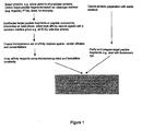
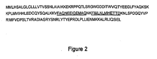


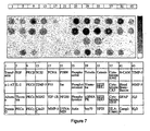


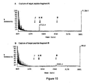
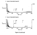
Classifications
-
- G—PHYSICS
- G01—MEASURING; TESTING
- G01N—INVESTIGATING OR ANALYSING MATERIALS BY DETERMINING THEIR CHEMICAL OR PHYSICAL PROPERTIES
- G01N33/00—Investigating or analysing materials by specific methods not covered by groups G01N1/00 - G01N31/00
- G01N33/48—Biological material, e.g. blood, urine; Haemocytometers
- G01N33/50—Chemical analysis of biological material, e.g. blood, urine; Testing involving biospecific ligand binding methods; Immunological testing
- G01N33/68—Chemical analysis of biological material, e.g. blood, urine; Testing involving biospecific ligand binding methods; Immunological testing involving proteins, peptides or amino acids
- G01N33/6803—General methods of protein analysis not limited to specific proteins or families of proteins
-
- C—CHEMISTRY; METALLURGY
- C40—COMBINATORIAL TECHNOLOGY
- C40B—COMBINATORIAL CHEMISTRY; LIBRARIES, e.g. CHEMICAL LIBRARIES
- C40B30/00—Methods of screening libraries
- C40B30/04—Methods of screening libraries by measuring the ability to specifically bind a target molecule, e.g. antibody-antigen binding, receptor-ligand binding
-
- G—PHYSICS
- G01—MEASURING; TESTING
- G01N—INVESTIGATING OR ANALYSING MATERIALS BY DETERMINING THEIR CHEMICAL OR PHYSICAL PROPERTIES
- G01N33/00—Investigating or analysing materials by specific methods not covered by groups G01N1/00 - G01N31/00
- G01N33/48—Biological material, e.g. blood, urine; Haemocytometers
- G01N33/50—Chemical analysis of biological material, e.g. blood, urine; Testing involving biospecific ligand binding methods; Immunological testing
- G01N33/53—Immunoassay; Biospecific binding assay; Materials therefor
- G01N33/543—Immunoassay; Biospecific binding assay; Materials therefor with an insoluble carrier for immobilising immunochemicals
-
- G—PHYSICS
- G01—MEASURING; TESTING
- G01N—INVESTIGATING OR ANALYSING MATERIALS BY DETERMINING THEIR CHEMICAL OR PHYSICAL PROPERTIES
- G01N33/00—Investigating or analysing materials by specific methods not covered by groups G01N1/00 - G01N31/00
- G01N33/48—Biological material, e.g. blood, urine; Haemocytometers
- G01N33/50—Chemical analysis of biological material, e.g. blood, urine; Testing involving biospecific ligand binding methods; Immunological testing
- G01N33/68—Chemical analysis of biological material, e.g. blood, urine; Testing involving biospecific ligand binding methods; Immunological testing involving proteins, peptides or amino acids
- G01N33/6803—General methods of protein analysis not limited to specific proteins or families of proteins
- G01N33/6845—Methods of identifying protein-protein interactions in protein mixtures
-
- G—PHYSICS
- G01—MEASURING; TESTING
- G01N—INVESTIGATING OR ANALYSING MATERIALS BY DETERMINING THEIR CHEMICAL OR PHYSICAL PROPERTIES
- G01N33/00—Investigating or analysing materials by specific methods not covered by groups G01N1/00 - G01N31/00
- G01N33/48—Biological material, e.g. blood, urine; Haemocytometers
- G01N33/50—Chemical analysis of biological material, e.g. blood, urine; Testing involving biospecific ligand binding methods; Immunological testing
- G01N33/68—Chemical analysis of biological material, e.g. blood, urine; Testing involving biospecific ligand binding methods; Immunological testing involving proteins, peptides or amino acids
- G01N33/6803—General methods of protein analysis not limited to specific proteins or families of proteins
- G01N33/6848—Methods of protein analysis involving mass spectrometry
Definitions
- This invention relates to affinity capture and detection of peptide fragments in a multi-dimension array.
- a cell can express a large number of different proteins, and the expression patterns (the number of proteins expressed and the expression levels) vary in different cell types, explaining why different cells perform different functions. Since many diseases associate with or even result from changes in protein expression pattern, comparing protein expression patterns between normal and disease conditions may reveal proteins whose changes are critical in the disease and thus identify proteins which are of therapeutic value or which aid diagnosis. Methods of detecting protein expression profiles also have important applications in, for example and without limitation, tissue typing, forensic identification, and clinical diagnosis.
- Microarray formats for the quantitative detection of proteins have been developed for diagnosis ( Ekins and Chu; Trends Biotechnol 1999, 17:217-8 ) and protein-protein interaction discovery ( Walter et al. Curr Opin Microbiol 2000, 3:298-302 ; U.S. Patent No. 6,197,599 ).
- development of a microarray affinity capture system for semi-quantitative or quantitative proteomics similar to those used for semi-quantitative mRNA expression analyses has lagged for a number of reasons.
- affinity capture agents e . g ., DNA, protein, small molecule
- affinity capture agents e . g ., DNA, protein, small molecule
- For mRNA analysis arrays the agents for affinity capture cDNA clones and gene sequences, UniGene - http://www.ncbi.nlm.nih.gov/UniGene/) were designed and available well in advance of the microarray system ( Southern, J. Mol Biol. 1975, 98:503-17 , Maskos and Southern, Nucleic Acids Res 1992, 20:1679-84 ).
- the protein agents to be captured exist in a variety of highly heterogeneousnon-homogenous form, e . g ., small soluble proteins (cytokines), large, highly post-translationally modified proteins (e . g . collagen) and proteins with multiple domains of varying hydrophilicity ( e . g . transmembrane receptors). Varying the pH in which the protein analyte is in a mixed population of heterogeneous protein types to correspond to, for example, subcellular location, may be required for folding.
- cytokines small soluble proteins
- large, highly post-translationally modified proteins e . g . collagen
- proteins with multiple domains of varying hydrophilicity e . g . transmembrane receptors
- any microarray affinity capture system for semi-quantitative or quantitative proteomics requires near impossible optimisation of capture affinity agent-protein interactions for a format to be achieved In which each affinity capture agent interacts with its corresponding target protein or family of target proteins in a uniform and predictable fashion. Alternatively, multiple arrays would be required to provide assay conditions specific for each different protein type such as those mentioned above.
- mRNA microarrays are well advanced and identification of the differential sequences is straightforward, there is a large inconsistency between mRNA and protein levels ( Gygi SP et al., Mol. Cell Biol. 1999, 19:1720-30 ) and since proteins are the primary agents responsible for disease and drug responses, predicting protein presence and amount by measurment of mRNA can lead to significant errors. Also current proteomics techniques such as 2D electrophoresis require complex procedures to identify differentially expressed proteins ( Parekh et al. Journal of Commercial Biotechnology 2000, 6:284-291 ). A microarray format, high throughput system for quantitative direct detection of proteins such as is required in proteomics applications is thus highly desirable for, without limitation, diagnosis, pharmacoproteomics, identification of markers of disease and drug target discovery.
- the present invention overcomes the deficiencies of current array-based proteomics techniques, and provides a system that permits qualitative and quantitative measurement of protein expression.
- An important aspect of the invention is a method for determining the presence of a protein in a sample.
- This method comprises submitting the sample to conditions that allow fragmentation of the protein into target peptide fragments.
- the target peptide fragments which may be labeled to facilitate detection, are contacted with an array that comprises a solid support on which a plurality of capture agents, each specific for a target peptide fragment of interest, are bound, i . e ., an array of antibodies on a solid support.
- the array may consist of capture agents selected to bind to one or more peptide fragments derived from each protein of interest.
- a plurality of target peptide fragment/capture agent combinations is employed for each protein, thus increasing the confidence of an analytical signal response.
- Specific binding of the target peptide fragments with the capture agents is indicative of the presence of the corresponding protein in the sample.
- binding can be detected by detecting the optional label, or directly by probing for the presence of bound target peptide fragment by a highly sensitive technique such as, without limitation, mass spectrometry.
- this method further comprises determining the relative amount of one or more proteins in a sample by quantitating the amount of the corresponding bound target peptide fragments: This may be achieved by determining the amount of each target peptide fragment bound to the matrix relative to the amount of one or more bound target peptide fragments from other expressed protein(s) in the same sample whose expression levels are already known or known to be invariant
- This invention additionally provides methods for the derivation of large numbers of affinity capture agents that recognize and bind to peptides derived from proteins, preferably under uniform, reproducible, standard binding conditions.
- Peptides generated by enzymatic or non-enzymatic cleavage of proteins result in a mixture of molecular entities that contains species, which are more suitable for interaction with affinity capture agents than a mixture of proteins.
- proteins contain many regions which cannot easily be accessed for binding to affinity capture agents: regions are shielded from external binding by glycosylation or other forms of post-translational modification; regions of high hydrophobicity such as membrane or transmembrane domains may bind with poor specificity; some binding sites arise as a result of 3-dimensional folding of non-contiguous segments of the protein. Fragmentation of such complex structures results in a plurality of smaller units (peptides) which are nevertheless representative of and unique to each fragmented protein.
- any proteins from any class can be assayed semi-quantitatively or quantitatively from any given starting material for example and without limitation, body fluid, whole cells and mixed cell types from organs and microbial.
- the invention additionally provides methods for design of the peptides used to derive the affinity capture agents. Also provided are methods for dispensing and immobilization of affinity capture agents in a microarray format; and for detection, quantitation and verification of the captured peptides. Furthermore, the identities of the peptides and thus the proteins responsible for differential signals are easily established.
- the present invention provides a method for producing an array for capturing one or more target peptide fragments of one or more proteins of known sequence.
- This method comprises immobilizing, without limitation, one or more, preferably at least five, more preferably at least ten, capture agents on a solid support. Each capture agent specifically recognizes a corresponding peptide compound and its corresponding target peptide fragment from a different protein of interest.
- This method may further comprise selecting the capture agent by detecting binding to the peptide compound.
- the target peptide fragment has a sequence determined by enzymatic or chemical cleavage of a protein by subjecting a list of proteins of known structure or conceptually translated nucleotides to theoretical cleavage and predicting the resulting "in silico" or theoretical sequence.
- the target peptide fragment has a sequence determined by directly sequencing a peptide fragment produced by enzymatic or chemical cleavage of a protein by methods well known to one skilled in the art.
- Capture agents may be, and preferably are, antibodies. In a specific embodiment all antibodies have a similar binding affinity for their respective target peptide fragment.
- the target peptide fragment may comprise one or more sites for post-translational modification of the protein.
- the peptide compound also may comprise one or more post-translational modification functional groups.
- the capture agent binds the target peptide fragment whether or not it has undergone post-translational modification.
- Preferably capture agents in the array recognize target peptide fragments from multiple proteins whose expression levels best correlate with a physiological or biochemical state, for example, and without limitation, as determined by multivariate analysis of protein expression levels.
- This physiological or biochemical state may be a response, such as, without limitation, a response to a xenobiotic stress; a hyperplastic, cancerous, or metastatic state; an apoptotic, dysfunctional or diseased state; or a particular phenotype.
- Central nervous system dysfunctions or diseases such as depression, schizophrenia, vascular dementia and other neurodegenerative conditions are particularly contemplated.
- Cancerous states such as breast cancer or hepatoma, also are encompassed.
- the present invention further provides an array that comprises a solid support on which capture agents, as defined according to the present invention, are immobilized.
- the conditions that allow fragmentation of the protein into target peptide fragments preferably comprise contacting the sample with a proteolytic enzyme.
- the cleavage pattern of the proteolytic enzyme is used to determine theoretical enzymatic cleavage of the protein, and the same enzyme is used to obtain sequences of target peptide fragments for use in selecting specific affinity capture agents.
- affinity capture agents may be selected using solid phase presentation of either peptide compounds or target peptide fragments of interest.
- the method for determining the presence of a protein in a sample may further comprise determining whether the target peptide fragment comprises a post-translational modification, for example and without limitation, using mass spectrometric analysis.
- the present invention greatly advances the effort to develop proteomics microarrays.
- the microarrays of the invention reduce the complexity of protein binding assays by providing a uniform, standardized binding system based on Interactions of capture agents (as defined below) with peptides.
- Peptides bind to binding partners (capture agents in the practice of the present invention) with relatively uniform kinetics and affinity (with some variation due to amino acid sequence), whether those binding partners are other peptides, antibodies, receptors, proteins, and even nucleic acids.
- the microarrays of the invention can have binding features more like those associated with nucleic acid hybridization arrays, and thus provide robust, standardized systems for detecting and, optionally, quantifying the total amount of a particular protein present. This is possible because the arrays of the invention are designed to detect peptides derived from cellular proteins, thus avoiding binding complexities of proteins.
- the array employs capture agents that specifically bind to peptides rather than proteins.
- Another feature of the microarrays of the invention lies in the ability to quantitate proteins of very different biophysical activities by detecting peptides, which have much more uniform biophysical activities. Again, use of peptide- (rather than protein-) specific capture agents facilitates this advantage.
- the microarrays of the invention are useful for proteomics, pharmacoproteomics, identification of markers of disease, drug target discovery, diagnostics, and in conjunction with therapy.
- the present invention represents an advance in efforts to develop proteomics microarrays.
- the inventors have discovered that it is possible to assay for the presence of peptide fragments derived from proteins rather than the entire, intact protein and that such assays can be performed with a level of consistency which is largely independent of the nature or origin of the protein of interest. This allows a semi-quantitative or more preferably a quantitative analysis to be performed.
- Such an approach has a number of advantages.
- target peptide fragments contacted with an array are well-exposed to the affinity capture agents present on that array.
- proteins may be subject to degeneration, misfolding, post-translational modification, and other environmental influences that may result in adoption of a conformation that reduces or blocks binding to antibody or results in unpredictable binding, whether by increasing or decreasing it.
- the invention further provides for generating affinity capture agents, such as antibodies, that are highly specific for unique sequences within the protein of interest. Using such a highly specific approach substantially diminishes cross reactivity which can be problematic when proteins of interest are related molecules.
- the peptide approach allows redundancy in quantitating the amount of protein present in a sample since it is possible to use a plurality of affinity capture agents each specific to one of a plurality of target peptide fragments derived from the same protein of interest. This results in a plurality of binding signals or responses being obtained for each protein of interest being analysed; a plurality of target peptide fragments used to generate the affinity capture agents will enable a more accurate quantitation of the protein of interest present in the sample by internal comparison of the detection signals from each target peptide fragment for any given protein of interest.
- the present invention increases the power of a proteomics microarray and simplifies this approach technically.
- the examples, Infra clearly demonstrate that the feasibility of quantitative protein detection using peptide specific capture agent arrays as set forth in the invention.
- the invention permits generating capture agents against target peptide fragments of proteins of known sequence from genomic, functional genomic, or proteomics analysis.
- the capture agent microarray can be used as a broad proteomics analytical tool, e.g., for analyzing protein expression, post translational modification, or other cell or tissue physiological characteristics.
- the invention permits performing multivariate analysis of levels of individual protein expression of all, most all, or a large number of proteins of interest in the cell.
- a general capture agent microarray of this sort provides a powerful tool for proteomics analysis to complement traditional two-dimensional electrophoresis.
- proteins of interest of known sequence that have a high correlation with the physiological or biochemical status of the cell or tissue are selected for producing capture agents for diagnosis for use in monitoring arrays, and for therapeutic purposes in accordance with the invention.
- proteins can be identified using multivariate analysis of levels of expression of all, most all, or a large number of proteins of interest in the cell, either by traditional biochemical approaches, two-dimensional gel proteomics analysis, or by using a microarray of the invention. For example, of the thousands of proteins in a cell, two hundred to three hundred proteins may be identified by traditional two-dimensional gel electrophoresis.
- an array of the invention can comprise capture agents for target peptide fragments from each of these 5 or more proteins of known sequence. More preferably, the array will comprise two or more capture agents specific for two or more unique target peptide fragments from each protein of interest of known sequence of primary interest.
- the binding affinity of each capture agent for its target peptide fragment is similar to that of the other capture agents for their respective target peptide fragments. Similar binding affinities mean that the variance of binding affinities among all of the capture agents for their corresponding target peptide fragment is within 100-fold, and preferably within 10-fold. Binding affinities within a 10-fold range will demonstrate similar binding characteristics in an assay and result in a more accurate semi-quantitative or quantitative method.
- the present invention contemplates pre-identification of proteins of known sequence.
- the arrays of the invention can be used to further refine the set of proteins of known sequence to be used in diagnosis, monitoring, and therapeutic indications.
- using the invention disclosed herein allows production of an array comprising capture agents for target peptide fragments from, for example, greater than 100 candidate proteins (fewer can also be evaluated with great advantage).
- Based on the quantititive data available from the arrays of the invention It will be possible to specifically identify those proteins of interest from the cell or tissue that, as set forth above, provide the most information about the biochemical or physiological status of the cell or tissue.
- proteins of known sequence are identified, it is then possible to design peptide compounds, prepare capture agents, prepare arrays comprising capture agents preferentially immobilized on a solid support, and test for the presence, and optionally, the amount of target peptide fragments from the proteins of interest.
- the peptide compounds used in the method of the invention may be designed by an in silico analysis of anticipated cleavage sites of a protein of interest. This in silico cleavage may be performed on:
- proteins of interest may be proteins previously identified for example In disease association studies, as described in greater detail below. They may also be proteins that are not known to be associated with diseases but may instead reflect the activation state or cycle or functionality, or phenotype or some other characteristic of the cell, any of which may be determined by the methods of the invention.
- a listing of real peptides that have been detected by mass spectrometry in proteomics experiments is constructed.
- target peptide fragments are selected from this listing, or subsets thereof.
- this listing provides relative analytical detection intensities for each target peptide fragment within the plurality of target peptide fragments from each protein of interest giving greater accuracy and precision of detection and improved quantitation.
- the proteins of interest are selected from proteins present in an experimentally observed proteime. Such a proteome can be generated by conducting two-dimensional gel experiments, with mass spectrometry analysis, using the same reference technologies referred to herein,
- the human genome is scanned beforehand with an exon-predicting software to extract DNA data most likely to represent expressed proteins.
- exon-predicting software examples include: GenScan ( Burge and Karlin J. Mol. Biol. 1997, 268:78-94 ; genes.mit.edu/GENSCAN.html on the World Wide Web) and GeneFinder (searchlauncher.bcm.tmc.edu/gene-finder/gfb.html on the World Wide Web).
- GenScan Burge and Karlin J. Mol. Biol. 1997, 268:78-94 ; genes.mit.edu/GENSCAN.html on the World Wide Web
- GeneFinder searchlauncher.bcm.tmc.edu/gene-finder/gfb.html on the World Wide Web.
- the extracted DNA sequences are subsequently translated into protein sequences that are in turn conceptually cleaved by any chosen method of proteolysis to provide target peptide fragments.
- the selection of target peptide fragments is based on sequences from the human genome that are known to unambiguously code for proteins, i.e. exons, through direct protein analysis. This provides a substantial improvement in the signal-to-noise ratio of the array detection by avoiding the possibility of non-specific binding of real peptides to antibodies raised against conceptual translations of noncoding human genome sequences.
- known recognition motifs or specific determinants within a protein of interest are used to select a sequence for preparing a capture agent, including without limitation known domain-peptide fragment or domain-domain interaction modules.
- interaction modules and specificity determinants could include, but are not limited to, concensus sequences determining the specificity of protein kinases and phosphatases, calmodulin recognition motifs ( Rhoads and Frisdberg, 1997, FASEB 11:331-340 ), LG/LNS domains ( Rudenko et al., 2001, TIBS 26:363-368 ), zinc finger domains ( Gillooly, et al., 2001, Biochem. J.
- One skilled in the art can also identify sequence information from peptides analyzed by mass spectrometry and/or tandem mass spectrometry using various spectral interpretation methods and database searching tools. Examples of some of these methods and tools can be found at the Swiss Institute of Bioinformatics web site on the World Wide Web at www.expasy.com, and the European Molecular Biology Laboratory web site on the World Wide Web at www.mann.embl-heidelberg.de/Services/PeptideSearch/.
- an array is constructed in which peptide fragments are arrayed and used as capture agents.
- the array can then be exposed to a sample containing one or more proteins of interest or a plurality of target peptide fragments from such proteins of interest to identify sequences in the protein of interest or its corresponding target peptide fragments which bind to the arrayed peptide fragments.
- This identifies arrayed peptide fragments of use as capture agents for specific target peptide fragments and also identifies peptide-peptide binding of potential biological or physiological significance.
- peptide fragments produced from a plurality of proteins, e are produced from a plurality of proteins, e .
- peptides identified by a plurality of exon-mapped sequences may be used as peptide compounds.
- the compounds can be used to produce arrays of capture agents that are then contacted with a test sample and a control. Any difference in signals between the test sample and the control sample indicates for a given capture agent that such a capture agent or plurality of capture agents is an effective tool for monitoring changes in such a sample.
- the preferred method is to design synthetic peptides as peptide compounds for selecting the capture agents with the required binding specificity.
- the peptide compounds defined by this invention for use in designing capture agents can be designed individually according to appropriate methods but should in general correspond to the predicted peptide sequences or part thereof, according to the chosen digestion reagents which will be used in the detection of experimental peptides derived from a protein of interest.
- the number of peptide fragments selected for each protein as target peptide fragments can be one and more preferably more than one.
- An example of the selection of such target peptide fragments from the protein BCMP 11 (SEQ ID no. 1) is shown in figure 2 .
- the chosen peptides should also preferably be unique to the proteins being assayed, as determined for example and without limitation by means of the Blast algorithm ( Altschul, et. al, J. Mol. Biol, 1990, 215:403-410 ) or other techniques known to one skilled in the art. Signal sequence peptides which are removed from the immature protein are also preferably avoided.
- target peptide fragments can be selected based on information regarding whether they may.be present in a protein of interest in a region subject to post-translational modifications and subject to such post-translational modifications. Such peptides may be used to design arrays useful to diagnose a disease associated with such specific post-translational modifications. This information can come directly from the literature ( e . g . Hoffman et. al, Biochemistry, 1996,35(47):14849-61.3 ), from direct evidence ( e . g ., tandem MS) or from prediction ( e . g . intracellular tyrosines are often subject to phosphorylation, PSORTII on the World Wide Web).
- post-transiational modifications include but are not limited to: N-formyl-L-methionine; L-selenocysteine; L-cystine; L-erythro-beta-hydroxyasparagine; L-erythro-beta-hydroxyaspartic acid; 5-hydroxy-L-lysine; 3-hydroxy-L-proline; 4-hydroxy-L-proline; 2-pyrrolidone-5-carboxylic acid; L-gamma-carboxyglutamic acid; L-aspartic 4-phosphoric anhydride; S-phospho-L-cysteine; 1'-phospho-L-histidine; 3'-phospho-L-histidine; O-phospho-L-serine; O-phospho-L-threonine; O4'-phospho-L-tyrosine; 2'-[3-carboxamido-3-(trimethylammonio)propyl]-L-h
- Examples of phosphorylation include, but are not limited to, Vanmechelen et. al, Neurosci. Lett. 2000, 285:49-52 ; Lutz et. al, Pancreas 1994, 9:418-24 ; Gitlits et. al J. Investig. Med. 2000, 48:172-82 ; or Quin and McGuckin, Int. J. Cancer. 2000, 87:499-506 .
- sulfation includes, but is not limited to, Manzella et. al J. Biol. Chem. 1995, 270S:21665-71 .
- post-translational modification by oxidation or reduction examples include, but are not limited to, Magsino et. al, Metabolism 2000, 49:799-803 ; or Stief et. al, Thromb. Res. 2000, 97:473-80 .
- ADP-ribosylation examples include, but are not limited to, Galluzzo et. al, Eur. J. Immunol. 1995, 25:2932-9 ; or Thraves et. al, Med. 1986, 50:961-72 .
- hydroxylation includes, but is not limited to, Brinckmann et. al, J. Invest. Dermatol. 1999, 113:617-21 .
- glycosylation examples include, but are not limited to, Johnson et. al, Br. J. , Cancer 1999, 81:1188-95 ; Fulop et. al, Biochem. 1996, J. 319;935-40 ; Dow et. al, Exp. Neurol. 1994, 28:233-8 ; Kelly et. al, J. Biol. Chem. 1993, 268:10416-24 ; Goss et al, Clin. Cancer Res. 1995, 1:935-44 ; or Sleat et. al, Biochem. J. 1998, 334:547-51 ..
- glucosylphosphatidylinositide addition includes, but is not limited to, Poncet et. al, Acta Neuropathol. 1996, 91:400-8 .
- ubiquitination includes, but is not limited to, Chu et. al, Mod. Pathol. 2000, 13:420-6 .
- translocation leading to a disease state includes, but is not limited to, Reddy et. al, Trends Neurosci. 1999, 22:248-55
- Synthesis and purification of these peptides can be achieved using commercially available peptide synthesisers (e . g ., Applied Biosystems, Framingham, MA) and protection residues, as described below.
- commercially available peptide synthesisers e . g ., Applied Biosystems, Framingham, MA
- protection residues as described below.
- amino acids used for peptide synthesis may be standard Boc (Nalpha-amino protected Nalpha-t butyloxycarbonyl) amino acid resin with the standard deprotecting, neutralization, coupling and wash protocols of the original solid phase procedure of Merrifield (J. Am. Chem. Soc. 1960, 85:2149-2154 ), or preferably the base-labile N-alpha ⁇ -amino protected 9-fluorenylmethoxy- carbonyl (Fmoc) amino acids first described by Carpino and Han (1972, J. Org.
- Solid phase peptide synthesis may be accomplished by techniques familiar to those in the art and provided, for example, in Stewart and Young, Solid Phase Synthesis, Second Edition, 1984, Pierce Chemical Co., Rockford, IL ; Fields and Noble, Int J. Pept. Protein Res. 1990, 35:161-214 , or using automated synthesizers, such as sold by Applied Biosystems (Framingham, MA).
- non-classical amino acids may be incorporated in peptides to introduce particular conformational motifs: 1,2,3,4-tetrahydroisoquinoline-3-carboxylate ( Kazmierski et al., 1991, J. Am. Chem. Soc. 1991,113:2275-2283 ); (2S,3S)-methylphenylalanine, (2S,3R)-methyl-phenylalanine, (2R,3S)-methyl-phenylalanine and (2R,3R)-methyl-phenylalanine ( Kazmierski and Hruby, Tetrahedron Lett. 1991 ,); 2-aminotetrahydronaphthalene-2-carboxylic acid ( Landis, 1989, Ph.D.
- LL-Acp LL-3-amino-2-propenidone-6-carboxylic acid
- beta-turn inducing dipeptide analog Kemp et al., J. Org. Chem. 1985, 50:5834-5838
- beta-sheet inducing analogs Kemp et al., Tetrahedron Lett. 1988, 29:5081-5082
- alpha-tum inducing analogs Kemp et al., Tetrahedron Lett. 1988, 29:5057-5060
- ⁇ -helix inducing analogs Kemp et al., 1988, Tetrahedron Lett.
- an agent that "recognizes" a peptide compound or a sequence thereof refers to the ability of this agent to specifically interact with this peptide compound.
- Capture agents may be generated de novo against peptide compounds or selected from libraries of agents. Examples of capture agents include antibodies ( e . g ., polyclonals from animals, monoclonals from hybridomas), single chain antibodies from phage display libraries ( Vaughan et al., Nat Biotechnol. 1996, 14:309-14 ), small peptides ( Norman et al. Science 1999, 285:591-5 ) or RNA-protein fusion libraries ( Kreider et al.
- the capture agents that bind to the target peptide fragment are themselves peptides and allow peptide-peptide interactions to be characterized.
- the agents that bind to the peptide compounds are antibodies.
- target peptide fragments known to be produced from a protein of interest by prior knowledge of, for example without limitation, tandem fragmentation analysis (MS/MS) by mass spectrometry are used to produced peptide compounds or capture agents.
- Polyclonal antibodies that may be used in the methods of the invention are heterogeneous populations of antibody molecules derived from the sera of immunized animals. Unfractionated immune serum can also be used. Various procedures known in the art may be used for the production of polyclonal antibodies to a selected peptide compound. In a particular embodiment, rabbit polyclonal antibodies to an epitope of said peptide compound can be obtained. For example, for the production of polyclonal or monoclonal antibodies, various host animals, including but not limited to rabbits; mice, rats, etc, can be immunized by injection with the native or a synthetic ( e . g ., recombinant) version of these peptide compounds.
- any technique that provides for the production of antibody molecules by continuous cell lines in culture may be used.
- the hybridoma technique originally developed by Kohler and Milstein (Nature 1975,256:495-497 ), as well as the trioma technique, the human B-cell hybridoma technique ( Kozbor et al., Immunology Today 1983; 4:72 ), and the EBV-hybridoma technique to produce human monoclonal antibodies ( Cole et al., 1985, in Monoclonal Antibodies and Cancer Therapy, Alan R. Liss, Inc., pp. 77-96 ).
- Such antibodies may be of any immunoglobulin class including IgG, IgM, IgE, IgA, IgD and any subclass thereof.
- the hybridoma producing the mAbs of the invention may be cultivated in vitro or in vivo.
- monoclonal antibodies can be produced in germ-free animals utilizing known technology (international Application PCT/US90/02545 ).
- the monoclonal antibodies include but are not limited to human monoclonal antibodies and chimeric monoclonal antibodies (e . g ., human-mouse chimeras).
- a chimeric antibody is a molecule in which different portions are derived from different animal species, such as those having a human immunoglobulin constant region and a variable region derived from a murine mAb (see, e . g ., U.S. Patent Nos. 4,816,567 and 4,816.397 ).
- Antibodies can also be generated using various phage display methods known in the art.
- phage display methods functional antibody domains are displayed on the surface of phage particles that carry the polynucleotide sequences encoding them.
- such phage can be utilized to display antigen binding domains expressed from a repertoire or combinatorial antibody library ( e . g ., human or murine).
- Phages expressing an antigen binding domain that binds the antigen of interest can be selected or identified with antigenic peptide compound s, e . g ., using labeled peptides or peptides bound or captured to a solid surface or bead.
- Phage used in these methods are typically filamentous phages including fd and M13 binding domains expresses from phage with Fab, Fv or disulfide stabilized Fv antibody domains recombinantly fused to either the phage gene III or gene VIII protein.
- Phage display methods that can be used to make the antibodies of the present invention include those disclosed in Brinkman et al., J. Immunol. Methods 1995, 182:41-50 ; Ames et al., J. Immunol. Methods 1995, 184:177-186 ; Kettleborough et al., Eur. J. Immunol.
- the antibody coding regions from the phage can be isolated and used to generate whole antibodies, including human antibodies, or any other desired antigen binding fragment, and expressed in any desired host, including mammalian cells, insect cells, plant cells, yeast, and bacteria, e . g ., as described in detail below.
- Fab, Fab' and F(ab')2 fragments can also be employed using methods known in the art such as those disclosed in PCT Publication WO 92/22324 ; Mullinax et al., BioTechniques 1992, 12:864-869 ; and Sawai et al., AJRI 1995, 34:26-34 ; and Better et al., Science 1998, 240:1041-1043 (said references incorporated by reference in their entireties).
- the peptide compounds may be immobilized, incubated with potential capture agents and washed.
- the high affinity binders are selected according to any method well-known by one skilled in the art ( Vaughan et. al 1998, Nat Biotechnol. 1996, 14:309-14 ).
- peptide compounds can be immobilized on activated hydrogel 1000 x 200 ⁇ m2 (20 ⁇ m depth) pads, separated in two dimensions each, each having a different peptide immobilized on it ( e . g ., Vasiliskov et. al. Biotechniques 1999, 27:592-4, 596-8 , 600).
- Libraries of potential affinity capture agents can be incubated with the immobilized peptide compounds in order to select capture agents for each peptide compound.
- the capture agents can be eluted individually from the hydrogel pads and amplified by, e . g ., phage propagation ( Vaughan et. al., Med Res Rev 2000, 20:212-5 ). Subsequent rounds of enrichment can be performed on these amplified capture agents by further incubation with similar immobilized peptides. Selection of single high affinity capture agents can be achieved by growing individual phages or cloning of RT-PCR products, for example.
- these phages may be directly selected against peptide compounds.
- phage cDNA is ligated with PCR-amplified mRNA light-chain joined to heavy-chain variable regions, isolated from na ⁇ ve splenocytes or bone-marrow stem cells.
- E.coli bacteria are co-transfected with Phage (pilus depolymerization), and with helper phage M13 to allow DNA packaging of the phagemid into phage particles.
- ScFv (single chain variable fragment) Ab (antibody) is displayed on gplll, that is, the coat protein used to bind to and infect E.coli .
- phage selection For phage selection, one can use a 96 well plate containing multiple 25-mers to specific peptides (5 per different peptide). The non-binders are washed away in each well. The binders then are used to re-transfect E.coli bacteria and grown to confluency. Phages are harvested, and re-exposed to the equivalent peptide to weed out non-specific binders ( e . g ., the ones that bind to plastic or any inherently sticky phagemids).
- non-specific binders e . g ., the ones that bind to plastic or any inherently sticky phagemids.
- E . coli samples that contain phagemids are spread onto plates containing a selection factor (1/10, 1/100). Resulting colonies are chosen and grown up in suspension containing the selection factor. The phages secreted into the medium are then recovered.
- Phages that display antibodies are used in ELISAs to identify strong binders that can be tracked back to the original clones.
- the capture agents selected according to the method described above are immobilized on a solid support to form an array. Selection of the solid support and the methods for immobilization depend on the type of capture agents (proteins, nucleic acids, etc .). Immobilization may be performed through covalent or non-covalent binding. In a preferred embodiment, the capture agents are covalently bound to the surface of the solid support via reactive groups or cross-linking agents.
- covalent immobilization may be achieved through free cysteines using thiol reactive surfaces, or through lysines using amine reactive surfaces.
- heterobifunctional crosslinkers may be used to cross link- NH groups the surface to sulfhydryl (SH) groups or cysteine residues.
- BMPS beta-maleimidopropionic acid N-hydroxysuccinimide ester, e . g . purchased from SIGMA
- SIGMA sulfur-maleimidopropionic acid N-hydroxysuccinimide ester
- Non-covalent interaction may be achieved by using Avidin- or Streptavidin-coated surfaces and biotinylated affinity capture agents; protein-A or protein-G coated surfaces and Fc containing affinity capture agents ( e . g ., immunoglobulins, etc.); and metal-chelate surfaces and Histidine tagged affinity capture agents.
- capture agents are multiple single chain antibodies (SCAs) specific to different peptides. They are purified from phages and solubilised in PBS at similar concentrations (about 0.1 mg ml-1). The specific SCAs are placed in multiwell plates ( e . g ., 96 well). These plates can then be used in a microarray system. It is preferable when dispensing proteins to employ a system that does not use steel pins or contact dispensing equipment. Thus, a preferred system is the Packard BCA Piezo robot ( U.S. Patent No. 5,927,547 ), which dispenses small volume drops (less than 1nL) from a glass capillary from above the surface of the microarray substrate.
- SCAs single chain antibodies
- a preferred substrate is aldehyde activated polyacrylamide pads immobilized on glass or oxidised silicon.
- a preferred size for the polyacrylamide pads is 2cm x 2cm x 20 ⁇ m.
- about 300pL of each SCA is dispensed into a discrete area within the polyacrylamide pad to create a 2-dimensional array in a 3-dimensional structure.
- the resulting spot is about 200 ⁇ m in diameter.
- the free aldehydes within the polyacrylamide react with the amines in the SCAs so that the SCAs are covalently immobilized. Once this reaction is complete (30 minutes after dispensing) any remaining aldehydes are reduced with sodium borohydride or blocked using reagents containing amino groups ( e . g ., TRIS buffer) and/or free amino acids.
- Another preferred substrate is hydrogel on which capture agents such as antibodies may be immobilized, e . g ., via -NH group of Lysines to form a Schiff base with aldehyde groups present in the hydrogel. Lysines also provide free -NH groups that can be cross-linked to -SH groups of cysteine residues in antibodies ( e . g ., via BMPS).
- Hydrogel immobilization has given better results in terms of signal to noise ratio and fluorescence specifically when compared to glass or silicon substrates.
- the substrate is produced by casting a thin agarose gel on a glass slide, using suitable gaskets or other spacers, and siliconised cover glass (or cover slips or similar solid surface for producing uniform thickness gel). Following casting, the gel is activated with CNBr.
- agarose gels is preferred over commercial Hydrogels (e.g. from Packard Instrument Co., Meriden, CT) as agarose retains moisture more efficiently and thus allows for spotting of dehydration-sensitive reagents.
- Another advantage of using thin agarose gel films is that they allow for faster diffusion of compounds due to a larger pore size. Therefore, shorter washing steps and lower affinity capture agents may be used.
- single chain Fv antibodies are histidine-tagged for the previously-described isolation step.
- the histidine tag can further be used for immobilization, e . g . using metal chelate affinity on nickel-modified surfaces, for instance.
- this system allows multiple affinity capture agents, specific to different target peptide fragments from the same or different proteins, to be put in the same spot, particularly when MALDI-MS is being used as a detection method. Additional immobilization chemistries are possible using other affinity agents and array substrates ( e . g . Lin, Science 1997, 278:840-843 ).
- immobilization examples include using thin membranes (such as nitrocellulose, nylon (charged or uncharged), PVDF and/or their derivatives) attached to or immobilized on glass slides or other solid surfaces.
- thin membranes such as nitrocellulose, nylon (charged or uncharged), PVDF and/or their derivatives
- attaching commercially available membranes nitrocellulose, supported nitrocellulose, nylon, charged nylon or PVDF membranes from Schleicher & Schuel, UK Ltd., Amersham, Millipore and other suppliers
- CASTTM layer of Nytran® SuPerCharge positively charged nylon membrane affixed to the glass
- FASTTM slides microporous polymeric surface cast onto glass
- affinity capture agents may be dispensed using piezo robots similar to the one described above for SCAs.
- One of the advantages of using nitrocellulose or nylon based supports is that they have a much higher protein binding capacity compared to Hydrogels or 2D solid supports (glass, silicon, plastic etc.). A higher binding capacity results in a stronger fluorescent signal, and therefore allows for the use of binders having lower affinity.
- Various solid support materials that may be used in the microarray are as follows:
- Another preferred immobilization technique uses a combination of non-covalent affinity immobilization coupled to covalent cross-linking.
- This technique is especially advantageous for immobilizing affinity capture agents such as antibodies, as it allows the antibodies to be attached in an orientation whereby their active sites are easily accessible, and at the same time, remain covalently cross-linked to the solid support.
- This technique includes, but is not limited to, using Hydrogels (Packard Instrument Co., Meriden, CT) for immobilization of protein-A or protein-G, followed by treatment with glutaraldehyde (high concentration for short time). This results in covalent cross-linking of the protein-A/G to the solid support, and also in aldehyde derivation of solution-exposed amino groups of the protein-A/G.
- Fc containing antibodies e.g. IgG
- Fc containing antibodies e.g. IgG
- protein-A/G eventually cross-links with the bound antibodies through aldehyde-derivatized groups on the protein-A/G and available amino groups on the Fc fragments.
- Glutaraldehyde pre-treatment of the immobilized Protein-A/G prior to antibody spotting is a preferred technique because it avoids incubating the affinity capture agents (i.e., the antibodies) with the cross-linking agent (i.e. glutaraldehyde), which could result in the derivatization of amino groups of the antibodies and, therefore, loss of the high affinity binding sites.
- affinity capture agents i.e., the antibodies
- the cross-linking agent i.e. glutaraldehyde
- nucleic acid array refers to "gene chips” and related arrays of oligonucleotides, cDNAs, and other nucleic acids, which are well known in the art (see for example the following: U.S. Patent Nos.
- nucleic acids that are attached to the solid support are preferably DNA and RNA aptamers.
- the nucleic acid is attached to a solid support, which may be made from glass, silicon, plastic ( e . g ., polypropylene, nylon), polyacrylamide, nitrocellulose, or other materials.
- a preferred method for attaching the nucleic acids to a surface is by printing on glass plates, as is described generally by Schena et al., Science 1995, 270:467-470 . This method is especially useful for preparing microarrays of cDNA. See also DeRisi et al., Nature Genetics 1996, 14:457-460 ; Shalon et al., Genome Res. 1996, 6:639-645 ; and Schena et al., Proc. Natl. Acad. Sci. USA 1995, 93:10539-11286 .
- a preferred method of making microarrays is by use of an inkjet printing process to bind genes or oligonucleotides directly on a solid phase, as described, e . g ., in U.S. Patent No. 5,965,352 .
- microarrays e . g ., by masking
- Maskos and Southern Nucl. Acids Res. 1992, 20:1679-1684
- any type of array for example, dot blots on a nylon membrane (see Sambrook et al., Molecular Cloning A Laboratory Manual (2nd Ed.), Vol. 1-3, Cold Spring Harbor Laboratory, Cold Spring Harbor, N.Y., 1989 , could be used, although, as is recognized by those of skill in the art, very small arrays are preferred because sample volumes are smaller.
- the present invention contemplates determining the presence of a protein in a sample. Any sample that is likely to contain a protein of interest may be tested. Such samples, referred to herein as biological samples, include body fluid (e . g . blood, serum, plasma, saliva, urine, plural effusions or cerebrospinal fluid), a tissue sample (e . g ., a biopsy, blood cells, smears) or homogenates and extracts, including cytoplasm, membranes, and organelles thereof. Cell cultures and culture fluid are also biological samples.
- body fluid e . g . blood, serum, plasma, saliva, urine, plural effusions or cerebrospinal fluid
- tissue sample e . g ., a biopsy, blood cells, smears
- homogenates and extracts including cytoplasm, membranes, and organelles thereof.
- Cell cultures and culture fluid are also biological samples.
- the sample may be pre-treated to obtain a protein preparation substantially free of contaminants.
- a treatment may comprise fractionation, differential extraction (membrane and cytosolic fractions); selective depletion (e . g ., for removal of albumin, haptoglobin, immunoglobin G); and application to any specific affinity column ( e . g ., Mannose-6-Phosphate receptor for lysosomal enzymes; Sleat and Lobel, J Biol Chem 1997, 272:731-8 ).
- Proteins present in the sample may be in native form or denatured ( Wilkins MR; et al., 1996, Biotechnology (N Y) 14(1):61-5 , e . g . by dissolving in 6M guanidine HCl (or 6-8M urea), 50 mM Tris-HCl (pH8), 2-5 mM DTT (or 2-mercaptoethanol). Proteins present in the sample may also be pre-treated with eg. glycosidases (glycoprotein deglycosylation kit available from CN biosciences, UK. Ltd.) to remove glycosylation side chains or other means of predictably varying post-translational modifications.
- glycosidases glycosidases
- the sample is then subjected to conditions that allow enzymatic or chemical cleavage of the individual proteins into target peptides.
- this method uses the same conditions initially used to digest or predict digestion of the protein sequence selected for generation of target peptide fragments from which the capture agents were prepared.
- other cleavage agents may be used on the condition that they do not disturb the capture agent binding site, e . g ., antigenic determinant of the target peptide fragment.
- a reduction/alkylation step can be performed prior to proteolysis.
- Dithiothreitol (DTT) may be used for reduction and iodoacetamide may be used for carboxyamidomethylation of cysteine.
- Reproducible peptide fragments can be generated from protein preparations using proteolytic and chemical methods (e . g . Schevchenko et al. Analytical Chemistry 1996, 68:850-858 ; Houthaeve et al., FEBS Letters 1995, 376:91-94 ; Wilkins et al., 1997, Springer ISBN 3-540-62753-7).
- the cleavage may be enzymatic cleavage. Preferably, it is a selective enzymatic cleavage, e . g .
- arginine endopeptidase arginine endopeptidase
- asparatic acid endopeptidase N asparatic acid endopeptidase N
- chymotrypsin glutamic acid endopeptidase C
- gluC glutamic acid endopeptidase C
- lysC lysine endopeptidase C
- trypsin or V8 endopeptidase.
- Trypsin is preferred, as it specifically cleaves proteins at the C-terminus of Arg or Lys.
- mammalian cells e . g . Jurkat cells
- RIPA buffer 20 mM Tris-HCl, pH 7.4, 150 mM NaCl, 1% Triton X-100 (v/v), 1% deoxycholate (w/v), 0.1% sodium dodecyl sulphate (w/v)
- the trypsin can be neutralized by the addition of a cocktail containing serine protease inhibitors. Tryptic peptides then can be purified and buffered with PBST by gel filtration. Alternatively, trypsin can be removed by immuno-precipitation or inactivated by boiling.
- Table 2 Protein cleavage using Proteolytic Enzymes Enzymes Preferred cleavage site Ancrod Arg-X, Arg-Gly Bromelain C-terminal to Lys, Ala and Tyr Chymotrypsin C-terminal to hydrophobic residues, e.g., Phe, Tyr, Trp.
- the optimum digestion time to produce the desired quality of peptide fragments may be determined for example by collecting aliquots every 2 hours and after an overnight digest.
- Target peptide fragments are preferably peptides that are between 6 and 25 residues in length.
- a sample containing proteins of interest can be split into two or more aliquots and each aliquot treated with a different enzyme or chemical agent to produce complementary overlapping target peptide fragments. Each proteolytically cleaved sample is then analysed as described herein.
- Target peptide fragments may be labeled according to any standard method to facilitate detection.
- the target peptide fragment may be directly labeled.
- a labeled secondary reagent may be used to detect binding of the target peptide fragment to the solid support containing the capture agent Binding may be detected by in situ formation of a chromophore by an enzyme label.
- Suitable enzymes include, but are not limited to, alkaline phosphatase and horseradish peroxidase (HRP).
- FITC fluorescein isothiocyanate
- PE phycoerythrin
- TR Texas red
- rhodamine free or chelated lanthanide series salts, especially Eu 3+ , to name a few fluorophores
- chemiluminescent molecules chemiluminescent molecules
- radio-isotopes 126 I, 32 P, 35 S, chelated Tc, etc.
- magnetic resonance imaging labels Two color assays may be performed using fluorophores that emit at different wavelengths.
- Metabolic (or biosynthetic) labeling with [ 35 S]-methionine is also contemplated as well as labeling with [ 14 C]-amino acids and [ 3 H]-amino acids (with the tritium substituted at non-labile positions).
- a preferred method makes use of NHS ester Bodipy TMR , Molecular Probes, Leiden, the Netherlands.
- Other methods include the use of Fluorophores - iodoacetamide, maleimide or succinimide groups ( e . g ., Molecular Probes) Cy3, Cy5 or TAMRA dyes for example via labeling cysteines or lysines ( e . g ., Alexa Fluor dyes).
- cysteines are readily modified by maleimide and iodoacetamide containing reagents. This allows enrichment for those peptides from the cleaved pool via e . g ., iodoacetamide-biotin (Molecular Probes, Leiden, Netherlands) and streptavidin capture. Further fluorescent signals can be increased via iodoacetamide-Bodipy TMR (Molecular Probes, Leiden, Netherlands), for example.
- Labeled or unlabeled target peptide fragments (about 1mg ml -1 ) in PBST or another suitable buffer are incubated with the array of immobilized capture agents.
- An example of incubation Buffer is 1 x PBS, pH 7.4, phosphate buffer (0.01 M) plus NaCl (0.137M), KCl (0.0027M), without Tween detergent.
- the incubation may proceed at room temperature or at 37°C in a sealed humidified chamber from 1 hour to 24 hours, in a volume of about 25 ⁇ l under a coverslip or 65 ⁇ l in a chamber formed with gaskets (Clontech). No agitation is necessary.
- Target peptide fragments that have bound non-specifically to the array are removed by 2 x 15 minute washes in PBST (200ml) at room temperature followed by 3 rinses in distilled water.
- the water rinses remove salt and detergent, which can interfere with detection methods.
- any method known in the art for detecting the binding of the labeled target peptide fragments to the capture agents may be performed according to the present invention.
- target peptide fragments are known and the affinity of the capture agent for each target peptide fragment can be measured by techniques known in the art, any array can be calibrated with respect to each target peptide.
- one or more target peptide fragment-capture agent pairs of known specificity and known affinity are available for use in an assay accurate semi-quantitative or quantitative results can be obtained with the array.
- the fluorescent signal may be analyzed by various methods, including fluorescence intensity or polarization ( Onnerfjord et al., J. Immuno. Methods 1998, 213:31-9 ), fluorescence correlation spectroscopy ( Tetin et al., Methods 2000, 20: 341-61 ), phase sensitive fluorescence, fluorescence anisotropy, FRET (Fluorescence Resonance Energy Transfer), BRET (Bioluminescence Resonance Energy Transfer), or time resolved fluorescence.
- radioisotope labeling Topic Count NXT Microplate Scintillation and Luminescence Counters from Packard
- SPR-Regnault et al. Br. J. Haematol 2000, 109:187-94 may also be contemplated.
- microarrays are washed and placed in commercially available confocal laser scanners, e . g ., ScanArray 3000 (Gsi lumonics, USA).
- the amount of fluorescence per spot (capture agent) is relative to the amount of captured labelled peptide which, in turn, is relative to the amount of protein containing that peptide in the protein preparation.
- Software such as Optiquant can be used to accurately measure the fluorescence from each spot.
- fluorescence assays are performed on hydrogel or agarose gel arrays.
- An example of arrays for fluorescence assays is 12x12mm hydrogels (20 ⁇ m thick) on glass microscope slides. These dimensions could be increased for higher numbers of antibodies to be immobilized.
- the captured target peptide fragments can be analyzed by mass spectrometry; this can be using primary mass spectrometry (MALDI-TOF MS) or tandem mass (fragmentation) spectrometry (MS/MS); both of these are discussed in some detail below.
- mass spectrometry can be using primary mass spectrometry (MALDI-TOF MS) or tandem mass (fragmentation) spectrometry (MS/MS); both of these are discussed in some detail below.
- the peptides are initially analysed using matrix-assisted laser-desorption time-of-flight mass spectrometry (MS).
- MS matrix-assisted laser-desorption time-of-flight mass spectrometry
- ppm parts-per-million
- MALDI MS is a non-quantitative method. However, this is usually due to variabilities in sample preparation where large volumes and non-homogenous matrix crystals are produced.
- MALDI assays are performed on silicon arrays.
- An example of an array for MALDI is 200 ⁇ m circular gel pads at 350 ⁇ m centres, on oxidised silicon.
- a hydrophobic surface (repellent surface) between gelpads further provides a more focused matrix/protein spot for MALDI, thereby improving signal upon quantitation.
- the spots produced using the system of this invention are less than 200 ⁇ m in diameter.
- the same system can deliver about 300pL of MALDI matrix (e . g . DHB, sinapinic acid) to the exact position of the affinity capture agent-target peptide fragment spot to create a homogeneous target peptide fragment/matrix crystal.
- MALDI matrix e . g . DHB, sinapinic acid
- Protein extracts (1 ⁇ g each) were incubated with identical arrays of antibodies immobilised on Hydrogel pads. Arrays were washed and analysed by MALDI-TOF-MS. Spectra for the anti-albumin antibody mediated captured of albumin from ovary (A) and serum (B) are shown ( Figure 3 ).
- Figure 4 shows a modified target holder for a Perseptive Voyager to hold a silicon-bound polyacrylamide pad.
- MALDI-TOF MS MS
- MS MALDI-TOF
- Masses due to the presence of the capture agent can be ignored and analysis focused on masses arising from the target peptide fragments.
- Intensities of each mass (m/z) feature in the mass spectrum are measured by methods known to those skilled in the art or as specified in the PCT Application number PCT/GB01/04034 filed on 10th September 2001 . Where more than one target peptide fragment is available for each protein of interest the intensities of masses in the mass spectra of such fragments can be normalized across such a plurality of signals resulting in greater accuracy and reliability.
- sample/matrix spot can be performed using any standard method of tandem mass spectrometry (MS/MS) and in particular using MALDI-TOF/TOF (Applied Biosystems, Framingham, MA) or MALDI II Q-TOF (Micromass) or Q-STAR (Sciex) all of which are systems which continue MALDI MS with tandem mass spectrometry.
- MS/MS tandem mass spectrometry
- MALDI-TOF/TOF Applied Biosystems, Framingham, MA
- MALDI II Q-TOF Micromass
- Q-STAR Q-STAR
- Database searching of the primary mass data provided by MALDI-TOF MS can be used to identify possible post-translational modifications (PTMs) of peptides.
- PTMs post-translational modifications
- MS/MS tandem mass spectrometry
- high energy CID provided by MALDI-TOF/TOF MS has been shown to unambiguously establish the site of peptide phosphorylation (Analysis of Post-Translational Modifications using a MALDI-TOF-TOF Mass Spectrometer, DeGnore et al . Poster presentation at the 49th ASMS conference on Mass Spectrometry and Allied Topics, Chicago).
- microprocessor i . e ., computer
- fluorescence detection and mass spectrometry employ well-known types of apparatus, e . g ., as set forth in the references noted above.
- the invention further specifically contemplates adapting such apparatus for the specific analysis of microarrays of the invention.
- the robust, standardizable, uniform assays of the present invention permit adaptation of specific features of the apparatus, including but not limited to incubation time, detection parameters, and processing software, that would be cumbersome if adapted for protein-specific capture agent microarrays.
- an apparatus of the invention whether for label detection (e . g ., fluorescence) or direct detection of peptide binding (such as mass spectrometry), can be specifically adapted to detect peptide binding in an array of the invention.
- label detection e . g ., fluorescence
- direct detection of peptide binding such as mass spectrometry
- an adaptation may include specific software designed for processing detection data.
- software specifically evaluates diagnostic arrays for the presence and amount of key disease markers.
- the software processes the detected peptides against a database of known markers for particular cellular conditions, and provides as output not raw binding intensity data but a most likely diagnosis.
- Such an apparatus has clear application in commercial diagnostic laboratories, where the volume of material to be analyzed is high and the capabilities of the technicians are not at the level of Ph.D. scientists.
- Diagnosis represents a major application of the method of the invention.
- a protein or a peptide that is differently expressed in a disease can be detected in a biological sample and noted as a marker of the disease or change in biochemical status.
- markers include Cystatin C for renal dysfunction, ( Fliser D. and Ritz E., Am. J. Kidney Dis. 2001, 37(1): 79-83 ); prostate-specific antigen (PSA) for prostate cancer, ( Millenbrand et al., Anticancer Res., 2000, 20(6D): 499-6 ); Anglotensin II/ACE for heart failure ( Kim SD; Biol. Res. Nurs. 2000, 1(3): 210-26 ).
- PSA prostate-specific antigen
- Data produced by the method of the invention can be analysed by sophisticated statistical techniques including uni-variate and multi-variate ananlysis tools.
- the following steps can be used to identify target peptide fragments arising from proteins which show an association with a disease or biochemical status.
- Changes such as fold changes, Wilcoxon rank sum test and t-test, are useful in determining the significance of the expression values of each target peptide fragment and its corresponding protein of interest.
- the first step is to identify a collection of target peptide fragment signal responses that individually show significant association with any particular condition.
- the association between the identified proteins and any particular condition need not be as highly significant as is desirable when an individual protein is used in diagnosis. Any of the tests discussed above (fold changes, wilcoxon rank sum test, etc.) can be used at this stage.
- a sophisticated multi-variate analysis capable of identifying clusters can then be used to estimate the significant multivariate associations with said disease or biochemical status.
- LDA Linear Discriminant Analysis
- a set of weights is associated with each variable so that the linear combination of weights and the measured values of the variables can identify the disease state by discriminating between subjects having a disease and subjects free from the disease.
- Enhancements to the LDA allow stepwise inclusion (or removal) of variables to optimize the discriminant power of the model.
- the result of the LDA is therefore a cluster of target peptide fragments and their corresponding proteins that can be used, without limitation, for diagnosis, prognosis, therapy or drug development.
- LDA Flexible Discriminant Analysis
- Other enhanced variations of LDA permit the use of non-linear combinations of variables to discriminate a disease state from a normal state.
- the results of the discriminant analysis can be verified by post-hoc tests and also by repeating the analysis using alternative techniques such as classification trees.
- a further category of target peptide fragments and their corresponding proteins can be identified by qualitative measures by comparing the percentage presence of proteins of interest in one group of samples (e.g., samples from diseased subjects) with the percentage presence of a protein of interest in another group of samples (e.g., samples from control subjects).
- the "percentage presence" of a protein is the percentage of samples in a group of samples in which the protein of interest is detectable by the detection method of choice.
- the method of the present invention can assist in monitoring a clinical study, e . g ., to evaluate drugs for therapy of a disease.
- candidate molecules can be tested for their ability to restore levels of protein in a diseased subject to levels found in control subjects or, in a treated subject, to preserve levels of protein at normal values.
- the levels of one or more proteins of interest can be assayed.
- the method of the present invention is used to screen candidates for a clinical study to identify individuals having a disease; such individuals can then be either excluded from or included in the study or can be placed In a separate cohort for treatment or analysis. Procedures for these screens are well known in the art.
- Arrays of the invention may be constructed using proteins and their corresponding target peptide fragments identified in the above applications. Arrays of the invention may also be useful to detect proteins associated with a particular biological pathway, e . g ., by detecting cell surface proteins or antigens. These include receptors, such asthose with 7-transmembrane receptordomains/angiotensin II-Receptor, cholecystokinin/gastrin receptor IL-8-Receptor, endothelin receptor, EGF-Receptor, beta-2-adrenergic receptor, etc or 5-HT receptors, as well as cell adhesion molecules, signal transduction molecules and adaptor molecules.
- receptors such asthose with 7-transmembrane receptordomains/angiotensin II-Receptor, cholecystokinin/gastrin receptor IL-8-Receptor, endothelin receptor, EGF-Receptor, beta-2-adrenergic receptor, etc or 5-HT receptors
- This may also include the capture of proteins associated with particular protein classes such as but not limited to membrane proteins.
- Arrays could be designed to capture specific domains, proteins containing particular structures, nucleic acid-binding sites and target peptide fragments associated with an intracellular compartment such as, but not limited to the golgi.
- Results obtained by analyzing proteins in samples of interest can be stored in a database and referenced subsequently.
- use of arrays of the invention to monitor patients with neurodegenerative disease or cancer allows longitudinal results to be maintained and monitored with time. Each new result can be compared with previous results from the same patient allowing the states of the disease to be monitored.
- an array of the invention comprises the array and a computer readable medium to store the analytical output of the array.
- the analytical output can be transmitted electronically to a remote computer location and database.
- the concentration of reagents (target peptide fragments and affinity capture agents) in solution, affinity of interaction, and signal strength (amount of bound capture agent-- target peptide fragments pairs) are interrelated parameters. Therefore, for an array experiment in which a number of different capture agent-target peptide fragment pairs are used, the actual affinity of capture molecules may need to be adjusted in accord with the corresponding target peptide fragment concentration in order to be quantitative for every pair of the target peptide fragments.
- the preferred embodiment of this principle is generating small protein arrays for specialized applications.
- the preferred format for larger arrays is a competitive format. The two most preferred embodiments of a competitive format are illustrated below.
- the affinity capture agents are immobilized in an array format and are contacted with one or more of the target peptide fragments in the sample (for example a mixture of proteins or peptides) in the presence of labeled synthetic peptides corresponding to the set of immobilized affinity capture agents used in the production of the array under conditions that permit binding' of peptide fragments to the capture agents.
- the preferred set of peptides used for labeling should be made from the peptide compounds used for the production of the affinity capture agents, or from the peptides containing sequences used in the production of affinity capture agents.
- Another preferred embodiment of the competitive format employs synthetic peptides or peptide compounds that are immobilized on the surface of a solid support.
- corresponding labeled affinity capture agents are contacted with the array in the presence of a test sample (comprising of a mixture of peptides obtained after enzymatic or chemical digestion of a test sample).
- Advantages of the described competitive format include firstly, the possible compensation for variability in the affinities of capture agents and/or variability in the concentration of protein, and hence peptides, in the test samples can be achieved by varying concentration of a displacement reagent (synthetic peptide) or an affinity capture agent. This allows for identical protein arrays to be used against a variety of different test samples.
- the use of labeled synthetic peptides or purified affinity capture agents means there is no requirement for labeling of test sample. This allows more efficient labeling and results in a lower background during binding.
- the following examples illustrate the invention without limiting its scope.
- a standard 75mm x 25mm microscope slide is modified by wiping with bind-silane (Sigma-Aldrich) and coverslips of any size are modified with repel-silane (Sigma-Aldrich).
- 50 ⁇ l of 8% acrylamide-bisacrylamide (19:1) containing ammonium persulfate (Sigma-Aldrich) and TEMED (Sigma-Aldrich) is spotted onto the modified slide, covered with a modified cover-slip and allowed to polymerise.
- a warm solution of low melting point agarose (0.5 to 1% in water) was applied to a standard microscope slide (optionally treated with a bind silane), attached to a GeneFrame gasket (ABgene, Surrey, UK), and covered with a repel silane-treated coverslip.
- Gels can be used immediately or stored under cover slips at +4°C in sealed containers.
- Agarose gels are activated using CNBr.
- Gel-covered slides are equilibrated with 50 mM sodium carbonate buffer, pH 10.5, followed by incubation In 200 mM CNBr/50 mM sodium carbonate, pH10.5. Subsequent washes with large volumes of 100 mM sodium citrate buffer, pH 6.5, and water are performed. Slides are now ready for attachment of capture agent or can be stored at +4°C in 100 mM sodium citrate/ 0.1 % sodium azide (w/v).
- Affinity agent attachment can be performed in a variety of ways, which can be categorized into three groups: covalent attachment, non-covalent attachment and a combination of the covalent and non-covalent attachment.
- Silicon slides with polyacrylamide gel pads are treated with glutaraldehyde (10-50%) overnight (optionally followed by 2% TFA for 5 min), extensively washed with water, and air-dried.
- Affinity capture agents are dispensed using an arrayer (for example BioChip Arrayer With Piezo-tips, Packard Bioscience, Meriden, Connecticut, USA) or by treating the whole or part of the slide with the solution containing the affinity capture agents. Slides are incubated with the affinity capture agent in a sealed, humidified chamber overnight.
- slides are washed in Tris-Buffered Saline (0.05M Tris, 0.15M NaCl, pH7.6):glycine (1.5% w/v in water) or in Tris-Buffered Saline alone, followed by a final rinse in water and drying (centrifuge- or air-dried).
- Tris-Buffered Saline 0.05M Tris, 0.15M NaCl, pH7.6:glycine (1.5% w/v in water
- Tris-Buffered Saline alone followed by a final rinse in water and drying (centrifuge- or air-dried).
- slides are immersed in 0.01 M Sodium Borohydride for 30 min, followed by extensive washes with water.
- Slides containing antibodies may be used immediately or can be stored at +4°C in a sealed, humidified chamber.
- Agarose slides (prepared as described above) are used for spotting of the affinity capture agent, as described for acrylamide gel pads above.
- Silicon slides with polyacrylamide gel pads are treated with glutaraldehyde overnight, followed by 2% TFA for 15 min and extensively washed with water. Following that the slides are incubated with 0.01 - 1 mg/ml solutions of poly-Lysine (or poly-Arginine), followed by incubation with 0.01-1 mg/ml solution of poly-Glutamic acid (or poly-Aspartic acid), followed by incubation with 0.01-1 mg/ml solution of Protein A. Each incubation step above is followed by several washes with water washing steps. Prior to antibody spotting, the prepared slides can be stored at +4°C in a sealed,humidified chamber.
- Antibodies can be dispensed either using an arrayer (for example BioChip Arrayer with piezo-tips, Packard Bioscience, Meriden, Connecticut, USA) or by treating the whole or part of the slide with the solution containing the affinity capture agent. To block the remaining reactive sites, and to remove unbound antibodies, slides are washed in Tris-Buffered Saline (0.05M Tris, 0.15M NaCl, pH7.6):glycine (1.5% w/v in water) or in Tris-Buffered Saline alone, followed by a final rinse in H 2 O and centrifuge-dried (or air dried). Slides containing antibody are used immediately or can be stored at +4°C in a sealed, humidified chamber.
- an arrayer for example BioChip Arrayer with piezo-tips, Packard Bioscience, Meriden, Connecticut, USA
- Tris-Buffered Saline 0.05M Tris, 0.15M NaCl, pH7.6:glycine (1.5% w/v
- Silicon slides containing, or coated with polyacrylamide gel pads are treated with glutaraldehyde overnight, followed by 2% TFA for 15 min and extensively washed with water.
- the slides then are incubated with a 0.01 - 1 mg/ml solution of poly-Lysine, followed by a 0.01-1 mg/ml solution of poly-Glutamic acid (or poly-Aspartic acid), followed by a 0.01-1 mg/ml solution of Protein A. Each incubation step above is followed by extensive washes with water. Following Protein-A treatment, the slides are incubated with glutaraidehyde solution (1-10%) for 15-30 min and then washed with water.
- Affinity capture agents are spotted as described above, or, alternatively, the whole or part of the slide is incubated with the solution containing the affinity capture agent. To block the remaining reactive sites, and to remove unbound antibodies, slides are washed in water, followed by Tris-Buffered Saline (0.05M Tris, 0.15M NaCl, pH7.6). Slides containing antibody are used immediately or can be stored at +4°C in a sealed-humidified chamber.
- Bovine serum albumin (Sigma) (1 ⁇ l) was C18 cleaned using a Zip Tip TM (Millipore) and eluted with 0.5 ul of a 10 mg/ml solution of sinapinic acid in 70% acetonitrile. This was spotted directly onto polyacrylamide immobilized onto oxidized silicon.
- the silicon-polyacrylamide chip was placed in the modified target holder ( figure 4 ).
- the spectrum in figure 5 was obtained using a Perseptive Voyager MALDI-MS in the linear mode and demonstrates the utility of silicon-immobilized polyacrylamide as a MALDI target suitable for proteins and peptides.
- Oxidised silicon was silanized with 3-amino propyl silane, and activated by incubation under a glass coverslip with maleimide-succinimide, creating a thiol reactive surface.
- Anti-EGF antibody (Sigma - 1 mg ml-1 in PBS) was spotted onto the reactive surface and allowed to dry. Unreacted maleimide groups were capped by incubating the whole silicon chip in 2-mercaptoethanol under a coverslip for 30 mins. 50ul of EGF peptide (Sigma - 1 mg ml-1 in PBS) was placed onto the silicon under a coverslip and incubated at room temperature for 1 hour.
- Unbound EGF peptide was removed by washing the silicon chip 2 x 15 minutes in PBST, and 3 rinses in distilled water.
- the silicon chip was placed in the modifed target holder and 0.5 ul of sinapinic acid (10 mg/ml in 70% acetonitrile) was spotted on top of the previous spot and allowed to dry before being subjected to MALDI-MS in a Perseptive Voyager using the linear mode.
- Figure 6 shows the desorption/ionization of the EGF peptide, demonstrating the use of MALDI-MS to elute and characterize affinity bound antigens from immobilized antibodies.
- 300pL of each of 45 antibodies (Sigma, CN Biosciences, see Table 1) at 0.1 mg ml-1 in PBS were arrayed onto hydrogel pads (Packard Biosciences) using a PiezoTip robot (Packard Biosciences) and allowed to dry. Unreacted aldehyde groups were reduced with sodium borohydride.
- the arrayed antibodies were incubated with 50 ⁇ l (1 ⁇ g) of brain protein extract (Clontech) that had been labeled with lodoacetamide-Cy5 (Oxford Glycosciences) In PBS for 3 hours under a coverslip. Unbound proteins were removed by 2 x 15 minute washes in PBST and 2 x rinses with distilled water.
- Fluorescent images were captured using a confocal laser scanner (ScanArray 3000 - Gsi lumonics). Each antibody has reproducibly generated a distinct level of fluorescence demonstrating the use of labeled protein and immobilized antibodies to generate quantitative signals proportional to protein abundance ( Figure 7 ).
- Antibodies 34, 35, and 36 are specific to different parts of the human factor IX protein, demonstrating the variability in spot morphology and affinity inherent in antibody-antigen systems that do not utilize the invention described herein.
- Affinity capture agents can be characterized and their specific antigenic determinants identified by contacting, under conditions which permit binding, with affinity reagents immobilized on a chip (Hydrogel on silicon slides) to a plurality of target peptide fragments, and using Mass Spectrometry to identify those target peptide fragments that are bound specifically to the immobilized affinity capture agents.
- This principle is exemplified by antibody capture of target peptide fragments generated from a complex tryptic digest of human serum albumin (HSA), followed by detection using MALDI-TOF-MS. This example also illustrates the ability of MS- based methods for scanning protein/peptide arrays.
- a polyclonal anti-albumin antibody was used for peptide capture.
- the substrate used consisted of 2 mm diameter acrylamide based Hydrogel pads on a silicon slide (obtained from Packard). Prior to antibody immobilization, the slides were treated as follows:
- Affinity capture agents were immobilized by immersing slides in a solution containing 0.01 mg/ml antibody in deionized water for 16 hours at 4°C, followed by a 30' wash with deionized water. Unreacted aldehyde groups were blocked using 1xTBS (2 hours at 4°C). Slides with immobilized antibody were rinsed with water and stored wet in sealed racks at 4°C.
- HSA Human Serum Albumin
- modified porcine trypsin Promega, Madison, WI
- the HSA was reduced and alkylated prior to trypsinolysis in order that di-sulfide bonds would not form between cystein groups.
- Dithiothreitol DTT, 10 mM in ammonium bicarbonate, 56 °C for 1 h
- the cysteine groups were then modified using iodoacetamide (55 mM in ammonium bicarbonate, 37 °C for 45 min).
- iodoacetamide 55 mM in ammonium bicarbonate, 37 °C for 45 min
- 50 ⁇ g of HSA was digested with 1 ug trypsin (in 50 mM ammonium bicarbonate, pH 7.8). For two hours at 37 °C.
- the tryptic digest was boiled for 3 min in a water bath followed by addition of a protease inhibitor, phenylmethanesulphonlylfluoride (PMSF), to a final concentration of 1 mM. Incubation was done on slides with GeneFrame gaskets (ABgene, Surrey, UK) attached to them. 200 ul of binding buffer (1xTBS) containing 9 ⁇ g of digested HSA were applied to each GeneFrame (without cover slips) and incubation was carried for 15 hours at 4°C.
- PMSF phenylmethanesulphonlylfluoride
- the slides were rinsed 10x times with deionized water, and further washed for 10' in water, 10' in 0.5xTBS, rinsed three times in water and further washed for 10' in water.
- Arrays which have undergone incubation and washing steps were dried and prepared for MALDI-TOF MS.
- the MALDI technique requires a matrix which is absorbent at the wavelength of the desorption laser.
- the matrix used for the analysis of target peptide fragments was alpha cyano-4-hydroxycinnamic acid (CHCA). This was applied in two layers to the Hydrogel pads. Layer 1 (10 mg/ml CHCA in 60% acetonitrile) was applied using a hand pipette ( ⁇ 0.2 ul) and allowed to dry in air before application of layer 2 (0.2 ul of 7 mg/ml CHCA in 25% acetonitrile, 0.2% TFA). Once the second layer had dried, the chips were cut to fit a modified MALDI target. The spectra obtained were acquired using a Voyager DE-STR (Applied Biosystems, Framingham, MA) in the reflectron mode.
- a MALDI-TOF MS spectrum of the crude tryptic digest of HSA that was used to contact the array under conditions that permit binding is shown in Figure 8 .
- the digest used contained more than 100 target peptide fragments, some of which represented incompletely digested HSA (as checked using mass matching against the known in silico digested HSA).
- the MALDI-TOF spectrum obtained when the deactivated HSA digest was incubated with the immobilized polyclonal anti-albumin antibody is illustrated on Figure 9 .
- HSA Tryptic Fragments SEQ ID number Peptide sequence of captured species from HSA tryptic digest Actual mass to charge ratio Detected mass to charge ratio (from figure 9 ) Mass Accuracy (ppm) 2 RHPDYSVVLLLR 1467.8436 1467.8418 0.3 3 DVFLGMFLYEYAR 1623.7881 1623.7877 0.3 4 RHPYFYAPELLFFAK or HPYFYAPELLFFAKR 1898.9958 1898.9887 1.3
- the three specifically captured target peptide fragments therefore, characterize the repertoire of interaction domains of the immobilized antibody.
- Example 4 Capture of Individual VCAM Peptides from Peptide Mixtures by scFv Antibodies.
- Silicon slides containing polyacrylamide gel pads (e.g. Packard Bioscience, Meriden, CT, USA) were treated with glutaraldehyde (50%) solution overnight, washed in H 2 O for 1hour, and air-dried.
- a 100nl volume of each of four anti-VCAM peptide scFv antibodies (Cambridge Antibody Technologies) at 0.1 mg ml-1 in PBS were spotted onto 2 mm Hydrogel pads and incubated overnight at room temperature in a sealed humidified container.
- Polyacrylamide gel pad arrays were washed and unreacted aldehyde groups blocked in a 1 x TBS/glycine (1.5% w/v), [1:1] solution at room temperature for 2 hours.
- a 65 ⁇ l sample containing 100ng of each of four VCAM peptides (K,B,M,J) in 1 x TBS buffer were incubated in sealed gaskets with identical anti- VCAM peptide scFv antibody arrays overnight at room temperature. Arrays were washed in 1 x TBS for 1 hour, H 2 O for 20mins, 1 x TBS for 20mins and dried.
Landscapes
- Health & Medical Sciences (AREA)
- Life Sciences & Earth Sciences (AREA)
- Engineering & Computer Science (AREA)
- Molecular Biology (AREA)
- Immunology (AREA)
- Chemical & Material Sciences (AREA)
- Biomedical Technology (AREA)
- Physics & Mathematics (AREA)
- Urology & Nephrology (AREA)
- Hematology (AREA)
- Medicinal Chemistry (AREA)
- Biochemistry (AREA)
- General Health & Medical Sciences (AREA)
- Bioinformatics & Computational Biology (AREA)
- Microbiology (AREA)
- Cell Biology (AREA)
- Biotechnology (AREA)
- Food Science & Technology (AREA)
- Pathology (AREA)
- Analytical Chemistry (AREA)
- General Physics & Mathematics (AREA)
- Bioinformatics & Cheminformatics (AREA)
- Proteomics, Peptides & Aminoacids (AREA)
- Biophysics (AREA)
- Organic Chemistry (AREA)
- Spectroscopy & Molecular Physics (AREA)
- Chemical Kinetics & Catalysis (AREA)
- General Chemical & Material Sciences (AREA)
- Investigating Or Analysing Biological Materials (AREA)
- Peptides Or Proteins (AREA)
- Other Investigation Or Analysis Of Materials By Electrical Means (AREA)
- Measuring Or Testing Involving Enzymes Or Micro-Organisms (AREA)
Abstract
Description
- This invention relates to affinity capture and detection of peptide fragments in a multi-dimension array.
- Monitoring the expressions and properties of a large number of proteins provides important information about the physiological or biochemical state of a cell. A cell can express a large number of different proteins, and the expression patterns (the number of proteins expressed and the expression levels) vary in different cell types, explaining why different cells perform different functions. Since many diseases associate with or even result from changes in protein expression pattern, comparing protein expression patterns between normal and disease conditions may reveal proteins whose changes are critical in the disease and thus identify proteins which are of therapeutic value or which aid diagnosis. Methods of detecting protein expression profiles also have important applications in, for example and without limitation, tissue typing, forensic identification, and clinical diagnosis.
- Microarray formats for the quantitative detection of proteins (including antibody and antigen capture) have been developed for diagnosis (Ekins and Chu; Trends Biotechnol 1999, 17:217-8) and protein-protein interaction discovery (Walter et al. Curr Opin Microbiol 2000, 3:298-302;
U.S. Patent No. 6,197,599 ). However, development of a microarray affinity capture system for semi-quantitative or quantitative proteomics similar to those used for semi-quantitative mRNA expression analyses (Schena et al. Trends Biotechnol. 1998, 16(7):301-6) has lagged for a number of reasons. - Firstly, there are few well-characterized, non-heterogeneous affinity capture agents (e.g., DNA, protein, small molecule) exhibiting similar affinities for each target or analyte protein which a researcher might wish to detect. For mRNA analysis arrays the agents for affinity capture (cDNA clones and gene sequences, UniGene - http://www.ncbi.nlm.nih.gov/UniGene/) were designed and available well in advance of the microarray system (Southern, J. Mol Biol. 1975, 98:503-17, Maskos and Southern, Nucleic Acids Res 1992, 20:1679-84).
- Secondly, the protein agents to be captured exist in a variety of highly heterogeneousnon-homogenous form, e.g., small soluble proteins (cytokines), large, highly post-translationally modified proteins (e.g. collagen) and proteins with multiple domains of varying hydrophilicity (e.g. transmembrane receptors). Varying the pH in which the protein analyte is in a mixed population of heterogeneous protein types to correspond to, for example, subcellular location, may be required for folding. This means that any microarray affinity capture system for semi-quantitative or quantitative proteomics requires near impossible optimisation of capture affinity agent-protein interactions for a format to be achieved In which each affinity capture agent interacts with its corresponding target protein or family of target proteins in a uniform and predictable fashion. Alternatively, multiple arrays would be required to provide assay conditions specific for each different protein type such as those mentioned above.
- Although mRNA microarrays are well advanced and identification of the differential sequences is straightforward, there is a large inconsistency between mRNA and protein levels (Gygi SP et al., Mol. Cell Biol. 1999, 19:1720-30) and since proteins are the primary agents responsible for disease and drug responses, predicting protein presence and amount by measurment of mRNA can lead to significant errors. Also current proteomics techniques such as 2D electrophoresis require complex procedures to identify differentially expressed proteins (Parekh et al. Journal of Commercial Biotechnology 2000, 6:284-291). A microarray format, high throughput system for quantitative direct detection of proteins such as is required in proteomics applications is thus highly desirable for, without limitation, diagnosis, pharmacoproteomics, identification of markers of disease and drug target discovery.
- The present invention overcomes the deficiencies of current array-based proteomics techniques, and provides a system that permits qualitative and quantitative measurement of protein expression. These and other advantages of the invention are set forth more fully in the accompanying Description and Drawings.
- An important aspect of the invention is a method for determining the presence of a protein in a sample. This method comprises submitting the sample to conditions that allow fragmentation of the protein into target peptide fragments. Following this, the target peptide fragments, which may be labeled to facilitate detection, are contacted with an array that comprises a solid support on which a plurality of capture agents, each specific for a target peptide fragment of interest, are bound, i.e., an array of antibodies on a solid support. The array may consist of capture agents selected to bind to one or more peptide fragments derived from each protein of interest. Preferably, a plurality of target peptide fragment/capture agent combinations is employed for each protein, thus increasing the confidence of an analytical signal response. Specific binding of the target peptide fragments with the capture agents is indicative of the presence of the corresponding protein in the sample. Such binding can be detected by detecting the optional label, or directly by probing for the presence of bound target peptide fragment by a highly sensitive technique such as, without limitation, mass spectrometry.
- In a particular embodiment, this method further comprises determining the relative amount of one or more proteins in a sample by quantitating the amount of the corresponding bound target peptide fragments: This may be achieved by determining the amount of each target peptide fragment bound to the matrix relative to the amount of one or more bound target peptide fragments from other expressed protein(s) in the same sample whose expression levels are already known or known to be invariant
- This invention additionally provides methods for the derivation of large numbers of affinity capture agents that recognize and bind to peptides derived from proteins, preferably under uniform, reproducible, standard binding conditions. Peptides generated by enzymatic or non-enzymatic cleavage of proteins result in a mixture of molecular entities that contains species, which are more suitable for interaction with affinity capture agents than a mixture of proteins. For example, and without limitation, proteins contain many regions which cannot easily be accessed for binding to affinity capture agents: regions are shielded from external binding by glycosylation or other forms of post-translational modification; regions of high hydrophobicity such as membrane or transmembrane domains may bind with poor specificity; some binding sites arise as a result of 3-dimensional folding of non-contiguous segments of the protein. Fragmentation of such complex structures results in a plurality of smaller units (peptides) which are nevertheless representative of and unique to each fragmented protein. By selecting target peptide fragments which are unique to each protein of interest and further selecting those which can interact with capture agents in a predictable and uniform fashion it is possible to achieve an accuracy, precision and reproducibility in detection of target peptide fragments exceeding that attainable with proteins in an array format. Thus any proteins from any class can be assayed semi-quantitatively or quantitatively from any given starting material for example and without limitation, body fluid, whole cells and mixed cell types from organs and microbial.
- The invention additionally provides methods for design of the peptides used to derive the affinity capture agents. Also provided are methods for dispensing and immobilization of affinity capture agents in a microarray format; and for detection, quantitation and verification of the captured peptides. Furthermore, the identities of the peptides and thus the proteins responsible for differential signals are easily established.
- The present invention provides a method for producing an array for capturing one or more target peptide fragments of one or more proteins of known sequence. This method comprises immobilizing, without limitation, one or more, preferably at least five, more preferably at least ten, capture agents on a solid support. Each capture agent specifically recognizes a corresponding peptide compound and its corresponding target peptide fragment from a different protein of interest. This method may further comprise selecting the capture agent by detecting binding to the peptide compound.
- In a preferred embodiment, the target peptide fragment has a sequence determined by enzymatic or chemical cleavage of a protein by subjecting a list of proteins of known structure or conceptually translated nucleotides to theoretical cleavage and predicting the resulting "in silico" or theoretical sequence. Alternatively, the target peptide fragment has a sequence determined by directly sequencing a peptide fragment produced by enzymatic or chemical cleavage of a protein by methods well known to one skilled in the art.
- Capture agents may be, and preferably are, antibodies. In a specific embodiment all antibodies have a similar binding affinity for their respective target peptide fragment.
- The target peptide fragment may comprise one or more sites for post-translational modification of the protein. The peptide compound also may comprise one or more post-translational modification functional groups. In a particular embodiment, the capture agent binds the target peptide fragment whether or not it has undergone post-translational modification.
- Preferably capture agents in the array recognize target peptide fragments from multiple proteins whose expression levels best correlate with a physiological or biochemical state, for example, and without limitation, as determined by multivariate analysis of protein expression levels. This physiological or biochemical state may be a response, such as, without limitation, a response to a xenobiotic stress; a hyperplastic, cancerous, or metastatic state; an apoptotic, dysfunctional or diseased state; or a particular phenotype. Central nervous system dysfunctions or diseases, such as depression, schizophrenia, vascular dementia and other neurodegenerative conditions are particularly contemplated. Cancerous states, such as breast cancer or hepatoma, also are encompassed.
- The present invention further provides an array that comprises a solid support on which capture agents, as defined according to the present invention, are immobilized.
- According to the present invention, the conditions that allow fragmentation of the protein into target peptide fragments preferably comprise contacting the sample with a proteolytic enzyme. In a preferred embodiment the cleavage pattern of the proteolytic enzyme is used to determine theoretical enzymatic cleavage of the protein, and the same enzyme is used to obtain sequences of target peptide fragments for use in selecting specific affinity capture agents.
- In another embodiment affinity capture agents may be selected using solid phase presentation of either peptide compounds or target peptide fragments of interest.
- The method for determining the presence of a protein in a sample may further comprise determining whether the target peptide fragment comprises a post-translational modification, for example and without limitation, using mass spectrometric analysis.
-
-
Figure 1 is a chart showing an example of a protocol for preparing an array and for the analysis of a protein preparation contacted with this array. -
Figure 2 is the full protein sequence (SEQ ID NO: 1) and chosen target peptide fragments for protein BCMP 11 (as detailed inFigure 1 ofPCT Application No. WO 0163289 -
Figure 3 is a MALDI-TOF mass spectrum showing the relative abundance of individual proteins present in ovary (A) and serum (B) samples captured by anti-albumin antibody. Protein extract (1µg each) were incubated with identical arrays of antibodies immobilised on Hydrogel pads. -
Figure 4 is a holder for mounting 2cm2 MALDI targets. In this case it was used to mount a 2cm2 silicon chip with immobilized aldehyde activated acrylamide containing proteins. This positions the surface of the 2cm2 chip at the same height as the standard Perseptive Voyager gold and steel chip within the Mass Spectrometer (Perseptive.Voyager, Applied Biosystems, Framingham, MA) -
Figure 5 is a MALDI-TOF mass spectrum showing desorption of albumin from a polyacrylamide pad. -
Figure 6 is a MALDI-TOFmass spectrum showing desorption/ionization of EGF peptide. -
Figure 7 shows the results of microarrays of antibodies incubated with a range of fluorescently-labeled human proteins; the protein labels indicated in the lower panel denote the binding specificity of the corresponding antibodies. -
Figure 8 is a MALDI-TOF mass spectrum resulting from peptides derived from a crudetryptic digest of human serum albumin. Triangles denote peptides which were captured by immobilized anti-albumin antibodies seeFigure 9 . -
Figure 9 is a MALDI-TOF mass spectrum showing specific capture of three peptides (denoted by the triangles inFigure 8 ), derived from the a tryptic digest of human serum albumin, by the polyclonal anti-albumin antibody immobilized on hydrogel pads. -
Figure 10 shows MALDI-TOF mass spectra identifying individual target peptide fragments captured from a mixture of VCAM peptides. Individual short chain antibodies, each specific to target peptide fragments K, B, M and J were immobilised on separate 2 mm diameter Hydrogel pads. Panels A, B, C and D show specific capture of the expected target peptide fragment only. Arrows in each panel indicate expected masses of all of the four target peptide fragments in mixtures. In panel D, the higher mass peak corresponds to a dimer of thetarget peptide fragment 4, as is often seen for intense species in MALDI TOF MS. In spectrum C the higher mass peaks are due to polyethylene glycol which was present in the TBS buffer. - The present invention greatly advances the effort to develop proteomics microarrays. The microarrays of the invention reduce the complexity of protein binding assays by providing a uniform, standardized binding system based on Interactions of capture agents (as defined below) with peptides. Peptides bind to binding partners (capture agents in the practice of the present invention) with relatively uniform kinetics and affinity (with some variation due to amino acid sequence), whether those binding partners are other peptides, antibodies, receptors, proteins, and even nucleic acids. In this respect, the microarrays of the invention can have binding features more like those associated with nucleic acid hybridization arrays, and thus provide robust, standardized systems for detecting and, optionally, quantifying the total amount of a particular protein present. This is possible because the arrays of the invention are designed to detect peptides derived from cellular proteins, thus avoiding binding complexities of proteins.
- One unique feature of this invention, then, is that the array employs capture agents that specifically bind to peptides rather than proteins. Another feature of the microarrays of the invention lies in the ability to quantitate proteins of very different biophysical activities by detecting peptides, which have much more uniform biophysical activities. Again, use of peptide- (rather than protein-) specific capture agents facilitates this advantage. The microarrays of the invention are useful for proteomics, pharmacoproteomics, identification of markers of disease, drug target discovery, diagnostics, and in conjunction with therapy.
-
- a) The term "capture agent" means any compound that is capable of interacting eg. binding to a peptide compound or a target peptide fragment. The Interaction should be as specific as possible, i.e., without substantial cross-reaction with other peptides or different proteins usually present in a biological sample. High affinity capture agents, e.g., with a Kaff of 10-6 M-1 or better or more preferably 10-7 M-1 or better, or still more preferably 10-8 M-1 or better are preferred. An agent that "recognizee" a peptide compound or a sequence thereof refers to the ability of this agent to specifically bind to this peptide compound. Exemplary capture agents include, without limitation, antibodies, receptors, proteins and binding domains thereof, nucleic acids, carbohydrates, lectins, and the like. Antibodies are the preferred capture agents because of their well characterized, uniform antigen binding properties and the relatively high affinity of binding.
- b) The term "diagnosis" refers to the measurement or monitoring of protein markers of disease presence, predisposition or progression in an animal and most particularly a human, characterizing, selecting animals or humans for study, including participants in preclinical and clinical trials, and identifying those at risk for, or having a particular disorder, or those most likely to respond to a particular therapeutic treatment, or for assessing or monitoring an animal or human response to a particular therapeutic or drug treatment
- c) The term "peptide compound" is used in its broadest sense to refer to a compound of two or more subunit amino acids, amino acid analogs or peptidomimetics. Preferably, for ease in synthesis, the peptide compound is 6 to about 25 amino acid residues in length (or the equivalent thereof of peptidomimetic subunits). The subunits may be linked by peptide bonds. In another embodiment, the subunit may be linked by other the bonds, e.g., ester, ether, etc. As used herein the term "amino acid" refers to either natural and/or unnatural or synthetic amino acids, including glycine and both the D or L optical isomers, and amino acid analogs and peptidomimetics. Thus, peptide compounds of the invention may comprise D-amino acids, a combination of D- and L-amino acids, and various "designer" amino acids (e.g., beta-methyl amino acids, C-alpha-methyl amino acids, and N-alpha-methyl amino acids, etc.). Additionally, by assigning specific amino acids at specific coupling steps, peptides with alpha-helices, beta-tums, beta-sheets, alpha-tums and cyclic peptide compounds can be generated. A peptide of three or more amino acids is commonly called an oligopeptide or peptide if the peptide chain is short e.g. less than about 30 amino acids. If the peptide chain is long, the peptide is commonly called a polypeptide or a protein.
A peptide compound may be a target peptide fragment or may comprise the sequence or part of the sequence of a target peptide fragment. The binding of a peptide compound is preferably indistinguishable from that of the capture agent to the target peptide fragment. Preferably, the peptide compound comprises the part of the sequence of the target peptide fragment that uniquely identifies the protein comprising that sequence. Typically, peptide compounds.are generated by chemical synthesis, e.g., solid phase peptide synthesis. However, peptide compounds can also be obtained by purifying the target peptide fragment or a subfragment thereof. Peptide compounds may be derivatized to facilitate their use, which includes generating capture agents and selecting capture agents. For example, the peptide compound may be derivatized to correspond to a post-translational modification of the protein or target peptide fragment. In addition, the peptide compound may contain a linker group or functional reagent for binding to a carrier molecule for immunization, or a solid support for screening for affinity capture agents that are specific for the peptide compound. - d) The term "protein of interest" means a protein, the detection of which is desired and that comprises a protein of known sequence or a protein whose sequence is at least partly known or is a conceptual translation of a nucleotide sequence considered to encode a protein or peptide.
Proteins of interest are not limited in accordance with the present invention, and thus include secreted proteins, integral membrane proteins (including receptors, cell adhesion molecules, and the like), cytoplasmic proteins, proteins from complexes (e.g., ribosomal proteins, polymerase proteins, intracellular signal proteins, etc.), organelle proteins (e.g., mitochondrial proteins, lysosomal proteins, nuclear proteins, endoplasmic reticulum proteins, etc., whether or not membrane associated), and nucleic acid binding proteins (e.g., histones, repressors, transcriptional activators, trans-acting enhancer factors, ribonucler proteins, etc.). As noted above, an advantage of the invention lies in the detection of peptide fragments of a protein of interest, which reduces or eliminates competitive interactions and anomalous binding resulting from endogenous protein characteristics. - e) As used herein, a "protein of known sequence" is any protein including, without limitation, a protein in a sample or body fluid or in a cell or tissue for which partial or full sequence information is available. Such sequence information can be available from any source, including, but not limited to, traditional biochemistry, genomics, functional genomics, proteomics analysis, and the like. Preferred methods for identifying a protein of known sequence are proteomics approaches, which are well known in the art. The proteomics approach has the advantage of correlating the presence of actual protein within a cell of interest to, for example, the physiological state of the cell of interest. In contrast, functional genomics, which measures messenger RNA levels, has a less direct correlation with protein expression and thus with the physiological or biochemical status of the cell.
- f) "Proteomics analysis" using the microarrays described in the invention can be used to determine the physiological or biochemical state of a body fluid, a tissue or a cell. The or physiological or biochemical state refers to the condition of a cell or tissue after it subjected to a stimulus or is contacted with a molecule, such as a drug, hormone, or other ligand that stimulates or effects cellular activity, after the cell or tissue is partially or completely transformed to become for example, but not limited to, hyperplastic, cancerous, or metastatic, where the cell has entered an apoptotic or other pathway, whether the cell is dysfunctional or diseased, and the type of the cell, i.e., the tissue from which the cell is derived. All of this information is available from the proteomics analysis. Proteomics analysis can also be used to determine the protein complement of body fluids or exudates.
- g) The term "specifically recognizes" as used herein refers to a capture agent that preferentially binds to its cognate target peptide fragment to a greater degree i.e. with greater affinity than any other molecule, such as the full length protein.
- h) A "target peptide fragment" comprises an identifiable sequence from the protein of interest. Preferably, a target peptide fragment of a protein of known sequence is produced by a reproducible, sequence specific proteolysis. For example, without limitation, many proteolytic enzymes such as, without limitation, endopeptidases, cleave proteins at known cleavage cites. Such enzymes are particularly useful for generating target peptide fragments in accordance with the present invention. The target peptide fragments and proteolytic enzymes for generating them, as well as other approaches for generating target peptide fragments, are discussed in greater detail infra. Target peptides fragments can also be produced by chemical proteolysis. A combination of one or more target peptide fragments is representative of the protein from which the target peptide fragments are derived and is indistinguishable from other target peptide fragments derived from other proteins.
- The present invention represents an advance in efforts to develop proteomics microarrays. The inventors have discovered that it is possible to assay for the presence of peptide fragments derived from proteins rather than the entire, intact protein and that such assays can be performed with a level of consistency which is largely independent of the nature or origin of the protein of interest. This allows a semi-quantitative or more preferably a quantitative analysis to be performed. Such an approach has a number of advantages. Firstly, it is consistent with approaches for determining the identity of a protein present in a biological sample by two-dimensional electrophoresis and mass spectrometry (see
US patents 6,064,754 and6,278,794 and in the PCT Application numberPCT/GB01/04034 filed on 10th September 2001 - The invention further provides for generating affinity capture agents, such as antibodies, that are highly specific for unique sequences within the protein of interest. Using such a highly specific approach substantially diminishes cross reactivity which can be problematic when proteins of interest are related molecules.
- Finally, the peptide approach allows redundancy in quantitating the amount of protein present in a sample since it is possible to use a plurality of affinity capture agents each specific to one of a plurality of target peptide fragments derived from the same protein of interest. This results in a plurality of binding signals or responses being obtained for each protein of interest being analysed; a plurality of target peptide fragments used to generate the affinity capture agents will enable a more accurate quantitation of the protein of interest present in the sample by internal comparison of the detection signals from each target peptide fragment for any given protein of interest.
- In short, the present invention increases the power of a proteomics microarray and simplifies this approach technically. The examples, Infra, clearly demonstrate that the feasibility of quantitative protein detection using peptide specific capture agent arrays as set forth in the invention.
- In a preferred embodiment, the invention permits generating capture agents against target peptide fragments of proteins of known sequence from genomic, functional genomic, or proteomics analysis. Thus, the capture agent microarray can be used as a broad proteomics analytical tool, e.g., for analyzing protein expression, post translational modification, or other cell or tissue physiological characteristics. In a specific embodiment, the invention permits performing multivariate analysis of levels of individual protein expression of all, most all, or a large number of proteins of interest in the cell. Because of the specificity conferred by the capture agents for target peptide fragments, including without limitation, especially post-translational modifications, combined with the prior knowledge of the sequence of the protein of interest and thus the identity of the protein from which any peptide is derived, a general capture agent microarray of this sort provides a powerful tool for proteomics analysis to complement traditional two-dimensional electrophoresis.
- In another preferred embodiment, proteins of interest of known sequence that have a high correlation with the physiological or biochemical status of the cell or tissue are selected for producing capture agents for diagnosis for use in monitoring arrays, and for therapeutic purposes in accordance with the invention. Such proteins can be identified using multivariate analysis of levels of expression of all, most all, or a large number of proteins of interest in the cell, either by traditional biochemical approaches, two-dimensional gel proteomics analysis, or by using a microarray of the invention. For example, of the thousands of proteins in a cell, two hundred to three hundred proteins may be identified by traditional two-dimensional gel electrophoresis. Of these, 5 or more, 10 or more, particularly 20 or more, and more particularly still 30, 40 or 50 or more, such proteins may be the most significant indicators of the physiological or biochemical state i.e. "marker proteins". Accordingly, to maximize efficiency, an array of the invention can comprise capture agents for target peptide fragments from each of these 5 or more proteins of known sequence. More preferably, the array will comprise two or more capture agents specific for two or more unique target peptide fragments from each protein of interest of known sequence of primary interest.
- As described herein, preferably, to enhance the accuracy and reproducibility of the system, especially for more effective quantitation, the binding affinity of each capture agent for its target peptide fragment is similar to that of the other capture agents for their respective target peptide fragments. Similar binding affinities mean that the variance of binding affinities among all of the capture agents for their corresponding target peptide fragment is within 100-fold, and preferably within 10-fold. Binding affinities within a 10-fold range will demonstrate similar binding characteristics in an assay and result in a more accurate semi-quantitative or quantitative method. Methods of detecting full length conformationally correct biologically active proteins using affinity-based capture agents in array formats are complicated by the fact that many sites of potential binding between the protein of interest and the affinity capture agent are obscured or inaccessible in the full length folded protein. In contrast each protein yields at least one or more target peptide fragments. As long as at least one such target peptide fragment is available from each protein of interest and is specific to each protein of interest and a capture agent binds with sufficient affinity to such target peptide fragments, the method of the invention will produce an assay system capable of detecting any protein of whatever biological, biochemical and biophysiological characteristic under a set of assay conditions which are largely independent of the characteristics of any protein of interest
- The present invention contemplates pre-identification of proteins of known sequence. However, the arrays of the invention can be used to further refine the set of proteins of known sequence to be used in diagnosis, monitoring, and therapeutic indications. Thus, using the invention disclosed herein allows production of an array comprising capture agents for target peptide fragments from, for example, greater than 100 candidate proteins (fewer can also be evaluated with great advantage). Based on the quantititive data available from the arrays of the invention, It will be possible to specifically identify those proteins of interest from the cell or tissue that, as set forth above, provide the most information about the biochemical or physiological status of the cell or tissue.
- Once proteins of known sequence are identified, it is then possible to design peptide compounds, prepare capture agents, prepare arrays comprising capture agents preferentially immobilized on a solid support, and test for the presence, and optionally, the amount of target peptide fragments from the proteins of interest.
- The peptide compounds used in the method of the invention may be designed by an in silico analysis of anticipated cleavage sites of a protein of interest. This in silico cleavage may be performed on:
- protein sequences that have been compiled using mass spectrometry or Edman analysis of a sample
- protein sequences noted in databases such as SwissProt (World Wide Web at ca.expasy.org/sprot/)
- protein sequences obtained by conceptual translation of nucleotide databases such as the EMBL nucleotide sequence database (World Wide Web at www.ebi.ac.uk/embl/index.html). In a preferred embodiment enzymatic (preferably an endopeptidase such a trypsin) or chemical (eg cyanogen bromide) cleavage patterns of proteins are used; these are readily predictable and reproducible (Chong et al. Rapid Commun Mass Spectrom 1998, 12(24):1986-93). The number of target peptide fragments selected for each protein of interest is at least one.
- Such proteins of interest may be proteins previously identified for example In disease association studies, as described in greater detail below. They may also be proteins that are not known to be associated with diseases but may instead reflect the activation state or cycle or functionality, or phenotype or some other characteristic of the cell, any of which may be determined by the methods of the invention.
- In one preferred embodiment, a listing of real peptides that have been detected by mass spectrometry in proteomics experiments (as described in
US patent 6,064,754 and in the PCT Application numberPCT/GB01/04034 filed on 10th September 2001 - In another embodiment, the human genome is scanned beforehand with an exon-predicting software to extract DNA data most likely to represent expressed proteins. Examples of such software programs are: GenScan (Burge and Karlin J. Mol. Biol. 1997, 268:78-94; genes.mit.edu/GENSCAN.html on the World Wide Web) and GeneFinder (searchlauncher.bcm.tmc.edu/gene-finder/gfb.html on the World Wide Web). The extracted DNA sequences are subsequently translated into protein sequences that are in turn conceptually cleaved by any chosen method of proteolysis to provide target peptide fragments.
- In a preferred embodiment the selection of target peptide fragments is based on sequences from the human genome that are known to unambiguously code for proteins, i.e. exons, through direct protein analysis. This provides a substantial improvement in the signal-to-noise ratio of the array detection by avoiding the possibility of non-specific binding of real peptides to antibodies raised against conceptual translations of noncoding human genome sequences.
- In yet another embodiment, known recognition motifs or specific determinants within a protein of interest are used to select a sequence for preparing a capture agent, including without limitation known domain-peptide fragment or domain-domain interaction modules. Such interaction modules and specificity determinants could include, but are not limited to, concensus sequences determining the specificity of protein kinases and phosphatases, calmodulin recognition motifs (Rhoads and Frisdberg, 1997, FASEB 11:331-340), LG/LNS domains (Rudenko et al., 2001, TIBS 26:363-368), zinc finger domains (Gillooly, et al., 2001, Biochem. J. 355:249-258), RING domains (Borden, 2000, J. Mol. Biol. 295: 1103-1112), SH3 and SH2 domains, EH, PDZ, SAMS, FYVE and PH domains and others as mentioned in Pawson and Nash, 2000, Genes Dev. 14:1027-1047(see also Schultz et al., 2000 Nucleic Acids Res. 28:251-234; World Wide Web at smart.embl.Heidelberg.de/).
- One skilled in the art can also identify sequence information from peptides analyzed by mass spectrometry and/or tandem mass spectrometry using various spectral interpretation methods and database searching tools. Examples of some of these methods and tools can be found at the Swiss Institute of Bioinformatics web site on the World Wide Web at www.expasy.com, and the European Molecular Biology Laboratory web site on the World Wide Web at www.mann.embl-heidelberg.de/Services/PeptideSearch/.
- In a specific embodiment, an array is constructed in which peptide fragments are arrayed and used as capture agents. The array can then be exposed to a sample containing one or more proteins of interest or a plurality of target peptide fragments from such proteins of interest to identify sequences in the protein of interest or its corresponding target peptide fragments which bind to the arrayed peptide fragments. This identifies arrayed peptide fragments of use as capture agents for specific target peptide fragments and also identifies peptide-peptide binding of potential biological or physiological significance. In a further specific embodiment peptide fragments produced from a plurality of proteins, e.g., peptides identified by a plurality of exon-mapped sequences, may be used as peptide compounds. The compounds can be used to produce arrays of capture agents that are then contacted with a test sample and a control. Any difference in signals between the test sample and the control sample indicates for a given capture agent that such a capture agent or plurality of capture agents is an effective tool for monitoring changes in such a sample.
- The preferred method is to design synthetic peptides as peptide compounds for selecting the capture agents with the required binding specificity. The peptide compounds defined by this invention for use in designing capture agents can be designed individually according to appropriate methods but should in general correspond to the predicted peptide sequences or part thereof, according to the chosen digestion reagents which will be used in the detection of experimental peptides derived from a protein of interest. The number of peptide fragments selected for each protein as target peptide fragments can be one and more preferably more than one. An example of the selection of such target peptide fragments from the protein BCMP 11 (SEQ ID no. 1) is shown in
figure 2 . The chosen peptides should also preferably be unique to the proteins being assayed, as determined for example and without limitation by means of the Blast algorithm (Altschul, et. al, J. Mol. Biol, 1990, 215:403-410) or other techniques known to one skilled in the art. Signal sequence peptides which are removed from the immature protein are also preferably avoided. - Furthermore, target peptide fragments can be selected based on information regarding whether they may.be present in a protein of interest in a region subject to post-translational modifications and subject to such post-translational modifications. Such peptides may be used to design arrays useful to diagnose a disease associated with such specific post-translational modifications. This information can come directly from the literature (e.g. Hoffman et. al, Biochemistry, 1996,35(47):14849-61.3), from direct evidence (e.g., tandem MS) or from prediction (e.g. intracellular tyrosines are often subject to phosphorylation, PSORTII on the World Wide Web).
- Over 250 post-translational modifications have been described (See the World Wide Web at pir.georgetown.edu/cgi-bin/pirwww/nbrfcg?db=R [retrieved on 2000-11-22]; also see Barker et al. Nucleic Acids Research 2000, 28:41-44, the contents of which are hereby incorporated by reference in their entirety).
- Examples of post-transiational modifications that may be identified, sequenced or mapped using the methods described herein, include but are not limited to:
N-formyl-L-methionine; L-selenocysteine; L-cystine; L-erythro-beta-hydroxyasparagine; L-erythro-beta-hydroxyaspartic acid; 5-hydroxy-L-lysine; 3-hydroxy-L-proline; 4-hydroxy-L-proline; 2-pyrrolidone-5-carboxylic acid; L-gamma-carboxyglutamic acid; L-aspartic 4-phosphoric anhydride; S-phospho-L-cysteine; 1'-phospho-L-histidine; 3'-phospho-L-histidine; O-phospho-L-serine; O-phospho-L-threonine; O4'-phospho-L-tyrosine; 2'-[3-carboxamido-3-(trimethylammonio)propyl]-L-histidine; N-acetyl-L-alanine; N-acetyl-L-aspartic acid; N-acetyl-L-cysteine; N-acetyl-L-glutamic acid; N-acetyl-L-glutamine; N-acetylglycine; N-acetyl-L-isoleucine; N2-acetyl-L-lysine; N-acetyl-L-methionine; N-acetyl-L-proline; N-acetyl-L-serine; N-acetyl-L-threonine; N-acetyl-L-tyrosine; N-acetyl-L-valine; N6-acetyl-L-lysine; S-acetyl-L-cysteine; N-formylglycine; D-glucuronyl-N-glycine; N-myristoyl-glycine; N-palmitoyl-L-cysteine; N-methyl-L-alanine; N,N,N-trimethyl-L-alanine; N-methylglycine; N-methyl-L-methionine; N-methyl-L-phenylaianine; N,N-dimethyl-L-proline; omega-N,omega-N'-dimethyl-L-arginine; omega-N,omega-N-dimethyl-L-arginine; omega-N-methyl-L-arginine; N4-methyl-L-asparagine; N5-methyl-L-glutamine; L-glutamic acid 5-methyl ester; 3'-methyl-L-histidine; N6,N6,N6-trimethyl-L-lysine; N6,N6-dimethyl-L-lysine; N6-methy-L-lysine; N6-palmitoyl-L-lysine; N6-myristoyl-L-lysine; O-palmitoyl-L-threonine; O-palmitoyl-L-serine; L-alanine amide; L-arginine amide; L-asparagine amide; L-aspartic acid 1-amide; L-cysteine amide; L-glutamine amide; L-glutamic acid 1-amide; glycine amide; L-histidine amide; L-isoleucine amide; L-leucine amide; L-lysine amide; L-methionine amide; L-phenylalanine amide; L-proline amide; L-serine amide; L-threonine amide; L-tryptophan amide; L-tyrosine amide; L-valine amide; L-cysteine methyl disulfide; S-famesyl-L-cysteine; S-12-hydroxylamesyl-L-cysteine; S-geranylgeranyl-L-cysteine; L-cysteine methyl ester, S-palmitoyl-L-cysteine; S-diacylglycerol-L-eysteine; S-(L-isoglutamyl)-L-cysteine; 2'-(S-L-cysteinyl)-L-histidine; L-lanthionine; meso-lanthionine; 3-methyl-L-lanthionine; 3'-(S-L-cysteinyl)-L-tyrosine; N6-carboxy-L-lysine; N6-1-carboxyethyl-L-lysine; N6-(4-amino-2-hydroxybutyl)-L-lysine; N6-biotinyl-L-lysine; N6-lipoyl-L-lysine; N8-pyridoxal phosphate-L-lysine; N6-retinal-L-lysine; L-allysine; L-lysinoalanine; N6-(L-isoglutamyl)-L-lysine; N6-glycyl-L-lysine; N-(L-isoaspartyl)-glycine; pyruvic acid; L-3-phenyllactic acid; 2-oxobutanoic acid; N2-succinyl-L-tryptophan; S-phycocyanobilin-L-cysteine; S-phycoerythrobilin-L-cysteine; S-phytochromobilin-L-cysteine; heme-bis-L-cysteine; heme-L-cysteine; tetrakis-L-cysteinyl iron; tetrakis-L-cysteinyl diiron disulfide; tris-L-cysteinyl triiron trisulfide; tris-L-cysteinyl triiron tetrasulfide; tetrakis-L-cysteinyl tetrairon tetrasulfide; L-cysteinyl homocitryl molybdenum-heptairon-nonasulfide; L-cysteinyl molybdopterin; S-(8alpha-FAD)-L-cysteine; 3'-(8alpha-FAD)-L-histidine; O4'-(8alpha-FAD)-L-tyrosine; L-3',4'-dihydroxyphenylalanine; L-2',4',5'-topaquinone; L-tryptophyl quinone; 4'-(L-tryptophan)-L-tryptophyl quinone; O-phosphopantetheine-L-serine; N4-glycosyl-L-asparagine; S-glycosyl-L-cysteine; O5-glycosyl-L-hydroxylysine; O-glycosyl-L-serine; O-glycosyl-L-threonine; 1'-glycosyl-L-tryptophan; O4'-glycosyl-L-tyrosine; N-asparaginyl-glycosylphosphatidylinositolethanolamine; N-aspartyl-glycosylphosphatidylinositolethanolamine; N-cysteinyl-glycosylphosphatidylinositolethanolamine; N-glycyl-glycosylphosphafidylinositolethanotamine; N-seryl-glycosylphosphatidylinositolethanolamine; N-alanyl-glycosylphosphatidylinositolethanolamine; N-seryl-glycosylsphingolipidinositolethanolamine; O-(phosphoribosyl dephospho-coenzyme A)-L-serine; omega-N-(ADP-ribosyl)-L-arginine; S-(ADP-ribosyl)-L-cysteine; L-glutamyl 5-glycerylphosphorylethanolamins; S-sulfo-L-cysteine; O4'-sulfo-L-tyrosine; L-bromohistidine; L-2'-bromophenylalanine; L-3'-bromophenylalanine; L-4'-bromophenylalanine; 3',3",5'-triiodo-L-thyronine; L-thyroxine; L-6'-bromotryptophan; dehydroalanine; (Z)-dehydrobutyrine; dehydrotyrosine; L-seryl-5-imidazolinone glycine; L-3-oxoalanine; lactic acid; L-alanyl-5-imidazolinane glycine; L-cysteinyl-5-imidazolinone glycine; D-alanine; D-allo-isoleucine; D-methionine; D-phenylalanine; D-serine; D-asparagine; D-leucine; D-tryptophan; L-isoglutamyl-polyglycine; L-isoglutamyl-polyglutamic acid; O4'-(phospho-5'-adenosine)-L-tyrosine; S-(2-aminovinyl)-D-cysteine; L-cysteine sulfenic acid; S-glycyl-L-cysteine; S-4-hydroxycinnamyl-L-cysteine; chondroitin sulfate D-glucuronyl-D-galactosyl-D-galactosyl-D-xylosyl-L-serine; dermatan 4-sulfate D-glucumnyl-D-galactosyl-D-galactosyl-D-xyiosyl-L-serine; heparan sulfate D-glucuronyl-D-galactosyl-D-galactosyl-D-xylosyl-L-serine; N6-formyl-L-lysine; O4-glycosyl-L-hydroxyproline; O-(phospho-5'-RNA)-L-serine; L-citrulline; 4-hydroxy-L-arginine; N-(L-isoaspartyl)-L-cysteine; 2'-alpha-mannosyl-L-tryptophan; N6-mureinyl-L-lysine; 1-chondroitin sulfate-L-aspartic acid ester, S-(6-PMN)-L-cysteine; 1'-(8alpha-FAD)-L-histidine; omega-N-phospho-L-arginine; S-diphytanylglycerol diether-L-cysteine; alpha-1-microglobulin-lg alpha complex chromophore; bis-L-cysteinyl bis-L-histidino diiron disulfide; hexakis-L-cysteinyl hexairon hexasulfide; N6-(phospho-5'-adenosine)-L-lysine; N6-(phospho-5'-guanosine)-L-lysine; L-cysteine glutathione disulfide; S-nitrosyl-L-cysteine; N4-(ADP-ribosyl)-L-asparagine; L-beta-methylthioaspartic acid; 5'-(N6-L-lysine)-L-topaquinone; S-methyl-L-cysteine; 4-hydroxy-L-lysine; N4-hydroxymethyl-L-asparagine; O-(ADP-ribosyl)-L-serine; L-cysteine oxazolecarboxylic acid; L-cysteine oxazolinecarboxylic acid; glycine oxazolecarboxylic acid; glycine thiazolecarboxylic acid; L-serine thiazolecarboxylic acid; L-phenyalanine thiazolecarboxylic acid; L-cysteine thiazolecarboxylic acid; L-lysine thiazolecarboxylic acid; O-(phospho-5'-DNA)-L-serine; keratan sulfate D-glucuronyl-D-galactosyl-D-galactosyl-D-xylosyl-L-threonine; L-selenocysteinyl molybdopterin guanine dinucleotide; O4'-(phospho-5'-RNA)-L-tyrosine; 3-(3'-L-histidyl)-L-tyrosine; L-methionine sulfone; dipyrrolylmethanemethyl-L-cysteine; S-(2-amlnovinyl)-3-methyl-D-ysteine; O4'-(phospho-5'-DNA)-L-tyrosine; O-(phospho-5'-DNA)-L-threonine; O4'-(phospho-5'-uridine)-L-tyrosine; N-(L-glutamyl)-L-tyrosine; S-phycobiliviolin-L-cysteine; phycoerythrobilin-bis-L-cysteine; phycourobilin-bis-L-cysteine; N-L-glutamyl-poly-L-glutamic acid; L-cysteine sulfinic acid; L-3',4',5'-trihydroxyphenylalanine; O-(sn-1-glycerophosphoryl)-L-serine; 1-thioglycine; heme P460-bis-L-cysteine-L-tyrosine; O-(phospho-5'-adenosine)-L-threonine; tris-L-cysteinyl-L-cysteine persulfido-bis-L-glutamato-L-histidino tetrairon disulfide trioxide; L-cysteine persulfide; 3'-(1'-L-histidyl)-L-tyrosine; heme P460-bis-L-cysteine-L-lysine; 5-methyl-L-arginine; 2-methyl-L-glutamine; N-pyruvic acid 2-iminyl-L-cysteine; N-pyruvic acid 2-iminyl-L-valine; heme-L-histidine; S-selenyl-L-cysteine; N6-methyl-N6-poly(N-methyl-propylamine)-L-lysine; hemediol-L-aspartyl ester-L-glutamyl ester; homediol-L-aspartyl ester-L-glutamyl ester-L-methionine sulfonium; L-cysteinyl molybdopterin guanine dinucleotide; trans-2,3-cis-3,4-dihydroxy-L-proline; pyrroloquinoline quinone; tris-L-cysteinyl-L-N1'-histidino tetrairon tetrasulfide; tris-L-cysteinyl-L-N3'-histidino tetrairon tetrasulfide; tris-L-cysteinyl-L-aspartato tetrairon tetrasulfide; N6-pyruvic acid 2-iminyl-L-lysine; tris-L-cysteinyl-L-serinyl tetrairon tetrasulfide; bis-L-cysteinyl-L-N3'-histidino-L-serinyl tetrairon tetrasulfide; O-octanoyl-L-serine. One of ordinary skill in the art would readily recognize that other post-translational modifications occur. - One of ordinary skill can readily recognize that the methods described herein may be used to detect a variety of post-translational modifications relevant to basic research or to the clinical diagnosis of disease. Examples of the types include, but are not limited to, alkylation, see e.g. Saragoni et al., Neurochem. Res. 2000, 25:59-70; Fanapour et. al, WMJ 1999, 98:51-4; Raju et. al, Exp. Cell Res. 1997, 235:145-54; Zhao et. al, Mol. Biol. Cell. 2000, 11:721-34; or Seabra, J. Biol. Chem. 1996, 271:14398-404.
- Examples of phosphorylation include, but are not limited to, Vanmechelen et. al, Neurosci. Lett. 2000, 285:49-52; Lutz et. al, Pancreas 1994, 9:418-24; Gitlits et. al J. Investig. Med. 2000, 48:172-82; or Quin and McGuckin, Int. J. Cancer. 2000, 87:499-506.
- An example of sulfation includes, but is not limited to, Manzella et. al J. Biol. Chem. 1995, 270S:21665-71.
- Examples of post-translational modification by oxidation or reduction include, but are not limited to, Magsino et. al, Metabolism 2000, 49:799-803; or Stief et. al, Thromb. Res. 2000, 97:473-80.
- Examples of ADP-ribosylation include, but are not limited to, Galluzzo et. al, Eur. J. Immunol. 1995, 25:2932-9; or Thraves et. al, Med. 1986, 50:961-72.
- An example of hydroxylation includes, but is not limited to, Brinckmann et. al, J. Invest. Dermatol. 1999, 113:617-21.
- Examples of glycosylation include, but are not limited to, Johnson et. al, Br. J. , Cancer 1999, 81:1188-95; Fulop et. al, Biochem. 1996, J. 319;935-40; Dow et. al, Exp. Neurol. 1994, 28:233-8; Kelly et. al, J. Biol. Chem. 1993, 268:10416-24; Goss et al, Clin. Cancer Res. 1995, 1:935-44; or Sleat et. al, Biochem. J. 1998, 334:547-51..
- An example of glucosylphosphatidylinositide addition includes, but is not limited to, Poncet et. al, Acta Neuropathol. 1996, 91:400-8.
- An example of ubiquitination includes, but is not limited to, Chu et. al, Mod. Pathol. 2000, 13:420-6.
- An example of a translocation leading to a disease state includes, but is not limited to, Reddy et. al, Trends Neurosci. 1999, 22:248-55
- Synthesis and purification of these peptides can be achieved using commercially available peptide synthesisers (e.g., Applied Biosystems, Framingham, MA) and protection residues, as described below.
- The coupling of the amino acids may be accomplished by techniques familiar to those in the art and provided, for example, in Stewart and Young, 1984, Solid Phase Synthesis, Second Edition, Pierce Chemical Co., Rockford, IL. Amino acids used for peptide synthesis may be standard Boc (Nalpha-amino protected Nalpha-t butyloxycarbonyl) amino acid resin with the standard deprotecting, neutralization, coupling and wash protocols of the original solid phase procedure of Merrifield (J. Am. Chem. Soc. 1960, 85:2149-2154), or preferably the base-labile N-alpha□-amino protected 9-fluorenylmethoxy- carbonyl (Fmoc) amino acids first described by Carpino and Han (1972, J. Org. Chem. 37:3403-3409). Both Fmoc and Boc alpha-amino protected amino acids can be obtained from Fluka, Bachem, Advanced Chemtech, Sigma, Cambridge Research Biochemical, Bachem, or Peninsula Labs or other chemical companies familiar to those who practice this art. In addition, the method of the invention can be used with other N-alpha□-protecting groups that are familiar to those skilled in this art. Many methods of activation may be used in the practice of the invention and include, for example, preformed symmetrical anhydrides (PSA), preformed mixed anhydride (PMA), acid chlorides, active esters, and in situ activation of the carboxylic acid, as set forth in Fields and Noble, Int. J. Pept. Protein Res. 1990, 35:161-214. Solid phase peptide synthesis may be accomplished by techniques familiar to those in the art and provided, for example, in Stewart and Young, Solid Phase Synthesis, Second Edition, 1984, Pierce Chemical Co., Rockford, IL; Fields and Noble, Int J. Pept. Protein Res. 1990, 35:161-214, or using automated synthesizers, such as sold by Applied Biosystems (Framingham, MA).
- The following non-classical amino acids may be incorporated in peptides to introduce particular conformational motifs: 1,2,3,4-tetrahydroisoquinoline-3-carboxylate (Kazmierski et al., 1991, J. Am. Chem. Soc. 1991,113:2275-2283); (2S,3S)-methylphenylalanine, (2S,3R)-methyl-phenylalanine, (2R,3S)-methyl-phenylalanine and (2R,3R)-methyl-phenylalanine (Kazmierski and Hruby, Tetrahedron Lett. 1991,); 2-aminotetrahydronaphthalene-2-carboxylic acid (Landis, 1989, Ph.D. Thesis, University of Arizona); hydroxy-1,2,3,4-tetrahydroisoquinoline-3-carboxylate (Miyake et al., J. Takeda Res. Labs. 1989, 43:53-76); beta-carboline (D and L) (Kazmierski, 1988, Ph.D. Thesis, University of Arizona); HIC (histidine isoquinoline carboxylic acid) (Zechel et al., Int. J. Pep. Protein Res. 1991, 43); and HIC (histidine cyclic urea) (Dharanipragada).
- The following amino acid analogs and peptidomimetics may be incorporated to induce or favor specific secondary structures: LL-Acp (LL-3-amino-2-propenidone-6-carboxylic acid), a beta-turn inducing dipeptide analog (Kemp et al., J. Org. Chem. 1985, 50:5834-5838); beta-sheet inducing analogs (Kemp et al., Tetrahedron Lett. 1988, 29:5081-5082); alpha-tum inducing analogs (Kemp et al., Tetrahedron Lett. 1988, 29:5057-5060); µ-helix inducing analogs (Kemp et al., 1988, Tetrahedron Lett. 29:4935-4938) and analogs provided by the following references: Kemp et a/., J. Org. Chem. 1989, 54:109:115; Nagal and Sato, Tetrahedron Lett. 1985, 26:647-650; DiMaio et al., J. Chem. Soc. Perkin Trans. 1989, p. 1687; also a Gly-Ala turn analog (Kahn et al., Tetrahedron Lett. 1989, 30:2317); amide bond isostere (Jones et al., Tetrahedron Lett. 1988, 29:3853-3858); tretrazol (Zabrocki et al., J. Am. Chem. Soc. 1988, 110:5875-5880); DTC (Samanen et al., Int. J. Protein Pep. Res. 1990, 35:501:509); and analogs taught in Olson et al., J. Am. Chem. Sci. 1990, 112:323-333 and Garvey et al., J. Org. Chem. 1990, 56:436.
- As used herein an agent that "recognizes" a peptide compound or a sequence thereof refers to the ability of this agent to specifically interact with this peptide compound. Capture agents may be generated de novo against peptide compounds or selected from libraries of agents. Examples of capture agents include antibodies (e.g., polyclonals from animals, monoclonals from hybridomas), single chain antibodies from phage display libraries (Vaughan et al., Nat Biotechnol. 1996, 14:309-14), small peptides (Norman et al. Science 1999, 285:591-5) or RNA-protein fusion libraries (Kreider et al. Med Res Rev 2000, 20:212-5), DNA and RNA aptamers (Kusser et al. J Biotechnol. 2000, Mar; 74:27-38.), small molecules (Macbeath et al. J. Am. Chem. Soc. 1999, 121:7967-7968), random length peptides and proteins (Walter et al. Curr Opin Microbiol. 2000, 3:298-302), as well as natural or recombinant receptor proteins, and their fragments (Alexander and Peters, 2000, Trends in Pharmacological Sciences, Published by Current Trends, London).
- In one embodiment the capture agents that bind to the target peptide fragment are themselves peptides and allow peptide-peptide interactions to be characterized.
- In a further preferred embodiment the agents that bind to the peptide compounds are antibodies.
- In another embodiment only those target peptide fragments known to be produced from a protein of interest by prior knowledge of, for example without limitation, tandem fragmentation analysis (MS/MS) by mass spectrometry are used to produced peptide compounds or capture agents.
- Polyclonal antibodies that may be used in the methods of the invention are heterogeneous populations of antibody molecules derived from the sera of immunized animals. Unfractionated immune serum can also be used. Various procedures known in the art may be used for the production of polyclonal antibodies to a selected peptide compound. In a particular embodiment, rabbit polyclonal antibodies to an epitope of said peptide compound can be obtained. For example, for the production of polyclonal or monoclonal antibodies, various host animals, including but not limited to rabbits; mice, rats, etc, can be immunized by injection with the native or a synthetic (e.g., recombinant) version of these peptide compounds.
- For preparation of monoclonal antibodies (mAbs) directed toward a selected peptide compound, any technique that provides for the production of antibody molecules by continuous cell lines in culture may be used. For example, the hybridoma technique originally developed by Kohler and Milstein (Nature 1975,256:495-497), as well as the trioma technique, the human B-cell hybridoma technique (Kozbor et al., Immunology Today 1983; 4:72), and the EBV-hybridoma technique to produce human monoclonal antibodies (Cole et al., 1985, in Monoclonal Antibodies and Cancer Therapy, Alan R. Liss, Inc., pp. 77-96). Such antibodies may be of any immunoglobulin class including IgG, IgM, IgE, IgA, IgD and any subclass thereof. The hybridoma producing the mAbs of the invention may be cultivated in vitro or in vivo. In an additional embodiment of the invention, monoclonal antibodies can be produced in germ-free animals utilizing known technology (international Application
PCT/US90/02545 - The monoclonal antibodies include but are not limited to human monoclonal antibodies and chimeric monoclonal antibodies (e.g., human-mouse chimeras). A chimeric antibody is a molecule in which different portions are derived from different animal species, such as those having a human immunoglobulin constant region and a variable region derived from a murine mAb (see, e.g.,
U.S. Patent Nos. 4,816,567 and4,816.397 ). - Antibodies can also be generated using various phage display methods known in the art. In phage display methods, functional antibody domains are displayed on the surface of phage particles that carry the polynucleotide sequences encoding them. In a particular embodiment, such phage can be utilized to display antigen binding domains expressed from a repertoire or combinatorial antibody library (e.g., human or murine). Phages expressing an antigen binding domain that binds the antigen of interest can be selected or identified with antigenic peptide compound s, e.g., using labeled peptides or peptides bound or captured to a solid surface or bead. Phage used in these methods are typically filamentous phages including fd and M13 binding domains expresses from phage with Fab, Fv or disulfide stabilized Fv antibody domains recombinantly fused to either the phage gene III or gene VIII protein. Phage display methods that can be used to make the antibodies of the present invention include those disclosed in Brinkman et al., J. Immunol. Methods 1995, 182:41-50; Ames et al., J. Immunol. Methods 1995, 184:177-186; Kettleborough et al., Eur. J. Immunol. 1994, 24:852-958; Perslc et al., Gene 1997, 187;9-18; Burton et al., Advances in immunology 1994, 57:191-280;
PCT Application No. PCT/GB91/01134 PCT Publications WO 90/02809 WO 91/10737 WO 92/01047 WO 92/18619 WO 93/11236 WO 95/15982 WO 95/20401 U.S. Patent Nos. 5,698,426 ;5,223,409 ;5,403,484 ;5,580,717 ;5,427,908 ;5,750,753 ;5,821,047 ;5,571,698 ;5,427,908 ;5,516,637 ;5,780,225 ;5,658,727 ;5,733,743 and5,969,108 . - As described in the above references, after phage selection, the antibody coding regions from the phage can be isolated and used to generate whole antibodies, including human antibodies, or any other desired antigen binding fragment, and expressed in any desired host, including mammalian cells, insect cells, plant cells, yeast, and bacteria, e.g., as described in detail below. For example, techniques to produce recombinant Fab, Fab' and F(ab')2 fragments can also be employed using methods known in the art such as those disclosed in
PCT Publication WO 92/22324 - Examples of techniques which can be used to produce single-chain Fvs and antibodies include those described in
U.S. Patents 4,946,778 and5,258,498 ; Huston et al., Methods in Enzymology 1991, 203:46-88; Shu et al., Proc. Natl. Acad. Sci. USA 1993, 90:7995-7999; and Skerra et al., Science 1988, 240:1038-1040. - To select affinity capture agents from libraries of potential capture agents, the peptide compounds may be immobilized, incubated with potential capture agents and washed. The high affinity binders are selected according to any method well-known by one skilled in the art (Vaughan et. al 1998, Nat Biotechnol. 1996, 14:309-14). In one embodiment, peptide compounds can be immobilized on activated hydrogel 1000 x 200µm2 (20µm depth) pads, separated in two dimensions each, each having a different peptide immobilized on it (e.g., Vasiliskov et. al. Biotechniques 1999, 27:592-4, 596-8, 600). Libraries of potential affinity capture agents can be incubated with the immobilized peptide compounds in order to select capture agents for each peptide compound. The capture agents can be eluted individually from the hydrogel pads and amplified by, e.g., phage propagation (Vaughan et. al., Med Res Rev 2000, 20:212-5). Subsequent rounds of enrichment can be performed on these amplified capture agents by further incubation with similar immobilized peptides. Selection of single high affinity capture agents can be achieved by growing individual phages or cloning of RT-PCR products, for example.
- Alternately, when capture agents are antibodies displayed by phage libraries, these phages may be directly selected against peptide compounds.
- In a specific embodiment, phage cDNA is ligated with PCR-amplified mRNA light-chain joined to heavy-chain variable regions, isolated from naïve splenocytes or bone-marrow stem cells. E.coli bacteria are co-transfected with Phage (pilus depolymerization), and with helper phage M13 to allow DNA packaging of the phagemid into phage particles. ScFv (single chain variable fragment) Ab (antibody) is displayed on gplll, that is, the coat protein used to bind to and infect E.coli.
- For phage selection, one can use a 96 well plate containing multiple 25-mers to specific peptides (5 per different peptide). The non-binders are washed away in each well. The binders then are used to re-transfect E.coli bacteria and grown to confluency. Phages are harvested, and re-exposed to the equivalent peptide to weed out non-specific binders (e.g., the ones that bind to plastic or any inherently sticky phagemids).
- After three rounds of selection, E. coli samples that contain phagemids are spread onto plates containing a selection factor (1/10, 1/100). Resulting colonies are chosen and grown up in suspension containing the selection factor. The phages secreted into the medium are then recovered.
- Phages that display antibodies are used in ELISAs to identify strong binders that can be tracked back to the original clones.
- The capture agents selected according to the method described above are immobilized on a solid support to form an array. Selection of the solid support and the methods for immobilization depend on the type of capture agents (proteins, nucleic acids, etc.). Immobilization may be performed through covalent or non-covalent binding. In a preferred embodiment, the capture agents are covalently bound to the surface of the solid support via reactive groups or cross-linking agents.
- When capture agents are proteins, such as receptors or antibodies, covalent immobilization may be achieved through free cysteines using thiol reactive surfaces, or through lysines using amine reactive surfaces.
- Alternatively, heterobifunctional crosslinkers may be used to cross link- NH groups the surface to sulfhydryl (SH) groups or cysteine residues. BMPS (beta-maleimidopropionic acid N-hydroxysuccinimide ester, e.g. purchased from SIGMA) can be used to cross link -NH groups to sulfhydryl (SH) groups on cysteine residues of antibodies may be used for that purpose.
- Non-covalent interaction may be achieved by using Avidin- or Streptavidin-coated surfaces and biotinylated affinity capture agents; protein-A or protein-G coated surfaces and Fc containing affinity capture agents (e.g., immunoglobulins, etc.); and metal-chelate surfaces and Histidine tagged affinity capture agents.
- In one preferred method, capture agents are multiple single chain antibodies (SCAs) specific to different peptides. They are purified from phages and solubilised in PBS at similar concentrations (about 0.1 mg ml-1). The specific SCAs are placed in multiwell plates (e.g., 96 well). These plates can then be used in a microarray system. It is preferable when dispensing proteins to employ a system that does not use steel pins or contact dispensing equipment. Thus, a preferred system is the Packard BCA Piezo robot (
U.S. Patent No. 5,927,547 ), which dispenses small volume drops (less than 1nL) from a glass capillary from above the surface of the microarray substrate. A preferred substrate is aldehyde activated polyacrylamide pads immobilized on glass or oxidised silicon. A preferred size for the polyacrylamide pads is 2cm x 2cm x 20µm. about 300pL of each SCA is dispensed into a discrete area within the polyacrylamide pad to create a 2-dimensional array in a 3-dimensional structure. The resulting spot is about 200µm in diameter. The free aldehydes within the polyacrylamide react with the amines in the SCAs so that the SCAs are covalently immobilized. Once this reaction is complete (30 minutes after dispensing) any remaining aldehydes are reduced with sodium borohydride or blocked using reagents containing amino groups (e.g., TRIS buffer) and/or free amino acids. - Another preferred substrate is hydrogel on which capture agents such as antibodies may be immobilized, e.g., via -NH group of Lysines to form a Schiff base with aldehyde groups present in the hydrogel. Lysines also provide free -NH groups that can be cross-linked to -SH groups of cysteine residues in antibodies (e.g., via BMPS).
- Hydrogel immobilization has given better results in terms of signal to noise ratio and fluorescence specifically when compared to glass or silicon substrates.
- In another preferred method the substrate is produced by casting a thin agarose gel on a glass slide, using suitable gaskets or other spacers, and siliconised cover glass (or cover slips or similar solid surface for producing uniform thickness gel). Following casting, the gel is activated with CNBr. The use of agarose gels is preferred over commercial Hydrogels (e.g. from Packard Instrument Co., Meriden, CT) as agarose retains moisture more efficiently and thus allows for spotting of dehydration-sensitive reagents. Another advantage of using thin agarose gel films is that they allow for faster diffusion of compounds due to a larger pore size. Therefore, shorter washing steps and lower affinity capture agents may be used. This is in contrast to acrylamide based Hydrogels, which require long washing times and, therefore, higher affinity capture agents. Further, agarose-based supports can be dehydrated (dried out) following incubation/washing steps, prior to scanning to improve signal and resolution. A further advantage of 3-dimensional gel-based immobilization techniques is that with careful choice of physical and physicochemical gel properties it is possible to allow target peptide fragment-capture agent binding to most closely approximate the binding likely to occur in free solution ie. less impeded by solid surfaces.
- In yet another embodiment, single chain Fv antibodies are histidine-tagged for the previously-described isolation step. The histidine tag can further be used for immobilization, e.g. using metal chelate affinity on nickel-modified surfaces, for instance.
- Furthermore, this system allows multiple affinity capture agents, specific to different target peptide fragments from the same or different proteins, to be put in the same spot, particularly when MALDI-MS is being used as a detection method. Additional immobilization chemistries are possible using other affinity agents and array substrates (e.g. Lin, Science 1997, 278:840-843).
- Other examples of immobilization include using thin membranes (such as nitrocellulose, nylon (charged or uncharged), PVDF and/or their derivatives) attached to or immobilized on glass slides or other solid surfaces. For example attaching commercially available membranes (nitrocellulose, supported nitrocellulose, nylon, charged nylon or PVDF membranes from Schleicher & Schuel, UK Ltd., Amersham, Millipore and other suppliers) to glass slides or by using CAST™ (layer of Nytran® SuPerCharge positively charged nylon membrane affixed to the glass) and FAST™ slides (microporous polymeric surface cast onto glass) from Schleicher & Schuel, UK, Ltd. may lower costs significantly. For this method, affinity capture agents may be dispensed using piezo robots similar to the one described above for SCAs. One of the advantages of using nitrocellulose or nylon based supports is that they have a much higher protein binding capacity compared to Hydrogels or 2D solid supports (glass, silicon, plastic etc.). A higher binding capacity results in a stronger fluorescent signal, and therefore allows for the use of binders having lower affinity. Various solid support materials that may be used in the microarray are as follows:
- Binding capacity (highest to lowest):
- a) immobilized nitrocellulose ∼ immobilized nylon (charged > uncharged)
- b) FAST™ ∼ CAST™
- c) Hydrogel
- d) derivatized solid surfaces (glass, silicon, plastics)
- Spot geometry (best to worst):
- a) derivatized solid surfaces (glass, silicon, plastics)
- b) FAST™ ∼ CAST™
- c) immobilized nitrocellulose ∼ immobilized nylons
- Variability in spotting (least variable to most variable):
- a) FAST™
- b) CAST™
- c) immobilized nitrocellulose
- d) immobilized nylons
- e) Hydrogels
- Moisture retaining (best to worst):
- a) agarose pads
- b) acrylamide pads (Hydrogels)
- c) immobilized membranes or derivatized solid surfaces (glass, silicon, plastics)
- Tolerance to glycerol in the spotting mixtures (highest to lowest):
- a) FAST™ ∼ immobilized nitrocellulose
- b) CAST™ ∼ immobilized nylons
- Another preferred immobilization technique uses a combination of non-covalent affinity immobilization coupled to covalent cross-linking. This technique is especially advantageous for immobilizing affinity capture agents such as antibodies, as it allows the antibodies to be attached in an orientation whereby their active sites are easily accessible, and at the same time, remain covalently cross-linked to the solid support. This technique includes, but is not limited to, using Hydrogels (Packard Instrument Co., Meriden, CT) for immobilization of protein-A or protein-G, followed by treatment with glutaraldehyde (high concentration for short time). This results in covalent cross-linking of the protein-A/G to the solid support, and also in aldehyde derivation of solution-exposed amino groups of the protein-A/G. Substrates are then washed to remove the excess free glutaraldehyde, while bound aldehyde groups remain un-blocked. Subsequently, Fc containing antibodies (e.g. IgG) are spotted or dispensed onto the derivatized protein-A/G, which then binds the Fc fragments of the antibodies. Following incubation, protein-A/G eventually cross-links with the bound antibodies through aldehyde-derivatized groups on the protein-A/G and available amino groups on the Fc fragments. This approach results in a high density antibody immobilization, with the antibody being correctly oriented and covalently cross-linked to the solid support Glutaraldehyde pre-treatment of the immobilized Protein-A/G prior to antibody spotting is a preferred technique because it avoids incubating the affinity capture agents (i.e., the antibodies) with the cross-linking agent (i.e. glutaraldehyde), which could result in the derivatization of amino groups of the antibodies and, therefore, loss of the high affinity binding sites.
- It is well known that many proteins contain nucleic acid-binding domains. As such the use of nucleic acids arrays capture agents will allow the capture of proteins containing nucleic aad-binding domains. As used herein, the term "nucleic acid array" refers to "gene chips" and related arrays of oligonucleotides, cDNAs, and other nucleic acids, which are well known in the art (see for example the following:
U.S. Patent Nos. 6,045,996 ;6,040,138 ;6,027,880 ;6,020,135 ;5,968,740 ;5,959,098 ;5,945,334 ;5,885,837 ;5,874,219 ;5,861,242 ;5,843,655 ;5,837,832 ;5,677,195 and5,593,839 ). In the context of the present invention, the nucleic acids that are attached to the solid support are preferably DNA and RNA aptamers. - The nucleic acid is attached to a solid support, which may be made from glass, silicon, plastic (e.g., polypropylene, nylon), polyacrylamide, nitrocellulose, or other materials. A preferred method for attaching the nucleic acids to a surface is by printing on glass plates, as is described generally by Schena et al., Science 1995, 270:467-470. This method is especially useful for preparing microarrays of cDNA. See also DeRisi et al., Nature Genetics 1996, 14:457-460; Shalon et al., Genome Res. 1996, 6:639-645; and Schena et al., Proc. Natl. Acad. Sci. USA 1995, 93:10539-11286.
- A preferred method of making microarrays is by use of an inkjet printing process to bind genes or oligonucleotides directly on a solid phase, as described, e.g., in
U.S. Patent No. 5,965,352 . - Other methods for making microarrays, e.g., by masking (Maskos and Southern, Nucl. Acids Res. 1992, 20:1679-1684), may also be used. In principal, any type of array, for example, dot blots on a nylon membrane (see Sambrook et al., Molecular Cloning A Laboratory Manual (2nd Ed.), Vol. 1-3, Cold Spring Harbor Laboratory, Cold Spring Harbor, N.Y., 1989, could be used, although, as is recognized by those of skill in the art, very small arrays are preferred because sample volumes are smaller.
- The present invention contemplates determining the presence of a protein in a sample. Any sample that is likely to contain a protein of interest may be tested. Such samples, referred to herein as biological samples, include body fluid (e.g. blood, serum, plasma, saliva, urine, plural effusions or cerebrospinal fluid), a tissue sample (e.g., a biopsy, blood cells, smears) or homogenates and extracts, including cytoplasm, membranes, and organelles thereof. Cell cultures and culture fluid are also biological samples.
- The sample may be pre-treated to obtain a protein preparation substantially free of contaminants. Such a treatment may comprise fractionation, differential extraction (membrane and cytosolic fractions); selective depletion (e.g., for removal of albumin, haptoglobin, immunoglobin G); and application to any specific affinity column (e.g., Mannose-6-Phosphate receptor for lysosomal enzymes; Sleat and Lobel, J Biol Chem 1997, 272:731-8).
- Proteins present in the sample may be in native form or denatured (Wilkins MR; et al., 1996, Biotechnology (N Y) 14(1):61-5, e.g. by dissolving in 6M guanidine HCl (or 6-8M urea), 50 mM Tris-HCl (pH8), 2-5 mM DTT (or 2-mercaptoethanol). Proteins present in the sample may also be pre-treated with eg. glycosidases (glycoprotein deglycosylation kit available from CN biosciences, UK. Ltd.) to remove glycosylation side chains or other means of predictably varying post-translational modifications. The sample is then subjected to conditions that allow enzymatic or chemical cleavage of the individual proteins into target peptides. Preferably, this method uses the same conditions initially used to digest or predict digestion of the protein sequence selected for generation of target peptide fragments from which the capture agents were prepared. However other cleavage agents may be used on the condition that they do not disturb the capture agent binding site, e.g., antigenic determinant of the target peptide fragment.
- To break disulfide bonds, which link proteins by cysteine residues, and to prevent residues from recombining, a reduction/alkylation step can be performed prior to proteolysis. Dithiothreitol (DTT) may be used for reduction and iodoacetamide may be used for carboxyamidomethylation of cysteine.
- Reproducible peptide fragments can be generated from protein preparations using proteolytic and chemical methods (e.g. Schevchenko et al. Analytical Chemistry 1996, 68:850-858; Houthaeve et al., FEBS Letters 1995, 376:91-94; Wilkins et al., 1997, Springer ISBN 3-540-62753-7). The cleavage may be enzymatic cleavage. Preferably, it is a selective enzymatic cleavage, e.g. with arginine endopeptidase (ArgC); asparatic acid endopeptidase N (aspN); chymotrypsin; glutamic acid endopeptidase C (gluC); lysine endopeptidase C (lysC); trypsin; or V8 endopeptidase.
- Trypsin is preferred, as it specifically cleaves proteins at the C-terminus of Arg or Lys. For trypsinolysis, mammalian cells e.g. Jurkat cells, can be harvested and lysed in RIPA buffer (20 mM Tris-HCl, pH 7.4, 150 mM NaCl, 1% Triton X-100 (v/v), 1% deoxycholate (w/v), 0.1% sodium dodecyl sulphate (w/v)), followed by addition of high purity porcine-trypsin at a concentration of 10 ng/ml, and incubated for 16 hours at 37° C. The trypsin can be neutralized by the addition of a cocktail containing serine protease inhibitors. Tryptic peptides then can be purified and buffered with PBST by gel filtration. Alternatively, trypsin can be removed by immuno-precipitation or inactivated by boiling.
- The tables below show examples of cleavage agents and cleavage sites.
Table 2: Protein cleavage using Proteolytic Enzymes Enzymes Preferred cleavage site Ancrod Arg-X, Arg-Gly Bromelain C-terminal to Lys, Ala and Tyr Chymotrypsin C-terminal to hydrophobic residues, e.g., Phe, Tyr, Trp. Less sensitive with Leu, Met, Ala Clostripain C-terminal to Arg residues Collagenase N-terminal to Gly (X-Gly) in Pro-X-Gly-Pro Elastase C-terminal to amino acids with small hydrophobic side chains Endoproteinase Arg-C C-terminal to Arg residues Endoproteinase Asp-N N-terminal to Asp and Cys Endoproteinase Glu-C C-terminal to Asp and Glu Endoproteinase Lys-C C-terminal to Lys Factor Xa C-terminal to Arg in Gly-Arg-X Ficin uncharged or aromatic amino acids Follipsin Arg-X Kallikrein C-terminal to Arg in (Phe-Arg-X or Leu-Arg-X) Pepsin Broad specificity; preference for cleavage C-terminal to Phe, Leu, and Glu Thermolysin N-terminal to amino acids with bulky hydrophobic side chains, e.g., lie, Leu, Val, and Phe Thrombin C-terminal to Arg Trypsin C-terminal to Lys and Arg V8 protease C-terminal to Glu, less active with Asp Table 3: Chemical cleavage of proteins Chemistry Cleavage site Cyanogen bromide Trp, (Met) Formic acid Asp - Pro HCl Asp-X, X-Asp Hydroxylamine (alkaline pH) Asn - Gly N-bromosuccinimide (NBS) or N- chlorosuccinimide Trp 2-Nitro-5-thiocyanobenzoate (NTCB) Cys - Regardless of the type of proteolytic agent used, the optimum digestion time to produce the desired quality of peptide fragments may be determined for example by collecting aliquots every 2 hours and after an overnight digest.
- Target peptide fragments are preferably peptides that are between 6 and 25 residues in length.
- In one embodiment a sample containing proteins of interest can be split into two or more aliquots and each aliquot treated with a different enzyme or chemical agent to produce complementary overlapping target peptide fragments. Each proteolytically cleaved sample is then analysed as described herein.
- Target peptide fragments may be labeled according to any standard method to facilitate detection.
- In one embodiment, the target peptide fragment may be directly labeled. In another embodiment, a labeled secondary reagent may be used to detect binding of the target peptide fragment to the solid support containing the capture agent Binding may be detected by in situ formation of a chromophore by an enzyme label. Suitable enzymes include, but are not limited to, alkaline phosphatase and horseradish peroxidase (HRP). Other labels for use in the invention include fluorescein isothiocyanate (FITC), phycoerythrin (PE), Texas red (TR), rhodamine, free or chelated lanthanide series salts, especially Eu3+, to name a few fluorophores), chemiluminescent molecules, radio-isotopes (126I, 32P, 35S, chelated Tc, etc.) or magnetic resonance imaging labels. Two color assays may be performed using fluorophores that emit at different wavelengths.
- Metabolic (or biosynthetic) labeling with [35S]-methionine, is also contemplated as well as labeling with [14C]-amino acids and [3H]-amino acids (with the tritium substituted at non-labile positions).
- A preferred method makes use of NHS ester Bodipy TMR , Molecular Probes, Leiden, the Netherlands. Other methods include the use of Fluorophores - iodoacetamide, maleimide or succinimide groups (e.g., Molecular Probes) Cy3, Cy5 or TAMRA dyes for example via labeling cysteines or lysines (e.g., Alexa Fluor dyes).
- One can also use activated biotin attached through a maleimide group to cysteine residues. Detection is performed using streptavidin- HRP to amplify signal. Reactive residues may also be included within the selected peptide fragment. For example, cysteines are readily modified by maleimide and iodoacetamide containing reagents. This allows enrichment for those peptides from the cleaved pool via e.g., iodoacetamide-biotin (Molecular Probes, Leiden, Netherlands) and streptavidin capture. Further fluorescent signals can be increased via iodoacetamide-Bodipy TMR (Molecular Probes, Leiden, Netherlands), for example.
- Labeled or unlabeled target peptide fragments (about 1mg ml-1) in PBST or another suitable buffer are incubated with the array of immobilized capture agents. An example of incubation Buffer is 1 x PBS, pH 7.4, phosphate buffer (0.01 M) plus NaCl (0.137M), KCl (0.0027M), without Tween detergent.
- The incubation may proceed at room temperature or at 37°C in a sealed humidified chamber from 1 hour to 24 hours, in a volume of about 25µl under a coverslip or 65µl in a chamber formed with gaskets (Clontech). No agitation is necessary.
- Target peptide fragments that have bound non-specifically to the array are removed by 2 x 15 minute washes in PBST (200ml) at room temperature followed by 3 rinses in distilled water. The water rinses remove salt and detergent, which can interfere with detection methods.
- Any method known in the art for detecting the binding of the labeled target peptide fragments to the capture agents may be performed according to the present invention. Where target peptide fragments are known and the affinity of the capture agent for each target peptide fragment can be measured by techniques known in the art, any array can be calibrated with respect to each target peptide. When one or more target peptide fragment-capture agent pairs of known specificity and known affinity are available for use in an assay accurate semi-quantitative or quantitative results can be obtained with the array.
- When fluorescent labeling is used, the fluorescent signal may be analyzed by various methods, including fluorescence intensity or polarization (Onnerfjord et al., J. Immuno. Methods 1998, 213:31-9), fluorescence correlation spectroscopy (Tetin et al., Methods 2000, 20: 341-61), phase sensitive fluorescence, fluorescence anisotropy, FRET (Fluorescence Resonance Energy Transfer), BRET (Bioluminescence Resonance Energy Transfer), or time resolved fluorescence. Alternatively, radioisotope labeling (Top Count NXT Microplate Scintillation and Luminescence Counters from Packard), or surface plasmon resonance (SPR-Regnault et al. Br. J. Haematol 2000, 109:187-94) may also be contemplated.
- In a preferred embodiment, microarrays are washed and placed in commercially available confocal laser scanners, e.g., ScanArray 3000 (Gsi lumonics, USA). The amount of fluorescence per spot (capture agent) is relative to the amount of captured labelled peptide which, in turn, is relative to the amount of protein containing that peptide in the protein preparation. Software such as Optiquant can be used to accurately measure the fluorescence from each spot.
- Preferably fluorescence assays are performed on hydrogel or agarose gel arrays. An example of arrays for fluorescence assays is 12x12mm hydrogels (20µm thick) on glass microscope slides. These dimensions could be increased for higher numbers of antibodies to be immobilized.
- The captured target peptide fragments can be analyzed by mass spectrometry; this can be using primary mass spectrometry (MALDI-TOF MS) or tandem mass (fragmentation) spectrometry (MS/MS); both of these are discussed in some detail below.
- In a preferred embodiment of this invention, the peptides are initially analysed using matrix-assisted laser-desorption time-of-flight mass spectrometry (MS). This instrument configuration is used to determine accurately the molecular weights (less than 100 parts-per-million (ppm)) of the modified and unmodified peptides. It is generally accepted that MALDI MS is a non-quantitative method. However, this is usually due to variabilities in sample preparation where large volumes and non-homogenous matrix crystals are produced. Preferably MALDI assays are performed on silicon arrays. An example of an array for MALDI is 200µm circular gel pads at 350µm centres, on oxidised silicon. A hydrophobic surface (repellent surface) between gelpads further provides a more focused matrix/protein spot for MALDI, thereby improving signal upon quantitation. The spots produced using the system of this invention (Packard BCA Piezo robot (
U.S. Patent No. 5,927,547 ), Packard Instrument Co., Meriden, CT) system are less than 200□µm in diameter. The same system can deliver about 300pL of MALDI matrix (e.g. DHB, sinapinic acid) to the exact position of the affinity capture agent-target peptide fragment spot to create a homogeneous target peptide fragment/matrix crystal. Desorption/Ionization (Karas, et al. Ion Processes, 1987, 78, 53-68 or Zenobi, et al. Mass Spectrom. Rev. 1998, 17, 337-366) from this crystal in a MALDI-MS (e.g. Perseptive Voyager) yields a mass spectrum where the height of the peak is relative to the amount of captured target peptide fragment (labeled or unlabeled) which in turn is relative to the amount of protein containing that target peptide fragment in the spotted sample. This detection system also verifies the identity of the captured target peptide fragments by way of their measured mass. The relative abundance of individual proteins present in ovary and serum samples was analysed by antibody arrays. Protein extracts (1µg each) were incubated with identical arrays of antibodies immobilised on Hydrogel pads. Arrays were washed and analysed by MALDI-TOF-MS. Spectra for the anti-albumin antibody mediated captured of albumin from ovary (A) and serum (B) are shown (Figure 3 ). -
Figure 4 shows a modified target holder for a Perseptive Voyager to hold a silicon-bound polyacrylamide pad. - Following the MALDI-TOF MS analysis the identified masses excluding those ignored are collected and used to search a listing containing sequence information produced by calculating in silico fragments which would be obtained by proteolysis of proteins of interest with an appropriate endopeptidase or chemical. The data collected using MALDI-TOF (MS) is represented as a list of parent ion masses. Masses due to the presence of the capture agent can be ignored and analysis focused on masses arising from the target peptide fragments. Intensities of each mass (m/z) feature in the mass spectrum are measured by methods known to those skilled in the art or as specified in the PCT Application number
PCT/GB01/04034 filed on 10th September 2001 - Only as necessary, where an identification is not not possible or is ambiguous or when further confimatory data is required, for example for the validation or in development of an array, further analysis of the sample/matrix spot can be performed using any standard method of tandem mass spectrometry (MS/MS) and in particular using MALDI-TOF/TOF (Applied Biosystems, Framingham, MA) or MALDI II Q-TOF (Micromass) or Q-STAR (Sciex) all of which are systems which continue MALDI MS with tandem mass spectrometry. This generates a fragmentation spectrum which is used to generate sequence information.
- Database searching of the primary mass data provided by MALDI-TOF MS can be used to identify possible post-translational modifications (PTMs) of peptides. Where there is more than one possible site of post-translational modification tandem mass spectrometry (MS/MS) can be used to provide specific information on the site of such PTMs. For example high energy CID provided by MALDI-TOF/TOF MS has been shown to unambiguously establish the site of peptide phosphorylation (Analysis of Post-Translational Modifications using a MALDI-TOF-TOF Mass Spectrometer, DeGnore et al. Poster presentation at the 49th ASMS conference on Mass Spectrometry and Allied Topics, Chicago).
- Various type of apparatus, typically microprocessor (i.e., computer) controlled, are available for the detection and quantitation of captured peptides fragments, whether by detecting binding of peptide to capture agent, or the absence of label (as detailed below). In particular, fluorescence detection and mass spectrometry employ well-known types of apparatus, e.g., as set forth in the references noted above. The invention further specifically contemplates adapting such apparatus for the specific analysis of microarrays of the invention. In some respects, the robust, standardizable, uniform assays of the present invention (because of binding of peptides to the capture agents) permit adaptation of specific features of the apparatus, including but not limited to incubation time, detection parameters, and processing software, that would be cumbersome if adapted for protein-specific capture agent microarrays.
- In particular, an apparatus of the invention, whether for label detection (e.g., fluorescence) or direct detection of peptide binding (such as mass spectrometry), can be specifically adapted to detect peptide binding in an array of the invention. In particular, such an adaptation may include specific software designed for processing detection data.
- In a specific embodiment, software specifically evaluates diagnostic arrays for the presence and amount of key disease markers. The software processes the detected peptides against a database of known markers for particular cellular conditions, and provides as output not raw binding intensity data but a most likely diagnosis. Such an apparatus has clear application in commercial diagnostic laboratories, where the volume of material to be analyzed is high and the capabilities of the technicians are not at the level of Ph.D. scientists.
- Diagnosis represents a major application of the method of the invention. In one embodiment, a protein or a peptide that is differently expressed in a disease can be detected in a biological sample and noted as a marker of the disease or change in biochemical status.
- Examples of such markers include Cystatin C for renal dysfunction, (Fliser D. and Ritz E., Am. J. Kidney Dis. 2001, 37(1): 79-83); prostate-specific antigen (PSA) for prostate cancer, (Millenbrand et al., Anticancer Res., 2000, 20(6D): 499-6); Anglotensin II/ACE for heart failure (Kim SD; Biol. Res. Nurs. 2000, 1(3): 210-26).
- Data produced by the method of the invention can be analysed by sophisticated statistical techniques including uni-variate and multi-variate ananlysis tools. The following steps can be used to identify target peptide fragments arising from proteins which show an association with a disease or biochemical status.
- Changes such as fold changes, Wilcoxon rank sum test and t-test, are useful in determining the significance of the expression values of each target peptide fragment and its corresponding protein of interest.
- The first step is to identify a collection of target peptide fragment signal responses that individually show significant association with any particular condition. The association between the identified proteins and any particular condition need not be as highly significant as is desirable when an individual protein is used in diagnosis. Any of the tests discussed above (fold changes, wilcoxon rank sum test, etc.) can be used at this stage. Once a suitable collection of target peptide fragments has been identified, a sophisticated multi-variate analysis capable of identifying clusters can then be used to estimate the significant multivariate associations with said disease or biochemical status.
- Linear Discriminant Analysis (LDA) is one such procedure, which can be used to detect significant association between a cluster of variables and the disease or perturbed biochemical status. In performing LDA, a set of weights is associated with each variable so that the linear combination of weights and the measured values of the variables can identify the disease state by discriminating between subjects having a disease and subjects free from the disease. Enhancements to the LDA allow stepwise inclusion (or removal) of variables to optimize the discriminant power of the model. The result of the LDA is therefore a cluster of target peptide fragments and their corresponding proteins that can be used, without limitation, for diagnosis, prognosis, therapy or drug development. Other enhanced variations of LDA, such as Flexible Discriminant Analysis permit the use of non-linear combinations of variables to discriminate a disease state from a normal state. The results of the discriminant analysis can be verified by post-hoc tests and also by repeating the analysis using alternative techniques such as classification trees.
- A further category of target peptide fragments and their corresponding proteins can be identified by qualitative measures by comparing the percentage presence of proteins of interest in one group of samples (e.g., samples from diseased subjects) with the percentage presence of a protein of interest in another group of samples (e.g., samples from control subjects). The "percentage presence" of a protein is the percentage of samples in a group of samples in which the protein of interest is detectable by the detection method of choice. For example, if a protein of interest is detectable in 95 percent of samples from diseased subjects, the percentage feature presence of that the protein of interest in that sample group is 95 percent If only 5 percent of samples from non-diseased subjects have detectable levels of the same protein of interest, detection of that protein of interest in the sample of a subject would suggest that it is likely that the subject suffers from the disease.
- The method of the present invention can assist in monitoring a clinical study, e.g., to evaluate drugs for therapy of a disease. For example, candidate molecules can be tested for their ability to restore levels of protein in a diseased subject to levels found in control subjects or, in a treated subject, to preserve levels of protein at normal values. The levels of one or more proteins of interest can be assayed.
- In another embodiment, the method of the present invention is used to screen candidates for a clinical study to identify individuals having a disease; such individuals can then be either excluded from or included in the study or can be placed In a separate cohort for treatment or analysis. Procedures for these screens are well known in the art.
- Diagnosis of cancers, such as breast cancer or prostate cancer are of particular interest. Many proteins of interest associated with various diseases or responses have already been identified by the applicants (see below). Reference to such proteins of interest can be found below in international Patent Publications and Applications. These are incorporated by reference in their entirety.
Disease State Publication No. Application No. Application Date Breast Cancer WO 00/55628 WO 01/13117 WO 01/62914 WO 01/63288 WO 01/63289 WO 01/63290 Hepatoma WO 99/41612 WO 01/13118 Schizophrenia WO 01/63293 Rheumatoid Arthritis WO/99/47925 Bipolar Affective Disorder WO 01/63294 Unipolar Depression WO 01/63294 Alzheimer's Disease PCT/US01/10908 03-Apr-01 Breast Cancer PCT/GB01/01219 20-Mar-01 Vascular Dementia PCT/GB01/01106 14-Mar-01 Alzheimer's Disease US prov. 60/254431 08-Dec-00 Kidney disease US prov. 60/260392 29-Dec-00 Dendritic cell response US prov. 60/266894 07-Feb-01 Vascular cell response US prov. 60/260387 29-Dec-00 - Arrays of the invention may be constructed using proteins and their corresponding target peptide fragments identified in the above applications. Arrays of the invention may also be useful to detect proteins associated with a particular biological pathway, e.g., by detecting cell surface proteins or antigens. These include receptors, such asthose with 7-transmembrane receptordomains/angiotensin II-Receptor, cholecystokinin/gastrin receptor IL-8-Receptor, endothelin receptor, EGF-Receptor, beta-2-adrenergic receptor, etc or 5-HT receptors, as well as cell adhesion molecules, signal transduction molecules and adaptor molecules. This may also include the capture of proteins associated with particular protein classes such as but not limited to membrane proteins. Arrays could be designed to capture specific domains, proteins containing particular structures, nucleic acid-binding sites and target peptide fragments associated with an intracellular compartment such as, but not limited to the golgi.
- Results obtained by analyzing proteins in samples of interest can be stored in a database and referenced subsequently. For example and without limitation, use of arrays of the invention to monitor patients with neurodegenerative disease or cancer allows longitudinal results to be maintained and monitored with time. Each new result can be compared with previous results from the same patient allowing the states of the disease to be monitored.
- In one embodiment an array of the invention comprises the array and a computer readable medium to store the analytical output of the array. In a further embodiment the analytical output can be transmitted electronically to a remote computer location and database.
- The concentration of reagents (target peptide fragments and affinity capture agents) in solution, affinity of interaction, and signal strength (amount of bound capture agent-- target peptide fragments pairs) are interrelated parameters. Therefore, for an array experiment in which a number of different capture agent-target peptide fragment pairs are used, the actual affinity of capture molecules may need to be adjusted in accord with the corresponding target peptide fragment concentration in order to be quantitative for every pair of the target peptide fragments. The preferred embodiment of this principle is generating small protein arrays for specialized applications. The preferred format for larger arrays (including generic protein arrays) is a competitive format. The two most preferred embodiments of a competitive format are illustrated below.
- In one such embodiment, the affinity capture agents are immobilized in an array format and are contacted with one or more of the target peptide fragments in the sample (for example a mixture of proteins or peptides) in the presence of labeled synthetic peptides corresponding to the set of immobilized affinity capture agents used in the production of the array under conditions that permit binding' of peptide fragments to the capture agents. The preferred set of peptides used for labeling should be made from the peptide compounds used for the production of the affinity capture agents, or from the peptides containing sequences used in the production of affinity capture agents.
- Another preferred embodiment of the competitive format employs synthetic peptides or peptide compounds that are immobilized on the surface of a solid support. In this embodiment, corresponding labeled affinity capture agents are contacted with the array in the presence of a test sample (comprising of a mixture of peptides obtained after enzymatic or chemical digestion of a test sample). Advantages of the described competitive format include firstly, the possible compensation for variability in the affinities of capture agents and/or variability in the concentration of protein, and hence peptides, in the test samples can be achieved by varying concentration of a displacement reagent (synthetic peptide) or an affinity capture agent. This allows for identical protein arrays to be used against a variety of different test samples. Secondly, the use of labeled synthetic peptides or purified affinity capture agents means there is no requirement for labeling of test sample. This allows more efficient labeling and results in a lower background during binding.
The following examples illustrate the invention without limiting its scope. - A standard 75mm x 25mm microscope slide is modified by wiping with bind-silane (Sigma-Aldrich) and coverslips of any size are modified with repel-silane (Sigma-Aldrich). 50µl of 8% acrylamide-bisacrylamide (19:1) containing ammonium persulfate (Sigma-Aldrich) and TEMED (Sigma-Aldrich) is spotted onto the modified slide, covered with a modified cover-slip and allowed to polymerise.
- A warm solution of low melting point agarose (0.5 to 1% in water) was applied to a standard microscope slide (optionally treated with a bind silane), attached to a GeneFrame gasket (ABgene, Surrey, UK), and covered with a repel silane-treated coverslip. Gels can be used immediately or stored under cover slips at +4°C in sealed containers. Agarose gels are activated using CNBr. Gel-covered slides are equilibrated with 50 mM sodium carbonate buffer, pH 10.5, followed by incubation In 200 mM CNBr/50 mM sodium carbonate, pH10.5. Subsequent washes with large volumes of 100 mM sodium citrate buffer, pH 6.5, and water are performed. Slides are now ready for attachment of capture agent or can be stored at +4°C in 100 mM sodium citrate/ 0.1 % sodium azide (w/v).
- Affinity agent attachment can be performed in a variety of ways, which can be categorized into three groups: covalent attachment, non-covalent attachment and a combination of the covalent and non-covalent attachment.
- Silicon slides with polyacrylamide gel pads (generated as described above or obtained from Packard Bioscience, Meriden, Connecticut, USA) are treated with glutaraldehyde (10-50%) overnight (optionally followed by 2% TFA for 5 min), extensively washed with water, and air-dried. Affinity capture agents (as previously defined) are dispensed using an arrayer (for example BioChip Arrayer With Piezo-tips, Packard Bioscience, Meriden, Connecticut, USA) or by treating the whole or part of the slide with the solution containing the affinity capture agents. Slides are incubated with the affinity capture agent in a sealed, humidified chamber overnight. To block the remaining reactive sites, and to remove unbound antibodies, slides are washed in Tris-Buffered Saline (0.05M Tris, 0.15M NaCl, pH7.6):glycine (1.5% w/v in water) or in Tris-Buffered Saline alone, followed by a final rinse in water and drying (centrifuge- or air-dried). Alternatively, slides are immersed in 0.01 M Sodium Borohydride for 30 min, followed by extensive washes with water. Slides containing antibodies may be used immediately or can be stored at +4°C in a sealed, humidified chamber. Agarose slides (prepared as described above) are used for spotting of the affinity capture agent, as described for acrylamide gel pads above. Following spotting, slides are incubated in humidified sealed chambers at 4°C overnight. Unreacted active groups are blocked by incubation of slides in 1xTBS, 1xTBS/1.5%Glycine or 2M ethanolamine solution for at least one hour. Following the blocking step, the agarose slides are equilibrated in the binding buffer.
- Silicon slides with polyacrylamide gel pads (for example obtained from Packard Bioscience, Meriden, Connecticut, USA) are treated with glutaraldehyde overnight, followed by 2% TFA for 15 min and extensively washed with water. Following that the slides are incubated with 0.01 - 1 mg/ml solutions of poly-Lysine (or poly-Arginine), followed by incubation with 0.01-1 mg/ml solution of poly-Glutamic acid (or poly-Aspartic acid), followed by incubation with 0.01-1 mg/ml solution of Protein A. Each incubation step above is followed by several washes with water washing steps. Prior to antibody spotting, the prepared slides can be stored at +4°C in a sealed,humidified chamber. Antibodies can be dispensed either using an arrayer (for example BioChip Arrayer with piezo-tips, Packard Bioscience, Meriden, Connecticut, USA) or by treating the whole or part of the slide with the solution containing the affinity capture agent. To block the remaining reactive sites, and to remove unbound antibodies, slides are washed in Tris-Buffered Saline (0.05M Tris, 0.15M NaCl, pH7.6):glycine (1.5% w/v in water) or in Tris-Buffered Saline alone, followed by a final rinse in H2O and centrifuge-dried (or air dried). Slides containing antibody are used immediately or can be stored at +4°C in a sealed, humidified chamber.
- Silicon slides containing, or coated with polyacrylamide gel pads (for example obtained from Packard Bioscience, Meriden, Connecticut, USA) are treated with glutaraldehyde overnight, followed by 2% TFA for 15 min and extensively washed with water. The slides then are incubated with a 0.01 - 1 mg/ml solution of poly-Lysine, followed by a 0.01-1 mg/ml solution of poly-Glutamic acid (or poly-Aspartic acid), followed by a 0.01-1 mg/ml solution of Protein A. Each incubation step above is followed by extensive washes with water. Following Protein-A treatment, the slides are incubated with glutaraidehyde solution (1-10%) for 15-30 min and then washed with water. Affinity capture agents are spotted as described above, or, alternatively, the whole or part of the slide is incubated with the solution containing the affinity capture agent. To block the remaining reactive sites, and to remove unbound antibodies, slides are washed in water, followed by Tris-Buffered Saline (0.05M Tris, 0.15M NaCl, pH7.6). Slides containing antibody are used immediately or can be stored at +4°C in a sealed-humidified chamber.
- Bovine serum albumin (Sigma) (1 µl) was C18 cleaned using a Zip Tip™ (Millipore) and eluted with 0.5 ul of a 10 mg/ml solution of sinapinic acid in 70% acetonitrile. This was spotted directly onto polyacrylamide immobilized onto oxidized silicon. The silicon-polyacrylamide chip was placed in the modified target holder (
figure 4 ). The spectrum infigure 5 was obtained using a Perseptive Voyager MALDI-MS in the linear mode and demonstrates the utility of silicon-immobilized polyacrylamide as a MALDI target suitable for proteins and peptides. - Oxidised silicon was silanized with 3-amino propyl silane, and activated by incubation under a glass coverslip with maleimide-succinimide, creating a thiol reactive surface. Anti-EGF antibody (Sigma - 1 mg ml-1 in PBS) was spotted onto the reactive surface and allowed to dry. Unreacted maleimide groups were capped by incubating the whole silicon chip in 2-mercaptoethanol under a coverslip for 30 mins. 50ul of EGF peptide (Sigma - 1 mg ml-1 in PBS) was placed onto the silicon under a coverslip and incubated at room temperature for 1 hour. Unbound EGF peptide was removed by washing the silicon chip 2 x 15 minutes in PBST, and 3 rinses in distilled water. The silicon chip was placed in the modifed target holder and 0.5 ul of sinapinic acid (10 mg/ml in 70% acetonitrile) was spotted on top of the previous spot and allowed to dry before being subjected to MALDI-MS in a Perseptive Voyager using the linear mode.
Figure 6 shows the desorption/ionization of the EGF peptide, demonstrating the use of MALDI-MS to elute and characterize affinity bound antigens from immobilized antibodies. - 300pL of each of 45 antibodies (Sigma, CN Biosciences, see Table 1) at 0.1 mg ml-1 in PBS were arrayed onto hydrogel pads (Packard Biosciences) using a PiezoTip robot (Packard Biosciences) and allowed to dry. Unreacted aldehyde groups were reduced with sodium borohydride. The arrayed antibodies were incubated with 50µl (1µg) of brain protein extract (Clontech) that had been labeled with lodoacetamide-Cy5 (Oxford Glycosciences) In PBS for 3 hours under a coverslip. Unbound proteins were removed by 2 x 15 minute washes in PBST and 2 x rinses with distilled water. Fluorescent images were captured using a confocal laser scanner (ScanArray 3000 - Gsi lumonics). Each antibody has reproducibly generated a distinct level of fluorescence demonstrating the use of labeled protein and immobilized antibodies to generate quantitative signals proportional to protein abundance (
Figure 7 ). Antibodies 34, 35, and 36 are specific to different parts of the human factor IX protein, demonstrating the variability in spot morphology and affinity inherent in antibody-antigen systems that do not utilize the invention described herein. - Affinity capture agents can be characterized and their specific antigenic determinants identified by contacting, under conditions which permit binding, with affinity reagents immobilized on a chip (Hydrogel on silicon slides) to a plurality of target peptide fragments, and using Mass Spectrometry to identify those target peptide fragments that are bound specifically to the immobilized affinity capture agents. This principle is exemplified by antibody capture of target peptide fragments generated from a complex tryptic digest of human serum albumin (HSA), followed by detection using MALDI-TOF-MS. This example also illustrates the ability of MS- based methods for scanning protein/peptide arrays.
- In this example, a polyclonal anti-albumin antibody was used for peptide capture. The substrate used consisted of 2 mm diameter acrylamide based Hydrogel pads on a silicon slide (obtained from Packard). Prior to antibody immobilization, the slides were treated as follows:
- 1. Incubation in 10% glutaraldehyde for 4 hours, followed by a 30' wash with deionized water
- 2. Incubation in 2% TFA for 30', followed by a 20' wash with deionized water
- 3. Incubation in 0.4 mg/ml poly-Lysine (189 kDa) for 30', followed by a 20' water wash
- 4. Incubation in 0.4 mg/ml poly-Glutamic acid (31 kDa) for 40, followed by a 10' water wash
- 5. Incubation in 0.1 mg/ml solution of Protein-A for 1 hour, followed by a 19' water wash
- 6. Incubation in 10% glutaraldehyde for 15', followed by washing with water for 15'
- Affinity capture agents were immobilized by immersing slides in a solution containing 0.01 mg/ml antibody in deionized water for 16 hours at 4°C, followed by a 30' wash with deionized water. Unreacted aldehyde groups were blocked using 1xTBS (2 hours at 4°C). Slides with immobilized antibody were rinsed with water and stored wet in sealed racks at 4°C.
- Human Serum Albumin (HSA) was digested using modified porcine trypsin (Promega, Madison, WI). The HSA was reduced and alkylated prior to trypsinolysis in order that di-sulfide bonds would not form between cystein groups. Dithiothreitol (DTT, 10 mM in ammonium bicarbonate, 56 °C for 1 h) was used for the reduction step and the cysteine groups were then modified using iodoacetamide (55 mM in ammonium bicarbonate, 37 °C for 45 min). Typically 50 µg of HSA was digested with 1 ug trypsin (in 50 mM ammonium bicarbonate, pH 7.8). For two hours at 37 °C. In order to deactivate the trypsin prior to contacting with the array, the tryptic digest was boiled for 3 min in a water bath followed by addition of a protease inhibitor, phenylmethanesulphonlylfluoride (PMSF), to a final concentration of 1 mM. Incubation was done on slides with GeneFrame gaskets (ABgene, Surrey, UK) attached to them. 200 ul of binding buffer (1xTBS) containing 9 µg of digested HSA were applied to each GeneFrame (without cover slips) and incubation was carried for 15 hours at 4°C. Following incuabtion and prior to MS scanning, the slides were rinsed 10x times with deionized water, and further washed for 10' in water, 10' in 0.5xTBS, rinsed three times in water and further washed for 10' in water.
- Arrays which have undergone incubation and washing steps were dried and prepared for MALDI-TOF MS. The MALDI technique requires a matrix which is absorbent at the wavelength of the desorption laser. The matrix used for the analysis of target peptide fragments was alpha cyano-4-hydroxycinnamic acid (CHCA). This was applied in two layers to the Hydrogel pads. Layer 1 (10 mg/ml CHCA in 60% acetonitrile) was applied using a hand pipette (<0.2 ul) and allowed to dry in air before application of layer 2 (0.2 ul of 7 mg/ml CHCA in 25% acetonitrile, 0.2% TFA). Once the second layer had dried, the chips were cut to fit a modified MALDI target. The spectra obtained were acquired using a Voyager DE-STR (Applied Biosystems, Framingham, MA) in the reflectron mode.
- A MALDI-TOF MS spectrum of the crude tryptic digest of HSA that was used to contact the array under conditions that permit binding is shown in
Figure 8 . The digest used contained more than 100 target peptide fragments, some of which represented incompletely digested HSA (as checked using mass matching against the known in silico digested HSA). The MALDI-TOF spectrum obtained when the deactivated HSA digest was incubated with the immobilized polyclonal anti-albumin antibody is illustrated onFigure 9 . The spectrum contains three peaks, which were present in the whole tryptic digest (denoted by triangles inFigure 8 ) and have been identified as HSA tryptic fragments from the known sequence:Table 4: HSA Tryptic Fragments SEQ ID number Peptide sequence of captured species from HSA tryptic digest Actual mass to charge ratio Detected mass to charge ratio (from figure 9 )Mass Accuracy (ppm) 2 RHPDYSVVLLLR 1467.8436 1467.8418 0.3 3 DVFLGMFLYEYAR 1623.7881 1623.7877 0.3 4 RHPYFYAPELLFFAK or HPYFYAPELLFFAKR 1898.9958 1898.9887 1.3 - The three specifically captured target peptide fragments, therefore, characterize the repertoire of interaction domains of the immobilized antibody.
- Silicon slides containing polyacrylamide gel pads (e.g. Packard Bioscience, Meriden, CT, USA) were treated with glutaraldehyde (50%) solution overnight, washed in H2O for 1hour, and air-dried. A 100nl volume of each of four anti-VCAM peptide scFv antibodies (Cambridge Antibody Technologies) at 0.1 mg ml-1 in PBS were spotted onto 2 mm Hydrogel pads and incubated overnight at room temperature in a sealed humidified container. Polyacrylamide gel pad arrays were washed and unreacted aldehyde groups blocked in a 1 x TBS/glycine (1.5% w/v), [1:1] solution at room temperature for 2 hours.
- A 65µl sample containing 100ng of each of four VCAM peptides (K,B,M,J) in 1 x TBS buffer were incubated in sealed gaskets with identical anti- VCAM peptide scFv antibody arrays overnight at room temperature. Arrays were washed in 1 x TBS for 1 hour, H2O for 20mins, 1 x TBS for 20mins and dried.
- Arrays of captured peptides were analysed by MALDI-TOF-MS exactly as described in the Example 3. MALDI-TOFmass spectra indicate that each scFv antibody captures their respective targets; no cross-reaction with unrelated peptides is noted (
Figure 10 ). - The present invention is not to be limited in scope by the specific embodiments described herein. Indeed, various modifications of the invention in addition to those described herein will become apparent to those skilled in the art from the foregoing description and the accompanying figures. Such modifications are intended to fall within the scope of the appended claims.
- It is further to be understood that all values are approximate, and are provided for description.
- Patents, patent applications, publications, product descriptions, and protocols are cited throughout this application, the disclosures of which are incorporated herein by reference in their entireties for all purposes.
-
- 1. A method for determining the presence of one or more proteins of interest in a sample, which method comprises the step of:
- a) submitting the sample to conditions that allow fragmentation of the protein into target peptide fragments; and
- b) contacting the target peptide fragments with an array of capture agents immobilized on a solid support, the capture agents comprising those that recognize a target protein fragment;
whereby the binding of the target peptide fragments with the capture agents is indicative of the presence of the protein(s) in the sample.
- 2. The method according to
clause 1, wherein the target peptide fragments are labeled. - 3. The method according to
clause 1 orclause 2, wherein the capture agents are antibodies. - 4. The method according to any preceding clause, which further comprises determining the relative amount of the protein in the sample by quantitating the amount of the bound target peptide fragments.
- 5. The method according to
clause 4, wherein the quantitation of the amount of the target peptide fragment comprises determining the amount of the peptide fragment relative to the amount of a peptide fragment of a constitutively expressed protein. - 6. The method according to any preceding clause, wherein step (a) comprises contacting the sample with a proteolytic enzyme.
- 7. The method according to clause 6, wherein the proteolytic enzyme is an enzyme used to determine theoretical enzymatic cleavage of the protein.
- 8. The method according to clause 6, wherein the proteolytic enzyme is an enzyme used to obtain sequences of peptide fragments produced by enzymatic cleavage of the proteins.
- 9. The method according to any preceding clause, which further comprises determining whether the target peptide fragment comprises a post-translational modification.
- 10. The method according to
clause 9, wherein the determination of a post-translational modification comprises mass spectrum analysis. - 11. The method according to any preceding clause, wherein the capture agents each have similar affinity for the respective target peptide fragment to which they bind specifically.
- 12. A method for producing an array for capturing a target peptide fragment of one or more proteins of interest, which method comprises immobilizing two or more capture agents on a solid support, wherein each capture agent specifically recognizes a sequence of a region of the target peptide fragment.
- 13. The method according to clause 12, wherein the capture agents are selected on the basis of their ability to recognize peptide compounds that comprise sequences of target peptides.
- 14. The method according to clause 12, wherein the target peptide.fragment has a sequence determined by theoretical enzymatic cleavage of the protein.
- 15. The method according to clause 12, wherein the target peptide fragment has a sequence determined by sequencing a peptide fragment produced by enzymatic cleavage of the protein.
- 16. The method according to any of clauses 12 to 15, wherein the capture agents are antibodies.
- 17. The method according to clause 16, wherein antibodies are generated against peptide compounds.
- 18. The method according to clause 16 or
claim 17, wherein the antibodies each have similar affinity for the respective target peptide fragment to which they bind specifically. - 19. The method according to any of clauses 12 to 18, wherein the array comprises capture agents for two target peptide fragments of a protein of known sequences.
- 20. The method according to any of clauses 12 to 19, wherein the target peptide fragment comprises a site for post-translational modification of the protein.
- 21. The method according to
clause 20, wherein the peptide compound comprises a post-translational modification functional group. - 22. The method according to
clause 20, wherein the capture agent binds the target peptide fragments whether or not it has undergone post-translational modification. - 23. The method according to any of clauses 12 to 22, wherein capture agents in the array recognize target peptide fragments from five or more proteins whose expression levels best correlate with a physiological or biochemical state.
- 24. The method according to clause 23, wherein the proteins best correlate with a cellular state as determined by multivariate analysis of protein expression levels.
- 25. The method according to clause 24, wherein the cellular state is selected from hyperplastic, cancerous, metastatic, apoptotic, dysfunctional or diseased states, a response and phenotype.
- 26. The method according to
clause 25, wherein the cellular state is cancerous, and the cancer is breast cancer or hepatoma. - 27. The method according to
clause 25, wherein the cellular state is dysfunctional or diseased and the dysfunction or disease is a central nervous system dysfunction or disease. - 28. The method according to clause 27, wherein the central nervous system dysfunction or disease is depression.
- 29. The method according to
clause 25, wherein the cellular state is a response. - 30. The method according to
clause 29, wherein the response is a dendritic cell response or a hepatotoxicity response. - 31. The method according to any of clauses 12 to 30, wherein the protein(s) of interest is/are identified by a proteomics analysis.
- 32. The method according to clause 31, wherein the proteomics analysis comprises mass spectrum determination of peptide sequence.
- 33. A device that comprises a solid support on which an array of capture agents is immobilized, wherein each capture agent specifically recognizes a region of a target peptide.
- 34. The device according to
clause 33, wherein the capture agents are selected on the basis of their ability to recognize peptide compounds that comprise sequences of target peptides. - 35. The device according to
clause 33 or clause 34, wherein the target peptide fragment has a sequence determined by theoretical enzymatic cleavage of the protein. - 36. The device according to
clause 33 or clause 34, wherein the target peptide fragment has a sequence determined by sequencing a peptide fragment produced by enzymatic cleavage of the protein - 37. The device according to any of
clauses 33 to 36, wherein the capture agents are antibodies. - 38. The device according to
clause 37 or 38, wherein antibodies are generated against peptide compounds. - 39. The array of
clause 37, wherein the antibodies have similar affinity for the respective target peptide fragment to which they bind specifically. - 40. The device according to any of
clauses 33 to 39, wherein the array comprises capture agents for two target peptide fragments of a protein of known sequences. - 41. The device according to
clauses 33 to 40, wherein the capture agents in the array recognize target peptide fragments from ten or more proteins whose expression levels best correlate with a cellular state. - 42. The device according to any of
clauses 33 to 41, wherein the proteins best correlate with a cellular state as determined by multivariate analysis of protein expression levels. - 43. A database of information relating to peptide fragments, obtainable by a method according to any of
clauses 1 to 11. - 44. A method of determining information about a target peptide, which comprises comparing fragments of the peptide obtained by a theoretical digest with the information in a database according to clause 43.
Claims (21)
- A method for producing an array for capturing plural target peptide fragments of plural proteins of interest in parallel, which method comprises immobilizing plural capture agents on a solid support, said capture agents being designed to specifically recognize predicted target peptide fragments according to a chosen fragmentation protocol of the proteins of interest.
- The method according to claim 1, wherein the capture agents are selected on the basis of their ability to recognize peptide compounds that comprise the predicted target peptide fragments sequences.
- The method according to claim 1, wherein each of said capture agents is respectively specific for a unique sequence.
- The method according to claim 1, wherein the target peptide fragments are determined by theoretical enzymatic cleavage of the proteins.
- The method according to claim 1, wherein the target peptide fragments are determined by sequencing a peptide fragment produced by enzymatic cleavage of the proteins.
- The method according to claim 1, wherein the predicted target peptide fragment has a sequence determined by sequencing a peptide fragment produced by chemical cleavage of the protein.
- The method according to any of claims 1 to 5, wherein the capture agents are antibodies.
- The method according to claim 7, wherein the antibodies are generated against synthesized peptide compounds.
- The method according to claim 7 or claim 8, wherein the antibodies have similar affinity for the respective target peptide fragments to which they bind specifically.
- The method according to any of claims 1 to 9, wherein the array comprises capture agents for two target peptide fragments derived from the same protein of known sequences.
- The method according to any of claims 1 to 10, wherein the predicted target peptide fragments comprise a site for post-translational modification of the protein.
- The method according to claim 11, wherein the capture agents are generated using peptide compounds corresponding to the predicted target peptide fragments, and said peptide compounds comprise a post-translational modification of a functional group.
- The method according to claim 11, wherein the capture agents bind the target peptide fragments whether or not they have undergone post-translational modification.
- The method according to any of claims 1 to 13, wherein the capture agents recognize target peptide fragments from five or more proteins whose expression levels best correlate with a physiological or biochemical state.
- The method according to claim 14, wherein the proteins best correlate with a cellular state as determined by multivariate analysis of protein expression levels.
- The method according to claim 15, wherein the cellular state is selected from a hyperplastic, a cancerous, a metastatic, an apoptotic, a dysfunctional, a diseased state, a response or a phenotype.
- The method according to any of claims 1 to 21, wherein the proteins of interest are identified by a proteomics analysis.
- A device that comprises a solid support on which an array of capture agents is immobilized, wherein the capture agents are designed to specifically recognize predicted target peptide fragments according to a chosen fragmentation of a protein.
- The device according to claim 18, wherein the capture agents are selected on the basis of their ability to recognize peptide compounds that comprise the predicted target peptide fragment sequences.
- The device according to claim 18 or claim 19, wherein the target peptide fragments have sequences determined by sequencing a peptide fragment produced by enzymatic cleavage of the protein.
- The device according to any of claims 18 to 20, wherein the capture agents are antibodies.
Applications Claiming Priority (2)
| Application Number | Priority Date | Filing Date | Title |
|---|---|---|---|
| GBGB0022978.1A GB0022978D0 (en) | 2000-09-19 | 2000-09-19 | Detection of peptides |
| EP01969937A EP1320754B8 (en) | 2000-09-19 | 2001-09-19 | Detection of peptides |
Related Parent Applications (1)
| Application Number | Title | Priority Date | Filing Date |
|---|---|---|---|
| EP01969937A Division EP1320754B8 (en) | 2000-09-19 | 2001-09-19 | Detection of peptides |
Publications (1)
| Publication Number | Publication Date |
|---|---|
| EP2128619A1 true EP2128619A1 (en) | 2009-12-02 |
Family
ID=9899740
Family Applications (2)
| Application Number | Title | Priority Date | Filing Date |
|---|---|---|---|
| EP09009355A Withdrawn EP2128619A1 (en) | 2000-09-19 | 2001-09-19 | Detection of peptides |
| EP01969937A Expired - Lifetime EP1320754B8 (en) | 2000-09-19 | 2001-09-19 | Detection of peptides |
Family Applications After (1)
| Application Number | Title | Priority Date | Filing Date |
|---|---|---|---|
| EP01969937A Expired - Lifetime EP1320754B8 (en) | 2000-09-19 | 2001-09-19 | Detection of peptides |
Country Status (8)
| Country | Link |
|---|---|
| US (1) | US20020055186A1 (en) |
| EP (2) | EP2128619A1 (en) |
| AT (1) | ATE437364T1 (en) |
| AU (1) | AU2001290062A1 (en) |
| DE (1) | DE60139326D1 (en) |
| ES (1) | ES2329761T3 (en) |
| GB (1) | GB0022978D0 (en) |
| WO (1) | WO2002025287A2 (en) |
Families Citing this family (58)
| Publication number | Priority date | Publication date | Assignee | Title |
|---|---|---|---|---|
| US20030027216A1 (en) * | 2001-07-02 | 2003-02-06 | Kiernan Urban A. | Analysis of proteins from biological fluids using mass spectrometric immunoassay |
| DE10054055A1 (en) * | 2000-10-31 | 2002-05-23 | Nmi Univ Tuebingen | Methods for analyzing proteins |
| IL140881A0 (en) * | 2001-01-14 | 2002-02-10 | Katz Emil Israel | A method for identification and quantification of proteins in a biopsy or other tissue sample and a kit for use thereof |
| US20040234971A1 (en) * | 2001-02-01 | 2004-11-25 | Joany Jackman | Diagnosis of pathogen infections using mass spectral analysis of immune system modulators in post-exposure biological samples |
| WO2002075301A2 (en) * | 2001-02-01 | 2002-09-26 | The Johns Hopkins University | Diagnosis of pathogen infections using mass spectral analysis of immune system modulators in post-exposure biological samples. |
| WO2002071067A1 (en) * | 2001-03-07 | 2002-09-12 | Bio-Rad Laboratories, Inc. | Assay system for simultaneous detection and measurement of multiple modified cellular proteins |
| US20040253637A1 (en) * | 2001-04-13 | 2004-12-16 | Biosite Incorporated | Markers for differential diagnosis and methods of use thereof |
| US20040126767A1 (en) * | 2002-12-27 | 2004-07-01 | Biosite Incorporated | Method and system for disease detection using marker combinations |
| US7713705B2 (en) | 2002-12-24 | 2010-05-11 | Biosite, Inc. | Markers for differential diagnosis and methods of use thereof |
| US20040121350A1 (en) * | 2002-12-24 | 2004-06-24 | Biosite Incorporated | System and method for identifying a panel of indicators |
| US20030054408A1 (en) * | 2001-04-20 | 2003-03-20 | Ramamoorthi Ravi | Methods and systems for identifying proteins |
| GB0200617D0 (en) * | 2002-01-11 | 2002-02-27 | Oxford Glycosciences Uk Ltd | Characterisation of analytes |
| US20030218130A1 (en) * | 2002-05-02 | 2003-11-27 | Ciphergen Biosystems, Inc. | Biochips with surfaces coated with polysaccharide-based hydrogels |
| US7445894B2 (en) * | 2002-05-03 | 2008-11-04 | Molecular Probes, Inc. | Compositions and methods for detection and isolation of phosphorylated molecules |
| US20040171034A1 (en) * | 2002-05-03 | 2004-09-02 | Brian Agnew | Compositions and methods for detection and isolation of phosphorylated molecules |
| AU2003299466A1 (en) * | 2002-05-03 | 2004-06-07 | Molecular Probes, Inc. | Compositions and methods for detection and isolation of phosphorylated molecules |
| US7460960B2 (en) | 2002-05-10 | 2008-12-02 | Epitome Biosystems, Inc. | Proteome epitope tags and methods of use thereof in protein modification analysis |
| US7618788B2 (en) * | 2002-05-10 | 2009-11-17 | Millipore Corporation | Proteome epitope tags and methods of use thereof in protein modification analysis |
| WO2003102018A2 (en) * | 2002-06-03 | 2003-12-11 | The Institute For Systems Biology | Methods for quantitative proteome analysis of glycoproteins |
| US20070269895A1 (en) * | 2002-06-03 | 2007-11-22 | The Institute For Systems Biology | Methods for quantitative proteome analysis of glycoproteins |
| US7045366B2 (en) * | 2003-09-12 | 2006-05-16 | Ciphergen Biosystems, Inc. | Photocrosslinked hydrogel blend surface coatings |
| EP2186879B1 (en) | 2002-10-03 | 2016-11-23 | Norman Leigh Anderson | High sensitivity quantitation of peptides by mass spectrometry |
| US7632686B2 (en) | 2002-10-03 | 2009-12-15 | Anderson Forschung Group | High sensitivity quantitation of peptides by mass spectrometry |
| US7846748B2 (en) | 2002-12-02 | 2010-12-07 | The University Of North Carolina At Chapel Hill | Methods of quantitation and identification of peptides and proteins |
| CA2511482A1 (en) * | 2002-12-24 | 2004-07-15 | Biosite Incorporated | Method and system for disease detection using marker combinations |
| US20040259161A1 (en) * | 2003-03-12 | 2004-12-23 | Fredrik Nilsson | Screening assay |
| GB0305656D0 (en) * | 2003-03-12 | 2003-04-16 | Bioinvent Int Ab | Screening assay |
| CA2521034A1 (en) * | 2003-04-02 | 2004-10-21 | Merck & Co., Inc. | Mass spectrometry data analysis techniques |
| WO2004094460A2 (en) * | 2003-04-17 | 2004-11-04 | Ciphergen Biosystems, Inc. | Polypeptides related to natriuretic peptides and methods of their identification and use |
| WO2005007677A2 (en) * | 2003-07-10 | 2005-01-27 | The Institute For Systems Biology | Affinity capture of peptides by microarray and related methods |
| JP2007502837A (en) * | 2003-08-18 | 2007-02-15 | テシス バイオサイエンス, インコーポレイテッド | Methods for reducing sample complexity using small epitope antibodies |
| KR100518953B1 (en) * | 2003-09-19 | 2005-10-12 | 주식회사 제노포커스 | Method for Whole Surrounding Surface Display of Target Proteins Using Exosporium of Bacillus cereus Group |
| WO2005090546A1 (en) * | 2004-03-19 | 2005-09-29 | Detroit R & D, Inc. | Chip production, hybridization and data interpretation for antibody and protein microarrays |
| CN102453763A (en) | 2004-06-03 | 2012-05-16 | 阿什洛米克斯控股有限公司 | Agents and methods for diagnosing stress |
| US20060154318A1 (en) * | 2004-06-09 | 2006-07-13 | Anderson Norman L | Stable isotope labeled polypeptide standards for protein quantitation |
| US20090054255A1 (en) * | 2004-07-01 | 2009-02-26 | The Regents Of The University Of California | Microfluidic devices and methods |
| US20090226552A1 (en) * | 2004-07-22 | 2009-09-10 | Colorado State University Research Foundation | Agents and methods for diagnosing osteoarthritis |
| US20070003954A1 (en) * | 2005-05-12 | 2007-01-04 | The Board Of Regents Of The University Of Texas System | Protein and antibody profiling using small molecule microarrays |
| EP1910565A4 (en) | 2005-07-07 | 2009-10-28 | Athlomics Pty Ltd | Polynucleotide marker genes and their expression, for diagnosis of endotoxemia |
| DE102006001793A1 (en) | 2006-01-12 | 2007-07-26 | Forschungsgesellschaft für Arbeitsphysiologie und Arbeitsschutz e.V. | Method and diagnostic kit for the simultaneous identification and quantitation of single or mixed biomolecules and their modifications |
| EP1996942A1 (en) * | 2006-03-10 | 2008-12-03 | Tethys Bioscience, Inc. | Multiplex protein fractionation |
| US7855057B2 (en) * | 2006-03-23 | 2010-12-21 | Millipore Corporation | Protein splice variant/isoform discrimination and quantitative measurements thereof |
| ATE495447T1 (en) | 2006-07-03 | 2011-01-15 | Univ Johns Hopkins | PEPTIDE ANTIBODIES DEPLETION AND APPLICATION TO SAMPLE PREPARATION FOR MASS SPECTROMETRY |
| JP4853637B2 (en) * | 2006-08-08 | 2012-01-11 | Kisco株式会社 | Cell culture substrate and method for producing the same |
| WO2008143494A1 (en) * | 2007-05-22 | 2008-11-27 | Erasmus University Medical Center Rotterdam | Assay for detection of prostate cancer by means of proteolytic hsa markers |
| EP2435827A1 (en) * | 2009-05-29 | 2012-04-04 | The Board of Regents of The University of Texas System | Peptoid ligands for isolation and treatment of autoimmune t-cells |
| CA2764153C (en) * | 2009-06-02 | 2021-07-27 | The Board Of Regents Of The University Of Texas System | Identification of small molecules recognized by antibodies in subjects with neurodegenerative diseases |
| TW201124726A (en) * | 2009-10-16 | 2011-07-16 | Univ Texas | Compositions and methods for producing coded peptoid libraries |
| US9383367B1 (en) | 2010-12-07 | 2016-07-05 | Chunli Liu | Methods of detecting conjugation site-specific and hidden epitope/antigen |
| AU2012271858B2 (en) * | 2011-06-13 | 2017-05-25 | Indevr, Inc. | Low density microarrays for vaccine related protein quantification, potency determination and efficacy evaluation |
| CN104903727B (en) | 2012-11-08 | 2019-10-22 | 马克法兰伯尼特医学研究与公共健康协会有限公司 | Diagnosis, prognosis, treatment and screening options |
| US10732180B2 (en) | 2014-06-04 | 2020-08-04 | Indevr, Inc. | Universal capture array for multiplexed subtype-specific quantification and stability determination of influenza proteins |
| AU2016331663B2 (en) | 2015-09-30 | 2022-04-07 | Immunexpress Pty Ltd | Pathogen biomarkers and uses therefor |
| US20200371099A1 (en) | 2015-12-24 | 2020-11-26 | Immunexpress Pty Ltd | Triage biomarkers and uses therefor |
| DK3548652T3 (en) * | 2016-12-01 | 2024-07-08 | Nautilus Subsidiary Inc | METHODS FOR PROTEIN ANALYSIS |
| JP2020533612A (en) * | 2017-09-07 | 2020-11-19 | アデプトリックス コーポレーションAdeptrix Corp. | Multiplex bead array for proteomics |
| US12241072B2 (en) | 2018-01-21 | 2025-03-04 | Whitehead Institute For Biomedical Research | Biosynthetic approach for heterologous production and diversification of bioactive lyciumin cyclic peptides |
| IL278616B2 (en) | 2018-05-10 | 2025-02-01 | Methodist Hospital | Methods for prognosis and management of disease |
Citations (42)
| Publication number | Priority date | Publication date | Assignee | Title |
|---|---|---|---|---|
| US4816397A (en) | 1983-03-25 | 1989-03-28 | Celltech, Limited | Multichain polypeptides or proteins and processes for their production |
| US4816567A (en) | 1983-04-08 | 1989-03-28 | Genentech, Inc. | Recombinant immunoglobin preparations |
| WO1990002809A1 (en) | 1988-09-02 | 1990-03-22 | Protein Engineering Corporation | Generation and selection of recombinant varied binding proteins |
| US4946778A (en) | 1987-09-21 | 1990-08-07 | Genex Corporation | Single polypeptide chain binding molecules |
| WO1991010737A1 (en) | 1990-01-11 | 1991-07-25 | Molecular Affinities Corporation | Production of antibodies using gene libraries |
| WO1992001047A1 (en) | 1990-07-10 | 1992-01-23 | Cambridge Antibody Technology Limited | Methods for producing members of specific binding pairs |
| WO1992018619A1 (en) | 1991-04-10 | 1992-10-29 | The Scripps Research Institute | Heterodimeric receptor libraries using phagemids |
| WO1992022324A1 (en) | 1991-06-14 | 1992-12-23 | Xoma Corporation | Microbially-produced antibody fragments and their conjugates |
| WO1993011236A1 (en) | 1991-12-02 | 1993-06-10 | Medical Research Council | Production of anti-self antibodies from antibody segment repertoires and displayed on phage |
| US5223409A (en) | 1988-09-02 | 1993-06-29 | Protein Engineering Corp. | Directed evolution of novel binding proteins |
| US5258498A (en) | 1987-05-21 | 1993-11-02 | Creative Biomolecules, Inc. | Polypeptide linkers for production of biosynthetic proteins |
| WO1994008597A1 (en) * | 1992-10-14 | 1994-04-28 | The Scripps Research Institute | Methods for the detection of antigen-specific immune complexes |
| WO1995015982A2 (en) | 1993-12-08 | 1995-06-15 | Genzyme Corporation | Process for generating specific antibodies |
| US5427908A (en) | 1990-05-01 | 1995-06-27 | Affymax Technologies N.V. | Recombinant library screening methods |
| WO1995020401A1 (en) | 1994-01-31 | 1995-08-03 | Trustees Of Boston University | Polyclonal antibody libraries |
| US5516637A (en) | 1994-06-10 | 1996-05-14 | Dade International Inc. | Method involving display of protein binding pairs on the surface of bacterial pili and bacteriophage |
| US5593839A (en) | 1994-05-24 | 1997-01-14 | Affymetrix, Inc. | Computer-aided engineering system for design of sequence arrays and lithographic masks |
| US5677195A (en) | 1991-11-22 | 1997-10-14 | Affymax Technologies N.V. | Combinatorial strategies for polymer synthesis |
| US5698426A (en) | 1990-09-28 | 1997-12-16 | Ixsys, Incorporated | Surface expression libraries of heteromeric receptors |
| US5733743A (en) | 1992-03-24 | 1998-03-31 | Cambridge Antibody Technology Limited | Methods for producing members of specific binding pairs |
| US5750753A (en) | 1996-01-24 | 1998-05-12 | Chisso Corporation | Method for manufacturing acryloxypropysilane |
| US5780225A (en) | 1990-01-12 | 1998-07-14 | Stratagene | Method for generating libaries of antibody genes comprising amplification of diverse antibody DNAs and methods for using these libraries for the production of diverse antigen combining molecules |
| US5821047A (en) | 1990-12-03 | 1998-10-13 | Genentech, Inc. | Monovalent phage display |
| US5837832A (en) | 1993-06-25 | 1998-11-17 | Affymetrix, Inc. | Arrays of nucleic acid probes on biological chips |
| US5843655A (en) | 1995-09-18 | 1998-12-01 | Affymetrix, Inc. | Methods for testing oligonucleotide arrays |
| US5861242A (en) | 1993-06-25 | 1999-01-19 | Affymetrix, Inc. | Array of nucleic acid probes on biological chips for diagnosis of HIV and methods of using the same |
| US5874219A (en) | 1995-06-07 | 1999-02-23 | Affymetrix, Inc. | Methods for concurrently processing multiple biological chip assays |
| US5885837A (en) | 1991-11-22 | 1999-03-23 | Affymetrix, Inc. | Very large scale immobilized polymer synthesis using mechanically directed flow paths |
| US5927547A (en) | 1996-05-31 | 1999-07-27 | Packard Instrument Company | System for dispensing microvolume quantities of liquids |
| US5945334A (en) | 1994-06-08 | 1999-08-31 | Affymetrix, Inc. | Apparatus for packaging a chip |
| US5959098A (en) | 1996-04-17 | 1999-09-28 | Affymetrix, Inc. | Substrate preparation process |
| US5965352A (en) | 1998-05-08 | 1999-10-12 | Rosetta Inpharmatics, Inc. | Methods for identifying pathways of drug action |
| US5968740A (en) | 1995-07-24 | 1999-10-19 | Affymetrix, Inc. | Method of Identifying a Base in a Nucleic Acid |
| WO2000004382A1 (en) * | 1998-07-14 | 2000-01-27 | Zyomyx, Inc. | Arrays of proteins and methods of use thereof |
| US6020135A (en) | 1998-03-27 | 2000-02-01 | Affymetrix, Inc. | P53-regulated genes |
| US6027880A (en) | 1995-08-02 | 2000-02-22 | Affymetrix, Inc. | Arrays of nucleic acid probes and methods of using the same for detecting cystic fibrosis |
| US6040138A (en) | 1995-09-15 | 2000-03-21 | Affymetrix, Inc. | Expression monitoring by hybridization to high density oligonucleotide arrays |
| US6045996A (en) | 1993-10-26 | 2000-04-04 | Affymetrix, Inc. | Hybridization assays on oligonucleotide arrays |
| US6064754A (en) | 1996-11-29 | 2000-05-16 | Oxford Glycosciences (Uk) Ltd. | Computer-assisted methods and apparatus for identification and characterization of biomolecules in a biological sample |
| US6197599B1 (en) | 1998-07-30 | 2001-03-06 | Guorong Chin | Method to detect proteins |
| WO2001063289A1 (en) | 2000-02-25 | 2001-08-30 | Oxford Glycosciences (Uk) Ltd. | Diagnosis of breast cancer using bcmp-11 as marker |
| US9002545B2 (en) | 2011-01-07 | 2015-04-07 | Wabtec Holding Corp. | Data improvement system and method |
Family Cites Families (1)
| Publication number | Priority date | Publication date | Assignee | Title |
|---|---|---|---|---|
| EP1159615A2 (en) * | 1999-03-10 | 2001-12-05 | National Institutes of Health, as represented by the Secretary, Department of Health and Human Services of the Government | Universal protein array system |
-
2000
- 2000-09-19 GB GBGB0022978.1A patent/GB0022978D0/en not_active Ceased
-
2001
- 2001-09-19 WO PCT/GB2001/004182 patent/WO2002025287A2/en not_active Ceased
- 2001-09-19 AT AT01969937T patent/ATE437364T1/en not_active IP Right Cessation
- 2001-09-19 US US09/956,751 patent/US20020055186A1/en not_active Abandoned
- 2001-09-19 EP EP09009355A patent/EP2128619A1/en not_active Withdrawn
- 2001-09-19 EP EP01969937A patent/EP1320754B8/en not_active Expired - Lifetime
- 2001-09-19 DE DE60139326T patent/DE60139326D1/en not_active Expired - Lifetime
- 2001-09-19 ES ES01969937T patent/ES2329761T3/en not_active Expired - Lifetime
- 2001-09-19 AU AU2001290062A patent/AU2001290062A1/en not_active Abandoned
Patent Citations (48)
| Publication number | Priority date | Publication date | Assignee | Title |
|---|---|---|---|---|
| US4816397A (en) | 1983-03-25 | 1989-03-28 | Celltech, Limited | Multichain polypeptides or proteins and processes for their production |
| US4816567A (en) | 1983-04-08 | 1989-03-28 | Genentech, Inc. | Recombinant immunoglobin preparations |
| US5258498A (en) | 1987-05-21 | 1993-11-02 | Creative Biomolecules, Inc. | Polypeptide linkers for production of biosynthetic proteins |
| US4946778A (en) | 1987-09-21 | 1990-08-07 | Genex Corporation | Single polypeptide chain binding molecules |
| WO1990002809A1 (en) | 1988-09-02 | 1990-03-22 | Protein Engineering Corporation | Generation and selection of recombinant varied binding proteins |
| US5571698A (en) | 1988-09-02 | 1996-11-05 | Protein Engineering Corporation | Directed evolution of novel binding proteins |
| US5403484A (en) | 1988-09-02 | 1995-04-04 | Protein Engineering Corporation | Viruses expressing chimeric binding proteins |
| US5223409A (en) | 1988-09-02 | 1993-06-29 | Protein Engineering Corp. | Directed evolution of novel binding proteins |
| WO1991010737A1 (en) | 1990-01-11 | 1991-07-25 | Molecular Affinities Corporation | Production of antibodies using gene libraries |
| US5780225A (en) | 1990-01-12 | 1998-07-14 | Stratagene | Method for generating libaries of antibody genes comprising amplification of diverse antibody DNAs and methods for using these libraries for the production of diverse antigen combining molecules |
| US5580717A (en) | 1990-05-01 | 1996-12-03 | Affymax Technologies N.V. | Recombinant library screening methods |
| US5427908A (en) | 1990-05-01 | 1995-06-27 | Affymax Technologies N.V. | Recombinant library screening methods |
| WO1992001047A1 (en) | 1990-07-10 | 1992-01-23 | Cambridge Antibody Technology Limited | Methods for producing members of specific binding pairs |
| US5969108A (en) | 1990-07-10 | 1999-10-19 | Medical Research Council | Methods for producing members of specific binding pairs |
| US5698426A (en) | 1990-09-28 | 1997-12-16 | Ixsys, Incorporated | Surface expression libraries of heteromeric receptors |
| US5821047A (en) | 1990-12-03 | 1998-10-13 | Genentech, Inc. | Monovalent phage display |
| WO1992018619A1 (en) | 1991-04-10 | 1992-10-29 | The Scripps Research Institute | Heterodimeric receptor libraries using phagemids |
| US5658727A (en) | 1991-04-10 | 1997-08-19 | The Scripps Research Institute | Heterodimeric receptor libraries using phagemids |
| WO1992022324A1 (en) | 1991-06-14 | 1992-12-23 | Xoma Corporation | Microbially-produced antibody fragments and their conjugates |
| US5885837A (en) | 1991-11-22 | 1999-03-23 | Affymetrix, Inc. | Very large scale immobilized polymer synthesis using mechanically directed flow paths |
| US5677195A (en) | 1991-11-22 | 1997-10-14 | Affymax Technologies N.V. | Combinatorial strategies for polymer synthesis |
| WO1993011236A1 (en) | 1991-12-02 | 1993-06-10 | Medical Research Council | Production of anti-self antibodies from antibody segment repertoires and displayed on phage |
| US5733743A (en) | 1992-03-24 | 1998-03-31 | Cambridge Antibody Technology Limited | Methods for producing members of specific binding pairs |
| WO1994008597A1 (en) * | 1992-10-14 | 1994-04-28 | The Scripps Research Institute | Methods for the detection of antigen-specific immune complexes |
| US5861242A (en) | 1993-06-25 | 1999-01-19 | Affymetrix, Inc. | Array of nucleic acid probes on biological chips for diagnosis of HIV and methods of using the same |
| US5837832A (en) | 1993-06-25 | 1998-11-17 | Affymetrix, Inc. | Arrays of nucleic acid probes on biological chips |
| US6045996A (en) | 1993-10-26 | 2000-04-04 | Affymetrix, Inc. | Hybridization assays on oligonucleotide arrays |
| WO1995015982A2 (en) | 1993-12-08 | 1995-06-15 | Genzyme Corporation | Process for generating specific antibodies |
| WO1995020401A1 (en) | 1994-01-31 | 1995-08-03 | Trustees Of Boston University | Polyclonal antibody libraries |
| US5593839A (en) | 1994-05-24 | 1997-01-14 | Affymetrix, Inc. | Computer-aided engineering system for design of sequence arrays and lithographic masks |
| US5945334A (en) | 1994-06-08 | 1999-08-31 | Affymetrix, Inc. | Apparatus for packaging a chip |
| US5516637A (en) | 1994-06-10 | 1996-05-14 | Dade International Inc. | Method involving display of protein binding pairs on the surface of bacterial pili and bacteriophage |
| US5874219A (en) | 1995-06-07 | 1999-02-23 | Affymetrix, Inc. | Methods for concurrently processing multiple biological chip assays |
| US5968740A (en) | 1995-07-24 | 1999-10-19 | Affymetrix, Inc. | Method of Identifying a Base in a Nucleic Acid |
| US6027880A (en) | 1995-08-02 | 2000-02-22 | Affymetrix, Inc. | Arrays of nucleic acid probes and methods of using the same for detecting cystic fibrosis |
| US6040138A (en) | 1995-09-15 | 2000-03-21 | Affymetrix, Inc. | Expression monitoring by hybridization to high density oligonucleotide arrays |
| US5843655A (en) | 1995-09-18 | 1998-12-01 | Affymetrix, Inc. | Methods for testing oligonucleotide arrays |
| US5750753A (en) | 1996-01-24 | 1998-05-12 | Chisso Corporation | Method for manufacturing acryloxypropysilane |
| US5959098A (en) | 1996-04-17 | 1999-09-28 | Affymetrix, Inc. | Substrate preparation process |
| US5927547A (en) | 1996-05-31 | 1999-07-27 | Packard Instrument Company | System for dispensing microvolume quantities of liquids |
| US6064754A (en) | 1996-11-29 | 2000-05-16 | Oxford Glycosciences (Uk) Ltd. | Computer-assisted methods and apparatus for identification and characterization of biomolecules in a biological sample |
| US6278794B1 (en) | 1996-11-29 | 2001-08-21 | Oxford Glycosciences (Uk) Ltd | Computer-assisted isolation and characterization of proteins |
| US6020135A (en) | 1998-03-27 | 2000-02-01 | Affymetrix, Inc. | P53-regulated genes |
| US5965352A (en) | 1998-05-08 | 1999-10-12 | Rosetta Inpharmatics, Inc. | Methods for identifying pathways of drug action |
| WO2000004382A1 (en) * | 1998-07-14 | 2000-01-27 | Zyomyx, Inc. | Arrays of proteins and methods of use thereof |
| US6197599B1 (en) | 1998-07-30 | 2001-03-06 | Guorong Chin | Method to detect proteins |
| WO2001063289A1 (en) | 2000-02-25 | 2001-08-30 | Oxford Glycosciences (Uk) Ltd. | Diagnosis of breast cancer using bcmp-11 as marker |
| US9002545B2 (en) | 2011-01-07 | 2015-04-07 | Wabtec Holding Corp. | Data improvement system and method |
Non-Patent Citations (115)
| Title |
|---|
| ALEXANDER; PETERS: "Trends in Pharmacological Sciences", 2000, CURRENT TRENDS |
| ALTSCHUL, J. MOL. BIOL., vol. 215, 1990, pages 403 - 410 |
| AMES ET AL., J. IMMUNOL. METHODS, vol. 184, 1995, pages 177 - 186 |
| BARKER ET AL., NUCLEIC ACIDS RESEARCH, vol. 28, 2000, pages 41 - 44 |
| BARRY RICHARD ET AL: "Quantitative protein profiling using antibody arrays", PROTEOMICS, vol. 4, no. 12, December 2004 (2004-12-01), pages 3717 - 3726, XP002545158, ISSN: 1615-9853 * |
| BETTER ET AL., SCIENCE, vol. 240, 1998, pages 1041 - 1043 |
| BORDEN, J. MOL. BIOL., vol. 295, 2000, pages 1103 - 1112 |
| BRINCKMANN, J. INVEST. DERMATOL., vol. 113, 1999, pages 617 - 21 |
| BRINKMAN ET AL., J. IMMUNOL. METHODS, vol. 182, 1995, pages 41 - 50 |
| BURGE; KARLIN, J. MOL. BIOL., vol. 268, 1997, pages 78 - 94 |
| BURTON ET AL., ADVANCES IN IMMUNOLOGY, vol. 57, 1994, pages 191 - 280 |
| CARPINO; HAN, J. ORG. CHEM., vol. 37, 1972, pages 3403 - 3409 |
| CHONG ET AL., RAPID COMMUN MASS SPECTROM, vol. 12, no. 24, 1998, pages 1986 - 93 |
| CHU, MOD. PATHOL., vol. 13, 2000, pages 420 - 6 |
| COLE: "Monoclonal Antibodies and Cancer Therapy", 1985, ALAN R. LISS, INC., pages: 77 - 96 |
| COTTRELL J S: "PROTEIN IDENTIFICATION BY PEPTIDE MASS FINGERPRINTING", PEPTIDE RESEARCH, NATICK, MA, US, vol. 7, no. 3, 1994, pages 115 - 124, XP002932021, ISSN: 1040-5704 * |
| DE WILDT RUUD M T ET AL: "Antibody arrays for high-throughput screening of antibody-antigen interactions", NATURE BIOTECHNOLOGY, NATURE PUBLISHING, US, vol. 18, no. 9, 18 September 2000 (2000-09-18), pages 989 - 994, XP002162316, ISSN: 1087-0156 * |
| DEGNORE ET AL.: "Analysis of Post-Translational Modifications using a MALDI-TOF-TOF Mass Spectrometer", POSTER PRESENTATION AT THE 49TH ASMS CONFERENCE ON MASS SPECTROMETRY AND ALLIED . TOPICS |
| DERISI, NATURE GENETICS, vol. 14, 1996, pages 457 - 460 |
| DIMAIO ET AL., J. CHEM. SOC. PERKIN TRANS., 1989, pages 1687 |
| DOW, EXP. NEUROL., vol. 28, 1994, pages 233 - 8 |
| EKINS; CHU, TRENDS BIOTECHNOL, vol. 17, 1999, pages 217 - 8 |
| FANAPOUR, WMJ, vol. 98, 1999, pages 51 - 4 |
| FIELDS; NOBLE, INT J. PEPT. PROTEIN RES., vol. 35, 1990, pages 161 - 214 |
| FIELDS; NOBLE, INT. J. PEPT. PROTEIN RES., vol. 35, 1990, pages 161 - 214 |
| FLISER D.; RITZ E., AM. J. KIDNEY DIS., vol. 37, no. 1, 2001, pages 79 - 83 |
| FULOP, BIOCHEM., vol. 319, 1996, pages 935 - 40 |
| GALLUZZO, EUR. J. LMMUNOL., vol. 25, 1995, pages 2932 - 9 |
| GARVEY ET AL., J. ORG. CHEM., vol. 56, 1990, pages 436 |
| GILLOOLY ET AL., BIOCHEM. J., vol. 355, 2001, pages 249 - 258 |
| GITLITS, J. INVESTIG. MED., vol. 48, 2000, pages 172 - 82 |
| GOSS, CLIN. CANCER RES., vol. 1, 1995, pages 935 - 44 |
| GYGI SP ET AL., MOL. CELL BIOL., vol. 19, 1999, pages 1720 - 30 |
| HOFFMAN, BIOCHEMISTRY, vol. 35, no. 47, 1996 |
| HOUTHAEVE ET AL., FEBS LETTERS, vol. 376, 1995, pages 91 - 94 |
| HUSTON ET AL., METHODS IN ENZYMOLOGY, vol. 203, 1991, pages 46 - 88 |
| JOHNSON, BR. J. CANCER, vol. 81, 1999, pages 1188 - 95 |
| JONES ET AL., TETRAHEDRON LETT., vol. 29, 1988, pages 3853 - 3856 |
| KAHN, TETRAHEDRON LETT., vol. 30, 1989, pages 2317 |
| KARAS, ION PROCESSES, vol. 78, 1987, pages 53 - 68 |
| KAZMIERSKI, J. AM. CHEM. SOC., vol. 113, 1991, pages 2275 - 2283 |
| KAZMIERSKI, PH.D. THESIS, 1988 |
| KAZMIERSKI; HRUBY, TETRAHEDRON LETT., 1991 |
| KELLY, J. BIOL. CHEM., vol. 268, 1993, pages 10416 - 24 |
| KEMP ET AL., J. ORG. CHEM., vol. 50, 1985, pages 5834 - 5838 |
| KEMP ET AL., J. ORG. CHEM., vol. 54, 1989, pages 109 - 115 |
| KEMP ET AL., TETRAHEDRON LETT., vol. 29, 1988, pages 4935 - 4938 |
| KEMP ET AL., TETRAHEDRON LETT., vol. 29, no. 0, 1988, pages 5057 - 5060 |
| KEMP, TETRAHEDRON LETT., vol. 29, 1988, pages 5081 - 5082 |
| KETTLEBOROUGH, EUR. J. IMMUNOL., vol. 24, 1994, pages 952 - 958 |
| KIM SD, BIOL. RES. NURS., vol. 1, no. 3, 2000, pages 210 - 26 |
| KOHLER; MILSTEIN, NATURE, vol. 256, 1975, pages 495 - 497 |
| KOZBOR ET AL., IMMUNOLOGY TODAY, vol. 4, 1983, pages 72 |
| KREIDER ET AL., MED RES REV, vol. 20, 2000, pages 212 - 5 |
| KUSSER ET AL., J BLOTECHNOL., vol. 74, March 2000 (2000-03-01), pages 27 - 38 |
| LANDIS, PH.D. THESIS, 1989 |
| LI JIANJUN ET AL: "Application of microfluidic devices to proteomics research: Identification of trace-level protein digests and affinity capture of target peptides.", MOLECULAR & CELLULAR PROTEOMICS, vol. 1, no. 2, February 2002 (2002-02-01), February, 2002, pages 157 - 168, XP002216222, ISSN: 1535-9476 * |
| LIN, SCIENCE, vol. 278, 1997, pages 840 - 843 |
| LUTZ, PANCREAS, vol. 9, 1994, pages 418 - 24 |
| MACBEATH ET AL., J. AM. CHEM. SOC., vol. 121, 1999, pages 7967 - 7968 |
| MAGSINO, METABOLISM, vol. 49, 2000, pages 799 - 803 |
| MANZELLA, J. BIOL. CHEM., vol. 270, 1995, pages 21665 - 71 |
| MASKOS; SOUTHERN, NUCL. ACIDS RES., vol. 20, 1992, pages 1679 - 1684 |
| MASKOS; SOUTHERN, NUCLEIC ACIDS RES, vol. 20, 1992, pages 1679 - 84 |
| MERRIFIELD, J. AM. CHEM. SOC., vol. 85, 1960, pages 2149 - 2154 |
| MILLENBRAND ET AL., ANTICANCER RES., vol. 20, no. 6D, 2000, pages 499 - 6 |
| MIYAKE ET AL., J. TAKEDA RES. LABS., vol. 43, 1989, pages 53 - 76 |
| MULLINAX ET AL., BIOTECHNIQUES, vol. 12, 1992, pages 864 - 869 |
| NAGAI; SATO, TETRAHEDRON LETT., vol. 26, 1985, pages 647 - 650 |
| NORMAN ET AL., SCIENCE, vol. 285, 1999, pages 591 - 5 |
| OLSON ET AL., J. AM. CHEM. SCI., vol. 112, 1990, pages 323 - 333 |
| ONNERFJORD ET AL., J. IMMUNO. METHODS, vol. 213, 1998, pages 31 - 9 |
| PAREKH, JOURNAL OF COMMERCIAL BIOTECHNOLOGY, vol. 6, 2000, pages 284 - 291 |
| PAWSON; NASH, GENES DEV., vol. 14, 2000, pages 1027 - 1047 |
| PERSLC, GENE, vol. 187, 1997, pages 9 - 18 |
| PONCET, ACTA NEUROPATHOL., vol. 91, 1996, pages 400 - 8 |
| QUIN; MCGUCKIN, INT. J. CANCER., vol. 87, 2000, pages 499 - 506 |
| RAJU, EXP. CELL RES., vol. 235, 1997, pages 145 - 54 |
| REDDY, TRENDS NEUROSCI., vol. 22, 1999, pages 248 - 55 |
| RHOADS; FRIEDBERG, FASEB, vol. 11, 1997, pages 331 - 340 |
| RUDENKO ET AL., TIBS, vol. 26, 2001, pages 363 - 368 |
| SAMANEN ET AL., INT. J. PROTEIN PEP., RES., vol. 35, 1990, pages 501 - 509 |
| SAMBROOK ET AL.: "Molecular Cloning A Laboratory Manual", vol. 1-3, 1989, COLD SPRING HARBOR LABORATORY |
| SARAGONI ET AL., NEUROCHEM, RES, vol. 25, 2000, pages 59 - 70 |
| SAWAI ET AL., AJRI, vol. 34, 1995, pages 26 - 34 |
| SCHENA ET AL., PROC. NATL. ACAD. SCI. USA, vol. 93, 1995, pages 10539 - 11286 |
| SCHENA ET AL., SCIENCE, vol. 270, 1995, pages 467 - 470 |
| SCHENA ET AL., TRENDS BIOTECHNOL., vol. 16, no. 7, 1998, pages 301 - 6 |
| SCHEVCHENKO ET AL., ANALYTICAL CHEMISTRY, vol. 68, 1996, pages 850 - 858 |
| SCHULTZ, NUCLEIC ACIDS RES., vol. 28, 2000, pages 231 - 234 |
| SEABRA, J. BIOL. CHEM., vol. 271, 1996, pages 14398 - 404 |
| SHALON ET AL., GENOME RES., vol. 6, 1996, pages 639 - 645 |
| SHU ET AL., PROA NATL. ACAD. SCI. USA, vol. 90, 1993, pages 7995 - 7999 |
| SKERRA ET AL., SCIENCE, vol. 240, 1988, pages 1038 - 1040 |
| SLEAT, BIOCHEM. J., vol. 334, 1998, pages 547 - 51 |
| SLEAT; LOBEL, J BIOL CHEM, vol. 272, 1997, pages 731 - 8 |
| SOUTHERN, J. MOL BIOL., vol. 98, 1975, pages 503 - 17 |
| SPR-REGNAULT ET AL., BR. J. HAEMATOL, vol. 109, 2000, pages 187 - 94 |
| STEWART; YOUNG: "Solid Phase Synthesis", 1984, PIERCE CHEMICAL CO. |
| STIEF ET AL., THROMB. RES., vol. 97, 2000, pages 473 - 80 |
| TETIN, METHODS, vol. 20, 2000, pages 341 - 61 |
| THRAVES, MED., vol. 50, 1986, pages 961 - 72 |
| VANMECHELEN, NEUROSCI. LETT, vol. 285, 2000, pages 49 - 52 |
| VASILISKOV, BIOTECHNIQUES, vol. 27, 1999, pages 592 - 4 |
| VAUGHAN ET AL., NAT BIOTECHNOL., vol. 14, 1996, pages 309 - 14 |
| VAUGHAN, MED RES REV, vol. 20, 2000, pages 212 - 5 |
| VAUGHAN, NAT BIOTECHNOL., vol. 14, 1996, pages 309 - 14 |
| WALTER, CURR OPIN MICROBIOL., vol. 3, 2000, pages 298 - 302 |
| WATTER ET AL., CURR OPIN MICROBIOL, vol. 3, 2000, pages 298 - 302 |
| WILKINS ET AL.: "FEBS LETTERS", 1997, SPRINGER |
| WILKINS MR ET AL., BIOTECHNOLOGY, vol. 14, no. 1, 1996, pages 61 - 5 |
| ZABROCKI ET AL., J. AM. CHEM. SOC., vol. 110, 1988, pages 5875 - 5880 |
| ZECHEL ET AL., INT. J. PEP. PROTEIN RES., 1991, pages 43 |
| ZENOBI, MASS SPECTROM. REV., vol. 17, 1998, pages 337 - 366 |
| ZHAO, MOL. BIOL. CELL, vol. 11, 2000, pages 721 - 34 |
Also Published As
| Publication number | Publication date |
|---|---|
| EP1320754B1 (en) | 2009-07-22 |
| EP1320754B8 (en) | 2009-11-04 |
| GB0022978D0 (en) | 2000-11-01 |
| WO2002025287A2 (en) | 2002-03-28 |
| ATE437364T1 (en) | 2009-08-15 |
| ES2329761T3 (en) | 2009-12-01 |
| DE60139326D1 (en) | 2009-09-03 |
| AU2001290062A1 (en) | 2002-04-02 |
| WO2002025287A3 (en) | 2003-01-23 |
| EP1320754A2 (en) | 2003-06-25 |
| WO2002025287A8 (en) | 2003-03-13 |
| US20020055186A1 (en) | 2002-05-09 |
Similar Documents
| Publication | Publication Date | Title |
|---|---|---|
| EP1320754B1 (en) | Detection of peptides | |
| US20240192221A1 (en) | Protein sequencing method and reagents | |
| US12013392B2 (en) | Global proteomic screening of random bead arrays using mass spectrometry imaging | |
| JP5350215B2 (en) | Method for detecting and / or concentrating analyte proteins and / or analyte peptides in complex protein mixtures | |
| Scrivener et al. | Peptidomics: A new approach to affinity protein microarrays | |
| US9988665B2 (en) | Methods for determining protein binding specificity using peptide libraries | |
| US7794947B2 (en) | Affinity capture of peptides by microarray and related methods | |
| US20210018513A1 (en) | Multiplexed bead-based analytical assays | |
| US20040253636A1 (en) | Method of protein analysis | |
| US20050255526A1 (en) | Anti-tag antibody chip for protein interaction analysis | |
| AU2004219906B9 (en) | Screening assay | |
| JP2007139787A (en) | Method for protein identification and relative quantification based on selective isolation of RRNK peptides to simplify complex mixtures of proteins | |
| Tapia et al. | Exploring and profiling protein function with peptide arrays |
Legal Events
| Date | Code | Title | Description |
|---|---|---|---|
| PUAI | Public reference made under article 153(3) epc to a published international application that has entered the european phase |
Free format text: ORIGINAL CODE: 0009012 |
|
| 17P | Request for examination filed |
Effective date: 20090724 |
|
| AC | Divisional application: reference to earlier application |
Ref document number: 1320754 Country of ref document: EP Kind code of ref document: P |
|
| AK | Designated contracting states |
Kind code of ref document: A1 Designated state(s): AT BE CH CY DE DK ES FI FR GB GR IE IT LI LU MC NL PT SE TR |
|
| 17Q | First examination report despatched |
Effective date: 20100928 |
|
| RAP1 | Party data changed (applicant data changed or rights of an application transferred) |
Owner name: EMD MILLIPORE CORPORATION |
|
| STAA | Information on the status of an ep patent application or granted ep patent |
Free format text: STATUS: THE APPLICATION IS DEEMED TO BE WITHDRAWN |
|
| 18D | Application deemed to be withdrawn |
Effective date: 20140401 |