EP2121764B1 - Viral polyhedra complexes and their use - Google Patents
Viral polyhedra complexes and their use Download PDFInfo
- Publication number
- EP2121764B1 EP2121764B1 EP08724020.6A EP08724020A EP2121764B1 EP 2121764 B1 EP2121764 B1 EP 2121764B1 EP 08724020 A EP08724020 A EP 08724020A EP 2121764 B1 EP2121764 B1 EP 2121764B1
- Authority
- EP
- European Patent Office
- Prior art keywords
- polyhedra
- polyhedron
- protein
- molecule
- polyhedrin
- Prior art date
- Legal status (The legal status is an assumption and is not a legal conclusion. Google has not performed a legal analysis and makes no representation as to the accuracy of the status listed.)
- Active
Links
Images







Classifications
-
- C—CHEMISTRY; METALLURGY
- C07—ORGANIC CHEMISTRY
- C07K—PEPTIDES
- C07K14/00—Peptides having more than 20 amino acids; Gastrins; Somatostatins; Melanotropins; Derivatives thereof
- C07K14/005—Peptides having more than 20 amino acids; Gastrins; Somatostatins; Melanotropins; Derivatives thereof from viruses
-
- A—HUMAN NECESSITIES
- A01—AGRICULTURE; FORESTRY; ANIMAL HUSBANDRY; HUNTING; TRAPPING; FISHING
- A01N—PRESERVATION OF BODIES OF HUMANS OR ANIMALS OR PLANTS OR PARTS THEREOF; BIOCIDES, e.g. AS DISINFECTANTS, AS PESTICIDES OR AS HERBICIDES; PEST REPELLANTS OR ATTRACTANTS; PLANT GROWTH REGULATORS
- A01N25/00—Biocides, pest repellants or attractants, or plant growth regulators, characterised by their forms, or by their non-active ingredients or by their methods of application, e.g. seed treatment or sequential application; Substances for reducing the noxious effect of the active ingredients to organisms other than pests
- A01N25/34—Shaped forms, e.g. sheets, not provided for in any other sub-group of this main group
-
- A—HUMAN NECESSITIES
- A61—MEDICAL OR VETERINARY SCIENCE; HYGIENE
- A61K—PREPARATIONS FOR MEDICAL, DENTAL OR TOILETRY PURPOSES
- A61K47/00—Medicinal preparations characterised by the non-active ingredients used, e.g. carriers or inert additives; Targeting or modifying agents chemically bound to the active ingredient
- A61K47/50—Medicinal preparations characterised by the non-active ingredients used, e.g. carriers or inert additives; Targeting or modifying agents chemically bound to the active ingredient the non-active ingredient being chemically bound to the active ingredient, e.g. polymer-drug conjugates
- A61K47/69—Medicinal preparations characterised by the non-active ingredients used, e.g. carriers or inert additives; Targeting or modifying agents chemically bound to the active ingredient the non-active ingredient being chemically bound to the active ingredient, e.g. polymer-drug conjugates the conjugate being characterised by physical or galenical forms, e.g. emulsion, particle, inclusion complex, stent or kit
- A61K47/6901—Conjugates being cells, cell fragments, viruses, ghosts, red blood cells or viral vectors
-
- A—HUMAN NECESSITIES
- A61—MEDICAL OR VETERINARY SCIENCE; HYGIENE
- A61K—PREPARATIONS FOR MEDICAL, DENTAL OR TOILETRY PURPOSES
- A61K47/00—Medicinal preparations characterised by the non-active ingredients used, e.g. carriers or inert additives; Targeting or modifying agents chemically bound to the active ingredient
- A61K47/50—Medicinal preparations characterised by the non-active ingredients used, e.g. carriers or inert additives; Targeting or modifying agents chemically bound to the active ingredient the non-active ingredient being chemically bound to the active ingredient, e.g. polymer-drug conjugates
- A61K47/69—Medicinal preparations characterised by the non-active ingredients used, e.g. carriers or inert additives; Targeting or modifying agents chemically bound to the active ingredient the non-active ingredient being chemically bound to the active ingredient, e.g. polymer-drug conjugates the conjugate being characterised by physical or galenical forms, e.g. emulsion, particle, inclusion complex, stent or kit
- A61K47/6949—Medicinal preparations characterised by the non-active ingredients used, e.g. carriers or inert additives; Targeting or modifying agents chemically bound to the active ingredient the non-active ingredient being chemically bound to the active ingredient, e.g. polymer-drug conjugates the conjugate being characterised by physical or galenical forms, e.g. emulsion, particle, inclusion complex, stent or kit inclusion complexes, e.g. clathrates, cavitates or fullerenes
-
- B—PERFORMING OPERATIONS; TRANSPORTING
- B82—NANOTECHNOLOGY
- B82Y—SPECIFIC USES OR APPLICATIONS OF NANOSTRUCTURES; MEASUREMENT OR ANALYSIS OF NANOSTRUCTURES; MANUFACTURE OR TREATMENT OF NANOSTRUCTURES
- B82Y5/00—Nanobiotechnology or nanomedicine, e.g. protein engineering or drug delivery
-
- C—CHEMISTRY; METALLURGY
- C07—ORGANIC CHEMISTRY
- C07K—PEPTIDES
- C07K14/00—Peptides having more than 20 amino acids; Gastrins; Somatostatins; Melanotropins; Derivatives thereof
- C07K14/435—Peptides having more than 20 amino acids; Gastrins; Somatostatins; Melanotropins; Derivatives thereof from animals; from humans
- C07K14/475—Growth factors; Growth regulators
-
- C—CHEMISTRY; METALLURGY
- C07—ORGANIC CHEMISTRY
- C07K—PEPTIDES
- C07K14/00—Peptides having more than 20 amino acids; Gastrins; Somatostatins; Melanotropins; Derivatives thereof
- C07K14/435—Peptides having more than 20 amino acids; Gastrins; Somatostatins; Melanotropins; Derivatives thereof from animals; from humans
- C07K14/475—Growth factors; Growth regulators
- C07K14/485—Epidermal growth factor [EGF], i.e. urogastrone
-
- C—CHEMISTRY; METALLURGY
- C07—ORGANIC CHEMISTRY
- C07K—PEPTIDES
- C07K14/00—Peptides having more than 20 amino acids; Gastrins; Somatostatins; Melanotropins; Derivatives thereof
- C07K14/435—Peptides having more than 20 amino acids; Gastrins; Somatostatins; Melanotropins; Derivatives thereof from animals; from humans
- C07K14/475—Growth factors; Growth regulators
- C07K14/50—Fibroblast growth factor [FGF]
-
- C—CHEMISTRY; METALLURGY
- C07—ORGANIC CHEMISTRY
- C07K—PEPTIDES
- C07K2319/00—Fusion polypeptide
- C07K2319/70—Fusion polypeptide containing domain for protein-protein interaction
- C07K2319/735—Fusion polypeptide containing domain for protein-protein interaction containing a domain for self-assembly, e.g. a viral coat protein (includes phage display)
-
- C—CHEMISTRY; METALLURGY
- C12—BIOCHEMISTRY; BEER; SPIRITS; WINE; VINEGAR; MICROBIOLOGY; ENZYMOLOGY; MUTATION OR GENETIC ENGINEERING
- C12N—MICROORGANISMS OR ENZYMES; COMPOSITIONS THEREOF; PROPAGATING, PRESERVING, OR MAINTAINING MICROORGANISMS; MUTATION OR GENETIC ENGINEERING; CULTURE MEDIA
- C12N2710/00—MICROORGANISMS OR ENZYMES; COMPOSITIONS THEREOF; PROPAGATING, PRESERVING, OR MAINTAINING MICROORGANISMS; MUTATION OR GENETIC ENGINEERING; CULTURE MEDIA dsDNA viruses
- C12N2710/00011—Details
- C12N2710/14011—Baculoviridae
- C12N2710/14041—Use of virus, viral particle or viral elements as a vector
- C12N2710/14043—Use of virus, viral particle or viral elements as a vector viral genome or elements thereof as genetic vectore
-
- C—CHEMISTRY; METALLURGY
- C12—BIOCHEMISTRY; BEER; SPIRITS; WINE; VINEGAR; MICROBIOLOGY; ENZYMOLOGY; MUTATION OR GENETIC ENGINEERING
- C12N—MICROORGANISMS OR ENZYMES; COMPOSITIONS THEREOF; PROPAGATING, PRESERVING, OR MAINTAINING MICROORGANISMS; MUTATION OR GENETIC ENGINEERING; CULTURE MEDIA
- C12N2720/00—MICROORGANISMS OR ENZYMES; COMPOSITIONS THEREOF; PROPAGATING, PRESERVING, OR MAINTAINING MICROORGANISMS; MUTATION OR GENETIC ENGINEERING; CULTURE MEDIA dsRNA viruses
- C12N2720/00011—Details
- C12N2720/12011—Reoviridae
- C12N2720/12022—New viral proteins or individual genes, new structural or functional aspects of known viral proteins or genes
Definitions
- the present invention relates to a method of introducing target molecules into a polyhedron.
- the invention also relates to complexes formed by introducing target molecules into polyhedron and to polyhedrin protein variants that are useful in the methods of the invention.
- the invention also relates to the use of the methods, complexes and polyhedrin protein variants for the stabilization of proteins in particular, for cell culture, biosensors, microarrays, and assays.
- Stable maintenance of molecules, and in particular functional molecules, on support surfaces can be critical for analytical and diagnostic tools - for example, in high throughput analysis and functional characterization of biomolecules.
- techniques for stably immobilizing and preserving functional proteins, biomolecules and biosensors on support surfaces have not been optimised.
- DNA or oligonucleotides bind specifically to complementary mRNA sequence tagged with a fluorescent dye.
- designing arrays for proteins is more complex and requires different consideration. Proteins must be immobilized on the surface of the chip such that they retain their native conformation and also such that their active site(s) are exposed rather than buried. Constructing a protein chip therefore involves more steps and more complex protein chemistry.
- prophylactic and therapeutic agents require special conditions to protect them from environmental damage during transportation and storage. This adds to the cost of the drug and, in certain instances, reduced availability in some communities eg. in remote and/or disadvantaged communities where conditions such as low temperature cannot easily be achieved during transportation over long distances.
- Insect virus infections result in the production of massive amounts of large protein crystals (occlusion bodies), termed polyhedra and many virus particles are occluded within the polyhedra.
- An insect virus, cypovirus cytoplasmic polyhedrosis viruses, CPV
- CPV cytoplasmic polyhedrosis viruses
- the virion consists of an icosahedral protein shell of 50-70 nm in diameter and twelve spikes on the 12 vertices of the shell.
- Polyhedra are the main vectors of virus particle transmission from insect to insect and are the main agents of survival of the virus between one insect generation and the next because they stabilize virions, allowing them to remain viable for long periods in the environment. Infection occurs when an insect ingests the alkali-soluble occlusion body and the virus particles are released by the high pH of the insect intestine.
- the virus particles of CPVs and baculoviruses produce micro-sized protein crystals called polyhedra in which virus particles can be embedded (Belloncik and Mori 1998; Miller 1997). Polyhedra exhibit remarkable stability and, as such, the embedded insect viruses can remain infectious for years in the environment.
- the virus particles are embedded in the polyhedron via the constituent CPV envelope protein, VP3, which binds to the polyhedrin protein, the major viral protein making up the polyhedron.
- Proteins that have been incorporated into polyhedra using the VP3 are non-membrane and membrane proteins including, enzymes such as polymerases, kinases and acryltransferases; structural proteins such as ribosome proteins and ribosome binding proteins; and transcription factors; and elongation factors. Accordingly; the skilled addressee would understand that a diverse range of different types of molecules may be incorporated into a polyhedron without the loss of function.
- IKEDA ET AL. 'Immobilization of diverse foreign proteins in viral polyhedra and potential application for microarrays' PROTEOMICS vol. 6, 2006, pages 54 - 66 is directed at cypoviruses which are insect viruses that produce a cytoplasmic crystalline particle called the polyhedron in which progeny virions are occluded.
- the virion structural protein, VP3 is implicated in the occlusion of viral particles into polyhedra.
- the amino acid sequence of VP3 required for occlusion of viral particles into polyhedra was determined.
- US 2005/0048075 A1 describes a fusion protein containing a polyhedrin of a virus and a foreign polypeptide sequence. Also mentioned is a recombinant occlusion body containing the fusion protein, an isolated nucleic acid containing a sequence encoding the fusion protein, and a recombinant virus containing the nucleic acid.
- Protein structure determination of polyhedra has been hampered by the small size of the crystals and a number of other factors. Surprisingly, the 2A crystal structures of both recombinant and infectious silkworm CPV polyhedra have now been determined using 5-12 micron crystals purified from insect cells. These are the smallest crystals yet used for de novo X-ray protein structure determination (Riekel et al 2005). A number of features of the structure became apparent that had previously not been recognised. It was unexpectedly found that some of these features could facilitate the accommodation of target molecules within a polyhedron crystal.
- polyhedra are made of trimers of the viral polyhedrin protein and contain nucleotides. Although the shape of these building blocks is reminiscent of some capsid trimers, polyhedrin has a new fold and has evolved to assemble in vivo into 3-D cubic crystals rather than icosahedral shells.
- the polyhedrin trimers are extensively cross-linked in polyhedra by non-covalent interactions and pack with an extraordinary molecular complementarity similar to that of antigen-antibody complexes. The resulting ultra-stable and sealed crystals shield the virus particles from environmental damage.
- polyhedra can serve as the basis for a large number of applications including the development of robust and versatile nanoparticles for biotechnological applications (Douglas and Young 2006) such as microarrays (Ikeda et al 2006) and bio-pesticides (Chang et al 2003).
- the crystal structure of the cypovirus polyhedra facilitates the identification of regions of the polyhedrin protein that play central roles in the architecture of the crystal structure and that attaching at least a portion of these regions to a target molecule allows the target molecule to be sequestered by the polyhedron crystal - either by incorporation into the lattice structure or by attachment to the surface of the crystal.
- the present invention relates to novel complexes, methods for their production and uses thereof that may, in certain embodiments, preserve, prolong or protect the functionality of a target molecule by complexing the target molecule within a polyhedron or part thereof.
- the invention provides:
- Immobilization of biomolecules is a key procedure in many biotechnological tools, including biochips and biosensors. See, e.g., Drummond et al. (2003); Zhu et al. (2003); Wulfkuhle et al. (2003); Tirumalai et al. (2003). Different approaches have been developed to anchor biomolecules on solid supports.
- Bio-recognition biosensors may be classified into immunochemical, enzymatic, non-enzymatic receptor, and DNA biosensors.
- Immuno-sensors are very sensitive, selective and versatile since antibodies can be generated to bind a wide range of compounds that are structurally different.
- enzymatic biosensors are based on the selective inhibition of specific enzymes by different classes of compounds, with the decrease in activity of the immobilized enzyme in the presence of the target molecule as the parameter that is frequently used for quantification.
- enzymes suitable for acting as recognition elements and very often their catalytic properties or substrate specificity can be modified by means of genetic engineering.
- Biosensors based on natural receptors can be built by integrating the specific receptor within a membrane and by coupling it to a transducing device.
- These natural receptors are proteins of non-catalytic or non-immunogenic origin, which span cell membranes and can specifically bind certain compounds.
- One of the limitations of these biosensors is that the biosensor reaction takes place on a solid support platform and there is a need to enhance the stability or preserve the functionality of bioactive molecules on these solid supports.
- the advantage of the polyhedra biosensor and bioassay disclosed herein lies in the stabilisation and/or the maintenance of active target molecules on a support platform. This would involve the complex comprising the polyhedra-embedded target molecule being bound to a support surface and binding to, or interacting with, molecules in a biological or environmental sample then detecting the binding or interaction.
- molecule detection systems are based on molecule-specific binding between a molecule and an molecule-binding receptor. Such systems typically require complex multi-component detection systems (such as ELISA sandwich assays) or electrochemical detection systems, or require that both the molecule and the receptor are labelled with detection molecules
- One method for detecting molecule-binding agent interactions involves a solid phase format employing a reporter labelled molecule-binding agent whose binding to, or release from, a solid surface is dependent on the presence of molecule.
- the molecule to be measured is a molecule with two or more binding sites, allowing molecule binding both to a receptor carried on a solid surface (the target molecule in the polyhedra), and to a reporter-labelled second receptor. The presence of the molecule is detected based on the presence of the reporter bound to the solid surface.
- bio-pesticide comprising a modified polyhedron or part thereof according to the invention or a complex according to the invention.
- the target molecule is is detrimental to a host insect.
- the target molecule is a toxin from Bacillus thuringiensis.
- a polyhedra complex according to the present invention could comprise insecticide toxin and may be sufficient to kill the host insect without the initiation of viral replication.
- target molecule includes, but is not limited to, a polypeptide, a protein, a glycoprotein, a carbohydrate, a nucleotide, a nucleic acid, a lipid, a lipoprotein, a drug, a cytokine, an antibody, an antibody fragment, a fluorescent molecule a dye, a pH sensitive molecule or a bioactive molecule, a growth factor a chemokine or a mitogen.
- complex includes, but is not limited to, the linkage of two or more agents by covalent or non-covalent binding, by ionic or non-ionic binding or by van der Waals forces.
- CPV and baculoviruses are notoriously difficult to eradicate because the virus particles are embedded in micron-sized protein crystals called polyhedra (Belloncik and Mori 1998 and Miller 1997).
- polyhedra micron-sized protein crystals called polyhedra
- the remarkable stability of polyhedra means that like bacterial spores these insect viruses remain infectious for years in soil.
- the environmental persistence of polyhedra is the cause of significant losses in silkworm cocoon harvests but has also been exploited against pests in biological alternatives to chemical insecticides (Summers 2006; Chang et al 2003). Although these unique in vivo protein crystals have been extensively characterized since the early 1900s (Glasser and Chapman 1916), their atomic organization remained elusive (Rohrmann 1986).
- CPVs are members of the Reoviridae, a virusfamily with characteristic icosahedral subviral particles that function as small 'replication machines' transcribing viral mRNAs from internal, segmented double-stranded RNA templates in the cytoplasm of infected cells (Fields et al 2001).
- CPV are the only known members of the Reoviridae where the virus particles are protected by polyhedra, each of which contains several thousand CPV particles (Bellonicik and Mori; 1998; Miller 1997; Rohrmann 1986; Zhang et al 1999; Hill et al 1999).
- the micron-sized polyhedra are the relevant biological assemblies and thus the polyhedron protein functions as a three-dimensional crystal, rather than as a single molecule or oligomer.
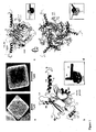
- a typical 2-micron crystal ( Fig. 1a ) has 200 body-centred cubic unit cells along each edge and contains approximately ten thousand virus particles. With a cell edge of 103 ⁇ , the unit cells of polyhedra are far smaller than a 720 ⁇ diameter icosahedral CPV particle. Electron microscopy revealed that virus particles are uniformly distributed in polyhedra and occupy holes in the lattice in place of at least 300 unit cells.
- the atomic structures were determined for polyhedra with and without CPV particles and also for polyhedra where the virus particles were replaced by a protein kinase through fusion with the polyhedra-targeting sequence of the CPV turret protein. No significant difference was observed and the refined structures are essentially identical. These observations demonstrate that cargos of strikingly different molecular weights can be stably incorporated into polyhedra without disrupting the overall order of the crystalline lattice.
- Polyhedra are made from trimeric building blocks of the 28 kDa polyhedrin protein interlocked into a tight scaffold generated by the N-terminal ⁇ helix.
- the fold of polyhedrin has the shape of a left hand with the thumb and index finger outstretched.
- the finger is an N-terminal ⁇ helix (H1) which extends from the fist formed by a compact 3-layer ⁇ sandwich core.
- This sandwich is made up of two ⁇ sheets of six (S1) and three (S2) anti-parallel strands ( Fig. 1b ).
- the topology of the S1-S2 sandwich is akin to the canonical jelly-roll found in many viral capsid proteins (BIDG-CHEF) although the exchange between strands A and I is novel.
- the third layer (S3) is formed by the C-terminal part of the protein and consists of a ⁇ hairpin preceded by a short ⁇ helix. S3 packs against the concave face of S1 forming a deep groove above S1 in the orientation shown in Fig. 1b .
- the central sandwich is flanked by additional helices with the H3 helix and adjacent loops forming a clamp-like protrusion above S1-S2 represented as the little finger in Fig 1B .
- the thumb is an amphipathic helix (H4) located next to thekafold axis.
- the H1helix, S1/S3 groove, clamp and H4 helix are all involved in the higher organization of polyhedra.
- the polyhedrin trimers are composed of a central barrel in which the sandwich domains are held together by a bundle of H4 helices and the three clamps ( Fig. 1 c-d ). They are formed mainly through hydrophobic interactions with over 25% (3800 ⁇ 2) of the total surface of a polyhedrin molecule buried in the trimer interfaces ( Fig. 5 ). The trimers have 3-fold crystallographic symmetry so that one polyhedrin molecule constitutes the asymmetric unit of the crystal.
- Polyhedrin has a similar central ⁇ sandwich and overall trimeric shape to capsid proteins of double-stranded RNA viruses such as VP7 of bluetongue virus (Grimes et al 1995), Mu1 of reovirus (Liemann et al 2002) or VP2 of birnavirus (Coulibaly et al 2005).
- capsid proteins of double-stranded RNA viruses such as VP7 of bluetongue virus (Grimes et al 1995), Mu1 of reovirus (Liemann et al 2002) or VP2 of birnavirus (Coulibaly et al 2005).
- the quaternary arrangement in the trimer is different from these viral capsid proteins and polyhedrin trimers are organized into a 3-D body-centered cubic lattice, rather than an icosahedral shell.
- the next level of organization in polyhedra is a tetrahedral cluster of four trimers at the centre of the 123 unit cell. All 12 polyhedrin molecules in this tetrahedral cluster are oriented with the thumbs pointing away from the centre of the cell and are linked together predominantly by interactions between the clamp regions ( Fig. 2a ). Two of these tetrahedral clusters constitute the unit cell of the crystal which is repeated a few hundred times along each axis to form micron-size cubic polyhedra. The tetrahedral clusters are tightly cross-linked in polyhedra essentially by non-covalent interactions mediated by the H1 helices ( Fig. 2b-c ).
- H1 helices protruding from the tetrahedral clusters dock into the S1/S3 grooves of trimers of surrounding clusters ( Fig. 2 finger-fist interactions). These are the most extended and intimate crystal contacts stabilizing polyhedra.
- a bundle of H1 helices around crystallographic dyads further strengthens the crystal through long-range interactions between tetrahedral clusters ( Fig. 2 finger-finger interactions).
- polyhedra The stability of polyhedra stems from an extensive network of interactions connecting the trimers and shielding over 70% of the polyhedrin surface from solvent.
- each polyhedrin contacts eighteen other molecules, more than in any other protein crystal with the same 123 symmetry (the average is 7.7).
- Some of these interactions are comparable in their extraordinar chemical and shape complementarity to antigen-antibody complexes Wodak et al 2002) (Surface Complementarity factor (Lawrence et al 1993) 0.70 for polyhedra and 0.64-0.68 for immune complexes).
- the H1 helix plays a central role in the architecture of polyhedra evident from the extent of crystal contacts that it mediates with three-quarters of its surface involved in intimate contacts with four neighbouring trimers ( Fig. 5 ).
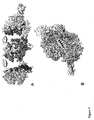
- polyhedrin molecules also results in one of the lowest solvent contents reported for protein crystals (19%). It is interrupted only by narrow channels between unit cells, along and perpendicular to the crystallographic dyads, and by closed cavities around each lattice point, at the centre of the tetrahedral clusters ( Fig. 3 ). The axial channels are narrow and blocked by H1 helices and the extensions of the cavity along the 3-fold axes of the crystal are blocked by H4 helices ( Fig. 6 ). Polyhedra thus form a sealed matrix shielding embedded virus particles from the external environment. Polyhedra are exceptionally dense protein crystals with one of the lowest solvent content observed in the Protein Data Bank (19% including nucleotides, 22% for the protein only).
- the tight packing of polyhedrin in the crystal leaves narrow channels and a central cavity.
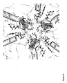
- the central cavity is located at each lattice point and contains twelve CTP molecules.
- the cavity is lined by the clamp regions of 12 polyhedrin molecules.
- the outer diameter is 28.4 ⁇ (distance Gly74-Gly74) and the inner diameter is 18.2 ⁇ (distance His76-His76). His76 is the only residue pointing inside the cavity and neighboring residues are all involved in the stabilizing the conformation of the loop 69-78.
- nucleotides at strategic places in both recombinant and infectious polyhedra. These nucleotides were determined to be ATP, GTP and CTP by mass spectrometry.
- these ligands have not been described previously and suggest that polyhedra can be modified to carry other small molecules such as drugs or fluorescent probes. Unlike existing virus-like nanoparticles (Douglas and Young 2006), these nanocontainers are easy to manipulate because of their size and strength, and they accommodate a wide range of cargos as shown here and in novel microarrays (Ikeda et al 2006) or promising bio-pesticides (Chang et al 2003).
- nanocontainers Another attractive feature of these nanocontainers is the possibility that the cargo can be released at alkaline pH. Despite their striking stability even at pH 2, polyhedra are primed for disassembly and dissolve readily above pH 10.5, releasing the virus particles in the alkaline midgut of larvae after ingestion. The difference in stability between low and high pH suggests that loss of hydrogen bonds and salt bridges is not sufficient to account for the dissolution of the crystals.
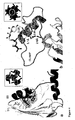
- a virus-release mechanism where deprotonation of a buried cluster of tyrosines (pKa ⁇ 10.1) leads to partial unfolding of S3 and loss of the S1/S3 grooves, destabilizing the crystal because of their critical role in binding H1 helices ( Fig. 4 ).
- disruption of a network of salt bridges at high pH destabilizes the clamps by the introduction of unpaired buried charges and the loss of crucial inter-trimer and nucleotide-binding interactions ( Fig.4 ).
- the polyhedrin forms unique intracellular crystals and has no close structural homologues.
- NPV nucleopolyhedrovirus
- Baculoviridae Baculoviridae
- CPV polyhedra a nucleopolyhedrovirus
- This protein is also predicted to have a long N-terminal helix comparable to the H1 helix which forms the scaffold of CPV polyhedra (Anduleit et al 2005).
- the architecture of polyhedra may therefore be shared by members of the Reoviridae and Baculoviridae families despite considerable evolutionary distance and fundamental differences in the life cycles of these viruses.
- cytokines protein molecules necessary to simulate the growth and differentiation of cells in culture. Like most protein molecules, cytokines are comparatively unstable, so cell culture media must be frequently replenished. For this reason alternative long-lasting cell culture systems containing stably immobilized growth factors potentially have important applications. Stably immobilized growth factors can be considered to be useful for slow release of cytokines and for tissue engineering and regenerative medicine by using many stem cells including embryonic stem (ES) and induced pluripotent stem (iPS) cells.
- ES embryonic stem
- iPS induced pluripotent stem
- a target molecule in a polyhedron according to the invention for stable long-lasting release of the target molecule into a cell culture system.
- the target molecule can comprise any molecule useful for eliciting a physiological response in a cell comprising but not limited to survival, growth, proliferation, phenotypic transformation, differentiation, or chemotaxis.
- a molecule that facilitates the expression of proteins from cells in culture systems can also be incorporated into a polyhedron and used in cell culture systems.
- target molecules may comprise but are not limited to a polypeptide, a protein, a glycoprotein, a carbohydrate, a nucleotide, a nucleic acid, a lipid, a lipoprotein, a drug, a cytokine, an antigen, an antibody, an antibody fragment, a fluorescent molecule, a dye, a pH sensitive molecule a toxin, a venom and a bioactive molecule, a growth factor a chemokine or a mitogen.
- modified polyhedron comprising a target molecule can introduced into a cell culture system by a number of methodologies comprising but not limited to adding modified polyhedra containing a target molecule into culture medium or applying modified polyhedra containing a target molecule to a solid support surface which can be incorporated into a cell culture system for contact with a culture medium. This can include attachment of modified polyhedra to the bottom of a well of a tissue culture plate, or to a surface adapted for inclusion in any cell culture system such as a fermentation culture or bioreactor.
- Crystals were spread on micromesh mounts (Thome et al 2003) leaving a thin film of 50% ethylene glycol and data were collected at 120K on the MD2 diffractometer of the X06SA beamline (Swiss Light Source) with a strongly attenuated beam focused onto the crystal. Data were processed with Denzo/Scalepack (Otwinowski et al 1997) in space group I23 and datasets from 2-4 crystals were merged because of severe radiation damage. Successful heavy atom soaks were performed on recombinant polyhedra at high concentrations, usually half saturation in appropriate buffer.
- oligomeric and crystal contact interfaces were characterized with AREAIMOL from CCP428 and the PISA29 server at the European Bioinformatics Institute. Solvent cavities and channels were analyzed with VOID0030 (Uppsala Software Factory). Illustrations were prepared with PyMOL v 0.99 (DeLano Scientific, San Carlos, CA, USA. http://www.pymol.org ).
- H1-EGFP fusion was designed to contain the N-terminal 30 amino acids of polyhedrin domain linked to an EGFP domain.
- a pEU-polyhedrin plasmid was amplified with the forward primer:, SEQ ID NO. 1 and reverse primer: SEQ ID NO 2.
- the forward primer contained EcoRI site while reverse primer contained BamHI site.
- This resulting PCR fragment was digested with EcoRI-BamHI and ligated into the EcoRI-BamHI site of pEGFP-N3 (CLONTECH) to create pH1EGFP.
- the H I EGFP construct was generated using the Gateway system (Invitrogen). These pEU-H76C and pH1EGFP plasmids were amplified by PCR using sets of primers, for H1EGFP: SEQ ID NO 3 and SEQ ID NO 4 containing an additional att B sequences for the BP recombination reaction. The amplified PCR fragment was introduced into plasmid vector pDONR221 (Invitrogen) via the BP recombination reaction, according to the recommendation of the manufacturer, to generate the entry clone pDNR-H1EGFP. The construct was then used to perform an LR recombination reaction with the Gateway system compatible expression ("DEST”) vector pDEST.
- DEST Gateway system compatible expression
- the resultant vector pDEST-H1EGFP was named the resultant vector pDEST-H1EGFP.
- the integrity of the constructs was confirmed by DNA sequencing (the PE Applied Biosystems Model 373A automated sequencer).
- the expression vector was co-transfected into Sf21 cells together with linearized AcNPV DNA (BaculoGold Baculovirus DNA, PharMingen). After incubation at 27 °C for 4 days, the culture medium was collected and centrifuged at 1000g for 10min, and the supernatant was used to infect more cells for amplification. Amplification of the recombinant virus was repeated three times, and a large amount of supernatant containing the recombinant baculovirus was obtained.
- the recombinant baculovirus obtained using pDEST-H1EGFP was designated AcH1EGFP.
- Sf21 cells (5x10 7 cells per 75 cm 2 flask) were inoculated with the recombinant virus at a multiplicity of infection (MOI) of 10 PFU/cell. For double infections, each virus was added at an MOI of 5 PFU/cell.
- MOI multiplicity of infection
- H1GFP-immobilized polyhedrin were isolated from the infected Sf21 cells and purified as described previously (Ikeda, 2006).
- H1EGFP was purified from Sf21 cells 5 days after infection with H1EGFP recombinant baculovirus by ion-exchange chromatography. Briefly, to prepare the soluble extracts, the cells were collected by centrifugation and lysed by sonication in a PBS buffer. Protein purification was carried out by the following steps: (1) ion-exchange chromatography on DEAE Sepharose with NaCl gradient elution (0-0.2 M), and (2) gel filtration on Sephadex G-75. All chromatography was performed in 20mM Tris buffer, pH 7.5, and the fraction containing H1EGFP were determined by the visible green fluorescence. The protein was dialyzed against 10mM Tris (pH 7.0), and concentrated using centrifugal concentrators (Millipore, Centricon YM1000).
- H1EGFP was crystallized using hanging-drop vapor diffusion method at room temperature by mixing 2 ⁇ L of H1EGFP with an equal volume of reservoir solution containing 1.5M ammonium sulfate and 15% glycerol and 100mM Tris, pH 8.5. Small crystals allow to grow for about 3days.
- Sf21 cells were infected with two recombinant baculoviruses AcH1EGFP and AcCP-H.
- Another polyhedra (VP3EGFP polyhedra) were also prepared by co-infection with AcVP3EGFP and AcCP-H.
- the polyhedra were recovered from the infected cells and purified as previously reported (Ikeda et al. (2001).
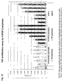
- the green fluorescence of H1EGFP polyhedra was compared with that of VP3EGFP polyhedra. The results are shown in Figure 10 .
- H1EGFP was expressed in Sf21 cells and purified by ion-exchange chromatography on DEAE Sepharose with NaCl gradient elution (0-0.2 M), and (2) gel filtration on Sephadex G-75.
- the purified H1EGFP was subjected to crystallization by using hanging-drop vapor diffusion method. Supplementary Information is linked to the online version of the Nature publication at www.nature.com/nature.
- stable CPV polyhedra containing the growth factor FGF-2 coupled with the H1 tag are shown to be active in stimulating growth in a cell culture assay using NIH 3T3 cells. Negative and positive control experiments were carried out to validate the experiment. The results are shown in Figure 12 .
- a nucleotide sequence of N-terminal alpha helix (H1) of polyhedrin derived Bombyx mori CPV (BmCPV) SEQ ID NO 5 was cloned into Not I and EcoR I site of a baculovirus transfer vector pVL1392 using standard PCR and cloning techniques well known in the art to produce a plasmid designated pVL1392/H1.
- the nucleotide sequence of the H1 corresponds to an amino acid sequence shown in SEQ ID NO 6: GATEWAY cloning technology (Invitrogen) was used to facilitate the high throughput expression and immobilization of EGFP, FGF-2 and FGF-7.
- a destination vector (pDEST/N-HI) was constructed by inserting a reading frame cassette B (Invitrogen) into the Xba I site of pVL1392 (Invitrogen).
- the FGF-2 gene was amplified by PCR from the full-length human cDNA library, in which the attB1 and attB2 sequence was included in the primers.
- the resulting attB-flanked PCR products were cloned into a donor vector (pDONR221) by BP reactions of GATEWAY cloning technology using the BP clonase enzyme mixture.
- the ORFs were cloned between the attL1 and attL2 sites of the resulting entry vectors and were transferred to pDEST/H1 via LR clonase reactions with the LR clonase enzyme mixture.
- the resulting expression vector (pAcH1) consisted of the N-terminal H1 sequence of BmCPV polyhedrin and foreign protein genes co-transfected into Sf21 cells with linearized AcNPV DNA (Baculogold Baculovirus DNA (Pharmingen)).
- the recombinant AcNPVs (AcH1/EGFP, AcH1/FGF-2, AcH1/FGF-7) were co-infected with AcCP-H which expressed BmCPV polyhedrin.
- the infected cells were collected, washed with phosphate-buffered saline (PBS; 20 mM NaH 2 PO 4 , 20 mM Na 2 HPO 4 , 150 mM NaCl, pH7.2) and homogenized to remove the cell membrane and nuclear envelope using the Ultrasonic Homogenizer VP-5S (TAITEC).
- the EGFP-polyhedra were purified by washing with sterile water.
- the FGF-2 polyhedra were then washed with sterile water containing penicillin (10,000 unit/ml) and streptomycin (10 mg/ml) by centrifugation at 15,000 rpm for 10 min.
- the FGF-2 was immobilized into the BmCPV polyhedra by using H1 sequence (H1/FGF-2 polyhedra).
- H1 sequence H1/FGF-2 polyhedra.
- the biological activity of FGF-2 immobilized into the polyhedra was assayed as follows.
- NIH3T3 cells were plated into a 96-well plate at a density of 4000 cells/well and cultured overnight in DME medium containing 10% fetal calf serum and then starved in alpha-MEM without fetal calf serum for 6h, resulting in cessation of cell growth..
- the starved cells were then cultured in alpha-MEM containing either unmodified polyhedra, FGF-2 polyhedra (0.1 x 10 4 to 1 x10 5 /4000 cells) or rFGF-2 (R & D systems) (0.1 ng to 100 ng) in the absence of fetal calf serum and for 4 days.
- Media containing rFGF-2 was replenished with fresh media containing rFGF-2 every two days.
- the proliferation of the NIH3T3 cells was measured by the WST-8 assay as known in the art. Briefly, WST-8 is reduced by dehydrogenases present in cells to give a yellow colored product (formazan), which is soluble in the tissue culture medium. The amount of the formazan dye generated by the activity of dehydrogenases in cells is directly proportional to the number of living cells.
- Cell-Counting Kit-8 solution Dojin Chemical Co., Ltd.
- Absorbance at 450 nm was determined using a microplate reader.
- the H1/FGF-2 polyhedra stimulated the proliferation of the starved NIH3T3 cells in a dose dependent manner comparable with the proliferation detected in cells treated with rFGF-2. Specifically, proliferation of the NIH3T3 cells induced by the addition of 2 x 10 4 H1/FGF-2 polyhedra was very similar to that of cells treated with 10 ng rFGF-2. NIH3T3 cell growth was unaffected by treatment with unmodified polyhedra. The. These results are shown in Figure 12 .
- stable CPV polyhedra containing the growth factor FGF-7 coupled with the H1 tag are shown to be active in stimulating the growth of human epidermal keratinocytes in a cell culture assay. Negative and positive control experiments were carried out to validate the experiment. The results are shown in figure 13 .
- the FGF-7 was immobilized into the BmCPV polyhedra by using H1 sequence (H1/FGF-7 polyhedra) using the methods described in Example 7, above.
- the biological activity of FGF-7 immobilized into the polyhedra was assayed.
- H1/FGF-7 polyhedra (0.2 x 10 4 to 1 x 10 5 ) and unmodified polyhedra were added to the surface of the wells of a 96-well plate and allowed to dry for 3 hr at room temperature.
- NHEK(F) Human Epidermal Keratinocytes
- the frozen keratinocytes were washed with Defined Keratinocyte-SFM (GIBCO) and suspended in Keratinocyte-SFM
- the keratinocytes were plated into the wells of the 96 well plate pre-coated polyhedra as described above at a density of 2500 cell/cm 2 in Defined Keratinocyte-SFM in the absence of any growth supplement.
- the growth supplement including insulin, EGF, and FGF (GIBCO) was added to Defined Keratinocyte-SFM as indicated in Figure 13 ..
- Medium containing the growth supplement was replenished with fresh medium containing growth supplement every two days.
- the H1/FGF-7 polyhedra stimulated the proliferation of the keratinocyte in the absence of fetal calf serum and the growthsupplement.
- the proliferation of the keratinocyte by the growth supplement was comparable with the addition of 5 x 10 4 H1/FGF-7 polyhedra.
- the shape of the keratinocyte proliferated by H1/FGF-7 polyhedra was very similar to that of the keratinocyte treated with growth supplement..
- Proliferation of the cells grown in medium containing FGF-7 polyhedra was sustained in the absence of replenishing the medium containing growth supplement.
- proliferation of cells treated with medium containing growth supplement required feeding every 2 days with fresh medium containing growth supplement.
- Thermanox coverslips were spotted with normal polyhedra, H1/FGF-2 polyhedra and rFGF-2 were spotted on the Thermanox coverslips. After complete desiccation, the coverslips were placed in 8-well plates, into which ATDC5 cells were seeded at a density of 1 x10⁴ cells per well. After 2 days incubation, the effects of these treatments on the proliferation of the cells were investigated.
- NP normal polyhedral
- F2P spotted and desiccated H1/FGF-2 polyhedra
- Scale bar 100 ⁇ m.
Landscapes
- Health & Medical Sciences (AREA)
- Chemical & Material Sciences (AREA)
- Life Sciences & Earth Sciences (AREA)
- General Health & Medical Sciences (AREA)
- Organic Chemistry (AREA)
- Medicinal Chemistry (AREA)
- Engineering & Computer Science (AREA)
- Gastroenterology & Hepatology (AREA)
- Biophysics (AREA)
- Molecular Biology (AREA)
- Biochemistry (AREA)
- Genetics & Genomics (AREA)
- Proteomics, Peptides & Aminoacids (AREA)
- Zoology (AREA)
- Toxicology (AREA)
- Bioinformatics & Cheminformatics (AREA)
- Pharmacology & Pharmacy (AREA)
- Veterinary Medicine (AREA)
- Nanotechnology (AREA)
- Virology (AREA)
- Epidemiology (AREA)
- Animal Behavior & Ethology (AREA)
- Public Health (AREA)
- Plant Pathology (AREA)
- Biotechnology (AREA)
- Pest Control & Pesticides (AREA)
- Hematology (AREA)
- Dentistry (AREA)
- Wood Science & Technology (AREA)
- Environmental Sciences (AREA)
- Agronomy & Crop Science (AREA)
- General Engineering & Computer Science (AREA)
- Medical Informatics (AREA)
- Crystallography & Structural Chemistry (AREA)
- Cell Biology (AREA)
- Micro-Organisms Or Cultivation Processes Thereof (AREA)
- Peptides Or Proteins (AREA)
- Measuring Or Testing Involving Enzymes Or Micro-Organisms (AREA)
- Agricultural Chemicals And Associated Chemicals (AREA)
Description
- The present invention relates to a method of introducing target molecules into a polyhedron. The invention also relates to complexes formed by introducing target molecules into polyhedron and to polyhedrin protein variants that are useful in the methods of the invention. The invention also relates to the use of the methods, complexes and polyhedrin protein variants for the stabilization of proteins in particular, for cell culture, biosensors, microarrays, and assays.
- Stable maintenance of molecules, and in particular functional molecules, on support surfaces can be critical for analytical and diagnostic tools - for example, in high throughput analysis and functional characterization of biomolecules. However, techniques for stably immobilizing and preserving functional proteins, biomolecules and biosensors on support surfaces have not been optimised.
- For DNA chips, designing capture molecules and developing read-out systems is relatively straightforward. DNA or oligonucleotides bind specifically to complementary mRNA sequence tagged with a fluorescent dye. However, designing arrays for proteins is more complex and requires different consideration. Proteins must be immobilized on the surface of the chip such that they retain their native conformation and also such that their active site(s) are exposed rather than buried. Constructing a protein chip therefore involves more steps and more complex protein chemistry. The following are some of the issues to consider when building a useful protein chip: 1) immobilization of proteins of diverse types such that they retain their secondary and tertiary structure, and thus their biological activity; 2) identification and isolation of an agent with which to capture the proteins of interest; 3) a means of measuring the degree of protein binding, ensuring both sensitivity and a suitable range of operation; and 4) extraction of the detected protein from the chip and analysis of the protein.
- Many prophylactic and therapeutic agents require special conditions to protect them from environmental damage during transportation and storage. This adds to the cost of the drug and, in certain instances, reduced availability in some communities eg. in remote and/or disadvantaged communities where conditions such as low temperature cannot easily be achieved during transportation over long distances.
- Further, there is a continuing need for new pharmaceutical formulations, excipients, and delivery devices, including nanodevices, to achieve maximum benefit from drugs. For example, slow release and/or organ-/tissue-specific release of prophylactic and therapeutic agents can provide increased drug efficacy, improve bioavailability and reduce drug dosage. There is a need for versatile and safe means for administering agents intact and with site-specific and dose-specific accuracy.
- Insect virus infections result in the production of massive amounts of large protein crystals (occlusion bodies), termed polyhedra and many virus particles are occluded within the polyhedra. An insect virus, cypovirus (cytoplasmic polyhedrosis viruses, CPV), is classified among the family Reoviridae and has a segmented genome composed of ten double-stranded RNAs. The virion consists of an icosahedral protein shell of 50-70 nm in diameter and twelve spikes on the 12 vertices of the shell. Polyhedra are the main vectors of virus particle transmission from insect to insect and are the main agents of survival of the virus between one insect generation and the next because they stabilize virions, allowing them to remain viable for long periods in the environment. Infection occurs when an insect ingests the alkali-soluble occlusion body and the virus particles are released by the high pH of the insect intestine.
- The virus particles of CPVs and baculoviruses produce micro-sized protein crystals called polyhedra in which virus particles can be embedded (Belloncik and Mori 1998; Miller 1997). Polyhedra exhibit remarkable stability and, as such, the embedded insect viruses can remain infectious for years in the environment. The virus particles are embedded in the polyhedron via the constituent CPV envelope protein, VP3, which binds to the polyhedrin protein, the major viral protein making up the polyhedron.
- The improved stability of viral particles within the polyhedra has prompted studies investigating whether target proteins can be embedded within polyhedra for improving their stability and facilitating their use in protein microarray applications (
US20060155114 and Ikeda et al 2006). This approach involved co-expressing target proteins fused to a portion of a virion structural capsid protein, VP3 during CPV infection. The results indicated that incorporation of these proteins into the polyhedron crystal was successful and that the proteins were protected from dehydration and stabilized against high temperatures without the loss of function. Proteins that have been incorporated into polyhedra using the VP3, are non-membrane and membrane proteins including, enzymes such as polymerases, kinases and acryltransferases; structural proteins such as ribosome proteins and ribosome binding proteins; and transcription factors; and elongation factors. Accordingly; the skilled addressee would understand that a diverse range of different types of molecules may be incorporated into a polyhedron without the loss of function. - Although these unique protein crystals have been characterised since the early 1900s (Glaser and Chapman 1916) determination of their atomic structure which would allow for the further development of the polyhedra into useful diagnostic, therapeutic and research tools has been elusive. Further there has been no suggestion that exogenous molecules such as proteins can be incorporated into a polyhedron crystal in the absence of a virion structural protein tag. There has been no suggestion that molecules may be incorporated into a polyhedron in locations other than that usually occupied by virus particles.
- IKEDA ET AL.: 'Immobilization of diverse foreign proteins in viral polyhedra and potential application for microarrays' PROTEOMICS vol. 6, 2006, pages 54 - 66 is directed at cypoviruses which are insect viruses that produce a cytoplasmic crystalline particle called the polyhedron in which progeny virions are occluded. The virion structural protein, VP3, is implicated in the occlusion of viral particles into polyhedra. In this study, the amino acid sequence of VP3 required for occlusion of viral particles into polyhedra was determined.
-
US 2005/0048075 A1 describes a fusion protein containing a polyhedrin of a virus and a foreign polypeptide sequence. Also mentioned is a recombinant occlusion body containing the fusion protein, an isolated nucleic acid containing a sequence encoding the fusion protein, and a recombinant virus containing the nucleic acid. - It is an object of the present invention to overcome or ameliorate at least one of the disadvantages of the prior art, or to provide a useful alternative.
- Protein structure determination of polyhedra has been hampered by the small size of the crystals and a number of other factors. Surprisingly, the 2A crystal structures of both recombinant and infectious silkworm CPV polyhedra have now been determined using 5-12 micron crystals purified from insect cells. These are the smallest crystals yet used for de novo X-ray protein structure determination (Riekel et al 2005).
A number of features of the structure became apparent that had previously not been recognised. It was unexpectedly found that some of these features could facilitate the accommodation of target molecules within a polyhedron crystal. - It was found that polyhedra are made of trimers of the viral polyhedrin protein and contain nucleotides. Although the shape of these building blocks is reminiscent of some capsid trimers, polyhedrin has a new fold and has evolved to assemble in vivo into 3-D cubic crystals rather than icosahedral shells. The polyhedrin trimers are extensively cross-linked in polyhedra by non-covalent interactions and pack with an exquisite molecular complementarity similar to that of antigen-antibody complexes. The resulting ultra-stable and sealed crystals shield the virus particles from environmental damage. The structure suggests that polyhedra can serve as the basis for a large number of applications including the development of robust and versatile nanoparticles for biotechnological applications (Douglas and Young 2006) such as microarrays (Ikeda et al 2006) and bio-pesticides (Chang et al 2003).
- Specifically, it has been found that the crystal structure of the cypovirus polyhedra facilitates the identification of regions of the polyhedrin protein that play central roles in the architecture of the crystal structure and that attaching at least a portion of these regions to a target molecule allows the target molecule to be sequestered by the polyhedron crystal - either by incorporation into the lattice structure or by attachment to the surface of the crystal.
- As such, the present invention relates to novel complexes, methods for their production and uses thereof that may, in certain embodiments, preserve, prolong or protect the functionality of a target molecule by complexing the target molecule within a polyhedron or part thereof.
- The use of the crystal structure of the CPV polyhedra in the present invention confers advantages not previously recognised or contemplated.
- Accordingly, the invention provides:
- 1. A method of designing and/or manufacturing a modified Bombyx
mori cypovirus 1 polyhedron, or a complex comprising the polyhedron, said polyhedron or complex thereof containing a target molecule,
wherein the target molecule is attached to a portion of the Bombyx moricypovirus 1, polyhedrin protein to produce a fusion molecule that can be included in a polyhedron crystal, and
wherein said portion of the polyhedrin protein is in a region that plays a central rale in the architecture of the polyhedron crystal structure, which region is identified using co-ordinates of the polyhedron crystal structure. - 2. A method of preserving, prolonging or protecting the functionality of a target molecule, said method comprising:
- a) preparing a fusion molecule that can be included in a Bombyx mori
cypovirus 1 polyhedron crystal or part thereof, said fusion comprising the target molecule attached to a portion of the Bombyx moricypovirus 1 polyhedrin protein located in a region that plays a central rale in the architecture of the polyhedron crystal structure; wherein said region is identified using co-ordinates of the Bombyx mori cypovirus 1 polyhedron crystal structure; and - b) incorporating the fusion molecule into a polyhedron by contacting the fusion molecule with a polyhedron, or by co-expressing the fusion molecule with a polyhedron.
- a) preparing a fusion molecule that can be included in a Bombyx mori
- 3. A method as in embodiment [1] or [2], wherein the region of the polyhedrin protein that contributes to the architecture of the polyhedron crystal comprises at least a portion of an N-terminal H1 helix of the polyhedrin protein.
- 4. A method as in embodiment [3], wherein the N-terminal H1 helix comprises SEQ ID NO:6.
- 5. A modified
Bombyx mori cypovirus 1 polyhedron prepared according to the methods of embodiments [1] to [4]. - 6. A complex comprising the modified
Bombyx mori cypovirus 1 polyhedron of embodiment [5]. - 7. Use of a modified polyhedron according to embodiment [4], or of a complex according to embodiment [6], applied to a solid surface, in the production of an active surface in which the functionality of the target molecule is prolonged, protected or preserved.
- 8. Use according to embodiment[7], wherein said modified
Bombyx mori cypovirus 1 polyhedron or complex is dessicated on the active surface. - Immobilization of biomolecules is a key procedure in many biotechnological tools, including biochips and biosensors. See, e.g., Drummond et al. (2003); Zhu et al. (2003); Wulfkuhle et al. (2003); Tirumalai et al. (2003). Different approaches have been developed to anchor biomolecules on solid supports.
- Bio-recognition biosensors may be classified into immunochemical, enzymatic, non-enzymatic receptor, and DNA biosensors. Immuno-sensors are very sensitive, selective and versatile since antibodies can be generated to bind a wide range of compounds that are structurally different. In general, enzymatic biosensors are based on the selective inhibition of specific enzymes by different classes of compounds, with the decrease in activity of the immobilized enzyme in the presence of the target molecule as the parameter that is frequently used for quantification. There is a wide range of enzymes suitable for acting as recognition elements and very often their catalytic properties or substrate specificity can be modified by means of genetic engineering. Biosensors based on natural receptors can be built by integrating the specific receptor within a membrane and by coupling it to a transducing device. These natural receptors are proteins of non-catalytic or non-immunogenic origin, which span cell membranes and can specifically bind certain compounds. One of the limitations of these biosensors is that the biosensor reaction takes place on a solid support platform and there is a need to enhance the stability or preserve the functionality of bioactive molecules on these solid supports.
- The advantage of the polyhedra biosensor and bioassay disclosed herein lies in the stabilisation and/or the maintenance of active target molecules on a support platform. This would involve the complex comprising the polyhedra-embedded target molecule being bound to a support surface and binding to, or interacting with, molecules in a biological or environmental sample then detecting the binding or interaction.
- The identification, analysis and monitoring of biological molecules (such as polypeptides, polynucleotides, polysaccharides and the like) or environmental molecules (such as pesticides, bio-warfare agents, food contaminants and the like) has become increasingly important for research and industrial applications. Conventionally, molecule detection systems are based on molecule-specific binding between a molecule and an molecule-binding receptor. Such systems typically require complex multi-component detection systems (such as ELISA sandwich assays) or electrochemical detection systems, or require that both the molecule and the receptor are labelled with detection molecules
- One method for detecting molecule-binding agent interactions involves a solid phase format employing a reporter labelled molecule-binding agent whose binding to, or release from, a solid surface is dependent on the presence of molecule. In a typical solid-phase sandwich type assay, for example, the molecule to be measured is a molecule with two or more binding sites, allowing molecule binding both to a receptor carried on a solid surface (the target molecule in the polyhedra), and to a reporter-labelled second receptor. The presence of the molecule is detected based on the presence of the reporter bound to the solid surface.
- Also described is a bio-pesticide comprising a modified polyhedron or part thereof according to the invention or a complex according to the invention. For the bio-pesticide embodiment the target molecule is is detrimental to a host insect. In a particularly preferred embodiment the target molecule is a toxin from Bacillus thuringiensis.
- Three strategies have been used to improve the efficacy of insect viruses as pesticides.
- (a) Inactivation or deletion of genes from the viral genome that prolong the life of the host (e.g.,
U.S. Pat. No. 5,858,353 ). - (b) Use of the virus to stimulate inappropriate expression of host genes to deregulate physiological or developmental processes reviewed in Black et al., "Commercialization of Baculoviral Insecticides" in Miller, L. (ed.), The Baculoviruses, 1997)
- (c) Engineering insect viruses to express insecticidal toxins, eg venom of natural host predators.
- In contrast, a polyhedra complex according to the present invention could comprise insecticide toxin and may be sufficient to kill the host insect without the initiation of viral replication.
- In the context of the present invention, the term "target molecule" includes, but is not limited to, a polypeptide, a protein, a glycoprotein, a carbohydrate, a nucleotide, a nucleic acid, a lipid, a lipoprotein, a drug, a cytokine, an antibody, an antibody fragment, a fluorescent molecule a dye, a pH sensitive molecule or a bioactive molecule, a growth factor a chemokine or a mitogen.
- In the context of the present invention, the term "complex" includes, but is not limited to, the linkage of two or more agents by covalent or non-covalent binding, by ionic or non-ionic binding or by van der Waals forces.
- In the context of the present invention, the words "comprise", "comprising" and the like are to be construed in their inclusive, as opposed to their exclusive, sense, that is in the sense of "including, but not limited to".
-
-
Figure 1 Compact trimers are the building blocks of polyhedra.
a, Scanning electron micrographs of recombinant (left) and infectious (right) polyhedra. Both crystals are made up of densely packed 103Å unit cells (Fig. 2 and3 ) but the indentations on the surface of infectious polyhedra are due to virus particles which replace blocks of at least 300 unit cells. b, Polyhedrin has the shape of a left hand (inset). The finger (H1, blue), thumb (H4, orange), fist (S1-S2, cyan and S3, red) and clamp (H3, green) all contribute to the organization of polyhedra. c and d, The polyhedrin trimer in two orthogonal views with bound nucleotides shown in a yellow surface. The insets represent trimers with one of the subunits highlighted. -
Figure 2 Polyhedra are built around a tight scaffold of H1 helices.
a, Four polyhedrin trimers assemble around each lattice point of polyhedra to form a central tetrahedral cluster (blue hands). These clusters then pack tightly to form the bodycentered cubic crystal. b, c Schematic and molecular models of the crystal packing along the 3-fold axis of a trimer. The eight trimers of one 123 unit cell are highlighted in b. Two of the non-covalent interactions cross-linking trimers in polyhedra are shown as diamonds (finger-fist interactions) and circles (finger-finger interactions). -
Figure 3 Polyhedra are dense and sealed microcrystals containing nucleotides.
a, The continuous protein matrix of the unit cell is represented as a brown surface and grey spheres. The dense packing leaves only narrow solvent channels and a central cavity (red surface) blocked by H1 helices (blue cylinders). These blocks are repeated along the lattice, interrupted by the presence of virus particles. b, Mesh representation of the solvent channels and the central cavity. The ATP and GTP molecules located in the channels and the CTP molecules bound in the central cavity are represented as sticks. H1 and H3 helices are shown as blue and green cylinders respectively. -
Figure 4 Proposed mechanism for the release of virus particles
Unpaired buried negative charges are introduced at pH>10.5 in a tyrosine cluster found between S1 and S3 (a) and in a chain of salt bridges (residues K69-E82-K99-D81-D78) of the clamp (b). These regions, circled in the insets, are important in the packing of polyhedra and we propose that their disruption leads to dissolution of the crystal and release of the virus particles in the alkaline mid-gut of larvae. -
Figure 5 . Polyhedra are made from trimeric building blocks cross-linked by helix H1
a, Surface representation of the three subunits of a polyhedrin trimer. Two of the subunits have been rotated by +/-120° around the axes indicated by vertical bars to reveal the contact interfaces. These interfaces are coloured in red in the rotated subunits and coloured in blue and orange in the central polyhedrin molecule for residues involved in polar and hydrophobic interactions respectively. b, Helix H1 of a polyhedrin molecule highlighted in blue. Five different trimers are cross-linked by H1. -
Figure 6 . The central cavity.
The central cavity viewed along a crystallographic 3-fold axis. The side chains of His76 and Phe201 are represented as sticks in molecular spheres. Helices H4 blocking the connection between neighbouring cavities are shown as orange cylinders. CTP molecules, shown as sticks, trapped in the cavity are bound at the N-terminal end of helices H3 (green cylinder). For clarity, the six front CTP were omitted and carbon atoms are yellow for the protein and cyan for CTP molecules. -
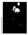
Figure 7 H1 derived occlusion of GFP -
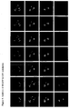
Figure 8 Encapsulation of a H1/EGFP in the polyhedra by co-expression of the H1/EGFP with polyhedrin protein. -
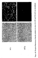
Figure 9 Encapsulation of an H1/EGFP fusion molecule in polyhedra by incubation with polyhedra. -
Figure 10 The green fluorescence from the polyhedra in which H1/GFP and VP3/GFP was immobilized. -
Figure 11 Crystallization of H1-GFP.1.5M Ammonium Sulfate, 15% Glycerol, 0.1M Tris-HCl pH8.5 -
Figure 12 Hl/FGF-2 polyhedra stimulate the growth of epithelial cells.
Bar graphs showing growth stimulation of starved NIH3T3 cells in the absence of fetal calf serum when incubated with H1/FGF-2 polyhedra (centre graph), but not when incubated with control polyhedra (left graph, negative control experiment). H1/FGF-2 polyhedra function as a stable replacement for recombinant FGF-2 with equivalent activity in this assay (right graph, positive control experiment). -
Figure 13 H1/FGF-7 polyhedra stimulate the growth of keratinocytes.
Bar graphs showing growth stimulation of human epidermal keratinocytes (NHEK cells) with H1/FGF-7 polyhedra (centre graph) to levels similar to that achieved using fetal calf serum supplement (right graph, positive control experiment). Incubation with control polyhedra does not stimulate growth (left graph, negative control experiment). -
Figure 14 Stability of H1/FGF-2 immobilized into the polyhedra after desiccation. Thermanox coverslips were spotted with unmodified polyhedra, H1/FGF-2 polyhedra and rFGF-2. After complete desiccation, the coverslips were placed in 8-well plates, into which ATDC5 cells were seeded at a density of 1x10⁴ cells per well. After 2 days incubation, the effects of these treatments on the proliferation of the cells were investigated. NP, normal polyhedral; F2P, spotted and desiccated H1/FGF-2 polyhedra; dF2, spotted and desiccated rFGF-2; F2, rFGF-2. - CPV and baculoviruses are notoriously difficult to eradicate because the virus particles are embedded in micron-sized protein crystals called polyhedra (Belloncik and Mori 1998 and Miller 1997). The remarkable stability of polyhedra means that like bacterial spores these insect viruses remain infectious for years in soil. The environmental persistence of polyhedra is the cause of significant losses in silkworm cocoon harvests but has also been exploited against pests in biological alternatives to chemical insecticides (Summers 2006; Chang et al 2003). Although these unique in vivo protein crystals have been extensively characterized since the early 1900s (Glasser and Chapman 1916), their atomic organization remained elusive (Rohrmann 1986).
- CPVs are members of the Reoviridae, a virusfamily with characteristic icosahedral subviral particles that function as small 'replication machines' transcribing viral mRNAs from internal, segmented double-stranded RNA templates in the cytoplasm of infected cells (Fields et al 2001). CPV are the only known members of the Reoviridae where the virus particles are protected by polyhedra, each of which contains several thousand CPV particles (Bellonicik and Mori; 1998; Miller 1997; Rohrmann 1986; Zhang et al 1999; Hill et al 1999).
- In the extracellular environment, virus particles embedded in polyhedra survive dehydration, freezing and enzymatic degradation (Rohrmann 1986). In addition, we found that polyhedra tolerate chemical treatments that would denature most proteins, including incubation in concentrated urea, acid and detergents, and still maintain their ordered crystalline organization (Table 1). By contrast, polyhedra dissolve readily when exposed to pH higher than 10.5 (Lawrence, M. C. & Colman 1993). The molecular basis of the remarkable stability of these in vivo crystals and how they are primed for disassembly in the alkaline mid-gut of insect larvae is unknown.
- We developed methods for analyzing polyhedra suspended in thin layers of cryoprotectant using low X-ray background micromesh mounts (Thorne et al 2003) and were able to observe single-crystal diffraction using the MD2 diffractometer at the X06SA beamline at the Swiss Light Source. Over 300 microcrystals purified from insect cells were tested to determine the atomic structure of CPV polyhedra to a resolution of 2Å. To our knowledge, this is the first report of the atomic structure of functional intracellular crystals (Doye and Poon 2006).
- The micron-sized polyhedra are the relevant biological assemblies and thus the polyhedron protein functions as a three-dimensional crystal, rather than as a single molecule or oligomer. A typical 2-micron crystal (
Fig. 1a ) has 200 body-centred cubic unit cells along each edge and contains approximately ten thousand virus particles. With a cell edge of 103Å, the unit cells of polyhedra are far smaller than a 720Å diameter icosahedral CPV particle. Electron microscopy revealed that virus particles are uniformly distributed in polyhedra and occupy holes in the lattice in place of at least 300 unit cells.Table 1 Stability of polyhedra Incubation Time Diffraction 8M Urea 2 days Yes Detergents 10 % SDS 2 days Yes Acids 20% acetic acid, pH 1.9 2 days Yes 1M HCl 2 days No Bases 20mM NaH2CO3, pH 10.5 <1 min Dissolved 10mM PBS, pH 11.0 <1 min Dissolved Solvent 100% Ethanol 30 min Yes Physical stress Dehydration* 24h Yes Freeze-Thaw - Yes 90°C 16h No * Polyhedra in water were left to dry on a micromesh mount at
relative humidity of 50% and frozen in liquid nitrogen without
rehydration or cryoprotectant. - The atomic structures were determined for polyhedra with and without CPV particles and also for polyhedra where the virus particles were replaced by a protein kinase through fusion with the polyhedra-targeting sequence of the CPV turret protein. No significant difference was observed and the refined structures are essentially identical. These observations demonstrate that cargos of strikingly different molecular weights can be stably incorporated into polyhedra without disrupting the overall order of the crystalline lattice.
- Structures of polyhedrins from infectious, recombinant and kinase-containing polyhedra have been deposited on 9 January 2007 in the RCSB Protein Data Bank with accession codes 2OH5, 2OH6 and 2OH7.
- Polyhedra are made from trimeric building blocks of the 28 kDa polyhedrin protein interlocked into a tight scaffold generated by the N-terminal α helix. The fold of polyhedrin has the shape of a left hand with the thumb and index finger outstretched. The finger is an N-terminal α helix (H1) which extends from the fist formed by a compact 3-layer β sandwich core. This sandwich is made up of two β sheets of six (S1) and three (S2) anti-parallel strands (
Fig. 1b ). The topology of the S1-S2 sandwich (IBADGF-CHE) is akin to the canonical jelly-roll found in many viral capsid proteins (BIDG-CHEF) although the exchange between strands A and I is novel. The third layer (S3) is formed by the C-terminal part of the protein and consists of a β hairpin preceded by a short α helix. S3 packs against the concave face of S1 forming a deep groove above S1 in the orientation shown inFig. 1b . The central sandwich is flanked by additional helices with the H3 helix and adjacent loops forming a clamp-like protrusion above S1-S2 represented as the little finger inFig 1B . The thumb is an amphipathic helix (H4) located next to the 3-fold axis. The H1helix, S1/S3 groove, clamp and H4 helix are all involved in the higher organization of polyhedra. - The polyhedrin trimers are composed of a central barrel in which the sandwich domains are held together by a bundle of H4 helices and the three clamps (
Fig. 1 c-d ). They are formed mainly through hydrophobic interactions with over 25% (3800Å2) of the total surface of a polyhedrin molecule buried in the trimer interfaces (Fig. 5 ). The trimers have 3-fold crystallographic symmetry so that one polyhedrin molecule constitutes the asymmetric unit of the crystal. Polyhedrin has a similar central β sandwich and overall trimeric shape to capsid proteins of double-stranded RNA viruses such as VP7 of bluetongue virus (Grimes et al 1995), Mu1 of reovirus (Liemann et al 2002) or VP2 of birnavirus (Coulibaly et al 2005). However, the quaternary arrangement in the trimer is different from these viral capsid proteins and polyhedrin trimers are organized into a 3-D body-centered cubic lattice, rather than an icosahedral shell. - The next level of organization in polyhedra is a tetrahedral cluster of four trimers at the centre of the 123 unit cell. All 12 polyhedrin molecules in this tetrahedral cluster are oriented with the thumbs pointing away from the centre of the cell and are linked together predominantly by interactions between the clamp regions (
Fig. 2a ). Two of these tetrahedral clusters constitute the unit cell of the crystal which is repeated a few hundred times along each axis to form micron-size cubic polyhedra. The tetrahedral clusters are tightly cross-linked in polyhedra essentially by non-covalent interactions mediated by the H1 helices (Fig. 2b-c ). Firstly, H1 helices protruding from the tetrahedral clusters dock into the S1/S3 grooves of trimers of surrounding clusters (Fig. 2 finger-fist interactions). These are the most extended and intimate crystal contacts stabilizing polyhedra. Secondly, a bundle of H1 helices around crystallographic dyads further strengthens the crystal through long-range interactions between tetrahedral clusters (Fig. 2 finger-finger interactions). - The stability of polyhedra stems from an extensive network of interactions connecting the trimers and shielding over 70% of the polyhedrin surface from solvent. In polyhedra, each polyhedrin contacts eighteen other molecules, more than in any other protein crystal with the same 123 symmetry (the average is 7.7). Some of these interactions are comparable in their exquisite chemical and shape complementarity to antigen-antibody complexes Wodak et al 2002) (Surface Complementarity factor (Lawrence et al 1993) 0.70 for polyhedra and 0.64-0.68 for immune complexes). In particular, the H1 helix plays a central role in the architecture of polyhedra evident from the extent of crystal contacts that it mediates with three-quarters of its surface involved in intimate contacts with four neighbouring trimers (
Fig. 5 ). - The dense packing of polyhedrin molecules also results in one of the lowest solvent contents reported for protein crystals (19%). It is interrupted only by narrow channels between unit cells, along and perpendicular to the crystallographic dyads, and by closed cavities around each lattice point, at the centre of the tetrahedral clusters (
Fig. 3 ). The axial channels are narrow and blocked by H1 helices and the extensions of the cavity along the 3-fold axes of the crystal are blocked by H4 helices (Fig. 6 ). Polyhedra thus form a sealed matrix shielding embedded virus particles from the external environment. Polyhedra are exceptionally dense protein crystals with one of the lowest solvent content observed in the Protein Data Bank (19% including nucleotides, 22% for the protein only). The tight packing of polyhedrin in the crystal leaves narrow channels and a central cavity. The central cavity is located at each lattice point and contains twelve CTP molecules. The cavity is lined by the clamp regions of 12 polyhedrin molecules. The outer diameter is 28.4Å (distance Gly74-Gly74) and the inner diameter is 18.2Å (distance His76-His76). His76 is the only residue pointing inside the cavity and neighboring residues are all involved in the stabilizing the conformation of the loop 69-78. - Extensions of the cavity along crystallographic three-fold axis are blocked by helix H4 and in particular the residue Phe201. The smallest diameter (9Å) of these extensions is defined by residues Tyr71 and Arg98-Asp96.
- Interestingly, we found nucleotides at strategic places in both recombinant and infectious polyhedra. These nucleotides were determined to be ATP, GTP and CTP by mass spectrometry.
- Stacked ATP and GTP molecules are bound at the interface between four tetrahedral clusters and a CTP molecule interacts with each of the twelve symmetry-related clamp regions lining the central cavity (
Fig. 1c-d ,3b andFig. 6 ). The specific interactions found at these two nucleotide binding sites near symmetry axes suggest that the nucleotides bind during crystallization and may play a role in nucleating the formation of polyhedra in the cytoplasm. - Once polyhedra are formed, the nucleotides are trapped in the crystal and were observed in crystals stored for three years in water.
- These ligands have not been described previously and suggest that polyhedra can be modified to carry other small molecules such as drugs or fluorescent probes. Unlike existing virus-like nanoparticles (Douglas and Young 2006), these nanocontainers are easy to manipulate because of their size and strength, and they accommodate a wide range of cargos as shown here and in novel microarrays (Ikeda et al 2006) or promising bio-pesticides (Chang et al 2003).
- Another attractive feature of these nanocontainers is the possibility that the cargo can be released at alkaline pH. Despite their striking stability even at
pH 2, polyhedra are primed for disassembly and dissolve readily above pH 10.5, releasing the virus particles in the alkaline midgut of larvae after ingestion. The difference in stability between low and high pH suggests that loss of hydrogen bonds and salt bridges is not sufficient to account for the dissolution of the crystals. - Instead, without wishing to be bound by theory, we propose a virus-release mechanism where deprotonation of a buried cluster of tyrosines (pKa ~ 10.1) leads to partial unfolding of S3 and loss of the S1/S3 grooves, destabilizing the crystal because of their critical role in binding H1 helices (
Fig. 4 ). We also propose that disruption of a network of salt bridges at high pH destabilizes the clamps by the introduction of unpaired buried charges and the loss of crucial inter-trimer and nucleotide-binding interactions (Fig.4 ). - The polyhedrin forms unique intracellular crystals and has no close structural homologues. However the polyhedrin of a nucleopolyhedrovirus (NPV, Baculoviridae) assembles into a body-centered cubic lattice with similar dimensions as CPV polyhedra (Fujiwara et al 1984, Anduleit et al 2005). This protein is also predicted to have a long N-terminal helix comparable to the H1 helix which forms the scaffold of CPV polyhedra (Anduleit et al 2005). The architecture of polyhedra may therefore be shared by members of the Reoviridae and Baculoviridae families despite considerable evolutionary distance and fundamental differences in the life cycles of these viruses.
- Cell culture systems are widely used in medicine, biomedical research and pharmacology and require specialized and costly media containing a variety of cell growth factors. These cell growth factors are cytokines, protein molecules necessary to simulate the growth and differentiation of cells in culture. Like most protein molecules, cytokines are comparatively unstable, so cell culture media must be frequently replenished. For this reason alternative long-lasting cell culture systems containing stably immobilized growth factors potentially have important applications. Stably immobilized growth factors can be considered to be useful for slow release of cytokines and for tissue engineering and regenerative medicine by using many stem cells including embryonic stem (ES) and induced pluripotent stem (iPS) cells. It is contemplated to incorporate a target molecule in a polyhedron according to the invention for stable long-lasting release of the target molecule into a cell culture system. The skilled addressee would understand that the target molecule can comprise any molecule useful for eliciting a physiological response in a cell comprising but not limited to survival, growth, proliferation, phenotypic transformation, differentiation, or chemotaxis. The skilled addressee would also understand that a molecule that facilitates the expression of proteins from cells in culture systems can also be incorporated into a polyhedron and used in cell culture systems. These target molecules may comprise but are not limited to a polypeptide, a protein, a glycoprotein, a carbohydrate, a nucleotide, a nucleic acid, a lipid, a lipoprotein, a drug, a cytokine, an antigen, an antibody, an antibody fragment, a fluorescent molecule, a dye, a pH sensitive molecule a toxin, a venom and a bioactive molecule, a growth factor a chemokine or a mitogen.
- The skilled addressee would understand that any type of cell culture system can be used comprising but not limited to any vessel adapted for culturing cells such as a cell culture plate, a roller bottle, a fermentation culture system and a bioreactor. It would also be clear to the skilled addressee that modified polyhedron comprising a target molecule can introduced into a cell culture system by a number of methodologies comprising but not limited to adding modified polyhedra containing a target molecule into culture medium or applying modified polyhedra containing a target molecule to a solid support surface which can be incorporated into a cell culture system for contact with a culture medium. This can include attachment of modified polyhedra to the bottom of a well of a tissue culture plate, or to a surface adapted for inclusion in any cell culture system such as a fermentation culture or bioreactor.
- Expression and purification of recombinant and wild-type Bombyx mori CPV polyhedra in insect cells were carried as described elsewhere (Mori et al 1993). Polyhedra containing the human ZIP-kinase (Accession BAA24955) fused to an N-terminal fragment of the CPV turret protein were produced as previously described (Ikeda et al 2006). Scanning electron microscopy was performed on Pt coated samples. Mass spectrometry analyses confirmed the presence of ribonucleotide tri-phosphates in purified polyhedra and showed that acetylation of the N-terminal alanine is the only polyhedrin post-translational modification.
- Crystals were spread on micromesh mounts (Thome et al 2003) leaving a thin film of 50% ethylene glycol and data were collected at 120K on the MD2 diffractometer of the X06SA beamline (Swiss Light Source) with a strongly attenuated beam focused onto the crystal. Data were processed with Denzo/Scalepack (Otwinowski et al 1997) in space group I23 and datasets from 2-4 crystals were merged because of severe radiation damage. Successful heavy atom soaks were performed on recombinant polyhedra at high concentrations, usually half saturation in appropriate buffer. Heavy atom sites for four isomorphous derivatives including selenomethionine-substituted crystals were found by SHELX24 and refined at 2.6Å using SHARP25. The structures of recombinant and infectious polyhedra and of polyhedra containing the ZIP-kinase were refined at resolutions of 2.1Å, 1.98.Å and 2.45Å respectively using Refmac526. The final models are essentially identical. The structure of infectious polyhedra is of high crystallographic quality for such small crystals (R=9.3%, Rfree=15.4% to 1.98Å, Tables 2 & 3) and all residues lie in the allowed region of the Ramachandran plot (Lovell et al 2003).
Table 2 Data collection and refinement statistics (Molecular replacement) infectious Kinase-containing Data collection Cell dimensions a=b=c (Å) 102.78 102.74 Resolution (Å) 20-1.98 (2.05-1.98) 20-2.45 (2.54-2.45) R sym or R merge 0.14 (0.50) 0.13 (0.35) I/σI 13.9(4.3) 12.6 (3.7) Completeness (%) 99.0(100) 99.9 (99.4) Redundancy 7.2 (7.3) 8.2 (3.5) Refinement Resolution (Å) 18.76-1.98 18.8-2.45 No. reflections 11,348 6,092 R work/ R free 0.093/0.154 0.112/0.221 No. atoms Protein 2056 2007 Ligand/ion 92/3 92/3 Water 286 137 B-factors Protein 11.4 20.6 Ligand/ion 41.5/27.1 54.7/45.5 Water 26.3 21.5 R.m.s deviations Bond lengths (A) 0.013 0.016 Bond angles (°) 1.490 1.680 The space group for all datasets is 123. The number of crystals merged to obtain a complete dataset is indicated in parenthesis for the following datasets: infectious (3) and kinase-containing (5).
*Highest resolution shell is shown in parenthesis.Table 3 Data collection, phasing and refinement statistics (recombinant polyhedra) Native KAuCN2 Kl/I2 AgNO3 Se-Met Data collection Cell dimensions a=b=c (Å) 102.96 103.11 102.36 102.92 103.00 Resolution (Å) 14.5-2.1 (2.17-2.10) 18.8-2.8 (2.9-2.8) 24.1-2.8 (2.9-2.8) 24.3-2.6 (2.69-2.60) 18.8-2.6 (2.69-2.6) R sym or R merge 0.13 (0.43) 0.13 (0.26) 0.13 (0.28) 0.14 (0.30) 0.15 (0.32) IlσI 7.6 (2.6) 7.5 (4.0) 12.7 (6.7) 12.1 (4.7) 13.1 (6.9) Completeness (%) 97.9 (98.2) 96.9 (96.6) 95.4 (98.2) 98.7 (97.1) 97.8 (99.6) Redundancy 3.6 (2.6) 2.6 (2.6) 5.8 (5.6) 5.3 (3.4) 7.3 (7.0) Refinement Resolution (Å) 14.5-2.1 No. reflections 9,434 R work/ R free 0.129/0.196 No. atoms Protein 2007 Ligand/ion 92/3 Water 141 B-factors Protein 17.3 Ligand/ion 56.6/42.3 Water 27.0 R.m.s deviations Bond lengths 0.012 (A) Bond angles (°) 1.378 The space group for all datasets is 23. The number of crystals merged to obtain a complete dataset is indicated in parenthesis for the following datasets: Native (2), KAuCN2 (2), KI/I2 (3), AgNO3 (4) and Se-Met (2).
*Highest resolution shell is shown in parenthesis. - The oligomeric and crystal contact interfaces were characterized with AREAIMOL from CCP428 and the PISA29 server at the European Bioinformatics Institute. Solvent cavities and channels were analyzed with VOID0030 (Uppsala Software Factory). Illustrations were prepared with PyMOL v0.99 (DeLano Scientific, San Carlos, CA, USA. http://www.pymol.org).
- H1-EGFP and polyhedrin were co-expressed and the fluorescence was observed by confocal microscopy (Z=0.7 um). Results are shown in
Figure 8 . - CPV polyhedra were produced using a baculovirus expression vector (Mori, H. et al. J. Gen. Virol. 74 (1), 99-102 (1993)) and then incubated with H1-EGFP suspension. One polyhedron was analysed and the fluorescence was detected from many spots on the surface (Z=0.7um) Results shown in
Figure 9 . - An H1-EGFP fusion was designed to contain the N-terminal 30 amino acids of polyhedrin domain linked to an EGFP domain. To create H1-EGFP, a pEU-polyhedrin plasmid was amplified with the forward primer:, SEQ ID NO. 1 and reverse primer:
SEQ ID NO 2. The forward primer contained EcoRI site while reverse primer contained BamHI site. This resulting PCR fragment was digested with EcoRI-BamHI and ligated into the EcoRI-BamHI site of pEGFP-N3 (CLONTECH) to create pH1EGFP. - The H I EGFP construct was generated using the Gateway system (Invitrogen). These pEU-H76C and pH1EGFP plasmids were amplified by PCR using sets of primers, for H1EGFP: SEQ ID NO 3 and SEQ ID NO 4 containing an additional attB sequences for the BP recombination reaction. The amplified PCR fragment was introduced into plasmid vector pDONR221 (Invitrogen) via the BP recombination reaction, according to the recommendation of the manufacturer, to generate the entry clone pDNR-H1EGFP. The construct was then used to perform an LR recombination reaction with the Gateway system compatible expression ("DEST") vector pDEST. We named the resultant vector pDEST-H1EGFP. The integrity of the constructs was confirmed by DNA sequencing (the PE Applied Biosystems Model 373A automated sequencer). The expression vector was co-transfected into Sf21 cells together with linearized AcNPV DNA (BaculoGold Baculovirus DNA, PharMingen). After incubation at 27 °C for 4 days, the culture medium was collected and centrifuged at 1000g for 10min, and the supernatant was used to infect more cells for amplification. Amplification of the recombinant virus was repeated three times, and a large amount of supernatant containing the recombinant baculovirus was obtained. The recombinant baculovirus obtained using pDEST-H1EGFP was designated AcH1EGFP. Sf21 cells (5x10⁷ cells per 75 cm2 flask) were inoculated with the recombinant virus at a multiplicity of infection (MOI) of 10 PFU/cell. For double infections, each virus was added at an MOI of 5 PFU/cell. In this study, we carried out experiments: with H1EGFP and double infections of AcH1EGFP and AcCP-H which was produced polyhedrin into Sf21 cells. H1GFP-immobilized polyhedrin were isolated from the infected Sf21 cells and purified as described previously (Ikeda, 2006).
- H1EGFP was purified from
Sf21 cells 5 days after infection with H1EGFP recombinant baculovirus by ion-exchange chromatography. Briefly, to prepare the soluble extracts, the cells were collected by centrifugation and lysed by sonication in a PBS buffer. Protein purification was carried out by the following steps: (1) ion-exchange chromatography on DEAE Sepharose with NaCl gradient elution (0-0.2 M), and (2) gel filtration on Sephadex G-75. All chromatography was performed in 20mM Tris buffer, pH 7.5, and the fraction containing H1EGFP were determined by the visible green fluorescence. The protein was dialyzed against 10mM Tris (pH 7.0), and concentrated using centrifugal concentrators (Millipore, Centricon YM1000). - H1EGFP was crystallized using hanging-drop vapor diffusion method at room temperature by mixing 2µL of H1EGFP with an equal volume of reservoir solution containing 1.5M ammonium sulfate and 15% glycerol and 100mM Tris, pH 8.5. Small crystals allow to grow for about 3days.
- Sf21 cells were infected with two recombinant baculoviruses AcH1EGFP and AcCP-H.
Another polyhedra (VP3EGFP polyhedra) were also prepared by co-infection with AcVP3EGFP and AcCP-H. The polyhedra were recovered from the infected cells and purified as previously reported (Ikeda et al. (2001). The green fluorescence of H1EGFP polyhedra was compared with that of VP3EGFP polyhedra. The results are shown inFigure 10 . - H1EGFP was expressed in Sf21 cells and purified by ion-exchange chromatography on DEAE Sepharose with NaCl gradient elution (0-0.2 M), and (2) gel filtration on Sephadex G-75. The purified H1EGFP was subjected to crystallization by using hanging-drop vapor diffusion method. Supplementary Information is linked to the online version of the Nature publication at www.nature.com/nature.
- In this example stable CPV polyhedra containing the growth factor FGF-2 coupled with the H1 tag are shown to be active in stimulating growth in a cell culture assay using NIH 3T3 cells. Negative and positive control experiments were carried out to validate the experiment. The results are shown in
Figure 12 . - A nucleotide sequence of N-terminal alpha helix (H1) of polyhedrin derived Bombyx mori CPV (BmCPV)
SEQ ID NO 5 was cloned into NotI and EcoRI site of a baculovirus transfer vector pVL1392 using standard PCR and cloning techniques well known in the art to produce a plasmid designated pVL1392/H1. The nucleotide sequence of the H1 corresponds to an amino acid sequence shown in SEQ ID NO 6: GATEWAY cloning technology (Invitrogen) was used to facilitate the high throughput expression and immobilization of EGFP, FGF-2 and FGF-7. A destination vector (pDEST/N-HI) was constructed by inserting a reading frame cassette B (Invitrogen) into the XbaI site of pVL1392 (Invitrogen). The FGF-2 gene was amplified by PCR from the full-length human cDNA library, in which the attB1 and attB2 sequence was included in the primers. The resulting attB-flanked PCR products were cloned into a donor vector (pDONR221) by BP reactions of GATEWAY cloning technology using the BP clonase enzyme mixture. The ORFs were cloned between the attL1 and attL2 sites of the resulting entry vectors and were transferred to pDEST/H1 via LR clonase reactions with the LR clonase enzyme mixture. The resulting expression vector (pAcH1) consisted of the N-terminal H1 sequence of BmCPV polyhedrin and foreign protein genes co-transfected into Sf21 cells with linearized AcNPV DNA (Baculogold Baculovirus DNA (Pharmingen)). In order to obtain foreign protein-immobilized polyhedra, the recombinant AcNPVs (AcH1/EGFP, AcH1/FGF-2, AcH1/FGF-7) were co-infected with AcCP-H which expressed BmCPV polyhedrin. The infected cells were collected, washed with phosphate-buffered saline (PBS; 20 mM NaH2PO4, 20 mM Na2HPO4, 150 mM NaCl, pH7.2) and homogenized to remove the cell membrane and nuclear envelope using the Ultrasonic Homogenizer VP-5S (TAITEC). The EGFP-polyhedra were purified by washing with sterile water. The FGF-2 polyhedra were then washed with sterile water containing penicillin (10,000 unit/ml) and streptomycin (10 mg/ml) by centrifugation at 15,000 rpm for 10 min. - The FGF-2 was immobilized into the BmCPV polyhedra by using H1 sequence (H1/FGF-2 polyhedra). The biological activity of FGF-2 immobilized into the polyhedra was assayed as follows.
- NIH3T3 cells were plated into a 96-well plate at a density of 4000 cells/well and cultured overnight in DME medium containing 10% fetal calf serum and then starved in alpha-MEM without fetal calf serum for 6h, resulting in cessation of cell growth.. The starved cells were then cultured in alpha-MEM containing either unmodified polyhedra, FGF-2 polyhedra (0.1 x 10⁴ to 1 x10⁵/4000 cells) or rFGF-2 (R & D systems) (0.1 ng to 100 ng) in the absence of fetal calf serum and for 4 days. Media containing rFGF-2 was replenished with fresh media containing rFGF-2 every two days.
- The proliferation of the NIH3T3 cells was measured by the WST-8 assay as known in the art. Briefly, WST-8 is reduced by dehydrogenases present in cells to give a yellow colored product (formazan), which is soluble in the tissue culture medium. The amount of the formazan dye generated by the activity of dehydrogenases in cells is directly proportional to the number of living cells. For the WST-8 assay, Cell-Counting Kit-8 solution (Dojin Chemical Co., Ltd.) was added to the cultures and incubated for 4 h at 37°C under 5% CO2 in air, according to the manufacturer's instructions. Absorbance at 450 nm was determined using a microplate reader.
- The H1/FGF-2 polyhedra stimulated the proliferation of the starved NIH3T3 cells in a dose dependent manner comparable with the proliferation detected in cells treated with rFGF-2. Specifically, proliferation of the NIH3T3 cells induced by the addition of 2 x 10⁴ H1/FGF-2 polyhedra was very similar to that of cells treated with 10 ng rFGF-2. NIH3T3 cell growth was unaffected by treatment with unmodified polyhedra. The. These results are shown in
Figure 12 . - Proliferation of the cells grown in medium containing H1/FGF-2 polyhedra was sustained in the absence of replenishing the H1/FGF-2 polyhedra. The proliferation was observed by only medium exchange without the addition of H1/FGF-2 polyheda. In contrast, proliferation of cells treated with medium containing rFGF-2 required feeding every 2 days with fresh medium containing rFGF-2.
- In this example stable CPV polyhedra containing the growth factor FGF-7 coupled with the H1 tag are shown to be active in stimulating the growth of human epidermal keratinocytes in a cell culture assay. Negative and positive control experiments were carried out to validate the experiment. The results are shown in
figure 13 . - The FGF-7 was immobilized into the BmCPV polyhedra by using H1 sequence (H1/FGF-7 polyhedra) using the methods described in Example 7, above. The biological activity of FGF-7 immobilized into the polyhedra was assayed. H1/FGF-7 polyhedra (0.2 x 10⁴ to 1 x 10⁵) and unmodified polyhedra were added to the surface of the wells of a 96-well plate and allowed to dry for 3 hr at room temperature.
- Human Epidermal Keratinocytes (NHEK(F)) were purchased from KURABO. The frozen keratinocytes were washed with Defined Keratinocyte-SFM (GIBCO) and suspended in Keratinocyte-SFM The keratinocytes were plated into the wells of the 96 well plate pre-coated polyhedra as described above at a density of 2500 cell/cm2 in Defined Keratinocyte-SFM in the absence of any growth supplement. As a positive control, the growth supplement including insulin, EGF, and FGF (GIBCO) was added to Defined Keratinocyte-SFM as indicated in
Figure 13 .. - Medium containing the growth supplement was replenished with fresh medium containing growth supplement every two days.
- After 5 days incubation, the proliferation of keratinocytes was assayed. Cell-Counting Kit-8 solution (Dojin Chemical Co., Ltd.) was added to the cultures and incubated for 4 h at 37°C under 5% CO2 in air, according to the manufacturer's instructions. Absorbance at 450 nm was determined using a microplate reader.
- The H1/FGF-7 polyhedra stimulated the proliferation of the keratinocyte in the absence of fetal calf serum and the growthsupplement. The proliferation of the keratinocyte by the growth supplement was comparable with the addition of 5 x 10⁴ H1/FGF-7 polyhedra. The shape of the keratinocyte proliferated by H1/FGF-7 polyhedra was very similar to that of the keratinocyte treated with growth supplement.. These results are shown in
Figure 13 . - Proliferation of the cells grown in medium containing FGF-7 polyhedra was sustained in the absence of replenishing the medium containing growth supplement. In contrast, proliferation of cells treated with medium containing growth supplement required feeding every 2 days with fresh medium containing growth supplement.
- Although the invention has been described with reference to specific examples, it will be appreciated by those skilled in the art that the invention may be embodied in many other forms, in keeping with the broad principles and the spirit of the invention described herein.
- Thermanox coverslips were spotted with normal polyhedra, H1/FGF-2 polyhedra and rFGF-2 were spotted on the Thermanox coverslips. After complete desiccation, the coverslips were placed in 8-well plates, into which ATDC5 cells were seeded at a density of 1 x10⁴ cells per well. After 2 days incubation, the effects of these treatments on the proliferation of the cells were investigated. NP, normal polyhedral; F2P, spotted and desiccated H1/FGF-2 polyhedra; dF2, spotted and desiccated rFGF-2; F2, rFGF-2. Scale bar, 100 µm.
-
- 1. Belloncik, S. & Mori, H. in The INSECT VIRUSES (eds Miller, L. K. and Ball, L. A.), 337- 369 (Plenum Publishing Corporation, New York, 1998).
- 2. Miller, L. K. The BACULOVIRUSES. (Plenum Publishing Corporation, New York, 1997).
- 3. Summers, M. D. in Adv. Virus Res. (eds Bonning, B. C., Maramorosch, K., and Shatkin, A.J.), 68, 3-73 (Academic Press, New York, 2006).
- 4. Chang, J. H. et al. An improved baculovirus insecticide producing occlusion bodies that contain Bacillus thuringiensis insect toxin. J. Invertebr. Pathol. 84 (1), 30-37 (2003).
- 5. Glaser, R. W. & Chapman, J. W. The Nature of the Polyhedral Bodies found in Insect Cells. Biol. Bull. 30, 367-390 (1916).
- 6. Rohrmann, G. F. Polyhedrin structure. J. Gen. Virol. 67 (Pt 8), 1499-1513 (1986).
- 7. Riekel, C., Burghammer, M., & Schertler, G. Protein crystallography microdiffraction. Curr. Opin. Struct. Biol. 15 (5), 556-562 (2005).
- 8. Douglas, T. & Young, M. Viruses: Making Friends with Old Foes. Science 312 (5775), 873-875 (2006).
- 9. Ikeda, K. et al. Immobilization of diverse foreign proteins in viral polyhedra and potential application for protein microarrays. Proteomics 6 (1), 54-66 (2006).
- 10. Fields, B. N., Knipe, D. M., Howley, P. M., & Griffin, D. E. Field's Virology. (Lippincott Williams & Wilkins, Philadelphia, 2001).
- 11. Zhang, H. et al. Visualization of Protein-RNA Interactions in Cytoplasmic Polyhedrosis Virus. J. Virol. 73 (2), 1624-1629 (1999).
- 12. Hill, C. L. et al. The structure of a cypovirus and the functional organization of dsRNA viruses. Nat. Struct. Mol. Biol. 6 (6), 565-568 (1999).
- 13. Thorne, R E., Stum, Z., Kmetko, J., O`Neill, K., & Gillilan, R. Microfabricated mounts for high-throughput macromolecular cryocrystallography. J. Appl. Crystallogr. 36 (6), 1455-1460 (2003).
- 14. Doye, J. P. K. & Poon, W. C. K. Protein Crystallization in vivo. Curr. Opin. Colloid Interface Sci. 11, 40-46 (2006).
- 15. Grimes, J., Basak, A. K., Roy, P., & Stuart, D. The crystal structure of bluetongue virus VP7. Nature 373, 167-170 (1995).
- 16. Liemann, S., Chandran, K., Baker, T. S., Nibert, M. L., & Harrison, S. C. Structure of the reovirus membrane-penetration protein, Mu1, in a complex with is protector protein, Sigma3. Cell 108 (2), 283-295 (2002).
- 17. Coulibaly, F. et al. The birnavirus crystal structure reveals structural relationships among icosahedral viruses. Cell 120 (6), 761-772 (2005).
- 18. Wodak, S.J. & Janin, J. Structural basis of macromolecular recognition. Adv. Protein Chem. 61, 9-73 (2002).
- 19. Lawrence, M. C. & Colman, P. M. Shape Complementarity at Protein/Protein Interfaces. J. Mol. Biol. 234 (4), 946-950 (1993).
- 20. Fujiwara, T. et al. Xray diffraction studies of the polyhedral inclusion bodies of nuclearand cytoplasmic polyhedrosis viruses. Appl. Entomol. Zool. 19, 402-403 (1984).
- 21. Anduleit, K. et al. Crystal lattice as biological phenotype for insect viruses. Protein Sci. 14 (10), 2741-2743 (2005).
- 22. Mori, H. et al. Expression of Bombyx mori cytoplasmic polyhedrosis virus polyhedrin in insect cells by using a baculovirus expression vector, and its assembly into polyhedra. J. Gen. Virol. 74 (1), 99-102 (1993).
- 23. Otwinowski, Z. & Minor, W. in Methods Enrymol. (eds Carter, C. W. and Sweet, R M.), 276, 307-326 (Academic Press, New York, 1997).
- 24. Schneider, T. R. & Sheldrick, G. M. Substructure solution with SHELXD. Acta Cryst. D58 (10 Part 2), 1772-1779 (2002).
- 25. de La Fortelle, E. & Bricogne, G. in Methods Enzymol. (eds Carter, C. W. and Sweet, R.M.), 276, 472-494 (Academic Press, New York, 1997).
- 26. Murshudov, G. N., Vagin, A. A., & Dodson, E. J. Refinement of Macromolecular Structures by the Maximum-Likelihood Method. Acta Cryst. D 53 (3), 240-255 (1997).
- 27. Lovell, S. C. et al. Structure validation by Calpha geometry: phi,psi and Cbeta deviation. Proteins 50 (3), 437-450 (2003).
- 28. Collaborative The CCP4 suite: programs for protein crystallography. Acta Cryst D 50 (5), 760-763 (1994).
- 29. Krissinel, E. & Henrick, K. in Lecture Notes in Computer Science (eds Berthold, M. R.), 3695, 163-174 (Springer, Berlin Heidelberg, 2005).
- 30. Kleywegt, G. J. & Jones, T. A. Detection, delineation, measurement and display of cavities in macromolecular structures. Acta Cryst. D 50 (2), 178-185 (1994).
- 31. Ohmori, D. et al., Biochim. Biophys. Acta, 794, 15-21 (1984).
- 33. Sato, S., et al., Biochim. Biophys. Acta, 668, 277-289 (1981).
- 34. Drummond et al. Nature Biotech. 21:1192-1199 (2003)
- 35. Zhu et al. Curr. Opin. Chem. Biol. 7:55-63 (2003)
- 36. Wulfkuhle et al. Nature Rev. Cancer 3:267-275 (2003)
- 37. Tirumalai et al. Mol. Cell. Proteomics 2:1096-1103 (2003)
- 38. Ikeda et al. J. Virol. 75, 988-995 (2001)
- 39.
US 2005/0048075 A1 -
- <110> AUCKLAND UNISERVICES LIMITED KYOTO INSTITUTE OF TECHNOLOGY PROTEIN CRYSTAL CORPORATION
- <120> viral polyhedra complexes and methods of use
- <130> PCT08920PC
- <140>
PCT/NZ2008/000030
<141> 2008-02-28 - <150>
553519 - <160> 6
- <170> PatentIn version 3.1
- <210> 1
<211> 45
<212> DNA
<213> Artificial - <220>
<223> primer - <400> 1
ccccgaattc atggcagacg tagcaggaac aagtaaccga gactt 45 - <210> 2
<211> 45
<212> DNA
<213> Artificial - <220>
<223> primer - <400> 2
ccccggatcc agctgttgtt atagttgtat tgttcgctat tgaat 45 - <210> 3
<211> 51
<212> DNA
<213> Artificial - <220>
<223> primer - <400> 3
ggggacaagt ttgtacaaaa aagcaggctt aatggcagac gtagcaggaa c 51 - <210> 4
<211> 51
<212> DNA
<213> Artificial - <220>
<223> primer - <400> 4
ggggaccact ttgtacaaga aagctgggta ttacttgtac agctcgtcca t 51 - <210> 5
<211> 90
<212> DNA
<213> Bombyx mori - <400> 5

- <210> 6
<211> 28
<212> PRT
<213> Bombyx mori - <400> 6

Claims (8)
- A method of designing and/or manufacturing a modified Bombyx mori cypovirus 1 polyhedron, or a complex comprising the polyhedron, said STET polyhedron or complex thereof containing a target molecule, wherein
the target molecule is attached to a portion of the Bombyx mori cypovirus 1 polyhedrin protein to produce a fusion molecule that can be included in a polyhedron crystal, and
wherein
said portion of the polyhedrin protein is in a region that plays a central role in the architecture of the polyhedron crystal structure, which region is identified by using co-ordinates of the Bombyx mori cypovirus 1 polyhedron crystal structure. - A method of preserving, prolonging or protecting the functionality of a target molecule, said method comprising:(a) preparing a fusion molecule that can be included in a Bombyx mori cypovirus 1 polyhedron crystal, said fusion comprising the target molecule attached to a portion of the the Bombyx mori cypovirus 1 polyhedrin protein located in a region that plays a central role in the architecture of the polyhedron crystal structure; wherein said region is identified by using co-ordinates of the polyhedron crystal structure; and(b) incorporating the fusion molecule into a polyhedron by contacting the fusion molecule with a polyhedron, or by co-expressing the fusion molecule with a polyhedron.
- A method of claim 1 or 2, wherein the region of the polyhedrin protein that contributes to the architecture of the polyhedron crystal comprises at least a portion of an N-terminal H1 helix of the polyhedrin protein.
- A method according to claim 3, wherein the N-terminal H1 helix comprises SEQ ID NO:6.
- A modified Bombyx mori cypovirus 1 polyhedron prepared according to the methods of claims 1-4.
- A complex comprising the modified Bombyx mori cypovirus 1 polyhedron of claim 5.
- Use of a modified Bombyx mori cypovirus 1 polyhedron according to claim 5, or of a complex according to claim 6, applied to a solid surface, in the production of an active surface in which the functionality of the target molecule is prolonged, protected or preserved.
- Use according to claim 7, wherein said modified Bombyx mori cypovirus 1 polyhedron or complex is dessicated on the active surface.
Applications Claiming Priority (2)
| Application Number | Priority Date | Filing Date | Title |
|---|---|---|---|
| NZ55351907 | 2007-02-28 | ||
| PCT/NZ2008/000030 WO2008105672A1 (en) | 2007-02-28 | 2008-02-28 | Viral polyhedra complexes and methods of use |
Publications (3)
| Publication Number | Publication Date |
|---|---|
| EP2121764A1 EP2121764A1 (en) | 2009-11-25 |
| EP2121764A4 EP2121764A4 (en) | 2010-09-08 |
| EP2121764B1 true EP2121764B1 (en) | 2014-07-16 |
Family
ID=39721454
Family Applications (1)
| Application Number | Title | Priority Date | Filing Date |
|---|---|---|---|
| EP08724020.6A Active EP2121764B1 (en) | 2007-02-28 | 2008-02-28 | Viral polyhedra complexes and their use |
Country Status (4)
| Country | Link |
|---|---|
| US (1) | US8554493B2 (en) |
| EP (1) | EP2121764B1 (en) |
| JP (1) | JP5234830B2 (en) |
| WO (1) | WO2008105672A1 (en) |
Families Citing this family (11)
| Publication number | Priority date | Publication date | Assignee | Title |
|---|---|---|---|---|
| US4907692A (en) | 1985-05-01 | 1990-03-13 | Fmc Corporation | Container translating and orienting apparatus and method |
| JPH0638815Y2 (en) | 1988-07-12 | 1994-10-12 | 三菱自動車工業株式会社 | Redirection transfer device |
| CA2803029A1 (en) | 2010-06-23 | 2011-12-29 | Monash University | Constrained immunogenic compositions and uses therefor |
| JP2012143220A (en) * | 2011-01-14 | 2012-08-02 | Sumitomo Bakelite Co Ltd | Culture apparatus and culture method |
| US10294280B2 (en) | 2014-07-21 | 2019-05-21 | Monash University | Constrained proteins and uses therefor |
| JP2018033404A (en) * | 2016-09-01 | 2018-03-08 | 国立大学法人東京工業大学 | Modified polyhedrin protein and use thereof |
| GB201917381D0 (en) | 2019-11-28 | 2020-01-15 | Cell Guidance Systems Ltd | Phagocytosed self-assembling proteins |
| GB202000840D0 (en) | 2020-01-21 | 2020-03-04 | Cell Guidance Systems Ltd | Compositions and methods |
| JP7694959B2 (en) | 2020-02-20 | 2025-06-18 | 国立大学法人東京科学大学 | Protein crystal production method and crystal structure analysis method |
| US12226452B2 (en) * | 2020-03-04 | 2025-02-18 | The Schepens Eye Research Institute, Inc. | Polyhedrin-based slow release growth factor system for neuroprotection of retinal ganglion cell neurons |
| WO2024177720A1 (en) * | 2023-02-26 | 2024-08-29 | The Schepens Eye Research Institute, Inc. | Secreted phosphoprotein 1 (spp1) as a neuroprotective therapy in neurodegenerative disease |
Family Cites Families (6)
| Publication number | Priority date | Publication date | Assignee | Title |
|---|---|---|---|---|
| US5041379A (en) * | 1987-03-16 | 1991-08-20 | American Biogenetic Science, Inc. | Heliothis expression systems |
| US4870023A (en) * | 1987-03-16 | 1989-09-26 | American Biogenetic Sciences, Inc. | Recombinant baculovirus occlusion bodies in vaccines and biological insecticides |
| KR100264953B1 (en) * | 1998-04-03 | 2001-03-02 | 박현우 | Recombinant baculovirus, process for preparing thereof and microbial insecticides containing same |
| JP4230222B2 (en) * | 2000-10-30 | 2009-02-25 | 株式会社プロテインクリスタル | Protein complex and method for producing the same |
| JP5253715B2 (en) * | 2003-01-10 | 2013-07-31 | 株式会社プロテインクリスタル | Protein complex, method for producing the same, and use thereof |
| US7282353B2 (en) * | 2003-08-19 | 2007-10-16 | Academia Sinica | Protein isolation |
-
2008
- 2008-02-28 US US12/529,110 patent/US8554493B2/en active Active
- 2008-02-28 JP JP2009551648A patent/JP5234830B2/en active Active
- 2008-02-28 EP EP08724020.6A patent/EP2121764B1/en active Active
- 2008-02-28 WO PCT/NZ2008/000030 patent/WO2008105672A1/en not_active Ceased
Non-Patent Citations (2)
| Title |
|---|
| DATABASE RCSB PROTEIN DATABASE [online] 9 January 2007 (2007-01-09), COULIBALY ET AL: "The crystal structure of the infectious cypovirus polyhedra", Database accession no. 2OH5, 2OH6, 2OH7 * |
| RAMAN S ET AL: "Structure-based design of peptides that self-assemble into regular polyhedral nanoparticles", NANOMEDICINE: NANOTECHNOLOGY, BIOLOGY AND MEDICINE, ELSEVIER, NL, vol. 2, no. 2, 1 June 2006 (2006-06-01), pages 95 - 102, XP025149630, ISSN: 1549-9634, [retrieved on 20060601], DOI: 10.1016/J.NANO.2006.04.007 * |
Also Published As
| Publication number | Publication date |
|---|---|
| JP2010520196A (en) | 2010-06-10 |
| EP2121764A1 (en) | 2009-11-25 |
| US20100216651A1 (en) | 2010-08-26 |
| WO2008105672A1 (en) | 2008-09-04 |
| US8554493B2 (en) | 2013-10-08 |
| EP2121764A4 (en) | 2010-09-08 |
| JP5234830B2 (en) | 2013-07-10 |
Similar Documents
| Publication | Publication Date | Title |
|---|---|---|
| US8554493B2 (en) | Viral polyhedra complexes and methods of use | |
| Chiu et al. | Insect virus polyhedra, infectious protein crystals that contain virus particles | |
| Ji et al. | How baculovirus polyhedra fit square pegs into round holes to robustly package viruses | |
| Coulibaly et al. | The molecular organization of cypovirus polyhedra | |
| Jarvis | Baculovirus expression vectors | |
| Schönherr et al. | Protein crystallization in living cells | |
| Van Oers et al. | Thirty years of baculovirus–insect cell protein expression: from dark horse to mainstream technology | |
| Roy | Functional mapping of bluetongue virus proteins and their interactions with host proteins during virus replication | |
| Whitfield et al. | Expression and characterization of a soluble form of tomato spotted wilt virus glycoprotein GN | |
| Irons et al. | Protein production using the baculovirus expression system | |
| Toniolo et al. | Excipient selection for thermally stable enveloped and non-enveloped viral vaccine platforms in dry powders | |
| Llauró et al. | Tuning viral capsid nanoparticle stability with symmetrical morphogenesis | |
| Sajjan et al. | Structural organization of baculovirus occlusion bodies and protective role of multilayered polyhedron envelope protein | |
| Duszenko et al. | In vivo protein crystallization in combination with highly brilliant radiation sources offers novel opportunities for the structural analysis of post-translationally modified eukaryotic proteins | |
| US8304211B2 (en) | Methods of screening molecular libraries and active molecules identified thereby | |
| Merckel et al. | The structure of the bacteriophage PRD1 spike sheds light on the evolution of viral capsid architecture | |
| Palomares et al. | Simultaneous expression of recombinant proteins in the insect cell-baculovirus system: production of virus-like particles | |
| Mullapudi et al. | Virion structure of Israeli acute bee paralysis virus | |
| Yang et al. | Improving baculovirus infectivity by efficiently embedding enhancing factors into occlusion bodies | |
| Swevers et al. | Mechanisms of cell entry by dsRNA viruses: Insights for efficient delivery of dsRNA and tools for improved RNAi-based pest control | |
| Xiang et al. | The Bombyx mori nucleopolyhedrovirus (BmNPV) ODV-E56 envelope protein is also a per os infectivity factor | |
| Shrestha et al. | Baculovirus expression vector system: an emerging host for high-throughput eukaryotic protein expression | |
| Pichard et al. | Insect cells-baculovirus system for the production of difficult to express proteins: From expression screening for soluble constructs to protein quality control | |
| JPWO2002036785A1 (en) | Protein complex and method for producing the same | |
| Bhella | Virus proteins and nucleoproteins: An overview |
Legal Events
| Date | Code | Title | Description |
|---|---|---|---|
| PUAI | Public reference made under article 153(3) epc to a published international application that has entered the european phase |
Free format text: ORIGINAL CODE: 0009012 |
|
| 17P | Request for examination filed |
Effective date: 20090928 |
|
| AK | Designated contracting states |
Kind code of ref document: A1 Designated state(s): AT BE BG CH CY CZ DE DK EE ES FI FR GB GR HR HU IE IS IT LI LT LU LV MC MT NL NO PL PT RO SE SI SK TR |
|
| RAP1 | Party data changed (applicant data changed or rights of an application transferred) |
Owner name: NATIONAL UNIVERSITY CORPORATION KYOTO INSTITUTE OF Owner name: PROTEIN CRYSTAL CORPORATION Owner name: AUCKLAND UNISERVICES LIMITED |
|
| DAX | Request for extension of the european patent (deleted) | ||
| RAP1 | Party data changed (applicant data changed or rights of an application transferred) |
Owner name: NATIONAL UNIVERSITY CORPORATION KYOTO INSTITUTE OF Owner name: PROTEIN CRYSTAL CORPORATION |
|
| A4 | Supplementary search report drawn up and despatched |
Effective date: 20100811 |
|
| 17Q | First examination report despatched |
Effective date: 20110526 |
|
| REG | Reference to a national code |
Ref country code: DE Ref legal event code: R079 Ref document number: 602008033299 Country of ref document: DE Free format text: PREVIOUS MAIN CLASS: C07K0017000000 Ipc: C12N0015620000 |
|
| RIC1 | Information provided on ipc code assigned before grant |
Ipc: C12N 7/04 20060101ALI20131209BHEP Ipc: C07K 17/00 20060101ALI20131209BHEP Ipc: C12N 15/62 20060101AFI20131209BHEP Ipc: C07K 14/14 20060101ALI20131209BHEP Ipc: C07K 19/00 20060101ALI20131209BHEP |
|
| GRAP | Despatch of communication of intention to grant a patent |
Free format text: ORIGINAL CODE: EPIDOSNIGR1 |
|
| INTG | Intention to grant announced |
Effective date: 20140131 |
|
| GRAS | Grant fee paid |
Free format text: ORIGINAL CODE: EPIDOSNIGR3 |
|
| GRAA | (expected) grant |
Free format text: ORIGINAL CODE: 0009210 |
|
| AK | Designated contracting states |
Kind code of ref document: B1 Designated state(s): AT BE BG CH CY CZ DE DK EE ES FI FR GB GR HR HU IE IS IT LI LT LU LV MC MT NL NO PL PT RO SE SI SK TR |
|
| REG | Reference to a national code |
Ref country code: GB Ref legal event code: FG4D |
|
| REG | Reference to a national code |
Ref country code: CH Ref legal event code: EP |
|
| REG | Reference to a national code |
Ref country code: IE Ref legal event code: FG4D |
|
| REG | Reference to a national code |
Ref country code: AT Ref legal event code: REF Ref document number: 677700 Country of ref document: AT Kind code of ref document: T Effective date: 20140815 |
|
| REG | Reference to a national code |
Ref country code: DE Ref legal event code: R096 Ref document number: 602008033299 Country of ref document: DE Effective date: 20140828 |
|
| REG | Reference to a national code |
Ref country code: NL Ref legal event code: VDEP Effective date: 20140716 |
|
| REG | Reference to a national code |
Ref country code: AT Ref legal event code: MK05 Ref document number: 677700 Country of ref document: AT Kind code of ref document: T Effective date: 20140716 |
|
| REG | Reference to a national code |
Ref country code: LT Ref legal event code: MG4D |
|
| PG25 | Lapsed in a contracting state [announced via postgrant information from national office to epo] |
Ref country code: ES Free format text: LAPSE BECAUSE OF FAILURE TO SUBMIT A TRANSLATION OF THE DESCRIPTION OR TO PAY THE FEE WITHIN THE PRESCRIBED TIME-LIMIT Effective date: 20140716 Ref country code: GR Free format text: LAPSE BECAUSE OF FAILURE TO SUBMIT A TRANSLATION OF THE DESCRIPTION OR TO PAY THE FEE WITHIN THE PRESCRIBED TIME-LIMIT Effective date: 20141017 Ref country code: BG Free format text: LAPSE BECAUSE OF FAILURE TO SUBMIT A TRANSLATION OF THE DESCRIPTION OR TO PAY THE FEE WITHIN THE PRESCRIBED TIME-LIMIT Effective date: 20141016 Ref country code: FI Free format text: LAPSE BECAUSE OF FAILURE TO SUBMIT A TRANSLATION OF THE DESCRIPTION OR TO PAY THE FEE WITHIN THE PRESCRIBED TIME-LIMIT Effective date: 20140716 Ref country code: SE Free format text: LAPSE BECAUSE OF FAILURE TO SUBMIT A TRANSLATION OF THE DESCRIPTION OR TO PAY THE FEE WITHIN THE PRESCRIBED TIME-LIMIT Effective date: 20140716 Ref country code: LT Free format text: LAPSE BECAUSE OF FAILURE TO SUBMIT A TRANSLATION OF THE DESCRIPTION OR TO PAY THE FEE WITHIN THE PRESCRIBED TIME-LIMIT Effective date: 20140716 Ref country code: NO Free format text: LAPSE BECAUSE OF FAILURE TO SUBMIT A TRANSLATION OF THE DESCRIPTION OR TO PAY THE FEE WITHIN THE PRESCRIBED TIME-LIMIT Effective date: 20141016 Ref country code: PT Free format text: LAPSE BECAUSE OF FAILURE TO SUBMIT A TRANSLATION OF THE DESCRIPTION OR TO PAY THE FEE WITHIN THE PRESCRIBED TIME-LIMIT Effective date: 20141117 |
|
| PG25 | Lapsed in a contracting state [announced via postgrant information from national office to epo] |
Ref country code: LV Free format text: LAPSE BECAUSE OF FAILURE TO SUBMIT A TRANSLATION OF THE DESCRIPTION OR TO PAY THE FEE WITHIN THE PRESCRIBED TIME-LIMIT Effective date: 20140716 Ref country code: IS Free format text: LAPSE BECAUSE OF FAILURE TO SUBMIT A TRANSLATION OF THE DESCRIPTION OR TO PAY THE FEE WITHIN THE PRESCRIBED TIME-LIMIT Effective date: 20141116 Ref country code: AT Free format text: LAPSE BECAUSE OF FAILURE TO SUBMIT A TRANSLATION OF THE DESCRIPTION OR TO PAY THE FEE WITHIN THE PRESCRIBED TIME-LIMIT Effective date: 20140716 Ref country code: PL Free format text: LAPSE BECAUSE OF FAILURE TO SUBMIT A TRANSLATION OF THE DESCRIPTION OR TO PAY THE FEE WITHIN THE PRESCRIBED TIME-LIMIT Effective date: 20140716 Ref country code: NL Free format text: LAPSE BECAUSE OF FAILURE TO SUBMIT A TRANSLATION OF THE DESCRIPTION OR TO PAY THE FEE WITHIN THE PRESCRIBED TIME-LIMIT Effective date: 20140716 Ref country code: CY Free format text: LAPSE BECAUSE OF FAILURE TO SUBMIT A TRANSLATION OF THE DESCRIPTION OR TO PAY THE FEE WITHIN THE PRESCRIBED TIME-LIMIT Effective date: 20140716 |
|
| REG | Reference to a national code |
Ref country code: DE Ref legal event code: R097 Ref document number: 602008033299 Country of ref document: DE |
|
| PG25 | Lapsed in a contracting state [announced via postgrant information from national office to epo] |
Ref country code: DK Free format text: LAPSE BECAUSE OF FAILURE TO SUBMIT A TRANSLATION OF THE DESCRIPTION OR TO PAY THE FEE WITHIN THE PRESCRIBED TIME-LIMIT Effective date: 20140716 Ref country code: SK Free format text: LAPSE BECAUSE OF FAILURE TO SUBMIT A TRANSLATION OF THE DESCRIPTION OR TO PAY THE FEE WITHIN THE PRESCRIBED TIME-LIMIT Effective date: 20140716 Ref country code: EE Free format text: LAPSE BECAUSE OF FAILURE TO SUBMIT A TRANSLATION OF THE DESCRIPTION OR TO PAY THE FEE WITHIN THE PRESCRIBED TIME-LIMIT Effective date: 20140716 Ref country code: IT Free format text: LAPSE BECAUSE OF FAILURE TO SUBMIT A TRANSLATION OF THE DESCRIPTION OR TO PAY THE FEE WITHIN THE PRESCRIBED TIME-LIMIT Effective date: 20140716 Ref country code: RO Free format text: LAPSE BECAUSE OF FAILURE TO SUBMIT A TRANSLATION OF THE DESCRIPTION OR TO PAY THE FEE WITHIN THE PRESCRIBED TIME-LIMIT Effective date: 20140716 Ref country code: CZ Free format text: LAPSE BECAUSE OF FAILURE TO SUBMIT A TRANSLATION OF THE DESCRIPTION OR TO PAY THE FEE WITHIN THE PRESCRIBED TIME-LIMIT Effective date: 20140716 |
|
| PLBE | No opposition filed within time limit |
Free format text: ORIGINAL CODE: 0009261 |
|
| STAA | Information on the status of an ep patent application or granted ep patent |
Free format text: STATUS: NO OPPOSITION FILED WITHIN TIME LIMIT |
|
| 26N | No opposition filed |
Effective date: 20150417 |
|
| PG25 | Lapsed in a contracting state [announced via postgrant information from national office to epo] |
Ref country code: BE Free format text: LAPSE BECAUSE OF NON-PAYMENT OF DUE FEES Effective date: 20150228 |
|
| PG25 | Lapsed in a contracting state [announced via postgrant information from national office to epo] |
Ref country code: LU Free format text: LAPSE BECAUSE OF FAILURE TO SUBMIT A TRANSLATION OF THE DESCRIPTION OR TO PAY THE FEE WITHIN THE PRESCRIBED TIME-LIMIT Effective date: 20150228 |
|
| REG | Reference to a national code |
Ref country code: CH Ref legal event code: PL |
|
| PG25 | Lapsed in a contracting state [announced via postgrant information from national office to epo] |
Ref country code: LI Free format text: LAPSE BECAUSE OF NON-PAYMENT OF DUE FEES Effective date: 20150228 Ref country code: CH Free format text: LAPSE BECAUSE OF NON-PAYMENT OF DUE FEES Effective date: 20150228 Ref country code: MC Free format text: LAPSE BECAUSE OF FAILURE TO SUBMIT A TRANSLATION OF THE DESCRIPTION OR TO PAY THE FEE WITHIN THE PRESCRIBED TIME-LIMIT Effective date: 20140716 |
|
| REG | Reference to a national code |
Ref country code: FR Ref legal event code: ST Effective date: 20151030 |
|
| PG25 | Lapsed in a contracting state [announced via postgrant information from national office to epo] |
Ref country code: SI Free format text: LAPSE BECAUSE OF FAILURE TO SUBMIT A TRANSLATION OF THE DESCRIPTION OR TO PAY THE FEE WITHIN THE PRESCRIBED TIME-LIMIT Effective date: 20140716 |
|
| REG | Reference to a national code |
Ref country code: IE Ref legal event code: MM4A |
|
| PG25 | Lapsed in a contracting state [announced via postgrant information from national office to epo] |
Ref country code: IE Free format text: LAPSE BECAUSE OF NON-PAYMENT OF DUE FEES Effective date: 20150228 |
|
| PG25 | Lapsed in a contracting state [announced via postgrant information from national office to epo] |
Ref country code: FR Free format text: LAPSE BECAUSE OF NON-PAYMENT OF DUE FEES Effective date: 20150302 |
|
| PG25 | Lapsed in a contracting state [announced via postgrant information from national office to epo] |
Ref country code: BE Free format text: LAPSE BECAUSE OF FAILURE TO SUBMIT A TRANSLATION OF THE DESCRIPTION OR TO PAY THE FEE WITHIN THE PRESCRIBED TIME-LIMIT Effective date: 20140716 |
|
| PG25 | Lapsed in a contracting state [announced via postgrant information from national office to epo] |
Ref country code: MT Free format text: LAPSE BECAUSE OF FAILURE TO SUBMIT A TRANSLATION OF THE DESCRIPTION OR TO PAY THE FEE WITHIN THE PRESCRIBED TIME-LIMIT Effective date: 20140716 |
|
| PG25 | Lapsed in a contracting state [announced via postgrant information from national office to epo] |
Ref country code: HU Free format text: LAPSE BECAUSE OF FAILURE TO SUBMIT A TRANSLATION OF THE DESCRIPTION OR TO PAY THE FEE WITHIN THE PRESCRIBED TIME-LIMIT; INVALID AB INITIO Effective date: 20080228 |
|
| PG25 | Lapsed in a contracting state [announced via postgrant information from national office to epo] |
Ref country code: HR Free format text: LAPSE BECAUSE OF FAILURE TO SUBMIT A TRANSLATION OF THE DESCRIPTION OR TO PAY THE FEE WITHIN THE PRESCRIBED TIME-LIMIT Effective date: 20140716 |
|
| PG25 | Lapsed in a contracting state [announced via postgrant information from national office to epo] |
Ref country code: TR Free format text: LAPSE BECAUSE OF FAILURE TO SUBMIT A TRANSLATION OF THE DESCRIPTION OR TO PAY THE FEE WITHIN THE PRESCRIBED TIME-LIMIT Effective date: 20140716 |
|
| PGFP | Annual fee paid to national office [announced via postgrant information from national office to epo] |
Ref country code: GB Payment date: 20260219 Year of fee payment: 19 |
|
| PGFP | Annual fee paid to national office [announced via postgrant information from national office to epo] |
Ref country code: DE Payment date: 20260218 Year of fee payment: 19 |