EP2113772A2 - Induction of aging phenotypes - Google Patents
Induction of aging phenotypes Download PDFInfo
- Publication number
- EP2113772A2 EP2113772A2 EP08008275A EP08008275A EP2113772A2 EP 2113772 A2 EP2113772 A2 EP 2113772A2 EP 08008275 A EP08008275 A EP 08008275A EP 08008275 A EP08008275 A EP 08008275A EP 2113772 A2 EP2113772 A2 EP 2113772A2
- Authority
- EP
- European Patent Office
- Prior art keywords
- age
- gene
- kuk
- nuclear
- flies
- Prior art date
- Legal status (The legal status is an assumption and is not a legal conclusion. Google has not performed a legal analysis and makes no representation as to the accuracy of the status listed.)
- Withdrawn
Links
- 230000032683 aging Effects 0.000 title claims description 56
- 230000006698 induction Effects 0.000 title description 13
- 108090000623 proteins and genes Proteins 0.000 claims abstract description 79
- 239000000126 substance Substances 0.000 claims abstract description 37
- 229920001184 polypeptide Polymers 0.000 claims abstract description 27
- 108090000765 processed proteins & peptides Proteins 0.000 claims abstract description 27
- 102000004196 processed proteins & peptides Human genes 0.000 claims abstract description 27
- 238000000034 method Methods 0.000 claims abstract description 20
- 238000003556 assay Methods 0.000 claims abstract description 16
- 238000012216 screening Methods 0.000 claims abstract description 15
- 108020004707 nucleic acids Proteins 0.000 claims abstract description 9
- 102000039446 nucleic acids Human genes 0.000 claims abstract description 9
- 150000007523 nucleic acids Chemical class 0.000 claims abstract description 9
- 230000014509 gene expression Effects 0.000 claims description 49
- 210000004027 cell Anatomy 0.000 claims description 36
- 108010047294 Lamins Proteins 0.000 claims description 24
- 102000006835 Lamins Human genes 0.000 claims description 23
- 210000005053 lamin Anatomy 0.000 claims description 23
- 230000006126 farnesylation Effects 0.000 claims description 19
- 108010077850 Nuclear Localization Signals Proteins 0.000 claims description 16
- 208000037265 diseases, disorders, signs and symptoms Diseases 0.000 claims description 10
- 108010077544 Chromatin Proteins 0.000 claims description 8
- 210000003483 chromatin Anatomy 0.000 claims description 8
- 230000004048 modification Effects 0.000 claims description 8
- 238000012986 modification Methods 0.000 claims description 8
- 230000003542 behavioural effect Effects 0.000 claims description 7
- 230000002068 genetic effect Effects 0.000 claims description 7
- 210000003463 organelle Anatomy 0.000 claims description 7
- 241000238631 Hexapoda Species 0.000 claims description 5
- 241000255601 Drosophila melanogaster Species 0.000 claims description 4
- 241001465754 Metazoa Species 0.000 claims description 4
- 240000004808 Saccharomyces cerevisiae Species 0.000 claims description 4
- 235000014680 Saccharomyces cerevisiae Nutrition 0.000 claims description 4
- 230000006378 damage Effects 0.000 claims description 4
- 231100000518 lethal Toxicity 0.000 claims description 4
- 230000001665 lethal effect Effects 0.000 claims description 4
- 230000009261 transgenic effect Effects 0.000 claims description 4
- 206010047571 Visual impairment Diseases 0.000 claims description 3
- 239000013543 active substance Substances 0.000 claims description 3
- 230000015572 biosynthetic process Effects 0.000 claims description 3
- 210000004962 mammalian cell Anatomy 0.000 claims description 3
- 230000004393 visual impairment Effects 0.000 claims description 3
- 241000894006 Bacteria Species 0.000 claims description 2
- 241000244203 Caenorhabditis elegans Species 0.000 claims description 2
- 241000233866 Fungi Species 0.000 claims description 2
- 241000244206 Nematoda Species 0.000 claims description 2
- 241000700605 Viruses Species 0.000 claims description 2
- 238000011321 prophylaxis Methods 0.000 claims description 2
- 230000001225 therapeutic effect Effects 0.000 claims description 2
- 239000013598 vector Substances 0.000 claims description 2
- 241000255925 Diptera Species 0.000 description 72
- 210000004940 nucleus Anatomy 0.000 description 40
- 108010021101 Lamin Type B Proteins 0.000 description 39
- 238000002474 experimental method Methods 0.000 description 23
- 210000003205 muscle Anatomy 0.000 description 23
- 210000000633 nuclear envelope Anatomy 0.000 description 20
- 102000004169 proteins and genes Human genes 0.000 description 19
- 210000001519 tissue Anatomy 0.000 description 19
- 235000018102 proteins Nutrition 0.000 description 18
- 238000010186 staining Methods 0.000 description 15
- 208000025500 Hutchinson-Gilford progeria syndrome Diseases 0.000 description 13
- 208000007932 Progeria Diseases 0.000 description 13
- 230000005778 DNA damage Effects 0.000 description 11
- 231100000277 DNA damage Toxicity 0.000 description 11
- 241000255581 Drosophila <fruit fly, genus> Species 0.000 description 11
- 230000000694 effects Effects 0.000 description 11
- 230000001965 increasing effect Effects 0.000 description 11
- 201000010099 disease Diseases 0.000 description 8
- 238000001325 log-rank test Methods 0.000 description 8
- 230000002018 overexpression Effects 0.000 description 8
- 239000002953 phosphate buffered saline Substances 0.000 description 8
- 238000012360 testing method Methods 0.000 description 8
- 101710160287 Heterochromatin protein 1 Proteins 0.000 description 7
- 210000004899 c-terminal region Anatomy 0.000 description 7
- 230000001419 dependent effect Effects 0.000 description 7
- LOKCTEFSRHRXRJ-UHFFFAOYSA-I dipotassium trisodium dihydrogen phosphate hydrogen phosphate dichloride Chemical compound P(=O)(O)(O)[O-].[K+].P(=O)(O)([O-])[O-].[Na+].[Na+].[Cl-].[K+].[Cl-].[Na+] LOKCTEFSRHRXRJ-UHFFFAOYSA-I 0.000 description 7
- 230000006870 function Effects 0.000 description 7
- 230000029251 gravitaxis Effects 0.000 description 7
- 230000001939 inductive effect Effects 0.000 description 7
- WSFSSNUMVMOOMR-UHFFFAOYSA-N Formaldehyde Chemical compound O=C WSFSSNUMVMOOMR-UHFFFAOYSA-N 0.000 description 6
- 230000004913 activation Effects 0.000 description 6
- 230000007547 defect Effects 0.000 description 6
- 210000002257 embryonic structure Anatomy 0.000 description 6
- 235000013305 food Nutrition 0.000 description 6
- 230000030279 gene silencing Effects 0.000 description 6
- 239000003446 ligand Substances 0.000 description 6
- 108020004414 DNA Proteins 0.000 description 5
- 108010034791 Heterochromatin Proteins 0.000 description 5
- 238000004113 cell culture Methods 0.000 description 5
- 210000002950 fibroblast Anatomy 0.000 description 5
- 210000004458 heterochromatin Anatomy 0.000 description 5
- 230000004807 localization Effects 0.000 description 5
- 210000000663 muscle cell Anatomy 0.000 description 5
- 230000035772 mutation Effects 0.000 description 5
- 210000004492 nuclear pore Anatomy 0.000 description 5
- 239000000049 pigment Substances 0.000 description 5
- 108010021099 Lamin Type A Proteins 0.000 description 4
- 241000699666 Mus <mouse, genus> Species 0.000 description 4
- 102000007999 Nuclear Proteins Human genes 0.000 description 4
- 108010089610 Nuclear Proteins Proteins 0.000 description 4
- 230000004075 alteration Effects 0.000 description 4
- 150000001413 amino acids Chemical class 0.000 description 4
- 230000006399 behavior Effects 0.000 description 4
- 210000000038 chest Anatomy 0.000 description 4
- 210000000349 chromosome Anatomy 0.000 description 4
- 239000012634 fragment Substances 0.000 description 4
- 208000015122 neurodegenerative disease Diseases 0.000 description 4
- 230000001105 regulatory effect Effects 0.000 description 4
- 208000024891 symptom Diseases 0.000 description 4
- 238000002560 therapeutic procedure Methods 0.000 description 4
- 108700028369 Alleles Proteins 0.000 description 3
- 102100039556 Galectin-4 Human genes 0.000 description 3
- 101000608765 Homo sapiens Galectin-4 Proteins 0.000 description 3
- 102000008201 Lamin Type A Human genes 0.000 description 3
- 102100026517 Lamin-B1 Human genes 0.000 description 3
- 206010028980 Neoplasm Diseases 0.000 description 3
- 230000001594 aberrant effect Effects 0.000 description 3
- 230000003213 activating effect Effects 0.000 description 3
- 201000011510 cancer Diseases 0.000 description 3
- 230000032677 cell aging Effects 0.000 description 3
- 230000001413 cellular effect Effects 0.000 description 3
- 230000008859 change Effects 0.000 description 3
- 230000007423 decrease Effects 0.000 description 3
- 230000002950 deficient Effects 0.000 description 3
- 238000011161 development Methods 0.000 description 3
- 230000018109 developmental process Effects 0.000 description 3
- 238000001493 electron microscopy Methods 0.000 description 3
- 125000004030 farnesyl group Chemical group [H]C([*])([H])C([H])=C(C([H])([H])[H])C([H])([H])C([H])([H])C([H])=C(C([H])([H])[H])C([H])([H])C([H])([H])C([H])=C(C([H])([H])[H])C([H])([H])[H] 0.000 description 3
- 210000002468 fat body Anatomy 0.000 description 3
- 238000012226 gene silencing method Methods 0.000 description 3
- 230000012010 growth Effects 0.000 description 3
- 210000003128 head Anatomy 0.000 description 3
- 108010052219 lamin B2 Proteins 0.000 description 3
- 208000026585 laminopathy Diseases 0.000 description 3
- 239000003550 marker Substances 0.000 description 3
- 238000005259 measurement Methods 0.000 description 3
- 230000007246 mechanism Effects 0.000 description 3
- 239000012528 membrane Substances 0.000 description 3
- 238000001543 one-way ANOVA Methods 0.000 description 3
- 230000002093 peripheral effect Effects 0.000 description 3
- 230000004083 survival effect Effects 0.000 description 3
- 238000013518 transcription Methods 0.000 description 3
- 230000035897 transcription Effects 0.000 description 3
- 238000011282 treatment Methods 0.000 description 3
- 238000005406 washing Methods 0.000 description 3
- 108091032973 (ribonucleotides)n+m Proteins 0.000 description 2
- 208000024827 Alzheimer disease Diseases 0.000 description 2
- IJGRMHOSHXDMSA-UHFFFAOYSA-N Atomic nitrogen Chemical compound N#N IJGRMHOSHXDMSA-UHFFFAOYSA-N 0.000 description 2
- LFQSCWFLJHTTHZ-UHFFFAOYSA-N Ethanol Chemical compound CCO LFQSCWFLJHTTHZ-UHFFFAOYSA-N 0.000 description 2
- 102000007317 Farnesyltranstransferase Human genes 0.000 description 2
- 108010007508 Farnesyltranstransferase Proteins 0.000 description 2
- DHMQDGOQFOQNFH-UHFFFAOYSA-N Glycine Chemical compound NCC(O)=O DHMQDGOQFOQNFH-UHFFFAOYSA-N 0.000 description 2
- 108010033040 Histones Proteins 0.000 description 2
- 101001003581 Homo sapiens Lamin-B1 Proteins 0.000 description 2
- 101001003584 Homo sapiens Prelamin-A/C Proteins 0.000 description 2
- 102100026519 Lamin-B2 Human genes 0.000 description 2
- 102000003789 Nuclear pore complex proteins Human genes 0.000 description 2
- 108090000163 Nuclear pore complex proteins Proteins 0.000 description 2
- 208000018737 Parkinson disease Diseases 0.000 description 2
- 206010063493 Premature ageing Diseases 0.000 description 2
- 208000032038 Premature aging Diseases 0.000 description 2
- RJKFOVLPORLFTN-LEKSSAKUSA-N Progesterone Chemical compound C1CC2=CC(=O)CC[C@]2(C)[C@@H]2[C@@H]1[C@@H]1CC[C@H](C(=O)C)[C@@]1(C)CC2 RJKFOVLPORLFTN-LEKSSAKUSA-N 0.000 description 2
- 108700019146 Transgenes Proteins 0.000 description 2
- 229920004890 Triton X-100 Polymers 0.000 description 2
- 239000013504 Triton X-100 Substances 0.000 description 2
- 230000035508 accumulation Effects 0.000 description 2
- 238000009825 accumulation Methods 0.000 description 2
- 210000000593 adipose tissue white Anatomy 0.000 description 2
- 230000003712 anti-aging effect Effects 0.000 description 2
- 238000009227 behaviour therapy Methods 0.000 description 2
- 230000027455 binding Effects 0.000 description 2
- 229910052799 carbon Inorganic materials 0.000 description 2
- 150000001875 compounds Chemical class 0.000 description 2
- 230000003247 decreasing effect Effects 0.000 description 2
- 238000012217 deletion Methods 0.000 description 2
- 230000037430 deletion Effects 0.000 description 2
- XBDQKXXYIPTUBI-UHFFFAOYSA-N dimethylselenoniopropionate Natural products CCC(O)=O XBDQKXXYIPTUBI-UHFFFAOYSA-N 0.000 description 2
- 229940079593 drug Drugs 0.000 description 2
- 239000003814 drug Substances 0.000 description 2
- 101150000123 elav gene Proteins 0.000 description 2
- 238000000605 extraction Methods 0.000 description 2
- 210000001538 fat body cell Anatomy 0.000 description 2
- 238000013537 high throughput screening Methods 0.000 description 2
- 210000005260 human cell Anatomy 0.000 description 2
- 108010025821 lamin C Proteins 0.000 description 2
- 239000002609 medium Substances 0.000 description 2
- 210000003470 mitochondria Anatomy 0.000 description 2
- 230000004660 morphological change Effects 0.000 description 2
- 210000001087 myotubule Anatomy 0.000 description 2
- 230000001537 neural effect Effects 0.000 description 2
- 210000002353 nuclear lamina Anatomy 0.000 description 2
- 210000000158 ommatidium Anatomy 0.000 description 2
- 229920000136 polysorbate Polymers 0.000 description 2
- 108010075398 prelamin A Proteins 0.000 description 2
- 239000001397 quillaja saponaria molina bark Substances 0.000 description 2
- 230000028112 regulation of heterochromatin assembly Effects 0.000 description 2
- 229930182490 saponin Natural products 0.000 description 2
- 150000007949 saponins Chemical class 0.000 description 2
- 230000009758 senescence Effects 0.000 description 2
- 238000000926 separation method Methods 0.000 description 2
- 239000000243 solution Substances 0.000 description 2
- 238000007619 statistical method Methods 0.000 description 2
- 230000001629 suppression Effects 0.000 description 2
- 230000008685 targeting Effects 0.000 description 2
- 230000009466 transformation Effects 0.000 description 2
- 238000004627 transmission electron microscopy Methods 0.000 description 2
- 238000007492 two-way ANOVA Methods 0.000 description 2
- 238000011144 upstream manufacturing Methods 0.000 description 2
- CRDAMVZIKSXKFV-FBXUGWQNSA-N (2-cis,6-cis)-farnesol Chemical compound CC(C)=CCC\C(C)=C/CC\C(C)=C/CO CRDAMVZIKSXKFV-FBXUGWQNSA-N 0.000 description 1
- 239000000260 (2E,6E)-3,7,11-trimethyldodeca-2,6,10-trien-1-ol Substances 0.000 description 1
- OJISWRZIEWCUBN-QIRCYJPOSA-N (E,E,E)-geranylgeraniol Chemical compound CC(C)=CCC\C(C)=C\CC\C(C)=C\CC\C(C)=C\CO OJISWRZIEWCUBN-QIRCYJPOSA-N 0.000 description 1
- TUSDEZXZIZRFGC-UHFFFAOYSA-N 1-O-galloyl-3,6-(R)-HHDP-beta-D-glucose Natural products OC1C(O2)COC(=O)C3=CC(O)=C(O)C(O)=C3C3=C(O)C(O)=C(O)C=C3C(=O)OC1C(O)C2OC(=O)C1=CC(O)=C(O)C(O)=C1 TUSDEZXZIZRFGC-UHFFFAOYSA-N 0.000 description 1
- 108020005345 3' Untranslated Regions Proteins 0.000 description 1
- FWBHETKCLVMNFS-UHFFFAOYSA-N 4',6-Diamino-2-phenylindol Chemical compound C1=CC(C(=N)N)=CC=C1C1=CC2=CC=C(C(N)=N)C=C2N1 FWBHETKCLVMNFS-UHFFFAOYSA-N 0.000 description 1
- 102000007469 Actins Human genes 0.000 description 1
- 108010085238 Actins Proteins 0.000 description 1
- 206010002091 Anaesthesia Diseases 0.000 description 1
- 208000010693 Charcot-Marie-Tooth Disease Diseases 0.000 description 1
- 208000010200 Cockayne syndrome Diseases 0.000 description 1
- 206010056370 Congestive cardiomyopathy Diseases 0.000 description 1
- 102000053602 DNA Human genes 0.000 description 1
- 230000004543 DNA replication Effects 0.000 description 1
- 230000006820 DNA synthesis Effects 0.000 description 1
- 230000004568 DNA-binding Effects 0.000 description 1
- 201000010046 Dilated cardiomyopathy Diseases 0.000 description 1
- 102000005454 Dimethylallyltranstransferase Human genes 0.000 description 1
- 108010006731 Dimethylallyltranstransferase Proteins 0.000 description 1
- 108700017583 Drosophila Lam Proteins 0.000 description 1
- 108010003183 Drosophila heterochromatin protein 1 Proteins 0.000 description 1
- 239000006144 Dulbecco’s modified Eagle's medium Substances 0.000 description 1
- 102100034239 Emerin Human genes 0.000 description 1
- 201000009344 Emery-Dreifuss muscular dystrophy Diseases 0.000 description 1
- 239000001263 FEMA 3042 Substances 0.000 description 1
- 208000003929 Familial Partial Lipodystrophy Diseases 0.000 description 1
- SXRSQZLOMIGNAQ-UHFFFAOYSA-N Glutaraldehyde Chemical compound O=CCCCC=O SXRSQZLOMIGNAQ-UHFFFAOYSA-N 0.000 description 1
- 239000004471 Glycine Substances 0.000 description 1
- 102000002812 Heat-Shock Proteins Human genes 0.000 description 1
- 108010004889 Heat-Shock Proteins Proteins 0.000 description 1
- 102100034533 Histone H2AX Human genes 0.000 description 1
- 241000282412 Homo Species 0.000 description 1
- 101000684275 Homo sapiens ADP-ribosylation factor 3 Proteins 0.000 description 1
- 101001067891 Homo sapiens Histone H2AX Proteins 0.000 description 1
- 101001130437 Homo sapiens Ras-related protein Rap-2b Proteins 0.000 description 1
- 102000012411 Intermediate Filament Proteins Human genes 0.000 description 1
- 108010061998 Intermediate Filament Proteins Proteins 0.000 description 1
- ZDXPYRJPNDTMRX-VKHMYHEASA-N L-glutamine Chemical compound OC(=O)[C@@H](N)CCC(N)=O ZDXPYRJPNDTMRX-VKHMYHEASA-N 0.000 description 1
- 229930182816 L-glutamine Natural products 0.000 description 1
- 201000009342 Limb-girdle muscular dystrophy Diseases 0.000 description 1
- 238000000585 Mann–Whitney U test Methods 0.000 description 1
- NPPQSCRMBWNHMW-UHFFFAOYSA-N Meprobamate Chemical compound NC(=O)OCC(C)(CCC)COC(N)=O NPPQSCRMBWNHMW-UHFFFAOYSA-N 0.000 description 1
- 101150076359 Mhc gene Proteins 0.000 description 1
- 241000699670 Mus sp. Species 0.000 description 1
- 102000003505 Myosin Human genes 0.000 description 1
- 108060008487 Myosin Proteins 0.000 description 1
- GXCLVBGFBYZDAG-UHFFFAOYSA-N N-[2-(1H-indol-3-yl)ethyl]-N-methylprop-2-en-1-amine Chemical compound CN(CCC1=CNC2=C1C=CC=C2)CC=C GXCLVBGFBYZDAG-UHFFFAOYSA-N 0.000 description 1
- 239000000020 Nitrocellulose Substances 0.000 description 1
- 241000283973 Oryctolagus cuniculus Species 0.000 description 1
- 241000282520 Papio Species 0.000 description 1
- LRBQNJMCXXYXIU-PPKXGCFTSA-N Penta-digallate-beta-D-glucose Natural products OC1=C(O)C(O)=CC(C(=O)OC=2C(=C(O)C=C(C=2)C(=O)OC[C@@H]2[C@H]([C@H](OC(=O)C=3C=C(OC(=O)C=4C=C(O)C(O)=C(O)C=4)C(O)=C(O)C=3)[C@@H](OC(=O)C=3C=C(OC(=O)C=4C=C(O)C(O)=C(O)C=4)C(O)=C(O)C=3)[C@H](OC(=O)C=3C=C(OC(=O)C=4C=C(O)C(O)=C(O)C=4)C(O)=C(O)C=3)O2)OC(=O)C=2C=C(OC(=O)C=3C=C(O)C(O)=C(O)C=3)C(O)=C(O)C=2)O)=C1 LRBQNJMCXXYXIU-PPKXGCFTSA-N 0.000 description 1
- 102000003992 Peroxidases Human genes 0.000 description 1
- 102100026531 Prelamin-A/C Human genes 0.000 description 1
- 102000009572 RNA Polymerase II Human genes 0.000 description 1
- 108010009460 RNA Polymerase II Proteins 0.000 description 1
- 230000006819 RNA synthesis Effects 0.000 description 1
- 102100031421 Ras-related protein Rap-2b Human genes 0.000 description 1
- 108700008625 Reporter Genes Proteins 0.000 description 1
- QNVSXXGDAPORNA-UHFFFAOYSA-N Resveratrol Natural products OC1=CC=CC(C=CC=2C=C(O)C(O)=CC=2)=C1 QNVSXXGDAPORNA-UHFFFAOYSA-N 0.000 description 1
- 101710184528 Scaffolding protein Proteins 0.000 description 1
- 241000593989 Scardinius erythrophthalmus Species 0.000 description 1
- 239000004098 Tetracycline Substances 0.000 description 1
- LUKBXSAWLPMMSZ-OWOJBTEDSA-N Trans-resveratrol Chemical compound C1=CC(O)=CC=C1\C=C\C1=CC(O)=CC(O)=C1 LUKBXSAWLPMMSZ-OWOJBTEDSA-N 0.000 description 1
- 241000269368 Xenopus laevis Species 0.000 description 1
- 201000006083 Xeroderma Pigmentosum Diseases 0.000 description 1
- 210000001015 abdomen Anatomy 0.000 description 1
- 210000000579 abdominal fat Anatomy 0.000 description 1
- 230000002159 abnormal effect Effects 0.000 description 1
- 238000010521 absorption reaction Methods 0.000 description 1
- 230000021736 acetylation Effects 0.000 description 1
- 238000006640 acetylation reaction Methods 0.000 description 1
- 230000009471 action Effects 0.000 description 1
- 230000029936 alkylation Effects 0.000 description 1
- 238000005804 alkylation reaction Methods 0.000 description 1
- 235000001014 amino acid Nutrition 0.000 description 1
- 230000003321 amplification Effects 0.000 description 1
- 206010002026 amyotrophic lateral sclerosis Diseases 0.000 description 1
- 230000037005 anaesthesia Effects 0.000 description 1
- 238000004458 analytical method Methods 0.000 description 1
- 235000009697 arginine Nutrition 0.000 description 1
- 150000001484 arginines Chemical class 0.000 description 1
- 238000003491 array Methods 0.000 description 1
- 210000001106 artificial yeast chromosome Anatomy 0.000 description 1
- 230000001580 bacterial effect Effects 0.000 description 1
- 230000008901 benefit Effects 0.000 description 1
- 230000033228 biological regulation Effects 0.000 description 1
- 239000000090 biomarker Substances 0.000 description 1
- 210000004556 brain Anatomy 0.000 description 1
- 125000003178 carboxy group Chemical group [H]OC(*)=O 0.000 description 1
- 230000001364 causal effect Effects 0.000 description 1
- 230000022131 cell cycle Effects 0.000 description 1
- 230000006369 cell cycle progression Effects 0.000 description 1
- 210000003855 cell nucleus Anatomy 0.000 description 1
- 238000005119 centrifugation Methods 0.000 description 1
- 210000002230 centromere Anatomy 0.000 description 1
- 239000003795 chemical substances by application Substances 0.000 description 1
- 238000005352 clarification Methods 0.000 description 1
- 230000009194 climbing Effects 0.000 description 1
- 230000019771 cognition Effects 0.000 description 1
- 239000002299 complementary DNA Substances 0.000 description 1
- 238000009833 condensation Methods 0.000 description 1
- 230000005494 condensation Effects 0.000 description 1
- 230000021615 conjugation Effects 0.000 description 1
- LUNQZVCDZKODKF-PFVVTREHSA-L copper acetic acid (2S)-6-amino-2-[[(2S)-2-[(2-aminoacetyl)amino]-3-(1H-imidazol-5-yl)propanoyl]amino]hexanoate (2S)-6-amino-2-[[(2S)-2-[(2-amino-1-oxidoethylidene)amino]-3-(1H-imidazol-5-yl)propanoyl]amino]hexanoate hydron Chemical compound [Cu+2].CC(O)=O.CC(O)=O.NCCCC[C@@H](C([O-])=O)NC(=O)[C@@H](NC(=O)CN)CC1=CN=CN1.NCCCC[C@@H](C([O-])=O)NC(=O)[C@@H](NC(=O)CN)CC1=CN=CN1 LUNQZVCDZKODKF-PFVVTREHSA-L 0.000 description 1
- 230000008878 coupling Effects 0.000 description 1
- 238000010168 coupling process Methods 0.000 description 1
- 238000005859 coupling reaction Methods 0.000 description 1
- 238000004132 cross linking Methods 0.000 description 1
- 238000012258 culturing Methods 0.000 description 1
- 230000001186 cumulative effect Effects 0.000 description 1
- 230000001086 cytosolic effect Effects 0.000 description 1
- 230000004665 defense response Effects 0.000 description 1
- 230000002939 deleterious effect Effects 0.000 description 1
- 235000015872 dietary supplement Nutrition 0.000 description 1
- 230000009699 differential effect Effects 0.000 description 1
- 208000035475 disorder Diseases 0.000 description 1
- 230000003828 downregulation Effects 0.000 description 1
- 230000035622 drinking Effects 0.000 description 1
- 238000009510 drug design Methods 0.000 description 1
- 238000007876 drug discovery Methods 0.000 description 1
- 230000004064 dysfunction Effects 0.000 description 1
- 239000003623 enhancer Substances 0.000 description 1
- 230000007613 environmental effect Effects 0.000 description 1
- 230000002255 enzymatic effect Effects 0.000 description 1
- 229940011871 estrogen Drugs 0.000 description 1
- 239000000262 estrogen Substances 0.000 description 1
- 229940043259 farnesol Drugs 0.000 description 1
- 229930002886 farnesol Natural products 0.000 description 1
- 108091005640 farnesylated proteins Proteins 0.000 description 1
- 108020001507 fusion proteins Proteins 0.000 description 1
- 102000037865 fusion proteins Human genes 0.000 description 1
- 238000001502 gel electrophoresis Methods 0.000 description 1
- 102000054767 gene variant Human genes 0.000 description 1
- XWRJRXQNOHXIOX-UHFFFAOYSA-N geranylgeraniol Natural products CC(C)=CCCC(C)=CCOCC=C(C)CCC=C(C)C XWRJRXQNOHXIOX-UHFFFAOYSA-N 0.000 description 1
- OJISWRZIEWCUBN-UHFFFAOYSA-N geranylnerol Natural products CC(C)=CCCC(C)=CCCC(C)=CCCC(C)=CCO OJISWRZIEWCUBN-UHFFFAOYSA-N 0.000 description 1
- 108010038983 glycyl-histidyl-lysine Proteins 0.000 description 1
- 239000000122 growth hormone Substances 0.000 description 1
- 239000003630 growth substance Substances 0.000 description 1
- 230000036541 health Effects 0.000 description 1
- 230000003284 homeostatic effect Effects 0.000 description 1
- 239000005556 hormone Substances 0.000 description 1
- 229940088597 hormone Drugs 0.000 description 1
- 102000057754 human LMNA Human genes 0.000 description 1
- 230000007062 hydrolysis Effects 0.000 description 1
- 238000006460 hydrolysis reaction Methods 0.000 description 1
- 238000003364 immunohistochemistry Methods 0.000 description 1
- 238000000338 in vitro Methods 0.000 description 1
- 238000001727 in vivo Methods 0.000 description 1
- 238000002347 injection Methods 0.000 description 1
- 239000007924 injection Substances 0.000 description 1
- 238000003780 insertion Methods 0.000 description 1
- 230000037431 insertion Effects 0.000 description 1
- 230000001788 irregular Effects 0.000 description 1
- 108010052263 lamin B1 Proteins 0.000 description 1
- 210000002414 leg Anatomy 0.000 description 1
- 238000012417 linear regression Methods 0.000 description 1
- AGBQKNBQESQNJD-UHFFFAOYSA-M lipoate Chemical compound [O-]C(=O)CCCCC1CCSS1 AGBQKNBQESQNJD-UHFFFAOYSA-M 0.000 description 1
- 235000019136 lipoic acid Nutrition 0.000 description 1
- 239000007788 liquid Substances 0.000 description 1
- 210000005228 liver tissue Anatomy 0.000 description 1
- 230000033001 locomotion Effects 0.000 description 1
- 230000004777 loss-of-function mutation Effects 0.000 description 1
- 235000018977 lysine Nutrition 0.000 description 1
- 150000002669 lysines Chemical class 0.000 description 1
- 238000012423 maintenance Methods 0.000 description 1
- 239000000463 material Substances 0.000 description 1
- 235000012054 meals Nutrition 0.000 description 1
- 230000001404 mediated effect Effects 0.000 description 1
- 230000011987 methylation Effects 0.000 description 1
- 238000007069 methylation reaction Methods 0.000 description 1
- 238000000386 microscopy Methods 0.000 description 1
- 238000002156 mixing Methods 0.000 description 1
- 239000000203 mixture Substances 0.000 description 1
- 239000003068 molecular probe Substances 0.000 description 1
- 230000000877 morphologic effect Effects 0.000 description 1
- 210000000653 nervous system Anatomy 0.000 description 1
- 210000002569 neuron Anatomy 0.000 description 1
- 229920001220 nitrocellulos Polymers 0.000 description 1
- 229910052757 nitrogen Inorganic materials 0.000 description 1
- 230000025308 nuclear transport Effects 0.000 description 1
- 238000003199 nucleic acid amplification method Methods 0.000 description 1
- 230000030648 nucleus localization Effects 0.000 description 1
- 210000000287 oocyte Anatomy 0.000 description 1
- 230000003647 oxidation Effects 0.000 description 1
- 238000007254 oxidation reaction Methods 0.000 description 1
- 229910052760 oxygen Inorganic materials 0.000 description 1
- 239000001301 oxygen Substances 0.000 description 1
- 230000001575 pathological effect Effects 0.000 description 1
- 108040007629 peroxidase activity proteins Proteins 0.000 description 1
- 230000026731 phosphorylation Effects 0.000 description 1
- 238000006366 phosphorylation reaction Methods 0.000 description 1
- 238000005375 photometry Methods 0.000 description 1
- 239000013600 plasmid vector Substances 0.000 description 1
- 229920003023 plastic Polymers 0.000 description 1
- 229920002401 polyacrylamide Polymers 0.000 description 1
- 229920000642 polymer Polymers 0.000 description 1
- 230000023603 positive regulation of transcription initiation, DNA-dependent Effects 0.000 description 1
- 230000001323 posttranslational effect Effects 0.000 description 1
- 230000008569 process Effects 0.000 description 1
- 239000000047 product Substances 0.000 description 1
- 239000000186 progesterone Substances 0.000 description 1
- 229960003387 progesterone Drugs 0.000 description 1
- 230000000750 progressive effect Effects 0.000 description 1
- 235000019260 propionic acid Nutrition 0.000 description 1
- 238000004445 quantitative analysis Methods 0.000 description 1
- IUVKMZGDUIUOCP-BTNSXGMBSA-N quinbolone Chemical compound O([C@H]1CC[C@H]2[C@H]3[C@@H]([C@]4(C=CC(=O)C=C4CC3)C)CC[C@@]21C)C1=CCCC1 IUVKMZGDUIUOCP-BTNSXGMBSA-N 0.000 description 1
- 102000016914 ras Proteins Human genes 0.000 description 1
- 108010014186 ras Proteins Proteins 0.000 description 1
- 238000011084 recovery Methods 0.000 description 1
- 230000009467 reduction Effects 0.000 description 1
- 230000008439 repair process Effects 0.000 description 1
- 230000003362 replicative effect Effects 0.000 description 1
- 230000004044 response Effects 0.000 description 1
- 235000021283 resveratrol Nutrition 0.000 description 1
- 229940016667 resveratrol Drugs 0.000 description 1
- 230000011273 social behavior Effects 0.000 description 1
- 241000894007 species Species 0.000 description 1
- 230000007480 spreading Effects 0.000 description 1
- 239000012128 staining reagent Substances 0.000 description 1
- 238000000528 statistical test Methods 0.000 description 1
- 230000035882 stress Effects 0.000 description 1
- 208000011580 syndromic disease Diseases 0.000 description 1
- 230000009885 systemic effect Effects 0.000 description 1
- 229920002258 tannic acid Polymers 0.000 description 1
- LRBQNJMCXXYXIU-NRMVVENXSA-N tannic acid Chemical compound OC1=C(O)C(O)=CC(C(=O)OC=2C(=C(O)C=C(C=2)C(=O)OC[C@@H]2[C@H]([C@H](OC(=O)C=3C=C(OC(=O)C=4C=C(O)C(O)=C(O)C=4)C(O)=C(O)C=3)[C@@H](OC(=O)C=3C=C(OC(=O)C=4C=C(O)C(O)=C(O)C=4)C(O)=C(O)C=3)[C@@H](OC(=O)C=3C=C(OC(=O)C=4C=C(O)C(O)=C(O)C=4)C(O)=C(O)C=3)O2)OC(=O)C=2C=C(OC(=O)C=3C=C(O)C(O)=C(O)C=3)C(O)=C(O)C=2)O)=C1 LRBQNJMCXXYXIU-NRMVVENXSA-N 0.000 description 1
- 229940033123 tannic acid Drugs 0.000 description 1
- 235000015523 tannic acid Nutrition 0.000 description 1
- 230000002123 temporal effect Effects 0.000 description 1
- 229960002180 tetracycline Drugs 0.000 description 1
- 229930101283 tetracycline Natural products 0.000 description 1
- 235000019364 tetracycline Nutrition 0.000 description 1
- 150000003522 tetracyclines Chemical class 0.000 description 1
- 229960002663 thioctic acid Drugs 0.000 description 1
- 239000003053 toxin Substances 0.000 description 1
- 231100000765 toxin Toxicity 0.000 description 1
- CRDAMVZIKSXKFV-UHFFFAOYSA-N trans-Farnesol Natural products CC(C)=CCCC(C)=CCCC(C)=CCO CRDAMVZIKSXKFV-UHFFFAOYSA-N 0.000 description 1
- 230000002103 transcriptional effect Effects 0.000 description 1
- 238000010361 transduction Methods 0.000 description 1
- 230000026683 transduction Effects 0.000 description 1
- 238000001890 transfection Methods 0.000 description 1
- 238000013519 translation Methods 0.000 description 1
- 230000005945 translocation Effects 0.000 description 1
- 230000017105 transposition Effects 0.000 description 1
- 230000035899 viability Effects 0.000 description 1
- 230000003612 virological effect Effects 0.000 description 1
- 238000001262 western blot Methods 0.000 description 1
- 210000003032 wing Anatomy 0.000 description 1
Images




Classifications
-
- G—PHYSICS
- G01—MEASURING; TESTING
- G01N—INVESTIGATING OR ANALYSING MATERIALS BY DETERMINING THEIR CHEMICAL OR PHYSICAL PROPERTIES
- G01N33/00—Investigating or analysing materials by specific methods not covered by groups G01N1/00 - G01N31/00
- G01N33/48—Biological material, e.g. blood, urine; Haemocytometers
- G01N33/50—Chemical analysis of biological material, e.g. blood, urine; Testing involving biospecific ligand binding methods; Immunological testing
- G01N33/68—Chemical analysis of biological material, e.g. blood, urine; Testing involving biospecific ligand binding methods; Immunological testing involving proteins, peptides or amino acids
- G01N33/6875—Nucleoproteins
-
- G—PHYSICS
- G01—MEASURING; TESTING
- G01N—INVESTIGATING OR ANALYSING MATERIALS BY DETERMINING THEIR CHEMICAL OR PHYSICAL PROPERTIES
- G01N33/00—Investigating or analysing materials by specific methods not covered by groups G01N1/00 - G01N31/00
- G01N33/48—Biological material, e.g. blood, urine; Haemocytometers
- G01N33/50—Chemical analysis of biological material, e.g. blood, urine; Testing involving biospecific ligand binding methods; Immunological testing
- G01N33/5005—Chemical analysis of biological material, e.g. blood, urine; Testing involving biospecific ligand binding methods; Immunological testing involving human or animal cells
- G01N33/5008—Chemical analysis of biological material, e.g. blood, urine; Testing involving biospecific ligand binding methods; Immunological testing involving human or animal cells for testing or evaluating the effect of chemical or biological compounds, e.g. drugs, cosmetics
- G01N33/502—Chemical analysis of biological material, e.g. blood, urine; Testing involving biospecific ligand binding methods; Immunological testing involving human or animal cells for testing or evaluating the effect of chemical or biological compounds, e.g. drugs, cosmetics for testing non-proliferative effects
- G01N33/5026—Chemical analysis of biological material, e.g. blood, urine; Testing involving biospecific ligand binding methods; Immunological testing involving human or animal cells for testing or evaluating the effect of chemical or biological compounds, e.g. drugs, cosmetics for testing non-proliferative effects on cell morphology
-
- G—PHYSICS
- G01—MEASURING; TESTING
- G01N—INVESTIGATING OR ANALYSING MATERIALS BY DETERMINING THEIR CHEMICAL OR PHYSICAL PROPERTIES
- G01N33/00—Investigating or analysing materials by specific methods not covered by groups G01N1/00 - G01N31/00
- G01N33/48—Biological material, e.g. blood, urine; Haemocytometers
- G01N33/50—Chemical analysis of biological material, e.g. blood, urine; Testing involving biospecific ligand binding methods; Immunological testing
- G01N33/5005—Chemical analysis of biological material, e.g. blood, urine; Testing involving biospecific ligand binding methods; Immunological testing involving human or animal cells
- G01N33/5008—Chemical analysis of biological material, e.g. blood, urine; Testing involving biospecific ligand binding methods; Immunological testing involving human or animal cells for testing or evaluating the effect of chemical or biological compounds, e.g. drugs, cosmetics
- G01N33/5082—Supracellular entities, e.g. tissue, organisms
- G01N33/5085—Supracellular entities, e.g. tissue, organisms of invertebrates
Definitions
- the present invention concerns the technical field of screening age-related candidate substances and age-related candidate genes. Furthermore, the invention relates to a nucleic acid encoding certain age-related polypeptides.
- Aging is generally characterized by a declining ability to respond to stress and an increasing homeostatic imbalance and an increasing risk of aging-associated degenerative diseases.
- aging-associated degenerative diseases like Alzheimer's disease, amyotrophic lateral sclerosis, cancer and Parkinson's disease
- function or structure of affected tissues progressively deteriorates pathologically or due to normal bodily wear over the time.
- Accelerated aging diseases like Hutchinson-Gilford Progeria syndrome (HGPS), Wemer's syndrome, Cockayne's syndrome or xeroderma pigmentosum, are conditions of childhood and youth indicated by premature aging symptoms.
- HGPS is a fatal disease caused by a mutation of lamin A gene on chromosome 1.
- Lamin A is a scaffolding protein located around the edge of the nucleus that helps organizing nuclear processes, such as RNA and DNA synthesis.
- the enzymatic recognition site that is required for cleaving prelamin A to lamin A is mutated because of a splicing defect. Therefore, lamin A cannot be produced, and prelamin A builds up on the nuclear membrane causing a characteristic nuclear blebbing. This dysfunction results in premature aging symptoms indicated by dysmorphic nuclear shape, increased DNA damage and down-regulation of some nuclear proteins (Moulson et al., 2007; Scaffidi and Misteli, 2006).
- Model organisms for studying aging phenomena should be short-living to be practicable in reference to time and cost savings.
- Bauer et al. (2004) developed an assay in Drosophila melanogaster that accelerates the ability to evaluate potential anti-aging therapies. Coupling the expression of an age-dependent molecular biomarker to a lethal toxin reduces the time needed to perform lifespan studies by 80%. However, in this model system aging cannot be induced user-defined.
- the invention concerns an assay for screening age-related candidate substances or age-related candidate genes comprising at least one means for expressing at least one polypeptide and a system for measuring at least one aging parameter, wherein the means comprises a nuclear localization signal and a farnesylation site.
- the present invention is directed to a nucleic acid encoding a polypeptide comprising a nuclear localization signal, a farnesylation site and a conditional expression sequence.
- the invention relates also to the use of the assay for screening at least one age-related candidate substance or at least one candidate age-related gene.
- the present invention is directed to a method for screening an age-related candidate substance or an age-related candidate gene comprising the steps of:
- screening means detecting age-related candidate substances from a pool of unknown substances and compounds to identify them as pharmacologically active substances, which modify aging.
- screening as used herein also means detecting age-related candidate genes from a pool of genes to identify them as genes, which regulate aging.
- the pool comprises at least one up to a plurality of substances or genes. High-throughput screening of a large pool of potential substances or genes is also enclosed, because it is an efficient method for drug discovery, drug design and gene identification.
- age-related refers to substances or genes that are associated with normal and pathological aging. Age-related substances impair symptoms and causes of aging.
- polypeptide refers to chains of amino acids regardless of length and conformation. Therefore, the term “polypeptide” comprises oligomers, polymers as well as proteins.
- nuclear localization signal refers to a natural or artificial amino acid sequence, which acts like a tag used to target a compound, e.g. a newly synthesized protein to the cell nucleus through the nuclear pore complex.
- the protein with the NLS is directed into the nucleus via its recognition by cytosolic nuclear transport receptors.
- Different types of NLS are known, but one common feature is a few short sequences of positively charged lysines or arginines. Different nuclear localized proteins may share the same NLS.
- farnesylation site refers to an amino acid sequence with a farnesyl group that is posttranslationally added by a prenyltransferase, e.g. a farnesyltransferase.
- the farnesyl group is a 15-carbon isoprenoid, like farnesol and geranylgeraniol, optionally bearing a four amino acid sequence, preferably a CaaX motif at the carboxyl terminus of a protein.
- Targets of farnesyltransferase include members of the Ras superfamily of small GTP-binding proteins critical to cell cycle progression.
- genes for genetic expression refers to any kind of natural and artificial vehicles of genetic information that is transformed into a functional gene product, like a protein or RNA.
- the means are in vivo, ex vivo and in vitro systems including their different developmental stages. It may concern wild type means or genetically manipulated means.
- applying refers to any kind of contacting the candidate substance with the means.
- applying refers also to inserting the gene to be investigated into an expression means that is the same or different to the one expressing at least one polypeptide comprising a nuclear localization signal and a farnesylation site. Further, the term “applying” also refers to the expression of the applied gene.
- Applying includes methods like injection, transposition, transduction, transfection, conjugation, transformation, splicing and activation.
- Applying includes also uncaging, releasing, blending, incubating, adding, dispensing, and administering.
- the gene or substance can be administered locally, e.g. topically, or systemically, e.g. orally.
- the candidate substances may be admixed with food or drinking.
- Genes and substances can be added to the means into any kind of laboratory vessel, like vials, tubes, or 96-well plates. For high-throughput screening an array can be used to contact a plurality of candidate substances or genes with the means.
- aging parameter refers to parameters that display the causes and symptoms of aging. It may concern molecular, cellular and physiological parameters, or the like.
- Aging parameters can be measured by any measuring system, like biochemical, diagnostic, histological, and physiological methods, e.g. histological stainings and behavioral tests.
- the term “measuring” also refers to reading out and analyzing data by calculating or statistical methods.
- the measuring system includes quantitative methods, qualitative methods and mixtures of both.
- the invention is based on the finding that polypeptides comprising a nuclear localization signal and a farnesylation site can induce aberrant nuclear shapes with nuclear envelope foldings, lobulations and extra membrane growth.
- scientists are still working on the question how changes in nuclear morphology relate to cellular aging and aging of organisms. It might be possible that irregular shaped nuclei are simply a consequence of aging in cells.
- H3K27me3 a marker for facultative heterochromatin, takes place before morphological changes of nuclei become visible (Shumaker et al., 2006) arguing that nuclear shape changes are a consequence of aging.
- the invention provides a new method and an assay to screen and investigate candidate substances and genes for their ability to affect aging.
- the invention provides a tool to discover and develop therapies and drugs that delay normal aging or remedy age-related diseases, like degenerative diseases or accelerated aging diseases.
- it provides a tool to identify genes that play a role in aging or age-related diseases.
- polypeptides according to the invention provides a homogeneous system that affords reproducible and comparable results.
- polypeptides are expressed in immortalized cell cultures that provide a stable system displaying aging phenotypes by induction without tedious culturing.
- the invention can be performed on the level of molecules, cells, organelles, or organisms. Therefore, a large variety of aging parameters can be investigated, and the output in terms of data, useful drugs and therapies or identified genes is broadly applicable.
- the aging parameter is selected from the group consisting of DNA stability and damage, nuclear morphology, chromatin formation, histone modification, gene expression, behavioral pattern of the means, organelle modification, and organelle distribution.
- DNA stability and damage includes oxidation, hydrolysis, alkylation and mismatch of bases, but also mutations, like point mutations, insertions, and deletions, and large scale aberrations in chromosomes, like inversions, amplifications and translocations.
- nuclear morphology refers to the stability and shape of the nucleus that is altered e.g. by lobulation or envelope foldings. It also refers to nucleoplasm, nuclear membrane, nuclear pores and nuclear proteins.
- chromatin formation refers to the DNA/protein structure of chromosomes that can be modified e.g. by condensation, inclusions, methylation, phosphorylation and acetylation, like posttranslational histone dimethylation.
- gene expression includes activating and silencing of gene transcription, translation and intermediate modifications, like splicing.
- behavioral pattern includes motor and social behavior, cognition, behavior due to learning and memory as well as behavior of structures, molecules and cells. Analysis of behavioral patterns is conducted by experimental behavioral tests.
- the means is selected from the group consisting of prokaryotes; animals; insects, preferably Drosophila melanogaster, nematodes, preferably Caenorhabditis elegans; plants; fungi, preferably Saccharomyces cerevisiae; protists; transgenic vectors, like viral, bacterial and plasmid vectors, artificial yeast chromosomes; viruses; cells, preferably insect amphibian or mammalian cells. Xenopus laevis oocytes, NIH3T3 and HeLa cells are most preferred. Drosophila melanogaster is a preferred means, because these flies can be kept economically in huge amounts, and have a fast alteration of generation. In addition, they show distinct behavioral patterns and can be easily genetically manipulated.
- polypeptide is selected from the group consisting of kugelkern (Kuk), A type lamin and B type lamin, preferably IaminB ⁇ N.
- Kuk contains a putative coiled-coil domain in the N-terminal half of the protein, a NLS, and a C-terminal CxxM-motif for targeting Kuk to the inner nuclear membrane.
- Kuk functions in nuclear elongation.
- Recent findings suggest that Kuk is a rate-determining factor for nuclear size increase (Brandt et al., 2006). Without being bound to any theory, the inventors believe that association of farnesylated Kuk with the inner nuclear membrane induces expansion of the nuclear surface area, allowing nuclear growth.
- Lamins are intermediate filament proteins regulating structural function and transcription in the cell's nucleus. These proteins localize to two regions of the nuclear compartment, the nuclear lamina and throughout the nucleoplasm. They are essential proteins that provide mechanical stability to a nuclear envelope. In addition, they influence DNA replication, and organize the chromatin bordering the inner nuclear membrane by binding to chromatin and inner nuclear membrane. Lamins are also required for RNA-polymerase II-dependent transcription and are involved in the localization of transcriptional factors. There are indications that lamins affect nuclear growth after the re-formation of the nuclear envelope.
- Lamin contains an alpha-helical rod domain centered between a non-helical N-terminal head domain and a C-terminal globular tail domain.
- the C-terminal domain contains a NLS, an Ig-fold, like domain, and in most, but not all cases a C-terminal CaaX box that is isoprenylated and carboxymethylated. These modifications confer a high hydrophobicity to the C-terminus and are essential for targeting to the inner nuclear membrane.
- lamin A and C There are several types of lamin, like A type lamins, e.g. lamin A and C, and B type lamins, e.g. lamin B1 and B2, which differ in their length and isoelectric point.
- a type lamins e.g. lamin A and C
- B type lamins e.g. lamin B1 and B2 which differ in their length and isoelectric point.
- LMNB1, LMNB2 and LMNA three differentially regulated genes are known, LMNB1, LMNB2 and LMNA.
- B type lamins, like B1 and B2 are present in every cell and are expressed from the LMNB1 and LMNB2 genes on 5q23 and 19q13, respectively.
- Lamin B ⁇ N is a lamin B variant truncated at the N-terminus.
- it comprises a nuclear localization signal and a C-terminal farnesylation site.
- Age-dependent morphological alterations can be induced ahead of time by overexpressing the polypeptides according to the invention.
- the lifespan of the means is reduced, preferably up to 60%, more preferably up to 70%.
- an age-related phenotype of the means appears within 48 h, preferably within 24 h.
- the polypeptide is conditionally expressed, preferably by a gene switch sequence.
- conditionally expressed refers to a restricted gene expression that occurs only under distinct conditions.
- the expression can be spatially or temporally restricted.
- One method to express transgenes in a spatially restricted manner is a light-induced activation of genes, like activation of a heat-shock protein by a laser micro beam or light activation of caged molecules.
- Another method is a bipartite expression system based on the yeast GAL4 protein and its upstream activating sequence.
- driver lines with defined expression patterns generated e.g. by using cloned promoter fragments or enhancer detector screens.
- Temporal control transgene expression can be accomplished by using e.g. a heat-shock promoter or tetracycline-dependent "tet-off" or "tet-on” transactivation systems.
- Spatially and temporally controlled expression of the polypeptide is a preferred embodiment. This can be accomplished by a fusion protein of GAL4 with receptors of inducing ligands, like hormones, e.g. estrogens or progesterone. It can also be accomplished by a gene switch.
- the gene switch is based on a chimeric gene that encodes the GAL4 DNA binding domain, a ligand binding receptor-domain, and an activation domain. In the presence of a ligand the chimeric molecule binds to the UAS and provides for ligand inducible activation of downstream target genes.
- Examples of a spatially and temporally controlled expression by using tissue specific promoters and a systemic application of the ligand is the RU486 induction of UASlamin/GS-MHC, UASlamin/GS-Sl 106, UASlamin/GS-elav, UASkuk/GS-MHC, UASkuk/GS-S1 106 or UASkuk/GS-elav.
- the present invention is also directed to an assay for screening age-related candidate substances or age-related candidate genes comprising at least one means for expressing at least one polypeptide and a system for measuring at least one aging parameter, wherein the means comprises a nuclear localization signal and a farnesylation site.
- the means comprises a conditional expression sequence, preferably a gene switch sequence.
- polypeptide is selected from the group consisting of Kuk, A type lamin and B type lamin, preferably IaminB ⁇ N.
- the assay further comprises at least one candidate substance or at least one candidate gene.
- the present invention also relates to a nucleic acid encoding a polypeptide comprising a nuclear localization signal, a farnesylation site and a conditional expression sequence.
- polypeptide is selected from the group consisting of kugelkern (Kuk), A type lamin and B type lamin, preferably IaminB ⁇ N.
- laminopathies like Emery-Dreifuss muscular dystrophy, familial partial lipodystrophy, limb girdle muscular dystrophy, dilated cardiomyopathy, Charcot-Marie-Tooth disease, and gain-of-function laminopathies like HGPS. Therefore, nucleic acids according to the invention encoding lamin or Kuk with different expression patterns or mutations can serve as models for these diseases or other age-related or degenerative diseases.
- conditional expression sequence is a gene switch sequence, preferably selected from the group consisting of the gene switch-myosin heavy chain sequence (GS-MHC), the gene switch-embryonic lethal abnormal vision sequence (GS-elav) and the gene switch-S1 106 sequence.
- GS-MHC gene switch-myosin heavy chain sequence
- GS-elav gene switch-embryonic lethal abnormal vision sequence
- S1 106 sequence the gene switch-S1 106 sequence.
- GS-MHC is a muscle specific GS driver that uses the promoter of the MHC gene with a panmuscular distribution.
- the GS-elav is a GS driver specific for neuronal tissue that uses the promoter from the elav gene with a panneural distribution.
- the GS-S1 106 drives gene expression in the fat body of insects and white adipose tissue.
- a reporter gene expression can be regulated by the dosage of the activating ligand RU486.
- the invention relates to the use of the assay according to the invention for screening at least one age-related candidate substance or at least one age-related candidate gene.
- the substance is a pharmaceutically active substance for therapeutic and/or prophylactic treatment of age-related disorders like Alzheimer's disease, cancer, Parkinson's disease, HGPS, and other laminopathies.
- NIH3T3 cells were cultured in DMEM (Invitrogen, GIBCO) supplemented with 10% FBS and 2 mM L-glutamine at 37°C.
- NIH3T3 cells were plated in 6-wells containing cover slips and were transiently transfected with Effectene (Qiagen), when they reached a confluence of 25 - 30% with pCS2HAkuk (Brandt et al., 2006) or pCS2XlaminB ⁇ NGFP (Prüfert et al., 2004) (2 ⁇ g construct/6-well).
- muscles were blocked in PBT with 5% BSA and stained in PBT containing primary antibodies, fluorescent secondary antibodies (4 ⁇ g/ml, Alexa, Molecular Probes) or DAPI and mounted in Aquapolymount (Polyscience).
- Antibodies used were A414 (Sigma, 1 ⁇ g/ml), Drosophila HP1 (DSHB, C1A9, H2A.X (Chemicon, 1:5000), mouse HP1 (Chemicon, 1:2500), PCS2HAkuk (0.2 ⁇ g/ml), Tri-Me-H3K9 (upstate, 0.2 ⁇ g/ml), 0.1 ⁇ g/ml), Drosophila lamin B (LaminDmO, 0.1 ⁇ g/ml), human IaminA/C (Santa Cruz Biotechnology, 0.2 ⁇ g/ml) and HA (Babco, 1:2000).
- Digital fluorescent images were either taken with a confocal microscope (Leica) or with a fluorescent microscope connected to a Progress camera and processed with Photoshop (Adobe). Measurements were performed with ImageJ by the perimeter tool to measure the nuclear perimeter, by the mean grey value tool to measure the relative fluorescent intensity and by the maximum intensity tool. For measuring relative fluorescent intensity of HP1 and Tri-Me-H3K9 stainings, three independent experiments were analyzed by the fluorescence microscope. For each experiment the maximum intensity was set as 100%. The number of H2AX foci per nucleus might be underestimated since only those foci that were in the focus plane of the picture were counted.
- HS-kuk or mutant HSkukCS was cloned as EcoRI-Smal fragments from QEH 10 ZZ-kuk or -kukCS into EcoRI-Stul of CasperHS.
- Transgenic flies were made by P-element mediated transformation.
- the genomic rescue constructs 4x and 6x kuk are located on the second and third chromosome.
- 0x kuk consists of a deletion of kuk and CG5169 together with a rescue construct of CG5169 over Df(3R)Ex6176.
- transgenic embryos containing the respective heat-shock constructs were heat-shocked for 45 min. After 45 min recovery the embryos were fixed and stained with Kuk or lamin B antibodies.
- Negative geotaxis was assessed as previously described in Gargano et al. (2005). Negative geotaxis behavior was recorded as the net distance in cm climbed by individual males in a vertical plastic tube during a 10 second test period that began immediately after being tapped to the bottom of the tube. For each genotype and treatment 25 males were tested per week. The mean distance climbed by the control flies in the first week was set as 100% negative geotaxis. One to three replicative experiments were performed and the data were analyzed using Prism 3.0 (GraphPad Software, San Diego, CA, USA).
- Proteins were analyzed by SDS polyacrylamid gel electrophoresis (SDS-PAGE) according to standard protocols. For western-blotting proteins separated by SDS-PAGE were transferred by semi-dry blotting to a nitrocellulose membrane (Protran, Schleicher and Schuell) and stained by Ponceau Red. The blots were developed with IgG coupled with peroxidase and chemiluminescence (ECLplus, Amersham). Following antibody concentrations were used: lamin B, polyclonal: 1:1000, Kuk: rabbit, 0.1 ⁇ g/ml.
- Figure 1A shows the morphology of longitudinal muscle nuclei in aging Drosophila flies by fluorescent antibody staining. Muscle nuclei of wt flies, UAS lamin B/GS-MHC, and UASkuk/GS-MHC longitudinal muscle nuclei at indicated ages were stained by fluorescent Kuk antibody (scale bar: 1 ⁇ m) ( Figure 1A ). Z-stacks were fused with the maximum intensity tool in ImageJ to show several nuclei.
- FIG. 1B perimeters of wild type (upper panel), UASlamin B/GS-MHC (middle panel), or UASkuk/GS MHC (lower panel) flies longitudinal muscle nuclei at indicated ages are plotted (grey bars: non-induced control flies, black bars: RU 486 induced flies).
- wt flies number (n) is between 45 and 95 nuclei per time-point
- UASlamin B/GS-MHC flies n is between 41 and 135 nuclei per time-point
- UASkuk/GS-MHC flies n is between 37 and 104 nuclei per time-point.
- Mean values are depicted with ⁇ standard error of means (s.e.m.).
- Nuclear architecture in adult Drosophila longitudinal muscle cells shows progressive and stochastic alteration as fly ages ( Figure 1 ).
- Most muscle nuclei in young animals were round and possessed a smooth surface. Over time, the nuclei increased significantly in size and assumed an uneven wrinkled shape ( Figure 1A, B ).
- Figure 1A, B When lamin B or kuk were overexpressed in adult muscle cells, a significant increase in nuclear perimeter, wrinkled, lobulated nuclei, and accumulations of Kuk ( Figure 1A, B ) or lamin B staining ( Figure 3A ) was observed at the nuclear envelope.
- Figures 1C-K show transmission-electron microscopy images of muscle cells with the following legend: (C) wt, 3 days old; (D) wt, 35 days old; (E) wt, 60 days old; (F) 6 genomic copies of kuk (6x kuk), 3 days old; (G) 6x kuk, 30 days old; (H) 6x kuk, 36 days old; (I) UASkuk/GS-MHC induced with RU 486, 3 days old; (J) UASlamin B/GS-MHC induced by RU 486, 1 week old; (K) UASkuk/GS-MHC induced by RU 486, 31 days old; muscle fibers (m); arrowheads mark nuclear protrusions; (h) organelle free holes in the tissue; arrows mark electron dense areas containing probably highly condensed chromatin; peripheral heterochromatin (p); lumen between inner and outer nuclear membrane (I); mitochondria (*); scale bar: 0.5 ⁇ m.
- Muscle nuclei of Figures 1C - K showed an age-dependent loss of peripheral heterochromatin and a strong increase of dark inclusions, which may contain highly condensed chromatin. Moreover, there was a separation of inner and outer nuclear membrane. In old nuclei, high numbers of ring-like structures with less than 50 nm in diameter in the nucleoplasm were found. These structures were too small to present a complete nuclear pore. The ring-like structures were not marked by the nuclear pore-marker AB414, lamin B, or Kuk antibodies in immuno-electron microscopy. No trilaminar unit-membrane structure was visible in the rings, which argues against vesicles.
- the kukCS mutant allele which lacks its farnesylation site in kuk deficient mutant fly embryos by heat-shock, was expressed, and its localization and action towards nuclear morphology were characterized.
- Predominantly nucleoplasmatic localizations of KukCS protein with only few small dots localized to the nuclear envelope were found.
- the non-farnesylated KukCS protein in wt flies was expressed there was a clear localization to the nuclear envelope.
- the mutated protein was able to induce lobulation of nuclear membrane, albeit weaker than that induced by normally farnesylated Kuk.
- the finding that KukCS cannot localize to the nuclear envelope in the kuk deficient background as well as its inability to induce nuclear lobulation argues that the farnesyl residue is indeed required for Kuk localization and function.
- Figure 2A shows the overexpression of proteins lamin B or Kuk that shortens Drosophila life span with 0, 4 or 6 genomic copies of kuk in cellularizing embryos. Nuclei were stained with lamin B antibodies, and apical sides of nuclei in early cycle 14 are shown. In the absence of kuk, the nuclei are round and smooth. With increasing amounts of kuk, the nuclei show deep infoldings of the nuclear envelope.
- Figure 2B shows that the mean life span of flies is reduced if they contain 4 or 6 genomic copies of kuk.
- Figure 2C displays survival experiments of flies overexpressing lamin or kuk in specific tissues via GS where expression was induced by RU 486 added to the food of adult flies (solid lines and circles). Control flies had the same genotype, but were not fed with RU 486 (dashed lines, open triangles). Log-rank tests compare induced and control flies.
- UASlamin/GS-embryonic lethal abnormal vision elav
- UASkuk/GS-MHC flies showed 60% and UASlamin B/GS-MHC flies showed 54% reduction of mean life span.
- Conditional expression of lamin B in the abdominal fat body, the fly equivalent of the liver and white adipose tissue via the GS-S1-106 driver reduced the mean life span to 46% of un-induced control flies.
- the variations between UASkuk and UASlamin B in the tissue specific driver lines may be due to different expression levels or may reflect tissue specific functions.
- nuclear size in fat body cells or in the optic lamina of three weeks old RU 486 induced flies vs. control flies was examined.
- the nuclear size in lamin B expressing fat body nuclei compared to control nuclei increased, but nuclear size in kuk expressing cells did not change.
- lamin B expressing neuronal tissue nuclear size increased.
- Figure 5 quantifies the tissue specific induction of lamin B and kuk expression in 10 days old males.
- the legend of Figure 5 is as follows: (+) means RU 486 induced expression, (-) means un-induced flies. The strongest induction of Kuk is observed with the GS-MHC driver.
- the nuclear morphology of lamin B or kuk expressing cells correlate with the tissue specific effects observed in life span experiments.
- the levels of lamin B or kuk expression in induced flies were investigated and a moderate induction of lamin B or kuk expression in the different tissues was displayed ( Figure 5 ).
- the GS-MHC driver showed the strongest increase in UASkuk or UASlamin B expression compared to the lower induction by GS-S1 106 or the GS-elav line.
- the more prominent induction of kuk or lamin B expression via the GS-MHC driver correlates with a strong effect on life span in these lines.
- Negative geotaxis an innate escape response during which flies climb the wall of a cylinder after being tapped to its bottom, is a well characterized behavior that senesces in Drosophila.
- Figure 2E - H negative geotaxis of RU 486 induced UASkuk/GS-MHC ( Figure 2E ; 3 replicate experiments), UASlamin B/GS-MHC ( Figure 2F ; 3 replicate experiments), UASlamin B/GS-S1 106 ( Figure 2G ; 3 replicate experiments), and UASkuk/GS-elav ( Figure 2H ; 1 experiment) expressing flies (closed circle) are compared with un-induced control flies (open triangle).
- FIG. 3A shows DNA damage and gene silencing in adult flies by H2A.X staining in 1 week or 5 weeks old wt longitudinal muscle nuclei. Z-stacks of confocal images were fused to show several nuclei. Position effect variegation (PEV) of the wm4 allele is presented in Figure 3B . In heterozygous kuk mutants (wm4; ⁇ kuk/+) the silencing of wm4 expression is suppressed.
- PEV Position effect variegation
- H2A.X foci accumulated in aging flies, baboons and mice, aged human cells, and cells from patients with HGPS. To find out whether kuk is indeed capable influencing gene silencing or heterochromatic spreading, it was tested whether kuk has an effect on PEV using the wm4 allele, where the white gene is translocated close to the heterochromatic region of the centromere.
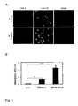
- H2A.X The total number of H2A.X foci in one focus plane (H2A.X) or the relative fluorescence intensities (%) of laminB ⁇ N or kuk transfected cells and not transfected control cells (Tri-Me-H3K9, HP1) are compared. Tests are two-tailed, mean values of three replicates per experiments are shown with error bars ⁇ s.e.m.. Indeed, the number of H2A.X foci containing nuclei was significantly higher in the group of transfected cells compared to the control group. Moreover, HP1 and Tri-Me-H3K9 staining levels in cells transfected with kuk or laminB ⁇ N compared to non-transfected cells were significantly reduced.
- kuk as well as laminB ⁇ N are able to induce cellular phenotypes similar to cultured fibroblasts from old individuals or cells from HGPS patients. Strikingly, A414 staining, a marker for nucleoporins, strongly increased in laminB ⁇ N or in kuk transfected cells, whereas the control cells showed a normal punctuated nuclear pore staining ( Figure 4 ). Similar to nucleoporin staining of late passage-HGPS cells, nuclear pores aggregated to large bright masses, which were associated with the infoldings of the nuclear envelope of highly lobulated nuclei.
Landscapes
- Health & Medical Sciences (AREA)
- Life Sciences & Earth Sciences (AREA)
- Engineering & Computer Science (AREA)
- Biomedical Technology (AREA)
- Immunology (AREA)
- Hematology (AREA)
- Chemical & Material Sciences (AREA)
- Urology & Nephrology (AREA)
- Molecular Biology (AREA)
- Cell Biology (AREA)
- Analytical Chemistry (AREA)
- Biochemistry (AREA)
- Biotechnology (AREA)
- Pathology (AREA)
- General Physics & Mathematics (AREA)
- Food Science & Technology (AREA)
- Medicinal Chemistry (AREA)
- Physics & Mathematics (AREA)
- General Health & Medical Sciences (AREA)
- Microbiology (AREA)
- Bioinformatics & Cheminformatics (AREA)
- Toxicology (AREA)
- Tropical Medicine & Parasitology (AREA)
- Physiology (AREA)
- Proteomics, Peptides & Aminoacids (AREA)
- Investigating Or Analysing Biological Materials (AREA)
- Measuring Or Testing Involving Enzymes Or Micro-Organisms (AREA)
Abstract
Description
- The present invention concerns the technical field of screening age-related candidate substances and age-related candidate genes. Furthermore, the invention relates to a nucleic acid encoding certain age-related polypeptides.
- Aging is generally characterized by a declining ability to respond to stress and an increasing homeostatic imbalance and an increasing risk of aging-associated degenerative diseases. In aging-associated degenerative diseases, like Alzheimer's disease, amyotrophic lateral sclerosis, cancer and Parkinson's disease, function or structure of affected tissues progressively deteriorates pathologically or due to normal bodily wear over the time. Accelerated aging diseases, like Hutchinson-Gilford Progeria syndrome (HGPS), Wemer's syndrome, Cockayne's syndrome or xeroderma pigmentosum, are conditions of childhood and youth indicated by premature aging symptoms. HGPS is a fatal disease caused by a mutation of lamin A gene on
chromosome 1. Lamin A is a scaffolding protein located around the edge of the nucleus that helps organizing nuclear processes, such as RNA and DNA synthesis. In HGPS the enzymatic recognition site that is required for cleaving prelamin A to lamin A is mutated because of a splicing defect. Therefore, lamin A cannot be produced, and prelamin A builds up on the nuclear membrane causing a characteristic nuclear blebbing. This dysfunction results in premature aging symptoms indicated by dysmorphic nuclear shape, increased DNA damage and down-regulation of some nuclear proteins (Moulson et al., 2007; Scaffidi and Misteli, 2006). - The process of aging is complex, and may derive from a variety of different mechanisms. Traditionally, theories that explain senescence have generally been divided between the programmed and stochastic theories of aging. Programmed theories imply that aging is regulated by biological clocks operating throughout the life span. This regulation would depend on changes in gene expression that affect systems responsible for maintenance, repair and defense responses. Stochastic theories blame environmental impacts on living organisms as the cause of aging, wherein cumulative damages at various levels are induced to DNA, tissues and cells by oxygen radicals and cross-linking. Scientific evidence suggests that cellular aging evolved in certain species as a mechanism to prevent the onset of cancer. If cells have built-in limits to how many times they can replicate, they must somehow overcome these limits before they can spread indefinitely.
- Life extension movement continues to grow rapidly in popularity and momentum among scientists and general public. Thus, there is a great demand for treatments slowing down aging processes to improve health and to maintain youthful vigor, but also to cure accelerated aging diseases. Strategies for life extension by nutritional supplements, growth hormones, and substances, like resveratrol and lipoic acid, are dubious in efficiency and side effects.
- To develop potential anti-aging therapies a test system is necessary that allows displaying and manipulating an aging phenotype. Thereby, it would be possible to detect the effectiveness of age-related substances and to investigate their mechanisms and side effects. Currently, it is possible to use cells of elderly humans or HGPS patients in primary cell cultures to study cellular aging (Scaffidi and Misteli, 2006). However, in these cell cultures aging phenotypes, like changes in nuclear morphology, chromatin or DNA damage do not occur until numerous cell cycles are passed, which is tedious and cost-intensive. Unfortunately, cells of these primary cultures are heterogeneous in reference to their aging phenotype, which make experimental data incomparable. Another disadvantage of primary cell cultures in senescence studies is that they can only be cultivated temporarily, because they are not immortalized.
- Model organisms for studying aging phenomena should be short-living to be practicable in reference to time and cost savings. Bauer et al. (2004) developed an assay in Drosophila melanogaster that accelerates the ability to evaluate potential anti-aging therapies. Coupling the expression of an age-dependent molecular biomarker to a lethal toxin reduces the time needed to perform lifespan studies by 80%. However, in this model system aging cannot be induced user-defined.
- Therefore, there is a great need for a cost and labor conscious method to screen for age-related substances that affect life span or accelerated aging diseases, wherein aging can be reproducibly induced.
- The solution to this problem is achieved by the embodiments of the present invention characterized by the claims, and described further below.
- It is an object of the present invention to provide a method for screening an age-related candidate substance or an age-related candidate gene comprising the steps of:
- a) expressing at least one polypeptide comprising a nuclear localization signal and a farnesylation site using means for genetic expression;
- b) applying at least one candidate substance or at least one candidate gene to the means; and
- c) measuring at least one aging parameter from the means,
- Furthermore, the invention concerns an assay for screening age-related candidate substances or age-related candidate genes comprising at least one means for expressing at least one polypeptide and a system for measuring at least one aging parameter, wherein the means comprises a nuclear localization signal and a farnesylation site.
- In addition, the present invention is directed to a nucleic acid encoding a polypeptide comprising a nuclear localization signal, a farnesylation site and a conditional expression sequence.
- The invention relates also to the use of the assay for screening at least one age-related candidate substance or at least one candidate age-related gene.
-
-
Figure 1A shows the morphology of longitudinal muscle nuclei in aging Drosophila flies by fluorescent antibody staining. Muscle nuclei of wild-type flies (wt), RU 486 induced upstream activated sequence (UAS) lamin B/Gene Switch-myosin heavy chain (Iamin B/GS-MHC) longitudinal muscle nuclei, and UASkuk/GS-MHC longitudinal muscle nuclei at indicated ages were stained by fluorescent kugelkern protein (Kuk) antibody. - In
Figure 1B perimeters of wild type (upper panel), UASIamin B/GS-MHC (middle panel), or UASkuk/GS MHC (lower panel) flies longitudinal muscle nuclei at indicated ages are plotted (grey bars: non-induced control flies, black bars: RU 486 induced flies). - Figures 1C-K show transmission-electron microscopy images of muscle cells of wt, kuk, UASkuk/GS-MHC, UASIamin B/GS-MHC and UASkuk/GS-MHC induced by RU 486.
-
Figure 2A shows the overexpression of protein lamin B or protein Kuk that may affect Drosophila life span with 0, 4 or 6 genomic copies of kuk in cellularizing embryos. -
Figure 2B shows that the mean life span of flies is reduced if they contain 4 or 6 genomic copies of kuk. -
Figure 2C displays survival experiments of flies overexpressing lamin or kuk in specific tissues via GS where expression was induced by RU 486 added to the food of adult flies (solid lines and circles). -
Figure 2D depicts age-specific mortality rates of induced and control flies plotted on a log scale versus time. -
Figure 3A shows DNA damage in adult flies by H2A.X staining and gene silencing by a reporter of position effect variegation in 1 week or 5 weeks old wt longitudinal muscle nuclei. - In
Figure 4 overexpression of IaminBΔN or kuk inducing DNA damage and changing levels of nuclear proteins in cultured mouse fibroblast cells are demonstrated. - In
Figure 5A and B the tissue specific induction of lamin B and kuk expression in 10 days old males is quantified, respectively. - The present invention is directed to a method for screening an age-related candidate substance or an age-related candidate gene comprising the steps of:
- a) expressing at least one polypeptide comprising a nuclear localization signal and a farnesylation site using means for genetic expression;
- b) applying at least one candidate substance or at least one candidate gene to the means; and
- c) measuring at least one aging parameter from the means,
- The term "screening" as used herein means detecting age-related candidate substances from a pool of unknown substances and compounds to identify them as pharmacologically active substances, which modify aging. The term "screening" as used herein also means detecting age-related candidate genes from a pool of genes to identify them as genes, which regulate aging. The pool comprises at least one up to a plurality of substances or genes. High-throughput screening of a large pool of potential substances or genes is also enclosed, because it is an efficient method for drug discovery, drug design and gene identification.
- The term "age-related" as used herein refers to substances or genes that are associated with normal and pathological aging. Age-related substances impair symptoms and causes of aging.
- The term "polypeptide" as used herein refers to chains of amino acids regardless of length and conformation. Therefore, the term "polypeptide" comprises oligomers, polymers as well as proteins.
- The term "nuclear localization signal" (NLS) as used herein refers to a natural or artificial amino acid sequence, which acts like a tag used to target a compound, e.g. a newly synthesized protein to the cell nucleus through the nuclear pore complex. The protein with the NLS is directed into the nucleus via its recognition by cytosolic nuclear transport receptors. Different types of NLS are known, but one common feature is a few short sequences of positively charged lysines or arginines. Different nuclear localized proteins may share the same NLS.
- The term "farnesylation site" as used herein refers to an amino acid sequence with a farnesyl group that is posttranslationally added by a prenyltransferase, e.g. a farnesyltransferase. The farnesyl group is a 15-carbon isoprenoid, like farnesol and geranylgeraniol, optionally bearing a four amino acid sequence, preferably a CaaX motif at the carboxyl terminus of a protein. Targets of farnesyltransferase include members of the Ras superfamily of small GTP-binding proteins critical to cell cycle progression.
- The term "means for genetic expression" as used herein refers to any kind of natural and artificial vehicles of genetic information that is transformed into a functional gene product, like a protein or RNA. The means are in vivo, ex vivo and in vitro systems including their different developmental stages. It may concern wild type means or genetically manipulated means.
- The term "applying" as used herein refers to any kind of contacting the candidate substance with the means. The term "applying" as used herein refers also to inserting the gene to be investigated into an expression means that is the same or different to the one expressing at least one polypeptide comprising a nuclear localization signal and a farnesylation site. Further, the term "applying" also refers to the expression of the applied gene. "Applying" includes methods like injection, transposition, transduction, transfection, conjugation, transformation, splicing and activation. "Applying" includes also uncaging, releasing, blending, incubating, adding, dispensing, and administering.
- The gene or substance can be administered locally, e.g. topically, or systemically, e.g. orally. The candidate substances may be admixed with food or drinking. Genes and substances can be added to the means into any kind of laboratory vessel, like vials, tubes, or 96-well plates. For high-throughput screening an array can be used to contact a plurality of candidate substances or genes with the means.
- The term "aging parameter" as used herein refers to parameters that display the causes and symptoms of aging. It may concern molecular, cellular and physiological parameters, or the like.
- Aging parameters can be measured by any measuring system, like biochemical, diagnostic, histological, and physiological methods, e.g. histological stainings and behavioral tests. The term "measuring" also refers to reading out and analyzing data by calculating or statistical methods. The measuring system includes quantitative methods, qualitative methods and mixtures of both.
- The invention is based on the finding that polypeptides comprising a nuclear localization signal and a farnesylation site can induce aberrant nuclear shapes with nuclear envelope foldings, lobulations and extra membrane growth. Scientists are still working on the question how changes in nuclear morphology relate to cellular aging and aging of organisms. It might be possible that irregular shaped nuclei are simply a consequence of aging in cells. In cultured HGPS cells, it has been shown that loss of H3K27me3, a marker for facultative heterochromatin, takes place before morphological changes of nuclei become visible (Shumaker et al., 2006) arguing that nuclear shape changes are a consequence of aging. However, the data shown by the inventors demonstrate that an abnormal nuclear architecture is able to promote the aging process itself. Nuclear shape changes were induced in a IaminΔ50 independent way, and their effect on age-related phenotypes on the level of cells and organisms were tested. Indeed, farnesylated lamina proteins, like lamin B or Kuk, cause aberrant nuclear shapes that shorten the life span of flies. Therefore, functional evidence is provided that kuk contributes to the regulation of heterochromatin formation in the fly. Moreover, even a truncated lamin B variant that only consists of the nuclear localization signal and the C-terminal farnesylation site as well as the Drosophila specific Kuk are able to induce age-related cellular defects. These new findings support that changes in the nuclear lamina may cause an aging process.
- By the expression of polypeptides comprising a nuclear localization signal and a farnesylation site, age-dependent nuclear changes can be induced. As a consequence of the nuclear changes aging-like phenotypes are provoked, wherein the nuclear changes are in a causal relationship to the aging-like phenotypes. Therefore, the invention provides a new method and an assay to screen and investigate candidate substances and genes for their ability to affect aging. Thus, the invention provides a tool to discover and develop therapies and drugs that delay normal aging or remedy age-related diseases, like degenerative diseases or accelerated aging diseases. In addition, it provides a tool to identify genes that play a role in aging or age-related diseases.
- The expression of polypeptides according to the invention provides a homogeneous system that affords reproducible and comparable results. In a preferred embodiment the polypeptides are expressed in immortalized cell cultures that provide a stable system displaying aging phenotypes by induction without tedious culturing.
- The invention can be performed on the level of molecules, cells, organelles, or organisms. Therefore, a large variety of aging parameters can be investigated, and the output in terms of data, useful drugs and therapies or identified genes is broadly applicable.
- In preferred embodiment the aging parameter is selected from the group consisting of DNA stability and damage, nuclear morphology, chromatin formation, histone modification, gene expression, behavioral pattern of the means, organelle modification, and organelle distribution. The term "DNA stability and damage" includes oxidation, hydrolysis, alkylation and mismatch of bases, but also mutations, like point mutations, insertions, and deletions, and large scale aberrations in chromosomes, like inversions, amplifications and translocations. The term "nuclear morphology" refers to the stability and shape of the nucleus that is altered e.g. by lobulation or envelope foldings. It also refers to nucleoplasm, nuclear membrane, nuclear pores and nuclear proteins. The term "chromatin formation" refers to the DNA/protein structure of chromosomes that can be modified e.g. by condensation, inclusions, methylation, phosphorylation and acetylation, like posttranslational histone dimethylation. The term "gene expression" includes activating and silencing of gene transcription, translation and intermediate modifications, like splicing. The term "behavioral pattern" includes motor and social behavior, cognition, behavior due to learning and memory as well as behavior of structures, molecules and cells. Analysis of behavioral patterns is conducted by experimental behavioral tests.
- In a another preferred embodiment the means is selected from the group consisting of prokaryotes; animals; insects, preferably Drosophila melanogaster, nematodes, preferably Caenorhabditis elegans; plants; fungi, preferably Saccharomyces cerevisiae; protists; transgenic vectors, like viral, bacterial and plasmid vectors, artificial yeast chromosomes; viruses; cells, preferably insect amphibian or mammalian cells. Xenopus laevis oocytes, NIH3T3 and HeLa cells are most preferred. Drosophila melanogaster is a preferred means, because these flies can be kept economically in huge amounts, and have a fast alteration of generation. In addition, they show distinct behavioral patterns and can be easily genetically manipulated.
- In a preferred embodiment the polypeptide is selected from the group consisting of kugelkern (Kuk), A type lamin and B type lamin, preferably IaminBΔN.
- Kuk contains a putative coiled-coil domain in the N-terminal half of the protein, a NLS, and a C-terminal CxxM-motif for targeting Kuk to the inner nuclear membrane. Kuk functions in nuclear elongation. Recent findings suggest that Kuk is a rate-determining factor for nuclear size increase (Brandt et al., 2006). Without being bound to any theory, the inventors believe that association of farnesylated Kuk with the inner nuclear membrane induces expansion of the nuclear surface area, allowing nuclear growth.
- Lamins are intermediate filament proteins regulating structural function and transcription in the cell's nucleus. These proteins localize to two regions of the nuclear compartment, the nuclear lamina and throughout the nucleoplasm. They are essential proteins that provide mechanical stability to a nuclear envelope. In addition, they influence DNA replication, and organize the chromatin bordering the inner nuclear membrane by binding to chromatin and inner nuclear membrane. Lamins are also required for RNA-polymerase II-dependent transcription and are involved in the localization of transcriptional factors. There are indications that lamins affect nuclear growth after the re-formation of the nuclear envelope.
- Lamin contains an alpha-helical rod domain centered between a non-helical N-terminal head domain and a C-terminal globular tail domain. The C-terminal domain contains a NLS, an Ig-fold, like domain, and in most, but not all cases a C-terminal CaaX box that is isoprenylated and carboxymethylated. These modifications confer a high hydrophobicity to the C-terminus and are essential for targeting to the inner nuclear membrane.
- There are several types of lamin, like A type lamins, e.g. lamin A and C, and B type lamins, e.g. lamin B1 and B2, which differ in their length and isoelectric point. In human cells three differentially regulated genes are known, LMNB1, LMNB2 and LMNA. B type lamins, like B1 and B2, are present in every cell and are expressed from the LMNB1 and LMNB2 genes on 5q23 and 19q13, respectively.
- Lamin BΔN is a lamin B variant truncated at the N-terminus. Preferably it comprises a nuclear localization signal and a C-terminal farnesylation site.
- Age-dependent morphological alterations can be induced ahead of time by overexpressing the polypeptides according to the invention. In a preferred embodiment the lifespan of the means is reduced, preferably up to 60%, more preferably up to 70%.
- In another preferred embodiment an age-related phenotype of the means appears within 48 h, preferably within 24 h. These advantages help saving costs, time and therefore labor input.
- In another preferred embodiment the polypeptide is conditionally expressed, preferably by a gene switch sequence. The term "conditionally expressed" as used herein refers to a restricted gene expression that occurs only under distinct conditions. The expression can be spatially or temporally restricted. One method to express transgenes in a spatially restricted manner is a light-induced activation of genes, like activation of a heat-shock protein by a laser micro beam or light activation of caged molecules. Another method is a bipartite expression system based on the yeast GAL4 protein and its upstream activating sequence. There is a wide variety of driver lines with defined expression patterns generated e.g. by using cloned promoter fragments or enhancer detector screens. Temporal control transgene expression can be accomplished by using e.g. a heat-shock promoter or tetracycline-dependent "tet-off" or "tet-on" transactivation systems.
- Spatially and temporally controlled expression of the polypeptide is a preferred embodiment. This can be accomplished by a fusion protein of GAL4 with receptors of inducing ligands, like hormones, e.g. estrogens or progesterone. It can also be accomplished by a gene switch. The gene switch is based on a chimeric gene that encodes the GAL4 DNA binding domain, a ligand binding receptor-domain, and an activation domain. In the presence of a ligand the chimeric molecule binds to the UAS and provides for ligand inducible activation of downstream target genes. Examples of a spatially and temporally controlled expression by using tissue specific promoters and a systemic application of the ligand is the RU486 induction of UASlamin/GS-MHC, UASlamin/GS-
Sl 106, UASlamin/GS-elav, UASkuk/GS-MHC, UASkuk/GS-S1 106 or UASkuk/GS-elav. - The present invention is also directed to an assay for screening age-related candidate substances or age-related candidate genes comprising at least one means for expressing at least one polypeptide and a system for measuring at least one aging parameter, wherein the means comprises a nuclear localization signal and a farnesylation site.
- In a preferred embodiment the means comprises a conditional expression sequence, preferably a gene switch sequence.
- In a further preferred embodiment of the assay the polypeptide is selected from the group consisting of Kuk, A type lamin and B type lamin, preferably IaminBΔN.
- In another preferred embodiment the assay further comprises at least one candidate substance or at least one candidate gene.
- The present invention also relates to a nucleic acid encoding a polypeptide comprising a nuclear localization signal, a farnesylation site and a conditional expression sequence.
- In a preferred embodiment the polypeptide is selected from the group consisting of kugelkern (Kuk), A type lamin and B type lamin, preferably IaminBΔN.
- Loss-of-function mutations in lamin genes lead to laminopathies, like Emery-Dreifuss muscular dystrophy, familial partial lipodystrophy, limb girdle muscular dystrophy, dilated cardiomyopathy, Charcot-Marie-Tooth disease, and gain-of-function laminopathies like HGPS. Therefore, nucleic acids according to the invention encoding lamin or Kuk with different expression patterns or mutations can serve as models for these diseases or other age-related or degenerative diseases.
- It is possible to use these mutants for studying aging processes, but it is difficult to distinguish defects caused throughout development and defects directly related to aging. Therefore, in a preferred embodiment the conditional expression sequence is a gene switch sequence, preferably selected from the group consisting of the gene switch-myosin heavy chain sequence (GS-MHC), the gene switch-embryonic lethal abnormal vision sequence (GS-elav) and the gene switch-
S1 106 sequence. - GS-MHC is a muscle specific GS driver that uses the promoter of the MHC gene with a panmuscular distribution. The GS-elav is a GS driver specific for neuronal tissue that uses the promoter from the elav gene with a panneural distribution. The GS-
S1 106 drives gene expression in the fat body of insects and white adipose tissue. A reporter gene expression can be regulated by the dosage of the activating ligand RU486. - Overexpression of lamin B, or C, human LMNA, or IaminΔ50 in Drosophila using the UAS/GAL system leads to a severely affected viability, resulting in low eclosing rates and early mortality. To approximate the situation in HGPS where IaminΔ50 acts dominantly, overexpression of the gene variants should be in a time and tissue specific manner. The nucleic acid comprising a gene switch system according to the invention allows to minimize the problem of genetic background as well as to circumvent defects which may arise during development. Therefore, it is possible to analyze effects of candidate substances on aging based on conditional lamin B or kuk expression in adult flies.
- Further, the invention relates to the use of the assay according to the invention for screening at least one age-related candidate substance or at least one age-related candidate gene.
- In a preferred embodiment the substance is a pharmaceutically active substance for therapeutic and/or prophylactic treatment of age-related disorders like Alzheimer's disease, cancer, Parkinson's disease, HGPS, and other laminopathies.
- NIH3T3 cells were cultured in DMEM (Invitrogen, GIBCO) supplemented with 10% FBS and 2 mM L-glutamine at 37°C. NIH3T3 cells were plated in 6-wells containing cover slips and were transiently transfected with Effectene (Qiagen), when they reached a confluence of 25 - 30% with pCS2HAkuk (Brandt et al., 2006) or pCS2XlaminBΔNGFP (Prüfert et al., 2004) (2 µg construct/6-well). They were cultivated for 48 h for heterochromatin protein 1 (HP1) and Tri-Me-H3K9 staining or for 72 h for H2A.X staining. After washing in phosphate buffered saline (PBS), cells were fixed with 2% formaldehyde in PBS containing 0.2% Tween and 0.5% NP-40 for 20 min at room temperature (RT). After washing in PBS cells were permeabilized in PBS with 0.5% Triton X-100 plus 0.5% Saponin (Sigma) for 10 min.
- Adult male flies were anaesthetized. The head, abdomen, legs, and wings were cut off. The thorax was transferred to ice cold Schneider cell medium, where it was split into half and subsequently fixed with 8% formaldehyde in PBS containing 0.2% Tween and 0.5% NP-40 for 40 min at RT. After washing in PBS the thoraces were permeabilized in PBS with 0.5% Triton X-100 plus 0.5% Saponin (Sigma) for 1 to 5 days at 4°. Before staining, the fixed thoraces were manipulated that only one end of the muscles stuck to the cuticle and the other end was free floating and better accessible to the staining reagents. Consecutively, muscles were blocked in PBT with 5% BSA and stained in PBT containing primary antibodies, fluorescent secondary antibodies (4 µg/ml, Alexa, Molecular Probes) or DAPI and mounted in Aquapolymount (Polyscience). Antibodies used were A414 (Sigma, 1 µg/ml), Drosophila HP1 (DSHB, C1A9, H2A.X (Chemicon, 1:5000), mouse HP1 (Chemicon, 1:2500), PCS2HAkuk (0.2 µg/ml), Tri-Me-H3K9 (upstate, 0.2 µg/ml), 0.1 µg/ml), Drosophila lamin B (LaminDmO, 0.1 µg/ml), human IaminA/C (Santa Cruz Biotechnology, 0.2 µg/ml) and HA (Babco, 1:2000).
- Digital fluorescent images were either taken with a confocal microscope (Leica) or with a fluorescent microscope connected to a Progress camera and processed with Photoshop (Adobe). Measurements were performed with ImageJ by the perimeter tool to measure the nuclear perimeter, by the mean grey value tool to measure the relative fluorescent intensity and by the maximum intensity tool. For measuring relative fluorescent intensity of HP1 and Tri-Me-H3K9 stainings, three independent experiments were analyzed by the fluorescence microscope. For each experiment the maximum intensity was set as 100%. The number of H2AX foci per nucleus might be underestimated since only those foci that were in the focus plane of the picture were counted. (Mann-Whitney U-Test, http://faculty.vassar.edu/lowry/VassarStats.html). For measurements of the muscle perimeters, confocal image z-stacks of the longitudinal muscles with a slice distance of approx. 1 µm were acquired and assembled in ImageJ with the z-stack/SUM function. For each time point three individual flies were analyzed. Statistical analysis was performed with Prism GraphPad software, (One- or Two-Way-ANOVA followed by Bonferronis post tests).
- Thoraces were fixed and processed for electron microscopy as described in Fryberg et al. (1990). In contrast to Fryberg et al., the tannic acid from the 3% glutaraldehyde solution was omitted. Ultra thin sections were inspected with a Zeiss EM900 and a Zeiss EM10 electron microscope (Oberkochen, Germany). Negatives were digitalized by scanning and processed with Adobe Photoshop.
- Gene switch-Gal4 flies and w (superscript: m4h) flies were provided and UASIamin B flies were prepared as described in Guillemin et al. (2001). For pUASkuk the cDNA from LD09231 was cloned as Notl-Apal/blunt fragment into the Notl-Xbal/blunt sites of pUASp. To obtain heat-shock inducible HS-kuk full length or mutant HSkukCSflies, kuk CDS with parts of the 3'-UTR as Ncol-Smal PCR fragments of CS-kuk or CS-kukCS into QEH10ZZ was cloned. Subsequently, HS-kuk or mutant HSkukCS was cloned as EcoRI-Smal fragments from QEH10ZZ-kuk or -kukCS into EcoRI-Stul of CasperHS. Transgenic flies were made by P-element mediated transformation. The genomic rescue constructs 4x and 6x kuk are located on the second and third chromosome. 0x kuk consists of a deletion of kuk and CG5169 together with a rescue construct of CG5169 over Df(3R)Ex6176. To induce conditional expression of the full-length kuk or the non farnesylated CaaX-mutant kuk (kukCS) in wt or in kuk deficient embryos (Δkuk), transgenic embryos containing the respective heat-shock constructs were heat-shocked for 45 min. After 45 min recovery the embryos were fixed and stained with Kuk or lamin B antibodies.
- All flies were raised and kept in a humidified, temperature-controlled incubator with 12 h on/off light cycle at 25°C in vials containing standard cornmeal medium (2.5% yeast, 2.18% treacle, 1% soy meal, 8% cornmeal, 8% malt, 1.25 % propionic acid). In the case of UASlamin B/GS-MHC, flies were raised at 18°C. Flies were collected under short anesthesia. Each demography cage was initiated with at least 150 newly eclosed males. The number of deceased flies was recorded every 2 to 3 days, when flies were transferred to fresh food plates. For induction with the GS system, RU 486 (Sigma) was added directly to the food to a final concentration of 200 µM. The data across 3 to 5 replicate demography cages per treatment and genotype were combined. Both Statview 5.0 and Prism GraphPad software were used to analyze survival data by a log-rank test and mortality curves by linear regression.
- Negative geotaxis was assessed as previously described in Gargano et al. (2005). Negative geotaxis behavior was recorded as the net distance in cm climbed by individual males in a vertical plastic tube during a 10 second test period that began immediately after being tapped to the bottom of the tube. For each genotype and
treatment 25 males were tested per week. The mean distance climbed by the control flies in the first week was set as 100% negative geotaxis. One to three replicative experiments were performed and the data were analyzed using Prism 3.0 (GraphPad Software, San Diego, CA, USA). - 10 newly eclosed males were aged for 5 days at 25°C, decapitated and immediately shock-frozen in liquid nitrogen. For extraction of the eye pigments, fly heads were homogenized in 0.1 M glycine/HCl (pH 2) containing 30% ethanol and kept in the dark for 24 h at room temperature. After clarification by several brief centrifugations, the absorption of the pigments was measured at 480 nm. Five independent extractions were performed for each genotype. VassarStat statistical software was used for Mann-Whitney tests.
- Proteins were analyzed by SDS polyacrylamid gel electrophoresis (SDS-PAGE) according to standard protocols. For western-blotting proteins separated by SDS-PAGE were transferred by semi-dry blotting to a nitrocellulose membrane (Protran, Schleicher and Schuell) and stained by Ponceau Red. The blots were developed with IgG coupled with peroxidase and chemiluminescence (ECLplus, Amersham). Following antibody concentrations were used: lamin B, polyclonal: 1:1000, Kuk: rabbit, 0.1 µg/ml.
-
Figure 1A shows the morphology of longitudinal muscle nuclei in aging Drosophila flies by fluorescent antibody staining. Muscle nuclei of wt flies, UAS lamin B/GS-MHC, and UASkuk/GS-MHC longitudinal muscle nuclei at indicated ages were stained by fluorescent Kuk antibody (scale bar: 1 µm) (Figure 1A ). Z-stacks were fused with the maximum intensity tool in ImageJ to show several nuclei. - In
Figure 1B perimeters of wild type (upper panel), UASlamin B/GS-MHC (middle panel), or UASkuk/GS MHC (lower panel) flies longitudinal muscle nuclei at indicated ages are plotted (grey bars: non-induced control flies, black bars: RU 486 induced flies). In wt flies number (n) is between 45 and 95 nuclei per time-point, in UASlamin B/GS-MHC flies n is between 41 and 135 nuclei per time-point and in UASkuk/GS-MHC flies n is between 37 and 104 nuclei per time-point. Mean values are depicted with ± standard error of means (s.e.m.). For wt flies a One-way ANOVA and for UASlamin B/GS-MHC or UASkuk/GS-MHC flies a Two-way ANOVA were followed by Bonferronis Multiple Comparison Test, * p < 0,05, ** p < 0,01, *** p < 0,0001. - Nuclear architecture in adult Drosophila longitudinal muscle cells shows progressive and stochastic alteration as fly ages (
Figure 1 ). Most muscle nuclei in young animals were round and possessed a smooth surface. Over time, the nuclei increased significantly in size and assumed an uneven wrinkled shape (Figure 1A, B ). When lamin B or kuk were overexpressed in adult muscle cells, a significant increase in nuclear perimeter, wrinkled, lobulated nuclei, and accumulations of Kuk (Figure 1A, B ) or lamin B staining (Figure 3A ) was observed at the nuclear envelope. - Figures 1C-K show transmission-electron microscopy images of muscle cells with the following legend: (C) wt, 3 days old; (D) wt, 35 days old; (E) wt, 60 days old; (F) 6 genomic copies of kuk (6x kuk), 3 days old; (G) 6x kuk, 30 days old; (H) 6x kuk, 36 days old; (I) UASkuk/GS-MHC induced with
RU 486, 3 days old; (J) UASlamin B/GS-MHC induced byRU 486, 1 week old; (K) UASkuk/GS-MHC induced by RU 486, 31 days old; muscle fibers (m); arrowheads mark nuclear protrusions; (h) organelle free holes in the tissue; arrows mark electron dense areas containing probably highly condensed chromatin; peripheral heterochromatin (p); lumen between inner and outer nuclear membrane (I); mitochondria (*); scale bar: 0.5 µm. - Muscle nuclei of Figures 1C - K showed an age-dependent loss of peripheral heterochromatin and a strong increase of dark inclusions, which may contain highly condensed chromatin. Moreover, there was a separation of inner and outer nuclear membrane. In old nuclei, high numbers of ring-like structures with less than 50 nm in diameter in the nucleoplasm were found. These structures were too small to present a complete nuclear pore. The ring-like structures were not marked by the nuclear pore-marker AB414, lamin B, or Kuk antibodies in immuno-electron microscopy. No trilaminar unit-membrane structure was visible in the rings, which argues against vesicles. Although actin/myosin arrays of the muscle fibers seemed to be intact in old flies, the number of degenerated mitochondria increased and organelle free zones were found. In lamin B or kuk overexpressing flies, loss of peripheral heterochromatin, separation of inner and outer nuclear membrane, accumulation of ring-like structures, and a strong increase of dark material in the nucleoplasm were observed even in 1 week old flies. In tissues, which were not overexpressing lamin B or kuk, nuclear size or shape did not alter. In conclusion, age-dependent morphological changes of the nucleus in Drosophila flies were described that can be induced ahead of time by overexpressing lamin B or kuk.
- The inventors surprisingly found that a farnesylation motif is relevant for kuk activity. The kukCS mutant allele, which lacks its farnesylation site in kuk deficient mutant fly embryos by heat-shock, was expressed, and its localization and action towards nuclear morphology were characterized. Predominantly nucleoplasmatic localizations of KukCS protein, with only few small dots localized to the nuclear envelope were found. Interestingly, when the non-farnesylated KukCS protein in wt flies was expressed, there was a clear localization to the nuclear envelope. Moreover, in the wt background, the mutated protein was able to induce lobulation of nuclear membrane, albeit weaker than that induced by normally farnesylated Kuk. The finding that KukCS cannot localize to the nuclear envelope in the kuk deficient background as well as its inability to induce nuclear lobulation argues that the farnesyl residue is indeed required for Kuk localization and function.
-
Figure 2A shows the overexpression of proteins lamin B or Kuk that shortens Drosophila life span with 0, 4 or 6 genomic copies of kuk in cellularizing embryos. Nuclei were stained with lamin B antibodies, and apical sides of nuclei inearly cycle 14 are shown. In the absence of kuk, the nuclei are round and smooth. With increasing amounts of kuk, the nuclei show deep infoldings of the nuclear envelope. -
Figure 2B shows that the mean life span of flies is reduced if they contain 4 or 6 genomic copies of kuk. -
Figure 2C displays survival experiments of flies overexpressing lamin or kuk in specific tissues via GS where expression was induced by RU 486 added to the food of adult flies (solid lines and circles). Control flies had the same genotype, but were not fed with RU 486 (dashed lines, open triangles). Log-rank tests compare induced and control flies. The legend ofFigure 2 is as follows: UASlamin/GS-MHC males: + RU 486, n = 120, 4 experiments; - RU 486, n = 214, 3 experiments; log-rank test: p < 0.001. UASlamin/GS-S1 106, expressed in fat bodies: + RU 486, n = 454, 3 experiments; - RU 486, n = 353, 3 experiments; log-rank test: p < 0.001. UASlamin/GS-embryonic lethal abnormal vision (elav): + RU 486, n = 441, 3 experiments; - RU 486, n = 421, 3 experiments; log-rank test: p < 0.001; UASkuk/GS-MHC: + RU 486, n = 558, 5 experiments; - RU 486, n = 811, 7 experiments; log-rank test: p < 0.001. UASkuk/GS-S1 106: + RU 486, n = 454, 3 experiments; - RU 486, n = 353, 3 experiments; log-rank test: p = 0.0181. UASkuk/GS-elav: + RU486, n = 441, 3 experiments; - RU 486, n = 421, 3 experiments; log-rank test: p < 0.001. -
Figure 2D depicts age-specific mortality rates of induced and control flies plotted on a log scale versus time, fitted by Gompertz model (In(mx) = In(m0) + ax), where mx = mortality rate at age x, m0 = baseline mortality (intercept as In(m0)) and ax = change of mortality with age (slope of the trajectory). The slope of the mortality trajectory only differs significantly between induced and control UASlamin B/GS-S1 06 flies (p = 0.005). Significantly different Y-intercepts between induced and control groups are found in UASlamin/GS-MHC (p = 0.0002), UASlamin/GS-elav (p = 0.01), or UASkuk/GS-MHC flies (p = 0.04). - Induced expression of farnesylated proteins affecting lamina structure also provoked aging-like phenotypes (
Figure 2A ). Flies containing 0, 4, or 6 genomic copies of kuk for their longevity were tested. An increase in the dosage of kuk or lamin shortened the life span of the adult flies(Figure 2B-D ). To minimize the problem of genetic background as well as defects which may arise during development, tissue specific drivers inducible with RU 486 were used, wherein the difference between the experimental and control condition is the presence or absence of the inducing agent in the food of the adult flies called GS system. Conditional expression of lamin B or kuk in the adult muscle cells via the muscle specific gene switch driver GS-MHC decreased the life span compared to un-induced control flies of the same genotype. UASkuk/GS-MHC flies showed 60% and UASlamin B/GS-MHC flies showed 54% reduction of mean life span. Conditional expression of lamin B in the abdominal fat body, the fly equivalent of the liver and white adipose tissue via the GS-S1-106 driver reduced the mean life span to 46% of un-induced control flies. Expression in the nervous system in UASlamin B/GS-elav flies from the onset of adulthood decreased the mean life span not significantly (90%). The increase of mortality rates of RU 486 induced kuk/GS-MHC, lamin B/GS-MHC, and lamin B/GS-elav flies were similar to non-induced flies, but their onset differed. An increase in the initial mortality may argue for a deleterious effect of lamin B or kuk overexpression. In lamin B/GS-S1 106 induced flies, a change in the demographic rate of aging was observed. - The variations between UASkuk and UASlamin B in the tissue specific driver lines may be due to different expression levels or may reflect tissue specific functions. To test whether the life span data correlate with the morphology of the nuclei in the lamin B or kuk expressing tissues, nuclear size in fat body cells or in the optic lamina of three weeks old RU 486 induced flies vs. control flies was examined. The nuclear size in lamin B expressing fat body nuclei compared to control nuclei increased, but nuclear size in kuk expressing cells did not change. In lamin B expressing neuronal tissue nuclear size increased. In kuk expressing brains only few neurons showed changes in nuclear size.
Figure 5 quantifies the tissue specific induction of lamin B and kuk expression in 10 days old males. The legend ofFigure 5 is as follows: (+) means RU 486 induced expression, (-) means un-induced flies. The strongest induction of Kuk is observed with the GS-MHC driver. - Thus, the nuclear morphology of lamin B or kuk expressing cells correlate with the tissue specific effects observed in life span experiments. To better analyze the system of the invention, the levels of lamin B or kuk expression in induced flies were investigated and a moderate induction of lamin B or kuk expression in the different tissues was displayed (
Figure 5 ). Between the different driver lines, the GS-MHC driver showed the strongest increase in UASkuk or UASlamin B expression compared to the lower induction by GS-S1 106 or the GS-elav line. The more prominent induction of kuk or lamin B expression via the GS-MHC driver correlates with a strong effect on life span in these lines. Induction driven by the GS-S1 106 or the GS-elav driver seems to be less pronounced and correlates with a more differential effect on life span. These flies appear to be more sensitive to lamin B than to kuk overexpression. In conclusion, these data argue for a correlation of induction levels with phenotypic strength and not for a tissue specific effect of lamin B or kuk expression. - Negative geotaxis, an innate escape response during which flies climb the wall of a cylinder after being tapped to its bottom, is a well characterized behavior that senesces in Drosophila. In
Figure 2E - H negative geotaxis of RU 486 induced UASkuk/GS-MHC (Figure 2E ;3 replicate experiments), UASlamin B/GS-MHC (Figure 2F ;3 replicate experiments), UASlamin B/GS-S1 106 (Figure 2G ;3 replicate experiments), and UASkuk/GS-elav (Figure 2H ;1 experiment) expressing flies (closed circle) are compared with un-induced control flies (open triangle). The mean distance climbed by the control flies in the first week is set as 100% negative geotaxis. One-way ANOVA was followed by Bonferronis Multiple Comparison Test, *** = p< 0,001. Statistical tests are two-tailed, mean values are shown with error bars ± s.e.m.. An early decline in the climbing ability in those flies that conditionally express lamin B in the muscle tissue or fat body cells or in flies which express kuk in muscle tissue was found(Figure 2E-H ). - Thus, the early onset of behavioral decline corresponds to the reduced life span of these flies.
- To investigate whether there is an increase in DNA damage in aging flies, 1 week and 5 weeks old muscle tissue was stained with the H2A.X antibody which indicates foci of double-stranded DNA damage.
Figure 3A shows DNA damage and gene silencing in adult flies by H2A.X staining in 1 week or 5 weeks old wt longitudinal muscle nuclei. Z-stacks of confocal images were fused to show several nuclei. Position effect variegation (PEV) of the wm4 allele is presented inFigure 3B . In heterozygous kuk mutants (wm4; Δkuk/+) the silencing of wm4 expression is suppressed. In homozygous kuk mutants (wm4; Δkuk/Δkuk) the suppression of PEV is even more pronounced compared to control flies (wm4; +/+). In Figure 3C photometric measurements of the eye pigments of flies from the genotypes described inFigure 3B are plotted. n = 50 males per genotype with error bars ± s.e.m.. - In young flies, only occasionally H2A.X foci were present, whereas the number of muscle nuclei with numerous H2A.X foci increased, when the animal became older, indicating more DNA damage (
Figure 3A ). H2A.X foci accumulated in aging flies, baboons and mice, aged human cells, and cells from patients with HGPS. To find out whether kuk is indeed capable influencing gene silencing or heterochromatic spreading, it was tested whether kuk has an effect on PEV using the wm4 allele, where the white gene is translocated close to the heterochromatic region of the centromere. In heterozygous kuk mutants the silencing of wm4 expression was suppressed, and there were patches of red-pigmented ommatidia in the fly eyes (Figure 3B ). In homozygous kuk mutants the suppression of PEV was pronounced, and a high number of ommatidia showed the red eye pigment. These results provide functional evidence that kuk contribute to the regulation of heterochromatin formation. - In cultured fibroblasts from aged individuals, in HGPS cells, and in cells overexpressing IaminΔ50 aberrant shaped nuclei, HP1 and Tri-Me-H3K9 staining levels were reduced, and DNA damage marked by H2A.X antibodies was increased. To investigate if the induction of these changes is a lamin specific function or whether unspecific proteins induced age-related phenotypes, kuk or laminBΔN, a truncated lamin B variant, which only consists of a nuclear localization signal and the C-terminal farnesylation site was expressed in mouse fibroblasts. In
Figure 4 overexpression of laminBΔN or kuk inducing DNA damage and changing levels of nuclear proteins in cultured mouse fibroblast cells are demonstrated. The total number of H2A.X foci in one focus plane (H2A.X) or the relative fluorescence intensities (%) of laminBΔN or kuk transfected cells and not transfected control cells (Tri-Me-H3K9, HP1) are compared. Tests are two-tailed, mean values of three replicates per experiments are shown with error bars ± s.e.m.. Indeed, the number of H2A.X foci containing nuclei was significantly higher in the group of transfected cells compared to the control group. Moreover, HP1 and Tri-Me-H3K9 staining levels in cells transfected with kuk or laminBΔN compared to non-transfected cells were significantly reduced. Therefore, kuk as well as laminBΔN are able to induce cellular phenotypes similar to cultured fibroblasts from old individuals or cells from HGPS patients. Strikingly, A414 staining, a marker for nucleoporins, strongly increased in laminBΔN or in kuk transfected cells, whereas the control cells showed a normal punctuated nuclear pore staining (Figure 4 ). Similar to nucleoporin staining of late passage-HGPS cells, nuclear pores aggregated to large bright masses, which were associated with the infoldings of the nuclear envelope of highly lobulated nuclei. - In conclusion, it was shown that a truncated lamin B construct reduced to its nuclear localization and farnesylation signal, and even the Drosophila specific kuk are able to induce age-related nuclear changes in mammalian cells.
- These studies demonstrate that aging phenotypes as reduced life span, early onset of behavioral decline, reduced heterochromatin levels, and increase in DNA damage is induced by lamin B and kuk.
-
- Bauer et al., Proc. Natl. Acad. Sci. 101, pp. 12980-12985, 2004
- Brandt et al., Current Biology 16, pp. 543-552, 2006
- Fryberg et al., J. Cell. Biol. 110, 1999-2011, 1990
- Gargano et al., Exp. Gerontol. 40, 386-395, 2005
- Guillemin et al., Nat. Cell. Biol. 3, pp. 848-851, 2001
- Moulson et al., Hum. Mutation 28(9), pp. 882-889, 2007
- Prüfert et al., J. Cell Sci. 117, pp. 6095-6104, 2004
- Scaffidi and Misteli, Nat. Med. 11, pp. 1059-1063, 2006
- Shumaker et al., Proc. Natl. Acad. Sci. 103, pp. 8703-8708, 2006
Claims (15)
- A method for screening an age-related candidate substance or an age-related candidate gene comprising the steps of:a) expressing at least one polypeptide comprising a nuclear localization signal and a farnesylation site using means for genetic expression;b) applying at least one candidate substance or at least one candidate gene to the means; andc) measuring at least one aging parameter from the means,wherein a modification of the aging parameter indicates an age-related substance or an age-related gene.
- The method of claim 1, wherein the aging parameter is selected from the group consisting of DNA stability and damage, nuclear morphology, chromatin formation, gene expression, behavioral pattern of the means, organelle modification and organelle distribution.
- The method of claim 1 or 2, wherein the means is selected from the group consisting of prokaryotes; animals; insects, preferably Drosophila melanogaster; nematodes, preferably Caenorhabditis elegans; plants; fungi, preferably Saccharomyces cerevisiae; protists; transgenic vectors; viruses; and cells, preferably insect, amphibian or mammalian cells.
- The method of any of the preceding claims, wherein the polypeptide is selected from the group consisting of kugelkern, A type lamin and B type lamin, preferably laminBΔN.
- The method of any of the preceding claims, wherein the lifespan of the means is reduced, preferably up to 60%, more preferably up to 70%.
- The method of any of the preceding claims, wherein an age-related phenotype of the means appears within 48 h, preferably within 24 h.
- The method of any of the preceding claims, wherein the polypeptide is conditionally expressed, preferably by a gene switch sequence.
- An assay for screening age-related candidate substances or age-related candidate genes comprising at least one means for expressing at least one polypeptide and a system for measuring at least one aging parameter, wherein the means comprises a nuclear localization signal and a farnesylation site.
- The assay of claim 8, wherein the means comprise a conditional expression sequence, preferably a gene switch sequence.
- The assay of claim 8 or 9, further comprising at least one candidate substance or at least one candidate gene.
- A nucleic acid encoding a polypeptide comprising a nuclear localization signal, a farnesylation site and a conditional expression sequence.
- The nucleic acid of claim 11, wherein the polypeptide is selected from the group consisting of kugelkern, A type lamin and B type lamin, preferably laminBΔN.
- The nucleic acid of claims 11 or 12, wherein the conditional expression sequence is a gene switch sequence, preferably selected from the group consisting of the gene switch-myosin heavy chain sequence, the gene switch-embryonic lethal abnormal vision sequence and the gene switch-S1 106 sequence.
- Use of the assay of claims 8 to 10 for screening at least one age-related candidate substance or at least one age-related candidate gene.
- Use of claim 14, wherein the substance is a pharmaceutically active substance for therapeutic and/or prophylactic treatment of age-related disorders.
Priority Applications (2)
| Application Number | Priority Date | Filing Date | Title |
|---|---|---|---|
| EP08008275A EP2113772A3 (en) | 2008-04-30 | 2008-04-30 | Induction of aging phenotypes |
| PCT/EP2009/055305 WO2009133192A1 (en) | 2008-04-30 | 2009-04-30 | Induction of aging phenotypes |
Applications Claiming Priority (1)
| Application Number | Priority Date | Filing Date | Title |
|---|---|---|---|
| EP08008275A EP2113772A3 (en) | 2008-04-30 | 2008-04-30 | Induction of aging phenotypes |
Publications (2)
| Publication Number | Publication Date |
|---|---|
| EP2113772A2 true EP2113772A2 (en) | 2009-11-04 |
| EP2113772A3 EP2113772A3 (en) | 2009-12-09 |
Family
ID=39671658
Family Applications (1)
| Application Number | Title | Priority Date | Filing Date |
|---|---|---|---|
| EP08008275A Withdrawn EP2113772A3 (en) | 2008-04-30 | 2008-04-30 | Induction of aging phenotypes |
Country Status (2)
| Country | Link |
|---|---|
| EP (1) | EP2113772A3 (en) |
| WO (1) | WO2009133192A1 (en) |
-
2008
- 2008-04-30 EP EP08008275A patent/EP2113772A3/en not_active Withdrawn
-
2009
- 2009-04-30 WO PCT/EP2009/055305 patent/WO2009133192A1/en not_active Ceased
Non-Patent Citations (9)
| Title |
|---|
| BAUER ET AL., PROC. NATL. ACAD. SCI., vol. 101, 2004, pages 12980 - 12985 |
| BRANDT ET AL., CURRENT BIOLOGY, vol. 16, 2006, pages 543 - 552 |
| FRYBERG ET AL., J. CELL. BIOL., vol. 110, 1990, pages 1999 - 2011 |
| GARGANO ET AL., EXP. GERONTOL., vol. 40, 2005, pages 386 - 395 |
| GUILLEMIN ET AL., NAT. CELL. BIOL., vol. 3, 2001, pages 848 - 851 |
| MOULSON ET AL., HUM. MUTATION, vol. 28, no. 9, 2007, pages 882 - 889 |
| PRUFERT ET AL., J. CELL SCI., vol. 117, 2004, pages 6095 - 6104 |
| SCAFFIDI; MISTELI, NAT. MED., vol. 11, 2006, pages 1059 - 1063 |
| SHUMAKER ET AL., PROC. NATL. ACAD. SCI., vol. 103, 2006, pages 8703 - 8708 |
Also Published As
| Publication number | Publication date |
|---|---|
| EP2113772A3 (en) | 2009-12-09 |
| WO2009133192A1 (en) | 2009-11-05 |
Similar Documents
| Publication | Publication Date | Title |
|---|---|---|
| Lindhout et al. | VAP‐SCRN1 interaction regulates dynamic endoplasmic reticulum remodeling and presynaptic function | |
| Sun et al. | Dstyk mutation leads to congenital scoliosis-like vertebral malformations in zebrafish via dysregulated mTORC1/TFEB pathway | |
| Wu et al. | Arc/Arg3. 1 regulates an endosomal pathway essential for activity-dependent β-amyloid generation | |
| Miguel et al. | Both cytoplasmic and nuclear accumulations of the protein are neurotoxic in Drosophila models of TDP-43 proteinopathies | |
| Stum et al. | An assessment of mechanisms underlying peripheral axonal degeneration caused by aminoacyl-tRNA synthetase mutations | |
| Thakur et al. | Phospholipase D activity couples plasma membrane endocytosis with retromer dependent recycling | |
| Brandt et al. | The farnesylated nuclear proteins KUGELKERN and LAMIN B promote aging‐like phenotypes in Drosophila flies | |
| Hope et al. | Glial overexpression of Dube3a causes seizures and synaptic impairments in Drosophila concomitant with down regulation of the Na+/K+ pump ATPα | |
| Jones et al. | Ankrd6 is a mammalian functional homolog of Drosophila planar cell polarity gene diego and regulates coordinated cellular orientation in the mouse inner ear | |
| Soustelle et al. | ALS-associated KIF5A mutation causes locomotor deficits associated with cytoplasmic inclusions, alterations of neuromuscular junctions, and motor neuron loss | |
| Fasano et al. | Dominant ARF3 variants disrupt Golgi integrity and cause a neurodevelopmental disorder recapitulated in zebrafish | |
| Bergamin et al. | Zebrafish Tg (hb9: MTS-Kaede): a new in vivo tool for studying the axonal movement of mitochondria | |
| Parreno et al. | Methodologies to unlock the molecular expression and cellular structure of ocular lens epithelial cells | |
| van’t Sant et al. | In vivo 5-ethynyluridine (EU) labelling detects reduced transcription in Purkinje cell degeneration mouse mutants, but can itself induce neurodegeneration | |
| Vettori et al. | Developmental defects and neuromuscular alterations due to mitofusin 2 gene (MFN2) silencing in zebrafish: a new model for Charcot-Marie-Tooth type 2A neuropathy | |
| Mermall et al. | Drosophila myosin V is required for larval development and spermatid individualization | |
| Leonard et al. | Loss of Elp1 disrupts trigeminal ganglion neurodevelopment in a model of familial dysautonomia | |
| Djebar et al. | Astrogliosis and neuroinflammation underlie scoliosis upon cilia dysfunction | |
| Copf | Developmental shaping of dendritic arbors in Drosophila relies on tightly regulated intra-neuronal activity of protein kinase A (PKA) | |
| Mukherjee et al. | Loss of the neuron-specific F-box protein FBXO41 models an ataxia-like phenotype in mice with neuronal migration defects and degeneration in the cerebellum | |
| Feuillette et al. | Neuron-to-neuron transfer of FUS in Drosophila primary neuronal culture is enhanced by ALS-associated mutations | |
| Caipo et al. | Slit neuronal secretion coordinates optic lobe morphogenesis in Drosophila | |
| Collin et al. | A splicing mutation in Slc4a5 results in retinal detachment and retinal pigment epithelium dysfunction | |
| Dubińska-Magiera et al. | Xenopus LAP2β protein knockdown affects location of lamin B and nucleoporins and has effect on assembly of cell nucleus and cell viability | |
| Yamamoto-Hino et al. | PIGB maintains nuclear lamina organization in skeletal muscle of Drosophila |
Legal Events
| Date | Code | Title | Description |
|---|---|---|---|
| PUAI | Public reference made under article 153(3) epc to a published international application that has entered the european phase |
Free format text: ORIGINAL CODE: 0009012 |
|
| AK | Designated contracting states |
Kind code of ref document: A2 Designated state(s): AT BE BG CH CY CZ DE DK EE ES FI FR GB GR HR HU IE IS IT LI LT LU LV MC MT NL NO PL PT RO SE SI SK TR |
|
| AX | Request for extension of the european patent |
Extension state: AL BA MK RS |
|
| PUAL | Search report despatched |
Free format text: ORIGINAL CODE: 0009013 |
|
| AK | Designated contracting states |
Kind code of ref document: A3 Designated state(s): AT BE BG CH CY CZ DE DK EE ES FI FR GB GR HR HU IE IS IT LI LT LU LV MC MT NL NO PL PT RO SE SI SK TR |
|
| AX | Request for extension of the european patent |
Extension state: AL BA MK RS |
|
| AKY | No designation fees paid | ||
| STAA | Information on the status of an ep patent application or granted ep patent |
Free format text: STATUS: THE APPLICATION IS DEEMED TO BE WITHDRAWN |
|
| 18D | Application deemed to be withdrawn |
Effective date: 20100610 |
|
| REG | Reference to a national code |
Ref country code: DE Ref legal event code: 8566 |