EP2098538A1 - Molecular module - Google Patents
Molecular module Download PDFInfo
- Publication number
- EP2098538A1 EP2098538A1 EP07859710A EP07859710A EP2098538A1 EP 2098538 A1 EP2098538 A1 EP 2098538A1 EP 07859710 A EP07859710 A EP 07859710A EP 07859710 A EP07859710 A EP 07859710A EP 2098538 A1 EP2098538 A1 EP 2098538A1
- Authority
- EP
- European Patent Office
- Prior art keywords
- substance
- protein
- tag
- spacer
- rod
- Prior art date
- Legal status (The legal status is an assumption and is not a legal conclusion. Google has not performed a legal analysis and makes no representation as to the accuracy of the status listed.)
- Withdrawn
Links
Images





Classifications
-
- C—CHEMISTRY; METALLURGY
- C12—BIOCHEMISTRY; BEER; SPIRITS; WINE; VINEGAR; MICROBIOLOGY; ENZYMOLOGY; MUTATION OR GENETIC ENGINEERING
- C12N—MICROORGANISMS OR ENZYMES; COMPOSITIONS THEREOF; PROPAGATING, PRESERVING, OR MAINTAINING MICROORGANISMS; MUTATION OR GENETIC ENGINEERING; CULTURE MEDIA
- C12N15/00—Mutation or genetic engineering; DNA or RNA concerning genetic engineering, vectors, e.g. plasmids, or their isolation, preparation or purification; Use of hosts therefor
- C12N15/09—Recombinant DNA-technology
- C12N15/11—DNA or RNA fragments; Modified forms thereof; Non-coding nucleic acids having a biological activity
- C12N15/62—DNA sequences coding for fusion proteins
-
- C—CHEMISTRY; METALLURGY
- C07—ORGANIC CHEMISTRY
- C07K—PEPTIDES
- C07K14/00—Peptides having more than 20 amino acids; Gastrins; Somatostatins; Melanotropins; Derivatives thereof
- C07K14/435—Peptides having more than 20 amino acids; Gastrins; Somatostatins; Melanotropins; Derivatives thereof from animals; from humans
- C07K14/46—Peptides having more than 20 amino acids; Gastrins; Somatostatins; Melanotropins; Derivatives thereof from animals; from humans from vertebrates
- C07K14/47—Peptides having more than 20 amino acids; Gastrins; Somatostatins; Melanotropins; Derivatives thereof from animals; from humans from vertebrates from mammals
-
- G—PHYSICS
- G01—MEASURING; TESTING
- G01N—INVESTIGATING OR ANALYSING MATERIALS BY DETERMINING THEIR CHEMICAL OR PHYSICAL PROPERTIES
- G01N33/00—Investigating or analysing materials by specific methods not covered by groups G01N1/00 - G01N31/00
- G01N33/48—Biological material, e.g. blood, urine; Haemocytometers
- G01N33/50—Chemical analysis of biological material, e.g. blood, urine; Testing involving biospecific ligand binding methods; Immunological testing
- G01N33/53—Immunoassay; Biospecific binding assay; Materials therefor
- G01N33/531—Production of immunochemical test materials
- G01N33/532—Production of labelled immunochemicals
-
- C—CHEMISTRY; METALLURGY
- C07—ORGANIC CHEMISTRY
- C07K—PEPTIDES
- C07K2319/00—Fusion polypeptide
- C07K2319/20—Fusion polypeptide containing a tag with affinity for a non-protein ligand
- C07K2319/23—Fusion polypeptide containing a tag with affinity for a non-protein ligand containing a GST-tag
Definitions
- the present invention relates to a molecular module for purifying or labeling a target compound; a tag and a label-fused protein based on the molecular module; and a method of protein purification using the molecular module.
- a molecule that could be a receptor for a specific ligand such as physiologically active substance, drug or antibody that acts upon binding to a cell composing, macromolecular component, e.g., protein or nucleic acid
- various techniques such as a technique of identifying an unknown receptor by such as photoaffinity labeling using a drug derivative or the like as a ligand; a technique of the intracellular localization of the receptor; a technique of isolating/purifying the receptor and examining its nature; or a technique of elucidating the binding site within the receptor (complex) molecule with a higher spatial resolution must be used jointly as the purpose demands.
- a technique of identifying an unknown receptor by such as photoaffinity labeling using a drug derivative or the like as a ligand
- a technique of the intracellular localization of the receptor a technique of isolating/purifying the receptor and examining its nature
- affinity labeling As a method of identifying those components to which a specific drug binds, affinity labeling is known.
- a ligand derivative to which a fluorescent dye or radioactive isotope has been added is photo-crosslinked to a target.
- information such as the molecular weight or amino acid sequence of the resultant labeled molecule is obtained using electrophoresis or various chromatographies.
- affinity purification For isolation and purification of a substance which will be a target binding partner for a ligand (such as receptor), a series of techniques called affinity purification is often used.
- the target is a protein or a complex thereof
- the classical chromatography is usually used in which resin beads immobilizing a ligand by covalent bond are packed in a column; a raw solution containing the target material is applied to the column; and the bound fractions alone are dissociated and eluted.
- the target is a large-sized membrane fraction or non-adherent cell each of which is difficult to apply to the column, a batch method is also used in which similar resin beads or magnetic beads are utilized to collect the target substance by centrifugation or magnetism.
- a ligand directly immobilized on the surfaces of resin or magnetic beads is difficult to contact the target substance in many cases.
- a spacer consisting of long straight carbon chain is inserted between the beads and the ligand.
- hydrophobicity often increases. This makes it highly possibility that the binding of the target substance by the ligand does not necessarily reflect their specificity. For alleviation of this problem, it is desirable to use a spacer capable of always retaining a long distance.
- a part or the whole of the protein prepared by a biochemical or molecular biological technique is used to prepare a specific antibody. Then, the localization (site) of the protein is elucidated by immunofluorescence or immunoelectron microscopy using the antibody. Further, for searching the binding site within the target molecule (complex), conventionally, structural data are collected by those means capable of obtaining atomic resolution, e.g., purifying and crystallizing the receptor/ligand complex and subjecting the crystal to X ray diffraction.
- Patent Document 1 Japanese Unexamined Patent Publication No. 2005-291836
- the present invention aims to provide a general-purpose experimental tool which specifically binds to a macromolecular substance that will be a receptor for a specific ligand such as drug, and is applicable throughout various processes to characterize the properties of the macromolecular substance.
- the present inventors have found that by adding to a target compound to be explored a tag and a label through a rod-like spacer substance, it is possible to give a molecular module a form suitable for wide-ranged exploration techniques (such as identification, isolation, microscopic observation, etc.) without altering the nature of the target compound.
- a tag and a label directly to a target substance.
- the following methods may be contemplated; a method in which the target compound to be explored is fused to a tag and a label, and a method in which a substance with affinity for the target compound is added to both tag and label, and the resultant tag and label are bound to the target compound.
- the former method involves a possibility that the insertion of tag and label may destroy or alter the conformation of the target compound, resulting in the loss of the inherent nature of the target compound.
- the label and the tag may not bind to the target compound well due to steric hindrance of the target compound or compounds adjacent thereto. Even when the label and the tag could bind to the target compound, they may not be sufficiently exposed from the surface to the target compound, resulting in insufficient function as a tag and a label.
- the present invention has been achieved based on the above-described findings.
- the present invention provides the following (1) to (14).
- the molecular module of the present invention binds to a target compound and is used for purifying or labeling the compound.
- the molecular module of the present invention has a rod-like spacer substance, an interacting substance that interacts with the target compound, a tag and a labeling substance, wherein the interacting substance is positioned at one end of the rod-like spacer substance, and the tag and the labeling substance are positioned at the other end of the rod-like spacer substance.
- the target compound is not particularly limited as long as the compound is capable of being purified or labeled by the molecular module of the present invention.
- Biopolymers such as protein or nucleic acid, or low molecular weight compounds binding thereto may be used widely.
- the rod-like spacer substance is not particularly limited as long as the substance can take a rod-like spacer structure.
- the rod-like spacer substance is a protein.
- Preferable examples include, but are not limited to, a protein taking an antiparallel coiled coil structure, a protein taking a spectrin repeat structure, a filamentous phage and a filamentous protein of a phage.
- the protein taking an antiparallel coiled coil structure for example, the stalk domain of dynein (a motor protein) or a protein based on the stalk domain of dynein to which an artificial mutation has been added in order to stabilize the antiparallel coiled coil structure may be given.
- Examples of such artificial mutation-added proteins include a protein consisting of a peptide represented by the amino acid sequence of SEQ ID NO: 6 and a peptide represented by the amino acid sequence of SEQ ID NO: 7 (short type) and a protein consisting of a peptide represented by the amino acid sequence of SEQ ID NO: 8 and a peptide represented by the amino acid sequence of SEQ ID NO: 9 (long type).
- the length of the long type rod-like spacer structure is 150% of the length of the short type rod-like spacer structure.
- antiparallel coiled coil structures may be prepared based on the disclosure in Current Opinion in Structural Biology 2001,11:450-457 and so forth.
- the rod domain of ⁇ -actinin or a protein based on the rod domain of ⁇ -actinin into which an artificial mutation has been added to stabilize the spectrin repeat structure may be given.
- the rod domain of ⁇ -actinin include a protein represented by the amino acid sequence of SEQ ID NO: 10 and a protein consisting of a peptide represented by the amino acid sequence of SEQ ID NO: 13 and a peptide represented by the amino acid sequence of SEQ ID NO: 14. In the latter protein consisting of two peptides, the labeling substance or the like is inserted between the two peptides.
- filamentous phage examples include f1 phage, fd phage and M13 phage.
- the rod-like spacer substance may be a substance other than protein. Examples of such non-proteinaceous substances include, but are not limited to, carbon nanotube, carbon nanohorn and amylose.
- the length and the diameter of rod-like spacer substance are not particularly limited as long as an appropriate distance can be secured between the target compound and the tag/the label.
- the length is preferably 5-50 nm, more preferably 10-30 nm.
- the diameter is preferably 1-10 nm, more preferably 2-5 nm.
- the interacting substance may be any substance as long as it interacts with the target substance. Proteins and low molecular weight ligands may be given. The interacting substance may be selected depending on the type of the target compound. For example, when the target compound is a protein, the interacting substance may be an antibody that recognizes the protein; when the target compound is an antibody, the interacting substance may be an antigen that the antibody recognizes; when the target compound is a receptor, the interacting substance may be a ligand for the receptor; and when the target compound is a ligand, the interacting substance may be a receptor for the ligand.
- the tag may be a conventional tag used in protein purification. Specific examples of such tag include, but are not limited to, histidine tags, biotin acceptor peptides (e.g., a peptide represented by the amino acid sequence of SEQ ID NO: 11), polyarginine and FK506 binding protein (FKBP).
- One or more tags may be used in the molecular module.
- the tag When the target compound is a protein, the tag may be inserted into the protein.
- the site of tag insertion is not particularly limited as long as the insertion does not impair the function of the protein used as a target compound.
- the insertion site in a loop structure may be, for example, between amino acid residues 173 and 174 of GFP or between amino acid residues 170 and 171 of DsRed.
- the labeling substance may be a conventional substance generally used for labeling biomolecules.
- labeling substance include, but are not limited to, fluorescent substances, dyes, heavy metal compounds, heavy metal colloids and oxidoreductases.
- the labeling substance is proteinaceous. More preferably, a fluorescent protein is used.
- GFP Aequorea victoria green fluorescence protein
- DsRed Discosoma sp. red fluorescence protein
- GFP enhanced green fluorescence protein
- YFP yellow fluorescence protein
- EYFP enhanced yellow fluorescence protein
- CFP cyan fluorescence protein
- ECFP enhanced cyan fluorescence protein
- BFP blue fluorescence protein
- EBFP enhanced blue fluorescence protein
- DsRed monomeric Banana yellow fluorescence protein
- mBanana monomeric Orange fluorescence protein
- mTangerine monomeric Tangerine fluorescence protein
- mStrawberry monomeric Strawberry red fluorescence protein
- mCherry monomeric Cherry red fluorescence protein
- a non-proteinaceous fluorescent substance may be used as a labeling substance.
- fluorescein, Rhodamine, eosin or NBD fluorescent substances, or the like may be used.
- Specific examples include, but are not limited to, fluorescein-5-isothiocyanate, diacyl (such as isobutyryl, acetyl or pivaloyl) fluorescein-5 and/or 6-carboxylic acid pentafluorophenyl ester, 6-(diacyl-5 and/or 6-carboxamide-fluorescein)aminohexanoic acid pentafluorophenyl ester, Texas Red (Trademark of Molecular Probes, Inc.), tetramethylrhodamine-5 (and 6) isothiocyanate, oesin-isothiocyanate, erythrosin-5-isothiocyanate, 4-chloro-7-nitrobenz-2-oxa-1,3-diazole, 4-fluor
- the three members of the molecular module of rod-like spacer substance, tag and labeling substance may be in the form of a fusion protein consisting of one polypeptide chain.
- this substance may be covalently bonded through an active group located at the end of the rod-like spacer substance (e.g., ⁇ -amino group of N-terminal amino acid, carboxyl group of C-terminal amino acid, or thiol group of cystein residue).
- an active group located at the end of the rod-like spacer substance e.g., ⁇ -amino group of N-terminal amino acid, carboxyl group of C-terminal amino acid, or thiol group of cystein residue.
- the interacting substance is a protein, it is also possible to prepare a fusion protein of four members including the interacting substance.
- the molecular module of the present invention is used for purifying and labeling a target compound.
- the molecular module may be used for the following purposes.
- the molecular module of the present invention When the molecular module of the present invention is added to a crude extract from a biological component, one end of the molecular module specifically binds to the biological component of interest to thereby form a complex.
- This complex may be recovered with resin beads or magnetic beads through the tag in the module.
- the target for purification may be purified alone or as the complex, or even as a still larger substance (such as intracellular organelle or cell as a whole) by adding to the solution salts or surfactants as the purpose demands.
- the labeling substance By using a fluorescent substance as the labeling substance, microscopic observation of fluorescence signals makes it possible to indicate the localization of the target compound in cells or intracellular organelles or to trace the kinetics of the target compound in vivo . Further, by immunoelectron microscopy using an appropriate gold colloid that binds to the tag, it is also possible to explore the ligand binding site in tissue samples. Furthermore, since the molecular module of the present invention has a unique shape with a spherical portion at the end of its rod, it is believed possible to show the binding domain in protein molecules or complexes directly with the use of high resolution electronmicroscopic images.
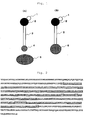
- the positional arrangement of a rod-like spacer substance, an interacting substance, a tag and a labeling substance is, for example, as shown in Fig. 1 (A) .
- the interacting substance [4] is positioned at one end of the rod-like spacer substance [3], and the tag [1] and the labeling substance [2] are positioned at the other end.
- the tag [1] is binding to the labeling substance [2] in this Figure, the tag may be binding to the rod-like spacer substance [3] directly.
- other arrangement opposite to this Figure may be taken in which the tag [1] is binding to the rod-like spacer substance [3] directly and the labeling substance [2] is binding to the tag [1]. With such arrangements, it is possible to retain a specific distance between the target compound [5] and the tag [1]/the labeling substance [2], which leads to elimination of various adverse effects resulting from the neighboring of these substances.
- the tag and label-fused protein of the present invention has a protein body, a rod-like spacer substance, a tag and a labeling substance, wherein the protein body is positioned at one end of the rod-like spacer substance, and the tag and the labeling substance are positioned at the other end of the rod-like spacer substance.
- the rod-like spacer substance, the tag and the labeling substance of the tag and label-fused protein of the present invention may be the same as those substances used in the molecular module of the present invention. Further, the positional arrangement of these substances may be the same as the arrangement in the molecular module ( Fig. 1 (B) ).
- the protein body is not particularly limited. For example, a protein playing an important role in the body, such as receptor protein, may be used. All of the rod-like spacer substance, the tag and the labeling substance are preferably a protein. More preferably, these substances and the protein body form a fusion protein consisting of one polypeptide chain. Specific examples of such fusion protein are given in Figs. 2 to 6 and Fig.
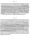
- Fig. 2 shows the amino acid sequence of a fusion protein in which a protein based on the stalk domain of dynein having a mutation introduced thereinto (short type) is used as a rod-like spacer substance; a histidine tag and a biotin acceptor peptide are used as tags; and GFP is used as a labeling substance.
- a protein based on the stalk domain of dynein having a mutation introduced thereinto short type
- a histidine tag and a biotin acceptor peptide are used as tags
- GFP is used as a labeling substance.
- FIG. 3 shows the amino acid sequence of a fusion protein in which a protein based on the stalk domain of dynein having a mutation introduced thereinto (short type) is used as a rod-like spacer substance; a histidine tag and a biotin acceptor peptide are used as tags; and DsRed is used as a labeling substance.
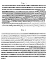
- Fig. 4 shows the amino acid sequence of a fusion protein in which a protein based on the stalk domain of dynein having a mutation introduced thereinto (long type) is used as a rod-like spacer substance; a histidine tag and a biotin acceptor peptide are used as tags; and GFP is used as a labeling substance.
- Fig. 4 shows the amino acid sequence of a fusion protein in which a protein based on the stalk domain of dynein having a mutation introduced thereinto (long type) is used as a rod-like spacer substance; a histidine tag and a biotin
- FIG. 5 shows the amino acid sequence of a fusion protein in which a protein based on the stalk domain of dynein having a mutation introduced thereinto (long type) is used as a rod-like spacer substance; a histidine tag and a biotin acceptor peptide are used as tags; and DsRed is used as a labeling substance.
- Fig. 6 shows the amino acid sequence of a fusion protein in which the rod domain of one polypeptide chain of ⁇ -actinin is used as a rod-like spacer substance; a histidine tag and a biotin acceptor peptide are used as tags; and GFP is used as a labeling substance.
- Fig. 6 shows the amino acid sequence of a fusion protein in which the rod domain of one polypeptide chain of ⁇ -actinin is used as a rod-like spacer substance; a histidine tag and a biotin acceptor peptide are used as tags; and GFP is used as a labeling substance.
- FIG. 19 shows the amino acid sequence of a fusion protein in which the rod domains of two polypeptide chains of ⁇ -actinin are used as a rod-like spacer substance; a histidine tag and a biotin acceptor peptide are used as tags; and GFP is used as a labeling substance.
- the amino acid sequences as shown in Fig. 2 , Fig. 3 , Fig. 4 , Fig. 5 , Fig. 6 and Fig. 19 are also shown in SEQ ID NOS: 1, 2, 3, 4, 5 and 12, respectively.
- the method of protein purification of the present invention is characterized by comprising the following steps: (i) a step of expressing a fusion protein-encoding DNA in a cell, wherein the fusion protein has a protein body, a rod-like spacer substance, a tag and a labeling substance, the protein body being positioned at one end of the rod-like spacer substance, and the tag and the labeling substance being positioned at the other end of the rod-like spacer substance; (ii) a step to homogenize the cell and contacting the resultant homogenate with a substance having affinity to the tag; and (iii) a step of collecting the fusion protein bound to the substance having affinity to the tag.
- This method of protein purification is one application of the tag and label-fused protein of the present invention. It should be noted here that this method may be performed in the same manner as conventional purification methods for tagged proteins except that a tag and a labeling protein are added through a rod-like spacer protein.
- the spacer module has GFP, a histidine tag (8xHis), a biotin acceptor peptide (biotin acceptor domain; BAD) and a protein taking an antiparallel coiled coil structure.
- Fig. 7 shows a schematic diagram of the structure of this spacer module. His-tag and a biotin acceptor domain are inserted into the loop domain of GFP (173-174); a protein taking an antiparallel coiled coil structure is added to the N-terminus and the C-terminus of GFP.
- the protein taking an antiparallel coiled coil structure is based on the stalk domain of cytoplasmic dynein. In order to improve stability, an artificial mutation has been introduced into the stalk domain.
- the protein taking an antiparallel coiled coil structure is different between the spacer module shown in Fig. 2 and the spacer module shown in Fig. 4 .
- the former uses a short type rod-like spacer structure, and the latter uses a long type rod-like spacer structure.
- cDNA fragments encoding the four types of spacer modules designed in Example 1 were inserted into pCold vectors separately and allowed mass expression in Escherichia coli (GFP-short, DsRed-short, GFP-long and DsRed-long).
- the cells were homogenized, and the supernatant was bound to Ni beads.
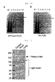
- the resultant beads were washed with a solution containing 20 mM imidazole and eluted with 300 mM imidazole. The eluted fraction was subjected to SDS-PAGE. The results are shown in Fig. 8A .
- the cDNA fragment encoding each of the above-described spacer modules was inserted into pCold TF vector and expressed as a fusion protein with trigger factor (an E. coli protein).
- the fusion protein was purified and observed by rotary shadowing. The results are shown in Fig. 9 .
- Both short type and long type spacer modules showed a dumbbell-like structure.
- the rod-like portion between spherical structures was longer in long type spacer modules. This is consistent with the above-mentioned prediction.
- GFP-short Spacer module alone or in the form of fusion proteins with other proteins, was expressed in HeLa cells, followed by detection of the intracellular GFP fluorescence.
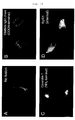
- Micrographs showing the intracellular localization of the spacer module when expressed alone, when expressed as a fusion protein with a clathrin light chain (C-terminal fusion), when expressed as a fusion protein with caveolin-1 (N-terminal fusion) and when expressed as a fusion protein with ryanodine receptor 1 (RyR1) (internal insertion, 1379-1380) are given in Fig. 10A, Fig. 10B, Fig. 10C and Fig. 10D , respectively.
- the spacer module When the spacer module was expressed alone, the spacer module was evenly dispersed within the cell other than the nucleus. When expressed as a fusion protein with a clathrin light chain or caveolin-1, the spacer module was localized as dots around the nucleus and in the cytoplasm. When expressed as a fusion protein with RyR1, the spacer module was localized in a mesh-like manner within the cell, which is consistent that the spacer module was present in endoplasmic reticulum. In any of the fusion proteins tested, the localization pattern was consistent with the endogenous protein. Thus, it was suggested that the spacer module does not affect the structure or function of a protein when fused thereto.
- a fusion protein in which GFP-short is fused to the C-terminus of a clathrin light chain and a fusion protein in which DsRed-short is fused to the N-terminus of caveolin-1 were expressed in HeLa cells, followed by observation of cells expressing both proteins. The results are shown in Fig. 11 .
- a DNA fragment encoding a fusion protein of GFP-short and IP3R1 (N-terminal fusion) was introduced into Flp-In T-REx HEK cells, followed by selection of cell clones expressing the fusion protein stably. After induction of expression with doxycycline, membrane fractions were prepared. IP3R1 was solubilized with CHAPS and applied to an Ni column. The column was washed with 100 mM imidazole, followed by elution with 300 mM imidazole. The proteins included in fractions from individual purification stages were separated by electrophoresis.
- IP3R1 to which a histidine tag had been added at its N-terminus was also expressed in the same manner, and the proteins included in fractions of individual purification stages were examined in the same manner. The results are shown in Fig. 12 .
- the spacer module fused protein was purified in the same manner as the histidine tag-inserted IP3R1 (N-terminus), it was revealed that the spacer module functions as a tag for purification.
- a DNA fragment encoding a fusion protein of GFP-short or GFP-long and a clathrin light chain (C-terminal fusion) was introduced into Flp-In T-REx HEK cells, followed by selection of cell clones expressing the fusion protein stably. After induction of expression with doxycycline, membrane fractions were prepared. Triskelion was extracted with 0.5 M NaCl and applied to a Ni column. The column was washed with 100 mM imidazole, followed by elution with 300 mM imidazole. The proteins contained in the 300 mM imidazole elution fraction were detected by electrophoresis. As a control, a clathrin light chain to which a histidine tag had been added at its C-terminus was expressed. The results are shown in Fig. 13 .
- clathrin heavy chain of 160 kDa was purified together with the spacer module-fused clathrin light chain, it was revealed that clathrin was purified as a protein complex.
- Example 8 The clathrin complexes purified in Example 8 were observed by rotary shadowing.
- the clathrin complex into which a histidine tag had been inserted at the C-terminus showed a typical triskelion structure.
- a spherical structure corresponding to GFP was observed at a site slightly away from the center of triskelion corresponding to the C-terminus of the clathrin light chain.
- the spherical structure corresponding to GFP and even the rod-like portion were observed at sites further away from the center of triskelion.
- a DNA fragment encoding a fusion protein of a spacer module (GFP which comprises a histidine tag and a biotin acceptor sequence and is flanked by TEV protease site on both sides) and a clathrin light chain (C-terminal fusion) was introduced into Flp-In T-REx HEK cells, followed by selection of cell clones expressing the fusion protein stably. After induction of expression with doxycycline, cells were homogenized in a solution containing 0.1 M MES, pH 6.5, 0.5 mM MgCl 2 and 1 mM EGTA, followed by preparation of membrane fractions.
- the resultant membrane fraction was bound to streptavidin magnetic beads, washed with the above-described buffer solution and then treated with TEV protease at room temperature for 1hr. Eluted fractions were observed with electron microscopy after negative staining. The results are shown in Fig. 15 .
- vesicles approximately 100 nm in diameter were observed in the eluted fraction. When enlarged, the vesicles were surrounded by soccer ball-like skeletons. It was confirmed that these vesicles represent the morphology of a typical clathrin coated vesicle.
- the proteins included in the coated vesicle fraction obtained in Example 10 were separated by electrophoresis.
- triskelion obtained by extracting the above fraction with 0.5 M NaCl was purified in the same manner. The results are shown in Fig. 16 .
- a spectrin repeat type spacer module (comprising a protein represented by the amino acid sequence of SEQ ID NO: 10), alone or in the form of a fusion protein with other protein, was expressed in HeLa cells, followed by detection of intracellular GFP fluorescence.
- Micrographs showing the intracellular localization of the spectrin repeat type spacer module when expressed alone and when expressed as a fusion protein with a cytoplasmic dynein light chain (TcTex-1) are given in Fig. 17A and Fig. 17B , respectively.
- the spacer module When the spectrin repeat type spacer module had been expressed alone, the spacer module was evenly dispersed within the cell except the nucleus. When the spacer module had been expressed as a fusion protein with a cytoplasmic dynein light chain, the spectrin repeat type spacer module was localized around the nucleus. Since its localization pattern was consistent with that of the endogenous protein, it was suggested that the spectrin repeat type spacer module does not affect the structure or function of a protein when fused thereto.
- a spectrin repeat type spacer module (comprising a peptide represented by the amino acid sequence of SEQ ID NO: 13 and a peptide represented by the amino acid sequence of SEQ ID NO: 14), alone or in the form of a fusion protein with other protein, was expressed in HeLa cells, followed by detection of intracellular GFP fluorescence.
- Micrographs showing the intracellular localization of the spectrin repeat type spacer module when expressed alone and when expressed as a fusion protein with a clathrin light chain are given in Fig. 18A and Fig. 18B , respectively.
- the spacer module When the spectrin repeat type spacer module had been expressed alone, the spacer module was evenly dispersed within the cell except the nucleus. When the spacer module had been expressed as a fusion protein with a clathrin light chain, the spectrin repeat type spacer module was localized in a dot-like manner around the nucleus and in the cytoplasm. It was suggested that the spectrin repeat type spacer module does not affect the structure or function of a protein when fused thereto.
Landscapes
- Health & Medical Sciences (AREA)
- Life Sciences & Earth Sciences (AREA)
- Engineering & Computer Science (AREA)
- Chemical & Material Sciences (AREA)
- Genetics & Genomics (AREA)
- Molecular Biology (AREA)
- Immunology (AREA)
- Biomedical Technology (AREA)
- Organic Chemistry (AREA)
- Biochemistry (AREA)
- General Health & Medical Sciences (AREA)
- Biotechnology (AREA)
- Zoology (AREA)
- Biophysics (AREA)
- Microbiology (AREA)
- Wood Science & Technology (AREA)
- Medicinal Chemistry (AREA)
- Physics & Mathematics (AREA)
- Bioinformatics & Cheminformatics (AREA)
- Hematology (AREA)
- General Engineering & Computer Science (AREA)
- Urology & Nephrology (AREA)
- Toxicology (AREA)
- Gastroenterology & Hepatology (AREA)
- Cell Biology (AREA)
- Proteomics, Peptides & Aminoacids (AREA)
- Pathology (AREA)
- General Physics & Mathematics (AREA)
- Plant Pathology (AREA)
- Analytical Chemistry (AREA)
- Food Science & Technology (AREA)
- Peptides Or Proteins (AREA)
- Preparation Of Compounds By Using Micro-Organisms (AREA)
Abstract
Description
- The present invention relates to a molecular module for purifying or labeling a target compound; a tag and a label-fused protein based on the molecular module; and a method of protein purification using the molecular module.
- In order to explore the nature of a molecule that could be a receptor for a specific ligand (such as physiologically active substance, drug or antibody that acts upon binding to a cell composing, macromolecular component, e.g., protein or nucleic acid), usually, several techniques selected from various existing technologies are used in combination. For example, various techniques such as a technique of identifying an unknown receptor by such as photoaffinity labeling using a drug derivative or the like as a ligand; a technique of the intracellular localization of the receptor; a technique of isolating/purifying the receptor and examining its nature; or a technique of elucidating the binding site within the receptor (complex) molecule with a higher spatial resolution must be used jointly as the purpose demands. Even when one ligand is to be investigated, it is necessary to prepare separately a number of ligand derivatives suitable for individual techniques, which imposes a tremendous burden.
- As a method of identifying those components to which a specific drug binds, affinity labeling is known. In this method, a ligand derivative to which a fluorescent dye or radioactive isotope has been added is photo-crosslinked to a target. Subsequently, information such as the molecular weight or amino acid sequence of the resultant labeled molecule is obtained using electrophoresis or various chromatographies.
- For isolation and purification of a substance which will be a target binding partner for a ligand (such as receptor), a series of techniques called affinity purification is often used. When the target is a protein or a complex thereof, the classical chromatography is usually used in which resin beads immobilizing a ligand by covalent bond are packed in a column; a raw solution containing the target material is applied to the column; and the bound fractions alone are dissociated and eluted. However, when the target is a large-sized membrane fraction or non-adherent cell each of which is difficult to apply to the column, a batch method is also used in which similar resin beads or magnetic beads are utilized to collect the target substance by centrifugation or magnetism. In particular, when a target substance is to be separated after binding to a protein that is embedded in the membrane (such as intracellular organelle or non-adherent cell), a ligand directly immobilized on the surfaces of resin or magnetic beads is difficult to contact the target substance in many cases. In order to solve this problem, a spacer consisting of long straight carbon chain is inserted between the beads and the ligand. However, when a long carbon chain is used, hydrophobicity often increases. This makes it highly possibility that the binding of the target substance by the ligand does not necessarily reflect their specificity. For alleviation of this problem, it is desirable to use a spacer capable of always retaining a long distance.
- On the other hand, in order to microscopically indicate the intracellular or extracellular localization site of a ligand-binding protein, a part or the whole of the protein prepared by a biochemical or molecular biological technique is used to prepare a specific antibody. Then, the localization (site) of the protein is elucidated by immunofluorescence or immunoelectron microscopy using the antibody. Further, for searching the binding site within the target molecule (complex), conventionally, structural data are collected by those means capable of obtaining atomic resolution, e.g., purifying and crystallizing the receptor/ligand complex and subjecting the crystal to X ray diffraction.
- Although comprehensive methods to explore a large number of materials have been invented (e.g., use of robots), basically, researchers have no choice but to combine these techniques and proceed step by step in order to achieve their initial purpose. Regardless of what processes are employed, it is necessary to prepare individual ligands suitable for selected techniques. This always imposes a considerable burden in terms of labor, time and cost.
[Patent Document 1] Japanese Unexamined Patent Publication No.2005-291836 - Under the above-described circumstances, a convenient material which does not affect the specific affinity between ligand and receptor and thus can be used throughout all the exploring processes will be able to reduce the above-mentioned burden greatly. It is desired to make full use of protein engineering or organic chemistry to thereby develop a general-purpose protein module that can be used for such a purpose. In reply to this demand, the present invention aims to provide a general-purpose experimental tool which specifically binds to a macromolecular substance that will be a receptor for a specific ligand such as drug, and is applicable throughout various processes to characterize the properties of the macromolecular substance.
- As a result of extensive and intensive researches toward the solution of the above-described problem, the present inventors have found that by adding to a target compound to be explored a tag and a label through a rod-like spacer substance, it is possible to give a molecular module a form suitable for wide-ranged exploration techniques (such as identification, isolation, microscopic observation, etc.) without altering the nature of the target compound.
- It is also possible to add a tag and a label directly to a target substance. For example, the following methods may be contemplated; a method in which the target compound to be explored is fused to a tag and a label, and a method in which a substance with affinity for the target compound is added to both tag and label, and the resultant tag and label are bound to the target compound. However, the former method involves a possibility that the insertion of tag and label may destroy or alter the conformation of the target compound, resulting in the loss of the inherent nature of the target compound. In the latter method, the label and the tag may not bind to the target compound well due to steric hindrance of the target compound or compounds adjacent thereto. Even when the label and the tag could bind to the target compound, they may not be sufficiently exposed from the surface to the target compound, resulting in insufficient function as a tag and a label.
- The present invention has been achieved based on the above-described findings.
- The present invention provides the following (1) to (14).
- (1) A molecular module which binds to a target compound and is used for purifying or labeling the target compound, wherein the molecular module has a rod-like spacer substance, an interacting substance that interacts with the target compound, a tag and a labeling substance, the interacting substance being positioned at one end of the rod-like spacer substance, and the tag and the labeling substance being positioned at the other end of the rod-like spacer substance.
- (2) The molecular module according to (1), wherein the rod-like spacer substance, the interacting substance, the tag and the labeling substance form a polypeptide chain.
- (3) The molecular module according to (1) or (2), wherein the rod-like spacer substance is a protein that takes an antiparallel coiled coil structure or a protein that takes a spectrin repeat structure.
- (4) The molecular module according to any one of (1) to (3), wherein the tag is a histidine tag or a biotin acceptor peptide.
- (5) The molecular module according to any one of (1) to (4), wherein the labeling substance is GFP or DsRed.
- (6) A tag and a label-fused protein having a protein body, a rod-like spacer substance, a tag and a labeling substance, wherein the protein body is positioned at one end of the rod-like spacer substance, and the tag and the labeling substance are positioned at the other end of the rod-like spacer substance.
- (7) The tag and label-fused protein according to (6), wherein the protein body, the rod-like spacer substance, the tag and the labeling substance form a polypeptide chain.
- (8) The tag and label-fused protein according to (6) or (7), wherein the rod-like spacer substance is a protein that takes an antiparallel coiled coil structure or a protein that takes a spectrin repeat structure.
- (9) The tag and label-fused protein according to any one of (6) to (8), wherein the tag is a histidine tag or a biotin acceptor peptide.
- (10) The tag and label-fused protein according to any one of (6) to (9), wherein the labeling substance is GFP or DsRed.
- (11) A method of protein purification comprising the following steps:
- (i) a step of expressing a fusion protein-encoding DNA in a cell, wherein the fusion protein has a protein body, a rod-like spacer substance, a tag and a labeling substance, the protein body being positioned at one end of the rod-like spacer substance, and the tag and the labeling substance being positioned at the other end of the rod-like spacer substance;
- (ii) a step of disrupting the cell and contacting the resultant homogenate with a substance having affinity for the tag; and
- (iii) a step of collecting the fusion protein bound to the substance having affinity to the tag.
- (12) The method according to (11), wherein the rod-like spacer substance is a protein that takes an antiparallel coiled coil structure or a protein that takes a spectrin repeat structure.
- (13) The method according to (11) or (12), wherein the tag is a histidine tag or a biotin acceptor peptide.
- (14) The method according to any one of (11) to (13), wherein the labeling substance is GFP or DsRed.
- Conventionally, for exploring the properties of a substance, it was necessary to convert a target substance into a form suitable for each of the exploring techniques selected. However, by using the molecular module of the present invention, it becomes possible to omit such complicated operations.
-
-
Fig. 1 shows a schematic diagram of the molecular module of the present invention (A) and a schematic diagram of the tag and label-fused protein of the present invention (B). -
Fig. 2 shows the amino acid sequence of a fusion protein comprising a protein based on the stalk domain of dynein having a mutation introduced thereinto (a short-type protein of rod-like spacer structure is used; hereafter, a protein of this type is referred to as the "short type"), a histidine tag, a biotin acceptor peptide and GFP. The portion marked with a single underline is GFP; the portion marked with a double underline is the biotin acceptor peptide; the portion marked with a dotted underline is the histidine tag; the portions enclosed with boxes are linkers; and the portions without any marking represent the dynein protein whose stalk domain has been mutated. -
Fig. 3 shows the amino acid sequence of a fusion protein comprising a protein based on the stalk domain of dynein having a mutation introduced thereinto (short type), a histidine tag, a biotin acceptor peptide and DsRed. The portion marked with a single underline is DsRed; the portion marked with a double underline is the biotin acceptor peptide; the portion marked with a dotted underline is the histidine tag; the portions enclosed with boxes are linkers; and the portions without any marking represent the dynein protein whose stalk domain has been mutated. -
Fig. 4 shows the amino acid sequence of a fusion protein comprising a protein based on the stalk domain of dynein having a mutation introduced thereinto (a long-type protein of rod-like spacer structure is used; hereinafter, a protein of this type is referred to as the "long type"), a histidine tag, a biotin acceptor peptide and GFP. The portion marked with a single underline is GFP; the portion marked with a double underline is the biotin acceptor peptide; the portion marked with a dotted underline is the histidine tag; the portions enclosed with boxes are linkers; and the portions without any marking represent the dynein protein whose stalk domain has been mutated. -
Fig. 5 shows the amino acid sequence of a fusion protein comprising a protein based on the stalk domain of dynein having a mutation introduced thereinto (long type), a histidine tag, a biotin acceptor peptide and DsRed. The portion marked with a single underline is DsRed; the portion marked with a double underline is the biotin acceptor peptide; the portion marked with a dotted underline is the histidine tag; the portions enclosed with boxes are linkers; and the portions without any marking represent the dynein protein whose stalk domain has been mutated. -
Fig. 6 shows the amino acid sequence of a fusion protein comprising the rod domain of one polypeptide chain of α-actinin, a histidine tag, a biotin acceptor peptide and GFP. The portion marked with a single underline is GFP; the portion marked with a double underline is the biotin acceptor peptide; the portion marked with a dotted underline is the histidine tag; the portions enclosed with boxes are linkers; and the portions without any marking represent the rod domain of α-actinin. -
Fig. 7 is a schematic diagram showing the structure of a spacer module. -
Fig. 8 shows an electrophoretic gel pattern of a spacer module purified with Ni beads (Panel A) and a diagram showing the results of an experiment examining the reactivity between a spacer module purified with Ni beads and streptavidin (Panel B). -
Fig. 9 shows the results of observation of spacer modules by rotary shadowing. Arrow marks in this Figure indicate spacer modules. -
Fig. 10 shows the intracellular localization of spacer module-linked fusion proteins. -
Fig. 11 shows the intracellular localization of a spacer module-linked clathrin light chain and a spacer module-linked caveolin-1. -
Fig. 12 shows electrophoretic gel pattern of fractions obtained from individual purification steps when a fusion protein of a spacer module and IP3R1 was purified with a Ni column. Panel A shows the results for a spacer module-fused IP3R1. Panel B shows the results for a His tag-added IP3R1. Lane 1: sample applied to the column; lane 2: those which passed through the column; lane 3: first washed fraction; lane 4: second washed fraction;lanes 5 to 8: eluted fractions. -
Fig. 13 shows an electrophoretic gel pattern obtained when an extract from HEK cells was purified with an Ni column, wherein the HEK cells express clathrin light chain to which a spacer module was linked at its C terminus. In the control, a histidine tag was added instead of a spacer module. -
Fig. 14 shows the observed images by rotary shadowing of clathrin molecules (triskelion) purified from a spacer module-linked clathrin light-chain expressing HEK cells using the module. Arrow marks in this Figure indicate the GFP of the spacer module. Although short type ("short" in the Figure) and long type ("long" in the Figure) spacer modules are different in length, both modules protrudes from the center of the molecule, indicating the localization of the other end of modules therein. -
Fig. 15 shows the observed images by negative staining of coated vesicles purified from a spacer module-fused clathrin light-chain expressing HEK cells. Most of the particles seen under low-magnification (lower panel) are coated vesicles. Individual particles indicated with arrow marks are enlarged in the upper panel. The inserted diagram is a schematic diagram showing molecular arrangement in the coated vesicle. -
Fig. 16 shows an (electrophoretic gel pattern?) of coated vesicles purified from a spacer module-fused clathrin light chain expressing HEK cells (right lane). The central lane represents clathrin molecules (triskelion) which were purified alone after solubilization. The left lane represents molecular weight marker. The fraction purified as coated vesicles contains a large number of component proteins other than clathrin. -
Fig. 17 shows the intracellular localization of a cytoplasmic dynein light chain linked to a spectrin repeat type spacer module. Panel A shows the spacer module alone, and Panel B shows the spacer linked to cytoplasmic dynein light chain. -
Fig. 18 shows the intracellular localization of a clathrin light chain fused to a spectrin repeat type spacer module. While no localization at a specific site is observed when the spacer module was expressed alone (Panel A), localization equivalent to that of wild-type clathrin was observed when the spacer module was fused to the clathrin light chain (Panel B). -
Fig. 19 shows the amino acid sequence of a fusion protein comprising the rod domains of two polypeptide chains of α-actinin, a histidine tag, a biotin acceptor peptide and GFP. The portion marked with a single underline is GFP; the portion marked with a double underline is the biotin acceptor peptide; the portion marked with a dotted underline is the histidine tag; the portions enclosed with boxes are linkers; and the portions without any marking represent the rod domains of α-actinin. -
- 1. Tag
- 2. Labeling substance
- 3. Rod-like spacer substance
- 4. Interacting substance
- 5. Target compound
- 6. Protein body
- Hereinbelow, the present invention will be described in detail.
- The molecular module of the present invention binds to a target compound and is used for purifying or labeling the compound. The molecular module of the present invention has a rod-like spacer substance, an interacting substance that interacts with the target compound, a tag and a labeling substance, wherein the interacting substance is positioned at one end of the rod-like spacer substance, and the tag and the labeling substance are positioned at the other end of the rod-like spacer substance.
- The target compound is not particularly limited as long as the compound is capable of being purified or labeled by the molecular module of the present invention. Biopolymers such as protein or nucleic acid, or low molecular weight compounds binding thereto may be used widely.
- The rod-like spacer substance is not particularly limited as long as the substance can take a rod-like spacer structure. Preferably, the rod-like spacer substance is a protein. Preferable examples include, but are not limited to, a protein taking an antiparallel coiled coil structure, a protein taking a spectrin repeat structure, a filamentous phage and a filamentous protein of a phage. As the protein taking an antiparallel coiled coil structure, for example, the stalk domain of dynein (a motor protein) or a protein based on the stalk domain of dynein to which an artificial mutation has been added in order to stabilize the antiparallel coiled coil structure may be given. Examples of such artificial mutation-added proteins include a protein consisting of a peptide represented by the amino acid sequence of SEQ ID NO: 6 and a peptide represented by the amino acid sequence of SEQ ID NO: 7 (short type) and a protein consisting of a peptide represented by the amino acid sequence of SEQ ID NO: 8 and a peptide represented by the amino acid sequence of SEQ ID NO: 9 (long type). In these examples, the length of the long type rod-like spacer structure is 150% of the length of the short type rod-like spacer structure. In addition to the above-listed examples, antiparallel coiled coil structures may be prepared based on the disclosure in Current Opinion in Structural Biology 2001,11:450-457 and so forth. As the protein taking a spectrin repeat structure, for example, the rod domain of α-actinin or a protein based on the rod domain of α-actinin into which an artificial mutation has been added to stabilize the spectrin repeat structure may be given. Examples of the rod domain of α-actinin include a protein represented by the amino acid sequence of SEQ ID NO: 10 and a protein consisting of a peptide represented by the amino acid sequence of SEQ ID NO: 13 and a peptide represented by the amino acid sequence of SEQ ID NO: 14. In the latter protein consisting of two peptides, the labeling substance or the like is inserted between the two peptides. Examples of filamentous phage include f1 phage, fd phage and M13 phage. The rod-like spacer substance may be a substance other than protein. Examples of such non-proteinaceous substances include, but are not limited to, carbon nanotube, carbon nanohorn and amylose.
- The length and the diameter of rod-like spacer substance are not particularly limited as long as an appropriate distance can be secured between the target compound and the tag/the label. The length is preferably 5-50 nm, more preferably 10-30 nm. The diameter is preferably 1-10 nm, more preferably 2-5 nm.
- The interacting substance may be any substance as long as it interacts with the target substance. Proteins and low molecular weight ligands may be given. The interacting substance may be selected depending on the type of the target compound. For example, when the target compound is a protein, the interacting substance may be an antibody that recognizes the protein; when the target compound is an antibody, the interacting substance may be an antigen that the antibody recognizes; when the target compound is a receptor, the interacting substance may be a ligand for the receptor; and when the target compound is a ligand, the interacting substance may be a receptor for the ligand.
- The tag may be a conventional tag used in protein purification. Specific examples of such tag include, but are not limited to, histidine tags, biotin acceptor peptides (e.g., a peptide represented by the amino acid sequence of SEQ ID NO: 11), polyarginine and FK506 binding protein (FKBP). One or more tags may be used in the molecular module. When the target compound is a protein, the tag may be inserted into the protein. The site of tag insertion is not particularly limited as long as the insertion does not impair the function of the protein used as a target compound. When the protein takes a loop structure, it is preferable to insert the tag into the loop. The insertion site in a loop structure may be, for example, between amino acid residues 173 and 174 of GFP or between amino acid residues 170 and 171 of DsRed.
- The labeling substance may be a conventional substance generally used for labeling biomolecules. Examples of such labeling substance include, but are not limited to, fluorescent substances, dyes, heavy metal compounds, heavy metal colloids and oxidoreductases. Preferably, the labeling substance is proteinaceous. More preferably, a fluorescent protein is used. As a preferable fluorescent protein, GFP (Aequorea victoria green fluorescence protein) or DsRed (Discosoma sp. red fluorescence protein) may be given. Other then these fluorescent proteins, variants of GFP such as enhanced green fluorescence protein (EGFP), yellow fluorescence protein (YFP), enhanced yellow fluorescence protein (EYFP), cyan fluorescence protein (CFP), enhanced cyan fluorescence protein (ECFP), blue fluorescence protein (BFP) and enhanced blue fluorescence protein (EBFP), as well as variants of DsRed such as monomeric Banana yellow fluorescence protein (mBanana), monomeric Orange fluorescence protein (mOrange), monomeric Tangerine fluorescence protein (mTangerine), monomeric Strawberry red fluorescence protein (mStrawberry) and monomeric Cherry red fluorescence protein (mCherry) may also be used. Alternatively, a non-proteinaceous fluorescent substance may be used as a labeling substance. For example, fluorescein, Rhodamine, eosin or NBD fluorescent substances, or the like may be used. Specific examples include, but are not limited to, fluorescein-5-isothiocyanate, diacyl (such as isobutyryl, acetyl or pivaloyl) fluorescein-5 and/or 6-carboxylic acid pentafluorophenyl ester, 6-(diacyl-5 and/or 6-carboxamide-fluorescein)aminohexanoic acid pentafluorophenyl ester, Texas Red (Trademark of Molecular Probes, Inc.), tetramethylrhodamine-5 (and 6) isothiocyanate, oesin-isothiocyanate, erythrosin-5-isothiocyanate, 4-chloro-7-nitrobenz-2-oxa-1,3-diazole, 4-fluoro-7-nitrobenz-2-oxa-1,3-diazol, 3-(7-nitrobenz-2-oxa- 1,3-diazol-4-yl)methylaminopropionitrile, 6-(7-nitrobenz-2-oxa-1,3-diazol-4-yl)aminohexanoic acid, succinimidyl 12-(N-methyl-N-(7-nitrobenz-2-oxa-1,3-diazol-4-71)aminododecanoate, 7-diethylamino-3-(4'-isothiocyanatophenyl)-4-methylcoumarin (CP); 7-hydroxycoumarin-4-acetic acid, 7-dimethylaminocoumarin-4-acetic acid, succinimidyl 7-dimethylaminocoumarin-4-acetate, 7-methoxycoumarin-4-acetic acid, 4-acetamide-4'-isothiocyanatostilbene-2-2'-disulfonic acid (SITS), 9-chloroacridine, succinimidyl 3-(9-carbazole)propionate, succinimidyl 1-pyrenebutyrate, succinimidyl 1-pyrenenonanoate, p-nitrophenyl 1-pyrenebutyrate, 9-anthracenepropionic acid, succinimidyl anthracene-9-propionate and 2-anthracenesulfonyl chloride.
- The three members of the molecular module of rod-like spacer substance, tag and labeling substance may be in the form of a fusion protein consisting of one polypeptide chain. In this case, when the interacting substance is a low molecular weight ligand, this substance may be covalently bonded through an active group located at the end of the rod-like spacer substance (e.g., α-amino group of N-terminal amino acid, carboxyl group of C-terminal amino acid, or thiol group of cystein residue). When the interacting substance is a protein, it is also possible to prepare a fusion protein of four members including the interacting substance.
- The molecular module of the present invention is used for purifying and labeling a target compound. Specifically, the molecular module may be used for the following purposes.
- When the molecular module of the present invention is added to a crude extract from a biological component, one end of the molecular module specifically binds to the biological component of interest to thereby form a complex. This complex may be recovered with resin beads or magnetic beads through the tag in the module. At the time of recovery, the target for purification may be purified alone or as the complex, or even as a still larger substance (such as intracellular organelle or cell as a whole) by adding to the solution salts or surfactants as the purpose demands.
- By using a fluorescent substance as the labeling substance, microscopic observation of fluorescence signals makes it possible to indicate the localization of the target compound in cells or intracellular organelles or to trace the kinetics of the target compound in vivo . Further, by immunoelectron microscopy using an appropriate gold colloid that binds to the tag, it is also possible to explore the ligand binding site in tissue samples. Furthermore, since the molecular module of the present invention has a unique shape with a spherical portion at the end of its rod, it is believed possible to show the binding domain in protein molecules or complexes directly with the use of high resolution electronmicroscopic images.
- The positional arrangement of a rod-like spacer substance, an interacting substance, a tag and a labeling substance is, for example, as shown in
Fig. 1 (A) . The interacting substance [4] is positioned at one end of the rod-like spacer substance [3], and the tag [1] and the labeling substance [2] are positioned at the other end. Although the tag [1] is binding to the labeling substance [2] in this Figure, the tag may be binding to the rod-like spacer substance [3] directly. Alternatively, other arrangement opposite to this Figure may be taken in which the tag [1] is binding to the rod-like spacer substance [3] directly and the labeling substance [2] is binding to the tag [1]. With such arrangements, it is possible to retain a specific distance between the target compound [5] and the tag [1]/the labeling substance [2], which leads to elimination of various adverse effects resulting from the neighboring of these substances. - The tag and label-fused protein of the present invention has a protein body, a rod-like spacer substance, a tag and a labeling substance, wherein the protein body is positioned at one end of the rod-like spacer substance, and the tag and the labeling substance are positioned at the other end of the rod-like spacer substance.
- The rod-like spacer substance, the tag and the labeling substance of the tag and label-fused protein of the present invention may be the same as those substances used in the molecular module of the present invention. Further, the positional arrangement of these substances may be the same as the arrangement in the molecular module (
Fig. 1 (B) ). The protein body is not particularly limited. For example, a protein playing an important role in the body, such as receptor protein, may be used. All of the rod-like spacer substance, the tag and the labeling substance are preferably a protein. More preferably, these substances and the protein body form a fusion protein consisting of one polypeptide chain. Specific examples of such fusion protein are given inFigs. 2 to 6 andFig. 19 (however, the protein body is not included therein).Fig. 2 shows the amino acid sequence of a fusion protein in which a protein based on the stalk domain of dynein having a mutation introduced thereinto (short type) is used as a rod-like spacer substance; a histidine tag and a biotin acceptor peptide are used as tags; and GFP is used as a labeling substance.Fig. 3 shows the amino acid sequence of a fusion protein in which a protein based on the stalk domain of dynein having a mutation introduced thereinto (short type) is used as a rod-like spacer substance; a histidine tag and a biotin acceptor peptide are used as tags; and DsRed is used as a labeling substance.Fig. 4 shows the amino acid sequence of a fusion protein in which a protein based on the stalk domain of dynein having a mutation introduced thereinto (long type) is used as a rod-like spacer substance; a histidine tag and a biotin acceptor peptide are used as tags; and GFP is used as a labeling substance.Fig. 5 shows the amino acid sequence of a fusion protein in which a protein based on the stalk domain of dynein having a mutation introduced thereinto (long type) is used as a rod-like spacer substance; a histidine tag and a biotin acceptor peptide are used as tags; and DsRed is used as a labeling substance.Fig. 6 shows the amino acid sequence of a fusion protein in which the rod domain of one polypeptide chain of α-actinin is used as a rod-like spacer substance; a histidine tag and a biotin acceptor peptide are used as tags; and GFP is used as a labeling substance.Fig. 19 shows the amino acid sequence of a fusion protein in which the rod domains of two polypeptide chains of α-actinin are used as a rod-like spacer substance; a histidine tag and a biotin acceptor peptide are used as tags; and GFP is used as a labeling substance. The amino acid sequences as shown inFig. 2 ,Fig. 3 ,Fig. 4 ,Fig. 5 ,Fig. 6 andFig. 19 are also shown in SEQ ID NOS: 1, 2, 3, 4, 5 and 12, respectively. - The method of protein purification of the present invention is characterized by comprising the following steps: (i) a step of expressing a fusion protein-encoding DNA in a cell, wherein the fusion protein has a protein body, a rod-like spacer substance, a tag and a labeling substance, the protein body being positioned at one end of the rod-like spacer substance, and the tag and the labeling substance being positioned at the other end of the rod-like spacer substance; (ii) a step to homogenize the cell and contacting the resultant homogenate with a substance having affinity to the tag; and (iii) a step of collecting the fusion protein bound to the substance having affinity to the tag.
- This method of protein purification is one application of the tag and label-fused protein of the present invention. It should be noted here that this method may be performed in the same manner as conventional purification methods for tagged proteins except that a tag and a labeling protein are added through a rod-like spacer protein.
- Hereinbelow, the present invention will be described more specifically with reference to the following Examples.
- Proteins consisting of the amino acid sequences as shown in
Fig. 2 andFig. 4 , respectively, were designed (hereinafter, these proteins are called "spacer modules"). The spacer module has GFP, a histidine tag (8xHis), a biotin acceptor peptide (biotin acceptor domain; BAD) and a protein taking an antiparallel coiled coil structure.Fig. 7 shows a schematic diagram of the structure of this spacer module. His-tag and a biotin acceptor domain are inserted into the loop domain of GFP (173-174); a protein taking an antiparallel coiled coil structure is added to the N-terminus and the C-terminus of GFP. The protein taking an antiparallel coiled coil structure is based on the stalk domain of cytoplasmic dynein. In order to improve stability, an artificial mutation has been introduced into the stalk domain. The protein taking an antiparallel coiled coil structure is different between the spacer module shown inFig. 2 and the spacer module shown inFig. 4 . The former uses a short type rod-like spacer structure, and the latter uses a long type rod-like spacer structure. - Further, similar spacer modules were also designed using DsRed instead of GFP (
Fig. 3 andFig. 5 ). - cDNA fragments encoding the four types of spacer modules designed in Example 1 were inserted into pCold vectors separately and allowed mass expression in Escherichia coli (GFP-short, DsRed-short, GFP-long and DsRed-long). The cells were homogenized, and the supernatant was bound to Ni beads. The resultant beads were washed with a solution containing 20 mM imidazole and eluted with 300 mM imidazole. The eluted fraction was subjected to SDS-PAGE. The results are shown in
Fig. 8A . - Since every spacer module was detected as a single band, it was demonstrated that the histidine tag is functioning as a tag for purification. It was predicted that the rod-like portion of the short type spacer module is 16 nm long, and that portion of the long type spacer module is 24 nm long.
- After SDS polyacrylamide-gel electrophoresis, the spacer modules purified with Ni beads were transferred onto a PVDF membrane, followed by blotting with streptavidin HRP. The results are shown in
Fig. 8B . - Every spacer module reacted with streptavidin, showing that every spacer module was biotinylated.
- The cDNA fragment encoding each of the above-described spacer modules was inserted into pCold TF vector and expressed as a fusion protein with trigger factor (an E. coli protein). The fusion protein was purified and observed by rotary shadowing. The results are shown in
Fig. 9 . - Both short type and long type spacer modules showed a dumbbell-like structure. The rod-like portion between spherical structures was longer in long type spacer modules. This is consistent with the above-mentioned prediction.
- Spacer module (GFP-short), alone or in the form of fusion proteins with other proteins, was expressed in HeLa cells, followed by detection of the intracellular GFP fluorescence. Micrographs showing the intracellular localization of the spacer module when expressed alone, when expressed as a fusion protein with a clathrin light chain (C-terminal fusion), when expressed as a fusion protein with caveolin-1 (N-terminal fusion) and when expressed as a fusion protein with ryanodine receptor 1 (RyR1) (internal insertion, 1379-1380) are given in
Fig. 10A, Fig. 10B, Fig. 10C and Fig. 10D , respectively. - When the spacer module was expressed alone, the spacer module was evenly dispersed within the cell other than the nucleus. When expressed as a fusion protein with a clathrin light chain or caveolin-1, the spacer module was localized as dots around the nucleus and in the cytoplasm. When expressed as a fusion protein with RyR1, the spacer module was localized in a mesh-like manner within the cell, which is consistent that the spacer module was present in endoplasmic reticulum. In any of the fusion proteins tested, the localization pattern was consistent with the endogenous protein. Thus, it was suggested that the spacer module does not affect the structure or function of a protein when fused thereto.
- A fusion protein in which GFP-short is fused to the C-terminus of a clathrin light chain and a fusion protein in which DsRed-short is fused to the N-terminus of caveolin-1 were expressed in HeLa cells, followed by observation of cells expressing both proteins. The results are shown in
Fig. 11 . - Although both fusion proteins were localized in dotted manner around the nucleus and in the cytoplasm, their distributions were different from each other.
- A DNA fragment encoding a fusion protein of GFP-short and IP3R1 (N-terminal fusion) was introduced into Flp-In T-REx HEK cells, followed by selection of cell clones expressing the fusion protein stably. After induction of expression with doxycycline, membrane fractions were prepared. IP3R1 was solubilized with CHAPS and applied to an Ni column. The column was washed with 100 mM imidazole, followed by elution with 300 mM imidazole. The proteins included in fractions from individual purification stages were separated by electrophoresis. For the purpose of comparison, IP3R1 to which a histidine tag had been added at its N-terminus was also expressed in the same manner, and the proteins included in fractions of individual purification stages were examined in the same manner. The results are shown in
Fig. 12 . - Since the spacer module fused protein was purified in the same manner as the histidine tag-inserted IP3R1 (N-terminus), it was revealed that the spacer module functions as a tag for purification.
- A DNA fragment encoding a fusion protein of GFP-short or GFP-long and a clathrin light chain (C-terminal fusion) was introduced into Flp-In T-REx HEK cells, followed by selection of cell clones expressing the fusion protein stably. After induction of expression with doxycycline, membrane fractions were prepared. Triskelion was extracted with 0.5 M NaCl and applied to a Ni column. The column was washed with 100 mM imidazole, followed by elution with 300 mM imidazole. The proteins contained in the 300 mM imidazole elution fraction were detected by electrophoresis. As a control, a clathrin light chain to which a histidine tag had been added at its C-terminus was expressed. The results are shown in
Fig. 13 . - Since a clathrin heavy chain of 160 kDa was purified together with the spacer module-fused clathrin light chain, it was revealed that clathrin was purified as a protein complex.
- The clathrin complexes purified in Example 8 were observed by rotary shadowing.
- The clathrin complex into which a histidine tag had been inserted at the C-terminus showed a typical triskelion structure. In the short type spacer module-fused clathrin light chain, a spherical structure corresponding to GFP was observed at a site slightly away from the center of triskelion corresponding to the C-terminus of the clathrin light chain. In the long type spacer module-fused clathrin light chain, the spherical structure corresponding to GFP and even the rod-like portion were observed at sites further away from the center of triskelion. Thus, the localization of the spacer module in the protein complexes could be confirmed.
- A DNA fragment encoding a fusion protein of a spacer module (GFP which comprises a histidine tag and a biotin acceptor sequence and is flanked by TEV protease site on both sides) and a clathrin light chain (C-terminal fusion) was introduced into Flp-In T-REx HEK cells, followed by selection of cell clones expressing the fusion protein stably. After induction of expression with doxycycline, cells were homogenized in a solution containing 0.1 M MES, pH 6.5, 0.5 mM MgCl2 and 1 mM EGTA, followed by preparation of membrane fractions. The resultant membrane fraction was bound to streptavidin magnetic beads, washed with the above-described buffer solution and then treated with TEV protease at room temperature for 1hr. Eluted fractions were observed with electron microscopy after negative staining. The results are shown in
Fig. 15 . - A great number of vesicles approximately 100 nm in diameter were observed in the eluted fraction. When enlarged, the vesicles were surrounded by soccer ball-like skeletons. It was confirmed that these vesicles represent the morphology of a typical clathrin coated vesicle.
- The proteins included in the coated vesicle fraction obtained in Example 10 were separated by electrophoresis. For the purpose of comparison, triskelion obtained by extracting the above fraction with 0.5 M NaCl was purified in the same manner. The results are shown in
Fig. 16 . - In the coated vesicle fraction, bands of a large number of other component proteins in addition to the clathrin heavy and light chains contained in triskelion were detected. The band around 27 kDa is derived from TEV protease. It was revealed that the spacer module functions effectively as a tag for purifying intracellular organelles.
- A spectrin repeat type spacer module (comprising a protein represented by the amino acid sequence of SEQ ID NO: 10), alone or in the form of a fusion protein with other protein, was expressed in HeLa cells, followed by detection of intracellular GFP fluorescence. Micrographs showing the intracellular localization of the spectrin repeat type spacer module when expressed alone and when expressed as a fusion protein with a cytoplasmic dynein light chain (TcTex-1) are given in
Fig. 17A and Fig. 17B , respectively. - When the spectrin repeat type spacer module had been expressed alone, the spacer module was evenly dispersed within the cell except the nucleus. When the spacer module had been expressed as a fusion protein with a cytoplasmic dynein light chain, the spectrin repeat type spacer module was localized around the nucleus. Since its localization pattern was consistent with that of the endogenous protein, it was suggested that the spectrin repeat type spacer module does not affect the structure or function of a protein when fused thereto.
- A spectrin repeat type spacer module (comprising a peptide represented by the amino acid sequence of SEQ ID NO: 13 and a peptide represented by the amino acid sequence of SEQ ID NO: 14), alone or in the form of a fusion protein with other protein, was expressed in HeLa cells, followed by detection of intracellular GFP fluorescence. Micrographs showing the intracellular localization of the spectrin repeat type spacer module when expressed alone and when expressed as a fusion protein with a clathrin light chain are given in
Fig. 18A and Fig. 18B , respectively. - When the spectrin repeat type spacer module had been expressed alone, the spacer module was evenly dispersed within the cell except the nucleus. When the spacer module had been expressed as a fusion protein with a clathrin light chain, the spectrin repeat type spacer module was localized in a dot-like manner around the nucleus and in the cytoplasm. It was suggested that the spectrin repeat type spacer module does not affect the structure or function of a protein when fused thereto.
- The present specification encompasses the disclosure of the specification and/or drawings of Japanese Patent Application No.
2006-332530 


Claims (14)
- A molecular module which binds to a target compound and is used for purifying or labeling the target compound, wherein the molecular module has a rod-like spacer substance, an interacting substance that interacts with the target compound, a tag and a labeling substance, the interacting substance being positioned at one end of the rod-like spacer substance, and the tag and the labeling substance being positioned at the other end of the rod-like spacer substance.
- The molecular module according to claim 1, wherein the rod-like spacer substance, the interacting substance, the tag and the labeling substance form a polypeptide chain.
- The molecular module according to claim 1 or 2, wherein the rod-like spacer substance is a protein that takes an antiparallel coiled coil structure or a protein that takes a spectrin repeat structure.
- The molecular module according to any one of claims 1 to 3, wherein the tag is a histidine tag or a biotin acceptor peptide.
- The molecular module according to any one of claims 1 to 4, wherein the labeling substance is GFP or DsRed.
- A tag and a label-fused protein having a protein body, a rod-like spacer substance, a tag and a labeling substance, wherein the protein body is positioned at one end of the rod-like spacer substance, and the tag and the labeling substance are positioned at the other end of the rod-like spacer substance.
- The tag and label-fused protein according to claim 6, wherein the protein body, the rod-like spacer substance, the tag and the labeling substance form a polypeptide chain.
- The tag and label-fused protein according to claim 6 or 7, wherein the rod-like spacer substance is a protein that takes an antiparallel coiled coil structure or a protein that takes a spectrin repeat structure.
- The tag and label-fused protein according to any one of claims 6 to 8, wherein the tag is a histidine tag or a biotin acceptor peptide.
- The tag and label-fused protein according to any one of claims 6 to 9, wherein the labeling substance is GFP or DsRed.
- A method of protein purification comprising the following steps:(i) a step of expressing a fusion protein-encoding DNA in a cell, wherein the fusion protein has a protein body, a rod-like spacer substance, a tag and a labeling substance, the protein body being positioned at one end of the rod-like spacer substance, and the tag and the labeling substance being positioned at the other end of the rod-like spacer substance;(ii) a step to homogenize the cell and make the resultant homogenate to contact with a substance having affinity for the tag; and(iii) a step of collecting the fusion protein bound to the substance having affinity to the tag.
- The method according to claim 11, wherein the rod-like spacer substance is a protein that takes an antiparallel coiled coil structure or a protein that takes a spectrin repeat structure.
- The method according to claim 11 or 12, wherein the tag is a histidine tag or a biotin acceptor peptide.
- The method according to any one of claims 11 to 13, wherein the labeling substance is GFP or DsRed.
Applications Claiming Priority (2)
| Application Number | Priority Date | Filing Date | Title |
|---|---|---|---|
| JP2006332530 | 2006-12-08 | ||
| PCT/JP2007/073482 WO2008069232A1 (en) | 2006-12-08 | 2007-12-05 | Molecular module |
Publications (2)
| Publication Number | Publication Date |
|---|---|
| EP2098538A1 true EP2098538A1 (en) | 2009-09-09 |
| EP2098538A4 EP2098538A4 (en) | 2009-12-02 |
Family
ID=39492115
Family Applications (1)
| Application Number | Title | Priority Date | Filing Date |
|---|---|---|---|
| EP20070859710 Withdrawn EP2098538A4 (en) | 2006-12-08 | 2007-12-05 | MOLECULAR MODULE |
Country Status (4)
| Country | Link |
|---|---|
| US (1) | US8304520B2 (en) |
| EP (1) | EP2098538A4 (en) |
| JP (1) | JP5152807B2 (en) |
| WO (1) | WO2008069232A1 (en) |
Families Citing this family (3)
| Publication number | Priority date | Publication date | Assignee | Title |
|---|---|---|---|---|
| US9322901B2 (en) * | 2013-02-20 | 2016-04-26 | Maxim Integrated Products, Inc. | Multichip wafer level package (WLP) optical device |
| CN112469736A (en) * | 2018-08-01 | 2021-03-09 | 国立大学法人鹿儿岛大学 | Peptide fusion proteins |
| JP2024501019A (en) * | 2020-12-28 | 2024-01-10 | ニューヨーク・ユニバーシティ | Bridging helix dimeric mimetic of SOS and methods of using it |
Family Cites Families (4)
| Publication number | Priority date | Publication date | Assignee | Title |
|---|---|---|---|---|
| EP1178117A1 (en) * | 2000-08-02 | 2002-02-06 | Erasmus Universiteit Rotterdam | Targeting through integrins |
| US20030170230A1 (en) * | 2002-02-05 | 2003-09-11 | Caterer Nigel Robert | Compositions and methods for assembly and stabilization of antibody Fv fragments via antiparallel heterogeneous coiled-coil peptide regions and uses thereof |
| JP2006332530A (en) | 2005-05-30 | 2006-12-07 | Nikon Corp | Projection optical system, exposure apparatus, and device manufacturing method |
| JP4543175B2 (en) * | 2005-06-08 | 2010-09-15 | 国立大学法人 東京大学 | Microscope analysis probe and microscope analysis method |
-
2007
- 2007-12-05 JP JP2008548307A patent/JP5152807B2/en not_active Expired - Fee Related
- 2007-12-05 US US12/517,964 patent/US8304520B2/en not_active Expired - Fee Related
- 2007-12-05 WO PCT/JP2007/073482 patent/WO2008069232A1/en not_active Ceased
- 2007-12-05 EP EP20070859710 patent/EP2098538A4/en not_active Withdrawn
Non-Patent Citations (4)
| Title |
|---|
| KHAN FARID ET AL: "Double-hexahistidine tag with high-affinity binding for protein immobilization, purification, and detection on ni-nitrilotriacetic acid surfaces." ANALYTICAL CHEMISTRY 1 MAY 2006, vol. 78, no. 9, 1 May 2006 (2006-05-01), pages 3072-3079, XP002551050 ISSN: 0003-2700 * |
| OAKLEY M G ET AL: "The design of antiparallel coiled coils." CURRENT OPINION IN STRUCTURAL BIOLOGY AUG 2001, vol. 11, no. 4, August 2001 (2001-08), pages 450-457, XP002551052 ISSN: 0959-440X * |
| ROUTZAHN KAREN M ET AL: "Differential effects of supplementary affinity tags on the solubility of MBP fusion proteins." JOURNAL OF STRUCTURAL AND FUNCTIONAL GENOMICS 2002, vol. 2, no. 2, 2002, pages 83-92, XP002551051 ISSN: 1345-711X * |
| See also references of WO2008069232A1 * |
Also Published As
| Publication number | Publication date |
|---|---|
| US8304520B2 (en) | 2012-11-06 |
| EP2098538A4 (en) | 2009-12-02 |
| JPWO2008069232A1 (en) | 2010-03-18 |
| WO2008069232A1 (en) | 2008-06-12 |
| JP5152807B2 (en) | 2013-02-27 |
| US20100105882A1 (en) | 2010-04-29 |
Similar Documents
| Publication | Publication Date | Title |
|---|---|---|
| US20220283171A1 (en) | Methods and systems for producing nanolipoprotein particles | |
| EP0711303B1 (en) | Biotinylation of proteins | |
| JP2023503653A5 (en) | ||
| EP2392586A1 (en) | Binding peptidomimetics and uses of the same | |
| US8759488B2 (en) | High stability streptavidin mutant proteins | |
| US12312392B2 (en) | Methods and composition involving thermophilic fibronectin type III (FN3) monobodies | |
| WO2002083906A9 (en) | Mhc tetramers | |
| EP1505073B1 (en) | Genetically encoded bioindicators of calcium-ions | |
| CN113777295A (en) | High-sensitivity quantum dot probe for detecting tumor marker PD-L1, preparation method and application | |
| US11360100B2 (en) | Methods and compositions useful in detecting proteins | |
| US8304520B2 (en) | Labeled fusion protein | |
| EP1151299A4 (en) | Method of site specific labeling of proteins and uses therefor | |
| Josan et al. | Solid-phase synthesis of heterobivalent ligands targeted to melanocortin and cholecystokinin receptors | |
| Rottach et al. | Generation and characterization of a rat monoclonal antibody specific for multiple red fluorescent proteins | |
| US6117976A (en) | Manufacture and use of polypeptides tagged using binding molecules | |
| Groysbeck et al. | Gold labelling of a green fluorescent protein (GFP)-tag inside cells using recombinant nanobodies conjugated to 2.4 nm thiolate-coated gold nanoparticles | |
| CA2785359C (en) | Protein display | |
| Smith et al. | Location of auxilin within a clathrin cage | |
| JP2006501141A (en) | Array of cytoplasmic accessory proteins immobilized on the surface and related methods | |
| EP1978093A1 (en) | Method of crosslinking two objects of interest | |
| WO2023154731A1 (en) | Ligand operable protein cages | |
| EP0726951B1 (en) | Manufacture and use of polypeptides tagged using binding molecules | |
| KR20110116930A (en) | Ion channels that specifically bind to ion channels |
Legal Events
| Date | Code | Title | Description |
|---|---|---|---|
| PUAI | Public reference made under article 153(3) epc to a published international application that has entered the european phase |
Free format text: ORIGINAL CODE: 0009012 |
|
| 17P | Request for examination filed |
Effective date: 20090625 |
|
| AK | Designated contracting states |
Kind code of ref document: A1 Designated state(s): AT BE BG CH CY CZ DE DK EE ES FI FR GB GR HU IE IS IT LI LT LU LV MC MT NL PL PT RO SE SI SK TR |
|
| A4 | Supplementary search report drawn up and despatched |
Effective date: 20091102 |
|
| 17Q | First examination report despatched |
Effective date: 20100210 |
|
| DAX | Request for extension of the european patent (deleted) | ||
| GRAP | Despatch of communication of intention to grant a patent |
Free format text: ORIGINAL CODE: EPIDOSNIGR1 |
|
| INTG | Intention to grant announced |
Effective date: 20151001 |
|
| STAA | Information on the status of an ep patent application or granted ep patent |
Free format text: STATUS: THE APPLICATION IS DEEMED TO BE WITHDRAWN |
|
| 18D | Application deemed to be withdrawn |
Effective date: 20160212 |