EP2089432B1 - Anti-glycoprotein vi scfv fragment for treatment of thrombosis - Google Patents
Anti-glycoprotein vi scfv fragment for treatment of thrombosis Download PDFInfo
- Publication number
- EP2089432B1 EP2089432B1 EP07821929.2A EP07821929A EP2089432B1 EP 2089432 B1 EP2089432 B1 EP 2089432B1 EP 07821929 A EP07821929 A EP 07821929A EP 2089432 B1 EP2089432 B1 EP 2089432B1
- Authority
- EP
- European Patent Office
- Prior art keywords
- seq
- scfv
- gpvi
- humanized
- fragment
- Prior art date
- Legal status (The legal status is an assumption and is not a legal conclusion. Google has not performed a legal analysis and makes no representation as to the accuracy of the status listed.)
- Active
Links
Images










Classifications
-
- C—CHEMISTRY; METALLURGY
- C07—ORGANIC CHEMISTRY
- C07K—PEPTIDES
- C07K16/00—Immunoglobulins [IG], e.g. monoclonal or polyclonal antibodies
- C07K16/18—Immunoglobulins [IG], e.g. monoclonal or polyclonal antibodies against material from animals or humans
- C07K16/28—Immunoglobulins [IG], e.g. monoclonal or polyclonal antibodies against material from animals or humans against receptors, cell surface antigens or cell surface determinants
-
- A—HUMAN NECESSITIES
- A61—MEDICAL OR VETERINARY SCIENCE; HYGIENE
- A61P—SPECIFIC THERAPEUTIC ACTIVITY OF CHEMICAL COMPOUNDS OR MEDICINAL PREPARATIONS
- A61P7/00—Drugs for disorders of the blood or the extracellular fluid
- A61P7/02—Antithrombotic agents; Anticoagulants; Platelet aggregation inhibitors
-
- A—HUMAN NECESSITIES
- A61—MEDICAL OR VETERINARY SCIENCE; HYGIENE
- A61P—SPECIFIC THERAPEUTIC ACTIVITY OF CHEMICAL COMPOUNDS OR MEDICINAL PREPARATIONS
- A61P9/00—Drugs for disorders of the cardiovascular system
-
- A—HUMAN NECESSITIES
- A61—MEDICAL OR VETERINARY SCIENCE; HYGIENE
- A61P—SPECIFIC THERAPEUTIC ACTIVITY OF CHEMICAL COMPOUNDS OR MEDICINAL PREPARATIONS
- A61P9/00—Drugs for disorders of the cardiovascular system
- A61P9/04—Inotropic agents, i.e. stimulants of cardiac contraction; Drugs for heart failure
-
- A—HUMAN NECESSITIES
- A61—MEDICAL OR VETERINARY SCIENCE; HYGIENE
- A61P—SPECIFIC THERAPEUTIC ACTIVITY OF CHEMICAL COMPOUNDS OR MEDICINAL PREPARATIONS
- A61P9/00—Drugs for disorders of the cardiovascular system
- A61P9/10—Drugs for disorders of the cardiovascular system for treating ischaemic or atherosclerotic diseases, e.g. antianginal drugs, coronary vasodilators, drugs for myocardial infarction, retinopathy, cerebrovascula insufficiency, renal arteriosclerosis
-
- C—CHEMISTRY; METALLURGY
- C07—ORGANIC CHEMISTRY
- C07K—PEPTIDES
- C07K16/00—Immunoglobulins [IG], e.g. monoclonal or polyclonal antibodies
- C07K16/18—Immunoglobulins [IG], e.g. monoclonal or polyclonal antibodies against material from animals or humans
- C07K16/28—Immunoglobulins [IG], e.g. monoclonal or polyclonal antibodies against material from animals or humans against receptors, cell surface antigens or cell surface determinants
- C07K16/2803—Immunoglobulins [IG], e.g. monoclonal or polyclonal antibodies against material from animals or humans against receptors, cell surface antigens or cell surface determinants against the immunoglobulin superfamily
-
- A—HUMAN NECESSITIES
- A61—MEDICAL OR VETERINARY SCIENCE; HYGIENE
- A61K—PREPARATIONS FOR MEDICAL, DENTAL OR TOILETRY PURPOSES
- A61K39/00—Medicinal preparations containing antigens or antibodies
- A61K2039/505—Medicinal preparations containing antigens or antibodies comprising antibodies
-
- A—HUMAN NECESSITIES
- A61—MEDICAL OR VETERINARY SCIENCE; HYGIENE
- A61K—PREPARATIONS FOR MEDICAL, DENTAL OR TOILETRY PURPOSES
- A61K39/00—Medicinal preparations containing antigens or antibodies
-
- C—CHEMISTRY; METALLURGY
- C07—ORGANIC CHEMISTRY
- C07H—SUGARS; DERIVATIVES THEREOF; NUCLEOSIDES; NUCLEOTIDES; NUCLEIC ACIDS
- C07H21/00—Compounds containing two or more mononucleotide units having separate phosphate or polyphosphate groups linked by saccharide radicals of nucleoside groups, e.g. nucleic acids
-
- C—CHEMISTRY; METALLURGY
- C07—ORGANIC CHEMISTRY
- C07K—PEPTIDES
- C07K2317/00—Immunoglobulins specific features
- C07K2317/20—Immunoglobulins specific features characterized by taxonomic origin
- C07K2317/24—Immunoglobulins specific features characterized by taxonomic origin containing regions, domains or residues from different species, e.g. chimeric, humanized or veneered
-
- C—CHEMISTRY; METALLURGY
- C07—ORGANIC CHEMISTRY
- C07K—PEPTIDES
- C07K2317/00—Immunoglobulins specific features
- C07K2317/30—Immunoglobulins specific features characterized by aspects of specificity or valency
- C07K2317/34—Identification of a linear epitope shorter than 20 amino acid residues or of a conformational epitope defined by amino acid residues
-
- C—CHEMISTRY; METALLURGY
- C07—ORGANIC CHEMISTRY
- C07K—PEPTIDES
- C07K2317/00—Immunoglobulins specific features
- C07K2317/60—Immunoglobulins specific features characterized by non-natural combinations of immunoglobulin fragments
- C07K2317/62—Immunoglobulins specific features characterized by non-natural combinations of immunoglobulin fragments comprising only variable region components
- C07K2317/622—Single chain antibody (scFv)
Definitions
- the present invention relates to a single chain variable fragment (scFv 9O12.2) directed against human glycoprotein VI, constituted of the VH and VL domains of 9012.2 monoclonal antibody linked via a (Gly 4 Ser) 3 peptide and followed by a "c-myc" flag, represented by SEQ ID NO:1.
- the invention also concerns functional variants of said scFv 9O12.2 fragment with identical heavy and light chains complementary determining regions 1, 2 and 3, and preferably humanized functional variants such as the humanized scFv fragments represented by SEQ ID NO:28 or SEQ ID NO:47.
- the invention also relates to nucleic acids encoding such a scFv fragment, expression vectors and host cells to produce such a scFv fragment, as well as therapeutic uses of such a scFv fragment.
- Acute coronary and cerebrovascular accidents are currently the first death cause in the world.
- the global incidence of recurrence and death in the 6 month posttreatment period after an acute coronary syndrome is still 8-15%.
- mechanical treatment with coronary angioplasty and introduction of a stent is highly efficient to urgently restore coronary artery flow, but does not prevent morbidity/mortality for about 15% of patients in the next 6 months.
- thrombolytic treatments which are based on long term fibrinolytic, anticoagulant and anti-aggregating drugs associations, give even less encouraging results. Indeed, despite improvements in medical treatment of thrombosis, morbidity/mortality at 6 months is similar to that observed for acute coronary syndrome without segment ST elevation.
- Glycoprotein VI Glycoprotein VI
- GPVI is implicated into platelets initial activation phase, and GPVI antagonists should thus prevent not only platelet aggregation, but also secondary agonists liberation as well a growth factors and cytokines secretion resulting into vascular lesions development.
- GPVI deficit is not associated with a high hemorrhagic risk, which is a crucial feature for patient's safety.
- GPVI expression is limited to platelets, and thus represents a perfectly specific target for anti-thrombosis treatment.
- GPVI antagonists should thus be efficient for specifically and efficiently preventing primary or secondary thrombosis, while involving only a low hemorrhagic risk.
- a soluble GPVI recombinant protein has been generated, which is a fusion protein between GPVI extracellular domain and human Ig Fc domain (see for instance WO 01/00810 (1) and WO 03/008454 (2)).
- soluble recombinant GPVI protein competes with platelet GPVI for binding collagen. Encouraging results were first obtained with this soluble GPVI protein in a thrombosis murine model (3), but these results were not confirmed (4).
- this approach involves structural, functional and pharmacological disadvantages.
- this compound is a high molecular weight protein ( ⁇ 160 kDa) the half life of which is expected to be short.
- GPVI contains at least one cleavage site for proteases
- the hydrolysis of the soluble recombinant GPVI-Fc protein has to be envisaged.
- the protein When bound to collagen, the protein will expose its Fc domain to the blood stream.
- Human platelets (but not mice platelets) and leucocytes express the low affinity Fc receptor (Fc ⁇ RIIA) at their surface.
- Cross-linking of the platelet Fc ⁇ RIIA by immobilized GPVI-Fc is susceptible to activate platelets and thus to have an opposite effect to the one expected. The timing and the dose to which the protein should be administrated also cause problem.
- Once bound to collagen platelets arc rapidly and irreversibly activated.
- GPVI-Fc should be administrated before platelet activation that is before the thrombotic event, a situation rare in current medicine.
- the amount of protein that should be administrated will vary as a function of the size and the nature of the vascular lesion, a parameter impossible to predict.
- EP 1224942 5
- EP 1228768 6
- JAQ1 antibody induces irreversible internalization of the GPVI receptor on mouse platelets.
- EP1538165 (7 ) describes another monoclonal anti-GPVI antibody hGP 5C4, which Fab fragment was found to have marked inhibitory effects on the main physiological functions of platelets induced by collagen stimulation: stimulation of collagen-mediated physiological activation parameters PAC-1 and CD 62P-Selectin was completely prevented by hGP 5C4 Fab, and hGP 5C4 Fab potently inhibited human platelet aggregation ex vivo without any intrinsic activity.
- WO 2005/111083 describes 4 monoclonal anti-GPVI antibodies OM1, OM2, OM3 and OM4, that were found to inhibit GPVI binding to collagen, collagen-induced secretion and thromboxane A2 (TXA2) formation in vitro, as well as ex vivo collagen-induced platelet aggregation after intravenous injection to Cynomolgus monkeys.
- OM4 also appears to inhibit thrombus formation in a rat thrombosis model.
- WO O1/00810 (1) also describes various monoclonal anti-GPVI antibodies named 7I0.2, 8M14.3, 3F8.1, 9E18.3, 3J24.2, 6E12.3, IP10.2, 4L7.3, 7H4.6, 9012.2, 7H14.1, and 9E18.2, as well as several scFv fragments named A9, A10, C9, A4, C10, B4, C3 and D11. Some of these antibodies and scFv fragments were found to inhibit GPVI binding to collagen, including antibodies 8M14.3, 3F8.1, 9E18.3, 3J24.2, 6E12.3, IP10.2, 4L7.3, 7H4.6, and 9012.2, and scFv fragments A10, A4, C10, B4, C3 and D11.
- 9O12.2 Fab fragments were found to completely block collagen-induced platelet aggregation and secretion, to inhibit the procoagulant activity of collagen-stimulated platelets and platelet adhesion to collagen in static conditions, to impair platelet adhesion, and to prevent thrombi formation under arterial flow conditions (9).
- scFv single chain variable fragment 9O12.2 which has the advantage to contain the functional inhibitory motif in a fragment of reduced size without any immunogenic constant domains (CH 1 and CL).
- CH 1 and CL immunogenic constant domains
- the invention thus concerns a single chain variable fragment directed against human glycoprotein VI, comprising:
- said scFv fragment according to the invention further comprises a peptide tag, useful for purification, and optionally a short peptide spacer between the core scFv fragment (comprising the VH and VL domains and the peptide linker) and the peptide tag.
- An antibody is a roughly Y-shaped molecule composed of two different polypeptide chains named "heavy” and "light” chains, an antibody being constituted of two heavy and two light chains linked by disulfide bonds.
- Each heavy chain is composed of a variable domain ("VH domain”) and 3 constant domains ("CH1", “CH2” and “CH3” domains), while each light chain is composed of a variable domain ("VL domain”) and a constant domain ("CL domain”).
- a “single chain variable fragment” or “scFv fragment” refers to a single folded polypeptide comprising the V H and V L domains of an antibody linked through a linker molecule.
- the V H and V L domains can be either in the V H - linker - V L or V L - linker - V H order.
- a scFv fragment may contain a tag molecule linked to the scFv via a spacer.
- a scFv fragment thus comprises the V H and V L domains implicated into antigen recognizing but not the immunogenic constant domains of corresponding antibody.
- each variable domain (VH or VL domain) is composed of 4 "framework regions” (FR1, FR2, FR3 and FR4), which display less variability among antibodies and are involved in the formation of ⁇ sheets forming the structural framework of the variable domain, and of 3 hypervariable regions commonly named “complementary determining regions" 1, 2 and 3 (CDR1, CDR2, CDR3), which correspond to 3 loops juxtaposed in the folded variable domain at the edge of each ⁇ sheet.
- the 3 CDR regions are crucial for determining an antibody or antibody fragment specificity since they are the part of the variable domain mainly in contact with the antigen, especially the CDR3 region of each chain, which corresponds to the rearranged region of the heavy and light chains and is even more variable and more directly in contact with the specific antigen.
- the invention encompasses all functional variants of scFv 9O12.2 which keep identical CDR regions.
- the scFv fragment 9O12.2 CDR regions data are presented in following Table 1: Table 1.
- a “peptide linker” is meant a flexible peptide that permits an appropriate folding of the scFv fragment, ie an appropriate folding of the VH and VL domains and their capacity to be brought together.
- a peptide linker should permit folding into a monomeric functional unit.
- a suitable linker should thus have at least 12 and preferably less than 25 aminoacids, preferably between 14-18, 14-16, or 15 amino acids, and should preferably comprise a high percentage of glycine residues, preferably at least 50%.
- suitable peptide linkers include peptides (G 4 S) 3 (SEQ ID NO:8), G 4 IAPSMVG 4 S (SEQ ID NO:9), G 4 KVEGAG 5 S (SEQ ID NO:10), G 4 SMKSHDG 4 S (SEQ ID NO:11), G 4 NLITIVG 4 S (SEQ ID NO:12), G 4 VVPSLG 4 S(SEQ ID NO:13) and G 2 EKSIPG 4 S(SEQ ID NO:14).
- a 18 amino acid residues linker is the minimal sequence size that can be used.
- a suitable peptide linker should then have between 18-25 aa, preferably between 18-21 aa.
- An example of a suitable linker is linker of sequence GSTSGSGKSSEGSGSTKG (SEQ ID NO: 15).
- a “peptide tag” is meant a peptide of 5-15 amino acids for which specific antibodies are available. Although optional in a scFv fragment according to the invention, such a peptide tag inserted at the C-terminal end of the scFv fragment permits to facilitate purification of the scFv fragment after recombinant production. Indeed, the peptide tag gives the protein a specific binding affinity it would not otherwise, which permits an easier purification using chromatography.
- Suitable peptide tags include a His6 tag (HHHHHH, SEQ ID NO: 16), which has affinity towards nickel ions and can thus be purified using a nickel ions containing chromatography column, or epitopes peptides with high affinity for their specific antibody and can thus be purified using a column containing immobilized antibody directed against peptide, such as a c-myc tag (EQKLISEEDLN, SEQ ID NO:17), a HA tag (YPYDVPDYA, SEQ ID NO:18), a Flag tag (DYKDDDDK, SEQ ID NO:19) a protein C tag (EDQVDPRLIDGK, SEQ ID NO:20), a Tag-100 tag (EETARFQPGYRS, SEQ ID NO:21), a V5 epitope tag (GKPIPNPLLGLDST, SEQ ID NO:22), a VSV-G tag (YTDIEMNRLGK, SEQ ID NO:23) or a Xpress tag (DLYDD
- a short peptide spacer is meant a peptide of 1-15 amino acids, preferably 1-12 or 1-10 amino acids, for instance 8 amino acids.
- a peptide spacer is intended to separate the true scFv part (VH and VL domains separated by a peptide linker) of the optional peptide tag. It is not necessary in a scFv fragment according to the invention, in particular when there is no peptide tag, but also when a peptide tag is included. Indeed a peptide tag may be directly fused to the VH or VL domain. However, a short peptide spacer of 1-12 amino acids may be useful. For instance, in the 9012.2 scFv fragment, a 8 amino acids peptide spacer of sequence RSRVTVSS (SEG ID NO: 25) has been used.
- the invention concerns a single chain variable fragment (scFv) directed against human glycoprotein VI, comprising or consisting of an amino acid sequence having at least 95%, at least 96%, at least 97%, at least 98%, at least 98.5%, at least 99%, at least 99.5%, at least 99.6%, at least 99.7%, at least 99.8%, identity with SEQ ID NO:1, in which amino acids 26 to 35, 50 to 66, 99 to 109, 159 to 174, 190 to 196, and 229 to 237 of said 266 amino acids sequence are constituted of SEQ ID NO: 2, SEQ ID NO:3, SEQ ID NO:4, SEQ ID NO:5, SEQ ID NO:6, and SEQ ID NO:7 respectively.
- scFv single chain variable fragment directed against human glycoprotein VI
- the invention concerns a single chain variable fragment comprising or consisting of SEQ ID NO:1.
- framework regions are mutated to generate a chimeric, in particular humanized, scFv fragment directed against human glycoprotein VI.
- a corresponding "chimeric" antibody or antibody fragment in which framework regions have been replaced by corresponding framework regions of another species is an antibody or antibody fragment with identical CDR regions in which framework regions have been replaced by human framework regions.
- the scFv 9012.2 fragment has been derived by the inventors from murine monoclonal antibody 9012.2, and thus has mouse framework regions. However, to reduce immunogenic effect of injection of this fragment in human patients, a humanized scFv fragment may be desirable, in which murine framework regions have been replaced by human framework regions.
- the invention thus concerns a single chain variable fragment according to any of claims 1-3, in which framework regions of the V H and V L domains have been replaced by V H and V L domains having framework regions of a human antibody.
- said VH and VL domains may have been replaced respectively by SEQ ID NO:26 and SEQ ID NO:27.
- an scFv fragment comprising or consisting of SEQ ID NO:1 when humanized, it may result in a first humanized scFv fragment comprising or consisting of SEQ ID NO:28 (hscFv 9O12.2 (1), see figure 14 ).
- CDR grafting involves selecting the complementary determining regions (CDRs) from a donor scFv fragment, and grafting them onto a human scFv fragment framework of known three dimensional structure (see, e. g., WO98/45322 (24); WO 87/02671 (25); US5,859,205 (26); US5,585,089 (27); US4,816,567 (28); EP0173494 (29); and references 20-21 and 30-31) or performing database searches to identify potential candidates.
- CDRs complementary determining regions
- the antigen binding loops of the monoclonal antibody to be humanized are superimposed onto the best fitting frameworks.
- the approach to humanize the scFv 9012.2 has been adapted from CDR grafting technology (32).
- the CDRs of murine 9O12.2 antibody have been grafted on a human variable-chain framework.
- the choice of the human antibody has been done using the following criteria : the crystallographic structure has been elucidated (1VGE in pdb library) ; the variable domains of the human antibody 1 VGE present a high degree of sequence homology with the variable domains of 9012.2 (VH 60,6 % ; VL 55,4 %) ; Structure comparison of the crystallographic data of the candidate human antibody 1VGE and the model of the variable domains of 9012.2 was done and allowed us to validate the choice of the human acceptor scaffold 1VGE. This strategy minimizes the risk of lowering the stability of the interaction between VH and VL domains while preserving the scaffold required for correct folding of the CDR, preserving a high affinity for the antigen.
- the human antibody 1VGE selected based on these analysis allowed to construct the humanized 9012.2 scFv.
- 10 residus after CDR H2 were not mutated.
- the nucleotide sequence encoding the humanized scFv 9O12.2 was optimized. First, restriction sites between CDR regions were introduced to make possible adjustments for optimal binding characteristics. Optimization was then performed using the bacterial codon usage in order to express the humanized scFv in the procaryotic expression system E. coli.
- a humanized 9012.2 scFv fragment in which V H and V L domains of the murine 9012.2 scFv fragment, corresponding to amino acids number 1-120 and 136-266 of SEQ ID NO:1 respectively, have been replaced by SEQ ID NO:26 and SEQ ID NO:27 respectively, i.e. a humanized 9012.2 scFv fragment consisting of SEQ ID NO: 28.
- said scFv fragment thus comprises or consists of SEQ ID NO:28.
- the invention thus concerns a single chain variable fragment according to any of claims 1-3, in which framework regions of the V H and V L domains have been replaced by V H and V L domains having framework regions of a human antibody.
- said VH and VL domains may have been replaced respectively by SEQ ID NO: 26 and SEQ ID NO: 46.
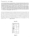
- an scFv fragment comprising or consisting of SEQ ID NO: 1 is humanized, it may thus also preferably result in a second humanized scFv fragment comprising or consisting of SEQ ID NO: 47 (hscFv 9012.2 (2), see figure 18 ).
- the invention further concerns a nucleic acid sequence encoding a single chain variable fragment according to the invention as described before.
- said scFv fragment comprises or consists of SEQ ID NO: 1
- said nucleic acid sequence may comprise or consist of SEQ ID NO:29, or any derived nucleic sequence encoding SEQ ID NO:1, for instance as a result of the degeneracy of the genetic code.
- said nucleic acid may comprise or consist of SEQ ID NO:30, or any derived nucleic sequence encoding SEQ ID NO:28, for instance as a result of the degeneracy of the genetic code.
- said nucleic acid may comprise or consist of SEQ ID NO:50 (see figure 18C ), or any derived nucleic sequence encoding SEQ ID NO:47, for instance as a result of the degeneracy of the genetic code.
- the invention also concerns an expression vector comprising a nucleic acid sequence as described.
- Such an expression vector also comprises appropriate nucleic acid sequences necessary for the expression of the operably linked scFv coding sequence in a particular host cell.
- Nucleic acid sequences necessary for expression in prokaryotes include a promoter, optionally an operator sequence, a ribosome binding site and possibly other sequences.
- Eukaryotic cells are known to utilize promoters, enhancers, and termination and polyadenylation signals in a host cell.
- Such an expression vector may also contain a leader sequence directing the expressed scFv to a particular cellular compartment, for instance a sequence directing a membrane expression or a secretion of the scFv fragment, or directing its expression in periplasm of bacteria. It may also contain under control of the same promoter a selection gene, the expression of which may be easily detected to select recombinant host cells transformed with the expression vector. Suitable selection genes include notably antibiotics resistance genes, fluorescent genes, or any other gene which expression may be easily monitored known by a person skilled in the art.
- the invention further concerns a host cell comprising an expression vector as described above.
- a recombinant host cell may be obtained by transfecting or transforming a host cell with an expression vector according to the invention.
- Such a host cell may be either prokaryotic or eukaryotic. Suitable prokaryotic host cells include gram-positive and gram-negative bacteria. Among gram-negative bacteria, a preferred host cell is represented by E. coli.
- the expression vector may preferably contain a leader sequence directing the expression of the scFv fragment into bacterial periplasm, corresponding to the space between the plasma membrane and the outer membrane of gram-negative bacteria or between the plasma membrane and the peptidoglycan layer (cell wall) of gram-positive bacteria.
- Suitable sequences directing the expression of a polypeptide to bacteria periplasm include ompA, ompF, ompT, LamB, ß-lactamase, cp VIII from M13, pelB, malE or phoA signal peptides or leader sequences.
- said leader sequence is pelB.
- a eukaryotic cell may be used, in particular a mammalian cell. Indeed, this may permit to directly generate a glycosylated scFv fragment.
- Mammalian cell lines available as hosts for expression are known in the art and include many immortalized cell lines available notably from the American Type Culture Collection (ATCC), including but not limited to, Chinese hamster ovary (CHO) cells, HeLa cells, baby hamster kidney (BHK) cells, monkey kidney cells (COS), human hepatocellular carcinoma cells (e.g., Hep G2), and a number of other cell lines.
- ATCC American Type Culture Collection
- yeast cells may also be used as eukaryotic cells.
- Pichia pastoris yeast cells a robust organism commonly used in fermentation processes which can be grown to high cell density in a chemically defined growth medium, have been modified by first eliminating endogenous yeast glycosylation pathways, while sequentially engineering into the organism a synthetic in vivo glycosylation pathway that enables the yeast to produce a complex human N-glycan, GlcNAc2Man3GlcNAc2, in vivo (see EP1297172 (33), EP1522590 (34), and references 35, 36).
- Such modified yeast cells with a humanized glycosylation pathway are able to secrete a human glycoprotein with uniform complex N-glycosylation.
- the invention also concerns a method for preparing a single chain variable fragment according to the invention as described above, comprising:
- Protocols and media for culturing host cells represent routine procedures and are readily available to any person skilled in the art.
- Concerning purification of the obtained scFv fragment it may be performed using well-known technologies, including affinity chromatography (e.g., using protein L-Sepharose) or ion-exchange chromatography, hydroxylapatite chromatography, gel electrophoresis, dialysis, etc. More precisely, when the scFv fragment comprises a His6 tag or an epitope tag as described above, HPLC with nickel ion columns or with columns containing immobilized specific antibodies.
- the invention further concerns a single chain variable fragment according to the invention as described above, as a medicament.
- the invention also relates to a pharmaceutical composition, comprising a single chain variable fragment according to the invention as described above and a pharmaceutically acceptable carrier.
- the invention also concerns the use of a single chain variable fragment according to the invention as described above for preparing a medicament for treating and/or preventing a cardiovascular disease selected from arterial and venous thrombosis, restenosis, acute coronary syndrome, and cerebrovascular accidents due to atheroscleroris.
- a cardiovascular disease selected from arterial and venous thrombosis, restenosis, acute coronary syndrome, and cerebrovascular accidents due to atheroscleroris.
- said cardiovascular disease is thrombosis.
- EXEMPLE 1 Synthesis and activity of a murine and a first humanized single chain variable fragment scFv 9012.2 directed against human glycoprotein VI
- LB Lia-Bertani
- DIFCO 402-17 LB-Agar, DIFCO 445-17 ; 2xTY (Trypton-Yeast), DIFCO 244020
- TES Tris-HCl 30 mM, EDTA 1 mM, sucrose 20%, pH 8.5
- PBS NaCl 0,14 M, KCl 13 mM, KH 2 PO 4 9 mM, Na 2 HPO 4 50 mM, pH 7.4
- BBS boric acid 50 mM, NaCl 150 mM, pH 7.6; Ampicillin, Euromedex EU-0400C ; Isopropyl ⁇ -D-thiogalactoside (IPTG), Euromedex EU0008-B ; Bovine serum albumin (BSA), Sigma B4287 ; FITC-coupled anti-c-myc IgG, Sigma F-2047 ; desoxyribonuclease A, aprotinin : Sigma A-62
- Oligodesoxyribonucleotides Name 5'-3' sequence SEQ ID NO T7 TAATACGACTCACTAGGGCGAAT SEQ ID NO : 33 pSWRev CGGCAGCCGCTGGATTGTTA SEQ ID NO : 34 pSWFor CGAGCTTAGCCCTTATAATTCAGATCCTC SEQ ID NO : 35 Link9012VLRev SEQ ID NO : 36 Link9012VHFor SEQ ID NO : 37 9O12.2mycFor GACCCTCGAGCGTTTGATCTCCAGCTTGGT SEQ ID NO : 38 9O12.2H3Rev GGCGACTGGTACTTCGATGTC SEQ ID NO : 39 9O12.2H2Rev AAATGGTGACACTTCCTTCAATCA SEQ ID NO : 40 9O12.2L1For GTTTTCAAGGCTCTGACTAGACCT SEQ ID NO : 41 MKC5For GG
- mRNA was isolated from freshly subcloned hybridoma 9012.2.2 cells which produce the immunoglobulin G (IgG) 9O12.2.2 directed against human GPVI.
- cDNA encoding the antibody variable domains were cloned after RT-PCR and the scFv 9012.2 gene was created by PCR splicing with overlap extensions.
- An expression vector derived from pUC 19 [pSW I vector (37)] was used for the production of the scFv 9012. It contains the LacZ promotor inducible with IPTG, the pel B leader sequence and, downstream, the gene coding for the scFv 9012.2 fused to the flag c-myc and a gene of resistance to ampicillin used to select the recombinant bacteria.
- the leader sequence pel B allows the recombinant scFv 9O12.2 to be addressed to the periplasm of bacteria transformed with the expression vector pSW scFv 9O12.2myc.
- the bacteria Toppl® non K12, Rif r F', proAB, lacI q Z ⁇ M15, Tn10, tet r ) (Stratagene, La Jolla, USA), colony 55T1 is used to produce the scFv 9012.2.
- Protein analysis was made by electrophoresis in polyacrylamide slab gels in the presence of SDS according to Laemmli; by immunoblotting after transfer of the proteins to nitrocellulose, incubation with an HRP coupled anti-c-myc antibody and detection using 4 chloro-naphtol.
- Mr of purified scFv is determined on a matrix-assisted laser desorption/ionization time-of-flight (MALDI-TOF) mass spectrometer (Voyager DE-PRO; PerSeptive Biosystems Inc., Framingham, MA).
- MALDI-TOF matrix-assisted laser desorption/ionization time-of-flight
- the saturated solution of ⁇ -cyano-4-hydroxycinnamic acid in 50% acetonitrile and 0.1% TFA is used as matrix and the spectrometer is calibrated using external standard peptide mixtures.
- scFv preparation were resolved by size-exclusion diffusion chromatography on a Superdex 75 HR column calibrated with standards of known molecular mass. 200 ⁇ L of sample was injected with a rate of 0.5 mL/min. Detection is monitored using UV detector at 226 nm. Fractions were used immediatly for analysis.
- Microtiter plates are coated with GPVI-Fc (10 ⁇ g/mL, 100 ⁇ L per well) overnight at 4°C. Wells are saturated with 100 ⁇ L BSA for 2 hours at room temperature.Then, increasing concentrations of scFv 9012 are added to the wells for 2 hours. Bound scFvs are detected after incubation for 2 h at room temperature with HRP-coupled anti-c-myc-antibody (1/750 en PBS).Then, the substrate solution (OPD) is added to the wells for 5 min. and absorbance is read at 492nm.
- GPVI-Fc 10 ⁇ g/mL, 100 ⁇ L per well
- the approach to humanize the scFv 9O12.2 has been adapted from CDR grafting technology 9.
- the CDRs of murine 9012.2 antibody have been grafted on a human variable-chain framework.
- the choice of the human antibody has been done using the following criteria : the crystallographic structure has been elucidated (1VGE in pdb library) ; the variable domains of the human antibody 1VGE present a high degree of sequence homology with the variable domains of 9O12.2 (VH 60,6 % ; VL 55,4 %) ; Structure comparison of the crystallographic data of the candidate human antibody 1VGE and the model of the variable domains of 9O12.2 was done and allowed us to validate the choice of the human acceptor scaffold 1 VGE.
- the human antibody 1 VGE selected based on these analysis allowed us to construct the humanized 9012.2 scFv.
- the nucleotide sequence encoding the humanized scFv 9012.2 was optimized. First, restriction sites between CDR regions were introduced to make possible adjustments for optimal binding characteristics. Optimization was then performed using the bacterial codon usage in order to express the humanized scFv in the procaryotic expression system E. coli.
- the scFv9O12.2 (100 ⁇ g/ml) was mixed with increasing concentrations of 9012.2 IgG before addition to GVI-Fc coated microtitration wells. Bound scFv was detected as above.
- Microtitration wells are coated with fibrilar type I collagen (Collagen Horn, Nycomed, Kunststoff 2 ⁇ g/well), saturated with BSA and washed. Increasing amounts of GPVI-Fc preincubated with PBS, scFv 9012.2 or Fab 9012.2 are added to the wells. After one hour at room temperature and washing bound GPVI-Fc is detected using a peroxydase-coupled anti- human Fc and OPD.
- Binding of the anti-GPVI scFv to recombinant GPVI-Fc is analyzed with surface plasmon resonance using a BIAcore 2000 system (Uppsala, Sweden). Binding studies is performed with scFv 9012, Proteolytic Fab and parental IgG.
- Recombinant GPVI-Fc is immobilized ( ⁇ 600 RU) onto a Carboxy-Methyl Dextran CM5 sensor chip using the amine coupling method (Wizard procedure).
- Antibody is then passed over the immobilized recombinant GPVI-Fc in HBS-EP buffer (0.01 M HEPES pH 7.4, 0.15 M NaCl, 0.005% polysorbate 20 (v/v)) at a flow rate of 20 ⁇ l/min at 25 °C.
- Kinetic constants ( k a , k d ) and affinity are determined using BIAevaluation version 3.1 software, by fitting data to binding model.
- HBS-EP is the running buffer.10 mM glycine-HCl pH 2.5 injected for 30 s at 20 ⁇ l/min is the regeneration buffer. All reagents and buffers used are from Biacore.
- Washed human platelets (2.10 7 /mL) are incubated for 30 minutes at room temperature with 0 to 100 ⁇ g/ml of purified scFv 9012 and then incubated again for 30 minutes at room temperature in the dark with 5 ⁇ L of FITC coupled anti-c-myc IgG (dilution 1:60). Cell fluorescence is measured using a flow cytometer (Epics XL, Coulter). Background is determined by using an irrelevant scFv in place of scFv 9012.2.
- Washed human platelets (3x 10 8 /mL) are preincubated for 5 min at 37°C without stirring with PBS, the scFv 9012.2 or the Fab 9012.2. Aggregation is initiated by adding type I collagen and the changes in light transmission is continuously recorded (Chronolog Aggregometer).
- Platelet adhesion to collagen under flow conditions was measured essentially as described elsewhere (9). Glass coverslips were coated with fibrillar type I collagen (50 ⁇ g.mL -1 ). Blood from healthy volunteers was collected on 40 ⁇ M PPACK, and labelled with DIOC-6 (1 ⁇ M). Blood aliquots were incubated for 15 minutes at room temperature with buffer or purified antibody fragment (Fab 9012, m scFv9O12, scFv 9C2) at a final concentration of 50 ⁇ g.mL -1 . The mixture was then perfused over the collagen-coated coverslips inserted in a flow-chamber at 1500 s -1 for 5 minutes. Transmission and fluorescent images were recorded in real time using a fluorescent microscope. Fluorescent images were obtained from at least ten different collagen-containing microscopic fields which were arbitrary chosen at the end of perfusion. Area coverage of fluorescent images was analyzed off-line using Histolab software (Microvision, Evry, France).
- Thrombin generation was continuously measured in platelet rich plasma (PRP) using the thrombogram method as previously described (38). Briefly, citrated PRP (1.5x10 8 platelets mL -1 ) was incubated with the antibody fragments for 10 min at 37° before adding the collagen. Ten minutes later, thrombin generation was initiated by transferring the samples into the wells of a microtitration plate containing tissue factor (0.5 pM). After 5 min at 37°C, the reaction was initiated by adding buffer containing CaCl 2 and the fluorescent thrombin substrate Z-GGR-AMC (Stago, Asbergers, France)). Fluorescence accumulation of the cleaved substrate was continuously measured at excitation and emission wavelengths of 390 and 460 nm respectively. First derivative curves of fluorescence accumulation were converted into thrombin concentration curves using a thrombin calibrator (39). The peak height is an indicator of the maximum rate of thrombin formation, and is sensitive to platelet activation.
- RNA 9012.2 is a murine IgG 1 (kappa chain).
- Total RNA was extracted from about 5.10 8 freshly subcloned hybridoma cells in a single-step procedure using Trizol. This RNA preparation was used as a template for first-strand cDNA synthesis. Double strand cDNAs encoding IgG 9O12.2.2 VH and VL domains were then amplified by PCR using primers sets (VHRev, VHFor) and (MKRevU, MKC5For) respectively. VH cDNA sequence was unique and this was confirmed by sequencing of the cDNA after cloning into pGEMT. The sequence of the PCR product corresponding to the VL domain was scrambled.
- VL sequences were identified. Data analysis using fasta analysis showed that one of the VL sequence derived from the MOPC-21 clone that express an endogeneous and aberrant non-functional Vk mRNA (Gene bank acc number M35669). The nucleotide and deduced amino acid sequences of the other VL gene and the VH gene are shown in Figure 2 as part of the scFv 9O12.2.
- CDRs were identified according to the following rules as deduced from Kabat et al. (1991) and Chotia and Lesk (1987):
- the VH cDNA sequence showed 92.96 % identity with the cDNA encoding another murine VH domain (MMMD52C). For the VL we found 98.51 % identity with the cDNAsequence encoding the VL domain of murine anti-acetyl-Lys (BD174891).
- the deduced amino acid sequences analysis also showed high homology with the sequence of murine Ig. 9O12.2 VH showed 89.256 % identity with the deduced amino-acid sequence of an anti-CD20 murine IgG (BD688655).
- 9O12.2 VL showed 96.43 % identity with the deduced amino-acid sequence of an anti-acetyl Lys murine IgG (BD581288).
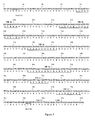
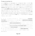
- the nucleotide and deduced amino acid sequences of the scFv 9012.2 is shown in Figure 2 .
- the scFv is 266 amino acids long and is composed of the VH and VL sequences linked together by a [G 4 S] 3 linker and followed by a short spacer (8 residues) and the c-myc flag sequence (11 residues).
- the theoretical molecular mass of the scFv is of 28393.5 as determined using ProtParam software. Its pI is 6.92 and its extinction coefficient 49640 M -1 cm -1 at 280 nm (Table 3). Table 3. Theoretical structural characteristics of scFv 9012 as deduced from bioinformatic analysis using ProtParam software Number of amino acids : 266 Molecular weight: 28393.5 Theoretied pt: 6.92 Amino Acid composition: Ala (A) 11 4.1% Arg (R) 9 3.4% Asn (H) 9 3.4% Asp (D) 11 4.1% Cys (C) 4 1.5% Gln (Q) 15 5.6% Glu (E) 11 4.1% Gly (G) 37 13.9% His (H) 2 0.8: Ile (I) 7 2.6% Leu (L) 24 9.0% Lys (K) 13 4.9% Met (M) 4 1.5% Phe (F) 9 3.4% Pro (P) 8 3.0% Ser (S) 36 13.5% Thr (T) 21
- Pdb files of murine variable domains used as a template are:
- Modeler 3.0 software was used for the modelling of variable domains. Twenty models were designed for each domain and the best was selected according to the RMSD value: VH (RMSD: 0.13 ⁇ ); VL (RMSD: 0.703 ⁇ ). The model is shown in Figure 3 .
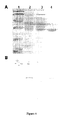
- the proteins contained in the periplasmic extract, the flow through fraction and the fraction eluted from the GPVI-sepharose column were analysed by SDS-PAGE and by immunoblot ( Figure 5A and 5B ).
- the eluted fraction appears as a major band of ⁇ 28 kDa that is in agreement with the theoretical mass. This band is also detected by immunoblot using an anti-c-myc antibody.
- the monomeric scFv was very stable, and the multimeric forms spontaneously reverted back to monomers when stored at 4°C, which is essential to preserve monovalent binding to GPVI and biological effects. Indeed, it is well-established that bivalent anti-GPVI molecules (IgGs or F(ab)' 2 ) can cause platelet activation (40;93).
- bivalent anti-GPVI molecules IgGs or F(ab)' 2
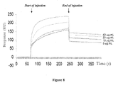
- the affinity of the 9012.2 IgG and of its Fab prepared after papain digestion (pFab) for GPVI-Fc determined by surface plasmon resonance are high with Kd of respectively 6.5 10 -9 M and 4.5 10 -9 M.
- the Kd of the scFv is of 2.5 10 -9 M ( Figure 8 ).
- the murine scFv 9012 still has very high functional affinity, with a K D value in the range of 10 -9 M, and this certainly contributes to the stability of the scFv- platelet GPVI complexes, which is essential to inhibit platelet aggregation under flow conditions.
- the purified 9012.2 scFv inhibits GPVI binding to collagen.
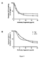
- mscFv 9012 inhibited GPVI binding to collagen with an IC 50 of approximately 1.17 ⁇ g.mL -1 (42 ⁇ M), 80% inhibition being reached at a concentration of 5 to 10 ⁇ g.mL -1 of m scFv 9012.
- This inhibitory capacity was comparable to that observed for Fab 9012 prepared after papain digestion of the parental IgG (2.1 ⁇ g.mL -1 ; 15 nM).
- the purified 9012.2 scFv binds to human platelets and inhibits collagen-induced platelet aggregation.
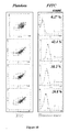
- Binding of purified scFv to human platelets was measured by flow cytometry. A shift to the right of the peak indicates that the scFv binds to human platelets in a dose dependant manner ( Figure 10 ). A non relevant purified scFv did not bind to platelets.
- the effect of the 9O12.2 scFv on GPVI function was tested by measuring collagen-induced platelet aggregation.
- the monomeric forms of the scFv were purified by size exclusion gel chromatography.
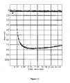
- the 9012.2 scFv 25 ⁇ g/mL completely prevented collagen-induced platelet aggregation ( Figure 11 ) as did the 9O12.2 pFab used at the same concentration.
- Fab 9012 is known to inhibit thrombin generation at the surface of collagen-stimulated platelets
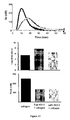
- the effect of the purified m scFv 9012 was tested using the thrombogram method ( Figure 13 ).
- m scFv 9012 and Fab 9012 reduced the thrombin peak to similar extents, and increased the lag preceding thrombin generation, indicating that m scFv 9012 is as efficient as Fab 9012 in inhibiting collagen-induced platelet procoagulant activity.
- the inventors have then humanized the murine 9012.2 scFv fragment as described in paragraph 1.1.2.
- the nucleotide sequence and deduced amino acid sequence of humanized VH and VL domains and of the first humanized 9012.2 scFv fragment (hscFv 9012.2(1)) are displayed in Figure 14 .
- This hscFv 9012.2(1) fragment was then further characterized using affinity binding to GPVI sepharose :
- the periplasmic fraction of recombinant bacteria was loaded onto a GPVI sepharose column.
- Retained proteins were analyzed by Western-Blot using an anti-c-myc IgG followed by anti-mouse antibody coupled to HRP and ECL revelation. A single band is detected with a molecular mass of 28.5 kDa ( Figure 15 ) as expected for the humanized scFv.
- hscFv 9012.2(1) was then analyzed and compared to that of murine scFv 9012.2 (mscFv 9012.2).
- Purified humanized scFv and mscFv were injected on GPVI-Fc immobilized on a CM5 sensorchip. Results are displayed on Figure 16 and show that hscFv 9012.2(1) also binds to GPVI-Fc, with what appears to be a higher affinity than mscFv.
- the murine 9012.2 scFv generated by the inventors from the 9012.2 monoclonal antibody specifically binds to human GPVI with a high affinity. In addition, it inhibits GPVI binding to collagen and prevents collagen-induced platelet aggregation, thus inhibiting GPVI function.
- This scFv fragment thus displays all the advantages of scFv fragments while retaining the affinity and inhibition capacity of corresponding Fab fragments.
- the inventors have also generated a first humanized version of the 9012.2 scFv fragment, which also binds to human GPVI with a high affinity despite new essential structural modifications (hscFv 9O12.2(1)).
- This first humanized 9012.2 scFv fragment (hscFv 9012.2) further has the advantage of a potentially minimally reduced immunogenicity.
- This application thus describes for the first time a low molecular weight, low immunogenic GPVI ligand with a high capacity to inhibit platelet aggregation, resulting in a very promising product for arterial thrombosis treatment.
- EXEMPLE 2 Synthesis and activity of a second optimized humanized single chain variable fragment hscfv 9012.2(2) directed against human glycoprotein VI
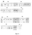
- VH FR3 exhibits 25/32 residues identity with 1 VGE. Only 3 residues of this framework (A H71 , K H73 , R H76 , Kabbat numbering) had no similarity with 1 VGE.
- the 9012 VL FR3 was substituted for its 1 VGE counterpart with the exception of two residues (L59P and D60S), essentially because L is not frequently encountered at this position, and D is an acidic residue.
- hscFv9O12.2(1) the main parameters that are usually affected by humanization were well-preserved in hscFv9O12.2(2).
- Affinity-purified hscFv9O12.2(2) was fully functional, and had a high affinity for GPVI, a major point for biological applications.
- FACS analysis also indicated that hscFv9O12.2(2) recognizes the same epitope on human platelets as mouse Fab 9012, because its binding was specifically blocked in the presence of a molar excess of Fab 9O12.
Landscapes
- Health & Medical Sciences (AREA)
- Chemical & Material Sciences (AREA)
- Organic Chemistry (AREA)
- Immunology (AREA)
- Life Sciences & Earth Sciences (AREA)
- General Health & Medical Sciences (AREA)
- Medicinal Chemistry (AREA)
- Bioinformatics & Cheminformatics (AREA)
- Engineering & Computer Science (AREA)
- Chemical Kinetics & Catalysis (AREA)
- Veterinary Medicine (AREA)
- Cardiology (AREA)
- General Chemical & Material Sciences (AREA)
- Nuclear Medicine, Radiotherapy & Molecular Imaging (AREA)
- Pharmacology & Pharmacy (AREA)
- Animal Behavior & Ethology (AREA)
- Public Health (AREA)
- Molecular Biology (AREA)
- Genetics & Genomics (AREA)
- Biophysics (AREA)
- Proteomics, Peptides & Aminoacids (AREA)
- Biochemistry (AREA)
- Heart & Thoracic Surgery (AREA)
- Urology & Nephrology (AREA)
- Vascular Medicine (AREA)
- Hospice & Palliative Care (AREA)
- Diabetes (AREA)
- Hematology (AREA)
- Peptides Or Proteins (AREA)
- Preparation Of Compounds By Using Micro-Organisms (AREA)
- Micro-Organisms Or Cultivation Processes Thereof (AREA)
- Medicines Containing Antibodies Or Antigens For Use As Internal Diagnostic Agents (AREA)
- Medicines That Contain Protein Lipid Enzymes And Other Medicines (AREA)
Description
- The present invention relates to a single chain variable fragment (scFv 9O12.2) directed against human glycoprotein VI, constituted of the VH and VL domains of 9012.2 monoclonal antibody linked via a (Gly4Ser)3 peptide and followed by a "c-myc" flag, represented by SEQ ID NO:1. The invention also concerns functional variants of said scFv 9O12.2 fragment with identical heavy and light chains complementary determining
regions - Acute coronary and cerebrovascular accidents are currently the first death cause in the world. In addition, the global incidence of recurrence and death in the 6 month posttreatment period after an acute coronary syndrome is still 8-15%. In the case of acute coronary syndrome with ST segment elevation, mechanical treatment with coronary angioplasty and introduction of a stent is highly efficient to urgently restore coronary artery flow, but does not prevent morbidity/mortality for about 15% of patients in the next 6 months.
- Thrombolytic treatments, which are based on long term fibrinolytic, anticoagulant and anti-aggregating drugs associations, give even less encouraging results. Indeed, despite improvements in medical treatment of thrombosis, morbidity/mortality at 6 months is similar to that observed for acute coronary syndrome without segment ST elevation.
- Concerning cerebrovascular ischemic accidents, treatments are still very limited due to the generally late caring of most patients and to the hemorrhagic risk of currently available anti-thrombotic treatments.
- There is thus a real pressing clinical need for improving treatments for cardiovascular diseases, and especially for new molecules with improved features compared to available molecules, in particular for molecules with a reduced hemorrhagic effect.
- Platelets-collagen interactions are critical in the appearance of acute arterial thrombosis and post-thrombotic vascular remodeling. Glycoprotein VI (GPVI), the main receptor for platelets activation by collagen, has been demonstrated in animals to play a role in experimental thrombosis, vascular remodeling, atherothrombosis and acute myocardial ischemia.
- Contrary to αIIbβ3 integrin antagonists, which are currently used in thrombosis treatment and inhibit platelets final activation phase, GPVI is implicated into platelets initial activation phase, and GPVI antagonists should thus prevent not only platelet aggregation, but also secondary agonists liberation as well a growth factors and cytokines secretion resulting into vascular lesions development. In addition, GPVI deficit is not associated with a high hemorrhagic risk, which is a crucial feature for patient's safety. Finally, GPVI expression is limited to platelets, and thus represents a perfectly specific target for anti-thrombosis treatment.
- GPVI antagonists should thus be efficient for specifically and efficiently preventing primary or secondary thrombosis, while involving only a low hemorrhagic risk.
- Various kinds of potential GPVI antagonists have been generated. In one approach, a soluble GPVI recombinant protein has been generated, which is a fusion protein between GPVI extracellular domain and human Ig Fc domain (see for instance
WO 01/00810 WO 03/008454 - Many others have tried to develop neutralizing monoclonal antibodies directed against human GPVI. For instance,
EP 1224942 (5 EP 1228768 (6 -
EP1538165 (7 -
WO 2005/111083 (8) describes 4 monoclonal anti-GPVI antibodies OM1, OM2, OM3 and OM4, that were found to inhibit GPVI binding to collagen, collagen-induced secretion and thromboxane A2 (TXA2) formation in vitro, as well as ex vivo collagen-induced platelet aggregation after intravenous injection to Cynomolgus monkeys. OM4 also appears to inhibit thrombus formation in a rat thrombosis model. -
WO O1/00810 - In addition, 9O12.2 Fab fragments were found to completely block collagen-induced platelet aggregation and secretion, to inhibit the procoagulant activity of collagen-stimulated platelets and platelet adhesion to collagen in static conditions, to impair platelet adhesion, and to prevent thrombi formation under arterial flow conditions (9).
- However, none of the currently known anti-GPVI antibodies have proven really efficient in vivo for preventing and/or treating cardiovascular diseases implicating platelet aggregation such as arterial and venous thrombosis, restenosis, acute coronary syndrome, and cerebrovascular accidents due to atheroscleroris. Until recently the different anti-GPVI antibodies that have been reported appeared not fitted for the development of an antithrombotic for medical use in human. Only few antibodies have been reported to have inhibitory properties. This is the case of JAQ1 that is directed to mouse GPVI and does not cross react with human GPVI (10). Human scFvs directed to human GPVI have also been reported to be inhibitory (11,12) but their affinity appears to be low. Very recently, new inhibitory antibodies with a good affinity for human GPVI have been characterised (13) and proposed to be developed as therapeutic tools.
- However, cross-linking of GPVI at the platelet surface by a divalent or multivalent ligand results in platelet activation. This is the case of the 9012.2 IgGs that activate platelets via GPVI dimerisation and via cross-linking of GPVI to the low affinity Fc receptor (FcγRIIA) (9). Fab'2 also activate platelets via GPVI dimerisation (9). In contrast, monovalent 9O12.2 Fab fragments are inhibitory. However these fragments could not be used in therapeutic due to their size and their animal origin which makes them immunogenic in human patients.
- There is thus still a need for an efficient neutralizing GPVI antagonist, which would inhibit with high efficiency the initial phase of platelets aggregation, with a low hemorrhagic risk, as well as a low immunogenic effect.
- From the 9012.2 monoclonal anti-GPVI antibody hybridoma, the inventors have generated a single chain variable fragment (scFv) 9O12.2 which has the advantage to contain the functional inhibitory motif in a fragment of reduced size without any immunogenic constant domains (CH1 and CL). The scFv is the starting material to produce fragments with an improved immune tolerance and stability in order to obtain a format adapted to a therapeutic use.
- Technologic approaches to design such fragments have previously been reported in details, however there are many difficulties to overpass making each scFv construct a specific case. These difficulties include the cloning of antibody V-genes by bypassing aberrant transcripts (14), identification of a linker well-suited for correct folding, association of V-domains into a monomeric scFv and finally the selection of a procaryotic expression system that enable expression of the scFv in a functional soluble form.
- The invention thus concerns a single chain variable fragment directed against human glycoprotein VI, comprising:
- a VH domain comprising CDR1, CDR2 and CDR3 regions constituted of SEQ ID NO: 2, SEQ ID NO: 3, and SEQ ID NO: 4,
- a peptide linker, and
- a VL domain comprising CDR1, CDR2 and CDR3 regions constituted of SEQ ID NO: 5, SEQ ID NO: 6, and SEQ ID NO: 7,
- In a preferred embodiment, said scFv fragment according to the invention further comprises a peptide tag, useful for purification, and optionally a short peptide spacer between the core scFv fragment (comprising the VH and VL domains and the peptide linker) and the peptide tag.
- An antibody is a roughly Y-shaped molecule composed of two different polypeptide chains named "heavy" and "light" chains, an antibody being constituted of two heavy and two light chains linked by disulfide bonds. Each heavy chain is composed of a variable domain ("VH domain") and 3 constant domains ("CH1", "CH2" and "CH3" domains), while each light chain is composed of a variable domain ("VL domain") and a constant domain ("CL domain").
- A "single chain variable fragment" or "scFv fragment" refers to a single folded polypeptide comprising the VH and VL domains of an antibody linked through a linker molecule. In such a scFv fragment, the VH and VL domains can be either in the VH - linker - VL or VL - linker - VH order. In addition to facilitate its production, a scFv fragment may contain a tag molecule linked to the scFv via a spacer. A scFv fragment thus comprises the VH and VL domains implicated into antigen recognizing but not the immunogenic constant domains of corresponding antibody.
- In addition, each variable domain (VH or VL domain) is composed of 4 "framework regions" (FR1, FR2, FR3 and FR4), which display less variability among antibodies and are involved in the formation of β sheets forming the structural framework of the variable domain, and of 3 hypervariable regions commonly named "complementary determining regions" 1, 2 and 3 (CDR1, CDR2, CDR3), which correspond to 3 loops juxtaposed in the folded variable domain at the edge of each β sheet. The 3 CDR regions are crucial for determining an antibody or antibody fragment specificity since they are the part of the variable domain mainly in contact with the antigen, especially the CDR3 region of each chain, which corresponds to the rearranged region of the heavy and light chains and is even more variable and more directly in contact with the specific antigen.
- Thus, the invention encompasses all functional variants of scFv 9O12.2 which keep identical CDR regions. The scFv fragment 9O12.2 CDR regions data are presented in following Table 1:
Table 1. scFv 9012.2 CDR regions. CDR regions were identified using Kabat and Chotia nomenclature (15, 16) CDR Position in scFv 9O12.2 (aa) Sequence VH CDR1 26-35 GYTFTSYNMH (SEQ ID NO:2) VH CDR2 50-66 GIYPGNGDTSFNQKFKG (SEQ ID NO:3) VH CDR3 99-109 GTVVGDWYFDV (SEQ ID NO:4) VL CDR1 159-174 RSSQSLENSNGNTYLN (SEQ ID NO:5) VL CDR2 190-196 RVSNRFS (SEQ ID NO:6) VL CDR3 229-237 LQLTHVPWT (SEQ ID NO:7) - By a "peptide linker" is meant a flexible peptide that permits an appropriate folding of the scFv fragment, ie an appropriate folding of the VH and VL domains and their capacity to be brought together. In addition, such a peptide linker should permit folding into a monomeric functional unit. When the scFv is assembled in the VH to VL orientation (VH - linker - VL), an scFv with a linker of 3 to 12 residues cannot fold into a functional Fv domain and instead associates with a second molecule to form a bivalent dimer. Reducing below 3 residues leads to trimers. In this case, a suitable linker should thus have at least 12 and preferably less than 25 aminoacids, preferably between 14-18, 14-16, or 15 amino acids, and should preferably comprise a high percentage of glycine residues, preferably at least 50%. Examples of suitable peptide linkers include peptides (G4S)3 (SEQ ID NO:8), G4IAPSMVG4S (SEQ ID NO:9), G4KVEGAG5S (SEQ ID NO:10), G4SMKSHDG4S (SEQ ID NO:11), G4NLITIVG4S (SEQ ID NO:12), G4VVPSLG4S(SEQ ID NO:13) and G2EKSIPG4S(SEQ ID NO:14). When the scFv is produced in the VL to VH orientation (VL - linker - VH), the distance between the C-terminus ofVL and N-terminus ofVH is slightly greater than between the C-terminus of VH and N-terminus of VL (39-43 Å versus 30-34Å). Therefore, a 18 amino acid residues linker is the minimal sequence size that can be used. A suitable peptide linker should then have between 18-25 aa, preferably between 18-21 aa. An example of a suitable linker is linker of sequence GSTSGSGKSSEGSGSTKG (SEQ ID NO: 15).
- By a "peptide tag" is meant a peptide of 5-15 amino acids for which specific antibodies are available. Although optional in a scFv fragment according to the invention, such a peptide tag inserted at the C-terminal end of the scFv fragment permits to facilitate purification of the scFv fragment after recombinant production. Indeed, the peptide tag gives the protein a specific binding affinity it would not otherwise, which permits an easier purification using chromatography. Examples of suitable peptide tags include a His6 tag (HHHHHH, SEQ ID NO: 16), which has affinity towards nickel ions and can thus be purified using a nickel ions containing chromatography column, or epitopes peptides with high affinity for their specific antibody and can thus be purified using a column containing immobilized antibody directed against peptide, such as a c-myc tag (EQKLISEEDLN, SEQ ID NO:17), a HA tag (YPYDVPDYA, SEQ ID NO:18), a Flag tag (DYKDDDDK, SEQ ID NO:19) a protein C tag (EDQVDPRLIDGK, SEQ ID NO:20), a Tag-100 tag (EETARFQPGYRS, SEQ ID NO:21), a V5 epitope tag (GKPIPNPLLGLDST, SEQ ID NO:22), a VSV-G tag (YTDIEMNRLGK, SEQ ID NO:23) or a Xpress tag (DLYDDDDK, SEQ ID NO:24).
- By a "short peptide spacer" is meant a peptide of 1-15 amino acids, preferably 1-12 or 1-10 amino acids, for
instance 8 amino acids. Such a peptide spacer is intended to separate the true scFv part (VH and VL domains separated by a peptide linker) of the optional peptide tag. It is not necessary in a scFv fragment according to the invention, in particular when there is no peptide tag, but also when a peptide tag is included. Indeed a peptide tag may be directly fused to the VH or VL domain. However, a short peptide spacer of 1-12 amino acids may be useful. For instance, in the 9012.2 scFv fragment, a 8 amino acids peptide spacer of sequence RSRVTVSS (SEG ID NO: 25) has been used. - In a preferred embodiment, the invention concerns a single chain variable fragment (scFv) directed against human glycoprotein VI, comprising or consisting of an amino acid sequence having at least 95%, at least 96%, at least 97%, at least 98%, at least 98.5%, at least 99%, at least 99.5%, at least 99.6%, at least 99.7%, at least 99.8%, identity with SEQ ID NO:1, in which
amino acids 26 to 35, 50 to 66, 99 to 109, 159 to 174, 190 to 196, and 229 to 237 of said 266 amino acids sequence are constituted of SEQ ID NO: 2, SEQ ID NO:3, SEQ ID NO:4, SEQ ID NO:5, SEQ ID NO:6, and SEQ ID NO:7 respectively. Such functional variants of scFv 9O12.2 not only keep identical CDR regions but also have a high identity percentage with the amino acid sequence of scFv 9O12.2. In a particular embodiment, the invention concerns a single chain variable fragment comprising or consisting of SEQ ID NO:1. - However, framework regions are mutated to generate a chimeric, in particular humanized, scFv fragment directed against human glycoprotein VI. With regards to a reference antibody or antibody fragment of a given species, a corresponding "chimeric" antibody or antibody fragment in which framework regions have been replaced by corresponding framework regions of another species. More particularly, a "humanized" antibody or antibody fragment is an antibody or antibody fragment with identical CDR regions in which framework regions have been replaced by human framework regions.
- The scFv 9012.2 fragment has been derived by the inventors from murine monoclonal antibody 9012.2, and thus has mouse framework regions. However, to reduce immunogenic effect of injection of this fragment in human patients, a humanized scFv fragment may be desirable, in which murine framework regions have been replaced by human framework regions.
- Several possible methods of humanizing antibody V-domains have been suggested. They include CDR-grafting, resurfacing V-domains or predictive computational analysis that explores a diversity of substitutions in a given V chain sequence with the aim of reducing immunogenicity whilst maintaining the antigen-specificity and affinity of the original molecule (17-19). None of these methods is simple, and all often result in impaired specificity and/or affinity (20-23).
- Despite these difficulties, the inventors have generated a first humanized scFv fragment in which the VH and VL domains of murine 9012.2 scFv fragment, corresponding to amino acids number 1-120 and 136-266 of SEQ ID NO:1, have been respectively replaced by humanized VH and VL domains SEQ ID NO:26 and SEQ ID NO:27, resulting in a humanized scFv fragment of amino acid sequence SEQ ID NO:28 (hscFv 9012.2 (1), see
figure 14 ). - In a first preferred embodiment, the invention thus concerns a single chain variable fragment according to any of claims 1-3, in which framework regions of the VH and VL domains have been replaced by VH and VL domains having framework regions of a human antibody. In particular, said VH and VL domains may have been replaced respectively by SEQ ID NO:26 and SEQ ID NO:27. More precisely, when an scFv fragment comprising or consisting of SEQ ID NO:1 is humanized, it may result in a first humanized scFv fragment comprising or consisting of SEQ ID NO:28 (hscFv 9O12.2 (1), see
figure 14 ). - To generate such a humanized scFv fragment, a well known technology called CDR grafting may be used, which involves selecting the complementary determining regions (CDRs) from a donor scFv fragment, and grafting them onto a human scFv fragment framework of known three dimensional structure (see, e. g.,
WO98/45322 WO 87/02671 US5,859,205 (26);US5,585,089 (27);US4,816,567 (28);EP0173494 (29); and references 20-21 and 30-31) or performing database searches to identify potential candidates. In a typical method aided by computer modeling and comparison to human germline sequences, the antigen binding loops of the monoclonal antibody to be humanized are superimposed onto the best fitting frameworks. The approach to humanize the scFv 9012.2 has been adapted from CDR grafting technology (32). The CDRs of murine 9O12.2 antibody have been grafted on a human variable-chain framework. The choice of the human antibody has been done using the following criteria : the crystallographic structure has been elucidated (1VGE in pdb library) ; the variable domains of thehuman antibody 1 VGE present a high degree of sequence homology with the variable domains of 9012.2 (VH VL 55,4 %) ; Structure comparison of the crystallographic data of the candidate human antibody 1VGE and the model of the variable domains of 9012.2 was done and allowed us to validate the choice of the human acceptor scaffold 1VGE. This strategy minimizes the risk of lowering the stability of the interaction between VH and VL domains while preserving the scaffold required for correct folding of the CDR, preserving a high affinity for the antigen. The human antibody 1VGE selected based on these analysis allowed to construct the humanized 9012.2 scFv. For structural raisons, 10 residus after CDR H2 were not mutated. The nucleotide sequence encoding the humanized scFv 9O12.2 was optimized. First, restriction sites between CDR regions were introduced to make possible adjustments for optimal binding characteristics. Optimization was then performed using the bacterial codon usage in order to express the humanized scFv in the procaryotic expression system E. coli. Said method resulted in a humanized 9012.2 scFv fragment in which VH and VL domains of the murine 9012.2 scFv fragment, corresponding to amino acids number 1-120 and 136-266 of SEQ ID NO:1 respectively, have been replaced by SEQ ID NO:26 and SEQ ID NO:27 respectively, i.e. a humanized 9012.2 scFv fragment consisting of SEQ ID NO: 28. In a preferred embodiment of a humanized scFv fragment according to the invention, said scFv fragment thus comprises or consists of SEQ ID NO:28. - Despite promising results obtained with this first humanized scFv fragment, this fragment was not optimal for production in bacteria, and a further optimized second humanized scFv fragment (hscFv 9012.2(2)) was derived from hscFv 9O12.2(1). Briefly, for VL FR1 and FR2 regions, the identity with
1VGE frameworks - Thus, in a second preferred embodiment, the invention thus concerns a single chain variable fragment according to any of claims 1-3, in which framework regions of the VH and VL domains have been replaced by VH and VL domains having framework regions of a human antibody. In particular, said VH and VL domains may have been replaced respectively by SEQ ID NO: 26 and SEQ ID NO: 46. More precisely, when an scFv fragment comprising or consisting of SEQ ID NO: 1 is humanized, it may thus also preferably result in a second humanized scFv fragment comprising or consisting of SEQ ID NO: 47 (hscFv 9012.2 (2), see
figure 18 ). - The invention further concerns a nucleic acid sequence encoding a single chain variable fragment according to the invention as described before. In a particular embodiment, when said scFv fragment comprises or consists of SEQ ID NO: 1, said nucleic acid sequence may comprise or consist of SEQ ID NO:29, or any derived nucleic sequence encoding SEQ ID NO:1, for instance as a result of the degeneracy of the genetic code.
- When said scFv fragment is humanized and VH and VL domains of the murine 9012.2 scFv fragment, corresponding to amino acids number 1-120 and 136-266 of SEQ ID NO:1 respectively, have been replaced by SEQ ID NO:26 and SEQ ID NO:27 respectively, resulting in a first humanized 9012.2 scFv fragment (hscFv 9O12.2(1)) comprising or consisting of SEQ ID NO:28, said nucleic acid may comprise or consist of SEQ ID NO:30, or any derived nucleic sequence encoding SEQ ID NO:28, for instance as a result of the degeneracy of the genetic code.
- Alternatively, when said scFv fragment is humanized and VH and VL domains of the murine 9012.2 scFv fragment, corresponding to amino acids number 1-120 and 136-266 47 respectively, resulting in a second humanized 9012.2 scFv fragment (hscFv 9012.2(2)) comprising or consisting of SEQ ID NO:47, said nucleic acid may comprise or consist of SEQ ID NO:50 (see
figure 18C ), or any derived nucleic sequence encoding SEQ ID NO:47, for instance as a result of the degeneracy of the genetic code. - The invention also concerns an expression vector comprising a nucleic acid sequence as described. Such an expression vector also comprises appropriate nucleic acid sequences necessary for the expression of the operably linked scFv coding sequence in a particular host cell. Nucleic acid sequences necessary for expression in prokaryotes include a promoter, optionally an operator sequence, a ribosome binding site and possibly other sequences. Eukaryotic cells are known to utilize promoters, enhancers, and termination and polyadenylation signals in a host cell.
- Such an expression vector may also contain a leader sequence directing the expressed scFv to a particular cellular compartment, for instance a sequence directing a membrane expression or a secretion of the scFv fragment, or directing its expression in periplasm of bacteria. It may also contain under control of the same promoter a selection gene, the expression of which may be easily detected to select recombinant host cells transformed with the expression vector. Suitable selection genes include notably antibiotics resistance genes, fluorescent genes, or any other gene which expression may be easily monitored known by a person skilled in the art.
- The invention further concerns a host cell comprising an expression vector as described above. Such a recombinant host cell may be obtained by transfecting or transforming a host cell with an expression vector according to the invention.
- Such a host cell may be either prokaryotic or eukaryotic. Suitable prokaryotic host cells include gram-positive and gram-negative bacteria. Among gram-negative bacteria, a preferred host cell is represented by E. coli. For use in bacteria, the expression vector may preferably contain a leader sequence directing the expression of the scFv fragment into bacterial periplasm, corresponding to the space between the plasma membrane and the outer membrane of gram-negative bacteria or between the plasma membrane and the peptidoglycan layer (cell wall) of gram-positive bacteria. Suitable sequences directing the expression of a polypeptide to bacteria periplasm include ompA, ompF, ompT, LamB, ß-lactamase, cp VIII from M13, pelB, malE or phoA signal peptides or leader sequences. In a particular embodiment, said leader sequence is pelB.
- Alternatively, a eukaryotic cell may be used, in particular a mammalian cell. Indeed, this may permit to directly generate a glycosylated scFv fragment. Mammalian cell lines available as hosts for expression are known in the art and include many immortalized cell lines available notably from the American Type Culture Collection (ATCC), including but not limited to, Chinese hamster ovary (CHO) cells, HeLa cells, baby hamster kidney (BHK) cells, monkey kidney cells (COS), human hepatocellular carcinoma cells (e.g., Hep G2), and a number of other cell lines. As eukaryotic cells, yeast cells may also be used. To obtain a glycosylation pattern closest to a natural human glycosylation pattern, it may be resorted to human cell lines as host cells. Alternatively, Pichia pastoris yeast cells, a robust organism commonly used in fermentation processes which can be grown to high cell density in a chemically defined growth medium, have been modified by first eliminating endogenous yeast glycosylation pathways, while sequentially engineering into the organism a synthetic in vivo glycosylation pathway that enables the yeast to produce a complex human N-glycan, GlcNAc2Man3GlcNAc2, in vivo (see
EP1297172 (33),EP1522590 (34), and references 35, 36). Such modified yeast cells with a humanized glycosylation pathway are able to secrete a human glycoprotein with uniform complex N-glycosylation. - The invention also concerns a method for preparing a single chain variable fragment according to the invention as described above, comprising:
- a) culturing a host cell according to the invention as described above, and
- b) purifying said single chain variable fragment.
- Protocols and media for culturing host cells represent routine procedures and are readily available to any person skilled in the art. Concerning purification of the obtained scFv fragment, it may be performed using well-known technologies, including affinity chromatography (e.g., using protein L-Sepharose) or ion-exchange chromatography, hydroxylapatite chromatography, gel electrophoresis, dialysis, etc. More precisely, when the scFv fragment comprises a His6 tag or an epitope tag as described above, HPLC with nickel ion columns or with columns containing immobilized specific antibodies.
- The invention further concerns a single chain variable fragment according to the invention as described above, as a medicament.
- The invention also relates to a pharmaceutical composition, comprising a single chain variable fragment according to the invention as described above and a pharmaceutically acceptable carrier.
- More precisely, the invention also concerns the use of a single chain variable fragment according to the invention as described above for preparing a medicament for treating and/or preventing a cardiovascular disease selected from arterial and venous thrombosis, restenosis, acute coronary syndrome, and cerebrovascular accidents due to atheroscleroris. In a preferred embodiment of such a use, said cardiovascular disease is thrombosis.
- Having generally described this invention, a further understanding of characteristics and advantages of the invention can be obtained by reference to certain specific examples and figures which are provided herein for purposes of illustration only and are not intended to be limiting unless otherwise specified.
-
-
Figure 1 . Schematic description of the method used to construct scFv 9012.2 expression vector. Step 1: PCR amplification 9O12.2.2 monoclonal antibody VH segment using T7 and Link9O12.2VHFor primers and VL segment using Link9O12.2VLRev and 9O12.2mycFor primers. Step 2: Overlap PCR amplification using pSWRev and 9O12.2mycFor primers followed by PCR amplification using pSWRev and pSWFor primers, thus generating a nucleic acid coding scFv 9O12.2. Step 3: DNA purification of PCR products. Step 4: Digestion of purified DNA using Pst1 and Xho1 restriction enzymes. Step 5: Cloning of the digested product into pSW1 expression vector. Step 6: Sequenceing of the obtained construction. -
Figure 2 . Nucleotide and deduced amino-acid sequences of scFv 9012.2 cloned into plasmid pSWscFv9O12.2myc. Nucleotide and deduced amino-acid sequences of scFv 9012.2 cloned between the restriction sites Pst1 and Xho1 into the expression vector pSW1. The nucleotide sequences corresponding to the primers used for PCR amplification or encoding the linker peptide are shown in italics. The deduced amino-acid sequence of the complementary determining regions (CDRs) of VH and VL are underlined. The deduced amino-acid sequence of the c-myc flag is shown. -
Figure 3 . Three-dimensional model of the scFv9O12.2. Model of the 9O12.2.2 antigen-binding domains (side view) The CDRs are indicated. -
Figure 4 . Analysis of bacterial periplasmic extracts by flow cytometry on human platelets. Bacterial periplasmic extracts of induced recombinant bacteria transformed with a plasmid encoding an irrelevant scFv-myc or the plasmid pSW scFv9012.2-myc were incubated with human platelets. Binding of scFvs was analysed using an FITC-coupled anti-cmyc antibody. A: irrelevant scFv, B: scFv 9O12.2. Left panel: forward versus side scatter; right panel: fluorescence histogram. -
Figure 5 . Periplasmic production and affinity-chromatography purification of a recombinant protein from pSW1-scFv9O12.2myc cultures in E. coli Topp1. (A): SDS-PAGE stained with Coomassie Brillant Blue. (B): Western blot analysis with anti-c-myc IgG.Lane 1, Molecular mass standards ;lane 2, periplasmic fraction of induced bacteria loaded onto a GPVI-sepharose column ;lane 3, GPVI-sepharose column flow through fraction ;lane 4 GPVI -sepharose column eluted fraction. -
Figure 6 . Size exclusion chromatography of native scfv. The size exclusion chromatography of affinity purified scFv 9012.2 was performed on a Superdex 75HR 10/30 column calibrated with standards of known molecular mass. -
Figure 7 . Binding of purified scFv 9012.2 to immobilized GPVI-Fc. scFv 9012.2 (dark) or irrelevant scFv (grey symbol) was incubated on GPVI-Fc coated plates and detected using HRP coupled anti-c-myc (A) : binding isotherm. (B) : scFv 9O12.2 (60 µg.mL-1) binding in competition with increasing amounts of IgG 9012.2. -
Figure 8 . Surface Plasmon Resonance analysis of scFv 9012.2 binding to immobilized GPVI-Fc. scFv 9O12.2 affinity for GPVI-Fc is high with a KD in the order of 2.5 x 10⁻⁹ M. KD of IgG and Fab 9O12.2.2 for GPVI-Fc are also in the nanomolar order (6.5 x 10⁻⁹ M and 4.5 x 10⁻⁹ M respectively). -
Figure 9 . Inhibition of GPVI binding to immobilized collagen. GPVI-Fc was incubated on immobilized collagen in the presence of increasing amounts of antibodies : scFv 9012.2 (triangle), or pFab (square) (A): 2 µg of GP VI-Fc ; (B): 4 µg of GPVI-Fc Bound GPVI-Fc was detected using an HRP coupled anti-Fc IgG. -
Figure 10 . Binding of purified scFv 9012.2 to platelets: Flow cytometry analysis. Washed platelets were incubated with an irrelevant scFv (top) or decreasing amounts of scFv 9012.2 (100, 50 and 25 mM). Binding of scFvs was analysed using an FITC-coupled anti-c-myc IgG. -
Figure 11 . Effect of scFv 9012.2 on platelet aggregation induced by collagen. Washed human platelets (2.108 mL) were incubated with an irrelevant scFv (bottom curve), Fab 9012.2 (25 µg/ml) (upper curve), isolated monomeric scFv 9012.2 (black) for 5 min at 37°C and the collagen was added. Aggregation was analyzed at 37°C with stirring conditions and change in light transmission was recorded. -
Figure 12 . Effect of mscFv 9012 on platelet aggregation induced by collagen under arterial flow conditions. Whole blood (5mL) was labeled with the cell permeable fluorochrome DIOC-6 platelets appear in white on the picture was incubated with PBS (A) or antibody fragments at 50 µg.mL-1 (B-D), and then perfused onto collagen-coated coverslips in a flow-chamber at 1 500 s-1. The formation of platelet aggregates bound to the collagen matrix was recorded with a fluorescent microscope. (B) irrelevant scFv 9C2. (C) mscFv9O12. (D)Fab 9012. -
Figure 13 Effect ofmscFv 9012 on thrombin generation induced by collagen in PRP. Platelet rich plasma (PRP) was preincubated with vehicle (black curve) or 50 µg.mL-1 of Fab9O12 (dark gray) or mscFv 9012 (light gray) before adding collagen (5 µg.mL-1). Thrombin generation was initiated by adding 0.5 pM tissue factor and 16.6 mM CaCl2. Thrombin concentration was determined using a fluorescent substrate and was calculated relative to a calibrator. The traces are from one representative experiment. Bars graphs represent mean ± SD of the lag phase and the peak values (n=3) (The bars corresponding to the SD are too small to be visible). -
Figure 14 . Nucleotide sequence and deduced amino acid sequence of humanized VH and VL domains and of the first humanized 9012.2 scFv fragment (hscFv 9O12.2(1)). A. Humanized 9012.2 VH (1) nucleotides (SEQ ID NO: 31) and amino acids (SEQ ID NO:26) are displayed. B. Humanized 9012.2 VL (1) nucleotides (SEQ ID NO: 32) and amino acids (SEQ ID NO:27) are displayed. C. Humanized hscFv9O12.2(1) nucleotides (SEQ ID NO:30) and amino acids (SEQ ID NO:28) are displayed. -
Figure 15 . Characterization of the first humanized scFv 9O12.2(hscFv 9O12.2(1)). Affinity binding to GPVI sepharose : The periplasmic fraction of recombinant bacteria was loaded onto a GPVI sepharose column. Retained proteins were analyzed by Western-Blot using an anti-c-myc IgG followed by anti-mouse antibody coupled to HRP and ECL revelation. A single band is detected with a molecular mass of 28.5 kDa as expected for the humanized scFv. -
Figure 16 . Binding of scFv 9012.2 to GPVI-Fc. Purified first humanized scFv (hscFv 9012.2(1), dark grey) and mscFv (light grey) were injected on GPVI-Fc immobilized on a CM5 sensorchip. Sensorgrams are shown after deduction of the blank signal. -
Figure 17 . Binding of the first humanized scFv9O12 (hscFv 9012.2 (1)) to human platelets. Human platelets were incubated with bacterial periplasmic extracts. scFv bound to platelets was detected using FITC coupled anti-c-myc antibody. Binding to platelet was analyzed on XL Epics Coulter Flow cytometer. An irrelevant scFv was used as a negative control (upper panel). The shift to the right of the histogram (lower panel) indicates that the humanized scFv 9O12.2 binds to platelet GPVI. -
Figure 18 . Nucleotide sequence and deduced amino acid sequence of humanized VH and VL domains and of the second optimized humanized 9012.2 scFv fragment (hscFv 9O12.2(2)). A. Humanized 9012.2 VH (2) nucleotides (SEQ ID NO: 48) and amino acids (SEQ ID NO:26) are displayed. B. Humanized 9012.2 VL (2) nucleotides (SEQ ID NO: 49) and amino acids (SEQ ID NO:46) are displayed. C. Humanized hscFv9O12.2(2)) nucleotides (SEQ ID NO:50) and amino acids (SEQ ID NO:47) are displayed. Enzymes restriction sites are in bold. The linker between VH and VL domains is underlined. CDR regions are highlighted in grey. -
Figure 19 . Optimized humanization of the 9012 antibody variable domains. Sequence analysis of antibody V-domains: murine 9012 (m9O12); 1 VGE; humanized 9012 (h9O12) and 1X9Q. (.) indicates residues identical to murine 9012. (-) indicates a gap. Residues of the humanized V-domains having no similarity with murine 9012 are shown in red and blue (residues AH71, KH73, RH76, LL59, DL60 according to the Kabbat nomenclature). CDRs are highlighted in gray. -
Figure 20 . Characterization of the second humanized (hscFv 9012.2 (2). A-Western-blot detection of recombinant scFvs using the anti-cMyc antibody. B- Flow cytometry analysis. Human platelets were pre-incubated with antibody fragments for 30 minutes. scFvs binding was detected using FITC conjugated anti-c-Myc antibody - LB (Luria-Bertani), DIFCO 402-17 ; LB-Agar, DIFCO 445-17 ; 2xTY (Trypton-Yeast), DIFCO 244020 ; TES : Tris-
HCl 30 mM,EDTA 1 mM,sucrose 20%, pH 8.5 ; PBS :NaCl 0,14 M, KCl 13 mM, KH2PO4 9 mM, Na2HPO4 50 mM, pH 7.4 ; BBS :boric acid 50 mM,NaCl 150 mM, pH 7.6; Ampicillin, Euromedex EU-0400C ; Isopropyl β-D-thiogalactoside (IPTG), Euromedex EU0008-B ; Bovine serum albumin (BSA), Sigma B4287 ; FITC-coupled anti-c-myc IgG, Sigma F-2047 ; desoxyribonuclease A, aprotinin : Sigma A-6279; HRP-coupled anti-c-myc IgG, Invitrogen R951-25; orthophenyl diamine (OPD) : Sigma P8787, Trizol : Invitrogen, Collagen Horm type I : Nycomed, pGEMT cloning vector, Promega. The pSW1 plasmid was used for scFv expression. 6H8 and 9C2 was used as non relevant scFv. - The used oligodesoxyribonucleotides are listed in the following Table 2:
Table 2. Oligodesoxyribonucleotides Name 5'-3' sequence SEQ ID NO T7 TAATACGACTCACTAGGGCGAAT SEQ ID NO : 33 pSWRev CGGCAGCCGCTGGATTGTTA SEQ ID NO : 34 pSWFor CGAGCTTAGCCCTTATAATTCAGATCCTC SEQ ID NO : 35 Link9012VLRev 
SEQ ID NO : 36 Link9012VHFor 
SEQ ID NO : 37 9O12.2mycFor GACCCTCGAGCGTTTGATCTCCAGCTTGGT SEQ ID NO : 38 9O12.2H3Rev GGCGACTGGTACTTCGATGTC SEQ ID NO : 39 9O12.2H2Rev AAATGGTGACACTTCCTTCAATCA SEQ ID NO : 40 9O12.2L1For GTTTTCAAGGCTCTGACTAGACCT SEQ ID NO : 41 MKC5For GGATTACAGTTGGTGCAGCATC SEQ ID NO : 42 MKRevU GAYATTGTGMTSACMCARWCT SEQ ID NO : 43 VHFor 
SEQ ID NO : 44 VHRev 
SEQ ID NO : 45 - Human platelets were prepared as previously described (9)
- "Simple FITC labelling of platelets program" flow cytometer Coulter Epics XL was used
-
- Reagents: CnBr-sepharose was from Amersham-Pharmacia. Recombinant soluble GPVI (GPVI-Fc) was produced and purified in the laboratory (9).
- Method: CnBr-sepharose (Amersham-Pharmacia) was prepared as indicated by the supplier. It was incubated with GPVI-Fc (8 mg/ mL of gel in NaHCO3 0.1 M; NaCl 0.5 M pH 8.3) for 18H under agitation. The gel was filtered and the protein concentration in the filtrate was measured to determine the yield of the coupling. After a blocking step with ethanolamine (1 M, pH 8.8) for two hours at room temperature in the dark with gentle stirring, the gel was filtered and washed successively with the coupling buffer and with sodium acetate 0.1 M, NaCl 0.5
M pH 4. The gel was stored at 4°C in PBS containing sodium azide (1%). - mRNA was isolated from freshly subcloned hybridoma 9012.2.2 cells which produce the immunoglobulin G (IgG) 9O12.2.2 directed against human GPVI. cDNA encoding the antibody variable domains were cloned after RT-PCR and the scFv 9012.2 gene was created by PCR splicing with overlap extensions. An expression vector derived from pUC 19 [pSW I vector (37)] was used for the production of the
scFv 9012. It contains the LacZ promotor inducible with IPTG, the pelB leader sequence and, downstream, the gene coding for the scFv 9012.2 fused to the flag c-myc and a gene of resistance to ampicillin used to select the recombinant bacteria. - The method used to construct the scFv 9O12.2 expression vector is summarized in
Figure 1 . - The leader sequence pelB allows the recombinant scFv 9O12.2 to be addressed to the periplasm of bacteria transformed with the expression vector pSW scFv 9O12.2myc. The bacteria Toppl® (non K12, Rifr F', proAB, lacIqZΔM15, Tn10, tetr) (Stratagene, La Jolla, USA), colony 55T1 is used to produce the scFv 9012.2.
- * J0: Plating of the 55T1 colony on LB-Agar supplemented with ampicillin (subcloning). Incubation overnight at 37°C.
- * J1: 4 PM: Selection of one clone and culture in 5 mL LB supplemented with ampicillin. Incubation overnight at 37°C with agitation (125 rpm).
- * J2: 8 AM: Measurement of the absorbance at 600 nm (expected value A600nm = 1.5 ± 0.1). Transfer of 4 mL to 500 mL 2xTY + Ampicillin. Incubation at 37°C with agitation (125 rpm) until A600nm reaches 1.5 ± 0.1 (∼ 8 H). Then, induction of the bacteria with 0.8 mM IPTG. Incubation for 16 H at 16°C with agitation (75 rpm).
- * J3: periplasmic proteins extraction: Bacterial cells arc collected by centrifugation (3600 g, 20 min at 4°C). Pellet is gently resuspended in 10 mL TES buffer and incubated for 30 min on ice. The cells are then subjected to a mild osmotic shock by addition of TES buffer, diluted 1:4. After incubation on ice for 30 min., insoluble material is removed by centrifugation at 15 000 g for 30 min at 4°C. Then deoxyribonuclease A (50 U) and proteases inhibitor (
Aprotinin 2 µg/mL) are added to the supernatant correponding to the periplasmic proteins extract. After that, the preparation is extensively dialysed against PBS at 4°C. A periplasmic proteins extract of bacteria expressing an irrelevant scFv is prepared according to the same procedure. - * J4: Centrifugation at 15 000 g for 30 min at 4°C. The supernatant is collected and its absorbance at 280nm measured (expected value: 1.5 to 3.0). One sample is taken for analysis and the preparation (∼ 35 mL ± 2.0) is stored at -20°C.
- * J5: Screening of the periplasm extracts by flow cytometry: washed human platelets (2 x 10⁷ /mL) are incubated with 100 µL of periplasmic proteins extract for 30 min at room temperature. The FITC-coupled anti-c-myc IgG is added and the incubation continued for 30 min at room temperature in the dark. A negative control is performed with the anti-c-myc antibody in the absence of periplasm extracts. Samples are analysed by flow cytometry (Coulter Epics XL).
- *J6: Purification of the scFv 9012.2 by affinity chromatography on GPVI-coupled sepharose. The periplasm extract (35 mL) is incubated with 500 µL GPVI-Sepharose for 12H at 4°C and 4H at room temperature. The mixture is loaded on a column. The flow-through fraction is collected before washing with PBS until A280nm= 0.001; Elution is performed with Glycine 0.1 M pH 3.0 and fractions of 400 µL are collected in tubes containing 5 µL Tris 3 M on ice. A280nm is measured and fractions with A280nm higher than 0.2 are pooled and extensively dialysed against PBS.
- *J7: The sample is centrifuged at 15000 g for 30 min. Protein concentration is determined after measuring A280nm. The ProtParam software (http://www.expasy.ch/tools/pi tool.html) is used to determine the theorical Mr of the scFv9O12.2myc and its extinction coefficient at 280 nm.
- Protein analysis was made by electrophoresis in polyacrylamide slab gels in the presence of SDS according to Laemmli; by immunoblotting after transfer of the proteins to nitrocellulose, incubation with an HRP coupled anti-c-myc antibody and detection using 4 chloro-naphtol.
- Mr of purified scFv is determined on a matrix-assisted laser desorption/ionization time-of-flight (MALDI-TOF) mass spectrometer (Voyager DE-PRO; PerSeptive Biosystems Inc., Framingham, MA). The saturated solution of α-cyano-4-hydroxycinnamic acid in 50% acetonitrile and 0.1% TFA is used as matrix and the spectrometer is calibrated using external standard peptide mixtures.
- Purified scFv preparation were resolved by size-exclusion diffusion chromatography on a Superdex 75 HR column calibrated with standards of known molecular mass. 200 µL of sample was injected with a rate of 0.5 mL/min. Detection is monitored using UV detector at 226 nm. Fractions were used immediatly for analysis.
- Microtiter plates are coated with GPVI-Fc (10 µg/mL, 100 µL per well) overnight at 4°C. Wells are saturated with 100 µL BSA for 2 hours at room temperature.Then, increasing concentrations of
scFv 9012 are added to the wells for 2 hours. Bound scFvs are detected after incubation for 2 h at room temperature with HRP-coupled anti-c-myc-antibody (1/750 en PBS).Then, the substrate solution (OPD) is added to the wells for 5 min. and absorbance is read at 492nm. Two controls are performed: the first one using an irrelevant scFv in place of scFv 9O12.2 and the second one by omitting coating with GPVI-Fc. Five washes with PBS containing 0.05 % Tween and 0.1 mg/mL BSA are carried out between each intermediate step. - The approach to humanize the scFv 9O12.2 has been adapted from CDR grafting technology 9. The CDRs of murine 9012.2 antibody have been grafted on a human variable-chain framework. The choice of the human antibody has been done using the following criteria : the crystallographic structure has been elucidated (1VGE in pdb library) ; the variable domains of the human antibody 1VGE present a high degree of sequence homology with the variable domains of 9O12.2 (
VH VL 55,4 %) ; Structure comparison of the crystallographic data of the candidate human antibody 1VGE and the model of the variable domains of 9O12.2 was done and allowed us to validate the choice of thehuman acceptor scaffold 1 VGE. This strategy minimizes the risk of lowering the stability of the interaction between VH and VL domains while preserving the scaffold required for correct folding of the CDR, preserving a high affinity for the antigen. Thehuman antibody 1 VGE selected based on these analysis allowed us to construct the humanized 9012.2 scFv. The nucleotide sequence encoding the humanized scFv 9012.2 was optimized. First, restriction sites between CDR regions were introduced to make possible adjustments for optimal binding characteristics. Optimization was then performed using the bacterial codon usage in order to express the humanized scFv in the procaryotic expression system E. coli. - More precisely, we first constructed a 3D-structural model of mscFv9O12 in silico after identifying the crystal structures with sequences most similar to the 9012 variable domains. All these sequences were of murine origin. The top four scoring structures of murine origin were used for modeling. For the VH gene, we used 1PLG, 1MNU, 1A5F and 1IGI which have 66-78% sequence identity (79-85% similarity) with 9012. For the VL gene, we used 1PLG, 1IGI, 1MNU and 1AXT, which have 87-90% sequence identity (94-95% similarity). The 3D structures of all these sequences were solved with a resolution higher than 2.8 Å. Twenty models were generated for each domain using Modeler 3.0 software, and the best one was selected on the basis of the RMSD value (0.13 Å for VH and 0.703 Å for VL) and detailed inspection.
- We then proceeded to the humanization of 9012 V-domains. To do this, FASTA searches were performed to independently align VH and VL amino acid sequences against a repertoire of human antibody sequences registered in the PDB data bank. Among the human V-domains that matched 9012 we first selected a VH and a VL from the same antibody molecule, in order to preserve the interdomain contacts that occur in a natural antibody. The
human antibody 1 VGE was selected because it had the best identity score with 9012 when the entire V-domain sequences were spanned, and found to exhibit 62% and 55% identity for VH and VL, respectively. When calculated over framework region sequences alone, the identity was even slightly better, showing 69.5% and 65.4% identity, respectively. In addition, the crystallographic structure of 1 VGE was solved at high resolution (2 Å and R value 0.18). We therefore decided to graft 9012 CDRs onto the 1VGE template in silico. A gene encoding this construct was chemically synthesized and inserted into pSWI exactly as had been done for mscFv9O12. TOPPI cells transformed with this vector were induced to express the recombinant protein. - This resulted in a first humanized scFv fragment named hscFv 9O12.2(1)
- The scFv9O12.2 (100 µg/ml) was mixed with increasing concentrations of 9012.2 IgG before addition to GVI-Fc coated microtitration wells. Bound scFv was detected as above.
- Microtitration wells are coated with fibrilar type I collagen (Collagen Horn, Nycomed,
Munich 2 µg/well), saturated with BSA and washed. Increasing amounts of GPVI-Fc preincubated with PBS, scFv 9012.2 or Fab 9012.2 are added to the wells. After one hour at room temperature and washing bound GPVI-Fc is detected using a peroxydase-coupled anti- human Fc and OPD. - Binding of the anti-GPVI scFv to recombinant GPVI-Fc is analyzed with surface plasmon resonance using a BIAcore 2000 system (Uppsala, Sweden). Binding studies is performed with
scFv 9012, Proteolytic Fab and parental IgG. - Recombinant GPVI-Fc is immobilized (~600 RU) onto a Carboxy-Methyl Dextran CM5 sensor chip using the amine coupling method (Wizard procedure). Antibody is then passed over the immobilized recombinant GPVI-Fc in HBS-EP buffer (0.01 M HEPES pH 7.4, 0.15 M NaCl, 0.005% polysorbate 20 (v/v)) at a flow rate of 20 µl/min at 25 °C. Kinetic constants (k a, k d) and affinity are determined using BIAevaluation version 3.1 software, by fitting data to binding model. HBS-EP is the running buffer.10 mM glycine-HCl pH 2.5 injected for 30 s at 20 µl/min is the regeneration buffer. All reagents and buffers used are from Biacore.
- Washed human platelets (2.107/mL) are incubated for 30 minutes at room temperature with 0 to 100 µg/ml of purified
scFv 9012 and then incubated again for 30 minutes at room temperature in the dark with 5µL of FITC coupled anti-c-myc IgG (dilution 1:60). Cell fluorescence is measured using a flow cytometer (Epics XL, Coulter). Background is determined by using an irrelevant scFv in place of scFv 9012.2. - Washed human platelets (
3x 10⁸/mL) are preincubated for 5 min at 37°C without stirring with PBS, the scFv 9012.2 or the Fab 9012.2. Aggregation is initiated by adding type I collagen and the changes in light transmission is continuously recorded (Chronolog Aggregometer). - Platelet adhesion to collagen under flow conditions was measured essentially as described elsewhere (9). Glass coverslips were coated with fibrillar type I collagen (50 µg.mL-1). Blood from healthy volunteers was collected on 40 µM PPACK, and labelled with DIOC-6 (1 µM). Blood aliquots were incubated for 15 minutes at room temperature with buffer or purified antibody fragment (
Fab 9012, mscFv9O12, scFv 9C2) at a final concentration of 50 µg.mL-1. The mixture was then perfused over the collagen-coated coverslips inserted in a flow-chamber at 1500 s-1 for 5 minutes. Transmission and fluorescent images were recorded in real time using a fluorescent microscope. Fluorescent images were obtained from at least ten different collagen-containing microscopic fields which were arbitrary chosen at the end of perfusion. Area coverage of fluorescent images was analyzed off-line using Histolab software (Microvision, Evry, France). - Thrombin generation was continuously measured in platelet rich plasma (PRP) using the thrombogram method as previously described (38). Briefly, citrated PRP (1.5x10⁸ platelets mL-1) was incubated with the antibody fragments for 10 min at 37° before adding the collagen. Ten minutes later, thrombin generation was initiated by transferring the samples into the wells of a microtitration plate containing tissue factor (0.5 pM). After 5 min at 37°C, the reaction was initiated by adding buffer containing CaCl2 and the fluorescent thrombin substrate Z-GGR-AMC (Stago, Asnières, France)). Fluorescence accumulation of the cleaved substrate was continuously measured at excitation and emission wavelengths of 390 and 460 nm respectively. First derivative curves of fluorescence accumulation were converted into thrombin concentration curves using a thrombin calibrator (39). The peak height is an indicator of the maximum rate of thrombin formation, and is sensitive to platelet activation.
- 9012.2 is a murine IgG1 (kappa chain). Total RNA was extracted from about 5.108 freshly subcloned hybridoma cells in a single-step procedure using Trizol. This RNA preparation was used as a template for first-strand cDNA synthesis. Double strand cDNAs encoding IgG 9O12.2.2 VH and VL domains were then amplified by PCR using primers sets (VHRev, VHFor) and (MKRevU, MKC5For) respectively. VH cDNA sequence was unique and this was confirmed by sequencing of the cDNA after cloning into pGEMT. The sequence of the PCR product corresponding to the VL domain was scrambled. After cloning into pGEMT and sequencing of several clones, two VL sequences were identified. Data analysis using fasta analysis showed that one of the VL sequence derived from the MOPC-21 clone that express an endogeneous and aberrant non-functional Vk mRNA (Gene bank acc number M35669). The nucleotide and deduced amino acid sequences of the other VL gene and the VH gene are shown in
Figure 2 as part of the scFv 9O12.2. These sequences were compared with immunoglobulin variable region sequences registered in several data banks (UNIPROT UNIREF100 UNIREF90 UNIREF50 UNIPARC SWISSPROT IPI PRINTS SGT PDB IMGTHLAP, PATENT: epop jpop uspop). (http://www.ebi.ac.uk/fasta33/) (DNA banks: EMBL, HUMAN, MOUSE, SYNTHETIC, PATENT). This allowed us to identify the three loops corresponding to the complementary determining regions of the VH and VL regions and the amino-acids residues involved in the canonical structures. - CDRs were identified according to the following rules as deduced from Kabat et al. (1991) and Chotia and Lesk (1987):
- ● CDR-L1:
- Start - Approx residue 24
- Residue before is always a Cys
- Residue after is always a Trp. Typically TRP-TYR-GLN, but also, TRP-LEU-GLN, TRP-PHE-GLN, TRP-TYR-LEU
-
Length 10 to 17 residues
- ● CDR-L2:
- Start - always 16 residues after the end of L1
- Residues before generally ILE-TYR, but also, VAL-TYR, ILE-LYS, ILE-PHE
- Length always 7 residues.
- ● CDR-L3:
- Start - always 33 residues after end of L2
- Residue before is always Cys
- Residues after always PHE-GLY-XXX-GLY
- Length 7 to 11 residues
- ● CDR-H1:
- Start - Approx residue 26 (always 4 after a CYS) [Chothia / AbM definition] Kabat definition starts 5 residues later
- Residues before always CYS-XXX-XXX-XXX
- Residues after always a TRP. Typically TRP-VAL, but also, TRP-ILE, TRP-ALA
-
Length 10 to 12 residues (AbM definition) Chothia definition excludes the last 4 residues
- ● CDR-H2:
- Start - always 15 residues after the end of Kabat / AbM definition) of CDR-H1
- Residues before typically LEU-GLU-TRP-ILE-GLY, but a number of variations
- Residues after LYS/ARG-LEU/ILE/VAL/PHE/THR/ALA-THR/SER/ILE/ALA
- Length Kabat definition 16 to 19 residues (AbM definition ends 7 residues earlier)
- ● CDR-H3:
- Start - always 33 residues after end of CDR-H2 (always 2 after a CYS)
- Residues before always CYS-XXX-XXX (typically CYS-ALA-ARG)
- Residues after always TRP-GLY-XXX-GLY
-
Length 3 to 25 residues
- The VH cDNA sequence showed 92.96 % identity with the cDNA encoding another murine VH domain (MMMD52C). For the VL we found 98.51 % identity with the cDNAsequence encoding the VL domain of murine anti-acetyl-Lys (BD174891). The deduced amino acid sequences analysis also showed high homology with the sequence of murine Ig. 9O12.2 VH showed 89.256 % identity with the deduced amino-acid sequence of an anti-CD20 murine IgG (BD688655). 9O12.2 VL showed 96.43 % identity with the deduced amino-acid sequence of an anti-acetyl Lys murine IgG (BD581288).
- The nucleotide and deduced amino acid sequences of the scFv 9012.2 is shown in
Figure 2 . The scFv is 266 amino acids long and is composed of the VH and VL sequences linked together by a [G4S]3 linker and followed by a short spacer (8 residues) and the c-myc flag sequence (11 residues). - The theoretical molecular mass of the scFv is of 28393.5 as determined using ProtParam software. Its pI is 6.92 and its extinction coefficient 49640 M-1 cm-1 at 280 nm (Table 3).
Table 3. Theoretical structural characteristics of scFv 9012 as deduced from bioinformatic analysis using ProtParam softwareNumber of amino acids : 266 Molecular weight: 28393.5 Theoretied pt: 6.92 Amino Acid composition: Ala (A) 11 4.1% Arg (R) 9 3.4% Asn (H) 9 3.4% Asp (D) 11 4.1% Cys (C) 4 1.5% Gln (Q) 15 5.6% Glu (E) 11 4.1% Gly (G) 37 13.9% His (H) 2 0.8: Ile (I) 7 2.6% Leu (L) 24 9.0% Lys (K) 13 4.9% Met (M) 4 1.5% Phe (F) 9 3.4% Pro (P) 8 3.0% Ser (S) 36 13.5% Thr (T) 21 7.9% Trp (U) 6 2.3% Tyr (Y) 11 4.1% Val (V) 18 6.8% Total number of negatively charged residues (Asp + Glu): 22 Total number of positively charged residues (Arg + Lys): 22 Atomic composition: Carbon C 1247 Hydrogen H 1934 Nitrogen N 340 Oxygen O 403 Sulfur S 8 Formula: C1247H1934N340O403S8 Total number of atoms: 3932 Extinction coefficients: Extinction coefficients are in units of N-1 cm-1, at 280 nm. Ext. coefficient 49640 Abs 0.1% (=1 g/l) 1.748, assuming ALL Cys residues appear as half cystines Ext. coefficient 49390 Abs 0.1% (=1 g/l) 1.739, assuming NO Cys residues appear as half cystines Estimated half-life: The N-terrminal of the sequence considered is Q (Gln). The estimated half-life is: 0.8 hours (mammalian reticulocytes. in vitro) 10 min (yeast. in vivo). 10 hours (Escherichia coli. in vivo) Aliphatic index: 69.21 Grand average of hydropathicity (GRAVY): -0.333 - Pdb files of murine variable domains used as a template are:
- for the VH domain: 1PLG :H ; 1MNU :H ; 1A5F :H ; 1IGI :H.
- for the VL domain : 1PLG :L ; 1IGI :L ; 1MNU :L ; 1AXT:L.
- Modeler 3.0. software was used for the modelling of variable domains. Twenty models were designed for each domain and the best was selected according to the RMSD value: VH (RMSD: 0.13Ǻ); VL (RMSD: 0.703Ǻ). The model is shown in
Figure 3 . - To assess whether the scFv had the expected anti-GPVI activity, the periplasmic extracts were tested using human platelets and an FITC-coupled anti-c-myc IgG.
Figure 4 shows a shift to the right of the platelet fluorescence in the presence of the 9O12.2 scFv (40% positive platelets) but not in the presence of the irrelevant scFv (0.17 % positive platelets). Furthermore, when the periplasmic extracts were incubated with immobilised recombinant soluble GPVI only binding of the scFv 9012.2 was detected. Together, these results indicate that the 9012.2 scFv retains anti-GPVI properties. - The proteins contained in the periplasmic extract, the flow through fraction and the fraction eluted from the GPVI-sepharose column were analysed by SDS-PAGE and by immunoblot (
Figure 5A and 5B ). The eluted fraction appears as a major band of~ 28 kDa that is in agreement with the theoretical mass. This band is also detected by immunoblot using an anti-c-myc antibody. These results indicate that affinity chromatography allows obtaining a rather pure scFv fraction. - When the purified preparation of scFv was applied on a Superdex 75
HR 10/30 column one major peak of ~25 kD corresponding to monomeric scFv was observed (Figure 6 ). This peak was preceded by two less important peaks at 45 and 68 kDa corresponding to dimeric and oligomeric scFv. - The monomeric scFv was very stable, and the multimeric forms spontaneously reverted back to monomers when stored at 4°C, which is essential to preserve monovalent binding to GPVI and biological effects. Indeed, it is well-established that bivalent anti-GPVI molecules (IgGs or F(ab)'2) can cause platelet activation (40;93).
- The purified scFv dose dependently bound to purified GPVI-Fc immobilized on a microtitration plate (
Figure 7A ). Specificity of the binding is indicated by the fact that a non relevant scFv did not bind to GPVI in the same conditions. - Furthermore binding of the scFv to purified GPVI was dose dependently inhibited by the parental IgG (
Figure 7B ). - The affinity of the 9012.2 IgG and of its Fab prepared after papain digestion (pFab) for GPVI-Fc determined by surface plasmon resonance are high with Kd of respectively 6.5 10-9 M and 4.5 10-9 M. The Kd of the scFv is of 2.5 10-9 M (
Figure 8 ). - In a second series of experiments, the following values of kinetic parameters were determined: kon = 6.5x10⁴ M-1s-1, koff = 1.7x10⁻⁴ s-1 and the dissociation constant KD= 2.6 nM for the 9O12.2 scFv; and KD= 2.3 nM for the 9012 proteolytic Fab fragment and KD= 4.0 nM for the parental IgG. These values are very similar to those obtained in the first experiments and confirm the nanomolar KD value.
- Thus, the
murine scFv 9012 still has very high functional affinity, with a KD value in the range of 10-9M, and this certainly contributes to the stability of the scFv- platelet GPVI complexes, which is essential to inhibit platelet aggregation under flow conditions. - The 9O12.2 scFv dose dependently inhibited the binding of purified GPVI-Fc to immobilized fibrillar collagen type I. (
Figure 9 ). Its inhibitory capacity is close to that of the 9012.2 pFab. - More precisely,
mscFv 9012 inhibited GPVI binding to collagen with an IC50 of approximately 1.17 µg.mL-1 (42 µM), 80% inhibition being reached at a concentration of 5 to 10 µg.mL-1 of mscFv 9012. This inhibitory capacity was comparable to that observed forFab 9012 prepared after papain digestion of the parental IgG (2.1 µg.mL-1; 15 nM). - The purified 9012.2 scFv binds to human platelets and inhibits collagen-induced platelet aggregation.
- Binding of purified scFv to human platelets was measured by flow cytometry. A shift to the right of the peak indicates that the scFv binds to human platelets in a dose dependant manner (
Figure 10 ). A non relevant purified scFv did not bind to platelets. - The effect of the 9O12.2 scFv on GPVI function was tested by measuring collagen-induced platelet aggregation. For this purpose, the monomeric forms of the scFv were purified by size exclusion gel chromatography. The 9012.2 scFv (25 µg/mL) completely prevented collagen-induced platelet aggregation (
Figure 11 ) as did the 9O12.2 pFab used at the same concentration. - In addition, the effects of m
scFv 9012 on platelet adhesion and aggregation to collagen was investigated under arterial flow conditions, and compared to those ofFab 9012 and an irrelevant scFv (seeFigure 12 ). Once again, platelet aggregation induced by collagen was inhibited. In the presence of the scFv or of the Fab, only isolated platelets were observed attached to the collagen fibers in agreement wih previous results (9; 41) and, in contrast to control conditions, no large platelet aggregates were observed. - Since
Fab 9012 is known to inhibit thrombin generation at the surface of collagen-stimulated platelets, the effect of the purified mscFv 9012 was tested using the thrombogram method (Figure 13 ). mscFv 9012 andFab 9012 reduced the thrombin peak to similar extents, and increased the lag preceding thrombin generation, indicating that mscFv 9012 is as efficient as Fab 9012 in inhibiting collagen-induced platelet procoagulant activity. - The inventors have then humanized the murine 9012.2 scFv fragment as described in paragraph 1.1.2. The nucleotide sequence and deduced amino acid sequence of humanized VH and VL domains and of the first humanized 9012.2 scFv fragment (hscFv 9012.2(1)) are displayed in
Figure 14 . This hscFv 9012.2(1) fragment was then further characterized using affinity binding to GPVI sepharose : The periplasmic fraction of recombinant bacteria was loaded onto a GPVI sepharose column. Retained proteins were analyzed by Western-Blot using an anti-c-myc IgG followed by anti-mouse antibody coupled to HRP and ECL revelation. A single band is detected with a molecular mass of 28.5 kDa (Figure 15 ) as expected for the humanized scFv. - The binding of hscFv 9012.2(1) to GPVI-Fc was then analyzed and compared to that of murine scFv 9012.2 (mscFv 9012.2). Purified humanized scFv and mscFv were injected on GPVI-Fc immobilized on a CM5 sensorchip. Results are displayed on
Figure 16 and show that hscFv 9012.2(1) also binds to GPVI-Fc, with what appears to be a higher affinity than mscFv. - Finally, the binding of hscFv9O12.2(1) to human platelets was studied. Human platelets were incubated with bacterial periplasmic extracts. hscFv 9012.2(1) bound to platelets was detected using FITC coupled anti-c-myc antibody. Binding to platelet were analyzed on XL Epics Coulter Flow cytometer. An irrelevant scFv was used as a negative control. Results are displayed in
Figure 17 and show a shift to the right of the histogram for hscFv, which indicates that the humanized hscFv 9O12.2(1) binds to platelet GPVI. - The results displayed above clearly show that despite essential structural modifications, the murine 9012.2 scFv generated by the inventors from the 9012.2 monoclonal antibody specifically binds to human GPVI with a high affinity. In addition, it inhibits GPVI binding to collagen and prevents collagen-induced platelet aggregation, thus inhibiting GPVI function.
- This scFv fragment thus displays all the advantages of scFv fragments while retaining the affinity and inhibition capacity of corresponding Fab fragments.
- In addition, the inventors have also generated a first humanized version of the 9012.2 scFv fragment, which also binds to human GPVI with a high affinity despite new essential structural modifications (hscFv 9O12.2(1)).
- This first humanized 9012.2 scFv fragment (hscFv 9012.2) further has the advantage of a potentially minimally reduced immunogenicity.
- This application thus describes for the first time a low molecular weight, low immunogenic GPVI ligand with a high capacity to inhibit platelet aggregation, resulting in a very promising product for arterial thrombosis treatment.
- Production of the recombinant first humanized hscFv 9O12.(1) fragment was difficult, and so some refinements in the construct were called for. A second optimized humanized hscFv 9O12.(2) fragment was thus derived from hscFv 9012.(1).
- We postulated that unforseen structural incompatibilities between the original murine CDRs and the human acceptor frameworks could have led to misfolding of the V-domains, and to the formation of insoluble inclusion bodies that remained sequestered in the bacterial cytoplasm. To get round these difficulties, some minor refinements were undertaken.
- First, we observed that 9012 CDR L1 loop is 5 residues longer than that of 1VGE, and that 9012 and
1VGE frameworks Figure 19 ). This suggested that the 1VGE light chain FR1 and FR2 are not suitable for the correct folding of CDR L1. - Extra FASTA searches were then performed using 9012 VL FR1 and FR2. An excellent match was found with human 1X9Q for FR1 and FR2 (95.6 and 86.6 identity scores respectively, and 100% similarities in both cases). In addition, loop L1 (CDR1) of the selected antibody 1X9Q was similar in length to that of 9012.
- We therefore decided to preserve the original murine 9012 VL FR1 and FR2 in the novel humanized hscFv 9012.(2) construction, since the sequences of these frameworks fit well with another human antibody framework (1X9Q), the CDR L1 of which is exactly the same size as that of 9012 (see
Figure 19 ). - Other refinements were carried out on the basis of close inspection of the model. Indeed, we also retained in the final hscFv 9012 construct a very limited number of residues that could influence the ability of CDR loops to adopt their conformation. Two critical areas were preserved. The first one was the dipeptide LD at position 59-60 in VL. L59 (P in the human template), considered as potentially significant, since it is in the close vicinity of the residues of L CDR2. Although L and P are both hydrophobic, P has a cyclical side chain and is known to induce specific effects on the protein backbone structure (MacArthur and Thornton, 1999). In addition, we noticed that L occurs much less often than P at this position (2%), and this may be indicative of a specific role (Honegger & Pluckthun, 2001). The other unmutated murine residues with no similarities to the human template (1VGE) were located in VH FR3 (AH71, KH73, RH76, Kabbat numbering). Finally, only 5 murine residues in VL and 10 in VH were maintained in the human frameworks selected for humanization.
- The final construct is shown in
Figure 19 . All the humanized hscFv 9012.(2) frameworks exhibit 100% similarity with human frameworks, apart from the VH and VL Frameworks 3 (90.62 and 93 .75% respectively) (see following Table 1).Table 1 : Identity and similarity scores of the 9012 humanized V-domain frameworks with human antibodies frameworks used as template. x is the number of residues in the humanized FR that are identical to residues from human FR. y is the total number of residues in the FR. Frameworks (FR) Identity Similarities x/y % % VH FR1 23/26 88.46 100 VH FR2 14/14 100 100 VH FR3 25/32 78.12 90.62 VH FR4 11/11 100 100 VL FR1 22/23 95.65 100 VL FR2 13/15 86.60 100 VL FR3 30/32 93.75 93.75 VL FR4 10/10 100 100 - Globally, the 11 N-terminal residues from the murine VH FR3 were preserved in the final construct, because they are clearly located close to the flat part of the pocket in which the antigen is expected, and so could interact with it. Nevertheless, VH FR3 exhibits 25/32 residues identity with 1 VGE. Only 3 residues of this framework (AH71, KH73, RH76, Kabbat numbering) had no similarity with 1 VGE. The 9012 VL FR3 was substituted for its 1 VGE counterpart with the exception of two residues (L59P and D60S), essentially because L is not frequently encountered at this position, and D is an acidic residue.
- All other experimental procedures for characterizing the second hscFv 9012.(2) fragment were performed as described in Example 1.
- The production of hscFv9O12.2(2) was better than that of hscFv9O12.2(1).
- In addition, the purified hscFv9O12.2(2) fragment conserved high affinity for its target, as demonstrated by SPR analysis against immobilized GPVI (kon = 5.8x10⁴ M-1s-1, koff= 1.86x10⁻⁴s-1 and dissociation constant KD= 3.2 nM).
- It was also able to bind to freshly prepared human platelets in flow cytometry, and the shift to the right of the fluorescence peak was always similar to that of cells labeled with the murine mscFv9O12 under similar experimental conditions (see
Figure 20B ). Near-total inhibition of hscFv9O12.2(2) binding was observed when platelets were pre-mixed with an excess ofFab 9012. These results demonstrated that the affinity-purified hscFv9O12.2(2) molecule retained the binding activity, affinity and specificity of the parent antibody for GPVI as well as for native GPVI exposed at the surface of human platelets. - As for hscFv9O12.2(1), the main parameters that are usually affected by humanization were well-preserved in hscFv9O12.2(2). Affinity-purified hscFv9O12.2(2) was fully functional, and had a high affinity for GPVI, a major point for biological applications. FACS analysis also indicated that hscFv9O12.2(2) recognizes the same epitope on human platelets as
mouse Fab 9012, because its binding was specifically blocked in the presence of a molar excess of Fab 9O12. -
- 1.
WO 01/00810 - 2.
WO 03/008454 - 3. Massberg S, Konrad I, Bultmann A, Schulz C, Munch G, Peluso M, Lorenz M, Schneider S, Besta F, Muller I, Hu B, Langer H, Kremmer E, Rudelius M, Heinzmann U, Ungerer M, Gawaz M. Soluble glycoprotein VI dimer inhibits platelet adhesion and aggregation to the injured vessel wall in vivo. Faseb J. 2004;18:397-399
- 4. Gruner S, Prostredna M, Koch M, Miura Y, Schulte V, Jung SM, Moroi M, Nieswandt B. Relative antithrombotic effect of soluble GPVI dimer compared with anti-GPVI antibodies in mice. Blood. 2005;105:1492-1499
- 5.
EP 1224942 - 6.
EP 1228768 - 7.
EP1538165 - 8.
WO 2005/111083 - 9. Lecut C, Feeney LA, Kingsbury G, Hopkins J, Lanza F, Gachet C, Villeval JL, Jandrot-Perrus M. Human platelet glycoprotein VI function is antagonized by monoclonal antibody-derived Fab fragments. J Thromb Haemost. 2003;1:2653-2662
- 10. Nieswandt B, Schulte V, Bergmeier W, Mokhtari-Nejad R, Rackebrandt K, Cazenave JP, Ohlmann P, Gachet C, Zirngibl H. Long-term antithrombotic protection by in vivo depletion of platelet glycoprotein VI in mice. J Exp Med. 2001;193:459-469.
- 11. Qian MD, Villeval JL, Xiong X, Jandrot-Perrus M, Nagashima K, Tonra J, McDonald K, Goodearl A, Gill D. Anti GPVI human antibodies neutralizing collagen-induced platelet aggregation isolated from a combinatorial phage display library. Hum Antibodies. 2002;11 :97-105.
- 12. Siljander PR, Munnix IC, Smethurst PA, Deckmyn H, Lindhout T, Ouwehand WH, Farndale RW, Heemskerk JW. Platelet receptor interplay regulates collagen-induced thrombus formation in flowing human blood. Blood. 2004;103:1333-1341.
- 13. Matsumoto Y, Takizawa H, Gong X, Le S, Lockyer S, Okuyama K, Tanaka M, Yoshitake M, Tandon NN, Kambayashi J. Highly potent anti-human GPVI monoclonal antibodies derived from GPVI knockout mouse immunization. Thromb Res. 2006.
- 14. Juste M, Muzard J, P. Billiald. Cloning of the antibody kappa light chain V-gene from murine hybridomas by bypassing the aberrant MOPC21-derived transcript. Anal Biochem. 2006 1;349(1):159-61
- 15. Kabat E.A., WU T.T., Perry H.M., Gottesman K.S., Foeller C (1991) Sequences of proteins of immunological interest. 5th edition, Public Health service, N.I.H., Washington DC
- 16. Chothia and Lesk. Canonical structures for the hypervariable regions of immunoglobulins (1987) J Mol Biol. 1987 .
- 17. Tsurushita N, Hinton PR & Kumar S (2005) Design of humanized antibodies: from anti-Tac to Zenapax. Methods 36, 69-83
- 18. Fontayne A, Vanhoorelbeke K, Pareyn I, Van Rompaey I, Meiring M, Lamprecht S, Roodt J, Desmet J & Deckmyn H (2006) Rational humanization of the powerful antithrombotic anti-GPIbalpha antibody: 6B4. Thromb Haemost. 96, 671-84.
- 19. Lazar GA, Desjarlais JR, Jacinto J, Karki S & Hammond PW (2007) A molecular immunology approach to antibody humanization and functional optimization. Mol Immunol. 44, 1986-1998.
- 20. Jones PT, Dear PH, Foote J, Neuberger MS, Winter G. "Replacing the complementarity-determining regions in a human antibody with those from a mouse". Nature. 1986;321:522-525
- 21. Verhoeyen M, Milstein C, Winter G. Reshaping human antibodies: grafting an antilysozyme activity. Science. 1988;239:1534-1536
- 22. Caldas C, Coclho V, Kalil J, Moro AM, Maranhão AQ & Brigido MM (2003) Humanization of the anti-CD18 antibody 6.7: an unexpected effect of a framework residue in binding to antigen. Mol Immunol. 39, 941-52
- 23. Gonzales NR, Padlan E, De Pascalis R, Schuck P, Schlom J & Kashmiri SV (2004) SDR grafting of a murine antibody using multiple human germline templates to minimize its immunogenicity. Mol Immunol. 41, 863-872.
- 24.
WO98/45322 - 25.
WO 87/02671 - 26.
US5,859,205 ; - 27.
US5,585,089 ; - 28.
US4,816,567 ; - 29.
EP0173494 - 30. Riechmann L, Clark M, Waldmann H, Winter G. Reshaping human antibodies for therapy. Nature. 1988;332:323-327
- 31. Winter G & Milstein C. Man-made antibodies. Nature. 1991 Jan 24; 349(6307):293-9.
- 32. Ewert S, Honegger A, Pluckthun A. Stability improvement of antibodies for extracellular and intracellular applications: CDR grafting to stable frameworks and structure-based framework engineering. Methods. 2004; 34(2):184-99.
- 33.
EP1297172 - 34.
EP1522590 - 35. Hamilton SR et al. Production of complex human glycoproteins in yeast. Science. 2003 Aug 29; 301(5637):1244-6.
- 36. Wildt S and Gerngross TU. The humanization of N-glycosylation pathways in yeast.
Nat Rev Microbiol. 2005 Feb; 3(2):119-28. - 37. Ward ES, Gussow D, Griffiths AD, Jones PT, Winter G. Binding activities of a repertoire of single immunoglobulin variable domains secreted from Eschcrichia coli. .
- 38. Lecut C, Feijge MA, Cosemans JM, Jandrot-Pen-us M & Heemskerk JW (2005) Fibrillar type I collagens enhance platelet-dependent thrombin generation via glycoprotein VI with direct support of alpha2betal but not alphaIIbbeta3 integrin. Thromb Haemost. 94, 107-114.
- 39. Hemker HC, Giesen PL, Ramjee M, Wagenvoord R & Béguin S (2000) The thrombogram: monitoring thrombin generation in platelet-rich plasma. Thromb Haemost. 83, 589-591.
- 40. Sugiyama T, Okuma M, Ushikubi F, Sensaki S, Kanaji K & Uchino H (1987) A novel platelet aggregating factor found in a patient with defective collagen-induced platelet aggregation and autoimmune thrombocytopenia. Blood 69, 1712-1720.
- 41. Lecut C, Schoolmeester A, Kuijpers MJ, Broers JL, van Zandvoort MA, Vanhoorelbeke K, Deckmyn H, Jandrot-Perrus M & Heemskerk JW (2004) Principal role of glycoprotein VI in alpha2beta1 and alphaIIbbeta3 activation during collagen-induced thrombus formation. Arterioscler Thromb Vasc Biol. 24, 1727-33.
-
- <110> Institut National de la Santé et de la Recherche Médicale (INSERM) Jandrot-Perrus, Martine Billiald, Philippe Muzard, Julien
- <120> Anti-glycoprotein VI SFCV fragment for treatment of thrombosis
- <130> D24429
- <150>
EP06291673.9
<151> 2006-10-26 - <160> 50
- <170> PatentIn version 3.3
- <210> 1
<211> 266
<212> PRT
<213> Artificial sequence - <220>
<223> Murine 9012.2 scFv fragment complete amino acid sequence - <400> 1


- <210> 2
<211> 10
<212> PRT
<213> Artificial sequence - <220>
<223> Murine 9012.2 scFv fragment VH domain CDR1 - <400> 2

- <210> 3
<211> 17
<212> PRT
<213> Artificial sequence - <220>
<223> Murine 9012.2 scFv fragment VH domain CDR2 - <400> 3

- <210> 4
<211> 11
<212> PRT
<213> Artificial sequence - <220>
<223> Murine 9012.2 scFv fragment VH domain CDR3 - <400> 4

- <210> 5
<211> 16
<212> PRT
<213> Artificial sequence - <220>
<223> Murine 9012.2 scFv fragment VL domain CDR1 - <400> 5

- <210> 6
<211> 7
<212> PRT
<213> Artificial sequence - <220>
<223> Murine 9012.2 scFv fragment VL domain CDR2 - <400> 6

- <210> 7
<211> 9
<212> PRT
<213> Artificial sequence - <220>
<223> Murine 9012.2 scFv fragment VL domain CDR3 - <400> 7

- <210> 8
<211> 15
<212> PRT
<213> Artificial sequence - <220>
<223> Suitable peptide linker - <400> 8

- <210> 9
<211> 15
<212> PRT
<213> Artificial sequence - <220>
<223> Suitable peptide linker - <400> 9

- <210> 10
<211> 15
<212> PRT
<213> Artificial sequence - <220>
<223> Suitable peptide linker - <400> 10

- <210> 11
<211> 15
<212> PRT
<213> Artificial sequence - <220>
<223> Suitable peptide linker - <400> 11

- <210> 12
<211> 15
<212> PRT
<213> Artificial sequence - <220>
<223> Suitable peptide linker - <400> 12

- <210> 13
<211> 14
<212> PRT
<213> Artificial sequence - <220>
<223> Suitable peptide linker - <400> 13

- <210> 14
<211> 12
<212> PRT
<213> Artificial sequence - <220>
<223> Suitable peptide linker - <400> 14

- <210> 15
<211> 18
<212> PRT
<213> Artificial sequence - <220>
<223> Suitable peptide linker - <400> 15

- <210> 16
<211> 6
<212> PRT
<213> Artificial sequence - <220>
<223> Suitable peptide tag - <400> 16

- <210> 17
<211> 11
<212> PRT
<213> Artificial sequence - <220>
<223> Suitable peptide tag - <400> 17

- <210> 18
<211> 9
<212> PRT
<213> Artificial sequence - <220>
<223> Suitable peptide tag - <400> 18

- <210> 19
<211> 8
<212> PRT
<213> Artificial sequence - <220>
<223> Suitable peptide tag - <400> 19

- <210> 20
<211> 12
<212> PRT
<213> Artificial sequence - <220>
<223> Suitable peptide tag - <400> 20

- <210> 21
<211> 12
<212> PRT
<213> Artificial sequence - <220>
<223> Suitable peptide tag - <400> 21

- <210> 22
<211> 14
<212> PRT
<213> Artificial sequence - <220>
<223> Suitable peptide tag - <400> 22

- <210> 23
<211> 11
<212> PRT
<213> Artificial sequence - <220>
<223> Suitable peptide tag - <400> 23

- <210> 24
<211> 8
<212> PRT
<213> Artificial sequence - <220>
<223> Suitable peptide tag - <400> 24

- <210> 25
<211> 8
<212> PRT
<213> Artificial sequence - <220>
<223> Suitable peptide spacer - <400> 25

- <210> 26
<211> 120
<212> PRT
<213> Artificial sequence - <220>
<223> First humanized 9012.2 scFv fragment VH amino acid sequence - <400> 26


- <210> 27
<211> 131
<212> PRT
<213> Artificial sequence - <220>
<223> First humanized 9012.2 scFv fragment VL amino acid sequence - <400> 27

- <210> 28
<211> 266
<212> PRT
<213> Artificial sequence - <220>
<223> First humanized 9012.2 scFv fragment complete amino acid sequence - <400> 28


- <210> 29
<211> 804
<212> DNA
<213> Artificial sequence - <220>
<223> First humanized 9012.2 scFv fragment VH nucleotide sequence - <400> 29

- <210> 30
<211> 808
<212> DNA
<213> Artificial sequence - <220>
<223> First humanized 9012.2 scFv fragment VL nucleotide sequence - <400> 30


- <210> 31
<211> 360
<212> DNA
<213> Artificial sequence - <220>
<223> First humanized 9012.2 scFv fragment complete nucleotide sequence - <400> 31

- <210> 32
<211> 403
<212> DNA
<213> Artificial sequence - <220>
<223> Humanized 9012.2 scFv fragment VL domain - <400> 32

- <210> 33
<211> 23
<212> DNA
<213> Artificial sequence - <220>
<223> Oligodesoxyribonucleotide - <400> 33
taatacgact cactagggcg aat 23 - <210> 34
<211> 20
<212> DNA
<213> Artificial sequence - <220>
<223> Oligodesoxyribonucleotide - <400> 34
cggcagccgc tggattgtta 20 - <210> 35
<211> 29
<212> DNA
<213> Artificial sequence - <220>
<223> Oligodesoxyribonucleotide - <400> 35
cgagcttagc ccttataatt cagatcctc 29 - <210> 36
<211> 68
<212> DNA
<213> Artificial sequence - <220>
<223> Oligodesoxyribonucleotide - <400> 36

- <210> 37
<211> 39
<212> DNA
<213> Artificial sequence - <220>
<223> Oligodesoxyribonucleotide - <400> 37
accaccggat ccgcctccgc ctgaggagac ggtgaccgt 39 - <210> 38
<211> 30
<212> DNA
<213> Artificial sequence - <220>
<223> Oligodesoxyribonucleotide - <400> 38
gaccctcgag cgtttgatct ccagcttggt 30 - <210> 39
<211> 21
<212> DNA
<213> Artificial sequence - <220>
<223> Oligodesoxyribonucleotide - <400> 39
ggcgactggt acttcgatgtc 21 - <210> 40
<211> 24
<212> DNA
<213> Artificial sequence - <220>
<223> Oligodesoxyribonucleotide - <400> 40
aaatggtgac acttccttca atca 24 - <210> 41
<211> 24
<212> DNA
<213> Artificial sequence - <220>
<223> Oligodesoxyribonucleotide - <400> 41
gttttcaagg ctctgactag acct 24 - <210> 42
<211> 22
<212> DNA
<213> Artificial sequence - <220>
<223> Oligodesoxyribonucleotide - <400> 42
ggattacagt tggtgcagca tc 22 - <210> 43
<211> 21
<212> DNA
<213> Artificial sequence - <220>
<223> Oligodesoxyribonucleotide - <400> 43
gayattgtgm tsacmcarwct 21 - <210> 44
<211> 32
<212> DNA
<213> Artificial sequence - <220>
<223> Oligodesoxyribonucleotide - <400> 44
cgggatcctc tagacagtgg ataracmgat gg 32 - <210> 45
<211> 35
<212> DNA
<213> Artificial sequence - <220>
<223> Oligodesoxyribonucleotide - <400> 45
cgggatcctc tagaggtsma rctgcagsag tcwgg 35 - <210> 46
<211> 131
<212> PRT
<213> Artificial sequence - <220>
<223> Second humanized 9012.2 scFv fragment VL amino acid sequence - <400> 46

- <210> 47
<211> 266
<212> PRT
<213> Artificial sequence - <220>
<223> Second humanized 9012.2 scFv fragment complete amino acid sequence - <400> 47

- <210> 48
<211> 360
<212> DNA
<213> Artificial sequence - <220>
<223> Second humanized 9012.2 scFv fragment VH nucleotide sequence - <400> 48

- <210> 49
<211> 399
<212> DNA
<213> Artificial sequence - <220>
<223> Second humanized 9012.2 scFv fragment VL nucleotide sequence. - <400> 49

- <210> 50
<211> 804
<212> DNA
<213> Artificial sequence - <220>
<223> Second humanized 9012.2 scFv fragment complete nucleotide sequence. - <400> 50

Claims (12)
- A humanized single chain variable fragment directed against human glycoprotein VI, comprising:- a VH domain comprising CDR1, CDR2 and CDR3 regions constituted of SEQ ID NO: 2, SEQ ID NO: 3, and SEQ ID NO: 4,- a peptide linker, and- a VL domain comprising CDR1, CDR2 and CDR3 regions constituted of SEQ ID NO: 5, SEQ ID NO: 6, and SEQ ID NO: 7in which framework regions of the VH and VL domains have been replaced by VH and VL domains having framework regions of a human antibody, wherein said humanized single chain variable fragment has a higher affinity for GPVI-Fc compared to the corresponding murine Fab which is 4.5 10-9 M as determined by surface plasmon resonance.
- A single chain variable fragment according to claim 1, further comprising a peptide tag, and optionally a short peptide spacer.
- A single chain variable fragment according to claim 2, comprising or consisting of a 266 amino acid sequence having at least 95% identity with SEQ ID NO: 1, in which amino acids 26 to 35, 50 to 66, 99 to 109, 159 to 174, 190 to 196, and 229 to 237 of said 266 amino acids sequence are constituted of SEQ ID NO: 2, SEQ ID NO: 3, SEQ ID NO: 4, SEQ ID NO: 5, SEQ ID NO: 6, and SEQ ID NO: 7 respectively.
- A single chain variable fragment according to claim 3, comprising or consisting of SEQ ID NO: 1.
- A humanized single chain variable fragment according to any one of claims 1-4, comprising or consisting of SEQ ID NO: 28 or SEQ ID NO: 47.
- A nucleic acid sequence encoding a single chain variable fragment according to any of claims 1-5.
- A nucleic acid sequence according to claim 6, comprising or consisting of SEQ ID NO: 29.
- A nucleic acid sequence according to claim 6, comprising or consisting of SEQ ID NO: 30 or SEQ ID NO: 49.
- An expression vector comprising a nucleic acid sequence according to any of claims 6-8.
- A host cell comprising an expression vector according to claim 9.
- A method for preparing a single chain variable fragment according to any of claims 1-5, comprising:a) culturing a host cell according to claim 10, andb) purifying said single chain variable fragment.
- A single chain variable fragment according to any of claims 1-5, for use as a medicament.
Priority Applications (2)
| Application Number | Priority Date | Filing Date | Title |
|---|---|---|---|
| EP07821929.2A EP2089432B1 (en) | 2006-10-26 | 2007-10-26 | Anti-glycoprotein vi scfv fragment for treatment of thrombosis |
| EP12195627.0A EP2567981A3 (en) | 2006-10-26 | 2007-10-26 | Anti-glycoprotein VI SCFV fragment for treatment of thrombosis |
Applications Claiming Priority (3)
| Application Number | Priority Date | Filing Date | Title |
|---|---|---|---|
| EP06291673A EP1916259A1 (en) | 2006-10-26 | 2006-10-26 | Anti-glycoprotein VI SCFV fragment for treatment of thrombosis |
| EP07821929.2A EP2089432B1 (en) | 2006-10-26 | 2007-10-26 | Anti-glycoprotein vi scfv fragment for treatment of thrombosis |
| PCT/EP2007/061569 WO2008049928A1 (en) | 2006-10-26 | 2007-10-26 | Anti-glycoprotein vi scfv fragment for treatment of thrombosis |
Related Child Applications (1)
| Application Number | Title | Priority Date | Filing Date |
|---|---|---|---|
| EP12195627.0 Division-Into | 2012-12-05 |
Publications (2)
| Publication Number | Publication Date |
|---|---|
| EP2089432A1 EP2089432A1 (en) | 2009-08-19 |
| EP2089432B1 true EP2089432B1 (en) | 2013-08-07 |
Family
ID=37835260
Family Applications (3)
| Application Number | Title | Priority Date | Filing Date |
|---|---|---|---|
| EP06291673A Ceased EP1916259A1 (en) | 2006-10-26 | 2006-10-26 | Anti-glycoprotein VI SCFV fragment for treatment of thrombosis |
| EP12195627.0A Withdrawn EP2567981A3 (en) | 2006-10-26 | 2007-10-26 | Anti-glycoprotein VI SCFV fragment for treatment of thrombosis |
| EP07821929.2A Active EP2089432B1 (en) | 2006-10-26 | 2007-10-26 | Anti-glycoprotein vi scfv fragment for treatment of thrombosis |
Family Applications Before (2)
| Application Number | Title | Priority Date | Filing Date |
|---|---|---|---|
| EP06291673A Ceased EP1916259A1 (en) | 2006-10-26 | 2006-10-26 | Anti-glycoprotein VI SCFV fragment for treatment of thrombosis |
| EP12195627.0A Withdrawn EP2567981A3 (en) | 2006-10-26 | 2007-10-26 | Anti-glycoprotein VI SCFV fragment for treatment of thrombosis |
Country Status (5)
| Country | Link |
|---|---|
| US (2) | US9045538B2 (en) |
| EP (3) | EP1916259A1 (en) |
| JP (2) | JP5501765B2 (en) |
| ES (1) | ES2437515T3 (en) |
| WO (1) | WO2008049928A1 (en) |
Families Citing this family (18)
| Publication number | Priority date | Publication date | Assignee | Title |
|---|---|---|---|---|
| EP2397495A1 (en) * | 2010-06-21 | 2011-12-21 | Sanofi | Novel antagonist antibodies and their Fab fragments against GPVI and uses thereof |
| EP2402371A1 (en) * | 2010-07-01 | 2012-01-04 | Sanofi | Novel antagonist antibodies and their Fab fragments against GPVI and uses thereof |
| EP2336188A1 (en) * | 2009-12-18 | 2011-06-22 | Sanofi-Aventis | Novel antagonist antibodies and their Fab fragments against GPVI and uses thereof |
| TW201132759A (en) * | 2009-12-18 | 2011-10-01 | Sanofi Aventis | Novel antagonist antibodies and their FaB fragments against GPVI and uses thereof |
| RU2013105371A (en) * | 2010-07-09 | 2014-08-20 | Мерк Патент Гмбх | SEMICONDUCTOR POLYMERS |
| PH12013501201A1 (en) * | 2010-12-09 | 2013-07-29 | Univ Pennsylvania | Use of chimeric antigen receptor-modified t cells to treat cancer |
| TWI654206B (en) | 2013-03-16 | 2019-03-21 | 諾華公司 | Treatment of cancer with a humanized anti-CD19 chimeric antigen receptor |
| IL322264A (en) | 2014-04-07 | 2025-09-01 | Novartis Ag | Treatment of cancer using anti-cd19 chimeric antigen receptor |
| MX2017012939A (en) | 2015-04-08 | 2018-05-22 | Novartis Ag | CD20 THERAPIES, CD22 THERAPIES AND COMBINATION THERAPIES WITH A CELL THAT EXPRESSES AN ANTIGEN (CAR) CHEMICAL RECEIVER OF CD19. |
| SMT202200317T1 (en) | 2015-08-05 | 2022-09-14 | Inst Nat Sante Rech Med | Novel anti-human gpvi antibodies and uses thereof |
| WO2018067992A1 (en) | 2016-10-07 | 2018-04-12 | Novartis Ag | Chimeric antigen receptors for the treatment of cancer |
| WO2018091720A1 (en) * | 2016-11-21 | 2018-05-24 | INSERM (Institut National de la Santé et de la Recherche Médicale) | Methods and pharmaceutical compositions for the prophylactic treatment of metastases |
| JP7360945B2 (en) * | 2017-02-03 | 2023-10-13 | アクティコー バイオテック | Inhibition of platelet aggregation using anti-human GPVI antibodies |
| CA3097253A1 (en) * | 2018-05-16 | 2019-11-21 | Morphosys Ag | Antibodies targeting glycoprotein vi |
| GB202020602D0 (en) * | 2020-12-24 | 2021-02-10 | Univ Birmingham | Nanobody |
| WO2023036815A1 (en) | 2021-09-07 | 2023-03-16 | Etablissement Francais Du Sang | Targeted regulation of platelet and megakaryocyte activation by heteroreceptor co-clustering |
| CN115991780B (en) * | 2022-12-29 | 2025-06-10 | 盖普思药业(重庆)有限公司 | Anti-human platelet collagen receptor GP-VI antibody or antigen binding fragment thereof and application thereof |
| EP4431526A1 (en) | 2023-03-16 | 2024-09-18 | Emfret Analytics GmbH & Co. KG | Anti-gpvi antibodies and functional fragments thereof |
Family Cites Families (13)
| Publication number | Priority date | Publication date | Assignee | Title |
|---|---|---|---|---|
| US4816567A (en) | 1983-04-08 | 1989-03-28 | Genentech, Inc. | Recombinant immunoglobin preparations |
| EP0173494A3 (en) | 1984-08-27 | 1987-11-25 | The Board Of Trustees Of The Leland Stanford Junior University | Chimeric receptors by dna splicing and expression |
| EP0247091B1 (en) | 1985-11-01 | 1993-09-29 | Xoma Corporation | Modular assembly of antibody genes, antibodies prepared thereby and use |
| US5530101A (en) | 1988-12-28 | 1996-06-25 | Protein Design Labs, Inc. | Humanized immunoglobulins |
| US5859205A (en) | 1989-12-21 | 1999-01-12 | Celltech Limited | Humanised antibodies |
| US20030017149A1 (en) * | 1996-10-10 | 2003-01-23 | Hoeffler James P. | Single chain monoclonal antibody fusion reagents that regulate transcription in vivo |
| AU7071598A (en) | 1997-04-10 | 1998-10-30 | Erasmus University Rotterdam | Diagnosis method and reagents |
| US7291714B1 (en) * | 1999-06-30 | 2007-11-06 | Millennium Pharmaceuticals, Inc. | Glycoprotein VI and uses thereof |
| ATE440959T1 (en) | 2000-06-28 | 2009-09-15 | Glycofi Inc | METHOD FOR PRODUCING MODIFIED GLYCOPROTEINS |
| EP1224942A1 (en) * | 2001-01-23 | 2002-07-24 | Bernhard Dr. Nieswandt | Use of JAQ1 (monoclonal antibody anti GPVI) as a medicament for the protection against thrombotic diseases |
| WO2003008454A2 (en) | 2001-07-18 | 2003-01-30 | Merck Patent Gmbh | Glycoprotein vi fusion proteins |
| EP1538165A1 (en) | 2003-12-03 | 2005-06-08 | Procorde GmbH | Inhibitors of glycoprotein VI based on monoclonal antibody hgp 5c4 |
| AU2005243193B2 (en) * | 2004-04-29 | 2011-01-27 | Otsuka Pharmaceutical Co., Ltd. | Antibodies specific for glycoprotein VI and methods of producing these antibodies |
-
2006
- 2006-10-26 EP EP06291673A patent/EP1916259A1/en not_active Ceased
-
2007
- 2007-10-26 WO PCT/EP2007/061569 patent/WO2008049928A1/en not_active Ceased
- 2007-10-26 ES ES07821929.2T patent/ES2437515T3/en active Active
- 2007-10-26 JP JP2009533871A patent/JP5501765B2/en active Active
- 2007-10-26 EP EP12195627.0A patent/EP2567981A3/en not_active Withdrawn
- 2007-10-26 US US12/312,080 patent/US9045538B2/en active Active
- 2007-10-26 EP EP07821929.2A patent/EP2089432B1/en active Active
-
2012
- 2012-12-21 US US13/724,134 patent/US9045540B2/en active Active
-
2013
- 2013-03-01 JP JP2013041008A patent/JP5744937B2/en active Active
Also Published As
| Publication number | Publication date |
|---|---|
| US9045540B2 (en) | 2015-06-02 |
| WO2008049928A1 (en) | 2008-05-02 |
| US9045538B2 (en) | 2015-06-02 |
| ES2437515T3 (en) | 2014-01-10 |
| US20110182899A1 (en) | 2011-07-28 |
| EP2567981A2 (en) | 2013-03-13 |
| EP1916259A1 (en) | 2008-04-30 |
| EP2567981A3 (en) | 2013-12-25 |
| JP2010507383A (en) | 2010-03-11 |
| JP2013150617A (en) | 2013-08-08 |
| US20130189259A1 (en) | 2013-07-25 |
| EP2089432A1 (en) | 2009-08-19 |
| JP5744937B2 (en) | 2015-07-08 |
| JP5501765B2 (en) | 2014-05-28 |
Similar Documents
| Publication | Publication Date | Title |
|---|---|---|
| US9045540B2 (en) | Anti-glycoprotein VI scFv fragment for treatment of thrombosis | |
| US20230295340A1 (en) | Muc1* antibodies | |
| JP6860601B2 (en) | Modified antibody composition, how to make and use it | |
| KR102501921B1 (en) | Inducible binding proteins and methods of use | |
| KR102845959B1 (en) | PSMA and CD3 bispecific T cell-engaged antibody constructs | |
| CN106255702B (en) | Antibody-based therapy for transthyretin (TTR) amyloidosis and its human antibody | |
| US5877305A (en) | DNA encoding biosynthetic binding protein for cancer marker | |
| JP7026610B2 (en) | Bispecific antibody constructs that bind to mesothelin and CD3 | |
| US9580493B2 (en) | Anti-α synuclein binding molecules | |
| EP2484697B1 (en) | Anti-podoplanin antibody, and pharmaceutical composition containing anti-podoplanin antibody | |
| KR20260028110A (en) | Bispecific t cell engaging antibody constructs | |
| AU2014292924A1 (en) | Multispecific antibodies, multispecific activatable antibodies and methods of using the same | |
| JP7419238B2 (en) | PD1 binder | |
| US8344109B2 (en) | Anti-ricin antibody | |
| KR20230104229A (en) | Polypeptide constructs that bind CD3 | |
| US20230406887A1 (en) | Antigen binding domain with reduced clipping rate | |
| HK40082756A (en) | Muc1* antibodies | |
| EA050517B1 (en) | ANTIGEN-BINDING DOMAIN WITH REDUCED RATE OF CLEAVAGE | |
| KR20220160670A (en) | Anti-PD-L1 and PD-L2 antibodies and their derivatives and uses | |
| HK40024078A (en) | Human-derived anti-transthyretin antibody, polynucleotide, vector and use thereof |
Legal Events
| Date | Code | Title | Description |
|---|---|---|---|
| PUAI | Public reference made under article 153(3) epc to a published international application that has entered the european phase |
Free format text: ORIGINAL CODE: 0009012 |
|
| 17P | Request for examination filed |
Effective date: 20090520 |
|
| AK | Designated contracting states |
Kind code of ref document: A1 Designated state(s): AT BE BG CH CY CZ DE DK EE ES FI FR GB GR HU IE IS IT LI LT LU LV MC MT NL PL PT RO SE SI SK TR |
|
| AX | Request for extension of the european patent |
Extension state: AL BA HR MK RS |
|
| 17Q | First examination report despatched |
Effective date: 20110630 |
|
| REG | Reference to a national code |
Ref country code: DE Ref legal event code: R079 Ref document number: 602007032140 Country of ref document: DE Free format text: PREVIOUS MAIN CLASS: C07K0016280000 Ipc: A61K0039395000 |
|
| RIC1 | Information provided on ipc code assigned before grant |
Ipc: A61K 39/395 20060101AFI20130107BHEP Ipc: C07K 16/28 20060101ALI20130107BHEP |
|
| GRAP | Despatch of communication of intention to grant a patent |
Free format text: ORIGINAL CODE: EPIDOSNIGR1 |
|
| GRAS | Grant fee paid |
Free format text: ORIGINAL CODE: EPIDOSNIGR3 |
|
| GRAA | (expected) grant |
Free format text: ORIGINAL CODE: 0009210 |
|
| AK | Designated contracting states |
Kind code of ref document: B1 Designated state(s): AT BE BG CH CY CZ DE DK EE ES FI FR GB GR HU IE IS IT LI LT LU LV MC MT NL PL PT RO SE SI SK TR |
|
| AX | Request for extension of the european patent |
Extension state: AL BA HR MK RS |
|
| REG | Reference to a national code |
Ref country code: GB Ref legal event code: FG4D |
|
| REG | Reference to a national code |
Ref country code: CH Ref legal event code: EP Ref country code: AT Ref legal event code: REF Ref document number: 625447 Country of ref document: AT Kind code of ref document: T Effective date: 20130815 |
|
| REG | Reference to a national code |
Ref country code: IE Ref legal event code: FG4D |
|
| REG | Reference to a national code |
Ref country code: DE Ref legal event code: R096 Ref document number: 602007032140 Country of ref document: DE Effective date: 20131002 |
|
| REG | Reference to a national code |
Ref country code: AT Ref legal event code: MK05 Ref document number: 625447 Country of ref document: AT Kind code of ref document: T Effective date: 20130807 |
|
| REG | Reference to a national code |
Ref country code: NL Ref legal event code: VDEP Effective date: 20130807 |
|
| REG | Reference to a national code |
Ref country code: ES Ref legal event code: FG2A Ref document number: 2437515 Country of ref document: ES Kind code of ref document: T3 Effective date: 20140110 |
|
| REG | Reference to a national code |
Ref country code: LT Ref legal event code: MG4D |
|
| PG25 | Lapsed in a contracting state [announced via postgrant information from national office to epo] |
Ref country code: SE Free format text: LAPSE BECAUSE OF FAILURE TO SUBMIT A TRANSLATION OF THE DESCRIPTION OR TO PAY THE FEE WITHIN THE PRESCRIBED TIME-LIMIT Effective date: 20130807 Ref country code: LT Free format text: LAPSE BECAUSE OF FAILURE TO SUBMIT A TRANSLATION OF THE DESCRIPTION OR TO PAY THE FEE WITHIN THE PRESCRIBED TIME-LIMIT Effective date: 20130807 Ref country code: AT Free format text: LAPSE BECAUSE OF FAILURE TO SUBMIT A TRANSLATION OF THE DESCRIPTION OR TO PAY THE FEE WITHIN THE PRESCRIBED TIME-LIMIT Effective date: 20130807 Ref country code: CY Free format text: LAPSE BECAUSE OF FAILURE TO SUBMIT A TRANSLATION OF THE DESCRIPTION OR TO PAY THE FEE WITHIN THE PRESCRIBED TIME-LIMIT Effective date: 20130807 Ref country code: PT Free format text: LAPSE BECAUSE OF FAILURE TO SUBMIT A TRANSLATION OF THE DESCRIPTION OR TO PAY THE FEE WITHIN THE PRESCRIBED TIME-LIMIT Effective date: 20131209 Ref country code: IS Free format text: LAPSE BECAUSE OF FAILURE TO SUBMIT A TRANSLATION OF THE DESCRIPTION OR TO PAY THE FEE WITHIN THE PRESCRIBED TIME-LIMIT Effective date: 20131207 |
|
| PG25 | Lapsed in a contracting state [announced via postgrant information from national office to epo] |
Ref country code: SI Free format text: LAPSE BECAUSE OF FAILURE TO SUBMIT A TRANSLATION OF THE DESCRIPTION OR TO PAY THE FEE WITHIN THE PRESCRIBED TIME-LIMIT Effective date: 20130807 Ref country code: FI Free format text: LAPSE BECAUSE OF FAILURE TO SUBMIT A TRANSLATION OF THE DESCRIPTION OR TO PAY THE FEE WITHIN THE PRESCRIBED TIME-LIMIT Effective date: 20130807 Ref country code: NL Free format text: LAPSE BECAUSE OF FAILURE TO SUBMIT A TRANSLATION OF THE DESCRIPTION OR TO PAY THE FEE WITHIN THE PRESCRIBED TIME-LIMIT Effective date: 20130807 Ref country code: BE Free format text: LAPSE BECAUSE OF FAILURE TO SUBMIT A TRANSLATION OF THE DESCRIPTION OR TO PAY THE FEE WITHIN THE PRESCRIBED TIME-LIMIT Effective date: 20130807 Ref country code: GR Free format text: LAPSE BECAUSE OF FAILURE TO SUBMIT A TRANSLATION OF THE DESCRIPTION OR TO PAY THE FEE WITHIN THE PRESCRIBED TIME-LIMIT Effective date: 20131108 Ref country code: LV Free format text: LAPSE BECAUSE OF FAILURE TO SUBMIT A TRANSLATION OF THE DESCRIPTION OR TO PAY THE FEE WITHIN THE PRESCRIBED TIME-LIMIT Effective date: 20130807 Ref country code: PL Free format text: LAPSE BECAUSE OF FAILURE TO SUBMIT A TRANSLATION OF THE DESCRIPTION OR TO PAY THE FEE WITHIN THE PRESCRIBED TIME-LIMIT Effective date: 20130807 |
|
| PG25 | Lapsed in a contracting state [announced via postgrant information from national office to epo] |
Ref country code: SK Free format text: LAPSE BECAUSE OF FAILURE TO SUBMIT A TRANSLATION OF THE DESCRIPTION OR TO PAY THE FEE WITHIN THE PRESCRIBED TIME-LIMIT Effective date: 20130807 Ref country code: DK Free format text: LAPSE BECAUSE OF FAILURE TO SUBMIT A TRANSLATION OF THE DESCRIPTION OR TO PAY THE FEE WITHIN THE PRESCRIBED TIME-LIMIT Effective date: 20130807 Ref country code: CZ Free format text: LAPSE BECAUSE OF FAILURE TO SUBMIT A TRANSLATION OF THE DESCRIPTION OR TO PAY THE FEE WITHIN THE PRESCRIBED TIME-LIMIT Effective date: 20130807 Ref country code: EE Free format text: LAPSE BECAUSE OF FAILURE TO SUBMIT A TRANSLATION OF THE DESCRIPTION OR TO PAY THE FEE WITHIN THE PRESCRIBED TIME-LIMIT Effective date: 20130807 Ref country code: RO Free format text: LAPSE BECAUSE OF FAILURE TO SUBMIT A TRANSLATION OF THE DESCRIPTION OR TO PAY THE FEE WITHIN THE PRESCRIBED TIME-LIMIT Effective date: 20130807 |
|
| PG25 | Lapsed in a contracting state [announced via postgrant information from national office to epo] |
Ref country code: MC Free format text: LAPSE BECAUSE OF FAILURE TO SUBMIT A TRANSLATION OF THE DESCRIPTION OR TO PAY THE FEE WITHIN THE PRESCRIBED TIME-LIMIT Effective date: 20130807 |
|
| REG | Reference to a national code |
Ref country code: CH Ref legal event code: PL |
|
| PLBE | No opposition filed within time limit |
Free format text: ORIGINAL CODE: 0009261 |
|
| STAA | Information on the status of an ep patent application or granted ep patent |
Free format text: STATUS: NO OPPOSITION FILED WITHIN TIME LIMIT |
|
| 26N | No opposition filed |
Effective date: 20140508 |
|
| REG | Reference to a national code |
Ref country code: IE Ref legal event code: MM4A |
|
| PG25 | Lapsed in a contracting state [announced via postgrant information from national office to epo] |
Ref country code: CH Free format text: LAPSE BECAUSE OF NON-PAYMENT OF DUE FEES Effective date: 20131031 Ref country code: LI Free format text: LAPSE BECAUSE OF NON-PAYMENT OF DUE FEES Effective date: 20131031 |
|
| REG | Reference to a national code |
Ref country code: DE Ref legal event code: R097 Ref document number: 602007032140 Country of ref document: DE Effective date: 20140508 |
|
| PG25 | Lapsed in a contracting state [announced via postgrant information from national office to epo] |
Ref country code: IE Free format text: LAPSE BECAUSE OF NON-PAYMENT OF DUE FEES Effective date: 20131026 |
|
| PG25 | Lapsed in a contracting state [announced via postgrant information from national office to epo] |
Ref country code: TR Free format text: LAPSE BECAUSE OF FAILURE TO SUBMIT A TRANSLATION OF THE DESCRIPTION OR TO PAY THE FEE WITHIN THE PRESCRIBED TIME-LIMIT Effective date: 20130807 |
|
| PG25 | Lapsed in a contracting state [announced via postgrant information from national office to epo] |
Ref country code: BG Free format text: LAPSE BECAUSE OF FAILURE TO SUBMIT A TRANSLATION OF THE DESCRIPTION OR TO PAY THE FEE WITHIN THE PRESCRIBED TIME-LIMIT Effective date: 20130807 Ref country code: LU Free format text: LAPSE BECAUSE OF NON-PAYMENT OF DUE FEES Effective date: 20131026 Ref country code: HU Free format text: LAPSE BECAUSE OF FAILURE TO SUBMIT A TRANSLATION OF THE DESCRIPTION OR TO PAY THE FEE WITHIN THE PRESCRIBED TIME-LIMIT; INVALID AB INITIO Effective date: 20071026 |
|
| PG25 | Lapsed in a contracting state [announced via postgrant information from national office to epo] |
Ref country code: MT Free format text: LAPSE BECAUSE OF FAILURE TO SUBMIT A TRANSLATION OF THE DESCRIPTION OR TO PAY THE FEE WITHIN THE PRESCRIBED TIME-LIMIT Effective date: 20130807 |
|
| REG | Reference to a national code |
Ref country code: FR Ref legal event code: PLFP Year of fee payment: 9 |
|
| REG | Reference to a national code |
Ref country code: FR Ref legal event code: PLFP Year of fee payment: 10 |
|
| REG | Reference to a national code |
Ref country code: FR Ref legal event code: PLFP Year of fee payment: 11 |
|
| REG | Reference to a national code |
Ref country code: FR Ref legal event code: PLFP Year of fee payment: 12 |
|
| P01 | Opt-out of the competence of the unified patent court (upc) registered |
Effective date: 20230530 |
|
| PGFP | Annual fee paid to national office [announced via postgrant information from national office to epo] |
Ref country code: IT Payment date: 20250923 Year of fee payment: 19 |
|
| PGFP | Annual fee paid to national office [announced via postgrant information from national office to epo] |
Ref country code: GB Payment date: 20250923 Year of fee payment: 19 |
|
| PGFP | Annual fee paid to national office [announced via postgrant information from national office to epo] |
Ref country code: FR Payment date: 20250924 Year of fee payment: 19 |
|
| PGFP | Annual fee paid to national office [announced via postgrant information from national office to epo] |
Ref country code: DE Payment date: 20250923 Year of fee payment: 19 |
|
| PGFP | Annual fee paid to national office [announced via postgrant information from national office to epo] |
Ref country code: ES Payment date: 20251103 Year of fee payment: 19 |