-
The present invention refers to methods and means for studying influenza virus particle release. In particular, the present invention refers to matrix protein M1 and its association with the cellular membrane. More particularly, the present invention refers to matrix protein M1 mutants comprising a fluorescent protein, and to nucleic acids related thereto. Furthermore, the present invention refers to hemagglutinin HA and its association with lipid-rafts. More particularly, the present invention refers to HA mutants and raft-marker mutants comprising a fluorescent protein, and to nucleic acids related thereto.
State of the art
-
Influenza, commonly known as flu, is an infectious disease of birds and mammals caused by RNA viruses of the family Orthomyxoviridae (the influenza viruses) characterized by the presence of an envelope and a single-strand genome. In humans, common symptoms of influenza are fever, sore throat, muscle pains, severe headache, coughing, weakness and general discomfort. In more serious cases, influenza causes pneumonia, which can be fatal, particularly in young children and the elderly.
-
To date, medicaments for the treatment of infections by influenza viruses are limited. Relenza® (GlaxoSmithKline) and Tamiflu® (Roche) both target the influenza virus membrane protein neuraminidase (NA). They interfere with the release of newly formed virus particles from infected cells by occupying the active site of the enzyme, thus blocking the enzymatic activity. Although the currently available medicaments show that therapeutic intervention in the release of influenza viruses in principle works, their effectiveness is limited. Moreover, the formation of resistant virus mutants is expected to become a more and more serious problem in the future. Therefore, there is a need for novel medicaments effective in the treatment of infections by influenza viruses, and, in particular, of active agents interfering with virus particle release targeting viral membrane proteins other than NA. However, a basic requirement for the development of such active agents is a better understanding of virus particle release.
-
Virus particle release is preceded by the assembly and budding of the enveloped viruses. This process requires local enrichment of viral components at the budding site of the host membranes. Two non-excluding mechanisms can account for this: inclusion of membrane proteins into and association of other viral components with subdomains of the plasma membrane (lipid-rafts), and specific interactions between viral proteins. The lipid-raft concept for virus budding is based on two observations: (i) membrane proteins of many viruses are present in rafts and (ii) disintegration of rafts inhibits assembly of virus particles.
-
Lipid-rafts are dynamic assemblies of cholesterol and sphingolipids that are present in the exoplasmic leaflet of cellular membranes. They are somehow connected to domains of unknown composition in the inner leaflet. Lipid-rafts can selectively incorporate proteins, most notably GPI-anchored proteins into the outer leaflet, double acylated proteins into the inner leaflet and several transmembrane proteins. Thus, rafts concentrate certain membrane proteins, which should favour interactions between the raft components. According to this concept rafts have been proposed to play a role in a multitude of cellular processes, such as numerous signal transduction pathways and vesicular trafficking reactions. Rafts are also considered as point of entry as well as assembly and budding of a wide range of viruses (Simons and Ikonen, 1997; Edidin, 2003; Mayor and Rao, 2004).
-
One of the first classes of proteins shown to be targeted to lipid-rafts contains a glycosylphospatidylinositol (GPI) moiety, which anchors the protein to the outer leaflet of the membrane bilayer. Many of the peripheral proteins in detergent-resistant membranes (DRMs), the biochemical correlate of lipid-rafts, are dually acylated, either with a myristate and a palmitate or with two palmitates. Some transmembrane proteins also require palmitoylation at cytoplasmic cysteine residues as well as hydrophobic residues in the exoplasmic leaflet of the transmembrane region (TMR) for inclusion into DRMs. The length of the transmembrane segment may also influence partitioning of proteins into rafts, probably because cholesterol-containing rafts are thicker and thus exclude proteins with a short transmembrane segment. Other proteinaceous raft components might not possess an intrinsic raft-localisation signal, but partition into rafts through a protein-protein interaction with an original raft component. The "lipid-shell" hypothesis for targeting of proteins to rafts postulates that raft-proteins have an intrinsic affinity for cholesterol and/or sphingolipids. This should lead to envelopment of each protein molecule by four to five lipid-layers corresponding to approximately 80 molecules. These "lipid-shells" have an affinity for preexisting rafts and thus target the proteins they encase specifically to these membrane domains (Anderson and Jacobson, 2002). Indeed, recent data suggest that apart from lipid-lipid interaction, lipid domain formation can be also driven by protein-lipid interaction. Proteins which preferentially sequester into cholesterol-rich domains will stabilize those domains and, thus, may alter the cholesterol distribution in the membrane. In contrast, cholesterol-rich domains can also be formed by unfavourable interactions between proteins and cholesterol (for a recent review see Epand, 2004). In summary, it appears that a multitude of signals, such as hydrophobic protein modifications, protein-based signals and lipid-shells, work in concert to target proteins to rafts.
-
In addition to NA, well-characterized membrane proteins of influenza virus are hemagglutinin (HA) and the matrix protein (M1) of influenza virus. Based on Triton-extraction experiments NA and HA are constituents of lipid-rafts, whereas M1 binds to rafts only when either NA or HA are present.
-
The HA of influenza virus was one of the first transmembrane proteins described as a component of DRMs (Fiedler et al. 1993) and its putative raft association has subsequently been characterized. Hydrophobic amino acids in the outer leaflet of the transmembrane region (TMR) and palmitoylation at cytoplasmic and transmembrane cysteine residues are required for partition of HA into DRMs (Melkonian et al. 1999; Scheiffele et al., 1997). HA acquires detergent resistance at a late stage during its transport to the cell surface, probably in the trans-Golgi-network (TGN, Skibens et al., 1989). It was postulated that cholesterol-sphingolipid clusters form vesicles in the TGN, which serve as carriers for lipids and entrapped proteins to the apical membrane in epithelial cells (Simons and van Meer, 1988). This model suggests that association with DRMs is a prerequisite for apical transport of HA. Indeed, lowering cholesterol levels blocked transport of HA from the TGN to the cell surface (Keller and Simons, 1998). Although molecular signals for apical transport and DRM-association of HA are both located in the TMR, several mutations have been described, which block association with DRMs, but not apical transport (Tall et al., 2003). Similar results have been obtained for the neuraminidase (NA), the second glycoprotein of influenza virus, which is also DRM-associated and apically transported (Barman et al. 2001). It is not known whether the hydrophobic amino acids in the TMR required for DRM-association interact with proteinaceous or lipidic components of rafts. A specific affinity for raft-lipids has only been investigated for the ion-channel protein M2, which binds to cholesterol, but is, however, not associated with DRMs (Zhang et al., 2000, Schroeder et al., 2004).
-
Rafts are supposed to concentrate proteins to facilitate stimulus-induced interactions between its components. It was shown with electron microscopy that raft-HA is distributed in clusters of 200-280 nm diameter at the surface of virus-infected cells, whereas non-raft HA is distributed randomly (Takeda et al. 2003). The large size of the HA-clusters suggests that they were formed by fusion of individual rafts. Interestingly, M2, which does not partition into DRMs, is present in only minute amounts in virus particles, although it is abundantly expressed at the cell surface (Zhang et al., 2000). This suggests that budding of influenza virus occurs primarily at rafts. Furthermore, influenza virus particles contain high amounts of detergent-insoluble lipids, whereas such complexes are absent from viruses (SFV, VSV) which do not utilize rafts for budding (Scheiffle et al., 1999).
-
The matrix protein M1 forms a proteinaceous layer underneath the envelope in virus particles and is thought to bind to both viral glycoproteins and to ribonucleotide particles (RNP) containing the nucleoprotein NP and the viral genome. The matrix protein is also known to oligomerize, a property which is thought to drive the budding reaction (Zhao et al., 1998). Indeed, overexpression of M1 leads to the formation of virus-like particles, which can incorporate viral glycoproteins when they are coexpressed (Gomez-Puertas et al., 2000; Latham et al., 2001).
-
The crystal structure of the N-terminal fragment (residues 2 to 158) of the 252-residue M1 protein of influenza virus has been solved (Sha and Luo, 1997). It is folded into two domains, each consisting of four alpha-helices. The N-terminal domain contains a region with potential lipid binding properties and the middle-domain possesses a positively charged region that might interact with the viral RNP. M1 is membrane-bound when expressed from a plasmid, but associates with DRMs when either HA or NA are coexpressed (Ali et al., 2000). Binding of M1 to DRMs is probably mediated by the cytoplasmic tails of HA and/or NA. The expression of tail-less glycoproteins using reverse genetics decreases the amount of M1 binding to DRMs. Viruses containing either tailless HA or NA have a slightly lower budding efficiency and infectivity and aberrant particle shape, but this effect is much more prominent when the cytoplasmic tails are deleted from both glycoproteins. Thus, the cytoplasmic tail signals are dually redundant (Jin et al., 1997).
-
The described data were combined for the following model of assembly and budding of influenza virus: The two glycoproteins of the virus, HA and NA, accumulate in rafts of the plasma membrane. Binding of the matrix protein M1 to the cytoplasmic tails of the glycoproteins attaches the protein to rafts. The M1 protein then polymerizes, which causes coalescence of rafts. Most cellular raft proteins are excluded from rafts because they do not interact with M1 and are sterically hindered from entering the growing viral envelope. Binding of ribonucleotide particles (containing the viral genome) to the M1-protein induces a curvature of the membrane that will favour the budding process (Briggs et al., 2003, Schmitt and Lamb, 2004; Simons and Vaz, 2004).
-
Another requirement for the development of active agents targeting influenza virus membrane proteins involved in the particle release are appropriate methods for study.
-
Detergent extraction is the most popular approach to identify raft components. The procedure involves treatment of intact cells on ice with the detergent Triton-X-100 and subsequent gradient centrifugation. Proteins which are resistant to extraction and float to a low density during centrifugation (and thus behave like cholesterol and sphingolipids) are considered as raft components (Simons and Ikonen, 1997).
-
Although probably hundreds of proteins have been localized to rafts with this method, it was questioned repeatedly whether partitioning of membrane components into these detergent-resistant membranes (DRMs) reflects their association with rafts inside living cells (e.g. Munro, 2003). Components might be enriched in DRMs simply because they possess common biophysical properties, but this does not necessarily provide evidence that they are clustered inside cells. Furthermore, partition of proteins into detergent-soluble and -insoluble fractions is seldom absolute, sometimes only very few percent of a protein population is present in DRMs. Recent studies with model membranes have shown that Triton-X-100 can even induce subdomain formation in a previously homogeneous membrane. On the other hand, not all proteins that are in rafts are resistant to extraction with nonionic detergents. Hence, "DRMs may differ from rafts in size, composition, structure, and even in existence" (Heerklotz, 2002).
-
Another approach used to probe raft involvement of a physiological process is the manipulation of cholesterol or sphingolipid levels thereby dissolving the rafts in the membrane. Cholesterol and sphingolipids are typically depleted by inhibitors of their synthesis, and cholesterol can be extracted with cyclodextrins. However, both lipids have functions in the plasma membrane apart from forming lipid rafts and therefore any functional effect caused by their depletion does not necessarily indicate a raft involvement (Munro, 2003).
-
If molecules are present in rafts, they are not randomly distributed over the whole membrane, but should cluster in certain subdomains of the membrane. Fluorescence microscopy in living cells has failed to reveal laterally segregated structures enriched in major raft components. Thus, preexisting cellular rafts must be smaller than the resolution of the fluorescence microscope (<300 nm) and methods with a higher resolution, such as fluorescence resonance energy transfer (FRET), must be applied. FRET is exceptionally well suited to demonstrate interactions between proteins (or other molecules of interest), because it occurs only if two molecules tagged with a fluorophore are in close proximity (2-6 nm). FRET can be performed in two ways, hetero-FRET and homo-FRET. Hetero-FRET relies on a transfer of energy from a donor to an acceptor fluorophore. For FRET to occur there must be a substantial overlap between the donor's emission spectrum and the acceptor's excitation spectrum. A suitable FRET-pair are cyan and yellow fluorescence (CFP and YFP, respectively), spectral variants of the green fluorescence protein (GFP), which can be attached by genetechnological means to proteins of interest. FRET can be measured as an increase in the acceptor's emission intensity, as an increase in the donor's fluorescence after photobleaching of the acceptor or as reduced lifetime of the excited state of the donor. Fluorescence lifetime imaging microscopy (FLIM) is independent of the concentrations of the fluorescent proteins (and hence their expression levels), but requires sophisticated instrumentation.
-
Homo-FRET occurs between identical fluorophores and can be measured as a loss of anisotropy (polarization) of the fluorescence emission. The basic idea is that a fluorophore excited by polarized light will also emit polarized light, the emission shows ansiotropy. When energy transfer between molecules occurs it will tend to eliminate the polarization of the light by radiating at different directions from the incident light. The amount of depolarization is calculated as decay in anisotropy. Besides FRET the mobility of the fluorophore also effects the polarized emission. The decay in anisotropy is greatest with fluorophores freely mobile in solution and decreases with decreased rates of mobility, for example by binding to another molecule (Sekar and Periasamy, 2003).
-
With a hetero-FRET approach Zacharias et al. (2002) could demonstrate clustering of proteins in membranes. YFP and CFP were fused to peptide-sequences directing hydrophobic modifications, either dual acylation or geranylgeranylation, which anchor the peptides to the inner leaflet of the membrane. FRET was observed between dually acylated peptides and between prenylated proteins, but not between prenylated and acylated peptides. Only FRET between acylated peptides was sensitive to cholesterol depletion and only acylated peptides cocluster with caveolin, a marker for caveolae-rafts. Thus, both acylated and prenylated peptides are clustered, but only acylated peptides reside in cholesterol and sphingolipid-rich rafts.
-
Another demonstration of clustering of potential raft-components was obtained with a homo-FRET approach. A small, but significant (20-40%) fraction of GPI-anchored proteins (including YFP or CFP fused to a GPI-moiety) form extremely high density clusters of nanometer size (4-5nm), each consisting of only a few (<4) molecules. The clusters are sensitive to cholesterol levels in living cells. Interestingly, the fraction of monomers and clusters is constant over a wide range of expression levels implicating an active mechanism for their formation and maintenance (Varma and Mayor, 1998, Sharma et al, 2004). Thus, the current concept favours very small and unstable (dynamic) rafts, which upon stimulation or crosslinking coalesce to form larger, more stable rafts (Harder et al., 1998).
-
An object of the present invention was to provide methods and means for studying the assembly of influenza virus proteins in lipid-rafts, preferably the association of HA with lipid-rafts.
-
M1 can be detected in the nucleus both by biochemical methods (i.e. cellular fractionation), and fluorescence microscopy. However, since M1 is predominantly localized in the nucleus, and since the nuclear fluorescence signal outshines any signals in other compartments, conventional fluorescence microscopy is less suitable for studying the effect of a candidate compound on the membrane binding of M1.
-
Another object of the present invention was to provide methods and means for studying the association of M1 with the cellular membrane.
-
Thus, the general object of the present invention was to provide methods and means for studying influenza virus particle release.
Detailed description of the invention
-
The object of the present invention is solved by a matrix protein M1 mutant comprising a nuclear export signal of Rev protein and a fluorescent protein or comprising a myristoylation signal of Lyn kinase and a fluorescent protein.
-
In one embodiment of the mutant, the fluorescent protein is cyan-fluorescent protein CFP.
-
In one embodiment, the mutant comprises the nuclear export signal of Rev protein at the C-terminus and the fluorescent protein at the N-terminus.
-
In one embodiment, the mutant comprises a linker.
-
In one embodiment, the mutant comprises a linker according to SEQ ID NO: 60 between the nuclear export signal of Rev protein and the fluorescent protein.
-
In one embodiment, the mutant comprises the myristoylation signal of Lyn kinase at the N-terminus and the fluorescent protein at the C-terminus.
-
The object of the present invention is further solved by a nucleic acid encoding the matrix protein M 1 mutant according to the present invention.
-
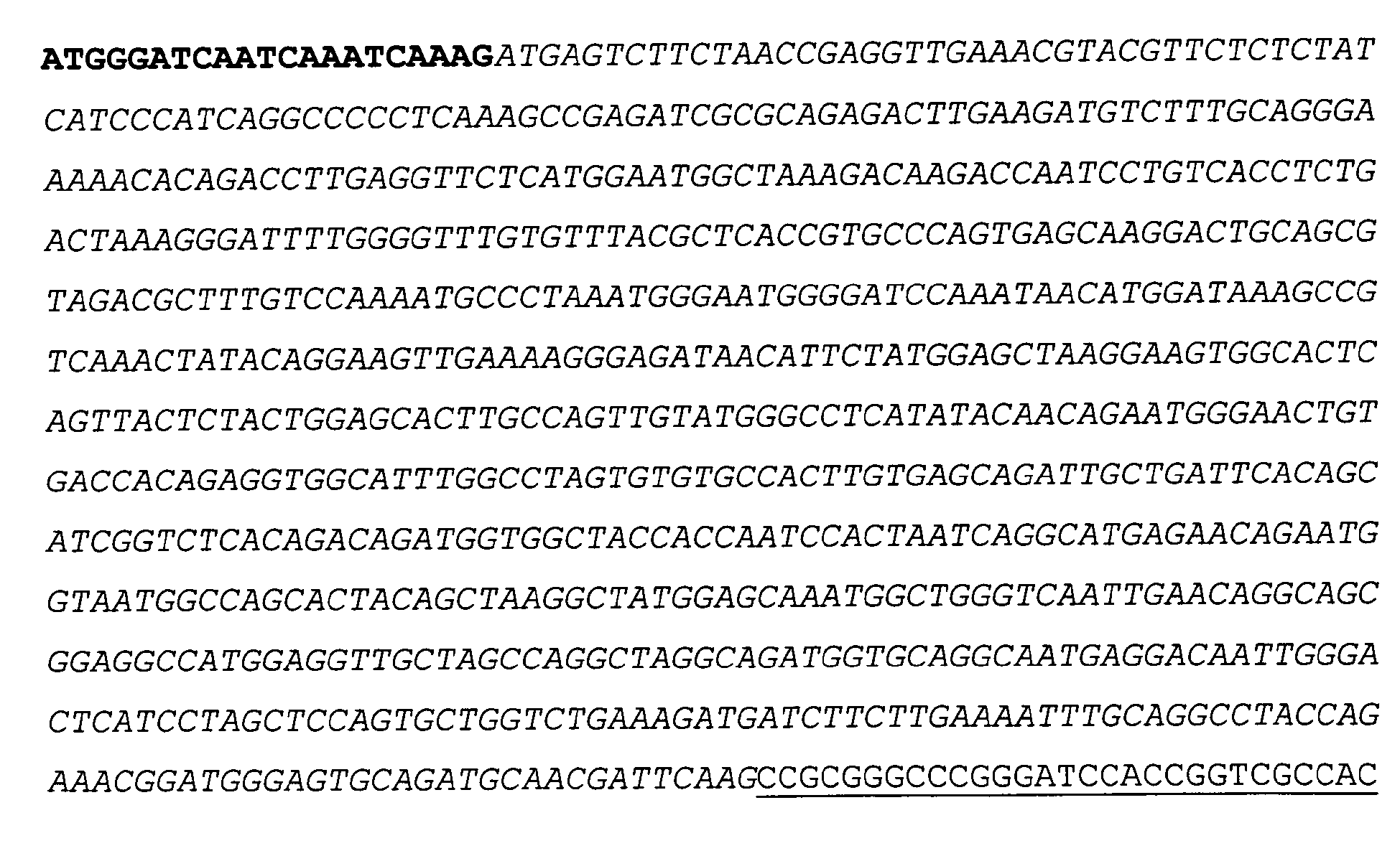
The object of the present invention is further solved by a nucleic acid comprising a nucleic acid sequence according to SEQ ID NO: 1 or SEQ ID NO: 3.
-
The object of the present invention is further solved by a nucleic acid comprising a nucleic acid sequence according to SEQ ID NO: 59.
-
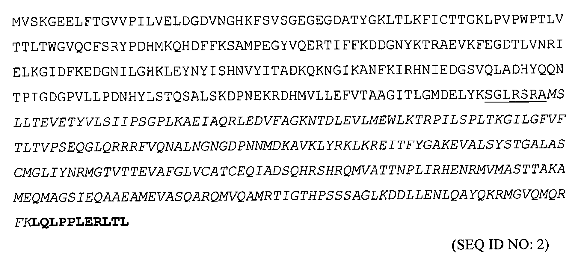
The object of the present invention is further solved by an amino acid sequence comprising an amino acid sequence according to SEQ ID NO: 2 or SEQ ID NO: 4.
-
The object of the present invention is further solved by a protein comprising an amino acid sequence according to SEQ ID NO: 2 or SEQ ID NO: 4.
-
The object of the present invention is further solved by a matrix protein M1 mutant according to the present invention comprising an amino acid sequence according to SEQ ID NO: 2 or SEQ ID NO: 4.
-
The object of the present invention is further solved by a vector comprising a nucleic acid according to the present invention, preferably a nucleic acid comprising a nucleic acid sequence according to SEQ ID NO: 1 or SEQ ID NO: 3.
-
In one embodiment, the vector used to generate the vector according to the present invention is pECFP-N1 or pECFP-C1. In one embodiment, the vector is pECFP-N1 in case of CFP-M1-NES. In another embodiment, the vector is pECFP-C1 in case of Myr-N1-NES.
-
The object of the present invention is further solved by a cell comprising a mutant according to the present invention and/or a nucleic acid according to the present invention and/or an amino acid sequence according to the present invention and/or a vector and/or a construct according to the present invention.
-
The object of the present invention is further solved by a use of a mutant according to the present invention and/or of a nucleic acid according to the present invention and/or of an amino acid sequence according to the present invention and/or of a vector and/or a construct according to the present invention and/or of a cell according to the present invention for an evaluation of an association of the matrix protein M1 mutant with a cellular membrane.
-
The object of the present invention is further solved by a method for an evaluation of an association of a matrix protein M1 mutant according to the present invention with a cellular membrane, preferably using fluorescence microscopy on an intact cell.
-
The object of the present invention is further solved by a method for an evaluation of an interference of a candidate compound with an association of a matrix protein M1 mutant according to the present invention with a cellular membrane, comprising the steps of:
- (a) incubating a cell according to the present invention with the candidate compound;
- (b) detecting the association of the matrixprotein M1 mutant with the cellular membrane in the cell.
-
In one embodiment, the method further comprising the steps of:
- (a') incubating a control cell according to the present invention without the candidate compound;
- (b') detecting the association of the matrix protein M1 mutant with the cellular membrane in the control cell;
- (c) comparing the results obtained in steps (b) and (b').
-
In one embodiment of the method, steps (b) and (b') involve fluorescence microscopy on the intact cell.
-
In one embodiment of the method, interference is an inhibition, and the candidate compound is a putative inhibitor of an association of the matrix protein M1 with the cellular membrane.
-
The object of the present invention is further solved by a primer comprising a nucleic acid sequence according to SEQ ID NO: 64, SEQ ID NO: 65, SEQ ID NO: 70, or SEQ ID NO: 71.
-
Further considered by the present invention is a high-throughput procedure for an evaluation of an interference of a candidate compound with an association of the matrix protein M1 with the membrane.
-
Generally considered is a matrix protein M1 mutant from any influenza virus comprising any nuclear export signal (as specified in LaCour et al., 2004 or predicted by the NES-predictor: www.cbs.dtu.dk), or any myristoylation signal (predicted by: http://expasy.org/tools/myristoylator/), and any fluorescent protein for which a cloned gene is available. Correspondingly, respective constructs are considered. Also considered is a any fluorescent protein, e.g. derived from GFP.
-
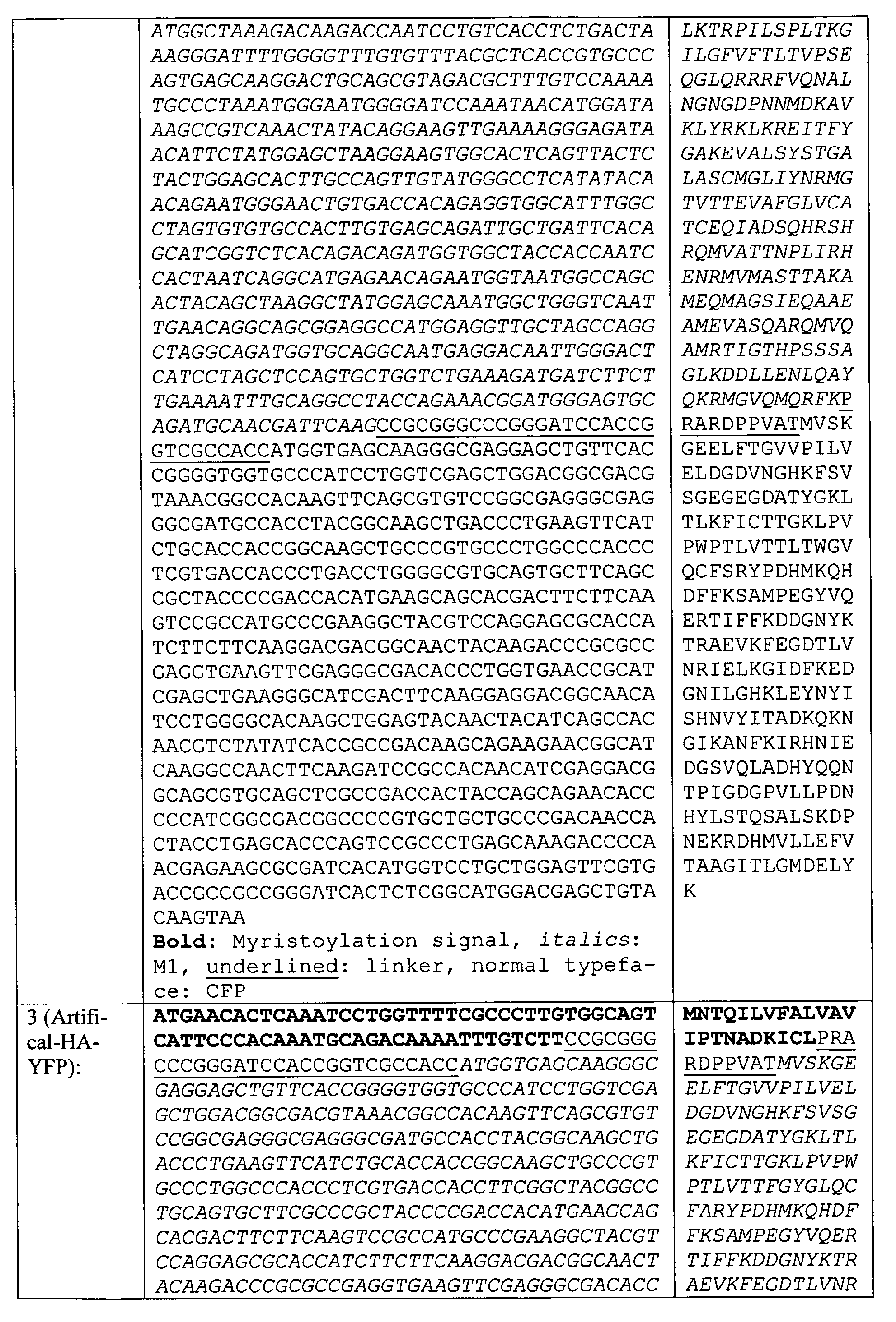
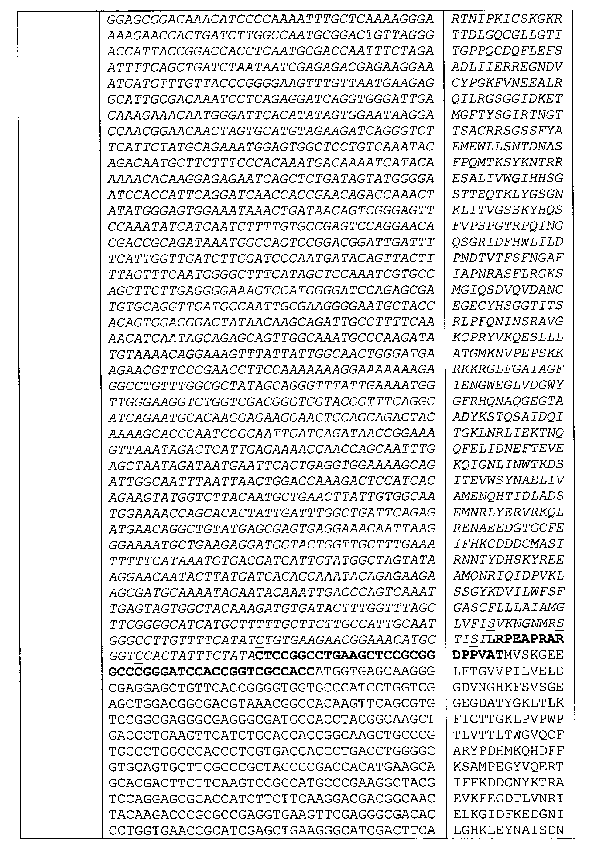
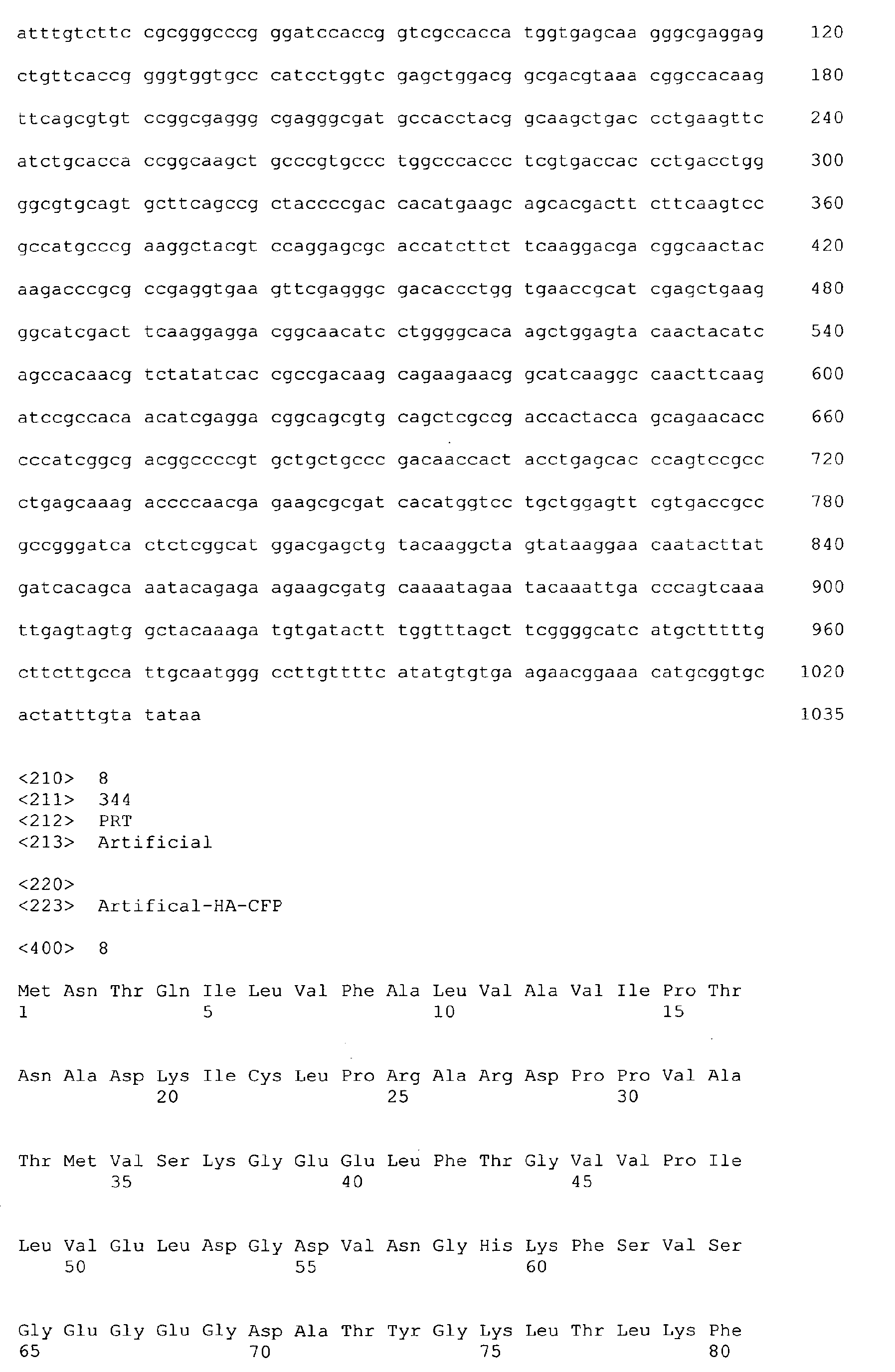
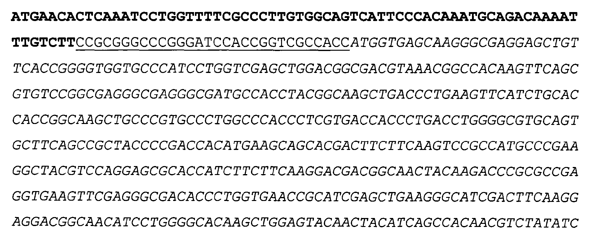
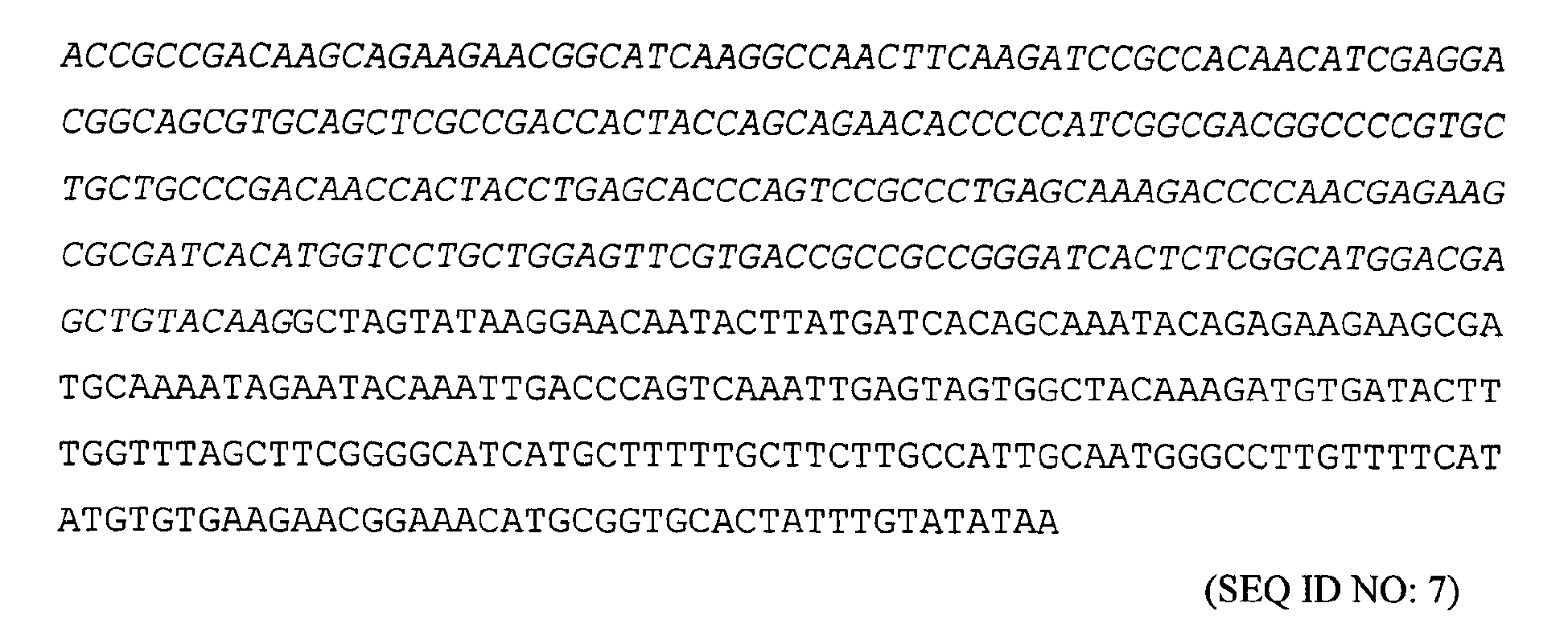
The object of the present invention is further solved by a construct selected from the group consisting of ARTIFICAL-HA-YFP (comprising a nucleic acid sequence according to SEQ ID NO: 5 encoding an amino acid sequence according to SEQ ID NO: 6), ARTIFICAL-HA-CFP (comprising a nucleic acid sequence according to SEQ ID NO: 7 encoding an amino acid sequence according to SEQ ID NO: 8), ARTIFICAL-HA-YFPM3 (comprising a nucleic acid sequence according to SEQ ID NO: 9 encoding an amino acid sequence according to SEQ ID NO: 10), and ARTIFICAL-HA-CFPM3 (comprising a nucleic acid sequence according to SEQ ID NO: 11 encoding an amino acid sequence according to SEQ ID NO: 12).
-
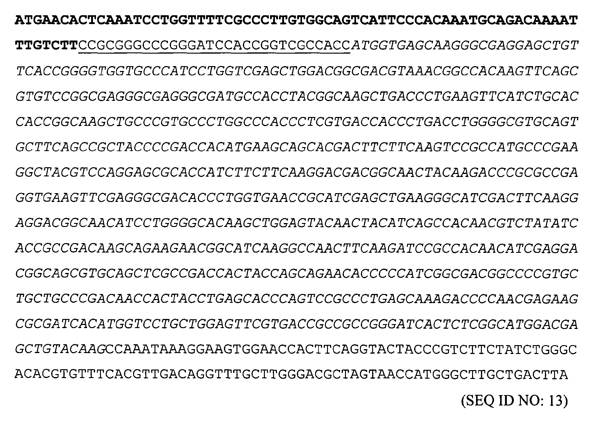
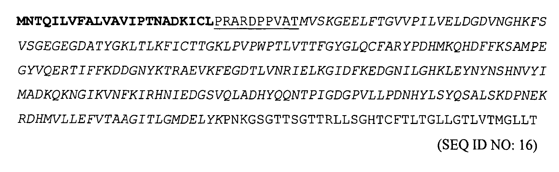
The object of the present invention is further solved by a construct selected from the group consisting of raft-marker GPI(DAF)-CFP (comprising a nucleic acid sequence according to SEQ ID NO: 13 encoding an amino acid sequence according to SEQ ID NO: 14), and GPI(DAF)-YFP (comprising a nucleic acid sequence according to SEQ ID NO: 15 encoding an amino acid sequence according to SEQ ID NO: 16).
-
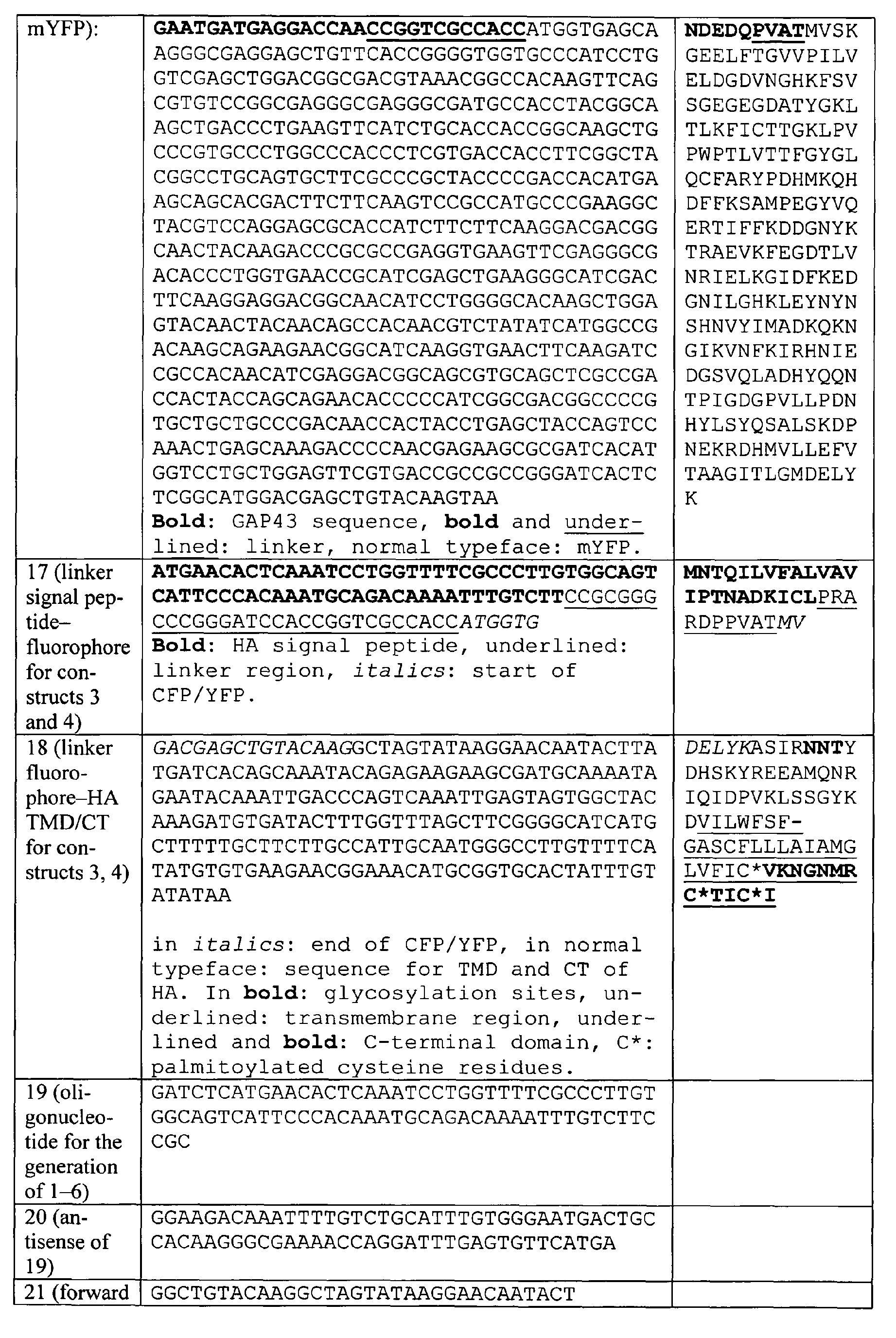
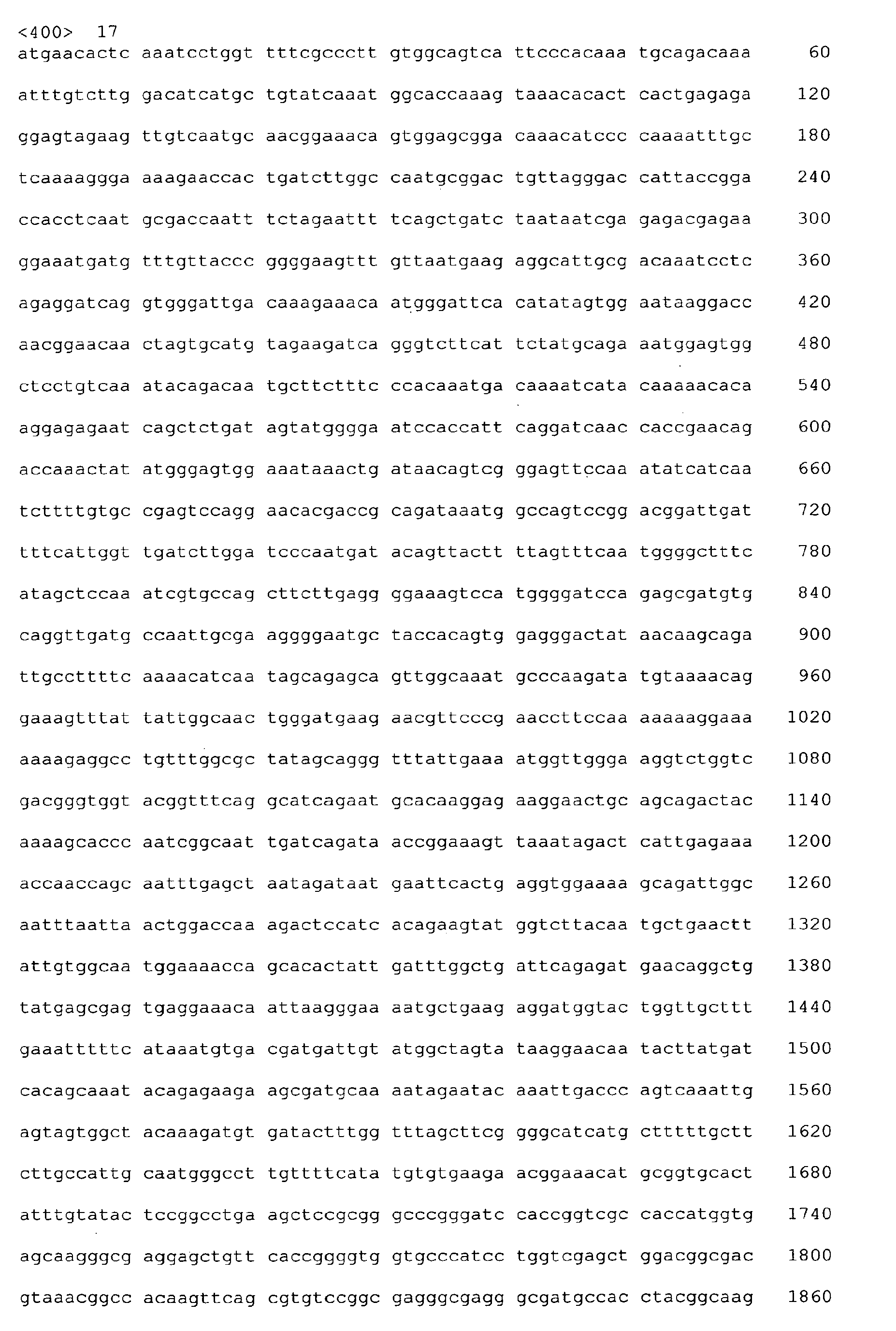
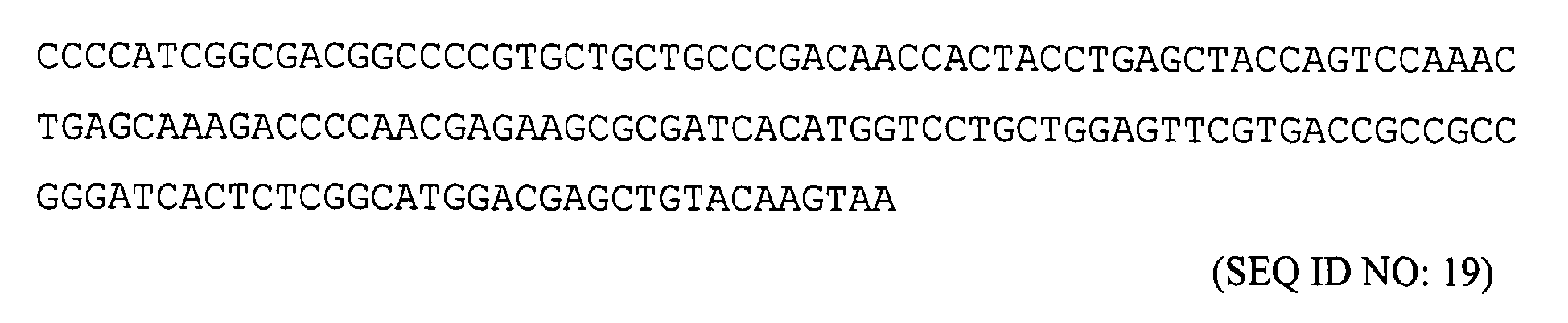
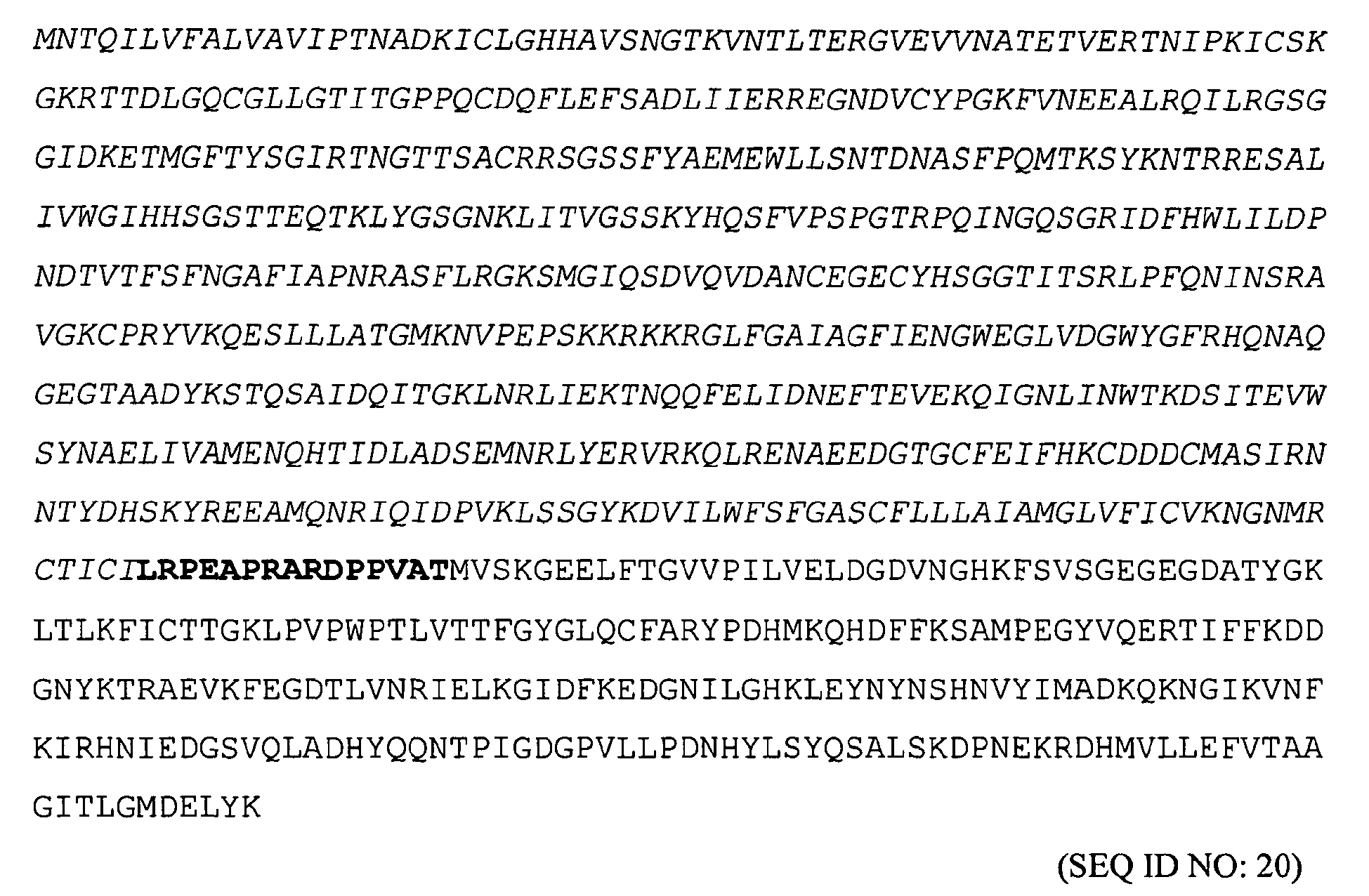
The object of the present invention is further solved by a construct selected from the group consisting of HA-mCer (comprising a nucleic acid sequence according to SEQ ID NO: 17 encoding an amino acid sequence according to SEQ ID NO: 18), HA-mYFP (comprising a nucleic acid sequence according to SEQ ID NO: 19 encoding an amino acid sequence according to SEQ ID NO: 20), M3-HA-mCer (comprising a nucleic acid sequence according to SEQ ID NO: 21 encoding an amino acid sequence according to SEQ ID NO: 22), and M3-HA-mYFP (comprising a nucleic acid sequence according to SEQ ID NO: 23 encoding an amino acid sequence according to SEQ ID NO: 24).
-
The object of the present invention is further solved by a construct selected from the group consisting of raft-marker Lyn-mCer (comprising a nucleic acid sequence according to SEQ ID NO: 25 encoding an amino acid sequence according to SEQ ID NO: 26), Lyn-mYFP (comprising a nucleic acid sequence according to SEQ ID NO: 27 encoding an amino acid sequence according to SEQ ID NO: 28), Mem-mCer (comprising a nucleic acid sequence according to SEQ ID NO: 29 encoding an amino acid sequence according to SEQ ID NO: 30), and Mem-mYFP (comprising a nucleic acid sequence according to SEQ ID NO: 31 encoding an amino acid sequence according to SEQ ID NO: 32).
-
The object of the present invention is further solved by a mutant protein expressed using a construct according to the present invention, preferably a HA mutant or a raft-marker mutant.
-
The object of the present invention is further solved by a nucleic acid comprising a nucleic acid sequence selected from the group consisting of SEQ ID NO: 5, SEQ ID NO: 7, SEQ ID NO: 9, SEQ ID NO: 11, SEQ ID NO: 13, SEQ ID NO: 15, SEQ ID NO: 17, SEQ ID NO: 19, SEQ ID NO: 21, SEQ ID NO: 23, SEQ ID NO: 25, SEQ ID NO: 27, SEQ ID NO: 29, and SEQ ID NO: 31.
-
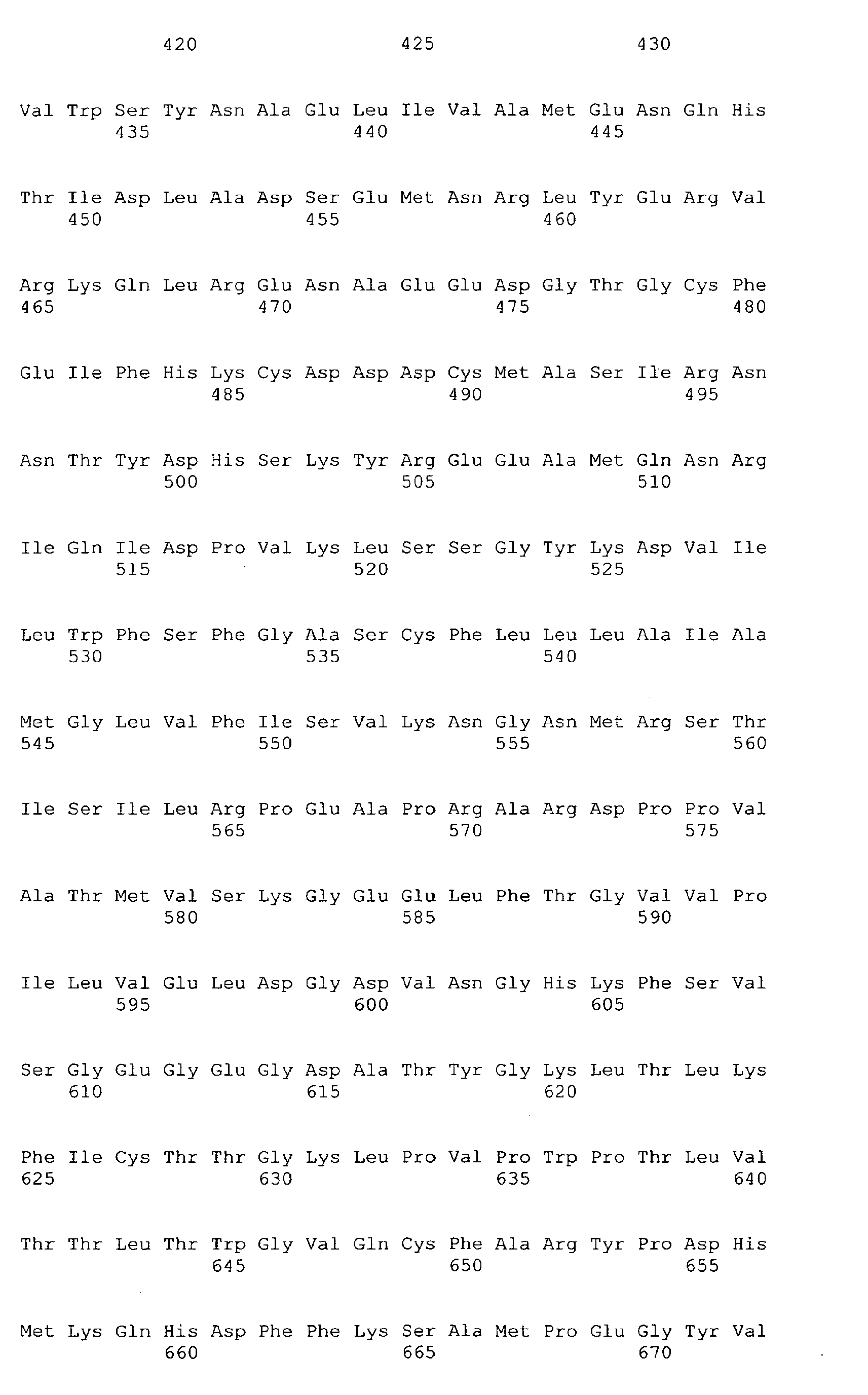
The object of the present invention is further solved by an amino acid sequence comprising an amino acid sequence selected from the group consisting of SEQ ID NO: 6, SEQ ID NO: 8, SEQ ID NO: 10, SEQ ID NO: 12, SEQ ID NO: 14, SEQ ID NO: 16, SEQ ID NO: 18, SEQ ID NO: 20, SEQ ID NO: 22, SEQ ID NO: 24, SEQ ID NO: 26, SEQ ID NO: 28, SEQ ID NO: 30, and SEQ ID NO: 32.
-
The object of the present invention is further solved by a protein comprising an amino acid sequence selected from the group consisting of SEQ ID NO: 6, SEQ ID NO: 8, SEQ ID NO: 10, SEQ ID NO: 12, SEQ ID NO: 14, SEQ ID NO: 16, SEQ ID NO: 18, SEQ ID NO: 20, SEQ ID NO: 22, SEQ ID NO: 24, SEQ ID NO: 26, SEQ ID NO: 28, SEQ ID NO: 30, and SEQ ID NO: 32.
-
The object of the present invention is further solved by a HA mutant comprising an amino acid sequence selected from the group consisting of SEQ ID NO: 6, SEQ ID NO: 8, SEQ ID NO: 10, SEQ ID NO: 12, SEQ ID NO: 18, SEQ ID NO: 20, SEQ ID NO: 22, and SEQ ID NO: 24.
-
The object of the present invention is further solved by a raft-marker mutant comprising an amino acid sequence selected from the group consisting of SEQ ID NO: 14, SEQ ID NO: 16, SEQ ID NO: 26, SEQ ID NO: 28, SEQ ID NO: 30, and SEQ ID NO: 32.
-
The object of the present invention is further solved by a vector comprising a nucleic acid according to the present invention, preferably a nucleic acid comprising a nucleic acid sequence selected from the group consisting of SEQ ID NO: 5, SEQ ID NO: 7, SEQ ID NO: 9, SEQ ID NO: 11, SEQ ID NO: 13, SEQ ID NO: 15, SEQ ID NO: 17, SEQ ID NO: 19, SEQ ID NO: 21, SEQ ID NO: 23, SEQ ID NO: 25, SEQ ID NO: 27, SEQ ID NO: 29, and SEQ ID NO: 31.
-
The object of the present invention is further solved by a cell comprising a mutant protein according to the present invention and/or a nucleic acid according to the present invention and/or an amino acid sequence according to the present invention and/or a vector and/or a construct according to the present invention.
-
The object of the present invention is further solved by a use of a mutant protein according to the present invention and/or of a nucleic acid according to the present invention and/or of an amino acid sequence according to the present invention and/or of a vector and/or a construct according to the present invention and/or of a cell according to the present invention for an evaluation of an association of a HA mutant according to the present invention and a raft-marker mutant according to the present invention.
-
The object of the present invention is further solved by a method for an evaluation of an association of a HA mutant according to the present invention and a raft-marker mutant according to the present invention, preferably using FRET on an intact cell.
-
The object of the present invention is further solved by a method for an evaluation of an interference of a candidate compound with an association of a HA mutant according to the present invention and a raft-marker mutant according to the present invention, comprising the steps of:
- (a) incubating a cell expressing a HA mutant according to the present invention and a raft-marker mutant according to the present invention with the candidate compound;
- (b) detecting the association of the HA mutant according to the present invention and the raft-marker mutant according to the present invention in the cell.
In one embodiment, the method further comprises the steps of:
- (a') incubating a control cell expressing a HA mutant according to the present invention and a raft-marker mutant according to the present invention without the candidate compound;
- (b') detecting the association of the HA mutant according to the present invention and the raft-marker mutant according to the present invention in the control cell;
- (c) comparing the results obtained in steps (b) and (b').
-
In one embodiment of the method, steps (b) and (b') involve FRET on the intact cell.
-
In one embodiment of the method, interference is an inhibition, and the candidate compound is a putative inhibitor of an association of HA with lipid-rafts.
-
Further considered by the present invention is a high-throughput procedure for an evaluation of an interference of a candidate compound with an association of HA with lipid-rafts.
-
The term "construct" means a genetically engineered nucleic acid, preferably contained in a vector or plasmid.
-
The present invention provides methods and means suitable for screening candidate compounds for interference with the binding of M1 to the membrane, and thus, for interference with the assembly of influenza viruses. The present invention further provides methods and means suitable for screening candidate compounds for interference with the accumulation of HA in lipid-rafts, and thus, for interference with the assembly of influenza viruses. Both assay systems can be used independently or in combination.
-
For carrying out method according to the present invention for an evaluation of an interference of a candidate compound with an association of a HA mutant and a raft-marker with a cellular membrane, any combination of a HA mutant according to the present invention and a raft-marker according to the present invention is considered beyond the explicit combinations described herein.
-
We constructed HA/raft-marker pairs where the fluorophores are either inside or outside of the cell. HA-mCer, HA-mYFP, M3-HA-mCer, M3-HA-mYFP, Lyn-mCer, Lyn-mYFP, Mem-mCer and Mem-mYFP have the fluorophore on the cytoplasmic side of the plasma membrane. ARTIFICAL-HA-YFP, ARTIFICAL-HA-CFP, ARTIFICAL-HA-YFPM3, ARTIFICAL-HA-CFPM3, GPI(DAF)-CFP and GPI(DAF)-YFP carry the fluorophore on the outside of the cell. To measure FRET, two constructs with the fluorophores on the same side of the membrane must be used.
-
Use of the described fusion proteins allows to measure FRET on both sides of the plasma membrane. The lipid-composition of rafts differ between the outer and the inner leaflet of the membrane and it is thus possible that certain compounds to be analyzed with the FRET-method described here might interfere differently with raft/HA pairs which carry the fluorophore on the outside or on the inside of the cell. Furthermore, two different fluorophores such as CFP (or its variant Cer) and YFP must be used. All constructs designated M3 have a substitution of the three cyteine residues in the C-terminal part of HA by serines. The cysteine serve as palmitoylation sites, a hydrophobic protein modification which is required for raft localisation of HA. The M3-constructs are intended to be used as a negative control for the FRET-signal, i.e. the signal given by the M3-constructs with a raft-marker is background, which can be substracted from the signal given by the HA constructs containing the palmitoylation site with the raft-marker.
-
The variants of GFP designated with an m, e.g. mYFP, represent a form of the protein which is less likely to form dimers on its own and thus gives rise to a false positive FRET-signal (Zacharias et al., 2002). However, mYFP or mCFP can be probably replaced by YFP and CFP, respectively and vice versa without fundamental effects on the assay.
-
Likewise, we have used the signal peptide of HA for construction of the ARTIFICAL-HA constructs and for GPI(DAF)-CFP. However, every signal peptide derived from the N-terminus of a secreted or transmembrane protein located in the ER, the Golgi or the plasma membrane of an eukaryotic cell should work in the assays. Moreover, the sequence from DAF encoding the attachment of the glykolipid anchor could probably be replaced by any other sequence from a protein attached to a glykolipid-anchor.
-
In order to investigate M1 binding to membranes using a fluorescently labelled construct, the intrinsic M1 localization to the cell nucleus had to be overcome because the signal would outshine or otherwise interfere with all other M1 signals. The exclusion of M1 from the nucleus was achieved by inclusion of (1) a nuclear export signal derived from Rev or (2) a myristoylation signal derived from Lyn kinase. The usefulness of the M1 constructs is not limited to the CFP-variant described here. In principle any fluorescent protein for which a cloned gene is available, e.g. GFP and spectral variants thereof or the red coral protein or spectral variants thereof, can be fused to M1 and used in the assay.
-
Moreover, despite fusion to variants of the green-fluorescent protein, our constructs are transported from their site of synthesis to the plasma membrane. To achieve this we had to construct and test several linker regions, but only the one present in the HA-construct described here was not retained inside the cell. Our constructs thus represent useful tools to study the effect of various compounds on the transport of viral proteins to the plasma membrane, the intracellular site of assembly of influenza virus and for studying interactions between HA and M1 with FRET, a further requirement for budding of infectious influenza virions.
-
SEQ ID NO: 1 to SEQ ID NO: 32 as indicated above are given below.
SEQ ID NO: 1 and SEQ ID NO: 2:
-
(normal typeface: CFP,
underlined: linker, italics: M1,
bold: NES)
SEQ ID NO: 3 and SEQ ID NO: 4:
-
(
bold: myristoylation signal,
italics: M1,
underlined: linker, normal typeface: CFP)
SEQ ID NO: 5 and SEQ ID NO: 6:
-
(
bold: HA signal sequence,
underlined: linker, in
italics: YFP, in normal typeface: HA-TMD/CT)
SEQ ID NO: 7 and SEQ ID NO: 8:
-
(
bold: HA signal sequence,
underlined: linker, in
italics: CFP, in normal typeface: HA-TMD/CT)
SEQ ID NO: 9 and SEQ ID NO: 10:
-
(
bold: HA signal peptide,
underlined: linker, in
italics: YFP,
underlined: residues changed for Cys → Ser mutation)
SEQ ID NO: 11 and SEQ ID NO: 12:
-
(
bold: HA signal peptide,
underlined: linker, in
italics: CFP,
underlined: residues changed for Cys → Ser mutation)
SEQ ID NO: 13 and SEQ ID NO: 14:
-
(
bold: HA signal peptide,
underlined: linker,
italics: CFP, normal typeface: GPI anchor sequence derived from DAF)
SEQ ID NO: 15 and SEQ ID NO: 16:
-
(
bold: HA signal peptide,
underlined: linker,
italics: YFP, normal typeface: GPI anchor sequence derived from DAF)
SEQ ID NO: 17 and SEQ ID NO: 18:
-
(
italics: HA,
bold: linker, normal typeface: mCer)
SEQ ID NO: 19 and SEQ ID NO: 20:
-
(
italics: HA,
bold: linker, normal typeface: mYFP)
SEQ ID NO: 21 and SEQ ID NO: 22:
-
(
italics: HA, underlined: mutations to obtain Cys → Ser exchange (no palmitoylation),
bold: linker, normal typeface: mCer)
SEQ ID NO: 23 and SEQ ID NO: 24:
-
(
italics: HA, underlined: mutations to obtain Cys → Ser exchange (no palmitoylation),
bold: linker, normal typeface: mYFP)
SEQ ID NO: 25 and SEQ ID NO: 26:
-
(bold: Lyn kinase sequence,
bold and
underlined: linker, normal typeface: mCer)
SEQ ID NO: 27 and SEQ ID NO: 28:
-
(bold: Lyn kinase sequence, bold and
underlined: linker, normal typeface: mYFP)
SEQ ID NO: 29 and SEQ ID NO: 30:
-
(bold: GAP43 sequence,
bold and
underlined: linker, normal typeface: mCer)
SEQ ID NO: 31 and SEQ ID NO: 32:
-
(bold: GAP43 sequence,
bold and
underlined: linker, normal typeface: mYF)
-
The invention shall be now further illustrated considering the accompanying figures and the following examples.
Figures
-
-
Figure 1
- shows the assembly of viral components at lipid-rafts. Upper panel: scanning electron microscopy picture. Lower panel: schematic diagram. HA, hemagglutinin; NA, neuraminidase; M1, matrix protein M1; vRNP, viral ribonucleoprotein.
-
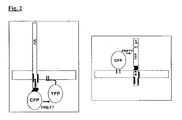
Figure 2
- is a schematic diagram showing the interactions of HA with raft-markers in transfected cells as studied with FRET. Left panel: Native HA-CFP (left) and Lyn-YFP (right). Right panel: GPI(DAF)-CFP (left) and ARTIFICAL-HA-YFP protein (right) containing the raft-targeting signal as well as the trans-membrane and cytoplasmic domain of HA. CFP, cyan fluorescence protein; YFP, yellow fluorescence protein; SP, signal peptide.
-
Figure 3
- shows the ARTIFICAL-HA-CFP construct (Fig. 3a) and the raft-marker GPI(DAF)-YFP (Fig. 3b). L, linker; TMD, transmembrane region; CT, cytoplasmatic domain; GPI, glycolipid anchor; DAF, decay-accelaration factor.
SP(HA), signal peptide from HA.
-
Figure 4
- shows CHO cells transiently expressing ARTIFICAL-HA-YFP (Fig. 4a), GPI(DAF)-CFP (Fig. 4b), GPI(DAF)-YFP (Fig. 4c). Confocal fluorescence microscopy.
-
Figure 5
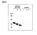
- shows a fluorography picture of a SDS-PAGE gel of immunoprecipitated proteins of [35S]methionin labelled CHO cells transfected with ARTIFICAL-HAYFP construct after digestion with endoglycosidase H (H) or PNGase F (F).
-
Figure 6
- shows the life time of raft-marker GPI(DAF)-CFP fluorescence in the presence and absence of ARTIFICAL-HA-YFP (Fig. 6a) and the FRET efficiency (%) of GPI(DAF)-CFP/HA-YFP prior to and after depletion of cholesterol (Fig. 6b).
-
Figure 7
- shows the HA-mCer construct (Fig. 7a), the raft-marker Lyn-YFP construct (Fig. 7b) and the raft-marker Mem-mYFP construct (Fig. 7c). mCer, mCerulean; m, monomerized form of YFP with the A206K mutation; GAP, GAP43.
-
Figure 8
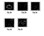
- shows CHO cells transiently expressing HA-mCerulean (Fig. 8a), stably expressing Mem-mYFP (Fig. 8b) or Lyn-mYFP (Fig. 8c) and MDCK cells stably expressing Mem-mYFP (Fig. 8d) and Lyn-mYFP (Fig. 8e). Confocal fluorescence microscopy.
-
Figure 9
- shows SDS-PAGE under reducing (red) or non-reducing (non-red) conditions and fluorography of proteins precipitated from [35S]methionine labelled CHO cells transfected with HA-mCer. Proteolytic cleavage (Fig. 9a), glycosylation (Fig. 9b) and palmitoylation (Fig. 9c).
-
Figure 10
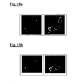
- shows CHO cells transfected with HA-mCerulean (left panel in Fig. 10a and Fig. 10b each) and Lyn-YFP (right panel in Fig. 10a and Fig 10b each). FRET analysis prior to bleaching (Fig. 10a) and after bleaching (Fig. 10b).
-
Figure 11
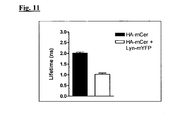
- shows the life time of HA-mCerulean in the presence and absence of raftmarker Lyn.
-
Figure 12
- shows the CFP-M1-NES construct (Fig. 12a) and the Myr-M1-CFP construct (Fig. 12b). M1, matrix protein; NES, nuclear export signal; Myr, myristoylation signal.
-
Figure 13
- shows CHO cells transfected with CFP-M1-wild type (a), CFP-M1-nuclear export signal (b) and Myr-M1-CFP (c). Fluorescence microscopy.
-
Figure 14
- shows the membrane association of CFP-M1-wt and CFP-M1-NES as assessed by biochemical fractionation. CHO cells were transfected with the indicated constructs, lysed, cleared of nuclei by centrifugation and then ultracentrifuged at 100,000x g for 60 min. (a) represents the total cell lysate, (b) the 100,000× g pellet (membranes and membrane-bound proteins) and (c) the 100,000× g supernatant (soluble proteins). Western Blot probed with an anti-M1 antibody, the position of M1 is indicated by an arrow. A negative control (untransfected cells) is included (right lanes).
EXAMPLES
EXAMPLE 1: Studies on the interaction of HA and raft-marker on the outside of the plasma membrane using Artifical-HA-YFP (or -CFP) and raft-marker-CFP (or -YFP) -GPI constructs
1.1 ARTIFICAL-HA-YFP
-
The ARTIFICAL-HA-YFP construct includes the "yellow-fluorescent protein" (YFP) fused to the signal peptide of HA via a linker region. Located at the C-terminus of YFP are 35 amino acids of the extracytoplasmatic domain of HA as well as the complete transmembrane region and the cytoplasmatic domain (Fig. 3a).
-
Also considered is the replacement of YFP by CFP (or by its variant Cer).
The linker between the signal peptide and the fluorophore (either CFP or YFP) is as follows:
-
(in
bold: HA signal peptide, underlined: linker region, in
italics: start of CFP or YFP sequence). Translation into protein:
MNTQILVFALVAVIPTNADKICL PRARDPPVAT MV (SEQ ID NO: 34)
-
The linker between the end of the fluorophore and TMD+CT of HA is as follows:
(in
italics: end of CFP/YFP, in normal typeface: sequence for TMD and CT of HA). Translation into protein:
DELYKASIR
NNTYDHSKYREEAMQNRIQIDPVKLSSGYKD
VILWFSFGASCFLLLAIAMGLV FIC*VKNGNMRC*TIC*I . (SEQ ID NO: 36)
(in
bold: glycosylation sites, underlined: transmembrane region,
underlined and
bold: C-terminal domain, C*: palmitoylated cysteine residues).
-
Also considered is a mutant of HA in which the three palmitoylated cysteine residues important for raft localization are replaced by serines. The cysteine mutants are called ARTIFICAL-HA-YFPM3, ARTIFICAL-HA-CFPM3, respectively.
1.2 Raft-marker GPI(DAF)-YFP
-
The raft-marker GPI(DAF)-YFP includes the signal peptide of HA fused to the YFP via a linker region. Located at the C-terminus of YFP are amino acids comprising the signals for attachment of the glycolipid anchor (GPI) from the cellular protein "decay-acceleration factor" (DAF) (Legler et al. 2005) (Fig. 3b).
-
Also considered is the replacement of YFP by CFP.
-
The DNA sequence of the GPI(DAF)-YFP construct is as follows:
(in
bold: HA signal peptide, underlined: linker,
italics: YFP, normal typeface: GPI anchor sequence derived from DAF).
-
The protein sequence of the GPI(DAF)-YFP construct is as follows:
1.3 ARTIFICAL-HA-YFP ARTIFICAL-HA-CFP, ARTIFICAL-HA-YFPM3, ARTIFICAL-HA-CFPM3, GPI(DAF)-CFP, and GPI(DAF)-YFP
-
ARTIFICAL-HA-YFP, ARTIFICAL-HA-CFP, ARTIFICAL-HA-YFPM3, ARTIFICAL-HA-CFPM3, GPI(DAF)-CFP, GPI(DAF)-YFP constructs were generated using the plasmids pEYFP-N1 and pECFP-N1 (Clontech). The HA signal peptide was designed as oligonucleotide having the following sequences:
GATCTCATGAACACTCAAATCCTGGTTTTCGCCCTTGTGGCAGTCATTCCCACAAATGCAGA CAAAATTTGTCTTCCGC (SEQ ID NO: 37)
sense oligonucleotide
GGAAGACAAATTTTGTCTGCATTTGTGGGAATGACTGCCACAAGGGCGAAAACCAGGATTTG AGTGTTCATGA (SEQ ID NO: 38)
antisense oligonucleotide
-
The oligonucleotide was ligated into the plasmids using the BglII and SacII restrictions sites within the multiple cloning site (MCS) (both for the HA and GPI constructs). For the HA constructs, the HA transmembrane domain and cytoplasmic tail was obtained as a single polymerase chain reaction (PCR) fragment using as a template the cDNA encoding the HA gene from influenza virus strain A/FPV/Rostock/34 (H7N1) and the cDNA encoding the HA gene from influenza virus strain A/FPV/Rostock/34 (H7N1) mutated at postions 551, 559, 562 (C→S).
-
The PCR fragment was generated using the following primers:
GGCTGTACAAGGCTAGTATAAGGAACAATACT (SEQ ID NO: 39)
sense primer
GCGCGGCCGCTTATATACAAATAGTGCACCGC (SEQ ID NO: 40)
HA wt antisense primer
GCGCGGCCGCTTATATAGAAATAGTGGACCGC (SEQ ID NO: 41)
HA M3 antisense primer
-
The PCR fragment was ligated into the plasmid using the
BsrGI and
NotI restrictions sites. For GPI(DAF) constructs (Legler
et al. 2004), the GPI signal of the DAF was designed as oligonucleotide having the following sequences:
-
The oligonucleotide was cloned into the BsrGI/NotI restriction sites of the vectors. Amplification of the plasmids was carried out in E. coli (chemically competent, DH-5α).
1.4 Microscopic and biochemical studies
-
Confocal fluorescence microscopy showed that in CHO cells transiently expressing ARTIFICAL-HA-YFP (Fig. 4a), GPI(DAF)-CFP (Fig. 4b) or GPI(DAF)-YFP (Fig. 4c) the proteins localized at the plasma membrane and in the Golgi apparatus, i.e. at intracellular sites where also the native non-labelled proteins are located.
-
Figure 5 shows that an ARTIFICAL-HA-YFP construct is glycosylated and able to pass the Golgi apparatus. After transfection and labeling of CHO cells with [35S]methionine, the immunoprecipitated proteins were digested with endoglycosidase H or PNGase F or remained undigested (-) and were subjected to SDS-PAGE and fluorography.
1.5 FRET and FLIM analysis
-
Intensity measurements as well as FRET measurements were carried out with transfected cells expressing donor alone (CFP or mCerulean) or both, donor (CFP or mCerulean) and acceptor (YFP or mYFP). Emission of CFP or mCerulean and YFP or mYFP was obtained by sequential excitation at 440 nm and 515 nm. FRET-induced emission of the acceptor was obtained by excitation at 440 nm.
Table 1: Laser and wavelength for the detection of the fluorophores | Fluorophore | Laser | Wavelength |
| CFP | Laser diode | 440 nm |
| YFP | Argon laser | 514 nm |
| YFP-photobleaching | Argon laser | 514 nm |
Table 2: Filters and beam splitter for the detection of the fluorophores | Flurophores | Excitation dichroic mirror | Emission dichroic mirror | Detection range |
| CFP | DM 458/515 | BS 490 | 461 nm-486 nm |
| YFP | DM 458/515 | BS 490 | 535 nm-575 nm |
-
Tested bleaching conditions:
- 100% laser power, 1 s
- 33% laser power, 30 s
- 50% laser power, 30 s, 45 s, 60 s
-
Microscope: Olympus FluoView 1000
-
Objective: UPLSAPO 60x Oil, NA 1.35 (Olympus)
-
Fluorescence intensity based FRET efficiencies were calculated according to:
wherein I
D is the fluorescence intensity of a donor in the absence of an acceptor, and I
DA is the intensity of a donor in the presence of an acceptor.
-
FRET efficiencies measured by acceptor photobleaching were calculated according to:
wherein I
D(prebleaching) is the fluorescence intensity of a donor in the presence of an acceptor (prebleaching), and I
D(postbleaching) is the intensity of a donor after complete bleaching of an acceptor (postbleaching).
-
FLIM measurements were carried out with tranfected cells expressing donor (CFP or mCerulean) and with transfected cells expressing donor (CFP or mCerulean) and acceptor (YFP or mYFP).
Table 3: Laser and wavelength for the detection of the fluorophores | Fluorophore | Laser | Wavelength |
| CFP | Pulsed laser diode | 440 nm |
-
Microscope: Olympus FluoView 1000 with LSMupgrade Kit (FLIM) from PicoQuant
-
Objective: UPLSAPO 60x Oil, NA 1.35 (Olympus)
-
For lifetime measurement a pulsed diode laser was used with a frequency of 20 MHz. FLIM pictures were accumulated for 90 seconds.
-
For lifetime measurement the DM 458/515 dichroic mirror was used. The fluorescence was detected by a single photon avalanche photodiode (PicoQuant) via a 470 ± 15 nm filter. The lifetimes were measured by the method of time-correlated single photon counting (TCSPC). The data were collected by the TimeHarp 200 PC card (PicoQuant). In general, the calculation of a fluorescence lifetime image is done by sorting all photons that correspond to one pixel into a histogram, which is then fitted to an exponential decay function to extract the lifetime information. The analysis was performed by the SymPhoTime (PicoQuant) software by deconvoluting the measured curve with the instrument response function (IRF). For single cell lifetime analysis only the fluorescence photons originated from the plasma membrane were taken into account.
-
Analysis of lifetimes can be done according to different models. In the first model we considered that the donor has an intrinsic complex decay with two components, but we considered that the donor is a single population.
-
Equation 3 describes the donor decay:
-
In this way FRET efficiency has been calculated using the average lifetime after normalizing the amplitudes (Eα
i=1, where α
i represents the fraction of the emitting molecules having a particular lifetime, τ
i).
-
FRET efficiency can be obtained by
where τ
D and τ
DA are the donor excited state lifetime in the absence and presence of the acceptor, respectively.
-
In the second model the complex decay of the donor is due to the existence of two populations of donors and it is described as:
(donor in the absence of acceptor)
(donor in the presence of acceptor)
-
The FRET efficiency is then calculated using equation 5, with τ1, τ2 and τq1, τq2 as the unquenched and quenched donor lifetimes, respectively.
-
Figure 6a shows the lifetime of fluorescence of the raft-marker GPI(DAF)-CFP in the presence and absence of ARTIFICAL-HA-YFP.
-
Figure 6b shows the FRET efficiency (%) of GPI(DAF)-CFP/ARTIFICAL-HA-YFP before and after depletion of cholesterol. In this study, cells were treated with 5 mM β-methylcyclodextrin for 1 min at 4 °C. FRET efficiency after cholesterol extraction is reduced due to resolution of the lipid-rafts.
EXAMPLE 2: Studies on the interaction of HA and raft-marker on the inner side of the plasma membrane using HA-mCer and raft-marker Lyn-mYFP or GAP43-mYFP constructs
2.1 HA-mCer
-
The HA-mCer construct includes mCerulean (mCer), an improved CFP (Rizzo et al. 2004), fused to the C-terminus of HA via a linker region (Fig. 7a). Since some of the HA-mCer constructs created are not transported to the cell surface, the construction of the linker region is of particular significance.
-
The wt-HA-mCer construct is as follows:
TGCACTATTTGTATA CTCCGGCCTGAAGCTCCGCGGGCCCGGGATCCACCGGTCGCCACCAT GGTGAGCAAG (SEQ ID NO: 44)
(in italics: end of HA sequence, palmitoylated cysteines underlined, in bold: linker, in normal typeface: start of mCerulean). Translation into protein:
CTICI LRPEAPRARDPPVATMVSK) (SEQ ID NO: 45)
-
Also considered is a mutant of HA (M3), in which the three palmitoylation sites important for raft localization are replaced.
2.2 Raft-marker Lyn-mYFP
-
The raft-marker Lyn-mYFP construct includes a myristoylation signal (i.e. a signal for the attachment of myristic acid) at the N-terminus and an attachement site for the long-chain fatty acid palmitic acid from the cellular Lyn kinase fused to the monomerized (m) YFP via a short linker (Fig. 7b).
-
The Lyn-mYFP construct is as follows:
(small letters: Kozak sequence, ATG: start codon of Lyn kinase, in bold: myristoylation and palmitoylation signal from Lyn kinase, in
bold and
underlined: linker, normal typeface: start of mYFP/mCer). Translation into protein:
MG*C*IKSKRKDNLNDDEPVAT MV (SEQ ID NO: 47)
(myristoylated glycine and palmitoylated cysteine residue marked with asterisk).
2.3 Raft-marker Mem-mYFP
-
The raft-marker Mem-mYFP construct includes two attachment sites for the long-chain fatty acid palmitic acid from the cellular protein GAP43 at the N-terminus, fused to the monomerized YFP via a short linker (Fig. 7c).
-
The raft-marker Mem-mYFP construct is as follows:
(small letters: Kozak sequence, ATG: start codon of Lyn kinase, in
bold: palmitoylation signal from GAP43, in
bold and
underlined: linker, normal typeface: start of mYFP/mCer). Translation into protein:
MLC*C*MRRTKQVEKNDEDQPVAT MV (SEQ ID NO: 49)
(palmitoylated cysteine residues marked with asterisk).
2.4 HA-mCer, HA-mYFP, M3-HA-mCer, M3-HA-mYFP, Lyn-mCer, Lyn-mYFP, Mem-mCer, and Mem-mYFP
-
The construction of HA-mCer, HA-mYFP, M3-HA-mCer, M3-HA-mYFP, Lyn-mCer, Lyn-mYFP, Mem-mCer, and Mem-mYFP was carried out using the plasmids pEYFP-N1 and pECFP-N1 (Clontech). mCerulean-C1 (mCer) was provided by D.W. Piston (Rizzo et al. 2004). mCerulean is an improved cyan-fluorescent protein variant and was ligated into the pECFP-N1 vector using the AgeI and BsrGI restriction sites, which are located directly before the start codon and near the stop codon of mCer and CFP.
-
The A206K monomeric mutation (Zacharias et al. 2002) in mYFP was made using the primers given below following the manufactures protocol of Quickchange site-directed mutagenesis kit (Stratagene):
GGGGTCTTTGCTCAGTTTGGACTGGTAGCTCAG (SEQ ID NO: 50)
sense primer
CTGAGCTACCAGTCCAAACTGAGCAAAGACCCC (SEQ ID NO: 51)
antisense primer
-
The HA constructs wt-HA and M3-HA were obtained as single PCR fragments using as a template the cDNA encoding the HA gene from influenza virus strain A/FPV/Rostock/34 (H7N1) and the cDNA encoding the HA gene from influenza virus strain A/FPV/Rostock/34 (H7N1) mutated at postions 551, 559, 562 (C→S).
-
For the PCR amplification the following primers were used for both constructs:
CTCAGATCTCATGAACACTCAAATCCTGGTTTTC (SEQ ID NO: 52)
sense primer
GACCCGCGGAGCTTCAGGCCGGAGTATACAAATAGTGCACCG (SEQ ID NO: 53)
antisense primer for wt HA
GACCCGCGGAGCTTCAGGCCGGAGTATAGAAATAGTGGACCG (SEQ ID NO: 54)
antisense primer for M3 HA
-
The PCR fragments were ligated into the plasmid using the BglII and SacII restriction sites within the MCS. The Lyn- and the Mem-peptides were designed as oligonucleotides. For Mem the following oligonucleotides were used:
GGGCCGCGGATGCTGTGCTGTATGAGAAGAACCAAACAGGTTGAAAAGAATGATGAGGACCA ACCGGTCAAA (SEQ ID NO: 55)
sense oligonucleotide
TTTGACCGGTTGGTCCTCATCATTCTTTTCAACCTGTTTGGTTCTTCTCATACAGCACAGCA TCCGCGGCCC (SEQ ID NO: 56)
antisense oligonucleotide
-
For Lyn the following oligonucleotides were used:
GGGCCGCGGACCATGGGATGTATTAAATCAAAAAGGAAAGACAATCTCAATGACGATGAACC GGTCCCC (SEQ ID NO: 57)
sense oligonucleotide
GGGGACCGGTTCATCGTCATTGAGATTGTCTTTCCTTTTTGATTTAATACATCCCATGGTCC GCGGCCC (SEQ ID NO: 58)
antisense oligonucleotide
-
These oligonucleotides were ligated into the vectors mCerulean-N1 and mYFP-N1 using the SacII and AgeI restriction sites within the MCS. Amplification of the plasmids was carried out in E.coli (chemically competent, XL-1 blue).
-
Transfection of CHO-K1 cells was performed with Lipofectamine 2000 transfection reagent (Invitrogen). The cells were analyzed for expression of the fluorescent protein 20-24 h after transfection using a confocal microscope Olympus FluoView 1000.
2.5 Microscopic and biochemical studies
-
Confocal fluorescence microscopy showed that in CHO cells transiently expressing HA-mCer (Fig. 8a) or stably expressing Mem-mYFP (Fig. 8b) or Lyn-mYFP (Fig. 8c), as well as MDCK cells stably expressing Mem-mYFP (Fig. 8d) or Lyn-mYFP (Fig. 8e) the proteins localized at the plasma membrane and in the Golgi apparatus, i.e. at intracellular sites where also the endogenous non-labelled proteins are located.
-
After transfection of HA-mCer and M3-HA-mCer and labelling of CHO cells with [35S]methionine, the immunoprecipitated proteins were subjected to SDS-PAGE under reduced and non-reduced conditions and to fluorography. Figure 9a shows that both the wild type HA-mCer and the M3-HA-mCer mutant are proteolytically cleaved into HA1 and HA2-mCer.
-
In the experiment underlying Figure 9b CHO cells were transfected with HA-mCer and M3-HA-mCer and labelled with [35S]methionine. Prior to SDS-PAGE and fluorography the immunoprecipitated proteins were digested with endoglycosidase H or PNGase F or remained undigested (-) and were subjected to SDS-PAGE and fluorography.
-
In the experiment underlying Figure 9c CHO cells were transfected with HA-mCer and M3-HA-mCer and labelled with [35S]methionine or [3H]palmitic acid. Proteins were immunoprecipitated and analysed for an incorporation of labelled fatty acid by SDS-PAGE and fluorography. As shown in Figure 9c, [3H]palmitic acid is incorporated into wild-type HA-mCer, but not into the mutant M3-HA-mCer.
-
The results shown in Figures 9a to 9c demonstrate that with regard to proteolytic cleavage, glycosylation and palmitoylation, i.e. activities related to infectivity, HA-mCer behaves like the native HA.
2.6 FRET and FLIM analysis
-
FRET and FLIM analysis was carried out as described above (see section 1.5 of Example 1 above).
-
Figures 10a and 10b shows the interaction between HA-mCer and Lyn-mYFP making use of an intensity enhancement of the donor signal after bleaching of the acceptor. Emission of HA-mCer (Fig. 10a, left panel) and Lyn-mYFP (Fig. 10a, right panel) in CHO cells transiently expressing these proteins was detected prior to bleaching. It is shown that both HA-mCer and Lyn-mYFP was expressed in three different cells. After bleaching, the cell marked by a circle did not show any YFP emission, but mCer emission was increased. In contrast, non-bleached cells did not show any change in mCer or YFP emission.
-
Figure 11 shows the lifetime of fluorescence of HA-mCer in the presence and absence of raft-marker Lyn-mYFP.
EXAMPLE 3: Studies on matrix protein M 1 membrane association
3.1 CFP-M1-NES
-
The CFP-M1-NES construct includes at the C-terminus an amino acid sequence from Rev protein of the human immunodeficiency virus (HIV-1) responsible for the export from the nucleus (nuclear export signal, NES) and, in addition, the CFP fused to the N-terminus of M1 (Fig. 12a).
-
To generate the CFP-M1-NES construct, M1 including the NES from Rev (HIV-1) was amplified using PCR and cloned into the MCS of the vector pECFP-N1 making use of the XhoI and KpnI restriction sites. As a result, a linker was formed located between the C-terminus of CFP and the N-terminus of M1:
TACAAGTCCGGACTCAGATCTCGAGCTATGAGT (SEQ ID NO: 59)
(in italics: end of CFP sequence, in bold: start of M1 sequence, normal typeface: linker sequence). Translation into protein:
YKSGLRSRAMS) (SEQ ID NO: 60)
-
The C-terminus of M 1 is followed by the NES consisting of eleven amino acids having the following sequence:
LQLPPLERLTL (SEQ ID NO: 61)
-
In more detail, the linkage between the C-terminus of M1 and the NES is as follows:
TTCAAG CTTCAGCTACCACCGCTTGAGAGACTTACTCTTTGA (SEQ ID NO: 62)
(in italics: M1 sequence, in bold: NES). Translation into protein:
FK LQLPPLERLTL (SEQ ID NO: 63)
-
For construction, the open reading frame (ORF) coding for M1 of influenza A/FPV/Rostock/34 was amplified by polymerase chain reaction (PCR) from plasmid pHH-M (Wagner et al. 2005) using the following primers:
GCTTACCTCGAGCTATGAGTCTTCTAACCGAGG (SEQ ID NO: 64)
GTCTCAGGTACCTCAAAGAGTAAGTCTCTCAAGCGGTGGTAGCTGAAGCTTGAATCGTTGC (SEQ ID NO: 65)
(underlined: recognition sequences for the restriction enzymes XhoI and KpnI, respectively; in italics: nucleotide sequence encoding the NES derived from HIV-1 Rev, amino acid sequence LQLPPLERLTL as in SEQ ID NO: 61).
-
PCR was carried out using AccuTaq LA polymerase (Sigma-Aldrich). The PCR product and the vector pECFP-C1 (Clontech) were digested with XhoI and KpnI (New England BioLabs) and ligated. After transformation of chemocompetent E. coli XL-1 blue (Stratagene), colonies were selected on YT-agar plates supplemented with 50 µg/ml kanamycin. The correctness of the obtained plasmid was checked by sequencing.
-
To check for expression, CHO-K1 cells were grown on coverslips and transfected with the construct using Lipofectamine 2000 (Invitrogen) according to the manufacturer's instructions. After 24 hours of expression, cells were washed with phosphate-buffered saline (PBS) and fixed with paraformaldehyde (3 % in PBS) for 10 min. Microscopy was conducted with a Leica SP2 confocal laser-scanning microscope.
3 .2 Myr-M1-NES
-
The Myr-M1-NES construct includes at the N-terminus a myristoylation signal (i.e. a signal for the attachment of myristic acid) from the cellular Lyn kinase, and, in addition, the CFP fused to the C-terminus of M1 (Fig. 12b).
-
To generate the Myr-M1-NES construct, M1 comprising the myristoylation signal from the Lyn kinase was amplified using PCR and cloned into the MCS of vector pECFP-C1 making use of the XhoI and KpnI restriction sites.
-
The linkage between the N-terminus of CFP and M1 is as follows:
ATGGGATCAATCAAATCAAAGATGAGTCTT (SEQ ID NO: 66)
(in bold: myristoylation signal derived from Lyn kinase, in normal typeface: M1 sequence). Translation into protein:
MGSIKSKMSL (SEQ ID NO: 67)
-
The linkage between the C-terminus of M1 and CFP is as follows:
TTCAAGCCGCGGGCCCGGGATCCACCGGTCGCCACC ATGGTGAGC (SEQ ID NO: 68)
(normal typeface: M1 sequence, in bold: linker, in italics: CFP sequence). Translation into protein:
FKPRARDPPVAT MVS (SEQ ID NO: 69)
-
For construction, the open reading frame (ORF) of M1 was amplified by PCR from plasmid pHH-M using the following primers:
GCTTACCTCGAG ATGGGATCAATCAAATCAAAGATGAGTCTTCTAACC (SEQ ID NO: 70)
GTCTTACCGCGGCTTGAATCGTTGCATCTGCAC (SEQ ID NO: 71)
(underlined: recognition sequences for the restriction enzymes XhoI and SacII, respectively; in italics: myristoylation sequence derived from Lyn kinase, amino acid sequence MGSIKSK as in SEQ ID NO: 67).
-
The PCR products were purified, digested with XhoI and KpnI (New England BioLabs), repurified and ligated with pECFP-N1 (Clontech) digested with the same enzymes. All other procedures were the same as for CFP-M1-NES (see section 3.1 above).
3.3 Microscopic and biochemical studies
-
CHO-K1 cells were transfected with (a) CFP-M1 wildtype, (b) CFP-M1-NES and (c) Myr-M1-CFP, and localization of the protein was analyzed using fluorescence microscopy (Fig. 13). In contrast to M1-wildtype, predominantly found in the nucleus, CFP-M1-NES is found at intracellular membranes, and Myr-M1-CFP is found in the Golgi apparatus as well as at the plasma membrane.
-
CHO-K1 cells were transfected with CFP-M1 wildtype and CFP-M1-NES; non-transfected cells served as a negative control. The cells were fractionated into membrane and soluble compounds by differential centrifugation and subjected to Western blot using anti-M1 antibody (Virostat). In Fig. 14, lane (a) shows complete cell lysate, lane (b) shows the membrane fraction (100,000× g fraction), and lane (c) shows the soluble fraction (100,000× g supernatant). In contrast to largely soluble CFP-M1 wildtype, CFP-M1-NES is detected nearly exclusively in the membrane fraction.
References
-
- Ali A, Avalos RT, Ponimaskin E, Nayak DP. (2000) Influenza virus assembly: effect of influenza virus glycoproteins on the membrane association of M1 protein. J Virol. 74:8709-8719.
- Anderson, RG and Jacobson, K (2002) A role for lipid shells in targeting proteins to caveolae, rafts and other lipid domains. Science 296, 1821-1825.
- Bacia K, Schuette CG, Kahya N, Jahn R, Schwille P. (2004) SNAREs prefer liquid-disordered over "raft" (liquid-ordered) domains when reconstituted into giant unilamellar vesicles. J Biol Chem. 279:37951-5.
- Barmann, S, Ali, A, Hui, EKW, Adhikary, L and Nayak, DP (2001) Transport of viral proteins to the apical membranes and interaction of matrix protein with glycoproteins in the assembly of influenza viruses. Virus Res. 77, 61-69.
- Baumgart, T, Hess, ST, Webb, WW. (2003) Imaging coexisting fluid domains in biomembrane models coupling curvature and line tension. Nature 425, 821-825.
- Briggs, JAG, Wilk, T and Fuller, SD (2003) Do lipid rafts mediate virus assembly and pseudotyping? J. gen. Virol. 84, 757-768.
- Carrasco, M, Amorim, MJ and Digard, P (2004) Lipid-raft dependent targeting of the influenza A virus nucleoprotein to the apical plasma membrane. Traffic 5 979-992.
- Dietrich, C, Bagatolli, LA, Volovyk, ZN, Thompson, NL, Levi, M., Jacobson, K, Gratton, E. (2001) Lipid rafts reconstituted in model membranes. Biophys. J. 80,1417-1428.
- Edidin, M (2003) The state of lipid rafts: from model membranes to cells. Annu Rev Biophys Biomol Struct. 32:257-83.
- Epand, RM (2004) Do proteins facilitate formation of cholesterol-rich domains? Biochim. Biophys. Acta 1666, 227-238.
- Fiedler, K, Kobayashi, T, Kurzchalia, TV and Simons K (1993) Glycosphingolipid-enriched, detergent-insoluble complexes in protein sorting in epithelial cells. Biochemistry 32, 6365-6373.
- Gomez-Puertas P, Albo C, Perez-Pastrana E, Vivo A, Portela A. (2000) Influenza virus matrix protein is the major driving force in virus budding. J Virol. 74, 11538-11547.
- Harder T, Scheiffele P, Verkade P, Simons K. (1998) Lipid domain structure of the plasma membrane revealed by patching of membrane components. J Cell Biol. 141:929-942.
- Heerklotz, H (2002) Triton promotes domain formation in lipid raft mixtures. Biophys J. 83, 2693-2701.
- Jin, H, Leser, GP, Zhang, J and Lamb, RA (1997) Influenza virus hemagglutinin and neuraminidase cytoplasmic tails control particle shape. EMBO. J 16, 1236-1247.
- Keller, P and Simons, K (1998) Cholesterol is required for surface transport of influenza virus hemagglutinin. J. Cell Biol. 140, 1357-1367.
- LaCour T, Kiemer L, Mølgaard A,Gupta R, Skriver K and Brunak S (2004) Analysis and prediction of leucine-rich nuclear export signals. Protein Engineering, Design & Selection 17 527-536.
- Latham T, Galarza JM. (2001) Formation of wild-type and chimeric influenza virus-like particles following simultaneous expression of only four structural proteins. J Virol. 75:6154-6165.
- Legler, D. F., Doucey, M., et al. (2005) Differential insertion of GPI-anchored GFPs into lipid rafts of live cells. The FASEB Journal 19: 73-75.
- Mayor, S and Rao, M (2004) Rafts: Scale dependent, active lipid organization at the cell surface. Traffic 5, 231-240.
- Melkonian KA, Ostermeyer AG, Chen JZ, Roth MG, Brown DA. (1999) Role of lipid modifications in targeting proteins to detergent-resistant membrane rafts. Many raft proteins are acylated, while few are prenylated. J Biol Chem. 274:3910-3917.
- Munro, S: (2003) Lipid rafts: elusive or illusive? Cell. 115, 377-388. Rizzo, M. A., G. H. Springer, et al. (2004) An improved cyan fluorescent protein variant useful for FRET. Nat Biotechnol 22(4): 445-9.
- Scheiffele, P, Roth, MG and Simons, K (1997) Interaction of influenza virus hemagglutinin with sphingolipid-cholesterol membrane domains via its transmembrane domain. EMBO-J. 16, 5501-5508.
- Scheiffele, P, Rietveld, A, Wilk, T and Simons, K (1999) Influenza virus select ordered lipid domains during budding from the plasma membrane. J. Biol. Chem. 274, 2038-2044.
- Schmitt, AP and Lamb, RA (2004) Escaping from the cell: assembly and budding of negative-strand RNA viruses. Curr Top Microbiol Immunol. 283:145-196.
- Schroeder C, Heider H, Moncke-Buchner E, Lin TI. (2004) The influenza virus ion channel and maturation cofactor M2 is a cholesterol-binding protein. Eur ; [Epub ahead of print]
- Sekar, RB and Periasamy, A (2003) Fluorescence resonance energy transfer (FRET) microscopy imaging of live cell protein localizations. J. Cell Biol. 160, 629-633.
- Sha, B. and Luo, M (1997) Structure of a bifunctional membrane-RNA binding protein, influenza virus matrix protein M1. Nat. Struct. Biol. 4, 239-244.
- Sharma, P., Varma, R., Sarasij, RC, Ira, Gousset, K., Krishnamoorthy, G., Rao, M. and Mayor, S. (2004) Nanoscale organization of multiple GPI-anchored proteins in living cell membranes. Cell 116, 577-589.
- Simons, K and van Meer, G (1988) Lipid sorting in epithelial cells. .
- Simons K, Ikonen E. (1997) Functional rafts in cell membranes. Nature. 387:569-72.
- Simons, K. and Vaz, WLC (2004) Model systems, lipid rafts and cell membranes. Annu Rev Biophys Biomol Struct 33: 269-295.
- Skibens, JE; Roth, MG and Matlin, KS (1989) Differential extractability of influenza virus hemagglutinin during intracellular transport in polarized epithelial cells and nonpolar fibroblasts. J. Cell. Biol. 108, 821-832.
- Tall, RD, Alonso, MA and Roth, MG (2003) Features of influenza HA required for apical sorting differ from those required for assocation with DRMs or MAL. Traffic 4, 838-849.
- Takeda M, Leser GP, Russell CJ, Lamb RA. (2003) Influenza virus hemagglutinin concentrates in lipid raft microdomains for efficient viral fusion.Proc Natl Acad Sci U S A. 100:14610-7.
- Varma., R and Mayor, S. (1998) GPI-anchored proteins are organized in submicron domains at the cell surface. Nature 394, 798-801.
- Wagner R, Herwig A, Azzouz N, Klenk HD: Acylation-Mediated Membrane Anchoring of Avian Influenza Virus Hemagglutinin Is Essential for Fusion Pore Formation and Virus Infectivity. J Virol 79 (2005), 6449-6458.
- Zacharias, DA, Violin, JD, Newton, AC and Tsien, RY (2002) Partitioning of lipid-modified monomeric GFPs into membrane microdomains of live cells. Science 296, 913-916.
-
Zhang, J, Pekosz, A and Lamb, RA (2000) Influenza virus assembly and lipid raft microdomains: a role for the cytoplsmic tails of the spike glycoproteins. J. Virol 74, 4634-4644
- Zhao H, Ekstrom M, Garoff H. (1998) The M1 and NP proteins of influenza A virus form homo- but not heterooligomeric complexes when coexpressed in BHK-21 cells. J Gen Virol. 79 2435-46.
Annex:
-







 (normal typeface: CFP, underlined: linker, italics: M1, bold: NES)
 (bold: myristoylation signal, italics: M1, underlined: linker, normal typeface: CFP)


 (bold: HA signal sequence, underlined: linker, in italics: YFP, in normal typeface: HA-TMD/CT)

 (bold: HA signal sequence, underlined: linker, in italics: CFP, in normal typeface: HA-TMD/CT)


 (bold: HA signal peptide, underlined: linker, in italics: YFP, underlined: residues changed for Cys → Ser mutation)


 (bold: HA signal peptide, underlined: linker, in italics: CFP, underlined: residues changed for Cys → Ser mutation)

 (bold: HA signal peptide, underlined: linker, italics: CFP, normal typeface: GPI anchor sequence derived from DAF)

 (bold: HA signal peptide, underlined: linker, italics: YFP, normal typeface: GPI anchor sequence derived from DAF)

 (italics: HA, bold: linker, normal typeface: mCer)


 (italics: HA, bold: linker, normal typeface: mYFP)


 (italics: HA, underlined: mutations to obtain Cys → Ser exchange (no palmitoylation), bold: linker, normal typeface: mCer)


 (italics: HA, underlined: mutations to obtain Cys → Ser exchange (no palmitoylation), bold: linker, normal typeface: mYFP)

 (bold: Lyn kinase sequence, bold and underlined: linker, normal typeface: mCer)

 (bold: Lyn kinase sequence, bold and underlined: linker, normal typeface: mYFP)


 (bold: GAP43 sequence, bold and underlined: linker, normal typeface: mCer)

 (bold: GAP43 sequence, bold and underlined: linker, normal typeface: mYF)