EP2065394A1 - Peptide for determining actin structures in living cells - Google Patents

Peptide for determining actin structures in living cells Download PDF

Info

Publication number
EP2065394A1
EP2065394A1 EP07023084A EP07023084A EP2065394A1 EP 2065394 A1 EP2065394 A1 EP 2065394A1 EP 07023084 A EP07023084 A EP 07023084A EP 07023084 A EP07023084 A EP 07023084A EP 2065394 A1 EP2065394 A1 EP 2065394A1
Authority
EP
European Patent Office
Prior art keywords
actin
peptide
lifeact
cell
cells
Prior art date
Legal status (The legal status is an assumption and is not a legal conclusion. Google has not performed a legal analysis and makes no representation as to the accuracy of the status listed.)
Ceased
Application number
EP07023084A
Other languages
German (de)
French (fr)
Inventor
designation of the inventor has not yet been filed The
Current Assignee (The listed assignees may be inaccurate. Google has not performed a legal analysis and makes no representation or warranty as to the accuracy of the list.)
Max Planck Gesellschaft zur Foerderung der Wissenschaften eV
Original Assignee
Max Planck Gesellschaft zur Foerderung der Wissenschaften eV
Priority date (The priority date is an assumption and is not a legal conclusion. Google has not performed a legal analysis and makes no representation as to the accuracy of the date listed.)
Filing date
Publication date
Application filed by Max Planck Gesellschaft zur Foerderung der Wissenschaften eV filed Critical Max Planck Gesellschaft zur Foerderung der Wissenschaften eV
Priority to EP07023084A priority Critical patent/EP2065394A1/en
Priority to EP20080854612 priority patent/EP2215109B1/en
Priority to AT08854612T priority patent/ATE528318T1/en
Priority to US12/744,822 priority patent/US8957029B2/en
Priority to PCT/EP2008/010093 priority patent/WO2009068295A1/en
Publication of EP2065394A1 publication Critical patent/EP2065394A1/en
Priority to US14/593,274 priority patent/US9255128B2/en
Ceased legal-status Critical Current

Links

Images

Classifications

    • CCHEMISTRY; METALLURGY
    • C07ORGANIC CHEMISTRY
    • C07KPEPTIDES
    • C07K14/00Peptides having more than 20 amino acids; Gastrins; Somatostatins; Melanotropins; Derivatives thereof
    • C07K14/37Peptides having more than 20 amino acids; Gastrins; Somatostatins; Melanotropins; Derivatives thereof from fungi
    • C07K14/39Peptides having more than 20 amino acids; Gastrins; Somatostatins; Melanotropins; Derivatives thereof from fungi from yeasts
    • C07K14/395Peptides having more than 20 amino acids; Gastrins; Somatostatins; Melanotropins; Derivatives thereof from fungi from yeasts from Saccharomyces
    • CCHEMISTRY; METALLURGY
    • C07ORGANIC CHEMISTRY
    • C07KPEPTIDES
    • C07K7/00Peptides having 5 to 20 amino acids in a fully defined sequence; Derivatives thereof
    • C07K7/04Linear peptides containing only normal peptide links
    • C07K7/08Linear peptides containing only normal peptide links having 12 to 20 amino acids
    • GPHYSICS
    • G01MEASURING; TESTING
    • G01NINVESTIGATING OR ANALYSING MATERIALS BY DETERMINING THEIR CHEMICAL OR PHYSICAL PROPERTIES
    • G01N33/00Investigating or analysing materials by specific methods not covered by groups G01N1/00 - G01N31/00
    • G01N33/48Biological material, e.g. blood, urine; Haemocytometers
    • G01N33/50Chemical analysis of biological material, e.g. blood, urine; Testing involving biospecific ligand binding methods; Immunological testing
    • G01N33/68Chemical analysis of biological material, e.g. blood, urine; Testing involving biospecific ligand binding methods; Immunological testing involving proteins, peptides or amino acids
    • G01N33/6887Chemical analysis of biological material, e.g. blood, urine; Testing involving biospecific ligand binding methods; Immunological testing involving proteins, peptides or amino acids from muscle, cartilage or connective tissue
    • GPHYSICS
    • G01MEASURING; TESTING
    • G01NINVESTIGATING OR ANALYSING MATERIALS BY DETERMINING THEIR CHEMICAL OR PHYSICAL PROPERTIES
    • G01N2333/00Assays involving biological materials from specific organisms or of a specific nature
    • G01N2333/435Assays involving biological materials from specific organisms or of a specific nature from animals; from humans
    • G01N2333/46Assays involving biological materials from specific organisms or of a specific nature from animals; from humans from vertebrates
    • G01N2333/47Assays involving proteins of known structure or function as defined in the subgroups
    • G01N2333/4701Details
    • G01N2333/4712Muscle proteins, e.g. myosin, actin, protein

Definitions

  • the present invention relates to novel peptides capable of binding to actin.
  • the peptides are useful in methods for detecting actin in vitro or in living cells.
  • Actin is involved in many cellular processes such as morphogenesis, intracellular transport, cell division, muscle contraction and cell migration.
  • the actin cytoskeleton is also altered in disease processes such as in invading tumour cells, myopathies or polycystic kidney disease (1-3) .
  • a reliable staining of the actin cytoskeleton is essential.
  • staining of fixed cells with the F-actin binding compound phalloidin coupled to fluorescent dyes is used.
  • some cells such as the plant pathogenic fungus Ustilago maydis, cannot be stained by phalloidin (4) .
  • the dynamics of actin filaments are much more sensitive readouts for cytoskeletal organization and processes such as cell polarization or cell migration can only be properly studied through analysis of cytoskeletal dynamics.
  • actin-GFP can be used in several cell types to monitor actin distribution it is to be used with great care if studying actin dynamics.
  • Actin-GFP is also limited in its application as it exhibits a strong background staining from labelled actin monomers and therefore requires low expression levels.
  • fusion of GFP to several actin binding domains have been used, notably from moesin in Drosophila (8) , LimE in Dictyostelium (9) , Abp120 in Dictyostelium and mammalian cells (10,11) and utrophin in Xenopus (12) .
  • the present invention describes the identification and application of short novel peptides, which specifically stain F-actin structures in a wide range of cell types when coupled to labelling groups such as fluorescent proteins or chemical labelling groups.
  • these novel peptides bind efficiently to actin and do not interfere with actin dynamics in vitro and in vivo.
  • the novel peptides were validated by visualizing and quantifying actin dynamics in several sensitive biological processes such as neuronal growth cone formation or dendritic cell migration.
  • the novel peptides of the present invention may be easily obtained by chemical synthesis or recombinant methods and allow visualization of actin dynamics in non-transfectable cells, such as primary neutrophils.
  • novel peptide probes are ideally suited as a marker for F-actin in living as well as fixed cells.
  • the probe is a 17 amino acid (aa) fragment of the yeast actin binding protein Abp140 that faithfully stains F-actin without interfering with actin polymer dynamics.
  • a first aspect of the present invention refers to a peptide having the amino acid sequence (I) (M) n GVADUKKFE wherein n is 0 or 1, or a variant thereof.
  • a further aspect of the present invention refers to a peptide having the amino acid sequence (II) (M) n GVADLIKKFESISKEE wherein n is 0 or 1, or a variant thereof.
  • Amino acid sequences (I) and (II) are derived from the N-terminal portion of the actin binding protein Abp140 from the yeast S . cerevisiae.
  • the present invention refers also to variants thereof, e.g. to variants derived from other fungal species, e.g. as disclosed Fig. 1e of the present application.
  • amino acid residues 2-10 are selected such that an ⁇ -helical structure is formed.
  • the invention encompasses peptides with one, two, three or even more amino acid substitutions within amino acid residues 2-11 of peptides (I) and (II) and optionally one, two, three, four or even more amino acid substitutions within amino acid residues 12-17 of peptide (II).
  • the amino acid residue G at position 2 may be replaced by S and/or the amino acid residue K at position 8 may be replaced by Q or R.
  • additional amino acid substitutions may occur e.g. at positions 12 to 17, e.g.
  • the peptides of the present invention are preferably capable of efficiently binding to actin, particularly to F-actin.
  • the dissociation constant K d is usually ⁇ 50 ⁇ M, preferably ⁇ 10 ⁇ M and more preferably ⁇ 5 ⁇ M.
  • the peptides of the invention may be linear or cyclic peptides.
  • the peptides may comprise modified or non-genetically encoded amino acid residues.
  • the peptides preferably have a length of up to 120, preferably up to 50, and more preferably up to 20 amino acid residues. More preferably, the peptides are selected from the N-terminal portion of fungal Abp140 polypeptides, e.g. from S.cerevisiae, or actin-binding fragments thereof.
  • the peptides are preferably coupled to heterologous molecules, e.g. labelling groups, which allow detection of the peptides.
  • the heterologous molecule is a peptide or polypeptide, e.g. a labelling peptide such as the FLAG epitope or a labelling polypeptide such as GFP or any variant thereof.
  • the heterologous molecule may be a non-peptidic molecule, e.g. a chemical labelling group, particularly a fluorescent chemical labelling group such as fluoresceine, rhodamine or another fluorescent labelling group.
  • the heterologous molecule may be coupled to the N- and/or to the C-terminus of the peptides (I) or (II).
  • fluorescent polypeptides such as GFP or mRFPruby may be genetically fused to the C-terminus of the peptide without affecting actin binding.
  • fluoresceine isothiocyanate FITC
  • peptidic linker sequences such as the sequence C-G may be added to the C-terminus for functional coupling of heterologous molecules via the SH-group of cysteine.
  • the peptides of the invention may be synthesized by chemical methods, e.g. using a solid phase peptide synthesizer or - particularly in the case of peptides fused to heterologous peptides or polypeptides - by recombinant methods in suitable host cells.
  • a further aspect of the present invention refers to a nucleic acid molecule encoding a peptide as described above, particularly a peptide fused to a heterologous peptide or polypeptide, particularly to a fluorescent polypeptide.
  • the nucleic acid molecule may be a single- or double-stranded DNA or RNA molecule.
  • the nucleic acid molecule is operatively linked to an expression control sequence, i.e. a sequence which is sufficient for effecting expression in a suitable host cell, e.g. a prokaryotic or eukaryotic host cell.
  • the expression control sequence is capable of providing expression in a vertebrate host cell, particularly in a mammalian host cell.
  • Still a further aspect of the present invention refers to recombinant cells transfected or transformed with a nucleic acid molecule as described above.
  • the recombinant cell may be a prokaryotic cell, e.g. an E.coli cell, or a eukaryotic cell, e.g. a fungal cell, an animal cell, particularly a vertebrate cell such as an insect, fish, bird or mammalian cell or a plant cell.
  • Transfection or transformation of the recombinant cell may be carried out by standard methods known in the art, e.g. calcium phosphate precipitation or electroporation.
  • Still a further aspect of the present invention is a non-human transgenic organism transfected or transformed with the nucleic acid molecule as described above.
  • the transgenic organism may be a fungus, an animal, e.g. a nematode like C.elegans, an insect such as Drosophila, a fish such as Danio rero, or a mammal such as a rodent, e.g. a mouse or a rat.
  • the organism may be a plant such a Arabidopsis thaliana.
  • the peptide, nucleic acid molecule, recombinant cell or non-human transgenic organism as described above are useful for the detection of actin, preferably for the detection of actin within a cell, particularly within a living cell.
  • actin-associated processes such as cell migration, cell division and/or cell differentiation may be detected.
  • Other relevant applications are research on cell polarity, neuronal development, muscle development, cell-matrix interaction, cancerogenesis, immune reactions, e.g. of neutrophils, T-cells or dendritic cells etc.
  • the present invention has also applications in pharmaceutical research.
  • actin may be used as marker or target in cell-based screening methods and therapeutic approaches.
  • the present invention is suitable for application in cell-based screens for drugs, e.g. cancer drugs, or in the study of actin-based diseases such as Myopathies or polycystic kidney disease.
  • the peptides of the present invention may be used for the modulation of actin-associated processes in therapeutic applications.
  • the peptides may serve as targeting molecules for the actin cytoskeleton.
  • the peptides may be coupled to effector groups such as oxygen radical producers or caged compounds which can be activated in a regulated way.
  • effector groups such as oxygen radical producers or caged compounds which can be activated in a regulated way.
  • functional domains of cellular regulators such as activators of Rho-type GTPases may be linked to the peptides of the invention and targeted to actin, thereby increasing the effects on actin-dependent processes.
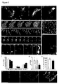
  • Figure 1 Identification of Lifeact. a) TIRF image of Abp140-GFP distribution in an unpolarized yeast cell. Scale bar: 5 ⁇ m. b) Fast blinking of Abp140-GFP signals on actin structures. Time in ms, scale bar: 2 ⁇ m. c) distribution of the minimal Lifeact- G in a yeast cell. Scale bar: 5 ⁇ m. d) Schematic organization of Abp140 and illustration of tested localization constructs. Numbers indicate amino acid positions. Localization scores: ++ comparable to full-length protein, - not localized, +/- localizes mostly to actin patches. e) ClustalW alignment of Lifeact with homologous sequences from related fungi.
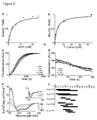
  • FIG. 2 Biochemistry of Lifeact. a) Measurement of F- Lifeact binding to rabbit muscle F-actin. Shown is the relative fluorescence of peptide co-sedimented with various concentrations of F-actin. b) Measurement of F- Lifeact binding to G-actin. Binding was determined by measuring relative changes in pyrene-G-actin fluorescence in the presence of varying amounts of F -Lifeact c) Actin polymerization assay. Polymerization of pyrene-actin was followed in the presence of indicated concentrations of F -Lifeact. d) Actin depolymerization assay.
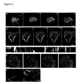
  • FIG. 3 Lifeact-GFP: visualization and functional characterization in vivo.
  • a-j Lifeact G was transiently expressed in mouse embryonic fibroblasts (a, j), primary rat hippocampal neurons (b, f, g, h), MDCK cells (c, i) and mouse dendritic cells (d, e) and imaged with TIRF microscopy.
  • e Time sequence of a dendritic cell chemotaxing towards a chemokine source.
  • f Cortical actin network of a hippocampal neuron.
  • g Time series of filopodial dynamics in a hippocampal neuron.
  • Figure 5 Actin dynamics in leukocytes a, b) Time series of primary human neutrophils scrape loaded with F -Lifeact spreading on plated immune complexes. c, d) kymographs of the indicated regions in (a) and (b), respectively. e) Lifeact- G expressing mouse dendritic cell undergoing lamellipodial extension and retraction. f) Kymograph of the indicated region in e). Solid lines: retrograde actin flow, dotted line: lamellipodial extension and dashed line: lamellipodial retraction. Size bars: 5 ⁇ m.
  • Yeast strains and plasmid constructions All Abp140-GFP plasmids were expressed in the S288C yeast background and based on the pRS315 backbone (LEU2, CEN). Expression was driven by the native Abp140 promoter (300bp) and a short GS linker was generated between Lifeact and GFP. For expression in mammalian cells the Lifeact sequence was cloned into pEGFP-N1 (Clonetech) or the respective plasmid with mRFPruby replacing GFP. In both cases the linker GDPPVAT was generated between Lifeact and the fluorescent protein. Actin-GFP was used in the same plasmid backbones. Cells were grown at 24°C and observed from logarithmically growing cultures (OD ⁇ 0.8). Standard yeast media and procedures were used.
  • TIRF images were captured on an iMic-stand from Till photonics with an 1.45 NA 100x objective from Olympus.
  • a 300mW Argon laser and a 20mW DPSS 561 nm laser were selected through an AOTF.
  • a 2-axis scan head (Yanus II) was used to digitally adjust the TIRF angle.
  • Images were collected with a cooled Imago QE CCD camera. Acquisition was controlled by the TILLvisION software package.
  • Confocal images were collected on a standard Leica SP2 setup.
  • Epifluorescence images were collected on a Zeiss axiovert 200M stand equipped with a climate control chamber from EMBL.
  • Metamorph Molecular Devices
  • sequences were routinely processed by sequential application of a local background subtraction filter and a 3x3 gauss low pass filter.
  • Color overlays and kymographs were created with the respective functions in Metamorph.
  • Muscle actin, non-muscle actin, ⁇ -actinin and pyrene actin were purchased from Cytoskeleton, Inc. Actin was diluted to working concentrations in G buffer (2mM Tris HCl, pH 8.0, 0.2mM ATP, 0.1 mM CaCl 2 , 0.5mM DTT).
  • Actin binding Polymerization of actin was induced by addition of 0.1 volume 10x KMEI buffer (50mM KCl, 1mM MgCl 2 , 1 mM EGTA, 10mM imidazole HCl, pH 7.0) and incubation for >1h at room temperature (RT). 44 ⁇ M of F- Lifeact was incubated 30min with F-actin and then spun 30min at 350,000xg at room temperature. The supernatant was removed and the pellet resuspended in 100 ⁇ l of 1x KMEI buffer. The amount of peptide was measured in a Cary Eclipse Fluorescence Spectrophotometer with excitation/emission set for FITC at 495nm/520nm. The bound/total ratio was calculated as the signal from the pellet divided by the total signal. The K d was obtained by fitting to a hyperbolic curve.
  • Binding to G-actin was determined from a spectral scan of pyrene actin in the presence of varying amounts of F -Lifeact. Averages of 5 emission scans between 370nm and 500nm were used with excitation set to 365nm. The bound/total ratio was calculated from the absolute emission difference at 385nm between a given Lifeact concentration and the control divided by the maximum difference observed. The K d was obtained by fitting to a hyperbolic curve.
  • Actin polymerization and depolymerization 20% pyrene-labelled actin was centrifuged at 350,000 x g for 30min at 24C to remove any nucleation seeds. Ca to Mg exchange was done adding 0.1 V of 10x ME buffer (50 ⁇ M MgCl 2 , 0.2mM EGTA) for 2 min. Polymerization was promoted by addition of 0.1V 10x KMEI buffer. The final volume was 100 ⁇ l. Pyrene fluorescence was monitored spectroscopically (excitation 365nm, emission 407nm).
  • CD measurements were performed on a Jasco J-715 spectropolarimeter with a peptide concentration of 4.4 ⁇ M.
  • NMR sample preparation and spectroscopy For NMR F -Lifeact was dissolved in PBS pH 7.1. Unlabelled Lifeact was dissolved in PBS at pH 3. In order to stabilize secondary structure of the peptide, 15% (v/v) of 1,1,1,3,3,3-hexafluoro-2-propanol-d 2 (HFP-d 2 ) was added to the sample of the unlabeled peptide. 10% of D 2 O (v/v) was added to all samples. NMR measurements were carried out at 600MHz on a Bruker DRX-600 spectrometer equipped with a cryoprobe at 300K.
  • HFP-d 2 1,1,1,3,3,3-hexafluoro-2-propanol-d 2
  • Mouse primary dendritic cells were generated from flushed bone marrow suspension as described previously 30 .
  • cells were nucleofected using the primary mouse T cell kit and the Amaxa nucleoporator according to the manufacturers recommendations and immediately after transfection 200ng/ml LPS were added over night and cells were subsequently sorted into GFP positive and negative fractions by using fluorescent activated cell sorting.
  • MDCK cells and mouse embryonic Fibroblasts were grown according to standard procedures and transfected using lipofectamine.
  • Chemotaxis assays PureColTM gels containing mature dendritic cells were cast as described previously. Gels were overlayed with 50 ⁇ l of 0.6 ⁇ g/ml CCL19 (R&D Systems) in RPMI, 10% FCS and imaged on an inverted Axiovert 40 (Zeiss) microscopes, equipped with custom built climate chambers (5% CO2, 37°C, humidified). Under agarose dendritic cell migration was performed as described 31 . Briefly, dendritic cells were added into wells punched into an agarose layer. Recombinant CCL19 was added into the adjacent well and, following the chemokine gradient, the cells entered the space between agarose and cover slip. Chemotaxing cells were subsequently imaged with TIRF microscopy.
  • hippocampal neurons were isolated from rat embryos as previously described. Briefly, hippocampi derived from E18 rats were dissected, trypsinized, and dissociated. Directly after dissociation, 5x10 5 hippocampal neurons were transfected by the Amaxa nucleofector system using 3 ⁇ g of highly purified plasmid DNA of pEGFP-Lifeact, pEGFP-actin or pEGFP-N2. The neurons were then immediately plated onto poly-L-lysine-coated glass coverslips in 6 cm petri dishes containing minimal essential medium (MEM) and 10% heat-inactivated horse serum.
  • MEM minimal essential medium
  • the cells were kept in 5% CO 2 at 36.5°C. 6-12 hrs after plating the glass coverslips were then transferred into 6cm petri dishes containing astrocytes in MEM and N2 supplements and kept in culture for 2DIV. Neurons were categorized into three stages: neurons with a single axon, multiple axons or no axon. A neurite length >35 ⁇ m was used as threshold to define the axon. Developmental stages were observed by live cell microscopy as described.
  • Human peripheral blood neutrophils were isolated by density centrifugation using a PancollTM gradient. Briefly, 10 ml blood containing EDTA was diluted in 10ml PBS and layered on 10ml pancoll. After 30min centrifugation at 500g neutrophil were separated from the erythrocyte rich pellet by dextran sedimentation. Residual erythrocytes were eliminated by hypertonic lysis and after washing in PBS, neutrophils were resuspended in RPMI containing 0.5% low endotoxin bovine serum albumin. Neutrophil purity was routinely ⁇ 95% as assessed by forward and side scatter with flow cytometry as well as by morphological analysis.
  • Abp140 has a highly conserved methyltransferase domain at its C-terminus and a charged N-terminal half that has no apparent similarity to proteins form higher eukaryotes.
  • TIRF imaging we set out to identify the actin binding domain of Abp140 through serial deletions.
  • a short N-terminal peptide of only 17aa was sufficient to achieve actin localization comparable to the fullength protein ( Fig. 1c, d ).
  • An even shorter 11 aa peptide still retained actin binding properties but was nearly exclusively restricted to actin patches. This might be due to reduced actin affinity of the peptide so that only structures of high actin density could be stained.
  • the identified 17aa peptide is highly conserved in Abp140-homologues from closely related fungi ( Fig. 1e ), indicating that this short sequence constitutes a conserved actin binding motif. No other protein in yeast or any other organisms contains related sequences.
  • the identified peptide is the shortest marker for actin in living cells identified so far. We termed it Lifeact and since it was so effective in staining F-actin structures in yeast cells we reasoned that Lifeact might be a good candidate for a general actin marker in eukaryotic cells.
  • Crystallographic analysis shows that one of nascent helices of thymosin ⁇ 4 becomes a fully stable ⁇ -helix after binding to G-actin (22,23).
  • Lifeact- G is a universal marker of F actin or if it selectively associates with subsets of actin structures.
  • TIRF microscopy was used to image cortical F-actin structures dynamically with high resolution.
  • fibroblasts and dendritic cells the flow of dynamic lamellipodial actin was prominent at the cell periphery ( Fig. 3a, e ), while stable stress fibres formed in the cell body of tightly adherent fibroblasts ( Fig. 3a ).
  • Neurons were rich in dynamic filopodia ( Fig. 3b ) that frequently underwent kinking ( Fig. 3g ) and torsion ( Fig. 3h ).
  • the surface of neuronal cell bodies was covered in an isotropic network of actin filaments ( Fig. 3f ) that was also seen in all other cell types.
  • MDCK cells showed stress fibres and circumferential actin belts at the cell periphery ( Fig. 3c ).
  • Fig. 3i During cytokinesis Lifeact- G highlighted the contractile rings of MDCK cells observed by conventional wide field optics ( Fig. 3i ).
  • Fig. 3i In all four cell types we also saw numerous bright F-actin patches that associated with the cortical network of actin cables ( Fig. 3f ) and were highly dynamic. In most cases the behaviour of theses dots was pronounced of actin patches in S . cerevisiae with patches appearing and disappearing without significant lateral motion, but occasionally we could observe dots that moved directionally over larger distances ( Fig. 3j ).
  • the short Lifeact peptide can be readily chemically synthesized in large amounts.
  • F- Lifeact fluorescein conjugated Lifeact
  • F-Lifeact fluorescein conjugated Lifeact
  • the fluorescence signal was maintained over 4-6 hours before degradation of peptide and/or fading of the fluorochrome.
  • scrape loaded human neutrophils we were able to show for the first time the rapid dynamics of actin in these cells that exhibited extensive undirected lamellar protrusions after attaching to the culture dish ( Fig. 4d ).
  • Neutrophils represent key players of the innate immune response and contribute to the tissue repair system of the body (25).
  • the rapid polymerization of actin filaments is fundamental to neutrophil effector functions, e.g., extravasation, chemotaxis and phagocytosis. Since neutrophils are terminally differentiated and therefore non-transfectable, the current knowledge about neutrophil actin reorganization in response to chemotactic stimuli is mainly based on studies using HL-60 cells, a neutrophil-like myeloid tumour cell line (26). Furthermore, the integrin-dependent cytoskeletal reorganization in response to immune complex (IC) deposits has only been studied on fixed cells using fluorescent phalloidin reagents (27).
  • F -Lifeact we were able, for the first time, to show actin dynamics in isolated human neutrophils and during their spreading on ICs.
  • F -Lifeact loaded human neutrophils spreading on immune complexes.
  • Lifeact a novel actin probe, Lifeact, which is superior to currently available probes in several ways. Lifeact only consists of 17aa and represents the smallest available actin probe to date. It is easy to synthesize, both as oligonucleotide for generation of fusion proteins or as chemically labelled peptide. This makes Lifeact derived probes cost effective and widely accessible. The simple synthesis of a linear 17aa peptide, which can then easily be derivatized opens up new possibilities for the generation of actin-directed markers or drugs. As a consequence of its small size Lifeact can be easily delivered into cells. Transgenic fusion of the Lifeact coding sequence to fluorescent proteins yields higher expression levels than large cDNAs.
  • Lifeact we successfully introduced Lifeact- G into all major mammalian cell lineages. We also found that F -Lifeact peptide can be used to stain actin in fixed or live mammalian cells as well as in Xenopus laevis oocytes (our unpublished observation). Lifeact, therefore, is an alternative to the widely used cyclic peptide actin-probe phalloidin, which is usually purified from its biological source, the mushroom Amanita phalloides, because its synthesis is difficult.
  • Lifeact as an actin probe in living cells is the lack of detectable interference with cellular processes. We could not measure significant effects of Lifeact expression on cytoskeletal dynamics, cell polarization or cell migration when we expressed Lifeact- G in living cells. Even sensitive processes such as neuronal polarization or leukocyte chemotaxis were undisturbed in the presence of high levels of Lifeact while lower fluorescent levels of actin GFP significantly influenced both processes.
  • the observation that Lifeact does not alter actin dynamics at the cellular level is consistent with its low binding affinity to actin in vitro and the lack of effects on actin polymerization and depolymerization. This feature is unique among available actin probes and makes it non toxic which is an advantage during production and handling. In addition Lifeact has no homologous sequences in higher eukaryotes, which makes competition with endogenous proteins less likely.

Landscapes

  • Life Sciences & Earth Sciences (AREA)
  • Health & Medical Sciences (AREA)
  • Chemical & Material Sciences (AREA)
  • Engineering & Computer Science (AREA)
  • Molecular Biology (AREA)
  • Organic Chemistry (AREA)
  • Biochemistry (AREA)
  • Biomedical Technology (AREA)
  • General Health & Medical Sciences (AREA)
  • Medicinal Chemistry (AREA)
  • Proteomics, Peptides & Aminoacids (AREA)
  • Mycology (AREA)
  • Hematology (AREA)
  • Urology & Nephrology (AREA)
  • Genetics & Genomics (AREA)
  • Biophysics (AREA)
  • Immunology (AREA)
  • Microbiology (AREA)
  • Cell Biology (AREA)
  • Biotechnology (AREA)
  • Gastroenterology & Hepatology (AREA)
  • Food Science & Technology (AREA)
  • Physics & Mathematics (AREA)
  • Analytical Chemistry (AREA)
  • General Physics & Mathematics (AREA)
  • Pathology (AREA)
  • Peptides Or Proteins (AREA)

Abstract

The present invention relates to novel peptides capable of binding to actin. The peptides are useful in methods for detecting actin in vitro or in living cells.

Description

  • The present invention relates to novel peptides capable of binding to actin. The peptides are useful in methods for detecting actin in vitro or in living cells.
  • Actin is involved in many cellular processes such as morphogenesis, intracellular transport, cell division, muscle contraction and cell migration. The actin cytoskeleton is also altered in disease processes such as in invading tumour cells, myopathies or polycystic kidney disease(1-3). In studying the various mentioned processes and conditions a reliable staining of the actin cytoskeleton is essential. In most cases staining of fixed cells with the F-actin binding compound phalloidin coupled to fluorescent dyes is used. However, some cells such as the plant pathogenic fungus Ustilago maydis, cannot be stained by phalloidin (4). In addition it is often desirable to image actin in living cells. The dynamics of actin filaments are much more sensitive readouts for cytoskeletal organization and processes such as cell polarization or cell migration can only be properly studied through analysis of cytoskeletal dynamics.
  • To study actin dynamics, researchers have relied either on the injection of fluorescently labelled actin or small amounts of phalloidin (5,6) or on the use of GFP fusion proteins. In the former case application is limited to large cells that can be injected, requires specialized equipment, relatively expensive probes and quantitative analysis is complicated by difficult control of the fluorescent actin concentration. Speckle analysis has provided a powerful tool for the detailed study of actin polymerization dynamics with only trace amounts of labelled molecules. Various GFP fusion proteins have been used to visualize actin in living cells. Most often actin itself has been fused to GFP but all documented actin-GFP fusion proteins exhibit reduced functionality and they can only be used in combination with non-tagged actin present (7). Even in cases where cells are not visibly affected by actin-GFP expression it is not certain whether actin dynamics is partially affected. Therefore, while actin-GFP can be used in several cell types to monitor actin distribution it is to be used with great care if studying actin dynamics. Actin-GFP is also limited in its application as it exhibits a strong background staining from labelled actin monomers and therefore requires low expression levels. Alternatively fusion of GFP to several actin binding domains have been used, notably from moesin in Drosophila (8), LimE in Dictyostelium (9), Abp120 in Dictyostelium and mammalian cells (10,11) and utrophin in Xenopus (12). In plants fusions to the actin binding domains of mouse talin (13) or fimbrin (14) have been used but each seems to stain only a subset of actin structures and can lead to artificial bundling of actin if expressed at high levels (15,16). In general the used fusion proteins are still quite large and are restricted to cells that can be transfected or injected.
  • The present invention describes the identification and application of short novel peptides, which specifically stain F-actin structures in a wide range of cell types when coupled to labelling groups such as fluorescent proteins or chemical labelling groups. Preferably, these novel peptides bind efficiently to actin and do not interfere with actin dynamics in vitro and in vivo. The novel peptides were validated by visualizing and quantifying actin dynamics in several sensitive biological processes such as neuronal growth cone formation or dendritic cell migration. The novel peptides of the present invention may be easily obtained by chemical synthesis or recombinant methods and allow visualization of actin dynamics in non-transfectable cells, such as primary neutrophils.
  • The novel peptide probes are ideally suited as a marker for F-actin in living as well as fixed cells. Preferably, the probe is a 17 amino acid (aa) fragment of the yeast actin binding protein Abp140 that faithfully stains F-actin without interfering with actin polymer dynamics.
  • A first aspect of the present invention refers to a peptide having the amino acid sequence (I) (M)nGVADUKKFE
    wherein n is 0 or 1, or a variant thereof.
  • A further aspect of the present invention refers to a peptide having the amino acid sequence (II) (M)nGVADLIKKFESISKEE
    wherein n is 0 or 1, or a variant thereof.
  • Amino acid sequences (I) and (II) are derived from the N-terminal portion of the actin binding protein Abp140 from the yeast S. cerevisiae. In addition to these sequences, the present invention refers also to variants thereof, e.g. to variants derived from other fungal species, e.g. as disclosed Fig. 1e of the present application. In preferred variants of the peptides (I) and (II), amino acid residues 2-10 are selected such that an α-helical structure is formed. Further, the invention encompasses peptides with one, two, three or even more amino acid substitutions within amino acid residues 2-11 of peptides (I) and (II) and optionally one, two, three, four or even more amino acid substitutions within amino acid residues 12-17 of peptide (II). For example, in peptide (I) the amino acid residue G at position 2 may be replaced by S and/or the amino acid residue K at position 8 may be replaced by Q or R. In peptide (II) additional amino acid substitutions may occur e.g. at positions 12 to 17, e.g. a substitution of S at position 12 by K, Q, D and/or T, a substitution of I at position 13 by F or Y, a substitution of S at position 14 by A or T, a substitution of K at position 15 by N, H, or Q, a substitution of E at position 16 by K, V, S or D, and/or a substitution of E at position 17 by K, D, S, G and/or P.
  • The peptides of the present invention are preferably capable of efficiently binding to actin, particularly to F-actin. The dissociation constant Kd is usually ≤ 50 µM, preferably ≤ 10 µM and more preferably ≤ 5µM.
  • The peptides of the invention may be linear or cyclic peptides. Optionally the peptides may comprise modified or non-genetically encoded amino acid residues. The peptides preferably have a length of up to 120, preferably up to 50, and more preferably up to 20 amino acid residues. More preferably, the peptides are selected from the N-terminal portion of fungal Abp140 polypeptides, e.g. from S.cerevisiae, or actin-binding fragments thereof.
  • The peptides are preferably coupled to heterologous molecules, e.g. labelling groups, which allow detection of the peptides. In one embodiment, the heterologous molecule is a peptide or polypeptide, e.g. a labelling peptide such as the FLAG epitope or a labelling polypeptide such as GFP or any variant thereof. In another embodiment, the heterologous molecule may be a non-peptidic molecule, e.g. a chemical labelling group, particularly a fluorescent chemical labelling group such as fluoresceine, rhodamine or another fluorescent labelling group. The heterologous molecule may be coupled to the N- and/or to the C-terminus of the peptides (I) or (II). For example, fluorescent polypeptides such as GFP or mRFPruby may be genetically fused to the C-terminus of the peptide without affecting actin binding. Further, fluoresceine isothiocyanate (FITC) may be coupled to the N-terminus of the peptides also without affecting binding to actin. Furthermore, peptidic linker sequences such as the sequence C-G may be added to the C-terminus for functional coupling of heterologous molecules via the SH-group of cysteine.
  • The peptides of the invention may be synthesized by chemical methods, e.g. using a solid phase peptide synthesizer or - particularly in the case of peptides fused to heterologous peptides or polypeptides - by recombinant methods in suitable host cells.
  • Thus, a further aspect of the present invention refers to a nucleic acid molecule encoding a peptide as described above, particularly a peptide fused to a heterologous peptide or polypeptide, particularly to a fluorescent polypeptide. For example, the nucleic acid molecule may be a single- or double-stranded DNA or RNA molecule. Preferably, the nucleic acid molecule is operatively linked to an expression control sequence, i.e. a sequence which is sufficient for effecting expression in a suitable host cell, e.g. a prokaryotic or eukaryotic host cell. Preferably, the expression control sequence is capable of providing expression in a vertebrate host cell, particularly in a mammalian host cell.
  • Still a further aspect of the present invention refers to recombinant cells transfected or transformed with a nucleic acid molecule as described above. The recombinant cell may be a prokaryotic cell, e.g. an E.coli cell, or a eukaryotic cell, e.g. a fungal cell, an animal cell, particularly a vertebrate cell such as an insect, fish, bird or mammalian cell or a plant cell. Transfection or transformation of the recombinant cell may be carried out by standard methods known in the art, e.g. calcium phosphate precipitation or electroporation.
  • Still a further aspect of the present invention is a non-human transgenic organism transfected or transformed with the nucleic acid molecule as described above. The transgenic organism may be a fungus, an animal, e.g. a nematode like C.elegans, an insect such as Drosophila, a fish such as Danio rero, or a mammal such as a rodent, e.g. a mouse or a rat. On the other hand, the organism may be a plant such a Arabidopsis thaliana.
  • The peptide, nucleic acid molecule, recombinant cell or non-human transgenic organism as described above are useful for the detection of actin, preferably for the detection of actin within a cell, particularly within a living cell. By means of the present invention, actin-associated processes such as cell migration, cell division and/or cell differentiation may be detected. Other relevant applications are research on cell polarity, neuronal development, muscle development, cell-matrix interaction, cancerogenesis, immune reactions, e.g. of neutrophils, T-cells or dendritic cells etc.
  • Further, the present invention has also applications in pharmaceutical research. As the actin cytoskeleton is a key component for nearly all types of cellular morphogenesis, actin may be used as marker or target in cell-based screening methods and therapeutic approaches. Thus, the present invention is suitable for application in cell-based screens for drugs, e.g. cancer drugs, or in the study of actin-based diseases such as Myopathies or polycystic kidney disease.
  • Furthermore, the peptides of the present invention may be used for the modulation of actin-associated processes in therapeutic applications. In this embodiment, the peptides may serve as targeting molecules for the actin cytoskeleton. The peptides may be coupled to effector groups such as oxygen radical producers or caged compounds which can be activated in a regulated way. Further, functional domains of cellular regulators such as activators of Rho-type GTPases may be linked to the peptides of the invention and targeted to actin, thereby increasing the effects on actin-dependent processes.
  • Further, the present invention shall be explained in more detail by the following Figures and Examples.
  • Figure Legend
  • Figure 1 Identification of Lifeact. a) TIRF image of Abp140-GFP distribution in an unpolarized yeast cell. Scale bar: 5µm. b) Fast blinking of Abp140-GFP signals on actin structures. Time in ms, scale bar: 2µm. c) distribution of the minimal Lifeact-G in a yeast cell. Scale bar: 5µm. d) Schematic organization of Abp140 and illustration of tested localization constructs. Numbers indicate amino acid positions. Localization scores: ++ comparable to full-length protein, - not localized, +/- localizes mostly to actin patches. e) ClustalW alignment of Lifeact with homologous sequences from related fungi.
  • Figure 2 Biochemistry of Lifeact. a) Measurement of F-Lifeact binding to rabbit muscle F-actin. Shown is the relative fluorescence of peptide co-sedimented with various concentrations of F-actin. b) Measurement of F-Lifeact binding to G-actin. Binding was determined by measuring relative changes in pyrene-G-actin fluorescence in the presence of varying amounts of F-Lifeact c) Actin polymerization assay. Polymerization of pyrene-actin was followed in the presence of indicated concentrations of F-Lifeact. d) Actin depolymerization assay. Depolymerization of pyrene-F-actin was followed after dilution below the critical concentration in the presence of indicated concentrations of F-Lifeact. e) Circular Dichroism (CD) measurements on F-Lifeact upon titration with 0-50% TFE. Inset: CD on F-Lifeact and Lifeact without TFE. f) Short and medium range NOE connectivities involving the NH and CαH protons (21,29). Blue bars represent measurements on F-Lifeact at pH 7.1, red bars represent measurements on Lifeact at pH 3.0 in the presence of 15% (v/v) HFP-d2.
  • Figure 3 Lifeact-GFP: visualization and functional characterization in vivo. a-j) Lifeact G was transiently expressed in mouse embryonic fibroblasts (a, j), primary rat hippocampal neurons (b, f, g, h), MDCK cells (c, i) and mouse dendritic cells (d, e) and imaged with TIRF microscopy. e) Time sequence of a dendritic cell chemotaxing towards a chemokine source. f) Cortical actin network of a hippocampal neuron. g) Time series of filopodial dynamics in a hippocampal neuron. h) Dynamic snapshot of a filopodium: green shows signals that decrease in intensity between two frames (3s apart), red shows signal that increases in the same time span. j) Time series of an MDCK cell undergoing cytokinesis, showing Lifeact-G staining in the contractile ring. j) Time series of actin patch movement within the cortex of a fibroblast. I-n) Functional influence of transiently expressed Lifeact-GFP. Open bars: untransfected; grey bars: Lifeact-G; black bars: actin-GFP. Bars show values +/- SE (I) or +/- SD (m, n). (I) Neuronal polarization 3 days after transfection. m) Velocity of lamellipodial retrograde actin flow in fibroblasts. n) Chemotactic speed of dendritic cells. Numbers are given in percent relative to untransfected cells. o) Widefield microscopy of MDCK cells double transfected with actin RFP and Lifeact-G. Scale bars: 5 µm except 3j: 1 µm. Figure 4 F-Lifeact staining in fixed and living samples. MDCK cells (a) and cryo sections of mouse skeletal muscle (b) were fixed with PFA and double stained with F-Lifeact and phalloidin-Cy3. Mouse embryonic fibroblasts (c) and human primary neutrophil granulocytes (d) were scrape loaded with F-Lifeact, re-plated and subsequently visualized with TIRF microscopy. Scale bars: 5µm.
  • Figure 5 Actin dynamics in leukocytes a, b) Time series of primary human neutrophils scrape loaded with F-Lifeact spreading on plated immune complexes. c, d) kymographs of the indicated regions in (a) and (b), respectively. e) Lifeact-G expressing mouse dendritic cell undergoing lamellipodial extension and retraction. f) Kymograph of the indicated region in e). Solid lines: retrograde actin flow, dotted line: lamellipodial extension and dashed line: lamellipodial retraction. Size bars: 5µm.
  • Examples 1. Material and Methods
  • Yeast strains and plasmid constructions. All Abp140-GFP plasmids were expressed in the S288C yeast background and based on the pRS315 backbone (LEU2, CEN). Expression was driven by the native Abp140 promoter (300bp) and a short GS linker was generated between Lifeact and GFP. For expression in mammalian cells the Lifeact sequence was cloned into pEGFP-N1 (Clonetech) or the respective plasmid with mRFPruby replacing GFP. In both cases the linker GDPPVAT was generated between Lifeact and the fluorescent protein. Actin-GFP was used in the same plasmid backbones. Cells were grown at 24°C and observed from logarithmically growing cultures (OD<0.8). Standard yeast media and procedures were used.
  • Microscopy and image acquisition. TIRF images were captured on an iMic-stand from Till photonics with an 1.45 NA 100x objective from Olympus. A 300mW Argon laser and a 20mW DPSS 561 nm laser were selected through an AOTF. A 2-axis scan head (Yanus II) was used to digitally adjust the TIRF angle. Images were collected with a cooled Imago QE CCD camera. Acquisition was controlled by the TILLvisION software package. Confocal images were collected on a standard Leica SP2 setup. Epifluorescence images were collected on a Zeiss axiovert 200M stand equipped with a climate control chamber from EMBL.
  • Image processing and data analysis. All image processing steps were performed in Metamorph (Molecular Devices). For visualization purposes sequences were routinely processed by sequential application of a local background subtraction filter and a 3x3 gauss low pass filter. Color overlays and kymographs were created with the respective functions in Metamorph.
  • Proteins. Muscle actin, non-muscle actin, α-actinin and pyrene actin were purchased from Cytoskeleton, Inc. Actin was diluted to working concentrations in G buffer (2mM Tris HCl, pH 8.0, 0.2mM ATP, 0.1 mM CaCl2, 0.5mM DTT).
  • Actin binding. Polymerization of actin was induced by addition of 0.1 volume 10x KMEI buffer (50mM KCl, 1mM MgCl2, 1 mM EGTA, 10mM imidazole HCl, pH 7.0) and incubation for >1h at room temperature (RT). 44µM of F- Lifeact was incubated 30min with F-actin and then spun 30min at 350,000xg at room temperature. The supernatant was removed and the pellet resuspended in 100µl of 1x KMEI buffer. The amount of peptide was measured in a Cary Eclipse Fluorescence Spectrophotometer with excitation/emission set for FITC at 495nm/520nm. The bound/total ratio was calculated as the signal from the pellet divided by the total signal. The Kd was obtained by fitting to a hyperbolic curve.
  • Binding to G-actin was determined from a spectral scan of pyrene actin in the presence of varying amounts of F-Lifeact. Averages of 5 emission scans between 370nm and 500nm were used with excitation set to 365nm. The bound/total ratio was calculated from the absolute emission difference at 385nm between a given Lifeact concentration and the control divided by the maximum difference observed. The Kd was obtained by fitting to a hyperbolic curve.
  • Actin polymerization and depolymerization. For polymerization assays, 20% pyrene-labelled actin was centrifuged at 350,000 x g for 30min at 24C to remove any nucleation seeds. Ca to Mg exchange was done adding 0.1 V of 10x ME buffer (50µM MgCl2, 0.2mM EGTA) for 2 min. Polymerization was promoted by addition of 0.1V 10x KMEI buffer. The final volume was 100µl. Pyrene fluorescence was monitored spectroscopically (excitation 365nm, emission 407nm). To test the effect of F-Lifeact on polymerization different amount of F-Lifeact were added to the pyrene-actin after centrifugation and incubated for 5min. Depolymerization was measured by monitoring pyrene fluorescence after diluting 100% pyrene-labelled F-actin in 1x KMEI buffer to <0.2µM. To test its effect on depolymerization the indicated concentrations of F-Lifeact were pre-incubated with F-actin for 5min before dilution.
  • Far UV CD Spectroscopy. CD measurements were performed on a Jasco J-715 spectropolarimeter with a peptide concentration of 4.4µM.
  • NMR sample preparation and spectroscopy. For NMR F-Lifeact was dissolved in PBS pH 7.1. Unlabelled Lifeact was dissolved in PBS at pH 3. In order to stabilize secondary structure of the peptide, 15% (v/v) of 1,1,1,3,3,3-hexafluoro-2-propanol-d2 (HFP-d2) was added to the sample of the unlabeled peptide. 10% of D2O (v/v) was added to all samples. NMR measurements were carried out at 600MHz on a Bruker DRX-600 spectrometer equipped with a cryoprobe at 300K. 2D nuclear Overhauser effect (NOESY) spectra were carried out with mixing time of 100ms, and total correlated spectroscopy (TOCSY) spectra were recorded with DIPS12 mixing sequence of 35ms and 80ms duration (for the unlabelled peptide in alcohol/water and labelled peptide in PBS). Water suppression was carried out using the WATERGATE sequence. Sequence specific resonance assignments were carried out as in21. Amino acids spin systems were identified by analysis of TOCSY spectra. NOESY spectra were used to observe contacts <5Å.
  • Cell culture. Mouse primary dendritic cells were generated from flushed bone marrow suspension as described previously30. At day 8-10 cells were nucleofected using the primary mouse T cell kit and the Amaxa nucleoporator according to the manufacturers recommendations and immediately after transfection 200ng/ml LPS were added over night and cells were subsequently sorted into GFP positive and negative fractions by using fluorescent activated cell sorting. MDCK cells and mouse embryonic Fibroblasts were grown according to standard procedures and transfected using lipofectamine.
  • Chemotaxis assays. PureCol™ gels containing mature dendritic cells were cast as described previously. Gels were overlayed with 50µl of 0.6µg/ml CCL19 (R&D Systems) in RPMI, 10% FCS and imaged on an inverted Axiovert 40 (Zeiss) microscopes, equipped with custom built climate chambers (5% CO2, 37°C, humidified). Under agarose dendritic cell migration was performed as described31 . Briefly, dendritic cells were added into wells punched into an agarose layer. Recombinant CCL19 was added into the adjacent well and, following the chemokine gradient, the cells entered the space between agarose and cover slip. Chemotaxing cells were subsequently imaged with TIRF microscopy.
  • Culturing, transfection and classification of primary hippocampal neurons. Primary hippocampal neurons were isolated from rat embryos as previously described. Briefly, hippocampi derived from E18 rats were dissected, trypsinized, and dissociated. Directly after dissociation, 5x10⁵ hippocampal neurons were transfected by the Amaxa nucleofector system using 3µg of highly purified plasmid DNA of pEGFP-Lifeact, pEGFP-actin or pEGFP-N2. The neurons were then immediately plated onto poly-L-lysine-coated glass coverslips in 6 cm petri dishes containing minimal essential medium (MEM) and 10% heat-inactivated horse serum. The cells were kept in 5% CO2 at 36.5°C. 6-12 hrs after plating the glass coverslips were then transferred into 6cm petri dishes containing astrocytes in MEM and N2 supplements and kept in culture for 2DIV. Neurons were categorized into three stages: neurons with a single axon, multiple axons or no axon. A neurite length >35µm was used as threshold to define the axon. Developmental stages were observed by live cell microscopy as described.
  • Human neutrophils and IC induced spreading
  • Human peripheral blood neutrophils were isolated by density centrifugation using a Pancoll™ gradient. Briefly, 10 ml blood containing EDTA was diluted in 10ml PBS and layered on 10ml pancoll. After 30min centrifugation at 500g neutrophil were separated from the erythrocyte rich pellet by dextran sedimentation. Residual erythrocytes were eliminated by hypertonic lysis and after washing in PBS, neutrophils were resuspended in RPMI containing 0.5% low endotoxin bovine serum albumin. Neutrophil purity was routinely ∼95% as assessed by forward and side scatter with flow cytometry as well as by morphological analysis.
  • To form ICs in vitro, glass slides were coated with 5mg/ml ovalbumin in PBS overnight at 4°C followed by washing and incubation in rabbit anti-ovalbumin serum at 50µg/ml specific IgG for 2h at room temperature. F-Lifeact loaded neutrophils were subjected to ICs in the presence of 10ng/ml tumour necrosis factor-α to study actin reorganization in response to ICs.
  • 2. Results 2.1 Identification of a novel F-actin binding domain in Abp140
  • Among all actin binding proteins from the yeast S. cerevisiae only Abp140-GFP has been shown consistently to localize to both actin patches and actin cables (17,18). As Abp140 is expressed at low levels and actin cables are mostly oriented along the cell cortex, it is very difficult to visualize Abp140-GFP labelled cables with conventional epifluorescence microscopy and we decided to use Total Internal Reflection (TIRF)-microscopy instead. With this technique we were able to improve image contrast greatly and to observe the full extent of cortical actin distribution for the first time (Fig. 1a). The improved signal to noise ratio also allowed us to image actin dynamics at high temporal resolution. Interestingly we found that at frame rates above 10/s Abp140-GFP exhibited a strong blinking behaviour (Fig 1 b) indicating a relatively low affinity of Abp140-GFP to actin.
  • Abp140 has a highly conserved methyltransferase domain at its C-terminus and a charged N-terminal half that has no apparent similarity to proteins form higher eukaryotes. Using TIRF imaging as readout we set out to identify the actin binding domain of Abp140 through serial deletions. When we fused various domains of Abp140 to GFP we were surprised to find that a short N-terminal peptide of only 17aa was sufficient to achieve actin localization comparable to the fullength protein (Fig. 1c, d). An even shorter 11 aa peptide still retained actin binding properties but was nearly exclusively restricted to actin patches. This might be due to reduced actin affinity of the peptide so that only structures of high actin density could be stained. Interestingly, in contrast to the rest of the N-terminal part of Abp140, the identified 17aa peptide is highly conserved in Abp140-homologues from closely related fungi (Fig. 1e), indicating that this short sequence constitutes a conserved actin binding motif. No other protein in yeast or any other organisms contains related sequences. The identified peptide is the shortest marker for actin in living cells identified so far. We termed it Lifeact and since it was so effective in staining F-actin structures in yeast cells we reasoned that Lifeact might be a good candidate for a general actin marker in eukaryotic cells. To this end we constructed mammalian expression vectors for Lifeact GFP and Lifeact-mRFPruby and also chemically synthesized the 17aa peptide and tagged it with fluorescein (F-Lifeact) at its N-terminus.
  • 2.2 Biochemical properties of the Lifeact peptide
  • An effective probe for actin should have no or very little influence on the dynamics of actin polymerization and depolymerization. We therefore tested the properties of F-Lifeact in in vitro assays for actin dynamics. To determine the affinity of Lifeact to filamentous actin we performed an F-actin co-sedimentation assay. The amounts of peptide in the pellet and in the supernatant were measured spectroscopically. F-Lifeact bound to muscle actin with a dissociation constant (Kd ) of 3.1±0.7µM (mean±95% Cl, n=3, Fig. 2a). The Kd for non-muscle actin was 5.4±2.6µM. This low affinity is consistent with the blinking behaviour observed in yeast cells. We also determined whether F-Lifeact binds to G-actin by monitoring fluorescence quenching of pyrene-labelled actin as a function of peptide concentration. We found that F-Lifeact bound to G-actin with a similar affinity of 3.0±0.8µM (Fig. 2b). We tested possible effects of the peptide on actin polymerization in pyrene assays (19). Concentrations from 1.1 µM to 11 µM of Lifeact had no visible effect on nucleation and elongation of F-actin (Fig. 2c). Only at 44µM a difference in nucleation could be observed where the lag phase of polymerization was reduced from 100s to approximately 30s (Fig. 2c, black circles). Even at 44µM peptide the elongation rate was not affected. We also tested potential effects on actin depolymerization by diluting pyrene labelled F-actin below the critical concentration in the presence of peptide. The difference in depolymerization rates of F-actin incubated with either low (1.1 µM) or high (44µM) concentrations of F-Lifeact was less than 10% (Fig. 2d).
  • To investigate whether Lifeact adopts a specific structure that allows it to bind to actin we examined its secondary structure by Far-UV Circular Dichroism (CD) spectroscopy and NMR. F-Lifeact dissolved in PBS had a slight propensity of adopting an α-helical structure (Fig. 2e blue line), which could be stabilized by increasing amounts of trifluorethanol (TFE) (Fig. 2e). Titration with TFE showed the typical α-helix polarization pattern (local minimum at 222 nm) at only 10% (Fig. 3e) indicating a strong tendency of F-Lifeact to form an α-helix. Unlabelled Lifeact peptide was not soluble in PBS and was therefore dissolved in 10% acetic acid (pH 3). In this buffer Lifeact did not show any helical properties (Fig. 2e inset). However, addition of 15% hexafluoro-2-propanol (HFP) stabilized the secondary structure of Lifeact and analysis of 2D NOESY NMR spectra of HFP stabilized peptide showed a typical α-helix signature from residues 2 to 10 (Fig. 2f red bars). In contrast, F-Lifeact at neutral pH formed a nascent helix covering residues 1-7 (Fig. 2f blue bars), where only short range NH-NH NOE contacts were observed (20). These features of Lifeact are reminiscent of the behaviour of thymosin β4, a 42 amino acid G-actin binding peptide, which forms a nascent helix in water that can be further stabilized by the addition of an alcohol (21).
  • Crystallographic analysis shows that one of nascent helices of thymosin β4 becomes a fully stable α-helix after binding to G-actin (22,23).
  • 2.3 Use of Lifeact-GFP in mammalian cells
  • To express Lifeact in mammalian cells we introduced the coding DNA for the 17aa peptide into the pEGFP-N1 backbone. The resulting plasmid expressing a Lifeact-GFP fusion (Lifeact-G) from the strong CMV promoter was then transiently transfected in a wide range of primary and immortalized mammalian cell types derived from different species. We covered all major cell lineages by transiently expressing Lifeact-G in immortalized mouse embryonic fibroblasts, Madin-Darby canine kidney (MDCK) cells (immortalized epithelial cells), primary rat hippocampal neurons and primary mouse dendritic cells (haematopoietic lineage of the myeloid type).
  • In all cells tested we obtained a clear and contrasted signal on F-actin structures that matched the patterns previously reported for these cell types (Fig. 3a-d). We noted no signs of cytotoxicity and transiently as well as stably transfected lines of MDCK and mouse embryonic fibroblasts maintained their normal morphology and did not show signs of growth retardation.
  • We next wanted to test if Lifeact-G is a universal marker of F actin or if it selectively associates with subsets of actin structures. TIRF microscopy was used to image cortical F-actin structures dynamically with high resolution. In fibroblasts and dendritic cells the flow of dynamic lamellipodial actin was prominent at the cell periphery (Fig. 3a, e), while stable stress fibres formed in the cell body of tightly adherent fibroblasts (Fig. 3a). Neurons were rich in dynamic filopodia (Fig. 3b) that frequently underwent kinking (Fig. 3g) and torsion (Fig. 3h). The surface of neuronal cell bodies was covered in an isotropic network of actin filaments (Fig. 3f) that was also seen in all other cell types. MDCK cells showed stress fibres and circumferential actin belts at the cell periphery (Fig. 3c). During cytokinesis Lifeact-G highlighted the contractile rings of MDCK cells observed by conventional wide field optics (Fig. 3i). In all four cell types we also saw numerous bright F-actin patches that associated with the cortical network of actin cables (Fig. 3f) and were highly dynamic. In most cases the behaviour of theses dots was reminiscent of actin patches in S. cerevisiae with patches appearing and disappearing without significant lateral motion, but occasionally we could observe dots that moved directionally over larger distances (Fig. 3j).
  • To test if Lifeact-G compromises cytoskeletal functions quantitatively, we measured three parameters that are sensitive readouts for actin dynamics: neuronal polarization, retrograde flow within the leading lamella of fibroblasts and directed migration of dendritic cells along gradients of chemokine. In all cases we compared Lifeact-G with actin GFP expressed from the same vector (CMV promoter). Neuronal polarization was slightly affected by the expression of Lifeact-G (Fig. 3l, 60.1±0.2% cells formed one axon compared to 69±9% of mock transfected cells), but much less than by a comparable expression of actin-GFP (52±4%). The speed of retrograde flow in lamellipodia of Lifeact-G transfected fibroblasts was indistinguishable from untransfected cells at 4µm/min, whereas it was reduced to about half in actin-GFP expressing cells (Fig. 3m). Finally Lifeact-G had no effect on the speed of chemotactic dendritic cell migration in a 3-dimensional collagen matrix, while actin-GFP expressing cells migrated slightly slower and less directionally (Fig. 3n and not shown).
  • Next we directly compared the quality of Lifeact-G labelling to that of the actin GFP fusion. We performed double transfections with Lifeact-G and actin-mRFPruby (Fig. 3o) (24) or with Lifeact-mRFPruby and actin-GFP (not shown) in fibroblasts. Both probes showed overlapping patterns with slightly different characteristics: while in TIRF microscopy both labels gave a clear signal, the actin-GFP was slightly blurred in wide field optics due to high background signal from G-actin. In contrast, Lifeact-G signal showed high contrast indicating that G-actin is not significantly bound by the peptide in living cells. Consequently the retrograde flow of lamellipodial actin in fibroblasts was easily visible with Lifeact-G in a widefield setup, whereas actin-GFP could not be used to track actin flow reliably (not shown).
  • 2.4 Labelling of cells with fluorescent peptides
  • The short Lifeact peptide can be readily chemically synthesized in large amounts. We used the fluorescein conjugated Lifeact (F-Lifeact) to label cells independently of genetic approaches. First, we tested if F-Lifeact could stain fixed cells and tissue sections. To compare F-Lifeact with the commonly used F-actin probe phalloidin directly, we performed double staining with Cy3-conjugated phalloidin on paraformaldehyde (PFA) fixed MDCK cells. Using TIRF microscopy to visualize the cell surface selectively, we observed nearly complete overlap of the two markers on the patch-like structures of the cortical actin network and in stress fibres (Fig. 4 a). We further double stained PFA fixed tissue sections of heart and skeletal muscle from mice and found a similar overlap of the actin probes in a banded pattern (Fig. 4 b). These findings demonstrate that the F-Lifeact peptide can be used as a nontoxic equivalent to phalloidin.
  • We next used the F-Lifeact peptide for loading into live cells and subsequent dynamic imaging. We performed so-called "scrape loading" (ref) of fibroblasts, MDCK cells and human neutrophils. This procedure allows diffusion of the peptide into the cytoplasm through transient membrane pores caused by mechanical removal of adherent cells from the culture dish. To avoid artefacts due to membrane damage after scrape loading or excessive loading of cells with peptide we concentrated on weakly labelled cells and used TIRF microscopy to observe actin structures. Fibroblasts scrape loaded with F-Lifeact revealed the typical distribution of F-actin in stress fibres and lamellipodia (Fig. 4c). The fluorescence signal was maintained over 4-6 hours before degradation of peptide and/or fading of the fluorochrome. In scrape loaded human neutrophils we were able to show for the first time the rapid dynamics of actin in these cells that exhibited extensive undirected lamellar protrusions after attaching to the culture dish (Fig. 4d).
  • 2.5 Biological validation
  • Neutrophils represent key players of the innate immune response and contribute to the tissue repair system of the body (25). The rapid polymerization of actin filaments is fundamental to neutrophil effector functions, e.g., extravasation, chemotaxis and phagocytosis. Since neutrophils are terminally differentiated and therefore non-transfectable, the current knowledge about neutrophil actin reorganization in response to chemotactic stimuli is mainly based on studies using HL-60 cells, a neutrophil-like myeloid tumour cell line (26). Furthermore, the integrin-dependent cytoskeletal reorganization in response to immune complex (IC) deposits has only been studied on fixed cells using fluorescent phalloidin reagents (27). Using F-Lifeact we were able, for the first time, to show actin dynamics in isolated human neutrophils and during their spreading on ICs. To validate the new probe we analyzed F-Lifeact loaded human neutrophils spreading on immune complexes. Using TIRF microscopy we observed two F actin populations in the spreading cells. Peripheral areas spread out rapidly with a speed of 14.1±2.8µm/min (n=10), while there were no signs of retrograde actin transport (Fig. 5a, c). In central areas stationary patches formed that rapidly extended into the periphery after cells stopped spreading (Fig. 5b, d, extension stopped at the 10 s time point).
  • We next analyzed Lifeact-G transfected dendritic cells during phases of lamellipodial protrusion. Kymographic analysis in an area of lamellipodial protrusion and subsequent retraction (Fig. 5e, f) showed that dendritic cells do not show a clear distinction between a leading lamellipodium and a lamella, in contrast to data recently published for fibroblasts. We could not detect signs of periodic extension-retraction cycles as was demonstrated in spreading fibroblasts. Force generation for lamellipodial extension clearly did not depend on retrograde actin flow, as during extension the actin network did not move relative to the substrate. Hence, lamellipodial extension occurred with velocities (4µm/min) that were comparable with the speed of retrograde flow during retraction or stagnation of the cell edge. Extension in the absence of retrograde flow is in line with recent findings that leukocyte migration does not depend on receptor mediated force coupling of the leading edge to the substrate (28).
  • 3. Discussion
  • We have developed a novel actin probe, Lifeact, which is superior to currently available probes in several ways. Lifeact only consists of 17aa and represents the smallest available actin probe to date. It is easy to synthesize, both as oligonucleotide for generation of fusion proteins or as chemically labelled peptide. This makes Lifeact derived probes cost effective and widely accessible. The simple synthesis of a linear 17aa peptide, which can then easily be derivatized opens up new possibilities for the generation of actin-directed markers or drugs. As a consequence of its small size Lifeact can be easily delivered into cells. Transgenic fusion of the Lifeact coding sequence to fluorescent proteins yields higher expression levels than large cDNAs. We successfully introduced Lifeact-G into all major mammalian cell lineages. We also found that F-Lifeact peptide can be used to stain actin in fixed or live mammalian cells as well as in Xenopus laevis oocytes (our unpublished observation). Lifeact, therefore, is an alternative to the widely used cyclic peptide actin-probe phalloidin, which is usually purified from its biological source, the mushroom Amanita phalloides, because its synthesis is difficult.
  • The signal we obtained from Lifeact-G was specific for F-actin structures and exhibited very low cytosolic background. In contrast actin GFP was usually expressed at lower levels and signals suffered from high background staining of G-actin, especially when using widefield optics. Likewise, using F-Lifeact peptide we did not observe any nonspecific labelling in histology as well as in both PFA fixed and living cells.
  • The most important advantage of Lifeact as an actin probe in living cells is the lack of detectable interference with cellular processes. We could not measure significant effects of Lifeact expression on cytoskeletal dynamics, cell polarization or cell migration when we expressed Lifeact-G in living cells. Even sensitive processes such as neuronal polarization or leukocyte chemotaxis were undisturbed in the presence of high levels of Lifeact while lower fluorescent levels of actin GFP significantly influenced both processes. The observation that Lifeact does not alter actin dynamics at the cellular level is consistent with its low binding affinity to actin in vitro and the lack of effects on actin polymerization and depolymerization. This feature is unique among available actin probes and makes it non toxic which is an advantage during production and handling. In addition Lifeact has no homologous sequences in higher eukaryotes, which makes competition with endogenous proteins less likely.
  • References
  • 1.
    Bamburg, J.R. & Wiggan, O.P. ADF/cofilin and actin dynamics in disease. ).
    2.
    Laing, N.G. Congenital myopathies. ).
    3.
    Suresh, S. Biomechanics and biophysics of cancer cells. ).
    4.
    Weinzierl, G. et al. Regulation of cell separation in the dimorphic fungus Ustilago maydis. Mol Microbiol 45, 219-231 (2002).
    5.
    Waterman-Storer, C.M., Desai, A., Bulinski, J.C. & Salmon, E.D. Fluorescent speckle microscopy, a method to visualize the dynamics of protein assemblies in living cells. ).
    6.
    Schmit, A.C. & Lambert, A.M. Microinjected fluorescent phalloidin in vivo reveals the F-actin dynamics and assembly in higher plant mitotic cells. ).
    7.
    Yamada, S., Pokutta, S., Drees, F., Weis, W.I. & Nelson, W.J. Deconstructing the cadherin-catenin-actin complex. ).
    8.
    Edwards, K.A., Demsky, M., Montague, R.A., Weymouth, N. & Kiehart, D.P. GFP-moesin illuminates actin cytoskeleton dynamics in living tissue and demonstrates cell shape changes during morphogenesis in Drosophila. Dev Biol 191, 103-117 (1997).
    9.
    Bretschneider, T. et al. Dynamic actin patterns and Arp2/3 assembly at the substrate-attached surface of motile cells. ).
    10.
    Lenart, P. et al. A contractile nuclear actin network drives chromosome congression in oocytes. Nature 436, 812-818 (2005).
    11.
    Pang, K.M., Lee, E. & Knecht, D.A. Use of a fusion protein between GFP and an actin-binding domain to visualize transient filamentous-actin structures. ).
    12.
    Burkel, B.M., von Dassow, G. & Bement, W.M. Versatile fluorescent probes for actin filaments based on the actin-binding domain of utrophin. Cell Motil Cytoskeleton 64, 822-832 (2007).
    13.
    Kost, B., Spielhofer, P. & Chua, N.H. A GFP-mouse talin fusion protein labels plant actin filaments in vivo and visualizes the actin cytoskeleton in growing pollen tubes. ).
    14.
    Sheahan, M.B., Staiger, C.J., Rose, R.J. & McCurdy, D.W. A green fluorescent protein fusion to actin-binding ).
    15.
    Holweg, C.L. Living markers for actin block myosin-dependent motility of plant organelles and auxin. Cell Motil Cytoskeleton 64, 69-81 (2007).
    16.
    Ketelaar, T., Anthony, R.G. & Hussey, P.J. Green fluorescent protein-mTalin causes defects in actin organization and cell expansion in Arabidopsis and inhibits actin depolymerizing factor's actin depolymerizing activity in vitro. Plant Physiol 136, 3990-3998 (2004).
    17.
    Asakura, T. et al. Isolation and characterization of a novel actin filament-binding protein from Saccharomyces cerevisiae. ).
    18.
    Yang, H.C. & Pon, L.A. Actin cable dynamics in budding yeast. Proc Natl Acad Sci U SA 99, 751-756 (2002).
    19.
    Cooper, J.A., Walker, S.B. & Pollard, T.D. Pyrene actin: documentation of the validity of a sensitive assay for actin polymerization. J Muscle ).
    20.
    Dyson, H.J., Rance, M., Houghten, R.A., Wright, P.E. & Lerner, R.A. Folding of immunogenic peptide fragments of proteins in water solution. II. The nascent helix. J Mol Biol 201, 201-217 (1988).
    21.
    Czisch, M., Schleicher, M., Horger, S., Voelter, W. & Holak, T.A. Conformation of ).
    22.
    Hertzog, M. et al. The beta-thymosin/WH2 domain; structural basis for the switch from inhibition to promotion of actin assembly. ).
    23.
    Irobi, E. et al. Structural basis of actin sequestration by thymosin-beta4: implications for WH2 proteins. EMBO J 23, 3599-3608 (2004).
    24.
    Fischer, M., Haase, I., Wiesner, S. & Muller-Taubenberger, A. Visualizing cytoskeleton dynamics in mammalian cells using a humanized variant of monomeric red fluorescent protein. FEBS Lett 580, 2495-2502 (2006).
    25.
    Nathan, C. Neutrophils and immunity: challenges and opportunities. ).
    26.
    Weiner, O.D. et al. Spatial control of actin polymerization during neutrophil chemotaxis. ).
    27.
    Tang, T. et al. A role for Mac-1 (CDIIb/CD18) in immune complex-stimulated neutrophil function in vivo: Mac-1 deficiency abrogates sustained Fcgamma receptor-dependent neutrophil adhesion and complement-dependent proteinuria in acute glomerulonephritis. J Exp Med 186, 1853-1863 (1997).
    28.
    Smith, L.A., Aranda-Espinoza, H., Haun, J.B., Dembo, M. & Hammer, D.A. Neutrophil traction stresses are concentrated in the uropod during migration. Biophys J 92, L58-60 (2007).
    29.
    Janke, C. et al. A versatile toolbox for PCR-based tagging of yeast genes: new fluorescent proteins, more markers and promoter substitution cassettes. Yeast 21, 947-962 (2004).
    30.
    Wüthrich, K. NMR of Proteins and Nucleic Acids (John Willey, New York 1986).
    31.
    De Hoop, M.J., Meyn, L. & Dotti, C.G. in Cell Biology: A Laboratory Handbook. (ed. J.E. Celis) (Academic Press, San Diego, CA; 1997).
    32.
    Bradke, F. & Dotti, C.G. in Microinjection and Transgenesis: Strategies and Protocols. (ed. A.C.-A.a.A. Garcia-Carrancá) 81-94 (Springer-Verlag, Heidelberg; 1998).

Claims (20)

  1. A peptide having the amino acid sequence (I)
    (M)nGVADLIKKFE
    wherein n is 0 or 1, or a variant thereof.
  2. The peptide of claim 1, wherein amino acid residues 2-10 form an α-helical structure.
  3. The peptide of claim 1 or 2 which binds to actin.
  4. The peptide of claim 3, wherein the dissociation constant Kd is ≤ 50 µM,
    preferably ≤ 10 µM and more preferably ≤ 5 µM.
  5. The peptide of any one of claims 1-4 having the amino acid sequence (II)
    (M)nGVADLIKKFESISKEE
    wherein n is 0 or 1, or a variant thereof.
  6. The peptide of any one of claims 1-5, which has an amino acid sequence derived from fungal species.
  7. The peptide of any one of claims 1-6, which has a length of up to 120, preferably up to 50, and more preferably up to 20 amino acid residues.
  8. The peptide of any one of claims 1-7, which is coupled to at least one heterologous molecule.
  9. The peptide of claim 8, wherein the heterologous molecule is a heterologous peptide or polypeptide.
  10. The peptide of claim 9, wherein the heterologous molecule is a fluorescent polypeptide such as GFP or a variant thereof.
  11. The peptide of claim 8, wherein the heterologous molecule is a non-peptidic molecule.
  12. The peptide of claim 11, wherein the heterologous molecule is a fluorescent labelling group such as fluorescein or rhodamin.
  13. The peptide of any one of claims 8-12, wherein the heterologous
    molecule is coupled to the N- and/or C-terminus of the peptide.
  14. A nucleic acid molecule encoding a peptide of any one of claims 1-7, 9 or 10.
  15. The nucleic acid molecule of claim 14 which is operatively linked to an expression control sequence.
  16. A recombinant cell transfected or transformed with a nucleic acid
    molecule of claim 14 or 15.
  17. A non-human transgenic organism transfected or transformed with a nucleic acid molecule of claim 14 or 15.
  18. Use of a peptide of any one of claims 1-13, a nucleic acid molecule of claim 14 or 15, a recombinant cell of claim 16 or a non-human transgenic organism of claim 17 for the detection and/or modulation of actin.
  19. Use of a peptide of claim 18 within a cell, particularly within a living cell.
  20. Use of a peptide of claim 18 or 19 for the detection and/or modulation of actin-associated processes such as cell migration, cell division and/or cell differentiation.
EP07023084A 2007-11-28 2007-11-28 Peptide for determining actin structures in living cells Ceased EP2065394A1 (en)

Priority Applications (6)

Application Number Priority Date Filing Date Title
EP07023084A EP2065394A1 (en) 2007-11-28 2007-11-28 Peptide for determining actin structures in living cells
EP20080854612 EP2215109B1 (en) 2007-11-28 2008-11-27 Peptide for determining actin structures in living cells
AT08854612T ATE528318T1 (en) 2007-11-28 2008-11-27 PEPTIDE FOR DETERMINING ACT INSTRUCTURES IN LIVING CELLS
US12/744,822 US8957029B2 (en) 2007-11-28 2008-11-27 Peptide for determining actin structures in living cells
PCT/EP2008/010093 WO2009068295A1 (en) 2007-11-28 2008-11-27 Peptide for determining actin structures in living cells
US14/593,274 US9255128B2 (en) 2007-11-28 2015-01-09 Nucleic acid for determining actin structures in living cells

Applications Claiming Priority (1)

Application Number Priority Date Filing Date Title
EP07023084A EP2065394A1 (en) 2007-11-28 2007-11-28 Peptide for determining actin structures in living cells

Publications (1)

Publication Number Publication Date
EP2065394A1 true EP2065394A1 (en) 2009-06-03

Family

ID=39205187

Family Applications (1)

Application Number Title Priority Date Filing Date
EP07023084A Ceased EP2065394A1 (en) 2007-11-28 2007-11-28 Peptide for determining actin structures in living cells

Country Status (1)

Country Link
EP (1) EP2065394A1 (en)

Cited By (1)

* Cited by examiner, † Cited by third party
Publication number Priority date Publication date Assignee Title
CN111018920A (en) * 2019-12-26 2020-04-17 安徽大学 Iridium complex with living cytoskeleton actin targeting property and preparation method and application thereof

Citations (1)

* Cited by examiner, † Cited by third party
Publication number Priority date Publication date Assignee Title
US6583275B1 (en) * 1997-07-02 2003-06-24 Genome Therapeutics Corporation Nucleic acid sequences and expression system relating to Enterococcus faecium for diagnostics and therapeutics

Patent Citations (1)

* Cited by examiner, † Cited by third party
Publication number Priority date Publication date Assignee Title
US6583275B1 (en) * 1997-07-02 2003-06-24 Genome Therapeutics Corporation Nucleic acid sequences and expression system relating to Enterococcus faecium for diagnostics and therapeutics

Non-Patent Citations (38)

* Cited by examiner, † Cited by third party
Title
ASAKURA TAKESHI ET AL: "Isolation and characterization of a novel actin filament-binding protein from Saccharomyces cerevisiae", ONCOGENE, vol. 16, no. 1, 8 January 1998 (1998-01-08), pages 121 - 130, XP002474754, ISSN: 0950-9232 *
ASAKURA, T. ET AL.: "Isolation and characterization of a novel actin filament-binding protein from Saccharomyces cerevisiae", ONCOGENE, vol. 16, 1998, pages 121 - 130, XP002474754, DOI: doi:10.1038/sj.onc.1201487
BAMBURG, J.R.; WIGGAN, O.P.: "ADF/cofilin and actin dynamics in disease", TRENDS CELL BIOL, vol. 12, 2002, pages 598 - 605
BRADKE, F.; DOTTI, C.G.: "Microinjection and Transgenesis: Strategies and Protocols", 1998, SPRINGER-VERLAG, pages: 81 - 94
BRETSCHNEIDER, T. ET AL.: "Dynamic actin patterns and Arp2/3 assembly at the substrate- attached surface of motile cells", CURR BIOL, vol. 14, 2004, pages 1 - 10
BURKEL, B.M.; VON DASSOW, G.; BEMENT, W.M.: "Versatile fluorescent probes for actin filaments based on the actin-binding domain of utrophin", CELL MOTIL CYTOSKELETON, vol. 64, 2007, pages 822 - 832
COOPER, J.A.; WALKER, S.B.; POLLARD, T.D.: "Pyrene actin: documentation of the validity of a sensitive assay for actin polymerization", J MUSCLE RES CELL MOTIL, vol. 4, 1983, pages 253 - 262
CZISCH, M. ET AL.: "Conformation of thymosin beta 4 in water determined by NMR spectroscopy", EUR J BIOCHEM, vol. 218, 1993, pages 335 - 344
DATABASE Geneseq [online] 1 January 2004 (2004-01-01), "E. faecium protein sequence SEQ ID 4243.", XP002474756, retrieved from EBI accession no. GSP:ADC94616 Database accession no. ADC94616 *
DATABASE UniProt [online] 1 January 1998 (1998-01-01), "Unconventional myosin.", XP002474757, retrieved from EBI accession no. UNIPROT:O24518 Database accession no. O24518 *
DE HOOP, M.J.; MEYN, L.; DOTTI, C.G.: "Cell Biology: A Laboratory Handbook", 1997, ACADEMIC PRESS
DE HOSTOS E L ET AL: "Coactosin, A 17 kDa F-actin binding protein from Dictyostelium discoideum", CELL MOTILITY AND THE CYTOSKELETON, vol. 26, no. 3, 1993, pages 181 - 191, XP002474758, ISSN: 0886-1544 *
DYSON, H.J. ET AL.: "Folding of immunogenic peptide fragments of proteins in water solution. II. The nascent helix", J MOL BIOL, vol. 201, 1988, pages 201 - 217, XP024015496, DOI: doi:10.1016/0022-2836(88)90447-0
EDWARDS, K.A. ET AL.: "GFP- moesin illuminates actin cytoskeleton dynamics in living tissue and demonstrates cell shape changes during morphogenesis in Drosophila", DEV BIOL, vol. 191, 1997, pages 103 - 117, XP009016158, DOI: doi:10.1006/dbio.1997.8707
FEHRENBACHER K L ET AL: "Live Cell Imaging of Mitochondrial Movement along Actin Cables in Budding Yeast", CURRENT BIOLOGY, CURRENT SCIENCE,, GB, vol. 14, no. 22, 23 November 2004 (2004-11-23), pages 1996 - 2004, XP004653110, ISSN: 0960-9822 *
FISCHER, M. ET AL.: "Visualizing cytoskeleton dynamics in mammalian cells using a humanized variant of monomeric red fluorescent protein", FEBS LETT, vol. 580, 2006, pages 2495 - 2502, XP028030494, DOI: doi:10.1016/j.febslet.2006.03.082
HERTZOG, M. ET AL.: "The beta-thymosin/WH2 domain; structural basis for the switch from inhibition to promotion of actin assembly", CELL, vol. 117, 2004, pages 611 - 623
HOLWEG, C.L.: "Living markers for actin block myosin-dependent motility of plant organelles and auxin", CELL MOTIL CYTOSKELETON, vol. 64, 2007, pages 69 - 81
IROBI, E. ET AL.: "Structural basis of actin sequestration by thymosin-beta4: implications for WH2 proteins", EMBO J, vol. 23, 2004, pages 3599 - 3608
JANKE, C. ET AL.: "A versatile toolbox for PCR-based tagging of yeast genes: new fluorescent proteins, more markers and promoter substitution cassettes", YEAST, vol. 21, 2004, pages 947 - 962
KETELAAR, T.; ANTHONY, R.G.; HUSSEY, P.J.: "Green fluorescent protein-mTalin causes defects in actin organization and cell expansion in Arabidopsis and inhibits actin depolymerizing factor's actin depolymerizing activity in vitro", PLANT PHYSIOL, vol. 136, 2004, pages 3990 - 3998
KOST, B.; SPIELHOFER, P.; CHUA, N.H.: "A GFP-mouse talin fusion protein labels plant actin filaments in vivo and visualizes the actin cytoskeleton in growing pollen tubes", PLANT J, vol. 16, 1998, pages 393 - 401
LAING, N.G.: "Congenital myopathies", CURR OPIN NEUROL, vol. 20, 2007, pages 583 - 589
LENART, P. ET AL.: "A contractile nuclear actin network drives chromosome congression in oocytes", NATURE, vol. 436, 2005, pages 812 - 818
NATHAN, C.: "Neutrophils and immunity: challenges and opportunities", NAT REV IMMUNOL, vol. 6, 2006, pages 173 - 182
PANG, K.M.; LEE, E.; KNECHT, D.A.: "Use of a fusion protein between GFP and an actin-binding domain to visualize transient filamentous-actin structures", CURR BIOL, vol. 8, 1998, pages 405 - 408
SCHMIT, A.C.; LAMBERT, A.M.: "Microinjected fluorescent phalloidin in vivo reveals the F-actin dynamics and assembly in higher plant mitotic cells", PLANT CELL, vol. 2, 1990, pages 129 - 138
SHEAHAN, M.B. ET AL.: "green fluorescent protein fusion to actin-binding domain 2 of Arabidopsis fimbrin highlights new features of a dynamic actin cytoskeleton in live plant cells", PLANT PHYSIOL, vol. 136, 2004, pages 3968 - 3978
SMITH, L.A. ET AL.: "Neutrophil traction stresses are concentrated in the uropod during migration", BIOPHYS J, vol. 92, 2007, pages 58 - 60
SURESH, S.: "Biomechanics and biophysics of cancer cells", ACTA BIOMATER, vol. 3, 2007, pages 413 - 438
TANG, T. ET AL.: "A role for Mac-1 (CDIIb/CD18) in immune complex-stimulated neutrophil function in vivo: Mac- deficiency abrogates sustained Fcgamma receptor- dependent neutrophil adhesion and complement-dependent proteinuria in acute glomerulonephritis", JEXP MED, vol. 186, 1997, pages 1853 - 1863
WATERMAN-STORER, C.M. ET AL.: "Fluorescent speckle microscopy, a method to visualize the dynamics of protein assemblies in living cells", CURR BIOL, vol. 8, 1998, pages 1227 - 1230
WEINER, O.D. ET AL.: "Spatial control of actin polymerization during neutrophil chemotaxis", NAT CELL BIOL, vol. 1, 1999, pages 75 - 81
WEINZIERL, G. ET AL.: "Regulation of cell separation in the dimorphic fungus Ustilago maydis", MOL MICROBIOL, vol. 45, 2002, pages 219 - 231
WUTHRICH, K.: "NMR of Proteins and Nucleic Acids", 1986, JOHN WILLEY
YAMADA, S. ET AL.: "Deconstructing the cadherin-catenin-actin complex", CELL, vol. 123, 2005, pages 889 - 901, XP029159229, DOI: doi:10.1016/j.cell.2005.09.020
YANG HYEONG-CHEOL ET AL: "Actin cable dynamics in budding yeast", PROCEEDINGS OF THE NATIONAL ACADEMY OF SCIENCES OF THE UNITED STATES OF AMERICA, vol. 99, no. 2, 22 January 2002 (2002-01-22), pages 751 - 756, XP002474755, ISSN: 0027-8424 *
YANG, H.C.; PON, L.A.: "Actin cable dynamics in budding yeast", PROC NATL ACAD SCI U SA, vol. 99, 2002, pages 751 - 756, XP002474755, DOI: doi:10.1073/pnas.022462899

Cited By (2)

* Cited by examiner, † Cited by third party
Publication number Priority date Publication date Assignee Title
CN111018920A (en) * 2019-12-26 2020-04-17 安徽大学 Iridium complex with living cytoskeleton actin targeting property and preparation method and application thereof
CN111018920B (en) * 2019-12-26 2023-02-14 安徽大学 A kind of iridium complex with living cytoskeleton actin targeting and its preparation method and application

Similar Documents

Publication Publication Date Title
Konecna et al. Calsyntenin-1 docks vesicular cargo to kinesin-1
Liu et al. The drosophila melanogaster cajal body
Ludin et al. GFP illuminates the cytoskeleton
US5756351A (en) Biomolecular optical sensors
Camarda et al. Chimeric G proteins in fluorimetric calcium assays: experience with opioid receptors
JP2002520595A (en) System for cell-based screening
Schaffeld et al. Vimentin and desmin of a cartilaginous fish, the shark Scyliorhinus stellaris: sequence, expression patterns and in vitro assembly
US9255128B2 (en) Nucleic acid for determining actin structures in living cells
Turnacioglu et al. An N-terminal fragment of titin coupled to green fluorescent protein localizes to the Z-bands in living muscle cells: overexpression leads to myofibril disassembly.
Müller-Taubenberger Application of fluorescent protein tags as reporters in live-cell imaging studies
Demaurex Calcium measurements in organelles with Ca2+-sensitive fluorescent proteins
Hashimura et al. Multi-color fluorescence live-cell imaging in Dictyostelium discoideum
JP5954728B2 (en) Probe reagent for measuring protein degradation activity
EP2065394A1 (en) Peptide for determining actin structures in living cells
Lutas et al. Genetic analysis in Drosophila reveals a role for the mitochondrial protein p32 in synaptic transmission
US8551693B2 (en) Mutated voltage-gated sodium channel Nav alpha subunit for identification of modulators
EP3172563B1 (en) Methods for detecting interactions in eukaryotic cells using microtubule structures and dynamics
Kolega Fluorescent analogues of myosin II for tracking the behavior of different myosin isoforms in living cells
US20120264231A1 (en) Assay for orai calcium channel regulators
GU et al. Genetically encoded calcium indicators for organelles
US20240410901A1 (en) Methods and compositions comprising actin binding proteins
Skobelkina et al. KIF2C-induced nuclear condensation concentrates PLK1 and phosphorylated BRCA2 at the kinetochore microtubules in mitosis
Zhuo et al. Interaction of human Crx and Nrl in live cells measured using fluorescence resonance energy transfer (FRET)
Oda et al. Actin filaments accumulate in the nucleus during interphase and remain in the vicinity of condensing chromosomes during prophase to metaphase in zebrafish early embryos
Meyer Characterization of the Complexin-SNARE Protein Network in Different Synaptic Systems

Legal Events

Date Code Title Description
PUAI Public reference made under article 153(3) epc to a published international application that has entered the european phase

Free format text: ORIGINAL CODE: 0009012

AK Designated contracting states

Kind code of ref document: A1

Designated state(s): AT BE BG CH CY CZ DE DK EE ES FI FR GB GR HU IE IS IT LI LT LU LV MC MT NL PL PT RO SE SI SK TR

AX Request for extension of the european patent

Extension state: AL BA HR MK RS

STAA Information on the status of an ep patent application or granted ep patent

Free format text: STATUS: THE APPLICATION HAS BEEN REFUSED

18R Application refused

Effective date: 20090626