EP2055308B1 - Pharmaceutical for promoting functional regeneration of damaged tissue - Google Patents
Pharmaceutical for promoting functional regeneration of damaged tissue Download PDFInfo
- Publication number
- EP2055308B1 EP2055308B1 EP07830867.3A EP07830867A EP2055308B1 EP 2055308 B1 EP2055308 B1 EP 2055308B1 EP 07830867 A EP07830867 A EP 07830867A EP 2055308 B1 EP2055308 B1 EP 2055308B1
- Authority
- EP
- European Patent Office
- Prior art keywords
- cells
- bone marrow
- protein
- hmgb1
- skin
- Prior art date
- Legal status (The legal status is an assumption and is not a legal conclusion. Google has not performed a legal analysis and makes no representation as to the accuracy of the status listed.)
- Active
Links
Images










Classifications
-
- A—HUMAN NECESSITIES
- A61—MEDICAL OR VETERINARY SCIENCE; HYGIENE
- A61K—PREPARATIONS FOR MEDICAL, DENTAL OR TOILETRY PURPOSES
- A61K38/00—Medicinal preparations containing peptides
-
- A—HUMAN NECESSITIES
- A61—MEDICAL OR VETERINARY SCIENCE; HYGIENE
- A61K—PREPARATIONS FOR MEDICAL, DENTAL OR TOILETRY PURPOSES
- A61K31/00—Medicinal preparations containing organic active ingredients
- A61K31/70—Carbohydrates; Sugars; Derivatives thereof
- A61K31/7088—Compounds having three or more nucleosides or nucleotides
- A61K31/711—Natural deoxyribonucleic acids, i.e. containing only 2'-deoxyriboses attached to adenine, guanine, cytosine or thymine and having 3'-5' phosphodiester links
-
- A—HUMAN NECESSITIES
- A61—MEDICAL OR VETERINARY SCIENCE; HYGIENE
- A61K—PREPARATIONS FOR MEDICAL, DENTAL OR TOILETRY PURPOSES
- A61K38/00—Medicinal preparations containing peptides
- A61K38/16—Peptides having more than 20 amino acids; Gastrins; Somatostatins; Melanotropins; Derivatives thereof
- A61K38/17—Peptides having more than 20 amino acids; Gastrins; Somatostatins; Melanotropins; Derivatives thereof from animals; from humans
-
- A—HUMAN NECESSITIES
- A61—MEDICAL OR VETERINARY SCIENCE; HYGIENE
- A61K—PREPARATIONS FOR MEDICAL, DENTAL OR TOILETRY PURPOSES
- A61K48/00—Medicinal preparations containing genetic material which is inserted into cells of the living body to treat genetic diseases; Gene therapy
-
- A—HUMAN NECESSITIES
- A61—MEDICAL OR VETERINARY SCIENCE; HYGIENE
- A61P—SPECIFIC THERAPEUTIC ACTIVITY OF CHEMICAL COMPOUNDS OR MEDICINAL PREPARATIONS
- A61P1/00—Drugs for disorders of the alimentary tract or the digestive system
- A61P1/04—Drugs for disorders of the alimentary tract or the digestive system for ulcers, gastritis or reflux esophagitis, e.g. antacids, inhibitors of acid secretion, mucosal protectants
-
- A—HUMAN NECESSITIES
- A61—MEDICAL OR VETERINARY SCIENCE; HYGIENE
- A61P—SPECIFIC THERAPEUTIC ACTIVITY OF CHEMICAL COMPOUNDS OR MEDICINAL PREPARATIONS
- A61P11/00—Drugs for disorders of the respiratory system
-
- A—HUMAN NECESSITIES
- A61—MEDICAL OR VETERINARY SCIENCE; HYGIENE
- A61P—SPECIFIC THERAPEUTIC ACTIVITY OF CHEMICAL COMPOUNDS OR MEDICINAL PREPARATIONS
- A61P17/00—Drugs for dermatological disorders
-
- A—HUMAN NECESSITIES
- A61—MEDICAL OR VETERINARY SCIENCE; HYGIENE
- A61P—SPECIFIC THERAPEUTIC ACTIVITY OF CHEMICAL COMPOUNDS OR MEDICINAL PREPARATIONS
- A61P17/00—Drugs for dermatological disorders
- A61P17/02—Drugs for dermatological disorders for treating wounds, ulcers, burns, scars, keloids, or the like
-
- A—HUMAN NECESSITIES
- A61—MEDICAL OR VETERINARY SCIENCE; HYGIENE
- A61P—SPECIFIC THERAPEUTIC ACTIVITY OF CHEMICAL COMPOUNDS OR MEDICINAL PREPARATIONS
- A61P17/00—Drugs for dermatological disorders
- A61P17/14—Drugs for dermatological disorders for baldness or alopecia
-
- A—HUMAN NECESSITIES
- A61—MEDICAL OR VETERINARY SCIENCE; HYGIENE
- A61P—SPECIFIC THERAPEUTIC ACTIVITY OF CHEMICAL COMPOUNDS OR MEDICINAL PREPARATIONS
- A61P19/00—Drugs for skeletal disorders
- A61P19/08—Drugs for skeletal disorders for bone diseases, e.g. rachitism, Paget's disease
-
- A—HUMAN NECESSITIES
- A61—MEDICAL OR VETERINARY SCIENCE; HYGIENE
- A61P—SPECIFIC THERAPEUTIC ACTIVITY OF CHEMICAL COMPOUNDS OR MEDICINAL PREPARATIONS
- A61P29/00—Non-central analgesic, antipyretic or antiinflammatory agents, e.g. antirheumatic agents; Non-steroidal antiinflammatory drugs [NSAID]
-
- A—HUMAN NECESSITIES
- A61—MEDICAL OR VETERINARY SCIENCE; HYGIENE
- A61P—SPECIFIC THERAPEUTIC ACTIVITY OF CHEMICAL COMPOUNDS OR MEDICINAL PREPARATIONS
- A61P37/00—Drugs for immunological or allergic disorders
- A61P37/02—Immunomodulators
- A61P37/06—Immunosuppressants, e.g. drugs for graft rejection
-
- A—HUMAN NECESSITIES
- A61—MEDICAL OR VETERINARY SCIENCE; HYGIENE
- A61P—SPECIFIC THERAPEUTIC ACTIVITY OF CHEMICAL COMPOUNDS OR MEDICINAL PREPARATIONS
- A61P43/00—Drugs for specific purposes, not provided for in groups A61P1/00-A61P41/00
-
- A—HUMAN NECESSITIES
- A61—MEDICAL OR VETERINARY SCIENCE; HYGIENE
- A61P—SPECIFIC THERAPEUTIC ACTIVITY OF CHEMICAL COMPOUNDS OR MEDICINAL PREPARATIONS
- A61P9/00—Drugs for disorders of the cardiovascular system
- A61P9/10—Drugs for disorders of the cardiovascular system for treating ischaemic or atherosclerotic diseases, e.g. antianginal drugs, coronary vasodilators, drugs for myocardial infarction, retinopathy, cerebrovascula insufficiency, renal arteriosclerosis
-
- C—CHEMISTRY; METALLURGY
- C07—ORGANIC CHEMISTRY
- C07K—PEPTIDES
- C07K14/00—Peptides having more than 20 amino acids; Gastrins; Somatostatins; Melanotropins; Derivatives thereof
- C07K14/435—Peptides having more than 20 amino acids; Gastrins; Somatostatins; Melanotropins; Derivatives thereof from animals; from humans
- C07K14/52—Cytokines; Lymphokines; Interferons
-
- G—PHYSICS
- G01—MEASURING; TESTING
- G01N—INVESTIGATING OR ANALYSING MATERIALS BY DETERMINING THEIR CHEMICAL OR PHYSICAL PROPERTIES
- G01N33/00—Investigating or analysing materials by specific methods not covered by groups G01N1/00 - G01N31/00
- G01N33/48—Biological material, e.g. blood, urine; Haemocytometers
- G01N33/50—Chemical analysis of biological material, e.g. blood, urine; Testing involving biospecific ligand binding methods; Immunological testing
- G01N33/5005—Chemical analysis of biological material, e.g. blood, urine; Testing involving biospecific ligand binding methods; Immunological testing involving human or animal cells
- G01N33/5008—Chemical analysis of biological material, e.g. blood, urine; Testing involving biospecific ligand binding methods; Immunological testing involving human or animal cells for testing or evaluating the effect of chemical or biological compounds, e.g. drugs, cosmetics
- G01N33/5044—Chemical analysis of biological material, e.g. blood, urine; Testing involving biospecific ligand binding methods; Immunological testing involving human or animal cells for testing or evaluating the effect of chemical or biological compounds, e.g. drugs, cosmetics involving specific cell types
- G01N33/5073—Stem cells
-
- G—PHYSICS
- G01—MEASURING; TESTING
- G01N—INVESTIGATING OR ANALYSING MATERIALS BY DETERMINING THEIR CHEMICAL OR PHYSICAL PROPERTIES
- G01N33/00—Investigating or analysing materials by specific methods not covered by groups G01N1/00 - G01N31/00
- G01N33/48—Biological material, e.g. blood, urine; Haemocytometers
- G01N33/50—Chemical analysis of biological material, e.g. blood, urine; Testing involving biospecific ligand binding methods; Immunological testing
- G01N33/68—Chemical analysis of biological material, e.g. blood, urine; Testing involving biospecific ligand binding methods; Immunological testing involving proteins, peptides or amino acids
- G01N33/6863—Cytokines, i.e. immune system proteins modifying a biological response such as cell growth proliferation or differentiation, e.g. TNF, CNF, GM-CSF, lymphotoxin, MIF or their receptors
-
- G—PHYSICS
- G01—MEASURING; TESTING
- G01N—INVESTIGATING OR ANALYSING MATERIALS BY DETERMINING THEIR CHEMICAL OR PHYSICAL PROPERTIES
- G01N2500/00—Screening for compounds of potential therapeutic value
- G01N2500/10—Screening for compounds of potential therapeutic value involving cells
Definitions
- the present invention relates to pharmaceuticals that promote functional regeneration of damaged tissues.
- stem cells that are mobilizable to lesion sites include tissue stem cells existing in lesion areas or nearby tissues, and bone marrow-derived stem cells existing in peripheral blood.
- tissue stem cells existing in lesion areas or nearby tissues
- bone marrow-derived stem cells existing in peripheral blood.
- contribution of bone marrow-derived cells to many types of damaged tissue regenerations has been reported, but the mechanism for mobilizing bone marrow-derived cells to lesion sites is unknown.
- Bone marrow-derived cells as used herein are distinguished from hematopoietic stem cells which have the potential to differentiate into blood cells (leukocytes and erythrocytes), and include stem cells represented by cells called bone marrow mesenchymal stem cells, or tissue progenitor cell groups exiting in the bone marrow.
- Bone marrow mesenchymal stem cells are undifferentiated stem cells having the potential to differentiate into osteoblasts, adipocytes, and chondrocytes, and can further differentiate into other mesenchymal cells such as fibroblasts, muscle cells, stromal cells, and tendon cells. Recently, it has been proved that bone marrow mesenchymal stem cells differentiate into nerve cells, and furthermore to epithelial cells (such as skin keratinocytes) and vascular endothelial cells (Non-patent Document 9).
- Tissue progenitor cells are defined as undifferentiated cells having a unidirectional potential to differentiate into specific tissues/cells other than those of the blood system, and include undifferentiated cells having the potential to differentiate into mesenchymal tissue, epithelial tissue, nerve tissue, parenchymatous organs, and vascular endothelium, as mentioned above.
- HMGB1 High Mobility Group Box 1: High mobility group 1 protein
- HMGB1 High Mobility Group Box 1 protein
- central nerve cells in the brain and the spinal cord were believed to be unregeneratable if once damaged.
- neural stem cells became known recently and the induction of these cells has become possible.
- the neural stem cell niche within the normal nerve system has also been identified. Therefore, the recovery of damaged central neurons, which was long considered impossible, is now expected to be feasible.
- research related to neuronal regeneration for brain and spinal cord injury, degenerative diseases, and the like are being vigorously developed.
- Main causes of brain tissue (cells) injury are traumatic cerebral contusion and cerebral ischemic diseases.
- Other causes can be injury resulting from brain surgeries such as brain tumor removal.
- the total removal of neuroglioma arising from cerebral parenchymal cells is difficult, and there is no choice but to stop at partial removal to avoid damage to motor and language functions.
- malignant neuroglioma has a worse prognosis, and none of the treatments from chemotherapy and radiotherapy to immunotherapy/gene therapy that are actively researched these days, have achieved satisfactory effects. Accordingly, an ideal treatment would be one that can remove as many tumor cells as possible, and restore damage to cerebral functions resulting from the removal.
- An objective of the present invention is to isolate/identify factors mobilizing cells that differentiate into damaged tissues to lesion sites, and to provide inventions utilizing such factors.
- the present inventors examined the possibility that bone marrow-derived cells might be mobilized to skin grafts from non-skin tissues in the process of skin graft transplantation into living tissue, thus contributing to skin tissue regeneration. As a result, they revealed the following for the first time in the world.
- inducers of bone marrow-derived cells comprising at least one of the following ingredients (a) to (i):
- bone marrow-derived cells such as bone marrow-derived mesenchymal stem cells
- an inducer can be used as a reagent necessary for basic and clinical research for the development of regenerative medicine and regeneration inducing medicine.
- tissue regeneration induction by mobilizing bone marrow-derived cells (such as bone marrow-derived mesenchymal stem cells) can be performed.
- regeneration of the damaged tissue can be promoted by using an inducer.
- an above inducer as a so-called preventive drug which prevents deteriorations in tissue/organ functions caused by the reduction of tissue stem cells, or as an anti-aging drug which delays the progress of age-related alterations are anticipated besides the use as an inducer/promoter of functional tissue regeneration.
- the present invention also provides tissue regeneration promoters, or kits comprising at least one of the following ingredients (a) to (i) for use in regenerative medicine for epithelial or neurological tissue:
- tissue regeneration promoters or kits for use according to the present invention are characterized in that administration to a damaged tissue area or the vicinity directs bone marrow-derived cells (such as bone marrow-derived mesenchymal stem cells) circulating in blood, from the peripheral blood to the damaged tissue (local induction).
- bone marrow-derived cells such as bone marrow-derived mesenchymal stem cells
- Cells to be immersed in a solvent are not specifically limited, and examples include tissue-derived cells and cell lines established from tissue-derived cells (such as Hela and HEK293, but not limited thereto), isolated cells, non-isolated cells (such as cells existing in isolated tissues), cells introduced with a DNA encoding an HMGB1, HMGB2, or HMGB3 protein.
- tissue-derived cells such as Hela and HEK293, but not limited thereto
- isolated cells such as cells existing in isolated tissues
- non-isolated cells such as cells existing in isolated tissues
- cells introduced with a DNA encoding an HMGB1, HMGB2, or HMGB3 protein cells introduced with a DNA encoding an HMGB1, HMGB2, or HMGB3 protein.
- Any tissue may be used as the tissue described as above.
- tissue include, but are not limited to, living skin tissues or tissues obtained from internal biopsies (operations) (such as brain, lung, heart, liver, stomach, small intestine, large intestine, pan
- the above solvent examples include, but are not limited to, physiological saline, PBS (phosphate-buffered saline), and TBS (Tris-buffered saline).
- the immersion time of cells or tissue in a solvent should be a duration necessary and sufficient for inducing cell necrosis, that is, 1 hour to 48 hours (such as 6 to 48 hours), and preferably 12 to 24 hours, but is not limited thereto. Therefore, the "step of immersing cells in a solvent" can be rephrased as "step of immersing cells in a solvent for a duration necessary and sufficient for inducing necrosis" or "step of necrosing cells”.
- examples of the temperature for immersing cells or tissue in a solvent include, but are not limited to, 4°C to 25°C (such as 4°C to 8°C), and preferably 4°C.
- examples of the pH for immersing cells or tissue in a solvent include, without limitation, pH 7 to 8, and preferably pH 7.5.
- examples of the buffer include, without limitation, a phosphate buffer solution at a concentration of 10 mM to 50 mM, preferably 10 to 20 mM.
- cells or tissues can be removed from a solvent containing them after immersing them in the solvent.
- the method for removing cells or tissues from a solvent is not particularly limited as long as the method is well known to those skilled in the art.
- cells or tissues can be removed from a solvent by centrifuging at a gravity acceleration of 10 G to 4000 G (for example, 440 G) at 4°C to 25°C (for example, 4°C), followed by separating the supernatant, but the removal method is not limited thereto.
- the supernatant can be used as an extract of cells or tissues.
- heparin-binding fraction having an activity of inducing bone marrow-derived cells which comprise the following steps.
- heparin-binding fractions produced by the above production method which have an activity of inducing bone marrow-derived cells.
- Immobilized heparin refers to heparin which is covalently bound to an insoluble carrier.
- the insoluble carrier include, but are not limited to, Sepharose beads (such as Sepharose 4B, Sepharose 6B and such: GE Healthcare).
- a commercially available immobilized heparin Hitrap Heparin HP column: GE Healthcare may also be used.
- Examples of conditions for contacting an extract of cells or tissues with immobilized heparin include, but are not limited to, about pH 7 to 8 (preferably pH 7.5), and a salt concentration of 0 to 200 mM, and preferably about 100 to 200 mM.
- the time the extract is in contact with immobilized heparin is not specifically limited, but the contact is preferably retained for 5 minutes or more in view of a sufficient absorption of the heparin-binding fraction onto immobilized heparin.
- Examples of the temperature include, but are not limited to, 4 to 8°C, and preferably 4°C.
- examples of the elution condition of the heparin-binding fraction absorbed onto the immobilized heparin include, but are not limited to, a pH of about 7 to 8 and a salt concentration of 200 to 1000 mM (preferably about 1000 mM).
- an anion exchanger-binding fraction having an activity of inducing bone marrow-derived cells which comprise the following steps.
- anion exchanger-binding fractions produced by the above production method and having an activity of inducing bone marrow-derived cells.
- anion exchanger examples include, but are not limited to, exchangers using DEAE (diethylaminoethyl) or Q (quaternary ammonium).
- Commercially available anion exchangers such as Source15Q Source30Q, MonoQ, MiniQ, PC3.2/3, Mini Q4.6/50 PE, HiTrap IEX Columns, HiTrap SP HP, Q Sepharose High Performance, Hiload 16/10 Q Sepharose HP, HiPrep 16/10 SP XL, Q Sepharose XL, HiPrer 16/10 Q FF, HiPrep 16/10DEAE FF, Q Sepharose Fast Flow, and DEAE Sepharose Fast Flow (all from GE Healthcare)) may also be used.
- Examples of the conditions for contacting a heparin-binding fraction with an anion exchanger are a pH of about 7 to 9 (preferably pH 8), and a salt concentration of 0 to 100 mM, preferably about 50 mM, but are not limited thereto.
- the time for contacting an extract with an anion exchanger is not particularly limited, but the contact is preferably retained for 5 minutes or more in view of a sufficient absorption of the anion exchanger-binding fraction onto the anion exchanger.
- Examples of the temperature include, but are not limited to, 4 to 16°C, and preferably 4°C.
- examples of conditions for eluting the anion exchanger-binding fraction absorbed onto the anion exchanger include, but are not limited to, a pH of about 7 to 9 (preferably about pH 8) and a salt concentration of 100 to 2000 mM (preferably about 1000 mM).
- inducers of bone marrow-derived cells comprising the above-mentioned extract, the above-mentioned fraction, or an extract or fraction produced by the above-mentioned method.
- bone marrow-derived cells such as bone marrow-derived mesenchymal stem cells
- the inducer can be used as a reagent necessary for basic and clinical research for the development of regenerative medicines and regeneration-inducing medicines.
- tissue regeneration induction by mobilizing bone marrow-derived cells (such as bone marrow-derived mesenchymal stem cells) can be performed.
- damaged tissue regeneration can be promoted by using the above inducer.
- the above inducer is also anticipated to be used as a so-called preventive drug that prevents tissue/organ function deteriorations caused by reduction of tissue stem cells, or as an anti-aging drug which delays the progress of age-related alterations.
- Bone marrow-derived cells such as bone marrow-derived mesenchymal stem cells
- bone marrow-derived mesenchymal stem cells that are mobilized to the damaged tissue differentiate into various types of cells to contribute to functional regeneration of the damaged tissue and maintenance/enhancement of the functions.
- damaged tissue include, but are not limited to, tissues damaged by various pathological conditions, trauma, burns, inflammation, autoimmunity, gene abnormalities, and the like causing ischemic/hypoperfusive/hypoxic conditions. Damaged tissue also includes necrosed tissues.
- Tissues in the present invention are epithelial and neurological tissues.
- treatments for inducing functional tissue regeneration becomes possible not only in cutaneous diseases such as intractable cutaneous ulcers, skin wounds, bullosis, and alopecia, but also in tissue damages such as cerebral infarction, myocardial infarction, bone fracture, pulmonary infarction, gastric ulcers, and enteritis.
- the types of animals to be administered with the above tissue regeneration promoter include human and non-human animals, which can be exemplified by, but are not limited to, humans, mice, rats, monkeys, pigs, dogs, rabbits, hamsters, and guinea pigs.
- Bone marrow-derived cells of the present disclosure are cells other than hematopoietic stem cells, or cells derived therefrom such as leukocytes, erythrocytes, and platelets, and include stem cells represented by cells which have been hitherto called bone marrow mesenchymal stem cells and tissue progenitor cell populations existing in the bone marrow.
- Bone marrow-derived cells of the present disclosure can be isolated from bone marrow blood collection or peripheral blood collection.
- Hematopoietic stem cells are nonadherent, while bone marrow-derived cells of the present disclosure are obtained as adherent cells by means of a cell culture of a monocyte fraction of blood obtained from the bone marrow blood collection or peripheral blood collection.
- bone marrow-derived cells of the present disclosure include mesenchymal stem cells, and have a potential to differentiate into, preferably, osteoblasts (the induction of differentiation can be identified by observing calcification), chondrocytes (which can be identified by alcian blue positive staining, safranin O positive staining, or the like), adipocytes (which can be identified by Sudan III positive staining), and other mesenchymal cells such as fibroblasts, smooth muscle cells, stromal cells, and tendon cells; and further nerve cells, epithelial cells (for example, epidermal keratinocytes and intestinal epithelial cells express cytokeratin family), and vascular endothelial cells.
- the cells to be differentiated into are not limited to the above cells, and the potential to differentiate into cells of parenchymatous organs such as liver, kidney, and pancreas are also included.
- bone marrow-derived mesenchymal stem cells refer to cells existing in the bone marrow, which are directly collected from the bone marrow or indirectly collected from other tissues (mesenchymal tissues such as blood, skin, and fat), and can be cultured and proliferated as adherent cells on a culture dish (made of plastic or glass). These cells are characterized in having a potential to differentiate into mesenchymal tissues such as bone, cartilage, and fat, and can be obtained from a collection of bone marrow blood, peripheral blood, or mesenchymal tissues.
- Bone marrow-derived mesenchymal stem cells are also characterized in having a potential to differentiate into epithelial tissues such as keratinocytes that constitute skin, by administrating these cells that have once adhered onto a culture dish to a lesion area of the living body.
- Bone marrow-derived mesenchymal stem cells of the present disclosure are multipotent stem cells, and have a potency to differentiate preferably into: osteoblasts (the induction of differentiation can be identified by observing calcification), chondrocytes (which can be identified by alcian blue positive staining, safranin O positive staining, or the like), adipocytes (which can be identified by Sudan III positive staining), and other mesenchymal cells such as fibroblasts, smooth muscle cells, skeletal muscle cells, stromal cells, and tendon cells; nerve cells, pigment cells, epidermal cells, hair follicle cells (which express cytokeratin family, hair keratin family, or the like), epithelial cells (for example, epidermal keratinocytes and intestinal epithelial cells express cytokeratin family or the like), and endothelial cells; and further preferably into cells of parenchymatous organs such as liver, kidney, and pancreas.
- human bone marrow mesenchymal stem cells can be exemplified by, but are not limited to, cells which can be directly obtained from collecting bone marrow blood, peripheral blood, or fat, or obtained as adherent cells through culturing of an isolated monocyte fraction.
- Markers for human bone marrow mesenchymal stem cells can be, for example, all or some of the markers of Lin-negative, CD45-negative, and CD44-positive, but are not limited to.
- mouse bone marrow mesenchymal stem cells can be exemplified by, but are not limited to, cells which can be obtained by methods described in the Examples. Markers for mouse bone marrow mesenchymal stem cells can be for example, all or some of the markers of Lin-negative, CD45-negative, CD44-positive, Sca-1 positive, and c-kit negative, but are not limited to.
- Tissue progenitor cells are defined as undifferentiated cells having a unidirectional potency to differentiate into specific tissues/cells other than the blood system, and include undifferentiated cells having the potency to differentiate into mesenchymal tissue, epithelial tissue, nerve tissue, parenchymatous organs, and vascular endothelium as mentioned above.
- tissue regeneration promoters for use according to the present invention, there is no particular limitation in components other than the at least one of the ingredients (a) to (i) mentioned above, so long as the component does not inhibit the induction of bone marrow-derived cells (such as bone marrow-derived mesenchymal stem cells) and the promotion of tissue regeneration.
- bone marrow-derived cells such as bone marrow-derived mesenchymal stem cells
- the tissue regeneration promoters for use according to the present invention may contain: molecules (molecular groups) related to the enhancement of the function of HMGB1, HMGB2, or HMGB3 to induce functional tissue regeneration; molecules (molecular groups) which inhibit unanticipated actions of HMGB1, HMGB2, or HMGB3; factors which regulate proliferation and differentiation of bone marrow-derived cells (such as bone marrow-derived mesenchymal stem cells); and other factors which enhance/maintain these factors or cellular functions.
- the types of animals which serve as a source of HMGB1, HMGB2, or HMGB3 protein for the tissue regeneration promoters for use according to the present invention include human and non-human animals, which can be exemplified by humans, mice, rats, monkeys, pigs, dogs, rabbits, hamsters, and guinea pigs, but the kind is preferably the same as the animal to be administered with the HMGB1, HMGB2, or HMGB3 protein.
- HMGB1 proteins in tissue regeneration promoters for use according to the present invention can be exemplified by, but are not limited to, proteins comprising the amino acid sequence of SEQ ID NO: 1, 3, or 5.
- HMGB1 proteins employed according to the present invention can also include proteins which are functionally equivalent to the protein comprising the amino acid sequence of SEQ ID NO: 1, 3, or 5.
- proteins include: 1) isolated proteins which comprise an amino acid sequence with one or more amino acid substitutions, deletions, insertions, and/or additions in the amino acid sequence of SEQ ID NO: 1, 3, or 5, and which are functionally equivalent to the protein comprising the amino acid sequence of SEQ ID NO: 1, 3, or 5; and 2) isolated proteins which are encoded by DNAs that hybridize under stringent conditions with DNAs comprising the nucleotide sequence of SEQ ID NO: 2, 4, or 6, and which are functionally equivalent to the protein comprising the amino acid sequence of SEQ ID NO: 1, 3, or 5.
- HMGB2 proteins in tissue regeneration promoters for use according to the present invention can be exemplified by, but are not limited to, proteins comprising the amino acid sequence of SEQ ID NO: 7, 9, or 11.
- HMGB2 proteins employed according to the present invention can also include proteins which are functionally equivalent to the protein comprising the amino acid sequence of SEQ ID NO: 7, 9, or 11.
- proteins include: 1) isolated proteins which comprise an amino acid sequence with one or more amino acid substitutions, deletions, insertions, and/or additions in the amino acid sequence of SEQ ID NO: 7, 9, or 11, and which are functionally equivalent to the protein comprising the amino acid sequence of SEQ ID NO: 7, 9, or 11; and 2) isolated proteins which are encoded by DNAs that hybridize under stringent conditions with DNAs comprising the nucleotide sequence of SEQ ID NO: 8, 10, or 12, and which are functionally equivalent to the protein comprising the amino acid sequence of SEQ ID NO: 7, 9, or 11.
- HMGB3 proteins in tissue regeneration promoters for use according to the present invention can be exemplified by, but are not limited to, proteins comprising the amino acid sequence of SEQ ID NO: 13 or 15.
- HMGB3 proteins employed according to the present invention can also include proteins which are functionally equivalent to the protein comprising the amino acid sequence of SEQ ID NO: 13 or 15.
- proteins include: 1) isolated proteins which comprise an amino acid sequence with one or more amino acid substitutions, deletions, insertions, and/or additions in the amino acid sequence of SEQ ID NO: 13 or 15, and which are functionally equivalent to the protein comprising the amino acid sequence of SEQ ID NO: 13 or 15; and 2) isolated proteins which are encoded by DNAs that hybridize under stringent conditions with DNAs comprising the nucleotide sequence of SEQ ID NO: 14 or 16, and which are functionally equivalent to the protein comprising the amino acid sequence of SEQ ID NO: 13 or 15.
- Isolated proteins which are functionally equivalent to the protein comprising the amino acid sequence of SEQ ID NO: 1, 3, 5, 7, 9, 11, 13 or 15 may be homologues or paralogues to the protein comprising the amino acid sequence of SEQ ID NO: 1, 3, 5, 7, 9, 11, 13 or 15.
- Those skilled in the art can isolate proteins which are functionally equivalent to the protein comprising the amino acid sequence of SEQ ID NO: 1, 3, 5, 7, 9, 11, 13 or 15, by known methods (supplementary volume of " Jikken Igaku (Experimental Medicine), Idenshi Kougaku Handbook (Genetic Engineering Handbook)", pp246-251, published by Yodosha Co., Ltd., 1991 ).
- proteins which are functionally equivalent to the protein comprising the amino acid sequence of SEQ ID NO: 1, 3, 5, 7, 9, 11, 13 or 15 include proteins having bone marrow-derived cell (such as bone marrow-derived mesenchymal stem cells)-inducing activity.
- Proteins which comprise an amino acid sequence with one or more amino acid substitutions, deletions, insertions, and/or additions in the amino acid sequence of SEQ ID NO: 1, 3, 5, 7, 9, 11, 13 or 15, and which are functionally equivalent to the protein comprising the amino acid sequence of SEQ ID NO: 1, 3, 5, 7, 9, 11, 13 or 15 include naturally-occurring proteins.
- eukaryotic genes have a polymorphism as known in interferon genes and such. Alterations in nucleotide sequences caused by the polymorphism, may result in one or more amino acid substitutions, deletions, insertions, and/or additions.
- Naturally-occurring proteins such as those comprising an amino acid sequence with one or more amino acid substitutions, deletions, insertions, and/or additions in the amino acid sequence of SEQ ID NO: 1, 3, 5, 7, 9, 11, 13 or 15, and which are functionally equivalent to the protein comprising the amino acid sequence of SEQ ID NO: 1, 3, 5, 7, 9, 11, 13 or 15 are included in HMGB1, HMGB2, or HMGB3 proteins of the present invention.
- the present invention also includes artificially-produced mutant proteins so long as they are functionally equivalent to the protein comprising the amino acid sequence of SEQ ID NO: 1, 3, 5, 7, 9, 11, 13 or 15.
- Known methods which cause random mutations to a given nucleotide sequence include substitution(s) of base pair(s) through nitrous acid treatment of DNA ( Hirose, S. et al., Proc. Natl. Acad. Sci. USA., 79:7258-7260, 1982 ). This method enables random introduction of substitution(s) of base pair(s) into a specific segment by nitrous acid treatment of the segment desired to be mutated.
- technologies for site-directing a target mutation include the gapped duplex method ( Kramer W.
- a vector-derived complementary single strand DNA linearized by a restriction enzyme is annealed with the cyclic single stranded vector, and the gap between the oligonucleotide and the vector is filled by using a DNA polymerase, which is then made into a complete double stranded vector by ligation.
- the number of amino acids to be modified would be typically within 50, preferably within 30, and more preferably within 5 amino acids (for example, one amino acid).
- Proteins employed according to the present invention include proteins resulting from a conservative substitution in the above substitution of amino acid(s), and which are functionally equivalent to the protein comprising the amino acid sequence of SEQ ID NO: 1, 3, 5, 7, 9, 11, 13 or 15. Conservative substitution is considered important when substituting amino acid(s) of domains important for protein activities. Such a conservative substitution of amino acid(s) is well known to those skilled in the art.
- amino acid groups suitable for conservative substitution include basic amino acids (such as lysine, arginine, and histidine), acidic amino acids (such as aspartic acid and glutamic acid), uncharged polar amino acids (such as glycine, asparagine, glutamine, serine, threonine, tyrosine, and cysteine), nonpolar amino acids (such as alanine, valine, leucine, isoleucine, proline, phenylalanine, methionine, and tryptophane), ⁇ branched amino acids (such as threonine, valine, and isoleucine), and aromatic amino acids (such as tyrosine, phenylalanine, tryptophane, and histidine).
- basic amino acids such as lysine, arginine, and histidine
- acidic amino acids such as aspartic acid and glutamic acid
- uncharged polar amino acids such as glycine, asparagine, glutamine
- non-conservative substitution may increase protein activities (for example, constitutively activated proteins).
- proteins which are functionally equivalent to the protein comprising the amino acid sequence of SEQ ID NO: 1, 3, 5, 7, 9, 11, 13 or 15 can be obtained by methods that utilize hybridization. That is to say, a DNA encoding HMGB1, HMGB2, or HMGB3 protein of the present invention as shown in the SEQ ID NO: 2, 4, 6, 8, 10, 12, 14, or 16, or a fragment thereof is used as a probe, and then DNAs that can hybridize to them are isolated. A hybridization reaction performed under stringent conditions leads to the selection of highly homologous DNA as a nucleotide sequence. This increases the chances of isolated proteins containing proteins that are functionally equivalent to the HMGB1, HMGB2, or HMGB3 protein. Examples of a highly homologous nucleotide sequence include those having 70% or more, and desirably 90% or more identity.
- stringent conditions refers to hybridization conditions with 6 x SSC, 40% formamide at 25°C and subsequent washing with 1x SSC at 55°C.
- the stringency depends on conditions such as salt concentration, formamide concentration, or temperature; however it is obvious for those skilled in the art to set these conditions so as to obtain a necessary stringency.
- DNAs encoding homologues of the HMGB1, HMGB2, or HMGB3 proteins other than those proteins comprising the amino acid sequence of SEQ ID NO: 1, 3, 5, 7, 9, 11, 13 or 15, can be isolated.
- Proteins which are functionally equivalent to a protein comprising the amino acid sequence of SEQ ID NO: 1, 3, 5, 7, 9, 11, 13 or 15 normally have a high homology with the amino acid sequence of SEQ ID NO: 1, 3, 5, 7, 9, 11, 13 or 15.
- the term "high homology” refers to a sequence identity of at least 30% or more, preferably 50% or more, more preferably 80% or more (for example, 95% or more).
- the identity of the nucleotide sequences and amino acid sequences can be determined using a homology search site via the internet (For example, homology searches such as FASTA, BLAST, PSI-BLAST, and SSEARCH can be used in the DNA Data Bank of Japan (DDBJ) [examples of which include the homology search page (Search and Analysis) at the DNA Data Bank of Japan (DDBJ) website; http://www.ddbj.nig.ac.jp/E-mail/homology-j.html].
- DDBJ DNA Data Bank of Japan
- NCBI National Center for Biotechnology Information
- the identity value (%) can be obtained by the following: blastp is used as the program, expect value is set at 10, all filters are set at OFF, BLOSUM62 is used for matrix, and gap existence cost, per residue gap cost, and lambda ratio are set at 11, 1, and 0.85, respectively (default parameters) ( Karlin, S. and S. F. Altschul (1990) Proc. Natl. Acad. Sci. USA 87:2264-68 ; Karlin, S. and S. F. Altschul (1993) Proc. Natl. Acad. Sci. USA 90:5873-7 ).
- Proteins employed according to the present invention, or proteins functionally equivalent thereto may be proteins subjected to various modifications such as physiological modification with sugar chains and the like, labeling with fluorescence or radioactive substances, or fusion with other proteins. Particularly in recombinants that will be described later, sugar chain modification may vary depending on the hosts used for expression. However, even if there is a difference in sugar chain modifications, all proteins having properties similar to those of HMGB1, HMGB2, or HMGB3 proteins disclosed herein are HMGB1, HMGB2, or HMGB3 proteins of the present invention or proteins functionally equivalent thereto.
- HMGB1, HMGB2, or HMGB3 proteins can be obtained not only from living materials, but also in the form of recombinants by incorporating genes that encode these proteins into an appropriate expression system.
- the above-mentioned DNAs which encode HMGB1, HMGB2, or HMGB3 proteins may be incorporated into an appropriate expression system, and they can then be expressed.
- host/vector systems applicable to the present invention include the expression vector pGEX and E. coli. With pGEX, foreign genes can be expressed as a fusion protein with glutathione-S-transferase (GST) ( Gene, 67:31-40, 1988 ).
- pGEX incorporated with a gene encoding HMGB1, HMGB2, or HMGB3 protein is introduced into an E. coli strain such as BL21 by heat shock, incubated for an appropriate time and then isopropylthio- ⁇ -D-galactoside (IPTG) is added to induce the expression of GST-fused HMGB1, GST-fused HMGB2, or GST-fused HMGB3 proteins. Since GST of the present invention absorbs onto Glutathione Sepharose 4B, the expression product is readily separated and purified by affinity column chromatography.
- HMGB1, HMGB2, or HMGB3 proteins may also be applied as host/vector systems to obtain recombinants of HMGB1, HMGB2, or HMGB3 proteins.
- expression vectors for fusion proteins that utilize histidine tag, HA tag, a FLAG tag, and the like are commercially available.
- yeasts yeasts belonging to the genus Pichia are known to be effective for the expression of sugar chain-containing proteins.
- expression systems that utilize baculovirus vector with insect cells as a host are also useful ( Bio/Technology, 6:47-55, 1988 ).
- transfection of a vector is carried out using promoters such as CMV, RSV, and SV40.
- genes can also be introduced using viral vectors such as retrovirus vectors, adenovirus vectors, and adeno-associated virus vectors.
- proteins employed according to the present invention may be isolated intracellularly or extracellularly (medium and such), and can be purified as proteins that are substantially pure and homogenous. Proteins may be separated and purified using separation and purification methods which are commonly used in protein purification, and are not particularly limited. For example, proteins can be separated and purified by appropriately selecting and combining a chromatography column, filter, ultrafiltration, salting out, solvent precipitation, solvent extraction, distillation, immunoprecipitation, SDS-polyacrylamide gel electrophoresis, isoelectric focusing electrophoresis, dialysis, recrystallization, and the like.
- chromatographies include affinity chromatography, ion exchange chromatography, hydrophobic chromatography, gel filtration, reverse phase chromatography, and adsorption chromatography ( Marshak et al., Strategies for Protein Purification and Characterization: A Laboratory Course Manual. Ed Daniel R. Cold Spring Harbor Laboratory Press, 1996 ). These chromatographies can be performed using liquid phase chromatographies such as HPLC and FPLC.
- proteins employed according to the present invention are preferably substantially purified proteins.
- substantially purified means that the protein purity of the present invention (proportion of the protein of the present invention in total protein components) is 50% or more, 60% or more, 70% or more, 80% or more, 90% or more, 95% or more, 100% or close to 100%.
- the upper limit for "close to 100%” depends on the purification techniques and analytical techniques of those skilled in the art, of which examples are 99.999%, 99.99%, 99.9%, 99%, and the like.
- a substantially purified protein includes any protein purified by any purification method as long as the protein purity is as mentioned above. Examples include, but are not limited to, proteins substantially purified by appropriately selecting and combining the above-mentioned chromatography column, filter, ultrafiltration, salting out, solvent precipitation, solvent extraction, distillation, immunoprecipitation, SDS-polyacrylamide gel electrophoresis, isoelectric focusing electrophoresis, dialysis, recrystallization, and the like.
- Cells where HMGB1, HMGB2, or HMGB3 proteins of the tissue regeneration promoters for use according to the present invention are released or secreted basically include all types of tissue-derived cells in vivo .
- Cells which can be readily collected and cultured are exemplified by, but are not limited to, fibroblasts (such as normal skin fibroblasts and cell lines derived therefrom).
- fibroblasts such as normal skin fibroblasts and cell lines derived therefrom.
- cells secreting HMGB1, HMGB2, or HMGB3 proteins can also be produced by the following manner.
- a vector is produced by inserting an HMGB1, HMGB2, or HMGB3 protein-encoding DNA, or an HMGB1, HMGB2, or HMGB3 protein-encoding DNA linked with a secretion signal-encoding DNA (ATG CAG ACA GAC ACA CTC CTG CTA TGG GTA CTG CTG CTG TGG GTT CCA GGT TCC ACT GGT GAC; SEQ ID NO: 17), into a known expression vector or a gene therapy vector.
- the produced vector is introduced into mammalian cells such as fibroblasts (such as normal skin fibroblasts and cell lines derived therefrom), insect cells, and other cells. There are no particular limitations in the animal types from which these cells derive, although cells from the animal type of the target animal subjected to tissue regeneration, cells from the target itself, or cells derived from a blood relative of the target subjected to tissue regeneration are preferably used.
- DNAs which encode HMGB1, HMGB2, or HMGB3 proteins of the tissue regeneration promoters for use according to the present invention may be cDNAs, genomic DNAs, natural DNAs, or artificially-synthesized DNAs so long as they encode the HMGB1, HMGB2, or HMGB3 protein.
- DNAs which encode HMGB1, HMGB2, or HMGB3 proteins are normally contained in tissue regeneration promoters for use according to the present invention in a form inserted in vectors (such as gene therapy vectors).
- Examples of the gene therapy vectors of the present invention include, but are not limited to, plasmid vectors, retrovirus vectors, lentivirus vectors, adenovirus vectors, adeno-associated virus vectors, Sendai virus vectors, Sendai virus envelope vectors, and papilloma virus vectors.
- the gene therapy vectors may contain promoter DNA sequences which effectively induce gene expression, factors that regulate gene expression, and molecules which are necessary for maintaining DNA stability.
- Tissue regeneration promoters for use according to the present invention may also contain: partial peptides of HMGB1, HMGB2, or HMGB3 protein which have an activity of inducing bone marrow-derived cells (such as bone marrow-derived mesenchymal stem cells); cells secreting these partial peptides; or vectors inserted with the DNAs encoding these partial peptides.
- Methods for administering tissue regeneration promoters for use according to the present invention include oral or parenteral administration.
- Specific examples of the administration methods include administration by injection, transnasal administration, transpulmonary administration, and percutaneous administration.
- administration by injection include intravenous injection, intramuscular injection, intraperitoneal injection, and hypodermic injection, by which inducers or tissue regeneration promoters of the present invention can be administered systemically or locally (such as subcutaneously or intracutaneously, or to the skin surface, eyeball, palpebral conjunctiva, rhinal mucosa, intraoral mucosa, gastrointestinal mucosa, vaginal/intrauterine mucosa, or lesion site).
- the method of administration may be appropriately selected according to the age and the symptoms of the patient.
- an HMGB1, HMGB2, or HMGB3 protein is administered, the dose per time of the protein can be selected within a range of 0.0000001 mg to 1000 mg per kg body weight of a patient. Alternatively, the dose can be selected within a range of 0.00001 mg to 100000mg per body of patient, for example.
- administering cells secreting HMGB1, HMGB2, or HMGB3 proteins or gene therapy vectors inserted with DNAs encoding HMGB1, HMGB2, or HMGB3 proteins they may be administered such that the amounts of HMGB1, HMGB2, or HMGB3 protein in the damaged tissues are within the above range.
- the dosage of the tissue regeneration promoters for use according to the present invention are not limited thereto.
- Tissue regeneration promoters for use according to the present invention can be formulated according to the usual methods (for example, Remington's Pharmaceutical Science, latest edition, Mark Publishing Company, Easton, U.S.A ), and may contain pharmaceutically acceptable carriers and additives together.
- pharmaceutically acceptable carriers and additives include surfactants, excipients, colorants, perfumes, preservatives, stabilizers, buffers, suspending agents, isotonizing agents, binders, disintegrants, lubricants, flow promoters, and flavoring agents, although they are not limited thereto and other common carriers may be appropriately used.
- Specific examples include light anhydrous silicic acid, lactose, crystalline cellulose, mannitol, starch, carmellose calcium, carmellose sodium, hydroxypropylcellulose, hydroxypropylmethylcellulose, polyvinylacetaldiethylamino acetate, polyvinylpyrrolidone, gelatin, medium-chain fatty acid triglyceride, polyoxyethylene hydrogenated castor oil 60, white sugar, carboxymethyl cellulose, corn starch, and inorganic salts.
- bone marrow has two stem cell systems: hematopoietic stem cells and mesenchymal stem cells. It is difficult to imagine that the large number of bone marrow-derived epithelial cells and mesenchymal cells that were mobilized into the transplanted skin (as shown by the present study) were supplied only from bone marrow-derived hematopoietic stem cells. This strongly suggests the possible contribution of bone marrow-derived mesenchymal stem cells towards the functional regeneration of transplanted tissues.
- the anti-HMGB1 polyclonal antibody strongly inhibits the bone marrow-derived mesenchymal stem cell migration activity in the purified preparation ( Fig. 8 ) and that the bone marrow-derived stem cell-mobilizing factor present in the skin extract is HMGB1.
- HMGB1 has a bone marrow-derived mesenchymal stem cell-mobilizing activity in vivo
- a silicon tube containing this purified preparation was subcutaneously inserted into the back of GFP bone marrow-transplanted mouse. Two weeks after, the properties of cells mobilized into the tube were examined ( Fig. 9 ).
- the HMGB1 purified preparation mobilized a greater number of GFP-positive bone marrow-derived cells into the tube (about three times) as compared to the comparative control (purified preparation used for Lane 4 in SDS-PAGE of Fig. 7 ) ( Fig. 10 ).
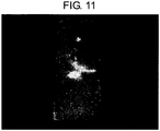
- Fig. 11 shows a high magnification image by a fluorescence stereoscopic microscope.
- GFP-positive cells mobilized into the tube were taken out, and were cultured in a DMEM/10% fetal bovine serum medium.
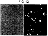
- round-shaped floating cells were observed immediately after culturing ( Fig. 12 ), however 24 hours after the GFP-positive bone marrow-derived cells were confirmed to adhere onto the culture dish and proliferated in the form of spindle-shaped fibroblast-like cells and further in the form of cylindroid-shaped epithelial-like cells ( Fig. 13 ).
- hair follicle-forming cells were observed among the GFP-positive bone marrow-derived cells ( Fig. 14A ; light field, low magnification, Fig.
- HMGB1 free skin pieces produce HMGB1; the produced HMGB1 has an activity of mobilizing a large amount of bone marrow-derived mesenchymal stem cells into the skin pieces; bone marrow-derived mesenchymal stem cells mobilized into the skin pieces are differentiated into mesenchymal cells such as fibroblasts, adipocytes, smooth muscle cells in the skin tissue, and further are differentiated into cells that form hair follicles of epidermal cells, to induce functional regeneration of transplanted skin tissues.
- mesenchymal cells such as fibroblasts, adipocytes, smooth muscle cells in the skin tissue, and further are differentiated into cells that form hair follicles of epidermal cells, to induce functional regeneration of transplanted skin tissues.
- the membrane was incubated with PBS containing 3% skim milk and 0.1% Tween 20 (S-T-PBS) at room temperature for 1 hour, and then was allowed to react with each of rabbit anti-mouse HMGB1 antibody, rabbit anti-mouse HMGB2 antibody, or rabbit anti-mouse HMGB3 antibody which were diluted 1000-fold with S-T-PBS, at 4°C for 16 hours. After the reaction, the PVDF membrane was washed with S-T-PBS five times for 5 minutes. Then, the PVDF membrane was incubated with 2000-fold diluted (diluted with S-T-PBS) peroxidase labeled goat anti-rabbit IgG antibody (GE Healthcare)at 25°C for 1 hour.
- S-T-PBS PBS containing 3% skim milk and 0.1% Tween 20
- the PVDF membrane was allowed to react with ECL Western Blotting Detection System (GE Healthcare).
- ECL Western Blotting Detection System GE Healthcare
- the ECL film was exposed and developed to detect the presence of HMGB 1, HMGB2, and HMGB3 proteins.
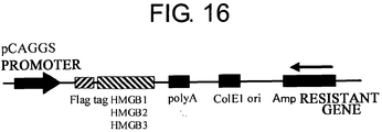
- RNA was extracted from the skin of neonatal mouse using Trizol (invitrogen), and further cDNA was synthesized using SuperScript III cDNA synthesis kit (Invitrogen). Using this cDNA as a template, cDNAs of HMGB 1, HMGB2, and HMGB3 were amplified using the PCR (polymerase chain reaction) method. The cDNAs were inserted into the plasmid vector pCAGGS for expressing proteins in mammalian cells, such that proteins with an additional Flag tag sequence (Asp-Tyr-Lys-Asp-Asp-Asp-Lys; SEQ ID: 18) at the N terminus of the amino acid sequence could be expressed.
- HEK293 Human embryonic kidney derived culture cell line
- HEK293 Human embryonic kidney derived culture cell line
- Cells expressing each of the HMGB1, HMGB2, and HMGB3 proteins and the culture supernatant were incubated at 4°C for 16 hours, which was then centrifuged at 4400 g for 5 minutes to collect the supernatant.
- 100 ⁇ L of the anti-Flag antibody gel (Sigma) was mixed into 50 mL of this supernatant, and was then incubated at 4°C for 16 hours. Centrifugation was then performed to collect the gel, and washed with PBS five times.
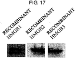
- the protein was eluted using 3X Flag peptide (final 100 ⁇ g/ml). Expressions of recombinant proteins were observed by the Western blot method using 1000-fold diluted (diluted with S-T-PBS) mouse anti-Flag antibody and 2000-fold diluted (diluted with S-T-PBS) peroxidase-labeled anti-mouse IgG antibody (GE Healthcare). The mouse bone marrow mesenchymal stem cell migration activity in these purified recombinant proteins was assessed in the same manner as in [Example 2] using a Boyden chamber.
- the dorsal skin of 8-week-old C57BL/6 mice was cut out in a circle having a diameter of 8 ⁇ m to prepare cutaneous ulcer models.
- Purified HMGB1, HMGB2, and HMGB3 100 ng were each mixed with the same amount of hyaluronic acid solution having a concentration of 1 g/100 mL of PBS, and 100 ⁇ L of it was administered to the ulcer surface.
- the ulcer surface was covered with a transparent adhesive wound dressing/protective material Tegaderm (3M Healthcare) to avoid drying, and the wound area was measured over time to determine the therapeutic effect.
- a Boyden chamber was used in the same manner as in [Example 2] for assessment.
- a human skin having an area of 1cm 2 was immersed in 1 ml PBS, and then was incubated at 4°C for 16 hours and subsequently centrifuged at 440 G at 4°C for 10 minutes. The supernatant alone was collected to be used as a human skin extract.
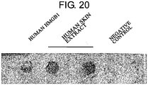
- human bone marrow mesenchymal stem cells (Cambrex) were used as the cells to be placed in the upper chamber of the Boyden chamber (as a result of surface antigen analysis by flow cytometry, these cells have been confirmed to be CD105-positive, CD166-positive, CD29-positive, CD44-positive, CD34-negative, and CD45-negative. They have also been found to differentiate into adipocytes, chondrocytes, and bone cells by differentiation induction tests). Moreover, 100 ng/well of human HMGB1 (R&D) and human skin extract diluted 10-fold with PBS and were placed in the lower chamber. PBS was used as a control.
- the mouse bone marrow mesenchymal stem cell migration activity was measured using these purified proteins, and the activity was confirmed in all of the proteins ( Fig. 18 ).
- the ulcer area produced in the back of the mouse was measured every 7 days, and a significant effect on reducing ulcer area was confirmed in the HMGB1, 2, and 3 treatment group, as compared to the non-treatment group ( Fig. 19 ).
- human HMGB1 and the human skin extract were revealed to have human bone marrow mesenchymal stem cell migration activity ( Fig. 20 ).
- HMGB2 and HMGB3 are known as proteins having high homologies to HMGB1. These proteins are also expected to have properties similar to HMGB1. It was confirmed that HMGB2 and HMGB3 of the HMGB1 family are also produced from the extract of the free skin section. Further, HMGB1/HMGB2/HMGB3 recombinant proteins were produced, and their in vitro bone marrow mesenchymal stem cell migration activity and the in vivo therapeutic effect on a cutaneous ulcer were also confirmed.
- HMGB family (HMGB1/HMGB2/HMGB3) and the recombinant HMGB family in the neonatal mouse free skin section have a bone marrow mesenchymal stem cell-inducing activity and an activity of locally inducing bone marrow-derived stem cells which are differentiatable into epithelium, and that the thus induced bone marrow-derived cell group differentiates into various cells such as epidermal keratinocytes, hair follicles, and fibroblasts in the damaged tissue to promote the recovery of the damaged tissue.
- bone marrow mesenchymal stem cells are multipotent stem cells
- the present inventors believe that therapeutic effects can also be expected in the same manner by systematic administration or local administration of the HMGB family to treat damaged states in other tissues, for example, tissue damages such as brain injury, myocardial infarction, and bone fracture.
- HMGB1 concentration contained in these samples was measured using an HMGB1 ELISA kit (Shino-Test). Further, tissue extracts of the brain, heart, and skin were allowed to bind to a heparin affinity column in the same manner as in [Example 2], and the bone marrow-derived mesenchymal stem cell-inducing activity in the protein-bound fraction was confirmed using Boyden chamber.
- the mouse brain extract contained an amount of HMGB1 equivalent to the neonatal mouse skin extract. Further, bone marrow mesenchymal stem cell-inducing activity was also observed in the mouse brain as well as in the skin. Although the mouse intestine extract and the mouse heart extract contained little HMGB1, bone marrow mesenchymal stem cell-inducing activities were observed. Moreover, the heparin column-bound fractions of mouse brain and mouse heart, as well as the heparin column-bound fraction of mouse skin, showed bone marrow mesenchymal stem cell-inducing activities ( Fig. 21 ).
- Table 1 shows the measurement results of the HMGB1 concentration and the bone marrow mesenchymal stem cell-inducing activity in each of the mouse tissue extracts.
- HMGB1 concentration ng/mL
- Bone marrow -mesenchymal stem cell -inducing activity Skin 110 Present Brain 140 Present Heart 4 Present Intestine 0 Present Kidney 115 ND Liver 61 ND ND: No Data
- HMGB1 can be conveniently extracted not only from the skin but also from the brain was developed by simply immersing these organs in a physiological buffer. This method is also applicable to other organs such as liver and kidney.
- a bone marrow mesenchymal stem cell-inducing activity was observed. This suggests these extracts contain other bone marrow mesenchymal stem cell-inducing substance(s) apart from HMGB1.
- Such substances contained in these extracts are originally present in each tissue, and are considered to physiologically induce bone marrow mesenchymal stem cells to the damaged tissue when the tissue is damaged.
- the present invention developed a novel method for conveniently and functionally extracting multiple bone marrow mesenchymal stem cell-inducing substances including HMGB1, from various organs. Further, a method for purifying bone marrow mesenchymal stem cell-inducing substances from a tissue extract using the binding to the heparin column was also developed. These substances having bone marrow mesenchymal stem cell-inducing activities can be purified from the brain and heart in the same manner as in the skin using a heparin column.
- Human embryonic kidney derived cultured cell line HEK293 and human cervix carcinoma cell line HeLa were each cultured in 10% fetal bovine serum-containing D-MEM (Nacalai). These cells were each washed with PBS, and then 10 7 cells were immersed in 5 ml of PBS (Nacalai) at 4°C for 16 hours. The solution was centrifuged at 440 G (acceleration of gravity) at 4°C for 5 minutes, and then the supernatant was collected.
- Human bone marrow mesenchymal stem cells were placed in the upper chamber of a Boyden chamber, and a 5-fold diluted (with DMEM) cell extract was placed in the lower chamber, to confirm the migration activity of human bone marrow mesenchymal stem cells.
- Bone marrow mesenchymal stem cell migration activators were successfully extracted by the convenient method of immersing cultured cells in PBS.
- Whether or not regeneration of neural cells can be induced is examined by producing mouse brain-defective models, to which a heparin-column purified fraction of skin extract is administered in a sustained-release manner at the local lesion site, by which stem cells contained in a mouse myeloid system is allowed to migrate into the local lesion site.
- mice were irradiated with single-dose of X ray at 10 Gy to produce myelosuppressive mice.
- Bone marrow cells were collected from both femurs and crus bones of GFP mice. These cells were administered to the myelosuppressive mice through the caudal vein 24 hours after the irradiation. The administration was carried out under inhalational anesthesia using isoflurane.
- mice transplanted with GFP mouse bone marrow cells were subjected to inhalational anesthesia using isoflurane, and pentobarbital (45 mg/kg) was intraperitoneally injected to the mice.
- the mice were fixed onto a brain stereotaxis apparatus and subjected to a midline incision in the head with a scalpel.
- Trepanation was carried out at 2.5 mm right-lateral and 12.5 mm anterior to the bregma using a drill ( Fig. 23A ).
- a 20G Surflow needle was inserted and fixed.
- a negative pressure was applied using a syringe to suck a part of the brain tissue ( Fig. 23B ).
- mice of the control group and the treatment group were used for the assessment. An appropriate elapsed time setting (over time) was determined, the mice were perfused with 4% paraformaldehyde and fixed and then the brain was cut out. Further, external fixation was performed with 4% paraformaldehyde. These were then dehydrated in a 15% and 30% sucrose gradient to produce frozen sections.
- the nucleus were stained with a DAPI (4',6-Diamidino-2-phenylindole, dihydrochloride) solution and the section was sealed using a photobleaching inhibitor.
- the accumulation of GFP-positive cells in the lesion site was assessed using a confocal laser microscope.
- the accumulation of GFP-positive cells is qualitatively shown for 2 weeks, and 6 weeks after the administration.
- the accumulation of GFP-positive cells tend to be higher in the lesion sites of the treatment group rather than the control group, for both 2 weeks (control; Fig. 23D , skin extract heparin-column-purified fraction; Fig. 23E ) and 6 weeks (control; Fig. 23F , skin extract heparin-column-purified fraction; Fig. 23G ) after the administration.
- the administration of the heparin-column-purified fraction of the skin extract resulted in the accumulation of bone marrow-derived cells in the brain tissue-defective site, which showed a nerve cell form.
- Bone marrow-derived mesenchymal stem cells are also known to differentiate into nerve cells and the result revealed that the heparin-column purified fraction of the skin extract is capable of inducing neural cell regeneration of the injured site in the brain. Moreover, this is also applicable to neuronal regeneration of damaged sites in brain tissues in cerebral ischemic diseases and cerebral contusions.
- Tissue regeneration promoters comprising HMGB1, HMGB2, or HMGB3 are provided.
- HMGB1, HMGB2, or HMGB3 in the form of bone marrow-derived cell-mobilizing drugs enable novel regenerative medicines which promote functional regeneration of intractable damaged tissues.
- HMGB1, HMGB2, and HMGB3 are released from cells/tissues heading towards necrosis due to a low-oxygen state caused by a decreased blood circulation, and have an effect of locally mobilizing bone marrow-derived cells or other various cells derived therefrom to such cells/tissues.
- the mobilized bone marrow-derived cells are differentiated into cell lineages required in each tissue, and promote the recovery of functions lost due to hypoperfusion/necrosis, namely promote the so-called functional tissue regeneration.
- the administration of HMGB1, HMGB2, or HMGB to an intractable cutaneous ulcer caused by blood circulation disorders of the skin leads to mobilization of bone marrow-derived cells to the ulcer surface.
- the mobilized cells Once the mobilized cells are differentiated into vascular endothelial cells, the local blood circulation would be improved.
- differentiation into fibroblasts, nerve cells, hair follicle cells, and further epidermal cells would lead to the induction and promotion of functional regeneration of skin with necessary dermal appendages, rather than just healing with fibrous scar tissues.
- HMGB1, HMGB2, and HMGB3 are expected to have a similar effect as functional tissue regeneration inducers for myocardial infarction, cerebral infarction, and hypoperfusive necrosis states of other organs.
Landscapes
- Health & Medical Sciences (AREA)
- Life Sciences & Earth Sciences (AREA)
- Chemical & Material Sciences (AREA)
- Engineering & Computer Science (AREA)
- Medicinal Chemistry (AREA)
- General Health & Medical Sciences (AREA)
- Immunology (AREA)
- Molecular Biology (AREA)
- Animal Behavior & Ethology (AREA)
- Pharmacology & Pharmacy (AREA)
- Public Health (AREA)
- Veterinary Medicine (AREA)
- Bioinformatics & Cheminformatics (AREA)
- Organic Chemistry (AREA)
- Biomedical Technology (AREA)
- General Chemical & Material Sciences (AREA)
- Chemical Kinetics & Catalysis (AREA)
- Nuclear Medicine, Radiotherapy & Molecular Imaging (AREA)
- Biochemistry (AREA)
- Urology & Nephrology (AREA)
- Hematology (AREA)
- Cell Biology (AREA)
- Proteomics, Peptides & Aminoacids (AREA)
- Epidemiology (AREA)
- Biotechnology (AREA)
- Pathology (AREA)
- Microbiology (AREA)
- General Physics & Mathematics (AREA)
- Analytical Chemistry (AREA)
- Physics & Mathematics (AREA)
- Food Science & Technology (AREA)
- Toxicology (AREA)
- Zoology (AREA)
- Gastroenterology & Hepatology (AREA)
- Genetics & Genomics (AREA)
- Dermatology (AREA)
- Tropical Medicine & Parasitology (AREA)
- Biophysics (AREA)
- Developmental Biology & Embryology (AREA)
- Physical Education & Sports Medicine (AREA)
Description
- The present invention relates to pharmaceuticals that promote functional regeneration of damaged tissues.
- Recent years have revealed that various stem cells contribute towards the repairing processes of damaged tissues, and novel regenerative medicines that induce functional tissue regeneration by mobilizing a large number of stem cells to lesion sites are being progressively developed. To realize these novel regenerative medicines, it is necessary that: (i) stem cells that are mobilizable to lesion sites are abundantly present in vivo; and (ii) factors that mobilize stem cells to lesion sites have been isolated/identified.
- Examples of stem cells that are mobilizable to lesion sites include tissue stem cells existing in lesion areas or nearby tissues, and bone marrow-derived stem cells existing in peripheral blood. In recent years, contribution of bone marrow-derived cells to many types of damaged tissue regenerations has been reported, but the mechanism for mobilizing bone marrow-derived cells to lesion sites is unknown. Bone marrow-derived cells as used herein are distinguished from hematopoietic stem cells which have the potential to differentiate into blood cells (leukocytes and erythrocytes), and include stem cells represented by cells called bone marrow mesenchymal stem cells, or tissue progenitor cell groups exiting in the bone marrow. Bone marrow mesenchymal stem cells are undifferentiated stem cells having the potential to differentiate into osteoblasts, adipocytes, and chondrocytes, and can further differentiate into other mesenchymal cells such as fibroblasts, muscle cells, stromal cells, and tendon cells. Recently, it has been proved that bone marrow mesenchymal stem cells differentiate into nerve cells, and furthermore to epithelial cells (such as skin keratinocytes) and vascular endothelial cells (Non-patent Document 9). Tissue progenitor cells are defined as undifferentiated cells having a unidirectional potential to differentiate into specific tissues/cells other than those of the blood system, and include undifferentiated cells having the potential to differentiate into mesenchymal tissue, epithelial tissue, nerve tissue, parenchymatous organs, and vascular endothelium, as mentioned above.
- HMGB1 (High Mobility Group Box 1:
High mobility group 1 protein) is a protein having a molecular weight of about 25,000 which exists in almost all kinds of cells in vivo. According to previous reports, the following functions are known: - 1) HMGB1 regulates gene expression by intracellularly binding with DNA to control chromatin structure (Non-patent Document 1);
- 2) HMGB1 is secreted from monocytes or macrophages existing in inflammatory tissues by the action of inflammatory cytokines TNF-α, IL-1, and LPS, and extracellularly binds to RAGE (Receptor for Advanced Glycation End products) (Non-patent Document 2) to induce strong inflammatory reactions (Non-patent Document 3);
- 3) HMGB1 is released from hypoperfusion-induced necrosed cells to the surrounding tissues (Non-patent Document 4);
- 4)
HMGB 1 is associated with inflammation progress in patients with septicemia, a severe infectious disease (Non-patent Document 5); - 5) HMGB1 administration to infarcted areas of myocardial infarction models promotes the division/proliferation of stem cells existing in the myocardium so as to promote the regeneration/functional recovery of the myocardium (Patent Document 1);
- 6) HMGB1 administration to model animals with hypoperfusive liver failure prior to the induction of hypoperfusive conditions alleviates the degree of hepatic impairment (Non-patent Document 6);
- 7) HMGB1 administration to lesion sites of muscle injury models directs simultaneously-administered vascular progenitor cells to lesion sites so as to promote muscular tissue regeneration (Non-patent Document 7); and
- 8) HMGB1 induces neurite formation in nerve cells (Non-patent Document 8). However, no previous reports showed that bone marrow-derived stem cells, in particular those mesenchymal stem cells that can differentiate into osteoblasts, chondrocytes, adipocytes, and the like, were mobilized to damaged tissues.
- Conventionally, central nerve cells in the brain and the spinal cord were believed to be unregeneratable if once damaged. However, the existence of neural stem cells became known recently and the induction of these cells has become possible. The neural stem cell niche within the normal nerve system has also been identified. Therefore, the recovery of damaged central neurons, which was long considered impossible, is now expected to be feasible. Currently, research related to neuronal regeneration for brain and spinal cord injury, degenerative diseases, and the like are being vigorously developed.
- Main causes of brain tissue (cells) injury are traumatic cerebral contusion and cerebral ischemic diseases. Other causes can be injury resulting from brain surgeries such as brain tumor removal. In particular, the total removal of neuroglioma arising from cerebral parenchymal cells is difficult, and there is no choice but to stop at partial removal to avoid damage to motor and language functions. Moreover, malignant neuroglioma has a worse prognosis, and none of the treatments from chemotherapy and radiotherapy to immunotherapy/gene therapy that are actively researched these days, have achieved satisfactory effects. Accordingly, an ideal treatment would be one that can remove as many tumor cells as possible, and restore damage to cerebral functions resulting from the removal.
- [Non-patent Document 1] Bustin et al. Mol Cell Biol, 19: 5237-5246, 1999
- [Non-patent Document 2] Horiet et al. J. Biol. Chem., 270, 25752-25761, 1995
- [Non-patent Document 3] Wang et al. Science, 285: 248-251, 1999
- [Non-patent Document 4] Muller et al. EMBO J, 20: 4337-4340, 2001
- [Non-patent Document 5] Wang et al. Science, 285: 248-251, 1999
- [Non-patent Document 6] Germani et al. J. Leukoc. Biol., 81, 2007, Electronic Journal Version
- [Non-patent Document 7] Palumbo et al. J. Cell Biol., 164: 441-449, 2004
- [Non-patent Document 8] Merenmies et al. J. Biol. Chem., 266: 16722-16729, 1991
- [Non-patent Document 9] Yaojiong et al. Stem cells, 25:2648-1659
- [Patent Document 1] Japanese Patent Kohyo Publication No. (
JP-A) 2005-537253 - An objective of the present invention is to isolate/identify factors mobilizing cells that differentiate into damaged tissues to lesion sites, and to provide inventions utilizing such factors.
- Once factors that mobilize cells differentiating into damaged tissues are revealed, the administration of such factors to lesion sites allows the mobilization of a large number of cells (which exist in peripheral blood or local tissues) that differentiate into damaged tissues. Thus, the development of novel regenerative medicines which promote functional tissue regeneration is made possible.
- The present inventors examined the possibility that bone marrow-derived cells might be mobilized to skin grafts from non-skin tissues in the process of skin graft transplantation into living tissue, thus contributing to skin tissue regeneration. As a result, they revealed the following for the first time in the world.
- 1) a large amount of bone marrow-derived cells are mobilized to the grafted skin;
- 2) the mobilized bone marrow-derived cells are differentiated into any of dermal fibroblasts, adipocytes, muscle cells, vascular endothelial cells, and epidermal keratinocytes in the grafted skin, and mobilized bone marrow-derived cells include bone marrow-derived mesenchymal stem cells;
- 3) the factors which mobilize bone marrow-derived mesenchymal stem cells from peripheral blood to the grafted skin are HMGB1, HMGB2, and HMGB3 released from the necrosed tissue of recipient skin;
- 4) purified HMGB1, HMGB2, and HMGB3 promote the migration of mesenchymal stem cells isolated and cultured from bone marrow;
- 5) activators containing HMGB1 which allow the migration of bone marrow mesenchymal stem cells can be conveniently purified from several organ extracts including skin, brain, and heart, by a column chromatography method using a heparin affinity column;
- 6) activators which allow the migration of bone marrow mesenchymal stem cells can be conveniently extracted from cultured cells; and
- 7) a heparin-column purified fraction of skin extract mobilizes a large amount of bone marrow-derived cells in case of brain injury.
- Based on these findings, the present application provides the following inventions:
- [1] a tissue regeneration promoter comprising any one of the following components (a) to (i) for use in regenerative medicine for epithelial or neurological tissue:
- (a) an HMGB1 protein;
- (b) a cell secreting an HMGB1 protein;
- (c) a vector inserted with a DNA encoding an HMGB1 protein;
- (d) an HMGB2 protein;
- (e) a cell secreting an HMGB2 protein;
- (f) a vector inserted with a DNA encoding an HMGB2 protein;
- (g) an HMGB3 protein;
- (h) a cell secreting an HMGB3 protein; and
- (i) a vector inserted with a DNA encoding an HMGB3 protein;
- [2] a kit comprising any one of the following components (a) to (i) for use in regenerative medicine for epithelial or neurological tissue:
- (a) an HMGB1 protein;
- (b) a cell secreting an HMGB1 protein;
- (c) a vector inserted with a DNA encoding an HMGB1 protein;
- (d) an HMGB2 protein;
- (e) a cell secreting an HMGB2 protein;
- (f) a vector inserted with a DNA encoding an HMGB2 protein;
- (g) an HMGB3 protein;
- (h) a cell secreting an HMGB3 protein; and
- (i) a vector inserted with a DNA encoding an HMGB3 protein.
-
-
Fig. 1 depicts a method for producing a GFP bone marrow-transplanted mouse. -
Fig. 2 presents photographs showing the accumulation of GFP fluorescence observed in a skin graft after skin transplantation to the back of a GFP bone marrow-transplanted mouse. Top left is an image of the skin transplantation area seen by the naked eye, top middle is an image of HE-stained tissue of a recipient skin in the vicinity of the boundary between the grafted skin and the recipient skin (shown by the arrow), and top right is an image of HE-stained tissue of the skin graft. Furthermore, bottom left image shows the accumulation of GFP fluorescence in the grafted skin, bottom middle is an enlarged image of the skin transplantation area, and bottom right is an enlarged image showing the accumulation of GFP fluorescence in the same enlarged image of the skin graft. -
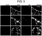
Fig. 3 presents a set of photographs showing bone marrow-derived epidermal cells and bone marrow-derived dermal fibroblasts that accumulated in the grafted skin at the back of the GFP bone marrow-transplanted mouse. The first row on top shows images of the skin of the transplantation area under low magnification (x 100), the middle row shows enlarged images of the same showing the epidermis/dermis boundary under a high magnification (x 200), and the bottom row shows further enlarged images of the same showing a hair follicle under a high magnification (x 200). The far left column shows DAPI staining (nuclear staining), the second column from left shows GFP fluorescence images of the respective regions of the first row. The third column from left shows the immunostaining images of keratin 5 (K5). The fourth column from left shows merged images of each of these fluorescences. Large numbers of GFP-positive epidermal cells and dermal fibroblasts are observed. -
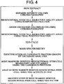
Fig. 4 is a flowchart showing a process for identifying a bone marrow-derived mesenchymal stem cell-directing factor in a skin tissue extract. -
Fig. 5 depicts a method for extracting a regeneration-inducing factor (bone marrow-derived mesenchymal stem cell-mobilizing factor) from an excised piece of skin. -
Fig. 6 presents a set of photographs showing assay results of the migratory ability/activity of bone marrow-derived mesenchymal stem cells in a skin extract using a Boyden chamber. The top left picture shows bone marrow mesenchymal stem cells adhered onto a silicone membrane on the lower chamber side, having migrated from the upper chamber of the Boyden chamber to the skin extract side (lower chamber side) through fine pores in the silicone membrane, which are stained with a blue pigment. The stained images are shown immediately after culturing (0 h), after 12 hours (12 h), and after 24 hours (24 h) (four wells each) from the top. The top right picture is an enlarged image (0 h) under a high magnification. Bottom left is an enlarged image (12 h) under a high magnification. Bottom right is an enlarged image (24 h) under a high power magnification. -
Fig. 7 presents a photograph showing the result of bone marrow-derived mesenchymal stem cells migratory ability/activity assay, examined in the skin extract-purified fraction preparation group using the Boyden chamber, and correspondence with the SDS-PAGE electrophoresis result for each purified fraction preparations. From the left, Lane 1 (M.W.): molecular weight marker; Lane 2 (C.E.): crude skin extract, Lane 3 (H.A.): heparin affinity column-binding fraction (semipurified fraction); andLanes 4 to 13 (A.E.): anion exchange column-binding fractions (final purified fraction) eluted with various NaCl concentrations, which were all stained with silver after migration. Further, each stained band in the final purified fraction of No. 4 which showed the strongest bone marrow-derived mesenchymal stem cells migration activity by a silver-stained image of the electrophoresis gel (lane 7), were cut out, and then mass spectrometry and database analysis were performed. The result revealed that the band indicated by the arrow is HMGB1. -
Fig. 8 presents a photograph showing the result of bone marrow-derived mesenchymal stem cell migratory activity assay for HMGB1 using a Boyden chamber. The two images on the top are stained images of bone marrow-derived mesenchymal stem cells, having migrated into the skin extract. The middle two images are stained images of bone marrow-derived mesenchymal stem cells, having migrated into the HMGB1 purified preparation. In the bottom are stained images of bone marrow-derived mesenchymal stem cells (virtual loss of migration activity), having migrated into a neutralized solution obtained by adding an anti-HMGB1 polyclonal antibody to the HMGB1 purified preparation used for the middle. -
Fig. 9 shows an in vivo assay method for bone marrow-derived mesenchymal stem cell directing activity. -
Fig. 10 presents a set of photographs showing the in vivo bone marrow-derived mesenchymal stem cell-mobilizing activity of HMGB1. The HMGB1 fraction (final purified fraction No. 4) showed about three times mobilization activity as compared to the comparative control (final purified fraction No. 1). -
Fig. 11 presents a photograph showing cells mobilized in vivo by the HMGB1 fraction (final purified fraction No. 4) under a high magnification. -
Fig. 12 presents a set of photographs showing images immediately after commencing culture of cells that migrated into a silicon tube. On the left is an image of migratory cells inoculated into a medium under a light field, and the right shows a GFP fluorescence image under a dark field. -
Fig. 13 presents a set of photographs showing images 24 hours after commencing culture of cells that migrated into the silicon tube. The left picture shows an image of fibroblast-like cells and epithelial-like cells that proliferated and adhered onto the plastic culture dish under a light field, and the right picture shows a GFP fluorescence image under a dark field. -
Fig. 14 presents a set ofphotographs showing images 2 weeks after commencing culture of cells that migrated into the silicon tube. The left and right photographs show the same field of view, in which the left shows images under a light field, whereas the right shows images through a fluorescence filter (GFP fluorescence is detected in B and D and fluorescence of keratin 5 is detected in F). A hair-like linear shape (indicated by the open triangle (arrow)) is observed on the left side of bone marrow-derived GFP-positive cell groups forming circular colonies on the plastic culture dish. F indicates that bone marrow-derived cells are morphologically transformed into a hair-like form, and are further expressing keratin 5 (indicated by the open triangle (arrow)). -
Fig. 15 presents a set of photographs showing the HMGB family in a newborn mouse skin extract, detected by the Western blot method. -
Fig. 16 shows an illustration of an expression vector map for the HMGB family in mammalian cells, which has, downstream of the promoter, a cytomegalovirus enhancer and a chicken β-actin promoter to synthesize a large amount of mRNAs encoded by the cDNA (complementary DNA) of the HMGB family. -
Fig. 17 presents a set of photographs showing the result of Western blotting of the purified recombinant Flag tag-HMGB family-fusion proteins expressed in HEK293 cells. -
Fig. 18 presents a set of graphs showing the migration activity of bone marrow mesenchymal stem cells by recombinant HMGB1/HMGB2/HMGB3 using a Boyden chamber. All recombinant proteins showed higher migration activities as compared to the control groups. -
Fig. 19 presents a set of graphs showing the result of treatment on mouse cutaneous ulcer treatment models using HMGB family. HMGB1, HMGB2, and HMGB3 all showed significant effects on reducing the ulcer area as compared to control groups. -
Fig. 20 presents a photograph which confirms human HMGB1's and a human skin extract's migration-inducing activity on human bone marrow-derived mesenchymal stem cells, using a Boyden chamber. -
Fig. 21 presents a set of photographs which confirm activities of bone marrow mesenchymal stem cell-inducing activators in the heart, brain, and skin extracts of the mouse using a Boyden chamber, after the activators were purified by a heparin column. -
Fig. 22 presents a set of photographs which confirm human bone marrow mesenchymal stem cell-migrating activities of a cultured cell line HEK293 extract and a HeLa extract, assessed using a Boyden chamber. Both cultured cell lines showed migrating activities on human bone marrow mesenchymal stem cells. -
Fig. 23A is a photograph showing a mouse fixed to a brain stereotaxic apparatus and subjected to a midline incision in the head with a scalpel, followed by trepanation using a drill.Fig. 23B is a photograph showing the brain to which a negative pressure is applied using a syringe to suck a part of the brain tissue.Fig. 23C is a photograph after injection of 5 µl heparin-column purified fraction of a skin extract dissolved in fibrin adhesive formulation (fibrinogen) to the brain, and a subsequent injection of 5 µl of fibrin glue formulation (thrombin).Fig. 23D and Fig. 23E are photographs of the brain injury model taken 2 weeks after the treatment. Higher accumulation of GFP-positive cells was observed in the treatment group using the heparin-column purified fraction of skin extract in 23E compared to the control in 23D.Fig. 23F and Fig. 23G are photographs of the brain injury model taken 6 weeks after the treatment. Higher accumulation of GFP-positive cells was observed in the treatment group using the heparin-column purified fraction of skin extract in 23G compared to the control in 23F. - Disclosed are inducers of bone marrow-derived cells, comprising at least one of the following ingredients (a) to (i):
- (a) an HMGB1 protein;
- (b) a cell secreting an HMGB1 protein;
- (c) a vector inserted with a DNA which encodes an
HMGB 1 protein; - (d) an HMGB2 protein;
- (e) a cell secreting an HMGB2 protein;
- (f) a vector inserted with a DNA which encodes an HMGB2 protein;
- (g) an HMGB3 protein;
- (h) a cell secreting an HMGB3 protein; and
- (i) a vector inserted with a DNA which encodes an HMGB3 protein.
- With use of such an inducer, bone marrow-derived cells (such as bone marrow-derived mesenchymal stem cells) are directed to local areas (area of administration/addition with the above ingredient, or the vicinity thereof), enabling the promotion of functional tissue regeneration. Therefore, an inducer can be used as a reagent necessary for basic and clinical research for the development of regenerative medicine and regeneration inducing medicine. For example, it becomes possible to mobilize bone marrow-derived cells (such as bone marrow-derived mesenchymal stem cells) to a required tissue in vivo in an experimental animal so as to examine the degree of tissue repairment and the reconstruction of tissue function. Moreover, in vitro research on tissue regeneration induction by mobilizing bone marrow-derived cells (such as bone marrow-derived mesenchymal stem cells) can be performed.
- Further, regeneration of the damaged tissue can be promoted by using an inducer. Use of an above inducer as a so-called preventive drug which prevents deteriorations in tissue/organ functions caused by the reduction of tissue stem cells, or as an anti-aging drug which delays the progress of age-related alterations are anticipated besides the use as an inducer/promoter of functional tissue regeneration.
- The present invention also provides tissue regeneration promoters, or kits comprising at least one of the following ingredients (a) to (i) for use in regenerative medicine for epithelial or neurological tissue:
- (a) an HMGB1 protein;
- (b) a cell secreting an HMGB1 protein;
- (c) a vector inserted with a DNA which encodes an
HMGB 1 protein; - (d) an HMGB2 protein;
- (e) a cell secreting an HMGB2 protein;
- (f) a vector inserted with a DNA which encodes an HMGB2 protein;
- (g) an HMGB3 protein;
- (h) a cell secreting an HMGB3 protein; and
- (i) a vector inserted with a DNA which encodes an HMGB3 protein.
- The tissue regeneration promoters or kits for use according to the present invention are characterized in that administration to a damaged tissue area or the vicinity directs bone marrow-derived cells (such as bone marrow-derived mesenchymal stem cells) circulating in blood, from the peripheral blood to the damaged tissue (local induction).
- Disclosed are methods for producing an extract of cells having an activity of inducing bone marrow-derived cells, which comprise the step of immersing the cells in a solvent. Moreover, disclosed are extracts of cells produced by the above production method and which have an activity of inducing bone marrow-derived cells.
- Cells to be immersed in a solvent are not specifically limited, and examples include tissue-derived cells and cell lines established from tissue-derived cells (such as Hela and HEK293, but not limited thereto), isolated cells, non-isolated cells (such as cells existing in isolated tissues), cells introduced with a DNA encoding an HMGB1, HMGB2, or HMGB3 protein. Any tissue may be used as the tissue described as above. For example, such tissues include, but are not limited to, living skin tissues or tissues obtained from internal biopsies (operations) (such as brain, lung, heart, liver, stomach, small intestine, large intestine, pancreas, kidney, bladder, spleen, uterus, testis, and blood).
- Examples of the above solvent include, but are not limited to, physiological saline, PBS (phosphate-buffered saline), and TBS (Tris-buffered saline). Moreover, the immersion time of cells or tissue in a solvent should be a duration necessary and sufficient for inducing cell necrosis, that is, 1 hour to 48 hours (such as 6 to 48 hours), and preferably 12 to 24 hours, but is not limited thereto. Therefore, the "step of immersing cells in a solvent" can be rephrased as "step of immersing cells in a solvent for a duration necessary and sufficient for inducing necrosis" or "step of necrosing cells". Moreover, examples of the temperature for immersing cells or tissue in a solvent include, but are not limited to, 4°C to 25°C (such as 4°C to 8°C), and preferably 4°C. Further, examples of the pH for immersing cells or tissue in a solvent include, without limitation, pH 7 to 8, and preferably pH 7.5. Examples of the buffer include, without limitation, a phosphate buffer solution at a concentration of 10 mM to 50 mM, preferably 10 to 20 mM.
- Moreover, cells or tissues can be removed from a solvent containing them after immersing them in the solvent. The method for removing cells or tissues from a solvent is not particularly limited as long as the method is well known to those skilled in the art. For example, cells or tissues can be removed from a solvent by centrifuging at a gravity acceleration of 10 G to 4000 G (for example, 440 G) at 4°C to 25°C (for example, 4°C), followed by separating the supernatant, but the removal method is not limited thereto. The supernatant can be used as an extract of cells or tissues.
- Further, disclosed are methods for producing a heparin-binding fraction having an activity of inducing bone marrow-derived cells, which comprise the following steps. Moreover, disclosed are heparin-binding fractions produced by the above production method, which have an activity of inducing bone marrow-derived cells.
- (a) a step of immersing the cells or tissue in a solvent;
- (b) a step of contacting an extract obtained by the step (a) with immobilized heparin; and
- (c) a step of eluting a heparin-binding fraction (may also be expressed as heparin-purified fraction or heparin-column purified fraction) from the immobilized heparin.
- "Immobilized heparin" refers to heparin which is covalently bound to an insoluble carrier. Examples of the insoluble carrier include, but are not limited to, Sepharose beads (such as Sepharose 4B, Sepharose 6B and such: GE Healthcare). A commercially available immobilized heparin (Hitrap Heparin HP column: GE Healthcare) may also be used.
- Examples of conditions for contacting an extract of cells or tissues with immobilized heparin include, but are not limited to, about pH 7 to 8 (preferably pH 7.5), and a salt concentration of 0 to 200 mM, and preferably about 100 to 200 mM. The time the extract is in contact with immobilized heparin is not specifically limited, but the contact is preferably retained for 5 minutes or more in view of a sufficient absorption of the heparin-binding fraction onto immobilized heparin. Examples of the temperature include, but are not limited to, 4 to 8°C, and preferably 4°C. Further, examples of the elution condition of the heparin-binding fraction absorbed onto the immobilized heparin include, but are not limited to, a pH of about 7 to 8 and a salt concentration of 200 to 1000 mM (preferably about 1000 mM).
- Further, disclosed are methods for producing an anion exchanger-binding fraction having an activity of inducing bone marrow-derived cells, which comprise the following steps.
- (a) a step of immersing cells or tissues in a solvent;
- (b) a step of contacting an extract obtained by step (a) with immobilized heparin;
- (c) a step of eluting a heparin-binding fraction from the immobilized heparin;
- (d) a step of contacting the heparin-binding fraction obtained by step (c) with an anion exchanger; and
- (e) a step of eluting an anion exchanger-binding fraction from the anion exchanger.
- Moreover, disclosed are anion exchanger-binding fractions produced by the above production method and having an activity of inducing bone marrow-derived cells.
- Examples of the anion exchanger include, but are not limited to, exchangers using DEAE (diethylaminoethyl) or Q (quaternary ammonium). Commercially available anion exchangers (such as Source15Q Source30Q, MonoQ, MiniQ, PC3.2/3, Mini Q4.6/50 PE, HiTrap IEX Columns, HiTrap SP HP, Q Sepharose High Performance, Hiload 16/10 Q Sepharose HP, HiPrep 16/10 SP XL, Q Sepharose XL, HiPrer 16/10 Q FF, HiPrep 16/10DEAE FF, Q Sepharose Fast Flow, and DEAE Sepharose Fast Flow (all from GE Healthcare)) may also be used.
- Examples of the conditions for contacting a heparin-binding fraction with an anion exchanger are a pH of about 7 to 9 (preferably pH 8), and a salt concentration of 0 to 100 mM, preferably about 50 mM, but are not limited thereto. The time for contacting an extract with an anion exchanger is not particularly limited, but the contact is preferably retained for 5 minutes or more in view of a sufficient absorption of the anion exchanger-binding fraction onto the anion exchanger. Examples of the temperature include, but are not limited to, 4 to 16°C, and preferably 4°C. Further, examples of conditions for eluting the anion exchanger-binding fraction absorbed onto the anion exchanger include, but are not limited to, a pH of about 7 to 9 (preferably about pH 8) and a salt concentration of 100 to 2000 mM (preferably about 1000 mM).
- Disclosed are inducers of bone marrow-derived cells, comprising the above-mentioned extract, the above-mentioned fraction, or an extract or fraction produced by the above-mentioned method.
- With the use of such an inducer, bone marrow-derived cells (such as bone marrow-derived mesenchymal stem cells) are directed to local areas (area to which the above ingredient is administered/added, or an area in the vicinity thereof) which allows the promotion of functional tissue regeneration. Therefore, the inducer can be used as a reagent necessary for basic and clinical research for the development of regenerative medicines and regeneration-inducing medicines. For example, it becomes possible to mobilize bone marrow-derived cells (such as bone marrow-derived mesenchymal stem cells) to a required tissue in vivo in an experimental animal so as to examine the degree of tissue repairment and reconstruction of tissue function. Moreover, in vitro research on tissue regeneration induction by mobilizing bone marrow-derived cells (such as bone marrow-derived mesenchymal stem cells) can be performed.
- Further, damaged tissue regeneration can be promoted by using the above inducer. Besides the use as an inducer/promoter of functional tissue regeneration, the above inducer is also anticipated to be used as a so-called preventive drug that prevents tissue/organ function deteriorations caused by reduction of tissue stem cells, or as an anti-aging drug which delays the progress of age-related alterations.
- Bone marrow-derived cells (such as bone marrow-derived mesenchymal stem cells) that are mobilized to the damaged tissue differentiate into various types of cells to contribute to functional regeneration of the damaged tissue and maintenance/enhancement of the functions. In the present invention, examples of damaged tissue include, but are not limited to, tissues damaged by various pathological conditions, trauma, burns, inflammation, autoimmunity, gene abnormalities, and the like causing ischemic/hypoperfusive/hypoxic conditions. Damaged tissue also includes necrosed tissues.
- Tissues in the present invention are epithelial and neurological tissues. Moreover, with use of the above tissue regeneration promoters, treatments for inducing functional tissue regeneration becomes possible not only in cutaneous diseases such as intractable cutaneous ulcers, skin wounds, bullosis, and alopecia, but also in tissue damages such as cerebral infarction, myocardial infarction, bone fracture, pulmonary infarction, gastric ulcers, and enteritis. The types of animals to be administered with the above tissue regeneration promoter include human and non-human animals, which can be exemplified by, but are not limited to, humans, mice, rats, monkeys, pigs, dogs, rabbits, hamsters, and guinea pigs.
- Bone marrow-derived cells of the present disclosure are cells other than hematopoietic stem cells, or cells derived therefrom such as leukocytes, erythrocytes, and platelets, and include stem cells represented by cells which have been hitherto called bone marrow mesenchymal stem cells and tissue progenitor cell populations existing in the bone marrow. Bone marrow-derived cells of the present disclosure can be isolated from bone marrow blood collection or peripheral blood collection. Hematopoietic stem cells are nonadherent, while bone marrow-derived cells of the present disclosure are obtained as adherent cells by means of a cell culture of a monocyte fraction of blood obtained from the bone marrow blood collection or peripheral blood collection. Moreover, bone marrow-derived cells of the present disclosure include mesenchymal stem cells, and have a potential to differentiate into, preferably, osteoblasts (the induction of differentiation can be identified by observing calcification), chondrocytes (which can be identified by alcian blue positive staining, safranin O positive staining, or the like), adipocytes (which can be identified by Sudan III positive staining), and other mesenchymal cells such as fibroblasts, smooth muscle cells, stromal cells, and tendon cells; and further nerve cells, epithelial cells (for example, epidermal keratinocytes and intestinal epithelial cells express cytokeratin family), and vascular endothelial cells. However, the cells to be differentiated into are not limited to the above cells, and the potential to differentiate into cells of parenchymatous organs such as liver, kidney, and pancreas are also included.
- In the present disclosure, bone marrow-derived mesenchymal stem cells refer to cells existing in the bone marrow, which are directly collected from the bone marrow or indirectly collected from other tissues (mesenchymal tissues such as blood, skin, and fat), and can be cultured and proliferated as adherent cells on a culture dish (made of plastic or glass). These cells are characterized in having a potential to differentiate into mesenchymal tissues such as bone, cartilage, and fat, and can be obtained from a collection of bone marrow blood, peripheral blood, or mesenchymal tissues. Bone marrow-derived mesenchymal stem cells are also characterized in having a potential to differentiate into epithelial tissues such as keratinocytes that constitute skin, by administrating these cells that have once adhered onto a culture dish to a lesion area of the living body.
- Bone marrow-derived mesenchymal stem cells of the present disclosure are multipotent stem cells, and have a potency to differentiate preferably into: osteoblasts (the induction of differentiation can be identified by observing calcification), chondrocytes (which can be identified by alcian blue positive staining, safranin O positive staining, or the like), adipocytes (which can be identified by Sudan III positive staining), and other mesenchymal cells such as fibroblasts, smooth muscle cells, skeletal muscle cells, stromal cells, and tendon cells; nerve cells, pigment cells, epidermal cells, hair follicle cells (which express cytokeratin family, hair keratin family, or the like), epithelial cells (for example, epidermal keratinocytes and intestinal epithelial cells express cytokeratin family or the like), and endothelial cells; and further preferably into cells of parenchymatous organs such as liver, kidney, and pancreas. However, differentiated cells are not limited to the above cells.
- Moreover, human bone marrow mesenchymal stem cells can be exemplified by, but are not limited to, cells which can be directly obtained from collecting bone marrow blood, peripheral blood, or fat, or obtained as adherent cells through culturing of an isolated monocyte fraction. Markers for human bone marrow mesenchymal stem cells can be, for example, all or some of the markers of Lin-negative, CD45-negative, and CD44-positive, but are not limited to.
- Moreover, mouse bone marrow mesenchymal stem cells can be exemplified by, but are not limited to, cells which can be obtained by methods described in the Examples. Markers for mouse bone marrow mesenchymal stem cells can be for example, all or some of the markers of Lin-negative, CD45-negative, CD44-positive, Sca-1 positive, and c-kit negative, but are not limited to.
- Tissue progenitor cells are defined as undifferentiated cells having a unidirectional potency to differentiate into specific tissues/cells other than the blood system, and include undifferentiated cells having the potency to differentiate into mesenchymal tissue, epithelial tissue, nerve tissue, parenchymatous organs, and vascular endothelium as mentioned above.
- For tissue regeneration promoters for use according to the present invention, there is no particular limitation in components other than the at least one of the ingredients (a) to (i) mentioned above, so long as the component does not inhibit the induction of bone marrow-derived cells (such as bone marrow-derived mesenchymal stem cells) and the promotion of tissue regeneration. For example, in addition to the at least one of the substances (a) to (i) mentioned above, the tissue regeneration promoters for use according to the present invention may contain: molecules (molecular groups) related to the enhancement of the function of HMGB1, HMGB2, or HMGB3 to induce functional tissue regeneration; molecules (molecular groups) which inhibit unanticipated actions of HMGB1, HMGB2, or HMGB3; factors which regulate proliferation and differentiation of bone marrow-derived cells (such as bone marrow-derived mesenchymal stem cells); and other factors which enhance/maintain these factors or cellular functions.
- The types of animals which serve as a source of HMGB1, HMGB2, or HMGB3 protein for the tissue regeneration promoters for use according to the present invention include human and non-human animals, which can be exemplified by humans, mice, rats, monkeys, pigs, dogs, rabbits, hamsters, and guinea pigs, but the kind is preferably the same as the animal to be administered with the HMGB1, HMGB2, or HMGB3 protein.
- HMGB1 proteins in tissue regeneration promoters for use according to the present invention can be exemplified by, but are not limited to, proteins comprising the amino acid sequence of SEQ ID NO: 1, 3, or 5. HMGB1 proteins employed according to the present invention can also include proteins which are functionally equivalent to the protein comprising the amino acid sequence of SEQ ID NO: 1, 3, or 5. Examples of such proteins include: 1) isolated proteins which comprise an amino acid sequence with one or more amino acid substitutions, deletions, insertions, and/or additions in the amino acid sequence of SEQ ID NO: 1, 3, or 5, and which are functionally equivalent to the protein comprising the amino acid sequence of SEQ ID NO: 1, 3, or 5; and 2) isolated proteins which are encoded by DNAs that hybridize under stringent conditions with DNAs comprising the nucleotide sequence of SEQ ID NO: 2, 4, or 6, and which are functionally equivalent to the protein comprising the amino acid sequence of SEQ ID NO: 1, 3, or 5.
- HMGB2 proteins in tissue regeneration promoters for use according to the present invention can be exemplified by, but are not limited to, proteins comprising the amino acid sequence of SEQ ID NO: 7, 9, or 11. HMGB2 proteins employed according to the present invention can also include proteins which are functionally equivalent to the protein comprising the amino acid sequence of SEQ ID NO: 7, 9, or 11. Examples of such proteins include: 1) isolated proteins which comprise an amino acid sequence with one or more amino acid substitutions, deletions, insertions, and/or additions in the amino acid sequence of SEQ ID NO: 7, 9, or 11, and which are functionally equivalent to the protein comprising the amino acid sequence of SEQ ID NO: 7, 9, or 11; and 2) isolated proteins which are encoded by DNAs that hybridize under stringent conditions with DNAs comprising the nucleotide sequence of SEQ ID NO: 8, 10, or 12, and which are functionally equivalent to the protein comprising the amino acid sequence of SEQ ID NO: 7, 9, or 11.
- HMGB3 proteins in tissue regeneration promoters for use according to the present invention can be exemplified by, but are not limited to, proteins comprising the amino acid sequence of SEQ ID NO: 13 or 15. HMGB3 proteins employed according to the present invention can also include proteins which are functionally equivalent to the protein comprising the amino acid sequence of SEQ ID NO: 13 or 15. Examples of such proteins include: 1) isolated proteins which comprise an amino acid sequence with one or more amino acid substitutions, deletions, insertions, and/or additions in the amino acid sequence of SEQ ID NO: 13 or 15, and which are functionally equivalent to the protein comprising the amino acid sequence of SEQ ID NO: 13 or 15; and 2) isolated proteins which are encoded by DNAs that hybridize under stringent conditions with DNAs comprising the nucleotide sequence of SEQ ID NO: 14 or 16, and which are functionally equivalent to the protein comprising the amino acid sequence of SEQ ID NO: 13 or 15.
- Isolated proteins which are functionally equivalent to the protein comprising the amino acid sequence of SEQ ID NO: 1, 3, 5, 7, 9, 11, 13 or 15 may be homologues or paralogues to the protein comprising the amino acid sequence of SEQ ID NO: 1, 3, 5, 7, 9, 11, 13 or 15. Those skilled in the art can isolate proteins which are functionally equivalent to the protein comprising the amino acid sequence of SEQ ID NO: 1, 3, 5, 7, 9, 11, 13 or 15, by known methods (supplementary volume of "Jikken Igaku (Experimental Medicine), Idenshi Kougaku Handbook (Genetic Engineering Handbook)", pp246-251, published by Yodosha Co., Ltd., 1991).
- Examples of proteins which are functionally equivalent to the protein comprising the amino acid sequence of SEQ ID NO: 1, 3, 5, 7, 9, 11, 13 or 15 include proteins having bone marrow-derived cell (such as bone marrow-derived mesenchymal stem cells)-inducing activity.
- Proteins which comprise an amino acid sequence with one or more amino acid substitutions, deletions, insertions, and/or additions in the amino acid sequence of SEQ ID NO: 1, 3, 5, 7, 9, 11, 13 or 15, and which are functionally equivalent to the protein comprising the amino acid sequence of SEQ ID NO: 1, 3, 5, 7, 9, 11, 13 or 15 include naturally-occurring proteins. Generally, eukaryotic genes have a polymorphism as known in interferon genes and such. Alterations in nucleotide sequences caused by the polymorphism, may result in one or more amino acid substitutions, deletions, insertions, and/or additions. Naturally-occurring proteins such as those comprising an amino acid sequence with one or more amino acid substitutions, deletions, insertions, and/or additions in the amino acid sequence of SEQ ID NO: 1, 3, 5, 7, 9, 11, 13 or 15, and which are functionally equivalent to the protein comprising the amino acid sequence of SEQ ID NO: 1, 3, 5, 7, 9, 11, 13 or 15 are included in HMGB1, HMGB2, or HMGB3 proteins of the present invention.
- The present invention also includes artificially-produced mutant proteins so long as they are functionally equivalent to the protein comprising the amino acid sequence of SEQ ID NO: 1, 3, 5, 7, 9, 11, 13 or 15. Known methods which cause random mutations to a given nucleotide sequence include substitution(s) of base pair(s) through nitrous acid treatment of DNA (Hirose, S. et al., Proc. Natl. Acad. Sci. USA., 79:7258-7260, 1982). This method enables random introduction of substitution(s) of base pair(s) into a specific segment by nitrous acid treatment of the segment desired to be mutated. Alternatively, technologies for site-directing a target mutation include the gapped duplex method (Kramer W. and Fritz HJ., Methods in Enzymol., 154:350-367, 1987) and the like. A cyclic double stranded vector in which a gene to be introduced with a mutation is cloned, is separated into single strands. These single strands are hybridized with a synthetic oligonucleotide mutated at the target site. A vector-derived complementary single strand DNA linearized by a restriction enzyme is annealed with the cyclic single stranded vector, and the gap between the oligonucleotide and the vector is filled by using a DNA polymerase, which is then made into a complete double stranded vector by ligation.
- The number of amino acids to be modified would be typically within 50, preferably within 30, and more preferably within 5 amino acids (for example, one amino acid).
- When an amino acid is artificially substituted, substitution with an amino acid having similar properties would result in maintaining the activity of the original protein. Proteins employed according to the present invention include proteins resulting from a conservative substitution in the above substitution of amino acid(s), and which are functionally equivalent to the protein comprising the amino acid sequence of SEQ ID NO: 1, 3, 5, 7, 9, 11, 13 or 15. Conservative substitution is considered important when substituting amino acid(s) of domains important for protein activities. Such a conservative substitution of amino acid(s) is well known to those skilled in the art.
- Examples of amino acid groups suitable for conservative substitution include basic amino acids (such as lysine, arginine, and histidine), acidic amino acids (such as aspartic acid and glutamic acid), uncharged polar amino acids (such as glycine, asparagine, glutamine, serine, threonine, tyrosine, and cysteine), nonpolar amino acids (such as alanine, valine, leucine, isoleucine, proline, phenylalanine, methionine, and tryptophane), β branched amino acids (such as threonine, valine, and isoleucine), and aromatic amino acids (such as tyrosine, phenylalanine, tryptophane, and histidine).
- Moreover, non-conservative substitution may increase protein activities (for example, constitutively activated proteins).
- In addition, proteins which are functionally equivalent to the protein comprising the amino acid sequence of SEQ ID NO: 1, 3, 5, 7, 9, 11, 13 or 15 can be obtained by methods that utilize hybridization. That is to say, a DNA encoding HMGB1, HMGB2, or HMGB3 protein of the present invention as shown in the SEQ ID NO: 2, 4, 6, 8, 10, 12, 14, or 16, or a fragment thereof is used as a probe, and then DNAs that can hybridize to them are isolated. A hybridization reaction performed under stringent conditions leads to the selection of highly homologous DNA as a nucleotide sequence. This increases the chances of isolated proteins containing proteins that are functionally equivalent to the HMGB1, HMGB2, or HMGB3 protein. Examples of a highly homologous nucleotide sequence include those having 70% or more, and desirably 90% or more identity.
- In a specific example, the term "stringent conditions" refers to hybridization conditions with 6 x SSC, 40% formamide at 25°C and subsequent washing with 1x SSC at 55°C. The stringency depends on conditions such as salt concentration, formamide concentration, or temperature; however it is obvious for those skilled in the art to set these conditions so as to obtain a necessary stringency.
- With the use of hybridization, for example, DNAs encoding homologues of the HMGB1, HMGB2, or HMGB3 proteins other than those proteins comprising the amino acid sequence of SEQ ID NO: 1, 3, 5, 7, 9, 11, 13 or 15, can be isolated.
- Proteins which are functionally equivalent to a protein comprising the amino acid sequence of SEQ ID NO: 1, 3, 5, 7, 9, 11, 13 or 15 normally have a high homology with the amino acid sequence of SEQ ID NO: 1, 3, 5, 7, 9, 11, 13 or 15. The term "high homology" refers to a sequence identity of at least 30% or more, preferably 50% or more, more preferably 80% or more (for example, 95% or more). The identity of the nucleotide sequences and amino acid sequences can be determined using a homology search site via the internet (For example, homology searches such as FASTA, BLAST, PSI-BLAST, and SSEARCH can be used in the DNA Data Bank of Japan (DDBJ) [examples of which include the homology search page (Search and Analysis) at the DNA Data Bank of Japan (DDBJ) website; http://www.ddbj.nig.ac.jp/E-mail/homology-j.html]. Furthermore, searches using BLAST can be carried out through the web site of the National Center for Biotechnology Information (NCBI) (examples of which include BLAST page at the homepage of NCBI website; http://www.ncbi.nlm.nih.govBLAST/; Altschul, S.F. et al., J. Mol. Biol., 1990, 215(3):403-10; Altschul, S.F. & Gish, W., Meth. Enzymol., 1996, 266:460-480; Altschul, S.F. et al., Nucleic Acids Res., 1997, 25:3389-3402)).
- For example, in the calculation of the identity of amino acid sequences using Advanced BLAST 2.1, the identity value (%) can be obtained by the following: blastp is used as the program, expect value is set at 10, all filters are set at OFF, BLOSUM62 is used for matrix, and gap existence cost, per residue gap cost, and lambda ratio are set at 11, 1, and 0.85, respectively (default parameters) (Karlin, S. and S. F. Altschul (1990) Proc. Natl. Acad. Sci. USA 87:2264-68; Karlin, S. and S. F. Altschul (1993) Proc. Natl. Acad. Sci. USA 90:5873-7).
- Proteins employed according to the present invention, or proteins functionally equivalent thereto may be proteins subjected to various modifications such as physiological modification with sugar chains and the like, labeling with fluorescence or radioactive substances, or fusion with other proteins. Particularly in recombinants that will be described later, sugar chain modification may vary depending on the hosts used for expression. However, even if there is a difference in sugar chain modifications, all proteins having properties similar to those of HMGB1, HMGB2, or HMGB3 proteins disclosed herein are HMGB1, HMGB2, or HMGB3 proteins of the present invention or proteins functionally equivalent thereto.
- HMGB1, HMGB2, or HMGB3 proteins can be obtained not only from living materials, but also in the form of recombinants by incorporating genes that encode these proteins into an appropriate expression system. In order to obtain HMGB1, HMGB2, or HMGB3 proteins by genetic engineering techniques, the above-mentioned DNAs which encode HMGB1, HMGB2, or HMGB3 proteins may be incorporated into an appropriate expression system, and they can then be expressed. Examples of host/vector systems applicable to the present invention include the expression vector pGEX and E. coli. With pGEX, foreign genes can be expressed as a fusion protein with glutathione-S-transferase (GST) (Gene, 67:31-40, 1988). pGEX incorporated with a gene encoding HMGB1, HMGB2, or HMGB3 protein is introduced into an E. coli strain such as BL21 by heat shock, incubated for an appropriate time and then isopropylthio-β-D-galactoside (IPTG) is added to induce the expression of GST-fused HMGB1, GST-fused HMGB2, or GST-fused HMGB3 proteins. Since GST of the present invention absorbs onto Glutathione Sepharose 4B, the expression product is readily separated and purified by affinity column chromatography.
- In addition, the following may also be applied as host/vector systems to obtain recombinants of HMGB1, HMGB2, or HMGB3 proteins. First, when bacteria are used as hosts, expression vectors for fusion proteins that utilize histidine tag, HA tag, a FLAG tag, and the like are commercially available. Regarding yeasts, yeasts belonging to the genus Pichia are known to be effective for the expression of sugar chain-containing proteins. In terms of the addition of sugar chains, expression systems that utilize baculovirus vector with insect cells as a host are also useful (Bio/Technology, 6:47-55, 1988). Further, using mammalian cells, transfection of a vector is carried out using promoters such as CMV, RSV, and SV40. Any of these host/vector systems can be used as an expression system of HMGB1, HMGB2, or HMGB3 proteins. Moreover, genes can also be introduced using viral vectors such as retrovirus vectors, adenovirus vectors, and adeno-associated virus vectors.
- Thus obtained proteins employed according to the present invention may be isolated intracellularly or extracellularly (medium and such), and can be purified as proteins that are substantially pure and homogenous. Proteins may be separated and purified using separation and purification methods which are commonly used in protein purification, and are not particularly limited. For example, proteins can be separated and purified by appropriately selecting and combining a chromatography column, filter, ultrafiltration, salting out, solvent precipitation, solvent extraction, distillation, immunoprecipitation, SDS-polyacrylamide gel electrophoresis, isoelectric focusing electrophoresis, dialysis, recrystallization, and the like.
- Examples of chromatographies include affinity chromatography, ion exchange chromatography, hydrophobic chromatography, gel filtration, reverse phase chromatography, and adsorption chromatography (Marshak et al., Strategies for Protein Purification and Characterization: A Laboratory Course Manual. Ed Daniel R. Cold Spring Harbor Laboratory Press, 1996). These chromatographies can be performed using liquid phase chromatographies such as HPLC and FPLC.
- Moreover, proteins employed according to the present invention are preferably substantially purified proteins. Here, the term "substantially purified" means that the protein purity of the present invention (proportion of the protein of the present invention in total protein components) is 50% or more, 60% or more, 70% or more, 80% or more, 90% or more, 95% or more, 100% or close to 100%. The upper limit for "close to 100%" depends on the purification techniques and analytical techniques of those skilled in the art, of which examples are 99.999%, 99.99%, 99.9%, 99%, and the like.
- Moreover, a substantially purified protein includes any protein purified by any purification method as long as the protein purity is as mentioned above. Examples include, but are not limited to, proteins substantially purified by appropriately selecting and combining the above-mentioned chromatography column, filter, ultrafiltration, salting out, solvent precipitation, solvent extraction, distillation, immunoprecipitation, SDS-polyacrylamide gel electrophoresis, isoelectric focusing electrophoresis, dialysis, recrystallization, and the like.
- Cells where HMGB1, HMGB2, or HMGB3 proteins of the tissue regeneration promoters for use according to the present invention are released or secreted basically include all types of tissue-derived cells in vivo. Cells which can be readily collected and cultured are exemplified by, but are not limited to, fibroblasts (such as normal skin fibroblasts and cell lines derived therefrom). Moreover, cells secreting HMGB1, HMGB2, or HMGB3 proteins can also be produced by the following manner. A vector is produced by inserting an HMGB1, HMGB2, or HMGB3 protein-encoding DNA, or an HMGB1, HMGB2, or HMGB3 protein-encoding DNA linked with a secretion signal-encoding DNA (ATG CAG ACA GAC ACA CTC CTG CTA TGG GTA CTG CTG CTG TGG GTT CCA GGT TCC ACT GGT GAC; SEQ ID NO: 17), into a known expression vector or a gene therapy vector. The produced vector is introduced into mammalian cells such as fibroblasts (such as normal skin fibroblasts and cell lines derived therefrom), insect cells, and other cells. There are no particular limitations in the animal types from which these cells derive, although cells from the animal type of the target animal subjected to tissue regeneration, cells from the target itself, or cells derived from a blood relative of the target subjected to tissue regeneration are preferably used.
- DNAs which encode HMGB1, HMGB2, or HMGB3 proteins of the tissue regeneration promoters for use according to the present invention may be cDNAs, genomic DNAs, natural DNAs, or artificially-synthesized DNAs so long as they encode the HMGB1, HMGB2, or HMGB3 protein. DNAs which encode HMGB1, HMGB2, or HMGB3 proteins are normally contained in tissue regeneration promoters for use according to the present invention in a form inserted in vectors (such as gene therapy vectors).
- Examples of the gene therapy vectors of the present invention include, but are not limited to, plasmid vectors, retrovirus vectors, lentivirus vectors, adenovirus vectors, adeno-associated virus vectors, Sendai virus vectors, Sendai virus envelope vectors, and papilloma virus vectors. The gene therapy vectors may contain promoter DNA sequences which effectively induce gene expression, factors that regulate gene expression, and molecules which are necessary for maintaining DNA stability.
- Tissue regeneration promoters for use according to the present invention may also contain: partial peptides of HMGB1, HMGB2, or HMGB3 protein which have an activity of inducing bone marrow-derived cells (such as bone marrow-derived mesenchymal stem cells); cells secreting these partial peptides; or vectors inserted with the DNAs encoding these partial peptides.
- Methods for administering tissue regeneration promoters for use according to the present invention include oral or parenteral administration. Specific examples of the administration methods include administration by injection, transnasal administration, transpulmonary administration, and percutaneous administration. Examples of administration by injection include intravenous injection, intramuscular injection, intraperitoneal injection, and hypodermic injection, by which inducers or tissue regeneration promoters of the present invention can be administered systemically or locally (such as subcutaneously or intracutaneously, or to the skin surface, eyeball, palpebral conjunctiva, rhinal mucosa, intraoral mucosa, gastrointestinal mucosa, vaginal/intrauterine mucosa, or lesion site).
- The method of administration may be appropriately selected according to the age and the symptoms of the patient. When an HMGB1, HMGB2, or HMGB3 protein is administered, the dose per time of the protein can be selected within a range of 0.0000001 mg to 1000 mg per kg body weight of a patient. Alternatively, the dose can be selected within a range of 0.00001 mg to 100000mg per body of patient, for example. When administering cells secreting HMGB1, HMGB2, or HMGB3 proteins or gene therapy vectors inserted with DNAs encoding HMGB1, HMGB2, or HMGB3 proteins they may be administered such that the amounts of HMGB1, HMGB2, or HMGB3 protein in the damaged tissues are within the above range. However, the dosage of the tissue regeneration promoters for use according to the present invention are not limited thereto.
- Tissue regeneration promoters for use according to the present invention can be formulated according to the usual methods (for example, Remington's Pharmaceutical Science, latest edition, Mark Publishing Company, Easton, U.S.A), and may contain pharmaceutically acceptable carriers and additives together. Examples include surfactants, excipients, colorants, perfumes, preservatives, stabilizers, buffers, suspending agents, isotonizing agents, binders, disintegrants, lubricants, flow promoters, and flavoring agents, although they are not limited thereto and other common carriers may be appropriately used. Specific examples include light anhydrous silicic acid, lactose, crystalline cellulose, mannitol, starch, carmellose calcium, carmellose sodium, hydroxypropylcellulose, hydroxypropylmethylcellulose, polyvinylacetaldiethylamino acetate, polyvinylpyrrolidone, gelatin, medium-chain fatty acid triglyceride, polyoxyethylene hydrogenated
castor oil 60, white sugar, carboxymethyl cellulose, corn starch, and inorganic salts. - Herein below, the present invention will be described with reference to the Examples, but it is not to be construed as being limited to these Examples.
- Objective: Assessment of the contribution of bone marrow-derived cells towards functional regeneration of skin tissue transplanted to a living body.
- Method: In view of the above objective, studies were carried out by the following methods.
- 1) Utilizing the live skin transplant system of GFP bone marrow-transplanted mice, the degree of contribution of bone marrow-derived cells towards functional regeneration of grafted skin was examined. Specifically, 6 to 8-week-old male C57BL/6 mice were irradiated with a lethal dose of radiation (10 Gy), and immediately after that, GFP (green fluorescent protein) transgenic mouse-derived bone marrow cells (5x10⁶ cells/0.1ml of physiological phosphate buffer solution at pH 7.4) were transplanted through the caudal vein (
Fig. 1 ) - 2) The transplanted bone marrow cells were allowed to engraft (for 6 weeks), and as a result, a GFP bone marrow-transplanted mice was obtained. Then, skin of a neonatal mouse (female) was transplanted to the dorsal skin of the GFP bone marrow-transplanted mice.
- 3) The skin graft was allowed to engraft and having had satisfactory skin tissue regeneration (4 weeks), the degree of GFP fluorescence accumulation in the grafted skin area was observed using a fluorescence stereoscopic microscope.
- 4) Under inhalational anesthesia, the skin graft was collected by biopsy. Then, frozen skin sections (6 µm) were prepared using a microtome with a cooling device, and then were fixed with 4% paraformaldehyde (for 30 minutes). Cell nuclei in the tissue were stained with DAPI. Immunostaining was performed using an antibody against epidermal cell-specific keratin 5. The tissue was sealed to examine the presence of GFP-positive bone marrow-derived cells with a confocal laser microscope. A part of the specimen was stained with HE to examine its tissue construction.
- Result: In the live skin transplant system of GFP bone marrow-transplanted mice, a strong GFP fluorescence accumulation corresponding to the regenerated skin region was observed (
Fig. 2 ). Moreover, with the histological observation using the HE specimen of the skin graft, functional regeneration of skin tissue containing a large number of hair follicles was observed (Fig. 2 ). With the observation using a confocal laser microscope, GFP fluorescence was seen in many keratin 5-expressing epidermal keratinocytes, dermal fibroblasts, and further smooth muscle cells and adipocytes, showing that these cells derive from the bone marrow (Fig. 3 ). That is to say, it was revealed for the first time that many of the epithelial and mesenchymal cells required for functional regeneration of the transplanted skin were supplied from bone marrow-derived stem cells. - Discussion: For the first time, these study results clearly showed a breakthrough discovery that bone marrow-derived cells greatly contribute towards skin regeneration following skin transplantation, which is routine clinical procedure.
- It is reported that the bone marrow has two stem cell systems: hematopoietic stem cells and mesenchymal stem cells. It is difficult to imagine that the large number of bone marrow-derived epithelial cells and mesenchymal cells that were mobilized into the transplanted skin (as shown by the present study) were supplied only from bone marrow-derived hematopoietic stem cells. This strongly suggests the possible contribution of bone marrow-derived mesenchymal stem cells towards the functional regeneration of transplanted tissues. That is to say, it was predicted that immediately after skin grafting, a factor for mobilizing bone marrow-derived mesenchymal stem cells is released from the recipient skin tissue heading towards hypoperfusion/necrosis , in which the mesenchymal stem cells are mobilized from the bone marrow through peripheral blood circulation to the transplanted skin piece, and thus inducing functional skin tissue regeneration.
- Objective: Identification of a bone marrow-derived mesenchymal stem cell-inducing factor in a skin tissue extract
- Method: With the objective of identifying a bone marrow-derived mesenchymal stem cell-mobilizing factor which is expected to be released from excised skin under hypoperfusive conditions, studies were carried out by the following methods.
- 1) To obtain mouse bone marrow-derived mesenchymal stem cells, bone marrow cells were collected from the femur or crus bone of C57BL/6 mice, and then were spread on a cell culture plate having a 10% fetal bovine serum-containing D-MEM (Nacalai) as a cell culture medium, and then were cultured under the condition of 5% CO2 at 37°C. When the cells proliferated to the point of occupying 70 to 100% of the bottom area of the culture plate, the cells were peeled off from the culture plate using 0.25% trypsin 1m MEDTA (Nacalai), and were then cultured under the above conditions. This passing and culturing procedure was repeated at least five times. Further, these adherent cells were isolated and cultured, followed by an analysis of cell surface antigens using flow cytometry, to confirm that these cells were Lin-negative, CD45-negative, CD44-positive, Sca-1-positive, and c-kit-negative. These cells were confirmed to be able to differentiate into bone cells and adipocytes and have properties of bone marrow mesenchymal stem cells.
- 2) Free skin pieces obtained from 400 neonatal mice were immersed in 400 ml of physiological phosphate buffer solution at pH 7.4 (PBS). The solution was incubated at 4°C for 24 hours, and then was centrifuged at 440 G at 4°C for 10 minutes to remove the tissue. The supernatant was collected to prepare a skin extract.
- 3) In order to confirm that the thus obtained skin tissue extract has an activity of inducing bone marrow-derived mesenchymal stem cells, the migration activity of C57BL6 mouse bone marrow-derived mesenchymal stem cells, of which the cell line has been already established by the present inventors, was examined using a Boyden chamber. Specifically, a skin extract (25 µl) was inserted into the lower chamber (volume: 25 µl) of the Boyden chamber, and a polycarbonate membrane having fine pores of 8 µm was placed on it. The upper chamber (volume: 50 µl) of the Boyden chamber was further placed on this in contact, and was filled with a bone marrow-derived mesenchymal stem cell suspension (5 x 10⁴ cells/50 ml of culture solution: DMEM/10% fetal bovine serum). The chamber was incubated in a CO2 incubator at 37°C for 4 to 24 hours. After culturing, the upper chamber was removed and the silicon membrane was taken out. The number of bone marrow-derived mesenchymal stem cells which had migrated to the lower chamber through the fine pores was quantitatively examined by staining.
- 4) To purify factors having a bone marrow-derived mesenchymal stem cell-mobilizing activity in the skin extract, heparin affinity column chromatography and anion exchange column (Q column) chromatography were carried out. The skin extract was diluted 10-fold with 9 volumes of 20 mM phosphate buffer at pH 7.5 at 4°C (diluted solution A). 20 mM phosphate buffer at pH 7.5 (30 ml) was poured into HiTrap Heparin HP column (column volume: 5 ml, GE Healthcare) in advance to equilibrate the column. Further, the diluted solution A was allowed to bind to the column. Then, the column was washed with 20 mM phosphate buffer at pH 7.5 with 100 mM NaCl (30 ml). To elute the absorbed proteins, 20 mM phosphate buffer at pH 7.5 with 1000 mM NaCl were poured into the column, to elute the fractions into the tubes. The fractions having the migratory ability according to the migration activity assessment method using a Boyden chamber as described in 2) were collected from each absorbed fraction. This was diluted with 9 volumes of 50 mM Tris HCl pH 8.0 (diluted solution B). 50 mM Tris HCl pH 8.0 (30 ml) was poured into HiTrap mono Q column (column volume: 1 ml, GE Healthcare) in advance to equilibrate the column. Further, the diluted solution B was allowed to bind to the column. In order to elute the absorbed proteins, Tris HCl pH 8.0 and 1000 mM NaCl were poured into the column, to eluate the fractions into tubes. The above purification process can all be performed at 4 to 16°C, but it is preferably 4 to 8°C, and more preferably 4°C. The eluates were assessed by the migration activity assessment method using Boyden chamber as described in 2).
- 5) The skin extract-derived purified preparations having the bone marrow-derived mesenchymal stem cell-mobilizing activity, which was obtained by combining the migration activity assessment using a Boyden chamber and column chromatography, were subjected to SDS-PAGE electrophoresis to separate within the gel based on the molecular weight, and the bands of migratory proteins were detected by silver staining.
- 6) Among the skin extract-derived protein groups that had been subjected to SDS-PAGE electrophoresis and that were separated within the gel as single bands by the silver staining of 5), all protein bands obtained from chromatography-purified preparations having the strongest bone marrow-derived mesenchymal stem cell-mobilizing activity were excised, and then the identification of these proteins by mass spectrometry and database analysis was carried out.
- 7) Among the identified protein groups, candidate proteins having the bone marrow-derived mesenchymal stem cell-mobilizing activity were selected. Purified preparations including such candidate proteins were treated with neutralizing antibodies (100 µl of purified preparation solution was incubated on ice for 30 minutes with 100-fold diluted polyclonal antibody of the candidate protein. Then, the degree of inhibition on the bone marrow-derived mesenchymal stem cell-mobilizing activity was examined by migratory ability assessment using a Boyden chamber.
- 8) The obtained purified bone marrow-derived mesenchymal stem cell preparations were mixed in Matrigel at about 10% volume. A silicon tube having a diameter of about 1 mm and a length of 5 mm was filled with the Matrigel, which was then subcutaneously transplanted to the back of GFP bone marrow-transplanted mouse. Two weeks after, the inserted tube was taken out, and GFP fluorescence emitting from bone marrow-derived cells which had migrated into the tube was quantitatively analyzed by a fluorimeter. Further, the migratory cells were taken out from the tube, and were inoculated into a DMEM/10% fetal bovine serum medium, followed by culturing in a CO2 incubator, to examine the in vivo bone marrow-derived mesenchymal stem cell-mobilizing activity. These cells that were continuously cultured for 2 weeks were fixed with 2% paraformaldehyde at 25°C for 10 minutes, and rinsed with PBS four times, 5 minutes each, to wash out the paraformaldehyde. Then this was treated with a 2% skim milk solution, and was allowed to react with 1000-fold dilution of anti-mouse keratin 5 antibody (diluted with 2% skim milk containing 0.5% tween 20) at 4°C for 16 hours. The antibody was washed out with PBS four times for 5 minutes each. This was then allowed to react with 1000-fold diluted Alexa546-labelled anti-rabbit IgG antibody (diluted with 2% skim milk) at 25°C for 1 hour. The above experimental protocol of 1) to 8) is summarized in
Fig. 4 . - Result: Starting from the extract solution of excised skin of neonatal mouse in PBS (
Fig. 5 ), proteins having the bone marrow-derived mesenchymal stem cell-mobilizing activity was subjected to identification and functional analysis by the above-mentioned methods. The migration activity assessment using a Boyden chamber showed that the skin extract has an extremely strong bone marrow-derived mesenchymal stem cell-inducing activity (Fig. 6 ). Using this activity as an index, a heparin affinity column and an anion exchange column (Q column) were used to proceed with the purification of the target factor. The obtained fractions were each analyzed by SDS-PAGE electrophoresis. As a result, a strong bone marrow-derived mesenchymal stem cell-mobilizing activity was shown by silver staining in the purified preparation containing several proteins that were separated within the gel in the form of single bands (Lane 7 inFig. 7 ). The obtained silver-stained bands were excised, and were then subjected to mass spectrometry and database analysis. As a result, it was revealed that the protein having a molecular weight of about 25,000 indicated by the arrow was HMGB1 (Fig. 7 ). To clarify that HMGB1 contained in this purified fraction (Lane 7) has the intended bone marrow-derived mesenchymal stem cell-mobilizing activity, a migration inhibition experiment was carried out using an anti-MGB1 polyclonal antibody. As a result, it was revealed that the anti-HMGB1 polyclonal antibody strongly inhibits the bone marrow-derived mesenchymal stem cell migration activity in the purified preparation (Fig. 8 ) and that the bone marrow-derived stem cell-mobilizing factor present in the skin extract is HMGB1. - Further, to confirm that HMGB1 has a bone marrow-derived mesenchymal stem cell-mobilizing activity in vivo, a silicon tube containing this purified preparation was subcutaneously inserted into the back of GFP bone marrow-transplanted mouse. Two weeks after, the properties of cells mobilized into the tube were examined (
Fig. 9 ). As a result, the HMGB1 purified preparation mobilized a greater number of GFP-positive bone marrow-derived cells into the tube (about three times) as compared to the comparative control (purified preparation used forLane 4 in SDS-PAGE ofFig. 7 ) (Fig. 10 ).Fig. 11 shows a high magnification image by a fluorescence stereoscopic microscope. Further, GFP-positive cells mobilized into the tube were taken out, and were cultured in a DMEM/10% fetal bovine serum medium. As a result, round-shaped floating cells were observed immediately after culturing (Fig. 12 ), however 24 hours after the GFP-positive bone marrow-derived cells were confirmed to adhere onto the culture dish and proliferated in the form of spindle-shaped fibroblast-like cells and further in the form of cylindroid-shaped epithelial-like cells (Fig. 13 ). When these cells were continuously cultured for another 2 weeks, hair follicle-forming cells were observed among the GFP-positive bone marrow-derived cells (Fig. 14A ; light field, low magnification,Fig. 14B ; GFP fluorescence, low magnification,Fig. 14C ; light field, high magnification,Fig. 14D ; GFP fluorescence, high magnification). Moreover, when immunohistochemical techniques were used for keratin 5, a marker for epithelial keratinocytes, keratin 5-positive cells were observed among the GFP-positive bone marrow-derived cells (Fig. 14E ; light field,Fig. 14F ; fluorescence of keratin 5-positive cells). - Discussion: This time, the present inventors have discovered for the first time in the world that: free skin pieces produce HMGB1; the produced HMGB1 has an activity of mobilizing a large amount of bone marrow-derived mesenchymal stem cells into the skin pieces; bone marrow-derived mesenchymal stem cells mobilized into the skin pieces are differentiated into mesenchymal cells such as fibroblasts, adipocytes, smooth muscle cells in the skin tissue, and further are differentiated into cells that form hair follicles of epidermal cells, to induce functional regeneration of transplanted skin tissues. It can be readily predicted that this mobilization of bone marrow-derived mesenchymal stem cells by HMGB1 and the resulting functional tissue regeneration functions, not only for transplanted skin regeneration, but also as a mechanism for inducing functional tissue regeneration in various damaged organs/tissues accompanying hypoperfusion/necrosis. The present inventors firmly believe that, if drug development using an HMGB1 formulation enables the mobilization of bone marrow-derived mesenchymal stem cells to the local area during regeneration of the damaged tissues, it would enable functional tissue regeneration-inducing therapy for vital functional organs, without the organs becoming dysfunctional due to fibrous scar healing.
- Objective: Identification of the HMGB1 family in the skin extract and examination of bone marrow mesenchymal stem cell-inducing activity
- Method: Whether or not the neonatal mouse skin extract contained the HMGB protein family was confirmed using the Western blot method. Ten µl of the skin extract obtained in [Example 2] was used as a sample and subjected to SDS-PAGE electrophoresis. The proteins separated within the gel were transferred onto a PVDF membrane using a blotting device (ATTO). The membrane was incubated with PBS containing 3% skim milk and 0.1% Tween 20 (S-T-PBS) at room temperature for 1 hour, and then was allowed to react with each of rabbit anti-mouse HMGB1 antibody, rabbit anti-mouse HMGB2 antibody, or rabbit anti-mouse HMGB3 antibody which were diluted 1000-fold with S-T-PBS, at 4°C for 16 hours. After the reaction, the PVDF membrane was washed with S-T-PBS five times for 5 minutes. Then, the PVDF membrane was incubated with 2000-fold diluted (diluted with S-T-PBS) peroxidase labeled goat anti-rabbit IgG antibody (GE Healthcare)at 25°C for 1 hour. Further, after washing with S-T-PBS five times for 5 minute, the PVDF membrane was allowed to react with ECL Western Blotting Detection System (GE Healthcare). The ECL film was exposed and developed to detect the presence of
HMGB 1, HMGB2, and HMGB3 proteins. - RNA was extracted from the skin of neonatal mouse using Trizol (invitrogen), and further cDNA was synthesized using SuperScript III cDNA synthesis kit (Invitrogen). Using this cDNA as a template, cDNAs of
HMGB 1, HMGB2, and HMGB3 were amplified using the PCR (polymerase chain reaction) method. The cDNAs were inserted into the plasmid vector pCAGGS for expressing proteins in mammalian cells, such that proteins with an additional Flag tag sequence (Asp-Tyr-Lys-Asp-Asp-Asp-Lys; SEQ ID: 18) at the N terminus of the amino acid sequence could be expressed. These plasmid vectors were introduced into HEK293 (Human embryonic kidney derived culture cell line) and cultured for 48 hours to express the proteins. Cells expressing each of the HMGB1, HMGB2, and HMGB3 proteins and the culture supernatant were incubated at 4°C for 16 hours, which was then centrifuged at 4400 g for 5 minutes to collect the supernatant. 100 µL of the anti-Flag antibody gel (Sigma) was mixed into 50 mL of this supernatant, and was then incubated at 4°C for 16 hours. Centrifugation was then performed to collect the gel, and washed with PBS five times. Further, the protein was eluted using 3X Flag peptide (final 100 µg/ml). Expressions of recombinant proteins were observed by the Western blot method using 1000-fold diluted (diluted with S-T-PBS) mouse anti-Flag antibody and 2000-fold diluted (diluted with S-T-PBS) peroxidase-labeled anti-mouse IgG antibody (GE Healthcare). The mouse bone marrow mesenchymal stem cell migration activity in these purified recombinant proteins was assessed in the same manner as in [Example 2] using a Boyden chamber. Moreover, in order to observe the in vivo drug efficacy of the HMGB family, the dorsal skin of 8-week-old C57BL/6 mice was cut out in a circle having a diameter of 8 µm to prepare cutaneous ulcer models. Purified HMGB1, HMGB2, and HMGB3 (100 ng) were each mixed with the same amount of hyaluronic acid solution having a concentration of 1 g/100 mL of PBS, and 100 µL of it was administered to the ulcer surface. The ulcer surface was covered with a transparent adhesive wound dressing/protective material Tegaderm (3M Healthcare) to avoid drying, and the wound area was measured over time to determine the therapeutic effect. - Further, to examine whether or not the human skin extract and the purified human HMGB1 has an activity to allow migration of human bone marrow mesenchymal stem cells, a Boyden chamber was used in the same manner as in [Example 2] for assessment. A human skin having an area of 1cm2 was immersed in 1 ml PBS, and then was incubated at 4°C for 16 hours and subsequently centrifuged at 440 G at 4°C for 10 minutes. The supernatant alone was collected to be used as a human skin extract. Moreover, human bone marrow mesenchymal stem cells (Cambrex) were used as the cells to be placed in the upper chamber of the Boyden chamber (as a result of surface antigen analysis by flow cytometry, these cells have been confirmed to be CD105-positive, CD166-positive, CD29-positive, CD44-positive, CD34-negative, and CD45-negative. They have also been found to differentiate into adipocytes, chondrocytes, and bone cells by differentiation induction tests). Moreover, 100 ng/well of human HMGB1 (R&D) and human skin extract diluted 10-fold with PBS and were placed in the lower chamber. PBS was used as a control.
- Result: As a result of Western blotting, bands of HMGB2 and HMGB3 were detected as well as the HMGB1 band. Therefore, the neonatal mouse skin extract was confirmed to contain the family proteins, HMGB2 and HMGB3, besides HMGB1 (
Fig. 15 ). Expression vectors of HMGB1/HMGB2/HMGB3 having a Flag tag added at the N-terminus of each protein, were prepared (Fig. 16 ). These expression vectors were introduced into HEK293 cells, and the expressed proteins were purified using the Flag tag, and Western blotting was carried out to observe these proteins (Fig. 17 ). The mouse bone marrow mesenchymal stem cell migration activity was measured using these purified proteins, and the activity was confirmed in all of the proteins (Fig. 18 ). The ulcer area produced in the back of the mouse was measured every 7 days, and a significant effect on reducing ulcer area was confirmed in the HMGB1, 2, and 3 treatment group, as compared to the non-treatment group (Fig. 19 ). Similar to the mouse case, human HMGB1 and the human skin extract were revealed to have human bone marrow mesenchymal stem cell migration activity (Fig. 20 ). - Discussion: HMGB2 and HMGB3 are known as proteins having high homologies to HMGB1. These proteins are also expected to have properties similar to HMGB1. It was confirmed that HMGB2 and HMGB3 of the HMGB1 family are also produced from the extract of the free skin section. Further, HMGB1/HMGB2/HMGB3 recombinant proteins were produced, and their in vitro bone marrow mesenchymal stem cell migration activity and the in vivo therapeutic effect on a cutaneous ulcer were also confirmed. It was revealed that the HMGB family (HMGB1/HMGB2/HMGB3) and the recombinant HMGB family in the neonatal mouse free skin section have a bone marrow mesenchymal stem cell-inducing activity and an activity of locally inducing bone marrow-derived stem cells which are differentiatable into epithelium, and that the thus induced bone marrow-derived cell group differentiates into various cells such as epidermal keratinocytes, hair follicles, and fibroblasts in the damaged tissue to promote the recovery of the damaged tissue. Moreover, since bone marrow mesenchymal stem cells are multipotent stem cells, the present inventors believe that therapeutic effects can also be expected in the same manner by systematic administration or local administration of the HMGB family to treat damaged states in other tissues, for example, tissue damages such as brain injury, myocardial infarction, and bone fracture.
- Moreover, it is known that, between human and mouse, amino acid sequence homology for HMGB1 is 98% (213/215), 96% (202/210) for HMGB2, and 97% (195/200) for HMGB3. Therefore, human HMGB and mouse HMGB are considered to have similar activities, and the results of the present Examples revealed that human skin extract and human HMGB1 have bone marrow mesenchymal stem cell-inducing activities in a manner same as those of mouse skin extract and
mouse HMGB 1. - Objective: Establishment of a method of producing a tissue extract containing bone marrow mesenchymal stem cell-inducing factors.
- Method: Brain, heart, intestine, kidney, and liver of a 6-week-old C57BL6 mouse and skin of a neonatal mouse were immersed in 1 ml of physiological phosphate buffer solution (PBS) at pH 7.4. The solutions were incubated at 4°C for 24 hours, and then centrifuged at 440 G at 4°C for 10 minutes to remove the tissues. The supernatants were collected to prepare tissue extracts. To confirm whether the thus obtained extract has a bone marrow-derived mesenchymal stem cell-inducing activity, the migration activity of bone marrow-derived mesenchymal stem cells was examined in the same manner as in [Example 2] using a Boyden chamber. Moreover, the HMGB1 concentration contained in these samples was measured using an HMGB1 ELISA kit (Shino-Test). Further, tissue extracts of the brain, heart, and skin were allowed to bind to a heparin affinity column in the same manner as in [Example 2], and the bone marrow-derived mesenchymal stem cell-inducing activity in the protein-bound fraction was confirmed using Boyden chamber.
- Result: The mouse brain extract contained an amount of HMGB1 equivalent to the neonatal mouse skin extract. Further, bone marrow mesenchymal stem cell-inducing activity was also observed in the mouse brain as well as in the skin. Although the mouse intestine extract and the mouse heart extract contained little HMGB1, bone marrow mesenchymal stem cell-inducing activities were observed. Moreover, the heparin column-bound fractions of mouse brain and mouse heart, as well as the heparin column-bound fraction of mouse skin, showed bone marrow mesenchymal stem cell-inducing activities (
Fig. 21 ). Table 1 shows the measurement results of the HMGB1 concentration and the bone marrow mesenchymal stem cell-inducing activity in each of the mouse tissue extracts.[Table 1] HMGB1 concentration (ng/mL) Bone marrow -mesenchymal stem cell -inducing activity Skin 110 Present Brain 140 Present Heart 4 Present Intestine 0 Present Kidney 115 ND Liver 61 ND ND: No Data - Discussion: A method in which HMGB1 can be conveniently extracted not only from the skin but also from the brain was developed by simply immersing these organs in a physiological buffer. This method is also applicable to other organs such as liver and kidney. Moreover, although the extracts from intestine and heart contain little HMGB1, a bone marrow mesenchymal stem cell-inducing activity was observed. This suggests these extracts contain other bone marrow mesenchymal stem cell-inducing substance(s) apart from HMGB1. Such substances contained in these extracts are originally present in each tissue, and are considered to physiologically induce bone marrow mesenchymal stem cells to the damaged tissue when the tissue is damaged. The present invention developed a novel method for conveniently and functionally extracting multiple bone marrow mesenchymal stem cell-inducing substances including HMGB1, from various organs. Further, a method for purifying bone marrow mesenchymal stem cell-inducing substances from a tissue extract using the binding to the heparin column was also developed. These substances having bone marrow mesenchymal stem cell-inducing activities can be purified from the brain and heart in the same manner as in the skin using a heparin column.
- Objective: Establishment of a method for extracting mesenchymal stem cell migration activators from cultured cells.
- Method: Human embryonic kidney derived cultured cell line HEK293 and human cervix carcinoma cell line HeLa were each cultured in 10% fetal bovine serum-containing D-MEM (Nacalai). These cells were each washed with PBS, and then 107 cells were immersed in 5 ml of PBS (Nacalai) at 4°C for 16 hours. The solution was centrifuged at 440 G (acceleration of gravity) at 4°C for 5 minutes, and then the supernatant was collected. Human bone marrow mesenchymal stem cells were placed in the upper chamber of a Boyden chamber, and a 5-fold diluted (with DMEM) cell extract was placed in the lower chamber, to confirm the migration activity of human bone marrow mesenchymal stem cells.
- Result: HEK293 extract and HeLa extract both showed similar bone marrow mesenchymal stem cell migration activities(
Fig. 22 ). - Discussion: Bone marrow mesenchymal stem cell migration activators were successfully extracted by the convenient method of immersing cultured cells in PBS.
- Objective: Whether or not regeneration of neural cells can be induced is examined by producing mouse brain-defective models, to which a heparin-column purified fraction of skin extract is administered in a sustained-release manner at the local lesion site, by which stem cells contained in a mouse myeloid system is allowed to migrate into the local lesion site.
- An excised skin section of a neonatal mouse was incubated in PBS (mouse/ml) at 4°C for 16 hours, and a skin extract was obtained. The skin extract was diluted 10-fold with 9 volumes of 20 mM phosphate buffer at pH 7.5 at 4°C. 20 mM phosphate buffer at pH 7.5 (30 ml) was poured into HiTrap Hepalin HP column (column volume: 5 ml, GE Healthcare) in advance to equilibrate the column. The diluted solution was then allowed to bind to the column. Thereafter, the column was washed with 20 mM phosphate buffer at pH 7.5 and 100 mM NaCl (30 ml). To elute the absorbed proteins, 20 mM phosphate buffer at pH 7.5 and 1000 mM NaCl were poured into the column, and the factions were eluted into the tubes. Each of the absorbed factions were evaluated according to the mouse bone marrow-derived cell migration activity assessment using the Boyden chamber method shown in Example 2, and fraction(s) having migratory activity was collected. Solution(s) having the activity was used as a heparin purified fraction(s) of the skin extract.
- Mice were irradiated with single-dose of X ray at 10 Gy to produce myelosuppressive mice.
- Bone marrow cells were collected from both femurs and crus bones of GFP mice. These cells were administered to the myelosuppressive mice through the caudal vein 24 hours after the irradiation. The administration was carried out under inhalational anesthesia using isoflurane.
- The myelosuppressive mice transplanted with GFP mouse bone marrow cells were subjected to inhalational anesthesia using isoflurane, and pentobarbital (45 mg/kg) was intraperitoneally injected to the mice. The mice were fixed onto a brain stereotaxis apparatus and subjected to a midline incision in the head with a scalpel. Trepanation was carried out at 2.5 mm right-lateral and 12.5 mm anterior to the bregma using a drill (
Fig. 23A ). At a 3 mm depth from this site, a 20G Surflow needle was inserted and fixed. Then, a negative pressure was applied using a syringe to suck a part of the brain tissue (Fig. 23B ). - Five µl of a heparin-column purified fraction of skin extract dissolved in fibrinogen of the fibrin tissue adhesive Bolheal (Kaketsuken) was injected to the above site, and subsequently, 5 µl of thrombin of Bolheal was injected using a Hamilton syringe and a 26G syringe (
Fig. 23C ). The aim of this operation was to exert the sustained-release agent effect of a heparin-column-purified fraction of the skin extract. - Mice of the control group and the treatment group were used for the assessment. An appropriate elapsed time setting (over time) was determined, the mice were perfused with 4% paraformaldehyde and fixed and then the brain was cut out. Further, external fixation was performed with 4% paraformaldehyde. These were then dehydrated in a 15% and 30% sucrose gradient to produce frozen sections.
- The nucleus were stained with a DAPI (4',6-Diamidino-2-phenylindole, dihydrochloride) solution and the section was sealed using a photobleaching inhibitor. The accumulation of GFP-positive cells in the lesion site (brain tissue-defective site) was assessed using a confocal laser microscope.
- Result: The accumulation of GFP-positive cells is qualitatively shown for 2 weeks, and 6 weeks after the administration. The accumulation of GFP-positive cells tend to be higher in the lesion sites of the treatment group rather than the control group, for both 2 weeks (control;
Fig. 23D , skin extract heparin-column-purified fraction;Fig. 23E ) and6 weeks (control;Fig. 23F , skin extract heparin-column-purified fraction;Fig. 23G ) after the administration. - Discussion: The administration of the heparin-column-purified fraction of the skin extract resulted in the accumulation of bone marrow-derived cells in the brain tissue-defective site, which showed a nerve cell form. Bone marrow-derived mesenchymal stem cells are also known to differentiate into nerve cells and the result revealed that the heparin-column purified fraction of the skin extract is capable of inducing neural cell regeneration of the injured site in the brain. Moreover, this is also applicable to neuronal regeneration of damaged sites in brain tissues in cerebral ischemic diseases and cerebral contusions.
- Tissue regeneration promoters comprising HMGB1, HMGB2, or HMGB3 are provided. The development of HMGB1, HMGB2, or HMGB3 in the form of bone marrow-derived cell-mobilizing drugs enable novel regenerative medicines which promote functional regeneration of intractable damaged tissues. HMGB1, HMGB2, and HMGB3 are released from cells/tissues heading towards necrosis due to a low-oxygen state caused by a decreased blood circulation, and have an effect of locally mobilizing bone marrow-derived cells or other various cells derived therefrom to such cells/tissues. The mobilized bone marrow-derived cells are differentiated into cell lineages required in each tissue, and promote the recovery of functions lost due to hypoperfusion/necrosis, namely promote the so-called functional tissue regeneration. For example, the administration of HMGB1, HMGB2, or HMGB to an intractable cutaneous ulcer caused by blood circulation disorders of the skin leads to mobilization of bone marrow-derived cells to the ulcer surface. Once the mobilized cells are differentiated into vascular endothelial cells, the local blood circulation would be improved. At the same time, differentiation into fibroblasts, nerve cells, hair follicle cells, and further epidermal cells would lead to the induction and promotion of functional regeneration of skin with necessary dermal appendages, rather than just healing with fibrous scar tissues. HMGB1, HMGB2, and HMGB3 are expected to have a similar effect as functional tissue regeneration inducers for myocardial infarction, cerebral infarction, and hypoperfusive necrosis states of other organs.
-
- <110> GENOMIX CO., LTD.
- <120> Pharmaceutical for promoting functional regeneration of damaged tissue
- <130> G 10013 EP
- <140>
EP 07830867.3
<141> 2007-10-30 - <150>
JP 2006-293582
<151> 2006-10-30 - <160> 18
- <170> PatentIn version 3.4
- <210> 1
<211> 215
<212> PRT
<213> Homo sapiens - <400> 1


- <210> 2
<211> 648
<212> DNA
<213> Homo sapiens - <400> 2

- <210> 3
<211> 215
<212> PRT
<213> Mus musculus - <400> 3


- <210> 4
<211> 648
<212> DNA
<213> Mus musculus - <400> 4


- <210> 5
<211> 215
<212> PRT
<213> Rattus norvegicus - <400> 5


- <210> 6
<211> 648
<212> DNA
<213> Rattus norvegicus - <400> 6

- <210> 7
<211> 209
<212> PRT
<213> Homo sapiens - <400> 7

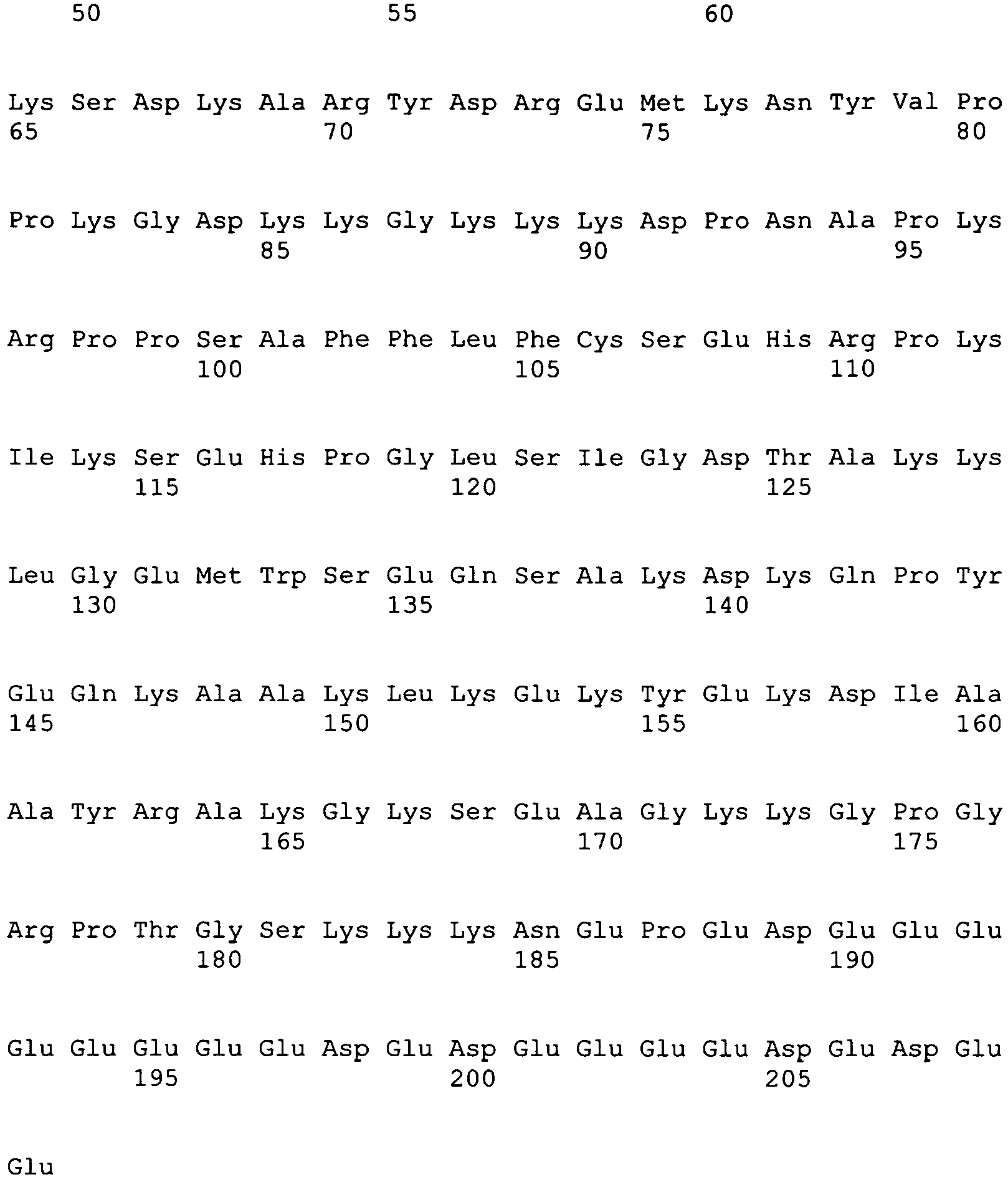
- <210> 8
<211> 630
<212> DNA
<213> Homo sapiens - <400> 8


- <210> 9
<211> 210
<212> PRT
<213> Mus musculus - <400> 9


- <210> 10
<211> 633
<212> DNA
<213> Mus musculus - <400> 10

- <210> 11
<211> 210
<212> PRT
<213> Rattus norvegicus - <400> 11


- <210> 12
<211> 633
<212> DNA
<213> Rattus norvegicus - <400> 12


- <210> 13
<211> 200
<212> PRT
<213> Homo sapiens - <400> 13

- <210> 14
<211> 603
<212> DNA
<213> Homo sapiens - <400> 14

- <210> 15
<211> 200
<212> PRT
<213> Mus musculus - <400> 15


- <210> 16
<211> 603
<212> DNA
<213> Mus musculus - <400> 16

- <210> 17
<211> 63
<212> DNA
<213> Artificial - <220>
<223> An artificially synthesized nucleotide sequence - <400> 17

- <210> 18
<211> 7
<212> PRT
<213> Artificial - <220>
<223> An artificially synthesized peptide sequence - <400> 18

Claims (2)
- A tissue regeneration promoter comprising any one of the following components (a) to (i) for use in regenerative medicine for epithelial or neurological tissue:(a) an HMGB1 protein;(b) a cell secreting an HMGB1 protein;(c) a vector inserted with a DNA encoding an HMGB1 protein;(d) an HMGB2 protein;(e) a cell secreting an HMGB2 protein;(f) a vector inserted with a DNA encoding an HMGB2 protein;(g) an HMGB3 protein;(h) a cell secreting an HMGB3 protein; and(i) a vector inserted with a DNA encoding an HMGB3 protein.
- A kit comprising any one of the following components (a) to (i) for use in regenerative medicine for epithelial or neurological tissue:(a) an HMGB1 protein;(b) a cell secreting an HMGB1 protein;(c) a vector inserted with a DNA encoding an HMGB1 protein;(d) an HMGB2 protein;(e) a cell secreting an HMGB2 protein;(f) a vector inserted with a DNA encoding an HMGB2 protein;(g) an HMGB3 protein;(h) a cell secreting an HMGB3 protein; and(i) a vector inserted with a DNA encoding an HMGB3 protein.
Priority Applications (1)
| Application Number | Priority Date | Filing Date | Title |
|---|---|---|---|
| PL07830867T PL2055308T3 (en) | 2006-10-30 | 2007-10-30 | Pharmaceutical for promoting functional regeneration of damaged tissue |
Applications Claiming Priority (2)
| Application Number | Priority Date | Filing Date | Title |
|---|---|---|---|
| JP2006293582 | 2006-10-30 | ||
| PCT/JP2007/071133 WO2008053892A1 (en) | 2006-10-30 | 2007-10-30 | Pharmaceutical for promoting functional regeneration of damaged tissue |
Publications (3)
| Publication Number | Publication Date |
|---|---|
| EP2055308A1 EP2055308A1 (en) | 2009-05-06 |
| EP2055308A4 EP2055308A4 (en) | 2010-10-27 |
| EP2055308B1 true EP2055308B1 (en) | 2017-05-10 |
Family
ID=39344234
Family Applications (1)
| Application Number | Title | Priority Date | Filing Date |
|---|---|---|---|
| EP07830867.3A Active EP2055308B1 (en) | 2006-10-30 | 2007-10-30 | Pharmaceutical for promoting functional regeneration of damaged tissue |
Country Status (13)
| Country | Link |
|---|---|
| US (2) | US20090202500A1 (en) |
| EP (1) | EP2055308B1 (en) |
| JP (2) | JP5308161B2 (en) |
| KR (2) | KR20090078304A (en) |
| CN (1) | CN101374538A (en) |
| AU (1) | AU2007315073A1 (en) |
| CA (1) | CA2636788A1 (en) |
| DK (1) | DK2055308T3 (en) |
| ES (1) | ES2629086T3 (en) |
| HU (1) | HUE035043T2 (en) |
| PL (1) | PL2055308T3 (en) |
| PT (1) | PT2055308T (en) |
| WO (1) | WO2008053892A1 (en) |
Families Citing this family (36)
| Publication number | Priority date | Publication date | Assignee | Title |
|---|---|---|---|---|
| JP5814549B2 (en) * | 2008-04-30 | 2015-11-17 | 株式会社ジェノミックス | Drugs for promoting functional regeneration of damaged tissues |
| JP5660889B2 (en) * | 2008-04-30 | 2015-01-28 | 株式会社ジェノミックス | Bone marrow-derived pluripotent stem cell mobilization to peripheral circulation |
| KR20110017371A (en) * | 2008-04-30 | 2011-02-21 | 가부시키가이샤 제노믹스 | High efficiency sampling of functional cells in vivo |
| JP5706081B2 (en) * | 2009-10-28 | 2015-04-22 | 株式会社ジェノミックス | Implantable biomaterial collection device |
| KR20120135190A (en) | 2009-10-28 | 2012-12-12 | 고꾸리쯔 다이가꾸 호우징 오사까 다이가꾸 | Tussue-regeneration promoter using recruitment of bone marrow mesenchymal stem cells and/or pluripotent stem cells in blood |
| JP5852773B2 (en) | 2010-06-01 | 2016-02-03 | ピアス株式会社 | Method for producing attractant for bone marrow mesenchymal stem cells, method for attracting bone marrow mesenchymal stem cells, and use for producing an attractant for bone marrow mesenchymal stem cells |
| KR102113809B1 (en) | 2011-04-26 | 2020-05-21 | 가부시키가이샤 스템림 | Peptide for inducing regeneration of tissue and use thereof |
| CN102864123B (en) * | 2011-07-06 | 2015-04-22 | 北京大学第三医院 | Acquisition method of peripheral blood mesenchymal stem cells and application thereof |
| CA2889275C (en) | 2012-10-25 | 2021-06-15 | Katsuto Tamai | Novel method for treating cardiac infarction using hmgb1 fragment |
| ES2673861T3 (en) * | 2012-10-25 | 2018-06-26 | Genomix Co., Ltd. | Novel method to treat spinal cord damage using the hmgb1 fragment |
| CN103316378B (en) * | 2013-06-27 | 2014-07-16 | 重庆大学 | HMBG (High Mobility Group Box) 1 modified bone tissue engineering bracket material and preparation method thereof |
| JP6288672B2 (en) * | 2014-03-28 | 2018-03-07 | 株式会社巴川製紙所 | Anisotropic optical film |
| JP6452107B2 (en) * | 2014-09-05 | 2019-01-16 | 国立大学法人 東京大学 | Pluripotent stem cells for the treatment of diabetic skin ulcers |
| JP6941417B2 (en) * | 2016-02-04 | 2021-09-29 | 雪印メグミルク株式会社 | Cartilage function improver |
| KR102359441B1 (en) * | 2016-06-29 | 2022-02-09 | (주)아모레퍼시픽 | Composition for promoting adipocyte differentiation and method for screening material for promoting adipocyte differentiation |
| TWI740964B (en) * | 2016-06-29 | 2021-10-01 | 南韓商愛茉莉太平洋股份有限公司 | Use of promoting adipocyte differentiation and method for screening materials for promoting adipocyte differentiation |
| PL3556378T3 (en) | 2017-01-27 | 2025-06-09 | StemRIM Inc. | Therapeutic agent for cardiomyopathy, old myocardial infarction and chronic heart failure |
| KR102681936B1 (en) | 2017-04-25 | 2024-07-04 | 시오노기세이야쿠가부시키가이샤 | Peptides and their use to induce tissue regeneration |
| CN106957821B (en) * | 2017-04-26 | 2020-04-21 | 南京医科大学第二附属医院 | A method for regulating the directed differentiation of mesenchymal stem cells |
| JPWO2019017355A1 (en) * | 2017-07-18 | 2020-05-28 | 株式会社 資生堂 | Mesenchymal stem cell attractant |
| CN118460462A (en) | 2017-12-01 | 2024-08-09 | 斯特姆里姆有限公司 | Exo-embryonic interlayer mesenchymal stem cells and method for producing same |
| US11298403B2 (en) | 2017-12-01 | 2022-04-12 | StemRIM Inc. | Therapeutic agent for inflammatory bowel disease |
| WO2019156137A1 (en) | 2018-02-08 | 2019-08-15 | 株式会社ステムリム | Therapeutic agent for psoriasis |
| WO2020071520A1 (en) | 2018-10-05 | 2020-04-09 | 株式会社ステムリム | Peptide possessing mesenchymal-stem-cell mobilizing activity |
| US12304933B2 (en) * | 2018-10-05 | 2025-05-20 | StemRIM Inc. | Disease treatment drug based on mesenchymal-stem-cell mobilization |
| US20220023350A1 (en) * | 2018-11-04 | 2022-01-27 | Figene, Llc | Treatment of cerebral hypoxia including stroke, chronic traumatic encephalopathy, and traumatic brain injury |
| CA3286605A1 (en) | 2018-11-04 | 2025-11-29 | Fibrobiologics, Inc. | Treatment of cachexia using fibroblast cells and products thereof |
| CN109957015A (en) * | 2019-04-01 | 2019-07-02 | 中国人民解放军海军军医大学国家肝癌科学中心 | A kind of preparation method and application of HMGB1 blocking antibody |
| JP7561348B2 (en) * | 2019-07-31 | 2024-10-04 | 株式会社ステムリム | Composition for promoting proliferation or inhibiting decline of mesenchymal stem cells |
| KR20220137876A (en) | 2019-11-12 | 2022-10-12 | 옥스포드 유니버시티 이노베이션 리미티드 | Polypeptides related to HMGB1 useful for promoting tissue regeneration, compositions comprising same, and uses thereof |
| CN116867909A (en) * | 2020-12-07 | 2023-10-10 | 社会福祉法人 三星生命公益财团 | Method for screening mesenchymal stem cells with improved self-sustainability and mesenchymal stem cells thus screened |
| WO2022231904A1 (en) * | 2021-04-27 | 2022-11-03 | Avita Medical, Inc. | Regenerative bioactive suspension derived from freshly disaggregated tissue and methods of use in clinical therapies |
| AU2022276188A1 (en) | 2021-05-19 | 2023-12-14 | Oxford University Innovation Limited | Polypeptides related to hmgb1 useful for promoting tissue regeneration, compositions comprising same, and uses thereof |
| WO2024145258A1 (en) | 2022-12-27 | 2024-07-04 | AVITA Medical Americas, LLC | System for automated preparation of a regenerative epidermal suspension and related methods of use |
| US12570949B2 (en) | 2022-12-27 | 2026-03-10 | AVITA Medical Americas, LLC | Systems and devices for wound therapy and related methods of use |
| USD1112812S1 (en) | 2023-12-22 | 2026-02-10 | AVITA Medical Americas, LLC | Tissue processing cartridge |
Family Cites Families (20)
| Publication number | Priority date | Publication date | Assignee | Title |
|---|---|---|---|---|
| US5658894A (en) * | 1989-04-23 | 1997-08-19 | The Trustees Of The University Of Pennsylvania | Compositions for inhibiting restenosis |
| JP3018313B2 (en) * | 1996-02-23 | 2000-03-13 | 雪印乳業株式会社 | Bone formation promotion and bone resorption inhibitor |
| BR0012787A (en) * | 1999-07-28 | 2002-07-30 | Univ Leland Stanford Junior | Nicotine receptor agonists in stem cell and progenitor cell recruitment |
| BR0108545A (en) * | 2000-02-24 | 2004-06-29 | Xcyte Therapies Inc | Simultaneous cell stimulation and concentration |
| US20040191246A1 (en) * | 2003-02-26 | 2004-09-30 | Connelly Patrick R. | Process for in vivo treatment of specific biological targets in bodily fluid |
| DE10121254A1 (en) * | 2001-04-30 | 2002-11-07 | Switch Biotech Ag | Functional assay on the activity of human MRP8/MRP14 heterodimer, for diagnosing and treating skin diseases, wounds or wound-healing disturbances with reduced quantity of the heterodimer, in particular a diabetic ulcer |
| US7220723B2 (en) * | 2001-05-15 | 2007-05-22 | The Feinstein Institute For Medical Research | Inhibitors of the interaction between HMGB polypeptides and toll-like receptor 2 as anti-inflammatory agents |
| ITMI20011986A1 (en) * | 2001-09-25 | 2003-03-25 | San Raffaele Centro Fond | METHOD AND COMPOSITION FOR THE ACTIVATION OF CELLS PRESENTING THE ANTIGEN |
| US20040265971A1 (en) * | 2001-11-19 | 2004-12-30 | Hidetaka Sato | Drug mobilizing pluripotent stem cells from tissue into peripheral blood |
| KR20050054907A (en) * | 2002-07-03 | 2005-06-10 | 폰다지오네 센트로 산 라파엘 델 몬테 테이보 | Use of hmgb1 in the treatment of tissue damage and/or to promote tissue repair |
| GB0226251D0 (en) * | 2002-11-11 | 2002-12-18 | San Raffaele Centro Fond | Acetylated protein |
| WO2004061456A2 (en) * | 2003-01-03 | 2004-07-22 | Alcedo Biotech Gmbh | Uses of hmgb, hmgn, hmga proteins |
| EP1608397B1 (en) * | 2003-03-28 | 2008-10-15 | Universite Laval | S100 protein as neutrophil activator for alleviating neutropenia in cancer treatment |
| WO2005025604A2 (en) * | 2003-09-10 | 2005-03-24 | The General Hospital Corporation | Use of hmgb and hmgb fragments to decrease specific immune response |
| WO2006008779A1 (en) * | 2004-07-20 | 2006-01-26 | Provincia Italiana Della Congregazione Dei Figli Dell'immacolata Concezione-Istituto Dermopatico Dell'immacolata | Use of hmgb1 for wound healing |
| KR100593397B1 (en) * | 2004-10-27 | 2006-06-28 | 한국원자력연구소 | Wound healing or wound healing promoters containing mesodermal stem cells and / or P substances, or cell therapeutics |
| WO2007015546A1 (en) * | 2005-08-04 | 2007-02-08 | Genomix Co., Ltd | Mesenchymal stem cell inducer, tissue regeneration promoter and method of preparing mesenchymal stem cell |
| WO2007031100A1 (en) * | 2005-09-14 | 2007-03-22 | Ostini, Marco | Active immunotherapy of life-threatening systemic inflammation |
| WO2007130725A2 (en) * | 2006-02-06 | 2007-11-15 | University Of Pittsburgh Of The Commonwealth System Of Higher Education | Use of hmgb1 for protection against ischemia reperfusion injury |
| KR20110017371A (en) * | 2008-04-30 | 2011-02-21 | 가부시키가이샤 제노믹스 | High efficiency sampling of functional cells in vivo |
-
2007
- 2007-10-30 EP EP07830867.3A patent/EP2055308B1/en active Active
- 2007-10-30 CA CA002636788A patent/CA2636788A1/en not_active Abandoned
- 2007-10-30 DK DK07830867.3T patent/DK2055308T3/en active
- 2007-10-30 PL PL07830867T patent/PL2055308T3/en unknown
- 2007-10-30 JP JP2008542139A patent/JP5308161B2/en active Active
- 2007-10-30 KR KR1020087018560A patent/KR20090078304A/en not_active Abandoned
- 2007-10-30 HU HUE07830867A patent/HUE035043T2/en unknown
- 2007-10-30 US US12/161,982 patent/US20090202500A1/en not_active Abandoned
- 2007-10-30 AU AU2007315073A patent/AU2007315073A1/en not_active Abandoned
- 2007-10-30 ES ES07830867.3T patent/ES2629086T3/en active Active
- 2007-10-30 PT PT78308673T patent/PT2055308T/en unknown
- 2007-10-30 WO PCT/JP2007/071133 patent/WO2008053892A1/en not_active Ceased
- 2007-10-30 CN CNA2007800038948A patent/CN101374538A/en active Pending
- 2007-10-30 KR KR1020147023740A patent/KR20140115373A/en not_active Ceased
-
2013
- 2013-06-28 JP JP2013136055A patent/JP2013227330A/en active Pending
-
2023
- 2023-01-10 US US18/152,249 patent/US20240050460A1/en not_active Abandoned
Non-Patent Citations (1)
| Title |
|---|
| TAGLIAFICO ENRICO ET AL: "TGFbeta/BMP activate the smooth muscle/bone differentiation programs in mesoangioblasts.", JOURNAL OF CELL SCIENCE 1 SEP 2004, vol. 117, no. Pt 19, 1 September 2004 (2004-09-01), pages 4377 - 4388, ISSN: 0021-9533 * |
Also Published As
| Publication number | Publication date |
|---|---|
| HUE035043T2 (en) | 2018-05-02 |
| JP2013227330A (en) | 2013-11-07 |
| WO2008053892A1 (en) | 2008-05-08 |
| PT2055308T (en) | 2017-07-05 |
| CA2636788A1 (en) | 2008-05-08 |
| CN101374538A (en) | 2009-02-25 |
| AU2007315073A1 (en) | 2008-05-08 |
| US20240050460A1 (en) | 2024-02-15 |
| KR20140115373A (en) | 2014-09-30 |
| KR20090078304A (en) | 2009-07-17 |
| EP2055308A1 (en) | 2009-05-06 |
| EP2055308A4 (en) | 2010-10-27 |
| DK2055308T3 (en) | 2017-07-17 |
| JP5308161B2 (en) | 2013-10-09 |
| PL2055308T3 (en) | 2017-11-30 |
| US20090202500A1 (en) | 2009-08-13 |
| ES2629086T3 (en) | 2017-08-07 |
| JPWO2008053892A1 (en) | 2010-02-25 |
Similar Documents
| Publication | Publication Date | Title |
|---|---|---|
| US20240050460A1 (en) | Pharmaceuticals That Promote Functional Regeneration of Damaged Tissues | |
| US8673580B2 (en) | Agent for recruitment of bone-marrow-derived pluripotent stem cell into peripheral circulation | |
| US20220047642A1 (en) | Agents for promoting tissue regeneration by recruiting bone marrow mesenchymal stem cells and/or pluripotent stem cells into blood | |
| JP5960735B2 (en) | Drugs for promoting functional regeneration of damaged tissues | |
| AU2014200688B2 (en) | Pharmaceuticals that promote functional regeneration of damaged tissues | |
| HK1126685A (en) | Pharmaceuticals that promote functional regeneration of damaged tissues | |
| HK1155641A (en) | Agent for recruitment of bone-marrow-derived pluripotent stem cell into peripheral circulation | |
| HK1174851B (en) | Tissue-regeneration promoter using recruitment of bone marrow mesenchymal stem cells and/or pluripotent stem cells in blood | |
| HK1174851A (en) | Tissue-regeneration promoter using recruitment of bone marrow mesenchymal stem cells and/or pluripotent stem cells in blood |
Legal Events
| Date | Code | Title | Description |
|---|---|---|---|
| PUAI | Public reference made under article 153(3) epc to a published international application that has entered the european phase |
Free format text: ORIGINAL CODE: 0009012 |
|
| 17P | Request for examination filed |
Effective date: 20080703 |
|
| AK | Designated contracting states |
Kind code of ref document: A1 Designated state(s): AT BE BG CH CY CZ DE DK EE ES FI FR GB GR HU IE IS IT LI LT LU LV MC MT NL PL PT RO SE SI SK TR |
|
| AX | Request for extension of the european patent |
Extension state: AL BA HR MK RS |
|
| RAP1 | Party data changed (applicant data changed or rights of an application transferred) |
Owner name: OSAKA UNIVERSITY Owner name: GENOMIX CO., LTD. |
|
| A4 | Supplementary search report drawn up and despatched |
Effective date: 20100929 |
|
| DAX | Request for extension of the european patent (deleted) | ||
| 17Q | First examination report despatched |
Effective date: 20140324 |
|
| RIC1 | Information provided on ipc code assigned before grant |
Ipc: A61P 9/10 20060101ALI20160609BHEP Ipc: A61K 35/76 20150101ALI20160609BHEP Ipc: A61K 48/00 20060101ALI20160609BHEP Ipc: A61P 17/00 20060101ALI20160609BHEP Ipc: A61P 17/14 20060101ALI20160609BHEP Ipc: A61K 38/00 20060101AFI20160609BHEP Ipc: A61K 35/12 20060101ALI20160609BHEP Ipc: A61P 17/02 20060101ALI20160609BHEP Ipc: A61K 31/711 20060101ALI20160609BHEP Ipc: A61P 1/04 20060101ALI20160609BHEP Ipc: A61P 11/00 20060101ALI20160609BHEP |
|
| GRAP | Despatch of communication of intention to grant a patent |
Free format text: ORIGINAL CODE: EPIDOSNIGR1 |
|
| GRAJ | Information related to disapproval of communication of intention to grant by the applicant or resumption of examination proceedings by the epo deleted |
Free format text: ORIGINAL CODE: EPIDOSDIGR1 |
|
| STAA | Information on the status of an ep patent application or granted ep patent |
Free format text: STATUS: GRANT OF PATENT IS INTENDED |
|
| INTG | Intention to grant announced |
Effective date: 20161025 |
|
| GRAP | Despatch of communication of intention to grant a patent |
Free format text: ORIGINAL CODE: EPIDOSNIGR1 |
|
| INTG | Intention to grant announced |
Effective date: 20161129 |
|
| GRAS | Grant fee paid |
Free format text: ORIGINAL CODE: EPIDOSNIGR3 |
|
| GRAA | (expected) grant |
Free format text: ORIGINAL CODE: 0009210 |
|
| STAA | Information on the status of an ep patent application or granted ep patent |
Free format text: STATUS: THE PATENT HAS BEEN GRANTED |
|
| AK | Designated contracting states |
Kind code of ref document: B1 Designated state(s): AT BE BG CH CY CZ DE DK EE ES FI FR GB GR HU IE IS IT LI LT LU LV MC MT NL PL PT RO SE SI SK TR |
|
| REG | Reference to a national code |
Ref country code: GB Ref legal event code: FG4D |
|
| REG | Reference to a national code |
Ref country code: AT Ref legal event code: REF Ref document number: 891605 Country of ref document: AT Kind code of ref document: T Effective date: 20170515 Ref country code: CH Ref legal event code: EP |
|
| RIN2 | Information on inventor provided after grant (corrected) |
Inventor name: TAMAI, KATSUTO Inventor name: KANEDA, YASUFUMI Inventor name: YAMAZAKI, TAKEHIKO |
|
| REG | Reference to a national code |
Ref country code: IE Ref legal event code: FG4D |
|
| REG | Reference to a national code |
Ref country code: DE Ref legal event code: R096 Ref document number: 602007050971 Country of ref document: DE |
|
| REG | Reference to a national code |
Ref country code: RO Ref legal event code: EPE |
|
| REG | Reference to a national code |
Ref country code: PT Ref legal event code: SC4A Ref document number: 2055308 Country of ref document: PT Date of ref document: 20170705 Kind code of ref document: T Free format text: AVAILABILITY OF NATIONAL TRANSLATION Effective date: 20170623 |
|
| REG | Reference to a national code |
Ref country code: DK Ref legal event code: T3 Effective date: 20170706 |
|
| REG | Reference to a national code |
Ref country code: SE Ref legal event code: TRGR |
|
| REG | Reference to a national code |
Ref country code: NL Ref legal event code: FP |
|
| REG | Reference to a national code |
Ref country code: ES Ref legal event code: FG2A Ref document number: 2629086 Country of ref document: ES Kind code of ref document: T3 Effective date: 20170807 |
|
| REG | Reference to a national code |
Ref country code: LT Ref legal event code: MG4D |
|
| REG | Reference to a national code |
Ref country code: FR Ref legal event code: PLFP Year of fee payment: 11 |
|
| PG25 | Lapsed in a contracting state [announced via postgrant information from national office to epo] |
Ref country code: LT Free format text: LAPSE BECAUSE OF FAILURE TO SUBMIT A TRANSLATION OF THE DESCRIPTION OR TO PAY THE FEE WITHIN THE PRESCRIBED TIME-LIMIT Effective date: 20170510 |
|
| REG | Reference to a national code |
Ref country code: GR Ref legal event code: EP Ref document number: 20170401924 Country of ref document: GR Effective date: 20171122 |
|
| PG25 | Lapsed in a contracting state [announced via postgrant information from national office to epo] |
Ref country code: IS Free format text: LAPSE BECAUSE OF FAILURE TO SUBMIT A TRANSLATION OF THE DESCRIPTION OR TO PAY THE FEE WITHIN THE PRESCRIBED TIME-LIMIT Effective date: 20170910 Ref country code: BG Free format text: LAPSE BECAUSE OF FAILURE TO SUBMIT A TRANSLATION OF THE DESCRIPTION OR TO PAY THE FEE WITHIN THE PRESCRIBED TIME-LIMIT Effective date: 20170810 Ref country code: LV Free format text: LAPSE BECAUSE OF FAILURE TO SUBMIT A TRANSLATION OF THE DESCRIPTION OR TO PAY THE FEE WITHIN THE PRESCRIBED TIME-LIMIT Effective date: 20170510 |
|
| PG25 | Lapsed in a contracting state [announced via postgrant information from national office to epo] |
Ref country code: SK Free format text: LAPSE BECAUSE OF FAILURE TO SUBMIT A TRANSLATION OF THE DESCRIPTION OR TO PAY THE FEE WITHIN THE PRESCRIBED TIME-LIMIT Effective date: 20170510 Ref country code: EE Free format text: LAPSE BECAUSE OF FAILURE TO SUBMIT A TRANSLATION OF THE DESCRIPTION OR TO PAY THE FEE WITHIN THE PRESCRIBED TIME-LIMIT Effective date: 20170510 |
|
| REG | Reference to a national code |
Ref country code: DE Ref legal event code: R097 Ref document number: 602007050971 Country of ref document: DE |
|
| PLBE | No opposition filed within time limit |
Free format text: ORIGINAL CODE: 0009261 |
|
| STAA | Information on the status of an ep patent application or granted ep patent |
Free format text: STATUS: NO OPPOSITION FILED WITHIN TIME LIMIT |
|
| 26N | No opposition filed |
Effective date: 20180213 |
|
| REG | Reference to a national code |
Ref country code: HU Ref legal event code: AG4A Ref document number: E035043 Country of ref document: HU |
|
| REG | Reference to a national code |
Ref country code: DK Ref legal event code: EBP Effective date: 20171031 |
|
| REG | Reference to a national code |
Ref country code: SE Ref legal event code: EUG |
|
| PG25 | Lapsed in a contracting state [announced via postgrant information from national office to epo] |
Ref country code: SI Free format text: LAPSE BECAUSE OF FAILURE TO SUBMIT A TRANSLATION OF THE DESCRIPTION OR TO PAY THE FEE WITHIN THE PRESCRIBED TIME-LIMIT Effective date: 20170510 Ref country code: MC Free format text: LAPSE BECAUSE OF FAILURE TO SUBMIT A TRANSLATION OF THE DESCRIPTION OR TO PAY THE FEE WITHIN THE PRESCRIBED TIME-LIMIT Effective date: 20170510 |
|
| REG | Reference to a national code |
Ref country code: CH Ref legal event code: PL |
|
| REG | Reference to a national code |
Ref country code: NL Ref legal event code: MM Effective date: 20171101 |
|
| REG | Reference to a national code |
Ref country code: IE Ref legal event code: MM4A |
|
| PG25 | Lapsed in a contracting state [announced via postgrant information from national office to epo] |
Ref country code: CH Free format text: LAPSE BECAUSE OF NON-PAYMENT OF DUE FEES Effective date: 20171031 Ref country code: NL Free format text: LAPSE BECAUSE OF NON-PAYMENT OF DUE FEES Effective date: 20171101 Ref country code: LU Free format text: LAPSE BECAUSE OF NON-PAYMENT OF DUE FEES Effective date: 20171030 Ref country code: CZ Free format text: LAPSE BECAUSE OF NON-PAYMENT OF DUE FEES Effective date: 20171030 Ref country code: LI Free format text: LAPSE BECAUSE OF NON-PAYMENT OF DUE FEES Effective date: 20171031 Ref country code: FI Free format text: LAPSE BECAUSE OF NON-PAYMENT OF DUE FEES Effective date: 20171030 |
|
| REG | Reference to a national code |
Ref country code: BE Ref legal event code: MM Effective date: 20171031 |
|
| PG25 | Lapsed in a contracting state [announced via postgrant information from national office to epo] |
Ref country code: GR Free format text: LAPSE BECAUSE OF NON-PAYMENT OF DUE FEES Effective date: 20180504 Ref country code: SE Free format text: LAPSE BECAUSE OF NON-PAYMENT OF DUE FEES Effective date: 20171031 Ref country code: BE Free format text: LAPSE BECAUSE OF NON-PAYMENT OF DUE FEES Effective date: 20171031 Ref country code: HU Free format text: LAPSE BECAUSE OF NON-PAYMENT OF DUE FEES Effective date: 20171031 Ref country code: RO Free format text: LAPSE BECAUSE OF NON-PAYMENT OF DUE FEES Effective date: 20170510 |
|
| PG25 | Lapsed in a contracting state [announced via postgrant information from national office to epo] |
Ref country code: MT Free format text: LAPSE BECAUSE OF NON-PAYMENT OF DUE FEES Effective date: 20171030 |
|
| REG | Reference to a national code |
Ref country code: FR Ref legal event code: PLFP Year of fee payment: 12 |
|
| PG25 | Lapsed in a contracting state [announced via postgrant information from national office to epo] |
Ref country code: PT Free format text: LAPSE BECAUSE OF NON-PAYMENT OF DUE FEES Effective date: 20180730 Ref country code: IT Free format text: LAPSE BECAUSE OF NON-PAYMENT OF DUE FEES Effective date: 20171030 Ref country code: IE Free format text: LAPSE BECAUSE OF NON-PAYMENT OF DUE FEES Effective date: 20171030 |
|
| REG | Reference to a national code |
Ref country code: DE Ref legal event code: R081 Ref document number: 602007050971 Country of ref document: DE Owner name: STEMRIM INC., IBARAKI-SHI, JP Free format text: FORMER OWNERS: GENOMIX CO., LTD., IBARAKI-CITY, OSAKA, JP; OSAKA UNIVERSITY, SUITA-SHI, OSAKA, JP |
|
| PG25 | Lapsed in a contracting state [announced via postgrant information from national office to epo] |
Ref country code: DK Free format text: LAPSE BECAUSE OF NON-PAYMENT OF DUE FEES Effective date: 20171031 |
|
| REG | Reference to a national code |
Ref country code: AT Ref legal event code: MM01 Ref document number: 891605 Country of ref document: AT Kind code of ref document: T Effective date: 20171030 |
|
| REG | Reference to a national code |
Ref country code: ES Ref legal event code: FD2A Effective date: 20181226 |
|
| PG25 | Lapsed in a contracting state [announced via postgrant information from national office to epo] |
Ref country code: AT Free format text: LAPSE BECAUSE OF NON-PAYMENT OF DUE FEES Effective date: 20171030 |
|
| PG25 | Lapsed in a contracting state [announced via postgrant information from national office to epo] |
Ref country code: ES Free format text: LAPSE BECAUSE OF NON-PAYMENT OF DUE FEES Effective date: 20171031 |
|
| PG25 | Lapsed in a contracting state [announced via postgrant information from national office to epo] |
Ref country code: PL Free format text: LAPSE BECAUSE OF NON-PAYMENT OF DUE FEES Effective date: 20171030 |
|
| PG25 | Lapsed in a contracting state [announced via postgrant information from national office to epo] |
Ref country code: CY Free format text: LAPSE BECAUSE OF NON-PAYMENT OF DUE FEES Effective date: 20170510 |
|
| REG | Reference to a national code |
Ref country code: AT Ref legal event code: UEP Ref document number: 891605 Country of ref document: AT Kind code of ref document: T Effective date: 20170510 |
|
| P01 | Opt-out of the competence of the unified patent court (upc) registered |
Effective date: 20230516 |
|
| PG25 | Lapsed in a contracting state [announced via postgrant information from national office to epo] |
Ref country code: TR Free format text: LAPSE BECAUSE OF NON-PAYMENT OF DUE FEES Effective date: 20171030 |
|
| PGFP | Annual fee paid to national office [announced via postgrant information from national office to epo] |
Ref country code: DE Payment date: 20241021 Year of fee payment: 18 |
|
| PGFP | Annual fee paid to national office [announced via postgrant information from national office to epo] |
Ref country code: GB Payment date: 20241022 Year of fee payment: 18 |
|
| PGFP | Annual fee paid to national office [announced via postgrant information from national office to epo] |
Ref country code: FR Payment date: 20241021 Year of fee payment: 18 |