EP2053059A1 - Complexes of TRPC domains and SESTD1 domains and methods and uses involving the same - Google Patents
Complexes of TRPC domains and SESTD1 domains and methods and uses involving the same Download PDFInfo
- Publication number
- EP2053059A1 EP2053059A1 EP07291300A EP07291300A EP2053059A1 EP 2053059 A1 EP2053059 A1 EP 2053059A1 EP 07291300 A EP07291300 A EP 07291300A EP 07291300 A EP07291300 A EP 07291300A EP 2053059 A1 EP2053059 A1 EP 2053059A1
- Authority
- EP
- European Patent Office
- Prior art keywords
- protein
- sestd1
- trpc
- domain
- interaction
- Prior art date
- Legal status (The legal status is an assumption and is not a legal conclusion. Google has not performed a legal analysis and makes no representation as to the accuracy of the status listed.)
- Ceased
Links
- 101000836397 Homo sapiens SEC14 domain and spectrin repeat-containing protein 1 Proteins 0.000 title claims abstract description 130
- 102100027289 SEC14 domain and spectrin repeat-containing protein 1 Human genes 0.000 title claims abstract description 122
- 238000000034 method Methods 0.000 title claims abstract description 32
- 102000027549 TRPC Human genes 0.000 title description 11
- 108060008648 TRPC Proteins 0.000 title description 11
- 108090000623 proteins and genes Proteins 0.000 claims abstract description 166
- 102000004169 proteins and genes Human genes 0.000 claims abstract description 161
- 102000011753 Transient Receptor Potential Channels Human genes 0.000 claims abstract description 83
- 108010037150 Transient Receptor Potential Channels Proteins 0.000 claims abstract description 83
- 230000003993 interaction Effects 0.000 claims abstract description 45
- 238000012360 testing method Methods 0.000 claims abstract description 44
- 108010019965 Spectrin Proteins 0.000 claims abstract description 15
- 102000005890 Spectrin Human genes 0.000 claims abstract description 15
- 238000012216 screening Methods 0.000 claims abstract description 15
- 230000027455 binding Effects 0.000 claims description 44
- 102000003621 TRPC5 Human genes 0.000 claims description 37
- 101150042815 TRPC5 gene Proteins 0.000 claims description 37
- 101100100680 Schizosaccharomyces pombe (strain 972 / ATCC 24843) trp4 gene Proteins 0.000 claims description 31
- 102000003622 TRPC4 Human genes 0.000 claims description 31
- 101150099990 Trpc4 gene Proteins 0.000 claims description 31
- 150000003904 phospholipids Chemical class 0.000 claims description 23
- 108050001421 Transient receptor potential channel, canonical 6 Proteins 0.000 claims description 20
- 239000000126 substance Substances 0.000 claims description 18
- 239000003550 marker Substances 0.000 claims description 16
- 241000282414 Homo sapiens Species 0.000 claims description 15
- 102100028150 HMG domain-containing protein 4 Human genes 0.000 claims description 8
- 125000000539 amino acid group Chemical group 0.000 claims description 8
- 201000010099 disease Diseases 0.000 claims description 8
- 208000037265 diseases, disorders, signs and symptoms Diseases 0.000 claims description 8
- 206010019280 Heart failures Diseases 0.000 claims description 7
- 102000043378 human SESTD1 Human genes 0.000 claims description 7
- PORPENFLTBBHSG-MGBGTMOVSA-N 1,2-dihexadecanoyl-sn-glycerol-3-phosphate Chemical compound CCCCCCCCCCCCCCCC(=O)OC[C@H](COP(O)(O)=O)OC(=O)CCCCCCCCCCCCCCC PORPENFLTBBHSG-MGBGTMOVSA-N 0.000 claims description 6
- 102100033306 Ankyrin repeat domain-containing protein 35 Human genes 0.000 claims description 6
- 102100033594 E3 ubiquitin-protein ligase makorin-1 Human genes 0.000 claims description 6
- 208000004248 Familial Primary Pulmonary Hypertension Diseases 0.000 claims description 6
- 101000732537 Homo sapiens Ankyrin repeat domain-containing protein 35 Proteins 0.000 claims description 6
- 101000613207 Homo sapiens Pre-B-cell leukemia transcription factor-interacting protein 1 Proteins 0.000 claims description 6
- 101000708766 Homo sapiens Structural maintenance of chromosomes protein 3 Proteins 0.000 claims description 6
- 108010076075 Makorin ring finger protein 1 Proteins 0.000 claims description 6
- 241001529936 Murinae Species 0.000 claims description 6
- 102100040882 Pre-B-cell leukemia transcription factor-interacting protein 1 Human genes 0.000 claims description 6
- 108010029485 Protein Isoforms Proteins 0.000 claims description 6
- 102000001708 Protein Isoforms Human genes 0.000 claims description 6
- 102100032723 Structural maintenance of chromosomes protein 3 Human genes 0.000 claims description 6
- 101710142305 Talin-2 Proteins 0.000 claims description 6
- 102100036980 Talin-2 Human genes 0.000 claims description 6
- 206010007572 Cardiac hypertrophy Diseases 0.000 claims description 5
- 208000006029 Cardiomegaly Diseases 0.000 claims description 5
- 206010020772 Hypertension Diseases 0.000 claims description 5
- 102100028167 NAD(P)H-hydrate epimerase Human genes 0.000 claims description 5
- 239000003814 drug Substances 0.000 claims description 5
- 201000001320 Atherosclerosis Diseases 0.000 claims description 4
- 102000001805 Bromodomains Human genes 0.000 claims description 4
- 108050009021 Bromodomains Proteins 0.000 claims description 4
- 102100034454 HAUS augmin-like complex subunit 8 Human genes 0.000 claims description 4
- 101710201219 HMG box-containing protein 4 Proteins 0.000 claims description 4
- 101001068141 Homo sapiens HAUS augmin-like complex subunit 8 Proteins 0.000 claims description 4
- 101000971144 Homo sapiens Tyrosine-protein kinase BAZ1B Proteins 0.000 claims description 4
- 229910019142 PO4 Inorganic materials 0.000 claims description 4
- 102100021575 Tyrosine-protein kinase BAZ1B Human genes 0.000 claims description 4
- HCHKCACWOHOZIP-UHFFFAOYSA-N Zinc Chemical compound [Zn] HCHKCACWOHOZIP-UHFFFAOYSA-N 0.000 claims description 4
- 208000006673 asthma Diseases 0.000 claims description 4
- 239000010452 phosphate Substances 0.000 claims description 4
- 229910052725 zinc Inorganic materials 0.000 claims description 4
- 239000011701 zinc Substances 0.000 claims description 4
- HKWJHKSHEWVOSS-OMDJCSNQSA-N 1,2-dihexadecanoyl-sn-glycero-3-phospho-(1D-myo-inositol-3,4-bisphosphate) Chemical compound CCCCCCCCCCCCCCCC(=O)OC[C@@H](OC(=O)CCCCCCCCCCCCCCC)COP(O)(=O)O[C@H]1[C@H](O)[C@@H](O)[C@H](OP(O)(O)=O)[C@@H](OP(O)(O)=O)[C@H]1O HKWJHKSHEWVOSS-OMDJCSNQSA-N 0.000 claims description 3
- 208000024172 Cardiovascular disease Diseases 0.000 claims description 3
- 208000014085 Chronic respiratory disease Diseases 0.000 claims description 3
- 201000010046 Dilated cardiomyopathy Diseases 0.000 claims description 3
- 206010048554 Endothelial dysfunction Diseases 0.000 claims description 3
- 101710191474 NAD(P)H-hydrate epimerase Proteins 0.000 claims description 3
- 208000001647 Renal Insufficiency Diseases 0.000 claims description 3
- 208000007536 Thrombosis Diseases 0.000 claims description 3
- 230000008702 acute hypoxemic pulmonary vasoconstriction Effects 0.000 claims description 3
- 230000001684 chronic effect Effects 0.000 claims description 3
- 201000011304 dilated cardiomyopathy 1A Diseases 0.000 claims description 3
- 230000008694 endothelial dysfunction Effects 0.000 claims description 3
- 206010020718 hyperplasia Diseases 0.000 claims description 3
- 230000002757 inflammatory effect Effects 0.000 claims description 3
- 208000028867 ischemia Diseases 0.000 claims description 3
- 201000006370 kidney failure Diseases 0.000 claims description 3
- 150000003905 phosphatidylinositols Chemical class 0.000 claims description 3
- DUYSYHSSBDVJSM-KRWOKUGFSA-N sphingosine 1-phosphate Chemical compound CCCCCCCCCCCCC\C=C\[C@@H](O)[C@@H](N)COP(O)(O)=O DUYSYHSSBDVJSM-KRWOKUGFSA-N 0.000 claims description 3
- 208000011580 syndromic disease Diseases 0.000 claims description 3
- DSSYKIVIOFKYAU-XCBNKYQSSA-N (R)-camphor Chemical compound C1C[C@@]2(C)C(=O)C[C@@H]1C2(C)C DSSYKIVIOFKYAU-XCBNKYQSSA-N 0.000 claims description 2
- 208000019693 Lung disease Diseases 0.000 claims description 2
- 208000034827 Neointima Diseases 0.000 claims description 2
- 229960000846 camphor Drugs 0.000 claims description 2
- 230000000414 obstructive effect Effects 0.000 claims description 2
- NBIIXXVUZAFLBC-UHFFFAOYSA-K phosphate Chemical compound [O-]P([O-])([O-])=O NBIIXXVUZAFLBC-UHFFFAOYSA-K 0.000 claims 3
- ZSZXYWFCIKKZBT-IVYVYLGESA-N 1,2-dihexadecanoyl-sn-glycero-3-phospho-(1D-myo-inositol-3,4,5-trisphosphate) Chemical compound CCCCCCCCCCCCCCCC(=O)OC[C@@H](OC(=O)CCCCCCCCCCCCCCC)COP(O)(=O)O[C@@H]1[C@H](O)[C@H](OP(O)(O)=O)[C@@H](OP(O)(O)=O)[C@H](OP(O)(O)=O)[C@H]1O ZSZXYWFCIKKZBT-IVYVYLGESA-N 0.000 claims 1
- TZCPCKNHXULUIY-RGULYWFUSA-N 1,2-distearoyl-sn-glycero-3-phosphoserine Chemical compound CCCCCCCCCCCCCCCCCC(=O)OC[C@H](COP(O)(=O)OC[C@H](N)C(O)=O)OC(=O)CCCCCCCCCCCCCCCCC TZCPCKNHXULUIY-RGULYWFUSA-N 0.000 claims 1
- WRGQSWVCFNIUNZ-GDCKJWNLSA-N 1-oleoyl-sn-glycerol 3-phosphate Chemical compound CCCCCCCC\C=C/CCCCCCCC(=O)OC[C@@H](O)COP(O)(O)=O WRGQSWVCFNIUNZ-GDCKJWNLSA-N 0.000 claims 1
- JZNWSCPGTDBMEW-UHFFFAOYSA-N Glycerophosphorylethanolamin Natural products NCCOP(O)(=O)OCC(O)CO JZNWSCPGTDBMEW-UHFFFAOYSA-N 0.000 claims 1
- ZWZWYGMENQVNFU-UHFFFAOYSA-N Glycerophosphorylserin Natural products OC(=O)C(N)COP(O)(=O)OCC(O)CO ZWZWYGMENQVNFU-UHFFFAOYSA-N 0.000 claims 1
- NBIIXXVUZAFLBC-UHFFFAOYSA-N Phosphoric acid Chemical compound OP(O)(O)=O NBIIXXVUZAFLBC-UHFFFAOYSA-N 0.000 claims 1
- 102000003623 TRPC6 Human genes 0.000 claims 1
- AWUCVROLDVIAJX-UHFFFAOYSA-N alpha-glycerophosphate Natural products OCC(O)COP(O)(O)=O AWUCVROLDVIAJX-UHFFFAOYSA-N 0.000 claims 1
- 125000005340 bisphosphate group Chemical group 0.000 claims 1
- 150000004712 monophosphates Chemical class 0.000 claims 1
- WTJKGGKOPKCXLL-RRHRGVEJSA-N phosphatidylcholine Chemical compound CCCCCCCCCCCCCCCC(=O)OC[C@H](COP([O-])(=O)OCC[N+](C)(C)C)OC(=O)CCCCCCCC=CCCCCCCCC WTJKGGKOPKCXLL-RRHRGVEJSA-N 0.000 claims 1
- 150000008104 phosphatidylethanolamines Chemical class 0.000 claims 1
- 235000018102 proteins Nutrition 0.000 description 128
- MWUXSHHQAYIFBG-UHFFFAOYSA-N Nitric oxide Chemical compound O=[N] MWUXSHHQAYIFBG-UHFFFAOYSA-N 0.000 description 76
- 210000004027 cell Anatomy 0.000 description 68
- 238000003556 assay Methods 0.000 description 36
- 230000014509 gene expression Effects 0.000 description 31
- 108091006146 Channels Proteins 0.000 description 30
- 102000005720 Glutathione transferase Human genes 0.000 description 26
- 108010070675 Glutathione transferase Proteins 0.000 description 26
- 240000004808 Saccharomyces cerevisiae Species 0.000 description 25
- 108020004459 Small interfering RNA Proteins 0.000 description 21
- 235000001014 amino acid Nutrition 0.000 description 21
- FAPWRFPIFSIZLT-UHFFFAOYSA-M Sodium chloride Chemical compound [Na+].[Cl-] FAPWRFPIFSIZLT-UHFFFAOYSA-M 0.000 description 20
- 239000000872 buffer Substances 0.000 description 20
- 239000012634 fragment Substances 0.000 description 20
- 102000011213 Transient receptor potential channel, canonical 6 Human genes 0.000 description 19
- 239000000370 acceptor Substances 0.000 description 19
- 230000007935 neutral effect Effects 0.000 description 19
- 239000004055 small Interfering RNA Substances 0.000 description 19
- 102000003627 TRPC1 Human genes 0.000 description 17
- 101150017559 TRPC1 gene Proteins 0.000 description 17
- 229940024606 amino acid Drugs 0.000 description 17
- 230000000694 effects Effects 0.000 description 17
- 150000001413 amino acids Chemical class 0.000 description 16
- 230000000903 blocking effect Effects 0.000 description 16
- 239000002609 medium Substances 0.000 description 16
- 101100426589 Neurospora crassa (strain ATCC 24698 / 74-OR23-1A / CBS 708.71 / DSM 1257 / FGSC 987) trp-3 gene Proteins 0.000 description 15
- 102000003629 TRPC3 Human genes 0.000 description 15
- 101150037542 Trpc3 gene Proteins 0.000 description 15
- 150000001875 compounds Chemical class 0.000 description 15
- 238000002866 fluorescence resonance energy transfer Methods 0.000 description 14
- 108090000765 processed proteins & peptides Proteins 0.000 description 14
- 238000006467 substitution reaction Methods 0.000 description 14
- 238000001262 western blot Methods 0.000 description 14
- QKNYBSVHEMOAJP-UHFFFAOYSA-N 2-amino-2-(hydroxymethyl)propane-1,3-diol;hydron;chloride Chemical compound Cl.OCC(N)(CO)CO QKNYBSVHEMOAJP-UHFFFAOYSA-N 0.000 description 13
- 229920001817 Agar Polymers 0.000 description 13
- 241000699670 Mus sp. Species 0.000 description 13
- 239000008272 agar Substances 0.000 description 13
- 230000001404 mediated effect Effects 0.000 description 13
- 125000003275 alpha amino acid group Chemical group 0.000 description 12
- 229910052747 lanthanoid Inorganic materials 0.000 description 12
- 239000000758 substrate Substances 0.000 description 12
- 238000001890 transfection Methods 0.000 description 12
- 241000283707 Capra Species 0.000 description 11
- 230000004913 activation Effects 0.000 description 11
- 239000000427 antigen Substances 0.000 description 11
- 108091007433 antigens Proteins 0.000 description 11
- 102000036639 antigens Human genes 0.000 description 11
- 239000011324 bead Substances 0.000 description 11
- 230000009460 calcium influx Effects 0.000 description 11
- 239000013612 plasmid Substances 0.000 description 11
- 239000000523 sample Substances 0.000 description 11
- XLYOFNOQVPJJNP-UHFFFAOYSA-N water Substances O XLYOFNOQVPJJNP-UHFFFAOYSA-N 0.000 description 11
- 102000004190 Enzymes Human genes 0.000 description 10
- 108090000790 Enzymes Proteins 0.000 description 10
- 108090000631 Trypsin Proteins 0.000 description 10
- 102000004142 Trypsin Human genes 0.000 description 10
- 238000001514 detection method Methods 0.000 description 10
- 229940088598 enzyme Drugs 0.000 description 10
- 230000001965 increasing effect Effects 0.000 description 10
- 150000002602 lanthanoids Chemical class 0.000 description 10
- 239000012139 lysis buffer Substances 0.000 description 10
- 239000012528 membrane Substances 0.000 description 10
- 239000011780 sodium chloride Substances 0.000 description 10
- 239000000243 solution Substances 0.000 description 10
- 239000012588 trypsin Substances 0.000 description 10
- 238000005406 washing Methods 0.000 description 10
- 241000894006 Bacteria Species 0.000 description 9
- 239000006180 TBST buffer Substances 0.000 description 9
- 239000012491 analyte Substances 0.000 description 9
- 238000005119 centrifugation Methods 0.000 description 9
- 238000002474 experimental method Methods 0.000 description 9
- 108020001507 fusion proteins Proteins 0.000 description 9
- 102000037865 fusion proteins Human genes 0.000 description 9
- 150000003907 phosphatidylinositol monophosphates Chemical class 0.000 description 9
- 210000000329 smooth muscle myocyte Anatomy 0.000 description 9
- 239000012116 Alexa Fluor 680 Substances 0.000 description 8
- ROHFNLRQFUQHCH-YFKPBYRVSA-N L-leucine Chemical compound CC(C)C[C@H](N)C(O)=O ROHFNLRQFUQHCH-YFKPBYRVSA-N 0.000 description 8
- 241000283973 Oryctolagus cuniculus Species 0.000 description 8
- 108010005774 beta-Galactosidase Proteins 0.000 description 8
- AIXAANGOTKPUOY-UHFFFAOYSA-N carbachol Chemical compound [Cl-].C[N+](C)(C)CCOC(N)=O AIXAANGOTKPUOY-UHFFFAOYSA-N 0.000 description 8
- 229960004484 carbachol Drugs 0.000 description 8
- 230000006870 function Effects 0.000 description 8
- 238000001086 yeast two-hybrid system Methods 0.000 description 8
- 102100026189 Beta-galactosidase Human genes 0.000 description 7
- 108020004414 DNA Proteins 0.000 description 7
- 238000002965 ELISA Methods 0.000 description 7
- 229920002684 Sepharose Polymers 0.000 description 7
- 239000000556 agonist Substances 0.000 description 7
- 239000002299 complementary DNA Substances 0.000 description 7
- 230000001419 dependent effect Effects 0.000 description 7
- 230000004576 lipid-binding Effects 0.000 description 7
- 239000006166 lysate Substances 0.000 description 7
- 230000035790 physiological processes and functions Effects 0.000 description 7
- 230000004850 protein–protein interaction Effects 0.000 description 7
- 230000002829 reductive effect Effects 0.000 description 7
- 239000000725 suspension Substances 0.000 description 7
- 238000002877 time resolved fluorescence resonance energy transfer Methods 0.000 description 7
- 210000001519 tissue Anatomy 0.000 description 7
- 102000014914 Carrier Proteins Human genes 0.000 description 6
- KCXVZYZYPLLWCC-UHFFFAOYSA-N EDTA Chemical compound OC(=O)CN(CC(O)=O)CCN(CC(O)=O)CC(O)=O KCXVZYZYPLLWCC-UHFFFAOYSA-N 0.000 description 6
- LFQSCWFLJHTTHZ-UHFFFAOYSA-N Ethanol Chemical compound CCO LFQSCWFLJHTTHZ-UHFFFAOYSA-N 0.000 description 6
- 102100039556 Galectin-4 Human genes 0.000 description 6
- 101000608765 Homo sapiens Galectin-4 Proteins 0.000 description 6
- ROHFNLRQFUQHCH-UHFFFAOYSA-N Leucine Natural products CC(C)CC(N)C(O)=O ROHFNLRQFUQHCH-UHFFFAOYSA-N 0.000 description 6
- ZMXDDKWLCZADIW-UHFFFAOYSA-N N,N-Dimethylformamide Chemical compound CN(C)C=O ZMXDDKWLCZADIW-UHFFFAOYSA-N 0.000 description 6
- 102000035195 Peptidases Human genes 0.000 description 6
- 108091005804 Peptidases Proteins 0.000 description 6
- 229920001213 Polysorbate 20 Polymers 0.000 description 6
- 239000004365 Protease Substances 0.000 description 6
- 241000700159 Rattus Species 0.000 description 6
- 101100426590 Schizosaccharomyces pombe (strain 972 / ATCC 24843) trp2 gene Proteins 0.000 description 6
- 210000000709 aorta Anatomy 0.000 description 6
- 108091008324 binding proteins Proteins 0.000 description 6
- 238000000749 co-immunoprecipitation Methods 0.000 description 6
- 238000003174 enzyme fragment complementation Methods 0.000 description 6
- 230000004927 fusion Effects 0.000 description 6
- RWSXRVCMGQZWBV-WDSKDSINSA-N glutathione Chemical compound OC(=O)[C@@H](N)CCC(=O)N[C@@H](CS)C(=O)NCC(O)=O RWSXRVCMGQZWBV-WDSKDSINSA-N 0.000 description 6
- 230000002018 overexpression Effects 0.000 description 6
- 239000000256 polyoxyethylene sorbitan monolaurate Substances 0.000 description 6
- 235000010486 polyoxyethylene sorbitan monolaurate Nutrition 0.000 description 6
- 102000004196 processed proteins & peptides Human genes 0.000 description 6
- 230000001105 regulatory effect Effects 0.000 description 6
- 238000002821 scintillation proximity assay Methods 0.000 description 6
- 238000012546 transfer Methods 0.000 description 6
- 101150079396 trpC2 gene Proteins 0.000 description 6
- 239000013598 vector Substances 0.000 description 6
- 241000588724 Escherichia coli Species 0.000 description 5
- DCXYFEDJOCDNAF-REOHCLBHSA-N L-asparagine Chemical compound OC(=O)[C@@H](N)CC(N)=O DCXYFEDJOCDNAF-REOHCLBHSA-N 0.000 description 5
- 229910009891 LiAc Inorganic materials 0.000 description 5
- 230000002378 acidificating effect Effects 0.000 description 5
- 230000033228 biological regulation Effects 0.000 description 5
- 210000004556 brain Anatomy 0.000 description 5
- 239000013592 cell lysate Substances 0.000 description 5
- 238000006243 chemical reaction Methods 0.000 description 5
- 238000003776 cleavage reaction Methods 0.000 description 5
- 108010048367 enhanced green fluorescent protein Proteins 0.000 description 5
- 238000001727 in vivo Methods 0.000 description 5
- 238000011534 incubation Methods 0.000 description 5
- 210000004072 lung Anatomy 0.000 description 5
- 230000035772 mutation Effects 0.000 description 5
- 239000013642 negative control Substances 0.000 description 5
- 239000008188 pellet Substances 0.000 description 5
- 229920001223 polyethylene glycol Polymers 0.000 description 5
- 230000007017 scission Effects 0.000 description 5
- 239000006152 selective media Substances 0.000 description 5
- QZAYGJVTTNCVMB-UHFFFAOYSA-N serotonin Chemical compound C1=C(O)C=C2C(CCN)=CNC2=C1 QZAYGJVTTNCVMB-UHFFFAOYSA-N 0.000 description 5
- 230000019491 signal transduction Effects 0.000 description 5
- 230000009870 specific binding Effects 0.000 description 5
- 239000006228 supernatant Substances 0.000 description 5
- 230000009466 transformation Effects 0.000 description 5
- 230000002792 vascular Effects 0.000 description 5
- 108010011170 Ala-Trp-Arg-His-Pro-Gln-Phe-Gly-Gly Proteins 0.000 description 4
- 229910052693 Europium Inorganic materials 0.000 description 4
- WQZGKKKJIJFFOK-GASJEMHNSA-N Glucose Natural products OC[C@H]1OC(O)[C@H](O)[C@@H](O)[C@@H]1O WQZGKKKJIJFFOK-GASJEMHNSA-N 0.000 description 4
- 208000020875 Idiopathic pulmonary arterial hypertension Diseases 0.000 description 4
- TWRXJAOTZQYOKJ-UHFFFAOYSA-L Magnesium chloride Chemical compound [Mg+2].[Cl-].[Cl-] TWRXJAOTZQYOKJ-UHFFFAOYSA-L 0.000 description 4
- 241000699666 Mus <mouse, genus> Species 0.000 description 4
- 229940124158 Protease/peptidase inhibitor Drugs 0.000 description 4
- 206010064911 Pulmonary arterial hypertension Diseases 0.000 description 4
- -1 TRPCs Proteins 0.000 description 4
- 108091023040 Transcription factor Proteins 0.000 description 4
- 102000040945 Transcription factor Human genes 0.000 description 4
- 210000000170 cell membrane Anatomy 0.000 description 4
- 230000001413 cellular effect Effects 0.000 description 4
- 230000002950 deficient Effects 0.000 description 4
- OGPBJKLSAFTDLK-UHFFFAOYSA-N europium atom Chemical compound [Eu] OGPBJKLSAFTDLK-UHFFFAOYSA-N 0.000 description 4
- 238000002875 fluorescence polarization Methods 0.000 description 4
- BTCSSZJGUNDROE-UHFFFAOYSA-N gamma-aminobutyric acid Chemical compound NCCCC(O)=O BTCSSZJGUNDROE-UHFFFAOYSA-N 0.000 description 4
- 238000001502 gel electrophoresis Methods 0.000 description 4
- 239000008103 glucose Substances 0.000 description 4
- 239000005090 green fluorescent protein Substances 0.000 description 4
- 230000012010 growth Effects 0.000 description 4
- 238000013537 high throughput screening Methods 0.000 description 4
- 230000006698 induction Effects 0.000 description 4
- 230000005764 inhibitory process Effects 0.000 description 4
- 230000003834 intracellular effect Effects 0.000 description 4
- XIXADJRWDQXREU-UHFFFAOYSA-M lithium acetate Chemical compound [Li+].CC([O-])=O XIXADJRWDQXREU-UHFFFAOYSA-M 0.000 description 4
- 238000004020 luminiscence type Methods 0.000 description 4
- 238000013507 mapping Methods 0.000 description 4
- 108020004999 messenger RNA Proteins 0.000 description 4
- 239000000203 mixture Substances 0.000 description 4
- 230000036961 partial effect Effects 0.000 description 4
- 239000000137 peptide hydrolase inhibitor Substances 0.000 description 4
- 239000000047 product Substances 0.000 description 4
- 238000000746 purification Methods 0.000 description 4
- 241000894007 species Species 0.000 description 4
- 230000000638 stimulation Effects 0.000 description 4
- 238000010396 two-hybrid screening Methods 0.000 description 4
- OPIFSICVWOWJMJ-AEOCFKNESA-N 5-bromo-4-chloro-3-indolyl beta-D-galactoside Chemical compound O[C@@H]1[C@@H](O)[C@@H](O)[C@@H](CO)O[C@H]1OC1=CNC2=CC=C(Br)C(Cl)=C12 OPIFSICVWOWJMJ-AEOCFKNESA-N 0.000 description 3
- UXVMQQNJUSDDNG-UHFFFAOYSA-L Calcium chloride Chemical compound [Cl-].[Cl-].[Ca+2] UXVMQQNJUSDDNG-UHFFFAOYSA-L 0.000 description 3
- 102000000584 Calmodulin Human genes 0.000 description 3
- 108010041952 Calmodulin Proteins 0.000 description 3
- 230000004568 DNA-binding Effects 0.000 description 3
- IAZDPXIOMUYVGZ-UHFFFAOYSA-N Dimethylsulphoxide Chemical compound CS(C)=O IAZDPXIOMUYVGZ-UHFFFAOYSA-N 0.000 description 3
- MYMOFIZGZYHOMD-UHFFFAOYSA-N Dioxygen Chemical compound O=O MYMOFIZGZYHOMD-UHFFFAOYSA-N 0.000 description 3
- WHUUTDBJXJRKMK-UHFFFAOYSA-N Glutamic acid Natural products OC(=O)C(N)CCC(O)=O WHUUTDBJXJRKMK-UHFFFAOYSA-N 0.000 description 3
- 108010024636 Glutathione Proteins 0.000 description 3
- 241000282412 Homo Species 0.000 description 3
- 101001028689 Homo sapiens Protein JTB Proteins 0.000 description 3
- 206010021143 Hypoxia Diseases 0.000 description 3
- 108090000862 Ion Channels Proteins 0.000 description 3
- 102000004310 Ion Channels Human genes 0.000 description 3
- WHUUTDBJXJRKMK-VKHMYHEASA-N L-glutamic acid Chemical compound OC(=O)[C@@H](N)CCC(O)=O WHUUTDBJXJRKMK-VKHMYHEASA-N 0.000 description 3
- 239000012097 Lipofectamine 2000 Substances 0.000 description 3
- 241000699660 Mus musculus Species 0.000 description 3
- 239000000020 Nitrocellulose Substances 0.000 description 3
- 108700008625 Reporter Genes Proteins 0.000 description 3
- 108010090804 Streptavidin Proteins 0.000 description 3
- 108090000190 Thrombin Proteins 0.000 description 3
- 239000008351 acetate buffer Substances 0.000 description 3
- 230000003213 activating effect Effects 0.000 description 3
- 239000012190 activator Substances 0.000 description 3
- 238000013459 approach Methods 0.000 description 3
- 238000002306 biochemical method Methods 0.000 description 3
- 230000015572 biosynthetic process Effects 0.000 description 3
- 239000011575 calcium Substances 0.000 description 3
- 239000001110 calcium chloride Substances 0.000 description 3
- 229910001628 calcium chloride Inorganic materials 0.000 description 3
- 210000004413 cardiac myocyte Anatomy 0.000 description 3
- 238000010367 cloning Methods 0.000 description 3
- 230000001447 compensatory effect Effects 0.000 description 3
- 230000003247 decreasing effect Effects 0.000 description 3
- 239000008367 deionised water Substances 0.000 description 3
- 229910021641 deionized water Inorganic materials 0.000 description 3
- 238000010790 dilution Methods 0.000 description 3
- 239000012895 dilution Substances 0.000 description 3
- 238000010494 dissociation reaction Methods 0.000 description 3
- 230000005593 dissociations Effects 0.000 description 3
- 238000009826 distribution Methods 0.000 description 3
- 210000002889 endothelial cell Anatomy 0.000 description 3
- 230000003511 endothelial effect Effects 0.000 description 3
- 210000003038 endothelium Anatomy 0.000 description 3
- 230000005284 excitation Effects 0.000 description 3
- GNBHRKFJIUUOQI-UHFFFAOYSA-N fluorescein Chemical compound O1C(=O)C2=CC=CC=C2C21C1=CC=C(O)C=C1OC1=CC(O)=CC=C21 GNBHRKFJIUUOQI-UHFFFAOYSA-N 0.000 description 3
- 239000007850 fluorescent dye Substances 0.000 description 3
- 229960003692 gamma aminobutyric acid Drugs 0.000 description 3
- 230000001434 glomerular Effects 0.000 description 3
- 229960003180 glutathione Drugs 0.000 description 3
- 125000000487 histidyl group Chemical group [H]N([H])C(C(=O)O*)C([H])([H])C1=C([H])N([H])C([H])=N1 0.000 description 3
- 230000008696 hypoxemic pulmonary vasoconstriction Effects 0.000 description 3
- 238000003018 immunoassay Methods 0.000 description 3
- 238000003119 immunoblot Methods 0.000 description 3
- 238000000338 in vitro Methods 0.000 description 3
- BPHPUYQFMNQIOC-NXRLNHOXSA-N isopropyl beta-D-thiogalactopyranoside Chemical compound CC(C)S[C@@H]1O[C@H](CO)[C@H](O)[C@H](O)[C@H]1O BPHPUYQFMNQIOC-NXRLNHOXSA-N 0.000 description 3
- 239000002502 liposome Substances 0.000 description 3
- 238000005259 measurement Methods 0.000 description 3
- 230000007246 mechanism Effects 0.000 description 3
- 229920001220 nitrocellulos Polymers 0.000 description 3
- 230000008506 pathogenesis Effects 0.000 description 3
- 230000010399 physical interaction Effects 0.000 description 3
- 210000000557 podocyte Anatomy 0.000 description 3
- 244000062804 prey Species 0.000 description 3
- 238000010379 pull-down assay Methods 0.000 description 3
- 230000004044 response Effects 0.000 description 3
- 239000007787 solid Substances 0.000 description 3
- 230000001225 therapeutic effect Effects 0.000 description 3
- 229960004072 thrombin Drugs 0.000 description 3
- 238000011830 transgenic mouse model Methods 0.000 description 3
- 230000032258 transport Effects 0.000 description 3
- YBJHBAHKTGYVGT-ZKWXMUAHSA-N (+)-Biotin Chemical compound N1C(=O)N[C@@H]2[C@H](CCCCC(=O)O)SC[C@@H]21 YBJHBAHKTGYVGT-ZKWXMUAHSA-N 0.000 description 2
- RYCNUMLMNKHWPZ-SNVBAGLBSA-N 1-acetyl-sn-glycero-3-phosphocholine Chemical compound CC(=O)OC[C@@H](O)COP([O-])(=O)OCC[N+](C)(C)C RYCNUMLMNKHWPZ-SNVBAGLBSA-N 0.000 description 2
- JKMHFZQWWAIEOD-UHFFFAOYSA-N 2-[4-(2-hydroxyethyl)piperazin-1-yl]ethanesulfonic acid Chemical compound OCC[NH+]1CCN(CCS([O-])(=O)=O)CC1 JKMHFZQWWAIEOD-UHFFFAOYSA-N 0.000 description 2
- BLZVCIGGICSWIG-UHFFFAOYSA-N 2-aminoethoxydiphenylborane Chemical compound C=1C=CC=CC=1B(OCCN)C1=CC=CC=C1 BLZVCIGGICSWIG-UHFFFAOYSA-N 0.000 description 2
- YJCCSLGGODRWKK-NSCUHMNNSA-N 4-Acetamido-4'-isothiocyanostilbene-2,2'-disulphonic acid Chemical compound OS(=O)(=O)C1=CC(NC(=O)C)=CC=C1\C=C\C1=CC=C(N=C=S)C=C1S(O)(=O)=O YJCCSLGGODRWKK-NSCUHMNNSA-N 0.000 description 2
- 102000007469 Actins Human genes 0.000 description 2
- 108010085238 Actins Proteins 0.000 description 2
- 229930024421 Adenine Natural products 0.000 description 2
- GFFGJBXGBJISGV-UHFFFAOYSA-N Adenine Chemical compound NC1=NC=NC2=C1N=CN2 GFFGJBXGBJISGV-UHFFFAOYSA-N 0.000 description 2
- 229920000936 Agarose Polymers 0.000 description 2
- 241000024188 Andala Species 0.000 description 2
- IJGRMHOSHXDMSA-UHFFFAOYSA-N Atomic nitrogen Chemical compound N#N IJGRMHOSHXDMSA-UHFFFAOYSA-N 0.000 description 2
- 241000972773 Aulopiformes Species 0.000 description 2
- 238000009020 BCA Protein Assay Kit Methods 0.000 description 2
- OYPRJOBELJOOCE-UHFFFAOYSA-N Calcium Chemical compound [Ca] OYPRJOBELJOOCE-UHFFFAOYSA-N 0.000 description 2
- 108091005462 Cation channels Proteins 0.000 description 2
- 229920002101 Chitin Polymers 0.000 description 2
- 208000006545 Chronic Obstructive Pulmonary Disease Diseases 0.000 description 2
- 241000196324 Embryophyta Species 0.000 description 2
- 241000283073 Equus caballus Species 0.000 description 2
- 108700039887 Essential Genes Proteins 0.000 description 2
- 241000282326 Felis catus Species 0.000 description 2
- 102100031181 Glyceraldehyde-3-phosphate dehydrogenase Human genes 0.000 description 2
- DHMQDGOQFOQNFH-UHFFFAOYSA-N Glycine Chemical compound NCC(O)=O DHMQDGOQFOQNFH-UHFFFAOYSA-N 0.000 description 2
- 239000007995 HEPES buffer Substances 0.000 description 2
- 101000632037 Homo sapiens NAD(P)H-hydrate epimerase Proteins 0.000 description 2
- 108010001336 Horseradish Peroxidase Proteins 0.000 description 2
- 125000000393 L-methionino group Chemical group [H]OC(=O)[C@@]([H])(N([H])[*])C([H])([H])C(SC([H])([H])[H])([H])[H] 0.000 description 2
- 108010085220 Multiprotein Complexes Proteins 0.000 description 2
- 102000007474 Multiprotein Complexes Human genes 0.000 description 2
- 239000012124 Opti-MEM Substances 0.000 description 2
- 241000009328 Perro Species 0.000 description 2
- 108091000080 Phosphotransferase Proteins 0.000 description 2
- 238000011529 RT qPCR Methods 0.000 description 2
- DBMJMQXJHONAFJ-UHFFFAOYSA-M Sodium laurylsulphate Chemical compound [Na+].CCCCCCCCCCCCOS([O-])(=O)=O DBMJMQXJHONAFJ-UHFFFAOYSA-M 0.000 description 2
- 108010076818 TEV protease Proteins 0.000 description 2
- 229910052771 Terbium Inorganic materials 0.000 description 2
- 239000013504 Triton X-100 Substances 0.000 description 2
- 229920004890 Triton X-100 Polymers 0.000 description 2
- 102000014384 Type C Phospholipases Human genes 0.000 description 2
- 108010079194 Type C Phospholipases Proteins 0.000 description 2
- 206010047139 Vasoconstriction Diseases 0.000 description 2
- 238000007792 addition Methods 0.000 description 2
- 229960000643 adenine Drugs 0.000 description 2
- 108010004469 allophycocyanin Proteins 0.000 description 2
- 230000003698 anagen phase Effects 0.000 description 2
- 238000004458 analytical method Methods 0.000 description 2
- 239000005557 antagonist Substances 0.000 description 2
- 230000008901 benefit Effects 0.000 description 2
- 210000004899 c-terminal region Anatomy 0.000 description 2
- 229910052791 calcium Inorganic materials 0.000 description 2
- 230000003185 calcium uptake Effects 0.000 description 2
- YKPUWZUDDOIDPM-SOFGYWHQSA-N capsaicin Chemical compound COC1=CC(CNC(=O)CCCC\C=C\C(C)C)=CC=C1O YKPUWZUDDOIDPM-SOFGYWHQSA-N 0.000 description 2
- 230000000747 cardiac effect Effects 0.000 description 2
- 150000001768 cations Chemical class 0.000 description 2
- 239000006285 cell suspension Substances 0.000 description 2
- 230000008859 change Effects 0.000 description 2
- 239000003153 chemical reaction reagent Substances 0.000 description 2
- HVYWMOMLDIMFJA-DPAQBDIFSA-N cholesterol Chemical compound C1C=C2C[C@@H](O)CC[C@]2(C)[C@@H]2[C@@H]1[C@@H]1CC[C@H]([C@H](C)CCCC(C)C)[C@@]1(C)CC2 HVYWMOMLDIMFJA-DPAQBDIFSA-N 0.000 description 2
- 238000004440 column chromatography Methods 0.000 description 2
- 230000000295 complement effect Effects 0.000 description 2
- 230000001086 cytosolic effect Effects 0.000 description 2
- 230000006378 damage Effects 0.000 description 2
- 238000012217 deletion Methods 0.000 description 2
- 230000037430 deletion Effects 0.000 description 2
- 238000011161 development Methods 0.000 description 2
- 230000018109 developmental process Effects 0.000 description 2
- 238000001378 electrochemiluminescence detection Methods 0.000 description 2
- 238000000295 emission spectrum Methods 0.000 description 2
- 230000008497 endothelial barrier function Effects 0.000 description 2
- 238000005516 engineering process Methods 0.000 description 2
- 230000002708 enhancing effect Effects 0.000 description 2
- DEFVIWRASFVYLL-UHFFFAOYSA-N ethylene glycol bis(2-aminoethyl)tetraacetic acid Chemical compound OC(=O)CN(CC(O)=O)CCOCCOCCN(CC(O)=O)CC(O)=O DEFVIWRASFVYLL-UHFFFAOYSA-N 0.000 description 2
- 238000000695 excitation spectrum Methods 0.000 description 2
- 201000005206 focal segmental glomerulosclerosis Diseases 0.000 description 2
- 231100000854 focal segmental glomerulosclerosis Toxicity 0.000 description 2
- VPSRLGDRGCKUTK-UHFFFAOYSA-N fura-2-acetoxymethyl ester Chemical compound CC(=O)OCOC(=O)CN(CC(=O)OCOC(C)=O)C1=CC=C(C)C=C1OCCOC(C(=C1)N(CC(=O)OCOC(C)=O)CC(=O)OCOC(C)=O)=CC2=C1OC(C=1OC(=CN=1)C(=O)OCOC(C)=O)=C2 VPSRLGDRGCKUTK-UHFFFAOYSA-N 0.000 description 2
- 238000003197 gene knockdown Methods 0.000 description 2
- 230000002068 genetic effect Effects 0.000 description 2
- 239000011521 glass Substances 0.000 description 2
- ZDXPYRJPNDTMRX-UHFFFAOYSA-N glutamine Natural products OC(=O)C(N)CCC(N)=O ZDXPYRJPNDTMRX-UHFFFAOYSA-N 0.000 description 2
- 108020004445 glyceraldehyde-3-phosphate dehydrogenase Proteins 0.000 description 2
- 239000011544 gradient gel Substances 0.000 description 2
- 210000000020 growth cone Anatomy 0.000 description 2
- HNDVDQJCIGZPNO-UHFFFAOYSA-N histidine Natural products OC(=O)C(N)CC1=CN=CN1 HNDVDQJCIGZPNO-UHFFFAOYSA-N 0.000 description 2
- 239000012133 immunoprecipitate Substances 0.000 description 2
- 210000004495 interstitial cells of cajal Anatomy 0.000 description 2
- 150000002500 ions Chemical class 0.000 description 2
- PHTQWCKDNZKARW-UHFFFAOYSA-N isoamylol Chemical compound CC(C)CCO PHTQWCKDNZKARW-UHFFFAOYSA-N 0.000 description 2
- 239000007788 liquid Substances 0.000 description 2
- 238000011068 loading method Methods 0.000 description 2
- 229910001629 magnesium chloride Inorganic materials 0.000 description 2
- 238000004949 mass spectrometry Methods 0.000 description 2
- 230000004899 motility Effects 0.000 description 2
- 210000002241 neurite Anatomy 0.000 description 2
- 230000014511 neuron projection development Effects 0.000 description 2
- 108020004707 nucleic acids Proteins 0.000 description 2
- 102000039446 nucleic acids Human genes 0.000 description 2
- 150000007523 nucleic acids Chemical class 0.000 description 2
- 230000036542 oxidative stress Effects 0.000 description 2
- 230000001575 pathological effect Effects 0.000 description 2
- 150000003906 phosphoinositides Chemical class 0.000 description 2
- 230000026731 phosphorylation Effects 0.000 description 2
- 238000006366 phosphorylation reaction Methods 0.000 description 2
- 102000020233 phosphotransferase Human genes 0.000 description 2
- 230000001376 precipitating effect Effects 0.000 description 2
- 230000008569 process Effects 0.000 description 2
- 238000000159 protein binding assay Methods 0.000 description 2
- 108020001580 protein domains Proteins 0.000 description 2
- 201000001474 proteinuria Diseases 0.000 description 2
- 235000019515 salmon Nutrition 0.000 description 2
- 239000012723 sample buffer Substances 0.000 description 2
- 238000000926 separation method Methods 0.000 description 2
- 238000012163 sequencing technique Methods 0.000 description 2
- 238000004904 shortening Methods 0.000 description 2
- 210000000952 spleen Anatomy 0.000 description 2
- 238000010561 standard procedure Methods 0.000 description 2
- 239000011550 stock solution Substances 0.000 description 2
- 230000001629 suppression Effects 0.000 description 2
- 230000004083 survival effect Effects 0.000 description 2
- GZCRRIHWUXGPOV-UHFFFAOYSA-N terbium atom Chemical compound [Tb] GZCRRIHWUXGPOV-UHFFFAOYSA-N 0.000 description 2
- 210000001550 testis Anatomy 0.000 description 2
- 230000002103 transcriptional effect Effects 0.000 description 2
- 230000003827 upregulation Effects 0.000 description 2
- 238000011144 upstream manufacturing Methods 0.000 description 2
- 210000004509 vascular smooth muscle cell Anatomy 0.000 description 2
- 230000025033 vasoconstriction Effects 0.000 description 2
- 238000003158 yeast two-hybrid assay Methods 0.000 description 2
- OGNSCSPNOLGXSM-UHFFFAOYSA-N (+/-)-DABA Natural products NCCC(N)C(O)=O OGNSCSPNOLGXSM-UHFFFAOYSA-N 0.000 description 1
- 108091032973 (ribonucleotides)n+m Proteins 0.000 description 1
- SZPQTEWIRPXBTC-KFOWTEFUSA-N 1,2-dipalmitoyl-sn-glycero-3-phospho-(1'D-myo-inositol-3'-phosphate) Chemical compound CCCCCCCCCCCCCCCC(=O)OC[C@@H](OC(=O)CCCCCCCCCCCCCCC)COP(O)(=O)O[C@H]1[C@H](O)[C@@H](O)[C@H](O)[C@@H](OP(O)(O)=O)[C@H]1O SZPQTEWIRPXBTC-KFOWTEFUSA-N 0.000 description 1
- CNWINRVXAYPOMW-FCNJXWMTSA-N 1-stearoyl-2-arachidonoyl-sn-glycero-3-phospho-1D-myo-inositol 4,5-biphosphate Chemical group CCCCC\C=C/C\C=C/C\C=C/C\C=C/CCCC(=O)O[C@H](COC(=O)CCCCCCCCCCCCCCCCC)COP(O)(=O)O[C@@H]1[C@H](O)[C@H](O)[C@@H](OP(O)(O)=O)[C@H](OP(O)(O)=O)[C@H]1O CNWINRVXAYPOMW-FCNJXWMTSA-N 0.000 description 1
- LEZWWPYKPKIXLL-UHFFFAOYSA-N 1-{2-(4-chlorobenzyloxy)-2-(2,4-dichlorophenyl)ethyl}imidazole Chemical compound C1=CC(Cl)=CC=C1COC(C=1C(=CC(Cl)=CC=1)Cl)CN1C=NC=C1 LEZWWPYKPKIXLL-UHFFFAOYSA-N 0.000 description 1
- OWEGMIWEEQEYGQ-UHFFFAOYSA-N 100676-05-9 Natural products OC1C(O)C(O)C(CO)OC1OCC1C(O)C(O)C(O)C(OC2C(OC(O)C(O)C2O)CO)O1 OWEGMIWEEQEYGQ-UHFFFAOYSA-N 0.000 description 1
- OBYNJKLOYWCXEP-UHFFFAOYSA-N 2-[3-(dimethylamino)-6-dimethylazaniumylidenexanthen-9-yl]-4-isothiocyanatobenzoate Chemical compound C=12C=CC(=[N+](C)C)C=C2OC2=CC(N(C)C)=CC=C2C=1C1=CC(N=C=S)=CC=C1C([O-])=O OBYNJKLOYWCXEP-UHFFFAOYSA-N 0.000 description 1
- 108010091324 3C proteases Proteins 0.000 description 1
- QXYRRCOJHNZVDJ-UHFFFAOYSA-N 4-pyren-1-ylbutanoic acid Chemical compound C1=C2C(CCCC(=O)O)=CC=C(C=C3)C2=C2C3=CC=CC2=C1 QXYRRCOJHNZVDJ-UHFFFAOYSA-N 0.000 description 1
- IODUMQSUUADSTG-UHFFFAOYSA-N 5-Hydroxytryptamin Natural products C1=C(O)C=C2C(CN)CNC2=C1 IODUMQSUUADSTG-UHFFFAOYSA-N 0.000 description 1
- YERWMQJEYUIJBO-UHFFFAOYSA-N 5-chlorosulfonyl-2-[3-(diethylamino)-6-diethylazaniumylidenexanthen-9-yl]benzenesulfonate Chemical compound C=12C=CC(=[N+](CC)CC)C=C2OC2=CC(N(CC)CC)=CC=C2C=1C1=CC=C(S(Cl)(=O)=O)C=C1S([O-])(=O)=O YERWMQJEYUIJBO-UHFFFAOYSA-N 0.000 description 1
- LIZDKDDCWIEQIN-UHFFFAOYSA-N 6-[2-[5-(3-ethyl-1,1-dimethyl-6,8-disulfobenzo[e]indol-2-ylidene)penta-1,3-dienyl]-1,1-dimethyl-6,8-disulfobenzo[e]indol-3-ium-3-yl]hexanoate Chemical compound C1=CC2=C(S(O)(=O)=O)C=C(S(O)(=O)=O)C=C2C(C2(C)C)=C1N(CC)\C2=C\C=C\C=C\C1=[N+](CCCCCC([O-])=O)C2=CC=C(C(=CC(=C3)S(O)(=O)=O)S(O)(=O)=O)C3=C2C1(C)C LIZDKDDCWIEQIN-UHFFFAOYSA-N 0.000 description 1
- TXSWURLNYUQATR-UHFFFAOYSA-N 6-amino-2-(3-ethenylsulfonylphenyl)-1,3-dioxobenzo[de]isoquinoline-5,8-disulfonic acid Chemical compound O=C1C(C2=3)=CC(S(O)(=O)=O)=CC=3C(N)=C(S(O)(=O)=O)C=C2C(=O)N1C1=CC=CC(S(=O)(=O)C=C)=C1 TXSWURLNYUQATR-UHFFFAOYSA-N 0.000 description 1
- YALJZNKPECPZAS-UHFFFAOYSA-N 7-(diethylamino)-3-(4-isothiocyanatophenyl)-4-methylchromen-2-one Chemical compound O=C1OC2=CC(N(CC)CC)=CC=C2C(C)=C1C1=CC=C(N=C=S)C=C1 YALJZNKPECPZAS-UHFFFAOYSA-N 0.000 description 1
- 206010001052 Acute respiratory distress syndrome Diseases 0.000 description 1
- 241000242764 Aequorea victoria Species 0.000 description 1
- 206010001488 Aggression Diseases 0.000 description 1
- HJCMDXDYPOUFDY-WHFBIAKZSA-N Ala-Gln Chemical compound C[C@H](N)C(=O)N[C@H](C(O)=O)CCC(N)=O HJCMDXDYPOUFDY-WHFBIAKZSA-N 0.000 description 1
- VJVQKGYHIZPSNS-FXQIFTODSA-N Ala-Ser-Arg Chemical compound C[C@H](N)C(=O)N[C@@H](CO)C(=O)N[C@H](C(O)=O)CCCN=C(N)N VJVQKGYHIZPSNS-FXQIFTODSA-N 0.000 description 1
- 102000002260 Alkaline Phosphatase Human genes 0.000 description 1
- 108020004774 Alkaline Phosphatase Proteins 0.000 description 1
- 102100033715 Apolipoprotein A-I Human genes 0.000 description 1
- 101100107610 Arabidopsis thaliana ABCF4 gene Proteins 0.000 description 1
- 239000004475 Arginine Substances 0.000 description 1
- HCAUEJAQCXVQQM-ACZMJKKPSA-N Asn-Glu-Asp Chemical compound [H]N[C@@H](CC(N)=O)C(=O)N[C@@H](CCC(O)=O)C(=O)N[C@@H](CC(O)=O)C(O)=O HCAUEJAQCXVQQM-ACZMJKKPSA-N 0.000 description 1
- AEZCCDMZZJOGII-DCAQKATOSA-N Asn-Met-Leu Chemical compound [H]N[C@@H](CC(N)=O)C(=O)N[C@@H](CCSC)C(=O)N[C@@H](CC(C)C)C(O)=O AEZCCDMZZJOGII-DCAQKATOSA-N 0.000 description 1
- DCXYFEDJOCDNAF-UHFFFAOYSA-N Asparagine Natural products OC(=O)C(N)CC(N)=O DCXYFEDJOCDNAF-UHFFFAOYSA-N 0.000 description 1
- 108090001008 Avidin Proteins 0.000 description 1
- 206010006187 Breast cancer Diseases 0.000 description 1
- 208000026310 Breast neoplasm Diseases 0.000 description 1
- 206010006482 Bronchospasm Diseases 0.000 description 1
- VYLJAYXZTOTZRR-BTPDVQIOSA-N CC(C)(O)[C@H]1CC[C@@]2(C)[C@H]1CC[C@]1(C)[C@@H]2CC[C@@H]2[C@@]3(C)CCCC(C)(C)[C@@H]3[C@@H](O)[C@H](O)[C@@]12C Chemical compound CC(C)(O)[C@H]1CC[C@@]2(C)[C@H]1CC[C@]1(C)[C@@H]2CC[C@@H]2[C@@]3(C)CCCC(C)(C)[C@@H]3[C@@H](O)[C@H](O)[C@@]12C VYLJAYXZTOTZRR-BTPDVQIOSA-N 0.000 description 1
- 102000017927 CHRM1 Human genes 0.000 description 1
- 101100356682 Caenorhabditis elegans rho-1 gene Proteins 0.000 description 1
- 102000004631 Calcineurin Human genes 0.000 description 1
- 108010042955 Calcineurin Proteins 0.000 description 1
- 102000003727 Caveolin 1 Human genes 0.000 description 1
- 108090000026 Caveolin 1 Proteins 0.000 description 1
- HEDRZPFGACZZDS-UHFFFAOYSA-N Chloroform Chemical compound ClC(Cl)Cl HEDRZPFGACZZDS-UHFFFAOYSA-N 0.000 description 1
- 101150073075 Chrm1 gene Proteins 0.000 description 1
- 108090000317 Chymotrypsin Proteins 0.000 description 1
- ZGERHCJBLPQPGV-ACZMJKKPSA-N Cys-Ser-Gln Chemical compound C(CC(=O)N)[C@@H](C(=O)O)NC(=O)[C@H](CO)NC(=O)[C@H](CS)N ZGERHCJBLPQPGV-ACZMJKKPSA-N 0.000 description 1
- 102000010831 Cytoskeletal Proteins Human genes 0.000 description 1
- 108010037414 Cytoskeletal Proteins Proteins 0.000 description 1
- 108090000204 Dipeptidase 1 Proteins 0.000 description 1
- 108700019917 Drosophila trp Proteins 0.000 description 1
- 206010013801 Duchenne Muscular Dystrophy Diseases 0.000 description 1
- 239000006144 Dulbecco’s modified Eagle's medium Substances 0.000 description 1
- 229910052692 Dysprosium Inorganic materials 0.000 description 1
- 108010013369 Enteropeptidase Proteins 0.000 description 1
- 102100029727 Enteropeptidase Human genes 0.000 description 1
- 108010074860 Factor Xa Proteins 0.000 description 1
- 108010088842 Fibrinolysin Proteins 0.000 description 1
- FTIJVMLAGRAYMJ-MNXVOIDGSA-N Gln-Ile-Leu Chemical compound CC(C)C[C@@H](C(O)=O)NC(=O)[C@H]([C@@H](C)CC)NC(=O)[C@@H](N)CCC(N)=O FTIJVMLAGRAYMJ-MNXVOIDGSA-N 0.000 description 1
- 239000004471 Glycine Substances 0.000 description 1
- 229930186217 Glycolipid Natural products 0.000 description 1
- HVLSXIKZNLPZJJ-TXZCQADKSA-N HA peptide Chemical compound C([C@@H](C(=O)N[C@@H](CC(O)=O)C(=O)N[C@@H](C(C)C)C(=O)N1[C@@H](CCC1)C(=O)N[C@@H](CC(O)=O)C(=O)N[C@@H](CC=1C=CC(O)=CC=1)C(=O)N[C@@H](C)C(O)=O)NC(=O)[C@H]1N(CCC1)C(=O)[C@@H](N)CC=1C=CC(O)=CC=1)C1=CC=C(O)C=C1 HVLSXIKZNLPZJJ-TXZCQADKSA-N 0.000 description 1
- 101150009006 HIS3 gene Proteins 0.000 description 1
- 101710154606 Hemagglutinin Proteins 0.000 description 1
- 206010019663 Hepatic failure Diseases 0.000 description 1
- RXVOMIADLXPJGW-GUBZILKMSA-N His-Asp-Glu Chemical compound [H]N[C@@H](CC1=CNC=N1)C(=O)N[C@@H](CC(O)=O)C(=O)N[C@@H](CCC(O)=O)C(O)=O RXVOMIADLXPJGW-GUBZILKMSA-N 0.000 description 1
- 102100024594 Histone-lysine N-methyltransferase PRDM16 Human genes 0.000 description 1
- 101000733802 Homo sapiens Apolipoprotein A-I Proteins 0.000 description 1
- 101000686942 Homo sapiens Histone-lysine N-methyltransferase PRDM16 Proteins 0.000 description 1
- 101001074439 Homo sapiens Polycystin-2 Proteins 0.000 description 1
- 101000846110 Homo sapiens Short transient receptor potential channel 6 Proteins 0.000 description 1
- 241000430519 Human rhinovirus sp. Species 0.000 description 1
- 206010020880 Hypertrophy Diseases 0.000 description 1
- RENBRDSDKPSRIH-HJWJTTGWSA-N Ile-Phe-Met Chemical compound N[C@@H]([C@@H](C)CC)C(=O)N[C@@H](CC1=CC=CC=C1)C(=O)N[C@@H](CCSC)C(=O)O RENBRDSDKPSRIH-HJWJTTGWSA-N 0.000 description 1
- 102000008394 Immunoglobulin Fragments Human genes 0.000 description 1
- 108010021625 Immunoglobulin Fragments Proteins 0.000 description 1
- ONIBWKKTOPOVIA-BYPYZUCNSA-N L-Proline Chemical compound OC(=O)[C@@H]1CCCN1 ONIBWKKTOPOVIA-BYPYZUCNSA-N 0.000 description 1
- QNAYBMKLOCPYGJ-REOHCLBHSA-N L-alanine Chemical compound C[C@H](N)C(O)=O QNAYBMKLOCPYGJ-REOHCLBHSA-N 0.000 description 1
- CKLJMWTZIZZHCS-REOHCLBHSA-N L-aspartic acid Chemical compound OC(=O)[C@@H](N)CC(O)=O CKLJMWTZIZZHCS-REOHCLBHSA-N 0.000 description 1
- AGPKZVBTJJNPAG-WHFBIAKZSA-N L-isoleucine Chemical compound CC[C@H](C)[C@H](N)C(O)=O AGPKZVBTJJNPAG-WHFBIAKZSA-N 0.000 description 1
- KDXKERNSBIXSRK-YFKPBYRVSA-N L-lysine Chemical compound NCCCC[C@H](N)C(O)=O KDXKERNSBIXSRK-YFKPBYRVSA-N 0.000 description 1
- FFEARJCKVFRZRR-BYPYZUCNSA-N L-methionine Chemical compound CSCC[C@H](N)C(O)=O FFEARJCKVFRZRR-BYPYZUCNSA-N 0.000 description 1
- COLNVLDHVKWLRT-QMMMGPOBSA-N L-phenylalanine Chemical compound OC(=O)[C@@H](N)CC1=CC=CC=C1 COLNVLDHVKWLRT-QMMMGPOBSA-N 0.000 description 1
- TYYLDKGBCJGJGW-UHFFFAOYSA-N L-tryptophan-L-tyrosine Natural products C=1NC2=CC=CC=C2C=1CC(N)C(=O)NC(C(O)=O)CC1=CC=C(O)C=C1 TYYLDKGBCJGJGW-UHFFFAOYSA-N 0.000 description 1
- QIVBCDIJIAJPQS-VIFPVBQESA-N L-tryptophane Chemical compound C1=CC=C2C(C[C@H](N)C(O)=O)=CNC2=C1 QIVBCDIJIAJPQS-VIFPVBQESA-N 0.000 description 1
- OUYCCCASQSFEME-QMMMGPOBSA-N L-tyrosine Chemical compound OC(=O)[C@@H](N)CC1=CC=C(O)C=C1 OUYCCCASQSFEME-QMMMGPOBSA-N 0.000 description 1
- KZSNJWFQEVHDMF-BYPYZUCNSA-N L-valine Chemical compound CC(C)[C@H](N)C(O)=O KZSNJWFQEVHDMF-BYPYZUCNSA-N 0.000 description 1
- YKIRNDPUWONXQN-GUBZILKMSA-N Lys-Asn-Gln Chemical compound C(CCN)C[C@@H](C(=O)N[C@@H](CC(=O)N)C(=O)N[C@@H](CCC(=O)N)C(=O)O)N YKIRNDPUWONXQN-GUBZILKMSA-N 0.000 description 1
- 239000004472 Lysine Substances 0.000 description 1
- KDXKERNSBIXSRK-UHFFFAOYSA-N Lysine Natural products NCCCCC(N)C(O)=O KDXKERNSBIXSRK-UHFFFAOYSA-N 0.000 description 1
- 102000043136 MAP kinase family Human genes 0.000 description 1
- 108091054455 MAP kinase family Proteins 0.000 description 1
- GUBGYTABKSRVRQ-PICCSMPSSA-N Maltose Natural products O[C@@H]1[C@@H](O)[C@H](O)[C@@H](CO)O[C@@H]1O[C@@H]1[C@@H](CO)OC(O)[C@H](O)[C@H]1O GUBGYTABKSRVRQ-PICCSMPSSA-N 0.000 description 1
- 101710175625 Maltose/maltodextrin-binding periplasmic protein Proteins 0.000 description 1
- 108010052285 Membrane Proteins Proteins 0.000 description 1
- 208000036626 Mental retardation Diseases 0.000 description 1
- 102000016193 Metabotropic glutamate receptors Human genes 0.000 description 1
- 108010010914 Metabotropic glutamate receptors Proteins 0.000 description 1
- 241001465754 Metazoa Species 0.000 description 1
- 208000019695 Migraine disease Diseases 0.000 description 1
- 102000016943 Muramidase Human genes 0.000 description 1
- 108010014251 Muramidase Proteins 0.000 description 1
- 102000007207 Muscarinic M1 Receptor Human genes 0.000 description 1
- 108010008406 Muscarinic M1 Receptor Proteins 0.000 description 1
- QPCDCPDFJACHGM-UHFFFAOYSA-N N,N-bis{2-[bis(carboxymethyl)amino]ethyl}glycine Chemical compound OC(=O)CN(CC(O)=O)CCN(CC(=O)O)CCN(CC(O)=O)CC(O)=O QPCDCPDFJACHGM-UHFFFAOYSA-N 0.000 description 1
- 108010062010 N-Acetylmuramoyl-L-alanine Amidase Proteins 0.000 description 1
- 230000004988 N-glycosylation Effects 0.000 description 1
- 102100023195 Nephrin Human genes 0.000 description 1
- 102000017954 Nuclear factor of activated T cells (NFAT) Human genes 0.000 description 1
- 108050007058 Nuclear factor of activated T cells (NFAT) Proteins 0.000 description 1
- 108091034117 Oligonucleotide Proteins 0.000 description 1
- 101710093908 Outer capsid protein VP4 Proteins 0.000 description 1
- 101710135467 Outer capsid protein sigma-1 Proteins 0.000 description 1
- 208000002193 Pain Diseases 0.000 description 1
- 108010067372 Pancreatic elastase Proteins 0.000 description 1
- 102000016387 Pancreatic elastase Human genes 0.000 description 1
- BQMFWUKNOCJDNV-HJWJTTGWSA-N Phe-Val-Ile Chemical compound [H]N[C@@H](CC1=CC=CC=C1)C(=O)N[C@@H](C(C)C)C(=O)N[C@@H]([C@@H](C)CC)C(O)=O BQMFWUKNOCJDNV-HJWJTTGWSA-N 0.000 description 1
- ISWSIDIOOBJBQZ-UHFFFAOYSA-N Phenol Chemical compound OC1=CC=CC=C1 ISWSIDIOOBJBQZ-UHFFFAOYSA-N 0.000 description 1
- 206010035664 Pneumonia Diseases 0.000 description 1
- 102100036142 Polycystin-2 Human genes 0.000 description 1
- 239000004793 Polystyrene Substances 0.000 description 1
- 206010062519 Poor quality sleep Diseases 0.000 description 1
- ONIBWKKTOPOVIA-UHFFFAOYSA-N Proline Natural products OC(=O)C1CCCN1 ONIBWKKTOPOVIA-UHFFFAOYSA-N 0.000 description 1
- 102000002020 Protease-activated receptors Human genes 0.000 description 1
- 108050009310 Protease-activated receptors Proteins 0.000 description 1
- 101710176177 Protein A56 Proteins 0.000 description 1
- 102000001253 Protein Kinase Human genes 0.000 description 1
- 101150111584 RHOA gene Proteins 0.000 description 1
- 101150105064 RPL37A gene Proteins 0.000 description 1
- 208000013616 Respiratory Distress Syndrome Diseases 0.000 description 1
- 101100394989 Rhodopseudomonas palustris (strain ATCC BAA-98 / CGA009) hisI gene Proteins 0.000 description 1
- 206010039163 Right ventricular failure Diseases 0.000 description 1
- 241000283984 Rodentia Species 0.000 description 1
- 230000006295 S-nitrosylation Effects 0.000 description 1
- 239000011542 SDS running buffer Substances 0.000 description 1
- FWLPKVQUECFKSW-UHFFFAOYSA-N SKF-96365 hydrochloride Chemical compound Cl.C1=CC(OC)=CC=C1CCCOC(C=1C=CC(OC)=CC=1)CN1C=NC=C1 FWLPKVQUECFKSW-UHFFFAOYSA-N 0.000 description 1
- 241000235070 Saccharomyces Species 0.000 description 1
- 101100068078 Saccharomyces cerevisiae (strain ATCC 204508 / S288c) GCN4 gene Proteins 0.000 description 1
- 229910052772 Samarium Inorganic materials 0.000 description 1
- 102100027732 Sarcoplasmic/endoplasmic reticulum calcium ATPase 2 Human genes 0.000 description 1
- 101710109123 Sarcoplasmic/endoplasmic reticulum calcium ATPase 2 Proteins 0.000 description 1
- MTCFGRXMJLQNBG-UHFFFAOYSA-N Serine Natural products OCC(N)C(O)=O MTCFGRXMJLQNBG-UHFFFAOYSA-N 0.000 description 1
- VMHLLURERBWHNL-UHFFFAOYSA-M Sodium acetate Chemical compound [Na+].CC([O-])=O VMHLLURERBWHNL-UHFFFAOYSA-M 0.000 description 1
- 229930006000 Sucrose Natural products 0.000 description 1
- CZMRCDWAGMRECN-UGDNZRGBSA-N Sucrose Chemical compound O[C@H]1[C@H](O)[C@@H](CO)O[C@@]1(CO)O[C@@H]1[C@H](O)[C@@H](O)[C@H](O)[C@@H](CO)O1 CZMRCDWAGMRECN-UGDNZRGBSA-N 0.000 description 1
- 102000003564 TRPC7 Human genes 0.000 description 1
- 101150095096 TRPM2 gene Proteins 0.000 description 1
- 102000003566 TRPV1 Human genes 0.000 description 1
- GQPQJNMVELPZNQ-GBALPHGKSA-N Thr-Ser-Trp Chemical compound C[C@H]([C@@H](C(=O)N[C@@H](CO)C(=O)N[C@@H](CC1=CNC2=CC=CC=C21)C(=O)O)N)O GQPQJNMVELPZNQ-GBALPHGKSA-N 0.000 description 1
- AYFVYJQAPQTCCC-UHFFFAOYSA-N Threonine Natural products CC(O)C(N)C(O)=O AYFVYJQAPQTCCC-UHFFFAOYSA-N 0.000 description 1
- 239000004473 Threonine Substances 0.000 description 1
- 241000723792 Tobacco etch virus Species 0.000 description 1
- 101710120037 Toxin CcdB Proteins 0.000 description 1
- 239000007984 Tris EDTA buffer Substances 0.000 description 1
- 101150033973 Trpc7 gene Proteins 0.000 description 1
- 101150016206 Trpv1 gene Proteins 0.000 description 1
- QIVBCDIJIAJPQS-UHFFFAOYSA-N Tryptophan Natural products C1=CC=C2C(CC(N)C(O)=O)=CNC2=C1 QIVBCDIJIAJPQS-UHFFFAOYSA-N 0.000 description 1
- LUMQYLVYUIRHHU-YJRXYDGGSA-N Tyr-Ser-Thr Chemical compound [H]N[C@@H](CC1=CC=C(O)C=C1)C(=O)N[C@@H](CO)C(=O)N[C@@H]([C@@H](C)O)C(O)=O LUMQYLVYUIRHHU-YJRXYDGGSA-N 0.000 description 1
- FZADUTOCSFDBRV-RNXOBYDBSA-N Tyr-Tyr-Trp Chemical compound C([C@H](N)C(=O)N[C@@H](CC=1C=CC(O)=CC=1)C(=O)N[C@@H](CC=1C2=CC=CC=C2NC=1)C(O)=O)C1=CC=C(O)C=C1 FZADUTOCSFDBRV-RNXOBYDBSA-N 0.000 description 1
- 208000000921 Urge Urinary Incontinence Diseases 0.000 description 1
- GVJUTBOZZBTBIG-AVGNSLFASA-N Val-Lys-Arg Chemical compound CC(C)[C@@H](C(=O)N[C@@H](CCCCN)C(=O)N[C@@H](CCCN=C(N)N)C(=O)O)N GVJUTBOZZBTBIG-AVGNSLFASA-N 0.000 description 1
- KZSNJWFQEVHDMF-UHFFFAOYSA-N Valine Natural products CC(C)C(N)C(O)=O KZSNJWFQEVHDMF-UHFFFAOYSA-N 0.000 description 1
- 208000024248 Vascular System injury Diseases 0.000 description 1
- 208000012339 Vascular injury Diseases 0.000 description 1
- 241000700605 Viruses Species 0.000 description 1
- 210000001766 X chromosome Anatomy 0.000 description 1
- 108010084455 Zeocin Proteins 0.000 description 1
- UZMPYXSDDZXMAI-OHKKONBVSA-N [(2r)-2-hexadecanoyloxy-3-[hydroxy-[(2r,3r,5s,6r)-2,4,6-trihydroxy-3,5-diphosphonooxycyclohexyl]oxyphosphoryl]oxypropyl] hexadecanoate Chemical compound CCCCCCCCCCCCCCCC(=O)OC[C@@H](OC(=O)CCCCCCCCCCCCCCC)COP(O)(=O)OC1[C@H](O)[C@@H](OP(O)(O)=O)C(O)[C@@H](OP(O)(O)=O)[C@H]1O UZMPYXSDDZXMAI-OHKKONBVSA-N 0.000 description 1
- JLCPHMBAVCMARE-UHFFFAOYSA-N [3-[[3-[[3-[[3-[[3-[[3-[[3-[[3-[[3-[[3-[[3-[[5-(2-amino-6-oxo-1H-purin-9-yl)-3-[[3-[[3-[[3-[[3-[[3-[[5-(2-amino-6-oxo-1H-purin-9-yl)-3-[[5-(2-amino-6-oxo-1H-purin-9-yl)-3-hydroxyoxolan-2-yl]methoxy-hydroxyphosphoryl]oxyoxolan-2-yl]methoxy-hydroxyphosphoryl]oxy-5-(5-methyl-2,4-dioxopyrimidin-1-yl)oxolan-2-yl]methoxy-hydroxyphosphoryl]oxy-5-(6-aminopurin-9-yl)oxolan-2-yl]methoxy-hydroxyphosphoryl]oxy-5-(6-aminopurin-9-yl)oxolan-2-yl]methoxy-hydroxyphosphoryl]oxy-5-(6-aminopurin-9-yl)oxolan-2-yl]methoxy-hydroxyphosphoryl]oxy-5-(6-aminopurin-9-yl)oxolan-2-yl]methoxy-hydroxyphosphoryl]oxyoxolan-2-yl]methoxy-hydroxyphosphoryl]oxy-5-(5-methyl-2,4-dioxopyrimidin-1-yl)oxolan-2-yl]methoxy-hydroxyphosphoryl]oxy-5-(4-amino-2-oxopyrimidin-1-yl)oxolan-2-yl]methoxy-hydroxyphosphoryl]oxy-5-(5-methyl-2,4-dioxopyrimidin-1-yl)oxolan-2-yl]methoxy-hydroxyphosphoryl]oxy-5-(5-methyl-2,4-dioxopyrimidin-1-yl)oxolan-2-yl]methoxy-hydroxyphosphoryl]oxy-5-(6-aminopurin-9-yl)oxolan-2-yl]methoxy-hydroxyphosphoryl]oxy-5-(6-aminopurin-9-yl)oxolan-2-yl]methoxy-hydroxyphosphoryl]oxy-5-(4-amino-2-oxopyrimidin-1-yl)oxolan-2-yl]methoxy-hydroxyphosphoryl]oxy-5-(4-amino-2-oxopyrimidin-1-yl)oxolan-2-yl]methoxy-hydroxyphosphoryl]oxy-5-(4-amino-2-oxopyrimidin-1-yl)oxolan-2-yl]methoxy-hydroxyphosphoryl]oxy-5-(6-aminopurin-9-yl)oxolan-2-yl]methoxy-hydroxyphosphoryl]oxy-5-(4-amino-2-oxopyrimidin-1-yl)oxolan-2-yl]methyl [5-(6-aminopurin-9-yl)-2-(hydroxymethyl)oxolan-3-yl] hydrogen phosphate Polymers Cc1cn(C2CC(OP(O)(=O)OCC3OC(CC3OP(O)(=O)OCC3OC(CC3O)n3cnc4c3nc(N)[nH]c4=O)n3cnc4c3nc(N)[nH]c4=O)C(COP(O)(=O)OC3CC(OC3COP(O)(=O)OC3CC(OC3COP(O)(=O)OC3CC(OC3COP(O)(=O)OC3CC(OC3COP(O)(=O)OC3CC(OC3COP(O)(=O)OC3CC(OC3COP(O)(=O)OC3CC(OC3COP(O)(=O)OC3CC(OC3COP(O)(=O)OC3CC(OC3COP(O)(=O)OC3CC(OC3COP(O)(=O)OC3CC(OC3COP(O)(=O)OC3CC(OC3COP(O)(=O)OC3CC(OC3COP(O)(=O)OC3CC(OC3COP(O)(=O)OC3CC(OC3COP(O)(=O)OC3CC(OC3COP(O)(=O)OC3CC(OC3CO)n3cnc4c(N)ncnc34)n3ccc(N)nc3=O)n3cnc4c(N)ncnc34)n3ccc(N)nc3=O)n3ccc(N)nc3=O)n3ccc(N)nc3=O)n3cnc4c(N)ncnc34)n3cnc4c(N)ncnc34)n3cc(C)c(=O)[nH]c3=O)n3cc(C)c(=O)[nH]c3=O)n3ccc(N)nc3=O)n3cc(C)c(=O)[nH]c3=O)n3cnc4c3nc(N)[nH]c4=O)n3cnc4c(N)ncnc34)n3cnc4c(N)ncnc34)n3cnc4c(N)ncnc34)n3cnc4c(N)ncnc34)O2)c(=O)[nH]c1=O JLCPHMBAVCMARE-UHFFFAOYSA-N 0.000 description 1
- 230000002159 abnormal effect Effects 0.000 description 1
- 238000009825 accumulation Methods 0.000 description 1
- 239000002253 acid Substances 0.000 description 1
- 230000001154 acute effect Effects 0.000 description 1
- 230000003044 adaptive effect Effects 0.000 description 1
- 210000004100 adrenal gland Anatomy 0.000 description 1
- 208000011341 adult acute respiratory distress syndrome Diseases 0.000 description 1
- 201000000028 adult respiratory distress syndrome Diseases 0.000 description 1
- 238000001261 affinity purification Methods 0.000 description 1
- 210000005091 airway smooth muscle Anatomy 0.000 description 1
- 210000005057 airway smooth muscle cell Anatomy 0.000 description 1
- 235000004279 alanine Nutrition 0.000 description 1
- 125000001931 aliphatic group Chemical group 0.000 description 1
- 239000003513 alkali Substances 0.000 description 1
- 230000004075 alteration Effects 0.000 description 1
- 230000006229 amino acid addition Effects 0.000 description 1
- AVKUERGKIZMTKX-NJBDSQKTSA-N ampicillin Chemical compound C1([C@@H](N)C(=O)N[C@H]2[C@H]3SC([C@@H](N3C2=O)C(O)=O)(C)C)=CC=CC=C1 AVKUERGKIZMTKX-NJBDSQKTSA-N 0.000 description 1
- 230000003321 amplification Effects 0.000 description 1
- 230000003510 anti-fibrotic effect Effects 0.000 description 1
- 210000002403 aortic endothelial cell Anatomy 0.000 description 1
- 239000008346 aqueous phase Substances 0.000 description 1
- ODKSFYDXXFIFQN-UHFFFAOYSA-N arginine Natural products OC(=O)C(N)CCCNC(N)=N ODKSFYDXXFIFQN-UHFFFAOYSA-N 0.000 description 1
- 108010062796 arginyllysine Proteins 0.000 description 1
- 230000004872 arterial blood pressure Effects 0.000 description 1
- 125000003118 aryl group Chemical group 0.000 description 1
- 235000009582 asparagine Nutrition 0.000 description 1
- 229960001230 asparagine Drugs 0.000 description 1
- 235000003704 aspartic acid Nutrition 0.000 description 1
- 238000002820 assay format Methods 0.000 description 1
- QVGXLLKOCUKJST-UHFFFAOYSA-N atomic oxygen Chemical compound [O] QVGXLLKOCUKJST-UHFFFAOYSA-N 0.000 description 1
- 230000028600 axonogenesis Effects 0.000 description 1
- GOOXRYWLNNXLFL-UHFFFAOYSA-H azane oxygen(2-) ruthenium(3+) ruthenium(4+) hexachloride Chemical compound N.N.N.N.N.N.N.N.N.N.N.N.N.N.[O--].[O--].[Cl-].[Cl-].[Cl-].[Cl-].[Cl-].[Cl-].[Ru+3].[Ru+3].[Ru+4] GOOXRYWLNNXLFL-UHFFFAOYSA-H 0.000 description 1
- 239000002585 base Substances 0.000 description 1
- 102000006995 beta-Glucosidase Human genes 0.000 description 1
- 108010047754 beta-Glucosidase Proteins 0.000 description 1
- OQFSQFPPLPISGP-UHFFFAOYSA-N beta-carboxyaspartic acid Natural products OC(=O)C(N)C(C(O)=O)C(O)=O OQFSQFPPLPISGP-UHFFFAOYSA-N 0.000 description 1
- 102000006635 beta-lactamase Human genes 0.000 description 1
- 230000009141 biological interaction Effects 0.000 description 1
- 229960002685 biotin Drugs 0.000 description 1
- 235000020958 biotin Nutrition 0.000 description 1
- 239000011616 biotin Substances 0.000 description 1
- 239000012496 blank sample Substances 0.000 description 1
- 230000017531 blood circulation Effects 0.000 description 1
- 210000001185 bone marrow Anatomy 0.000 description 1
- GJPICJJJRGTNOD-UHFFFAOYSA-N bosentan Chemical compound COC1=CC=CC=C1OC(C(=NC(=N1)C=2N=CC=CN=2)OCCO)=C1NS(=O)(=O)C1=CC=C(C(C)(C)C)C=C1 GJPICJJJRGTNOD-UHFFFAOYSA-N 0.000 description 1
- 229960003065 bosentan Drugs 0.000 description 1
- 210000004958 brain cell Anatomy 0.000 description 1
- 210000000481 breast Anatomy 0.000 description 1
- 239000004044 bronchoconstricting agent Substances 0.000 description 1
- 230000007885 bronchoconstriction Effects 0.000 description 1
- 230000003435 bronchoconstrictive effect Effects 0.000 description 1
- 239000008364 bulk solution Substances 0.000 description 1
- 230000004094 calcium homeostasis Effects 0.000 description 1
- 230000028956 calcium-mediated signaling Effects 0.000 description 1
- 229940041514 candida albicans extract Drugs 0.000 description 1
- 229960002504 capsaicin Drugs 0.000 description 1
- 235000017663 capsaicin Nutrition 0.000 description 1
- 210000001054 cardiac fibroblast Anatomy 0.000 description 1
- 210000004903 cardiac system Anatomy 0.000 description 1
- 210000000748 cardiovascular system Anatomy 0.000 description 1
- 230000020411 cell activation Effects 0.000 description 1
- 230000003915 cell function Effects 0.000 description 1
- 230000009087 cell motility Effects 0.000 description 1
- 210000002421 cell wall Anatomy 0.000 description 1
- 210000004671 cell-free system Anatomy 0.000 description 1
- 230000005754 cellular signaling Effects 0.000 description 1
- 239000001913 cellulose Substances 0.000 description 1
- 229920002678 cellulose Polymers 0.000 description 1
- 210000003169 central nervous system Anatomy 0.000 description 1
- 210000001638 cerebellum Anatomy 0.000 description 1
- 238000001311 chemical methods and process Methods 0.000 description 1
- 235000012000 cholesterol Nutrition 0.000 description 1
- 238000011210 chromatographic step Methods 0.000 description 1
- 239000003593 chromogenic compound Substances 0.000 description 1
- 208000020832 chronic kidney disease Diseases 0.000 description 1
- 229960002376 chymotrypsin Drugs 0.000 description 1
- 238000001553 co-assembly Methods 0.000 description 1
- 210000001072 colon Anatomy 0.000 description 1
- 230000001332 colony forming effect Effects 0.000 description 1
- 238000012790 confirmation Methods 0.000 description 1
- 239000000470 constituent Substances 0.000 description 1
- 230000008878 coupling Effects 0.000 description 1
- 238000010168 coupling process Methods 0.000 description 1
- 238000005859 coupling reaction Methods 0.000 description 1
- 235000018417 cysteine Nutrition 0.000 description 1
- XUJNEKJLAYXESH-UHFFFAOYSA-N cysteine Natural products SCC(N)C(O)=O XUJNEKJLAYXESH-UHFFFAOYSA-N 0.000 description 1
- 210000004292 cytoskeleton Anatomy 0.000 description 1
- 230000007423 decrease Effects 0.000 description 1
- 230000007547 defect Effects 0.000 description 1
- 238000004925 denaturation Methods 0.000 description 1
- 230000036425 denaturation Effects 0.000 description 1
- 239000003599 detergent Substances 0.000 description 1
- 239000005546 dideoxynucleotide Substances 0.000 description 1
- 235000014113 dietary fatty acids Nutrition 0.000 description 1
- 239000001177 diphosphate Substances 0.000 description 1
- XPPKVPWEQAFLFU-UHFFFAOYSA-J diphosphate(4-) Chemical class [O-]P([O-])(=O)OP([O-])([O-])=O XPPKVPWEQAFLFU-UHFFFAOYSA-J 0.000 description 1
- 235000011180 diphosphates Nutrition 0.000 description 1
- KPBGWWXVWRSIAY-UHFFFAOYSA-L disodium;2',4',5',7'-tetraiodo-6-isothiocyanato-3-oxospiro[2-benzofuran-1,9'-xanthene]-3',6'-diolate Chemical compound [Na+].[Na+].O1C(=O)C2=CC=C(N=C=S)C=C2C21C1=CC(I)=C([O-])C(I)=C1OC1=C(I)C([O-])=C(I)C=C21 KPBGWWXVWRSIAY-UHFFFAOYSA-L 0.000 description 1
- 230000003828 downregulation Effects 0.000 description 1
- 229940079593 drug Drugs 0.000 description 1
- 238000007876 drug discovery Methods 0.000 description 1
- 239000003596 drug target Substances 0.000 description 1
- 239000000975 dye Substances 0.000 description 1
- KBQHZAAAGSGFKK-UHFFFAOYSA-N dysprosium atom Chemical compound [Dy] KBQHZAAAGSGFKK-UHFFFAOYSA-N 0.000 description 1
- 230000008482 dysregulation Effects 0.000 description 1
- 229960003913 econazole Drugs 0.000 description 1
- 230000002526 effect on cardiovascular system Effects 0.000 description 1
- 238000004520 electroporation Methods 0.000 description 1
- 208000028208 end stage renal disease Diseases 0.000 description 1
- 201000000523 end stage renal failure Diseases 0.000 description 1
- 230000008753 endothelial function Effects 0.000 description 1
- 239000002308 endothelin receptor antagonist Substances 0.000 description 1
- 239000003623 enhancer Substances 0.000 description 1
- 230000002255 enzymatic effect Effects 0.000 description 1
- 230000000763 evoking effect Effects 0.000 description 1
- 230000002964 excitative effect Effects 0.000 description 1
- 230000005281 excited state Effects 0.000 description 1
- 239000013604 expression vector Substances 0.000 description 1
- 229930195729 fatty acid Natural products 0.000 description 1
- 239000000194 fatty acid Substances 0.000 description 1
- 150000004665 fatty acids Chemical class 0.000 description 1
- 230000004720 fertilization Effects 0.000 description 1
- 229940067594 flufenamate Drugs 0.000 description 1
- LPEPZBJOKDYZAD-UHFFFAOYSA-N flufenamic acid Chemical compound OC(=O)C1=CC=CC=C1NC1=CC=CC(C(F)(F)F)=C1 LPEPZBJOKDYZAD-UHFFFAOYSA-N 0.000 description 1
- YFHXZQPUBCBNIP-UHFFFAOYSA-N fura-2 Chemical compound CC1=CC=C(N(CC(O)=O)CC(O)=O)C(OCCOC=2C(=CC=3OC(=CC=3C=2)C=2OC(=CN=2)C(O)=O)N(CC(O)=O)CC(O)=O)=C1 YFHXZQPUBCBNIP-UHFFFAOYSA-N 0.000 description 1
- 239000007789 gas Substances 0.000 description 1
- 210000001035 gastrointestinal tract Anatomy 0.000 description 1
- 239000000499 gel Substances 0.000 description 1
- 230000030279 gene silencing Effects 0.000 description 1
- 238000007446 glucose tolerance test Methods 0.000 description 1
- 235000013922 glutamic acid Nutrition 0.000 description 1
- 239000004220 glutamic acid Substances 0.000 description 1
- 239000000185 hemagglutinin Substances 0.000 description 1
- 125000000623 heterocyclic group Chemical group 0.000 description 1
- 210000004295 hippocampal neuron Anatomy 0.000 description 1
- 210000001320 hippocampus Anatomy 0.000 description 1
- 239000011539 homogenization buffer Substances 0.000 description 1
- VYLJAYXZTOTZRR-UHFFFAOYSA-N hopane-6alpha,7beta,22-triol Natural products C12CCC3C4(C)CCCC(C)(C)C4C(O)C(O)C3(C)C1(C)CCC1C2(C)CCC1C(C)(O)C VYLJAYXZTOTZRR-UHFFFAOYSA-N 0.000 description 1
- 102000043698 human TRPC6 Human genes 0.000 description 1
- 125000002887 hydroxy group Chemical group [H]O* 0.000 description 1
- 230000003463 hyperproliferative effect Effects 0.000 description 1
- 230000035874 hyperreactivity Effects 0.000 description 1
- 206010020745 hyperreflexia Diseases 0.000 description 1
- 230000001631 hypertensive effect Effects 0.000 description 1
- 208000018875 hypoxemia Diseases 0.000 description 1
- 230000007954 hypoxia Effects 0.000 description 1
- 230000001146 hypoxic effect Effects 0.000 description 1
- 238000005286 illumination Methods 0.000 description 1
- NBZBKCUXIYYUSX-UHFFFAOYSA-N iminodiacetic acid Chemical compound OC(=O)CNCC(O)=O NBZBKCUXIYYUSX-UHFFFAOYSA-N 0.000 description 1
- 230000003053 immunization Effects 0.000 description 1
- 230000001771 impaired effect Effects 0.000 description 1
- 238000011065 in-situ storage Methods 0.000 description 1
- 230000001939 inductive effect Effects 0.000 description 1
- 239000005550 inflammation mediator Substances 0.000 description 1
- 230000028709 inflammatory response Effects 0.000 description 1
- 206010022000 influenza Diseases 0.000 description 1
- 238000003331 infrared imaging Methods 0.000 description 1
- 239000003112 inhibitor Substances 0.000 description 1
- 230000002401 inhibitory effect Effects 0.000 description 1
- CDAISMWEOUEBRE-GPIVLXJGSA-N inositol Chemical group O[C@H]1[C@H](O)[C@@H](O)[C@H](O)[C@H](O)[C@@H]1O CDAISMWEOUEBRE-GPIVLXJGSA-N 0.000 description 1
- 238000003780 insertion Methods 0.000 description 1
- 230000037431 insertion Effects 0.000 description 1
- 230000003914 insulin secretion Effects 0.000 description 1
- 230000010354 integration Effects 0.000 description 1
- 210000001153 interneuron Anatomy 0.000 description 1
- 210000004020 intracellular membrane Anatomy 0.000 description 1
- 238000011835 investigation Methods 0.000 description 1
- 238000002955 isolation Methods 0.000 description 1
- AGPKZVBTJJNPAG-UHFFFAOYSA-N isoleucine Natural products CCC(C)C(N)C(O)=O AGPKZVBTJJNPAG-UHFFFAOYSA-N 0.000 description 1
- 229960000310 isoleucine Drugs 0.000 description 1
- 150000002540 isothiocyanates Chemical class 0.000 description 1
- 238000005304 joining Methods 0.000 description 1
- SBUJHOSQTJFQJX-NOAMYHISSA-N kanamycin Chemical compound O[C@@H]1[C@@H](O)[C@H](O)[C@@H](CN)O[C@@H]1O[C@H]1[C@H](O)[C@@H](O[C@@H]2[C@@H]([C@@H](N)[C@H](O)[C@@H](CO)O2)O)[C@H](N)C[C@@H]1N SBUJHOSQTJFQJX-NOAMYHISSA-N 0.000 description 1
- 210000003734 kidney Anatomy 0.000 description 1
- 238000011813 knockout mouse model Methods 0.000 description 1
- 238000002372 labelling Methods 0.000 description 1
- 150000002597 lactoses Chemical class 0.000 description 1
- 229910021644 lanthanide ion Inorganic materials 0.000 description 1
- 210000000265 leukocyte Anatomy 0.000 description 1
- 239000003446 ligand Substances 0.000 description 1
- 150000002632 lipids Chemical class 0.000 description 1
- 238000009630 liquid culture Methods 0.000 description 1
- 210000004185 liver Anatomy 0.000 description 1
- 208000007903 liver failure Diseases 0.000 description 1
- 231100000835 liver failure Toxicity 0.000 description 1
- DLBFLQKQABVKGT-UHFFFAOYSA-L lucifer yellow dye Chemical compound [Li+].[Li+].[O-]S(=O)(=O)C1=CC(C(N(C(=O)NN)C2=O)=O)=C3C2=CC(S([O-])(=O)=O)=CC3=C1N DLBFLQKQABVKGT-UHFFFAOYSA-L 0.000 description 1
- 229960000274 lysozyme Drugs 0.000 description 1
- 239000004325 lysozyme Substances 0.000 description 1
- 235000010335 lysozyme Nutrition 0.000 description 1
- 108010026228 mRNA guanylyltransferase Proteins 0.000 description 1
- 230000014759 maintenance of location Effects 0.000 description 1
- 210000005171 mammalian brain Anatomy 0.000 description 1
- 230000035800 maturation Effects 0.000 description 1
- 108010042456 member 1 subfamily C transient receptor potential cation channel Proteins 0.000 description 1
- 229910052751 metal Inorganic materials 0.000 description 1
- 239000002184 metal Substances 0.000 description 1
- 229910021645 metal ion Inorganic materials 0.000 description 1
- 229930182817 methionine Natural products 0.000 description 1
- 206010027599 migraine Diseases 0.000 description 1
- 238000001823 molecular biology technique Methods 0.000 description 1
- 239000003068 molecular probe Substances 0.000 description 1
- 238000012544 monitoring process Methods 0.000 description 1
- 210000001616 monocyte Anatomy 0.000 description 1
- 239000000472 muscarinic agonist Substances 0.000 description 1
- 230000004118 muscle contraction Effects 0.000 description 1
- 229930014626 natural product Natural products 0.000 description 1
- 108010027531 nephrin Proteins 0.000 description 1
- 230000004770 neurodegeneration Effects 0.000 description 1
- 210000002569 neuron Anatomy 0.000 description 1
- 230000007996 neuronal plasticity Effects 0.000 description 1
- 230000003957 neurotransmitter release Effects 0.000 description 1
- MGFYIUFZLHCRTH-UHFFFAOYSA-N nitrilotriacetic acid Chemical compound OC(=O)CN(CC(O)=O)CC(O)=O MGFYIUFZLHCRTH-UHFFFAOYSA-N 0.000 description 1
- 229910052757 nitrogen Inorganic materials 0.000 description 1
- 102000037979 non-receptor tyrosine kinases Human genes 0.000 description 1
- 108091008046 non-receptor tyrosine kinases Proteins 0.000 description 1
- 238000003199 nucleic acid amplification method Methods 0.000 description 1
- 238000007899 nucleic acid hybridization Methods 0.000 description 1
- 235000015097 nutrients Nutrition 0.000 description 1
- 210000000056 organ Anatomy 0.000 description 1
- 210000001672 ovary Anatomy 0.000 description 1
- 239000001301 oxygen Substances 0.000 description 1
- 229910052760 oxygen Inorganic materials 0.000 description 1
- 210000000496 pancreas Anatomy 0.000 description 1
- 239000002245 particle Substances 0.000 description 1
- 230000004963 pathophysiological condition Effects 0.000 description 1
- 230000001991 pathophysiological effect Effects 0.000 description 1
- 230000035778 pathophysiological process Effects 0.000 description 1
- 230000007310 pathophysiology Effects 0.000 description 1
- 230000002093 peripheral effect Effects 0.000 description 1
- 230000035699 permeability Effects 0.000 description 1
- 230000000144 pharmacologic effect Effects 0.000 description 1
- COLNVLDHVKWLRT-UHFFFAOYSA-N phenylalanine Natural products OC(=O)C(N)CC1=CC=CC=C1 COLNVLDHVKWLRT-UHFFFAOYSA-N 0.000 description 1
- 239000003016 pheromone Substances 0.000 description 1
- CWCMIVBLVUHDHK-ZSNHEYEWSA-N phleomycin D1 Chemical compound N([C@H](C(=O)N[C@H](C)[C@@H](O)[C@H](C)C(=O)N[C@@H]([C@H](O)C)C(=O)NCCC=1SC[C@@H](N=1)C=1SC=C(N=1)C(=O)NCCCCNC(N)=N)[C@@H](O[C@H]1[C@H]([C@@H](O)[C@H](O)[C@H](CO)O1)O[C@@H]1[C@H]([C@@H](OC(N)=O)[C@H](O)[C@@H](CO)O1)O)C=1N=CNC=1)C(=O)C1=NC([C@H](CC(N)=O)NC[C@H](N)C(N)=O)=NC(N)=C1C CWCMIVBLVUHDHK-ZSNHEYEWSA-N 0.000 description 1
- 150000003915 phosphatidylinositol 4,5-bisphosphates Chemical class 0.000 description 1
- 239000003504 photosensitizing agent Substances 0.000 description 1
- 210000002826 placenta Anatomy 0.000 description 1
- 238000013492 plasmid preparation Methods 0.000 description 1
- 229940012957 plasmin Drugs 0.000 description 1
- 238000007747 plating Methods 0.000 description 1
- 229920000729 poly(L-lysine) polymer Polymers 0.000 description 1
- 208000030761 polycystic kidney disease Diseases 0.000 description 1
- 229920001184 polypeptide Polymers 0.000 description 1
- 229920002223 polystyrene Polymers 0.000 description 1
- 230000001242 postsynaptic effect Effects 0.000 description 1
- 230000003389 potentiating effect Effects 0.000 description 1
- 238000001556 precipitation Methods 0.000 description 1
- 238000012545 processing Methods 0.000 description 1
- 230000000750 progressive effect Effects 0.000 description 1
- 230000002035 prolonged effect Effects 0.000 description 1
- 210000002307 prostate Anatomy 0.000 description 1
- 230000001681 protective effect Effects 0.000 description 1
- 108060006633 protein kinase Proteins 0.000 description 1
- 239000012460 protein solution Substances 0.000 description 1
- 230000006337 proteolytic cleavage Effects 0.000 description 1
- 210000001147 pulmonary artery Anatomy 0.000 description 1
- 230000002685 pulmonary effect Effects 0.000 description 1
- 210000000449 purkinje cell Anatomy 0.000 description 1
- 230000005855 radiation Effects 0.000 description 1
- 239000002287 radioligand Substances 0.000 description 1
- 239000011541 reaction mixture Substances 0.000 description 1
- 230000008707 rearrangement Effects 0.000 description 1
- 102000005962 receptors Human genes 0.000 description 1
- 108020003175 receptors Proteins 0.000 description 1
- 238000003259 recombinant expression Methods 0.000 description 1
- 230000011514 reflex Effects 0.000 description 1
- 238000007634 remodeling Methods 0.000 description 1
- 108091008146 restriction endonucleases Proteins 0.000 description 1
- 230000002441 reversible effect Effects 0.000 description 1
- PYWVYCXTNDRMGF-UHFFFAOYSA-N rhodamine B Chemical compound [Cl-].C=12C=CC(=[N+](CC)CC)C=C2OC2=CC(N(CC)CC)=CC=C2C=1C1=CC=CC=C1C(O)=O PYWVYCXTNDRMGF-UHFFFAOYSA-N 0.000 description 1
- KZUNJOHGWZRPMI-UHFFFAOYSA-N samarium atom Chemical compound [Sm] KZUNJOHGWZRPMI-UHFFFAOYSA-N 0.000 description 1
- 238000007790 scraping Methods 0.000 description 1
- 230000035807 sensation Effects 0.000 description 1
- 230000035945 sensitivity Effects 0.000 description 1
- 229940076279 serotonin Drugs 0.000 description 1
- 239000002356 single layer Substances 0.000 description 1
- 210000002027 skeletal muscle Anatomy 0.000 description 1
- 210000003491 skin Anatomy 0.000 description 1
- 210000000813 small intestine Anatomy 0.000 description 1
- 150000003384 small molecules Chemical class 0.000 description 1
- 239000001632 sodium acetate Substances 0.000 description 1
- 235000017281 sodium acetate Nutrition 0.000 description 1
- 235000019333 sodium laurylsulphate Nutrition 0.000 description 1
- ALZJERAWTOKHNO-UHFFFAOYSA-M sodium;dodecyl sulfate;3-morpholin-4-ylpropane-1-sulfonic acid Chemical compound [Na+].OS(=O)(=O)CCCN1CCOCC1.CCCCCCCCCCCCOS([O-])(=O)=O ALZJERAWTOKHNO-UHFFFAOYSA-M 0.000 description 1
- 238000002764 solid phase assay Methods 0.000 description 1
- 238000000527 sonication Methods 0.000 description 1
- 238000001179 sorption measurement Methods 0.000 description 1
- 230000003595 spectral effect Effects 0.000 description 1
- 210000000278 spinal cord Anatomy 0.000 description 1
- 238000010186 staining Methods 0.000 description 1
- 239000007858 starting material Substances 0.000 description 1
- 210000002784 stomach Anatomy 0.000 description 1
- 210000003518 stress fiber Anatomy 0.000 description 1
- 230000006918 subunit interaction Effects 0.000 description 1
- 239000005720 sucrose Substances 0.000 description 1
- 239000013589 supplement Substances 0.000 description 1
- 230000003956 synaptic plasticity Effects 0.000 description 1
- 230000008685 targeting Effects 0.000 description 1
- 230000000542 thalamic effect Effects 0.000 description 1
- 210000001103 thalamus Anatomy 0.000 description 1
- 231100000331 toxic Toxicity 0.000 description 1
- 230000002588 toxic effect Effects 0.000 description 1
- 210000003437 trachea Anatomy 0.000 description 1
- 238000013518 transcription Methods 0.000 description 1
- 230000035897 transcription Effects 0.000 description 1
- 230000001052 transient effect Effects 0.000 description 1
- 102000042565 transient receptor (TC 1.A.4) family Human genes 0.000 description 1
- 108091053409 transient receptor (TC 1.A.4) family Proteins 0.000 description 1
- 238000013519 translation Methods 0.000 description 1
- 101150035767 trp gene Proteins 0.000 description 1
- 239000012137 tryptone Substances 0.000 description 1
- 108010044292 tryptophyltyrosine Proteins 0.000 description 1
- 238000003160 two-hybrid assay Methods 0.000 description 1
- OUYCCCASQSFEME-UHFFFAOYSA-N tyrosine Natural products OC(=O)C(N)CC1=CC=C(O)C=C1 OUYCCCASQSFEME-UHFFFAOYSA-N 0.000 description 1
- 241000712461 unidentified influenza virus Species 0.000 description 1
- 210000004291 uterus Anatomy 0.000 description 1
- 239000004474 valine Substances 0.000 description 1
- 210000005167 vascular cell Anatomy 0.000 description 1
- 210000003556 vascular endothelial cell Anatomy 0.000 description 1
- 230000008728 vascular permeability Effects 0.000 description 1
- 239000005526 vasoconstrictor agent Substances 0.000 description 1
- 230000001196 vasorelaxation Effects 0.000 description 1
- 230000000007 visual effect Effects 0.000 description 1
- 238000003260 vortexing Methods 0.000 description 1
- 230000029663 wound healing Effects 0.000 description 1
- 210000005253 yeast cell Anatomy 0.000 description 1
- 239000012138 yeast extract Substances 0.000 description 1
Images


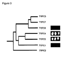

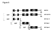
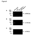


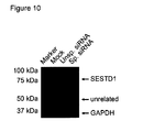


Classifications
-
- C—CHEMISTRY; METALLURGY
- C07—ORGANIC CHEMISTRY
- C07K—PEPTIDES
- C07K14/00—Peptides having more than 20 amino acids; Gastrins; Somatostatins; Melanotropins; Derivatives thereof
- C07K14/435—Peptides having more than 20 amino acids; Gastrins; Somatostatins; Melanotropins; Derivatives thereof from animals; from humans
- C07K14/46—Peptides having more than 20 amino acids; Gastrins; Somatostatins; Melanotropins; Derivatives thereof from animals; from humans from vertebrates
- C07K14/47—Peptides having more than 20 amino acids; Gastrins; Somatostatins; Melanotropins; Derivatives thereof from animals; from humans from vertebrates from mammals
-
- A—HUMAN NECESSITIES
- A61—MEDICAL OR VETERINARY SCIENCE; HYGIENE
- A61P—SPECIFIC THERAPEUTIC ACTIVITY OF CHEMICAL COMPOUNDS OR MEDICINAL PREPARATIONS
- A61P11/00—Drugs for disorders of the respiratory system
- A61P11/06—Antiasthmatics
-
- A—HUMAN NECESSITIES
- A61—MEDICAL OR VETERINARY SCIENCE; HYGIENE
- A61P—SPECIFIC THERAPEUTIC ACTIVITY OF CHEMICAL COMPOUNDS OR MEDICINAL PREPARATIONS
- A61P17/00—Drugs for dermatological disorders
-
- A—HUMAN NECESSITIES
- A61—MEDICAL OR VETERINARY SCIENCE; HYGIENE
- A61P—SPECIFIC THERAPEUTIC ACTIVITY OF CHEMICAL COMPOUNDS OR MEDICINAL PREPARATIONS
- A61P9/00—Drugs for disorders of the cardiovascular system
-
- C—CHEMISTRY; METALLURGY
- C07—ORGANIC CHEMISTRY
- C07K—PEPTIDES
- C07K14/00—Peptides having more than 20 amino acids; Gastrins; Somatostatins; Melanotropins; Derivatives thereof
- C07K14/435—Peptides having more than 20 amino acids; Gastrins; Somatostatins; Melanotropins; Derivatives thereof from animals; from humans
- C07K14/705—Receptors; Cell surface antigens; Cell surface determinants
-
- G—PHYSICS
- G01—MEASURING; TESTING
- G01N—INVESTIGATING OR ANALYSING MATERIALS BY DETERMINING THEIR CHEMICAL OR PHYSICAL PROPERTIES
- G01N33/00—Investigating or analysing materials by specific methods not covered by groups G01N1/00 - G01N31/00
- G01N33/48—Biological material, e.g. blood, urine; Haemocytometers
- G01N33/50—Chemical analysis of biological material, e.g. blood, urine; Testing involving biospecific ligand binding methods; Immunological testing
- G01N33/53—Immunoassay; Biospecific binding assay; Materials therefor
- G01N33/566—Immunoassay; Biospecific binding assay; Materials therefor using specific carrier or receptor proteins as ligand binding reagents where possible specific carrier or receptor proteins are classified with their target compounds
-
- G—PHYSICS
- G01—MEASURING; TESTING
- G01N—INVESTIGATING OR ANALYSING MATERIALS BY DETERMINING THEIR CHEMICAL OR PHYSICAL PROPERTIES
- G01N2405/00—Assays, e.g. immunoassays or enzyme assays, involving lipids
- G01N2405/04—Phospholipids, i.e. phosphoglycerides
-
- G—PHYSICS
- G01—MEASURING; TESTING
- G01N—INVESTIGATING OR ANALYSING MATERIALS BY DETERMINING THEIR CHEMICAL OR PHYSICAL PROPERTIES
- G01N2500/00—Screening for compounds of potential therapeutic value
- G01N2500/02—Screening involving studying the effect of compounds C on the interaction between interacting molecules A and B (e.g. A = enzyme and B = substrate for A, or A = receptor and B = ligand for the receptor)
Definitions
- the present invention relates to an isolated complex comprising a first protein comprising or consisting of a transient receptor potential channel (TRPC) or a functionally active variant thereof and a second protein comprising or consisting of the first spectrin (Spec 1) domain of SEC14 and spectrin domains 1 (SESTD1), a test system comprising the first protein and the second protein as well as means for detecting interaction of the first and the second protein, a method for screening for a TRPC modulator using the system and the use of the test system for the identification of a TRPC modulator.
- TRPC transient receptor potential channel
- SESTD1 spectrin domains 1
- TRPCs Classical transient receptor potential channel proteins
- TRPV1-targeting drugs This channel also belongs to the TRP superfamily and its physiological function was unravelled by the use of a specific activator (capsaicin). Ten years after its cloning, several clinical trials are ongoing and test the therapeutic value of TRPV1 modulation in a number of indications, e.g. in different types of pain, migraine, and urinary urge incontinence.
- one object of the present invention was to develop new pharmacological tools that may be used to gain a better understanding of TRPC channel function in cells and beyond, and to identify novel regulators or modulators of TRPC channel complexes.
- the new tools provide for economic and convenient testing of substances, such as high throughput screening methods.
- the object of the present invention was solved by providing an isolated complex comprising
- An alternative isolated complex comprises
- an isolated complex in the context of the present invention relates to a complex of the first and the second protein as defined herein which is not in its natural environment. Accordingly, the "isolated complex" is not associated with proteins not being part of the signal transduction of the TRPC and the SESTD1 as defined herein or it (or its components) is isolated from a cell in which it normally occurs or is isolated from a cell in which the nucleic acid coding for the same has been expressed or is essentially free from other proteins from the same cellular source not involved in the respective signal transduction pathway.
- the complex may be a naturally occurring complex, preferably a naturally occurring complex of TRPC and SESTD1 or part thereof (as defined herein).
- proteins of the complex may also be artificial in that they do not naturally occur, in that they may encompass one or more sections which are naturally not connected to each other, for example, a fusion protein comprising or consisting of a part of TRPC or SESTD1 as defined herein and a further protein such as a second domain or other component used for, e.g., detection and/or purification purposes.
- the term "isolated complex” means a protein complex which is essentially separated from other cellular components of its natural environment. However, after isolation of the proteins of the complex, cellular components may be added again, e.g., for measuring signal transduction pathways. Additionally, the skilled person will understand that the isolated complex is to be kept under suitable conditions allowing activity of the isolated complex, e.g., suitable buffers, pH values, ions, etc.
- the first protein of the complex of the invention comprises or consists of:
- TRPC may be any naturally occurring TRPC. Based on amino acid sequence homology and functional similarities TRPCs can be subclassified into four groups. Being quite unique within the TRPC family, TRPC1 and TRPC2 each constitute a subfamily by themselves while TRPC4 and -5 are merged just as TRPC3, -6, and -7. Heteromeric interactions within these subfamilies have been shown as well as coassembly of TRPC1 with either TRPC4/5- or TRPC3/6/7-subfamily members. It was long thought that no cross-association occurs between the TRPC4/5 and TRPC3/6/7 subgroups, but recently an endogenous redox-sensitive TRPC3/4 heteromer has been reported in porcine aortic endothelial cells.
- TRPCs will be introduced with their possible in vivo functions below.
- TRPC1 subfamily Investigation of the broadly expressed homomeric TRPC1 has been hampered by absent plasma membrane targeting of the ectopic protein in cell lines. Depending on the overexpression system used, reports range from lack of robust TRPC1 signals due to retention in intracellular membranes to detailed description of channel properties in sf9 cells. Possible explanations are the absence of auxiliary subunits or interacting proteins in overexpression systems. Plasma membrane expression of TRPC1 has been shown to depend on interaction with other proteins, e.g. TRPCs, caveolin-1 and RhoA. Also in sf9 cells it is not certain whether homo- or heteromeric or even native channels, which could be stimulated by TRPC1, are measured.
- TRPC1 ectopically expressed homomeric TRPC1 was described as receptor-, DAG-, IP 3 R- or stretch-activated channel or as a non-functional channel subunit and the existence of a native TRPC1 homomer was not unequivocally proven so far. But the endogenous protein is required for the excitatory postsynaptic conductance (EPSC) in Purkinje cells therefore it could be involved in neuronal plasticity. Moreover, TRPC1 is up-regulated in neointimal hyperplasia and cardiac hypertrophy and its function could be affected in patients suffering from Duchenne muscular dystrophy. Recently, a role for TRPC1 was proposed in breast cancer.
- TRPC1 has also been reported to interact with TRPP2, a distantly related TRP protein involved in development of polycystic kidney disease.
- TRPC1 seems to be engaged in many patho- and physiological processes and some of its functional roles are not easily compensated by related TRPCs. These characteristics along with its accessibility to blocking extracellular antibodies make it a potential drug target, but it is probably not easily applicable due to its broad expression and favoured heteromultimerization.
- TRPC2 subfamily TRPC2 is a pseudogene in humans but essential for pheromone sensation in rodents. Male mice lacking this channel do not show typical male-male aggressive behaviour and even court consexuals. Antibodies directed to an extracellular domain of TRPC2 inhibit the acrosomal reaction pointing towards its importance in fertilization. However, TRPC2 knock-out mice showed no defects in reproduction. TRPC2 does not seem to heteromultimerize with other TRPC channels.
- TRPC3 / 6 / 7 subfamily Its members share 70 - 80 % amino acid identity and are directly activated by the PLC product DAG. TRPC3 and -6 activities are regulated by N-glycosylation and phosphorylation through the non-receptor tyrosine kinases Src and Fyn.
- TRPC3 is highly expressed in brain, smooth muscle and vascular endothelial cells. It seems to be involved in axon growth guidance, synaptic plasticity around the time of birth and cardiac Ca 2+ homeostasis. In cardiomyocytes, abnormal accumulation of intracellular Na + levels due to TRPC3 has been shown to reverse the Na + /Ca 2+ exchanger (NCX1) mode. This mode reversal transports Ca 2+ into the cell and might be involved in pathophysiological processes, e.g. heart failure and ischemia. TRPC3 was found to coassemble with TRPC4 into a redox-sensitive channel. It is hypothesized that oxidative stress in brain, for example induced by stroke, could activate redox-sensitive TRPC channels leading to uncontrolled Ca 2+ influx. The resulting neurodegeneration could eventually be minimized by TRPC antagonists.
- TRPC6 is present both in vascular and airway smooth muscle cells (SMC) and seems to play essential roles in the vascular and cardiac system. Its stimulation by agonists or increasing intravascular pressure is postulated to depolarize the membrane. Subsequently, L-type voltage-gated Ca 2+ channels are activated which finally mediate muscle contraction and reflex vasoconstriction (Bayliss effect). On the contrary, agonist-induced bronchoconstriction mainly depends on Ca 2+ influx mediated by voltage-independent channels (such as TRPC6), hence, L-type Ca 2+ channel blockers are not effective, e.g. in asthma and chronic obstructive pulmonary disease (COPD).
- SMC airway smooth muscle cells
- Pulmonary artery SMC (PASMC) of patients suffering from idiopathic pulmonary arterial hypertension (IPAH) are hyperproliferative and TRPC6 (and TRPC3) expression is significantly increased.
- PASMC hyperproliferation was markedly reduced after treatment with TRPC6 small-interferring RNA (siRNA) or bosentan, an endothelin receptor blocker approved for the treatment of IPAH patients.
- TRPC6 small-interferring RNA
- bosentan an endothelin receptor blocker approved for the treatment of IPAH patients.
- TRPC6 inhibition seems to be an interesting therapeutic strategy for the treatment of IPAH, a progressive, life-shortening disease (right heart failure), and other chronic respiratory diseases. But TRPC6 also has physiological functions in airway SMC that should rather not be blocked.
- TRPC6 is essential in HPV but not involved in pulmonary vascular hypertension and remodeling resulting from chronic hypoxia. Convincing evidence for TRPC6 involvement in hereditary focal segmental glomerulosclerosis (FSGS), a significant cause of end-stage renal disease, has also been presented. TRPC6 gain-of-function mutants lead to increased Ca 2+ influx into podocyte foot processes, but it is not known whether and how this is disease-causing.
- FSGS hereditary focal segmental glomerulosclerosis
- TRPC6 is overexpressed and mislocated in podocytes of these mice. Recently, it was also shown in vitro that TRPC6 expression is up-regulated in complement-treated podocytes leading to actin cytoskeleton rearrangement, whereas transient channel overexpression in vivo led to proteinuria in mice. TRPC6 deficient mice have an unexpected and surprising phenotype.
- TRPC6 in the pathogenesis of cardiac hypertrophy.
- Cardiac specific TRPC overexpression in transgenic mice leads to increased Ca 2+ influx that couples via calcineurin to nuclear factor of activated T cells (NFAT) stimulation.
- NFAT nuclear factor of activated T cells
- Pathological heart remodelling was accelerated and these mice had a shortened life expectancy.
- TRPC6 upregulation in cardiomyocytes participates in hypertrophy, it seems to have protective antifibrotic functions in cardiac fibroblasts in vitro . Further in vivo studies are needed to estimate the therapeutic value of TRPC6 modulation and the involvement of TRPC3 and TRPC3/6 heteromers in the pathogenesis of heart failure.
- TRPC7 is expressed in eye, heart and lung and lower transcript levels are found in brain, spleen and testis but its physiological functions remain obscure.
- TRPC4 / 5 subfamily These channels share 64% identity and are most closely related to TRPC1 (persuading some groups to classify TRPC1 within this subfamily).
- a unique feature among TRP and other store-operated channels is their potentiation by lanthanides (e.g. Gd 3+ , La 3+ ) after G q/11 -receptor mediated activation.
- lanthanides e.g. Gd 3+ , La 3+
- S1 P sphingosine 1-phosphate
- S-nitrosylation e.g. by nitric oxide (NO) has been shown to activate both TRPC4 and TRPC5.
- TRPC4 was the first TRP gene to be knocked out in mice. It is widely expressed and found in endothelial and smooth muscle cells. TRPC4 -/- mice are viable and reach maturation. However, agonist-induced Ca 2+ entry into endothelial cells of TRPC4 -/- mice is markedly reduced resulting in decreased endothelium-dependent vasorelaxation. Further studies were performed with thrombin, an important inflammation mediator that is involved in the pathogenesis of vascular injury. In lungs, it increases vascular permeability and thus tissue water content. Lung EC of TRPC4 -/- mice lack thrombin-induced actin stress fiber formation, cell retraction is impaired, and lung microvascular permeability subsequently reduced by about 50%.
- TRPC4 is also expressed in different cells within the central nervous system and seems to be involved in neurotransmitter release.
- GABA ⁇ -aminobutyric acid
- 5-HT 5-hydroxytryptamin
- TRPC4 could participate in processing of visual information depending on the sleep/wake cycle.
- TRPC4 expressed in pancreatic ⁇ -islets could be involved in insulin secretion, but glucose-tolerance test results were similar in wild-type and TRPC4-deficient mice.
- the channel seems to be involved in regulating the motility of the gastrointestinal tract by modulating the pacemaker activity of interstitial cells of Cajal (ICC).
- TRPC5 is highly enriched in brain but also found peripheral, e.g. in SMC. In humans, the gene is located on a region of the X chromosome associated with non-syndromic mental retardation. TRPC5 homomers expressed in growth cones of rat hippocampal neurons regulate neurite outgrowth as well as growth cone morphology. Functional channel suppression by transfection of a dominant-negative mutant led to abnormally prolonged neurites whereas overexpression resulted in neurite outgrowth inhibition. Phosphatidyl-inositiol 4-phosphate 5-kinase (PIP(5)K ⁇ )-dependent channel insertion from vesicles into the plasmamembrane was demonstrated to be crucial for neurite length regulation by TRPC5.
- PIP(5)K ⁇ Phosphatidyl-inositiol 4-phosphate 5-kinase
- TRPC5 could also be multifunctionally important within the cardiovascular system.
- a dominant-negative TRPC5 mutant inhibited cell motility evoked by sphingosine 1-phosphate (a TRPC5 activator) of vascular smooth muscle cells, the same effect was accomplished with an anti-TRPC5 antibody.
- SMC motility is crucial in physiological adaptive processes like wound healing but is also involved in inflammatory occlusive diseases, e.g. atherosclerosis. In monocytes from hypertensive patients increased TRPC5 expression and channel-mediated Ca 2+ influx was reported.
- TRPC5 protein expression and activity was observed by siRNA mediated downregulation of the Ca 2+ -ATPase SERCA2 in neonatal rat cardiac myocytes.
- TRPC5 was even selectively upregulated, whereas TRPC3 mRNA and protein was not detectable and TRPC1, -4, and -6 expression levels were not altered.
- TRPC5 and TRPC4 in cardiac hypertrophy is conceivable.
- the first protein may also comprise or consists of a functionally active variant of any of the TRPCs as defined herein.
- the functionally active variant of the TRPC may be derived from the TRPC by one or more amino acid additions, deletions or substitutions, wherein the variant is still capable of binding to a natural occurring SESTD1.
- the variant is a fragment of the TRPC still comprising a SESTD1 binding domain providing for the capability of binding to the SESTD1 and/or Spec 1 of SESTD1.
- the binding of the variant to SESTD1 may be examined as described in Examples 1, 2, 3, 4, 5 or 6.
- the affinity of a protein for another molecule may be determined by a variety of standard techniques including binding studies using a labeled marker such as a radioligand and fluorescence marker etc and is well known to the skilled practitioner.
- the functionally active variant may be a fragment of the any naturally occurring TRPC.
- the functionally active variant may also be a chimeric TRPC comprising domains or segments of two or more different TRPCs.
- the functionally active variant may also encompass one or more segments with mutations (e.g. substitutions, additions and/or deletions) which may alter the binding properties of the variant to the TRPC.
- the variant is defined in that it is still capable of binding to a natural occurring SESTD1 and/or Spec 1 of SESTD1.
- the functionally active variant is defined in that it is a TRPC variant comprising or consisting of a sequence homologous to SEQ ID NO:1, SEQ ID NO:2 or SEQ ID NO:3.
- this sequence is a fragment of a naturally occurring TRPC.
- sequences of various TRPC proteins are derivable from protein databases such as NCBi (National Center for Biotechnology information) and homologous sequences may be identified by suitable alignment programs as known to the skilled practitioner.
- the first protein of the present invention comprises a marker, particularly a tag.
- a marker in the context of the present invention may be any kind of molecule which can be easily detected.
- the molecule is bound to TRPC or a functionally active variant thereof, therefore, the presence of the marker is indicative for the presence of the first protein.
- Markers also referred to as labels
- markers include, for example, radiolabels (such as 3 H, 32 P, 35 S or 14 C), fluorescence markers (such as fluorescein, green fluorescence protein, yellow fluorescence protein or DyLight 488), enzymes (such as horseradish peroxidase, ⁇ -lactamase, alkaline phosphatase or ⁇ -glucosidase) or an antigen-detectable by a suitable antibody or antibody fragment.
- the marker is a tag.
- Tags are usually peptides or proteins which are used as biochemical indicators. They may be included into a protein, such as a recombinant, expressed protein and can serve several purposes. Preferably, they are used for purifying the proteins to which they are attached using standard conditions suitable for the particular tag. However, the tags may be also used as indicators in order to detect the presence of a particular protein.
- a number of (affinity) tags are known at present. These are usually divided into 3 classes according to their size: small tags have a maximum of 12 amino acids, medium-sized ones have a maximum of 60 and large ones have more than 60.
- the small tags include the Arg-tag, the His-tag, the Strep-tag, the Flag-tag, the T7-tag, the V5-peptide-tag and the c-Myc-tag, the medium-sized ones include the S-tag, the HAT-tag, the calmodulin-binding peptide, the chitin-binding peptide and some cellulose-binding domains.
- the latter can contain up to 189 amino acids and are then regarded, like the GST- (glutathione-S-transferase-) and MBP-tag (maltose binding protein-tag), as large affinity tags.
- double tags or tandem tags were developed.
- the proteins are purified in two separate chromatography steps, in each case utilizing the affinity of a first and then of a second tag.
- double or tandem tags are the GST-His-tag (glutathione-S-transferase fused to a polyhistidine-tag), the 6xHis-Strep-tag (6 histidine residues fused to a Strep-tag), the 6xHis-tag100-tag (6 histidine residues fused to a 12-amino-acid protein of mammalian MAP-kinase 2), 8xHis-HA-tag (8 histidine residues fused to a haemagglutinin-epitope-tag), His-MBP (His-tag fused to a maltose-binding protein), FLAG-HA-tag (FLAG-tag fused to a hemagglutinin-epitope-tag), and the
- the first protein of the present invention comprises a tag selected from the group consisting of His-tag, Arg-tag, Strep-tag, Flag-tag, T7-tag, V5-peptide-tag, c-Myc-tag, S-tag, HAT-tag, calmodulin-binding peptide-tag, chitin-binding peptide-tag, GST-tag and MBP-tag.
- a tag selected from the group consisting of His-tag, Arg-tag, Strep-tag, Flag-tag, T7-tag, V5-peptide-tag, c-Myc-tag, S-tag, HAT-tag, calmodulin-binding peptide-tag, chitin-binding peptide-tag, GST-tag and MBP-tag.
- any other tag may be also used, but some tags such as His-tag, Arg-tag, Strep-tag, Flag-tag or GST-tag are preferred. Suitable examples for TRPCs or variants thereof encompassing a tag
- the first protein comprises a marker or tag, wherein the marker or tag is removable from the protein by proteolytic cleavage at a specific cleavage site, for example a cleavage site for an enzyme.
- a specific cleavage site for example a cleavage site for an enzyme.
- the cleavage site could for example be a protease cleavage site.
- proteases are chymotrypsin, trypsin, elastase, and plasmin; the corresponding cleavage sites are known to a person skilled in the art. Since the molecule to be purified is a protein, specific proteases, especially proteases from viruses that normally attack plants, are preferred.
- Suitable specific proteases are thrombin, factor Xa, Igase, TEV-protease from the "Tobacco Etch Virus", the protease PreScission (Human Rhinovirus 3C Protease), enterokinase or Kex2. TEV-protease and PreScission are especially preferred.
- the first protein of the complex of the invention is a domain of a classical transient receptor potential channel (TRPC) or a functionally active variant thereof, the domain having the sequence of SEQ ID NO:1 as defined above.
- TRPC transient receptor potential channel
- obligatory sequence of SEQ ID NO: 1 may be further defined as follows, wherein the variable amino acids X are specified by the following indices: LX 1 X 2 X 3 X 4 X 5 YQEVX 6 RNLVKRYVAAMIRX 7 X 8 KT (SEQ ID NO:1):
- sequence of SEQ ID NO: 1 may be further defined as follows:
- amino acid side chains are summarized in the following: Amino Acid Polarity Acidity/Basicity Alanine nonpolar neutral Arginine polar basic Asparagine polar neutral Aspartic acid polar acidic Cysteine polar neutral Glutamic acid polar acidic Glutamine polar neutral Glycine nonpolar neutral Histidine polar basic Isoleucine nonpolar neutral Leucine nonpolar neutral Lysine polar basic Methionine nonpolar neutral Phenylalanine nonpolar neutral Proline nonpolar neutral Serine polar neutral Threonine polar neutral Tryptophan nonpolar neutral Tyrosine polar neutral Valine nonpolar neutral neutral
- TRPC transient receptor potential channel
- the functionally active variant may be defined as above, wherein the sequence of SEQ ID NO:1 is further defined in that
- Conservative substitutions are those that take place within a family of amino acids that are related in their side chains and chemical properties. Examples of such families are amino acids with basic side chains, with acidic side chains, with non-polar aliphatic side chains, with non-polar aromatic side chains, with uncharged polar side chains, with small side chains, with large side chains etc.
- one conservative substitution is included in the peptide.
- two conservative substitutions or less are included in the peptide.
- three conservative substitutions or less are included in the peptide.
- conservative amino acid substitutions include, but are not limited to, those listed below: Original Residue Conservative Substitutions Ala Ser Arg Lys Asn Gln; His Asp Glu Cys Ser Gln Asn Glu Asp His Asn; Gln Ile Leu, Val Leu Ile; Val Lys Arg; Gln; Asn Met Leu; Ile Phe Met; Leu; Tyr Ser Thr Thr Ser Trp Tyr Tyr Trp; Phe Val Ile; Leu
- the TRPC is preferably selected from the group consisting of TRPC4, TRPC5, and TRPC6, particularly it is TRPC4 or TRPC5, especially TRPC4 ⁇ or TRPC4 ⁇ or TRPC5.
- the first protein comprises the sequence LRRHHQYQEVMRNLVKRYVAAMIREAKTE (SEQ ID NO:2) or LIQNQHYQEVIRNLVKRYVAAMIRNSKTN (SEQ ID NO:3).
- the TRPC may be derived from any species.
- mammalian TRPCs such as those derived from human, rat, mouse, cat, dog or horse, particularly murine or human TRPCs, are preferred.
- the second protein of the complex of the invention comprises or consists of the Spec 1 domain of SESTD1.
- the sequence of human SESTD1 is disclosed in GenBank accession number NP_835224. However natural occurring variants are also known, e.g. one was found full length in the human aorta cDNA library. Its amino acid sequence is identical to GenBank accession no. NP_835224 apart from one exchange (H508Q). It is based on a point mutation at 1571 (NM_178123) that can also be found in a genomic blast. Another point mutation found at 1748 is silent.
- the Spec 1 domain consists of amino acids 233 to 381 of the SESTD1 as specified above (see also Figure 1 ).
- homologous Spec 1 sequences derived from alternative naturally occurring SESTD1 may be also used.
- the alternative SESTD1 may be derived from another species or organ or subject.
- the sequences of various SESTD1 proteins are derivable from protein databases such as NCBI (National Center for Biotechnology Information) and homologous sequences may be identified by suitable alignment programs.
- the second protein may also be a fragment of a naturally occurring SESTD1 provided that the Spec 1 domain is still present.
- the Spec 1 domain may also have added (C- and/or N-terminally) one or more amino acid sequences not related to any naturally occurring SESTD1.
- one or more amino acid sequence is added to the SESTD1, it/they is/are segments of naturally occurring SESTD1 (leading to fragments or chimeras), which may also encompass amino acids substitutions (preferably a limited number such as up to 10), preferably conservative substitutions, as defined above.
- the second protein may also encompass a marker or tag, as defined in connection with the first protein of the invention.
- a protein comprising SESTD1 or fragments thereof and a GST-tag are illustrated in Figure 5 .
- the second protein comprises or consists of the full length SESTD1, wherein the SESTD1 may be as defined above.
- the SESTD1 may be derived from any species. However, mammalian SESTD1s, such as those derived from human, rat, mouse, cat, dog or horse, particularly rat, murine or human SESTD1, are preferred.
- a third aspect of the invention relates to a test system comprising
- test system of the invention may be used in order to elucidate the function and activity of TRPCs.
- mechanisms involving TRPC and SESTD1 can be studied by using the test system of the invention.
- test system may be used and to which the test system may be adapted.
- This may be a heterogeneous or homogeneous assay.
- a heterogeneous assay is an assay which includes one or more washing steps, whereas in a homogeneous assay such washing steps are not necessary. The reagents and compounds are only mixed and measured.
- the assay is an ELISA (enzyme linked immuno sorbent assay), a DELFIA (dissociation enhanced lanthanide fluoro immuno assay), an SPA (scintillation proximity assay) a flashplate assay, a FRET (fluorescence resonance energy transfer) assay, TR-FRET (time-resolved fluorescence resonance energy transfer) assay, a FP (fluorescence polarisation) assay, an ALPHA (amplified luminescent proximity homogenous assay), an EFC (enzyme fragment complementation) assay, a two hybrid assay or a coimmunoprecipitation assay.
- ELISA enzyme linked immuno sorbent assay
- DELFIA dissociation enhanced lanthanide fluoro immuno assay
- SPA sintillation proximity assay
- FRET fluorescence resonance energy transfer
- TR-FRET time-resolved fluorescence resonance energy transfer
- FP fluorescence polarisation
- ELISA enzyme linked immuno sorbent assay
- ELISA-based assays are offered by various companies. It is a biochemical technique used to detect the presence of an antibody or an antigen in a sample. Performing an ELISA involves at least one antibody with specificity for a particular antigen (e.g a segment of the first or second protein). In general, an unknown amount of antigen in a sample is immobilized on a surface. One then washes a particular antibody over the surface. This antibody is linked to an enzyme that produces a detecable signal such as a change in colour or fluorescene.
- the sample with an unknown amount of antigen is immobilized on a solid support (usually a microtiter plate) either non-specifically (via adsorption to the surface) or specifically (via capture by another antibody specific to the same antigen, in a "sandwich” ELISA).
- a solid support usually a microtiter plate
- the detection antibody is added, forming a complex with the antigen.
- the detection antibody can be covalently linked to an enzyme, or can itself be detected by a secondary antibody which is linked to an enzyme through bioconjugation.
- the plate is typically washed with a mild detergent solution to remove any proteins or antibodies that are not specifically bound.
- the plate is developed by adding an enzymatic substrate to produce a visible signal, which indicates the quantity of antigen in the sample.
- Older ELISAs utilize chromogenic substrates, though newer assays employ fluorogenic substrates with much higher sensitivity.
- DELFIA dissociation enhanced lanthanide fluoro immuno assay
- DELFIA-based assays are solid phase assay.
- the antibody is usually labelled with Europium or another lanthanide and the Europium fluorescence is detected after having washed away un-bound Europium-labelled antibodies.
- SPA sintillation proximity assay
- the flashplate assay usually exploit biotin/avidin interactions for capturing radiolabelled substrates.
- the reaction mixture includes the kinase, a biotinylated peptide substrate and ⁇ -[P 33 ]ATP.
- streptavidin is bound on scintillant containing beads
- streptavidin is bound to the interior of the well of scintillant containing microplates. Once immobilized, the radiolabelled substrate is close enough to the scintillant to stimulate the emission of light.
- Fluorescence resonance energy transfer describes a radiation-free energy transfer between two chromophores.
- a donor chromophore in its excited state can transfer energy by a non-radiative long-range dipole-dipole coupling mechanism to an acceptor fluorophore in close proximity (typically ⁇ 10 nm).
- the energy transfer is often referred to as “fluorescence resonance energy transfer", although the energy is not actually transferred by fluorescence.
- FRET is a useful tool to detect and quantify protein-protein interactions, protein-DNA interactions, and protein-conformational changes.
- one of the molecules is labeled with a donor and the other with an acceptor and these fluorophore-labeled molecules are mixed.
- donor emission is detected upon donor excitation.
- the donor and acceptor are brought in proximity and the acceptor emission is predominantly observed because of the intermolecular FRET from the donor to the acceptor.
- Suitable neighbors for FRET are known in the art and the skilled practitioner will be able to choose a suitable combination of labels for both antibodies.
- corresponding refers to an acceptor fluorescent moiety having an excitation spectrum that overlaps with the emission spectrum of the donor.
- the wavelength maximum of the emission spectrum of the acceptor should preferably be at least 30 nm, more preferably at least 50 nm, such as at least 80 nm, at least 100 nm or at least 150 nm greater than the wavelength maximum of the excitation spectrum of the donor.
- Representative donor fluorescent moieties that can be used with various acceptor fluorescent moieties in FRET technology include fluorescein, Lucifer Yellow, B-phyco-erythrin, 9-acridineisothiocyanate, Lucifer Yellow VS, 4-acetamido-4'-isothiocyanatostilbene-2,2'-disulfonic acid, 7-diethylamino-3-(4'-isothiocyanatophenyl)-4-methylcoumarin, succinimdyl 1-pyrenebutyrate, and 4-acetamido-4'-isothiocyanatostilbene-2,2'-disulfonic acid derivatives.
- acceptor fluorescent moieties depending upon the donor fluorescent moiety used, include LC-Red 610, LC-Red 640, LC-Red 670, LC-Red 705, Cy5, Cy5.5, Lissamine rhodamine B sulfonyl chloride, tetramethyl rhodamine isothiocyanate, rhodamine x isothiocyanate, erythrosine isothiocyanate, fluorescein, diethylenetriamine pentaacetate or other chelates of lanthanide ions (e.g., europium, or terbium).
- Donor and acceptor fluorescent moieties can be obtained, for example, from Molecular Probes (Junction City, OR) or Sigma Chemical Co. (St. Louis, MO).
- TR-FRET time-resolved fluorescence resonance energy transfer
- TR-FRET unites TRF (time-resolved fluorescence) and the FRET principle. This combination combines the low background benefits of TRF and the homogeneous assay format of FRET. While FRET has already been described above, TRF takes advantage of the unique properties of lanthanides or any other donor with long half-life. Suitable donors for TR-FRET include, amongst others, lanthanide chelates (cryptates) and some other metal ligand complexes, which can have fluorescent half-life in the micro- to millisecond time range and which, therefore, also allow the energy transfer to occur in micro- to millisecond measurements.
- Fluorescence lanthanide chelates have been used as energy donors in the late seventies.
- the commonly used lanthanides include samarium (Sm), europium (Eu), terbium (Tb) and dysprosium (Dy). Because of their specific photophysical and spectral properties, complexes of lanthanides are of major interest for fluorescence application in biology. Specifically, they have a large Stokes' shift and extremely long emission half-lives (from microseconds to milliseconds) when compared to more traditional fluorophores.
- organic chromophores are used as acceptors. These include allophycocyanin (APC). Suitable details on TR-FRET as well as acceptors are described in WO 98/15830 .
- Fluorescence polarisation (FP)-based assays are assays which use polarized light to excite fluorescent substrate peptides in solution. These fluorescent peptides are free in solution and tumble, causing the emitted light to become depolarised. When the substrate peptide binds to a larger molecule, its tumbling rates are greatly decreased, and the emitted light remains highly polarized.
- the test system of the invention is adapted for an amplified luminescence proximity homogeneous assay (ALPHA).
- ALPHA is a solution-based assay which was originally developed by Packard BioScience.
- ALPHA is a luminescence-based proximity assay, wherein one interaction partner is attached to donor beads, while the other is coupled to acceptor beads, both with a diameter of only about 250 nm.
- a photosensitizer compound is embedded into the donor bead. With this compound upon illumination with laser light at a wavelength of about 680 nm, ambient oxygen is converted into energy-rich, short-life singlet oxygen. When no acceptor bead is in proximity, the singlet oxygen decays without producing a signal.
- donor and acceptor beads are brought together (ca. 250 nm) by the biological interaction of the attached biomolecules, the singlet oxygen released by the donor bead initiates a luminescence/fluorescence cascade in the nearby acceptor bead, leading to a highly amplified signal in the 520-620 nm range.
- the luminescence signal is detected in a suitable reader.
- ALPHA techniques see Ullman et al., 1994, Proc. Natl. Acad. Sci., USA 91, 5426-5430 .
- EFC (enzyme fragment complementation)-based assays or equivalent assays can be used in particular for high throughput screening of compounds.
- the EFC assay is based on an engineered ⁇ -galactosidase enzyme that consists of two fragments - the enzyme acceptor (EA) and the enzyme donor (ED). When the fragments are separated, there is no ⁇ -galactosidase activity, but when the fragments are together they associate (complement) to form active enzyme.
- EFC assay utilizes an ED-analyte conjugate in which the analyte may be recognized by a specific binding protein, such as an antibody or receptor.
- the ED-analyte conjugate In the absence of the specific binding protein, the ED-analyte conjugate is capable of complementing EA to form active ⁇ -galactosidase, producing a positive luminescent signal. If the ED-analyte conjugate is bound by a specific binding protein, complementation with EA is prevented, and there is no signal. If free analyte is provided (in a sample), it will compete with the ED-analyte conjugate for binding to the specific binding protein. Free analyte will release ED-analyte conjugate for complementation with EA, producing a signal dependent upon the amount of free analyte present in the sample.
- Two-hybrid screening is a molecular biology technique used to discover protein-protein interactions by testing for physical interactions (such as binding) between two proteins.
- the premise behind the test is the activation of downstream reporter gene(s) by the binding of a transcription factor onto an upstream activating sequence.
- the transcription factor is split into two separate fragments, called the binding domain (BD) and activating domain (AD).
- the BD is the domain responsible for binding to the UAS
- the AD is the domain responsible for activation of transcription.
- a suitable two-hybrid system is described in Example 1.
- Co-immunoprecipitation can be used for the identification of protein complexes by precipitating one protein believed to be in a complex because additional members of the complex are captured as well and can be identified.
- the protein complexes, once bound to the specific antibody, are removed from the bulk solution by capture with an antibody-binding protein attached to a solid support such as an agarose bead.
- antibody-binding proteins Protein A, Protein G, Protein L
- These antibody-binding proteins were initially isolated from bacteria and recognize a wide variety of antibodies. Following the initial capture of a protein or protein complex, the solid support is washed several times to remove any proteins not specifically and tightly bound through the antibody.
- co-immunoprecipitation is a standard method to assess protein-protein interaction.
- a suitable test system involving coimmunoprecipition is described in Example 3.
- the means for detecting the interaction between the first and second protein may be adapted to measure one or more phospholipid(s), preferably phosphatidyl-inositol-phosphate.
- the measuring may include the determination of the concentration of one or more phospholipid(s), preferably phosphatidyl-inositol-phosphate.
- the concentration may be measured in response to a potential TRPC modulator as described in the method for screening a modulator of the invention.
- Means and methods for determining concentrations of one or more phospholipid(s), preferably phosphatidyl-inositol-phosphate are well known to the skilled person and include such involving radio-labelled phospholipid(s), preferably phosphatidyl-inositol-phosphate(s), including purification by e.g. column chromatography, or mass spectrometry.
- means and methods involving a PIP strip phospholipid overlay assay or a Cova-PIP plate binding assay are preferred.
- the test system is adapted for high throughput screening.
- a large number of compounds is screened against the TRPC in question in either cell-free or whole-cell assays.
- these screenings are carried out in 96 well plates using automated, robotic station based technologies or in higher-density array (“chip”) formats.
- each of the proteins of the complex of the invention may be any of the proteins as specified in the above aspects and embodiments, particularly preferred embodiments. It could be shown that TRPC is able to interact with a series of further proteins. Accordingly, the test system may further comprise at least one protein selected from the group consisting of ankyrin repeat domain 35 (ANKRD35), apolipoprotein A-I binding protein (APOA1 BP), bromodomain adjacent to zinc finger domain (BAZ1B), high-mobility group protein 2-like1 isoform b (HMG2L1), makorin RING finger protein 1 (MKRN1), pre-B-cell leukemia homeobox interacting protein 1 (PBXIP1), sarcoma antigen NY-SAR-48, spectrin alpha non-erythrocytic 1 (SPTAN1), structural maintenance of chromosomes 3 (SMC3) and talin 2 (TLN2).
- ANKRD35 ankyrin repeat domain 35
- APOA1 BP apolip
- exemplary proteins may be obtained from any species as suitable in the prevailing test system.
- exemplary proteins and their sequences are as follows: Definition Gene name GenBank Accession No. Ankyrin repeat domain 35 ANKRD35 NM_144698 Apolipoprotein A-I binding protein APOA1BP NM_144772 Bromodomain adjacent to zinc finger domain BAZ1B AB032253 High-mobility group protein 2-like1 isoform b HMG2L1 CR456504 Makorin RING finger protein 1 MKRN1 NM_013446 Pre-B-cell leukemia homeobox interacting protein 1 PBXIP1 NM_020524 Sarcoma antigen NY-SAR-48 NM_033417 Spectrin, alpha, non-erythrocytic 1 SPTAN1 U83867 Structural maintenance of chromosomes 3 SMC3 AF067163 Talin 2 TLN2 NM_015059
- test system may e.g. be used for the below method for screening for a TRPC modulator.
- the present invention relates to a method for screening for a TRPC modulator comprising the steps of:
- test system may be specified as detailed in the present description of the invention, particularly as detailed in the preferred embodiments.
- the test system may be encompassed in a cell or it may be a cell-free system.
- the test system may be contacted with a test substance under conditions suitable to detect a measurable signal. This includes a suitable temperature, chemical environment (buffers, pH value, etc.) as well as a suitable concentration of the substance and an appropriate time of contact.
- a signal is observed wherein the detection of a signal is indicative of the interaction of the first and the second protein.
- the influence of the substance on the interaction between the first and the second protein as defined above is detected directly, i.e. that the proximity of the first and the second protein is detected by the induction of a signal and not a signal downstream of the TRPC/SESTD1 signal transduction.
- the induction of a signal may be effected by labeling of each of the components, wherein the proximity of the labels induces a detectable signal.
- the induced detectable signal may be a chemiluminescent signal, a change in color, a fluorescence signal, a radiation or any other suitable signal.
- the signal may be induced by interaction of two labels, wherein each label is bound to one component of the test system, i.e. the isolated protein and the co-factor.
- Such signals include a radiolabel, such as 125 I, on the one hand and a suitable quencher on the other hand in order to detect proximity in a scintillation counter (scintillation proximity assay).
- the components may encompass antigens accessible to antibodies labeled in a manner to detect FRET (fluorescence resonance energy transfer, see above).
- one of the proteins has bound to its surface a biomolecule capable of phosphorylation by a kinase and the other component has a trivalent metal ion complexed to its surface, e.g., via a suitable linker such as nitrilotriacetic acid, iminodiacetic acid or an appropriately substituted N-containing heterocycle, for example a triazoheterocycle, for example a triazocyclononaneononane, such as 1-propylamino-4-acetato-1,4,7-triazacyclononane.
- a chemiluminescent signal is generated when the donor and acceptor particles are in close proximity, which occurs upon binding of the protein to the co-factor (luminescent proximity assay).
- the means for detecting the interaction between the proteins includes at least one antibody specific for the first protein or the second protein.
- the isolated protein may include an antigen for a specific antibody and the detection of the antibody.
- the test system comprises two antibodies, wherein the first antibody is specific for the first protein and the second antibody is specific for the second protein.
- the antibody may be labeled with a Suitable marker, which is indicative of the presence of the respective component.
- the above-mentioned primary antibody may be detected by a suitable secondary antibody directed against the primary antibody. The secondary antibody may be used in order to detected the presence of the primary antibody, e.g. when bound to the secondary antibody.
- the primary or secondary antibody may be labeled with a marker as defined above, for example, an enzyme, a radiolabel, a fluorescence marker, a chemiluminescent marker, etc.
- a marker as defined above, for example, an enzyme, a radiolabel, a fluorescence marker, a chemiluminescent marker, etc.
- the proteins may encompass a tag, which is detectable with a suitable antibody.
- the test system may be used in a manner that a purification system for the separation of at least one component of the complex is used and the presence of the complex or of each of the components of the complex of the proteins is detected.
- the complex may be purified by gel electrophoresis, column chromatography, affinity purification, etc. and the complex may be detected by the presence of one signal or two signals indicative of one component of the complex or both components of the complex.
- the detection method may be limited to the other component.
- the purification method may be not specific for one of the components, such as gel electrophoresis, and the formation of the complex may be detected by two signals, e.g.
- the first and/or the second protein comprise(s) a detectable marker.
- the detecting of a measurable signal involves also the use of a specific antibody against the first protein and/or a specific antibody against the second protein, as detailed above.
- the TRPC modulator enhances or preferably impairs or inhibits binding of the first protein to the second protein.
- a modulator of TRPC alters the activity of the TRPC in question. The alteration of activity may be activation or inhibition, thus enhancing or impairing signal transduction of the TRPC, respectively.
- a modulator enhancing, improving or increasing activity of the TRPC is referred to as agonist, whereas a TRPC modulator impairing, inhibiting or decreasing the activity of the TRPC is referred to as antagonist.
- the modulation of activity is based on specific interaction with the TRPC and not the result of unspecific interaction with the TRPC (e.g. denaturation).
- the measurable signal is one or more phospholipid(s), preferably phosphatidyl-inositol-phosphate(s).
- Phospholipids are a class of lipids, and a major component of all biological membranes, along with glycolipids, Cholesterol and proteins.
- Phosphatidyl-inositol is the substrate for a large number of enzymes which are involved in cell signaling because it can be phosphorylated by a variety of kinases on the hydroxyl groups 3, 4 and 5 on the inositol ring in seven different combinations.
- the concentration of phospholipid(s), preferably phosphatidyl-inositol-phosphate(s) may be determined as described above.
- the test compound (substance) is provided in the form of a chemical compound library.
- Chemical compound libraries include are plurality of chemical compounds and have been assembled from any of multiple sources, including chemically synthesized molecules and natural products, or have been generated by combinatorial chemistry techniques. They are especially suitable for high throughput screening. They may be comprised of chemical compounds of a particular structure or compounds of a particular creature such as a plant.
- the chemical compound library is preferably a library comprising proteins and polypeptides or small molecules.
- methods of the invention may be used for screening for a medicament for preventing and/or treating a disease involving endothelial dysfunction.
- the method may be used for screening for a medicament for preventing and/or treating a cardiovascular disease, cardiac hypertrophy, heart failure, ischemia, neointima hyperplasia, idiopathic dilated cardiomyopathy, hypertension, coronary syndrome, heart failure, renal failure, thrombosis; asthma, a chronic respiratory disease, chronic obstructive pulmonary disorder, idiopathic pulmonary hypertension, acute hypoxic pulmonary vasoconstriction, inflammatory occlusive disease and atherosclerosis.
- a cardiovascular disease cardiac hypertrophy, heart failure, ischemia, neointima hyperplasia, idiopathic dilated cardiomyopathy, hypertension, coronary syndrome, heart failure, renal failure, thrombosis
- asthma a chronic respiratory disease, chronic obstructive pulmonary disorder, idiopathic pulmonary hypertension, acute hypoxic pulmonary vasoconstriction, inflammatory occlusive disease and atherosclerosis.
- the invention relates to the use of a test system according to the invention as specified above for the identification of a TRPC modulator, wherein the above details on the test system of the invention and the screening method of the invention are to be applied analogously.
- Example 1 Yeast two-hybrid (Y2H) screen with TRPC as bait
- TRPC4 plays a critical role in endothelial function, it is associated with regulation of endothelium-dependent vascular relaxation, may contribute to oxidative-stress induced endothelial damage and is involved in endothelial barrier function.
- the MATCHMAKER two-hybrid system 3 was to screen for novel proteins that interact with the cytosolic C-terminus of mTRPC4 ⁇ (aa 615-974) in yeast strain AH109.
- the system is based on the binding of the transcription factor GAL4 upstream of four reporter genes (ADE2, HIS3, IacZ, MEL1) activating their translation and protein expression. DNA binding and activation are mediated by two different protein domains which can be physically separated ( Fields S & Song O (1989).
- the C-terminus of mTRPC4 ⁇ (aa 615-974) served as bait.
- the bait was constructed by fusing the C-terminus of murine TRPC4 ⁇ (NM_016984) with a leucine zipper domain that has been shown to direct protein tetramerization ( Harbury PB, Zhang T, Kim PS, & Alber T (1993). A switch between two-, three-, and four-stranded coiled coils in GCN4 leucine zipper mutants. Science 262, 1401-1407 ).
- the amino acid sequence of the zipper (underlined) and flanking sequences is GGGSG S RMKQ IEDKL EEILS KLYHI ENELA RIKKL LGER G GSGS AAA (SEQ ID NO: 4).
- This construct was finally integrated into pGBKT7 (Clontech, Mountain View, USA), leading to its fusion with the GAL4-DNA binding domain (mTRPC4 ⁇ (615-974)/leucine zipper/pGBKT7).
- Other baits for directed yeast two-hybrid screens were constructed the same way with the C-termini of human TRPC1 (NM_003304), murine TRPC5 (NM_009428) and human TRPC6 (NM_004621).
- EGFP/leucine zipper/pGBKT7 Enhanced Green Fluorescent Protein
- a human aorta cDNA library in pACT2, Clontech, Mountain View, USA
- AD GAL4 activation domain
- GAL4 activity led to the expression of ⁇ -galactosidase that could be detected in yeast colonies by conversion of X-gal into a blue product.
- Prey plasmids from such colonies were isolated and analytically digested with the restriction endonucleases EcoRI/XhoI that cut the cDNA inserts out of the vector multiple cloning site. The resulting fragments were separated by gel electrophoresis whereby fragments of identical size pointed towards identical preys.
- Prey plasmids from surviving clones were finally sequenced and the sequences were compared with Genbank data by using BLAST software.
- the human aorta MATCHMAKER cDNA library (Clontech, Mountain View, USA) was provided transformed into E . coli BNN132 and was amplified to gain enough plasmid for screening in yeast.
- First its titer was determined by plating dilutions of 1x 10 -6 , 1x 10 -7 , and 2x 10 -7 on LB/amp agar plates. After they were incubated for 2 days (30 °C), the number of grown colonies was counted and resulted in 1.8x 10 8 colony forming units (cfu) per mL.
- the DNA-BD/bait and the library were sequentially polyethylenglycol/lithium acetate (PEG/LiAc)-mediated transformed into yeast ( Ito H, Fukuda Y, Murata K, & Kimura A (1983). Transformation of intact yeast cells treated with alkali cations. J Bacteriol 153, 163-168 ). Through a small-scale transformation, the DNA-BD/bait was introduced into yeast.
- Yeast transformed with the DNA-BD/bait grew on -Trp medium, cotransformants with a prey grew on -Trp/-Leu medium. It was verified that the bait is not intrinsic DNA-binding and/or transcriptional active by cotransformation with the negative control vector pGADT7.
- pGADT7 is an empty vector leading to sole expression of the GAL4-AD in yeast. Since this plasmid does not express a TRPC4-interacting protein, cotransformation with the bait is not sufficient to reconstitute GAL4, thus yeast did not survive on -Trp/-Leu/-Ade/-His plates. Single DNA-BD/bait transformants were used as inoculates for the following large-scale transformation with the AD fusion library.
- the suspension was subdivided onto several 50 mL Falcon tubes, pelleted by centrifugation (5 min, RT, 2,000 x g), washed with 300 mL sterile deionized water and reunited in 10 mL sterile deionized water. After washing with 50 mL lithium acetate buffer (100 mM LiAc, 10 mM Tris-HCl, 1 mM EDTA), cells were resuspended in 4 mL lithium acetate buffer.
- lithium acetate buffer 100 mM LiAc, 10 mM Tris-HCl, 1 mM EDTA
- the pellets were each resuspended in 25 mL -Trp/-Leu medium, reunited and grown at 250 rpm and 30 °C for one hr. After centrifugation (5 min, 2,000 g, RT), the cells were resuspended in 12 mL -Trp/-Leu medium, plated in 300 ⁇ L aliquots on -Trp/-Leu/-His agar plates (150 mm diameter), and incubated at 30°C for 2 to 5 days. Grown colonies were transferred to -Trp/- Leu/-His/-Ade agar plates and incubated for 2 to 3 more days.
- ⁇ -galactosidase assay clones grown on Trp/-Leu/-His/-Ade agar plates were top-layered with X-gal agar (0.5 % agarose, 500 mM NaPO 4 buffer (pH 7), 1 % sodium dodecyl sulphate (SDS), 2 % X-gal in dimethylformamide (DMF)) and incubated at 30°C up to 12 hrs. Generation of a blue product was visually checked.
- X-gal agar 0.5 % agarose, 500 mM NaPO 4 buffer (pH 7), 1 % sodium dodecyl sulphate (SDS), 2 % X-gal in dimethylformamide (DMF)
- clones surviving on -Trp/-Leu/-His/-Ade agar plates and positive reacting in the ⁇ -galactosidase assay were resuspended in 2 mL -Leu medium and grown 24 hrs at 30°C and 250 rpm. They were pelleted by centrifugation, and resuspended in 200 ⁇ L lysis buffer (2% Triton X-100, 1 % SDS, 100 mM NaCl, 10 mM Tris-HCl (pH 8), 1mM EDTA).
- DNA cycle sequencing reactions were performed based on the dideoxy terminator method (Sanger et al ., 1977) with four differentially fluorescent labelled dideoxynucleotides (Parker et al ., 1996).
- PCR fragments generated by using the ABI PRISM BigDye terminator cycle sequencing ready reaction kit, were electrophoretically separated, detected, and analyzed on an ABI PRISM 3100 genetic analyzer.
- SESTD1 was found full length in the human aorta cDNA library. Its amino acid sequence is identical to GenBank accession no. NP_835224 apart from one exchange (H508Q). It is based on a point mutation at bp 1571 (NM_178123) that can also be found in a genomic blast. Another point mutation found at bp 1748 is silent.
- Example 2 In order to define the interaction site of TRPC4 with SESTD1 in more detail a directed yeast two-hybrid screen (see Example 1) was performed. Yeast was cotransformed with SESTD1 and various C-terminal mTRPC4 ⁇ fragments and plated on selective media. Experiments were performed in analogy to those in Example 1. However, for mapping of the SESTD1 interaction site on TRPC4, monomeric bait constructs were generated (lacking the zipper) to facilitate cloning. By iterative shortening of TRPC4 fragments we found that a small sequence of 29 amino acids (aa 700-728) was sufficient to mediate the interaction with full length SESTD1 ( Figure 2 ).
- GST pulldown assays were performed.
- a construct for the recombinant expression of a Glutathione S-transferase (GST) fusion protein in bacteria was prepared by inserting human SESTD1 (NM_178123) into the pGEX-4T-1 vector (Amersham, Kunststoff, Germany) thus leading to its N-terminal fusion with GST.
- GST Glutathione S-transferase
- GST fusion protein expression from pGEX vectors is under control of the tac promoter and inducible by the lactose analog isopropyl ⁇ -D thiogalactoside (IPTG).
- Protein expression was induced by adding 10 mM (GST) or 20 mM (GST-SESTD1, GST-SESTD1-fragments) IPTG and incubation was continued for 6 hrs.
- GST 10 mM
- GST-SESTD1 20 mM
- GST-SESTD1-fragments 20 mM
- IPTG 10 mM
- the bacteria suspension was pelleted in 5 mL portions, which were washed once with 2 mL D-PBS (w/o Ca 2+ , Mg 2+ ) and stored at -80°C or lysed immediately. Each pellet was resuspended in 250 ⁇ L lysis buffer (containing 1 mg/mL lysozyme) and incubated on ice for 20 min.
- HEK293 cells transiently transfected with the mTRPC4 ⁇ -C-terminus or full length mTRPC4 ⁇ were lysed and protein concentration determined using the BCA protein assay kit.
- An equivalent of 50 ⁇ g total protein was added to GST or GST-SESTD1 bound to glutathione sepharose, filled up to 1 mL with lysis buffer and incubated on a rotator (2 hrs, RT). Sepharose was washed three times with 500 ⁇ L lysis buffer and a G27 needle before 20 ⁇ L 1x LDS sample buffer (containing 5 % ⁇ -ME) (Invitrogen, Düsseldorf, Germany) were added.
- Probes were denaturated (95 °C, 3 min), centrifugated and analyzed by Western blot.
- samples were loaded on Bis-Tris-HCl gradient gels (4-12%) and electrophoretically separated in NuPAGE MOPS SDS running buffer for 75 min at 150 V. Gels were blotted onto a nitrocellulose membrane using NuPAGE Transfer Buffer (Invitrogen, Düsseldorf, Germany) and a Novex XCell 2 Blotmodul (Invitrogen, Düsseldorf, Germany) or the iBlot Dry System (Invitrogen, Düsseldorf, Germany) according to the manufacturer's instruction.
- the proteins on the nitrocellulose membrane were stained with 0.1% Ponceau S Solution (Sigma, Kunststoff, Germany) for 1 min and excessive dye was removed by washing with water.
- the membrane was blocked with blocking buffer (Odyssey blocking buffer diluted 1:1 with 0.6% Tween 20 containing TBS (500 mM NaCl, 20 mM Tris-HCl, pH 7.4) for one hr (RT) before incubating it with the primary antibody (1 hr, RT) (see below list of antibodies).
- GST pulldown assays were performed to confirm the physical interaction between SESTD1 and mTRPC4 by a protein biochemical method.
- E. coli expressed and purified recombinant GST-SESTD1 was first used to pull down overexpressed full length mTRPC4 ⁇ from a HEK293 cell lysate.
- the channel signal detected by an anti-TRPC4 antibody could be feigned by unspecific binding to SESTD1.
- the smaller mTRPC4 ⁇ C-terminus (aa 615-974) was used in further pulldown experiments with GST-SESTD1. In these experiments it could be demonstrated that SESTD1 and the mTRPC4 ⁇ -C-terminus can physically interact ( Figure 4 ).
- the GST pulldown approach was adapted and used to investigate the interaction site between mTRPC4 ⁇ and SESTD1. According to the predicted domains three SESTD1-constructs (see Figure 5 ) were cloned into pGEX-5X-3 (Amersham, Kunststoff, Germany) and expressed as GST fusion proteins in One shot BL21 star (DE3) chemically competent E. coli (Invitrogen, Düsseldorf, Germany).
- mTRPC4 ⁇ - ⁇ and mTRPC5 all strongly interacted with the first spectrin domain and in some blots a weak binding to the second domain could also be observed.
- the first spectrin domain seems to be the main site of interaction but one cannot exclude participation of the SEC14p-like lipid-binding domain ( Figure 6 ).
- SESTD1-Sec 14 (aa 1-192), SESTD1-Spec 1 (aa 193-406) and SESTD1-Spec 2 (aa 407-696) were cloned analogously to the depicted constructs in Figure 5 by inserting the respective SESTD1 gene fragments into the yeast expression vector pACT2 (Clontech, Mountain View, USA) leading to their expression as a fusion to the GAL4 activation domain.
- Anti-SESTD1 #147 antibody was directed against a sequence within the first spectrin domain (aa 265-280, CRQRSKRTQLEEIQQK; SEQ ID NO:5) and anti-SESTD1 #148 was generated by immunizing rabbits with the C-terminus (aa 682-696, KRQQLRHPEMVTTES; SEQ ID NO:6). Antibodies were supplied affinity-purified on the respective peptides.
- hSESTD1 was inserted into the vector pCMV-HA (Invitrogen, Düsseldorf, Germany) leading to its expression as a N-terminal fusion protein with the hemagglutinin antigen epitope of human influenza virus (HA tag: YPYDV PDYA; SEQ ID NO: 7).
- HM1 cells a HEK293 cell line stably expressing the muscarinic M1 receptor ( Peralta EG, Ashkenazi A, Winslow JW, Ramachandran J, & Capon DJ (1988).
- transiently transfected with HA-SESTD1 as well as non-transfected HM1 cells were lysed (1 mM EDTA, 150 mM NaCl, 50 mM Tris-HCl, 1% Triton X-100, supplemented with Complete protease inhibitor) and the protein concentration was determined using the BCA protein assay kit.
- HM1 cells were cotransfected with HA-tagged SESTD1 and FLAG-tagged mTRPC4 ⁇ , GFP-tagged mTRPC5 or pcDNA3.1 (negative control) respectively.
- Cells were harvested by placing them on ice for 10 min before washing them twice with 10 mL ice-cold D-PBS (w/o Ca 2+ , Mg 2+ ) 24 hrs after transfection.
- Cell membranes were isolated by scraping cells from one dish into 600 ⁇ l homogenization buffer (320 mM sucrose, 5 mM HEPES, pH 7.4, supplemented with Complete protease inhibitor).
- the cell suspensions from four dishes per transfection condition were united, sonicated (1 x 5 sec), centrifugated (100 x g, 10 min) and the resulting supernatant was further centrifugated partitioned onto three vials (100,000 x g, 30 min).
- Membrane proteins were lysed from the resulting pellets with 600 ⁇ L lysis buffer (supplemented with Complete protease inhibitor) per pellet, incubated on ice for 1 hr and centrifugated (15 min, 16000 x g, 4°C). The supernatants were transferred to a fresh vial and united. An aliquot was kept as input control and the rest of the lysate was divided onto two vials.
- Sepharose was washed three times with a gauge 27 needle and 500 ⁇ L lysis buffer before 20 ⁇ L 2x LDS sample buffer (containing 10% ⁇ -ME) were added. Probes were denaturated (95°C, 3 min), centrifugated and analyzed by Western blot (see Example 2) with anti-SESTD1 antibody. Successful precipitation of the ion channels was checked by probing with anti-FLAG (mTRPC4 ⁇ ) and anti-TRPC5.
- HM1 cell line stably expressing mTRPC5-YFP (HM1-C5Y cells) was generated.
- HM1 cells were transfected with 4 ⁇ g mTRPC5-YFP construct using Lipofectamine 2000.
- the construct was generated by inserting a Xbal/Notl fragment of mTRPC5-GFP ( Strubing C, Krapivinsky G, Krapivinsky L, & Clapham DE (2003). Formation of novel TRPC channels by complex subunit interactions in embryonic brain. J Biol Chem 278, 39014-39019 ) into pcDNA3.1(-)zeo.
- the YFP fusion was obtained by joining a Notl/EcoRl cut YFP-PCR fragment to the channel C-terminus.
- clonal selection started by cultivating the cells in complete medium (DMEM/Nutrient F12 (with glutaMAX I; Invitrogen, Düsseldorf, Germany) supplemented with 10% (v/v) FBS (PAA, Pasching, Austria), 1 mM glutamine 400 ⁇ g/mL geneticine, 50 ⁇ g/mL zeocin (all from Invitrogen, Düsseldorf, Germany)). After several days selection single clones were isolated by manual picking of grown colonies. One clone was selected for further studies. Functional expression of the ion channel was tested by electrophysiological measurements.
- E standard extracellular solution
- 135 mM NaCl 1 mM MgCl 2 , 5.4 mM KCl, 2 mM CaCl 2, ,10 mM HEPES, 10 mM glucose (pH 7.35) and supplemented with 2 ⁇ M fura-2 AM (30 min, 37°C) and allowed to de-esterify four 15 min at 37°C in 80 ⁇ L E.
- Intracellular calcium signals were ratiometrically measured in a benchtop scanning fluorometer (FLEX; Molecular Devices, Sunnyvale, CA, USA). Fluorescence was excited alternating at 340 nm and 380 nm, long-pass filtered at 495 nm and captured at 4 sec intervals The 340/380 nm excitation ratio was calculated using the SoftMax Pro software (Molecular Devices). Baseline fluorescence was detected for 30 sec before the muscarinic agonist carbachol or the protease-activated receptor (PARs) agonist trypsin was applied with the FLEX pipettor.
- FLEX benchtop scanning fluorometer
- Knock-down of SESTD1 was accomplished by transfection of cells with a pool of three siRNA duplexes directed against different SESTD1 sequences (Dharmacon, sequences see Table 2). Since FBS had no significant effect on transfection efficiency, cells were transfected in full medium using Lipofectamine 2000 (Invitrogen, Düsseldorf, Germany). Only the liposomes were formed and loaded in transfection medium (Opti-MEM, also from Invitrogen). 3x10 5 HM1 cells/2mL/well were seeded in a 6-well plate, growing to 30-50 % confluency next day.
- HM1-C5Y cells transfected with 20 nM pooled SESTD-siRNA duplexes, an unspecific non-silencing control siRNA ( Silencer R Negative Control #2, Ambion) or liposomes only were functionally tested in fluorometric [Ca 2+ ] ⁇ measurements.
- TRPC5-independent Ca 2+ release from internal stores after M1 and PAR activation was not significantly different between the three groups.
- TRPC5-mediated Ca 2+ influx following application of carbachol or trypsin was significantly reduced in cells treated with specific SESTD1 siRNA.
- SESTD1 in a cDNA library made from human aorta and were interested to see in which vascular cell type the protein is expressed. Hence lysates of human primary cells were analyzed by Western blot for SESTD1 expression. SESTD1 was present both in aortic (AoSMC) and coronary (CASMC) smooth muscle cells as well as in aortic (HAEC) and microvascular (HMVEC-d) endothelial cells ( Figure 12 ).
- AoSMC aortic
- CASMC coronary smooth muscle cells
- HMVEC-d microvascular endothelial cells
- Example 8 In vitro phospholipid binding
- the N-terminal SEC14p-like lipid-binding domain of SESTD1 is eponym of a complete eukaryotic protein family whose members are able to specifically bind and transport different phospholipids.
- TRPC channel activity Kwon Y, Hofmann T, & Montell C (2007). Integration of phosphoinositide- and calmodulin-mediated regulation of TRPC6. Mol Cell 25, 491-503 ) as well as many other cellular functions (e.g. cell shape and adhesion, Raucher D, Stauffer T, Chen W, Shen K, Guo S, York JD, Sheetz MP, & Meyer T (2000).
- Phosphatidylinositol 4,5-bisphosphate functions as a second messenger that regulates cytoskeleton-plasma membrane adhesion.
- Cell 100, 221-228 can be modulated by phospholipid content and distribution. Therefore, we developed assays that may be used to detect and quantify phospholipid binding to SESTD1.
- PIP strips (Invitrogen, Düsseldorf, Germany) are commercially available nitrocellulose membranes containing 100 pmol samples of 15 different phospholipids and a blank sample. They were blocked with different blocking buffers for PIP strips that are composed of 3% essentially fatty acid free BSA (Sigma, Kunststoff Germany) in
- SESTD1 Specific binding of SESTD1 to all physiological found phosphatidyl-inositol-mono- and diphosphates (PIP, PIP 2 ) as well as phosphatidic acid was detected in a phospholipid overlay assay. Binding of SESTD1 to these substrates changed depending on the Ca 2+ concentration. SESTD1 strongly bound PIPs and to a lesser extent the different PIP 2 and phosphatidic acid in presence of 60 nM Ca 2+ , the approximate physiological concentration in quiescent cells.
- PIP phosphatidyl-inositol-mono- and diphosphates
- the GST-tagged PH-domain of LL5- ⁇ , GST alone or GST-SESTD1 were diluted in blocking buffer a) for PIP strips (Example 8a).
- Cova-PIP specifity plates (Echelon, Salt Lake City, USA) loaded with 10 or 100 pmol PIP n per well were incubated with 100 ⁇ L protein solution per well for 3 hrs at RT. Plates were washed manually 3 times with blocking buffer a) for PIP strips before 100 ⁇ L anti-GST (1:1,000 in blocking buffer a) for PIP strips were added to each well and incubated 1 hr at RT. Plate was washed 4 times with TBST (150 mM NaCl, 10 mM Tris-HCl, 0.1 % Tween 20, pH8) using an automatic plate washer.
- TBST 150 mM NaCl, 10 mM Tris-HCl, 0.1 % Tween 20, pH8
- DELFIA dissociation-enhanced lanthanide fluorescence immunoassays
- the GST-tagged PH-domain of LL5- ⁇ is suggested as a control reagent that recognizes all phosphoinositides. Therefore, the binding of GST-SESTD1 and LL5- ⁇ was first compared on Cova PIP specifity plates loaded with 10 pmol substrate per well. Whereas there was significant binding of GST-tagged PH-domain of LL5- ⁇ under the chosen conditions no significant binding of GST-SESTD1 was observed.
Landscapes
- Health & Medical Sciences (AREA)
- Life Sciences & Earth Sciences (AREA)
- Chemical & Material Sciences (AREA)
- Organic Chemistry (AREA)
- Engineering & Computer Science (AREA)
- General Health & Medical Sciences (AREA)
- Medicinal Chemistry (AREA)
- Immunology (AREA)
- Molecular Biology (AREA)
- Biochemistry (AREA)
- Zoology (AREA)
- Gastroenterology & Hepatology (AREA)
- Urology & Nephrology (AREA)
- Hematology (AREA)
- Cell Biology (AREA)
- Biomedical Technology (AREA)
- Proteomics, Peptides & Aminoacids (AREA)
- Genetics & Genomics (AREA)
- Biophysics (AREA)
- Toxicology (AREA)
- Bioinformatics & Cheminformatics (AREA)
- Animal Behavior & Ethology (AREA)
- Nuclear Medicine, Radiotherapy & Molecular Imaging (AREA)
- Chemical Kinetics & Catalysis (AREA)
- General Chemical & Material Sciences (AREA)
- Public Health (AREA)
- Veterinary Medicine (AREA)
- Pharmacology & Pharmacy (AREA)
- Food Science & Technology (AREA)
- Pathology (AREA)
- Physics & Mathematics (AREA)
- Pulmonology (AREA)
- Microbiology (AREA)
- General Physics & Mathematics (AREA)
- Biotechnology (AREA)
- Analytical Chemistry (AREA)
- Cardiology (AREA)
- Heart & Thoracic Surgery (AREA)
- Dermatology (AREA)
- Peptides Or Proteins (AREA)
Abstract
The present invention relates to an isolated complex comprising a first protein comprising or consisting of a classical transient receptor potential channel (TRPC) or a functionally active variant thereof and a second protein comprising or consisting of the first spectrin (Spec 1) domain of SEC14 and spectrin domains 1 (SESTD1), a test system comprising the first protein and the second protein as well as means for detecting interaction of the first and the second protein, a method for screening for a TRPC modulator using the system and the use of the test system for the identification of a TRPC modulator.
Description
- The present invention relates to an isolated complex comprising a first protein comprising or consisting of a transient receptor potential channel (TRPC) or a functionally active variant thereof and a second protein comprising or consisting of the first spectrin (Spec 1) domain of SEC14 and spectrin domains 1 (SESTD1), a test system comprising the first protein and the second protein as well as means for detecting interaction of the first and the second protein, a method for screening for a TRPC modulator using the system and the use of the test system for the identification of a TRPC modulator.
- Classical transient receptor potential channel proteins (TRPCs) constitute a family of nonspecific cation channels. They were the first found mammalian homologues of Drosophila TRP and are the subfamily closest related to its well characterized invertebrate ancestor. Despite these facts, there are many open questions regarding their native composition and activation mechanisms, physiological functions, and roles in pathophysiology and disease.
- In situ identification of native TRPC channels is complicated by their wide and partially overlapping distribution, potential heteromultimerization, similar electrophysiological properties and a paucity of tool compounds to unequivocally trace these channels. Known organic inhibitors, e.g. 2-aminoethoxydiphenyl borate (2-APB), econazole, flufenamate, ruthenium red, SK&F 96365, and inorganic blockers, e.g. Gd3+, La3+, are not potent and specific enough. Studies in transgenic mice already have proven to be valuable in unravelling possible physiological functions of certain TRPCs. However, these model systems do not exist for each of the seven mammalian TRPC channels so far, their generation and analysis is time- and cost-consuming and they also have limitations as they are vulnerable to compensatory effects by closely related channels. Studies with dominant negative channel subunits or gene disruption with small interfering RNA (siRNA) depend on induction or suppression, respectively, of protein expression. As this might take some time, compensatory effects are possible as well, which are not expected to be seen when channels are instantaneously blocked with a selective tool compound.
- The importance and value of discriminating tool compounds is underscored by the rapid development of TRPV1-targeting drugs. This channel also belongs to the TRP superfamily and its physiological function was unravelled by the use of a specific activator (capsaicin). Ten years after its cloning, several clinical trials are ongoing and test the therapeutic value of TRPV1 modulation in a number of indications, e.g. in different types of pain, migraine, and urinary urge incontinence.
- Dysregulation of endothelial calcium signaling is involved in many cardiovascular pathological effects, such as atherosclerosis, coronary syndrome, heart and renal failure, hypertension and thrombosis. Evidence from TRPC4-deficient mice suggests its necessity for agonist-induced endothelium-dependent vascular relaxation and its involvement in regulating endothelial barrier function. Therefore, modulating TRPCs, such as TRPC4, may be a promising approach to treat the aforementioned pathophysiological conditions. However, drug discovery for TRPCs is hampered by difficulties to faithfully reconstitute native currents in heterologous expression systems.
- Accordingly, one object of the present invention was to develop new pharmacological tools that may be used to gain a better understanding of TRPC channel function in cells and beyond, and to identify novel regulators or modulators of TRPC channel complexes. Preferably, the new tools provide for economic and convenient testing of substances, such as high throughput screening methods.
- The object of the present invention was solved by providing an isolated complex comprising
- a first protein comprising or consisting of a transient receptor potential channel (TRPC) or a functionally active variant thereof and
- a second protein comprising or consisting of the first spectrin (Spec 1) domain of SEC14 and spectrin domains 1 (SESTD1).
- An alternative isolated complex comprises
- a first protein comprising or consisting of a domain of a transient receptor potential channel (TRPC) or a functionally active variant thereof, the domain having the sequence LXXXXXYQEVXRNLVKRYVAAMIRXXKT (SEQ ID NO:1), wherein each X represents any amino acid residue and
- a second protein comprising or consisting of the
Spec 1 domain of SESTD1.
Accordingly, a first and second aspect of the present invention relates to an isolated complex comprising - a first protein comprising or consisting of a transient receptor potential channel (TRPC) or a functionally active variant thereof and
- a second protein comprising or consisting of the first spectrin (Spec 1) domain of SEC14 and spectrin domains 1 (SESTD1)
- a first protein comprising or consisting of a domain of a transient receptor potential channel (TRPC) or a functionally active variant thereof, the domain having the sequence LXXXXXYQEVXRNLVKRYVAAMIRXXKT (SEQ ID NO:1), wherein each X represents any amino acid residue and
- a second protein comprising or consisting of the
Spec 1 domain of SESTD1. - An isolated complex in the context of the present invention relates to a complex of the first and the second protein as defined herein which is not in its natural environment. Accordingly, the "isolated complex" is not associated with proteins not being part of the signal transduction of the TRPC and the SESTD1 as defined herein or it (or its components) is isolated from a cell in which it normally occurs or is isolated from a cell in which the nucleic acid coding for the same has been expressed or is essentially free from other proteins from the same cellular source not involved in the respective signal transduction pathway. The complex may be a naturally occurring complex, preferably a naturally occurring complex of TRPC and SESTD1 or part thereof (as defined herein). However, the proteins of the complex may also be artificial in that they do not naturally occur, in that they may encompass one or more sections which are naturally not connected to each other, for example, a fusion protein comprising or consisting of a part of TRPC or SESTD1 as defined herein and a further protein such as a second domain or other component used for, e.g., detection and/or purification purposes.
- Preferably, the term "isolated complex" means a protein complex which is essentially separated from other cellular components of its natural environment. However, after isolation of the proteins of the complex, cellular components may be added again, e.g., for measuring signal transduction pathways. Additionally, the skilled person will understand that the isolated complex is to be kept under suitable conditions allowing activity of the isolated complex, e.g., suitable buffers, pH values, ions, etc.
- The first protein of the complex of the invention comprises or consists of:
- in a first alternative a transient receptor potential channel (TRPC) or a functionally active variant thereof
or - in a second alternative a domain of a transient receptor potential channel (TRPC) or a functionally active variant thereof, the domain having the sequence LXXXXXYQEVXRNLVKRYVAAMIRXXKT (SEQ ID NO:1), wherein each X represents any amino acid residue.
- The TRPC may be any naturally occurring TRPC. Based on amino acid sequence homology and functional similarities TRPCs can be subclassified into four groups. Being quite unique within the TRPC family, TRPC1 and TRPC2 each constitute a subfamily by themselves while TRPC4 and -5 are merged just as TRPC3, -6, and -7. Heteromeric interactions within these subfamilies have been shown as well as coassembly of TRPC1 with either TRPC4/5- or TRPC3/6/7-subfamily members. It was long thought that no cross-association occurs between the TRPC4/5 and TRPC3/6/7 subgroups, but recently an endogenous redox-sensitive TRPC3/4 heteromer has been reported in porcine aortic endothelial cells.
- The seven TRPCs will be introduced with their possible in vivo functions below.
- TRPC1 subfamily: Investigation of the broadly expressed homomeric TRPC1 has been hampered by absent plasma membrane targeting of the ectopic protein in cell lines. Depending on the overexpression system used, reports range from lack of robust TRPC1 signals due to retention in intracellular membranes to detailed description of channel properties in sf9 cells. Possible explanations are the absence of auxiliary subunits or interacting proteins in overexpression systems. Plasma membrane expression of TRPC1 has been shown to depend on interaction with other proteins, e.g. TRPCs, caveolin-1 and RhoA. Also in sf9 cells it is not certain whether homo- or heteromeric or even native channels, which could be stimulated by TRPC1, are measured. So far, ectopically expressed homomeric TRPC1 was described as receptor-, DAG-, IP3R- or stretch-activated channel or as a non-functional channel subunit and the existence of a native TRPC1 homomer was not unequivocally proven so far. But the endogenous protein is required for the excitatory postsynaptic conductance (EPSC) in Purkinje cells therefore it could be involved in neuronal plasticity. Moreover, TRPC1 is up-regulated in neointimal hyperplasia and cardiac hypertrophy and its function could be affected in patients suffering from Duchenne muscular dystrophy. Recently, a role for TRPC1 was proposed in breast cancer. TRPC1 has also been reported to interact with TRPP2, a distantly related TRP protein involved in development of polycystic kidney disease. In conclusion, TRPC1 seems to be engaged in many patho- and physiological processes and some of its functional roles are not easily compensated by related TRPCs. These characteristics along with its accessibility to blocking extracellular antibodies make it a potential drug target, but it is probably not easily applicable due to its broad expression and favoured heteromultimerization.
- TRPC2 subfamily: TRPC2 is a pseudogene in humans but essential for pheromone sensation in rodents. Male mice lacking this channel do not show typical male-male aggressive behaviour and even court consexuals. Antibodies directed to an extracellular domain of TRPC2 inhibit the acrosomal reaction pointing towards its importance in fertilization. However, TRPC2 knock-out mice showed no defects in reproduction. TRPC2 does not seem to heteromultimerize with other TRPC channels.
- TRPC3/6/7 subfamily: Its members share 70 - 80 % amino acid identity and are directly activated by the PLC product DAG. TRPC3 and -6 activities are regulated by N-glycosylation and phosphorylation through the non-receptor tyrosine kinases Src and Fyn.
- In humans TRPC3 is highly expressed in brain, smooth muscle and vascular endothelial cells. It seems to be involved in axon growth guidance, synaptic plasticity around the time of birth and cardiac Ca2+ homeostasis. In cardiomyocytes, abnormal accumulation of intracellular Na+ levels due to TRPC3 has been shown to reverse the Na+/Ca2+ exchanger (NCX1) mode. This mode reversal transports Ca2+ into the cell and might be involved in pathophysiological processes, e.g. heart failure and ischemia. TRPC3 was found to coassemble with TRPC4 into a redox-sensitive channel. It is hypothesized that oxidative stress in brain, for example induced by stroke, could activate redox-sensitive TRPC channels leading to uncontrolled Ca2+ influx. The resulting neurodegeneration could eventually be minimized by TRPC antagonists.
- TRPC6 is present both in vascular and airway smooth muscle cells (SMC) and seems to play essential roles in the vascular and cardiac system. Its stimulation by agonists or increasing intravascular pressure is postulated to depolarize the membrane. Subsequently, L-type voltage-gated Ca2+ channels are activated which finally mediate muscle contraction and reflex vasoconstriction (Bayliss effect). On the contrary, agonist-induced bronchoconstriction mainly depends on Ca2+ influx mediated by voltage-independent channels (such as TRPC6), hence, L-type Ca2+ channel blockers are not effective, e.g. in asthma and chronic obstructive pulmonary disease (COPD). Pulmonary artery SMC (PASMC) of patients suffering from idiopathic pulmonary arterial hypertension (IPAH) are hyperproliferative and TRPC6 (and TRPC3) expression is significantly increased. PASMC hyperproliferation was markedly reduced after treatment with TRPC6 small-interferring RNA (siRNA) or bosentan, an endothelin receptor blocker approved for the treatment of IPAH patients. TRPC6 is also found in leukocytes probably mediating inflammatory responses in asthma and COPD. Therefore, TRPC6 inhibition seems to be an interesting therapeutic strategy for the treatment of IPAH, a progressive, life-shortening disease (right heart failure), and other chronic respiratory diseases. But TRPC6 also has physiological functions in airway SMC that should rather not be blocked. It is necessary to regulate acute hypoxic pulmonary vasoconstriction (HPV). Blood flow is directed from poorly to well ventilated areas maintaining proper gas exchange under acute hypoxic conditions. Disturbances in HPV as occurring in pneumonia, the adult respiratory distress syndrome, and liver failure, can cause life-threatening hypoxemia. TRPC6 is essential in HPV but not involved in pulmonary vascular hypertension and remodeling resulting from chronic hypoxia. Convincing evidence for TRPC6 involvement in hereditary focal segmental glomerulosclerosis (FSGS), a significant cause of end-stage renal disease, has also been presented. TRPC6 gain-of-function mutants lead to increased Ca2+ influx into podocyte foot processes, but it is not known whether and how this is disease-causing. These cells together with the glomerular slit diaphragm form a crucial part of the glomerular filter whose damage results in proteinuria. The glomerular filter is disturbed in transgenic mice deficient of nephrin, a vital slit diaphragm component. TRPC6 is overexpressed and mislocated in podocytes of these mice. Recently, it was also shown in vitro that TRPC6 expression is up-regulated in complement-treated podocytes leading to actin cytoskeleton rearrangement, whereas transient channel overexpression in vivo led to proteinuria in mice. TRPC6 deficient mice have an unexpected and surprising phenotype. Contrary to the proposed physiological functions of the channel described above, these animals showed airway smooth muscle hyperreactivity in response to bronchoconstrictors, an elevated mean arterial blood pressure that could be further increased by inhibition of nitric oxide (NO) synthase, and a lowered threshold of the intravascular pressure necessary to induce the Bayliss effect (exaggerated reflex vasoconstriction). Also the basal and agonist-induced cation entry in SMC of TRPC6-/- mice is higher. Partly, this can be explained by an increased expression of a closely related channel, TRPC3. It has a higher basal activity, is less tightly regulated by vasoconstrictors and has consequently overcompensated TRPC6 knock-out, demonstrating that both channels are not functionally redundant. The opposite approach revealed a role for TRPC6 in the pathogenesis of cardiac hypertrophy. Cardiac specific TRPC overexpression in transgenic mice leads to increased Ca2+ influx that couples via calcineurin to nuclear factor of activated T cells (NFAT) stimulation. Pathological heart remodelling was accelerated and these mice had a shortened life expectancy. Whereas in vivo TRPC6 upregulation in cardiomyocytes participates in hypertrophy, it seems to have protective antifibrotic functions in cardiac fibroblasts in vitro. Further in vivo studies are needed to estimate the therapeutic value of TRPC6 modulation and the involvement of TRPC3 and TRPC3/6 heteromers in the pathogenesis of heart failure.
- TRPC7 is expressed in eye, heart and lung and lower transcript levels are found in brain, spleen and testis but its physiological functions remain obscure.
- TRPC4/5 subfamily: These channels share 64% identity and are most closely related to TRPC1 (persuading some groups to classify TRPC1 within this subfamily). A unique feature among TRP and other store-operated channels is their potentiation by lanthanides (e.g. Gd3+, La3+) after Gq/11-receptor mediated activation. In contrast to the TRPC3/6/7 subgroup they are not directly activated by DAG application but several endogenous TRPC5 activators were identified recently: lysophosphatidylcholine (LPC) and sphingosine 1-phosphate (S1 P). S-nitrosylation, e.g. by nitric oxide (NO), has been shown to activate both TRPC4 and TRPC5.
- TRPC4 was the first TRP gene to be knocked out in mice. It is widely expressed and found in endothelial and smooth muscle cells. TRPC4-/- mice are viable and reach maturation. However, agonist-induced Ca2+ entry into endothelial cells of TRPC4-/- mice is markedly reduced resulting in decreased endothelium-dependent vasorelaxation. Further studies were performed with thrombin, an important inflammation mediator that is involved in the pathogenesis of vascular injury. In lungs, it increases vascular permeability and thus tissue water content. Lung EC of TRPC4-/- mice lack thrombin-induced actin stress fiber formation, cell retraction is impaired, and lung microvascular permeability subsequently reduced by about 50%. TRPC4 is also expressed in different cells within the central nervous system and seems to be involved in neurotransmitter release. In F2 terminals of thalamic interneurones from TRPC4-/- mice release of γ-aminobutyric acid (GABA) following application of 5-hydroxytryptamin (5-HT, serotonin) is drastically reduced. In contrast, GABA release upon stimulation of metabotropic glutamate receptors is not changed. The thalamus regulates sleep and wakefulness and TRPC4 could participate in processing of visual information depending on the sleep/wake cycle. TRPC4 expressed in pancreatic β-islets could be involved in insulin secretion, but glucose-tolerance test results were similar in wild-type and TRPC4-deficient mice. Finally, the channel seems to be involved in regulating the motility of the gastrointestinal tract by modulating the pacemaker activity of interstitial cells of Cajal (ICC).
- TRPC5 is highly enriched in brain but also found peripheral, e.g. in SMC. In humans, the gene is located on a region of the X chromosome associated with non-syndromic mental retardation. TRPC5 homomers expressed in growth cones of rat hippocampal neurons regulate neurite outgrowth as well as growth cone morphology. Functional channel suppression by transfection of a dominant-negative mutant led to abnormally prolonged neurites whereas overexpression resulted in neurite outgrowth inhibition. Phosphatidyl-inositiol 4-phosphate 5-kinase (PIP(5)Kα)-dependent channel insertion from vesicles into the plasmamembrane was demonstrated to be crucial for neurite length regulation by TRPC5. TRPC5 could also be multifunctionally important within the cardiovascular system. A dominant-negative TRPC5 mutant inhibited cell motility evoked by sphingosine 1-phosphate (a TRPC5 activator) of vascular smooth muscle cells, the same effect was accomplished with an anti-TRPC5 antibody. SMC motility is crucial in physiological adaptive processes like wound healing but is also involved in inflammatory occlusive diseases, e.g. atherosclerosis. In monocytes from hypertensive patients increased TRPC5 expression and channel-mediated Ca2+ influx was reported. Furthermore, compensatory upregulation of TRPC5 protein expression and activity (as weii as of TRPC4, NCX and several transcription factors) was observed by siRNA mediated downregulation of the Ca2+-ATPase SERCA2 in neonatal rat cardiac myocytes. In human failing heart from patients with end-stage idiopathic dilated cardiomyopathy TRPC5 was even selectively upregulated, whereas TRPC3 mRNA and protein was not detectable and TRPC1, -4, and -6 expression levels were not altered. On the basis of these data, involvement of TRPC5 (and TRPC4) in cardiac hypertrophy is conceivable.
- In accordance with the present invention, the first protein may also comprise or consists of a functionally active variant of any of the TRPCs as defined herein. The functionally active variant of the TRPC may be derived from the TRPC by one or more amino acid additions, deletions or substitutions, wherein the variant is still capable of binding to a natural occurring SESTD1. Preferably, the variant is a fragment of the TRPC still comprising a SESTD1 binding domain providing for the capability of binding to the SESTD1 and/or
Spec 1 of SESTD1. The binding of the variant to SESTD1 may be examined as described in Examples 1, 2, 3, 4, 5 or 6. - Preferably, the affinity (KD = koff/kon) of the functionally active variant to the SESTD1 amounts to at least about 0.1 KD of the naturally occurring TRPC, especially TRPC4 or TRPC5, more preferably to least about 0.3 KD, still more preferably to least about 0.5 KD, even more preferably to least about 0.7 KD and most preferably to least about 1 KD. The affinity of a protein for another molecule may be determined by a variety of standard techniques including binding studies using a labeled marker such as a radioligand and fluorescence marker etc and is well known to the skilled practitioner.
- The functionally active variant may be a fragment of the any naturally occurring TRPC. Alternatively, the functionally active variant may also be a chimeric TRPC comprising domains or segments of two or more different TRPCs. The functionally active variant may also encompass one or more segments with mutations (e.g. substitutions, additions and/or deletions) which may alter the binding properties of the variant to the TRPC. However, the variant is defined in that it is still capable of binding to a natural occurring SESTD1 and/or
Spec 1 of SESTD1. - In one embodiment of the invention the functionally active variant is defined in that it is a TRPC variant comprising or consisting of a sequence homologous to SEQ ID NO:1, SEQ ID NO:2 or SEQ ID NO:3. Preferably, this sequence is a fragment of a naturally occurring TRPC. The sequences of various TRPC proteins are derivable from protein databases such as NCBi (National Center for Biotechnology information) and homologous sequences may be identified by suitable alignment programs as known to the skilled practitioner.
- In one embodiment of the present invention, the first protein of the present invention comprises a marker, particularly a tag.
- A marker in the context of the present invention may be any kind of molecule which can be easily detected. In the present invention, the molecule is bound to TRPC or a functionally active variant thereof, therefore, the presence of the marker is indicative for the presence of the first protein. Markers (also referred to as labels) are known to a skilled person and include, for example, radiolabels (such as 3H, 32P, 35S or 14C), fluorescence markers (such as fluorescein, green fluorescence protein, yellow fluorescence protein or DyLight 488), enzymes (such as horseradish peroxidase, β-lactamase, alkaline phosphatase or β-glucosidase) or an antigen-detectable by a suitable antibody or antibody fragment.
- Preferably, the marker is a tag. Tags are usually peptides or proteins which are used as biochemical indicators. They may be included into a protein, such as a recombinant, expressed protein and can serve several purposes. Preferably, they are used for purifying the proteins to which they are attached using standard conditions suitable for the particular tag. However, the tags may be also used as indicators in order to detect the presence of a particular protein.
- A number of (affinity) tags are known at present. These are usually divided into 3 classes according to their size: small tags have a maximum of 12 amino acids, medium-sized ones have a maximum of 60 and large ones have more than 60. The small tags include the Arg-tag, the His-tag, the Strep-tag, the Flag-tag, the T7-tag, the V5-peptide-tag and the c-Myc-tag, the medium-sized ones include the S-tag, the HAT-tag, the calmodulin-binding peptide, the chitin-binding peptide and some cellulose-binding domains. The latter can contain up to 189 amino acids and are then regarded, like the GST- (glutathione-S-transferase-) and MBP-tag (maltose binding protein-tag), as large affinity tags.
- In order to produce especially pure proteins, so-called double tags or tandem tags were developed. In this case the proteins are purified in two separate chromatography steps, in each case utilizing the affinity of a first and then of a second tag. Examples of such double or tandem tags are the GST-His-tag (glutathione-S-transferase fused to a polyhistidine-tag), the 6xHis-Strep-tag (6 histidine residues fused to a Strep-tag), the 6xHis-tag100-tag (6 histidine residues fused to a 12-amino-acid protein of mammalian MAP-kinase 2), 8xHis-HA-tag (8 histidine residues fused to a haemagglutinin-epitope-tag), His-MBP (His-tag fused to a maltose-binding protein), FLAG-HA-tag (FLAG-tag fused to a hemagglutinin-epitope-tag), and the FLAG-Strep-tag.
- Preferably, the first protein of the present invention comprises a tag selected from the group consisting of His-tag, Arg-tag, Strep-tag, Flag-tag, T7-tag, V5-peptide-tag, c-Myc-tag, S-tag, HAT-tag, calmodulin-binding peptide-tag, chitin-binding peptide-tag, GST-tag and MBP-tag. However, any other tag may be also used, but some tags such as His-tag, Arg-tag, Strep-tag, Flag-tag or GST-tag are preferred. Suitable examples for TRPCs or variants thereof encompassing a tag are also shown in the Examples.
- In an embodiment of the invention the first protein comprises a marker or tag, wherein the marker or tag is removable from the protein by proteolytic cleavage at a specific cleavage site, for example a cleavage site for an enzyme. This may be located between the TRPC or functionally active variant thereof and the marker or tag. The cleavage site could for example be a protease cleavage site. Examples of proteases are chymotrypsin, trypsin, elastase, and plasmin; the corresponding cleavage sites are known to a person skilled in the art. Since the molecule to be purified is a protein, specific proteases, especially proteases from viruses that normally attack plants, are preferred. Examples of suitable specific proteases are thrombin, factor Xa, Igase, TEV-protease from the "Tobacco Etch Virus", the protease PreScission (Human Rhinovirus 3C Protease), enterokinase or Kex2. TEV-protease and PreScission are especially preferred.
- With respect to the second alternative, the first protein of the complex of the invention is a domain of a classical transient receptor potential channel (TRPC) or a functionally active variant thereof, the domain having the sequence of SEQ ID NO:1 as defined above.
- In a preferred embodiment of the invention obligatory sequence of SEQ ID NO: 1 may be further defined as follows, wherein the variable amino acids X are specified by the following indices: LX1X2X3X4X5YQEVX6RNLVKRYVAAMIRX7X8KT (SEQ ID NO:1):
- X1X2X3X4X5 comprises preferably at least 2, preferably 3, more preferably 4 or even 5 polar (such as Asn, or Gln), optionally basic amino acids residues (such as Arg or His), especially wherein the partial sequence of X1X2X3X4X5 may be as defined in SEQ ID NO:2 or SEQ ID NO:3;
- X6 is preferably larger, nonpolar amino acids residue (such as Ile, Leu or Met); and/or
- X7X8 is preferably a combination of a acidic and a polar/neutral amino acid residue (such as Glu and Ala) or a combination of a two polar/neutral amino acid residues (such as Asn and Ser), wherein the partial sequence of X7X8 may be as defined in SEQ ID NO:2 or SEQ ID NO:3.
- In a more preferred embodiment of the invention the sequence of SEQ ID NO: 1 may be further defined as follows:
- X1X2X3X4X5 comprises preferably at least 2, preferably 3, more preferably 4 or even 5 polar (such as Asn or Gln ), optionally basic amino acids residues (such as Arg or His), especially wherein the partial sequence of X1X2X3X4X5 may be as defined in SEQ ID NO:2 or SEQ ID NO:3;
- X6 is preferably larger, nonpolar amino acids residue (such as Ile, Leu or Met); and
- X7X8 is preferably a combination of a acidic and a polar/neutral amino acid residue (such as Glu and Ala) or a combination of a two polar/neutral amino acid residues (such as Asn and Ser), wherein the partial sequence of X7X8 may be as defined in SEQ ID NO:2 or SEQ ID NO:3.
- The properties of the amino acid side chains are summarized in the following:
Amino Acid Polarity Acidity/Basicity Alanine nonpolar neutral Arginine polar basic Asparagine polar neutral Aspartic acid polar acidic Cysteine polar neutral Glutamic acid polar acidic Glutamine polar neutral Glycine nonpolar neutral Histidine polar basic Isoleucine nonpolar neutral Leucine nonpolar neutral Lysine polar basic Methionine nonpolar neutral Phenylalanine nonpolar neutral Proline nonpolar neutral Serine polar neutral Threonine polar neutral Tryptophan nonpolar neutral Tyrosine polar neutral Valine nonpolar neutral - The functionally active variant of a classical transient receptor potential channel (TRPC) with respect to the second alternative of the invention is defined as that of the first alternative for the first protein.
- In a preferred embodiment the functionally active variant may be defined as above, wherein the sequence of SEQ ID NO:1 is further defined in that
- it is derived from SEQ ID NO:2 or SEQ ID NO:3 by one or more amino acid substitutions, preferably at most 8, 7, 6 or 5, more preferably at most 4, still more preferably at most 3, even more preferably at most 2 and most preferably at most 1 amino acid substitution; preferably one or more of these amino acid substitutions is a conservative amino acid substitution as defined below
or - it is derived from any naturally occurring TRPC (see above).
- Conservative substitutions are those that take place within a family of amino acids that are related in their side chains and chemical properties. Examples of such families are amino acids with basic side chains, with acidic side chains, with non-polar aliphatic side chains, with non-polar aromatic side chains, with uncharged polar side chains, with small side chains, with large side chains etc. In one embodiment, one conservative substitution is included in the peptide. In another embodiment, two conservative substitutions or less are included in the peptide. In a further embodiment, three conservative substitutions or less are included in the peptide.
- Examples of conservative amino acid substitutions include, but are not limited to, those listed below:
Original Residue Conservative Substitutions Ala Ser Arg Lys Asn Gln; His Asp Glu Cys Ser Gln Asn Glu Asp His Asn; Gln Ile Leu, Val Leu Ile; Val Lys Arg; Gln; Asn Met Leu; Ile Phe Met; Leu; Tyr Ser Thr Thr Ser Trp Tyr Tyr Trp; Phe Val Ile; Leu - In the context of the first and second alternative of the first protein, the TRPC is preferably selected from the group consisting of TRPC4, TRPC5, and TRPC6, particularly it is TRPC4 or TRPC5, especially TRPC4α or TRPC4β or TRPC5.
- In another preferred embodiment of the invention the first protein comprises the sequence LRRHHQYQEVMRNLVKRYVAAMIREAKTE (SEQ ID NO:2) or LIQNQHYQEVIRNLVKRYVAAMIRNSKTN (SEQ ID NO:3).
- It should be understood that in the context of the present invention, the TRPC may be derived from any species. However, mammalian TRPCs, such as those derived from human, rat, mouse, cat, dog or horse, particularly murine or human TRPCs, are preferred.
- The second protein of the complex of the invention comprises or consists of the
Spec 1 domain of SESTD1. The sequence of human SESTD1 is disclosed in GenBank accession number NP_835224. However natural occurring variants are also known, e.g. one was found full length in the human aorta cDNA library. Its amino acid sequence is identical to GenBank accession no. NP_835224 apart from one exchange (H508Q). It is based on a point mutation at 1571 (NM_178123) that can also be found in a genomic blast. Another point mutation found at 1748 is silent. TheSpec 1 domain consists of amino acids 233 to 381 of the SESTD1 as specified above (see alsoFigure 1 ). In accordance with the present inventionhomologous Spec 1 sequences derived from alternative naturally occurring SESTD1 may be also used. The alternative SESTD1 may be derived from another species or organ or subject. The sequences of various SESTD1 proteins are derivable from protein databases such as NCBI (National Center for Biotechnology Information) and homologous sequences may be identified by suitable alignment programs. - In accordance with the present invention the second protein may also be a fragment of a naturally occurring SESTD1 provided that the
Spec 1 domain is still present. However, theSpec 1 domain may also have added (C- and/or N-terminally) one or more amino acid sequences not related to any naturally occurring SESTD1. Preferably, if one or more amino acid sequence is added to the SESTD1, it/they is/are segments of naturally occurring SESTD1 (leading to fragments or chimeras), which may also encompass amino acids substitutions (preferably a limited number such as up to 10), preferably conservative substitutions, as defined above. - Additionally, the second protein may also encompass a marker or tag, as defined in connection with the first protein of the invention. Examples of a protein comprising SESTD1 or fragments thereof and a GST-tag are illustrated in
Figure 5 . - In another preferred embodiment of the invention the second protein comprises or consists of the full length SESTD1, wherein the SESTD1 may be as defined above.
- It should be understood that in the context of the present invention, the SESTD1 may be derived from any species. However, mammalian SESTD1s, such as those derived from human, rat, mouse, cat, dog or horse, particularly rat, murine or human SESTD1, are preferred.
- A third aspect of the invention relates to a test system comprising
- a first protein as defined above in the context of the present invention,
- a second protein as defined above in the context of the present invention and
- means for detecting interaction of the first and the second protein.
- As detailed above the physiological, pathophysiological and biochemical role of the members of the TRPC family is not yet fully elucidated. Accordingly, tools for studying the function and control of TRPCs and substances modulating the same are required. The test system of the invention may be used in order to elucidate the function and activity of TRPCs. Particularly, mechanisms involving TRPC and SESTD1 can be studied by using the test system of the invention.
- A series of tests are known in the art in which the test system may be used and to which the test system may be adapted. This may be a heterogeneous or homogeneous assay. As used herein, a heterogeneous assay is an assay which includes one or more washing steps, whereas in a homogeneous assay such washing steps are not necessary. The reagents and compounds are only mixed and measured.
- In an embodiment the assay is an ELISA (enzyme linked immuno sorbent assay), a DELFIA (dissociation enhanced lanthanide fluoro immuno assay), an SPA (scintillation proximity assay) a flashplate assay, a FRET (fluorescence resonance energy transfer) assay, TR-FRET (time-resolved fluorescence resonance energy transfer) assay, a FP (fluorescence polarisation) assay, an ALPHA (amplified luminescent proximity homogenous assay), an EFC (enzyme fragment complementation) assay, a two hybrid assay or a coimmunoprecipitation assay.
- ELISA (enzyme linked immuno sorbent assay)-based assays are offered by various companies. It is a biochemical technique used to detect the presence of an antibody or an antigen in a sample. Performing an ELISA involves at least one antibody with specificity for a particular antigen (e.g a segment of the first or second protein). In general, an unknown amount of antigen in a sample is immobilized on a surface. One then washes a particular antibody over the surface. This antibody is linked to an enzyme that produces a detecable signal such as a change in colour or fluorescene. For example, the sample with an unknown amount of antigen is immobilized on a solid support (usually a microtiter plate) either non-specifically (via adsorption to the surface) or specifically (via capture by another antibody specific to the same antigen, in a "sandwich" ELISA). After the antigen is immobilized the detection antibody is added, forming a complex with the antigen. The detection antibody can be covalently linked to an enzyme, or can itself be detected by a secondary antibody which is linked to an enzyme through bioconjugation. Between each step the plate is typically washed with a mild detergent solution to remove any proteins or antibodies that are not specifically bound. After the final wash step the plate is developed by adding an enzymatic substrate to produce a visible signal, which indicates the quantity of antigen in the sample. Older ELISAs utilize chromogenic substrates, though newer assays employ fluorogenic substrates with much higher sensitivity.
- DELFIA (dissociation enhanced lanthanide fluoro immuno assay)-based assays are solid phase assay. The antibody is usually labelled with Europium or another lanthanide and the Europium fluorescence is detected after having washed away un-bound Europium-labelled antibodies.
- SPA (scintillation proximity assay) and the flashplate assay usually exploit biotin/avidin interactions for capturing radiolabelled substrates. Generally the reaction mixture includes the kinase, a biotinylated peptide substrate and γ-[P33]ATP. After the reaction, the biotinylated peptides are captured by streptavidin. In the SPA detection, streptavidin is bound on scintillant containing beads whereas in the flashplate detection, streptavidin is bound to the interior of the well of scintillant containing microplates. Once immobilized, the radiolabelled substrate is close enough to the scintillant to stimulate the emission of light.
- Fluorescence resonance energy transfer (FRET) describes a radiation-free energy transfer between two chromophores. A donor chromophore in its excited state can transfer energy by a non-radiative long-range dipole-dipole coupling mechanism to an acceptor fluorophore in close proximity (typically <10 nm). As both molecules are fluorescent, the energy transfer is often referred to as "fluorescence resonance energy transfer", although the energy is not actually transferred by fluorescence. FRET is a useful tool to detect and quantify protein-protein interactions, protein-DNA interactions, and protein-conformational changes. For monitoring binding of one protein to another or one protein to DNA, one of the molecules is labeled with a donor and the other with an acceptor and these fluorophore-labeled molecules are mixed. When they are present in an unbound state, donor emission is detected upon donor excitation. Upon binding of the molecules, the donor and acceptor are brought in proximity and the acceptor emission is predominantly observed because of the intermolecular FRET from the donor to the acceptor. Suitable neighbors for FRET are known in the art and the skilled practitioner will be able to choose a suitable combination of labels for both antibodies. As used herein with respect to donor and corresponding acceptor, "corresponding" refers to an acceptor fluorescent moiety having an excitation spectrum that overlaps with the emission spectrum of the donor. However, both signals should be separable from each other. Accordingly, the wavelength maximum of the emission spectrum of the acceptor should preferably be at least 30 nm, more preferably at least 50 nm, such as at least 80 nm, at least 100 nm or at least 150 nm greater than the wavelength maximum of the excitation spectrum of the donor.
- Representative donor fluorescent moieties that can be used with various acceptor fluorescent moieties in FRET technology include fluorescein, Lucifer Yellow, B-phyco-erythrin, 9-acridineisothiocyanate, Lucifer Yellow VS, 4-acetamido-4'-isothiocyanatostilbene-2,2'-disulfonic acid, 7-diethylamino-3-(4'-isothiocyanatophenyl)-4-methylcoumarin, succinimdyl 1-pyrenebutyrate, and 4-acetamido-4'-isothiocyanatostilbene-2,2'-disulfonic acid derivatives. Representative acceptor fluorescent moieties, depending upon the donor fluorescent moiety used, include LC-Red 610, LC-Red 640, LC-Red 670, LC-Red 705, Cy5, Cy5.5, Lissamine rhodamine B sulfonyl chloride, tetramethyl rhodamine isothiocyanate, rhodamine x isothiocyanate, erythrosine isothiocyanate, fluorescein, diethylenetriamine pentaacetate or other chelates of lanthanide ions (e.g., europium, or terbium). Donor and acceptor fluorescent moieties can be obtained, for example, from Molecular Probes (Junction City, OR) or Sigma Chemical Co. (St. Louis, MO).
- Alternatively, time-resolved fluorescence resonance energy transfer (TR-FRET) may be used for the test system of the present invention. TR-FRET unites TRF (time-resolved fluorescence) and the FRET principle. This combination combines the low background benefits of TRF and the homogeneous assay format of FRET. While FRET has already been described above, TRF takes advantage of the unique properties of lanthanides or any other donor with long half-life. Suitable donors for TR-FRET include, amongst others, lanthanide chelates (cryptates) and some other metal ligand complexes, which can have fluorescent half-life in the micro- to millisecond time range and which, therefore, also allow the energy transfer to occur in micro- to millisecond measurements. Fluorescence lanthanide chelates have been used as energy donors in the late seventies. The commonly used lanthanides include samarium (Sm), europium (Eu), terbium (Tb) and dysprosium (Dy). Because of their specific photophysical and spectral properties, complexes of lanthanides are of major interest for fluorescence application in biology. Specifically, they have a large Stokes' shift and extremely long emission half-lives (from microseconds to milliseconds) when compared to more traditional fluorophores.
- Usually, organic chromophores are used as acceptors. These include allophycocyanin (APC). Suitable details on TR-FRET as well as acceptors are described in
WO 98/15830 - Fluorescence polarisation (FP)-based assays are assays which use polarized light to excite fluorescent substrate peptides in solution. These fluorescent peptides are free in solution and tumble, causing the emitted light to become depolarised. When the substrate peptide binds to a larger molecule, its tumbling rates are greatly decreased, and the emitted light remains highly polarized.
- In a further embodiment of the invention, the test system of the invention is adapted for an amplified luminescence proximity homogeneous assay (ALPHA). ALPHA is a solution-based assay which was originally developed by Packard BioScience. ALPHA is a luminescence-based proximity assay, wherein one interaction partner is attached to donor beads, while the other is coupled to acceptor beads, both with a diameter of only about 250 nm. A photosensitizer compound is embedded into the donor bead. With this compound upon illumination with laser light at a wavelength of about 680 nm, ambient oxygen is converted into energy-rich, short-life singlet oxygen. When no acceptor bead is in proximity, the singlet oxygen decays without producing a signal. If donor and acceptor beads are brought together (ca. 250 nm) by the biological interaction of the attached biomolecules, the singlet oxygen released by the donor bead initiates a luminescence/fluorescence cascade in the nearby acceptor bead, leading to a highly amplified signal in the 520-620 nm range. The luminescence signal is detected in a suitable reader. For more details regarding ALPHA techniques, see Ullman et al., 1994, Proc. Natl. Acad. Sci., USA 91, 5426-5430.
- EFC (enzyme fragment complementation)-based assays or equivalent assays can be used in particular for high throughput screening of compounds. The EFC assay is based on an engineered β-galactosidase enzyme that consists of two fragments - the enzyme acceptor (EA) and the enzyme donor (ED). When the fragments are separated, there is no β-galactosidase activity, but when the fragments are together they associate (complement) to form active enzyme. The EFC assay utilizes an ED-analyte conjugate in which the analyte may be recognized by a specific binding protein, such as an antibody or receptor. In the absence of the specific binding protein, the ED-analyte conjugate is capable of complementing EA to form active β-galactosidase, producing a positive luminescent signal. If the ED-analyte conjugate is bound by a specific binding protein, complementation with EA is prevented, and there is no signal. If free analyte is provided (in a sample), it will compete with the ED-analyte conjugate for binding to the specific binding protein. Free analyte will release ED-analyte conjugate for complementation with EA, producing a signal dependent upon the amount of free analyte present in the sample.
- Two-hybrid screening is a molecular biology technique used to discover protein-protein interactions by testing for physical interactions (such as binding) between two proteins. The premise behind the test is the activation of downstream reporter gene(s) by the binding of a transcription factor onto an upstream activating sequence. For the purposes of two-hybrid screening, the transcription factor is split into two separate fragments, called the binding domain (BD) and activating domain (AD). The BD is the domain responsible for binding to the UAS and the AD is the domain responsible for activation of transcription. A suitable two-hybrid system is described in Example 1.
- Co-immunoprecipitation can be used for the identification of protein complexes by precipitating one protein believed to be in a complex because additional members of the complex are captured as well and can be identified. The protein complexes, once bound to the specific antibody, are removed from the bulk solution by capture with an antibody-binding protein attached to a solid support such as an agarose bead. These antibody-binding proteins (Protein A, Protein G, Protein L) were initially isolated from bacteria and recognize a wide variety of antibodies. Following the initial capture of a protein or protein complex, the solid support is washed several times to remove any proteins not specifically and tightly bound through the antibody. After washing, the precipitated protein(s) are eluted and analyzed using gel electrophoresis, mass spectrometry, Western blotting, or any number of other methods for identifying constituents in the complex. Thus, co-immunoprecipitation is a standard method to assess protein-protein interaction. A suitable test system involving coimmunoprecipition is described in Example 3.
- In another preferred embodiment of the invention the means for detecting the interaction between the first and second protein may be adapted to measure one or more phospholipid(s), preferably phosphatidyl-inositol-phosphate. The measuring may include the determination of the concentration of one or more phospholipid(s), preferably phosphatidyl-inositol-phosphate. The concentration may be measured in response to a potential TRPC modulator as described in the method for screening a modulator of the invention. Means and methods for determining concentrations of one or more phospholipid(s), preferably phosphatidyl-inositol-phosphate, are well known to the skilled person and include such involving radio-labelled phospholipid(s), preferably phosphatidyl-inositol-phosphate(s), including purification by e.g. column chromatography, or mass spectrometry. However, means and methods involving a PIP strip phospholipid overlay assay or a Cova-PIP plate binding assay (as exemplified in Example 8a and 8b, respectively) are preferred.
- Exemplary test systems and their use are described in Example 1 to 8.
- Preferably, the test system is adapted for high throughput screening. In this method a large number of compounds is screened against the TRPC in question in either cell-free or whole-cell assays. Typically, these screenings are carried out in 96 well plates using automated, robotic station based technologies or in higher-density array ("chip") formats.
- In accordance with the present invention, each of the proteins of the complex of the invention may be any of the proteins as specified in the above aspects and embodiments, particularly preferred embodiments. It could be shown that TRPC is able to interact with a series of further proteins. Accordingly, the test system may further comprise at least one protein selected from the group consisting of ankyrin repeat domain 35 (ANKRD35), apolipoprotein A-I binding protein (APOA1 BP), bromodomain adjacent to zinc finger domain (BAZ1B), high-mobility group protein 2-like1 isoform b (HMG2L1), makorin RING finger protein 1 (MKRN1), pre-B-cell leukemia homeobox interacting protein 1 (PBXIP1), sarcoma antigen NY-SAR-48, spectrin alpha non-erythrocytic 1 (SPTAN1), structural maintenance of chromosomes 3 (SMC3) and talin 2 (TLN2).
- The above proteins may be obtained from any species as suitable in the prevailing test system. However, exemplary proteins and their sequences are as follows:
Definition Gene name GenBank Accession No. Ankyrin repeat domain 35 ANKRD35 NM_144698 Apolipoprotein A-I binding protein APOA1BP NM_144772 Bromodomain adjacent to zinc finger domain BAZ1B AB032253 High-mobility group protein 2-like1 isoform b HMG2L1 CR456504 Makorin RING finger protein 1MKRN1 NM_013446 Pre-B-cell leukemia homeobox interacting protein 1PBXIP1 NM_020524 Sarcoma antigen NY-SAR-48 NM_033417 Spectrin, alpha, non-erythrocytic 1 SPTAN1 U83867 Structural maintenance of chromosomes 3SMC3 AF067163 Talin 2 TLN2 NM_015059 - The test system may e.g. be used for the below method for screening for a TRPC modulator.
- In a forth aspect the present invention relates to a method for screening for a TRPC modulator comprising the steps of:
- a) contacting the test system according to the invention as specified above with a substance and
- b) detecting a measurable signal upon interaction of the substance with the test system, thereby identifying the substance as a TRPC modulator.
- In accordance with the present invention, the test system may be specified as detailed in the present description of the invention, particularly as detailed in the preferred embodiments. The test system may be encompassed in a cell or it may be a cell-free system. The test system may be contacted with a test substance under conditions suitable to detect a measurable signal. This includes a suitable temperature, chemical environment (buffers, pH value, etc.) as well as a suitable concentration of the substance and an appropriate time of contact.
- After or simultaneously with the contacting, a signal is observed wherein the detection of a signal is indicative of the interaction of the first and the second protein.
- In a preferred embodiment of the invention the influence of the substance on the interaction between the first and the second protein as defined above is detected directly, i.e. that the proximity of the first and the second protein is detected by the induction of a signal and not a signal downstream of the TRPC/SESTD1 signal transduction. The induction of a signal may be effected by labeling of each of the components, wherein the proximity of the labels induces a detectable signal. The induced detectable signal may be a chemiluminescent signal, a change in color, a fluorescence signal, a radiation or any other suitable signal. The signal may be induced by interaction of two labels, wherein each label is bound to one component of the test system, i.e. the isolated protein and the co-factor. Such signals include a radiolabel, such as 125I, on the one hand and a suitable quencher on the other hand in order to detect proximity in a scintillation counter (scintillation proximity assay). The components may encompass antigens accessible to antibodies labeled in a manner to detect FRET (fluorescence resonance energy transfer, see above). In another alternative, one of the proteins has bound to its surface a biomolecule capable of phosphorylation by a kinase and the other component has a trivalent metal ion complexed to its surface, e.g., via a suitable linker such as nitrilotriacetic acid, iminodiacetic acid or an appropriately substituted N-containing heterocycle, for example a triazoheterocycle, for example a triazocyclononaneononane, such as 1-propylamino-4-acetato-1,4,7-triazacyclononane. A chemiluminescent signal is generated when the donor and acceptor particles are in close proximity, which occurs upon binding of the protein to the co-factor (luminescent proximity assay).
- Suitable tests and assays for detection protein-protein interactions are detailed above.
- In one preferred embodiment of the invention, the means for detecting the interaction between the proteins includes at least one antibody specific for the first protein or the second protein. As detailed above, the isolated protein may include an antigen for a specific antibody and the detection of the antibody. In an even more preferred embodiment of the invention, the test system comprises two antibodies, wherein the first antibody is specific for the first protein and the second antibody is specific for the second protein. The antibody may be labeled with a Suitable marker, which is indicative of the presence of the respective component. Alternatively, the above-mentioned primary antibody may be detected by a suitable secondary antibody directed against the primary antibody. The secondary antibody may be used in order to detected the presence of the primary antibody, e.g. when bound to the secondary antibody. The primary or secondary antibody may be labeled with a marker as defined above, for example, an enzyme, a radiolabel, a fluorescence marker, a chemiluminescent marker, etc. Alternatively, the proteins may encompass a tag, which is detectable with a suitable antibody.
- The test system may be used in a manner that a purification system for the separation of at least one component of the complex is used and the presence of the complex or of each of the components of the complex of the proteins is detected. For example, the complex may be purified by gel electrophoresis, column chromatography, affinity purification, etc. and the complex may be detected by the presence of one signal or two signals indicative of one component of the complex or both components of the complex. For example, if a separation technique is used which is specific for one complex of the component, the detection method may be limited to the other component. Alternatively, the purification method may be not specific for one of the components, such as gel electrophoresis, and the formation of the complex may be detected by two signals, e.g. two antibodies labeled with distinguishable fluorescence markers, wherein the presence of both fluorescence markers e.g. at the same area of a gel is indicative of the complex. In accordance with this, the first and/or the second protein comprise(s) a detectable marker. Preferably, the detecting of a measurable signal involves also the use of a specific antibody against the first protein and/or a specific antibody against the second protein, as detailed above.
- In a preferred embodiment of the invention the TRPC modulator enhances or preferably impairs or inhibits binding of the first protein to the second protein. A modulator of TRPC alters the activity of the TRPC in question. The alteration of activity may be activation or inhibition, thus enhancing or impairing signal transduction of the TRPC, respectively. A modulator enhancing, improving or increasing activity of the TRPC is referred to as agonist, whereas a TRPC modulator impairing, inhibiting or decreasing the activity of the TRPC is referred to as antagonist. The modulation of activity is based on specific interaction with the TRPC and not the result of unspecific interaction with the TRPC (e.g. denaturation).
- In another preferred embodiment of the invention the measurable signal is one or more phospholipid(s), preferably phosphatidyl-inositol-phosphate(s). Phospholipids are a class of lipids, and a major component of all biological membranes, along with glycolipids, Cholesterol and proteins. Phosphatidyl-inositol is the substrate for a large number of enzymes which are involved in cell signaling because it can be phosphorylated by a variety of kinases on the
hydroxyl groups - In another embodiment of the screening methods of the invention the test compound (substance) is provided in the form of a chemical compound library. Chemical compound libraries include are plurality of chemical compounds and have been assembled from any of multiple sources, including chemically synthesized molecules and natural products, or have been generated by combinatorial chemistry techniques. They are especially suitable for high throughput screening. They may be comprised of chemical compounds of a particular structure or compounds of a particular creature such as a plant. In the context with the present invention the chemical compound library is preferably a library comprising proteins and polypeptides or small molecules.
- In accordance with the above functions of TRPC, methods of the invention may be used for screening for a medicament for preventing and/or treating a disease involving endothelial dysfunction.
- Particularly, the method may be used for screening for a medicament for preventing and/or treating a cardiovascular disease, cardiac hypertrophy, heart failure, ischemia, neointima hyperplasia, idiopathic dilated cardiomyopathy, hypertension, coronary syndrome, heart failure, renal failure, thrombosis; asthma, a chronic respiratory disease, chronic obstructive pulmonary disorder, idiopathic pulmonary hypertension, acute hypoxic pulmonary vasoconstriction, inflammatory occlusive disease and atherosclerosis.
- In a further aspect the invention relates to the use of a test system according to the invention as specified above for the identification of a TRPC modulator, wherein the above details on the test system of the invention and the screening method of the invention are to be applied analogously.
- In the following, the present invention is illustrated by examples and figures which are not intended to limit the scope of the present invention.
- Endothelial dysfunction is believed to be the major cause of various cardiovascular diseases. Among others TRPC4 plays a critical role in endothelial function, it is associated with regulation of endothelium-dependent vascular relaxation, may contribute to oxidative-stress induced endothelial damage and is involved in endothelial barrier function.
- The MATCHMAKER two-hybrid system 3 (Clontech, Mountain View, USA) was to screen for novel proteins that interact with the cytosolic C-terminus of mTRPC4α (aa 615-974) in yeast strain AH109. The system is based on the binding of the transcription factor GAL4 upstream of four reporter genes (ADE2, HIS3, IacZ, MEL1) activating their translation and protein expression. DNA binding and activation are mediated by two different protein domains which can be physically separated (Fields S & Song O (1989). A novel genetic system to detect protein-protein interactions. Nature 340, 245-246). The C-terminus of mTRPC4α (aa 615-974) served as bait. So far, no X-ray structures of TRPC channels exist but due to similarity with voltage-gated K+ channels, they are expected to form tetramers. We wanted to make sure that the protein assembled in the physiological multimeric state. Therefore, the bait was constructed by fusing the C-terminus of murine TRPC4α (NM_016984) with a leucine zipper domain that has been shown to direct protein tetramerization (Harbury PB, Zhang T, Kim PS, & Alber T (1993). A switch between two-, three-, and four-stranded coiled coils in GCN4 leucine zipper mutants. Science 262, 1401-1407). The amino acid sequence of the zipper (underlined) and flanking sequences is GGGSG SRMKQ IEDKL EEILS KLYHI ENELA RIKKL LGERG GSGS AAA (SEQ ID NO: 4). This construct was finally integrated into pGBKT7 (Clontech, Mountain View, USA), leading to its fusion with the GAL4-DNA binding domain (mTRPC4α (615-974)/leucine zipper/pGBKT7). Other baits for directed yeast two-hybrid screens were constructed the same way with the C-termini of human TRPC1 (NM_003304), murine TRPC5 (NM_009428) and human TRPC6 (NM_004621). For control experiments the channel C-termini were replaced by the Enhanced Green Fluorescent Protein (EGFP/leucine zipper/pGBKT7).
A human aorta cDNA library (in pACT2, Clontech, Mountain View, USA) expressed as a fusion to the GAL4 activation domain (AD) was cotransfected with the bait into yeast. Only interaction of the bait with a library protein brought DNA-BD and AD into sufficient spatial proximity reconstituting the functional GAL4 and thus leading to the expression of the reporter genes. These allowed survival of the auxotroph yeast strain AH109 on selective media lacking the otherwise essential adenine (Ade) and histidine (His). Moreover, GAL4 activity led to the expression of β-galactosidase that could be detected in yeast colonies by conversion of X-gal into a blue product. Prey plasmids from such colonies were isolated and analytically digested with the restriction endonucleases EcoRI/XhoI that cut the cDNA inserts out of the vector multiple cloning site. The resulting fragments were separated by gel electrophoresis whereby fragments of identical size pointed towards identical preys. Subsequently, they were tested for intrinsic DNA-binding and/or transcriptional activity by cotransformation with the unrelated enhanced green fluorescent protein from Aequorea victoria that was also tetramerized and fused to the DNA-BD (EGFP/leucine zipper/pGBKT7). Yeast cotransformed with preys that did not interact unspecifically with EGFP did not survive on -Trp/-Leu/-Ade/-His plates. In parallel, prey plasmids had been cotransformed with the bait and were retested for growth on -Trp/-Leu/-His/-Ade agar plates. Prey plasmids from surviving clones were finally sequenced and the sequences were compared with Genbank data by using BLAST software.
The human aorta MATCHMAKER cDNA library (Clontech, Mountain View, USA) was provided transformed into E. coli BNN132 and was amplified to gain enough plasmid for screening in yeast. First its titer was determined by plating dilutions of1x 10-6,1x 10⁻⁷, and2x 10-7 on LB/amp agar plates. After they were incubated for 2 days (30 °C), the number of grown colonies was counted and resulted in 1.8x 10⁸ colony forming units (cfu) per mL. To obtain at least 2 - 3x the number of independent clones present in the library before amplification (3.5x 10⁶ as provided by the manufacturer) 45,000 cfu/ 150-mm plate were spread on 160 LB/amp agar plates (1.5% Bacto agar, 1% Bacto tryptone, 0.5% Bacto yeast extract, 1% NaCl, 100 µg/mL ampicilline). After 36 hrs incubation at 30 °C, cells were scraped into liquid LB/amp, pooled, partitioned into 4 aliquotes, and pelleted by centrifugation. Bacteria were lysed and plasmids purified using the QlAfilter Plasmid Giga Kit (Qiagen, Hilden, Germany). - For transformation of yeast the DNA-BD/bait and the library were sequentially polyethylenglycol/lithium acetate (PEG/LiAc)-mediated transformed into yeast (Ito H, Fukuda Y, Murata K, & Kimura A (1983). Transformation of intact yeast cells treated with alkali cations. J Bacteriol 153, 163-168).
Through a small-scale transformation, the DNA-BD/bait was introduced into yeast. An overnight culture of Saccharomyces cerevisae AH109 grown at 30°C in liquid YPAD medium (30 mg/L adenine, 0.65g/L complete supplement mixture, 6.7g Difco yeast nitrogen base w/o amino acids, 2% (w/v) glucose) was transferred to 10 mL YPAD medium resulting in an A600=0.1. The cells suspension had been grown at 30°C until the logarithmic growth phase was reached (A600=0.5), washed with sterile deionized water and resuspended in 200 µL lithium acetate buffer (100 mM LiAc, 10 mM Tris-HCl, 1 mM EDTA). 1.5 µg DNA-BD/bait (3 µg plasmids for coexpressions) were mixed with 18 µL heat-denaturated carrier DNA (from salmon sperm, Sigma, Munich, Germany), boiled for 40 seconds and transferred to ice. To 200 µL of the yeast suspension 1.2 mL PEG (40% PEG, 100 mM LiAc, 10 mM Tris-HCl, 1 mM EDTA) were added and incubated for 30 min at 30°C. Cells were heat-shocked at 42°C for 15 min, centrifugated, resuspended in 100 µL TE-Buffer, spread on selective media, and incubated at 30°C for at least 2 days. Yeast transformed with the DNA-BD/bait grew on -Trp medium, cotransformants with a prey grew on -Trp/-Leu medium. It was verified that the bait is not intrinsic DNA-binding and/or transcriptional active by cotransformation with the negative control vector pGADT7. pGADT7 is an empty vector leading to sole expression of the GAL4-AD in yeast. Since this plasmid does not express a TRPC4-interacting protein, cotransformation with the bait is not sufficient to reconstitute GAL4, thus yeast did not survive on -Trp/-Leu/-Ade/-His plates.
Single DNA-BD/bait transformants were used as inoculates for the following large-scale transformation with the AD fusion library. Three overnight cultures were transferred to 150 mL -Trp medium, divided into 12 parts in 50 mL Falcon tubes and incubated overnight at 30 °C. Next day these starter cultures were pooled, pelleted by centrifugation (3 min, 2,000 x g, RT) and resuspended in 10 mL -Trp medium. This suspension was added to 900 mL -Trp medium (A600=0.15-0.25) and cells grew until the logarithmic growth phase was reached (A600=0.45-0.75). The suspension was subdivided onto several 50 mL Falcon tubes, pelleted by centrifugation (5 min, RT, 2,000 x g), washed with 300 mL sterile deionized water and reunited in 10 mL sterile deionized water. After washing with 50 mL lithium acetate buffer (100 mM LiAc, 10 mM Tris-HCl, 1 mM EDTA), cells were resuspended in 4 mL lithium acetate buffer. In triplicates, 40 µg cDNA of the library were mixed with 145 µL heat denaturated carrier DNA (from salmon sperm), 1.2 mL cell suspension and 8.6 mL PEG (40% PEG, 100 mM LiAc, 10 mM Tris-HCl, 1mM EDTA) and incubated at 30°C for half an hr at 250 rpm. After addition of 1 mL DMSO per tube, cells were heat shocked 15 min at 42°C, then cooled down on ice and pelleted by centrifugation (5 min, 2,000 x g, RT). The pellets were each resuspended in 25 mL -Trp/-Leu medium, reunited and grown at 250 rpm and 30 °C for one hr. After centrifugation (5 min, 2,000 g, RT), the cells were resuspended in 12 mL -Trp/-Leu medium, plated in 300 µL aliquots on -Trp/-Leu/-His agar plates (150 mm diameter), and incubated at 30°C for 2 to 5 days. Grown colonies were transferred to -Trp/- Leu/-His/-Ade agar plates and incubated for 2 to 3 more days. Surviving colonies were tested with the β-galactosidase assay and plasmids of positive reacting clones were isolated and sequenced. To test the transformation efficiency, dilutions of 10-3 to 10-6 were plated on -Trp/-His agar plates and the number of cfu was counted after incubating the plates for 2 days at 30°C. - For the β-galactosidase assay clones grown on Trp/-Leu/-His/-Ade agar plates were top-layered with X-gal agar (0.5 % agarose, 500 mM NaPO4 buffer (pH 7), 1 % sodium dodecyl sulphate (SDS), 2 % X-gal in dimethylformamide (DMF)) and incubated at 30°C up to 12 hrs. Generation of a blue product was visually checked.
- For plasmid preparation from yeast, clones surviving on -Trp/-Leu/-His/-Ade agar plates and positive reacting in the β-galactosidase assay were resuspended in 2 mL -Leu medium and grown 24 hrs at 30°C and 250 rpm. They were pelleted by centrifugation, and resuspended in 200 µL lysis buffer (2% Triton X-100, 1 % SDS, 100 mM NaCl, 10 mM Tris-HCl (pH 8), 1mM EDTA). 200 µL phenole/chloroforme/isoamyl alcohol (25:24:1) and 100 µL acid-treated glass beads were added and the cell walls were broken by vortexing this mixture. After centrifugation (5 min, 10,000 x g, RT) 135 µL of the aqueous phase were transferred to a new tube, mixed with 15 µL sodium acetate solution (10 %) and 375 µL ethanol, and the DNA was precipitated by centrifugation (30 min, 10,000 x g, RT). The DNA pellet was washed with ethanol (75%), dried at 37°C for 30 min, and solved in 10 µL Tris-HCl (10 mM, pH 8.5). 2 µL DNA were transformed by electroporation into E. coli, bacteria were spread on LB kanamycine agar plates and the plasmids were amplified and purified using the QlAprep Spin Miniprep Kit (Qiagen, Hilden, Germany).
- DNA cycle sequencing reactions were performed based on the dideoxy terminator method (Sanger et al., 1977) with four differentially fluorescent labelled dideoxynucleotides (Parker et al., 1996). PCR fragments, generated by using the ABI PRISM BigDye terminator cycle sequencing ready reaction kit, were electrophoretically separated, detected, and analyzed on an ABI PRISM 3100 genetic analyzer.
- Eleven proteins (listed in following table) were found to physically interact with the mTRPC4α C-terminus in the Y2H assay.
Table 1: YTH preys. Definition Gene name Gene Bank Accession No. Ankyrin repeat domain 35 ANKRD35 NM_144698 Apolipoprotein A-I binding protein APOA1BP NM_144772 Bromodomain adjacent to zinc finger domain BAZ1B AB032253 High-mobility group protein 2-like1 isoform b HMG2L1 CR456504 Makorin RING finger protein 1MKRN1 AM_013446 Pre-B-cell leukemia homeobox interacting protein 1PBXIP1 NM_020524 Sarcoma antigen NY-SAR-48 NM_033417 SEC14 and spectrin domains 1SESTD1 NM_178123 Spectrin, alpha, non-erythrocytic 1 SPTAN1 U83867 Structural maintenance of chromosomes 3SMC3 AF067163 Talin 2 TLN2 NM_015059 - None of these proteins has been described before to interact with the TRPC4 channel. SESTD1 seemed to be the most interesting potential interaction partner because a domain search (Marchler-Bauer A & Bryant SH (2004). CD-Search: protein domain annotations on the fly. Nucleic Acids Res 32, W327-W331) revealed an N-terminal SEC14p-like lipid-binding domain in SESTD1 (
Figure 1 ). The conserved SEC14-motif is known to bind and transport cellular phospholipids. - Several reports have shown regulation of TRP channels by phospholipids, in particular PIP2. This let us to assume that SESTD1 is involved in the regulation of TRPC4. In addition to the SEC14p-like lipid-binding domain, two helical structures called spectrin repeats were found in SESTD1. These domains are known to mediate protein-protein interactions and are found in several cytoskeletal proteins.
- SESTD1 was found full length in the human aorta cDNA library. Its amino acid sequence is identical to GenBank accession no. NP_835224 apart from one exchange (H508Q). It is based on a point mutation at bp 1571 (NM_178123) that can also be found in a genomic blast. Another point mutation found at bp 1748 is silent.
- In order to define the interaction site of TRPC4 with SESTD1 in more detail a directed yeast two-hybrid screen (see Example 1) was performed. Yeast was cotransformed with SESTD1 and various C-terminal mTRPC4α fragments and plated on selective media. Experiments were performed in analogy to those in Example 1. However, for mapping of the SESTD1 interaction site on TRPC4, monomeric bait constructs were generated (lacking the zipper) to facilitate cloning.
By iterative shortening of TRPC4 fragments we found that a small sequence of 29 amino acids (aa 700-728) was sufficient to mediate the interaction with full length SESTD1 (Figure 2 ). This putative SESTD1 binding sequence is conserved in the shorter TRPC4β isoform as well as in TRPC5.
To verify the specificity of the interaction between SESTD1 and TRPC4/5 yeast was cotransformed with SESTD1 and the C-terminus of hTRPC1, mTRPC4β, mTRPC5 or hTRPC6 and plated on selective media. The results indicated a specific interaction of SESTD1 with the C-terminal tails of TRPC4 and TRPC5 (Figure 3 ). - In order to confirm the physical interaction between SESTD1 and mTRPC4 by a protein biochemical method GST pulldown assays were performed.
A construct for the recombinant expression of a Glutathione S-transferase (GST) fusion protein in bacteria was prepared by inserting human SESTD1 (NM_178123) into the pGEX-4T-1 vector (Amersham, Munich, Germany) thus leading to its N-terminal fusion with GST. - GST fusion protein expression from pGEX vectors is under control of the tac promoter and inducible by the lactose analog isopropyl β-D thiogalactoside (IPTG). BL21 star (DE3) one shot chemically competent E. coli (Invitrogen, Karlsruhe, Germany) transformed with the respective plasmid were cultivated overnight in selective LB medium (containing 0.2% (m/v) glucose to repress protein expression) and subsequently transferred into 100 mL selective LB medium (with 0.2% (m/v) glucose) resulting in A600=0.1. This liquid culture was grown at room temperature and 250 rpm to A600=0.6-0.8. Protein expression was induced by adding 10 mM (GST) or 20 mM (GST-SESTD1, GST-SESTD1-fragments) IPTG and incubation was continued for 6 hrs. By centrifugation (16,000 x g, 1 min, RT) the bacteria suspension was pelleted in 5 mL portions, which were washed once with 2 mL D-PBS (w/o Ca2+, Mg2+) and stored at -80°C or lysed immediately. Each pellet was resuspended in 250 µL lysis buffer (containing 1 mg/mL lysozyme) and incubated on ice for 20 min. After sonication (
1x 5 sec) the lysate was centrifugated 30 min at 16,000 x g and 4°C and the supernatant was transferred to a fresh vial. 50 µL Glutathione sepharose suspensions were washed with 500 µL lysis buffer (using a G27 needle) three times before the supernatant was added and filled up to 1 mL with lysis buffer. After incubation for 1 hr on a rotator (room temperature) the beads were washed three times with 500 µL lysis buffer. GST fusion proteins bound to glutathione sepharose were used for the GST pulldown assays. - HEK293 cells transiently transfected with the mTRPC4α-C-terminus or full length mTRPC4α were lysed and protein concentration determined using the BCA protein assay kit. An equivalent of 50 µg total protein was added to GST or GST-SESTD1 bound to glutathione sepharose, filled up to 1 mL with lysis buffer and incubated on a rotator (2 hrs, RT). Sepharose was washed three times with 500 µL lysis buffer and a G27 needle before 20 µL 1x LDS sample buffer (containing 5 % β-ME) (Invitrogen, Karlsruhe, Germany) were added. Probes were denaturated (95 °C, 3 min), centrifugated and analyzed by Western blot. For Western blotting, samples were loaded on Bis-Tris-HCl gradient gels (4-12%) and electrophoretically separated in NuPAGE MOPS SDS running buffer for 75 min at 150 V. Gels were blotted onto a nitrocellulose membrane using NuPAGE Transfer Buffer (Invitrogen, Karlsruhe, Germany) and a
Novex XCell 2 Blotmodul (Invitrogen, Karlsruhe, Germany) or the iBlot Dry System (Invitrogen, Karlsruhe, Germany) according to the manufacturer's instruction. To verify transfer, the proteins on the nitrocellulose membrane were stained with 0.1% Ponceau S Solution (Sigma, Munich, Germany) for 1 min and excessive dye was removed by washing with water. The membrane was blocked with blocking buffer (Odyssey blocking buffer diluted 1:1 with 0.6% Tween 20 containing TBS (500 mM NaCl, 20 mM Tris-HCl, pH 7.4) for one hr (RT) before incubating it with the primary antibody (1 hr, RT) (see below list of antibodies). After washing it four times with TBST (500 mM NaCl, 20 mM Tris-HCl, 0.05% Tween 20, pH 7.4) it was incubated with the fluorescent labelled antibody (45 min, RT) (see list of antibodies) and washed again four times with TBST. Membranes were analyzed using the Odyssey Infrared Imaging System (LiCor, Lincoln, USA) or the Lumi Imager (Roche, Mannheim, Germany).Table 2: List of used antibodies (for this and the following examples). Alexa Fluor 680 anti-mouse (rabbit) Invitrogen, Karlsruhe, Germany Alexa Fluor 680 anti-rabbit (goat) Invitrogen, Karlsruhe, Germany Alexa Fluor 680 anti-rat (goat Invitrogen, Karlsruhe, Germany anti-FLAG (rabbit) Rockland, Gilbertsville, USA anti-GAPDH (mouse) Chemicon, Wiesbaden, Germany anti-GFP (mouse) Roche, Mannheim, Germany anti-GST (rabbit) Sigma, Munich, Germany anti-GST (rabbit), Eu-N1 labelled Perkin Elmer, Waltham, USA anti-HA (rat) Roche, Mannheim, Germany anti-rabbit (goat), Eu-N1 labelled Perkin Elmer, Waltham, USA anti-rabbit (goat), horseradish peroxidase Pierce, Rockford, USA (HRP)-conjugated anti-CECTD1 # 147/148 (rabbit), 0.18 mg/mLEurogentec, Seraing, Belgium anti-TRPC4 (rabbit) Alomone, Jerusalem, Israel anti-TRPC5 (rabbit) Alomone, Jerusalem, Israel - GST pulldown assays were performed to confirm the physical interaction between SESTD1 and mTRPC4 by a protein biochemical method. In E. coli expressed and purified recombinant GST-SESTD1 was first used to pull down overexpressed full length mTRPC4α from a HEK293 cell lysate. However, as both proteins have the same size the channel signal detected by an anti-TRPC4 antibody could be feigned by unspecific binding to SESTD1. To circumvent this problem, the smaller mTRPC4α C-terminus (aa 615-974) was used in further pulldown experiments with GST-SESTD1. In these experiments it could be demonstrated that SESTD1 and the mTRPC4α-C-terminus can physically interact (
Figure 4 ). - The GST pulldown approach (see Example 3) was adapted and used to investigate the interaction site between mTRPC4α and SESTD1. According to the predicted domains three SESTD1-constructs (see
Figure 5 ) were cloned into pGEX-5X-3 (Amersham, Munich, Germany) and expressed as GST fusion proteins in One shot BL21 star (DE3) chemically competent E. coli (Invitrogen, Karlsruhe, Germany). - Under the chosen conditions it was not possible to purify desired amounts of GST-Sec 14 (aa 1-192) with the SEC14p-like lipid-binding domain from bacteria. Induction of its expression seemed to be toxic for bacteria as they grew much slower than bacteria transformed with GST-Spec 1 (aa 193-406), GST-Spec 2 (aa 407-696) or full length GST-SESTD1.
- As the mapped SESTD1-binding site in mTRPC4α is also present in the TRPC4-β isoform and is highly conserved in mTRPC5 we tested for interaction of all three channels with the GST-linked SESTD1 fragments. mTRPC4α -β and mTRPC5 all strongly interacted with the first spectrin domain and in some blots a weak binding to the second domain could also be observed. The first spectrin domain seems to be the main site of interaction but one cannot exclude participation of the SEC14p-like lipid-binding domain (
Figure 6 ). - Because we were unable to perform pulldown experiments with the
SEC 14 domain, a directed yeast two-hybrid screen was employed to define in yet more detail the involvement of the different SESTD1 domains in TRPC4/5 binding. Three SESTD1 constructs, SESTD1-Sec 14 (aa 1-192), SESTD1-Spec 1 (aa 193-406) and SESTD1-Spec 2 (aa 407-696) were cloned analogously to the depicted constructs inFigure 5 by inserting the respective SESTD1 gene fragments into the yeast expression vector pACT2 (Clontech, Mountain View, USA) leading to their expression as a fusion to the GAL4 activation domain. - The experimental results confirmed the first spectrin domain as site of interaction between SESTD1 and the mTRPC4α C-terminus. It also showed that the SEC14p-like lipid-binding domain is not involved (
Figure 7 ). The weak interaction between theSpec 2 domain and the mTRPC4α C-terminus observed in some GST pulldown experiments was not strong enough to allow survival of cotransformed yeast on selective media. In contrast the mTRPC5-C-terminus independently interacted with theSpec 1 and theSpec 2 domain in the directed yeast two-hybrid. In summary the results from the adapted GST pulldown and the directed yeast two-hybrid assay identify theSpec 1 domain as the main interaction side in SESTD1. - The previous Y2H and GST pulldown experiments had shown that SESTD1 interacts with mTRPC4□, -β and mTRPC5. Coimmunoprecipitation experiments were performed to verify that these protein-protein interactions also occur in vivo. As there are no commercial antibodies against SESTD1 available, two polyclonal peptide antibodies were custom-made by Eurogentec (Seraing, Belgium) and characterized.
Anti-SESTD1 # 147 antibody was directed against a sequence within the first spectrin domain (aa 265-280, CRQRSKRTQLEEIQQK; SEQ ID NO:5) andanti-SESTD1 # 148 was generated by immunizing rabbits with the C-terminus (aa 682-696, KRQQLRHPEMVTTES; SEQ ID NO:6). Antibodies were supplied affinity-purified on the respective peptides. - hSESTD1 was inserted into the vector pCMV-HA (Invitrogen, Karlsruhe, Germany) leading to its expression as a N-terminal fusion protein with the hemagglutinin antigen epitope of human influenza virus (HA tag: YPYDV PDYA; SEQ ID NO: 7). HM1 cells (a HEK293 cell line stably expressing the muscarinic M1 receptor (Peralta EG, Ashkenazi A, Winslow JW, Ramachandran J, & Capon DJ (1988). Nature 334, 434-437)) transiently transfected with HA-SESTD1 as well as non-transfected HM1 cells were lysed (1 mM EDTA, 150 mM NaCl, 50 mM Tris-HCl, 1% Triton X-100, supplemented with Complete protease inhibitor) and the protein concentration was determined using the BCA protein assay kit..
- For Western blotting, equal amounts of protein samples were loaded on Bis-Tris-HCl gradient gels (4-12%) and electrophoretically separated (see Example 3). When tested in Western blot, both antibodies detected overexpressed HA-tagged SESTD1 in HM1 cell lysates (expected size 79 kDa). In addition, in nontransfected cells an endogenous protein co-migrating with HA-SESTD1 was recognized by both antibodies strongly suggesting that this protein is native SESTD1. In addition to SESTD1,
anti-SESTD1 # 147 recognized several other proteins on Western blots independently of the dilution used andanti-SESTD1 # 148 also detected another protein with an apparent mass of 50 kDa (Figure 8 ). - HM1 cells were cotransfected with HA-tagged SESTD1 and FLAG-tagged mTRPC4β, GFP-tagged mTRPC5 or pcDNA3.1 (negative control) respectively. Cells were harvested by placing them on ice for 10 min before washing them twice with 10 mL ice-cold D-PBS (w/o Ca2+, Mg2+) 24 hrs after transfection. Cell membranes were isolated by scraping cells from one dish into 600 µl homogenization buffer (320 mM sucrose, 5 mM HEPES, pH 7.4, supplemented with Complete protease inhibitor). The cell suspensions from four dishes per transfection condition were united, sonicated (1 x 5 sec), centrifugated (100 x g, 10 min) and the resulting supernatant was further centrifugated partitioned onto three vials (100,000 x g, 30 min). Membrane proteins were lysed from the resulting pellets with 600 µL lysis buffer (supplemented with Complete protease inhibitor) per pellet, incubated on ice for 1 hr and centrifugated (15 min, 16000 x g, 4°C). The supernatants were transferred to a fresh vial and united. An aliquot was kept as input control and the rest of the lysate was divided onto two vials. To each
probe 4 µg of the first antibody were added and they were incubated overnight at 4 °C on a rotator. Cell lysates were gained from four dishes per transfection condition. Cells from one dish were directly scrapped into 600 µL lysis buffer (supplemented with Complete protease inhibitor), incubated on ice for 1 hr and centrifugated (15 min, 16000 x g, 4°C). The supernatant was transferred to a fresh vial, united with the lysates of the 3 remaining dishes and treated as described above. Next day 30 µL protein A-sepharose or protein G-sepharose suspension, respectively, were washed and added to the probes. They were incubated 2 more hrs at 4°C. Sepharose was washed three times with agauge 27 needle and 500 µL lysis buffer before 20 µL 2x LDS sample buffer (containing 10% β-ME) were added. Probes were denaturated (95°C, 3 min), centrifugated and analyzed by Western blot (see Example 2) with anti-SESTD1 antibody. Successful precipitation of the ion channels was checked by probing with anti-FLAG (mTRPC4β) and anti-TRPC5. - When mTRPC4β and mTRPC5 were precipitated from HM1 cell lysates, SESTD1 was found in the precipitated samples. A very small amount of SESTD1 was also precipitated by FLAG and GFP antibodies from control HM1 cell lysates that only expressed HA-SESTD1 but no FLAG-tagged mTRPC4β or GFP-tagged mTRPC5. This unspecific binding was seen under different precipitating condition. It was always much lower than coimmunoprecipitation with the ion channel proteins (
Figure 9 ). - For functional interaction studies of TRPC5 and SESTD1 a HM1 cell line stably expressing mTRPC5-YFP (HM1-C5Y cells) was generated.
- HM1 cells were transfected with 4 µg mTRPC5-YFP construct using Lipofectamine 2000. The construct was generated by inserting a Xbal/Notl fragment of mTRPC5-GFP (Strubing C, Krapivinsky G, Krapivinsky L, & Clapham DE (2003). Formation of novel TRPC channels by complex subunit interactions in embryonic brain. J Biol Chem 278, 39014-39019) into pcDNA3.1(-)zeo. The YFP fusion was obtained by joining a Notl/EcoRl cut YFP-PCR fragment to the channel C-terminus. 24 hrs post transfection clonal selection started by cultivating the cells in complete medium (DMEM/Nutrient F12 (with glutaMAX I; Invitrogen, Karlsruhe, Germany) supplemented with 10% (v/v) FBS (PAA, Pasching, Austria), 1 mM glutamine 400 µg/mL geneticine, 50 µg/mL zeocin (all from Invitrogen, Karlsruhe, Germany)). After several days selection single clones were isolated by manual picking of grown colonies. One clone was selected for further studies. Functional expression of the ion channel was tested by electrophysiological measurements. Current densities of carbachol and trypsin-induced ion currents measured as described previously (Strubing C, Krapivinsky G, Krapivinsky L, & Clapham DE (2001). TRPC1 and TRPC5 form a novel cation channel in mammalian brain. ) were 15.9 ± 5.5 pA/pF (n = 10, carbachol) and 127.8 ± 57.6 pA/pF (n = 6, trypsin). In contrast, no significant currents were induced by carbachol or trypsin in non-transfected HM1 cells.
- Changes in cytosolic calcium [Ca2+]¡ were measured using the Ca2+-sensitive fluorescent dye fura-2 AM (Invitrogen, Karlsruhe, Germany), a widely used indicator whose fluorescent excitation maximum, upon Ca2+ binding, shifts towards shorter wavelengths while the fluorescence emission maximum is relatively unchanged. Typically the ratio of the fluorescence intensities excited at 340 nm and 380 nm is measured.
- 20,000 - 40,000 cells/well were seeded in black poly-L-lysine coated glass bottom 96-well plates (Greiner, Frickenhausen, Germany) and grew overnight to an almost confluent monolayer. They were loaded in 100 µL standard extracellular solution (E) composed of 135 mM NaCl, 1 mM MgCl2, 5.4 mM KCl, 2 mM CaCl2,,10 mM HEPES, 10 mM glucose (pH 7.35) and supplemented with 2 µM fura-2 AM (30 min, 37°C) and allowed to de-esterify four 15 min at 37°C in 80 µL E. Intracellular calcium signals were ratiometrically measured in a benchtop scanning fluorometer (FLEX; Molecular Devices, Sunnyvale, CA, USA). Fluorescence was excited alternating at 340 nm and 380 nm, long-pass filtered at 495 nm and captured at 4 sec intervals The 340/380 nm excitation ratio was calculated using the SoftMax Pro software (Molecular Devices). Baseline fluorescence was detected for 30 sec before the muscarinic agonist carbachol or the protease-activated receptor (PARs) agonist trypsin was applied with the FLEX pipettor. Both carbachol and trypsin couple, via M1-receptors and PARs respectively, to phospholipase C activation in HM1 cells. Phospholipase C in turn stimulates Ca2+ release from intracellular stores (PI response) as well as activation of TRPC5. Ca2+ release from stores and Ca2+ entry through TRPC5 is measured fluorometrically in fura-2-loaded cells. When extracellular Ca2+ is absent, the carbachol- and trypsin-induced rise in intracellular Ca2+ is solely due to release from internal stores and independent of TRPC5. Thus, Ca2+ release was calculated as the area under the fluorescence curve measured in Ca2+ free extracellular solution. TRPC5-mediated Ca2+ influx was calculated by subtracting Ca2+ release from the area under the curve measured in standard (Ca2+ containing) extracellular solution.
- When HA-SESTD1 was overexpressed in HM1-CY5 cells, TRPC5-mediated Ca2+ influx following application of carbachol or trypsin did not significantly differ from control cells transfected with an unrelated protein (β-galactosidase, bGAL). Ca2+ releases from internal stores were also not significantly changed in the presence or absence of HA-tagged SESTD1.
- Overexpression of HA-SESTD1 in a HM1 cell line that endogenously expresses SESTD1 (Example 5) did not influence TRPC5-mediated Ca2+ influx. Therefore, it was tested whether knock-down of SESTD1 protein expression in these cells modified TRPC5 function.
- Knock-down of SESTD1 was accomplished by transfection of cells with a pool of three siRNA duplexes directed against different SESTD1 sequences (Dharmacon, sequences see Table 2). Since FBS had no significant effect on transfection efficiency, cells were transfected in full medium using Lipofectamine 2000 (Invitrogen, Karlsruhe, Germany). Only the liposomes were formed and loaded in transfection medium (Opti-MEM, also from Invitrogen). 3x10⁵ HM1 cells/2mL/well were seeded in a 6-well plate, growing to 30-50 % confluency next day. 2.5
µL 20 µM siRNA stock solution or 1µL 50 µM siRNA stock solution, respectively, were diluted in 250 µL transfection medium and incubated for 20 mins (RT) with 5 µL Lipofectamine 2000 (also diluted in 250 µL Opti-MEM). The mixture was applied to the cells leading to a final siRNA concentration of 20 nmol/L. 48 hrs post transfection, SESTD1 was almost completely knocked down by 20 nM siRNA (6.6 µM each) whereas expression of an unrelated protein (GAPDH) was not altered (Figure 10 ).Table 3: List of used siRNAs. SiRNA Sequence Provider or SEQ ID NO: catalogue number siGENOME Set of 4, human SESTD1, Duplex 1 
(SEQ ID NO: 8) D-018379-01 
(SEQ ID NO: 9) siGENOME Set of 4, human SESTD1, Duplex 3 
(SEQ ID NO: 10) D-018379-03 
(SEQ ID NO: 11) siGENOME Set of 4, human SESTD1, Duplex 4 
(SEQ ID NO: 12) D-018379-04 
(SEQ ID NO: 13) SilencerR Negative Control # 2Ambion 4613 - HM1-C5Y cells transfected with 20 nM pooled SESTD-siRNA duplexes, an unspecific non-silencing control siRNA (SilencerR
Negative Control # 2, Ambion) or liposomes only were functionally tested in fluorometric [Ca2+]¡ measurements. TRPC5-independent Ca2+ release from internal stores after M1 and PAR activation was not significantly different between the three groups. In contrast TRPC5-mediated Ca2+ influx following application of carbachol or trypsin was significantly reduced in cells treated with specific SESTD1 siRNA. TRPC5-mediated Ca2+ influx following carbachol stimulation was reduced to 45.4 ± 2.8% (n = 14) compared to mock transfected cells or 49.6 ± 3.1% (n = 14) compared to cells transfected with control siRNA. When cells were activated with 100 nM trypsin; TRPC5-mediated Ca2+ influx was reduced to 51.4 ± 3.7% (n = 15) compared to mock transfected cells or 57.5 ± 4.2% (n = 15) compared to cells transfected with control siRNA. - As the tissue distribution of SESTD1 has not been described, real-time quantitative PCR (TaqMan, Livak KJ, Flood SJ, Marmaro J, Giusti W, & Deetz K (1995). Oligonucleotides with fluorescent dyes at opposite ends provide a quenched probe system useful for detecting PCR product and nucleic acid hybridization. ) analysis of different tissues was performed. The results showed that SESTD1 mRNA is ubiquitously expressed in human tissue (see
Figure 11 ). - We found SESTD1 in a cDNA library made from human aorta and were interested to see in which vascular cell type the protein is expressed. Hence lysates of human primary cells were analyzed by Western blot for SESTD1 expression. SESTD1 was present both in aortic (AoSMC) and coronary (CASMC) smooth muscle cells as well as in aortic (HAEC) and microvascular (HMVEC-d) endothelial cells (
Figure 12 ). - The N-terminal SEC14p-like lipid-binding domain of SESTD1 is eponym of a complete eukaryotic protein family whose members are able to specifically bind and transport different phospholipids. TRPC channel activity (Kwon Y, Hofmann T, & Montell C (2007). Integration of phosphoinositide- and calmodulin-mediated regulation of TRPC6. ) as well as many other cellular functions (e.g. cell shape and adhesion, Raucher D, Stauffer T, Chen W, Shen K, Guo S, York JD, Sheetz MP, & Meyer T (2000). ) can be modulated by phospholipid content and distribution. Therefore, we developed assays that may be used to detect and quantify phospholipid binding to SESTD1.
- PIP strips (Invitrogen, Karlsruhe, Germany) are commercially available nitrocellulose membranes containing 100 pmol samples of 15 different phospholipids and a blank sample. They were blocked with different blocking buffers for PIP strips that are composed of 3% essentially fatty acid free BSA (Sigma, Munich Germany) in
- a) TBST (150 mM NaCl, 10 mM Tris-HCl, 0.1
% Tween 20, pH 8), - b) TBST with 0.06 µM free Ca2+ (150 mM NaCl, 10 mM Tris-HCl, 1 mM EGTA, 1 mM MgCl2,0.9 mM CaCl2, 0.1
% Tween 20, pH 8) - c) TBST with 2.5 µM free Ca2+ (150 mM NaCl, 10 mM Tris-HCl, 1 mM EGTA, 1 mM MgCl2, 1 mM CaCl2, 0.1
% Tween 20, pH 8) - Specific binding of SESTD1 to all physiological found phosphatidyl-inositol-mono- and diphosphates (PIP, PIP2) as well as phosphatidic acid was detected in a phospholipid overlay assay. Binding of SESTD1 to these substrates changed depending on the Ca2+ concentration. SESTD1 strongly bound PIPs and to a lesser extent the different PIP2 and phosphatidic acid in presence of 60 nM Ca2+, the approximate physiological concentration in quiescent cells. Simulating cell activation by raising the Ca2+ concentration (2.5 µM) led to increased binding of PI(3,5)P2, PI(4,5)2, phosphatidic acid as wells as of PI(3,4)P2, PI(3)P and-PI(4)P (
Figure 13 ). - The GST-tagged PH-domain of LL5-α, GST alone or GST-SESTD1 were diluted in blocking buffer a) for PIP strips (Example 8a). Cova-PIP specifity plates (Echelon, Salt Lake City, USA) loaded with 10 or 100 pmol PIPn per well were incubated with 100 µL protein solution per well for 3 hrs at RT. Plates were washed manually 3 times with blocking buffer a) for PIP strips before 100 µL anti-GST (1:1,000 in blocking buffer a) for PIP strips were added to each well and incubated 1 hr at RT. Plate was washed 4 times with TBST (150 mM NaCl, 10 mM Tris-HCl, 0.1
% Tween 20, pH8) using an automatic plate washer. - For dissociation-enhanced lanthanide fluorescence immunoassays (DELFIA) 100 µL secondary Eu-N1-labbeled anti-rabbit antibody (500 ng/mL in blocking buffer a) for PIP strips were added per well and incubated 1 hr at RT. Plate was washed 4 times with TBST with an automatic plate washer before 100 µL enhancer solution (Perkin Elmer, Waltham, USA) were added per well. After 20 min incubation fluorescence of the lanthanide was excited (λexc = 340 nm) and read (λexc = 620 nm) in a Tecan Ultra plate reader (Tecan, Crailsheim, Germany).
- The GST-tagged PH-domain of LL5-α is suggested as a control reagent that recognizes all phosphoinositides. Therefore, the binding of GST-SESTD1 and LL5-α was first compared on Cova PIP specifity plates loaded with 10 pmol substrate per well. Whereas there was significant binding of GST-tagged PH-domain of LL5-α under the chosen conditions no significant binding of GST-SESTD1 was observed.
- We reasoned that increasing the amount of substrate loading per well might increase the signal observed with GST-SESTD1. Hence, binding of varying amounts of SESTD1 was tested analogously using plates loaded with 100 pmol substrate per well. Indeed, SESTD1 bound specifically to all phosphoinositides with highest affinity to PI(4,5)P2 and PI(3,4)P2 (
Figure 14 ). -
-
Figure 1 : Topology of SESTD1. The full length protein consists of 696 amino acids (expected molecular weight 79 kDa) and is composed of three structural domains. Sec 14 (aa 8-147): SEC14p-like lipid-binding domain; Spec 1 (aa 233-381), Spec 2 (aa 430-605): spectrin repeats 1 and 2. -
Figure 2 : Mapping of the SESTD1-binding site of the mTRPC4α-C-terminus. (A) Schematic representation of the mTRPC4α C-terminus truncations screened for SESTD1 binding. (B) Yeast colonies cotransformed with truncation mutants of mTRPC4α C-terminus (bait) and full length hSESTD1 (prey) were plated on selective -Trp/-Leu/-Ade/-His agar plates. Yeast growth (bright colour) indicates interaction of the constructs. -
Figure 3 : Yeast two-hybrid assay of the interaction between the C-terminus of different TRPCs and SESTD1. Shown are yeast colonies cotransformed with the C-terminus of the indicated TRPC channels (bait) and full length hSESTD1 (prey) plated on selective -Trp/-Leu/-Ade/-His agar plates. Yeast growth (bright colour) indicates interaction of the constructs. -
Figure 4 : GST-SESTD1 pulldown of mTRPC4α-C-terminus (aa 615-974). Anti-TRPC4 (1:200) immunoblot of samples precipitated with GST-SESTD1 (lane 3) or GST (negative control, lane 4) from HEK293 cells overexpressing mTRPC4α-C terminus (aa 615-974). The blot was developed with secondary Alexa Fluor 680 goat anti-rabbit (1:2,500) antibody. -
Figure 5 : Schematic depiction of GST-SESTD1 fusion proteins. GST fusion proteins containing different portions of SESTD1 were constructed according to the given scheme. Proteins were expressed and purified from bacteria for GST pulldown studies. -
Figure 6 : Identification ofSpec 1 as interaction site in SESTD1. (A, B) Anti-TRPC4 immunoblot of samples precipitated with the indicated GST fusion proteins or GST from HEK293 cells overexpressing mTRPC4α (A) or mTRPC4β (B). (C) Anti-TRPC5 immunoblot of a similar experiment with HEK 293 cells overexpressing mTRPC5. Blots were developed with secondary Alexa Fluor 680 goat anti-rabbit (1:2,500) antibody. -
Figure 7 : Confirmation ofSpec 1 as TRPC4/5-interaction site in SESTD1. Yeast colonies cotransformed with the mTRPC4α- or mTRPC5-C-terminus and full length SESTD1 or SESTD1-Sec 14, -Spec 1, or -Spec 2 constructs plated on -Trp/-Leu/-His/-Ade are shown. Yeast growth (bright colour) indicates interaction of the constructs. -
Figure 8 : Polyclonal SESTD1 antibodies detect endogenous and overexpressed SESTD1. Lysates from non-transfected (N) HM1 cells and from HM1 cells transfected with HA-tagged SESTD1 (T) were probed on Western blots with anti-HA and anti-SESTD1 #147 (A) or anti-SESTD1 #148 (B) antibodies and secondary Alexa Fluor 680 goat anti-rabbit (1:2,500) antibody. Both antibodies detected HA-SESTD1 as well as endogenous SESTD1 (in non-transfected cells) as proteins with an apparent mass of about 80 kDa. -
Figure 9 : SESTD1 coimmunoprecipitates with mTRPC4β and mTRPC5. (A) Western blot of anti-TRPC4 immunoprecipitates (P) and the corresponding lysates (L) from membranes of HM1 cells cotransfected with HA-tagged SESTD1 and FLAG-tagged mTRPC4β or pcDNA3.1. (B) Western blot of anti-GFP immunoprecipitates (P) and the corresponding lysates (L) from HM1 cells cotransfected with HA-tagged SESTD1 and GFP-tagged mTRPC5 or pcDNA3.1. Both blots (A, B) were probed with anti-SESTD1 antibody # 148 (1:5,000) and Alexa Fluor 680 goat anti-rabbit (1:2,500). -
Figure 10 : Transfection of siRNA efficiently reduces expression of SESTD1 protein in HM1-C5Y cells. HM1-C5Y cells were transfected with 20 nM specific SESTD1 siRNA (Sp. siRNA), unspecific control siRNA (Unsp. siRNA) or liposomes only (Mock) and analyzed 48 hr post transfection. Western blots incubated with anti-SESTD1 and anti-GAPDH antibodies showed significant decrease of endogenous SESTD1 protein levels in siRNA transfected cells. -
Figure 11 : Expression of SESTD1 mRNA in human tissues. SESTD1 mRNA expression was determined in different human tissues with qRT-PCR and normalized to expression of the housekeeping gene RPL37a. Data shown is the mean of duplicates.
1 brain; 2 cerebellum; 3 hippocampus; 4 cortex; 5 spinal cord; 6 adrenal gland, 7 heart; 8 aorta; 9 adipose; 10 spleen; 11 bone marrow; 12 skeletal muscle; 13 skin; 14 trachea; 15 lung; 16 stomach; 17 small intestine; 18 colon; 19 liver; 20 pancreas; 21 kidney; 22 breast; 23 ovary; 24 uterus; 25 placenta; 26 testis; 27 prostate; 28 AoSMC; 29 HUVEC. Asterisks denote tissues in which significant expression of TRPC4 or TRPC5 has been reported. -
Figure 12 : SESTD1 expression in human primary cells. Western blot of the indicated cell samples developed with anti- SESTD1 #148 (1:5,000) and secondary Alexa Fluor 680 goat anti-rabbit antibody (1:2,500). Each lane was loaded with 15 µg protein. Equal loading was verified by parallel staining of the housekeeping gene GAPDH. -
Figure 13 : SESTD1 binding of phospholipids is Ca2+-dependent. GST-SESTD1 specifically bound various phosphatidylinositolphosphates (PIPs) and phosphatidic acid (PA) but not other phospholipids in a Ca2+ dependent manner. PIP strips (Echelon) were probed with GST-SESTD1 in blocking buffer containing 60 nM or 2.5 µM free Ca2+, or with GST in blocking buffer followed by anti-GST antibodies (1:2,000) and HRP-coupled goat anti-rabbit antibodies (1:20,000). Signals were detected by enhanced chemiluminescence (ECL). -
Figure 14 : Detection of SESTD1-phospholipid binding in Cova-PIP specifity plates using DELFIA. Polystyrene microtiter wells each loaded with 100 pmol PIPn were incubated 3 hrs with the given concentrations GST-SESTD1 or buffer only. Bound protein was detected with anti-GST (1:1,000) and secondary Eu-N1-labbeled goat anti-rabbit antibody (50 ng/well). Lanthanide fluorescence (λexc= 340 nm, Aem = 620 nm) was measured.


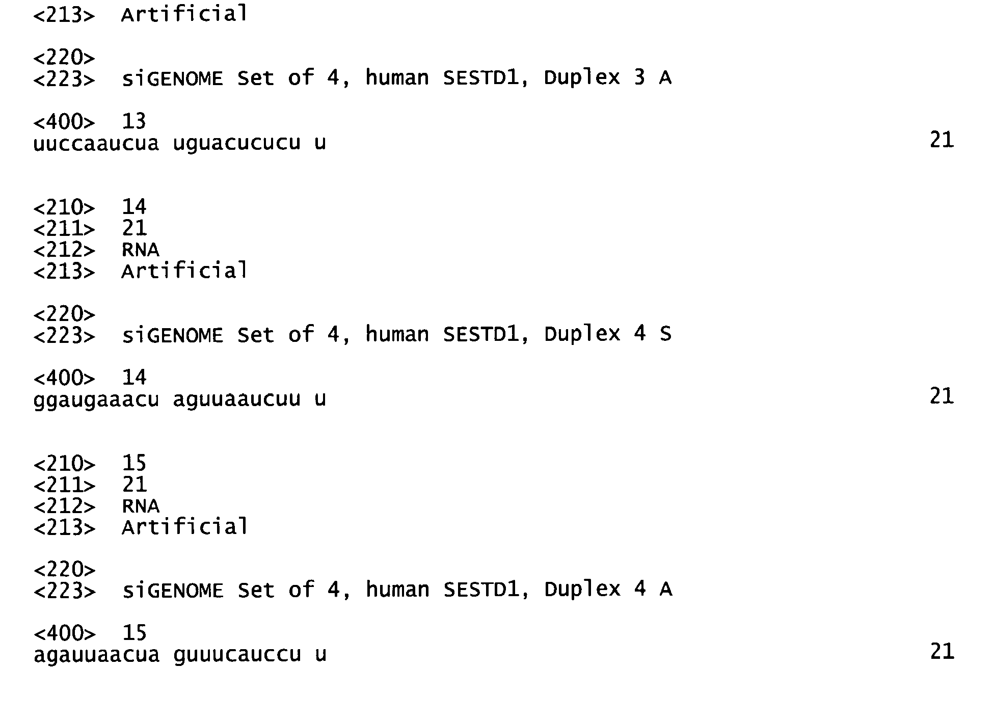
Claims (20)
- An isolated complex comprising- a first protein comprising or consisting of a transient receptor potential channel (TRPC) or a functionally active variant thereof and- a second protein comprising or consisting of the first spectrin (Spec 1) domain of SEC14 and spectrin domains 1 (SESTD1).
- An isolated complex comprising- a first protein comprising or consisting of a domain of a transient receptor potential channel (TRPC) or a functionally active variant thereof, the domain having the sequence LXXXXXYQEVXRNLVKRYVAAMIRXXKT (SEQ ID NO:1), wherein each X represents any amino acid residue and- a second protein comprising or consisting of the Spec 1 domain of SESTD1.
- An isolated complex comprising- a first protein comprising or consisting of a transient receptor potential channel (TRPC) or a functionally active variant thereof and- a second protein comprising or consisting of at least one protein selected from the group consisting of ankyrin repeat domain 35 (ANKRD35), apolipoprotein A-I binding protein (APOA1BP), bromodomain adjacent to zinc finger domain (BAZ1B), high-mobility group protein 2-like1 isoform b (HMG2L1), makorin RING finger protein 1 (MKRN1), pre-B-cell leukemia homeobox interacting protein 1 (PBXIP1), sarcoma antigen NY-SAR-48, spectrin alpha non-erythrocytic 1 (SPTAN1), structural maintenance of chromosomes 3 (SMC3) and talin 2 (TLN2).
- The complex of any of claims 1 or 3, wherein the TRPC is selected from the group consisting of TRPC4, TRPC5 and TRPC6, particularly TRPC4 or TRPC5, especially TRPC4α or TRPC4β or TRPC5.
- The complex of any of claims 1 to 4, wherein the first protein comprises the sequence LRRHHQYQEVMRNLVKRYVAAMIREAKTE (SEQ ID NO:2) or LIQNQHYQEVIRNLVKRYVAAMIRNSKTN (SEQ ID NO:3).
- The complex of any of claims 1 to 5, wherein the TRPC is mammalian TRPC, particularly murine or human TRPC.
- The complex of any of claims 1, 2, 4, 5 or 6, wherein the second protein comprises or consists of SESTD1.
- The complex of any of claims 1, 2, 4, 5, 6 or 7, wherein the SESTD1 is mammalian SESTD1, particularly rat, murine or human SESTD1.
- A test system comprising- a first protein as defined in any of claims 1 to 6- a second protein as defined in any of claims 1, 2, 3, 7, and 8, and- means for detecting interaction of the first and the second protein.
- A method for screening for a TRPC modulator comprising the steps of:a) contacting the test system of claim 9 with a substance andb) detecting a measurable signal upon interaction of the substance with the test system, thereby identifying the substance as a TRPC modulator.
- The method of claim 10, wherein the influence of the substance on the interaction between the first and the second protein of the test system is detected directly.
- The method of claim 10 or 11, wherein the first and/or the second protein comprise(s) a detectable marker.
- The method of any of claims 10 to 12, wherein the detecting of a measurable signal involves the use of a specific antibody against the first protein and/or a specific antibody against the second protein.
- The method of any of claims 10 to 13, wherein the TRPC modulator enhances or preferably impairs or inhibits binding of the first protein to the second protein.
- A test system comprising- a protein as defined as a second protein in any of claims 1, 2, 7,and 8, and- a phospholipid, and- means for detecting the interaction of the protein and the phospholipid.
- The method of claim 15 wherein the phospholipide is selected from the group consisting of lysophosphatidic acid, lysophosphocholine, phosphatidyl-inositol-(3)phosphate, phosphatidyl-inositol-(4)phosphate, phosphatidyl-inositol-(5)phosphate, Phosphatidylethanolamine, Phosphatidylcholine,, Sphingosine-1-Phosphate, Phosphatidylinositol (3,4) bisphosphate, Phosphatidylinositol (3,5) phosphat,, Phosphatidylinositol (4,5) bisphosphate, Phosphatidylinositol (3,4,5) trisphosphate, phosphatidic acid, Phosphatidylserine, or a monophosphate.
- A method for screening of a modulator of the interaction of a protein and a phospholipid comprising the steps of:c) contacting the test system of claim 15 or 16 with a substance andd) detecting a measurable signal upon interaction of the substance with the test system, thereby identifying the substance as a modulator of the interaction between the protein and the phospholipid.
- The method of any of claims 10 to 17, wherein the method is used for screening for a medicament for preventing and/or treating a disease involving endothelial dysfunction.
- The method of any of claims 10 to 17, wherein the method is used for screening for a medicament for preventing and/or treating a cardiovascular disease, cardiac hypertrophy, heart failure, ischemia, neointima hyperplasia, idiopathic dilated cardiomyopathy, hypertension, coronary syndrome, heart failure, renal failure, thrombosis, a chronic respiratory disease, asthma, chronic obstructive pulmonary disorder, idiopathic pulmonary hypertension, acute hypoxic pulmonary vasoconstriction, inflammatory occlusive disease and atherosclerosis.
- Use of a test system according to claim 9 for the identification of a TRPC modulator.
Priority Applications (13)
| Application Number | Priority Date | Filing Date | Title |
|---|---|---|---|
| EP07291300A EP2053059A1 (en) | 2007-10-26 | 2007-10-26 | Complexes of TRPC domains and SESTD1 domains and methods and uses involving the same |
| EP08843272A EP2205628A1 (en) | 2007-10-26 | 2008-10-14 | Complexes of trpc domains and sestd1 domains and methods and uses involving the same |
| CN200880113322A CN101848933A (en) | 2007-10-26 | 2008-10-14 | Complexes of TRPC domains and SESTD1 domains and methods and uses involving the same |
| MX2010004097A MX2010004097A (en) | 2007-10-26 | 2008-10-14 | Complexes of trpc domains and sestd1 domains and methods and uses involving the same. |
| PCT/EP2008/008670 WO2009052971A1 (en) | 2007-10-26 | 2008-10-14 | Complexes of trpc domains and sestd1 domains and methods and uses involving the same |
| CA2702508A CA2702508A1 (en) | 2007-10-26 | 2008-10-14 | Complexes of trpc domains and sestd1 domains and methods and uses involving the same |
| KR1020107009065A KR20100076992A (en) | 2007-10-26 | 2008-10-14 | Complexes of trpc domains and sestd1 domains and methods and uses involving the same |
| AU2008315951A AU2008315951A1 (en) | 2007-10-26 | 2008-10-14 | Complexes of TRPC domains and SESTD1 domains and methods and uses involving the same |
| JP2010530308A JP2011501757A (en) | 2007-10-26 | 2008-10-14 | TRPC domain and SESTD1 domain complex and methods and uses requiring the same |
| US12/738,337 US20100330697A1 (en) | 2007-10-26 | 2008-10-14 | Complexes of trpc domains and sestd1 domains and methods and uses involving the same |
| TW097140558A TW200934789A (en) | 2007-10-26 | 2008-10-23 | Complexes of TRPC domains and SESTD1 domains and methods and uses involving the same |
| ARP080104623A AR069007A1 (en) | 2007-10-26 | 2008-10-23 | COMPLEXES OF THE TRPC AND SESTD1 DOMAINS AND METHODS AND USES THAT WRAP UP THE SAME |
| IL205218A IL205218A0 (en) | 2007-10-26 | 2010-04-21 | Complexes of trpc domains and sestd1 domains and methods and uses involving the same |
Applications Claiming Priority (1)
| Application Number | Priority Date | Filing Date | Title |
|---|---|---|---|
| EP07291300A EP2053059A1 (en) | 2007-10-26 | 2007-10-26 | Complexes of TRPC domains and SESTD1 domains and methods and uses involving the same |
Publications (1)
| Publication Number | Publication Date |
|---|---|
| EP2053059A1 true EP2053059A1 (en) | 2009-04-29 |
Family
ID=39232772
Family Applications (2)
| Application Number | Title | Priority Date | Filing Date |
|---|---|---|---|
| EP07291300A Ceased EP2053059A1 (en) | 2007-10-26 | 2007-10-26 | Complexes of TRPC domains and SESTD1 domains and methods and uses involving the same |
| EP08843272A Withdrawn EP2205628A1 (en) | 2007-10-26 | 2008-10-14 | Complexes of trpc domains and sestd1 domains and methods and uses involving the same |
Family Applications After (1)
| Application Number | Title | Priority Date | Filing Date |
|---|---|---|---|
| EP08843272A Withdrawn EP2205628A1 (en) | 2007-10-26 | 2008-10-14 | Complexes of trpc domains and sestd1 domains and methods and uses involving the same |
Country Status (12)
| Country | Link |
|---|---|
| US (1) | US20100330697A1 (en) |
| EP (2) | EP2053059A1 (en) |
| JP (1) | JP2011501757A (en) |
| KR (1) | KR20100076992A (en) |
| CN (1) | CN101848933A (en) |
| AR (1) | AR069007A1 (en) |
| AU (1) | AU2008315951A1 (en) |
| CA (1) | CA2702508A1 (en) |
| IL (1) | IL205218A0 (en) |
| MX (1) | MX2010004097A (en) |
| TW (1) | TW200934789A (en) |
| WO (1) | WO2009052971A1 (en) |
Cited By (1)
| Publication number | Priority date | Publication date | Assignee | Title |
|---|---|---|---|---|
| CN116930507A (en) * | 2023-07-24 | 2023-10-24 | 武汉大学中南医院 | Application of MKRN1 as cardiac hypertrophy biomarker and therapeutic target |
Families Citing this family (3)
| Publication number | Priority date | Publication date | Assignee | Title |
|---|---|---|---|---|
| CN103954770B (en) * | 2014-04-25 | 2015-09-30 | 江南大学 | EVs-TRPC5 is detecting the application in breast carcinoma resistance degree |
| CN110777186A (en) * | 2019-10-29 | 2020-02-11 | 浙江树人学院(浙江树人大学) | Experimental method for influence of TRPC1 on function of endothelial cells cultured with high sugar |
| CN116891523B (en) * | 2023-05-24 | 2024-05-17 | 深圳晶蛋生物医药科技有限公司 | TRPM3 truncate, cell line containing the same and use thereof |
Citations (3)
| Publication number | Priority date | Publication date | Assignee | Title |
|---|---|---|---|---|
| WO1998015830A2 (en) | 1996-10-04 | 1998-04-16 | Wallac Oy | Homogenous luminescence energy transfer assays |
| WO2001073014A1 (en) * | 2000-03-24 | 2001-10-04 | Merck Patent Gmbh | Human survivin interacting protein 1 (sip-1) |
| WO2005061548A1 (en) * | 2003-12-23 | 2005-07-07 | Nono Inc. | Polypeptides for modulating binding of trp channel proteins and trp-associated proteins. |
-
2007
- 2007-10-26 EP EP07291300A patent/EP2053059A1/en not_active Ceased
-
2008
- 2008-10-14 US US12/738,337 patent/US20100330697A1/en not_active Abandoned
- 2008-10-14 AU AU2008315951A patent/AU2008315951A1/en not_active Withdrawn
- 2008-10-14 CN CN200880113322A patent/CN101848933A/en active Pending
- 2008-10-14 JP JP2010530308A patent/JP2011501757A/en not_active Abandoned
- 2008-10-14 WO PCT/EP2008/008670 patent/WO2009052971A1/en not_active Ceased
- 2008-10-14 KR KR1020107009065A patent/KR20100076992A/en not_active Withdrawn
- 2008-10-14 MX MX2010004097A patent/MX2010004097A/en unknown
- 2008-10-14 EP EP08843272A patent/EP2205628A1/en not_active Withdrawn
- 2008-10-14 CA CA2702508A patent/CA2702508A1/en not_active Withdrawn
- 2008-10-23 AR ARP080104623A patent/AR069007A1/en unknown
- 2008-10-23 TW TW097140558A patent/TW200934789A/en unknown
-
2010
- 2010-04-21 IL IL205218A patent/IL205218A0/en unknown
Patent Citations (3)
| Publication number | Priority date | Publication date | Assignee | Title |
|---|---|---|---|---|
| WO1998015830A2 (en) | 1996-10-04 | 1998-04-16 | Wallac Oy | Homogenous luminescence energy transfer assays |
| WO2001073014A1 (en) * | 2000-03-24 | 2001-10-04 | Merck Patent Gmbh | Human survivin interacting protein 1 (sip-1) |
| WO2005061548A1 (en) * | 2003-12-23 | 2005-07-07 | Nono Inc. | Polypeptides for modulating binding of trp channel proteins and trp-associated proteins. |
Non-Patent Citations (16)
| Title |
|---|
| CIOFFI DONNA L ET AL: "Activation of the endothelial store-operated ISOC Ca2+ channel requires interaction of protein 4.1 with TRPC4.", CIRCULATION RESEARCH 25 NOV 2005, vol. 97, no. 11, 25 November 2005 (2005-11-25), pages 1164 - 1172, XP002475459, ISSN: 1524-4571 * |
| FIELDS S; SONG O: "A novel genetic system to detect protein-protein interactions", NATURE, vol. 340, 1989, pages 245 - 246 |
| HARBURY PB ET AL.: "A switch between two-, three-, and four-stranded coiled coils in GCN4 leucine zipper mutants", SCIENCE, vol. 262, 1993, pages 1401 - 1407 |
| HOFMANN T ET AL: "SUBUNIT COMPOSITION OF MAMMALIAN TRANSIENT RECEPTOR POTENTIAL CHANNELS IN LIVING CELLS", PROCEEDINGS OF THE NATIONAL ACADEMY OF SCIENCES OF USA, NATIONAL ACADEMY OF SCIENCE, WASHINGTON, DC, US, vol. 99, no. 11, 28 May 2002 (2002-05-28), pages 7461 - 7466, XP009051268, ISSN: 0027-8424 * |
| ITO H ET AL.: "Transformation of intact yeast cells treated with alkali cations", J BACTERIOL, vol. 153, 1983, pages 163 - 168 |
| KOBAYASHI ET AL: "Identification and characterization of GSRP-56, a novel Golgi-localized spectrin repeat-containing protein", EXPERIMENTAL CELL RESEARCH, SAN DIEGO, CA, US, vol. 312, no. 16, 1 October 2006 (2006-10-01), pages 3152 - 3164, XP005624212, ISSN: 0014-4827 * |
| KWON Y; HOFMANN T; MONTELL C: "Integration of phosphoinositide- and calmodulin-mediated regulation of TRPC6", MOL CELL, vol. 25, 2007, pages 491 - 503 |
| MARCHLER-BAUER A; BRYANT SH: "CD-Search: protein domain annotations on the fly", NUCLEIC ACIDS RES, vol. 32, 2004, pages W327 - W331 |
| MONU GOEL ET AL: "Proteomic analysis of TRPC5 and TRPC6-binding partners reveals interaction with the plasmalemmal Na+/K+-ATPase", PFLÜGERS ARCHIV - EUROPEAN JOURNAL OF PHYSIOLOGY ;, SPRINGER-VERLAG, BE, vol. 451, no. 1, 1 October 2005 (2005-10-01), pages 87 - 98, XP019344077, ISSN: 1432-2013 * |
| PERALTA EG ET AL., NATURE, vol. 334, 1988, pages 434 - 437 |
| RAUCHER D ET AL.: "Phosphatidylinositol 4,5-bisphosphate functions as a second messenger that regulates cytoskeleton-plasma membrane adhesion", CELL, vol. 100, 2000, pages 221 - 228 |
| STRUBING C ET AL.: "Formation of novel TRPC channels by complex subunit interactions in embryonic brain", J BIOL CHEM, vol. 278, 2003, pages 39014 - 39019 |
| STRUBING C ET AL.: "TRPC1 and TRPC5 form a novel cation channel in mammalian brain", NEURON, vol. 29, 2001, pages 645 - 655 |
| TAQMAN ET AL.: "Oligonucleotides with fluorescent dyes at opposite ends provide a quenched probe system useful for detecting PCR product and nucleic acid hybridization", PCR METHODS APPL, vol. 4, 1995, pages 357 - 362 |
| TSIOKAS L ET AL: "Specific association of the gene product of PKD2 with the TRPC1 channel.", PROCEEDINGS OF THE NATIONAL ACADEMY OF SCIENCES OF THE UNITED STATES OF AMERICA 30 MAR 1999, vol. 96, no. 7, 30 March 1999 (1999-03-30), pages 3934 - 3939, XP002475460, ISSN: 0027-8424 * |
| ULLMAN ET AL., PROC. NATL. ACAD. SCI., vol. 91, 1994, pages 5426 - 5430 |
Cited By (1)
| Publication number | Priority date | Publication date | Assignee | Title |
|---|---|---|---|---|
| CN116930507A (en) * | 2023-07-24 | 2023-10-24 | 武汉大学中南医院 | Application of MKRN1 as cardiac hypertrophy biomarker and therapeutic target |
Also Published As
| Publication number | Publication date |
|---|---|
| TW200934789A (en) | 2009-08-16 |
| IL205218A0 (en) | 2010-11-30 |
| KR20100076992A (en) | 2010-07-06 |
| WO2009052971A1 (en) | 2009-04-30 |
| EP2205628A1 (en) | 2010-07-14 |
| AR069007A1 (en) | 2009-12-23 |
| JP2011501757A (en) | 2011-01-13 |
| AU2008315951A1 (en) | 2009-04-30 |
| MX2010004097A (en) | 2010-04-30 |
| CA2702508A1 (en) | 2009-04-30 |
| CN101848933A (en) | 2010-09-29 |
| US20100330697A1 (en) | 2010-12-30 |
Similar Documents
| Publication | Publication Date | Title |
|---|---|---|
| Ercan et al. | Molecular basis of accessible plasma membrane cholesterol recognition by the GRAM domain of GRAMD1b | |
| Tan et al. | A phosphoinositide signalling pathway mediates rapid lysosomal repair | |
| Kowalczyk et al. | Direct binding of GABAA receptor β2 and β3 subunits to gephyrin | |
| Buxton et al. | Identification and characterization of Snapin as a ubiquitously expressed SNARE-binding protein that interacts with SNAP23 in non-neuronal cells | |
| Subra et al. | VAP-A intrinsically disordered regions enable versatile tethering at membrane contact sites | |
| US9057726B2 (en) | Neuropeptide Q as modulator of GPCR GALR2 and uses thereof | |
| Ohno et al. | The neuronal glycine transporter 2 interacts with the PDZ domain protein syntenin-1 | |
| Gong et al. | PTRN‐1/CAMSAP promotes CYK‐1/formin‐dependent actin polymerization during endocytic recycling | |
| Goebeler et al. | Annexin A8 displays unique phospholipid and F-actin binding properties | |
| Li et al. | Direct binding of α‐actinin enhances TRPP3 channel activity | |
| JP2017525936A (en) | Compositions and methods for modulating mTORC1 | |
| US20100330697A1 (en) | Complexes of trpc domains and sestd1 domains and methods and uses involving the same | |
| Lee et al. | A WD40 repeat protein, Arabidopsis Sec13 homolog 1, may play a role in vacuolar trafficking by controlling the membrane association of AtDRP2A | |
| Smith et al. | Location of auxilin within a clathrin cage | |
| Imai et al. | Exocyst subunits are involved in isoproterenol‐induced amylase release from rat parotid acinar cells | |
| Känel et al. | The tobacco phosphatidylethanolamine-binding protein NtFT4 increases the lifespan of Drosophila melanogaster by interacting with the proteostasis network | |
| HK1148755A (en) | Complexes of trpc domains and sestd1 domains and methods and uses involving the same | |
| Chen et al. | Interactions between CAP70 and actinfilin are important for integrity of actin cytoskeleton structures in neurons | |
| US20060252677A1 (en) | Postsynaptic proteins | |
| McPherson et al. | The molecular machinery for synaptic vesicle endocytosis | |
| WO2024030898A2 (en) | Compositions and methods for evaluating microtubule dynamics | |
| Hamati | MuRF3 binds to the retromer subunit SNX5 inhibiting its MuRF2-mediated degradation and leading to its stabilization | |
| Kawakami | Structure/Function Analysis of Type IIa Receptor Tyrosine Phosphatases on Drosophila Muscle Integrity | |
| Noakes | Identification and functional characterisation of the novel OCRL1 binding partners IPIP27A and B | |
| Lee | Protein acetylation and deacetylation as a novel regulatory mechanism for endothelin receptor A-mediated downstream effects |
Legal Events
| Date | Code | Title | Description |
|---|---|---|---|
| PUAI | Public reference made under article 153(3) epc to a published international application that has entered the european phase |
Free format text: ORIGINAL CODE: 0009012 |
|
| AK | Designated contracting states |
Kind code of ref document: A1 Designated state(s): AT BE BG CH CY CZ DE DK EE ES FI FR GB GR HU IE IS IT LI LT LU LV MC MT NL PL PT RO SE SI SK TR |
|
| AX | Request for extension of the european patent |
Extension state: AL BA HR MK RS |
|
| STAA | Information on the status of an ep patent application or granted ep patent |
Free format text: STATUS: THE APPLICATION HAS BEEN REFUSED |
|
| 18R | Application refused |
Effective date: 20090522 |