EP2050469B1 - Diagnostic agent - Google Patents
Diagnostic agent Download PDFInfo
- Publication number
- EP2050469B1 EP2050469B1 EP07791696.3A EP07791696A EP2050469B1 EP 2050469 B1 EP2050469 B1 EP 2050469B1 EP 07791696 A EP07791696 A EP 07791696A EP 2050469 B1 EP2050469 B1 EP 2050469B1
- Authority
- EP
- European Patent Office
- Prior art keywords
- acid
- diagnostic agent
- colorant
- lesion
- wall
- Prior art date
- Legal status (The legal status is an assumption and is not a legal conclusion. Google has not performed a legal analysis and makes no representation as to the accuracy of the status listed.)
- Ceased
Links
Images


Classifications
-
- A—HUMAN NECESSITIES
- A61—MEDICAL OR VETERINARY SCIENCE; HYGIENE
- A61K—PREPARATIONS FOR MEDICAL, DENTAL OR TOILETRY PURPOSES
- A61K49/00—Preparations for testing in vivo
-
- A—HUMAN NECESSITIES
- A61—MEDICAL OR VETERINARY SCIENCE; HYGIENE
- A61K—PREPARATIONS FOR MEDICAL, DENTAL OR TOILETRY PURPOSES
- A61K49/00—Preparations for testing in vivo
- A61K49/001—Preparation for luminescence or biological staining
- A61K49/006—Biological staining of tissues in vivo, e.g. methylene blue or toluidine blue O administered in the buccal area to detect epithelial cancer cells, dyes used for delineating tissues during surgery
-
- A—HUMAN NECESSITIES
- A61—MEDICAL OR VETERINARY SCIENCE; HYGIENE
- A61B—DIAGNOSIS; SURGERY; IDENTIFICATION
- A61B5/00—Measuring for diagnostic purposes; Identification of persons
- A61B5/42—Detecting, measuring or recording for evaluating the gastrointestinal, the endocrine or the exocrine systems
-
- A—HUMAN NECESSITIES
- A61—MEDICAL OR VETERINARY SCIENCE; HYGIENE
- A61P—SPECIFIC THERAPEUTIC ACTIVITY OF CHEMICAL COMPOUNDS OR MEDICINAL PREPARATIONS
- A61P1/00—Drugs for disorders of the alimentary tract or the digestive system
-
- A—HUMAN NECESSITIES
- A61—MEDICAL OR VETERINARY SCIENCE; HYGIENE
- A61B—DIAGNOSIS; SURGERY; IDENTIFICATION
- A61B1/00—Instruments for performing medical examinations of the interior of cavities or tubes of the body by visual or photographical inspection, e.g. endoscopes; Illuminating arrangements therefor
-
- A—HUMAN NECESSITIES
- A61—MEDICAL OR VETERINARY SCIENCE; HYGIENE
- A61B—DIAGNOSIS; SURGERY; IDENTIFICATION
- A61B1/00—Instruments for performing medical examinations of the interior of cavities or tubes of the body by visual or photographical inspection, e.g. endoscopes; Illuminating arrangements therefor
- A61B1/273—Instruments for performing medical examinations of the interior of cavities or tubes of the body by visual or photographical inspection, e.g. endoscopes; Illuminating arrangements therefor for the upper alimentary canal, e.g. oesophagoscopes, gastroscopes
-
- A—HUMAN NECESSITIES
- A61—MEDICAL OR VETERINARY SCIENCE; HYGIENE
- A61B—DIAGNOSIS; SURGERY; IDENTIFICATION
- A61B5/00—Measuring for diagnostic purposes; Identification of persons
- A61B5/0059—Measuring for diagnostic purposes; Identification of persons using light, e.g. diagnosis by transillumination, diascopy, fluorescence
Definitions
- the present invention relates to a diagnostic agent for use in an in vivo diagnostic method, characterized in that said agent is to be used in endoscopic observation of the inner wall of the gastrointestinal tract.
- JP 10-194998 A discloses a thickener and a living body marking preparation for operations which contains an intermediate color or cool color colorant applicable to the living body. This document reports that injecting the living body marking preparation to a site to be resected with a syringe allow the resection area to become clear during a surgical operation.
- Patent document 1 JP 10-194998 A
- Guelrud and Herrera Guelrud, M. and Herrera, I.; Gastrointestinal Endoscopy; 47(6); 1998; pp. 512-515
- Guelrud et al. Guelrud, M. et al.; Gastrointestinal Endoscopy; 53(6); 2001; pp. 559-565
- Togashi et al. Togashi, K.
- the present invention was made in order to solve the above-described problems.
- the invention intends to provide a diagnostic agent which makes it possible, by being sprinkled to the inner wall of a gastrointestine in endoscopic observation, to observe clearly a lesion difficult to be determined.
- a diagnostic agent for use in an in vivo diagnostic method characterized in that said agent is for being sprinkled to the inner wall of the gastrointestinal tract in endoscopic observation, comprising an acidic aqueous solution of pH 1 to 5 containing a colorant.
- the colorant is an intermediate color or cool color colorant.
- the acidic aqueous solution contain at least one acid selected from the group consisting of a carboxylic acid, hydrochloric acid, sulfuric acid and phosphoric acid. It is preferable that the acid is at least one carboxylic acid selected from the group consisting of acetic acid, malic acid and citric acid. Moreover, it is also preferable that the concentration of the carboxylic acid is 0.01 to 1 mol/L.
- the diagnostic agent of the present invention can make it possible, by being sprinkled to the inner wall of a gastrointestine at the time of endoscopic observation, to observe clearly a lesion difficult to be determined. It, therefore, can be preferably used to detection of early cancer or the like.
- the diagnostic agent of the present invention is a diagnostic agent for use in an in vivo diagnostic method, characterized in that said agent is for being sprinkled to the inner wall of the gastrointestinal tract in endoscopic observation, comprising an acidic aqueous solution of pH 1 to 5 containing a colorant.
- the diagnostic agent of the present invention is sprinkled to the inner wall of the gastrointestinal tract when the gastrointestinal tract is observed with an endoscope.
- the diagnostic agent can make it easier to observe lesions on the basis of the degree of the adhesion of the agent to the surface of the inner wall of the gastrointestinal tract.
- the method of sprinkling is not particularly restricted as long as the diagnostic agent of the present invention can be supplied to the inner wall of the gastrointestinal tract, and it may be applying, spraying, or the like.
- the term "gastrointestinal tract” includes esophagus, stomach, small intestine and large intestine or the like.
- the diagnostic agent of the present invention can be used suitably for stomach and Barrett's esophagus, the inner surface of which is composed of columnar epithelium.
- a columnar epithelium is a tissue which is particularly suitable to be applied the diagnostic agent of the present invention because it has a characteristic that its surface, which ordinarily is transparent, becomes whitish on change in pH, particularly when the acidity has become strong.
- the diagnostic agent of the present invention contains a colorant. Because of this fact, the amount of the diagnostic agent having adhered to the surface to be observed is emphasized by the difference in colorant concentration, so that the margin line between a lesion and a normal mucosa can be observed clearly. Moreover, it becomes easy to observe irregularities on the surface.
- the colorant preferably is of an intermediate color or cool color, and more preferably is of a cool color.
- the intermediate color as referred to herein include colors other than warm colors and cool colors and examples thereof include green, purple and brown. Examples of the cool colors include blue.
- the form of the colorant to be used in the present invention is not particularly restricted and it may be either a dye or a pigment, it is preferable, from the viewpoint of the easiness with which a washing operation is performed after the sprinkling, that the colorant is in the form of a dye.
- the colorant to be used in the present invention is not particularly restricted, and examples thereof include Fast Green (Food Green No.3), copper chlorophyll, sodium copper chlorophyllin, sulfobromophthalein sodium, Indocyanine Green, Methylene Blue, Brilliant Blue (Food Blue No.1), indigo carmine (Food Blue No.2), gardenia blue, toluidine blue, pyoktanin blue, and caramel colors.
- the colorant preferably is indigo carmine (Food Blue No.2), gardenia blue, or a caramel color, and more preferably is indigo carmine (Food Blue No.2) or gardenia blue, each of which is a cool color colorant.
- the concentration of the colorant to be used in the present invention may vary depending upon the type or the application site of the colorant, it is preferable, from the viewpoint that the margin line between a lesion and a normal mucosa can be observed clearly, that the colorant concentration is within the range of 0.1 to 200 mmol/L.
- concentration of the colorant is less than 0.1 mmol/L, it may become difficult to observe the difference from the color of the inner wall of a gastrointestine. It, therefore, is more preferable that the colorant concentration is 1 mmol/L or more.
- the concentration of colorant exceeds 200 mmol/L, the color contrast decreases and, as a result, it may become difficult to observe the color contrast. It, therefore, is more preferable that the colorant concentration is 100 mmol/L or less.
- the diagnostic agent of the present invention is composed of an acidic aqueous solution of pH 1 to 5 containing a colorant. Because of the fact that pH is within such a range, in many cases when the diagnostic agent of the present invention is sprinkled, secretion of mucus is promoted at a lesion, especially a lesion having cancer cells while almost no mucus is secreted at a normal mucosa. As a result, the secreted mucus makes a solution containing a colorant less prone to adhere to lesions, so that it becomes possible to clearly observe the margin line between a lesion and a normal mucosa on the basis of the difference in contrast of the colorant.
- the margin line between a lesion and a normal mucosa is prone to become clear.
- the inner wall of the gastrointestinal tract may be damaged when the pH is less than 1, the pH is preferably 2 or more, and is more preferably 3 or more.
- the pH is preferably 4 or less because when the pH is greater than 5, the mucus secretion at a lesion may be promoted insufficiently and, as a result, the margin line between the lesion and a normal mucosa may become unclear.
- the solution containing a colorant is acidic. It has been known that the surface of a lesion and the surface of a normal mucosa become whitish to different degrees when an aqueous acetic acid solution is sprinkled to the inner wall of the gastrointestinal tract. Observations, however, cannot be performed easily on the basis of only this fact. The present inventor has confirmed that a colorant is less prone to adhere either to lesions or to normal mucosa when an aqueous indigo carmine solution has been sprinkled following the sprinkle of an aqueous acetic acid solution. Therefore, it is important that the solution containing a colorant is acidic.
- the kind of the acidic aqueous solution is not particularly restricted, it is preferable that the solution contain an acid selected from among carboxylic acid, hydrochloric acid, sulfuric acid, phosphoric acid, and so on.
- an acid selected from among carboxylic acid, hydrochloric acid, sulfuric acid, phosphoric acid, and so on.
- a carboxylic acid is used preferably.
- carboxylic acid to be used is not particularly restricted, moonocarboxylic acids, such as acetic acid, propionic acid and butyric acid; oxymonocarboxylic acids, such as lactic acid, glycolic acid and gluconic acid; polycarboxylic acids, such as oxalic acid, succinic acid, glutaric acid, adipic acid, and fumaric acid; hydroxypolycarboxylic acids, such as malic acid and citric acid, and so on can be used.
- moonocarboxylic acids such as acetic acid, propionic acid and butyric acid
- oxymonocarboxylic acids such as lactic acid, glycolic acid and gluconic acid
- polycarboxylic acids such as oxalic acid, succinic acid, glutaric acid, adipic acid, and fumaric acid
- hydroxypolycarboxylic acids such as malic acid and citric acid, and so on
- the concentration of the carboxylic acid in use in the diagnostic agent of the present invention is not particularly limited, it is more preferably 0.01 to 1 mol/L.
- the concentration of the carboxylic acid preferably is 0.05 mol/L or more because when the concentration of the carboxylic acid is less than 0.01 mol/L, the mucus secretion at a lesion may be promoted insufficiently and, as a result, the margin line between the lesion and a normal mucosa may become unclear.
- the concentration of the carboxylic acid is more preferably 0.5 mol/L or less because when the concentration of the carboxylic acid is greater than 1 mol/L, the inner wall of a gastrointestine may be damaged.
- the use of the diagnostic agent of the present invention makes it possible to detect and observe early gastrointestinal cancers or the like, which have heretofore been difficult to be determined.
- the diagnostic agent of the present invention can be used suitably for early gastric cancer or early Barrett' s esophagus cancer particularly among the gastrointestinal cancers.
- a diagnostic agent of the present invention was obtained by mixing 25 ml of a 0.4 wt% aqueous indigo carmine solution (produced by Daiichi Pharmaceutical Co., Ltd.) and 15 ml of a 1.5 wt% aqueous acetic acid solution.
- the resulting diagnostic agent had an indigo carmine concentration of 5.3 mmol/L, an acetic acid concentration of 0.1 mol/L, and a pH of 3.0.
Landscapes
- Health & Medical Sciences (AREA)
- Life Sciences & Earth Sciences (AREA)
- Engineering & Computer Science (AREA)
- General Health & Medical Sciences (AREA)
- Biomedical Technology (AREA)
- Veterinary Medicine (AREA)
- Public Health (AREA)
- Animal Behavior & Ethology (AREA)
- Physics & Mathematics (AREA)
- Nuclear Medicine, Radiotherapy & Molecular Imaging (AREA)
- Epidemiology (AREA)
- Endocrinology (AREA)
- Medical Informatics (AREA)
- Oncology (AREA)
- Biodiversity & Conservation Biology (AREA)
- Gastroenterology & Hepatology (AREA)
- Physiology (AREA)
- Biophysics (AREA)
- Pathology (AREA)
- Heart & Thoracic Surgery (AREA)
- Optics & Photonics (AREA)
- Molecular Biology (AREA)
- Surgery (AREA)
- Bioinformatics & Cheminformatics (AREA)
- Chemical & Material Sciences (AREA)
- Chemical Kinetics & Catalysis (AREA)
- General Chemical & Material Sciences (AREA)
- Medicinal Chemistry (AREA)
- Organic Chemistry (AREA)
- Pharmacology & Pharmacy (AREA)
- Medicines Containing Antibodies Or Antigens For Use As Internal Diagnostic Agents (AREA)
Description
- The present invention relates to a diagnostic agent for use in an in vivo diagnostic method, characterized in that said agent is to be used in endoscopic observation of the inner wall of the gastrointestinal tract.
- In observation of the inner wall of a gastrointestine using an endoscope, a procedure in which the observation is made easier by sprinkling a colorant has been adopted. The colorant which is widely used today is aqueous indigo carmine solution.
- In operations has been used a procedure in which a resection area is clarified by marking a site to be resected with a colorant. For example,
JP 10-194998 A - It, however, is not always possible to make observation easily when it is difficult to identify a lesion in the case of early cancer or the like. Moreover, it was not easy in operations to determine the margin line between a lesion and a normal mucosa or to properly resect only a lesion. Therefore, a technique by which a lesion can be observed clearly in an endoscopic observation has been awaited.
- Patent document 1:
JP 10-194998 A - In this context, Guelrud and Herrera (Guelrud, M. and Herrera, I.; Gastrointestinal Endoscopy; 47(6); 1998; pp. 512-515) describes the effects of acetic acid on the identification of remnant islands of Barrett's epithelium after endoscopic treatment. Further, Guelrud et al. (Guelrud, M. et al.; Gastrointestinal Endoscopy; 53(6); 2001; pp. 559-565) describes a method for the identification of specialized intestinal metaplasia in Barrett's esophagus, comprising the application of acetic acid to the intestinal tissue. Furthermore, Togashi et al. (Togashi, K. et al.; Endoscopy; 38(6); 2006; pp. 613-616) describes the use of acetic acid for the endoscopic pit pattern analysis of colonic polyps. Finally, Yao and Kida (Yao, K. and Kida, M.; Digestive Endoscopy; 17(Suppl.); 2005; pp. S2-S4) reviews clinical applications of upper gastrointestinal zoom endoscopy.
- The present invention was made in order to solve the above-described problems. The invention intends to provide a diagnostic agent which makes it possible, by being sprinkled to the inner wall of a gastrointestine in endoscopic observation, to observe clearly a lesion difficult to be determined.
- The above-mentioned problems are solved by providing a diagnostic agent for use in an in vivo diagnostic method, characterized in that said agent is for being sprinkled to the inner wall of the gastrointestinal tract in endoscopic observation, comprising an acidic aqueous solution of pH 1 to 5 containing a colorant.
- In this embodiment, it is preferable that the colorant is an intermediate color or cool color colorant. It is preferable that the acidic aqueous solution contain at least one acid selected from the group consisting of a carboxylic acid, hydrochloric acid, sulfuric acid and phosphoric acid. It is preferable that the acid is at least one carboxylic acid selected from the group consisting of acetic acid, malic acid and citric acid. Moreover, it is also preferable that the concentration of the carboxylic acid is 0.01 to 1 mol/L.
- The diagnostic agent of the present invention can make it possible, by being sprinkled to the inner wall of a gastrointestine at the time of endoscopic observation, to observe clearly a lesion difficult to be determined. It, therefore, can be preferably used to detection of early cancer or the like.
-
- [
Fig. 1 ]
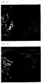
Fig. 1 is a photograph of the inner wall of the stomach observed with an endoscope in Test Example 1. - [
Fig. 2 ]
Fig. 2 is a photograph of the inner wall of the stomach observed after sprinkling only an aqueous indigo carmine solution in Test Example 1. - [
Fig. 3 ]
Fig. 3 is a photograph of the inner wall of the stomach observed after sprinkling a diagnostic agent of the present invention in Test Example 1. - [
Fig. 4 ]
Fig. 4 is a photograph of the inner wall of the stomach observed with an endoscope in Test Example 2. - [
Fig. 5 ]
Fig. 5 is a photograph of the inner wall of the stomach observed after sprinkling only an aqueous indigo carmine solution in Test Example 2. - [
Fig. 6 ]
Fig. 6 is a photograph of the inner wall of the stomach observed after sprinkling a diagnostic agent of the present invention in Test Example 2. - The diagnostic agent of the present invention is a diagnostic agent for use in an in vivo diagnostic method, characterized in that said agent is for being sprinkled to the inner wall of the gastrointestinal tract in endoscopic observation, comprising an acidic aqueous solution of pH 1 to 5 containing a colorant.
- The diagnostic agent of the present invention is sprinkled to the inner wall of the gastrointestinal tract when the gastrointestinal tract is observed with an endoscope. By being sprinkled to the inner wall of the gastrointestinal tract, the diagnostic agent can make it easier to observe lesions on the basis of the degree of the adhesion of the agent to the surface of the inner wall of the gastrointestinal tract. In the present invention, the method of sprinkling is not particularly restricted as long as the diagnostic agent of the present invention can be supplied to the inner wall of the gastrointestinal tract, and it may be applying, spraying, or the like.
- In the present invention, the term "gastrointestinal tract" includes esophagus, stomach, small intestine and large intestine or the like. Among these, the diagnostic agent of the present invention can be used suitably for stomach and Barrett's esophagus, the inner surface of which is composed of columnar epithelium. A columnar epithelium is a tissue which is particularly suitable to be applied the diagnostic agent of the present invention because it has a characteristic that its surface, which ordinarily is transparent, becomes whitish on change in pH, particularly when the acidity has become strong.
- The diagnostic agent of the present invention contains a colorant. Because of this fact, the amount of the diagnostic agent having adhered to the surface to be observed is emphasized by the difference in colorant concentration, so that the margin line between a lesion and a normal mucosa can be observed clearly. Moreover, it becomes easy to observe irregularities on the surface. From the viewpoint of being distinguishable from, for example, the color of the inner wall of the gastrointestinal tract and the color of the blood or the like, the colorant preferably is of an intermediate color or cool color, and more preferably is of a cool color. The intermediate color as referred to herein include colors other than warm colors and cool colors and examples thereof include green, purple and brown. Examples of the cool colors include blue. While the form of the colorant to be used in the present invention is not particularly restricted and it may be either a dye or a pigment, it is preferable, from the viewpoint of the easiness with which a washing operation is performed after the sprinkling, that the colorant is in the form of a dye.
- The colorant to be used in the present invention is not particularly restricted, and examples thereof include Fast Green (Food Green No.3), copper chlorophyll, sodium copper chlorophyllin, sulfobromophthalein sodium, Indocyanine Green, Methylene Blue, Brilliant Blue (Food Blue No.1), indigo carmine (Food Blue No.2), gardenia blue, toluidine blue, pyoktanin blue, and caramel colors. From the viewpoint of being safe to the human body without causing any adverse reaction, the colorant preferably is indigo carmine (Food Blue No.2), gardenia blue, or a caramel color, and more preferably is indigo carmine (Food Blue No.2) or gardenia blue, each of which is a cool color colorant.
- While the concentration of the colorant to be used in the present invention may vary depending upon the type or the application site of the colorant, it is preferable, from the viewpoint that the margin line between a lesion and a normal mucosa can be observed clearly, that the colorant concentration is within the range of 0.1 to 200 mmol/L. When the concentration of the colorant is less than 0.1 mmol/L, it may become difficult to observe the difference from the color of the inner wall of a gastrointestine. It, therefore, is more preferable that the colorant concentration is 1 mmol/L or more. On the other hand, when the concentration of colorant exceeds 200 mmol/L, the color contrast decreases and, as a result, it may become difficult to observe the color contrast. It, therefore, is more preferable that the colorant concentration is 100 mmol/L or less.
- The diagnostic agent of the present invention is composed of an acidic aqueous solution of pH 1 to 5 containing a colorant. Because of the fact that pH is within such a range, in many cases when the diagnostic agent of the present invention is sprinkled, secretion of mucus is promoted at a lesion, especially a lesion having cancer cells while almost no mucus is secreted at a normal mucosa. As a result, the secreted mucus makes a solution containing a colorant less prone to adhere to lesions, so that it becomes possible to clearly observe the margin line between a lesion and a normal mucosa on the basis of the difference in contrast of the colorant. Moreover, while the mucosa surface becomes whitish simultaneously because of the fact that the diagnostic agent of the present invention is an acidic aqueous solution, a lesion and a normal mucosa often become whitish to different degrees. Therefore, the margin line between a lesion and a normal mucosa is prone to become clear. Since the inner wall of the gastrointestinal tract may be damaged when the pH is less than 1, the pH is preferably 2 or more, and is more preferably 3 or more. On the other hand, the pH is preferably 4 or less because when the pH is greater than 5, the mucus secretion at a lesion may be promoted insufficiently and, as a result, the margin line between the lesion and a normal mucosa may become unclear.
- In the present invention, it is important that the solution containing a colorant is acidic. It has been known that the surface of a lesion and the surface of a normal mucosa become whitish to different degrees when an aqueous acetic acid solution is sprinkled to the inner wall of the gastrointestinal tract. Observations, however, cannot be performed easily on the basis of only this fact. The present inventor has confirmed that a colorant is less prone to adhere either to lesions or to normal mucosa when an aqueous indigo carmine solution has been sprinkled following the sprinkle of an aqueous acetic acid solution. Therefore, it is important that the solution containing a colorant is acidic.
- While the kind of the acidic aqueous solution is not particularly restricted, it is preferable that the solution contain an acid selected from among carboxylic acid, hydrochloric acid, sulfuric acid, phosphoric acid, and so on. In particular, a carboxylic acid is used preferably. While the carboxylic acid to be used is not particularly restricted, moonocarboxylic acids, such as acetic acid, propionic acid and butyric acid; oxymonocarboxylic acids, such as lactic acid, glycolic acid and gluconic acid; polycarboxylic acids, such as oxalic acid, succinic acid, glutaric acid, adipic acid, and fumaric acid; hydroxypolycarboxylic acids, such as malic acid and citric acid, and so on can be used. Among these, acetic acid, malic acid, or citric acid is more preferable from the viewpoint of safety to the human body.
- While the concentration of the carboxylic acid in use in the diagnostic agent of the present invention is not particularly limited, it is more preferably 0.01 to 1 mol/L. The concentration of the carboxylic acid preferably is 0.05 mol/L or more because when the concentration of the carboxylic acid is less than 0.01 mol/L, the mucus secretion at a lesion may be promoted insufficiently and, as a result, the margin line between the lesion and a normal mucosa may become unclear. On the other hand, the concentration of the carboxylic acid is more preferably 0.5 mol/L or less because when the concentration of the carboxylic acid is greater than 1 mol/L, the inner wall of a gastrointestine may be damaged.
- The use of the diagnostic agent of the present invention makes it possible to detect and observe early gastrointestinal cancers or the like, which have heretofore been difficult to be determined. The diagnostic agent of the present invention can be used suitably for early gastric cancer or early Barrett' s esophagus cancer particularly among the gastrointestinal cancers.
- The present invention is illustrated below more concretely with reference to examples.
- A diagnostic agent of the present invention was obtained by mixing 25 ml of a 0.4 wt% aqueous indigo carmine solution (produced by Daiichi Pharmaceutical Co., Ltd.) and 15 ml of a 1.5 wt% aqueous acetic acid solution. The resulting diagnostic agent had an indigo carmine concentration of 5.3 mmol/L, an acetic acid concentration of 0.1 mol/L, and a pH of 3.0.
- An upper gastrointestinal tract videoendoscope was inserted into the stomach of a 68-year-old male patient and an endoscopy was conducted. As a result, however, the presence of lesions could not be confirmed clearly as shown in
Fig. 1 . Only a 0.4 wt% aqueous indigo carmine solution (produced by Daiichi Pharmaceutical Co., Ltd.) was sprinkled to the inner wall of the stomach. As a result, a suspicious lesion was found as shown inFig. 2 , but no lesion could be determined clearly. The inner wall of the stomach was washed with distilled water for injection and then the diagnostic agent of the present invention prepared by the method described above. As a result, a red depressed lesion was observed clearly, as shown inFig. 3 , in a blue-stained normal mucosa as a background. The lesion was removed by endoscopic submucosal dissection, so that a well differentiated adenocarcinoma was confirmed. - An upper gastrointestinal tract videoendoscope was inserted into the stomach of a 29-year-old male patient and an endoscopy was conducted. As a result, however, the presence of lesions was not confirmed clearly as shown in
Fig. 4 . Only a 0.4 wt% aqueous indigo carmine solution (produced by Daiichi Pharmaceutical Co., Ltd.) was sprinkled to the inner wall of the stomach. As a result, a suspicious lesion was found as shown inFig. 5 , but no lesion could be determined clearly. The inner wall of the stomach was washed with distilled water for injection and then the diagnostic agent of the present invention prepared by the method described above. As a result, a lesion surrounded by a blue-stained linear depression was observed clearly as shown inFig. 6 . When this lesion was removed by surgical resection, the presence of a signet ring cell carcinoma was confirmed. The fact that it is very difficult to determine the extended area of a signet ring cell carcinoma by endoscopic diagnosis shows that the diagnostic agent of the present invention is very useful using an endoscope.
Claims (5)
- A diagnostic agent for use in an in vivo endoscopic method, characterized in that said agent is for being sprinkled to the inner wall of the gastrointestinal tract in endoscopic observation, comprising an acidic aqueous solution of pH 1 to 5 containing a colorant.
- The diagnostic agent of claim 1 for use in the method according to claim 1, wherein the colorant is an intermediate color or cool color colorant.
- The diagnostic agent of claim 1 or 2 for use in the method according to claim 1, wherein the acidic aqueous solution contains at least one acid selected from the group consisting of a carboxylic acid, hydrochloric acid, sulfuric acid and phosphoric acid.
- The diagnostic agent of claim 3 for use in the method according to claim 1, wherein the acid is at least one carboxylic acid selected from the group consisting of acetic acid, malic acid and citric acid.
- The diagnostic agent of claim 3 or 4 for use in the method according to claim 1, wherein the concentration of the carboxylic acid is 0.01 to 1 mol/L.
Applications Claiming Priority (2)
| Application Number | Priority Date | Filing Date | Title |
|---|---|---|---|
| JP2006215437 | 2006-08-08 | ||
| PCT/JP2007/065012 WO2008018327A1 (en) | 2006-08-08 | 2007-07-31 | Diagnostic agent |
Publications (3)
| Publication Number | Publication Date |
|---|---|
| EP2050469A1 EP2050469A1 (en) | 2009-04-22 |
| EP2050469A4 EP2050469A4 (en) | 2011-04-06 |
| EP2050469B1 true EP2050469B1 (en) | 2014-05-28 |
Family
ID=39032861
Family Applications (1)
| Application Number | Title | Priority Date | Filing Date |
|---|---|---|---|
| EP07791696.3A Ceased EP2050469B1 (en) | 2006-08-08 | 2007-07-31 | Diagnostic agent |
Country Status (5)
| Country | Link |
|---|---|
| US (2) | US20090246135A1 (en) |
| EP (1) | EP2050469B1 (en) |
| JP (1) | JP4719911B2 (en) |
| KR (1) | KR101429576B1 (en) |
| WO (1) | WO2008018327A1 (en) |
Families Citing this family (4)
| Publication number | Priority date | Publication date | Assignee | Title |
|---|---|---|---|---|
| GB0503738D0 (en) | 2005-02-23 | 2005-03-30 | Optinose As | Powder delivery devices |
| WO2010018723A1 (en) * | 2008-08-12 | 2010-02-18 | マイラン製薬株式会社 | Liquid indigo carmine preparation |
| WO2012053306A1 (en) * | 2010-10-22 | 2012-04-26 | 学校法人 福岡大学 | Endoscopic observation method and composition for improving diagnostic performance involving applying useful white opaque substance to diagnosis of gastric epithelial tumors (adenoma/gastric cancer) |
| WO2015114548A1 (en) * | 2014-01-29 | 2015-08-06 | Cosmo Technologies Limited | Liquid composition in the form of emulsion or microemulsion for rectal administration containing at least one dye, and its use in a diagnostic endoscopic procedure of sigmoid colon and/or rectum |
Family Cites Families (2)
| Publication number | Priority date | Publication date | Assignee | Title |
|---|---|---|---|---|
| JP3251186B2 (en) | 1997-01-10 | 2002-01-28 | 宗明 佐野 | Surgical biomarker |
| JP3943008B2 (en) * | 2002-08-28 | 2007-07-11 | 日本電信電話株式会社 | Ozone gas detection element, detection device, and detection method |
-
2007
- 2007-07-31 EP EP07791696.3A patent/EP2050469B1/en not_active Ceased
- 2007-07-31 US US12/310,019 patent/US20090246135A1/en not_active Abandoned
- 2007-07-31 JP JP2008528786A patent/JP4719911B2/en not_active Expired - Fee Related
- 2007-07-31 WO PCT/JP2007/065012 patent/WO2008018327A1/en not_active Ceased
- 2007-07-31 KR KR1020097004745A patent/KR101429576B1/en not_active Expired - Fee Related
-
2011
- 2011-08-04 US US13/137,295 patent/US8388934B2/en not_active Expired - Fee Related
Also Published As
| Publication number | Publication date |
|---|---|
| JP4719911B2 (en) | 2011-07-06 |
| KR101429576B1 (en) | 2014-08-12 |
| WO2008018327A1 (en) | 2008-02-14 |
| US20110293523A1 (en) | 2011-12-01 |
| EP2050469A1 (en) | 2009-04-22 |
| JPWO2008018327A1 (en) | 2009-12-24 |
| EP2050469A4 (en) | 2011-04-06 |
| US8388934B2 (en) | 2013-03-05 |
| KR20090053805A (en) | 2009-05-27 |
| US20090246135A1 (en) | 2009-10-01 |
Similar Documents
| Publication | Publication Date | Title |
|---|---|---|
| Canto | Staining in gastrointestinal endoscopy: the basics | |
| JP2009543607A (en) | Method and apparatus for the treatment of atrial fibrillation | |
| US8388934B2 (en) | Diagnostic agent | |
| US11389067B1 (en) | Method and device for scanning blood vessels | |
| Navaneethan et al. | Approach to patients with esophageal dysphagia | |
| CN113891673A (en) | Method for detecting pathological changes | |
| Feussner et al. | The place of esophageal manometry in the diagnosis of dysphagia | |
| Cichoz-Lach et al. | Modern methods of endoscopic diagnosis of gastrointestinal tract | |
| JP6267210B2 (en) | Solid oral composition containing a dye used for endoscopic diagnosis | |
| CN107362373B (en) | Application of red cabbage pigment in preparation of endoscope coloring agent | |
| CN115399713B (en) | Magnetic control capsule endoscope traction cap and kit with spraying function | |
| RU2756489C1 (en) | Method for endoscopic submucosal dissection of colorectal formations | |
| US8298514B2 (en) | Agent for enhancing contrast of image of fluorescent staining of lumen of digestive tract | |
| RU2491029C1 (en) | Method of diagnosing neoplastic changes of digestive tract mucosa | |
| Tanaka et al. | Magnifying endoscopy in combination with narrow-band imaging and acetic acid instillation in the diagnosis of Barrett’s esophagus | |
| CN1151755C (en) | body cavity mucosal calibration biopsy forceps | |
| RU2782204C1 (en) | Device for endoscopic examinations (options) | |
| CICHOŻ-LACH et al. | MODERN METHODS OF ENDOSCOPIC DIAGNOSIS | |
| Missale et al. | Lower Gastrointestinal Endoscopy for Polyps and Polyposis | |
| Tytgat | Non-Surgical Management of Upper Gastro-Intestinal Bleeding | |
| HK40029312A (en) | Solid oral composition containing dyes for use in endoscopic diagnosis | |
| Kadayifci et al. | Upper gastrointestinal endoscopy | |
| Swain | Endoscopy for Gastrointestinal Emergencies | |
| Liu et al. | Reversibility of Optical-Cleared Porcine Ileal Specimens Enables Integration of 3-Dimensional Microscopy With the Standard H&E Tissue Analysis | |
| Tanaka et al. | Endoscopic submucosal dissection (ESD) method for colorectal laterally spreading tumor larger than 20 mm in diameter |
Legal Events
| Date | Code | Title | Description |
|---|---|---|---|
| PUAI | Public reference made under article 153(3) epc to a published international application that has entered the european phase |
Free format text: ORIGINAL CODE: 0009012 |
|
| 17P | Request for examination filed |
Effective date: 20090212 |
|
| AK | Designated contracting states |
Kind code of ref document: A1 Designated state(s): AT BE BG CH CY CZ DE DK EE ES FI FR GB GR HU IE IS IT LI LT LU LV MC MT NL PL PT RO SE SI SK TR |
|
| AX | Request for extension of the european patent |
Extension state: AL BA HR MK RS |
|
| RBV | Designated contracting states (corrected) |
Designated state(s): DE IT |
|
| A4 | Supplementary search report drawn up and despatched |
Effective date: 20110308 |
|
| 17Q | First examination report despatched |
Effective date: 20120704 |
|
| DAX | Request for extension of the european patent (deleted) | ||
| GRAP | Despatch of communication of intention to grant a patent |
Free format text: ORIGINAL CODE: EPIDOSNIGR1 |
|
| INTG | Intention to grant announced |
Effective date: 20140124 |
|
| GRAS | Grant fee paid |
Free format text: ORIGINAL CODE: EPIDOSNIGR3 |
|
| GRAA | (expected) grant |
Free format text: ORIGINAL CODE: 0009210 |
|
| AK | Designated contracting states |
Kind code of ref document: B1 Designated state(s): DE IT |
|
| REG | Reference to a national code |
Ref country code: DE Ref legal event code: R096 Ref document number: 602007036932 Country of ref document: DE Effective date: 20140710 |
|
| RAP2 | Party data changed (patent owner data changed or rights of a patent transferred) |
Owner name: NATIONAL UNIVERSITY CORPORATION OKAYAMA UNIVERSITY |
|
| REG | Reference to a national code |
Ref country code: DE Ref legal event code: R097 Ref document number: 602007036932 Country of ref document: DE |
|
| PLBE | No opposition filed within time limit |
Free format text: ORIGINAL CODE: 0009261 |
|
| STAA | Information on the status of an ep patent application or granted ep patent |
Free format text: STATUS: NO OPPOSITION FILED WITHIN TIME LIMIT |
|
| 26N | No opposition filed |
Effective date: 20150303 |
|
| REG | Reference to a national code |
Ref country code: DE Ref legal event code: R097 Ref document number: 602007036932 Country of ref document: DE Effective date: 20150303 |
|
| PGFP | Annual fee paid to national office [announced via postgrant information from national office to epo] |
Ref country code: IT Payment date: 20150611 Year of fee payment: 9 |
|
| PGFP | Annual fee paid to national office [announced via postgrant information from national office to epo] |
Ref country code: DE Payment date: 20150727 Year of fee payment: 9 |
|
| REG | Reference to a national code |
Ref country code: DE Ref legal event code: R119 Ref document number: 602007036932 Country of ref document: DE |
|
| PG25 | Lapsed in a contracting state [announced via postgrant information from national office to epo] |
Ref country code: DE Free format text: LAPSE BECAUSE OF NON-PAYMENT OF DUE FEES Effective date: 20170201 |
|
| PG25 | Lapsed in a contracting state [announced via postgrant information from national office to epo] |
Ref country code: IT Free format text: LAPSE BECAUSE OF NON-PAYMENT OF DUE FEES Effective date: 20160731 |