EP2048954B1 - An isolated dna fragment of the sparc human promoter and its use - Google Patents
An isolated dna fragment of the sparc human promoter and its use Download PDFInfo
- Publication number
- EP2048954B1 EP2048954B1 EP07776311A EP07776311A EP2048954B1 EP 2048954 B1 EP2048954 B1 EP 2048954B1 EP 07776311 A EP07776311 A EP 07776311A EP 07776311 A EP07776311 A EP 07776311A EP 2048954 B1 EP2048954 B1 EP 2048954B1
- Authority
- EP
- European Patent Office
- Prior art keywords
- sequence
- gene
- cells
- promoter
- dna
- Prior art date
- Legal status (The legal status is an assumption and is not a legal conclusion. Google has not performed a legal analysis and makes no representation as to the accuracy of the status listed.)
- Not-in-force
Links
- 239000012634 fragment Substances 0.000 title description 38
- 108090000623 proteins and genes Proteins 0.000 claims abstract description 90
- 206010028980 Neoplasm Diseases 0.000 claims abstract description 58
- 238000000034 method Methods 0.000 claims abstract description 14
- 206010021143 Hypoxia Diseases 0.000 claims abstract description 9
- 230000007954 hypoxia Effects 0.000 claims abstract description 8
- 239000013603 viral vector Substances 0.000 claims abstract description 7
- 239000008194 pharmaceutical composition Substances 0.000 claims abstract description 5
- 230000005855 radiation Effects 0.000 claims abstract description 5
- 241000701161 unidentified adenovirus Species 0.000 claims description 67
- 239000013598 vector Substances 0.000 claims description 43
- 239000013612 plasmid Substances 0.000 claims description 25
- 239000013604 expression vector Substances 0.000 claims description 17
- 230000003362 replicative effect Effects 0.000 claims description 16
- 238000011282 treatment Methods 0.000 claims description 16
- 101710199711 Early E1A protein Proteins 0.000 claims description 12
- 150000007523 nucleic acids Chemical class 0.000 claims description 12
- 230000001225 therapeutic effect Effects 0.000 claims description 11
- 102000039446 nucleic acids Human genes 0.000 claims description 9
- 108020004707 nucleic acids Proteins 0.000 claims description 9
- 230000000174 oncolytic effect Effects 0.000 claims description 7
- 239000003814 drug Substances 0.000 claims description 6
- 108020004440 Thymidine kinase Proteins 0.000 claims description 3
- 238000004519 manufacturing process Methods 0.000 claims description 3
- 230000001105 regulatory effect Effects 0.000 claims description 3
- 102000015696 Interleukins Human genes 0.000 claims description 2
- 108010063738 Interleukins Proteins 0.000 claims description 2
- 108700026220 vif Genes Proteins 0.000 claims description 2
- 241001529453 unidentified herpesvirus Species 0.000 claims 1
- 108020004414 DNA Proteins 0.000 abstract description 85
- 230000014509 gene expression Effects 0.000 abstract description 53
- 210000004881 tumor cell Anatomy 0.000 abstract description 24
- 108091028043 Nucleic acid sequence Proteins 0.000 abstract description 15
- 238000003259 recombinant expression Methods 0.000 abstract description 10
- 101150036757 SPARC gene Proteins 0.000 abstract description 3
- 210000004027 cell Anatomy 0.000 description 156
- 241000700605 Viruses Species 0.000 description 45
- 230000000694 effects Effects 0.000 description 39
- 238000003556 assay Methods 0.000 description 36
- 201000001441 melanoma Diseases 0.000 description 36
- IRSCQMHQWWYFCW-UHFFFAOYSA-N ganciclovir Chemical compound O=C1NC(N)=NC2=C1N=CN2COC(CO)CO IRSCQMHQWWYFCW-UHFFFAOYSA-N 0.000 description 31
- 229960002963 ganciclovir Drugs 0.000 description 30
- 210000002950 fibroblast Anatomy 0.000 description 24
- 210000001072 colon Anatomy 0.000 description 23
- 235000018102 proteins Nutrition 0.000 description 23
- 102000004169 proteins and genes Human genes 0.000 description 23
- 230000002101 lytic effect Effects 0.000 description 21
- 102000004190 Enzymes Human genes 0.000 description 20
- 108090000790 Enzymes Proteins 0.000 description 20
- 210000000481 breast Anatomy 0.000 description 18
- 208000015181 infectious disease Diseases 0.000 description 18
- 230000003612 virological effect Effects 0.000 description 18
- 241001465754 Metazoa Species 0.000 description 16
- 101000881168 Homo sapiens SPARC Proteins 0.000 description 15
- 102000045436 human SPARC Human genes 0.000 description 14
- 108010030074 endodeoxyribonuclease MluI Proteins 0.000 description 13
- 108020004999 messenger RNA Proteins 0.000 description 13
- 239000002609 medium Substances 0.000 description 12
- 208000029742 colonic neoplasm Diseases 0.000 description 11
- 230000010076 replication Effects 0.000 description 11
- 241001135569 Human adenovirus 5 Species 0.000 description 10
- 108060001084 Luciferase Proteins 0.000 description 10
- 239000013078 crystal Substances 0.000 description 10
- 238000002360 preparation method Methods 0.000 description 10
- 241000283690 Bos taurus Species 0.000 description 9
- 238000010367 cloning Methods 0.000 description 9
- 230000000120 cytopathologic effect Effects 0.000 description 9
- 238000001727 in vivo Methods 0.000 description 9
- 229940002612 prodrug Drugs 0.000 description 9
- 239000000651 prodrug Substances 0.000 description 9
- 238000010276 construction Methods 0.000 description 8
- 230000000875 corresponding effect Effects 0.000 description 8
- 210000002889 endothelial cell Anatomy 0.000 description 8
- 238000005462 in vivo assay Methods 0.000 description 8
- 230000036961 partial effect Effects 0.000 description 8
- 108091008146 restriction endonucleases Proteins 0.000 description 8
- 238000010186 staining Methods 0.000 description 8
- 238000013518 transcription Methods 0.000 description 8
- 230000035897 transcription Effects 0.000 description 8
- 206010009944 Colon cancer Diseases 0.000 description 7
- 239000005089 Luciferase Substances 0.000 description 7
- 201000011510 cancer Diseases 0.000 description 7
- 210000003679 cervix uteri Anatomy 0.000 description 7
- 239000002299 complementary DNA Substances 0.000 description 7
- 238000012217 deletion Methods 0.000 description 7
- 230000037430 deletion Effects 0.000 description 7
- 238000002347 injection Methods 0.000 description 7
- 239000007924 injection Substances 0.000 description 7
- 210000002510 keratinocyte Anatomy 0.000 description 7
- 239000000047 product Substances 0.000 description 7
- 230000004614 tumor growth Effects 0.000 description 7
- 239000006144 Dulbecco’s modified Eagle's medium Substances 0.000 description 6
- 108700039691 Genetic Promoter Regions Proteins 0.000 description 6
- 238000004458 analytical method Methods 0.000 description 6
- 238000006243 chemical reaction Methods 0.000 description 6
- 238000001415 gene therapy Methods 0.000 description 6
- 238000000099 in vitro assay Methods 0.000 description 6
- 239000002356 single layer Substances 0.000 description 6
- 210000001519 tissue Anatomy 0.000 description 6
- 230000001173 tumoral effect Effects 0.000 description 6
- 208000026310 Breast neoplasm Diseases 0.000 description 5
- 102000053602 DNA Human genes 0.000 description 5
- WQZGKKKJIJFFOK-GASJEMHNSA-N Glucose Natural products OC[C@H]1OC(O)[C@H](O)[C@@H](O)[C@@H]1O WQZGKKKJIJFFOK-GASJEMHNSA-N 0.000 description 5
- 231100000002 MTT assay Toxicity 0.000 description 5
- 238000000134 MTT assay Methods 0.000 description 5
- 108700019961 Neoplasm Genes Proteins 0.000 description 5
- 102000048850 Neoplasm Genes Human genes 0.000 description 5
- 108700008625 Reporter Genes Proteins 0.000 description 5
- 108020005202 Viral DNA Proteins 0.000 description 5
- 108010005774 beta-Galactosidase Proteins 0.000 description 5
- 230000029087 digestion Effects 0.000 description 5
- 210000003038 endothelium Anatomy 0.000 description 5
- 230000006870 function Effects 0.000 description 5
- 239000008103 glucose Substances 0.000 description 5
- 238000000338 in vitro Methods 0.000 description 5
- 239000012212 insulator Substances 0.000 description 5
- 210000002752 melanocyte Anatomy 0.000 description 5
- 238000012986 modification Methods 0.000 description 5
- 230000004048 modification Effects 0.000 description 5
- 239000002773 nucleotide Substances 0.000 description 5
- 125000003729 nucleotide group Chemical group 0.000 description 5
- 238000011002 quantification Methods 0.000 description 5
- 230000004083 survival effect Effects 0.000 description 5
- 238000011144 upstream manufacturing Methods 0.000 description 5
- 206010006187 Breast cancer Diseases 0.000 description 4
- 101100216045 Candida albicans (strain SC5314 / ATCC MYA-2876) GAM1 gene Proteins 0.000 description 4
- 206010061902 Pancreatic neoplasm Diseases 0.000 description 4
- 241000700584 Simplexvirus Species 0.000 description 4
- 230000030833 cell death Effects 0.000 description 4
- 230000009089 cytolysis Effects 0.000 description 4
- 229940079593 drug Drugs 0.000 description 4
- 230000008030 elimination Effects 0.000 description 4
- 238000003379 elimination reaction Methods 0.000 description 4
- 230000001605 fetal effect Effects 0.000 description 4
- 230000003211 malignant effect Effects 0.000 description 4
- 108091033319 polynucleotide Proteins 0.000 description 4
- 102000040430 polynucleotide Human genes 0.000 description 4
- 239000002157 polynucleotide Substances 0.000 description 4
- 239000000243 solution Substances 0.000 description 4
- 231100000331 toxic Toxicity 0.000 description 4
- 230000002588 toxic effect Effects 0.000 description 4
- 230000029812 viral genome replication Effects 0.000 description 4
- 108091032973 (ribonucleotides)n+m Proteins 0.000 description 3
- 102100040164 ADP-ribosylation factor-binding protein GGA1 Human genes 0.000 description 3
- 108090000331 Firefly luciferases Proteins 0.000 description 3
- WSFSSNUMVMOOMR-UHFFFAOYSA-N Formaldehyde Chemical compound O=C WSFSSNUMVMOOMR-UHFFFAOYSA-N 0.000 description 3
- PEDCQBHIVMGVHV-UHFFFAOYSA-N Glycerine Chemical compound OCC(O)CO PEDCQBHIVMGVHV-UHFFFAOYSA-N 0.000 description 3
- 101001037093 Homo sapiens ADP-ribosylation factor-binding protein GGA1 Proteins 0.000 description 3
- OKKJLVBELUTLKV-UHFFFAOYSA-N Methanol Chemical compound OC OKKJLVBELUTLKV-UHFFFAOYSA-N 0.000 description 3
- 241001529936 Murinae Species 0.000 description 3
- 241000699670 Mus sp. Species 0.000 description 3
- 108010077077 Osteonectin Proteins 0.000 description 3
- 241000714474 Rous sarcoma virus Species 0.000 description 3
- 108700009124 Transcription Initiation Site Proteins 0.000 description 3
- 208000036142 Viral infection Diseases 0.000 description 3
- 230000002238 attenuated effect Effects 0.000 description 3
- 102000005936 beta-Galactosidase Human genes 0.000 description 3
- 210000002449 bone cell Anatomy 0.000 description 3
- AIYUHDOJVYHVIT-UHFFFAOYSA-M caesium chloride Chemical compound [Cl-].[Cs+] AIYUHDOJVYHVIT-UHFFFAOYSA-M 0.000 description 3
- 229910000389 calcium phosphate Inorganic materials 0.000 description 3
- 239000001506 calcium phosphate Substances 0.000 description 3
- 235000011010 calcium phosphates Nutrition 0.000 description 3
- 210000000234 capsid Anatomy 0.000 description 3
- 238000012512 characterization method Methods 0.000 description 3
- 230000003511 endothelial effect Effects 0.000 description 3
- 239000003623 enhancer Substances 0.000 description 3
- 230000002255 enzymatic effect Effects 0.000 description 3
- 238000011156 evaluation Methods 0.000 description 3
- 230000006801 homologous recombination Effects 0.000 description 3
- 238000002744 homologous recombination Methods 0.000 description 3
- 230000005764 inhibitory process Effects 0.000 description 3
- 230000002934 lysing effect Effects 0.000 description 3
- 230000001404 mediated effect Effects 0.000 description 3
- 230000002503 metabolic effect Effects 0.000 description 3
- 201000010225 mixed cell type cancer Diseases 0.000 description 3
- 208000029638 mixed neoplasm Diseases 0.000 description 3
- 230000035772 mutation Effects 0.000 description 3
- 244000309459 oncolytic virus Species 0.000 description 3
- 210000000496 pancreas Anatomy 0.000 description 3
- 230000003389 potentiating effect Effects 0.000 description 3
- 238000000746 purification Methods 0.000 description 3
- 238000003753 real-time PCR Methods 0.000 description 3
- 238000011160 research Methods 0.000 description 3
- 210000002966 serum Anatomy 0.000 description 3
- 239000013605 shuttle vector Substances 0.000 description 3
- 210000002536 stromal cell Anatomy 0.000 description 3
- 238000013519 translation Methods 0.000 description 3
- QORWJWZARLRLPR-UHFFFAOYSA-H tricalcium bis(phosphate) Chemical compound [Ca+2].[Ca+2].[Ca+2].[O-]P([O-])([O-])=O.[O-]P([O-])([O-])=O QORWJWZARLRLPR-UHFFFAOYSA-H 0.000 description 3
- 230000009385 viral infection Effects 0.000 description 3
- OPIFSICVWOWJMJ-AEOCFKNESA-N 5-bromo-4-chloro-3-indolyl beta-D-galactoside Chemical compound O[C@@H]1[C@@H](O)[C@@H](O)[C@@H](CO)O[C@H]1OC1=CNC2=CC=C(Br)C(Cl)=C12 OPIFSICVWOWJMJ-AEOCFKNESA-N 0.000 description 2
- KDCGOANMDULRCW-UHFFFAOYSA-N 7H-purine Chemical compound N1=CNC2=NC=NC2=C1 KDCGOANMDULRCW-UHFFFAOYSA-N 0.000 description 2
- 108010085238 Actins Proteins 0.000 description 2
- 102000007469 Actins Human genes 0.000 description 2
- 101710197337 Adenovirus death protein Proteins 0.000 description 2
- 101100203828 Arabidopsis thaliana SPP3A gene Proteins 0.000 description 2
- IJGRMHOSHXDMSA-UHFFFAOYSA-N Atomic nitrogen Chemical compound N#N IJGRMHOSHXDMSA-UHFFFAOYSA-N 0.000 description 2
- 101100297347 Caenorhabditis elegans pgl-3 gene Proteins 0.000 description 2
- 206010010144 Completed suicide Diseases 0.000 description 2
- 108091035707 Consensus sequence Proteins 0.000 description 2
- 102100031780 Endonuclease Human genes 0.000 description 2
- 101100404740 Glycine max INR2 gene Proteins 0.000 description 2
- 101100478240 Homo sapiens SPARC gene Proteins 0.000 description 2
- 102100021244 Integral membrane protein GPR180 Human genes 0.000 description 2
- 102000013462 Interleukin-12 Human genes 0.000 description 2
- 108010065805 Interleukin-12 Proteins 0.000 description 2
- 239000012097 Lipofectamine 2000 Substances 0.000 description 2
- 101000881173 Mus musculus SPARC Proteins 0.000 description 2
- 101100534124 Oryza sativa subsp. japonica SPP3 gene Proteins 0.000 description 2
- 102000009890 Osteonectin Human genes 0.000 description 2
- 108010052090 Renilla Luciferases Proteins 0.000 description 2
- 238000012300 Sequence Analysis Methods 0.000 description 2
- IQFYYKKMVGJFEH-XLPZGREQSA-N Thymidine Chemical compound O=C1NC(=O)C(C)=CN1[C@@H]1O[C@H](CO)[C@@H](O)C1 IQFYYKKMVGJFEH-XLPZGREQSA-N 0.000 description 2
- 229960004150 aciclovir Drugs 0.000 description 2
- MKUXAQIIEYXACX-UHFFFAOYSA-N aciclovir Chemical compound N1C(N)=NC(=O)C2=C1N(COCCO)C=N2 MKUXAQIIEYXACX-UHFFFAOYSA-N 0.000 description 2
- 230000009471 action Effects 0.000 description 2
- 235000001014 amino acid Nutrition 0.000 description 2
- 150000001413 amino acids Chemical group 0.000 description 2
- 230000003321 amplification Effects 0.000 description 2
- 230000003602 anti-herpes Effects 0.000 description 2
- 230000000259 anti-tumor effect Effects 0.000 description 2
- 210000002403 aortic endothelial cell Anatomy 0.000 description 2
- 210000000988 bone and bone Anatomy 0.000 description 2
- 230000010261 cell growth Effects 0.000 description 2
- 230000006037 cell lysis Effects 0.000 description 2
- 230000003833 cell viability Effects 0.000 description 2
- 230000001413 cellular effect Effects 0.000 description 2
- 239000003795 chemical substances by application Substances 0.000 description 2
- 230000000052 comparative effect Effects 0.000 description 2
- 230000002079 cooperative effect Effects 0.000 description 2
- 230000034994 death Effects 0.000 description 2
- 230000002950 deficient Effects 0.000 description 2
- 108010048367 enhanced green fluorescent protein Proteins 0.000 description 2
- 238000002474 experimental method Methods 0.000 description 2
- 239000000835 fiber Substances 0.000 description 2
- 210000003734 kidney Anatomy 0.000 description 2
- 238000003670 luciferase enzyme activity assay Methods 0.000 description 2
- 208000015486 malignant pancreatic neoplasm Diseases 0.000 description 2
- 210000001161 mammalian embryo Anatomy 0.000 description 2
- 238000003199 nucleic acid amplification method Methods 0.000 description 2
- 230000008520 organization Effects 0.000 description 2
- 230000002018 overexpression Effects 0.000 description 2
- 208000008443 pancreatic carcinoma Diseases 0.000 description 2
- 239000002245 particle Substances 0.000 description 2
- BASFCYQUMIYNBI-UHFFFAOYSA-N platinum Chemical compound [Pt] BASFCYQUMIYNBI-UHFFFAOYSA-N 0.000 description 2
- 229920001184 polypeptide Polymers 0.000 description 2
- 102000004196 processed proteins & peptides Human genes 0.000 description 2
- 108090000765 processed proteins & peptides Proteins 0.000 description 2
- 230000002685 pulmonary effect Effects 0.000 description 2
- 230000007115 recruitment Effects 0.000 description 2
- 231100000241 scar Toxicity 0.000 description 2
- 238000012163 sequencing technique Methods 0.000 description 2
- UCSJYZPVAKXKNQ-HZYVHMACSA-N streptomycin Chemical compound CN[C@H]1[C@H](O)[C@@H](O)[C@H](CO)O[C@H]1O[C@@H]1[C@](C=O)(O)[C@H](C)O[C@H]1O[C@@H]1[C@@H](NC(N)=N)[C@H](O)[C@@H](NC(N)=N)[C@H](O)[C@H]1O UCSJYZPVAKXKNQ-HZYVHMACSA-N 0.000 description 2
- 239000006228 supernatant Substances 0.000 description 2
- 238000012360 testing method Methods 0.000 description 2
- 230000002103 transcriptional effect Effects 0.000 description 2
- 238000010361 transduction Methods 0.000 description 2
- 230000026683 transduction Effects 0.000 description 2
- 238000012546 transfer Methods 0.000 description 2
- 241001430294 unidentified retrovirus Species 0.000 description 2
- 230000035899 viability Effects 0.000 description 2
- 210000002845 virion Anatomy 0.000 description 2
- JKMHFZQWWAIEOD-UHFFFAOYSA-N 2-[4-(2-hydroxyethyl)piperazin-1-yl]ethanesulfonic acid Chemical compound OCC[NH+]1CCN(CCS([O-])(=O)=O)CC1 JKMHFZQWWAIEOD-UHFFFAOYSA-N 0.000 description 1
- QKNYBSVHEMOAJP-UHFFFAOYSA-N 2-amino-2-(hydroxymethyl)propane-1,3-diol;hydron;chloride Chemical compound Cl.OCC(N)(CO)CO QKNYBSVHEMOAJP-UHFFFAOYSA-N 0.000 description 1
- 102100040190 ADP-ribosylation factor-binding protein GGA2 Human genes 0.000 description 1
- 108020005544 Antisense RNA Proteins 0.000 description 1
- 101100452776 Arabidopsis thaliana IQD21 gene Proteins 0.000 description 1
- DWRXFEITVBNRMK-UHFFFAOYSA-N Beta-D-1-Arabinofuranosylthymine Natural products O=C1NC(=O)C(C)=CN1C1C(O)C(O)C(CO)O1 DWRXFEITVBNRMK-UHFFFAOYSA-N 0.000 description 1
- 102100038710 Capping protein-inhibiting regulator of actin dynamics Human genes 0.000 description 1
- 208000017897 Carcinoma of esophagus Diseases 0.000 description 1
- 102000008198 Coxsackie and Adenovirus Receptor Like Membrane Protein Human genes 0.000 description 1
- 108010035601 Coxsackie and Adenovirus Receptor Like Membrane Protein Proteins 0.000 description 1
- 241000700108 Ctenophora <comb jellyfish phylum> Species 0.000 description 1
- 102000004127 Cytokines Human genes 0.000 description 1
- 108090000695 Cytokines Proteins 0.000 description 1
- 102000012410 DNA Ligases Human genes 0.000 description 1
- 108010061982 DNA Ligases Proteins 0.000 description 1
- 230000007018 DNA scission Effects 0.000 description 1
- 208000016192 Demyelinating disease Diseases 0.000 description 1
- 206010012305 Demyelination Diseases 0.000 description 1
- 241000702421 Dependoparvovirus Species 0.000 description 1
- 241000255581 Drosophila <fruit fly, genus> Species 0.000 description 1
- 238000003718 Dual-Luciferase Reporter Assay System Methods 0.000 description 1
- ZGTMUACCHSMWAC-UHFFFAOYSA-L EDTA disodium salt (anhydrous) Chemical compound [Na+].[Na+].OC(=O)CN(CC([O-])=O)CCN(CC(O)=O)CC([O-])=O ZGTMUACCHSMWAC-UHFFFAOYSA-L 0.000 description 1
- 108010042407 Endonucleases Proteins 0.000 description 1
- 241000701533 Escherichia virus T4 Species 0.000 description 1
- 102000010834 Extracellular Matrix Proteins Human genes 0.000 description 1
- 108010037362 Extracellular Matrix Proteins Proteins 0.000 description 1
- 206010016654 Fibrosis Diseases 0.000 description 1
- 208000032612 Glial tumor Diseases 0.000 description 1
- 206010018338 Glioma Diseases 0.000 description 1
- 101100404734 Glycine max INR1 gene Proteins 0.000 description 1
- 102000003886 Glycoproteins Human genes 0.000 description 1
- 108090000288 Glycoproteins Proteins 0.000 description 1
- 206010018691 Granuloma Diseases 0.000 description 1
- NYHBQMYGNKIUIF-UUOKFMHZSA-N Guanosine Chemical class C1=NC=2C(=O)NC(N)=NC=2N1[C@@H]1O[C@H](CO)[C@@H](O)[C@H]1O NYHBQMYGNKIUIF-UUOKFMHZSA-N 0.000 description 1
- 239000007995 HEPES buffer Substances 0.000 description 1
- 241000282412 Homo Species 0.000 description 1
- 101001037082 Homo sapiens ADP-ribosylation factor-binding protein GGA2 Proteins 0.000 description 1
- 101000827785 Homo sapiens Alpha-fetoprotein Proteins 0.000 description 1
- 101000957909 Homo sapiens Capping protein-inhibiting regulator of actin dynamics Proteins 0.000 description 1
- 101000848653 Homo sapiens Tripartite motif-containing protein 26 Proteins 0.000 description 1
- 241000701149 Human adenovirus 1 Species 0.000 description 1
- 241000701109 Human adenovirus 2 Species 0.000 description 1
- 241000193096 Human adenovirus B3 Species 0.000 description 1
- 241000598171 Human adenovirus sp. Species 0.000 description 1
- 241000700588 Human alphaherpesvirus 1 Species 0.000 description 1
- 102000000589 Interleukin-1 Human genes 0.000 description 1
- 108010002352 Interleukin-1 Proteins 0.000 description 1
- 101150008942 J gene Proteins 0.000 description 1
- 241000254158 Lampyridae Species 0.000 description 1
- 241000131894 Lampyris noctiluca Species 0.000 description 1
- 108010076502 Matrix Metalloproteinase 11 Proteins 0.000 description 1
- 102000002274 Matrix Metalloproteinases Human genes 0.000 description 1
- 108010000684 Matrix Metalloproteinases Proteins 0.000 description 1
- 208000037196 Medullary thyroid carcinoma Diseases 0.000 description 1
- 206010027476 Metastases Diseases 0.000 description 1
- 241000699666 Mus <mouse, genus> Species 0.000 description 1
- 206010061309 Neoplasm progression Diseases 0.000 description 1
- 206010030155 Oesophageal carcinoma Diseases 0.000 description 1
- 108091034117 Oligonucleotide Proteins 0.000 description 1
- 108090000854 Oxidoreductases Proteins 0.000 description 1
- 102000004316 Oxidoreductases Human genes 0.000 description 1
- 229930040373 Paraformaldehyde Natural products 0.000 description 1
- 229930182555 Penicillin Natural products 0.000 description 1
- JGSARLDLIJGVTE-MBNYWOFBSA-N Penicillin G Chemical compound N([C@H]1[C@H]2SC([C@@H](N2C1=O)C(O)=O)(C)C)C(=O)CC1=CC=CC=C1 JGSARLDLIJGVTE-MBNYWOFBSA-N 0.000 description 1
- 108091000080 Phosphotransferase Proteins 0.000 description 1
- 241000254064 Photinus pyralis Species 0.000 description 1
- 102000009572 RNA Polymerase II Human genes 0.000 description 1
- 108010009460 RNA Polymerase II Proteins 0.000 description 1
- 108010092799 RNA-directed DNA polymerase Proteins 0.000 description 1
- 241000242739 Renilla Species 0.000 description 1
- 241000242743 Renilla reniformis Species 0.000 description 1
- 101710100111 SPARC Proteins 0.000 description 1
- 102100037599 SPARC Human genes 0.000 description 1
- 102000017303 Stromelysin-3 Human genes 0.000 description 1
- 101150003725 TK gene Proteins 0.000 description 1
- 108010006785 Taq Polymerase Proteins 0.000 description 1
- 102000006601 Thymidine Kinase Human genes 0.000 description 1
- 108091023040 Transcription factor Proteins 0.000 description 1
- 108700029229 Transcriptional Regulatory Elements Proteins 0.000 description 1
- 108700019146 Transgenes Proteins 0.000 description 1
- 238000010162 Tukey test Methods 0.000 description 1
- 208000006105 Uterine Cervical Neoplasms Diseases 0.000 description 1
- 108700005077 Viral Genes Proteins 0.000 description 1
- 108010067390 Viral Proteins Proteins 0.000 description 1
- JLCPHMBAVCMARE-UHFFFAOYSA-N [3-[[3-[[3-[[3-[[3-[[3-[[3-[[3-[[3-[[3-[[3-[[5-(2-amino-6-oxo-1H-purin-9-yl)-3-[[3-[[3-[[3-[[3-[[3-[[5-(2-amino-6-oxo-1H-purin-9-yl)-3-[[5-(2-amino-6-oxo-1H-purin-9-yl)-3-hydroxyoxolan-2-yl]methoxy-hydroxyphosphoryl]oxyoxolan-2-yl]methoxy-hydroxyphosphoryl]oxy-5-(5-methyl-2,4-dioxopyrimidin-1-yl)oxolan-2-yl]methoxy-hydroxyphosphoryl]oxy-5-(6-aminopurin-9-yl)oxolan-2-yl]methoxy-hydroxyphosphoryl]oxy-5-(6-aminopurin-9-yl)oxolan-2-yl]methoxy-hydroxyphosphoryl]oxy-5-(6-aminopurin-9-yl)oxolan-2-yl]methoxy-hydroxyphosphoryl]oxy-5-(6-aminopurin-9-yl)oxolan-2-yl]methoxy-hydroxyphosphoryl]oxyoxolan-2-yl]methoxy-hydroxyphosphoryl]oxy-5-(5-methyl-2,4-dioxopyrimidin-1-yl)oxolan-2-yl]methoxy-hydroxyphosphoryl]oxy-5-(4-amino-2-oxopyrimidin-1-yl)oxolan-2-yl]methoxy-hydroxyphosphoryl]oxy-5-(5-methyl-2,4-dioxopyrimidin-1-yl)oxolan-2-yl]methoxy-hydroxyphosphoryl]oxy-5-(5-methyl-2,4-dioxopyrimidin-1-yl)oxolan-2-yl]methoxy-hydroxyphosphoryl]oxy-5-(6-aminopurin-9-yl)oxolan-2-yl]methoxy-hydroxyphosphoryl]oxy-5-(6-aminopurin-9-yl)oxolan-2-yl]methoxy-hydroxyphosphoryl]oxy-5-(4-amino-2-oxopyrimidin-1-yl)oxolan-2-yl]methoxy-hydroxyphosphoryl]oxy-5-(4-amino-2-oxopyrimidin-1-yl)oxolan-2-yl]methoxy-hydroxyphosphoryl]oxy-5-(4-amino-2-oxopyrimidin-1-yl)oxolan-2-yl]methoxy-hydroxyphosphoryl]oxy-5-(6-aminopurin-9-yl)oxolan-2-yl]methoxy-hydroxyphosphoryl]oxy-5-(4-amino-2-oxopyrimidin-1-yl)oxolan-2-yl]methyl [5-(6-aminopurin-9-yl)-2-(hydroxymethyl)oxolan-3-yl] hydrogen phosphate Polymers Cc1cn(C2CC(OP(O)(=O)OCC3OC(CC3OP(O)(=O)OCC3OC(CC3O)n3cnc4c3nc(N)[nH]c4=O)n3cnc4c3nc(N)[nH]c4=O)C(COP(O)(=O)OC3CC(OC3COP(O)(=O)OC3CC(OC3COP(O)(=O)OC3CC(OC3COP(O)(=O)OC3CC(OC3COP(O)(=O)OC3CC(OC3COP(O)(=O)OC3CC(OC3COP(O)(=O)OC3CC(OC3COP(O)(=O)OC3CC(OC3COP(O)(=O)OC3CC(OC3COP(O)(=O)OC3CC(OC3COP(O)(=O)OC3CC(OC3COP(O)(=O)OC3CC(OC3COP(O)(=O)OC3CC(OC3COP(O)(=O)OC3CC(OC3COP(O)(=O)OC3CC(OC3COP(O)(=O)OC3CC(OC3COP(O)(=O)OC3CC(OC3CO)n3cnc4c(N)ncnc34)n3ccc(N)nc3=O)n3cnc4c(N)ncnc34)n3ccc(N)nc3=O)n3ccc(N)nc3=O)n3ccc(N)nc3=O)n3cnc4c(N)ncnc34)n3cnc4c(N)ncnc34)n3cc(C)c(=O)[nH]c3=O)n3cc(C)c(=O)[nH]c3=O)n3ccc(N)nc3=O)n3cc(C)c(=O)[nH]c3=O)n3cnc4c3nc(N)[nH]c4=O)n3cnc4c(N)ncnc34)n3cnc4c(N)ncnc34)n3cnc4c(N)ncnc34)n3cnc4c(N)ncnc34)O2)c(=O)[nH]c1=O JLCPHMBAVCMARE-UHFFFAOYSA-N 0.000 description 1
- 230000002378 acidificating effect Effects 0.000 description 1
- 239000002671 adjuvant Substances 0.000 description 1
- 239000011543 agarose gel Substances 0.000 description 1
- 238000000540 analysis of variance Methods 0.000 description 1
- 238000000137 annealing Methods 0.000 description 1
- 239000003242 anti bacterial agent Substances 0.000 description 1
- 230000000840 anti-viral effect Effects 0.000 description 1
- 229940088710 antibiotic agent Drugs 0.000 description 1
- 210000000709 aorta Anatomy 0.000 description 1
- 238000013459 approach Methods 0.000 description 1
- 101150036080 at gene Proteins 0.000 description 1
- 239000012298 atmosphere Substances 0.000 description 1
- QVGXLLKOCUKJST-UHFFFAOYSA-N atomic oxygen Chemical compound [O] QVGXLLKOCUKJST-UHFFFAOYSA-N 0.000 description 1
- 238000011888 autopsy Methods 0.000 description 1
- WQZGKKKJIJFFOK-FPRJBGLDSA-N beta-D-galactose Chemical compound OC[C@H]1O[C@@H](O)[C@H](O)[C@@H](O)[C@H]1O WQZGKKKJIJFFOK-FPRJBGLDSA-N 0.000 description 1
- IQFYYKKMVGJFEH-UHFFFAOYSA-N beta-L-thymidine Natural products O=C1NC(=O)C(C)=CN1C1OC(CO)C(O)C1 IQFYYKKMVGJFEH-UHFFFAOYSA-N 0.000 description 1
- 230000003115 biocidal effect Effects 0.000 description 1
- 230000004071 biological effect Effects 0.000 description 1
- 230000033228 biological regulation Effects 0.000 description 1
- 230000008499 blood brain barrier function Effects 0.000 description 1
- 210000001218 blood-brain barrier Anatomy 0.000 description 1
- 108010006025 bovine growth hormone Proteins 0.000 description 1
- 210000004556 brain Anatomy 0.000 description 1
- OQUUTERJWTYTHP-UHFFFAOYSA-N butanedioate;1h-tetrazol-1-ium Chemical compound [NH2+]1C=NN=N1.[NH2+]1C=NN=N1.[O-]C(=O)CCC([O-])=O OQUUTERJWTYTHP-UHFFFAOYSA-N 0.000 description 1
- 230000000981 bystander Effects 0.000 description 1
- 239000000969 carrier Substances 0.000 description 1
- 230000015556 catabolic process Effects 0.000 description 1
- 238000004113 cell culture Methods 0.000 description 1
- 230000022534 cell killing Effects 0.000 description 1
- 230000008619 cell matrix interaction Effects 0.000 description 1
- 239000013553 cell monolayer Substances 0.000 description 1
- 238000001516 cell proliferation assay Methods 0.000 description 1
- 210000003850 cellular structure Anatomy 0.000 description 1
- 230000006364 cellular survival Effects 0.000 description 1
- 201000010881 cervical cancer Diseases 0.000 description 1
- 239000003153 chemical reaction reagent Substances 0.000 description 1
- 238000002512 chemotherapy Methods 0.000 description 1
- 230000001684 chronic effect Effects 0.000 description 1
- 238000003776 cleavage reaction Methods 0.000 description 1
- 238000007398 colorimetric assay Methods 0.000 description 1
- 239000003184 complementary RNA Substances 0.000 description 1
- 150000001875 compounds Chemical class 0.000 description 1
- 239000000356 contaminant Substances 0.000 description 1
- 230000008094 contradictory effect Effects 0.000 description 1
- XUJNEKJLAYXESH-UHFFFAOYSA-N cysteine Natural products SCC(N)C(O)=O XUJNEKJLAYXESH-UHFFFAOYSA-N 0.000 description 1
- 231100000433 cytotoxic Toxicity 0.000 description 1
- 230000001472 cytotoxic effect Effects 0.000 description 1
- 238000002784 cytotoxicity assay Methods 0.000 description 1
- 231100000263 cytotoxicity test Toxicity 0.000 description 1
- 229940087451 cytovene Drugs 0.000 description 1
- 238000007405 data analysis Methods 0.000 description 1
- 238000011161 development Methods 0.000 description 1
- 230000018109 developmental process Effects 0.000 description 1
- 238000010790 dilution Methods 0.000 description 1
- 239000012895 dilution Substances 0.000 description 1
- 238000009826 distribution Methods 0.000 description 1
- 229940000406 drug candidate Drugs 0.000 description 1
- 238000010864 dual luciferase reporter gene assay Methods 0.000 description 1
- 238000005538 encapsulation Methods 0.000 description 1
- 238000005516 engineering process Methods 0.000 description 1
- 230000006862 enzymatic digestion Effects 0.000 description 1
- 201000005619 esophageal carcinoma Diseases 0.000 description 1
- 229940011871 estrogen Drugs 0.000 description 1
- 239000000262 estrogen Substances 0.000 description 1
- 229960005542 ethidium bromide Drugs 0.000 description 1
- ZMMJGEGLRURXTF-UHFFFAOYSA-N ethidium bromide Chemical compound [Br-].C12=CC(N)=CC=C2C2=CC=C(N)C=C2[N+](CC)=C1C1=CC=CC=C1 ZMMJGEGLRURXTF-UHFFFAOYSA-N 0.000 description 1
- 230000001747 exhibiting effect Effects 0.000 description 1
- 210000002744 extracellular matrix Anatomy 0.000 description 1
- 230000004761 fibrosis Effects 0.000 description 1
- 230000008014 freezing Effects 0.000 description 1
- 238000007710 freezing Methods 0.000 description 1
- 230000002068 genetic effect Effects 0.000 description 1
- 208000005017 glioblastoma Diseases 0.000 description 1
- 210000000224 granular leucocyte Anatomy 0.000 description 1
- 230000012010 growth Effects 0.000 description 1
- 239000003102 growth factor Substances 0.000 description 1
- 239000001963 growth medium Substances 0.000 description 1
- XLYOFNOQVPJJNP-ZSJDYOACSA-N heavy water Substances [2H]O[2H] XLYOFNOQVPJJNP-ZSJDYOACSA-N 0.000 description 1
- 231100000283 hepatitis Toxicity 0.000 description 1
- 208000006454 hepatitis Diseases 0.000 description 1
- 206010073071 hepatocellular carcinoma Diseases 0.000 description 1
- 231100000844 hepatocellular carcinoma Toxicity 0.000 description 1
- 230000005745 host immune response Effects 0.000 description 1
- 102000046101 human AFP Human genes 0.000 description 1
- 230000006872 improvement Effects 0.000 description 1
- 230000006698 induction Effects 0.000 description 1
- 230000006882 induction of apoptosis Effects 0.000 description 1
- 230000001939 inductive effect Effects 0.000 description 1
- 230000000977 initiatory effect Effects 0.000 description 1
- 239000002054 inoculum Substances 0.000 description 1
- 238000003780 insertion Methods 0.000 description 1
- 230000037431 insertion Effects 0.000 description 1
- 238000009413 insulation Methods 0.000 description 1
- 102000006495 integrins Human genes 0.000 description 1
- 108010044426 integrins Proteins 0.000 description 1
- 229940117681 interleukin-12 Drugs 0.000 description 1
- 230000002601 intratumoral effect Effects 0.000 description 1
- 210000003292 kidney cell Anatomy 0.000 description 1
- 238000001638 lipofection Methods 0.000 description 1
- 239000002502 liposome Substances 0.000 description 1
- 239000007788 liquid Substances 0.000 description 1
- 210000004185 liver Anatomy 0.000 description 1
- 238000001325 log-rank test Methods 0.000 description 1
- 210000004698 lymphocyte Anatomy 0.000 description 1
- 239000006166 lysate Substances 0.000 description 1
- 210000002540 macrophage Anatomy 0.000 description 1
- 238000013507 mapping Methods 0.000 description 1
- 239000003550 marker Substances 0.000 description 1
- 238000013178 mathematical model Methods 0.000 description 1
- 239000011159 matrix material Substances 0.000 description 1
- 208000023356 medullary thyroid gland carcinoma Diseases 0.000 description 1
- 230000009401 metastasis Effects 0.000 description 1
- 210000003470 mitochondria Anatomy 0.000 description 1
- 239000000203 mixture Substances 0.000 description 1
- VMGAPWLDMVPYIA-HIDZBRGKSA-N n'-amino-n-iminomethanimidamide Chemical compound N\N=C\N=N VMGAPWLDMVPYIA-HIDZBRGKSA-N 0.000 description 1
- 238000007857 nested PCR Methods 0.000 description 1
- 230000007935 neutral effect Effects 0.000 description 1
- 210000000440 neutrophil Anatomy 0.000 description 1
- 229910052757 nitrogen Inorganic materials 0.000 description 1
- 231100000252 nontoxic Toxicity 0.000 description 1
- 230000003000 nontoxic effect Effects 0.000 description 1
- 238000011275 oncology therapy Methods 0.000 description 1
- 229910052760 oxygen Inorganic materials 0.000 description 1
- 239000001301 oxygen Substances 0.000 description 1
- 229920002866 paraformaldehyde Polymers 0.000 description 1
- 229940049954 penicillin Drugs 0.000 description 1
- 102000020233 phosphotransferase Human genes 0.000 description 1
- 239000000049 pigment Substances 0.000 description 1
- 229910052697 platinum Inorganic materials 0.000 description 1
- 238000003752 polymerase chain reaction Methods 0.000 description 1
- 238000012545 processing Methods 0.000 description 1
- 238000004393 prognosis Methods 0.000 description 1
- 230000035755 proliferation Effects 0.000 description 1
- 238000001959 radiotherapy Methods 0.000 description 1
- 239000011535 reaction buffer Substances 0.000 description 1
- 102000005962 receptors Human genes 0.000 description 1
- 108020003175 receptors Proteins 0.000 description 1
- 238000010188 recombinant method Methods 0.000 description 1
- 230000022532 regulation of transcription, DNA-dependent Effects 0.000 description 1
- 102000037983 regulatory factors Human genes 0.000 description 1
- 108091008025 regulatory factors Proteins 0.000 description 1
- BOLDJAUMGUJJKM-LSDHHAIUSA-N renifolin D Natural products CC(=C)[C@@H]1Cc2c(O)c(O)ccc2[C@H]1CC(=O)c3ccc(O)cc3O BOLDJAUMGUJJKM-LSDHHAIUSA-N 0.000 description 1
- 230000035806 respiratory chain Effects 0.000 description 1
- 230000004044 response Effects 0.000 description 1
- 230000002441 reversible effect Effects 0.000 description 1
- 210000003705 ribosome Anatomy 0.000 description 1
- 230000007017 scission Effects 0.000 description 1
- 230000035945 sensitivity Effects 0.000 description 1
- 238000000926 separation method Methods 0.000 description 1
- 238000013207 serial dilution Methods 0.000 description 1
- 210000003491 skin Anatomy 0.000 description 1
- 210000000952 spleen Anatomy 0.000 description 1
- 230000004936 stimulating effect Effects 0.000 description 1
- 238000003756 stirring Methods 0.000 description 1
- 229960005322 streptomycin Drugs 0.000 description 1
- 238000006467 substitution reaction Methods 0.000 description 1
- 230000001629 suppression Effects 0.000 description 1
- 238000003786 synthesis reaction Methods 0.000 description 1
- 230000008685 targeting Effects 0.000 description 1
- 125000003831 tetrazolyl group Chemical group 0.000 description 1
- 238000010257 thawing Methods 0.000 description 1
- 238000002560 therapeutic procedure Methods 0.000 description 1
- 229940104230 thymidine Drugs 0.000 description 1
- 208000013818 thyroid gland medullary carcinoma Diseases 0.000 description 1
- 229950002376 tirapazamine Drugs 0.000 description 1
- QVMPZNRFXAKISM-UHFFFAOYSA-N tirapazamine Chemical compound C1=CC=C2[N+]([O-])=NC(=N)N(O)C2=C1 QVMPZNRFXAKISM-UHFFFAOYSA-N 0.000 description 1
- 239000003053 toxin Substances 0.000 description 1
- 238000001890 transfection Methods 0.000 description 1
- ILJSQTXMGCGYMG-UHFFFAOYSA-N triacetic acid Chemical compound CC(=O)CC(=O)CC(O)=O ILJSQTXMGCGYMG-UHFFFAOYSA-N 0.000 description 1
- 230000005751 tumor progression Effects 0.000 description 1
- 231100000747 viability assay Toxicity 0.000 description 1
- 238000003026 viability measurement method Methods 0.000 description 1
- 230000006490 viral transcription Effects 0.000 description 1
- 238000001262 western blot Methods 0.000 description 1
Images


Classifications
-
- C—CHEMISTRY; METALLURGY
- C07—ORGANIC CHEMISTRY
- C07K—PEPTIDES
- C07K14/00—Peptides having more than 20 amino acids; Gastrins; Somatostatins; Melanotropins; Derivatives thereof
- C07K14/435—Peptides having more than 20 amino acids; Gastrins; Somatostatins; Melanotropins; Derivatives thereof from animals; from humans
- C07K14/46—Peptides having more than 20 amino acids; Gastrins; Somatostatins; Melanotropins; Derivatives thereof from animals; from humans from vertebrates
- C07K14/47—Peptides having more than 20 amino acids; Gastrins; Somatostatins; Melanotropins; Derivatives thereof from animals; from humans from vertebrates from mammals
- C07K14/4701—Peptides having more than 20 amino acids; Gastrins; Somatostatins; Melanotropins; Derivatives thereof from animals; from humans from vertebrates from mammals not used
- C07K14/4748—Tumour specific antigens; Tumour rejection antigen precursors [TRAP], e.g. MAGE
-
- A—HUMAN NECESSITIES
- A61—MEDICAL OR VETERINARY SCIENCE; HYGIENE
- A61P—SPECIFIC THERAPEUTIC ACTIVITY OF CHEMICAL COMPOUNDS OR MEDICINAL PREPARATIONS
- A61P35/00—Antineoplastic agents
-
- A—HUMAN NECESSITIES
- A61—MEDICAL OR VETERINARY SCIENCE; HYGIENE
- A61P—SPECIFIC THERAPEUTIC ACTIVITY OF CHEMICAL COMPOUNDS OR MEDICINAL PREPARATIONS
- A61P37/00—Drugs for immunological or allergic disorders
- A61P37/02—Immunomodulators
- A61P37/04—Immunostimulants
-
- A—HUMAN NECESSITIES
- A61—MEDICAL OR VETERINARY SCIENCE; HYGIENE
- A61P—SPECIFIC THERAPEUTIC ACTIVITY OF CHEMICAL COMPOUNDS OR MEDICINAL PREPARATIONS
- A61P43/00—Drugs for specific purposes, not provided for in groups A61P1/00-A61P41/00
Definitions
- the present invention refers to the field of gene therapy.
- the present invention refers to an isolated DNA sequence having promoter activity, capable of driving the expression of a gene of interest, particularly in a tumor cell.
- the present invention refers to vectors containing a DNA fragment isolated from the SPARC promoter associated to a gene of interest, to pharmaceutical compositions and its use in cancer therapy.
- the SPARC protein secreted protein, acidic and cystein-rich protein also known as osteonectin or BM40, is a secreted glycoprotein, highly distributed in human and non-human tissue, which functions and effects are wide and various. It has been found that it interacts with extracellular matrix components, with growth factors, with cytokines and with the expression of matrix metalloproteinases.
- the SPARC protein has been initially described by Ledda M.F. et. al. ( Ledda, M.F., Adris, S., Bravo, A.I., Kairiyama, C., Bover, L., Chernajovsky, Y., Mordoh, J., and Podhajcer, O.L., Suppression of SPARC expression by antisense RNA abrogates the tumorigenicity of human melanoma cells. Nat Med, 3: 171-176, 1997 ) as having a central role in the malignity of human melanoma.
- the SPARC protein is highly expressed both in endothelium and activated fibroblasts of in vivo tumors ( Lane, T.F. and Sage, E.H. The biology of SPARC, a protein that modulates cell-matrix interactions. Faseb J, 8: 163-173, 1994 ).
- Human SPARC promoters Hafner, M., Zimmermann, K., Pottgiesser, J., Krieg, T., and Nischt, R.
- a purine-rich sequence in the human BM-40 gene promoter region is a prerequisite for maximum transcription.
- Matrix Biol, 14: 733-741, 1995 murine SPARC promoters ( McVey, J.H., Nomura, S., Kelly, P., Mason, I.J., and Hogan, B.L. Characterization of the mouse SPARC/osteonectin gene. Intron/exon organization and an unusual promoter region. J Biol Chem, 263: 11111-11116, 1988 ) and bovine SPARC promoters ( Young, M.
- the structure of the human SPARC promoter is shown in Figure 1 , wherein the first exon, the GGA1 and GGA2 boxes, the 10 nucleotide Inter-CGA region separating them and the TATA non-consensus sequence are depicted.
- the human SPARC promoter lacks a TATA consensus box ( Breathnach, R. et al., Organization and expression of eucaryotic split genes coding for proteins, Annu Rev Biochem, 50: 349-383, 1981 ) but contains a so-called TATA-like element that shares some bases with the conventional sequence.
- the promoter has two GCA1 and GCA2 boxes, of which the GCA1 box exhibits a great similarity between the human and bovine species.
- Hafner et al . observed that the GCA1 box is necessary and sufficient for obtaining a maximum transcriptional activity, while the spacing element separating the two CGA boxes has a negative effect on its expression (Hafner M. et al ., 1995). It is important to note that this group has demonstrated that, in humans, the promoter region containing only the CGA boxes is not sufficient by itself to confer expression specificity in different cell lines. Dominguez et. al. described the region between bases -504 to +11 of the bovine promoter as a positive element for SPARC transcription in fetal bovine cells.
- This fragment also confers specific expression, showing higher activity in cells with a higher expression level of SPARC mRNA ( Dominguez, P., Ibaraki, K., Robey, P.G., Hefferan, T.E., Termine, J.D., and Young, M.F. Expression of the osteonectin gene potentially controlled by multiple cis-and trans-acting factors in cultured bone cells. J Bone Miner Res, 6: 1127-1136, 1991 ). They also observed that only the GC box (which is a common element in many promoter regions and their consensus sequence is GGGCGG, may be present in more than one copy. It is located between the -40 to -100 bp) and the GCA1 box are not sufficient for the maximum SPARC expression in bovine bone cells, and that the region located between the bases -927 to -504 produces a dramatic inhibition of transcription.
- the gene therapy potentially represents one of the most important developments taking place in medicine.
- therapeutic genes In order to modify a specific cell or tissue type, therapeutic genes have to be efficiently administered to the cell so that the gene expresses in the appropriate level and for a sufficient amount of time.
- Two types of strategy are being applied for DNA supply to cells, these are by viral and non-viral vectors.
- the first generation adenovirus are defective in E1A protein, hence they do not replicate.
- the early E1A protein is the first protein that produces the viral DNA inside the cell.
- E1A has many functions such as helping other viral proteins to be produced and stimulating the cell growth by bonding Rb and releasing E2F, facilitating viral transcription and replication. Even though those E1A protein defective adenovirus were successfully used as vectors in cancer pre-clinical models, the same results were not achieved when used in clinical trials, being its low in vivo transduction capacity one of the major problems ( Vile, R. Cancer gene therapy-new approaches to tumour cell killing. J Gene Med, 2: 141-143, 2000 ).
- CRAd Conditionally Replicative Adenovirus or Oncolytic adenovirus
- E3 is a viral DNA fragment encoding 9 proteins, the main function of which is the inhibition of cell death induced by the host immune response.
- ADP Addenoviral Death Protein
- ADP E3-11.6-kDa adenovirus death protein
- E3-11.6K The adenovirus death protein (E3-11.6K) is required at very late stages of infection for efficient cell lysis and release of adenovirus from infected cells, J Virol, 70: 2296-2306, 1996 ; Kruyt, F.A. et al., A new generation of conditionally replicating adenoviruses: pairing tumor selectivity with maximal oncolysis, Hum Gene Ther, 13: 485-495, 2002 .).
- suicide genes One of the most attractive ways of gene therapy is the use of suicide genes.
- the basis of the system consists of introducing a gene encoding an enzyme with the.capacity of metabolizing a non-toxic prodrug turning it into a toxic drug.
- One of the mostly used genes is the Herpes simplex virus thymidine kinase or HSV/TK, which codifies for an enzyme capable of phosphorilating the prodrug acyclovir/ganciclovir (commonly used antiviral for viral infections), a guanosine analogue.
- HSV/TK Herpes simplex virus thymidine kinase
- acyclovir/ganciclovir commonly used antiviral for viral infections
- a guanosine analogue In its phosphorilated form, the anti-herpetic agent is incorporated to the DNA molecule, avoiding its duplication and causing cell death ( Moolten, F.
- a tumor is formed by tumor cells, fibroblasts and endothelial cells. This is why an effective therapy with viral vectors requires the virus to be able to replicate in these three cell types, which are responsible of tumor progression.
- SPARC is over-expressed in all these cell types, it represents a good candidate for the construction of a CRAd, such that the SPARC promoter drives the gene of interest, such as an E1A gene and eventually another therapeutic gene. In this sense, the tumor cells would be eliminated by the replication of the virus itself or by the action of the toxic drug produced in the tumor environment.
- the invention provides an isolated DNA sequence comprising a nucleic acid promoter having a sequence consisting of SEQ ID NO 1 that corresponds to a region of the human SPARC promoter from base pair -513 to base pair +35.
- an isolated DNA recombinant expression construct that comprises the promoter sequence of the invention operably linked to a gene of interest.
- polynucleotide sequence SEQ ID NO 1, that corresponds to a region of the human promoter of the SPARC gene according to the present invention may also be associated to any other promoter/regulating sequence, such as sequences responsive to radiation, hypoxia, free-radicals, etc.
- a viral recombinant expression vector containing the previously defined DNA promoter sequence of the invention and/or the previously defined construct of the invention, wherein the DNA promoter sequence is operably linked to a therapeutic gene of interest.
- the invention also provides a method for expressing foreign DNA in a host cell that comprises introducing in the host cell a DNA recombinant expression construct or a viral recombinant expression vector of the invention comprising the promoter DNA molecule of polynucleotide sequence SEQ ID NO 1 operably linked to a foreign DNA encoding a desired polypeptide or RNA, wherein said foreign DNA is expressed.
- the present application describes a method for treating a tumor in a patient suffering therefrom, that comprises administering to the patient an effective amount of a pharmaceutical composition comprising a DNA recombinant expression construct or a viral recombinant expression vector, comprising the promoter sequence of the invention, capable of driving the viral replication and/or the expression of a therapeutic gene of interest, operably linked thereto.
- the present invention provides in a main aspect, a DNA isolated sequence comprising a nucleic acid promoter having a sequence consisting of SEQ ID NO 1, which corresponds to a region of the human SPARC gene promoter from base pair -513 to base pair +35.
- isolated means substantially separated or purified with respect to contaminant sequences in the cell or organism in which the nucleic acid is naturally present and includes nucleic acids purified by standard purification techniques as well as nucleic acids prepared either by recombinant technique or chemical synthesis.
- variant refers to a DNA molecule wherein the nucleotide sequence is substantially identical to the sequence established as SEQ ID NO 1.
- the variant may be achieved by means of modifications such as insertion, substitution or deletion of one or more nucleotides, provided that those modifications are neutral mutations and they do not affect the performance of the DNA molecule.
- a "fragment" of nucleic acid sequence according to the present invention is a nucleic acid sequence portion that is shorter in length than the complete sequence and comprises at least a minimum length capable of getting specifically hybridized with the nucleic acid sequence of the present invention under astringent conditions, said fragment keeping the biological conditions required in the present invention.
- the present invention also provides a recombinant expression construct effective in driving transcription of a selected codifying sequence that comprises: (a) a DNA sequence corresponding to the human SPARC promoter; and (b) a sequence codifying for a gene of interest, operably linked to the promoter in (a) in such a way that the sequence of the gene of interest may be transcripted and translated in a host cell.
- the preferred DNA sequence corresponding to the human SPARC promoter goes from base pair -513 base pair to +35 according to sequence shown in SEQ ID NO 1 of the Sequence Listing.
- heterologous gene as used herein, means a DNA sequence encoding an amino acid or protein sequence of interest, in association with another DNA sequence, such that said association is not present in nature.
- a therapeutic gene means a DNA sequence encoding an amino acid or protein sequence, capable of eliciting a therapeutic effect on the host cells.
- the host cells are tumor cells, more particularly, the tumor cells are melanoma cells, breast cells, colon cells, cervix cells.
- the gene of interest may be selected from: the E1A gene, a suicide gene such as the hsv-TK gene, the adenoviral genomic region called E3, the gene of an interleukin such as IL-10, IL-12 or IL-23, etc.
- the polynucleotide sequence SEQ ID NO 1 that corresponds to a region of the human SPARC gene promoter may drive genes by itself or may be associated with sequences responsive to radiation, hypoxia, free-radicals, etc. These are defined DNA sequences, which are usually located upstream of said promoter.
- One characteristic example of this kind of combinations is the use therein of the hypoxia responsive element (HRE) which has already been used for potentiating the transcriptional activity of a promoter or responsive element under low oxygen pressure conditions.
- HRE hypoxia responsive element
- hypoxia responsive elements HRE
- estrogen responsive elements EEE
- Said responsive elements have been combined with radiation responsive elements ( Greco O. et al., Novel chimeric gene promoters responsive to hypoxia and ionizing ra diation. Gene Ther 2002; 9: 1403-1411 ).
- a method for expressing foreign DNA in a host cell which comprises introducing in the host cell a DNA recombinant expression construct or a viral recombinant expression vector of the invention, comprising the promoter molecule of polynucleotide sequence SEQ ID NO 1 operably linked to a foreign DNA encoding a desired polypeptide or MRNA, wherein said foreign DNA is expressed.
- DNA in the host cells may be carried out by means of any construct and includes plasmids, DNA virus, retrovirus, as well as isolated nucleotide molecules. Liposome mediated transfer may also be used.
- Adenovirus are an example of said DNA virus that may be used in the present invention. More than 40 different serotypes of human adenovirus are well known, being the Ad5 adenovirus especially preferred as a viral vector in the present invention; however, modified capsid and/or fiber Ad5 adenovirus, such as with the adenovirus 3 capsid or the fiber modification with a RGD motif, are not discarded.
- suitable vectors containing the promoter sequence and the sequence of the desired therapeutic gene may be performed by standard linking and restriction techniques, which are well known in the art.
- the DNA cleavages in a specific site were performed by treatment with the appropriate restriction enzymes, under conditions indicated by the manufacturer, for approximately 3-16 hs.
- the restriction results may be verified by electrophoretic separation in agarose gels (0.8-1.6%) in TAE solution (40 mM triacetate, 2mM Na 2 EDTA.2H 2 O, pH 8.5), using ethidium bromide and visualized with UV light under trans-illuminator (Ultraviolet Products Inc., Upland, CA).
- Marker systems are known that in general comprise a gene the expression of which confers an identifiable phenotype to the transformed cells when the cells are grown in an appropriate media.
- the ⁇ -galactosidase gene is for instance a detectable gene in clones exhibiting a blue phenotype in plaques with X-gal.
- This invention involves driving a gene of interest towards a tumor cell so that the protein encoded by the gene is expressed and said protein directly or indirectly enhances the patient state.
- a CRAd or oncolytic vector (Conditionally Replicative or Oncolytic Adenovirus) is prepared upon the basis of an adenovirus, comprising a gene of the E1A protein, under the regulation of a DNA sequence fragment of the SPARC promoter.
- the CRAds of the invention drive the expression of E1A in different types of tumor cells (melanoma, breast, colon, cervix) causing their lysis and elimination through the replication of the virus itself.
- the CRAds of the invention containing the E1A gene have attenuated lytic activity in normal cells (mesenchymal, endothelial and fibroblasts) given that its expression is driven by a promoter that expresses mainly in tumor cells.
- CRAd vectors further comprising a suicide gene such as, e.g., Herpes simplex virus thimidine kinase (hsv-TK) codifying for an enzyme capable of phosphorilating the prodrugs acyclovir/ganciclovir.
- hsv-TK Herpes simplex virus thimidine kinase
- the anti-herpetic agent is incorporated to the DNA molecule, avoiding its duplication and causing cell death.
- the CRADs prepared according to this particular embodiment drive hsv-TK expression in different tumor cell types, completing the lytic action and at the same time having an attenuated lytic activity in normal cells given that its expression is driven by a promoter that expresses mainly in tumor cells
- CRAd vectors further comprising a genome adenoviral region called E3 codifying for 9 proteins, is prepared.
- the ADP Addenoviral Death Protein
- the CRAds containing the E3 region potentiate the lytic activity of E1A, at the same time it has an attenuated lytic activity in normal cells given that its expression is driven by a promoter that expresses mainly in tumor cells.
- constructs or vectors of the present invention may be administered to a patient in need thereof, by injection, oral or topic administration, vehiculized in a suitable carrier.
- suitable carriers may be aqueous, lipidic, liposomal, etc.
- Example 1 Evaluation of the expression of SPARC mRNA in different tumor and normal lines
- mRNA messenger RNA
- the human lines HeLa (cervix cancer, ATCC No. CCL-2), T-47D (breast cancer, ATCC No. HTB-133), WI-38 (fetal pulmonary fibroblasts, ATCC No. CCL-75), WI-38 VA (transformed fetal pulmonary fibroblasts, ATCC No. CCL-75.1), HFL-1 (fibroblasts, ATCC No. CCL-153), 293 (embryo kidney, ATCC No. CRL-1573), LoVo (colon cancer, ATCC No. CCL-229), HCT-116 (colon cancer, ATCC No. CCL-247), CaCO2 (colon cancer, ATCC No. HTB-37), HT-29 (colon cancer, ATCC No.
- HTB-38 colon cancer, ATCC No. CCL-248
- aortic endothelial cells Bovine Aortic Endothelial Cells, BAEC ATCC No. CRL1395.
- ATCC American Tissue Culture Collection, Rockville, MD, USA.
- the human melanoma cell lines IIB-MEL-LES and IIB-MEL-J-N were previously described by Ledda et. al., 1997; the human melanoma lines SB2, A375N and MEL-888 were kindly provided by Dr. Estela Medrano (Houston, Texas).
- All of the cells were cultured in the recommended medium supplemented with 10% bovine fetal serum (provided by Natocor, Carlos Paz, Argentina), 2.5 U/ml de penicillin (Sigma-Aldrich Corp., St. Louis, MO) and 2.5 ⁇ g/ml streptomycin (Sigma-Aldrich Corp., St. Louis, MO) and kept at 37°C in an atmosphere with 5% CO 2 .
- BAEC cells were supplemented with 5% BFS.
- Tri Reagent Sigma-Aldrich Co., St. Louis, MO
- 5 ⁇ g of RNA were retro-transcripted with 200U SuperScript II Reverse Transcriptase (Invitrogen, Carlsbad, CA) using 500 ng Oligo(dT) primers.
- the cDNA Real-Time PCR reaction was performed in an iCycler iQ System (Bio-Rad Laboratories, Hercules, CA, USA) thermocycler.
- reaction was carried out in a 25 ⁇ l volume containing 1 Platinum ® Taq DNA polymerase (Invitrogen) unit, 1X PCR Reaction Buffer (20 mM Tris-HCl, pH 8.4, and 50 mM KCl), 1.5 mM Mg 2 Cl, 2.5 ⁇ g BSA, 0.01% glycerol, 0.4 ⁇ M of each specific primer: SPARC (SRTse; AACCGAAGAGGAGGTGGTG, SEQ ID NO 2 /SRTas; GCAAAGAAGTGGCAGGAAGA, SEQ ID NO 3) and ⁇ -actin (Acse; AGAAAATCTGGCACCACACC, SEQ ID NO 4 /Acas; CAGAGGCGTACAGGGATAGC, SEQ ID NO 5) 200 ⁇ M dNTPs and 0.3 X SYBR Green solution.
- SPARC SRTse; AACCGAAGAGGAGGTGGTG, SEQ ID NO 2 /SRTas; GCAAAGAAGTGG
- reaction conditions were: 90 seconds at 94°C and then 30 cycles of 30 seconds at 94°C, 30 seconds at 60°C and 30 seconds at 72°C.
- Each reaction was performed in triplicate and the results obtained for SPARC were normalized with the results obtained simultaneously for ⁇ -actin.
- Example 2 Cloning of 11 fragments of the human SPARC promoter in the pGEM and TOPO plasmids.
- a sequence analysis of the human SPARC promoter performed by the present inventors revealed a DPE sequence (Downstream Promoter Elements) present between the bases +29/+33 as well as the 2 possible transcription initiation sites which had already been described, INR1 and INR2 (see above cited Figure 1 ).
- the DPE sequence was described in Drosophila promoters and is considered to have a role in the transcriptosome assembly in those promoters not containing TATA sequences ( Kadonaga, J.T.
- the DPE a core promoter element for transcription by RNA polymerase II. Exp Mol Med, 34: 259-264, 2002 ).
- present inventors carried out a directed mutation of the promoter.
- FIG. 1 shows two schemes of the SPARC promoter where the different fragments are derived from. The locations of the GGA boxes, the TATA-like sequence of the possible transcription initiation sites (INR) and the DPE sequence are indicated. Different limits are indicated that were used for constructing the SPARC promoter fragments.
- the 5' end was cloned up to the bases -1175, -513 and -120; while from the 3' end the bases were cloned up to +24, +28, +35 and +71.
- the deleted region between the GGA boxes is also indicated, this deletion gives origin to the fragment -1175/+71A10.
- a fragment of 1246 bp from the human SPARC promoter region was amplified by PCR (-1175 to +71 bp relative to the transcription initiation site) from genomic DNA of human lymphocytes with the Spfse and SPP3'2 oligonucleotides (see Table 1).
- This PCR product, -1175/+71 was cloned in the pGEM-T-easy vector (Promega Corp., Madison, WI) for obtaining pGEM (-1175/+71).
- This plasmid was used as a template of the promoter variants, the fragments of which were in turn amplified by PCR using the primers as detailed in the following Table 1.
- the amplification cycle corresponds to an initial denaturalization at 94 °C for 3 minutes, followed by 35 amplification cycles: 94°C, 1 minute, annealing T (variable, depending on each product, Table 1), 1 minute and 72°C 2 minutes, followed by a final extension at 72°C for 10 minutes.
- the PCRs were performed in the PTC-200 thermocycler (MJ Research Inc., Whaltam, MA ).
- PCR products were firstly cloned in the pGEM-t-easy vector (Promega Corp., Madison, WI) or TOPO-pCR4 vector (Invitrogen Corp., Carlsbad, CA) thus obtaining the vectors:
- Example 3A Selection of a fragment of the human SPARC promoter
- the promoters obtained according to Example 2 were subcloned in the MluI / BglII , Mlu I/ Xho I or NheI / XhoI sites of the pGL3-Basic plasmid (Promega Corp., Madison, WI, USA) upstream of the luciferase reporter gene.
- the pGL3-Basic plasmid contains the modified Firefly luciferase reporter gene ( luc +) for preventing the binding of genic regulatory factors, removing the restriction sites, preventing the protein transport to peroxisomas and it contains a Kozak sequence in the 5' end of the luciferase gene for optimizing the translation efficiency.
- the presence of the luciferase reporter gene allowed for the quantification of SPARC promoter activity in the 11 fragments obtained in Example 2, by measuring the luciferase enzymatic activity.
- the assay was performed in three cell lines, using the (melanoma) A375N line as a model since it expresses high SPARC levels and the (cervix) HeLa and (breast) T-47D lines, not expressing SPARC, according to the teachings of Example 1 above.
- the cells were seeded in 24-well plates with a density of 4 x 10 4 cells/well. After 24 hours they were transfected using Lipofectamine2000 (Invitrogen Corp., Carlsbad, CA) according to the conditions indicated by the manufacturer. Each treatment was carried out at least in duplicate for each cell line, incubating 0.8 ⁇ g of treatment plasmid with 0.1 ⁇ g of pRL-CMV plasmid for 5 minutes with 50 ⁇ l DMEM medium without antibiotic and simultaneously, 1 ⁇ l Lipofectamine2000 with 50 ⁇ l of the same medium. These two preparations were mixed and incubated for 20 minutes at room temperature.
- Lipofectamine2000 Invitrogen Corp., Carlsbad, CA
- the medium with cell serum was removed, washed with PBS and 200 ⁇ l high glucose DMEM was added without serum and without antibiotics; subsequently, 100 ⁇ l medium containing the lipofection mixture was added and 800 ⁇ l of culture medium corresponding to each cell line, supplemented with BFS, was added 4 hours later.
- the cells used were kept in a stove for 46 hours at 37°C with 5% CO 2 .
- the Dual Luciferase Reporter Assay System kit (Promega Corp., Madison, WI) was used for the luciferase assay.
- the data are expressed as induction amount relative to the activity obtained with the pGL3-Basic control plasmid (without promoter).
- the results are shown in Figure 4A .
- the pGl3-promoter plasmid (pGL3-prom, Promega Corp., Madison, WI) wherein the viral promoter from SV40 drives the expression of the luciferase enzyme, was used as a transfection control.
- the lower promoter activity in melanoma lines was observed with the complete SPARC promoter (-1175/+71).
- the higher unspecificity was observed with the fragment -120/+71 and the fragment showing the higher specificity and higher activity in the line expressing SPARC turned out to be the fragment -513/+35 ( Figure 4A ).
- the promoter -513/+35, from now onwards F512 was selected through this analysis in order to continue with its characterization in comparison to the promoter -1175/+71A10, called Spdel given that it had the same activity than F512, in the A375N melanoma line.
- Example 3B Selection of a fragment from the human SPARC promoter
- the two promoters F512 and Spdel having similar promoter activity and as identified by the method of Example 3A, were assessed in different tumor and normal cell lines (see references in Example 1). The results are shown in Figure 4B .
- Spdel presents an average of 140 times higher activity, with respect to the empty vector pGL3-Basic, both in tumor and normal lines, with the exception of the melanoma line MEL888, where its activity is 430 times higher ( Figure 4B ).
- the Spdel activity results independent of SPARC expression in the cell line used.
- the F512 promoter turned out to be active in the melanoma lines and in the endothelium BAEC line having the higher SPARC expression.
- the SB2 line has the lower promoter activity, approximately 1/3 of the activity observed in A375N, this result being coincident with the SPARC mRNA expression ratio ( Figures 2 and 4B ).
- the activity in two melanoma lines, Mel888 and MelJ is 3.5- to 4-fold higher than for A375N.
- F512 presents lower activity in those lines with little or null SPARC expression such as colon (T84, LoVo), glioma (U87) or cervix (HeLa) ( Figure 4B ).
- this activity is not negligible but, quite on the contrary, in a few cases like T84, it reaches half the activity observed for the melanoma lines.
- the promoter is also active in the fibroblast line used (WI-38 VA).
- Example 4 Construction of a shuttle plasmid containing F512 upstream of E1A gene.
- a shuttle plasmid containing the genes of interest was carried out starting from pADPSY shuttle vector, containing the human adenovirus type 5 whose genes in regions E1 (necessary for its replication) and E3, had been deleted. Then, Rous sarcoma virus (RSV) promoter and the polyadenilation signal of SV40 ( Mariano J. Alvarez, Federico Prada, Edgardo Salvatierra, Alicia I. Bravo, Viviana P. Lutzky, Cecilia Carbone, Fernando J. Pitossi, H. Eduardo Chuluyan and Osvaldo L.
- RSV Rous sarcoma virus
- MCS multiple cloning site
- the adenovirus type 5 E1A enhancer contains two functionally distinct domains: one is specific for E1A and the other modulates all early units in cis. Cell, 45: 229-236, 1986 .).
- This problem can be partially avoided by the use of insulators, i.e. sequences that isolate the promoter activity and allow it to develop its specificity (Steinwaerder, D. S. et al., 2000; Martin-Duque, P., Jezzard, S., Kaftansis, L., and Vassaux, G. Direct comparison of the insulating properties of two genetic elements in an adenoviral vector containing two different expression cassettes. Hum Gene Ther, 15: 995-1002, 2004 .).
- an insulator sequence (the stop signal of the bovine growth hormone) (Martin-Duque, P. et al., 2004)) yielding new pAd(I)-Xp shuttle vector (SEQ. ID. NO 33).
- the insulator sequence was amplified by PCR using the INSU-F-SpeI (CCACTAGTGCTAGAGCTCGCTGATCAGC; SEQ. ID. NO 34) and INSU-R-KpnI (CGGTACCATCCCCAGCATGCCTGC; SEQ. ID. NO 35) primers.
- the cDNA of E1A protein was cloned by PCR starting from the genomic DNA of 293 human embryo kidney cells (ATCC No. CRL-1573) that constitutively express it. A fragment corresponding to 560-1632 nucleotides of the virus genome was amplified by PCR. This fragment was cloned in TOPO-pCR4 (SEQ ID NO 36 and SEQ ID NO 37) vector and sequenced in order to verify the identity of the sequence. The constructs were subcloned in pcDNA3 expression vector and expressed in HeLa cells. A Western Blot of proteins from the complete lysate was carried out and they could be identified with anti-E1A antibody (BD Pharmigen, #554155).
- E1A protein gene was cloned in pAd(I)-Xp and pAd-Xp vectors.
- the E1A protein cDNA was extracted from TOPO-pCR4-E1A vector (SEQ IND NO 36) with Bgl II and BamH I enzymes and was inserted in Bgl II site of pAd(I)-Xp and pAd-Xp yielding pAd(I)-Xp-E1A (SEQ. ID. NO 38) and pAd-Xp-EIA (SEQ. ID. NO 39).
- F512 was extracted from pGL3(-513/+35) with Mlu I and Bgl II and it was cloned in Mlu I and Bgl II sites of pAd(I)-Xp-E1A and pAd-Xp-E1A, leaving the E1A protein cDNA downstream of F512.
- pAd(I)-F512 SEQ. ID. NO 40
- pAd-F512 SEQ. ID. NO 41
- Example 5 Construction of a shuttle plasmid containing F512 upstream of hsv-TK suicide gene.
- cDNA coding for hsv -TK was amplified by PCR using as template the DNA of pAGO plasmid ( Berenstein, M., Adris, S., Ledda, F., Wolfmann, C., Medina, J., Bravo, A., Mordoh, J., Chernajovsky, Y., and Podhajcer, O. L.
- pAGO plasmid Berenstein, M., Adris, S., Ledda, F., Wolfmann, C., Medina, J., Bravo, A., Mordoh, J., Chernajovsky, Y., and Podhajcer, O. L.
- the cDNA of hsv -TK enzyme was extracted from TK-TOPO-pCR4 plasmid (SEQ ID NO 45) by enzymatic digestion with Nco I and Sal I.
- This Nco I-TK- Sal I fragment was cloned in pCITE vector (Invitrogen, Carlsbad, CA) downstream of a internal ribosomal entry site (IRES) allowing for a greater translation efficiency without capping.
- the resulting pCITE-TK vector was confirmed by digestion with restriction enzymes.
- the IRES-TK fragment of the pCITE-TK vector was extracted by digesting with EcoR I and Sal I enzymes.
- Example 6 Construction of a shuttle plasmid containing F512 upstream of the gene coding for EGFP green protein.
- the EGFP protein cDNA linked to an IRES sequence at its 5' end was extracted from pDC315-iGFP plasmid (modified plasmid from pDC315 commercial plasmid from Microbix Biosystems Inc., Toronto, Ontario, Canada) by digestion with EcoR I and Sal I enzymes.
- the EcoR I site was filled with Klenow enzyme and the IRES-EGFP- Sal I fragment was subcloned in the pAd(I)-F512 vector obtained according to what is disclosed in Example 4, previously digested with EcoR V and Sal I enzymes.
- pAd(I)-F512-EGFP (SEQ ID NO 49 and SEQ ID NO 50) was confirmed by digestion with restriction enzymes and sequenciation with pAd-sense (TGTTTTTCTCAGGTGTTTTCCG; SEQ ID NO 42) and pAd-antisense (CACAAATTTCACAAATAAAGCATTT; SEQ ID NO 48) primers.
- Example 7 obtaining of Ad-F512 recombinant virus.
- Ad-F512 recombinant adenovirus was cloned and a stock purification was made by means of a cesium chloride double gradient ( Lieber, A., He, C. Y., Kirillova, I., and Kay, M. A. Recombinant adenoviruses with large deletions generated by Cre-mediated excision exhibit different biological properties compared with first-generation vectors in vitro and in vivo. J Virol, 70: 8944-8960, 1996 ).
- the adenovirus preparation was titrated by means of DICT50 (Lieber, A. et al., 1996) in 293 cells obtaining 10 12 vp/ml for Ad-F512.
- Ad5-wt was included as a positive replication control in the assay, obtaining a preparation therefrom the title of which was 6,8 x 10 11 vp/ml.
- a viral DNA preparation was made, which was used to be digested with restriction enzymes and to be sequenciated using pAd-sense (TGTTTTTCTCAGGTGTTTTCCG; SEQ ID NO 42) and pAd-antisense (CACAAATTTCACAAATAAAGCATTT; SEQ ID NO 48) internal primers, (See Figure 5B ).
- Example 8 Obtaining of the Ad(I)-F512-TK recombinant virus.
- Example 7 the pAd(I)-F512-TK (SEQ ID NO 46 and SEQ ID NO 47) plasmid obtained in Example 5, was linearized with Fsp I enzyme and cotransfected together with a Ad5 fragment, previously restricted with Cla I enzyme, including from mu 2.6 to mu 100, with deletion in region E3. Cotransfection was carried on in 293 cells by means of the calcium phosphate method (Ferrari, C.C. et al.; 2004) and according to Nevins' protocol in 293 cells (Nevins, J.R.; 1980).
- Ad(I)-F512-TK SEQ ID NO 53 and SEQ ID NO 54
- a scheme thereof is shown in Figure 5A .
- Ad(I)-F512-TK adenovirus was cloned and a stock purification was made by means of a cesium chloride double gradient (Lieber, A. et al.; 1996).
- the adenovirus preparation was titrated by means of DICT50 (Lieber, A. et al., 1996) in 293 cells yielding a titer of 10 12 vp/ml.
- Ad5-wt was included as a positive replication control in the assay, obtaining a preparation therefrom the title of which was 6,8 x 10 11 vp/ml.
- a viral DNA preparation was made, which was used to be digested with restriction enzymes and to be sequenciated using pAd-sense (TGTTTTTCTCAGGTGTTTTCCG; SEQ ID NO 42) and pAd-antisense (CACAAATTTCACAAATAAAGCATTT; SEQ ID NO 48) internal primers, (See Figure 5B ).
- Example 9 Obtaining of the Ad(I)-F512(E3) recombinant virus.
- the cotransfection was carried out in 293 cells by means of calcium phosphate method (Ferrari, C.C. et al.; 2004) and according to the Nevins' protocol in 293 cells (Nevins, J.R.; 1980).
- Ad(I)-F512(E3) SEQ ID NO 55 and SEQ ID NO 56
- the recombinant adenovirus containing the E3 region was cloned and the stock was purified by means of a double cesium chloride gradient (Lieber, A. et al., 1996).
- the adenovirus preparation was titrated by means of DICT50 (Lieber, A. et al., 1996) in 293 cells, obtaining a titer of 1,5 x 10 12 vp/ml.
- Ad5-wt was included, obtaining a preparation therefrom the title of which was 6,8 x 10 11 vp/ml.
- a viral DNA preparation was carried out, which was used to be digested with restriction enzymes and to be sequenciated using pAd-sense (TGTTTTTCTCAGGTGTTTTCCG; SEQ ID NO 42) and pAd-antisense (CACAAATTTCACAAATAAAGCATTT; SEQ ID NO 48), internal primers (see Figure 5B ).
- Example 10 In vitro assays with Ad-F512, Ad(I)-F512-TK and Ad(I)-F512 (E3) adenoviruses for the determination of cell infectivity.
- Example 7 For in vitro assays the adenoviruses obtained in Examples 7, 8 and 9 were used. The cell lines used have already been disclosed in Example 1.
- the viruses used in the present invention are based on Ad5, the entrance via of which is through CAR receptor. (coxsackie-adenovirus receptor) and integrins ( Kanerva, A. and Hemminki, A. Adenoviruses for treatment of cancer. Ann Med, 37: 33-43, 2005 ). Given that the CAR expression is heterogeneous, translation assays were made in different cell lines with a non-replicative adenovirus expressing ⁇ -galactosidase enzyme (Ad- ⁇ -gal).
- the different tumor and normal lines were infected at different multiplicities of infection (MOI) with Ad- ⁇ -gal, and the infection percentage was registered (see Table 2). After 3 days an assay with X-gal was carried out in order to reveal ⁇ -galactosidase. Blue cells were counted (infected cell indicator) and the percentage regarding non-infected cells was calculated. At least three different fields were counted.
- MOI multiplicities of infection
- the infection capability of the tested cell lines would be SB2>T84>HeLa>WI-38VA>WI-38>LoVo>BAEC>A375N.
- Example 11 In vitro assays with Ad-F512, Ad(I)-F512-TK and Ad(I)-F512(E3) adenovirus for the assessment of oncolytic capability.
- CRAds adenovirus
- Ad5-wt wild type
- the different tumor lines were infected with Ad-F512 (SEQ ID NO 51 and SEQ ID NO 52), Ad(I)-F512-TK (SEQ ID NO 53 and SEQ ID NO 54), Ad-(I)F512(E3) (SEQ ID NO 55 and SEQ ID NO 56) and Ad5-wt adenoviruses.
- Ad-F512 SEQ ID NO 51 and SEQ ID NO 52
- Ad(I)-F512-TK SEQ ID NO 53 and SEQ ID NO 54
- Ad-(I)F512(E3) SEQ ID NO 55 and SEQ ID NO 56
- Ad5-wt adenoviruses Ad5-wt adenoviruses.
- the adenoviruses lytic effect was assessed by staining with violet crystal the cells that remained attached to the plate after the experiment and through the quantification of the metabolic activity by means of the MTT assay.
- the lytic effect of adenoviruses was assessed by staining with violet crystal: Referring to Figure 6 , the monolayer infection proceeding for the study of the cytopathic effect is as follows: the cells were seeded in 24-well plates at a density of 1 x 10 4 cells/well. The following day they were infected in 200 ⁇ l of high glucose DMEM 2% BFS during 3 hours at different multiplicities of infection (MOIs). After the infection 800 ⁇ l of the corresponding medium for each cell line were added. Ten days later the cells were stained with violet crystal (solution 0.75% in 40% methanol) or stained with ⁇ -galactosidase, and in this last case two or more fields were counted in order to register the infection percentage.
- Figure 6 shows a photograph of the assay of staining with violet crystal in plate, for the different cell lines employed.
- the lytic effect of the adenoviruses was assessed through quantification of metabolic activity by means of MTT assay:
- the MTT assay (cell viability) is based on the cleavage of the yellow tetrazolium salt in a purple compound (formazan). This reaction only takes place in metabolically active cells having succinate-tetrazolium reductase enzyme present in the mitochondria respiratory chain.
- the lytic effect measured as a function of metabolic activity (MTT assay) is shown in Figure 7 . In this assay 96-well plates were seeded at a density of 5 x 10 3 cells/well and the following day they were infected during 2 hours in 25 ⁇ l (high glucose DMEM 2%).
- the viability assays with MTT are shown in Figure 7 .
- a lytic activity is noted for the various cell types similar to the one shown through violet crystal; but in this case a greater viability was noted for each MOI than those of the corresponding staining with violet crystal.
- three lines of breast cancer (578T, MCF7 and T-47D) were included, all of them with very low SPARC expression, and in the case of T-47D with low F512 activity.
- two lines (MCF7 and T-47D) turned out to be susceptible to the CRAds at a MOI of 100.
- SB2 is lysed more effectively with Ad(I)-F512(E3) > Ad-F512 - Ad(I)-F512-TK ⁇ GCV, while in MelJ (wherein the promoter showed a much higher activity) Ad(I)-F512(E3) ⁇ Ad-F512 > Ad(I)-F512-TK ⁇ GCV.
- T84 colon and LoVo lines turned out to be very susceptible to Ad-F512. In the case of LoVo this effect is stronger than the one observed for SB2 melanoma cells.
- Ad-F512 was capable of lysing the WI-38 fibroblasts (which express SPARC protein in a moderate way), at a MOI of 500, while the two viruses having an insulator sequence between the virus ITR and the promoter, turned out to be lytic at a MOI of 1000.
- the addition of GCV slightly increases the lytic capability of Ad(I)-F512-TK.
- Two additional fibroblast lines -WI-38VA and HFL-1- analyzed with Ad-F512 virus were poorly lysed at a MOI of 1000.
- Ad5-wt was capable of lysing at a MOI of 1.
- the endothelial cells were not susceptible to Ad-F512, independently of their high SPARC expression.
- Primary mesenchymal cells derived from the marrow bone were not affected by Ad-F512 but by Ad5-wt, at a MOI of 100.
- F512 within three adenovectors turns out to be lytic in the most part of tumor lines, while it does not affect normal cells like mesenchymal and endothelial cells and some fibroblasts.
- Example 12 Virus yield in different cell lines
- the adenovirus yield in the different lines is expressed as viral particles per ml (vp/ml), and each bar represents the average of at least two independent assays.
- the adenovirus yield is independent of the SPARC mRNA expression.
- all of the lines show greater Ad(I)-F512(E3) replication than that of Ad-F512 virus.
- the Ad5-wt adenovirus yield differs among the different lines in a ratio of up to 10 5 (e.g., comparing 578T to LoVo), what in part can be due to the low infectivity of some lines.
- the lines producing greater amounts of CRAds are those having greater susceptibility to CRAds (colon lines).
- a non-replicative adenovirus requires the provision of the E1A protein in trans, so as to initiate replication.
- an non-replicative adenovirus is amplified, they are used 293 cells that have been modified in order to constitutively express the E1A protein, but this protein can also be provided by a replicative adenovirus.
- Ad- ⁇ -gal a non-replicative adenovirus
- T84 colon cells were infected with a constant amount of Ad- ⁇ -gal (MOI 10) and increasing amounts of Ad-F512.
- Ad-F512 is capable of complementing the non-replicative adenovirus, allowing its replication and distribution through neighbour cells, since as the Ad-F512 increases, an increase in the blue-stained cells is observed, denoting a local increase in Ad- ⁇ -gal virus.
- Figure 9 shows the staining of ⁇ gal for the different assayed MOIs in T84 colon cells.
- Example 14 In vivo assays using Ad-F512 and Ad(I)-F512-TK CRAds
- the animals were subcutaneously injected with an inoculum of 4 x 10 6 SB2 melanoma cells. When tumors reached a mean volume of 100 mm 3 (about 20 days after injection), the intratumoral injection of adenovirus started at 10 10 vp/30 ⁇ l per dose. Each animal received three doses on days 0, 2 and 8 counting from the beginning of the treatment.
- the treatment with adenovirus was alternated with the injection of ganciclovir prodrug (GCV, Cytovene ® , Rotang), according to what is shown in Figure 10A , wherein the arrows indicate CRAd injections as well as the treatment with GCV.
- GCV ganciclovir prodrug
- the control group received injections of an non-replicative adenovirus (Ad- ⁇ -gal), also expressing a non-therapeutic gene, instead of one of the two CRAds used in the present assay.
- Ad- ⁇ -gal non-replicative adenovirus
- Figure 10A shows the tumor growth curves in the assay using Ad-F512-TK CRAd, wherein the administered CRAd injections and the ganciclovir administration are also indicated.
- Figure 10B shows the curves of tumor growth in the mice used in the assay with Ad-F512.
- Figure 10C is a photograph of the tumoral zone in a control mouse (injected with Ad- ⁇ -gal) and in one treated with Ad-F512 CRAd.
- Figure 10D shows photographs of histological cuts made on some animals after 14 days from the beginning of the treatment.
- Figures 10E and 10F are Kaplan-Meier curves (survival %) for both assays realized with Ad-F512.
- the assay with the CRAd expressing the suicide TK gene did not show statistically significant differences in the presence or in the absence of GCV, but the virus results equally effective compared to the control.
- Example 15 In vitro assays using Ad-F512 , Ad (I) -F512-TK and Ad (I) -F512 (E3) CRAds
- Example 11 the previous analysis carried out as described in Example 11 was extended by testing other tumor lines of melanoma, colon, breast and pancreas cancers.
- the cytopathic effect of viruses on the cells was analyzed by staining with violet crystal the cells that remained attached to the well after 10 days post-infection with the adenovirus. The results are shown in Figure 11 .
- the Mel888 melanoma cells (kindly provided by Dr. Estela Medrano, Houston, TX) were lysed by the adenoviruses at same viral concentrations than for the assays with SB2 and IIB-Mel-J cells (the former kindly provided by Dr. Estela Medrano, Houston, TX and the latter by Ledda et al., Suppression of SPARC expression by antisense RNA abrogates the tumorigenicity of human melanoma cells. Nat Med. 1997 Feb; 3(2):171-6 ) tested in Example 11 above.
- the two colon lines HT29 (ATTC No. HTB-38) and CaCO-2 (ATCC No.
- HTB-37 included in these new assays also show the susceptibility of colon cells to the constructed CRAds. It has been found that the MIA-PaCa-2 pancreas cancer line (ATCC No. CRL-1420) is sensitive to a 5 x 10 7 vp/ml adenovirus concentration, while the four analyzed 578T breast lines (ATCC No. HTB-126), T-47D (ATCC No. HTB-133), MCF-7 (ATCC No. HTB-22) and MDA-231 (ATCC No. HTB-26) are not affected by CRAds, (see Figure 11 ). In all cases, the wild type virus (AD5-wt) is capable of eliminating all tumor cells.
- AD5-wt wild type virus
- an oncolytic adenovirus in a clinical assay is that it should be active in tumor cells and inactive in normal cells.
- their activity was analyzed in a panel of said cells. Normal melanocyte, colon and breast cell lines were included as well as fibroblasts, keratinocytes and human microendothelial cells. After 10 days post-infection with 5 x 10 6 viral particles/ml of Ad-F512, Ad(I)-F512-TK or Ad(I)-F512(E3) the viability of melanocytes was greater than 95% while 100% of cells were lysed by the Ad-wt (see Figure 12A ).
- the former do not produce SPARC while the latter have an intermediate SPARC expression when compared to A375N tumor cell line (see Table 3 below).
- the viral effect on keratinocytes (HaCaT, kindly provided by Craveri Laboratory), on CCD1140 fibroblast cells (ATCC No. CRL-2714) and on Malme-3 (ATCC No. HTB-102) is shown in Figure 12E . It was noted that neither the keratinocytes nor the fibroblast lines were lysed by the adenoviruses.
- CRAds do not lyse normal cells which do not express SPARC (colon, breast, melanocytes, keratinocytes,), lysing, instead, hMEC-1 microendothelial cells which do express SPARC.
- Table 3 Relative SPARC expression of the cell lines used in the study. The average SPARC mRNA expression is shown in relation to the A375N tumoral line expression. SD: Standard deviation.
- Example 16 In vivo assays using Ad-F512 and Ad(I)-F512-TK CRAds
- Figure 14A shows the results of an assay in which animals carried SB2/hMEC-1/WI-38 mixed tumors and were treated with Ad-F512. It was noted that treatment with the adenovirus produces a relevant delay in growth tumor, even when the malignant cells were co-injected with endothelial cells (hMEC-1) and fibroblasts (WI-38), showing that this virus can be effective, in a similar situation occurring in a tumor.
- hMEC-1 endothelial cells
- WI-38 fibroblasts
- Mia-PaCa-2 (ATCC No. CRL-1420) (malignant, pancreas)/hMEC-1 mixed tumors were treated with Ad-F512 and Ad(I)-F512-TK+GCV, showing, surprisingly, that these tumors are completely eliminated ( Figure 15C ).
Landscapes
- Health & Medical Sciences (AREA)
- Chemical & Material Sciences (AREA)
- Life Sciences & Earth Sciences (AREA)
- Organic Chemistry (AREA)
- General Health & Medical Sciences (AREA)
- Immunology (AREA)
- Medicinal Chemistry (AREA)
- Pharmacology & Pharmacy (AREA)
- Public Health (AREA)
- Veterinary Medicine (AREA)
- Animal Behavior & Ethology (AREA)
- Nuclear Medicine, Radiotherapy & Molecular Imaging (AREA)
- General Chemical & Material Sciences (AREA)
- Chemical Kinetics & Catalysis (AREA)
- Biophysics (AREA)
- Proteomics, Peptides & Aminoacids (AREA)
- Molecular Biology (AREA)
- Gastroenterology & Hepatology (AREA)
- Toxicology (AREA)
- Genetics & Genomics (AREA)
- Zoology (AREA)
- Biochemistry (AREA)
- Engineering & Computer Science (AREA)
- Bioinformatics & Cheminformatics (AREA)
- Medicines That Contain Protein Lipid Enzymes And Other Medicines (AREA)
- Medicines Containing Material From Animals Or Micro-Organisms (AREA)
- Pharmaceuticals Containing Other Organic And Inorganic Compounds (AREA)
Abstract
Description
- The present invention refers to the field of gene therapy. In particular, the present invention refers to an isolated DNA sequence having promoter activity, capable of driving the expression of a gene of interest, particularly in a tumor cell. More particularly, the present invention refers to vectors containing a DNA fragment isolated from the SPARC promoter associated to a gene of interest, to pharmaceutical compositions and its use in cancer therapy.
- The SPARC protein (secreted protein, acidic and cystein-rich protein) also known as osteonectin or BM40, is a secreted glycoprotein, highly distributed in human and non-human tissue, which functions and effects are wide and various. It has been found that it interacts with extracellular matrix components, with growth factors, with cytokines and with the expression of matrix metalloproteinases.
- The SPARC protein has been initially described by Ledda M.F. et. al. (Ledda, M.F., Adris, S., Bravo, A.I., Kairiyama, C., Bover, L., Chernajovsky, Y., Mordoh, J., and Podhajcer, O.L., Suppression of SPARC expression by antisense RNA abrogates the tumorigenicity of human melanoma cells. Nat Med, 3: 171-176, 1997) as having a central role in the malignity of human melanoma. Subsequent studies showed that SPARC over-expression is associated to the malignant progression of various tumor types (Porte, H., Triboulet, J.P., Kotelevets, L., Carrat, F., Prevot, S., Nordlinger, B., DiGioia, Y., Wurtz, A., Comoglio, P., Gespach, C., and Chastre, E. Overexpression of stromelysin-3, BM-40/SPARC, and MET genes in human esophageal carcinoma: implications for prognosis. Clin Cancer Res, 4: 1375-1382, 1998). The SPARC protein is highly expressed both in endothelium and activated fibroblasts of in vivo tumors (Lane, T.F. and Sage, E.H. The biology of SPARC, a protein that modulates cell-matrix interactions. Faseb J, 8: 163-173, 1994).
- Human SPARC promoters (Hafner, M., Zimmermann, K., Pottgiesser, J., Krieg, T., and Nischt, R. A purine-rich sequence in the human BM-40 gene promoter region is a prerequisite for maximum transcription. Matrix Biol, 14: 733-741, 1995), murine SPARC promoters (McVey, J.H., Nomura, S., Kelly, P., Mason, I.J., and Hogan, B.L. Characterization of the mouse SPARC/osteonectin gene. Intron/exon organization and an unusual promoter region. J Biol Chem, 263: 11111-11116, 1988) and bovine SPARC promoters (Young, M. F., Findlay, D. M., Dominguez, P., Burbelo, P. D., McQuillan, C., Kopp, J. B., Robey, P. G., and Termine, J. D. Osteonectin promoter. DNA sequence analysis and S1 endonuclease site potentially associated with transcriptional control in bone cells. J Biol Chem, 264: 450-456, 1989) have been cloned and characterized. The comparison between these promoters shows that, similarly to what it is observed at gene level, a high sequence homology occurs.
- The structure of the human SPARC promoter is shown in
Figure 1 , wherein the first exon, the GGA1 and GGA2 boxes, the 10 nucleotide Inter-CGA region separating them and the TATA non-consensus sequence are depicted. The human SPARC promoter lacks a TATA consensus box (Breathnach, R. et al., Organization and expression of eucaryotic split genes coding for proteins, Annu Rev Biochem, 50: 349-383, 1981) but contains a so-called TATA-like element that shares some bases with the conventional sequence. The promoter has two GCA1 and GCA2 boxes, of which the GCA1 box exhibits a great similarity between the human and bovine species. - Hafner et al. observed that the GCA1 box is necessary and sufficient for obtaining a maximum transcriptional activity, while the spacing element separating the two CGA boxes has a negative effect on its expression (Hafner M. et al., 1995). It is important to note that this group has demonstrated that, in humans, the promoter region containing only the CGA boxes is not sufficient by itself to confer expression specificity in different cell lines. Dominguez et. al. described the region between bases -504 to +11 of the bovine promoter as a positive element for SPARC transcription in fetal bovine cells. This fragment also confers specific expression, showing higher activity in cells with a higher expression level of SPARC mRNA (Dominguez, P., Ibaraki, K., Robey, P.G., Hefferan, T.E., Termine, J.D., and Young, M.F. Expression of the osteonectin gene potentially controlled by multiple cis-and trans-acting factors in cultured bone cells. J Bone Miner Res, 6: 1127-1136, 1991). They also observed that only the GC box (which is a common element in many promoter regions and their consensus sequence is GGGCGG, may be present in more than one copy. It is located between the -40 to -100 bp) and the GCA1 box are not sufficient for the maximum SPARC expression in bovine bone cells, and that the region located between the bases -927 to -504 produces a dramatic inhibition of transcription.
- The gene therapy potentially represents one of the most important developments taking place in medicine. In order to modify a specific cell or tissue type, therapeutic genes have to be efficiently administered to the cell so that the gene expresses in the appropriate level and for a sufficient amount of time. Two types of strategy are being applied for DNA supply to cells, these are by viral and non-viral vectors. Even though a great number of virus destined to gene transfer have been developed, the major interest has been centered in retrovirus, adenovirus, adeno-associated virus and herpes
simplex virus type 1. The first generation adenovirus are defective in E1A protein, hence they do not replicate. The early E1A protein is the first protein that produces the viral DNA inside the cell. E1A has many functions such as helping other viral proteins to be produced and stimulating the cell growth by bonding Rb and releasing E2F, facilitating viral transcription and replication. Even though those E1A protein defective adenovirus were successfully used as vectors in cancer pre-clinical models, the same results were not achieved when used in clinical trials, being its low in vivo transduction capacity one of the major problems (Vile, R. Cancer gene therapy-new approaches to tumour cell killing. J Gene Med, 2: 141-143, 2000). - One way to overcome this drawback has been the creation of a new generation of vectors capable of conditionally replicating in the tumor environment; these vectors are called CRAd (Conditionally Replicative Adenovirus or Oncolytic adenovirus). CRAds are constructed by modifying the adenoviral genome in order to regulate the expression of E1A protein with a promoter that is specifically active in the required tissue or cell type, in such a way to prevent damage to surrounding tissues.
- In the last years, several research groups have devoted to the recombinant adenovirus construction. This way, some of the viral genes that had been removed in the past are being re-inserted again given that they enhance viral replication. That is the case of the E3 region. E3 is a viral DNA fragment encoding 9 proteins, the main function of which is the inhibition of cell death induced by the host immune response. Among the 9 proteins, ADP (Adenoviral Death Protein) stands out, has a contradictory function when compared to their E3 mates, because it promotes the late cell lysis in the viral infection cycle to allow for the release of mature virions to the cell microenvironment. Cells infected with an non-expressing ADP adenovirus have been shown to remain viable for a longer time than cells infected with the wild type adenovirus (Tollefson, A.E. et al. The E3-11.6-kDa adenovirus death protein (ADP) is required for efficient cell death: characterization of cells infected with adp mutants, Virology, 220: 152-162, 1996; Tollefson, A.E et al., The adenovirus death protein (E3-11.6K) is required at very late stages of infection for efficient cell lysis and release of adenovirus from infected cells, J Virol, 70: 2296-2306, 1996; Kruyt, F.A. et al., A new generation of conditionally replicating adenoviruses: pairing tumor selectivity with maximal oncolysis, Hum Gene Ther, 13: 485-495, 2002.).
- One of the most attractive ways of gene therapy is the use of suicide genes. The basis of the system consists of introducing a gene encoding an enzyme with the.capacity of metabolizing a non-toxic prodrug turning it into a toxic drug. One of the mostly used genes is the Herpes simplex virus thymidine kinase or HSV/TK, which codifies for an enzyme capable of phosphorilating the prodrug acyclovir/ganciclovir (commonly used antiviral for viral infections), a guanosine analogue. In its phosphorilated form, the anti-herpetic agent is incorporated to the DNA molecule, avoiding its duplication and causing cell death (Moolten, F. L., Drug sensitivity ("suicide") genes for selective cancer chemotherapy, Cancer Gene Ther, 1: 279-287, 1994). The neighbor non-transduced tumor cells may also be eliminated by the so-called bystander effect, which allows the toxic metabolites to be transferred from an affected cell to a non-affected cell.
- A tumor is formed by tumor cells, fibroblasts and endothelial cells. This is why an effective therapy with viral vectors requires the virus to be able to replicate in these three cell types, which are responsible of tumor progression. Given that SPARC is over-expressed in all these cell types, it represents a good candidate for the construction of a CRAd, such that the SPARC promoter drives the gene of interest, such as an E1A gene and eventually another therapeutic gene. In this sense, the tumor cells would be eliminated by the replication of the virus itself or by the action of the toxic drug produced in the tumor environment.
- It is therefore an object of the present invention to provide an isolated DNA sequence, with promoter activity, capable of driving the expression of a gene of interest, particularly in a tumor cell.
- The invention provides an isolated DNA sequence comprising a nucleic acid promoter having a sequence consisting of
SEQ ID NO 1 that corresponds to a region of the human SPARC promoter from base pair -513 to base pair +35. - According to an additional aspect, it is provided an isolated DNA recombinant expression construct that comprises the promoter sequence of the invention operably linked to a gene of interest.
- According to.another additional aspect, the polynucleotide sequence
SEQ ID NO 1, that corresponds to a region of the human promoter of the SPARC gene according to the present invention may also be associated to any other promoter/regulating sequence, such as sequences responsive to radiation, hypoxia, free-radicals, etc. - According to yet another relevant aspect of the present invention, it is provided a viral recombinant expression vector, containing the previously defined DNA promoter sequence of the invention and/or the previously defined construct of the invention, wherein the DNA promoter sequence is operably linked to a therapeutic gene of interest.
- The invention also provides a method for expressing foreign DNA in a host cell that comprises introducing in the host cell a DNA recombinant expression construct or a viral recombinant expression vector of the invention comprising the promoter DNA molecule of polynucleotide sequence
SEQ ID NO 1 operably linked to a foreign DNA encoding a desired polypeptide or RNA, wherein said foreign DNA is expressed. - The present application describes a method for treating a tumor in a patient suffering therefrom, that comprises administering to the patient an effective amount of a pharmaceutical composition comprising a DNA recombinant expression construct or a viral recombinant expression vector, comprising the promoter sequence of the invention, capable of driving the viral replication and/or the expression of a therapeutic gene of interest, operably linked thereto.
-
-
Figure 1 shows a structure scheme of the human SPARC promoter, HSBM40DNA (Genbank # X88259) -
Figure 2 shows a comparative assessment of the expression levels of SPARC mRNA in different tumor and normal lines. -
Figure 3 shows two schemes of the SPARC promoter region where the different fragments derive from. -
Figure 4A shows the promoter activity of 11 fragments of the SPARC promoter by measuring the enzymatic activity of the Luciferase reporter gene in A375N (melanoma), HeLa (cervix) and T-47D (breast) cell lines. -
Figure 4B compares the promoter activity of the fragments -513/+35 (F512) and -1175/+71A10 (Spdel) of the SPARC promoter by measuring the enzymatic activity of the Luciferase reporter gene in various normal and tumor cell lines. -
Figure 5A shows schemes of Ad-F512, Ad(I)-F512-TK and Ad(I)-F512(E3) adenoviruses, constructed according to the present invention. -
Figure 5B shows the restriction profile of Ad-F512, Ad(I)-F512-TK and Ad(I)-F512(E3) adenoviruses of the present invention with HindIII enzyme. -
Figure 6 shows the monolayer cytopathic effect in different tumor lines of Ad-F512, Ad(I)-F512-TK and Ad(I)-F512(E3) adenoviruses, constructed according to the present invention, using Ad5-wt as a control. -
Figures 7A, 7B ,7C and 7D show the monolayer lytic effect (MTT assay) of Ad-F512 and Ad(I)-F512(E3) adenoviruses of the present invention, atdifferent MOIs 10 days after infection; A- Melanoma, SB2 cells; B- Melanoma, Mel-J cells; C- Colon cells; D- Breast cells. -
Figure 8 shows the production of Ad-F512 and Ad(I)-F512(E3) adenoviruses of the present invention in different human tumor lines. -
Figure 9 shows the cooperative effect of the E1A gene given by Ad-F512 on the replication of the Ad-β-gal adenovirus. -
Figures 10A to 10F show the in vivo assay results.Figure 10A shows the tumor growth curve for the assay with Ad(I)F512-TK.Figure 10B shows the tumor growth curve of the animals in the first assay with Ad-F512.Figure 10C shows tumors treated with Ad-β-gal and with Ad-F512.Figure 10D shows histological photographs of the regions shown in 10C, 14 days after.Figure 10E shows Kaplan-Meier curve for assay B.Figure 10F shows Kaplan-Meier curve for the second assay with Ad-F512. -
Figure 11 shows the cytopathic effect in monolayer of Ad-F512, Ad(I)-F512-TK and Ad(I)-F512(E3) CRAds of the invention. The lytic effect (staining with violet crystal) of the CRAds in cell monolayers at different concentrations after 10 days post-infection is shown. -
Figures 12A to 12E show the cytopathic effect of Ad-F512, Ad(I)-F512-TK and AD(I)-F512(E3) CRAds of the invention, in normal cells;Figure 12A shows CRAds photographs taken after 10 days post-infection in normal melanocytes; photographs of AD-wt and no-treatment (PBS) controls are included;Figure 12B shows the cytopathic effect on normal CCD841 colon cells;Figure 12C shows the cytopathic effect on normal MCF12A breast cells;Figure 12D shows the lytic effect on microendothelial cells;Figure 12E shows the viral effect on keratinocytes and fibroblasts (CCD1140 and Malme-3). -
Figures 13A to 13C show the monolayer cytopathic effect of Ad(I)-F512-TK; the lytic effect (staining with violet crystal or survival measured by MTT) of Ad(I)-F512-TK on monolayers of cells at different viral concentrations after 10 days post-infection in the presence or absence of GCV prodrug;Figure 13A shows the assay with SB2 melanoma cells;Figure 13B shows the survival of hMEC-1 cells in the presence of GCV and GCV + virus;Figure 13C shows the cytopathic effect on BAEC cells. -
Figures 14A to 14D show in vivo assays with melanoma tumors;Figure 14A shows assays with SB2/WI-38/hMEC-1 tumors treated with Ad-F512;Figure 14B shows assays with SB2/WI-38 tumors treated with Ad-F512;Figure 14C shows SB2/hMEC-1 tumors treated with Ad-F512;Figure 14D shows SB2/WI-38 tumors treated with Ad(I)-F512-TK + GCV, n = 5 or PBS+GCV n = 4, where n is the number of treated animals. -
Figures 15A to 15D show in vivo assays with colon and pancreas tumors;Figure 15A shows assays on LoVo tumors treated with Ad-F512 (n = 7), Ad(I) -F512-TK (n = 6) or PBS (n = 7);Figure 15B shows the average of LoVo/hMEC-1 tumors treated with Ad-F512 (n = 6), Ad(I)-F512-TK + GCV (n = 6) or PBS + GCV (n = 6);Figure 15C shows the tumor growth of Mia-PaCa/hMEC-1 treated with Ad-F512 (n = 6), Ad(I)-F512-TK + GCV (n = 6), or PBS + GCV (n = 5). In all cases n indicates the number of treated animals. - As disclosed above, the present invention provides in a main aspect, a DNA isolated sequence comprising a nucleic acid promoter having a sequence consisting of
SEQ ID NO 1, which corresponds to a region of the human SPARC gene promoter from base pair -513 to base pair +35. - The term "isolated" as used herein, means substantially separated or purified with respect to contaminant sequences in the cell or organism in which the nucleic acid is naturally present and includes nucleic acids purified by standard purification techniques as well as nucleic acids prepared either by recombinant technique or chemical synthesis.
- The term "variant" as used herein, refers to a DNA molecule wherein the nucleotide sequence is substantially identical to the sequence established as
SEQ ID NO 1. The variant may be achieved by means of modifications such as insertion, substitution or deletion of one or more nucleotides, provided that those modifications are neutral mutations and they do not affect the performance of the DNA molecule. - A "fragment" of nucleic acid sequence according to the present invention is a nucleic acid sequence portion that is shorter in length than the complete sequence and comprises at least a minimum length capable of getting specifically hybridized with the nucleic acid sequence of the present invention under astringent conditions, said fragment keeping the biological conditions required in the present invention.
- The present invention also provides a recombinant expression construct effective in driving transcription of a selected codifying sequence that comprises: (a) a DNA sequence corresponding to the human SPARC promoter; and (b) a sequence codifying for a gene of interest, operably linked to the promoter in (a) in such a way that the sequence of the gene of interest may be transcripted and translated in a host cell.
- According to one embodiment, the preferred DNA sequence corresponding to the human SPARC promoter goes from base pair -513 base pair to +35 according to sequence shown in
SEQ ID NO 1 of the Sequence Listing. - A "heterologous" gene as used herein, means a DNA sequence encoding an amino acid or protein sequence of interest, in association with another DNA sequence, such that said association is not present in nature.
- In general, "a therapeutic gene" as used herein, means a DNA sequence encoding an amino acid or protein sequence, capable of eliciting a therapeutic effect on the host cells. Preferably, according to one embodiment of the present invention the host cells are tumor cells, more particularly, the tumor cells are melanoma cells, breast cells, colon cells, cervix cells.
- According to a preferred embodiment, the gene of interest may be selected from: the E1A gene, a suicide gene such as the hsv-TK gene, the adenoviral genomic region called E3, the gene of an interleukin such as IL-10, IL-12 or IL-23, etc.
- According to particular embodiments, the polynucleotide sequence
SEQ ID NO 1 that corresponds to a region of the human SPARC gene promoter, according to the present invention, may drive genes by itself or may be associated with sequences responsive to radiation, hypoxia, free-radicals, etc. These are defined DNA sequences, which are usually located upstream of said promoter. One characteristic example of this kind of combinations is the use therein of the hypoxia responsive element (HRE) which has already been used for potentiating the transcriptional activity of a promoter or responsive element under low oxygen pressure conditions. Hernandez-Alcoceba et. al. have used the hypoxia responsive elements (HRE) for potentiating the promoter response containing estrogen responsive elements (ERE) in breast tumors (Hernandez-Alcoceba R, Pihalja M, Nunez G, Clarke MF, Evaluation of a new dual-specificity promoter for selective induction of apoptosis in breast cancer cells Cancer Gene Ther. 2001 Apr; 8(4):298-307). Said responsive elements have been combined with radiation responsive elements (Greco O. et al., Novel chimeric gene promoters responsive to hypoxia and ionizing ra diation. Gene Ther 2002; 9: 1403-1411). They may be a part of the replicative or non-replicative adenovirus (Ido A., Uto H., Moriuchi A., Nagata K., Onaga Y., Onaga M., Hori T., Hirono S., Hayashi K., Tamaoki T., Tsubouchi H., Gene therapy targeting for hepatocellular carcinoma: selective and enhanced suicide gene expression regulated by a hypoxia-inducible enhancer linked to a human alpha-fetoprotein promoter, Cancer Res. 2001 ; Park J.O., Lopez C.A., Gupta V.K., Brown C.K., Mauceri H.J., Darga T.E., Manan A., Hellman S., Posner M.C., Kufe D.W., Weichselbaum R.R., Transcriptional control of viral gene therapy by cisplatin. J Clin Invest. 2002 Aug; 110(3):403-10; Cowen R.L., Williams K.J., Chinje E.C., Jaffar M., Sheppard F.C., Telfer B.A., Wind N.S., Stratford I.J., Hypoxia targeted gene therapy to increase the efficacy of tirapazamine as an adjuvant to radiotherapy: reversing tumor radioresistance and effecting cure. Cancer Res. 2004 ). - According to the invention, it is also provided a method for expressing foreign DNA in a host cell, which comprises introducing in the host cell a DNA recombinant expression construct or a viral recombinant expression vector of the invention, comprising the promoter molecule of polynucleotide sequence
SEQ ID NO 1 operably linked to a foreign DNA encoding a desired polypeptide or MRNA, wherein said foreign DNA is expressed. - The introduction of DNA in the host cells may be carried out by means of any construct and includes plasmids, DNA virus, retrovirus, as well as isolated nucleotide molecules. Liposome mediated transfer may also be used.
- Adenovirus are an example of said DNA virus that may be used in the present invention. More than 40 different serotypes of human adenovirus are well known, being the Ad5 adenovirus especially preferred as a viral vector in the present invention; however, modified capsid and/or fiber Ad5 adenovirus, such as with the
adenovirus 3 capsid or the fiber modification with a RGD motif, are not discarded. - The construction of suitable vectors containing the promoter sequence and the sequence of the desired therapeutic gene may be performed by standard linking and restriction techniques, which are well known in the art. The DNA cleavages in a specific site were performed by treatment with the appropriate restriction enzymes, under conditions indicated by the manufacturer, for approximately 3-16 hs. In general, the restriction results may be verified by electrophoretic separation in agarose gels (0.8-1.6%) in TAE solution (40 mM triacetate, 2mM Na2EDTA.2H2O, pH 8.5), using ethidium bromide and visualized with UV light under trans-illuminator (Ultraviolet Products Inc., Upland, CA). Ligations are made by DNA ligase from bacteriophage T4, following the manufacturer's protocol (New Englands Biolabs Inc., Beverly, MA). Insert:vector ratios of from 1:1 to 3:1 were used, calculating the ratio between the fragments through the following formula:

- In the vector construction it is advantageous to be able to distinguish the vector incorporating foreign DNA from non-modified vectors by means of a quick assay. Marker systems are known that in general comprise a gene the expression of which confers an identifiable phenotype to the transformed cells when the cells are grown in an appropriate media. The β-galactosidase gene is for instance a detectable gene in clones exhibiting a blue phenotype in plaques with X-gal.
- This invention involves driving a gene of interest towards a tumor cell so that the protein encoded by the gene is expressed and said protein directly or indirectly enhances the patient state.
- According to a particular embodiment of the invention, a CRAd or oncolytic vector (Conditionally Replicative or Oncolytic Adenovirus) is prepared upon the basis of an adenovirus, comprising a gene of the E1A protein, under the regulation of a DNA sequence fragment of the SPARC promoter. Advantageously, the CRAds of the invention drive the expression of E1A in different types of tumor cells (melanoma, breast, colon, cervix) causing their lysis and elimination through the replication of the virus itself. Also advantageously, the CRAds of the invention containing the E1A gene, such as exemplified below, have attenuated lytic activity in normal cells (mesenchymal, endothelial and fibroblasts) given that its expression is driven by a promoter that expresses mainly in tumor cells.
- Also, according to another particular embodiment, CRAd vectors further comprising a suicide gene such as, e.g., Herpes simplex virus thimidine kinase (hsv-TK) codifying for an enzyme capable of phosphorilating the prodrugs acyclovir/ganciclovir. In its phosphorilated form, the anti-herpetic agent is incorporated to the DNA molecule, avoiding its duplication and causing cell death. The CRADs prepared according to this particular embodiment drive hsv-TK expression in different tumor cell types, completing the lytic action and at the same time having an attenuated lytic activity in normal cells given that its expression is driven by a promoter that expresses mainly in tumor cells
- Also, according to another particular embodiment, CRAd vectors further comprising a genome adenoviral region called E3 codifying for 9 proteins, is prepared. Among them, the ADP (Adenoviral Death Protein) stands out, which promotes the late cellular lysis within the viral infection cycle for allowing the release of mature virions to the cellular microenvironment. The CRAds containing the E3 region potentiate the lytic activity of E1A, at the same time it has an attenuated lytic activity in normal cells given that its expression is driven by a promoter that expresses mainly in tumor cells.
- The constructs or vectors of the present invention may be administered to a patient in need thereof, by injection, oral or topic administration, vehiculized in a suitable carrier. Suitable carriers may be aqueous, lipidic, liposomal, etc.
- For the data analysis of luciferase, spheroids and in vivo studies, ANOVA variance analysis was used, followed by Tukey's test. A P-value lower than 0.05 was considered significant. Also, the survival curves were performed according to the Kaplan-Meier method and the statistical comparisons between the different groups was performed applying the log-rank test.
- The present invention is illustrated below by means of detailed experimental examples. Said examples are intended to provide for a better understanding of the invention, but they should not be deemed to limit the invention in any way, since the scope of the invention shall be established in the annexed claims.
- The levels of produced messenger RNA (mRNA) from the SPARC protein were assessed by Real-Time PCR in tumor cell lines and normal cell lines.
- The human lines HeLa (cervix cancer, ATCC No. CCL-2), T-47D (breast cancer, ATCC No. HTB-133), WI-38 (fetal pulmonary fibroblasts, ATCC No. CCL-75), WI-38 VA (transformed fetal pulmonary fibroblasts, ATCC No. CCL-75.1), HFL-1 (fibroblasts, ATCC No. CCL-153), 293 (embryo kidney, ATCC No. CRL-1573), LoVo (colon cancer, ATCC No. CCL-229), HCT-116 (colon cancer, ATCC No. CCL-247), CaCO2 (colon cancer, ATCC No. HTB-37), HT-29 (colon cancer, ATCC No. HTB-38), T84 (colon cancer, ATCC No. CCL-248) and aortic endothelial cells (Bovine Aortic Endothelial Cells, BAEC ATCC No. CRL1395) were obtained from ATCC (American Tissue Culture Collection, Rockville, MD, USA). The human melanoma cell lines IIB-MEL-LES and IIB-MEL-J-N were previously described by Ledda et. al., 1997; the human melanoma lines SB2, A375N and MEL-888 were kindly provided by Dr. Estela Medrano (Houston, Texas). All of the cells were cultured in the recommended medium supplemented with 10% bovine fetal serum (provided by Natocor, Carlos Paz, Argentina), 2.5 U/ml de penicillin (Sigma-Aldrich Corp., St. Louis, MO) and 2.5 µg/ml streptomycin (Sigma-Aldrich Corp., St. Louis, MO) and kept at 37°C in an atmosphere with 5% CO2. BAEC cells were supplemented with 5% BFS.
- The relative quantification of the SPARC mRNA levels was performed according to Pfaff1, M.W. A new mathematical model for relative quantification in real-time RT-PCR. Nucleic Acids Res, 29: e45, 2001. Total RNA was extracted by using Tri Reagent (Sigma-Aldrich Co., St. Louis, MO). 5 µg of RNA were retro-transcripted with 200U SuperScript II Reverse Transcriptase (Invitrogen, Carlsbad, CA) using 500 ng Oligo(dT) primers. The cDNA Real-Time PCR reaction was performed in an iCycler iQ System (Bio-Rad Laboratories, Hercules, CA, USA) thermocycler. The reaction was carried out in a 25 µl volume containing 1 Platinum® Taq DNA polymerase (Invitrogen) unit, 1X PCR Reaction Buffer (20 mM Tris-HCl, pH 8.4, and 50 mM KCl), 1.5 mM Mg2Cl, 2.5 µg BSA, 0.01% glycerol, 0.4 µM of each specific primer: SPARC (SRTse; AACCGAAGAGGAGGTGGTG,
SEQ ID NO 2 /SRTas; GCAAAGAAGTGGCAGGAAGA, SEQ ID NO 3) and β-actin (Acse; AGAAAATCTGGCACCACACC, SEQ ID NO 4 /Acas; CAGAGGCGTACAGGGATAGC, SEQ ID NO 5) 200 µM dNTPs and 0.3 X SYBR Green solution. The reaction conditions were: 90 seconds at 94°C and then 30 cycles of 30 seconds at 94°C, 30 seconds at 60°C and 30 seconds at 72°C. Each reaction was performed in triplicate and the results obtained for SPARC were normalized with the results obtained simultaneously for β-actin. - The results are shown in
Figure 2 , wherein it can be seen that the A375N melanoma cells express the higher SPARC mRNA levels. The rest of the melanoma lines express low to moderate mRNA levels compared to A375N. It was observed that the breast (T-47D), cervix (HeLa) and colon (LoVo, HCT-116, CaCO2, HT-29 and T84) cancer lines express negligible SPARC levels. Low SPARC expression levels were observed for the kidney line, while the fibroblasts lines show moderate expression levels and one aortic bovine endothelium line (BAEC) shows a higher SPARC expression level than A375N. - A sequence analysis of the human SPARC promoter performed by the present inventors revealed a DPE sequence (Downstream Promoter Elements) present between the bases +29/+33 as well as the 2 possible transcription initiation sites which had already been described, INR1 and INR2 (see above cited
Figure 1 ). The DPE sequence was described in Drosophila promoters and is considered to have a role in the transcriptosome assembly in those promoters not containing TATA sequences (Kadonaga, J.T. The DPE, a core promoter element for transcription by RNA polymerase II. Exp Mol Med, 34: 259-264, 2002). As a result of this analysis, present inventors carried out a directed mutation of the promoter. For that purpose, they worked on the 5' ends: -1175 (for cloning the complete promoter), -513 (by analogy with the bovine promoter) and -120 (which includes exactly the GGA1 sequence). The 3' end mutations include thecomplete exon 1 and diverse deletions including, or not, the DPE sequence (+24, +28, +35, +71).Figure 3 shows two schemes of the SPARC promoter where the different fragments are derived from. The locations of the GGA boxes, the TATA-like sequence of the possible transcription initiation sites (INR) and the DPE sequence are indicated. Different limits are indicated that were used for constructing the SPARC promoter fragments. The 5' end was cloned up to the bases -1175, -513 and -120; while from the 3' end the bases were cloned up to +24, +28, +35 and +71. The deleted region between the GGA boxes is also indicated, this deletion gives origin to the fragment -1175/+71A10. - A fragment of 1246 bp from the human SPARC promoter region was amplified by PCR (-1175 to +71 bp relative to the transcription initiation site) from genomic DNA of human lymphocytes with the Spfse and SPP3'2 oligonucleotides (see Table 1). This PCR product, -1175/+71, was cloned in the pGEM-T-easy vector (Promega Corp., Madison, WI) for obtaining pGEM (-1175/+71). This plasmid was used as a template of the promoter variants, the fragments of which were in turn amplified by PCR using the primers as detailed in the following Table 1.
Table 1 Fragment Primer Enzyme T mel 5'-3' Sequence SEQ ID NO -1175/+71 SPfSE NheI T=64.3 CTAGCTAGCAGCTGGGTGTTGTGGCAT 6 SPP3'2 SalI T=70.2 ACGCGTCGACCTCAGTGGCAGGCA 7 1175/+71Δ10 SPfSE NheI T=64.3 CTAGCTAGCAGCTGGGTGTTGTGGCAT 6 Δ10r - T=75.4 
8 Δ10f - T=65.2 CTAGCTAGCGGGAGAAGGAGGAGGCC 9 R71 BglII T=59.8 GCAGATCTCCTCAGTGGCAGGC 10 -1175/+35 F1175 .MluI T=65.2 GCACGCGTAGCTGGGTGTTGTGG 11 R35 BglII T=60.8 CGAGATCTGCTCTCCGGGCAG 12 -1175/+28 F1175 MluI T=65.2 GCACGCGTAGCTGGGTGTTGTGG 11) R28 BglII T=63.1 CGAGATCTGGGCAGTCTGAAGGACC 13 -513/+71 F512 MluI T=63.7 CGACGCGTGCAGCTTGTCTTGTC 14 R71 BglII T=59.8 GCAGATCTCCTCAGTGGCAGGC 10 -513/+35 F512 MluI T=63.7 CGACGCGTGCAGCTTGTCTTGTC 14 R35 BglII T=60.8 CGAGATCTGCTCTCCGGGCAG 12 -513/+28 F512 MluI T=63.7 CGACGCGTGCAGCTTGTCTTGTC 14 R28 BglII T=63.1 CGAGATCTGGGCAGTCTGAAGGACC 13 -513/+24 F512 MluI T=63.7 CGACGCGTGCAGCTTGTCTTGTC 14 R24 BglII T=60.2 GCAGATCTAGTCTGAAGGACCGCG 15 -120/+71 F120 MluI T=60.9 GAACGCGTGGGAGAAGGAGGAG 16 R71 BglII T=59.8 GCAGATCTCCTCAGTGGCAGGC 10 -120/+35 F120 MluI T=60.9 GAACGCGTGGGAGAAGGAGGAG 16 R35 BglII T=60.8 CGAGATCTGCTCTCCGGGCAG 12 -120/+28 F120 MluI T=60.9 GAACGCGTGGGAGAAGGAGGAG 16 R28 BglII T=63.1 CGAGATCTGGGCAGTCTGAAGGACC 13 - The amplification cycle corresponds to an initial denaturalization at 94 °C for 3 minutes, followed by 35 amplification cycles: 94°C, 1 minute, annealing T (variable, depending on each product, Table 1), 1 minute and 72°
C 2 minutes, followed by a final extension at 72°C for 10 minutes. The PCRs were performed in the PTC-200 thermocycler (MJ Research Inc., Whaltam, MA ). - The PCR products were firstly cloned in the pGEM-t-easy vector (Promega Corp., Madison, WI) or TOPO-pCR4 vector (Invitrogen Corp., Carlsbad, CA) thus obtaining the vectors:
- a-pGEM (-1175/+71), (SEQ ID NO 17), complete promoter. The sequence +1/+70, includes the first exon, not translated.
- b-pGEM (-1175/+71Δ10) (SEQ ID NO 18) . This promoter is a modification of the complete SPARC promoter -1175/+71, wherein a sequence of 10 bp considered to inhibit transcription was deleted, located between the two GGA boxes (Hafner, M. et al., 1995). For this purpose, the splicing by overlap extension PCR (PCR-SOE) was applied using two SPARC promoter fragments that had been previously cloned by the present inventors: Spde11.1Kb of 1084 base pairs and GGA1 of 209 base pairs. The final PCR product was cloned in the pGEM vector and the deletion was confirmed by sequencing.
- c-TOPO (-1175/+35), (SEQ ID NO 19), includes the DPE sequence
- d-TOPO (-1175/+28), (SEQ ID NO 20), excludes the DPE sequence
- e-TOPO (-513/+71), (SEQ ID NO 21), includes the complete first exon.
- f- TOPO (-513/+35), (SEQ ID NO. 1), includes the DPE sequence
- g-TOPO (-513/+28), (SEQ ID NO. 22), excludes the DPE sequence
- h-TOPO (-513/+24), (SEQ ID NO 23), includes up to the INR2 sequence
- i-TOPO (-120/+71), (SEQ ID NO 24), includes the complete first exon.
- j- TOPO (-120/+35), (SEQ ID NO 25), includes the DPE sequence
- k- TOPO (-120/+28), (SEQ ID NO 26), excludes the DPE sequence
- The promoters obtained according to Example 2 were subcloned in the MluI/BglII, MluI/XhoI or NheI/XhoI sites of the pGL3-Basic plasmid (Promega Corp., Madison, WI, USA) upstream of the luciferase reporter gene. All of the clonings was confirmed by restriction profiles and by sequencing the vectors (pGEM, TOPO and/or pGL3) with the universal primers T7 (TACGACTCACTATAGGG; SEQ ID NO 27); Sp6 (ATTTAGGTGACACTATAG; SEQ ID NO 28), T3 (ATTAACCCTCACTAAAGGGA; SEQ ID NO 29) or P2 (CTTTATGTTTTTGGCGTCTTCCA; SEQ ID NO 30) and P3 (CTAGCAAAATAGGCTGTCCCC; SEQ ID NO 31).
- The pGL3-Basic plasmid contains the modified Firefly luciferase reporter gene (luc+) for preventing the binding of genic regulatory factors, removing the restriction sites, preventing the protein transport to peroxisomas and it contains a Kozak sequence in the 5' end of the luciferase gene for optimizing the translation efficiency. The presence of the luciferase reporter gene allowed for the quantification of SPARC promoter activity in the 11 fragments obtained in Example 2, by measuring the luciferase enzymatic activity. The assay was performed in three cell lines, using the (melanoma) A375N line as a model since it expresses high SPARC levels and the (cervix) HeLa and (breast) T-47D lines, not expressing SPARC, according to the teachings of Example 1 above.
- The cells were seeded in 24-well plates with a density of 4 x 10⁴ cells/well. After 24 hours they were transfected using Lipofectamine2000 (Invitrogen Corp., Carlsbad, CA) according to the conditions indicated by the manufacturer. Each treatment was carried out at least in duplicate for each cell line, incubating 0.8 µg of treatment plasmid with 0.1 µg of pRL-CMV plasmid for 5 minutes with 50 µl DMEM medium without antibiotic and simultaneously, 1 µl Lipofectamine2000 with 50 µl of the same medium. These two preparations were mixed and incubated for 20 minutes at room temperature. The medium with cell serum was removed, washed with PBS and 200 µl high glucose DMEM was added without serum and without antibiotics; subsequently, 100 µl medium containing the lipofection mixture was added and 800 µl of culture medium corresponding to each cell line, supplemented with BFS, was added 4 hours later. The cells used were kept in a stove for 46 hours at 37°C with 5% CO2. The Dual Luciferase Reporter Assay System kit (Promega Corp., Madison, WI) was used for the luciferase assay. This system implies the simultaneous expression of two individual reporter enzymes in the same system, allowing for the evaluation of the activity produced by the luciferase enzymes from the Firefly glow-worm (Photinus pyralis) and the Renilla coelenterate (Renilla reniformis) in only one sequential assay (Sherf, B.A. et al., Dual Luciferase Reporter Assay: An Advanced Co-Reporter Technology Integrating Firefly and Renilla Luciferase Assays. Promega Notes Magazine: 2-9, 1996). The data were normalized in the following way:

- The data are expressed as induction amount relative to the activity obtained with the pGL3-Basic control plasmid (without promoter). The results are shown in
Figure 4A . The pGl3-promoter plasmid (pGL3-prom, Promega Corp., Madison, WI) wherein the viral promoter from SV40 drives the expression of the luciferase enzyme, was used as a transfection control. - The lower promoter activity in melanoma lines was observed with the complete SPARC promoter (-1175/+71). The higher unspecificity was observed with the fragment -120/+71 and the fragment showing the higher specificity and higher activity in the line expressing SPARC turned out to be the fragment -513/+35 (
Figure 4A ). The promoter -513/+35, from now onwards F512, was selected through this analysis in order to continue with its characterization in comparison to the promoter -1175/+71A10, called Spdel given that it had the same activity than F512, in the A375N melanoma line. - The two promoters F512 and Spdel, having similar promoter activity and as identified by the method of Example 3A, were assessed in different tumor and normal cell lines (see references in Example 1). The results are shown in
Figure 4B . - From the comparative results, it can be noted that Spdel presents an average of 140 times higher activity, with respect to the empty vector pGL3-Basic, both in tumor and normal lines, with the exception of the melanoma line MEL888, where its activity is 430 times higher (
Figure 4B ). Hence, the Spdel activity results independent of SPARC expression in the cell line used. - The F512 promoter turned out to be active in the melanoma lines and in the endothelium BAEC line having the higher SPARC expression. The SB2 line has the lower promoter activity, approximately 1/3 of the activity observed in A375N, this result being coincident with the SPARC mRNA expression ratio (
Figures 2 and4B ). Surprisingly, the activity in two melanoma lines, Mel888 and MelJ, is 3.5- to 4-fold higher than for A375N. These results show that the F512 fragment is active in the melanoma lines but its behavior is not always coincident with the SPARC mRNA expression. Unlike Spdel, which has the same activity in all lines, F512 presents lower activity in those lines with little or null SPARC expression such as colon (T84, LoVo), glioma (U87) or cervix (HeLa) (Figure 4B ). However, this activity is not negligible but, quite on the contrary, in a few cases like T84, it reaches half the activity observed for the melanoma lines. In addition, the promoter is also active in the fibroblast line used (WI-38 VA). - These results allowed for the selection of the F512 promoter for driving the expression of genes of interest in an adenovector.
- The construction of a shuttle plasmid containing the genes of interest was carried out starting from pADPSY shuttle vector, containing the
human adenovirus type 5 whose genes in regions E1 (necessary for its replication) and E3, had been deleted. Then, Rous sarcoma virus (RSV) promoter and the polyadenilation signal of SV40 (Mariano J. Alvarez, Federico Prada, Edgardo Salvatierra, Alicia I. Bravo, Viviana P. Lutzky, Cecilia Carbone, Fernando J. Pitossi, H. Eduardo Chuluyan and Osvaldo L. Podhajcer; Secreted Protein Acidic and Rich in Cysteine Produced by Human Melanoma Cells Modulates Polymorphonuclear Leukocyte Recruitment and Antitumor Cytotoxic Capacity; Cancer Research 65, 5123-5132, June 15, 2005) have been inserted in ΔE1 region. In order to improve this vector so as to increase the variety of unique cloning sites, a multiple cloning site (MCS) was designed and included, replacing RSV promoter, obtaining pAd-Xp plasmid (SEQ. ID. NO 32); this vector was sequenciated in order to verify the presence of the multiple cloning site. - In the last years many studies showed that primers that were specific for a given cell type, did not act as such when they were introduced in the viral genome (Steinwaerder, D.S. and Lieber, A. Insulation from viral transcriptional regulatory elements improves inducible transgene expression from adenovirus vectors in vitro and in vivo. Gene Ther, 7: 556-567, 2000.). This is due to the fact that ITR sequences (inverted terminal repetitions) and the adenovirus genome encapsulation signal have enhancers influencing the promoters activity modifying their specificity (Hearing, P. and Shenk, T. The .). This problem can be partially avoided by the use of insulators, i.e. sequences that isolate the promoter activity and allow it to develop its specificity (Steinwaerder, D. S. et al., 2000; Martin-Duque, P., Jezzard, S., Kaftansis, L., and Vassaux, G. Direct comparison of the insulating properties of two genetic elements in an adenoviral vector containing two different expression cassettes. Hum Gene Ther, 15: 995-1002, 2004.). From these findings the present inventors decided to clone, in the pAd-Xp shuttle vector MCS, an insulator sequence (the stop signal of the bovine growth hormone) (Martin-Duque, P. et al., 2004)) yielding new pAd(I)-Xp shuttle vector (SEQ. ID. NO 33). The insulator sequence was amplified by PCR using the INSU-F-SpeI (CCACTAGTGCTAGAGCTCGCTGATCAGC; SEQ. ID. NO 34) and INSU-R-KpnI (CGGTACCATCCCCAGCATGCCTGC; SEQ. ID. NO 35) primers. The product of this reaction was cloned in TOPO-pCR4 (Invitrogen Corp., Carlsbad, CA) and subsequently subcloned in SpeI and KpnI sites of pAd-Xp, yielding pAd(I)-Xp.
- The cDNA of E1A protein was cloned by PCR starting from the genomic DNA of 293 human embryo kidney cells (ATCC No. CRL-1573) that constitutively express it. A fragment corresponding to 560-1632 nucleotides of the virus genome was amplified by PCR. This fragment was cloned in TOPO-pCR4 (SEQ ID NO 36 and SEQ ID NO 37) vector and sequenced in order to verify the identity of the sequence. The constructs were subcloned in pcDNA3 expression vector and expressed in HeLa cells. A Western Blot of proteins from the complete lysate was carried out and they could be identified with anti-E1A antibody (BD Pharmigen, #554155).
- In a first step, E1A protein gene was cloned in pAd(I)-Xp and pAd-Xp vectors. For that purpose, the E1A protein cDNA was extracted from TOPO-pCR4-E1A vector (SEQ IND NO 36) with BglII and BamHI enzymes and was inserted in BglII site of pAd(I)-Xp and pAd-Xp yielding pAd(I)-Xp-E1A (SEQ. ID. NO 38) and pAd-Xp-EIA (SEQ. ID. NO 39). In a second step F512 was extracted from pGL3(-513/+35) with MluI and BglII and it was cloned in MluI and BglII sites of pAd(I)-Xp-E1A and pAd-Xp-E1A, leaving the E1A protein cDNA downstream of F512. Thus, pAd(I)-F512 (SEQ. ID. NO 40) and pAd-F512 (SEQ. ID. NO 41) vectors were obtained; both vector sequences were confirmed by sequenciation with pAd-sense (TGTTTTTCTCAGGTGTTTTCCG; SEQ. ID. NO 42) primer.
- cDNA coding for hsv-TK was amplified by PCR using as template the DNA of pAGO plasmid (Berenstein, M., Adris, S., Ledda, F., Wolfmann, C., Medina, J., Bravo, A., Mordoh, J., Chernajovsky, Y., and Podhajcer, O. L. Different efficacy of in vivo herpes simplex virus thymidine kinase gene transduction and ganciclovir treatment on the inhibition of tumor growth of murine and human melanoma cells and rat glioblastoma cells. Cancer Gene Ther, 6: 358-366, 1999) and FTK/RTK (GCCCATGGCTTCGTACCCCGGCC; SEQ. ID. NO 43 /GCGTCGACTCAGTTAGCCTCCCCCATCTC; SEQ. ID. NO 44) primers. The product of this PCR reaction was cloned in TOPO-pCR4 vector. The obtained TK-TOPO-pCR4 plasmid (SEQ. ID. NO 45) was confirmed by digestion with restriction enzymes and by sequenciation using T3 (ATTAACCCTCACTAAAGGGA; SEQ. ID. NO 29) and T7 (TAATACGACTCACTATAGGG; SEQ. ID. NO 27) universal primers.
- The cDNA of hsv-TK enzyme was extracted from TK-TOPO-pCR4 plasmid (SEQ ID NO 45) by enzymatic digestion with NcoI and SalI. This NcoI-TK-SalI fragment was cloned in pCITE vector (Invitrogen, Carlsbad, CA) downstream of a internal ribosomal entry site (IRES) allowing for a greater translation efficiency without capping. The resulting pCITE-TK vector was confirmed by digestion with restriction enzymes. Subsequently, the IRES-TK fragment of the pCITE-TK vector was extracted by digesting with EcoRI and SalI enzymes. EcoRI site was filled with Klenow enzyme and the IRES-TK-SalI fragment was subcloned in pAd(I)-F512 vector obtained in Example 4, previously digested with EcoRV and SalI enzymes. The obtaining of the resulting vector from said cloning, pAd(I)-F512-TK (SEQ ID NO 46 and SEQ ID NO 47), was confirmed by digestion with restriction enzymes and sequenciation with pAd-sense (TGTTTTTCTCAGGTGTTTTCCG; SEQ. ID. NO 42) and pAd-antisense (CACAAATTTCACAAATAAAGCATTT; SEQ. ID. N° 48) primers.
- The EGFP protein cDNA linked to an IRES sequence at its 5' end was extracted from pDC315-iGFP plasmid (modified plasmid from pDC315 commercial plasmid from Microbix Biosystems Inc., Toronto, Ontario, Canada) by digestion with EcoRI and SalI enzymes. The EcoRI site was filled with Klenow enzyme and the IRES-EGFP-SalI fragment was subcloned in the pAd(I)-F512 vector obtained according to what is disclosed in Example 4, previously digested with EcoRV and SalI enzymes. The obtaining of the vector resulting from this cloning, pAd(I)-F512-EGFP (SEQ ID NO 49 and SEQ ID NO 50), was confirmed by digestion with restriction enzymes and sequenciation with pAd-sense (TGTTTTTCTCAGGTGTTTTCCG; SEQ ID NO 42) and pAd-antisense (CACAAATTTCACAAATAAAGCATTT; SEQ ID NO 48) primers.
- The pAd-F512 plasmid (SEQ ID NO 41) obtained according to what is disclosed in Example 4, was linearized with FspI enzyme and cotransfected together with a
adenovirus type 5 fragment previously restricted with ClaI enzyme, including from mu 2.6 tomu 100 with deletion in E3 region. Cotransfection was carried out in 293 cells by means of calcium phosphate (Ferrari, C. C., Depino, A. M., Prada, F., Muraro, N., Campbell, S., podhajcer, O., Perry, V. H., Anthony, D. C., and Pitossi, F. J. Reversible demyelination, blood-brain barrier breakdown, and pronounced neutrophil recruitment induced by chronic IL-1 expression in the brain. Am J Pathol, 165: 1827-1837, 2004) and according to Nevins et al. protocol in 293 cells (Nevins, J. R. Definition and mapping of ). By homologous recombination between the adenoviral regions of the two transfected fragments Ad-F512 (SEQ ID NO 51 and SEQ ID NO 52) the recombinant adenovirus was obtained. An Ad-F512 constructed adenovirus scheme is shown inFigure 5A . - Once obtained, the Ad-F512 recombinant adenovirus was cloned and a stock purification was made by means of a cesium chloride double gradient (Lieber, A., He, C. Y., Kirillova, I., and Kay, M. A. Recombinant adenoviruses with large deletions generated by Cre-mediated excision exhibit different biological properties compared with first-generation vectors in vitro and in vivo. J Virol, 70: 8944-8960, 1996). The adenovirus preparation was titrated by means of DICT50 (Lieber, A. et al., 1996) in 293 cells obtaining 1012 vp/ml for Ad-F512. Ad5-wt was included as a positive replication control in the assay, obtaining a preparation therefrom the title of which was 6,8 x 10¹¹ vp/ml.
- Besides, in order to confirm the identity of the viral stock, a viral DNA preparation was made, which was used to be digested with restriction enzymes and to be sequenciated using pAd-sense (TGTTTTTCTCAGGTGTTTTCCG; SEQ ID NO 42) and pAd-antisense (CACAAATTTCACAAATAAAGCATTT; SEQ ID NO 48) internal primers, (See
Figure 5B ). - In a similar way as described in Example 7, the pAd(I)-F512-TK (SEQ ID NO 46 and SEQ ID NO 47) plasmid obtained in Example 5, was linearized with FspI enzyme and cotransfected together with a Ad5 fragment, previously restricted with ClaI enzyme, including from mu 2.6 to
mu 100, with deletion in region E3. Cotransfection was carried on in 293 cells by means of the calcium phosphate method (Ferrari, C.C. et al.; 2004) and according to Nevins' protocol in 293 cells (Nevins, J.R.; 1980). By homologous recombination between adenoviral regions of the two cotransfected fragments, the Ad(I)-F512-TK (SEQ ID NO 53 and SEQ ID NO 54) recombinant adenovirus was obtained. A scheme thereof is shown inFigure 5A . - Once obtained, the Ad(I)-F512-TK adenovirus was cloned and a stock purification was made by means of a cesium chloride double gradient (Lieber, A. et al.; 1996). The adenovirus preparation was titrated by means of DICT50 (Lieber, A. et al., 1996) in 293 cells yielding a titer of 1012 vp/ml. Ad5-wt was included as a positive replication control in the assay, obtaining a preparation therefrom the title of which was 6,8 x 10¹¹ vp/ml.
- Besides, in order to confirm the identity of the viral stock, a viral DNA preparation was made, which was used to be digested with restriction enzymes and to be sequenciated using pAd-sense (TGTTTTTCTCAGGTGTTTTCCG; SEQ ID NO 42) and pAd-antisense (CACAAATTTCACAAATAAAGCATTT; SEQ ID NO 48) internal primers, (See
Figure 5B ). - The pAd(I)-F512-EGFP plasmid (SEQ ID NO 49 and SEQ ID NO 50) obtained in Example 6, was linearized with FspI enzyme, like in Examples 7 and 8, but in this case the cotransfection was carried out together with another plasmid, JM17 (Microbix Biosystems Inc., Toronto, Ontario, Canada) containing the whole Ad5 genome, except for the E1 region, but containing the E3 region. The cotransfection was carried out in 293 cells by means of calcium phosphate method (Ferrari, C.C. et al.; 2004) and according to the Nevins' protocol in 293 cells (Nevins, J.R.; 1980). By homologous recombination between the adenoviral regions of the two cotransfected fragments, the Ad(I)-F512(E3) (SEQ ID NO 55 and SEQ ID NO 56) recombinant adenovirus was obtained, the scheme of which is shown in
Figure 5A , and that contains the E3 region of the original virus (Ad5). - Once obtained, the recombinant adenovirus containing the E3 region was cloned and the stock was purified by means of a double cesium chloride gradient (Lieber, A. et al., 1996). The adenovirus preparation was titrated by means of DICT50 (Lieber, A. et al., 1996) in 293 cells, obtaining a titer of 1,5 x 10¹² vp/ml. As a positive replication control in the assay, Ad5-wt was included, obtaining a preparation therefrom the title of which was 6,8 x 10¹¹ vp/ml.
- Besides, in order to confirm the identity of the viral stock, a viral DNA preparation was carried out, which was used to be digested with restriction enzymes and to be sequenciated using pAd-sense (TGTTTTTCTCAGGTGTTTTCCG; SEQ ID NO 42) and pAd-antisense (CACAAATTTCACAAATAAAGCATTT; SEQ ID NO 48), internal primers (see
Figure 5B ). - For in vitro assays the adenoviruses obtained in Examples 7, 8 and 9 were used. The cell lines used have already been disclosed in Example 1.
- The viruses used in the present invention are based on Ad5, the entrance via of which is through CAR receptor. (coxsackie-adenovirus receptor) and integrins (Kanerva, A. and Hemminki, A. Adenoviruses for treatment of cancer. Ann Med, 37: 33-43, 2005). Given that the CAR expression is heterogeneous, translation assays were made in different cell lines with a non-replicative adenovirus expressing β-galactosidase enzyme (Ad-β-gal).
- The different tumor and normal lines were infected at different multiplicities of infection (MOI) with Ad-β-gal, and the infection percentage was registered (see Table 2). After 3 days an assay with X-gal was carried out in order to reveal β-galactosidase. Blue cells were counted (infected cell indicator) and the percentage regarding non-infected cells was calculated. At least three different fields were counted.
Table 2 % infection MOI A375N HeLa LoVo T84 SB2 WI-38 WI-38 VA BAEC 1 0.03125 0.25325 0.006 0.0215 5.833333 0.02 0.24 0.0266 10 3.05 7.5 0.0095 0.64 27.9 0.41 2.86 0.85 100 27.675 21.5 2.15 10 95.2 24.69 20.5 13.9 500 35.33333 77 32.5 80.5 NA 60.35 75.25 52.6 1000 53.33333 84.75 73.33334 85 NA 91 79.65 55.94 - It was noted that, at a multiplicity of infection (MOI) of 1000, almost all of the lines are infected at least in a 75%, excepting those of BAEC and A375N cells, which are only infected in a 50%. At a MOI lower than 100 a low cell infectivity is observed (about 25%) excepting the SB2 melanoma line having an infectivity of more than 95%. At a MOI of 10 almost no infected cells can be seen in any line, but the SB2.
- Summarizing, the infection capability of the tested cell lines would be SB2>T84>HeLa>WI-38VA>WI-38>LoVo>BAEC>A375N.
- In order to test the oncolytic capability of the adenovirus (CRAds) constructed according to the present invention, they were used to infect in vitro cells having different levels of SPARC expression. Particularly, they were used SB2 melanoma, A375N and MelJ lines; WI-38, WI-38 VA and HFL-1 fibroblasts lines; T84 and LoVo colon lines; HeLa cervix line; BAEC endothelial line; and normal mesenchymal cells. The. Ad5-wt (wild type) was used as control. The different tumor lines were infected with Ad-F512 (SEQ ID NO 51 and SEQ ID NO 52), Ad(I)-F512-TK (SEQ ID NO 53 and SEQ ID NO 54), Ad-(I)F512(E3) (SEQ ID NO 55 and SEQ ID NO 56) and Ad5-wt adenoviruses. The adenoviruses lytic effect was assessed by staining with violet crystal the cells that remained attached to the plate after the experiment and through the quantification of the metabolic activity by means of the MTT assay.
- The lytic effect of adenoviruses was assessed by staining with violet crystal: Referring to
Figure 6 , the monolayer infection proceeding for the study of the cytopathic effect is as follows: the cells were seeded in 24-well plates at a density of 1 x 10⁴ cells/well. The following day they were infected in 200 µl of high glucose DMEM 2% BFS during 3 hours at different multiplicities of infection (MOIs). After the infection 800 µl of the corresponding medium for each cell line were added. Ten days later the cells were stained with violet crystal (solution 0.75% in 40% methanol) or stained with β-galactosidase, and in this last case two or more fields were counted in order to register the infection percentage.Figure 6 shows a photograph of the assay of staining with violet crystal in plate, for the different cell lines employed. - The lytic effect of the adenoviruses was assessed through quantification of metabolic activity by means of MTT assay: The MTT assay (cell viability) is based on the cleavage of the yellow tetrazolium salt in a purple compound (formazan). This reaction only takes place in metabolically active cells having succinate-tetrazolium reductase enzyme present in the mitochondria respiratory chain. The lytic effect measured as a function of metabolic activity (MTT assay) is shown in
Figure 7 . In this assay 96-well plates were seeded at a density of 5 x 10³ cells/well and the following day they were infected during 2 hours in 25 µl (high glucose DMEM 2%). After 2hours 100 µl of the corresponding medium for each cell line were added. Ten days later the cell viability was accounted by means of the MTT method (Mosmann, T. Rapid colorimetric assay for cellular growth and survival: application to proliferation and cytotoxicity assays. J Immunol Methods, 65: 55-63, 1983). - The viability assays with MTT are shown in
Figure 7 . Generally, a lytic activity is noted for the various cell types similar to the one shown through violet crystal; but in this case a greater viability was noted for each MOI than those of the corresponding staining with violet crystal. In these assays three lines of breast cancer (578T, MCF7 and T-47D) were included, all of them with very low SPARC expression, and in the case of T-47D with low F512 activity. Surprisingly, two lines (MCF7 and T-47D) turned out to be susceptible to the CRAds at a MOI of 100. - Surprisingly, all of the tumor cells were lysed by the CRAds. However, the lytic activity of the CRAds in the tumor cells turned out to be independent of the SPARC mARN expression degree or of the promoter activity in the cell line. Thus, e.g., for melanoma cells, all of the viruses resulted more effective in SB2 than in MelJ or A375N. This result is coincident with the cell infectivity but not with the promoter activity or with the SPARC expression. Given that the viruses have different infection capability in each line, the comparison between the different viruses was made mainly within the same cell line. In the first place, SB2 is lysed more effectively with Ad(I)-F512(E3) > Ad-F512 - Ad(I)-F512-TK±GCV, while in MelJ (wherein the promoter showed a much higher activity) Ad(I)-F512(E3) ∼ Ad-F512 > Ad(I)-F512-TK±GCV.
- From this comparison it is clear that the less powerful virus is that which has got included the insulator sequence, and that the addition of the E3 region makes the lysis more effective. Besides, no increase of the lytic effect is noted upon adding GCV to the cell culture infected with Ad(I)-F512-TK.
- On the other hand, it was noted that T84 colon and LoVo lines turned out to be very susceptible to Ad-F512. In the case of LoVo this effect is stronger than the one observed for SB2 melanoma cells. It can be noted that Ad-F512 was capable of lysing the WI-38 fibroblasts (which express SPARC protein in a moderate way), at a MOI of 500, while the two viruses having an insulator sequence between the virus ITR and the promoter, turned out to be lytic at a MOI of 1000. The addition of GCV slightly increases the lytic capability of Ad(I)-F512-TK. Two additional fibroblast lines -WI-38VA and HFL-1- analyzed with Ad-F512 virus were poorly lysed at a MOI of 1000. In the three fibroblast lines, Ad5-wt was capable of lysing at a MOI of 1. The endothelial cells were not susceptible to Ad-F512, independently of their high SPARC expression. Primary mesenchymal cells derived from the marrow bone were not affected by Ad-F512 but by Ad5-wt, at a MOI of 100.
- As a conclusion, F512 within three adenovectors turns out to be lytic in the most part of tumor lines, while it does not affect normal cells like mesenchymal and endothelial cells and some fibroblasts.
- In order to assess the capability of replication of CRAds in different tumor lines, an assay on virus yield was carried out. The results thereof are shown in
Figure 8 . - The general proceeding for the virus production in different cell lines is disclosed below:
- On
day 0, 100000 cells were seeded in each well of a 6-well plate. Onday 1 cells were infected at a 50 MOI in a volume of 300 µl per well in high glucose DMEM medium 2% BFS. The infection was carried out during 1 hour at 37°C with at least two stirring operations. Subsequently, the medium was removed, washed twice with PBS and 1 ml of 10% BFS medium was added. After three days, cells were kept at room temperature for 15 minutes and were picked up together with the medium. The supernatant was placed into an Eppendorf tube and three freezing/thawing cycles were made using liquid nitrogen. Subsequently, it was centrifuged at 4500 rpm and 1/10 serial dilutions of said supernatant were made in high glucose DMEM medium 5% BFS and 0.01M HEPES. 100 µl of each dilution were seeded (sextuplicate) in a well of a 96-well plate that was seeded the day before with 293 cells at a rate of 20000 cells/well. Five days later the cells were fixed with 4% paraformaldehyde and stained with violet crystal solution. - Referring to
Figure 8 , the adenovirus yield in the different lines (melanoma, colon, breast, fibroblasts and endothelium) is expressed as viral particles per ml (vp/ml), and each bar represents the average of at least two independent assays. - In the first place, it can be pointed out that the adenovirus yield is independent of the SPARC mRNA expression. In the second place, all of the lines show greater Ad(I)-F512(E3) replication than that of Ad-F512 virus. In the third place, the Ad5-wt adenovirus yield differs among the different lines in a ratio of up to 105 (e.g., comparing 578T to LoVo), what in part can be due to the low infectivity of some lines. Besides, the lines producing greater amounts of CRAds are those having greater susceptibility to CRAds (colon lines).
- As it was stated above, a non-replicative adenovirus requires the provision of the E1A protein in trans, so as to initiate replication. When an non-replicative adenovirus is amplified, they are used 293 cells that have been modified in order to constitutively express the E1A protein, but this protein can also be provided by a replicative adenovirus. In order to study the cooperation of Ad-F512 and a non-replicative adenovirus (Ad-β-gal), T84 colon cells were infected with a constant amount of Ad-β-gal (MOI 10) and increasing amounts of Ad-F512. Ad-F512 is capable of complementing the non-replicative adenovirus, allowing its replication and distribution through neighbour cells, since as the Ad-F512 increases, an increase in the blue-stained cells is observed, denoting a local increase in Ad-β-gal virus.
Figure 9 shows the staining of βgal for the different assayed MOIs in T84 colon cells. - 6-8 week-old Athymic male N:NIH(S)-nu mice (provided by the Veterinary College of the University of La Plata) were used for the assays. Three different in vivo assays were carried out using the recombinant adenoviruses obtained according to the present invention: two assays with Ad-F512 and one with Ad-(I)F512-TK.
- The animals were subcutaneously injected with an inoculum of 4 x 10⁶ SB2 melanoma cells. When tumors reached a mean volume of 100 mm3 (about 20 days after injection), the intratumoral injection of adenovirus started at 1010 vp/30 µl per dose. Each animal received three doses on
days Figure 10A , wherein the arrows indicate CRAd injections as well as the treatment with GCV. This drug was administered once a day at 30 mg/kg. - The control group received injections of an non-replicative adenovirus (Ad-β-gal), also expressing a non-therapeutic gene, instead of one of the two CRAds used in the present assay.
-
Figure 10A shows the tumor growth curves in the assay using Ad-F512-TK CRAd, wherein the administered CRAd injections and the ganciclovir administration are also indicated.Figure 10B shows the curves of tumor growth in the mice used in the assay with Ad-F512.Figure 10C is a photograph of the tumoral zone in a control mouse (injected with Ad-β-gal) and in one treated with Ad-F512 CRAd.Figure 10D shows photographs of histological cuts made on some animals after 14 days from the beginning of the treatment. Finally,Figures 10E and 10F are Kaplan-Meier curves (survival %) for both assays realized with Ad-F512. - In both assays with Ad-F512 it was noted a total remission in most part of the treated tumors (
Figures 10B ,10E and 10F ) leaving a scar in the tumor site (shown by means of an arrow inFigure 10C ). Referring again toFigures 10E and 10F , these curves indicate that most part of the animals treated with Ad-F512 are still alive after 90 days, either because their tumors do not grow, they totally regress or they grow at a lower rate, while the control animals (tumor + Ad-β-gal) are dying because their tumors keep on growing. - Histological studies were carried out in some animals, by fixing in
formaldehyde 10% and subsequent processing. This way, a histological study of the scar zone showed differences betweendays 14 and 90 counting from the beginning of the treatment: while onday 14 abundant macrophage infiltrate with pigments inside, with polymorphonuclear foci, nuclear remainings and granulomatose vascularization can be observed (seeFigure 10D ), on day 90 it can be noted that the tissue has been repaired completely. In both cases an autopsy was carried out in the whole animal, not finding metastasis; however, on day 90 in one of the animals, the spleen with a fibrosis focus and the liver with a hepatitis focus were observed. None of the analyzed controls showed these characteristics. - The survival curves (
Figures 10E and 10F ) in both assays with Ad-F512 are very similar to each other and they are significantly different from the control. - Besides, referring to
Figure 10A , the assay with the CRAd expressing the suicide TK gene did not show statistically significant differences in the presence or in the absence of GCV, but the virus results equally effective compared to the control. - In this step, the previous analysis carried out as described in Example 11 was extended by testing other tumor lines of melanoma, colon, breast and pancreas cancers. The cytopathic effect of viruses on the cells was analyzed by staining with violet crystal the cells that remained attached to the well after 10 days post-infection with the adenovirus. The results are shown in
Figure 11 . - The Mel888 melanoma cells (kindly provided by Dr. Estela Medrano, Houston, TX) were lysed by the adenoviruses at same viral concentrations than for the assays with SB2 and IIB-Mel-J cells (the former kindly provided by Dr. Estela Medrano, Houston, TX and the latter by Ledda et al., Suppression of SPARC expression by antisense RNA abrogates the tumorigenicity of human melanoma cells. Nat Med. 1997 Feb; 3(2):171-6) tested in Example 11 above. The two colon lines HT29 (ATTC No. HTB-38) and CaCO-2 (ATCC No. HTB-37) included in these new assays also show the susceptibility of colon cells to the constructed CRAds. It has been found that the MIA-PaCa-2 pancreas cancer line (ATCC No. CRL-1420) is sensitive to a 5 x 10⁷ vp/ml adenovirus concentration, while the four analyzed 578T breast lines (ATCC No. HTB-126), T-47D (ATCC No. HTB-133), MCF-7 (ATCC No. HTB-22) and MDA-231 (ATCC No. HTB-26) are not affected by CRAds, (see
Figure 11 ). In all cases, the wild type virus (AD5-wt) is capable of eliminating all tumor cells. - One of the requirements for using an oncolytic adenovirus in a clinical assay is that it should be active in tumor cells and inactive in normal cells. In order to establish the attenuation level in normal cells of the three constructed oncolytic viruses, their activity was analyzed in a panel of said cells. Normal melanocyte, colon and breast cell lines were included as well as fibroblasts, keratinocytes and human microendothelial cells. After 10 days post-infection with 5 x 10⁶ viral particles/ml of Ad-F512, Ad(I)-F512-TK or Ad(I)-F512(E3) the viability of melanocytes was greater than 95% while 100% of cells were lysed by the Ad-wt (see
Figure 12A ). It was shown (seeFigure 12B ) that the oncolytic viruses also had no effect on normal CCD841 colon cells (ATCC No. CRL-1790) and normal MCF-12A breast cells (ATCC No. CRL-10782) (seeFigure 12C ). These lines have no SPARC expression (see Table 3 below). hMEC-1 human microendothelial cells were also analyzed (kindly provided by Isaiah Fidler, Houston, TX) with SPARC expression and it was noted that they are lysed by the adenoviruses at a high MOI (seeFigure 12D ). Subsequently other cell components present in the skin, like keratinocytes or fibroblasts were analyzed. The former do not produce SPARC while the latter have an intermediate SPARC expression when compared to A375N tumor cell line (see Table 3 below). The viral effect on keratinocytes (HaCaT, kindly provided by Craveri Laboratory), on CCD1140 fibroblast cells (ATCC No. CRL-2714) and on Malme-3 (ATCC No. HTB-102) is shown inFigure 12E . It was noted that neither the keratinocytes nor the fibroblast lines were lysed by the adenoviruses. - Therefore, CRAds do not lyse normal cells which do not express SPARC (colon, breast, melanocytes, keratinocytes,), lysing, instead, hMEC-1 microendothelial cells which do express SPARC.
Table 3: Relative SPARC expression of the cell lines used in the study. The average SPARC mRNA expression is shown in relation to the A375N tumoral line expression. SD: Standard deviation. Cell line Origin Average SD A375N Melanoma 1 0 Mel888 Melanoma 0.1523333 0.00617342 HT-29 Colon 0.00083554 1.4217E-05 CaC02 Colon 0.00194873 0.00047591 578T Breast 1.585 0.345 T-47D Breast 0.00152448 0.00045275 MCF-7 Breast 0 0 MDA- 231 Breast 0 0 BxPC3 Pancreas 0 0 MiaPaca-2 Pancreas 0 0 NHM Normal melanocytes 0.05 0 CCD841 Normal colon 0.04033333 0.01125956 MCF12- A Normal breast 0 0 hMEC-1 microendothelial 0.20225 0.01271728 HACAT keratinocytes 0.0005 0.0005 CCD1140 fibroblasts 0.417 0.0609836 - In vitro assays with tumoral stromal cells (endothelial and fibroblasts) showed that even with high or moderated SPARC expression the adenoviruses did not have the same activity as in tumor cells. According to the present inventors' hypothesis, normal cells have more resistance to viral replication. In this case, adding a toxic gene would allow for the enhancement of the lytic capability in a specific manner in those cells wherein the SPARC promoter was active. Thus, in vitro assays with Ad(I)-F512-TK were carried out, in which the cells were also treated by adding ganciclovir (GCV) prodrug. In the first place, it was noted that adding GCV improves virus lysis in SB2 melanoma cells (see
Figure 13A ); this improvement is more effective if the addition of GCV starts 72 hs after the beginning of the oncolytic virus action. Regarding endothelial cells, they are sensitive to the presence of the prodrug alone (see inFigure 13B the first three bars on the left). Adding the virus at a MOI of 750 and in presence ofCGV 50 µM or 100 µM allows for the elimination of almost all of the cells (last two columns inFigure 13B ). Other analyzed endothelial cells were those of bovine aorta (BAEC, kindly provided by Helene Sage, Seattle, USA). However, these cells which express higher SPARC values than A375N and in which the F512 human promoter is very active (see Examples 1 and 3B above), do not get properly infected with the Ad5 capsid. Similarly to hMEC-1, the cells are sensitive to the prodrug, but the presence of the GCV prodrug in addition to the virus allows for the elimination of cells at viral concentrations in which the virus itself has no effect (seeFigure 13C ). - In vivo assays were carried out in animals (6-8 week-old athymic male mice N:NIH(S)-nu), that were injected with mixed melanoma tumors comprising tumoral and stromal cells. Subsequently, these tumors were treated with the CRAds obtained according to the present invention, using the same protocol than that for tumors of only melanoma cells.
-
Figure 14A shows the results of an assay in which animals carried SB2/hMEC-1/WI-38 mixed tumors and were treated with Ad-F512. It was noted that treatment with the adenovirus produces a relevant delay in growth tumor, even when the malignant cells were co-injected with endothelial cells (hMEC-1) and fibroblasts (WI-38), showing that this virus can be effective, in a similar situation occurring in a tumor. - The same result was obtained when repeating the assay (data not shown).
- It was also assessed the efficacy of the adenoviral treatment on mixed tumors of tumoral cells and fibroblasts. It was noted that the treatment of SB2/WI38 tumors with Ad-F512 resulted partially effective (
Figure 14B ). When SB2/hMEC-1 cells were injected, it was noted that some of the animals responded much better to the treatment, even though there were no rejections (Figure 14C ). Finally, SB2/WI-38 tumors were treated with Ad(I)-F512-TK+GCV, noting that 3 animals of a total of 5 animals completely rejected the tumor, indicating that the addition of the TK toxic gene significantly improves the virus effect (Figure 14D ). - Besides, it has been disclosed (Yamazaki, M., Straus, F. H., Messina, M., Robinson, B. G., Takeda, T., Hashizume, K., and DeGroot, L. J. Adenovirus-mediated tumor-specific combined gene therapy using Herpes simplex virus thymidine/ganciclovir system and murine interleukin-12 induces effective antitumor activity against medullary thyroid carcinoma. Cancer Gene Ther, 11: 8-15, 2004.) that many CRAds show greater unspecificity in vitro than in vivo. As disclosed in Example 11, the viruses of the present invention were able to eliminate colon cancer cells in vitro. However, previous studies (Yamazaki et al., 2004, above) suggested that this could be due to unspecific effects. Therefore, in vivo assays with colon and pancreas cancer tumors were carried out.
- A remarkable in vivo effect on colon tumors was noted. In fact, Ad-F512 and Ad(I)-F512-TK were not able to eliminate in vivo the LoVo cell tumor (
Figure 15A ); however, the addition of hMEC-1 endothelial cells to those tumors and in the presence of GCV allowed for the delay in the colon tumor growth, compared to the tumors of the control group, indicating that the presence of endothelial cells expressing SPARC could enhance the elimination of tumors the malignant cells of which do not express SPARC (Figure 15B ). - On the same basis, in another group of experiments, Mia-PaCa-2 (ATCC No. CRL-1420) (malignant, pancreas)/hMEC-1 mixed tumors were treated with Ad-F512 and Ad(I)-F512-TK+GCV, showing, surprisingly, that these tumors are completely eliminated (
Figure 15C ). -
- <110> INIS Biotech LLC
- <120> An Isolated DNA Fragment of the SPARC Human Promoter and Its Use for Driving the Expression of An Heterologous Gene in Tumor Cells
- <130> SPARC
- <160> 56
- <170> PatentIn version 3.4
- <210> 1
<211> 548
<212> DNA
<213> Artificial sequence - <220>
<223> Promoter sequence derived from the complete human promoter of the human SPARC protein - <400> 1
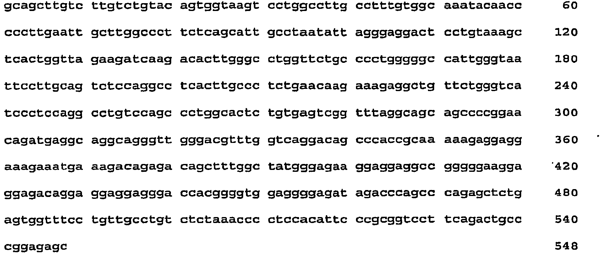
- <210> 2
<211> 19
<212> DNA
<213> Artificial sequence - <220>
<223> Chemically synthesized primer - <400> 2
aaccgaagag gaggtggtg 19 - <210> 3
<211> 20
<212> DNA
<213> Artificial sequence . - <220>
<223> Chemically synthesized primer - <400> 3
gcaaagaagt ggcaggaaga 20 - <210> 4
<211> 20
<212> DNA
<213> Artificial sequence - <220>
<223> Chemically synthesized primer - <400> 4
agaaaatctg gcaccacacc 20 - <210> 5
<211> 20
<212> DNA
<213> Artificial sequence - <220>
<223> Chemically synthesized primer - <400> 5
cagaggcgta cagggatagc 20 - <210> 6
<211> 27
<212> DNA
<213> Artificial sequence - <220>
<223> Chemically synthesized primer - <400> 6
ctagctagca gctgggtgtt gtggcat 27 - <210> 7
<211> 24
<212> DNA
<213> Artificial sequence - <220>
<223> Chemically synthesized primer - <400> 7
acgcgtcgac ctcagtggca ggca 24 - <210> 8
<211> 38
<212> DNA
<213> Artificial sequence - <220>
<223> Chemically synthesized primer - <400> 8
cggcctcctc cttctcccct gtctctgtct ttcatttc 38 - <210> 9
<211> 26
<212> DNA
<213> Artificial sequence - <220>
<223> Chemically synthesized primer - <400> 9
ctagctagcg ggagaaggag gaggcc 26 - <210> 10
<211> 22
<212> DNA
<213> Artificial sequence - <220>
<223> Chemically synthesized primer - <400> 10
gcagatctcc tcagtggcag gc 22 - <210> 11
<211> 23
<212> DNA
<213> Artificial sequence - <220>
<223> Chemically synthesized primer - <400> 11
gcacgcgtag ctgggtgttg tgg 23 - <210> 12
<211> 21
<212> DNA
<213> Artificial sequence - <220>
<223> Chemically synthesized primer - <400> 12
cgagatctgc tctccgggca g 21 - <210> 13
<211> 25
<212> DNA
<213> Artificial sequence - <220>
<223> Chemically synthesized primer - <400> 13
cgagatctgg gcagtctgaa ggacc 25 - <210> 14
<211> 23
<212> DNA
<213> Artificial sequence - <220>
<223> Chemically synthesized primer - <400> 14
cgacgcgtgc agcttgtctt gtc 23 - <210> 15
<211> 24
<212> DNA
<213> Artificial sequence - <220>
<223> Chemically synthesized primer . - <400> 15
gcagatctag tctgaaggac cgcg 24 - <210> 16
<211> 22
<212> DNA
<213> Artificial sequence - <220>
<223> Chemically synthesized primer - <400> 16
gaacgcgtgg gagaaggagg ag 22 - <210> 17
<211> 1246
<212> DNA
<213> Artificial sequence - <220>
<223> Sequence of the human SPARC promoter corresponding to the bases
-1175/+71 from the initiation of transcription - <400> 17


- <210> 18
<212> 1236
<212> DNA
<213> Artificial sequence - <220>
<223> This promoter is a modification of the complete SPARC promoter
-1175/+71, wherein a 10 pb sequence comprised between the two GGA boxes
has been deleted - <400> 18


- <210> 19
<211> 1210
<212> DNA
<213> Artificial sequence - <220>
<223> Chemically synthesized - <400> 19

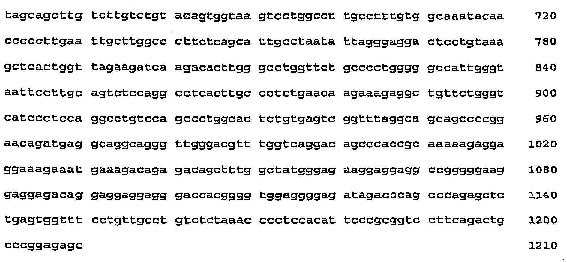
- <210> 20
<211> 1203
<212> DNA
<213> Artificial sequence - <220>
<223> Chemically synthesized - <400> 20


- <210> 21
<211> 584
<212> DNA
<213> Artificial sequence - <220>
<223> Chemically synthesized - <400> 21

- <210> 22
<211> 541
<212> DNA
<213> Artificial sequence - <220>
<223> Chemically synthesized - <400> 22


- <210> 23
<211> 537
<212> DNA
<213> Artificial sequence - <220>
<223> Chemically synthesized - <400> 23

- <210> 24
<211> 191
<212> DNA
<213> Artificial sequence - <220>
<223> Chemically synthesized - <400> 24

- <210> 25
<211> 155
<212> DNA
<213> Artificial sequence - <220>
<223> Chemically synthesized - <400> 25

- <210> 26
<211> 148
<212> DNA
<213> Artificial sequence - <220>
<223> Chemically synthesized - <400> 26

- <210> 27
<211> 17
<212> DNA
<213> Artificial sequence - <220>
<223> Chemically synthesized primer - <400> 27
tacgactcac tataggg 17 - <210> 28
<211> 18
<212> DNA
<213> Artificial sequence - <220>
<223> Chemically synthesized primer - <400> 28
atttaggtga cactatag 18 - <210> 29
<211> 20
<212> DNA
<213> Artificial sequence - <220>
<223> chemically synthesized primer - <400> 29
attaaccctc actaaaggga 20 - <210> 30
<211> 23
<212> DNA
<213> Artificial sequence - <220>
<223> Chemically synthesized primer - <400> 30
ctttatgttt ttggcgtctt cca 23 - <210> 31
<211> 21
<212> DNA
<213> Artificial sequence - <220>
<223> Chemically synthesized primer - <400> 31
ctagcaaaat aggctgtccc c 21 - <210> 32
<211> 93
<212> DNA
<213> Artificial sequence - <220>
<223> Multiple cloning site, included in the pADPSY vector, chemically synthesized - <400> 32

- <210> 33
<211> 7303
<212> DNA
<213> Artificial sequence - <220>
<223> Chemically synthesized - <400> 33


- <210> 34
<211> 28
<212> DNA
<213> Artificial sequence - <220>
<223> Chemically synthesized primer - <400> 34
ccactagtgc tagagctcgc tgatcagc 28 - <210> 35
<211> 24
<212> DNA
<213> Artificial sequence - <220>
<223> Chemically synthesized primer - <400> 35
cggtaccatc cccagcatgc ctgc 24 - <210> 36
<211> 1020
<212> DNA
<213> Artificial sequence - <220>
<223> Sequence obtained with T3 primer - <220>
<221> misc_feature
<222> (1009).. (1009)
<223> n is a, c, g, or t - <220>
<221> misc feature
<222> (1013)..(1013)
<223> n is a, c, g, or t - <400> 36


- <210> 37
<211> 1029
<212> DNA
<213> Artificial sequence - <220>
<223> Sequence obtained with T7 primer - <220>
<221> misc_feature
<222> (15)..(15)
<223> n is a, c, g, or t - <220>
<221> misc_feature
<222> (32).. (32)
<223> n is a, c, g, or t - <220>
<221> misc_feature
<222> (57) .. (57)
<223> n is a, c, g, or t - <220>
<221> misc_feature
<222> (75) .. (75)
<223> n is a, c, g, or t - <220>
<221> misc_feature
<222> (87) ..(87)
<223> n is a, c, g, or t - <400> 37


- <210> 38
<211> 1562
<212> DNA
<213> Artificial sequence - <220>
<223> Chemically synthesized - <400> 38

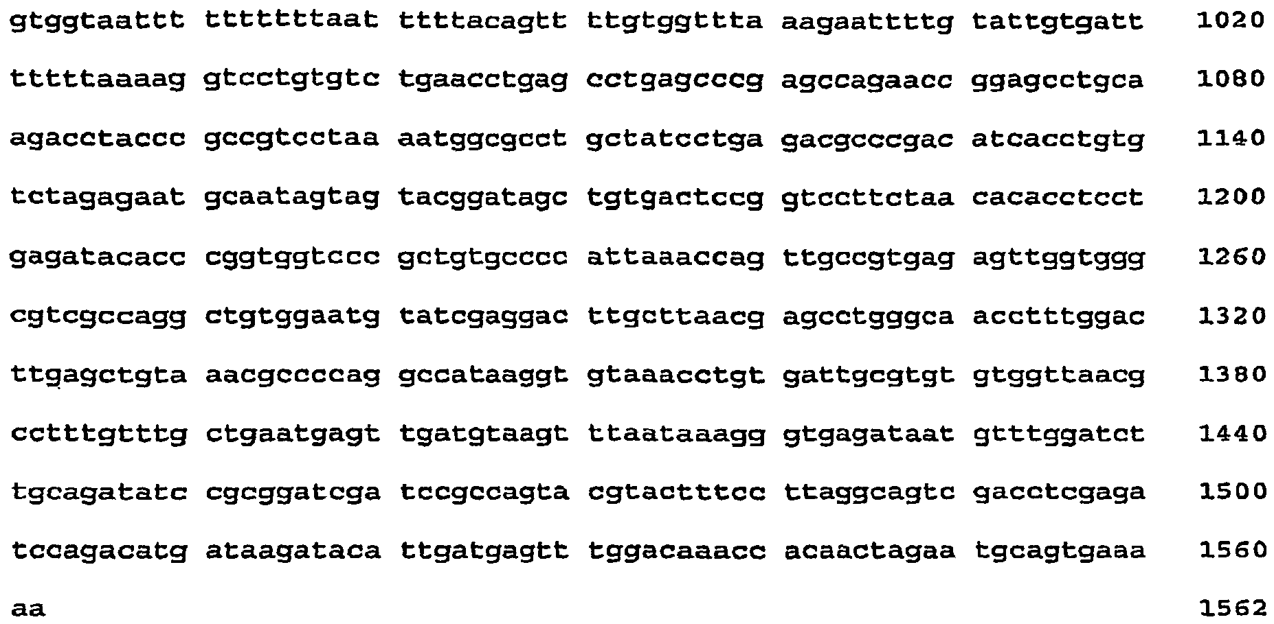
- <210> 39
<211> 1335
<212> DNA
<213> Artificial sequence - <220>
<223> Chemically synthesized - <400> 39


- <210> 40
<211> 776
<212> DNA
<213> Artificial sequence - <220>
<223> Chemically synthesized - <220>
<221> misc_feature
<222> (5)..(5)
<223> n is a, c, g, or t - <220>
<221> misc_feature
<222> (39).. (39)
<223> n is a, c, g, or t - <220>
<221> misc_feature
<222> (128) .. (128)
<223> n is a, c, g, or t - <220>
<221> misc_feature
<222> (684)..(684)
<223> n is a, c, g, or t - <220>
<221> misc_feature
<222> (721) .. (721)
<223> n is a, c, g, or t - <220>
<221> misc_feature
<222> (727)..(727)
<223> n is a, c, g, or t - <220>
<221> misc_feature
<222> (734)..(734)
<223> n is a, c, g, or t - <220>
<221> misc_feature
<222> (738) .. (739)
<223> n is a, c, g, or t - <220>
<221> misc_feature
<222> (752)..(752)
<223> n is a, c, g, or t - <220>
<221> misc_feature
<222> (755) .. (755)
<223> n is a, c, g, or t - <220>
<221> misc_feature
<222> (758)..(758)
<223> n is a, c, g, or t - <400> 40
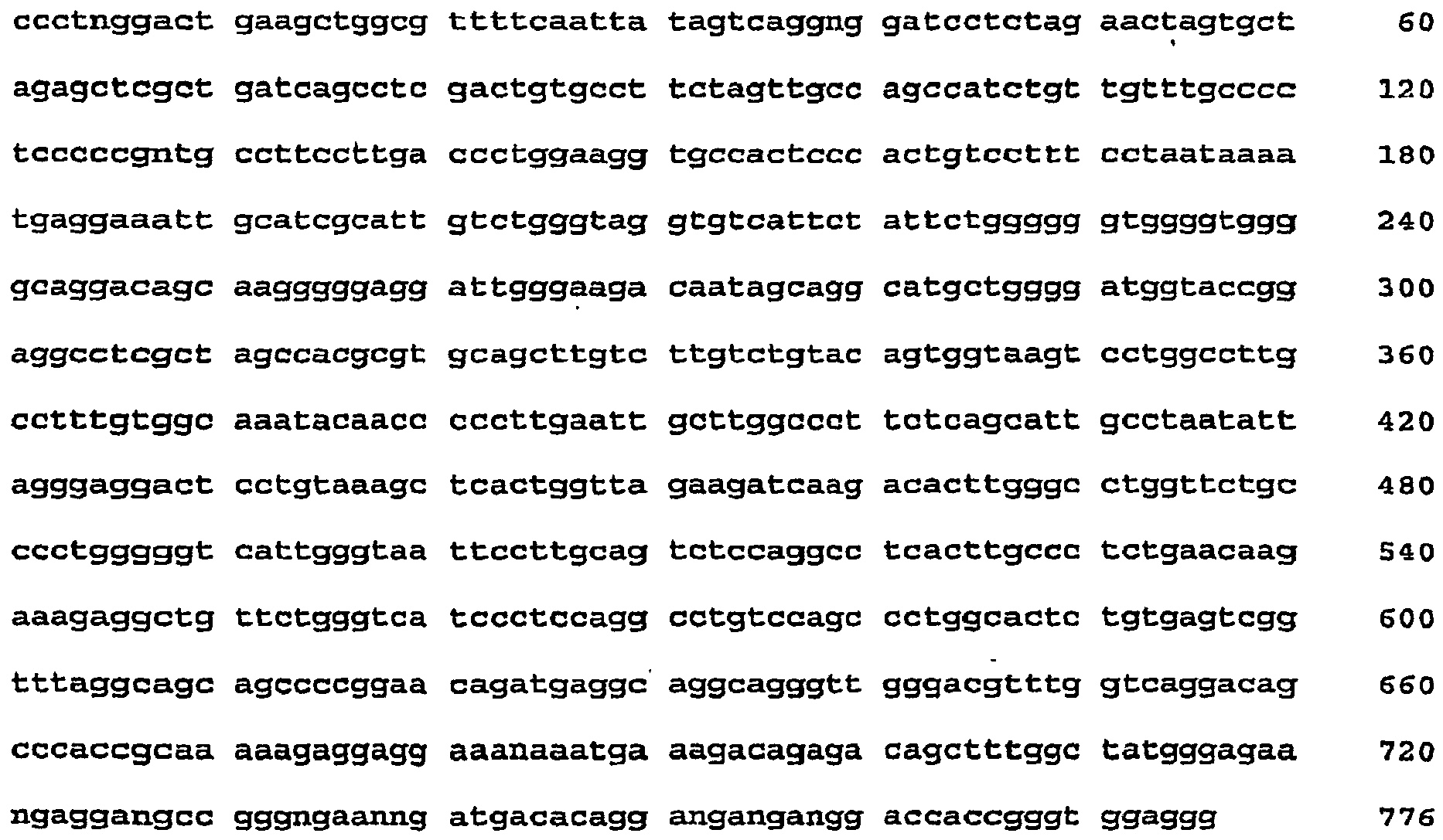
- <210> 41
<211> 507
<212> DNA
<213> Artificial sequence - <220>
<223> Sequence obtained with pad-sense primer - <220>
<221> misc_feature
<222> (2) .. (2)
<223> n is a, c, g, or t - <220>
<221> misc_feature
<222> (8) .. (8)
<223> n is a, c, g, or t - <220>
<221> misc_feature
<222> (26) .. (26)
<223> n is a, c, g, or t - <220>
<221> misc_feature
<222> (498) .. (498)
<223> n is a, c, g, or t - <400> 41

- <210> 42
<211> 22
<212> DNA
c213> Artificial sequence - <220>
<223> Chemically synthesized primer - <400> 42
tgtttttctc aggtgttttc cg 22 - <210> 43
<211> 23
<212> DNA
<213> Artificial sequence - <220>
<223> Chemically synthesized primer - <400> 43
gcccatggct tcgtaccccg gcc - <210> 44
<211> 29
<212> DNA
<213> Artificial sequence - <220>
<223> Chemically synthesized primer - <400> 44
gcgtcgactc agttagcctc ccccatctc 29 - <210> 45
<211> 957
<212> DNA
<213> Artificial sequence - <220>
<223> Sequence obtained with T3 primer - <400> 45

- <210> 46
<211> 238
<212> DNA
<213> Artificial sequence - <220>
<223> Sequence obtained with pAd-sense priner - <400> 46

- <210> 47
<211> 965
<212> DNA
<213> Artificial seqeunce - <220>
<223> Sequence obtained with pAd-antisense primer - <400> 47

- <210> 48
<211> 25
<212> DNA
<213> Artificial sequence - <220>
<223> Chemically synthesized primer - <400> 48
cacaaatttc acaaataaag cattt 25 - <210> 49
<211> 911
<212> DNA
<213> Artificial sequence - <220>
<223> Sequence obtained with pAd-sense primer, partial sequence of the virus - <220>
<221> misc_feature
<222> (5)..(5)
<223> n is a, c, g, or t - <220>
<221> misc_feature
<222> (8) .. (8)
<223> n is a, c, g, or t - <220>
<221> misc_feature
<222> (22)..(22)
<223> n is a, c, g, or t - <220>
<221> misc_feature
<222> (34).. (34)
<223 > n is a, c, g, or t - <220>
<221> misc_feature
<222> (751) .. (751)
<223> n is a, c, g, or t - <220>
<221> misc feature
<222> (854)..(854)
<223> n is a, c, g, or t - <400> 49


- <210> 50
<211> 908
<212> DNA
<213> Artificial sequence - <220>
<223> Sequence obtained with pAd-antisense primer, partial sequence of the virus - <220>
<221> misc_feature
<222> (11) .. (11)
<223> n is a, c, g, or t, - <220>
<221> misc_feature
<222> (18) ..(18)
<223> n is a, c, g, or t - <220>
<221> misc_feature
<222> (30) .. (31)
<223> n is a, c, g, or t - <400> 50


- <210> 51
<211> 842
<212> DNA
<213> Artificial sequence - <220>
<223> Sequence obtained with pAd-sense primer, partial sequence of the virus - <220>
<221> misc_feature
<222> (4) .. (5)
<223> n is a, c, g, or t - <220>
<221> misc_feature
<222> (14) .. (15)
<223> n is a, c, g, or t - <220>
<221> misc_feature
<222> (19) .. (19)
<223> n is a, c, g, or t - <220>
<221> misc_feature
<222> (24)..(25)
<223> n is a, c, g, or t - <220>
<221> misc_feature
<222> (37) .. (39)
<223> n is a, c, g, or t - <220>
<221> misc_feature
<222> (61) .. (61)
<223> n is a, c, g, or t - <220>
<221> misc_feature
<222> (159) .. (159)
<223> n is a, c, g, or t - <220>
<221> misc_feature
<222> (795) .. (795)
<223> n is a, c, g, or t - <400> 51

- <210> 52
<211> 851
<212> DNA
<213> Artificial seqeunce - <220>
<223> Sequence obtained with pAd-antisense primer, partial sequence of the virus - <220>
<221> misc_feature
<222> (5) .. (5)
<223> n is a, c, g, or t - <220>
<221> misc_feature
<222> (10) .. (10)
<223> n is a, c, g, or t - <220>
<221> misc_feature
<222> (12)..(12)
<223> n is a, c, g, or t - <220>
<221> misc_feature
<222> (14) .. (14)
<223> n is a, c, g, or t - <220>
<221> misc_feature
<222> (19) .. (19)
<223> n is a, c, g, or t - <220>
<221> misc_feature
<222> (29) .. (29)
<223> n is a, c, g, or t - <220>
<221> misc_feature
<222> (34).. (34)
<223> n is a, c, g, or t - <220>
<221> misc_feature
<222> (42) ..(42)
<223> n is a, c, g, or t - <220>
<221> misc_feature
<222> (65)..(65)
<223> n is a, c, g, or t - <220>
<221> misc_feature
<222> (90) ..(90)
<223> n is a, c, g, or t - <220>
<221> misc_feature
<222> (103)..(103)
<223> n is a, c, g, or t - <220>
<221> misc_feature
<222> (130) .. (130)
<223> n is a, c, g, or t - <220>
<221> misc_feature
<222> (393) .. (393)
<223> n is a, c, g, or t - <220>
<221> misc_feature
<222> (539) .. (539)
<223> n is a., c, g, or t - <400> 52


- <210> 53
<211> 854
<212> DNA
<213> Artificial sequence - <220>
<223> Sequence obtained with pAd-sense primer, partial sequence of the virus - <400> 53


- <210> 54
<211> 840
<212> DNA
<213> Artificial sequence - <220>
<223> Sequence obtained with pAd-antisense primer, partial sequence of the virus - <400> 54

- <210> 55
<211> 833
<212> DNA
<213> Artificial sequence - <220>
<223> Sequence obtained with pAd-sense primer, partial sequence of the virus - <400> 55


- <210> 56
<211> 816
<212> DNA
<213> Artificial sequence - <220>
<223> Sequence obtained with pAd-antisense primer, partial sequence of the virus - <400> 56


Claims (19)
- An isolated DNA comprising a nucleic acid promoter having a sequence consisting of SEQ ID NO:1.
- The isolated DNA of claim 1, wherein the promoter sequence is operably linked to a heterologous gene.
- The isolated DNA of claim 1, further comprising a second nucleic acid promoter.
- The isolated DNA of claim 3, further comprising a regulating sequence selected from a radiation responsive sequence, a hypoxia responsive sequence or a free-radical responsive sequence.
- The isolated DNA of claim 2, wherein the heterologous gene is a therapeutic gene.
- The isolated DNA of claim 2, wherein the heterologous gene is selected from the group consisting of: an E1A gene, a suicide gene, the adenoviral genome E3 region and a gene encoding an interleukin.
- An expression vector comprising the isolated DNA of claim 1 and a heterologous gene operably linked thereto.
- The expression vector of claim 7, wherein the vector is a plasmid or a viral vector.
- The expression vector of claim 8, wherein the viral vector is a recombinant adenovirus.
- The expression vector of claim 8, wherein the viral vector is a Conditionally Replicative Oncolytic Adenovirus.
- The expression vector of claim 7, wherein the heterologous gene is a therapeutic gene.
- The expression vector of claim 11, wherein the therapeutic heterologous gene is an E1A protein gene.
- The expression vector of claim 7, wherein the heterologous gene is a suicide gene.
- The expression vector of claim 13, wherein the suicide gene is the thymidine kinase gene of a herpes virus (hsvTK).
- The expression vector of claim 7, further comprising an adenoviral genome E3 region.
- An ex-vivo method for expressing foreign DNA in a host cell, the method comprising introducing into the host cell the expression vector of claim 7.
- A pharmaceutical com position comprising the expression vector of claim 7 in a pharmaceutically suitable carrier.
- The use of a pharmaceutical composition of claim 17 in the manufacture of a medicament for the treatment of a tumor in a patient suffering there from.
- An expression vector comprising a nucleic acid promoter having a sequence consisting of SEQ ID NO 1 according to claim 1.
Applications Claiming Priority (2)
| Application Number | Priority Date | Filing Date | Title |
|---|---|---|---|
| ARP060101724A AR053246A1 (en) | 2006-04-28 | 2006-04-28 | AN ISOLATED DNA FRAGMENT FROM SPARC'S HUMAN PROMOTER AND ITS USE TO DIRECT THE EXPRESSION OF A HETEROLOGICAL GENE IN TUMOR CELLS |
| PCT/US2007/010196 WO2007127347A2 (en) | 2006-04-28 | 2007-04-26 | An isolated dna fragment of the sparc human promoter and its use |
Publications (3)
| Publication Number | Publication Date |
|---|---|
| EP2048954A2 EP2048954A2 (en) | 2009-04-22 |
| EP2048954A4 EP2048954A4 (en) | 2009-12-02 |
| EP2048954B1 true EP2048954B1 (en) | 2012-03-28 |
Family
ID=38006653
Family Applications (1)
| Application Number | Title | Priority Date | Filing Date |
|---|---|---|---|
| EP07776311A Not-in-force EP2048954B1 (en) | 2006-04-28 | 2007-04-26 | An isolated dna fragment of the sparc human promoter and its use |
Country Status (9)
| Country | Link |
|---|---|
| US (1) | US8436160B2 (en) |
| EP (1) | EP2048954B1 (en) |
| JP (1) | JP5215290B2 (en) |
| AR (1) | AR053246A1 (en) |
| AT (1) | ATE550943T1 (en) |
| AU (1) | AU2007243252B2 (en) |
| ES (1) | ES2384736T3 (en) |
| NZ (1) | NZ572350A (en) |
| WO (1) | WO2007127347A2 (en) |
Families Citing this family (4)
| Publication number | Priority date | Publication date | Assignee | Title |
|---|---|---|---|---|
| ES2304281B1 (en) * | 2006-02-01 | 2009-08-12 | Dnatrix Inc. | ADENOVIRUS ONCOLITICOS FOR THE TREATMENT OF CANCER. |
| US9056133B2 (en) * | 2011-05-25 | 2015-06-16 | Cti-S.A. | Pharmaceutical kit and method for treating cancer |
| CN113710810A (en) * | 2019-02-21 | 2021-11-26 | 安利舒免疫溶瘤技术公司 | Oncolytic adenoviral vectors and methods of use |
| JP2023532279A (en) | 2020-06-26 | 2023-07-27 | ソレント・セラピューティクス・インコーポレイテッド | Oncolytic Herpes Simplex Virus (HSV) Expressing Immunomodulatory Fusion Proteins |
Family Cites Families (5)
| Publication number | Priority date | Publication date | Assignee | Title |
|---|---|---|---|---|
| US5772993A (en) * | 1997-01-21 | 1998-06-30 | The University Of Virginia Patent Foundation | Osteocalcin promoter-based toxic gene therapy for the treatment of calcified tumors and tissues |
| IL146794A0 (en) * | 1999-05-28 | 2002-07-25 | Univ Virginia | Osteonectin based toxic gene therapy for the treatment of calcified tumors and tissues |
| US7563748B2 (en) | 2003-06-23 | 2009-07-21 | Cognis Ip Management Gmbh | Alcohol alkoxylate carriers for pesticide active ingredients |
| US7879542B2 (en) * | 2003-06-24 | 2011-02-01 | The Johns Hopkins University | Methylated gene biomarkers for detecting cancer |
| EP1895898B1 (en) | 2005-06-29 | 2011-02-16 | Compumedics Limited | Sensor assembly with conductive bridge |
-
2006
- 2006-04-28 AR ARP060101724A patent/AR053246A1/en active IP Right Grant
-
2007
- 2007-04-26 US US12/298,743 patent/US8436160B2/en active Active - Reinstated
- 2007-04-26 AT AT07776311T patent/ATE550943T1/en active
- 2007-04-26 JP JP2009507816A patent/JP5215290B2/en not_active Expired - Fee Related
- 2007-04-26 NZ NZ572350A patent/NZ572350A/en not_active IP Right Cessation
- 2007-04-26 WO PCT/US2007/010196 patent/WO2007127347A2/en not_active Ceased
- 2007-04-26 ES ES07776311T patent/ES2384736T3/en active Active
- 2007-04-26 EP EP07776311A patent/EP2048954B1/en not_active Not-in-force
- 2007-04-26 AU AU2007243252A patent/AU2007243252B2/en not_active Ceased
Also Published As
| Publication number | Publication date |
|---|---|
| EP2048954A2 (en) | 2009-04-22 |
| WO2007127347A2 (en) | 2007-11-08 |
| ATE550943T1 (en) | 2012-04-15 |
| ES2384736T3 (en) | 2012-07-11 |
| WO2007127347A3 (en) | 2008-01-03 |
| AU2007243252A1 (en) | 2007-11-08 |
| NZ572350A (en) | 2011-12-22 |
| US20090312401A1 (en) | 2009-12-17 |
| AR053246A1 (en) | 2007-04-25 |
| US8436160B2 (en) | 2013-05-07 |
| JP5215290B2 (en) | 2013-06-19 |
| AU2007243252B2 (en) | 2013-06-20 |
| EP2048954A4 (en) | 2009-12-02 |
| JP2009535031A (en) | 2009-10-01 |
Similar Documents
| Publication | Publication Date | Title |
|---|---|---|
| US20180265890A1 (en) | Efficient and safe transposon integration system and use thereof | |
| US7109029B2 (en) | Vector constructs | |
| AU4504396A (en) | Vectors for tissue-specific replication | |
| JP2023014163A (en) | Tumor-targeting synthetic adenoviruses and uses thereof | |
| JP4845327B2 (en) | Tumor specific promoter | |
| JPWO2002010368A1 (en) | Tumor-specific promoters | |
| US8436160B2 (en) | Isolated DNA fragment of the SPARC human promoter and its use for driving the expression of an heterologous gene in tumor cells | |
| EP1601772B1 (en) | Modified tert promoter with enhanced tumor-specificity and strength and recombinant vector comprising the same | |
| Hernandez-Alcoceba et al. | Evaluation of a new dual-specificity promoter for selective induction of apoptosis in breast cancer cells | |
| Li et al. | Enhancement of antiangiogenic effects of human canstatin with a hypoxia-regulated transgene vector in lung cancer model | |
| US8034589B2 (en) | Method of preparing a proliferation-regulated recombinant adenoviral vector efficiently and kit for preparing the same | |
| KR100969171B1 (en) | Gene carriers with improved tumor specific expression | |
| US20230391836A1 (en) | Amino acid sequence that can destroy cells, and related nucleotide sequence and related uses thereof | |
| US8188252B2 (en) | Rad51 derived cancer cell specific promoters for targeted anti-cancer therapy | |
| EP2702158B1 (en) | Mutant cd83 promoter and use thereof | |
| KR100760320B1 (en) | An anticancer composition comprising a recombinant vector expressing an importin α gene and a recombinant 355 gene | |
| JP6913930B2 (en) | Antineoplastic agent for squamous cell carcinoma | |
| CN115651932A (en) | Construction method and application of targeted digestive tract tumor dual-targeted oncolytic adenovirus | |
| JP2000143548A (en) | Methods and compositions for increasing retroviral infectivity | |
| KR20020063452A (en) | Antitumor drug having Importin αgene and/or recombinant p53 gene | |
| WO2004016652A2 (en) | Mammaglobin promoter | |
| Lee | hTERT or AFP promoter specific expression of HSV-tk induces apoptosis in tumor cells | |
| Iggo et al. | Replicating Adenoviruses That Target | |
| AU2002252061A1 (en) | Vector constucts | |
| JPWO1999009191A1 (en) | Cancer treatment vectors |
Legal Events
| Date | Code | Title | Description |
|---|---|---|---|
| PUAI | Public reference made under article 153(3) epc to a published international application that has entered the european phase |
Free format text: ORIGINAL CODE: 0009012 |
|
| 17P | Request for examination filed |
Effective date: 20081127 |
|
| AK | Designated contracting states |
Kind code of ref document: A2 Designated state(s): AT BE BG CH CY CZ DE DK EE ES FI FR GB GR HU IE IS IT LI LT LU LV MC MT NL PL PT RO SE SI SK TR |
|
| AX | Request for extension of the european patent |
Extension state: AL BA HR MK RS |
|
| DAX | Request for extension of the european patent (deleted) | ||
| A4 | Supplementary search report drawn up and despatched |
Effective date: 20091104 |
|
| RAP1 | Party data changed (applicant data changed or rights of an application transferred) |
Owner name: INIS BIOTECH SA Owner name: INIS BIOTECH LLC |
|
| 17Q | First examination report despatched |
Effective date: 20100802 |
|
| RIC1 | Information provided on ipc code assigned before grant |
Ipc: A01N 63/00 20060101AFI20110512BHEP Ipc: A61K 39/12 20060101ALI20110512BHEP |
|
| GRAP | Despatch of communication of intention to grant a patent |
Free format text: ORIGINAL CODE: EPIDOSNIGR1 |
|
| GRAC | Information related to communication of intention to grant a patent modified |
Free format text: ORIGINAL CODE: EPIDOSCIGR1 |
|
| RAP1 | Party data changed (applicant data changed or rights of an application transferred) |
Owner name: INIS BIOTECH SA Owner name: INIS BIOTECH LLC |
|
| GRAS | Grant fee paid |
Free format text: ORIGINAL CODE: EPIDOSNIGR3 |
|
| GRAA | (expected) grant |
Free format text: ORIGINAL CODE: 0009210 |
|
| AK | Designated contracting states |
Kind code of ref document: B1 Designated state(s): AT BE BG CH CY CZ DE DK EE ES FI FR GB GR HU IE IS IT LI LT LU LV MC MT NL PL PT RO SE SI SK TR |
|
| REG | Reference to a national code |
Ref country code: GB Ref legal event code: FG4D |
|
| REG | Reference to a national code |
Ref country code: CH Ref legal event code: EP |
|
| REG | Reference to a national code |
Ref country code: AT Ref legal event code: REF Ref document number: 550943 Country of ref document: AT Kind code of ref document: T Effective date: 20120415 |
|
| REG | Reference to a national code |
Ref country code: IE Ref legal event code: FG4D |
|
| REG | Reference to a national code |
Ref country code: DE Ref legal event code: R096 Ref document number: 602007021638 Country of ref document: DE Effective date: 20120524 |
|
| REG | Reference to a national code |
Ref country code: ES Ref legal event code: FG2A Ref document number: 2384736 Country of ref document: ES Kind code of ref document: T3 Effective date: 20120711 |
|
| REG | Reference to a national code |
Ref country code: NL Ref legal event code: VDEP Effective date: 20120328 |
|
| PG25 | Lapsed in a contracting state [announced via postgrant information from national office to epo] |
Ref country code: LT Free format text: LAPSE BECAUSE OF FAILURE TO SUBMIT A TRANSLATION OF THE DESCRIPTION OR TO PAY THE FEE WITHIN THE PRESCRIBED TIME-LIMIT Effective date: 20120328 |
|
| LTIE | Lt: invalidation of european patent or patent extension |
Effective date: 20120328 |
|
| PG25 | Lapsed in a contracting state [announced via postgrant information from national office to epo] |
Ref country code: FI Free format text: LAPSE BECAUSE OF FAILURE TO SUBMIT A TRANSLATION OF THE DESCRIPTION OR TO PAY THE FEE WITHIN THE PRESCRIBED TIME-LIMIT Effective date: 20120328 Ref country code: GR Free format text: LAPSE BECAUSE OF FAILURE TO SUBMIT A TRANSLATION OF THE DESCRIPTION OR TO PAY THE FEE WITHIN THE PRESCRIBED TIME-LIMIT Effective date: 20120629 Ref country code: LV Free format text: LAPSE BECAUSE OF FAILURE TO SUBMIT A TRANSLATION OF THE DESCRIPTION OR TO PAY THE FEE WITHIN THE PRESCRIBED TIME-LIMIT Effective date: 20120328 |
|
| REG | Reference to a national code |
Ref country code: AT Ref legal event code: MK05 Ref document number: 550943 Country of ref document: AT Kind code of ref document: T Effective date: 20120328 |
|
| PG25 | Lapsed in a contracting state [announced via postgrant information from national office to epo] |
Ref country code: CY Free format text: LAPSE BECAUSE OF FAILURE TO SUBMIT A TRANSLATION OF THE DESCRIPTION OR TO PAY THE FEE WITHIN THE PRESCRIBED TIME-LIMIT Effective date: 20120328 |
|
| PG25 | Lapsed in a contracting state [announced via postgrant information from national office to epo] |
Ref country code: EE Free format text: LAPSE BECAUSE OF FAILURE TO SUBMIT A TRANSLATION OF THE DESCRIPTION OR TO PAY THE FEE WITHIN THE PRESCRIBED TIME-LIMIT Effective date: 20120328 Ref country code: SI Free format text: LAPSE BECAUSE OF FAILURE TO SUBMIT A TRANSLATION OF THE DESCRIPTION OR TO PAY THE FEE WITHIN THE PRESCRIBED TIME-LIMIT Effective date: 20120328 Ref country code: SE Free format text: LAPSE BECAUSE OF FAILURE TO SUBMIT A TRANSLATION OF THE DESCRIPTION OR TO PAY THE FEE WITHIN THE PRESCRIBED TIME-LIMIT Effective date: 20120328 Ref country code: BE Free format text: LAPSE BECAUSE OF FAILURE TO SUBMIT A TRANSLATION OF THE DESCRIPTION OR TO PAY THE FEE WITHIN THE PRESCRIBED TIME-LIMIT Effective date: 20120328 Ref country code: IS Free format text: LAPSE BECAUSE OF FAILURE TO SUBMIT A TRANSLATION OF THE DESCRIPTION OR TO PAY THE FEE WITHIN THE PRESCRIBED TIME-LIMIT Effective date: 20120728 Ref country code: RO Free format text: LAPSE BECAUSE OF FAILURE TO SUBMIT A TRANSLATION OF THE DESCRIPTION OR TO PAY THE FEE WITHIN THE PRESCRIBED TIME-LIMIT Effective date: 20120328 Ref country code: PL Free format text: LAPSE BECAUSE OF FAILURE TO SUBMIT A TRANSLATION OF THE DESCRIPTION OR TO PAY THE FEE WITHIN THE PRESCRIBED TIME-LIMIT Effective date: 20120328 Ref country code: CZ Free format text: LAPSE BECAUSE OF FAILURE TO SUBMIT A TRANSLATION OF THE DESCRIPTION OR TO PAY THE FEE WITHIN THE PRESCRIBED TIME-LIMIT Effective date: 20120328 |
|
| PG25 | Lapsed in a contracting state [announced via postgrant information from national office to epo] |
Ref country code: SK Free format text: LAPSE BECAUSE OF FAILURE TO SUBMIT A TRANSLATION OF THE DESCRIPTION OR TO PAY THE FEE WITHIN THE PRESCRIBED TIME-LIMIT Effective date: 20120328 Ref country code: MC Free format text: LAPSE BECAUSE OF NON-PAYMENT OF DUE FEES Effective date: 20120430 Ref country code: PT Free format text: LAPSE BECAUSE OF FAILURE TO SUBMIT A TRANSLATION OF THE DESCRIPTION OR TO PAY THE FEE WITHIN THE PRESCRIBED TIME-LIMIT Effective date: 20120730 |
|
| REG | Reference to a national code |
Ref country code: CH Ref legal event code: PL |
|
| PGFP | Annual fee paid to national office [announced via postgrant information from national office to epo] |
Ref country code: ES Payment date: 20120621 Year of fee payment: 6 |
|
| REG | Reference to a national code |
Ref country code: IE Ref legal event code: MM4A |
|
| RAP2 | Party data changed (patent owner data changed or rights of a patent transferred) |
Owner name: FUNDACION INSTITUTO LELOIR Owner name: CONSEJO NACIONAL DE INVESTIGACIONES CIENTIFICAS Y |
|
| PG25 | Lapsed in a contracting state [announced via postgrant information from national office to epo] |
Ref country code: DK Free format text: LAPSE BECAUSE OF FAILURE TO SUBMIT A TRANSLATION OF THE DESCRIPTION OR TO PAY THE FEE WITHIN THE PRESCRIBED TIME-LIMIT Effective date: 20120328 Ref country code: CH Free format text: LAPSE BECAUSE OF NON-PAYMENT OF DUE FEES Effective date: 20120430 Ref country code: LI Free format text: LAPSE BECAUSE OF NON-PAYMENT OF DUE FEES Effective date: 20120430 Ref country code: IE Free format text: LAPSE BECAUSE OF NON-PAYMENT OF DUE FEES Effective date: 20120426 Ref country code: NL Free format text: LAPSE BECAUSE OF FAILURE TO SUBMIT A TRANSLATION OF THE DESCRIPTION OR TO PAY THE FEE WITHIN THE PRESCRIBED TIME-LIMIT Effective date: 20120328 Ref country code: AT Free format text: LAPSE BECAUSE OF FAILURE TO SUBMIT A TRANSLATION OF THE DESCRIPTION OR TO PAY THE FEE WITHIN THE PRESCRIBED TIME-LIMIT Effective date: 20120328 |
|
| PLBE | No opposition filed within time limit |
Free format text: ORIGINAL CODE: 0009261 |
|
| STAA | Information on the status of an ep patent application or granted ep patent |
Free format text: STATUS: NO OPPOSITION FILED WITHIN TIME LIMIT |
|
| PG25 | Lapsed in a contracting state [announced via postgrant information from national office to epo] |
Ref country code: IT Free format text: LAPSE BECAUSE OF FAILURE TO SUBMIT A TRANSLATION OF THE DESCRIPTION OR TO PAY THE FEE WITHIN THE PRESCRIBED TIME-LIMIT Effective date: 20120328 |
|
| 26N | No opposition filed |
Effective date: 20130103 |
|
| REG | Reference to a national code |
Ref country code: DE Ref legal event code: R097 Ref document number: 602007021638 Country of ref document: DE Effective date: 20130103 |
|
| REG | Reference to a national code |
Ref country code: DE Ref legal event code: R081 Ref document number: 602007021638 Country of ref document: DE Owner name: FUNDACION INSTITUTO LELOIR, AR Free format text: FORMER OWNERS: INIS BIOTECH LLC, MILFORD, KENT COUNTY, DEL., US; INIS BIOTECH SA, BUENOS AIRES, AR Effective date: 20130314 Ref country code: DE Ref legal event code: R081 Ref document number: 602007021638 Country of ref document: DE Owner name: CONSEJO NACIONAL DE INVESTIGACIONES CIENTIFICA, AR Free format text: FORMER OWNERS: INIS BIOTECH LLC, MILFORD, KENT COUNTY, DEL., US; INIS BIOTECH SA, BUENOS AIRES, AR Effective date: 20130314 Ref country code: DE Ref legal event code: R081 Ref document number: 602007021638 Country of ref document: DE Owner name: CONSEJO NACIONAL DE INVESTIGACIONES CIENTIFICA, AR Free format text: FORMER OWNER: INIS BIOTECH LLC, INIS BIOTECH SA, , AR Effective date: 20130314 Ref country code: DE Ref legal event code: R081 Ref document number: 602007021638 Country of ref document: DE Owner name: FUNDACION INSTITUTO LELOIR, AR Free format text: FORMER OWNER: INIS BIOTECH LLC, INIS BIOTECH SA, , AR Effective date: 20130314 |
|
| REG | Reference to a national code |
Ref country code: GB Ref legal event code: 732E Free format text: REGISTERED BETWEEN 20130613 AND 20130619 |
|
| PG25 | Lapsed in a contracting state [announced via postgrant information from national office to epo] |
Ref country code: MT Free format text: LAPSE BECAUSE OF FAILURE TO SUBMIT A TRANSLATION OF THE DESCRIPTION OR TO PAY THE FEE WITHIN THE PRESCRIBED TIME-LIMIT Effective date: 20120328 Ref country code: BG Free format text: LAPSE BECAUSE OF FAILURE TO SUBMIT A TRANSLATION OF THE DESCRIPTION OR TO PAY THE FEE WITHIN THE PRESCRIBED TIME-LIMIT Effective date: 20120628 |
|
| PGFP | Annual fee paid to national office [announced via postgrant information from national office to epo] |
Ref country code: GB Payment date: 20130424 Year of fee payment: 7 Ref country code: DE Payment date: 20130423 Year of fee payment: 7 |
|
| PGFP | Annual fee paid to national office [announced via postgrant information from national office to epo] |
Ref country code: FR Payment date: 20130529 Year of fee payment: 7 |
|
| PG25 | Lapsed in a contracting state [announced via postgrant information from national office to epo] |
Ref country code: TR Free format text: LAPSE BECAUSE OF FAILURE TO SUBMIT A TRANSLATION OF THE DESCRIPTION OR TO PAY THE FEE WITHIN THE PRESCRIBED TIME-LIMIT Effective date: 20120328 |
|
| PG25 | Lapsed in a contracting state [announced via postgrant information from national office to epo] |
Ref country code: LU Free format text: LAPSE BECAUSE OF NON-PAYMENT OF DUE FEES Effective date: 20120426 |
|
| PG25 | Lapsed in a contracting state [announced via postgrant information from national office to epo] |
Ref country code: HU Free format text: LAPSE BECAUSE OF FAILURE TO SUBMIT A TRANSLATION OF THE DESCRIPTION OR TO PAY THE FEE WITHIN THE PRESCRIBED TIME-LIMIT Effective date: 20070426 |
|
| REG | Reference to a national code |
Ref country code: DE Ref legal event code: R119 Ref document number: 602007021638 Country of ref document: DE |
|
| GBPC | Gb: european patent ceased through non-payment of renewal fee |
Effective date: 20140426 |
|
| REG | Reference to a national code |
Ref country code: FR Ref legal event code: ST Effective date: 20141231 |
|
| REG | Reference to a national code |
Ref country code: DE Ref legal event code: R119 Ref document number: 602007021638 Country of ref document: DE Effective date: 20141101 |
|
| PG25 | Lapsed in a contracting state [announced via postgrant information from national office to epo] |
Ref country code: DE Free format text: LAPSE BECAUSE OF NON-PAYMENT OF DUE FEES Effective date: 20141101 Ref country code: GB Free format text: LAPSE BECAUSE OF NON-PAYMENT OF DUE FEES Effective date: 20140426 |
|
| PG25 | Lapsed in a contracting state [announced via postgrant information from national office to epo] |
Ref country code: FR Free format text: LAPSE BECAUSE OF NON-PAYMENT OF DUE FEES Effective date: 20140430 |
|
| PG25 | Lapsed in a contracting state [announced via postgrant information from national office to epo] |
Ref country code: ES Free format text: LAPSE BECAUSE OF NON-PAYMENT OF DUE FEES Effective date: 20140427 |