EP2047853A1 - The application of marine steroid in preparing the medicine of treating neurons damaging - Google Patents
The application of marine steroid in preparing the medicine of treating neurons damaging Download PDFInfo
- Publication number
- EP2047853A1 EP2047853A1 EP06775552A EP06775552A EP2047853A1 EP 2047853 A1 EP2047853 A1 EP 2047853A1 EP 06775552 A EP06775552 A EP 06775552A EP 06775552 A EP06775552 A EP 06775552A EP 2047853 A1 EP2047853 A1 EP 2047853A1
- Authority
- EP
- European Patent Office
- Prior art keywords
- cgns
- ischemia
- medicine
- cell
- steroid
- Prior art date
- Legal status (The legal status is an assumption and is not a legal conclusion. Google has not performed a legal analysis and makes no representation as to the accuracy of the status listed.)
- Withdrawn
Links
- 239000003814 drug Substances 0.000 title claims abstract description 18
- 150000003431 steroids Chemical class 0.000 title claims abstract description 14
- 210000002569 neuron Anatomy 0.000 title abstract description 20
- 229940079593 drug Drugs 0.000 claims description 10
- 239000000126 substance Substances 0.000 claims description 10
- 230000004693 neuron damage Effects 0.000 claims description 4
- 230000001681 protective effect Effects 0.000 abstract description 4
- 230000009471 action Effects 0.000 abstract description 3
- 241001400381 Nephthea Species 0.000 abstract description 2
- 230000007512 neuronal protection Effects 0.000 abstract description 2
- 231100000331 toxic Toxicity 0.000 abstract description 2
- 230000002588 toxic effect Effects 0.000 abstract description 2
- 238000000034 method Methods 0.000 description 25
- 210000004027 cell Anatomy 0.000 description 23
- 241000700159 Rattus Species 0.000 description 22
- 230000000694 effects Effects 0.000 description 21
- IAZDPXIOMUYVGZ-UHFFFAOYSA-N Dimethylsulphoxide Chemical compound CS(C)=O IAZDPXIOMUYVGZ-UHFFFAOYSA-N 0.000 description 19
- 201000006474 Brain Ischemia Diseases 0.000 description 15
- 206010008120 Cerebral ischaemia Diseases 0.000 description 15
- 241000283973 Oryctolagus cuniculus Species 0.000 description 15
- 206010008118 cerebral infarction Diseases 0.000 description 15
- WHUUTDBJXJRKMK-UHFFFAOYSA-N Glutamic acid Natural products OC(=O)C(N)CCC(O)=O WHUUTDBJXJRKMK-UHFFFAOYSA-N 0.000 description 14
- 230000000302 ischemic effect Effects 0.000 description 14
- 238000011282 treatment Methods 0.000 description 14
- 230000008569 process Effects 0.000 description 12
- 230000002490 cerebral effect Effects 0.000 description 11
- 125000000325 methylidene group Chemical group [H]C([H])=* 0.000 description 11
- 208000036982 Spinal cord ischaemia Diseases 0.000 description 10
- 208000028867 ischemia Diseases 0.000 description 10
- CHADEQDQBURGHL-UHFFFAOYSA-N (6'-acetyloxy-3-oxospiro[2-benzofuran-1,9'-xanthene]-3'-yl) acetate Chemical compound O1C(=O)C2=CC=CC=C2C21C1=CC=C(OC(C)=O)C=C1OC1=CC(OC(=O)C)=CC=C21 CHADEQDQBURGHL-UHFFFAOYSA-N 0.000 description 9
- 206010008089 Cerebral artery occlusion Diseases 0.000 description 9
- 210000005013 brain tissue Anatomy 0.000 description 9
- 230000006870 function Effects 0.000 description 9
- 239000008187 granular material Substances 0.000 description 9
- 201000007309 middle cerebral artery infarction Diseases 0.000 description 9
- 230000006907 apoptotic process Effects 0.000 description 8
- 230000015572 biosynthetic process Effects 0.000 description 8
- 230000006931 brain damage Effects 0.000 description 8
- 231100000874 brain damage Toxicity 0.000 description 8
- 208000029028 brain injury Diseases 0.000 description 8
- 230000006378 damage Effects 0.000 description 8
- 230000034994 death Effects 0.000 description 8
- 229940011871 estrogen Drugs 0.000 description 8
- 239000000262 estrogen Substances 0.000 description 8
- 238000000338 in vitro Methods 0.000 description 8
- 230000001537 neural effect Effects 0.000 description 8
- 238000003786 synthesis reaction Methods 0.000 description 8
- 206010021143 Hypoxia Diseases 0.000 description 7
- 238000004458 analytical method Methods 0.000 description 7
- 208000026106 cerebrovascular disease Diseases 0.000 description 7
- 238000006243 chemical reaction Methods 0.000 description 7
- 230000007423 decrease Effects 0.000 description 7
- 239000002609 medium Substances 0.000 description 7
- 238000002560 therapeutic procedure Methods 0.000 description 7
- 230000035899 viability Effects 0.000 description 7
- 210000004369 blood Anatomy 0.000 description 6
- 239000008280 blood Substances 0.000 description 6
- 210000005056 cell body Anatomy 0.000 description 6
- 230000007954 hypoxia Effects 0.000 description 6
- 230000007246 mechanism Effects 0.000 description 6
- 238000011160 research Methods 0.000 description 6
- 230000002537 thrombolytic effect Effects 0.000 description 6
- 206010061216 Infarction Diseases 0.000 description 5
- 241001465754 Metazoa Species 0.000 description 5
- 208000006011 Stroke Diseases 0.000 description 5
- 230000001154 acute effect Effects 0.000 description 5
- 230000010100 anticoagulation Effects 0.000 description 5
- 210000000702 aorta abdominal Anatomy 0.000 description 5
- 230000004872 arterial blood pressure Effects 0.000 description 5
- 210000001168 carotid artery common Anatomy 0.000 description 5
- 235000013922 glutamic acid Nutrition 0.000 description 5
- 239000004220 glutamic acid Substances 0.000 description 5
- 210000004940 nucleus Anatomy 0.000 description 5
- 239000000243 solution Substances 0.000 description 5
- 208000024827 Alzheimer disease Diseases 0.000 description 4
- BSYNRYMUTXBXSQ-UHFFFAOYSA-N Aspirin Chemical compound CC(=O)OC1=CC=CC=C1C(O)=O BSYNRYMUTXBXSQ-UHFFFAOYSA-N 0.000 description 4
- 206010059484 Haemodilution Diseases 0.000 description 4
- HTTJABKRGRZYRN-UHFFFAOYSA-N Heparin Chemical compound OC1C(NC(=O)C)C(O)OC(COS(O)(=O)=O)C1OC1C(OS(O)(=O)=O)C(O)C(OC2C(C(OS(O)(=O)=O)C(OC3C(C(O)C(O)C(O3)C(O)=O)OS(O)(=O)=O)C(CO)O2)NS(O)(=O)=O)C(C(O)=O)O1 HTTJABKRGRZYRN-UHFFFAOYSA-N 0.000 description 4
- ZLMJMSJWJFRBEC-UHFFFAOYSA-N Potassium Chemical compound [K] ZLMJMSJWJFRBEC-UHFFFAOYSA-N 0.000 description 4
- 206010063837 Reperfusion injury Diseases 0.000 description 4
- 208000027418 Wounds and injury Diseases 0.000 description 4
- 229960001138 acetylsalicylic acid Drugs 0.000 description 4
- 230000000702 anti-platelet effect Effects 0.000 description 4
- 239000003146 anticoagulant agent Substances 0.000 description 4
- 230000036770 blood supply Effects 0.000 description 4
- 210000004556 brain Anatomy 0.000 description 4
- 230000003247 decreasing effect Effects 0.000 description 4
- 208000037265 diseases, disorders, signs and symptoms Diseases 0.000 description 4
- 229960002897 heparin Drugs 0.000 description 4
- 229920000669 heparin Polymers 0.000 description 4
- 230000007574 infarction Effects 0.000 description 4
- 208000014674 injury Diseases 0.000 description 4
- 230000004770 neurodegeneration Effects 0.000 description 4
- 229960000715 nimodipine Drugs 0.000 description 4
- 230000003647 oxidation Effects 0.000 description 4
- 238000007254 oxidation reaction Methods 0.000 description 4
- 239000011591 potassium Substances 0.000 description 4
- 229910052700 potassium Inorganic materials 0.000 description 4
- 150000003254 radicals Chemical class 0.000 description 4
- 239000006228 supernatant Substances 0.000 description 4
- 210000001519 tissue Anatomy 0.000 description 4
- 238000001644 13C nuclear magnetic resonance spectroscopy Methods 0.000 description 3
- 238000005160 1H NMR spectroscopy Methods 0.000 description 3
- UIAGMCDKSXEBJQ-IBGZPJMESA-N 3-o-(2-methoxyethyl) 5-o-propan-2-yl (4s)-2,6-dimethyl-4-(3-nitrophenyl)-1,4-dihydropyridine-3,5-dicarboxylate Chemical compound COCCOC(=O)C1=C(C)NC(C)=C(C(=O)OC(C)C)[C@H]1C1=CC=CC([N+]([O-])=O)=C1 UIAGMCDKSXEBJQ-IBGZPJMESA-N 0.000 description 3
- AURFZBICLPNKBZ-FZCSVUEKSA-N 3beta-hydroxy-5alpha-pregnan-20-one Chemical compound C([C@@H]1CC2)[C@@H](O)CC[C@]1(C)[C@@H]1[C@@H]2[C@@H]2CC[C@H](C(=O)C)[C@@]2(C)CC1 AURFZBICLPNKBZ-FZCSVUEKSA-N 0.000 description 3
- 206010002091 Anaesthesia Diseases 0.000 description 3
- 229940127291 Calcium channel antagonist Drugs 0.000 description 3
- 206010008190 Cerebrovascular accident Diseases 0.000 description 3
- 108010077544 Chromatin Proteins 0.000 description 3
- LFQSCWFLJHTTHZ-UHFFFAOYSA-N Ethanol Chemical compound CCO LFQSCWFLJHTTHZ-UHFFFAOYSA-N 0.000 description 3
- 208000032843 Hemorrhage Diseases 0.000 description 3
- 238000005481 NMR spectroscopy Methods 0.000 description 3
- 206010029350 Neurotoxicity Diseases 0.000 description 3
- 206010033546 Pallor Diseases 0.000 description 3
- AURFZBICLPNKBZ-UHFFFAOYSA-N Pregnanolone Natural products C1CC2CC(O)CCC2(C)C2C1C1CCC(C(=O)C)C1(C)CC2 AURFZBICLPNKBZ-UHFFFAOYSA-N 0.000 description 3
- 208000007536 Thrombosis Diseases 0.000 description 3
- 206010044221 Toxic encephalopathy Diseases 0.000 description 3
- 238000000246 agarose gel electrophoresis Methods 0.000 description 3
- 230000037005 anaesthesia Effects 0.000 description 3
- 210000002226 anterior horn cell Anatomy 0.000 description 3
- 210000001367 artery Anatomy 0.000 description 3
- 210000004958 brain cell Anatomy 0.000 description 3
- 210000003483 chromatin Anatomy 0.000 description 3
- FMGSKLZLMKYGDP-USOAJAOKSA-N dehydroepiandrosterone Chemical compound C1[C@@H](O)CC[C@]2(C)[C@H]3CC[C@](C)(C(CC4)=O)[C@@H]4[C@@H]3CC=C21 FMGSKLZLMKYGDP-USOAJAOKSA-N 0.000 description 3
- 238000011161 development Methods 0.000 description 3
- 230000018109 developmental process Effects 0.000 description 3
- 201000010099 disease Diseases 0.000 description 3
- 206010015037 epilepsy Diseases 0.000 description 3
- 238000002474 experimental method Methods 0.000 description 3
- 230000014509 gene expression Effects 0.000 description 3
- 239000003112 inhibitor Substances 0.000 description 3
- 239000000543 intermediate Substances 0.000 description 3
- 210000003141 lower extremity Anatomy 0.000 description 3
- 230000004048 modification Effects 0.000 description 3
- 238000012986 modification Methods 0.000 description 3
- 230000007658 neurological function Effects 0.000 description 3
- 231100000228 neurotoxicity Toxicity 0.000 description 3
- 230000007135 neurotoxicity Effects 0.000 description 3
- 230000037361 pathway Effects 0.000 description 3
- 239000008188 pellet Substances 0.000 description 3
- INAAIJLSXJJHOZ-UHFFFAOYSA-N pibenzimol Chemical compound C1CN(C)CCN1C1=CC=C(N=C(N2)C=3C=C4NC(=NC4=CC=3)C=3C=CC(O)=CC=3)C2=C1 INAAIJLSXJJHOZ-UHFFFAOYSA-N 0.000 description 3
- 102000005962 receptors Human genes 0.000 description 3
- 108020003175 receptors Proteins 0.000 description 3
- 238000001228 spectrum Methods 0.000 description 3
- 210000000278 spinal cord Anatomy 0.000 description 3
- 208000010110 spontaneous platelet aggregation Diseases 0.000 description 3
- 238000010186 staining Methods 0.000 description 3
- 230000008961 swelling Effects 0.000 description 3
- 230000001225 therapeutic effect Effects 0.000 description 3
- 230000000451 tissue damage Effects 0.000 description 3
- 230000000304 vasodilatating effect Effects 0.000 description 3
- GVJHHUAWPYXKBD-UHFFFAOYSA-N (±)-α-Tocopherol Chemical compound OC1=C(C)C(C)=C2OC(CCCC(C)CCCC(C)CCCC(C)C)(C)CCC2=C1C GVJHHUAWPYXKBD-UHFFFAOYSA-N 0.000 description 2
- VOXZDWNPVJITMN-ZBRFXRBCSA-N 17β-estradiol Chemical compound OC1=CC=C2[C@H]3CC[C@](C)([C@H](CC4)O)[C@@H]4[C@@H]3CCC2=C1 VOXZDWNPVJITMN-ZBRFXRBCSA-N 0.000 description 2
- CIWBSHSKHKDKBQ-JLAZNSOCSA-N Ascorbic acid Chemical compound OC[C@H](O)[C@H]1OC(=O)C(O)=C1O CIWBSHSKHKDKBQ-JLAZNSOCSA-N 0.000 description 2
- 206010065559 Cerebral arteriosclerosis Diseases 0.000 description 2
- 108091006146 Channels Proteins 0.000 description 2
- 108020004414 DNA Proteins 0.000 description 2
- FMGSKLZLMKYGDP-UHFFFAOYSA-N Dehydroepiandrosterone Natural products C1C(O)CCC2(C)C3CCC(C)(C(CC4)=O)C4C3CC=C21 FMGSKLZLMKYGDP-UHFFFAOYSA-N 0.000 description 2
- KCXVZYZYPLLWCC-UHFFFAOYSA-N EDTA Chemical compound OC(=O)CN(CC(O)=O)CCN(CC(O)=O)CC(O)=O KCXVZYZYPLLWCC-UHFFFAOYSA-N 0.000 description 2
- 241001492222 Epicoccum Species 0.000 description 2
- 229940123457 Free radical scavenger Drugs 0.000 description 2
- WQZGKKKJIJFFOK-GASJEMHNSA-N Glucose Natural products OC[C@H]1OC(O)[C@H](O)[C@@H](O)[C@@H]1O WQZGKKKJIJFFOK-GASJEMHNSA-N 0.000 description 2
- 101001050288 Homo sapiens Transcription factor Jun Proteins 0.000 description 2
- MHAJPDPJQMAIIY-UHFFFAOYSA-N Hydrogen peroxide Chemical compound OO MHAJPDPJQMAIIY-UHFFFAOYSA-N 0.000 description 2
- DGABKXLVXPYZII-UHFFFAOYSA-N Hyodeoxycholic acid Natural products C1C(O)C2CC(O)CCC2(C)C2C1C1CCC(C(CCC(O)=O)C)C1(C)CC2 DGABKXLVXPYZII-UHFFFAOYSA-N 0.000 description 2
- KFZMGEQAYNKOFK-UHFFFAOYSA-N Isopropanol Chemical compound CC(C)O KFZMGEQAYNKOFK-UHFFFAOYSA-N 0.000 description 2
- CSNNHWWHGAXBCP-UHFFFAOYSA-L Magnesium sulfate Chemical compound [Mg+2].[O-][S+2]([O-])([O-])[O-] CSNNHWWHGAXBCP-UHFFFAOYSA-L 0.000 description 2
- PCLIMKBDDGJMGD-UHFFFAOYSA-N N-bromosuccinimide Chemical compound BrN1C(=O)CCC1=O PCLIMKBDDGJMGD-UHFFFAOYSA-N 0.000 description 2
- 239000004677 Nylon Substances 0.000 description 2
- 208000018737 Parkinson disease Diseases 0.000 description 2
- ISWSIDIOOBJBQZ-UHFFFAOYSA-N Phenol Chemical compound OC1=CC=CC=C1 ISWSIDIOOBJBQZ-UHFFFAOYSA-N 0.000 description 2
- 102100023132 Transcription factor Jun Human genes 0.000 description 2
- 210000001015 abdomen Anatomy 0.000 description 2
- 230000032683 aging Effects 0.000 description 2
- VLSMHEGGTFMBBZ-UHFFFAOYSA-N alpha-Kainic acid Natural products CC(=C)C1CNC(C(O)=O)C1CC(O)=O VLSMHEGGTFMBBZ-UHFFFAOYSA-N 0.000 description 2
- 230000002253 anti-ischaemic effect Effects 0.000 description 2
- QVGXLLKOCUKJST-UHFFFAOYSA-N atomic oxygen Chemical compound [O] QVGXLLKOCUKJST-UHFFFAOYSA-N 0.000 description 2
- 230000000903 blocking effect Effects 0.000 description 2
- 238000010241 blood sampling Methods 0.000 description 2
- 239000000480 calcium channel blocker Substances 0.000 description 2
- 238000004113 cell culture Methods 0.000 description 2
- 230000005779 cell damage Effects 0.000 description 2
- 208000037887 cell injury Diseases 0.000 description 2
- 210000002932 cholinergic neuron Anatomy 0.000 description 2
- 231100000673 dose–response relationship Toxicity 0.000 description 2
- 229960005309 estradiol Drugs 0.000 description 2
- 229930182833 estradiol Natural products 0.000 description 2
- 239000004060 excitotoxin Substances 0.000 description 2
- 239000008103 glucose Substances 0.000 description 2
- RWSXRVCMGQZWBV-WDSKDSINSA-N glutathione Chemical compound OC(=O)[C@@H](N)CCC(=O)N[C@@H](CS)C(=O)NCC(O)=O RWSXRVCMGQZWBV-WDSKDSINSA-N 0.000 description 2
- DGABKXLVXPYZII-SIBKNCMHSA-N hyodeoxycholic acid Chemical compound C([C@H]1[C@@H](O)C2)[C@H](O)CC[C@]1(C)[C@@H]1[C@@H]2[C@@H]2CC[C@H]([C@@H](CCC(O)=O)C)[C@@]2(C)CC1 DGABKXLVXPYZII-SIBKNCMHSA-N 0.000 description 2
- 230000002401 inhibitory effect Effects 0.000 description 2
- 201000005851 intracranial arteriosclerosis Diseases 0.000 description 2
- 238000001990 intravenous administration Methods 0.000 description 2
- 230000002427 irreversible effect Effects 0.000 description 2
- VLSMHEGGTFMBBZ-OOZYFLPDSA-N kainic acid Chemical compound CC(=C)[C@H]1CN[C@H](C(O)=O)[C@H]1CC(O)=O VLSMHEGGTFMBBZ-OOZYFLPDSA-N 0.000 description 2
- 229950006874 kainic acid Drugs 0.000 description 2
- 230000013016 learning Effects 0.000 description 2
- 210000005230 lumbar spinal cord Anatomy 0.000 description 2
- BDAGIHXWWSANSR-UHFFFAOYSA-N methanoic acid Natural products OC=O BDAGIHXWWSANSR-UHFFFAOYSA-N 0.000 description 2
- 238000012544 monitoring process Methods 0.000 description 2
- 230000000877 morphologic effect Effects 0.000 description 2
- 210000005036 nerve Anatomy 0.000 description 2
- 208000015122 neurodegenerative disease Diseases 0.000 description 2
- 230000019581 neuron apoptotic process Effects 0.000 description 2
- 230000016273 neuron death Effects 0.000 description 2
- 239000004090 neuroprotective agent Substances 0.000 description 2
- 229920001778 nylon Polymers 0.000 description 2
- 238000006053 organic reaction Methods 0.000 description 2
- 239000007800 oxidant agent Substances 0.000 description 2
- 230000001590 oxidative effect Effects 0.000 description 2
- 239000001301 oxygen Substances 0.000 description 2
- 229910052760 oxygen Inorganic materials 0.000 description 2
- XQYZDYMELSJDRZ-UHFFFAOYSA-N papaverine Chemical compound C1=C(OC)C(OC)=CC=C1CC1=NC=CC2=CC(OC)=C(OC)C=C12 XQYZDYMELSJDRZ-UHFFFAOYSA-N 0.000 description 2
- 238000002135 phase contrast microscopy Methods 0.000 description 2
- 229940068196 placebo Drugs 0.000 description 2
- 239000000902 placebo Substances 0.000 description 2
- 229960002847 prasterone Drugs 0.000 description 2
- 238000001556 precipitation Methods 0.000 description 2
- 238000002360 preparation method Methods 0.000 description 2
- 239000000047 product Substances 0.000 description 2
- 235000018102 proteins Nutrition 0.000 description 2
- 102000004169 proteins and genes Human genes 0.000 description 2
- 108090000623 proteins and genes Proteins 0.000 description 2
- 239000002516 radical scavenger Substances 0.000 description 2
- 238000011084 recovery Methods 0.000 description 2
- 210000000664 rectum Anatomy 0.000 description 2
- 230000001105 regulatory effect Effects 0.000 description 2
- 230000002441 reversible effect Effects 0.000 description 2
- 239000002904 solvent Substances 0.000 description 2
- 239000003270 steroid hormone Substances 0.000 description 2
- 238000012360 testing method Methods 0.000 description 2
- 231100000827 tissue damage Toxicity 0.000 description 2
- 238000003026 viability measurement method Methods 0.000 description 2
- XLYOFNOQVPJJNP-UHFFFAOYSA-N water Substances O XLYOFNOQVPJJNP-UHFFFAOYSA-N 0.000 description 2
- ZGDVRBVTNMQMEX-LDHZKLTISA-N (8r,9s,10s,13r,14s,17r)-10,13-dimethyl-17-[(2r)-6-methylhept-6-en-2-yl]-2,3,4,5,6,7,8,9,11,12,14,15,16,17-tetradecahydro-1h-cyclopenta[a]phenanthrene Chemical compound C1CC2CCCC[C@]2(C)[C@@H]2[C@@H]1[C@@H]1CC[C@H]([C@@H](CCCC(C)=C)C)[C@@]1(C)CC2 ZGDVRBVTNMQMEX-LDHZKLTISA-N 0.000 description 1
- PUMPMCQFSLQDOJ-UHFFFAOYSA-N 1-(2-acetyl-3-iodophenyl)ethanone Chemical compound CC(=O)C1=CC=CC(I)=C1C(C)=O PUMPMCQFSLQDOJ-UHFFFAOYSA-N 0.000 description 1
- QKNYBSVHEMOAJP-UHFFFAOYSA-N 2-amino-2-(hydroxymethyl)propane-1,3-diol;hydron;chloride Chemical compound Cl.OCC(N)(CO)CO QKNYBSVHEMOAJP-UHFFFAOYSA-N 0.000 description 1
- NHQDETIJWKXCTC-UHFFFAOYSA-N 3-chloroperbenzoic acid Chemical compound OOC(=O)C1=CC=CC(Cl)=C1 NHQDETIJWKXCTC-UHFFFAOYSA-N 0.000 description 1
- OSWFIVFLDKOXQC-UHFFFAOYSA-N 4-(3-methoxyphenyl)aniline Chemical compound COC1=CC=CC(C=2C=CC(N)=CC=2)=C1 OSWFIVFLDKOXQC-UHFFFAOYSA-N 0.000 description 1
- 229930008281 A03AD01 - Papaverine Natural products 0.000 description 1
- 206010001497 Agitation Diseases 0.000 description 1
- 102000009027 Albumins Human genes 0.000 description 1
- 108010088751 Albumins Proteins 0.000 description 1
- 241000124001 Alcyonacea Species 0.000 description 1
- 101710137189 Amyloid-beta A4 protein Proteins 0.000 description 1
- 102100022704 Amyloid-beta precursor protein Human genes 0.000 description 1
- 101710151993 Amyloid-beta precursor protein Proteins 0.000 description 1
- 206010002660 Anoxia Diseases 0.000 description 1
- 241000976983 Anoxia Species 0.000 description 1
- 206010003084 Areflexia Diseases 0.000 description 1
- 206010003210 Arteriosclerosis Diseases 0.000 description 1
- 201000001320 Atherosclerosis Diseases 0.000 description 1
- 239000005528 B01AC05 - Ticlopidine Substances 0.000 description 1
- WDPFQABQVGJEBZ-MAKOZQESSA-N Bothermon Chemical compound OC1=CC=C2[C@H]3CC[C@](C)([C@H](CC4)O)[C@@H]4[C@@H]3CCC2=C1.O=C1CC[C@]2(C)[C@H]3CC[C@](C)([C@H](CC4)O)[C@@H]4[C@@H]3CCC2=C1 WDPFQABQVGJEBZ-MAKOZQESSA-N 0.000 description 1
- 206010048962 Brain oedema Diseases 0.000 description 1
- 206010008132 Cerebral thrombosis Diseases 0.000 description 1
- 102000009660 Cholinergic Receptors Human genes 0.000 description 1
- 108010009685 Cholinergic Receptors Proteins 0.000 description 1
- UHDGCWIWMRVCDJ-CCXZUQQUSA-N Cytarabine Chemical compound O=C1N=C(N)C=CN1[C@H]1[C@@H](O)[C@H](O)[C@@H](CO)O1 UHDGCWIWMRVCDJ-CCXZUQQUSA-N 0.000 description 1
- FBPFZTCFMRRESA-KVTDHHQDSA-N D-Mannitol Chemical compound OC[C@@H](O)[C@@H](O)[C@H](O)[C@H](O)CO FBPFZTCFMRRESA-KVTDHHQDSA-N 0.000 description 1
- ZZZCUOFIHGPKAK-UHFFFAOYSA-N D-erythro-ascorbic acid Natural products OCC1OC(=O)C(O)=C1O ZZZCUOFIHGPKAK-UHFFFAOYSA-N 0.000 description 1
- 206010012289 Dementia Diseases 0.000 description 1
- 241000196324 Embryophyta Species 0.000 description 1
- 229940123511 Excitatory amino acid receptor antagonist Drugs 0.000 description 1
- 102000009123 Fibrin Human genes 0.000 description 1
- 108010073385 Fibrin Proteins 0.000 description 1
- BWGVNKXGVNDBDI-UHFFFAOYSA-N Fibrin monomer Chemical compound CNC(=O)CNC(=O)CN BWGVNKXGVNDBDI-UHFFFAOYSA-N 0.000 description 1
- 108010049003 Fibrinogen Proteins 0.000 description 1
- 102000008946 Fibrinogen Human genes 0.000 description 1
- 102000005915 GABA Receptors Human genes 0.000 description 1
- 108010005551 GABA Receptors Proteins 0.000 description 1
- CEAZRRDELHUEMR-URQXQFDESA-N Gentamicin Chemical compound O1[C@H](C(C)NC)CC[C@@H](N)[C@H]1O[C@H]1[C@H](O)[C@@H](O[C@@H]2[C@@H]([C@@H](NC)[C@@](C)(O)CO2)O)[C@H](N)C[C@@H]1N CEAZRRDELHUEMR-URQXQFDESA-N 0.000 description 1
- 229930182566 Gentamicin Natural products 0.000 description 1
- 108010024636 Glutathione Proteins 0.000 description 1
- 206010018985 Haemorrhage intracranial Diseases 0.000 description 1
- 206010019468 Hemiplegia Diseases 0.000 description 1
- 208000008574 Intracranial Hemorrhages Diseases 0.000 description 1
- 201000001429 Intracranial Thrombosis Diseases 0.000 description 1
- 108091054455 MAP kinase family Proteins 0.000 description 1
- 102000043136 MAP kinase family Human genes 0.000 description 1
- FYYHWMGAXLPEAU-UHFFFAOYSA-N Magnesium Chemical compound [Mg] FYYHWMGAXLPEAU-UHFFFAOYSA-N 0.000 description 1
- 229930195725 Mannitol Natural products 0.000 description 1
- 108090000744 Mitogen-Activated Protein Kinase Kinases Proteins 0.000 description 1
- 102000004232 Mitogen-Activated Protein Kinase Kinases Human genes 0.000 description 1
- 102000004868 N-Methyl-D-Aspartate Receptors Human genes 0.000 description 1
- 108090001041 N-Methyl-D-Aspartate Receptors Proteins 0.000 description 1
- 206010028851 Necrosis Diseases 0.000 description 1
- 108010032605 Nerve Growth Factor Receptors Proteins 0.000 description 1
- 102000007072 Nerve Growth Factors Human genes 0.000 description 1
- 102000038030 PI3Ks Human genes 0.000 description 1
- 108091007960 PI3Ks Proteins 0.000 description 1
- 108020002230 Pancreatic Ribonuclease Proteins 0.000 description 1
- 102000005891 Pancreatic ribonuclease Human genes 0.000 description 1
- 229930040373 Paraformaldehyde Natural products 0.000 description 1
- 208000037273 Pathologic Processes Diseases 0.000 description 1
- 229920002123 Pentastarch Polymers 0.000 description 1
- CXOFVDLJLONNDW-UHFFFAOYSA-N Phenytoin Chemical compound N1C(=O)NC(=O)C1(C=1C=CC=CC=1)C1=CC=CC=C1 CXOFVDLJLONNDW-UHFFFAOYSA-N 0.000 description 1
- 108010039918 Polylysine Proteins 0.000 description 1
- 108091008611 Protein Kinase B Proteins 0.000 description 1
- 108010094028 Prothrombin Proteins 0.000 description 1
- 102100027378 Prothrombin Human genes 0.000 description 1
- 102100033479 RAF proto-oncogene serine/threonine-protein kinase Human genes 0.000 description 1
- 101710141955 RAF proto-oncogene serine/threonine-protein kinase Proteins 0.000 description 1
- 108010034782 Ribosomal Protein S6 Kinases Proteins 0.000 description 1
- 102000009738 Ribosomal Protein S6 Kinases Human genes 0.000 description 1
- VMHLLURERBWHNL-UHFFFAOYSA-M Sodium acetate Chemical compound [Na+].CC([O-])=O VMHLLURERBWHNL-UHFFFAOYSA-M 0.000 description 1
- 102000019197 Superoxide Dismutase Human genes 0.000 description 1
- 108010012715 Superoxide dismutase Proteins 0.000 description 1
- 108090000190 Thrombin Proteins 0.000 description 1
- 229940122388 Thrombin inhibitor Drugs 0.000 description 1
- 239000007983 Tris buffer Substances 0.000 description 1
- 229920004890 Triton X-100 Polymers 0.000 description 1
- 239000013504 Triton X-100 Substances 0.000 description 1
- 108090000631 Trypsin Proteins 0.000 description 1
- 102000004142 Trypsin Human genes 0.000 description 1
- 101710162629 Trypsin inhibitor Proteins 0.000 description 1
- 229940122618 Trypsin inhibitor Drugs 0.000 description 1
- 206010047163 Vasospasm Diseases 0.000 description 1
- 229930003268 Vitamin C Natural products 0.000 description 1
- 229930003427 Vitamin E Natural products 0.000 description 1
- HOBWAPHTEJGALG-JKCMADFCSA-N [(1r,5s)-8-methyl-8-azoniabicyclo[3.2.1]octan-3-yl] 3-hydroxy-2-phenylpropanoate;sulfate Chemical compound [O-]S([O-])(=O)=O.C([C@H]1CC[C@@H](C2)[NH+]1C)C2OC(=O)C(CO)C1=CC=CC=C1.C([C@H]1CC[C@@H](C2)[NH+]1C)C2OC(=O)C(CO)C1=CC=CC=C1 HOBWAPHTEJGALG-JKCMADFCSA-N 0.000 description 1
- 230000004913 activation Effects 0.000 description 1
- 239000000853 adhesive Substances 0.000 description 1
- 230000001070 adhesive effect Effects 0.000 description 1
- 239000003194 amino acid receptor blocking agent Substances 0.000 description 1
- DZHSAHHDTRWUTF-SIQRNXPUSA-N amyloid-beta polypeptide 42 Chemical compound C([C@@H](C(=O)N[C@@H](C)C(=O)N[C@@H](CCC(O)=O)C(=O)N[C@@H](CC(O)=O)C(=O)N[C@H](C(=O)NCC(=O)N[C@@H](CO)C(=O)N[C@@H](CC(N)=O)C(=O)N[C@@H](CCCCN)C(=O)NCC(=O)N[C@@H](C)C(=O)N[C@H](C(=O)N[C@@H]([C@@H](C)CC)C(=O)NCC(=O)N[C@@H](CC(C)C)C(=O)N[C@@H](CCSC)C(=O)N[C@@H](C(C)C)C(=O)NCC(=O)NCC(=O)N[C@@H](C(C)C)C(=O)N[C@@H](C(C)C)C(=O)N[C@@H]([C@@H](C)CC)C(=O)N[C@@H](C)C(O)=O)[C@@H](C)CC)C(C)C)NC(=O)[C@H](CC=1C=CC=CC=1)NC(=O)[C@@H](NC(=O)[C@H](CC(C)C)NC(=O)[C@H](CCCCN)NC(=O)[C@H](CCC(N)=O)NC(=O)[C@H](CC=1N=CNC=1)NC(=O)[C@H](CC=1N=CNC=1)NC(=O)[C@@H](NC(=O)[C@H](CCC(O)=O)NC(=O)[C@H](CC=1C=CC(O)=CC=1)NC(=O)CNC(=O)[C@H](CO)NC(=O)[C@H](CC(O)=O)NC(=O)[C@H](CC=1N=CNC=1)NC(=O)[C@H](CCCNC(N)=N)NC(=O)[C@H](CC=1C=CC=CC=1)NC(=O)[C@H](CCC(O)=O)NC(=O)[C@H](C)NC(=O)[C@@H](N)CC(O)=O)C(C)C)C(C)C)C1=CC=CC=C1 DZHSAHHDTRWUTF-SIQRNXPUSA-N 0.000 description 1
- 230000003444 anaesthetic effect Effects 0.000 description 1
- 210000003484 anatomy Anatomy 0.000 description 1
- 238000010171 animal model Methods 0.000 description 1
- 230000007953 anoxia Effects 0.000 description 1
- 230000003042 antagnostic effect Effects 0.000 description 1
- 210000002551 anterior cerebral artery Anatomy 0.000 description 1
- 230000003110 anti-inflammatory effect Effects 0.000 description 1
- 230000003064 anti-oxidating effect Effects 0.000 description 1
- 230000006851 antioxidant defense Effects 0.000 description 1
- 201000007201 aphasia Diseases 0.000 description 1
- 230000001640 apoptogenic effect Effects 0.000 description 1
- 208000011775 arteriosclerosis disease Diseases 0.000 description 1
- 230000001174 ascending effect Effects 0.000 description 1
- 238000003556 assay Methods 0.000 description 1
- 229960002028 atropine sulfate Drugs 0.000 description 1
- 230000006399 behavior Effects 0.000 description 1
- 230000003542 behavioural effect Effects 0.000 description 1
- 230000008901 benefit Effects 0.000 description 1
- 230000008499 blood brain barrier function Effects 0.000 description 1
- 210000000601 blood cell Anatomy 0.000 description 1
- 230000017531 blood circulation Effects 0.000 description 1
- 239000012503 blood component Substances 0.000 description 1
- 238000009194 blood irradiation therapy Methods 0.000 description 1
- 210000001218 blood-brain barrier Anatomy 0.000 description 1
- 230000005978 brain dysfunction Effects 0.000 description 1
- 208000006752 brain edema Diseases 0.000 description 1
- 239000000872 buffer Substances 0.000 description 1
- NKWPZUCBCARRDP-UHFFFAOYSA-L calcium bicarbonate Chemical compound [Ca+2].OC([O-])=O.OC([O-])=O NKWPZUCBCARRDP-UHFFFAOYSA-L 0.000 description 1
- 229910000020 calcium bicarbonate Inorganic materials 0.000 description 1
- 210000004413 cardiac myocyte Anatomy 0.000 description 1
- 210000000269 carotid artery external Anatomy 0.000 description 1
- 210000004004 carotid artery internal Anatomy 0.000 description 1
- 230000030833 cell death Effects 0.000 description 1
- 210000000170 cell membrane Anatomy 0.000 description 1
- 239000006285 cell suspension Substances 0.000 description 1
- 230000001413 cellular effect Effects 0.000 description 1
- 230000019522 cellular metabolic process Effects 0.000 description 1
- 210000003169 central nervous system Anatomy 0.000 description 1
- 208000015114 central nervous system disease Diseases 0.000 description 1
- 230000003727 cerebral blood flow Effects 0.000 description 1
- 230000008859 change Effects 0.000 description 1
- 238000012512 characterization method Methods 0.000 description 1
- 239000003153 chemical reaction reagent Substances 0.000 description 1
- RNFNDJAIBTYOQL-UHFFFAOYSA-N chloral hydrate Chemical compound OC(O)C(Cl)(Cl)Cl RNFNDJAIBTYOQL-UHFFFAOYSA-N 0.000 description 1
- 229960002327 chloral hydrate Drugs 0.000 description 1
- YTRQFSDWAXHJCC-UHFFFAOYSA-N chloroform;phenol Chemical compound ClC(Cl)Cl.OC1=CC=CC=C1 YTRQFSDWAXHJCC-UHFFFAOYSA-N 0.000 description 1
- 230000001713 cholinergic effect Effects 0.000 description 1
- 238000004587 chromatography analysis Methods 0.000 description 1
- DERZBLKQOCDDDZ-JLHYYAGUSA-N cinnarizine Chemical compound C1CN(C(C=2C=CC=CC=2)C=2C=CC=CC=2)CCN1C\C=C\C1=CC=CC=C1 DERZBLKQOCDDDZ-JLHYYAGUSA-N 0.000 description 1
- 229960000876 cinnarizine Drugs 0.000 description 1
- 230000007012 clinical effect Effects 0.000 description 1
- 230000015271 coagulation Effects 0.000 description 1
- 238000005345 coagulation Methods 0.000 description 1
- 230000003930 cognitive ability Effects 0.000 description 1
- 229910000365 copper sulfate Inorganic materials 0.000 description 1
- ARUVKPQLZAKDPS-UHFFFAOYSA-L copper(II) sulfate Chemical compound [Cu+2].[O-][S+2]([O-])([O-])[O-] ARUVKPQLZAKDPS-UHFFFAOYSA-L 0.000 description 1
- 230000001054 cortical effect Effects 0.000 description 1
- 239000003246 corticosteroid Substances 0.000 description 1
- 230000007850 degeneration Effects 0.000 description 1
- 230000003111 delayed effect Effects 0.000 description 1
- 238000013461 design Methods 0.000 description 1
- 230000029087 digestion Effects 0.000 description 1
- 125000004925 dihydropyridyl group Chemical group N1(CC=CC=C1)* 0.000 description 1
- 238000010790 dilution Methods 0.000 description 1
- 239000012895 dilution Substances 0.000 description 1
- 239000000539 dimer Substances 0.000 description 1
- 230000008034 disappearance Effects 0.000 description 1
- 208000035475 disorder Diseases 0.000 description 1
- 239000012153 distilled water Substances 0.000 description 1
- 238000009826 distribution Methods 0.000 description 1
- 230000003291 dopaminomimetic effect Effects 0.000 description 1
- 238000001035 drying Methods 0.000 description 1
- 230000000339 effect on hypoxia Effects 0.000 description 1
- 230000002505 effect on nervous system Effects 0.000 description 1
- 238000000921 elemental analysis Methods 0.000 description 1
- 230000008451 emotion Effects 0.000 description 1
- 238000005516 engineering process Methods 0.000 description 1
- 150000002148 esters Chemical class 0.000 description 1
- ZMMJGEGLRURXTF-UHFFFAOYSA-N ethidium bromide Chemical compound [Br-].C12=CC(N)=CC=C2C2=CC=C(N)C=C2[N+](CC)=C1C1=CC=CC=C1 ZMMJGEGLRURXTF-UHFFFAOYSA-N 0.000 description 1
- 229960005542 ethidium bromide Drugs 0.000 description 1
- 210000000416 exudates and transudate Anatomy 0.000 description 1
- 210000001105 femoral artery Anatomy 0.000 description 1
- 229950003499 fibrin Drugs 0.000 description 1
- 229940012952 fibrinogen Drugs 0.000 description 1
- 239000012530 fluid Substances 0.000 description 1
- 238000012757 fluorescence staining Methods 0.000 description 1
- 235000019253 formic acid Nutrition 0.000 description 1
- 239000012634 fragment Substances 0.000 description 1
- WIGCFUFOHFEKBI-UHFFFAOYSA-N gamma-tocopherol Natural products CC(C)CCCC(C)CCCC(C)CCCC1CCC2C(C)C(O)C(C)C(C)C2O1 WIGCFUFOHFEKBI-UHFFFAOYSA-N 0.000 description 1
- 239000007789 gas Substances 0.000 description 1
- 229960002518 gentamicin Drugs 0.000 description 1
- 229960003180 glutathione Drugs 0.000 description 1
- 230000012010 growth Effects 0.000 description 1
- 230000036541 health Effects 0.000 description 1
- 238000010438 heat treatment Methods 0.000 description 1
- 230000000971 hippocampal effect Effects 0.000 description 1
- 210000001320 hippocampus Anatomy 0.000 description 1
- 230000004694 hippocampus damage Effects 0.000 description 1
- 229940088597 hormone Drugs 0.000 description 1
- 239000005556 hormone Substances 0.000 description 1
- 230000006872 improvement Effects 0.000 description 1
- 238000001727 in vivo Methods 0.000 description 1
- 238000011534 incubation Methods 0.000 description 1
- 230000002757 inflammatory effect Effects 0.000 description 1
- 230000028709 inflammatory response Effects 0.000 description 1
- 230000004941 influx Effects 0.000 description 1
- 238000001802 infusion Methods 0.000 description 1
- 230000005764 inhibitory process Effects 0.000 description 1
- 238000011081 inoculation Methods 0.000 description 1
- 230000002452 interceptive effect Effects 0.000 description 1
- 230000003834 intracellular effect Effects 0.000 description 1
- PNDPGZBMCMUPRI-UHFFFAOYSA-N iodine Chemical compound II PNDPGZBMCMUPRI-UHFFFAOYSA-N 0.000 description 1
- 230000002147 killing effect Effects 0.000 description 1
- JEHCHYAKAXDFKV-UHFFFAOYSA-J lead tetraacetate Chemical compound CC(=O)O[Pb](OC(C)=O)(OC(C)=O)OC(C)=O JEHCHYAKAXDFKV-UHFFFAOYSA-J 0.000 description 1
- 230000033001 locomotion Effects 0.000 description 1
- 230000007774 longterm Effects 0.000 description 1
- 239000011777 magnesium Substances 0.000 description 1
- 229910052749 magnesium Inorganic materials 0.000 description 1
- 229910052943 magnesium sulfate Inorganic materials 0.000 description 1
- 235000019341 magnesium sulphate Nutrition 0.000 description 1
- 210000001161 mammalian embryo Anatomy 0.000 description 1
- 239000000594 mannitol Substances 0.000 description 1
- 235000010355 mannitol Nutrition 0.000 description 1
- 238000004519 manufacturing process Methods 0.000 description 1
- 230000001404 mediated effect Effects 0.000 description 1
- 238000002844 melting Methods 0.000 description 1
- 230000008018 melting Effects 0.000 description 1
- 239000012528 membrane Substances 0.000 description 1
- 230000015654 memory Effects 0.000 description 1
- 230000007087 memory ability Effects 0.000 description 1
- 210000002418 meninge Anatomy 0.000 description 1
- 230000002503 metabolic effect Effects 0.000 description 1
- 210000003657 middle cerebral artery Anatomy 0.000 description 1
- 230000009456 molecular mechanism Effects 0.000 description 1
- 230000004660 morphological change Effects 0.000 description 1
- 210000002161 motor neuron Anatomy 0.000 description 1
- 210000003205 muscle Anatomy 0.000 description 1
- 230000017074 necrotic cell death Effects 0.000 description 1
- 210000004126 nerve fiber Anatomy 0.000 description 1
- 210000004498 neuroglial cell Anatomy 0.000 description 1
- 230000009223 neuronal apoptosis Effects 0.000 description 1
- 230000008587 neuronal excitability Effects 0.000 description 1
- 230000003961 neuronal insult Effects 0.000 description 1
- 230000007996 neuronal plasticity Effects 0.000 description 1
- 230000000324 neuroprotective effect Effects 0.000 description 1
- 230000000508 neurotrophic effect Effects 0.000 description 1
- 239000003900 neurotrophic factor Substances 0.000 description 1
- 238000011587 new zealand white rabbit Methods 0.000 description 1
- 210000002267 nissl body Anatomy 0.000 description 1
- 238000005457 optimization Methods 0.000 description 1
- 230000036542 oxidative stress Effects 0.000 description 1
- 230000036284 oxygen consumption Effects 0.000 description 1
- 206010033675 panniculitis Diseases 0.000 description 1
- 229960001789 papaverine Drugs 0.000 description 1
- 229920002866 paraformaldehyde Polymers 0.000 description 1
- 230000036285 pathological change Effects 0.000 description 1
- 231100000915 pathological change Toxicity 0.000 description 1
- 230000001575 pathological effect Effects 0.000 description 1
- 230000009054 pathological process Effects 0.000 description 1
- 229940101738 pentastarch Drugs 0.000 description 1
- 229960001412 pentobarbital Drugs 0.000 description 1
- WEXRUCMBJFQVBZ-UHFFFAOYSA-N pentobarbital Chemical compound CCCC(C)C1(CC)C(=O)NC(=O)NC1=O WEXRUCMBJFQVBZ-UHFFFAOYSA-N 0.000 description 1
- 229960002036 phenytoin Drugs 0.000 description 1
- 230000026731 phosphorylation Effects 0.000 description 1
- 238000006366 phosphorylation reaction Methods 0.000 description 1
- 229920001223 polyethylene glycol Polymers 0.000 description 1
- 229920000656 polylysine Polymers 0.000 description 1
- 239000012286 potassium permanganate Substances 0.000 description 1
- 230000003518 presynaptic effect Effects 0.000 description 1
- 230000002265 prevention Effects 0.000 description 1
- 230000003449 preventive effect Effects 0.000 description 1
- 230000035755 proliferation Effects 0.000 description 1
- 230000001737 promoting effect Effects 0.000 description 1
- 210000004129 prosencephalon Anatomy 0.000 description 1
- 230000009145 protein modification Effects 0.000 description 1
- 229940039716 prothrombin Drugs 0.000 description 1
- 238000000425 proton nuclear magnetic resonance spectrum Methods 0.000 description 1
- 210000001147 pulmonary artery Anatomy 0.000 description 1
- 230000010349 pulsation Effects 0.000 description 1
- 238000000746 purification Methods 0.000 description 1
- 238000001953 recrystallisation Methods 0.000 description 1
- 230000009467 reduction Effects 0.000 description 1
- 230000008929 regeneration Effects 0.000 description 1
- 238000011069 regeneration method Methods 0.000 description 1
- 210000002254 renal artery Anatomy 0.000 description 1
- 230000010410 reperfusion Effects 0.000 description 1
- 230000002336 repolarization Effects 0.000 description 1
- 230000000754 repressing effect Effects 0.000 description 1
- 238000000518 rheometry Methods 0.000 description 1
- 238000006798 ring closing metathesis reaction Methods 0.000 description 1
- 230000003248 secreting effect Effects 0.000 description 1
- 210000002966 serum Anatomy 0.000 description 1
- 239000012679 serum free medium Substances 0.000 description 1
- 231100000004 severe toxicity Toxicity 0.000 description 1
- 230000019491 signal transduction Effects 0.000 description 1
- 239000001632 sodium acetate Substances 0.000 description 1
- 235000017281 sodium acetate Nutrition 0.000 description 1
- 230000003595 spectral effect Effects 0.000 description 1
- 208000020431 spinal cord injury Diseases 0.000 description 1
- 230000000087 stabilizing effect Effects 0.000 description 1
- 210000004304 subcutaneous tissue Anatomy 0.000 description 1
- 239000013589 supplement Substances 0.000 description 1
- 208000024891 symptom Diseases 0.000 description 1
- 230000005062 synaptic transmission Effects 0.000 description 1
- 230000002195 synergetic effect Effects 0.000 description 1
- 230000009885 systemic effect Effects 0.000 description 1
- 229960004072 thrombin Drugs 0.000 description 1
- 239000003868 thrombin inhibitor Substances 0.000 description 1
- 229960005001 ticlopidine Drugs 0.000 description 1
- 231100000419 toxicity Toxicity 0.000 description 1
- 230000001988 toxicity Effects 0.000 description 1
- 238000013417 toxicology model Methods 0.000 description 1
- 239000003053 toxin Substances 0.000 description 1
- 238000013518 transcription Methods 0.000 description 1
- 230000035897 transcription Effects 0.000 description 1
- 238000013519 translation Methods 0.000 description 1
- 230000014616 translation Effects 0.000 description 1
- LENZDBCJOHFCAS-UHFFFAOYSA-N tris Chemical compound OCC(N)(CO)CO LENZDBCJOHFCAS-UHFFFAOYSA-N 0.000 description 1
- 239000012588 trypsin Substances 0.000 description 1
- 239000002753 trypsin inhibitor Substances 0.000 description 1
- 238000011179 visual inspection Methods 0.000 description 1
- 235000019154 vitamin C Nutrition 0.000 description 1
- 239000011718 vitamin C Substances 0.000 description 1
- 235000019165 vitamin E Nutrition 0.000 description 1
- 229940046009 vitamin E Drugs 0.000 description 1
- 239000011709 vitamin E Substances 0.000 description 1
- 102000038650 voltage-gated calcium channel activity Human genes 0.000 description 1
- 108091023044 voltage-gated calcium channel activity Proteins 0.000 description 1
- 238000005303 weighing Methods 0.000 description 1
Images




Classifications
-
- A—HUMAN NECESSITIES
- A61—MEDICAL OR VETERINARY SCIENCE; HYGIENE
- A61K—PREPARATIONS FOR MEDICAL, DENTAL OR TOILETRY PURPOSES
- A61K31/00—Medicinal preparations containing organic active ingredients
- A61K31/56—Compounds containing cyclopenta[a]hydrophenanthrene ring systems; Derivatives thereof, e.g. steroids
-
- A—HUMAN NECESSITIES
- A61—MEDICAL OR VETERINARY SCIENCE; HYGIENE
- A61P—SPECIFIC THERAPEUTIC ACTIVITY OF CHEMICAL COMPOUNDS OR MEDICINAL PREPARATIONS
- A61P25/00—Drugs for disorders of the nervous system
-
- A—HUMAN NECESSITIES
- A61—MEDICAL OR VETERINARY SCIENCE; HYGIENE
- A61P—SPECIFIC THERAPEUTIC ACTIVITY OF CHEMICAL COMPOUNDS OR MEDICINAL PREPARATIONS
- A61P25/00—Drugs for disorders of the nervous system
- A61P25/14—Drugs for disorders of the nervous system for treating abnormal movements, e.g. chorea, dyskinesia
- A61P25/16—Anti-Parkinson drugs
-
- A—HUMAN NECESSITIES
- A61—MEDICAL OR VETERINARY SCIENCE; HYGIENE
- A61P—SPECIFIC THERAPEUTIC ACTIVITY OF CHEMICAL COMPOUNDS OR MEDICINAL PREPARATIONS
- A61P25/00—Drugs for disorders of the nervous system
- A61P25/28—Drugs for disorders of the nervous system for treating neurodegenerative disorders of the central nervous system, e.g. nootropic agents, cognition enhancers, drugs for treating Alzheimer's disease or other forms of dementia
-
- A—HUMAN NECESSITIES
- A61—MEDICAL OR VETERINARY SCIENCE; HYGIENE
- A61P—SPECIFIC THERAPEUTIC ACTIVITY OF CHEMICAL COMPOUNDS OR MEDICINAL PREPARATIONS
- A61P43/00—Drugs for specific purposes, not provided for in groups A61P1/00-A61P41/00
-
- A—HUMAN NECESSITIES
- A61—MEDICAL OR VETERINARY SCIENCE; HYGIENE
- A61P—SPECIFIC THERAPEUTIC ACTIVITY OF CHEMICAL COMPOUNDS OR MEDICINAL PREPARATIONS
- A61P9/00—Drugs for disorders of the cardiovascular system
-
- A—HUMAN NECESSITIES
- A61—MEDICAL OR VETERINARY SCIENCE; HYGIENE
- A61P—SPECIFIC THERAPEUTIC ACTIVITY OF CHEMICAL COMPOUNDS OR MEDICINAL PREPARATIONS
- A61P9/00—Drugs for disorders of the cardiovascular system
- A61P9/10—Drugs for disorders of the cardiovascular system for treating ischaemic or atherosclerotic diseases, e.g. antianginal drugs, coronary vasodilators, drugs for myocardial infarction, retinopathy, cerebrovascula insufficiency, renal arteriosclerosis
Definitions
- This invention relates to the application of neuron related steroids, concretely the use of marine steroid YC-1 (24-methylene-cholest-3 ⁇ ,5 ⁇ ,6 ⁇ ,19-tetrol) in preparing medicines for treating neuron damages.
- Cerebrovascular disease happens frequently and has become one of the three major diseases endangering human's health. Among annual death number caused by diseases, that caused by cerebrovascular ranks 2nd in Japan, 3rd in the US and has always been No.1 in China recent years. Cerebrovascular disease can be basically considered as a senile disease. Its incidence increases with age. For people above 55, the incidence doubles in every decade. As increasing of living standard, prolonging of longevity and intensifying of population aging, the incidence of cerebrovascular disease is constantly increasing. At the same time, sufferers of cerebrovascular disease are becoming younger and younger due to change of life style. Among cerebrovascular diseases, about 60-80% is caused by ischemia (abbr. cerebral ischemia).
- cerebral arteriosclerosis The main reason for cerebral ischemia is cerebral arteriosclerosis.
- the mechanism of cerebral arteriosclerosis is as follows: systemic arteriosclerosis usually happens first, followed by general atherosclerosis, angiostenosis and small vessel occlusion, which reduces the blood supply for brain parenchyma, even causes cerebral thrombosis, complete vessel occlusion and extensive acute infarct of relevant blood supplying brain tissues, which is called stroke or apoplexy, this will finally lead to brain dysfunction or even permanent function losing.
- Means to improve and recover blood supply for ischemic brain tissue include thrombolysis, anticoagulation, and anti-platelet aggregation, et al.
- thrombolysis especially those of ultra-early and high-dose has become the first choice for ischemic brain damage since 1980s. Comparing with the placebo group, thrombolysis significantly increased the incidence of intracranial hemorrhage and other death. So the risk of thrombolysis is high and doctors have to master the indications and administration time, especially for areas of long-time ischemia, such as ischemic core and those of vulnerabal, for easy occurrence of reperfusion injury, postinfarction hemorrhage and severe brain edema.
- anticoagulation It is very important to apply anticoagulation asap for although anticoagulation can prevent the conversion of prothrombin to thrombin and fibrinogen to fibrin together with platelet aggregation, it has no direct effect on existing thrombosis.
- immediate therapy may reduce thrombosis in deep venous and pulmonary artery, this effect was counteracted by the risk of intra- and extracranial hemorrhage.
- Hemodilution therapy functions through lowering packed cell volume , reducing blood viscosity and increasing regional cerebral blood flow.
- early normovolemic hemodilution and adhesive exudates with long half-life like synthesized protein Pentastarch who can carry oxygen or albumin
- Selective blood component dilution is a new therapy based on hemodilution and ultraviolet blood irradiation therapy used worldwide. It changes or abandons plasma or abandons blood cells according to different kinds of blood rheology of ischemic brain damaged patients.
- vasodilative medicines are cinnarizine, papaverine, calcium bicarbonate and calcium antagonist nimodipine, et al.
- vasodilative medicines are cinnarizine, papaverine, calcium bicarbonate and calcium antagonist nimodipine, et al.
- nimodipine As voltage-sensitive calcium channel antagonist, nimodipine is lipophilic and easy to pass blood-brain barriers. Among randomized controlled study with 3719 patients, no significant effect was seen with 120mg oral administration; The risk of bad result decreased 38% for patients receiving treatment within 12hrs from onset. The effect of using nimodipine within 6hrs is still in process of clinical III research in Netherlands. Small sample research of magnesium shows good patient tolerance; Most patients treated showed neural functional improvement and less readmission within 6 months. Study from a group of 60 patients showed that after regular magnesium sulfate treatment, the mortality and morbidity rate for the treated group is 30% and the number is 40% for the placebo group and study of the exact effect of this medicine is still in process.
- Glutamic acid release inhibitors cure acute ischemic brain damage through inhibiting the synthesis and release of presynaptic glutamic acid. Animal experiments showed glutamic acid release inhibitors have obvious brain protection function but its clinical application is to be verified.
- neuroactive steroids are gaining increasing attention for their widely effect on nervous system.
- NASs Neuroactive steroids
- GABA receptor GABA receptor
- Glu receptor acetylcholine receptor
- Sigmal receptor intracellular Sigmal receptor
- kainic acid can cause hippocampus CA1 and CA3 neuron loss while allopregnanolone (AL) can relieve the loss.
- AL also showed such protection in irreversible neurotoxicity cell model cultured in vitro (low oxygen and Glu).
- DHEA dehydroepiandrosterone
- DHEA can decrease the neurotoxicity of Glu analogues and corticosteroid, its concentration in vivo decreases with age and this is considered to correlate with neurodegenerative change accompanying aging.
- DHEA also showed such protection in reversible rabbit spinal cord ischemia model.
- Certain amount of DHEA can significantly increase in vitro rat embryo cortical center anoxia neuronal viability.
- Estrogen is known as the most powerful neuro-protective NAS.
- estrogen has fine protection on nigral dopaminergic neurotoxicity. Postmenopausal women stop secreting estrogen, this will cause increase of amyloid precursor protein in brain and sedimentary of A ⁇ , while A ⁇ 's level decreases with administration of estrogen. This suggests the internal connection between estrogen and Alzheimer's Disease (AD). Estrogen can not only decrease neuronal death caused by ischemia, apoplexy and many other factors, but also postpone kainic acid induced epilepsy and reduce epilepsy caused hippocampal damage.
- estradiol and testosterone have similar neurotrophic function and they can promote regeneration after central nervous system damage. Like calcium channel antagonists, estradiol can decrease cerebral ischemic reperfusion injury, decrease and postpone animal's epilepsy attack. Estradiol and plant estrogen can decrease the central nerve system cholinergic neuron degeration in castrated rats, reduce the loss of basal forebrain cholinergic neuron and hippocampal cholinergic nerve fibers, and improve animal's cognitive ability.
- Marine steroid YC-1 (24-methylene-cholest-3 ⁇ ,5 ⁇ ,6 ⁇ ,19-tetrol) is an keto-sterol with uncommon side chain.
- YC-1's dose-dependent inhibition on low potassium induced cerebellar granule neuron apoptosis suggests YC-1 plays its role through inhibiting c-Jun's gene expression and reducing c-Jun's phosphorylation, and PI3K/AKT pathway may be involved in the process.
- YC-1 can also dose-dependently inhibit cerebellar granule neuron apoptosis induced by hypoxia.
- both behavioral index and tissue's activity assay indicated YC-1 has obvious preventive and therapeutic effect on not only rat MCAO induced focal cerebral ischemia injury but also rabbit spinal cord ischemia injury.
- YC-1 and its analogue soft coral cholestene can indirectly show its antioxidant defense through repressing glutathione(gsh)'s exhaustion etc, and show its anti-inflammatory function through stabilizing membrane and decreasing inflammatory factor PEG2s' release.
- YC-1 may be an effective anti-ischemic brain damage chemical aiming at multiple targets and through various mechanisms and a potential clinical medicine anti-ischemic brain damage.
- YC-1 is a greatly promising candidate of medicines treating neurodegenerative diseases.
- the keto-sterols compounds marine steroid YC-1 extracted from Nephthea albida is protective to various neuron damages and showed no toxic reaction under effective protective dosage.
- the yield of YC-1 extracted from natural resources is limited and is not suitable for mass production.
- synthesis can solve the problem. Starting with cheap hyodeoxycholic acid, YC-1 can be synthesized through 14 steps of organic reactions; Then characterize the chemical with data like IR, 1H NMR, 13C NMR, MS and EA got from techniques like IR, NMR, MS and EA etc.
- This invention lays a firm foundation for optimizing and perfecting YC-1's structure and efficiency, finishing pre-clinical and clinical trials, and developing YC-1 to a new neuron protective medicine.
- YC-1 and the intermediates were characterized data like IR, 1H NMR, 13C NMR, MS and EA etc got from techniques like IR, NMR, MS and EA etc.
- YC-1's spectral data is:
- YC-1 has poor water solubility. To improve its solubility, the 3-hydroxy group in 3-hydroxy-cholest-5-ene-24-ketone went through hydrophilic modification.
- the brain tissue in the Sham group had normal color and morphology with no pallor and swelling; Those in the DMSO group showed obvious pallor and swelling and no obvious pallor and swelling in the YC-1 group.
- TTC dye showed that YC-1 group had smaller infarct volume than DMSO group (the infarct size was 32 ⁇ 11% and 62 ⁇ 8% respectively) while no ischemia was observed in the Sham group.
- YC-1 group 30min before spinal cord ischemia
- 8mg/Kg YC-1 was intravenous injected through the rabbit's ear.
- CGNs rat cerebellar granule neurons
- Rat CGNs in vitro cultured in serum free BME medium containing 25mM KCl for 8 days showed intact cell bodies and processes.
Landscapes
- Health & Medical Sciences (AREA)
- Bioinformatics & Cheminformatics (AREA)
- Engineering & Computer Science (AREA)
- Animal Behavior & Ethology (AREA)
- Chemical & Material Sciences (AREA)
- Veterinary Medicine (AREA)
- Public Health (AREA)
- Medicinal Chemistry (AREA)
- General Health & Medical Sciences (AREA)
- Pharmacology & Pharmacy (AREA)
- Life Sciences & Earth Sciences (AREA)
- Nuclear Medicine, Radiotherapy & Molecular Imaging (AREA)
- General Chemical & Material Sciences (AREA)
- Chemical Kinetics & Catalysis (AREA)
- Organic Chemistry (AREA)
- Neurology (AREA)
- Neurosurgery (AREA)
- Biomedical Technology (AREA)
- Cardiology (AREA)
- Heart & Thoracic Surgery (AREA)
- Epidemiology (AREA)
- Vascular Medicine (AREA)
- Hospice & Palliative Care (AREA)
- Urology & Nephrology (AREA)
- Psychiatry (AREA)
- Psychology (AREA)
- Pharmaceuticals Containing Other Organic And Inorganic Compounds (AREA)
- Medicines Containing Material From Animals Or Micro-Organisms (AREA)
- Steroid Compounds (AREA)
Abstract
Description
- This invention relates to the application of neuron related steroids, concretely the use of marine steroid YC-1 (24-methylene-cholest-3β,5α,6β,19-tetrol) in preparing medicines for treating neuron damages.
- Cerebrovascular disease happens frequently and has become one of the three major diseases endangering human's health. Among annual death number caused by diseases, that caused by cerebrovascular ranks 2nd in Japan, 3rd in the US and has always been No.1 in China recent years. Cerebrovascular disease can be basically considered as a senile disease. Its incidence increases with age. For people above 55, the incidence doubles in every decade. As increasing of living standard, prolonging of longevity and intensifying of population aging, the incidence of cerebrovascular disease is constantly increasing. At the same time, sufferers of cerebrovascular disease are becoming younger and younger due to change of life style. Among cerebrovascular diseases, about 60-80% is caused by ischemia (abbr. cerebral ischemia). The main reason for cerebral ischemia is cerebral arteriosclerosis. The mechanism of cerebral arteriosclerosis is as follows: systemic arteriosclerosis usually happens first, followed by general atherosclerosis, angiostenosis and small vessel occlusion, which reduces the blood supply for brain parenchyma, even causes cerebral thrombosis, complete vessel occlusion and extensive acute infarct of relevant blood supplying brain tissues, which is called stroke or apoplexy, this will finally lead to brain dysfunction or even permanent function losing. The high incidence, morbidity, mortality, recurrence rate or cerebral ischemia and symptoms caused by them, like hemiplegia, aphasia, dementia, have become heavy burdens of modem families.
- So far there are two kinds of major treatment for damages caused by cerebral ischemia: to improve and recover blood supply of the damaged brain tissue asap or protect the tissue from further injury of toxic metabolites. The aim of recanalization of the occlusive vessels asap is to supple blood for involved brain tissue timely, block the chain reaction of chemicals killing neurons, enhance the tolerance of brain tissue, and salvage functions of those tissue in penumbral area before irreversible damage happens. While protection of neurons can be achieved by blocking various harmful pathological process caused by ischemia, preventing brain damages induced by localized ischemia, reducing brain cell death and promoting functional recovery. If both of them are applied, they may cause synergistic effect, produce more pertinent and scientific therapy and improve the curative effect.
- Means to improve and recover blood supply for ischemic brain tissue include thrombolysis, anticoagulation, and anti-platelet aggregation, et al.
- Thrombolysis especially those of ultra-early and high-dose has become the first choice for ischemic brain damage since 1980s. Comparing with the placebo group, thrombolysis significantly increased the incidence of intracranial hemorrhage and other death. So the risk of thrombolysis is high and doctors have to master the indications and administration time, especially for areas of long-time ischemia, such as ischemic core and those of vulnerabal, for easy occurrence of reperfusion injury, postinfarction hemorrhage and severe brain edema.
- It is very important to apply anticoagulation asap for although anticoagulation can prevent the conversion of prothrombin to thrombin and fibrinogen to fibrin together with platelet aggregation, it has no direct effect on existing thrombosis. However some scholars hold different ideas. They think immediate anticoagulation (like heparin, low mocular heparin, heparin factors, or special thrombin inhibitors) at the moment of thrombosis didn't show long- or short-term improving effect. Although immediate therapy may reduce thrombosis in deep venous and pulmonary artery, this effect was counteracted by the risk of intra- and extracranial hemorrhage.
- With function of anti-platelet aggregation, aspirin has been widely used on ischemic brain damage, but the dosage has not been unified during these years. Laboratory research proved that about 30-50mg/d of aspirin is enough to do the job. So clinically low dose aspirin has been used for treatment and prevention of ischemic cerebrovascular disease. These years new anti-platelet medicine-ticlopidine has been used on ischemic cerebrovascular disease and its clinical effect is recognized better than aspirin.
- Hemodilution therapy functions through lowering packed cell volume , reducing blood viscosity and increasing regional cerebral blood flow. According to literature reports, early normovolemic hemodilution and adhesive exudates with long half-life (like synthesized protein Pentastarch who can carry oxygen or albumin) etc are expected to show better effects. Selective blood component dilution is a new therapy based on hemodilution and ultraviolet blood irradiation therapy used worldwide. It changes or abandons plasma or abandons blood cells according to different kinds of blood rheology of ischemic brain damaged patients.
- Common used vasodilative medicines are cinnarizine, papaverine, calcium bicarbonate and calcium antagonist nimodipine, et al. However some people don't intent to use vasodilative medicines for they think application of them at the acute stage of ischemic cerebrovascular can cause reperfusion injury easily and aggravate brain tissue damages of penumbral area.
- On the other hand, it has recently become a research hotspot of cerebral ischemic therapy to protect the ischemic brain tissue and neurons. Many neuroprotective agents are in clinical development. Main action pathways of neuroprotective agents for acute ischemic brain damage include: preventing influx of Ca2+, clearing the free radicals, using excitatory amino acid receptor antagonist, neurotrophic factor, and γ-aminobutyric acid receptor agonist et al. More attention has been focused on the following pathways:
- The earliest and most extensively studied Ca2+ channel blocker is dihydropyridine type medicines. Let's take nimodipine as an example. As voltage-sensitive calcium channel antagonist, nimodipine is lipophilic and easy to pass blood-brain barriers. Among randomized controlled study with 3719 patients, no significant effect was seen with 120mg oral administration; The risk of bad result decreased 38% for patients receiving treatment within 12hrs from onset. The effect of using nimodipine within 6hrs is still in process of clinical III research in Netherlands. Small sample research of magnesium shows good patient tolerance; Most patients treated showed neural functional improvement and less readmission within 6 months. Study from a group of 60 patients showed that after regular magnesium sulfate treatment, the mortality and morbidity rate for the treated group is 30% and the number is 40% for the placebo group and study of the exact effect of this medicine is still in process.
- Many free radicals are produced in acute cerebral ischemia and this will damage cell membrane and neurons. The free radicals also cause vasospasm and intravascular coagulation in the penumbral area, widen the infracted area and exacerbate the brain tissue damage. So it is crucial to scavenge the free radicals. Vitamin E, vitamin C, superoxide dismutase(SOD), hormone and mannitol etc are commonly used free radical scavenger and they can protect ischemic brain cells.
- Glutamic acid release inhibitors cure acute ischemic brain damage through inhibiting the synthesis and release of presynaptic glutamic acid. Animal experiments showed glutamic acid release inhibitors have obvious brain protection function but its clinical application is to be verified.
- Researchers worldwide have always devoted to solve the problem of post-ischemia brain cell protection. For the pathological mechanism of neural tissue and cell damage induced by cerebral ischemia involves complex time-space cascade and reciprocal reactions, any interferefering therapeutic test merely aiming at a single step or molecular mechanism is almost doomed to bring little effect. That is why most clinical tests are disappointing so far in spite of the promising therapeutic methods verified on animal models and constant effort for developing anti-cerebral ischemia chemicals. Maybe it is because of the neglect of the multistage, multi-center mechanism and multi-direction character of the cell damage process derived from cerebral ischemia.
- On the whole, although human been has deeply understood the mechanism and pathological changes of cerebral ischemia, there is still a great need of relevant effective therapeutic means. So far only thrombolysis and recanalization are mainly used for cerebral ischemia. However these treatments only recovered the blood supply, there are still risk of hemorrhage and other limitations in application, and they are almost helpless for post-ischemic neuron damages especially reperfusion injury and delayed death. Although general efficacy is not ideal, the yearly expenditure on treating apoplexy reaches hundred billion RMB, and this doesn't include the huge cost for the long term brain damage recovery and neural functional maintain that may occur among the patient's whole life. Thus it is extraordinary important to find medicines interfering cerebral ischemia's development in multiple steps and mechanisms. These medicines will have a profound influence on therapy of cerebral ischemia, greatly reduce the spiritual and economic burden of the society, family and individuals, take a place in the great potential medicine market, and bring remarkable social and economic benefits.
- Some pharmacologists predict promising effective anti-cerebral ischemia medicines would arise from chemicals that can bind multiple targets. Among the numerous research objects, neuroactive steroids are gaining increasing attention for their widely effect on nervous system.
- Neuroactive steroids(NASs) is the general designation of all natural or synthesized neuroactive steroid hormones. NASs have the same typical action mode as general steroid hormones which is regulating various receptors and proteins' gene expression in neuron and glial cells, and this is called genomic effect. Genomic effect includes DNA transcription, translation and protein modification and it functions slowly. NASs can also fast modulate activity of GABA receptor, Glu receptor, acetylcholine receptor and intracellular Sigmal receptor, directly affect central activities (like emotion, neuronal plasticity and excitability, synaptic transmission, learning and memory, et al) in non-genomic way. Recent research showed some NASs' have obvious modulation on neuronal damage, death, and variant central nervous system diseases. For example, kainic acid can cause hippocampus CA1 and CA3 neuron loss while allopregnanolone (AL) can relieve the loss. AL also showed such protection in irreversible neurotoxicity cell model cultured in vitro (low oxygen and Glu). Though dehydroepiandrosterone (DHEA) can decrease the neurotoxicity of Glu analogues and corticosteroid, its concentration in vivo decreases with age and this is considered to correlate with neurodegenerative change accompanying aging. DHEA also showed such protection in reversible rabbit spinal cord ischemia model. Certain amount of DHEA can significantly increase in vitro rat embryo cortical center anoxia neuronal viability. Estrogen is known as the most powerful neuro-protective NAS. Consistent with Parkinson's Disease's (PD) gender differences, estrogen has fine protection on nigral dopaminergic neurotoxicity. Postmenopausal women stop secreting estrogen, this will cause increase of amyloid precursor protein in brain and sedimentary of Aβ, while Aβ's level decreases with administration of estrogen. This suggests the internal connection between estrogen and Alzheimer's Disease (AD). Estrogen can not only decrease neuronal death caused by ischemia, apoplexy and many other factors, but also postpone kainic acid induced epilepsy and reduce epilepsy caused hippocampal damage. Estrogen's obvious neuronal protection in several toxicity models suggests its various functions on receptor activation, antioxidation, antagonizing biological toxicity, regulating signal transduction pathways and gene expression, et al. Some researchers showed that estradiol and testosterone have similar neurotrophic function and they can promote regeneration after central nervous system damage. Like calcium channel antagonists, estradiol can decrease cerebral ischemic reperfusion injury, decrease and postpone animal's epilepsy attack. Estradiol and plant estrogen can decrease the central nerve system cholinergic neuron degeration in castrated rats, reduce the loss of basal forebrain cholinergic neuron and hippocampal cholinergic nerve fibers, and improve animal's cognitive ability.
- Marine steroid YC-1 (24-methylene-cholest-3β,5α,6β,19-tetrol) is an keto-sterol with uncommon side chain. Some documents show YC-1 can not only improve learning and memory ability, but also inhibit Glu induced cerebellar granule neuron excitotoxin death in a non-genomic dose dependent and non-NMDA receptor mediated way. YC-1's dose-dependent inhibition on low potassium induced cerebellar granule neuron apoptosis suggests YC-1 plays its role through inhibiting c-Jun's gene expression and reducing c-Jun's phosphorylation, and PI3K/AKT pathway may be involved in the process. YC-1 can also dose-dependently inhibit cerebellar granule neuron apoptosis induced by hypoxia. In whole animal experiment, both behavioral index and tissue's activity assay indicated YC-1 has obvious preventive and therapeutic effect on not only rat MCAO induced focal cerebral ischemia injury but also rabbit spinal cord ischemia injury. YC-1 and its analogue soft coral cholestene can indirectly show its antioxidant defense through repressing glutathione(gsh)'s exhaustion etc, and show its anti-inflammatory function through stabilizing membrane and decreasing inflammatory factor PEG2s' release. Thus YC-1 may be an effective anti-ischemic brain damage chemical aiming at multiple targets and through various mechanisms and a potential clinical medicine anti-ischemic brain damage. Excitotoxin, oxidative stress, inflammatory response , neuronal apoptosis and necrosis play an important role in development of neurodegenerative diseases like PD(Parkinson's Disease) and AD (Alzheimer's Disease). Thus YC-1 is a greatly promising candidate of medicines treating neurodegenerative diseases.
- The keto-sterols compounds marine steroid YC-1 extracted from Nephthea albida is protective to various neuron damages and showed no toxic reaction under effective protective dosage. The yield of YC-1 extracted from natural resources is limited and is not suitable for mass production. However synthesis can solve the problem. Starting with cheap hyodeoxycholic acid, YC-1 can be synthesized through 14 steps of organic reactions; Then characterize the chemical with data like IR, 1H NMR, 13C NMR, MS and EA got from techniques like IR, NMR, MS and EA etc.
- This invention lays a firm foundation for optimizing and perfecting YC-1's structure and efficiency, finishing pre-clinical and clinical trials, and developing YC-1 to a new neuron protective medicine.
-
-
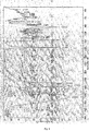
Fig.1 represents the synthetic route of YC-1 (24-methylene-cholest-3β,5α,6β, 19-tetrol). -
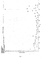
Fig.2 represents YC-1's c-nmr spectra. -
Fig.3 represents YC-1's 1h nmr spectra. -
Fig.4 represents YC-1's infrared (ir) spectra. -
Fig.5 represents YC-1's masspectrometry (ms). -
Fig.6 indicates YC-1 can reduce hemisphere infarct volume in permanent middle cerebral artery occlusion (MCAO). -
Fig.7 represents YC-1's protection on rats with permanent middle cerebral artery occlusion (MCAO). -
Fig.8 represents YC-1's effect on neurological function score of rabbit spinal cord ischemia model. -
Fig.9 represents YC-1's effect on behavior of rabbit spinal cord ischemia model. -
Fig.10 indicates YC-1 can increase the number of spinal cord anterior horn neurons after rabbit spinal cord ischemia. -
Fig.11 is a HE dye result indicating YC-1's effect on spinal cord anterior horn neurons after rabbit spinal cord ischemia. -
Fig.12 represents CGNs morphology revealed by phase contrast microscope after sequential treatment of YC-1 and H/R. -
Fig.13 is a FDA dye result of viable cells indicating YC-1 can inhibit H/R induced CGNs apoptosis. -
Fig.14 is the post-apoptosis Hoechst33258 nuclear morphology analysis indicating YC-1 can inhibit H/R induced CGNs apoptosis. -
Fig.15 represents CGNs morphology revealed by phase contrast microscope after sequential treatment of YC-1 and LK. -
Fig.16 is a FDA dye result of viable cells indicating YC-1 can inhibit LK induced CGNs apoptosis. -
Fig.17 is the post-apoptosis Hoechst33258 nuclear morphology analysis indicating YC-1 can inhibit LK induced CGNs apoptosis. -
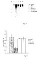
Fig.18 is the agarose gel electrophoresis analysis indicating YC-1 can reduce DNA fragments produced from LK induced CGNs apoptosis. -
Fig.19 is a FDA dye result of viable cells indicating YC-1 can inhibit Glu induced CGNs apoptosis. - Starting with cheap hyodeoxycholic acid, we synthesized YC-1 and some of it analogues through 14 steps of organic reactions; Then characterized the intermediates and target chemicals using spectrum technology.
- It was synthesized through reaction of 3-acetoxy-cholest-5-ene-24-ketone and N-bromosuccinimide. The yield was low and there were multiple isomers in the product. The reaction was optimized by trying different conditions like solvents, temperature etc. Then tried different purification methods like recrystallization, solvent precipitation, chromatography etc.
- It was synthesized by remote oxidative ring-closure of 6-OH in 3-acetoxy-cholest-5-Br-6-OH-24-ketone. Lead tetraacetate and diacetyl iodobenzene were used for the oxidation under uv light and in ultrasonic wave respectively and also optimized the reaction condition.
- It was done by oxidation of 5,6 double bond in 3,19-diacetoxy-5ene-24-ketone. Oxidation products got with different oxidant have different cis-trans ratio and yield. Totally there oxidants were used for the oxidation: m-chloroperoxybenzoic acid, hydrogen peroxide/formic acid system, and potassium permanganate/copper sulfate system. Then optimized the reaction condition.
- YC-1 and the intermediates were characterized data like IR, 1H NMR, 13C NMR, MS and EA etc got from techniques like IR, NMR, MS and EA etc. Among whichYC-1's spectral data is:
- Melting point: 223-225°C
- Elemental analysis: C28H4804
- Measured value: C, 75.20; H, 10.36
- Theoretical value: C, 74.95; H, 10.78
- MS : [M-H2O]+=432,
- IR : 3416 , 3028 , 2936 , 2868 , 1642, 1464 , 1377 , 1057
- 5.25(1H,d,6β-OH), 4.71(1H,d,J=1.5Hz,28-CH), 4.65(1H,d,J=1.5Hz,28-CH), 4.51(1H,d, 19-OH), 4.16(1H,d,3-CH), 4.01(1H,d,19-CH), 3.84(1H,m,3-CH), 3.64(1H,s,5-OH), 3.23 (1H,m,6-CH), 2.25(1H,m,25-CH), 1.13(6H,d,26-CH3,27-CH3), 1.01(3H,s, 19 -CH3), 0.87(3H,d, 21-CH3), 0.67(3H,s,18-CH3).
- 156.6(C), 107.3(CH2), 75.4(C), 74.7(CH), 66.7(CH), 63.0(CH2), 57.4(CH), 56.7(CH), 45.9(CH), 43.5(C), 43.3(CH), 42.8(C), 41.3(CH2), 40.9(CH), 37.4(CH2), 36.1(CH), 33.9(CH2), 31.8(CH2), 31.4(CH2), 30.5(CH2), 28.7(CH2), 27.9(CH2), 24.70(CH2), 22.64(CH3),22.57(CH3), 19.4(CH3), 13.1(CH3) .
-
- (1) 3-hydroxycholest series analogues
- (2) 3,6-dihydroxycholest series analogues
- (3) 3,5,6-trihydroxycholest series analogues
- (4) 3,19-dihydroxycholest series analogues
- (5) 3,5,6,19-tetrahydroxycholest series analogues
- (6) 3-hydroxycholest dimer
- (7) 3,5,6,19-tetrahydroxycholest dimmer
- (8) The side chain at position 17 of chemicals in (1)-(7) can be: 24-carbonyl, 24-methylene, 24-hydroxy or 24-hydrazone etc. Then characterized them using techniques like IR, NMR, MS and EA etc.
- YC-1 has poor water solubility. To improve its solubility, the 3-hydroxy group in 3-hydroxy-cholest-5-ene-24-ketone went through hydrophilic modification.
- 25 healthy male SD rats were divided into 3 groups randomly:
- YC-1 group (n=11): 10min, 6h, 24h and 7d after occluding the middle cerebral artery, 12mg/Kg YC-1 was continuously injected intraperitoneally.
- DMSO group (n=11): injected same volume of DMSO (1ml/Kg) in the same way.
- Sham group (n=3): just very short intraluminal thread was inserted and didn't occlude the MCA.
- The following method was a modification of Zea Longa's method (Longa EZ, Weinstein PR, Carlson S, Cummins R. Reversible middle cerebral artery occlusion without craniectomy in rats. Stroke, 1989, 20: 84-91).
- (1) The rats drinked freely but fasted one night before the operation.
- (2) 10min before anesthesia, intraperitoneally (ip) injected atropine sulfate (0.5mg/Kg).
- (3) Narcotized the rat with ip injected 10% chloral hydrate, 3.5ml anesthetic per Kg rat; The areflexia of hindlimb retraction and corneal were used as anesthesia index.
- (4) Supinely fixed the rat after anesthesia, cut the cervical skin and subcutaneous tissue, separated left cervical skin from digastric, sternocleidomastoid and omohyoid, exposed the common carotid artery (CCA) bifurcation, including the internal and external carotid artery (ICA, ECA) extended from CCA bifurcation in the triangle formed by the three muscles.
- (5) Ligated the CCA and ECA at the beginning of ascending pharyngeal artery as follows: prepared a thread at the proximal end of ICA, incised CCA at the bifurcation, inserted a 5cm nylon intraluminal thread to ICA's proximal end, stopped when inserted about 17-18mm and felt resistance. At this moment the nylon thread entered ICA and anterior cerebral artery, blocked all blood sources for MCA, and timed immediately when blocking the blood flow.
- (6) Fastened the prepared thread at proximal ICA, trimmed the redundant thread, disinfected with iodine tincture, sutured the skin and returned the rat back to cage.
- (7) Rectum temperature was measured using Meikangti electronic temperature measure instrument the whole process and it was strictly controlled at 36.5-37.5°C.
- (8) Except using shorter intraluminal thread (10mm) which didn't reach MCA, all other steps for the Sham group were the same as above.
- Result: By visual inspection, the brain tissue in the Sham group had normal color and morphology with no pallor and swelling; Those in the DMSO group showed obvious pallor and swelling and no obvious pallor and swelling in the YC-1 group. TTC dye showed that YC-1 group had smaller infarct volume than DMSO group (the infarct size was 32±11% and 62±8% respectively) while no ischemia was observed in the Sham group.
- 24 male new Zealand white rabbits were divided evenly into 3 groups (n=8): YC-1 group: 30min before spinal cord ischemia, 8mg/Kg YC-1 was intravenous injected through the rabbit's ear.
- DMSO group: injected same volume of DMSO (1ml/Kg) in the same way.
- Sham group: just exposed the abdominal aorta but didn't clip it.
- Referring to Johnson etc, the model was prepared as follows:
- (1) The rabbits drinked freely but fasted one night before the operation.
- (2) Anesthetized the rabbit by ear intravenous injected 3% sodium pentobarbital (30mg/Kg). On the other side of the ear set a vein-detaining needle (24G) for medicine and other fluid infusion.
- (3) Set an artery-detaining needle (24G) in the right ear artery for monitoring the proximal arterial pressure and blood sampling during the operation.
- (4) Set an artery-detaining needle (24G) in a femoral artery for monitoring the distal arterial pressure during the operation.
- (5) Supinely fixed the rabbit, incised at the median abdomen, exposed the abdominal aorta.
- (6) Injected heparin (150U/Kg) through the vein-detaining needle, then clipped the abdominal aorta at the 0.5-1.0cm spot below origin of the left renal artery to cause spinal cord ischemia.
- (7) Once the abdominal aorta was blocked, the distal arterial pressure decreased immediately and the femoral arterial pulsation disappeared.
- (8) Removed the clip 20min after ischemia and sutured the abdomen.
- (9) Then intramuscularly injected 40,000U gentamicin, returned the rabbit back to cage and observed for 48h.
- (10) The proximal arterial pressure, distal arterial pressure and heart rate (Spacelab, US) were continuously monitored during the operation. Rectum temperature was maintained at 38.5±0.5°C using heating pad and roasting lamp.
- (11) Blood gas (AVL-2, Switzerland) and glucose (One Touch II, USA) were monitored by ear artery blood sampling 10min before ischemia, 10min after ischemia and 10min after reperfusion.
- Result: See
Fig.8 for neurological function score of animals from each group. Hindlimb nerve in the sham group functioned normally (4points) during the whole observation period. None rabbit in DMSO group could stand. Six rabbits in the CPA (YC-1)group could stand and the other two rabbits' hindlimbs also showed obvious movement. At each observation time point, neurological function score in YC-1 and sham groups were obviously higher than that in DMSO group (p<0.05). Histopathological examination showed that the lumbar spinal cord in DMSO group was severely injured manifesting as reduction of motor neurons, disappearance of nissl bodies and nucleus and vacuolar degeneration, while lumbar spinal cord injury in YC-1 group was obviously relieved, seeFig.9 . Comparing to DMSO group, number of normal spinal cord anterior horn neurons in CPA(YC-1) and sham groups increased evidently (p<0.01), as shown inFig.10 . - According to Yan, etc[34], prepared the neurons as follows:
- (1) Took a clean 7-8d SD rat weighing about 15-20g, separated the cerebella under sterile condition.
- (2) Got rid of the meninges and vessels in Kreb's anatomy solution ( Ca2+ and Mg2+ free ).
- (3) Sheared the cerebella into pieces of 1mm3 with ophthalmic scissors.
- (4) Digested in 0.25g/L trypsin solution for 15min at 37°C.
- (5) Stopped the digestion with solution containing 0.5g/L trypsin inhibitor and 0.05g/L Dnase I, pipetted up and down to get single cell suspension.
- (6) Centrifuged at 200g for 5min, rinsed the precipitation once with rinse solution.
- (7) Span down again at 200g for 5min, discarded the supernatant.
- (8) Diluted the cell pellet to cell density of 1.5-1.8 × 10⁶ cells/ml with BME medium containing 10% (v/v) FBA and 25mM KCl.
- (9) Inoculated them into cell culture dishes pre-coated with polylysine and incubated at 37°C incubator with 5% CO2.
- (10) Twenty-four hours after inoculation, added 10 µmol/L Ara-C to inhibit non-neuron cells' growth and proliferation and to make the purity of CGNs above 95%.
- (11) Added glucose to 5mM on day7 incubation to supplement energy needed by cell metabolism.
- (12) Did the following experiment on day8.
- Referred to Seko (Seko Y, Tobe K, Ueki K, et al. Hypoxia and hypoxia/reoxygenation activate Raf-1, mitogen-activated protein kinase kinase, mitogen-activated protein kinases, and S6 kinase in cultured rat cardiac myocytes. Circ Res. 1996 Jan;78(1):82-90) etc for the establishment method:
- (1) On day8, put the treated CGNs in airtight box with oxygen consumption reagent and indicator and incubated at 37°C for 3h.
- (2) Transferred the cells to 37°C incubator with 5% CO2 for reoxygenation treatment.
- The following were done referring to Yan (Yan G M, Irwin R P, Lin S Z, et al. Diphenylhydantoin induces apoptotic cell death of cultured rat cerebellar granule neurons. J Pharmacol Exp Ther, 1995, 274(2): 983-8) et al.:
- (1) CGNs were cultured in 35mm dishes, on day8, removed the medium after various factors treatment, rinsed the cells twice with 4°C PBS containing Ca2+ and Mg2+.
- (2) Fixed the cells with 40g/L paraformaldehyde (4°C, dissolved in PBS) for 10min.
- (3) Removed the fixation solution, rinsed the cells twice with distilled water, air dried at 4°C.
- (4) Observed the cell morphological change and randomly photographed under inverted fluorescence microscope (Olympus).
-
- (1) Before hypoxia, CGNs cell bodies were plump and lucent; Cell processes and the network formed by them were tight, clear and intact.
- (2) After 3h hypoxia and 24h reoxygenation (H/R) treatment, the CGNs cell body dwindled; The structure became unclear; The processes and network showed disorder and fracture.
- (3) First treated the cells with 10µM MK801 or 12µM YC-1 or 12µM YC-2 for 3h, then did the H/R treatment and observation. Most CGNs cell body kept intact, plump and lucent; The processes and network were tight, clear and intact.
-
- (1) After H/R treatment, neuronal viability decreased obviously (16.56±3.21%).
- (2) After treated by 10µM MK801 or 12µM YC-1 or 12µM YC-2, neuronal viability reached 86.73±3.21%, 84.29±1.26% and 83.54±4.78% respectively.
-
- (1) Normal CGNs had intact nucleus and the chromatin distributed evenly.
- (2) After H/R treatment, observed CGNs nucleus and chromatin pyknosis together with apoptotic bodies.
- (3) First treated the cells with 12µM YC-1 YC-2, then did the H/R treatment and observation. Most CGNs showed normal nuclear morphology and even chromatin distribution.
- The methods for rat CGNs cell culture, cellular morphologic observation and neuronal viability measurement were the same as above. Then check the in vitro cultured cerebellar granule neurons death through agarose gel electrophoresis according to reference[34]:
- (1) inoculated CGNs in 35mm2 dishes.
- (2) On day8, 24h after adding various medicines, removed the medium, rinsed the cells twice with PBS.
- (3) Span down the collected cells with a low temperature high speed centrifuge (Biofuge22R, Heraeus, Germany) at 5000rpm, 4°C for 5min.
- (4) Resuspended the cell pellet with 600µl buffer containing 10mmol/L Tris-HCl, 10 mmol/L EDTA and 2g/L Triton X-100 (pH 7.5) and incubated for 15min.
- (5) Span down at 12000rpm, 4°C for 10min.
- (6) Extracted the supernatant once with equal volume of phenol, span down.
- (7) Extracted the supernatant once with equal volume of phenol-chloroform (1:1), span down.
- (8) Took the supernatant, added 300mmol/L Sodium Acetate and equal volume of isopropanol, precipitated for overnight.
- (9) Span down the pellet, rinsed 2-3 times with 70% ethanol.
- (10) After drying, added some TE (Tris 10mmol.L-1, EDTA 1.0 mmol.L-1, pH 7.4) and 0.6g.L-1 RNase A and incubated at 37°C for 30min.

- (11) Analyzed the samples with 20g/L agarose gel electrophoresis for 1h.
- (12) Dyed with ethidium bromide and photographed under UV light.
-
- (1) Transferred above cells to serum free medium with 5mM KCl and incubated for another 24h, the cell dodies dwindled and the nucleus fractured; FDA fluorescence staining indicated only 40.2±14.04% CGNs survived.
- (2) If 12µM YC-1 or YC-2 were added 3h before and the same time changing to low potassium medium, then cultured the cells in the low potassium medium for another 24h, most CGNs kept intact cell bodies and processes; FDA staining indicated 94.3± 14.02% and 84.2±13.34% CGNs survived respectively.
- (4) These results showed marine steroid YC-1 and YC-2 obviously improved rat CGNs' viability in low potassium medium.
-
- (1) Isolatation and primary culture of rat CGNs.
- (2) Morphology observation of neuron cell body and processes under phase contrast microscopy.
- (3) Analysis of neuronal metabolic activity and viability using fluorescein diacetate (FDA) staining.
-
- (1) Took rat CGNs in vitro cultured for 8 days, added 150µM Glu, incubated for 24h and observed under phase contrast microscopy. The neuronal cell bodies dwindled obviously, the nucleus showed pyknosis and the processes fractured or disappeared.
- (2) If 12µM YC-1 or YC-2 were added 1h before adding Glu, then observed 24h later. The result showed that both YC-1 and YC-2 could obviously inhibited Glu induced CGNs apoptosis.
- (3) CGNs' Viability analysis using FDA staining showed that 12µM YC-1 and YC-2 clearly improved CGNs' viability (respectively 92.6±2.59% and 91.6±10.11%) comparing to the Glu control group (29.5±8.23%).
Claims (2)
- An application of marine steroid chemicals in preparing medicines for neuron damage.
- An application according to claim1, the marine steroid chemical is 24-methylene-cholest-3β,5α,6β,19-tetrol.
Applications Claiming Priority (2)
| Application Number | Priority Date | Filing Date | Title |
|---|---|---|---|
| CNA2006100369261A CN101164540A (en) | 2006-08-03 | 2006-08-03 | Application of marine steroids compound in preparing medicine for treating neuronal damage |
| PCT/CN2006/002235 WO2008017216A1 (en) | 2006-08-03 | 2006-08-31 | The application of marine steroid in preparing the medicine of treating neurons damaging |
Publications (2)
| Publication Number | Publication Date |
|---|---|
| EP2047853A1 true EP2047853A1 (en) | 2009-04-15 |
| EP2047853A4 EP2047853A4 (en) | 2011-01-26 |
Family
ID=39032614
Family Applications (1)
| Application Number | Title | Priority Date | Filing Date |
|---|---|---|---|
| EP06775552A Withdrawn EP2047853A4 (en) | 2006-08-03 | 2006-08-31 | USE OF MARINE STEROID IN THE PREPARATION OF A MEDICAMENT FOR THE TREATMENT OF NEURONAL INJURIES |
Country Status (5)
| Country | Link |
|---|---|
| US (1) | US20100152150A1 (en) |
| EP (1) | EP2047853A4 (en) |
| JP (1) | JP2009545535A (en) |
| CN (1) | CN101164540A (en) |
| WO (1) | WO2008017216A1 (en) |
Cited By (1)
| Publication number | Priority date | Publication date | Assignee | Title |
|---|---|---|---|---|
| EP3415145A3 (en) * | 2017-06-12 | 2019-03-06 | Arjil Biotech Holding Company Limited | Method for treating stroke or reducing nerve injury |
Families Citing this family (14)
| Publication number | Priority date | Publication date | Assignee | Title |
|---|---|---|---|---|
| FI2753632T3 (en) | 2011-09-08 | 2023-08-02 | Sage Therapeutics Inc | Neuroactive steroids, compositions, and uses thereof |
| CN105012313B (en) * | 2014-04-25 | 2018-03-16 | 广州市赛普特医药科技股份有限公司 | Application of 5α-androsta-3β, 5,6β-triol and their analogues in preventing or treating altitude sickness caused by hypobaric hypoxia |
| RS67266B1 (en) * | 2013-03-13 | 2025-10-31 | Sage Therapeutics Inc | Neuroactive steriods and methods of use thereof |
| ES2970222T3 (en) | 2014-06-18 | 2024-05-27 | Sage Therapeutics Inc | Oxysterols and procedures for their use |
| FI3828194T3 (en) | 2015-07-06 | 2025-07-17 | Sage Therapeutics Inc | Oxysterols and methods of use thereof |
| MX390271B (en) | 2015-07-06 | 2025-03-20 | Sage Therapeutics Inc | Oxysterols and methods of use thereof |
| US20190330259A1 (en) | 2016-04-01 | 2019-10-31 | Sage Therapeutics, Inc. | Oxysterols and methods of use thereof |
| WO2017193046A1 (en) | 2016-05-06 | 2017-11-09 | Sage Therapeutics, Inc. | Oxysterols and methods of use thereof |
| JP7149266B2 (en) | 2016-09-30 | 2022-10-06 | セージ セラピューティクス, インコーポレイテッド | Methods as C7-Substituted Oxysterols and NMDA Modulators |
| MA46566A (en) | 2016-10-18 | 2019-08-28 | Sage Therapeutics Inc | OXYSTEROLS AND THEIR METHODS OF USE |
| IL293231B2 (en) | 2016-10-18 | 2025-10-01 | Sage Therapeutics Inc | Oxysterols and methods of using them |
| CN108524525A (en) * | 2018-06-21 | 2018-09-14 | 中山大学 | 3 β of courage Gona-4-ene-3,6 salmefamols are preparing the application in treating neure damage drug |
| CN108659089B (en) * | 2018-07-31 | 2020-08-25 | 温州医科大学 | A kind of sterol compound with antioxidant effect and its application in preparing medicine |
| CN111012765A (en) * | 2019-11-29 | 2020-04-17 | 福建中医药大学 | Application of pseudopterogorgia-hancei alcohol as active substance in preparing medicine for treating cerebral apoplexy |
Family Cites Families (1)
| Publication number | Priority date | Publication date | Assignee | Title |
|---|---|---|---|---|
| CN1448400A (en) * | 2003-05-06 | 2003-10-15 | 中山大学 | Synthetic method of 24-methylene-cholesterol-3 beta, 5 alpha, 6 beta,19-tetraalcohol |
-
2006
- 2006-08-03 CN CNA2006100369261A patent/CN101164540A/en active Pending
- 2006-08-31 US US12/376,217 patent/US20100152150A1/en not_active Abandoned
- 2006-08-31 JP JP2009522070A patent/JP2009545535A/en active Pending
- 2006-08-31 EP EP06775552A patent/EP2047853A4/en not_active Withdrawn
- 2006-08-31 WO PCT/CN2006/002235 patent/WO2008017216A1/en not_active Ceased
Non-Patent Citations (2)
| Title |
|---|
| No further relevant documents disclosed * |
| See also references of WO2008017216A1 * |
Cited By (1)
| Publication number | Priority date | Publication date | Assignee | Title |
|---|---|---|---|---|
| EP3415145A3 (en) * | 2017-06-12 | 2019-03-06 | Arjil Biotech Holding Company Limited | Method for treating stroke or reducing nerve injury |
Also Published As
| Publication number | Publication date |
|---|---|
| EP2047853A4 (en) | 2011-01-26 |
| US20100152150A1 (en) | 2010-06-17 |
| CN101164540A (en) | 2008-04-23 |
| WO2008017216A1 (en) | 2008-02-14 |
| JP2009545535A (en) | 2009-12-24 |
Similar Documents
| Publication | Publication Date | Title |
|---|---|---|
| Sabogal-Guáqueta et al. | Linalool attenuates oxidative stress and mitochondrial dysfunction mediated by glutamate and NMDA toxicity | |
| Li et al. | Astragaloside IV protects blood-brain barrier integrity from LPS-induced disruption via activating Nrf2 antioxidant signaling pathway in mice | |
| Yang et al. | Hydroxysafflor yellow a protects brain microvascular endothelial cells against oxygen glucose deprivation/reoxygenation injury: involvement of inhibiting autophagy via class I PI3K/Akt/mTOR signaling pathway | |
| Lazarowski et al. | Neuronal and glial expression of the multidrug resistance gene product in an experimental epilepsy model | |
| US20100152150A1 (en) | The application of marine steroid in preparing the medicine of treating neurons damaging | |
| Yan et al. | Autophagy activation is involved in neuroprotection induced by hyperbaric oxygen preconditioning against focal cerebral ischemia in rats | |
| Sarmah et al. | Sirtuin-1-mediated NF-κB pathway modulation to mitigate inflammasome signaling and cellular apoptosis is one of the neuroprotective effects of intra-arterial mesenchymal stem cell therapy following ischemic stroke | |
| Chen et al. | Panax notoginseng saponins inhibit ischemia-induced apoptosis by activating PI3K/Akt pathway in cardiomyocytes | |
| Zhang et al. | Ursolic acid reduces oxidative stress to alleviate early brain injury following experimental subarachnoid hemorrhage | |
| US7235267B1 (en) | Brain cell or nerve cell-protecting agents comprising medicinal ginseng | |
| Zhang et al. | Neuroprotective effects of leptin on cerebral ischemia through JAK2/STAT3/PGC-1-mediated mitochondrial function modulation | |
| Qin et al. | Geniposide alleviated bile acid-associated NLRP3 inflammasome activation by regulating SIRT1/FXR signaling in bile duct ligation-induced liver fibrosis | |
| Zhou et al. | Xuesaitong exerts long-term neuroprotection for stroke recovery by inhibiting the ROCKII pathway, in vitro and in vivo | |
| AT506095A1 (en) | USE OF PROTEASES | |
| Kang et al. | Sappanone A protects mice against cisplatin-induced kidney injury | |
| CN101683348B (en) | Application of cholestane-3 beta, 5 alpha, 6 beta-triol in preparation of neuronal protection medicine | |
| JP2015028076A (en) | Use of an extract from rabbit skin inflamed by vaccinia virus in the manufacture of a medicament for the treatment of acute cerebrovascular disease | |
| Liu et al. | Involvement of estrogen receptor activation in kaempferol-3-O-glucoside's protection against aging-related cognition impairment and microglial inflammation | |
| Yulug et al. | Rifampicin attenuates brain damage in focal ischemia | |
| CN101129356A (en) | Application of lovastatin in the preparation of drugs for inhibiting apoptosis of bone marrow mesenchymal stem cells | |
| TW201932130A (en) | Lipocalin-type prostaglandin D2 synthase production promoter | |
| JP6368219B2 (en) | Estrogen receptor β activator | |
| KR101314694B1 (en) | Therapeutic uses of steroidal compounds | |
| Yuan et al. | Total Saponins from Panax Japonicus Promote Lipolysis of Aging Fat Through the Tyrosine Hydroxylase Signaling Pathway | |
| KR20250115035A (en) | Pharmaceutical composition for preventing or treating endometriosis comprising S-allyl-L-cysteine |
Legal Events
| Date | Code | Title | Description |
|---|---|---|---|
| PUAI | Public reference made under article 153(3) epc to a published international application that has entered the european phase |
Free format text: ORIGINAL CODE: 0009012 |
|
| 17P | Request for examination filed |
Effective date: 20090123 |
|
| AK | Designated contracting states |
Kind code of ref document: A1 Designated state(s): AT BE BG CH CY CZ DE DK EE ES FI FR GB GR HU IE IS IT LI LT LU LV MC NL PL PT RO SE SI SK TR |
|
| AX | Request for extension of the european patent |
Extension state: AL BA HR MK RS |
|
| RIN1 | Information on inventor provided before grant (corrected) |
Inventor name: CAI, XIANG Inventor name: SANG, HANFEI Inventor name: LIU, AILING Inventor name: SU, XINGWEN Inventor name: YIN, WEI Inventor name: HUANG, YIJUN Inventor name: QIU, PENGXIN Inventor name: YAN, GUANGMEI |
|
| A4 | Supplementary search report drawn up and despatched |
Effective date: 20101228 |
|
| STAA | Information on the status of an ep patent application or granted ep patent |
Free format text: STATUS: THE APPLICATION IS DEEMED TO BE WITHDRAWN |
|
| 18D | Application deemed to be withdrawn |
Effective date: 20110301 |