EP2042515A1 - Ligand binding domains of nuclear receptors in controllable form and methods involving the same - Google Patents
Ligand binding domains of nuclear receptors in controllable form and methods involving the same Download PDFInfo
- Publication number
- EP2042515A1 EP2042515A1 EP07291163A EP07291163A EP2042515A1 EP 2042515 A1 EP2042515 A1 EP 2042515A1 EP 07291163 A EP07291163 A EP 07291163A EP 07291163 A EP07291163 A EP 07291163A EP 2042515 A1 EP2042515 A1 EP 2042515A1
- Authority
- EP
- European Patent Office
- Prior art keywords
- protein
- tag
- receptor
- ligand
- cell
- Prior art date
- Legal status (The legal status is an assumption and is not a legal conclusion. Google has not performed a legal analysis and makes no representation as to the accuracy of the status listed.)
- Ceased
Links
- 108020005497 Nuclear hormone receptor Proteins 0.000 title claims abstract description 111
- 102000006255 nuclear receptors Human genes 0.000 title claims abstract description 111
- 108020004017 nuclear receptors Proteins 0.000 title claims abstract description 111
- 108020001756 ligand binding domains Proteins 0.000 title claims abstract description 72
- 238000000034 method Methods 0.000 title claims abstract description 35
- 108090000623 proteins and genes Proteins 0.000 claims abstract description 202
- 102000004169 proteins and genes Human genes 0.000 claims abstract description 180
- 239000003446 ligand Substances 0.000 claims abstract description 54
- 238000012360 testing method Methods 0.000 claims abstract description 34
- 238000012216 screening Methods 0.000 claims abstract description 7
- -1 isoprenyl salt Chemical class 0.000 claims description 65
- 210000004027 cell Anatomy 0.000 claims description 62
- 230000027455 binding Effects 0.000 claims description 58
- 239000000556 agonist Substances 0.000 claims description 33
- 238000002866 fluorescence resonance energy transfer Methods 0.000 claims description 32
- 108091008731 RAR-related orphan receptors α Proteins 0.000 claims description 28
- 239000003550 marker Substances 0.000 claims description 27
- 239000012190 activator Substances 0.000 claims description 26
- 150000007523 nucleic acids Chemical class 0.000 claims description 26
- 102000039446 nucleic acids Human genes 0.000 claims description 22
- 108020004707 nucleic acids Proteins 0.000 claims description 22
- 238000003556 assay Methods 0.000 claims description 21
- 102000005962 receptors Human genes 0.000 claims description 21
- 108020003175 receptors Proteins 0.000 claims description 21
- 239000003599 detergent Substances 0.000 claims description 18
- 238000003776 cleavage reaction Methods 0.000 claims description 15
- 230000007017 scission Effects 0.000 claims description 15
- 230000003993 interaction Effects 0.000 claims description 13
- YFVGRULMIQXYNE-UHFFFAOYSA-M lithium;dodecyl sulfate Chemical compound [Li+].CCCCCCCCCCCCOS([O-])(=O)=O YFVGRULMIQXYNE-UHFFFAOYSA-M 0.000 claims description 13
- 239000000126 substance Substances 0.000 claims description 13
- 238000002877 time resolved fluorescence resonance energy transfer Methods 0.000 claims description 13
- 229910021653 sulphate ion Inorganic materials 0.000 claims description 12
- 159000000011 group IA salts Chemical class 0.000 claims description 11
- 239000005557 antagonist Substances 0.000 claims description 10
- 229920006395 saturated elastomer Polymers 0.000 claims description 10
- 108091008773 RAR-related orphan receptors γ Proteins 0.000 claims description 9
- BVKZGUZCCUSVTD-UHFFFAOYSA-L Carbonate Chemical compound [O-]C([O-])=O BVKZGUZCCUSVTD-UHFFFAOYSA-L 0.000 claims description 8
- 108091008730 RAR-related orphan receptors β Proteins 0.000 claims description 8
- 201000010099 disease Diseases 0.000 claims description 8
- 208000037265 diseases, disorders, signs and symptoms Diseases 0.000 claims description 8
- 238000004519 manufacturing process Methods 0.000 claims description 8
- 102000016978 Orphan receptors Human genes 0.000 claims description 7
- 108070000031 Orphan receptors Proteins 0.000 claims description 7
- 238000004113 cell culture Methods 0.000 claims description 7
- 102000003702 retinoic acid receptors Human genes 0.000 claims description 7
- 108090000064 retinoic acid receptors Proteins 0.000 claims description 7
- 241000238631 Hexapoda Species 0.000 claims description 6
- 108010085012 Steroid Receptors Proteins 0.000 claims description 6
- QAOWNCQODCNURD-UHFFFAOYSA-L Sulfate Chemical compound [O-]S([O-])(=O)=O QAOWNCQODCNURD-UHFFFAOYSA-L 0.000 claims description 6
- 238000004020 luminiscence type Methods 0.000 claims description 6
- 108010011170 Ala-Trp-Arg-His-Pro-Gln-Phe-Gly-Gly Proteins 0.000 claims description 5
- 208000029078 coronary artery disease Diseases 0.000 claims description 4
- 238000012258 culturing Methods 0.000 claims description 4
- 239000003862 glucocorticoid Substances 0.000 claims description 4
- 102000000584 Calmodulin Human genes 0.000 claims description 3
- 108010041952 Calmodulin Proteins 0.000 claims description 3
- 229920002101 Chitin Polymers 0.000 claims description 3
- 208000001132 Osteoporosis Diseases 0.000 claims description 3
- 125000000217 alkyl group Chemical group 0.000 claims description 3
- 210000004102 animal cell Anatomy 0.000 claims description 3
- MOTZDAYCYVMXPC-UHFFFAOYSA-N dodecyl hydrogen sulfate Chemical compound CCCCCCCCCCCCOS(O)(=O)=O MOTZDAYCYVMXPC-UHFFFAOYSA-N 0.000 claims description 3
- 210000005253 yeast cell Anatomy 0.000 claims description 3
- 208000032928 Dyslipidaemia Diseases 0.000 claims description 2
- 241000588724 Escherichia coli Species 0.000 claims description 2
- 101000668058 Infectious salmon anemia virus (isolate Atlantic salmon/Norway/810/9/99) RNA-directed RNA polymerase catalytic subunit Proteins 0.000 claims description 2
- 208000017170 Lipid metabolism disease Diseases 0.000 claims description 2
- 230000002060 circadian Effects 0.000 claims description 2
- 239000003814 drug Substances 0.000 claims description 2
- 230000004770 neurodegeneration Effects 0.000 claims description 2
- 208000015122 neurodegenerative disease Diseases 0.000 claims description 2
- 230000006337 proteolytic cleavage Effects 0.000 claims description 2
- 208000019116 sleep disease Diseases 0.000 claims description 2
- 208000020685 sleep-wake disease Diseases 0.000 claims description 2
- 201000001320 Atherosclerosis Diseases 0.000 claims 1
- 102000007451 Steroid Receptors Human genes 0.000 claims 1
- 235000018102 proteins Nutrition 0.000 description 147
- 108020004414 DNA Proteins 0.000 description 23
- 239000000370 acceptor Substances 0.000 description 23
- 150000001413 amino acids Chemical class 0.000 description 20
- FAPWRFPIFSIZLT-UHFFFAOYSA-M Sodium chloride Chemical compound [Na+].[Cl-] FAPWRFPIFSIZLT-UHFFFAOYSA-M 0.000 description 19
- 229940024606 amino acid Drugs 0.000 description 19
- 235000001014 amino acid Nutrition 0.000 description 18
- 108010060434 Co-Repressor Proteins Proteins 0.000 description 16
- 102000008169 Co-Repressor Proteins Human genes 0.000 description 16
- 239000011324 bead Substances 0.000 description 15
- 239000013598 vector Substances 0.000 description 15
- 238000000746 purification Methods 0.000 description 14
- 108091005804 Peptidases Proteins 0.000 description 13
- 239000004365 Protease Substances 0.000 description 13
- 230000014509 gene expression Effects 0.000 description 13
- 230000019491 signal transduction Effects 0.000 description 13
- 238000013518 transcription Methods 0.000 description 13
- 230000001270 agonistic effect Effects 0.000 description 12
- 230000000694 effects Effects 0.000 description 12
- 230000035897 transcription Effects 0.000 description 12
- 102000004971 Nuclear receptor coactivator 2 Human genes 0.000 description 9
- 108090001144 Nuclear receptor coactivator 2 Proteins 0.000 description 9
- HVYWMOMLDIMFJA-DPAQBDIFSA-N cholesterol Chemical compound C1C=C2C[C@@H](O)CC[C@]2(C)[C@@H]2[C@@H]1[C@@H]1CC[C@H]([C@H](C)CCCC(C)C)[C@@]1(C)CC2 HVYWMOMLDIMFJA-DPAQBDIFSA-N 0.000 description 9
- 108020004999 messenger RNA Proteins 0.000 description 9
- 239000011780 sodium chloride Substances 0.000 description 9
- 102000035195 Peptidases Human genes 0.000 description 8
- 230000006870 function Effects 0.000 description 8
- 108090000765 processed proteins & peptides Proteins 0.000 description 8
- 238000001890 transfection Methods 0.000 description 8
- 230000004568 DNA-binding Effects 0.000 description 7
- 102100039614 Nuclear receptor ROR-alpha Human genes 0.000 description 7
- YBJHBAHKTGYVGT-ZKWXMUAHSA-N (+)-Biotin Chemical compound N1C(=O)N[C@@H]2[C@H](CCCCC(=O)O)SC[C@@H]21 YBJHBAHKTGYVGT-ZKWXMUAHSA-N 0.000 description 6
- 102000004190 Enzymes Human genes 0.000 description 6
- 108090000790 Enzymes Proteins 0.000 description 6
- 239000007983 Tris buffer Substances 0.000 description 6
- 230000004913 activation Effects 0.000 description 6
- 108010004469 allophycocyanin Proteins 0.000 description 6
- BHYOQNUELFTYRT-DPAQBDIFSA-N cholesterol sulfate Chemical compound C1C=C2C[C@@H](OS(O)(=O)=O)CC[C@]2(C)[C@@H]2[C@@H]1[C@@H]1CC[C@H]([C@H](C)CCCC(C)C)[C@@]1(C)CC2 BHYOQNUELFTYRT-DPAQBDIFSA-N 0.000 description 6
- 229940088598 enzyme Drugs 0.000 description 6
- 238000000926 separation method Methods 0.000 description 6
- LENZDBCJOHFCAS-UHFFFAOYSA-N tris Chemical compound OCC(N)(CO)CO LENZDBCJOHFCAS-UHFFFAOYSA-N 0.000 description 6
- BHYOQNUELFTYRT-UHFFFAOYSA-N Cholesterol sulfate Natural products C1C=C2CC(OS(O)(=O)=O)CCC2(C)C2C1C1CCC(C(C)CCCC(C)C)C1(C)CC2 BHYOQNUELFTYRT-UHFFFAOYSA-N 0.000 description 5
- 101001103036 Homo sapiens Nuclear receptor ROR-alpha Proteins 0.000 description 5
- 102100023421 Nuclear receptor ROR-gamma Human genes 0.000 description 5
- 102100037486 Reverse transcriptase/ribonuclease H Human genes 0.000 description 5
- 230000015572 biosynthetic process Effects 0.000 description 5
- 230000003915 cell function Effects 0.000 description 5
- 235000012000 cholesterol Nutrition 0.000 description 5
- 150000001875 compounds Chemical class 0.000 description 5
- 238000001514 detection method Methods 0.000 description 5
- 238000011161 development Methods 0.000 description 5
- 230000018109 developmental process Effects 0.000 description 5
- 238000002955 isolation Methods 0.000 description 5
- 229910052747 lanthanoid Inorganic materials 0.000 description 5
- 150000002602 lanthanoids Chemical class 0.000 description 5
- 239000000203 mixture Substances 0.000 description 5
- 102000005969 steroid hormone receptors Human genes 0.000 description 5
- 239000011550 stock solution Substances 0.000 description 5
- 238000003860 storage Methods 0.000 description 5
- JKMHFZQWWAIEOD-UHFFFAOYSA-N 2-[4-(2-hydroxyethyl)piperazin-1-yl]ethanesulfonic acid Chemical compound OCC[NH+]1CCN(CCS([O-])(=O)=O)CC1 JKMHFZQWWAIEOD-UHFFFAOYSA-N 0.000 description 4
- 229910052693 Europium Inorganic materials 0.000 description 4
- 102000003893 Histone acetyltransferases Human genes 0.000 description 4
- 108090000246 Histone acetyltransferases Proteins 0.000 description 4
- 102000006947 Histones Human genes 0.000 description 4
- 108010033040 Histones Proteins 0.000 description 4
- 108091028043 Nucleic acid sequence Proteins 0.000 description 4
- 108091008554 ROR receptors Proteins 0.000 description 4
- 108010076818 TEV protease Proteins 0.000 description 4
- 102000001307 androgen receptors Human genes 0.000 description 4
- 108010080146 androgen receptors Proteins 0.000 description 4
- 239000000872 buffer Substances 0.000 description 4
- 230000008859 change Effects 0.000 description 4
- OGPBJKLSAFTDLK-UHFFFAOYSA-N europium atom Chemical compound [Eu] OGPBJKLSAFTDLK-UHFFFAOYSA-N 0.000 description 4
- 125000000487 histidyl group Chemical group [H]N([H])C(C(=O)O*)C([H])([H])C1=C([H])N([H])C([H])=N1 0.000 description 4
- 230000002209 hydrophobic effect Effects 0.000 description 4
- 229910003002 lithium salt Inorganic materials 0.000 description 4
- 159000000002 lithium salts Chemical class 0.000 description 4
- 125000003136 n-heptyl group Chemical group [H]C([H])([H])C([H])([H])C([H])([H])C([H])([H])C([H])([H])C([H])([H])C([H])([H])* 0.000 description 4
- 125000001280 n-hexyl group Chemical group C(CCCCC)* 0.000 description 4
- 230000001105 regulatory effect Effects 0.000 description 4
- 239000000523 sample Substances 0.000 description 4
- 239000000725 suspension Substances 0.000 description 4
- 230000002103 transcriptional effect Effects 0.000 description 4
- 238000012546 transfer Methods 0.000 description 4
- 102100038495 Bile acid receptor Human genes 0.000 description 3
- IAZDPXIOMUYVGZ-UHFFFAOYSA-N Dimethylsulphoxide Chemical compound CS(C)=O IAZDPXIOMUYVGZ-UHFFFAOYSA-N 0.000 description 3
- MYMOFIZGZYHOMD-UHFFFAOYSA-N Dioxygen Chemical compound O=O MYMOFIZGZYHOMD-UHFFFAOYSA-N 0.000 description 3
- 241000196324 Embryophyta Species 0.000 description 3
- PEDCQBHIVMGVHV-UHFFFAOYSA-N Glycerine Chemical compound OCC(O)CO PEDCQBHIVMGVHV-UHFFFAOYSA-N 0.000 description 3
- 102100039617 Nuclear receptor ROR-beta Human genes 0.000 description 3
- 108091027981 Response element Proteins 0.000 description 3
- 102000034527 Retinoid X Receptors Human genes 0.000 description 3
- 108010038912 Retinoid X Receptors Proteins 0.000 description 3
- 241000723792 Tobacco etch virus Species 0.000 description 3
- 241000700605 Viruses Species 0.000 description 3
- 238000001261 affinity purification Methods 0.000 description 3
- 239000000427 antigen Substances 0.000 description 3
- 102000036639 antigens Human genes 0.000 description 3
- 108091007433 antigens Proteins 0.000 description 3
- 229960002685 biotin Drugs 0.000 description 3
- 235000020958 biotin Nutrition 0.000 description 3
- 239000011616 biotin Substances 0.000 description 3
- 230000001413 cellular effect Effects 0.000 description 3
- 230000001419 dependent effect Effects 0.000 description 3
- 229940011871 estrogen Drugs 0.000 description 3
- 239000000262 estrogen Substances 0.000 description 3
- 102000015694 estrogen receptors Human genes 0.000 description 3
- 108010038795 estrogen receptors Proteins 0.000 description 3
- GNBHRKFJIUUOQI-UHFFFAOYSA-N fluorescein Chemical compound O1C(=O)C2=CC=CC=C2C21C1=CC=C(O)C=C1OC1=CC(O)=CC=C21 GNBHRKFJIUUOQI-UHFFFAOYSA-N 0.000 description 3
- 102000037865 fusion proteins Human genes 0.000 description 3
- 108020001507 fusion proteins Proteins 0.000 description 3
- 229940088597 hormone Drugs 0.000 description 3
- 239000005556 hormone Substances 0.000 description 3
- RAXXELZNTBOGNW-UHFFFAOYSA-N imidazole Natural products C1=CNC=N1 RAXXELZNTBOGNW-UHFFFAOYSA-N 0.000 description 3
- 102000004311 liver X receptors Human genes 0.000 description 3
- 108090000865 liver X receptors Proteins 0.000 description 3
- 210000004962 mammalian cell Anatomy 0.000 description 3
- 239000012528 membrane Substances 0.000 description 3
- 239000002395 mineralocorticoid Substances 0.000 description 3
- 210000004940 nucleus Anatomy 0.000 description 3
- 239000013612 plasmid Substances 0.000 description 3
- 230000007115 recruitment Effects 0.000 description 3
- 230000004044 response Effects 0.000 description 3
- 108091008146 restriction endonucleases Proteins 0.000 description 3
- 238000002415 sodium dodecyl sulfate polyacrylamide gel electrophoresis Methods 0.000 description 3
- 239000007790 solid phase Substances 0.000 description 3
- 239000000243 solution Substances 0.000 description 3
- 238000010561 standard procedure Methods 0.000 description 3
- 239000005495 thyroid hormone Substances 0.000 description 3
- 229940036555 thyroid hormone Drugs 0.000 description 3
- 230000014616 translation Effects 0.000 description 3
- 102000009310 vitamin D receptors Human genes 0.000 description 3
- 108050000156 vitamin D receptors Proteins 0.000 description 3
- 108091032973 (ribonucleotides)n+m Proteins 0.000 description 2
- 108010091324 3C proteases Proteins 0.000 description 2
- YJCCSLGGODRWKK-NSCUHMNNSA-N 4-Acetamido-4'-isothiocyanostilbene-2,2'-disulphonic acid Chemical compound OS(=O)(=O)C1=CC(NC(=O)C)=CC=C1\C=C\C1=CC=C(N=C=S)C=C1S(O)(=O)=O YJCCSLGGODRWKK-NSCUHMNNSA-N 0.000 description 2
- SHGAZHPCJJPHSC-ZVCIMWCZSA-N 9-cis-retinoic acid Chemical compound OC(=O)/C=C(\C)/C=C/C=C(/C)\C=C\C1=C(C)CCCC1(C)C SHGAZHPCJJPHSC-ZVCIMWCZSA-N 0.000 description 2
- 241000894006 Bacteria Species 0.000 description 2
- 101710128505 Bile acid receptor Proteins 0.000 description 2
- VZHHNDCSESIXJW-UHFFFAOYSA-N C(=CC(C)=C)OP(=O)(O)OP(=O)(O)O Chemical compound C(=CC(C)=C)OP(=O)(O)OP(=O)(O)O VZHHNDCSESIXJW-UHFFFAOYSA-N 0.000 description 2
- 108090000317 Chymotrypsin Proteins 0.000 description 2
- 108010013369 Enteropeptidase Proteins 0.000 description 2
- 102100029727 Enteropeptidase Human genes 0.000 description 2
- 108010074860 Factor Xa Proteins 0.000 description 2
- GLZPCOQZEFWAFX-UHFFFAOYSA-N Geraniol Chemical compound CC(C)=CCCC(C)=CCO GLZPCOQZEFWAFX-UHFFFAOYSA-N 0.000 description 2
- 108090000079 Glucocorticoid Receptors Proteins 0.000 description 2
- 102100033417 Glucocorticoid receptor Human genes 0.000 description 2
- 108010049606 Hepatocyte Nuclear Factors Proteins 0.000 description 2
- 102000008088 Hepatocyte Nuclear Factors Human genes 0.000 description 2
- 102100022054 Hepatocyte nuclear factor 4-alpha Human genes 0.000 description 2
- 102000003964 Histone deacetylase Human genes 0.000 description 2
- 108090000353 Histone deacetylase Proteins 0.000 description 2
- 101001045740 Homo sapiens Hepatocyte nuclear factor 4-alpha Proteins 0.000 description 2
- 101000686034 Homo sapiens Nuclear receptor ROR-gamma Proteins 0.000 description 2
- 101000603323 Homo sapiens Nuclear receptor subfamily 0 group B member 1 Proteins 0.000 description 2
- 101000734702 Homo sapiens Proline-, glutamic acid- and leucine-rich protein 1 Proteins 0.000 description 2
- 241000430519 Human rhinovirus sp. Species 0.000 description 2
- DGAQECJNVWCQMB-PUAWFVPOSA-M Ilexoside XXIX Chemical compound C[C@@H]1CC[C@@]2(CC[C@@]3(C(=CC[C@H]4[C@]3(CC[C@@H]5[C@@]4(CC[C@@H](C5(C)C)OS(=O)(=O)[O-])C)C)[C@@H]2[C@]1(C)O)C)C(=O)O[C@H]6[C@@H]([C@H]([C@@H]([C@H](O6)CO)O)O)O.[Na+] DGAQECJNVWCQMB-PUAWFVPOSA-M 0.000 description 2
- RRHGJUQNOFWUDK-UHFFFAOYSA-N Isoprene Chemical compound CC(=C)C=C RRHGJUQNOFWUDK-UHFFFAOYSA-N 0.000 description 2
- ROHFNLRQFUQHCH-YFKPBYRVSA-N L-leucine Chemical compound CC(C)C[C@H](N)C(O)=O ROHFNLRQFUQHCH-YFKPBYRVSA-N 0.000 description 2
- ROHFNLRQFUQHCH-UHFFFAOYSA-N Leucine Natural products CC(C)CC(N)C(O)=O ROHFNLRQFUQHCH-UHFFFAOYSA-N 0.000 description 2
- WHXSMMKQMYFTQS-UHFFFAOYSA-N Lithium Chemical compound [Li] WHXSMMKQMYFTQS-UHFFFAOYSA-N 0.000 description 2
- YJPIGAIKUZMOQA-UHFFFAOYSA-N Melatonin Natural products COC1=CC=C2N(C(C)=O)C=C(CCN)C2=C1 YJPIGAIKUZMOQA-UHFFFAOYSA-N 0.000 description 2
- 102100039019 Nuclear receptor subfamily 0 group B member 1 Human genes 0.000 description 2
- 108010067372 Pancreatic elastase Proteins 0.000 description 2
- 102000016387 Pancreatic elastase Human genes 0.000 description 2
- 102000003728 Peroxisome Proliferator-Activated Receptors Human genes 0.000 description 2
- 108090000029 Peroxisome Proliferator-Activated Receptors Proteins 0.000 description 2
- 102100034729 Proline-, glutamic acid- and leucine-rich protein 1 Human genes 0.000 description 2
- 102000007056 Recombinant Fusion Proteins Human genes 0.000 description 2
- 108010008281 Recombinant Fusion Proteins Proteins 0.000 description 2
- 240000004808 Saccharomyces cerevisiae Species 0.000 description 2
- 108010090804 Streptavidin Proteins 0.000 description 2
- 229910052771 Terbium Inorganic materials 0.000 description 2
- 108090000190 Thrombin Proteins 0.000 description 2
- 102000040945 Transcription factor Human genes 0.000 description 2
- 108091023040 Transcription factor Proteins 0.000 description 2
- 108090000631 Trypsin Proteins 0.000 description 2
- 102000004142 Trypsin Human genes 0.000 description 2
- 229930003316 Vitamin D Natural products 0.000 description 2
- QYSXJUFSXHHAJI-XFEUOLMDSA-N Vitamin D3 Natural products C1(/[C@@H]2CC[C@@H]([C@]2(CCC1)C)[C@H](C)CCCC(C)C)=C/C=C1\C[C@@H](O)CCC1=C QYSXJUFSXHHAJI-XFEUOLMDSA-N 0.000 description 2
- 238000001042 affinity chromatography Methods 0.000 description 2
- 229960001445 alitretinoin Drugs 0.000 description 2
- SHGAZHPCJJPHSC-YCNIQYBTSA-N all-trans-retinoic acid Chemical compound OC(=O)\C=C(/C)\C=C\C=C(/C)\C=C\C1=C(C)CCCC1(C)C SHGAZHPCJJPHSC-YCNIQYBTSA-N 0.000 description 2
- 230000004075 alteration Effects 0.000 description 2
- 239000003098 androgen Substances 0.000 description 2
- 229940030486 androgens Drugs 0.000 description 2
- 125000000129 anionic group Chemical group 0.000 description 2
- 230000008901 benefit Effects 0.000 description 2
- 239000001506 calcium phosphate Substances 0.000 description 2
- 229910000389 calcium phosphate Inorganic materials 0.000 description 2
- 235000011010 calcium phosphates Nutrition 0.000 description 2
- 125000002091 cationic group Chemical group 0.000 description 2
- 210000004671 cell-free system Anatomy 0.000 description 2
- 238000004587 chromatography analysis Methods 0.000 description 2
- 229960002376 chymotrypsin Drugs 0.000 description 2
- 238000010367 cloning Methods 0.000 description 2
- 230000003297 denaturating effect Effects 0.000 description 2
- 230000004069 differentiation Effects 0.000 description 2
- 239000000539 dimer Substances 0.000 description 2
- 238000006471 dimerization reaction Methods 0.000 description 2
- 238000010494 dissociation reaction Methods 0.000 description 2
- 230000005593 dissociations Effects 0.000 description 2
- 238000001962 electrophoresis Methods 0.000 description 2
- 238000000295 emission spectrum Methods 0.000 description 2
- 230000037149 energy metabolism Effects 0.000 description 2
- 238000005516 engineering process Methods 0.000 description 2
- 230000005284 excitation Effects 0.000 description 2
- 238000000695 excitation spectrum Methods 0.000 description 2
- 239000007789 gas Substances 0.000 description 2
- 238000001502 gel electrophoresis Methods 0.000 description 2
- 239000003102 growth factor Substances 0.000 description 2
- 239000001963 growth medium Substances 0.000 description 2
- 230000013632 homeostatic process Effects 0.000 description 2
- JYGXADMDTFJGBT-VWUMJDOOSA-N hydrocortisone Chemical compound O=C1CC[C@]2(C)[C@H]3[C@@H](O)C[C@](C)([C@@](CC4)(O)C(=O)CO)[C@@H]4[C@@H]3CCC2=C1 JYGXADMDTFJGBT-VWUMJDOOSA-N 0.000 description 2
- 230000028993 immune response Effects 0.000 description 2
- 230000006698 induction Effects 0.000 description 2
- 150000002500 ions Chemical class 0.000 description 2
- 229910052744 lithium Inorganic materials 0.000 description 2
- 239000006166 lysate Substances 0.000 description 2
- 238000005259 measurement Methods 0.000 description 2
- 239000002609 medium Substances 0.000 description 2
- DRLFMBDRBRZALE-UHFFFAOYSA-N melatonin Chemical compound COC1=CC=C2NC=C(CCNC(C)=O)C2=C1 DRLFMBDRBRZALE-UHFFFAOYSA-N 0.000 description 2
- 229960003987 melatonin Drugs 0.000 description 2
- 229910021645 metal ion Inorganic materials 0.000 description 2
- 239000000178 monomer Substances 0.000 description 2
- 239000008188 pellet Substances 0.000 description 2
- NBIIXXVUZAFLBC-UHFFFAOYSA-K phosphate Chemical compound [O-]P([O-])([O-])=O NBIIXXVUZAFLBC-UHFFFAOYSA-K 0.000 description 2
- 229940037129 plain mineralocorticoids for systemic use Drugs 0.000 description 2
- XAEFZNCEHLXOMS-UHFFFAOYSA-M potassium benzoate Chemical compound [K+].[O-]C(=O)C1=CC=CC=C1 XAEFZNCEHLXOMS-UHFFFAOYSA-M 0.000 description 2
- 239000002244 precipitate Substances 0.000 description 2
- 239000000583 progesterone congener Substances 0.000 description 2
- 230000004845 protein aggregation Effects 0.000 description 2
- 229930002330 retinoic acid Natural products 0.000 description 2
- 150000003839 salts Chemical class 0.000 description 2
- 210000002027 skeletal muscle Anatomy 0.000 description 2
- 229910052708 sodium Inorganic materials 0.000 description 2
- 239000011734 sodium Substances 0.000 description 2
- 238000003153 stable transfection Methods 0.000 description 2
- 239000006228 supernatant Substances 0.000 description 2
- 229940037128 systemic glucocorticoids Drugs 0.000 description 2
- GZCRRIHWUXGPOV-UHFFFAOYSA-N terbium atom Chemical compound [Tb] GZCRRIHWUXGPOV-UHFFFAOYSA-N 0.000 description 2
- 229960004072 thrombin Drugs 0.000 description 2
- 102000004217 thyroid hormone receptors Human genes 0.000 description 2
- 108090000721 thyroid hormone receptors Proteins 0.000 description 2
- 230000009466 transformation Effects 0.000 description 2
- 238000013519 translation Methods 0.000 description 2
- QORWJWZARLRLPR-UHFFFAOYSA-H tricalcium bis(phosphate) Chemical compound [Ca+2].[Ca+2].[Ca+2].[O-]P([O-])([O-])=O.[O-]P([O-])([O-])=O QORWJWZARLRLPR-UHFFFAOYSA-H 0.000 description 2
- 239000012588 trypsin Substances 0.000 description 2
- 235000019166 vitamin D Nutrition 0.000 description 2
- 239000011710 vitamin D Substances 0.000 description 2
- 150000003710 vitamin D derivatives Chemical class 0.000 description 2
- 229940046008 vitamin d Drugs 0.000 description 2
- CRDAMVZIKSXKFV-FBXUGWQNSA-N (2-cis,6-cis)-farnesol Chemical compound CC(C)=CCC\C(C)=C/CC\C(C)=C/CO CRDAMVZIKSXKFV-FBXUGWQNSA-N 0.000 description 1
- RZPAXNJLEKLXNO-UHFFFAOYSA-N (20R,22R)-3beta,22-Dihydroxylcholest-5-en Natural products C1C=C2CC(O)CCC2(C)C2C1C1CCC(C(C)C(O)CCC(C)C)C1(C)CC2 RZPAXNJLEKLXNO-UHFFFAOYSA-N 0.000 description 1
- RZPAXNJLEKLXNO-GFKLAVDKSA-N (22R)-22-hydroxycholesterol Chemical compound C1C=C2C[C@@H](O)CC[C@]2(C)[C@@H]2[C@@H]1[C@@H]1CC[C@H]([C@H](C)[C@H](O)CCC(C)C)[C@@]1(C)CC2 RZPAXNJLEKLXNO-GFKLAVDKSA-N 0.000 description 1
- WVXOMPRLWLXFAP-KQOPCUSDSA-N (25R)-3beta-hydroxycholest-5-en-26-oic acid Chemical compound C1C=C2C[C@@H](O)CC[C@]2(C)[C@@H]2[C@@H]1[C@@H]1CC[C@H]([C@@H](CCC[C@@H](C)C(O)=O)C)[C@@]1(C)CC2 WVXOMPRLWLXFAP-KQOPCUSDSA-N 0.000 description 1
- 239000000260 (2E,6E)-3,7,11-trimethyldodeca-2,6,10-trien-1-ol Substances 0.000 description 1
- OWEGMIWEEQEYGQ-UHFFFAOYSA-N 100676-05-9 Natural products OC1C(O)C(O)C(CO)OC1OCC1C(O)C(O)C(O)C(OC2C(OC(O)C(O)C2O)CO)O1 OWEGMIWEEQEYGQ-UHFFFAOYSA-N 0.000 description 1
- OBYNJKLOYWCXEP-UHFFFAOYSA-N 2-[3-(dimethylamino)-6-dimethylazaniumylidenexanthen-9-yl]-4-isothiocyanatobenzoate Chemical compound C=12C=CC(=[N+](C)C)C=C2OC2=CC(N(C)C)=CC=C2C=1C1=CC(N=C=S)=CC=C1C([O-])=O OBYNJKLOYWCXEP-UHFFFAOYSA-N 0.000 description 1
- OINNEUNVOZHBOX-QIRCYJPOSA-K 2-trans,6-trans,10-trans-geranylgeranyl diphosphate(3-) Chemical compound CC(C)=CCC\C(C)=C\CC\C(C)=C\CC\C(C)=C\COP([O-])(=O)OP([O-])([O-])=O OINNEUNVOZHBOX-QIRCYJPOSA-K 0.000 description 1
- VWFJDQUYCIWHTN-YFVJMOTDSA-N 2-trans,6-trans-farnesyl diphosphate Chemical compound CC(C)=CCC\C(C)=C\CC\C(C)=C\CO[P@](O)(=O)OP(O)(O)=O VWFJDQUYCIWHTN-YFVJMOTDSA-N 0.000 description 1
- OCUAPVNNQFAQSM-UHFFFAOYSA-N 3-Methyl-3-butenyl acetate Chemical compound CC(=C)CCOC(C)=O OCUAPVNNQFAQSM-UHFFFAOYSA-N 0.000 description 1
- FWMNVWWHGCHHJJ-SKKKGAJSSA-N 4-amino-1-[(2r)-6-amino-2-[[(2r)-2-[[(2r)-2-[[(2r)-2-amino-3-phenylpropanoyl]amino]-3-phenylpropanoyl]amino]-4-methylpentanoyl]amino]hexanoyl]piperidine-4-carboxylic acid Chemical compound C([C@H](C(=O)N[C@H](CC(C)C)C(=O)N[C@H](CCCCN)C(=O)N1CCC(N)(CC1)C(O)=O)NC(=O)[C@H](N)CC=1C=CC=CC=1)C1=CC=CC=C1 FWMNVWWHGCHHJJ-SKKKGAJSSA-N 0.000 description 1
- QXYRRCOJHNZVDJ-UHFFFAOYSA-N 4-pyren-1-ylbutanoic acid Chemical compound C1=C2C(CCCC(=O)O)=CC=C(C=C3)C2=C2C3=CC=CC2=C1 QXYRRCOJHNZVDJ-UHFFFAOYSA-N 0.000 description 1
- YERWMQJEYUIJBO-UHFFFAOYSA-N 5-chlorosulfonyl-2-[3-(diethylamino)-6-diethylazaniumylidenexanthen-9-yl]benzenesulfonate Chemical compound C=12C=CC(=[N+](CC)CC)C=C2OC2=CC(N(CC)CC)=CC=C2C=1C1=CC=C(S(Cl)(=O)=O)C=C1S([O-])(=O)=O YERWMQJEYUIJBO-UHFFFAOYSA-N 0.000 description 1
- LIZDKDDCWIEQIN-UHFFFAOYSA-N 6-[2-[5-(3-ethyl-1,1-dimethyl-6,8-disulfobenzo[e]indol-2-ylidene)penta-1,3-dienyl]-1,1-dimethyl-6,8-disulfobenzo[e]indol-3-ium-3-yl]hexanoate Chemical compound C1=CC2=C(S(O)(=O)=O)C=C(S(O)(=O)=O)C=C2C(C2(C)C)=C1N(CC)\C2=C\C=C\C=C\C1=[N+](CCCCCC([O-])=O)C2=CC=C(C(=CC(=C3)S(O)(=O)=O)S(O)(=O)=O)C3=C2C1(C)C LIZDKDDCWIEQIN-UHFFFAOYSA-N 0.000 description 1
- TXSWURLNYUQATR-UHFFFAOYSA-N 6-amino-2-(3-ethenylsulfonylphenyl)-1,3-dioxobenzo[de]isoquinoline-5,8-disulfonic acid Chemical compound O=C1C(C2=3)=CC(S(O)(=O)=O)=CC=3C(N)=C(S(O)(=O)=O)C=C2C(=O)N1C1=CC=CC(S(=O)(=O)C=C)=C1 TXSWURLNYUQATR-UHFFFAOYSA-N 0.000 description 1
- YALJZNKPECPZAS-UHFFFAOYSA-N 7-(diethylamino)-3-(4-isothiocyanatophenyl)-4-methylchromen-2-one Chemical compound O=C1OC2=CC(N(CC)CC)=CC=C2C(C)=C1C1=CC=C(N=C=S)C=C1 YALJZNKPECPZAS-UHFFFAOYSA-N 0.000 description 1
- PQSUYGKTWSAVDQ-ZVIOFETBSA-N Aldosterone Chemical compound C([C@@]1([C@@H](C(=O)CO)CC[C@H]1[C@@H]1CC2)C=O)[C@H](O)[C@@H]1[C@]1(C)C2=CC(=O)CC1 PQSUYGKTWSAVDQ-ZVIOFETBSA-N 0.000 description 1
- PQSUYGKTWSAVDQ-UHFFFAOYSA-N Aldosterone Natural products C1CC2C3CCC(C(=O)CO)C3(C=O)CC(O)C2C2(C)C1=CC(=O)CC2 PQSUYGKTWSAVDQ-UHFFFAOYSA-N 0.000 description 1
- 108020004774 Alkaline Phosphatase Proteins 0.000 description 1
- 102000002260 Alkaline Phosphatase Human genes 0.000 description 1
- 101000894393 Arabidopsis thaliana C-terminal binding protein AN Proteins 0.000 description 1
- 101100421779 Arabidopsis thaliana SNL3 gene Proteins 0.000 description 1
- 240000003291 Armoracia rusticana Species 0.000 description 1
- 235000011330 Armoracia rusticana Nutrition 0.000 description 1
- 206010003210 Arteriosclerosis Diseases 0.000 description 1
- 108091003079 Bovine Serum Albumin Proteins 0.000 description 1
- UXVMQQNJUSDDNG-UHFFFAOYSA-L Calcium chloride Chemical compound [Cl-].[Cl-].[Ca+2] UXVMQQNJUSDDNG-UHFFFAOYSA-L 0.000 description 1
- 108010077544 Chromatin Proteins 0.000 description 1
- 108091033380 Coding strand Proteins 0.000 description 1
- 108010029704 Constitutive Androstane Receptor Proteins 0.000 description 1
- 241000701022 Cytomegalovirus Species 0.000 description 1
- XUIIKFGFIJCVMT-GFCCVEGCSA-N D-thyroxine Chemical compound IC1=CC(C[C@@H](N)C(O)=O)=CC(I)=C1OC1=CC(I)=C(O)C(I)=C1 XUIIKFGFIJCVMT-GFCCVEGCSA-N 0.000 description 1
- 102000004163 DNA-directed RNA polymerases Human genes 0.000 description 1
- 108090000626 DNA-directed RNA polymerases Proteins 0.000 description 1
- 241000702421 Dependoparvovirus Species 0.000 description 1
- 108090000204 Dipeptidase 1 Proteins 0.000 description 1
- 101100464671 Drosophila melanogaster pnr gene Proteins 0.000 description 1
- 229910052692 Dysprosium Inorganic materials 0.000 description 1
- 102100034189 E3 ubiquitin-protein ligase RNF14 Human genes 0.000 description 1
- 108700001429 E3 ubiquitin-protein ligase RNF14 Proteins 0.000 description 1
- 102100031855 Estrogen-related receptor gamma Human genes 0.000 description 1
- VWFJDQUYCIWHTN-UHFFFAOYSA-N Farnesyl pyrophosphate Natural products CC(C)=CCCC(C)=CCCC(C)=CCOP(O)(=O)OP(O)(O)=O VWFJDQUYCIWHTN-UHFFFAOYSA-N 0.000 description 1
- 108010088842 Fibrinolysin Proteins 0.000 description 1
- 101000609762 Gallus gallus Ovalbumin Proteins 0.000 description 1
- GLZPCOQZEFWAFX-YFHOEESVSA-N Geraniol Natural products CC(C)=CCC\C(C)=C/CO GLZPCOQZEFWAFX-YFHOEESVSA-N 0.000 description 1
- 239000005792 Geraniol Substances 0.000 description 1
- GVVPGTZRZFNKDS-YFHOEESVSA-N Geranyl diphosphate Natural products CC(C)=CCC\C(C)=C/COP(O)(=O)OP(O)(O)=O GVVPGTZRZFNKDS-YFHOEESVSA-N 0.000 description 1
- OINNEUNVOZHBOX-XBQSVVNOSA-N Geranylgeranyl diphosphate Natural products [P@](=O)(OP(=O)(O)O)(OC/C=C(\CC/C=C(\CC/C=C(\CC/C=C(\C)/C)/C)/C)/C)O OINNEUNVOZHBOX-XBQSVVNOSA-N 0.000 description 1
- WQZGKKKJIJFFOK-GASJEMHNSA-N Glucose Natural products OC[C@H]1OC(O)[C@H](O)[C@@H](O)[C@@H]1O WQZGKKKJIJFFOK-GASJEMHNSA-N 0.000 description 1
- 108010070675 Glutathione transferase Proteins 0.000 description 1
- 239000007995 HEPES buffer Substances 0.000 description 1
- 102000002812 Heat-Shock Proteins Human genes 0.000 description 1
- 108010004889 Heat-Shock Proteins Proteins 0.000 description 1
- 102100029100 Hematopoietic prostaglandin D synthase Human genes 0.000 description 1
- 102000006752 Hepatocyte Nuclear Factor 4 Human genes 0.000 description 1
- 108010093488 His-His-His-His-His-His Proteins 0.000 description 1
- 101000603876 Homo sapiens Bile acid receptor Proteins 0.000 description 1
- 101001103039 Homo sapiens Inactive tyrosine-protein kinase transmembrane receptor ROR1 Proteins 0.000 description 1
- 101001103034 Homo sapiens Nuclear receptor ROR-beta Proteins 0.000 description 1
- 101001103033 Homo sapiens Tyrosine-protein kinase transmembrane receptor ROR2 Proteins 0.000 description 1
- 102000008394 Immunoglobulin Fragments Human genes 0.000 description 1
- 108010021625 Immunoglobulin Fragments Proteins 0.000 description 1
- AGPKZVBTJJNPAG-WHFBIAKZSA-N L-isoleucine Chemical compound CC[C@H](C)[C@H](N)C(O)=O AGPKZVBTJJNPAG-WHFBIAKZSA-N 0.000 description 1
- 102000003960 Ligases Human genes 0.000 description 1
- 108090000364 Ligases Proteins 0.000 description 1
- 102000043136 MAP kinase family Human genes 0.000 description 1
- 108091054455 MAP kinase family Proteins 0.000 description 1
- GUBGYTABKSRVRQ-PICCSMPSSA-N Maltose Natural products O[C@@H]1[C@@H](O)[C@H](O)[C@@H](CO)O[C@@H]1O[C@@H]1[C@@H](CO)OC(O)[C@H](O)[C@H]1O GUBGYTABKSRVRQ-PICCSMPSSA-N 0.000 description 1
- 101710175625 Maltose/maltodextrin-binding periplasmic protein Proteins 0.000 description 1
- 108010034263 Member 1 Group A Nuclear Receptor Subfamily 6 Proteins 0.000 description 1
- 108010029313 Member 2 Group F Nuclear Receptor Subfamily 1 Proteins 0.000 description 1
- 108010093163 Member 3 Group A Nuclear Receptor Subfamily 4 Proteins 0.000 description 1
- 241001465754 Metazoa Species 0.000 description 1
- 102000003979 Mineralocorticoid Receptors Human genes 0.000 description 1
- 108090000375 Mineralocorticoid Receptors Proteins 0.000 description 1
- QPCDCPDFJACHGM-UHFFFAOYSA-N N,N-bis{2-[bis(carboxymethyl)amino]ethyl}glycine Chemical compound OC(=O)CN(CC(O)=O)CCN(CC(=O)O)CCN(CC(O)=O)CC(O)=O QPCDCPDFJACHGM-UHFFFAOYSA-N 0.000 description 1
- 102000034570 NR1 subfamily Human genes 0.000 description 1
- 108020001305 NR1 subfamily Proteins 0.000 description 1
- 108020002144 NR4 subfamily Proteins 0.000 description 1
- 101150026563 NR4A2 gene Proteins 0.000 description 1
- 108020001028 NR5 subfamily Proteins 0.000 description 1
- 108020002397 NR6 subfamily Proteins 0.000 description 1
- 108091008636 NR7 subfamily Proteins 0.000 description 1
- 108091061960 Naked DNA Proteins 0.000 description 1
- 229930193140 Neomycin Natural products 0.000 description 1
- 206010028980 Neoplasm Diseases 0.000 description 1
- 108091093105 Nuclear DNA Proteins 0.000 description 1
- 102000007399 Nuclear hormone receptor Human genes 0.000 description 1
- 102100030569 Nuclear receptor corepressor 2 Human genes 0.000 description 1
- 101710153660 Nuclear receptor corepressor 2 Proteins 0.000 description 1
- 102100023172 Nuclear receptor subfamily 0 group B member 2 Human genes 0.000 description 1
- 102100023170 Nuclear receptor subfamily 1 group D member 1 Human genes 0.000 description 1
- 101710157490 Nuclear receptor subfamily 1 group D member 1 Proteins 0.000 description 1
- 102100038494 Nuclear receptor subfamily 1 group I member 2 Human genes 0.000 description 1
- 102100038512 Nuclear receptor subfamily 1 group I member 3 Human genes 0.000 description 1
- 102100022673 Nuclear receptor subfamily 4 group A member 3 Human genes 0.000 description 1
- 102100022669 Nuclear receptor subfamily 5 group A member 2 Human genes 0.000 description 1
- 101710105538 Nuclear receptor subfamily 5 group A member 2 Proteins 0.000 description 1
- 102100022670 Nuclear receptor subfamily 6 group A member 1 Human genes 0.000 description 1
- 108090000854 Oxidoreductases Proteins 0.000 description 1
- 102000004316 Oxidoreductases Human genes 0.000 description 1
- 229910019142 PO4 Inorganic materials 0.000 description 1
- 102000023984 PPAR alpha Human genes 0.000 description 1
- 108010016731 PPAR gamma Proteins 0.000 description 1
- 102100038825 Peroxisome proliferator-activated receptor gamma Human genes 0.000 description 1
- 108091000080 Phosphotransferase Proteins 0.000 description 1
- 108010001511 Pregnane X Receptor Proteins 0.000 description 1
- 102000001708 Protein Isoforms Human genes 0.000 description 1
- 108010029485 Protein Isoforms Proteins 0.000 description 1
- 102000042839 ROR family Human genes 0.000 description 1
- 108091082331 ROR family Proteins 0.000 description 1
- 230000010799 Receptor Interactions Effects 0.000 description 1
- 108050002653 Retinoblastoma protein Proteins 0.000 description 1
- 102100023606 Retinoic acid receptor alpha Human genes 0.000 description 1
- 102100033909 Retinoic acid receptor beta Human genes 0.000 description 1
- 102100033912 Retinoic acid receptor gamma Human genes 0.000 description 1
- 229940121908 Retinoid X receptor agonist Drugs 0.000 description 1
- 101100042631 Saccharomyces cerevisiae (strain ATCC 204508 / S288c) SIN3 gene Proteins 0.000 description 1
- 229910052772 Samarium Inorganic materials 0.000 description 1
- 229920002684 Sepharose Polymers 0.000 description 1
- 108010034546 Serratia marcescens nuclease Proteins 0.000 description 1
- DBMJMQXJHONAFJ-UHFFFAOYSA-M Sodium laurylsulphate Chemical compound [Na+].CCCCCCCCCCCCOS([O-])(=O)=O DBMJMQXJHONAFJ-UHFFFAOYSA-M 0.000 description 1
- 102100036832 Steroid hormone receptor ERR1 Human genes 0.000 description 1
- 102100036831 Steroid hormone receptor ERR2 Human genes 0.000 description 1
- 108010048349 Steroidogenic Factor 1 Proteins 0.000 description 1
- 102100029856 Steroidogenic factor 1 Human genes 0.000 description 1
- 229930182558 Sterol Natural products 0.000 description 1
- 239000012505 Superdex™ Substances 0.000 description 1
- 102100028702 Thyroid hormone receptor alpha Human genes 0.000 description 1
- 102100033451 Thyroid hormone receptor beta Human genes 0.000 description 1
- AUYYCJSJGJYCDS-LBPRGKRZSA-N Thyrolar Chemical class IC1=CC(C[C@H](N)C(O)=O)=CC(I)=C1OC1=CC=C(O)C(I)=C1 AUYYCJSJGJYCDS-LBPRGKRZSA-N 0.000 description 1
- 101100405120 Xenopus laevis nr4a1 gene Proteins 0.000 description 1
- HCHKCACWOHOZIP-UHFFFAOYSA-N Zinc Chemical compound [Zn] HCHKCACWOHOZIP-UHFFFAOYSA-N 0.000 description 1
- 230000009471 action Effects 0.000 description 1
- 239000013543 active substance Substances 0.000 description 1
- 210000001789 adipocyte Anatomy 0.000 description 1
- 239000003470 adrenal cortex hormone Substances 0.000 description 1
- 230000001919 adrenal effect Effects 0.000 description 1
- 229960002478 aldosterone Drugs 0.000 description 1
- 238000004458 analytical method Methods 0.000 description 1
- 230000033115 angiogenesis Effects 0.000 description 1
- 238000005349 anion exchange Methods 0.000 description 1
- 230000003042 antagnostic effect Effects 0.000 description 1
- 239000003242 anti bacterial agent Substances 0.000 description 1
- 238000013459 approach Methods 0.000 description 1
- 239000012736 aqueous medium Substances 0.000 description 1
- 208000011775 arteriosclerosis disease Diseases 0.000 description 1
- 239000012131 assay buffer Substances 0.000 description 1
- 238000002820 assay format Methods 0.000 description 1
- QVGXLLKOCUKJST-UHFFFAOYSA-N atomic oxygen Chemical compound [O] QVGXLLKOCUKJST-UHFFFAOYSA-N 0.000 description 1
- 230000009286 beneficial effect Effects 0.000 description 1
- 102000006995 beta-Glucosidase Human genes 0.000 description 1
- 108010047754 beta-Glucosidase Proteins 0.000 description 1
- 102000006635 beta-lactamase Human genes 0.000 description 1
- 230000003115 biocidal effect Effects 0.000 description 1
- 230000009141 biological interaction Effects 0.000 description 1
- 210000004369 blood Anatomy 0.000 description 1
- 239000008280 blood Substances 0.000 description 1
- 210000000988 bone and bone Anatomy 0.000 description 1
- 229940098773 bovine serum albumin Drugs 0.000 description 1
- 210000000481 breast Anatomy 0.000 description 1
- 239000007975 buffered saline Substances 0.000 description 1
- 244000309466 calf Species 0.000 description 1
- 201000011510 cancer Diseases 0.000 description 1
- 238000005251 capillar electrophoresis Methods 0.000 description 1
- 239000012876 carrier material Substances 0.000 description 1
- 210000000170 cell membrane Anatomy 0.000 description 1
- 230000004663 cell proliferation Effects 0.000 description 1
- 239000001913 cellulose Substances 0.000 description 1
- 229920002678 cellulose Polymers 0.000 description 1
- 210000003169 central nervous system Anatomy 0.000 description 1
- 238000005119 centrifugation Methods 0.000 description 1
- 208000025434 cerebellar degeneration Diseases 0.000 description 1
- 210000001638 cerebellum Anatomy 0.000 description 1
- 239000003795 chemical substances by application Substances 0.000 description 1
- 210000004978 chinese hamster ovary cell Anatomy 0.000 description 1
- 150000001841 cholesterols Chemical class 0.000 description 1
- 210000003483 chromatin Anatomy 0.000 description 1
- 238000011210 chromatographic step Methods 0.000 description 1
- 210000000349 chromosome Anatomy 0.000 description 1
- 238000004440 column chromatography Methods 0.000 description 1
- 239000000470 constituent Substances 0.000 description 1
- RKHQGWMMUURILY-UHRZLXHJSA-N cortivazol Chemical compound C([C@H]1[C@@H]2C[C@H]([C@]([C@@]2(C)C[C@H](O)[C@@H]1[C@@]1(C)C2)(O)C(=O)COC(C)=O)C)=C(C)C1=CC1=C2C=NN1C1=CC=CC=C1 RKHQGWMMUURILY-UHRZLXHJSA-N 0.000 description 1
- 230000008878 coupling Effects 0.000 description 1
- 238000010168 coupling process Methods 0.000 description 1
- 238000005859 coupling reaction Methods 0.000 description 1
- 230000009089 cytolysis Effects 0.000 description 1
- 210000000805 cytoplasm Anatomy 0.000 description 1
- 210000000172 cytosol Anatomy 0.000 description 1
- 230000009849 deactivation Effects 0.000 description 1
- CSMFSDCPJHNZRY-UHFFFAOYSA-N decyl hydrogen sulfate Chemical compound CCCCCCCCCCOS(O)(=O)=O CSMFSDCPJHNZRY-UHFFFAOYSA-N 0.000 description 1
- 230000007812 deficiency Effects 0.000 description 1
- 229960003957 dexamethasone Drugs 0.000 description 1
- UREBDLICKHMUKA-CXSFZGCWSA-N dexamethasone Chemical compound C1CC2=CC(=O)C=C[C@]2(C)[C@]2(F)[C@@H]1[C@@H]1C[C@@H](C)[C@@](C(=O)CO)(O)[C@@]1(C)C[C@@H]2O UREBDLICKHMUKA-CXSFZGCWSA-N 0.000 description 1
- 235000014113 dietary fatty acids Nutrition 0.000 description 1
- RGLYKWWBQGJZGM-ISLYRVAYSA-N diethylstilbestrol Chemical compound C=1C=C(O)C=CC=1C(/CC)=C(\CC)C1=CC=C(O)C=C1 RGLYKWWBQGJZGM-ISLYRVAYSA-N 0.000 description 1
- 238000010790 dilution Methods 0.000 description 1
- 239000012895 dilution Substances 0.000 description 1
- XPPKVPWEQAFLFU-UHFFFAOYSA-J diphosphate(4-) Chemical compound [O-]P([O-])(=O)OP([O-])([O-])=O XPPKVPWEQAFLFU-UHFFFAOYSA-J 0.000 description 1
- 235000011180 diphosphates Nutrition 0.000 description 1
- 229940042399 direct acting antivirals protease inhibitors Drugs 0.000 description 1
- KPBGWWXVWRSIAY-UHFFFAOYSA-L disodium;2',4',5',7'-tetraiodo-6-isothiocyanato-3-oxospiro[2-benzofuran-1,9'-xanthene]-3',6'-diolate Chemical compound [Na+].[Na+].O1C(=O)C2=CC=C(N=C=S)C=C2C21C1=CC(I)=C([O-])C(I)=C1OC1=C(I)C([O-])=C(I)C=C21 KPBGWWXVWRSIAY-UHFFFAOYSA-L 0.000 description 1
- 238000004090 dissolution Methods 0.000 description 1
- 238000009826 distribution Methods 0.000 description 1
- 231100000673 dose–response relationship Toxicity 0.000 description 1
- 230000009977 dual effect Effects 0.000 description 1
- KBQHZAAAGSGFKK-UHFFFAOYSA-N dysprosium atom Chemical compound [Dy] KBQHZAAAGSGFKK-UHFFFAOYSA-N 0.000 description 1
- 150000002066 eicosanoids Chemical class 0.000 description 1
- 238000004520 electroporation Methods 0.000 description 1
- 238000010828 elution Methods 0.000 description 1
- 239000012149 elution buffer Substances 0.000 description 1
- 239000000598 endocrine disruptor Substances 0.000 description 1
- 230000002255 enzymatic effect Effects 0.000 description 1
- 108091008559 estrogen-related receptor alpha Proteins 0.000 description 1
- 108091008558 estrogen-related receptor beta Proteins 0.000 description 1
- 108091008557 estrogen-related receptor gamma Proteins 0.000 description 1
- 108020004067 estrogen-related receptors Proteins 0.000 description 1
- 210000003527 eukaryotic cell Anatomy 0.000 description 1
- 238000001704 evaporation Methods 0.000 description 1
- 230000008020 evaporation Effects 0.000 description 1
- 230000005281 excited state Effects 0.000 description 1
- 238000002474 experimental method Methods 0.000 description 1
- 239000013604 expression vector Substances 0.000 description 1
- 238000000605 extraction Methods 0.000 description 1
- 229930002886 farnesol Natural products 0.000 description 1
- 229940043259 farnesol Drugs 0.000 description 1
- 239000000194 fatty acid Substances 0.000 description 1
- 229930195729 fatty acid Natural products 0.000 description 1
- 150000004665 fatty acids Chemical class 0.000 description 1
- 238000011049 filling Methods 0.000 description 1
- 238000013100 final test Methods 0.000 description 1
- 239000012634 fragment Substances 0.000 description 1
- 235000021588 free fatty acids Nutrition 0.000 description 1
- 230000004927 fusion Effects 0.000 description 1
- 238000002523 gelfiltration Methods 0.000 description 1
- 230000030279 gene silencing Effects 0.000 description 1
- 230000002068 genetic effect Effects 0.000 description 1
- 238000010353 genetic engineering Methods 0.000 description 1
- 229940113087 geraniol Drugs 0.000 description 1
- GVVPGTZRZFNKDS-JXMROGBWSA-N geranyl diphosphate Chemical compound CC(C)=CCC\C(C)=C\CO[P@](O)(=O)OP(O)(O)=O GVVPGTZRZFNKDS-JXMROGBWSA-N 0.000 description 1
- 239000008103 glucose Substances 0.000 description 1
- PCHJSUWPFVWCPO-UHFFFAOYSA-N gold Chemical compound [Au] PCHJSUWPFVWCPO-UHFFFAOYSA-N 0.000 description 1
- 239000010931 gold Substances 0.000 description 1
- 229910052737 gold Inorganic materials 0.000 description 1
- 230000005484 gravity Effects 0.000 description 1
- 210000003128 head Anatomy 0.000 description 1
- 210000003494 hepatocyte Anatomy 0.000 description 1
- 108091008634 hepatocyte nuclear factors 4 Proteins 0.000 description 1
- 125000000623 heterocyclic group Chemical group 0.000 description 1
- 239000000833 heterodimer Substances 0.000 description 1
- 238000004128 high performance liquid chromatography Methods 0.000 description 1
- 210000005260 human cell Anatomy 0.000 description 1
- 229960000890 hydrocortisone Drugs 0.000 description 1
- KETHQOOVMIVLCH-UHFFFAOYSA-N hydron;nonyl sulfate Chemical compound CCCCCCCCCOS(O)(=O)=O KETHQOOVMIVLCH-UHFFFAOYSA-N 0.000 description 1
- 125000002887 hydroxy group Chemical group [H]O* 0.000 description 1
- 108010002685 hygromycin-B kinase Proteins 0.000 description 1
- 238000005286 illumination Methods 0.000 description 1
- NBZBKCUXIYYUSX-UHFFFAOYSA-N iminodiacetic acid Chemical compound OC(=O)CNCC(O)=O NBZBKCUXIYYUSX-UHFFFAOYSA-N 0.000 description 1
- 230000001771 impaired effect Effects 0.000 description 1
- 238000000099 in vitro assay Methods 0.000 description 1
- 208000015181 infectious disease Diseases 0.000 description 1
- 208000027866 inflammatory disease Diseases 0.000 description 1
- 238000004255 ion exchange chromatography Methods 0.000 description 1
- 208000028867 ischemia Diseases 0.000 description 1
- AGPKZVBTJJNPAG-UHFFFAOYSA-N isoleucine Natural products CCC(C)C(N)C(O)=O AGPKZVBTJJNPAG-UHFFFAOYSA-N 0.000 description 1
- 229960000310 isoleucine Drugs 0.000 description 1
- XXIKYCPRDXIMQM-UHFFFAOYSA-N isoprenyl acetate Natural products CC(C)=CCOC(C)=O XXIKYCPRDXIMQM-UHFFFAOYSA-N 0.000 description 1
- 150000002540 isothiocyanates Chemical class 0.000 description 1
- 238000002372 labelling Methods 0.000 description 1
- 229910021644 lanthanide ion Inorganic materials 0.000 description 1
- VNYSSYRCGWBHLG-AMOLWHMGSA-M leukotriene B4(1-) Chemical compound CCCCC\C=C/C[C@@H](O)\C=C\C=C\C=C/[C@@H](O)CCCC([O-])=O VNYSSYRCGWBHLG-AMOLWHMGSA-M 0.000 description 1
- 230000037356 lipid metabolism Effects 0.000 description 1
- 150000002632 lipids Chemical class 0.000 description 1
- 239000002502 liposome Substances 0.000 description 1
- GSVDIUKVDVOPRI-UHFFFAOYSA-M lithium;decyl sulfate Chemical compound [Li+].CCCCCCCCCCOS([O-])(=O)=O GSVDIUKVDVOPRI-UHFFFAOYSA-M 0.000 description 1
- WYDFFQSRCYBJRV-UHFFFAOYSA-M lithium;nonyl sulfate Chemical compound [Li+].CCCCCCCCCOS([O-])(=O)=O WYDFFQSRCYBJRV-UHFFFAOYSA-M 0.000 description 1
- ZDAWPHAZVCXDAI-UHFFFAOYSA-M lithium;pentadecyl sulfate Chemical compound [Li+].CCCCCCCCCCCCCCCOS([O-])(=O)=O ZDAWPHAZVCXDAI-UHFFFAOYSA-M 0.000 description 1
- MLDSWUBFGPXMOM-UHFFFAOYSA-M lithium;tetradecyl sulfate Chemical compound [Li+].CCCCCCCCCCCCCCOS([O-])(=O)=O MLDSWUBFGPXMOM-UHFFFAOYSA-M 0.000 description 1
- YYTXHTJBNMMRTM-UHFFFAOYSA-M lithium;tridecyl sulfate Chemical compound [Li+].CCCCCCCCCCCCCOS([O-])(=O)=O YYTXHTJBNMMRTM-UHFFFAOYSA-M 0.000 description 1
- JMHWKSWJRNCGKL-UHFFFAOYSA-M lithium;undecyl sulfate Chemical compound [Li+].CCCCCCCCCCCOS([O-])(=O)=O JMHWKSWJRNCGKL-UHFFFAOYSA-M 0.000 description 1
- 102000027528 liver X receptor-like Human genes 0.000 description 1
- 108091008780 liver X receptor-like Proteins 0.000 description 1
- DLBFLQKQABVKGT-UHFFFAOYSA-L lucifer yellow dye Chemical compound [Li+].[Li+].[O-]S(=O)(=O)C1=CC(C(N(C(=O)NN)C2=O)=O)=C3C2=CC(S([O-])(=O)=O)=CC3=C1N DLBFLQKQABVKGT-UHFFFAOYSA-L 0.000 description 1
- 230000005296 lymph node development Effects 0.000 description 1
- 238000012423 maintenance Methods 0.000 description 1
- 239000000463 material Substances 0.000 description 1
- 230000007246 mechanism Effects 0.000 description 1
- 230000010534 mechanism of action Effects 0.000 description 1
- 108010003814 member 2 group B nuclear receptor subfamily 0 Proteins 0.000 description 1
- 102000006240 membrane receptors Human genes 0.000 description 1
- 108020004084 membrane receptors Proteins 0.000 description 1
- 230000002503 metabolic effect Effects 0.000 description 1
- 229910052751 metal Inorganic materials 0.000 description 1
- 239000002184 metal Substances 0.000 description 1
- 239000000693 micelle Substances 0.000 description 1
- 230000011278 mitosis Effects 0.000 description 1
- 238000002156 mixing Methods 0.000 description 1
- 230000004048 modification Effects 0.000 description 1
- 238000012986 modification Methods 0.000 description 1
- 239000003068 molecular probe Substances 0.000 description 1
- 238000012544 monitoring process Methods 0.000 description 1
- 239000002105 nanoparticle Substances 0.000 description 1
- 229960004927 neomycin Drugs 0.000 description 1
- MGFYIUFZLHCRTH-UHFFFAOYSA-N nitrilotriacetic acid Chemical compound OC(=O)CN(CC(O)=O)CC(O)=O MGFYIUFZLHCRTH-UHFFFAOYSA-N 0.000 description 1
- 101150042122 nr2e1 gene Proteins 0.000 description 1
- 108091008732 nuclear receptors type III Proteins 0.000 description 1
- 102000027495 nuclear receptors type III Human genes 0.000 description 1
- 239000002773 nucleotide Substances 0.000 description 1
- 125000003729 nucleotide group Chemical group 0.000 description 1
- 235000015097 nutrients Nutrition 0.000 description 1
- 239000001301 oxygen Substances 0.000 description 1
- 229910052760 oxygen Inorganic materials 0.000 description 1
- 239000002245 particle Substances 0.000 description 1
- 230000001991 pathophysiological effect Effects 0.000 description 1
- COUMKTRLCGRAAA-UHFFFAOYSA-N pentadecyl hydrogen sulfate Chemical compound CCCCCCCCCCCCCCCOS(O)(=O)=O COUMKTRLCGRAAA-UHFFFAOYSA-N 0.000 description 1
- 239000000137 peptide hydrolase inhibitor Substances 0.000 description 1
- 210000002824 peroxisome Anatomy 0.000 description 1
- 239000003614 peroxisome proliferator Substances 0.000 description 1
- 108091008725 peroxisome proliferator-activated receptors alpha Proteins 0.000 description 1
- 230000000144 pharmacologic effect Effects 0.000 description 1
- 239000010452 phosphate Substances 0.000 description 1
- 230000026731 phosphorylation Effects 0.000 description 1
- 238000006366 phosphorylation reaction Methods 0.000 description 1
- 102000020233 phosphotransferase Human genes 0.000 description 1
- 239000003504 photosensitizing agent Substances 0.000 description 1
- 239000013600 plasmid vector Substances 0.000 description 1
- 229940012957 plasmin Drugs 0.000 description 1
- 230000023603 positive regulation of transcription initiation, DNA-dependent Effects 0.000 description 1
- 230000029279 positive regulation of transcription, DNA-dependent Effects 0.000 description 1
- ONQDVAFWWYYXHM-UHFFFAOYSA-M potassium lauryl sulfate Chemical compound [K+].CCCCCCCCCCCCOS([O-])(=O)=O ONQDVAFWWYYXHM-UHFFFAOYSA-M 0.000 description 1
- 238000001556 precipitation Methods 0.000 description 1
- 125000002924 primary amino group Chemical group [H]N([H])* 0.000 description 1
- 230000008569 process Effects 0.000 description 1
- 102000004196 processed proteins & peptides Human genes 0.000 description 1
- 239000000047 product Substances 0.000 description 1
- 102000003998 progesterone receptors Human genes 0.000 description 1
- 108090000468 progesterone receptors Proteins 0.000 description 1
- UQOQENZZLBSFKO-POPPZSFYSA-N prostaglandin J2 Chemical compound CCCCC[C@H](O)\C=C\[C@@H]1[C@@H](C\C=C/CCCC(O)=O)C=CC1=O UQOQENZZLBSFKO-POPPZSFYSA-N 0.000 description 1
- 102000021127 protein binding proteins Human genes 0.000 description 1
- 108091011138 protein binding proteins Proteins 0.000 description 1
- 230000004850 protein–protein interaction Effects 0.000 description 1
- 230000005855 radiation Effects 0.000 description 1
- 230000014493 regulation of gene expression Effects 0.000 description 1
- 238000001403 relative X-ray reflectometry Methods 0.000 description 1
- 238000007634 remodeling Methods 0.000 description 1
- 230000000717 retained effect Effects 0.000 description 1
- 108091008726 retinoic acid receptors α Proteins 0.000 description 1
- 108091008761 retinoic acid receptors β Proteins 0.000 description 1
- 108091008760 retinoic acid receptors γ Proteins 0.000 description 1
- 150000004492 retinoid derivatives Chemical class 0.000 description 1
- 102000027483 retinoid hormone receptors Human genes 0.000 description 1
- 108091008679 retinoid hormone receptors Proteins 0.000 description 1
- PYWVYCXTNDRMGF-UHFFFAOYSA-N rhodamine B Chemical compound [Cl-].C=12C=CC(=[N+](CC)CC)C=C2OC2=CC(N(CC)CC)=CC=C2C=1C1=CC=CC=C1C(O)=O PYWVYCXTNDRMGF-UHFFFAOYSA-N 0.000 description 1
- KZUNJOHGWZRPMI-UHFFFAOYSA-N samarium atom Chemical compound [Sm] KZUNJOHGWZRPMI-UHFFFAOYSA-N 0.000 description 1
- 238000002821 scintillation proximity assay Methods 0.000 description 1
- 239000013049 sediment Substances 0.000 description 1
- 210000002966 serum Anatomy 0.000 description 1
- 238000010008 shearing Methods 0.000 description 1
- 230000035939 shock Effects 0.000 description 1
- 102000035025 signaling receptors Human genes 0.000 description 1
- 108091005475 signaling receptors Proteins 0.000 description 1
- 239000002356 single layer Substances 0.000 description 1
- 238000001542 size-exclusion chromatography Methods 0.000 description 1
- 210000000329 smooth muscle myocyte Anatomy 0.000 description 1
- 235000019333 sodium laurylsulphate Nutrition 0.000 description 1
- URLJMZWTXZTZRR-UHFFFAOYSA-N sodium myristyl sulfate Chemical compound CCCCCCCCCCCCCCOS(O)(=O)=O URLJMZWTXZTZRR-UHFFFAOYSA-N 0.000 description 1
- 239000007787 solid Substances 0.000 description 1
- 239000002904 solvent Substances 0.000 description 1
- 238000000527 sonication Methods 0.000 description 1
- 238000001179 sorption measurement Methods 0.000 description 1
- 125000006850 spacer group Chemical group 0.000 description 1
- 230000009870 specific binding Effects 0.000 description 1
- 230000003595 spectral effect Effects 0.000 description 1
- 150000003431 steroids Chemical class 0.000 description 1
- 235000003702 sterols Nutrition 0.000 description 1
- 239000013589 supplement Substances 0.000 description 1
- 238000003786 synthesis reaction Methods 0.000 description 1
- 150000003505 terpenes Chemical group 0.000 description 1
- 235000007586 terpenes Nutrition 0.000 description 1
- 108091008646 testicular receptors Proteins 0.000 description 1
- 238000002560 therapeutic procedure Methods 0.000 description 1
- 108091008762 thyroid hormone receptors ß Proteins 0.000 description 1
- 108091008763 thyroid hormone receptors α Proteins 0.000 description 1
- 229940034208 thyroxine Drugs 0.000 description 1
- XUIIKFGFIJCVMT-UHFFFAOYSA-N thyroxine-binding globulin Natural products IC1=CC(CC([NH3+])C([O-])=O)=CC(I)=C1OC1=CC(I)=C(O)C(I)=C1 XUIIKFGFIJCVMT-UHFFFAOYSA-N 0.000 description 1
- CRDAMVZIKSXKFV-UHFFFAOYSA-N trans-Farnesol Natural products CC(C)=CCCC(C)=CCCC(C)=CCO CRDAMVZIKSXKFV-UHFFFAOYSA-N 0.000 description 1
- 230000001052 transient effect Effects 0.000 description 1
- 238000003146 transient transfection Methods 0.000 description 1
- 230000005945 translocation Effects 0.000 description 1
- 238000011282 treatment Methods 0.000 description 1
- 229960001727 tretinoin Drugs 0.000 description 1
- QTUIJRIDZOSXHJ-UHFFFAOYSA-N tridecyl hydrogen sulfate Chemical compound CCCCCCCCCCCCCOS(O)(=O)=O QTUIJRIDZOSXHJ-UHFFFAOYSA-N 0.000 description 1
- 238000002604 ultrasonography Methods 0.000 description 1
- OBYIEPMKXIBQEV-UHFFFAOYSA-N undecyl hydrogen sulfate Chemical compound CCCCCCCCCCCOS(O)(=O)=O OBYIEPMKXIBQEV-UHFFFAOYSA-N 0.000 description 1
- 241000701161 unidentified adenovirus Species 0.000 description 1
- 241000701447 unidentified baculovirus Species 0.000 description 1
- 230000003827 upregulation Effects 0.000 description 1
- 238000011144 upstream manufacturing Methods 0.000 description 1
- 239000013603 viral vector Substances 0.000 description 1
- 229940088594 vitamin Drugs 0.000 description 1
- 229930003231 vitamin Natural products 0.000 description 1
- 235000013343 vitamin Nutrition 0.000 description 1
- 239000011782 vitamin Substances 0.000 description 1
- 239000011719 vitamin A Substances 0.000 description 1
- 235000019155 vitamin A Nutrition 0.000 description 1
- 235000019156 vitamin B Nutrition 0.000 description 1
- 239000011720 vitamin B Substances 0.000 description 1
- 238000005406 washing Methods 0.000 description 1
- 239000002676 xenobiotic agent Substances 0.000 description 1
- 230000002034 xenobiotic effect Effects 0.000 description 1
- 229910052725 zinc Inorganic materials 0.000 description 1
- 239000011701 zinc Substances 0.000 description 1
- DGVVWUTYPXICAM-UHFFFAOYSA-N β‐Mercaptoethanol Chemical compound OCCS DGVVWUTYPXICAM-UHFFFAOYSA-N 0.000 description 1
Images


Classifications
-
- C—CHEMISTRY; METALLURGY
- C07—ORGANIC CHEMISTRY
- C07K—PEPTIDES
- C07K14/00—Peptides having more than 20 amino acids; Gastrins; Somatostatins; Melanotropins; Derivatives thereof
- C07K14/435—Peptides having more than 20 amino acids; Gastrins; Somatostatins; Melanotropins; Derivatives thereof from animals; from humans
- C07K14/705—Receptors; Cell surface antigens; Cell surface determinants
- C07K14/70567—Nuclear receptors, e.g. retinoic acid receptor [RAR], RXR, nuclear orphan receptors
Definitions
- the present invention relates to an isolated protein comprising a ligand binding domain of a nuclear receptor in controllable form, a method of producing the same, its use for the identification of a ligand, a test system comprising the isolated protein and a method for screening for a ligand for a nuclear receptor using the test system.
- Nuclear receptors represent a superfamily of proteins which are found within cells and which induce signals of ligands such as hormones and vitamins. In response, agonist-activated nuclear receptors usually increase expression of specific genes upon activation in general together with other proteins.
- nuclear receptors act as agonist-induced transcription factors which directly interact as monomers, homodimers or heterodimers with DNA response element of target genes as well as through signaling pathways.
- nuclear receptors reside within cells, either in cytoplasm or in the nucleus.
- nuclear receptors comprise a class of intercellular, soluble, ligand-regulated factors which are found in eukaryotic cells.
- Nuclear receptors have the ability to directly bind to DNA and regulate the expression of adjacent genes; hence these receptors are classified as transcription factors.
- the regulation of gene expression by nuclear receptor is ligand-dependent, wherein nuclear receptors are normally only active in the presence of an agonist. Ligand binding to a nuclear receptor results in a conformational change in the receptor, which in turn activates the receptor resulting in general in up-regulation of gene expression.
- nuclear receptors Due to their unique ability to directly interact with and control the expression of genomic DNA, nuclear receptors play a key role in development and homeostasis of organisms.
- the members of the superfamily of nuclear receptors display an overall structural motif of four modular domains:
- ROR ⁇ 1 As an example, the structure of ROR ⁇ 1 is shown in Figure 1A .
- nuclear receptors NRs
- NRs nuclear receptors
- Type I NRs are nuclear receptors located in the cytosol. Binding of a ligand to type I NRs results in dissociation of heat shock proteins, homo-dimerization, translocation to the nucleus and binding to HREs consisting of two half sites separated by variable length of DNA and the second half site having a sequence inverted from the first (inverted repeat). After formation of a nuclear receptor/DNA complex, other proteins are recruited which transcribe DNA downstream from the HRE into mRNA and, eventually, a protein which causes a change in cell function.
- Type II NRs remain in the nucleus in the presence and absence of a ligand. They bind as heterodimers (usually with RXR) to DNA. In the absence of a ligand, type II NRs are often complexed with co-repressor proteins. Ligand binding to the nuclear receptor causes dissociation of co-repressors and recruitment of co-activator proteins and further proteins including RNA polymerase, which effects translation of DNA into mRNA.
- Type III nuclear receptors are similar to type I NRs, but bind to direct repeat instead of inverted repeat HREs.
- Type IV NRs bind either as monomers or dimers, but only a single DNA binding domain of the receptor binds to a half site HRE.
- co-regulatory protein may be a co-activator, which often has an intrinsic histone acetyltransferase (HAT) activity which weakens the association of histones to DNA and, therefore, promotes transcription.
- HAT histone acetyltransferase
- co-repressors which are preferably bound upon the binding of an agonist to NR, recruit histone deacetylases (HDACs), which promotes the association of histones to DNA and, therefore, represses transcription.
- HDACs histone deacetylases
- glucocorticoids glucocorticoids
- ARs androgens
- MRs mineralocorticoids
- PRs progestins
- ERs estrogens
- TRs thyroid hormones
- VDRs vitamin D
- RARs and RXRs retinoids
- XPARs and PPARs peroxisomes
- IRs icosanoids
- nuclear receptors Due to their role in development and homeostasis, nuclear receptors are an interesting target for studying their involvement in particular functions. Additionally, some of the nuclear receptors are so-called "orphan receptors", whose natural ligand is still unknown. Accordingly, it is of particular interest to identify these yet unknown natural ligands. Additionally, due to their involvement in physiological and pathophysiological functions of the body, nuclear receptors are an interesting target in pharmacological sciences. Data on functional interactions between nuclear receptors and co-regulators offer new chances in the development of novel pharmaceutical therapies for a wide range of diseases. Clinical strategies addressing the role of co-activators and co-repressors involved in cell proliferation with steroid receptors, may offer new treatments for, e.g. cancer. Furthermore, the functional importance of co-regulators and signaling receptors involved in energy metabolism may offer new opportunities for diseases with impaired energy metabolism.
- the inventor succeeded in providing an isolated protein comprising a ligand binding domain of a nuclear receptor in a controllable form.
- the protein could be prepared by culturing a cell comprising a nucleic acid coding for the protein under suitable conditions and isolating the protein from the cell culture. Thereafter, the isolated protein was contacted with a detergent, particularly lithium dodecyl sulphate (LDS), which restored controllability of the isolated protein.
- a detergent particularly lithium dodecyl sulphate (LDS)
- a first aspect of the invention relates to an isolated protein comprising a ligand binding domain of a nuclear receptor in controllable form.
- the ligand binding domain (see also above) of a nuclear receptor is that domain of the nuclear receptor which acts in response to ligand binding, which causes a conformational change in the nuclear receptor to induce a response, thereby acting as a molecular switch to turn on transcriptional activity.
- the ligand binding domain is a flexible unit, wherein the binding of a ligand stabilizes its conformation which in turn favors co-factor binding to modify receptor activity.
- the co-activator may bind to the activator function 2 (AF-2) at the same terminal end of the ligand binding domain. The binding of different ligands may alter the conformation of the ligand binding domain, which ultimately affects the DNA-binding specificity of the DNA binding domain of the nuclear receptor.
- the ligand binding domains of various nuclear receptors are well known in the art and are summarized, for example, at EMBL-EBI (www.ebi.ac.uk) or InterPro: IPR000536 (see http://srs.ebi.ac.uk/srsbin/cgibin/wgetz?[interpro-AccNumber:IPR000536]+-e).
- Suitable ligand binding domains include:
- the nuclear receptor may be any known nuclear receptor. Depending on their sequence homologies nuclear receptors are divided into seven subfamilies.
- Subfamily 1 includes thyroid hormone receptor-like, including thyroid hormone receptor- ⁇ and - ⁇ , retinoic acid receptor- ⁇ , - ⁇ and - ⁇ , peroxisome proliferators-activated receptor- ⁇ , - ⁇ / ⁇ , ⁇ , Rev-ErbA- ⁇ and - ⁇ , RAR-related orphan receptors ⁇ , ⁇ and ⁇ , liver X receptor-like ⁇ and ⁇ , farnesoid X receptor, vitamin D receptor, pregnane X receptor and constitutive androstane receptor.
- Subfamily 2 relates to retinoic X receptor-like including, for example, hepatocyte nuclear receptor-4 ( ⁇ and ⁇ ), retinoic X receptor ( ⁇ , ⁇ and ⁇ ), testicular receptor (2 and 4), human homologue of the Drosophila tailless gene, photoreceptor cell-specific nuclear receptor, chicken ovalbumin upstream promoter-transcription factor (I and II) and V-erbA-related.
- retinoic X receptor-like including, for example, hepatocyte nuclear receptor-4 ( ⁇ and ⁇ ), retinoic X receptor ( ⁇ , ⁇ and ⁇ ), testicular receptor (2 and 4), human homologue of the Drosophila tailless gene, photoreceptor cell-specific nuclear receptor, chicken ovalbumin upstream promoter-transcription factor (I and II) and V-erbA-related.
- Subfamily 3 relates to estrogen receptor-like including, amongst others, estrogen receptor ( ⁇ and ⁇ ), estrogen related receptor ( ⁇ , ⁇ and ⁇ ), corticoid receptor, mineralocorticoid receptor, progesterone receptor and androgen receptor.
- Subfamily 4 relates to nerve growth factor IB-like including receptors such as nerve growth factor IB, nuclear receptor related 1 and neuron-derived orphan receptor 1.
- Subfamily 5 relates to steroidogenic factor-like including, for example, steroidogenic factor 1 and liver receptor homolog-1.
- Subfamily 6 relates to germ cell nuclear factor-like including germ cell nuclear factor.
- a further subfamily includes miscellaneous receptors such as dosage-sensitive sex reversal, adrenal hypoplasia critical region, on chromosome X, gene 1 (DAX1), small heterodimer partner and nuclear receptors with two DNA binding domains (2DBD-NR).
- miscellaneous receptors such as dosage-sensitive sex reversal, adrenal hypoplasia critical region, on chromosome X, gene 1 (DAX1), small heterodimer partner and nuclear receptors with two DNA binding domains (2DBD-NR).
- the ligand binding domain of the nuclear receptor is comprised in an isolated protein.
- An isolated protein in the context of the present invention relates to a protein which is not in its natural environment. Accordingly, the "isolated protein" is not associated with proteins, it is normally found within nature or is isolated from a cell in which it normally occurs or is isolated from a cell in which the nucleic acid coding for the same has been expressed or is essentially free from other proteins from the same cellular source.
- the protein may be a naturally occurring protein, preferably a naturally occurring nuclear receptor or part thereof, wherein the part encompasses the ligand binding domain.
- the protein may also be artificial in that it does not naturally occur or in that it may encompass one or more sections which are naturally not connected to the ligand binding domain, for example, a fusion protein comprising or consisting of a ligand binding domain of a nuclear receptor and a further protein such as a second domain used for, e.g., purification or detection purposes.
- a fusion protein comprising or consisting of a ligand binding domain of a nuclear receptor and a further protein such as a second domain used for, e.g., purification or detection purposes.
- isolated protein means a protein molecule which is essentially separated from other cellular components of its natural environment. However, after isolation of the protein, cellular components may be added again, e.g., for measuring signal transduction pathways. Additionally, the skilled person will understand that the isolated protein is to be kept under suitable conditions allowing activity of the isolated protein, e.g., suitable buffers, pH values, ions, etc.
- Controllable form in the context of the isolated protein of the invention comprising a ligand binding domain of a nuclear receptor relates to a protein, which is still amendable to activation upon agonist binding to the ligand binding domain.
- the LBD is activated upon binding of an agonistic ligand to the same, which alters gene expression of a target gene.
- RORalpha protein or many other isolated proteins comprising an LBD of a nuclear receptor which could be controlled or regulated, i.e. there was no significant or only little difference of activity in the presence or absence of an agonistic ligand for the respective LBD.
- an isolated protein of the invention in controllable form can be detected by comparing activity in the presence or absence of an agonistic ligand for the respective LBD.
- Activity of the LBD may be determined in any suitable matter, e.g., by determining influence on the downstream elements of the respective signal transduction pathway, such as binding to any of the downstream components of the respective signal transduction pathway, such as co-regulator and/or target DNA. An example of such a task is described in the Example 2 and illustrated in Figure 1C .
- the activity of the isolated protein comprising an LBD of an NR in controllable form amounts to at least 1.2, more preferably at least 1.5, still more preferably at least 2, 3, 4 or 5, and most preferably at least 10, if the activity in the presence of an agonistic ligand is compared to that in the absence of an agonistic ligand for the respective LBD.
- An isolated protein of the invention particularly relates to an isolated protein comprising a ligand binding domain of an NR in controllable form, wherein the protein is not constitutively active, which means that the protein is not active in the absence of an agonistic ligand for the respective LBD.
- the isolated protein may comprise or consist of the full amino acid sequence of a naturally occurring nuclear receptor.
- the isolated protein may comprise or consist of a part of a naturally occurring nuclear receptor, provided that the LBD is still present in the part of the nuclear receptor.
- the isolated protein may comprise or consist of any of the nuclear receptors as defined above.
- the nuclear receptor may be the isolated protein of it may be fused to a further domain, e.g., in order to ease purification of the protein or to detect the protein or to measure activity of the protein.
- the isolated protein may also comprise or consist of a part of the nuclear receptor as long as the LBD of the nuclear receptor is part of the protein. Accordingly, the isolated protein may also comprise the amino terminal regulatory domain, the DNA binding domain, a hinge region connecting the DNA binding domain and the ligand binding domain, and/or a carboxy-terminal domain of a nuclear receptor.
- the additional domains and regions may independent from each other, be derived from the same nuclear receptor as the LBD or from one or more other nuclear receptors.
- the nuclear receptor is a retinoic acid receptor-related orphan receptor (ROR), particularly ROR ⁇ , ROR ⁇ or ROR ⁇ , especially ROR ⁇ .
- ROR retinoic acid receptor-related orphan receptor
- ROR The orphan receptors ROR, also referred to as RZR, constitute a subfamily of nuclear receptors for which initially no ligand had been identified. Presently, three subtypes of ROR receptors have been identified - ROR ⁇ , ROR ⁇ and ROR ⁇ . ROR receptors bind in monomeric or dimeric form, each to a specific response element consisting of a sequence rich in A/T preceding a sequence of the PuGGTCA type and modulate transcription of the target genes.
- the ROR ⁇ gene leads to four isoforms ⁇ 1, ⁇ 2, ⁇ 3 and ⁇ 4 RZRA, which differ in their N-terminal domain and show DNA recognition and distinct transactivation properties.
- any mammalian ROR receptor is preferred, and human ROR receptors are even more preferred.
- ROR ⁇ (also referred to as RAR-related orphan receptor A, RZRA, ROR1, ROR2, ROR3, NR1F1) has been sequenced, and its sequence is available from the NCBI (National Center for Biotechnology Information) data bank under accession no. U04897, which provides the human mRNA and protein sequence.
- Known agonistic ligands for ROR ⁇ include cholesterol, derivatives thereof and possibly melatonin.
- ROR ⁇ (also referred to as RAR-related orphan receptor B, RZRB, NR1F2) has been sequenced and its sequence is available from the NCBI (National Center for Biotechnology Information) data bank under accession no. Y08639, which provides the human mRNA and protein sequence.
- a known agonistic ligand for ROR ⁇ is retinoic acid.
- ROR ⁇ also referred to as RAR-related orphan receptor C, RZRG, RORG, NR1F3, TOR
- RZRG RAR-related orphan receptor C
- RORG RORG
- NR1F3 NR1F3
- NCBI National Center for Biotechnology Information
- ROR The three forms of ROR fulfill a number of critical roles including:
- the isolated protein encompasses the domain of SEQ ID NO: 1 with a His- tag (HHHHHH; SEQ ID NO: 3) and PreScission cleavage site (LEVLFQGP; SEQ ID NO: 4) inserted at amino acid 270 of ROR ⁇ 1.
- His-tag may be substituted with another suitable tag e.g. as described herein as well as with another suitable cleavage site e.g. as described below. Examples of those are shown in Fig. 1B .
- the isolated protein of the present invention comprises a marker, particularly a tag.
- a marker in the context of the present invention may be any kind of molecule which can be easily detected.
- the molecule is bound to the isolated protein, therefore, the presence of the marker is indicative for the presence of the isolated protein.
- Markers also referred to as labels
- Markers include, for example, radiolabels (such as 3 H, 32 P, 35 S or 14 C), fluorescence markers (such as fluorescein, green fluorescence protein, or DyLight 488), enzymes (such as horse radish oxidase, ⁇ -lactamase, alkaline phosphatase or ⁇ -glucosidase) or an antigene detectable by a suitable antibody or antibody fragment.
- the marker is a tag.
- Tags are usually proteins which are used as biochemical indicators. They may be included into a protein, such as a recombinant, expressed protein and can serve several purposes. Preferably, they are used for purifying the proteins to which they are attached using standard conditions suitable for the particular tag. However, the tags may be also used as indicators in order to detect the presence of a particular protein.
- a number of (affinity) tags are known at present. These are usually divided into 3 classes according to their size: small tags have a maximum of 12 amino acids, medium-sized ones have a maximum of 60 and large ones have more than 60.
- the small tags include the Arg-tag, the His-tag, the Strep-tag, the Flag-tag, the T7-tag, the V5-peptide-tag and the c-Myc-tag, the medium-sized ones include the S-tag, the HAT-tag, the calmodulin-binding peptide, the chitin-binding peptide and some cellulose-binding domains.
- the latter can contain up to 189 amino acids and are then regarded, like the GST- (glutathione-S-transferase-) and MBP-tag (maltose binding protein-tag), as large affinity tags.
- double tags or tandem tags were developed.
- the proteins are purified in two separate chromatography steps, in each case utilizing the affinity of a first and then of a second tag.
- double or tandem tags are the GST-His-tag (glutathione-S-transferase fused to a polyhistidine-tag), the 6xHis-Strep-tag (6 histidine residues fused to a Strep-tag), the 6xHis-tag100-tag (6 histidine residues fused to a 12-amino-acid protein of mammalian MAP-kinase 2), 8xHis-HA-tag (8 histidine residues fused to a haemagglutinin-epitope-tag), His-MBP (His-tag fused to a maltose-binding protein, FLAG-HA-tag (FLAG-tag fused to a hemagglutinin-epitope-tag), and the
- the isolated protein of the present invention comprises a tag selected from the group consisting of His-tag, Arg-tag, Strep-tag, Flag-tag, T7-tag, V5-peptide-tag, c-Myc-tag, S-tag, HAT-tag, calmodulin-binding peptide-tag, chitin-binding peptide-tag, GST-tag and MBP-tag.
- a tag selected from the group consisting of His-tag, Arg-tag, Strep-tag, Flag-tag, T7-tag, V5-peptide-tag, c-Myc-tag, S-tag, HAT-tag, calmodulin-binding peptide-tag, chitin-binding peptide-tag, GST-tag and MBP-tag.
- any other tag may be also used, but some tags such as His-tag, Arg-tag, Strep-tag, Flag-tag or GST-tag are preferred.
- the isolated protein comprises a marker or tag, wherein the marker or tag is removable from the protein by proteolytic cleavage at a specific cleavage site, for example a cleavage site for an enzyme. This may be located between the LBD and the marker or tag.
- the cleavage site could for example be a protease cleavage site.
- proteases are chymotrypsin, trypsin, elastase, and plasmin; the corresponding cleavage sites are known to a person skilled in the art. Since the molecule to be purified is a protein, specific proteases, especially proteases from viruses that normally attack plants, are preferred.
- Suitable specific proteases are thrombin, factor Xa, Igase, TEV-protease from the "Tobacco Etch Virus", the protease PreScission (Human Rhinovirus 3C Protease), enterokinase or Kex2. TEV-protease and PreScission are especially preferred.
- SEQ ID NO. 2 An example of a protein comprising an LBD, a His-tag and a precision cleaving site is disclosed in SEQ ID NO. 2.
- a suitable nucleic acid and a vector encoding that protein are given in SEQ ID NO: 5 and SEQ ID NO: 6, respectively.
- exemplary isolated proteins of the invention are illustrated in Figure 1B .
- a further aspect of the present invention relates to a method of producing the isolated protein of the present invention, comprising the steps of:
- steps a) and b) of the method of the invention are well known to the skilled person. However, in the following, they will be briefly summarized and illustrated by examples. The skilled person will understand that the described method may be modified depending on the respective protein of the invention to be isolated, the cell used for cell culture, etc.
- recombinant protein i.e. for synthesis of an exogene gene product in a living cell
- a multiplicity of expression systems is available. These include series of well-known organisms and cell lines (bacteria, insect cells, yeasts, mammalian cells, etc.) as well as various expression vectors with different promoters, selection markers and optionally fusion partners.
- the production of a recombinant protein usually includes the introduction of the coding gene into a plasmid or any other suitable vector. In general, the selection of a suitable vector or plasmid depends on the intended host cell. This vector is then introduced into the chosen target cell (transformation or transfection) and the target cell is cultivated. Depending on the promoter, the expression of gene may occur throughout a period of cultivation or may be induced by an external signal.
- nucleic acid coding for the protein of the invention or a vector containing the same.
- the nucleic acid coding for a protein of the invention may be a naturally occurring gene coding for a nuclear receptor or it may be a part thereof coding for a part of the nuclear receptor as defined above.
- the isolation of nucleic acids coding for naturally occurring proteins or a part thereof is well known to the skilled person and may include the isolation using suitable probes and separation methods, or the nucleic acid may be derived from a commercial supplier. If a nucleic acid is used which is not comprised by a naturally occurring gene (e.g., nucleic acid coding for a fusion protein comprising a full or partial NR and a tag) the respective nucleic acid coding for the fusion protein may be produced in accordance with standard procedures including well-known methods of genetic engineering.
- suitable restriction endonucleases are used to cut DNA at specific sites.
- the fragments formed by restriction enzymes may be joined together with ligase.
- the DNA may be introduced into a vector suitable for transferring genetic material into a cell.
- the vector may be a viral vector or plasmid vector.
- Suitable vectors include adenovirus, adeno-associated virus, cytomegalovirus, etc. Examples of comerially available vectors are pBR322, the pUC series, pBluescript, pTZ, pSP and pGEM.
- naked DNA may be introduced into a cell. If the nucleic acid, i.e.
- transfection may be carried out by mixing it with a cationic lipid to produce liposomes, which fuse with the cell membrane and deposit the nucleic acid inside the cell.
- the transfection may be also carried out by calcium phosphate, wherein, e.g., HEPES-buffered saline solution containing phosphate ions is combined with calcium chloride solution containing the DNA to be transfected. When the two solutions are combined, a fine precipitate of calcium phosphate is formed, binding the DNA to be transfected on its surface. The suspension of the precipitate is then added to the cell to be transfected (usually a cell culture grown in a monolayer).
- transfection agents such as Lipofectamine, Fugene, etc.
- a further approach is the "gene gun", where the DNA is coupled to a nanoparticle of an inert solid (usually gold) which is then shot directly into the target cells.
- the transfection of a cell may be transient or stable.
- the nucleic acid introduced into a cell during a transfection process is not inserted into the nuclear genome, the foreign nucleic acid may be lost at a later stage, when the cell undergoes mitosis. This is called transient transfection.
- the transfection is a stable transfection, wherein the nucleic acid remains in the genome of the cell.
- selection techniques are used, wherein the nucleic acid is co-transfected with another gene, providing for selective selection.
- the additional gene may confer resistance towards a certain condition or substance, e.g. an antibiotic or metabolic deficiency. Examples of suitable genes include neomycin resistant gene, hygromycin phosphotransferase gene, etc.
- the cell After introduction of the nucleic acid coding the protein of the invention into the host, the cell is grown under suitable conditions.
- a series of different host cells for the production of proteins is known to the skilled person including bacteria, insect cells, yeasts and mammalian cells. Examples of such cells are Sf9, Sf21, HEK 293 cells, CHO cells, HeLa cells, CaCo cells or NIH 3T3 cells.
- the host cell may be either a primary cell or it may be a cell line, wherein cell lines are preferred.
- the cell comprising the nucleic acid of the invention is grown and maintained under conditions conducive to the production of the cell.
- Culture conditions may vary widely for each cell type and are known to the skilled practitioner.
- the expression may take place for example in insect cells after transformation with suitable baculovirus vector systems. In such a case the temperature is kept at 26 °C wheras control of CO 2 is not required.
- growth medium Aside from temperature and gas mixture, the most commonly varied factor in cell culture systems is the growth medium. Recipes for growth media can vary in pH, glucose concentration, growth factor and the presence of other nutrient components among others. Growth factors used for supplement media are often derived from animal blood such as calf serum.
- nucleic acid sequence which may be DNA or RNA, from a protein sequence, taking into account the genetic code. He also knows how to produce such a nucleic acid sequence using standard techniques of molecular biology. This can be accomplished, for example, by the use and combination of existing sequences using restriction enzymes.
- the nucleic acid suitably also contains further elements, e.g., a promoter and a transcription start and stop signal and a translation start and stop signal.
- the protein is isolated from the cell culture by any suitable separation or purification method known to the skilled person. If a sufficient amount of the target protein has been secreted into the medium, the isolation can continue with the same. Otherwise, it may be necessary to disrupt the cells. This can be effected, for example, by lysis of the cell, e.g., by means of ultrasound or hypertonic medium or by shearing. To remove insoluble components, the sample can, for example, be centrifuged, especially at 10,000 x g to 15,000 x g and the supernatant obtained can be used for step c) or may be further purified or concentrated.
- step b) may alternatively or additionally include well-known purification concentration steps such as extraction, precipitation, electrophoretic methods, chromatographic methods, etc. Examples of those include cell electrophoresis, ion exchange chromatography, size exclusion chromatography, SDS-PAGE chromatography, or affinity chromatography particularly immobilized metal ion affinity chromatography (IMAC).
- purification concentration steps such as extraction, precipitation, electrophoretic methods, chromatographic methods, etc. Examples of those include cell electrophoresis, ion exchange chromatography, size exclusion chromatography, SDS-PAGE chromatography, or affinity chromatography particularly immobilized metal ion affinity chromatography (IMAC).
- IMAC immobilized metal ion affinity chromatography
- Affinity purification is particularly envisioned, if the isolated protein of the invention comprises a suitable marker or tag, as defined above.
- Affinity purification is a special form of adsorption purification, in which there are, on a carrier, groups (binding partners) with high affinity and therefore high binding strength to one of the two domains, so that these can be adsorbed preferentially and thus separated from other substances.
- Purification can be carried out using a first and a second tag (e.g His-tag and GST-Tag). Purification takes place by specific binding to a suitable binding partner.
- the binding partner is preferably bound to a solid phase.
- the solid phase can be usual carrier materials, for example Sepharose, Superflow, Macroprep, POROS 20 or POROS 50.
- Separation is then carried out for example chromatographically, e.g. by gravity, HPLC or FPLC.
- the protein of interest may be eluted from the solid phase by altering the conditions, so that the changed conditions no longer permit binding between affinity marker or tag and binding partner (e.g. alteration of the pH value or the ionic strength), or by separating the molecule from the domain bound to the binding partner. Separation can be effected by cleavage of the bond between molecule and binding partner, e.g. by chemical means or using specific enzymes, as was described in detail above. Alternatively, it is also possible to use specific competitors, which are added in excess. Alternatively, the binding partner can also be bound to beads, especially magnetic beads.
- the suspension can then be centrifuged for example, so that the labeled molecule sediments with the bead, and other components remain in the supernatant, from where they can be removed.
- the suspension is separated utilizing the magnetic properties of the beads.
- the suspension is applied to a column, which is located in a magnetic field. As the magnetic beads and the molecule bound to them are retained in the magnetic field, other constituents of the sample can be washed out in several washing operations.
- the molecule of interest can then for example be washed from the beads using a suitable elution buffer, or can be separated from the beads by enzymatic cleavage e.g. at the cleavage site between the LBD and the tag or marker.
- the isolated protein is contacted with a detergent, in order to provide an LBD in a controllable form.
- a detergent in the biological sense are membrane-active substances commonly used to disrupt the bipolar lipid membrane of cells in order to free and solubilize membrane-bound proteins.
- the value of the detergents is derived from their amphophilic nature. Each detergent molecule is characterized by a hydrophilic "head” region and a hydrophobic "tail” region. The result of this characteristic is the formation of a thermodynamically stable micelle with hydrophobic course in aqueous media.
- the hydrophobic core provides an environment that allows for the dissolution of hydrophobic molecules or domains of proteins.
- the detergent can be anionic, cationic, zwitterionic or non-ionic.
- Anionic and cationic detergents typically modify protein structure to a greater extent than the other two classes. The degree of modification varies with the individual protein and the particular detergent. Ionic detergents are also more sensitive to pH, ionic strength and the nature of the counter ion and can interfere with charge based analytical methods. Alternatively, most non-ionic detergents are non-denaturating, but are less effective at disrupting protein aggregation. Zwitterionic detergents uniquely offer some intermediate class properties that are superior to the other three detergent types in some applications offering the low denaturating and net zero charge characteristics of non-ionic detergents. Zwitterionics also efficiently disrupt protein aggregations.
- the detergent is an alkaline salt of a saturated unbranched C6-C20 alkyl sulphate or carbonate or is an isoprenyl salt.
- the alkaline salts lithium, sodium and potassium salt, especially lithium salts, are preferred.
- the saturated unbranched C6-C20 alkyl sulphate may be an n-hexyl, n-heptyl, n-octyl, n-nonyl, n-decyl, n-undecyl, n-dodecyl, n-tridecyl, n-tetradecyl, n-pentadecyl, n-hexadecyl, n-heptadecyl, n-octadecyl, n-nonadecyl, n-icosayl sulphate.
- the saturated unbranched C6-C20 alkyl carbonate may be an n-hexyl, n-heptyl, n-octyl, n-nonyl, n-decyl, n-undecyl, n-dodecyl, n-tridecyl, n-tetradecyl, n-pentadecyl, n-hexadecyl, n-heptadecyl, n-octadecyl, n-nonadecyl, n-icosayl carbonate.
- alkaline salt is a lithium salt, preferably a lithium salt of n-hexyl, n-heptyl, n-octyl, n-nonyl, n-decyl, n-undecyl, n-dodecyl, n-tridecyl, n-tetradecyl, n-pentadecyl, n-hexadecyl, n-heptadecyl, n-octadecyl, n-nonadecyl, n-icosayl sulphate or a lithium salt of n-hexyl, n-heptyl, n-octyl, n-nonyl, n-decyl, n-undecyl, n-dodecyl, n-tridecyl, n-tetradecyl
- the method of producing an isolated protein of the invention further comprises removing the marker or tag after step b) or step c).
- the removal of the marker may, for example, be carried out by cleaving off the marker.
- the marker may be cleaved-off by a suitable enzyme which specifically cleaves proteins at specific a cleaving site.
- the cleaving site could be a protease cleaving site, which may be located in a spacer between the marker or tag and the LBD. Examples of proteases are chymotrypsin, trypsin, elastase and plasmid; the corresponding cleaving sites are known to a person skilled in the art.
- proteases especially proteases from viruses that normally are attack plants are preferred.
- suitable specific proteases are thrombin, Factor Xa, Igase, TEV-protease from tobacco etch virus, protease PreScission (human rhinovirus 3C protease), enterokinase or Kex2 TEV-protease and PreScission are especially preferred.
- the alkaline salt of a saturated unbranched C6-C20 alkyl sulphate or carbonate is an alkaline salt of a saturated unbranched C9-C15 alkyl sufate, preferably an alkaline salt of dodecyl sulphate, more preferably lithium dodecyl sulphate (LDS).
- LDS lithium dodecyl sulphate
- the saturated unbranched C9-C15 alkyl sulphate may be a lithium, sodium or potassium salt of n-nonyl sulphate, n-decyl sulphate, n-undecyl sulphate, n-dodecyl sulphate, n-tridecyl sulphate, n-tetradecyl sulphate, or n-pentadecyl sulphate, particularly lithium n-nonyl sulphate, lithium n-decyl sulphate, lithium n-undecyl sulphate, lithium n-dodecyl sulphate, lithium n-tridecyl sulphate, lithium n-tetradecyl sulphate, or lithium n-pentadecyl sulphate, more particularly lithium dodecyl sulphate, sodium dodecyl sulphate, potassium
- the detergent is an isoprenyl salt.
- the isoprenyl salt may be any salt of isoprene, such as isoprenyl acetate, isoprenyl diphosphate, isoprenyl pyrophosphate.
- the detergent is a terpene consisting of 1-4 isoprenyl-units and a hydroxyl-group, or an alkaline or ammounium salt of the corresponding carbonate, sulphate, phosphate or pyrophosphate particularly geraniol, farnesol, geranylpyrophosphate, farnesylpyrophosphate or geranylgeranylpyrophosphate.
- steps b) and/or c) may be performed in the presence of an agonist for the LBD.
- the agonist may be the naturally occurring agonist or a functionally active derivative thereof or it may be an agonist different from the naturally occurring agonist.
- Naturally occurring agonistic ligands that bind to and activate nuclear receptors include lypophilic substances such as endogenous hormones, vitamins A and B and xenobiotic endocrine disrupters.
- thyroid hormone receptors are activated by binding of thyroid hormone, particularly, thyroxine (T 4 ).
- the naturally occurring ligands for retinoic acid receptors are all-trans retinoic acid and 9-cis retinoic acid.
- Ligands for peroxisome proliferator-activated receptors are free fatty acids and eicosanoids; PPAR ⁇ is activated by PGJ 2 (a prostaglandine) and PPAR ⁇ is activated by leukotriene B 4 .
- Liver X receptor ⁇ and ⁇ form heterodimers with the obligate partner RXR.
- the heterodimer can be activated also with an LXR agonist (e.g. oxysterols) or an RXR agonist (such as 9-cis retinoic acid).
- Oxysterols are the oxygenated derivatives of cholesterol, such as 22 (R)-hydroxy cholesterol, 24 (S)-hydroxysterol, 27-hydroxy sterol and cholestenoic acid.
- agonists may be vitamin D (vitamin D receptor), steroids (estrogen receptor, progesterone receptor, androgen receptor), cortisol (glucocorticoid receptor), aldosterone (mineral corticoid receptor) or fatty acids (hepatocyte nuclear factor 4).
- Not naturally occurring agonistic ligands include dexamethasone for glucocorticoid receptor or diethylstilbestrol (DES) for estrogen receptor.
- Suitable agonistic ligands for the members of the ROR family include, cholesterol and derivatives thereof, such as cholesterol sulfate, or melatonin.
- the isolated protein of the invention is produced by culturing a cell comprising nucleic acid coding for the protein under suitable conditions conducive to the production of the protein.
- the cell is selected from the group consisting of an animal cell, a plant cell, a yeast cell. Suitable cells of animal cells include mammalian cells, in particular human cells. Examples of those cells are mentioned above.
- the cell may be a yeast cell, such as an E. coli cell, or an insect cell.
- Example 1 An exemplary method of producing an isolated protein of the invention is described in Example 1.
- a further aspect of the present invention relates to an isolated protein comprising a ligand binding domain of a nuclear receptor in controllable form produced according to the method of the invention.
- isolated protein can be as defined above in any of the embodiments of the invention, particularly of the preferred embodiments of the invention.
- a further aspect of the invention relates to the use of an isolated protein for the identification of a ligand for a ligand binding domain of a nuclear receptor, particularly of an agonist or antagonist, especially an agonist.
- the isolated protein of the invention may be used for the identification of a ligand for LBD of a nuclear receptor, particularly an agonist or an antagonist, e.g., by switching on or switching off the downstream signal transduction.
- any downstream signal may be detected or evaluated in order to detect binding of a ligand. If it is searched for an agonist, the agonist may be identified by the activation of the downstream signal pathway. On the other hand, an antagonist might be identified by switching off the downstream signal transduction.
- the signal transduction of nuclear receptor in general includes formation of a multimer, particularly a dimer, binding of a co-activator and/or co-repressor, or alteration in gene transcription, often an induction of gene transcription, accordingly, production of mRNA and a protein and, therefore, changed cell function.
- changed signal transduction may be assessed at each level of signal transduction including binding of the protein of interest to a second protein (such as co-activators or co-repressors), binding to a target gene, determination of the amount of mRNA or a protein, or altered cell function.
- a second protein such as co-activators or co-repressors
- Methods of determining binding of a protein to a further protein, or a target gene are well known to the skilled person and include those defined herein.
- Methods for determining the amount of mRNA or protein are also well known to the skilled person.
- Methods of observing changed cell function largely depend on the type of cell function and are also well known to the skilled practitioner.
- a still further aspect of the present invention relates to a test system comprising
- the isolated protein of the invention may be any of the proteins as specified in the above aspects and embodiments, particularly preferred embodiments.
- the co-factor also referred to as co-regulator, co-regulatory protein, or transcription co-regulator including also a co-activator or a co-repressor; see also above
- co-factor is bound by a nuclear receptor activated by the binding of an agonistic ligand
- co-repressor is bound by a nuclear receptor upon binding of an antagonistic ligand.
- NR nuclear receptor
- Co-factors with dual function activator/repressor include:
- the co-activator may be chosen depending on the LBD which is encompassed in the isolated protein according to the invention.
- the skilled person will understand that the co-factor depends on the signal transduction of the nuclear receptor the LBD is derived from and he will be able to choose a suitable co-factor for the test system of the invention to detect the interaction between the co-factor and the isolated protein comprising the LBD upon binding of a ligand.
- the co-factor is glucocorticoid receptor-inactivating protein-1 (GRIP-1) or steroid receptor co-activator-1 (SRC-1), optionally, labelled with a marked, preferably a tag.
- GRIP-1 glucocorticoid receptor-inactivating protein-1
- SRC-1 steroid receptor co-activator-1
- GRIP-1 is a transcriptional co-regulatory protein which contains several nuclear receptor interacting domains and an intrinsic histone acetyl transferase activity. GRIP-1 is recruited to DNA promotion sites by ligand-activated nuclear receptors, such as ROR, particularly ROR ⁇ . GRIP-1, in turn, acetylates histones which promotes DNA transcription. GRIP-1 is also referred to as SRC-2 (steroid receptor co-activator-2) or transcriptional mediators / intermediary factor 2 (TIF-2) or nuclear receptor co-activator 2 (NCOA2).
- SRC-2 steroid receptor co-activator-2
- TNF-2 transcriptional mediators / intermediary factor 2
- NCOA2 nuclear receptor co-activator 2
- SCR-1 is also a transcriptional co-regulatory protein which also contains several nuclear receptor interaction domains and an instrinsic histone acetyl transferase activity. SRC-1 is recruited to DNA promotion sites by ligand-activated nuclear receptors and, in turn, acetylates histones, which promotes downstream DNA transcription. SRC-1 is also referred to as nuclear receptor co-activator-1 (NCOA-1).
- NCOA-1 nuclear receptor co-activator-1
- the co-factor may be labelled with a marker, preferably with a tag.
- the marker or tag may be defined as above.
- the marker may be used for purification of the co-factor or it may be used in order to detect the interaction between the isolated protein and the co-factor. Suitable markers include antibodies, antigens, enzymes, radiolabels, etc.
- the co-factor should be labelled in a manner still allowing interaction with other components of the test system in order to allow detection of a signal indicative of the interaction between the isolated protein and the co-factor.
- test system is designed in a manner, wherein the proximity of the protein to the co-factor induces a detectably signal.
- the proximity of the isolated protein and the co-factor may be reached by binding of the isolated protein to the co-factor.
- the induction of a signal may be effected by labeling of each of the components, wherein the proximity of the labels induces a detectable signal.
- the induced detectable signal may be a chemiluminescent signal, a change in color, a fluorescence signal, a radiation or any other suitable signal.
- the signal may be induced by interaction of two labels, wherein each label is bound to one component of the test system, i.e. the isolated protein and the co-factor.
- Such signals include a radiolabel, such as 125 I, one the one hand and a suitable quencher on the other hand in order to detect proximity in a scintillation counter (scintillation proximity assay).
- the components may encompass antigens accessible to antibodies labeled in a manner to detect FRET (fluorescence resonance energy transfer, see below).
- one of the proteins has bound to its surface a biomolecule capable of phosphorylation by a kinase and the other component has a trivalent metal ion complexed to its surface, e.g., via a suitable linker such as nitrilotriacetic acid, iminodiacetic acid or an appropriately substituted N-containing heterocycle, for example a triazoheterocycle, for example a triazocyclononaneononane, such as 1-propylamino-4-acetato-1,4,7-triazacyclononane.
- a chemiluminescent signal is generated when the donor and acceptor particles are in close proximity, which occurs upon binding of the protein to the co-factor (luminescent proximity assay).
- the means for detecting the interaction between the protein and the co-factor include at least one antibody specific for the protein or the co-factor.
- the isolated protein may include an antigen for a specific antibody and the detection of the antibody.
- the test system comprises two antibodies, wherein the first antibody is specific for the protein and the second antibody is specific for the co-factor.
- the antibody may be labeled with a suitable marker, which is indicative of the presence of the respective component.
- the above-mentioned primary antibody may be detected by a suitable secondary antibody directed against the primary antibody. The secondary antibody may be used in order to detected the presence of the primary antibody, e.g. when bound to the secondary antibody.
- the primary or secondary antibody may be labeled with a marker as defined above, for example, an enzyme, a radiolabel, a fluorescence marker, a chemiluminescent marker, etc.
- a marker as defined above, for example, an enzyme, a radiolabel, a fluorescence marker, a chemiluminescent marker, etc.
- the components may encompass a tag, which is detectable with a suitable antibody.
- the test system may be used in a manner that a purification system for the separation of at least one component of the complex is used and the presence of the complex or of each of the components of the complex of the protein and the co-factor is detected.
- the complex may be purified by gel electrophoresis, column chromatography, affinity purification, etc. and the complex may be detected by the presence of one signal or two signals indicative of one component of the complex or both components of the complex.
- the detection method may be limited to the other component.
- the purification method may be not specific for one of the components, such as gel electrophoresis, and the formation of the complex may be detected by two signals, e.g. two antibodies labeled with distinguishable fluorescence markers, wherein the presence of both fluorescence markers e.g. at the same area of a gel is indicative of the complex.
- test system of the invention is characterized in that
- Fluorescence resonance energy transfer describes a radiation-free energy transfer between two chromophores.
- a donor chromophore in its excited state can transfer energy by a non-radiative long-range dipole-dipole coupling mechanism to an acceptor fluorophore in close proximity (typically ⁇ 10 nm).
- the energy transfer is often referred to as “fluorescence resonance energy transfer", although the energy is not actually transferred by fluorescence.
- FRET is a useful tool to detect and quantify protein-protein interactions, protein-DNA interactions, and protein-conformational changes.
- one of the molecules is labeled with a donor and the other with an acceptor and these fluorophore-labeled molecules are mixed.
- donor emission is detected upon donor excitation.
- the donor and acceptor are brought in proximity and the acceptor emission is predominantly observed because of the intermolecular FRET from the donor to the acceptor.
- Suitable neighbors for FRET are known in the art and the skilled practitioner will be able to choose a suitable combination of labels for both antibodies.
- corresponding refers to an acceptor fluorescent moiety having an emission spectrum that overlaps with the excitation spectrum of the donor.
- the wavelength maximum of the emission spectrum of the acceptor should preferably be at least 30 nm, more preferably at least 50 nm, such as at least 80 nm, at least 100 nm or at least 150 nm greater than the wavelength maximum of the excitation spectrum of the donor.
- Representative donor fluorescent moieties that can be used with various acceptor fluorescent moieties in FRET technology include fluorescein, Lucifer Yellow, B-phycoerythrin, 9-acridineisothiocyanate, Lucifer Yellow VS, 4-acetamido-4'-isothiocyanatostilbene-2,2'-disulfonic acid, 7-diethylamino-3-(4'-isothiocyanatophenyl)-4-methylcoumarin, succinimdyl 1-pyrenebutyrate, and 4-acetamido-4'-isothiocyanatostilbene-2,2'-disulfonic acid derivatives.
- acceptor fluorescent moieties depending upon the donor fluorescent moiety used, include LC-Red 610, LC -Red 640, LC-Red 670, LC -Red 705, Cy5, Cy5.5, Lissamine rhodamine B sulfonyl chloride, tetramethyl rhodamine isothiocyanate, rhodamine x isothiocyanate, erythrosine isothiocyanate, fluorescein, diethylenetriamine pentaacetate or other chelates of Lanthanide ions (e.g., Europium, or Terbium).
- Donor and acceptor fluorescent moieties can be obtained, for example, from Molecular Probes (Junction City, OR) or Sigma Chemical Co. (St. Louis, MO).
- TR-FRET time-resolved fluorescence resonance energy transfer
- TR-FRET unites TRF (time-resolved fluorescence) and the FRET principle. This combination combines the low background benefits of TRF and the homogeneous assay format of FRET. While FRET has already been described above, TRF takes advantage of the unique properties of lanthanides or any other donor with long half-life. Suitable donors for TR-FRET include, amongst others, lanthanide chelates (cryptates) and some other metal ligand complexes, which can have fluorescent half-life in the micro- to millisecond time range and which, therefore, also allow the energy transfer to occur in micro- to millisecond measurements.
- Fluorescence lanthanide chelates have been used as energy donors in the late seventies.
- the commonly used lanthanides include samarium (Sm), europium (Eu), terbium (Tb) and dysprosium (Dy). Because of their specific photophysical and spectral properties, complexes of lanthanides are of major interest for fluorescence application in biology. Specifically, they have a large stroke's shift and extremely long emission half-lives (from microseconds to milliseconds) when compared to more traditional fluorophores.
- organic chromophores are used as acceptors. These include allophycocyanin (APC). Suitable details on TR-FRET as well as acceptors are described in WO 98/15830 .
- the test system of the invention is adapted for an amplified luminescence proximity homogeneous assay (ALPHA).
- ALPHA is a solution-based assay which was originally developed by Packard BioScience.
- ALPHA is a luminescence-based proximity assay, wherein one interaction partner is attached to donor beads, while the other is coupled to acceptor beads, both with a diameter of only about 250 nm.
- a photosensitizer compound is embedded into the donor bead. With this compound upon illumination with laser light at a wavelength of about 680 nm, ambient oxygen is converted into energy-rich, short-life singlet oxygen. When no acceptor bead is in proximity, the singlet oxygen decays without producing a signal.
- donor and acceptor bead are brought together (ca. 250 nm) by the biological interaction of the attached biomolecules, the singlet oxygen released by the donor bead initiates a luminescence/fluorescence cascade in the nearby acceptor bead, leading to a highly amplified signal in the 520-620 nm range.
- the luminescence signal is detected in a suitable reader.
- ALPHA techniques see Ullman et al., 1994, Proc. Natl. Acad. Sci., USA 91, 5426-5430 .
- Example 2 An exemplary test system and its use are described in Example 2 and illustrated in Figure 1C .
- Still a further aspect of the present invention relates to a method of screening a ligand for a ligand binding domain of a nuclear receptor comprising the steps of:
- test system may be specified as detailed in the present description of the invention, particularly as detailed in the preferred embodiments.
- the test system may be encompassed in a cell or it may be a cell-free system.
- a cell-free system is preferred.
- the test system may be contacted with a test substance under conditions suitable to detect a measurable signal. This includes a suitable temperature, chemical environment (buffers, pH-value, etc.) as well as a suitable concentration of the substance and an appropriate time of contact.
- a signal is observed wherein the detection of a signal is indicative of a ligand for the LBD.
- the signal may be any signal as detailed above in the context of the test system of the invention and the isolated protein of the invention and its use.
- the test system includes a first antibody labeled with a donor for FRET or TR-FRET and a second antibody labeled with an acceptor moiety for FRET or T-FRET or vice versa. Additionally, the presence of FRET is indicative of an agonist.
- the screening method is used for screening for a medicament for preventing and/or treating a coronary artery disease (CAD), arteriosclerosis, dyslipidemia, a neurodegenerative disease, sleep disorder, a disease of circadian rhythmicity or osteoporosis.
- CAD coronary artery disease
- arteriosclerosis arteriosclerosis
- dyslipidemia a neurodegenerative disease
- sleep disorder a disease of circadian rhythmicity or osteoporosis.
- ROR particularly ROR ⁇ . Accordingly, it is assumed that an agonist or an antagonist of an ROR, particularly an ROR ⁇ LBD, will have a beneficial influence on these diseases and may therefore be used in order to prevent or treat these diseases.
- Figure 1A shows a schematic illustration of ROR ⁇ 1, indicating the various domain of this nuclear receptor.
- Figure 1B shows various constructs of the invention, wherein the LBD of ROR ⁇ 1 is linked to a cleavage site (TEV or PreScission (PreSci)), a His-tag (His) and optionally Glutathione-S-transferase-tag (GST).
- TSV cleavage site
- PreSci PreScission
- His His-tag
- GST Glutathione-S-transferase-tag
- FIG. 1C shows an exemplary illustration of an FRET assay according to the invention.
- ROR ⁇ is detected by a specific antibody bound to FRET donor Europium (Eu).
- the co-activator (CA) is biotin labeled.
- the biotin label is detecting by streptavidin, which encompasses the FRET acceptor allophycocyanin (APC) marker.
- APC FRET acceptor allophycocyanin
- Figure 2 shows the results of the assay of Example 2. Cholesterol and cholesterol sulfate dose-dependently induce binding of a co-activator peptide SRC1-NR1+2 to ROR ⁇ as measured by an increase in fluorescence intensity ratio in a fluorescence resonance energy transfer assay.
- the upper line (+) represents an assay perform in the presence of increasing concentrations of cholesterol sulfate, whereas the middle line ( ⁇ ) shows the assay carried out in the presence of increasing concentrations of cholesterol.
- the bottom lines (D and ⁇ ) represent experiments carried out in the presence of solvent only.
- RORalpha protein Expression and purification of RORalpha protein was performed according to Kallen et al.. 2002, Structure, 10 (1697 ).
- DNA of ROR ⁇ 1 encompassing the ligand binding domain (LBD, amino acids 271-523) was cloned into the pVL1393 vector with an N-terminal stretch of 6 consecutive histidine-residues and a recognition sequence for the HRV 3C protease.
- Virus generation and expression in Sf9 insect cells was done following standard procedures. Infected cells were harvested 72 hrs post infection and cell pellets were stored at -80°C.
- Frozen cell pellets were resuspended in 500 mM NaCl, 50 mM Tris, pH 8.0, 5 mM ⁇ -mercaptoethanol.
- Protease inhibitors Complete EDTA-free, Roche Diagnostics
- Benzonase Novagen
- the lysate was stirred for 30 minutes on ice and cells were finally disrupted by sonication. After centrifugation, the cleared lysate was loaded onto a Ni 2+ -charged HisTrap FF (GE Healthcare) column equilibrated with 500 mM NaCl, 50 mM Tris, pH 8.0.
- protein was loaded onto a HiTrap Q FF anion exchange column (GE Healthcare), equilibrated with 30 mM Hepes, pH 7.0, 20 mM NaCl, 2 mM DTT. Protein was eluted over a 20 CV gradient to 30 mM Hepes, pH 7.0, 1 M NaCl, 2 mM DTT. Corresponding fractions were pooled and further purified on a Superdex 200 26/60 gelfiltration column (GE Healthcare) equlilibrated in 150 mM NaCl, 5 mM DTT, 50 mM Tris, pH 7.5. Fractions containing ROR ⁇ LBD were pooled and concentrated. The protein fractions were at least 95% pure as judged by SDS-PAGE and capillary electrophoresis (Agilent 2100 Bioanalyzer).
- Protein (2.3 mg/ml) was aliquoted and stored at -80°C. Prior usage for in vitro assays, protein was dialyzed against 20 mM Tris, pH 7.5 for 8 hrs at room temperature. Lithium dodecylsulphate (LDS) was added to a final concentration of 3 mM and the mixture was further incubated at room temperature with gently shaking over night. Protein was aliqoted and stored at -80°C.
- LDS Lithium dodecylsulphate
- the purified and LDS-treated ROR ⁇ LBD protein was shown to interact with glucocorticoid receptor-interacting protein 1 (GRIP1) and steroid receptor co-activator 1 (SRC1) in a co-factor recruitment fluorescence resonance energy transfer assay (FRET).
- GRIP1-NR1, GRIP1-NR2 and SRC1-NR1+2 - derived from the different nuclear receptor boxes (NR) of the two co-factors - were found to interact with ROR ⁇ .
- the assay was carried out as follows:
- a second step 5 ⁇ l of the agonist compound was added as a 4-fold concentrated solution.
- Cholesterol sulphate was typically diluted from 300 ⁇ M final compound concentration downwards in 2-fold dilution steps and yields an EC 50 of about 10 ⁇ M. Plates were sealed to avoid evaporation.
- the plates were incubated for 2h in subdued light at room temperature and read on a Tecan ULTRA at room temperature with 340 nm excitation and 612 nm as Europium reference and 665 nm as APC FRET signal. The results of this test are shown in Fig. 2 .
Landscapes
- Health & Medical Sciences (AREA)
- Chemical & Material Sciences (AREA)
- Life Sciences & Earth Sciences (AREA)
- Organic Chemistry (AREA)
- Biochemistry (AREA)
- Genetics & Genomics (AREA)
- Toxicology (AREA)
- Zoology (AREA)
- Gastroenterology & Hepatology (AREA)
- Cell Biology (AREA)
- Physics & Mathematics (AREA)
- Biophysics (AREA)
- General Health & Medical Sciences (AREA)
- Immunology (AREA)
- Medicinal Chemistry (AREA)
- Molecular Biology (AREA)
- Proteomics, Peptides & Aminoacids (AREA)
- High Energy & Nuclear Physics (AREA)
- Peptides Or Proteins (AREA)
- Preparation Of Compounds By Using Micro-Organisms (AREA)
- Investigating Or Analysing Biological Materials (AREA)
Abstract
The present invention relates to an isolated protein comprising a ligand binding domain of a nuclear receptor in controllable form, a method of producing the same, its use for the identification of a ligand, a test system comprising the isolated protein and a method for screening for a ligand for a nuclear receptor using the test system.
Description
- The present invention relates to an isolated protein comprising a ligand binding domain of a nuclear receptor in controllable form, a method of producing the same, its use for the identification of a ligand, a test system comprising the isolated protein and a method for screening for a ligand for a nuclear receptor using the test system.
- Nuclear receptors represent a superfamily of proteins which are found within cells and which induce signals of ligands such as hormones and vitamins. In response, agonist-activated nuclear receptors usually increase expression of specific genes upon activation in general together with other proteins.
- Thus, nuclear receptors act as agonist-induced transcription factors which directly interact as monomers, homodimers or heterodimers with DNA response element of target genes as well as through signaling pathways. In contrast to membrane receptors and membrane-associated receptors, nuclear receptors reside within cells, either in cytoplasm or in the nucleus. Thus, nuclear receptors comprise a class of intercellular, soluble, ligand-regulated factors which are found in eukaryotic cells. Nuclear receptors have the ability to directly bind to DNA and regulate the expression of adjacent genes; hence these receptors are classified as transcription factors. As detailed above, the regulation of gene expression by nuclear receptor is ligand-dependent, wherein nuclear receptors are normally only active in the presence of an agonist. Ligand binding to a nuclear receptor results in a conformational change in the receptor, which in turn activates the receptor resulting in general in up-regulation of gene expression.
- Due to their unique ability to directly interact with and control the expression of genomic DNA, nuclear receptors play a key role in development and homeostasis of organisms.
- The members of the superfamily of nuclear receptors display an overall structural motif of four modular domains:
- A variable amino-terminal domain (also referred to as N-terminal regulatory domain), which contains activation function 1 (AF-1), whose action is independent of the presence of a ligand. The transcriptional activation of AF-1 is normally weak, but synergizes with AF-2 to up-regulate gene expression. This domain is highly variable in sequence between various nuclear receptors.
- A highly conserved DNA-binding domain (DBD) contains two zinc fingers and binds to hormone response elements (HREs).
- A less conserved ligand binding domain (LBD), though only moderately conserved in sequence, is highly conserved in structure among the various nuclear receptors. The structure of the LBD is referred to as an alpha-helical sandwich fold. The ligand binding cavity is within the interior of the LBD just below three anti-parallel alpha helices forming the "sandwich filling". Along with the DBD, the LBD contributes to the dimerization interface of the receptor and, in addition, binds co-activator and co-repressor proteins. Additionally, it contains the activation function 2 (AF-2), whose activation is dependent on the presence of bound ligand and which synergizes with AF-1 (see above).
- A variable carboxy-terminal domain which is variable in sequence between various nuclear receptors.
- As an example, the structure of RORα1 is shown in
Figure 1A . - Depending on their mechanism of action and subcellular distribution in the absence of ligand, nuclear receptors (NRs) are classified into four classes.
- Type I NRs are nuclear receptors located in the cytosol. Binding of a ligand to type I NRs results in dissociation of heat shock proteins, homo-dimerization, translocation to the nucleus and binding to HREs consisting of two half sites separated by variable length of DNA and the second half site having a sequence inverted from the first (inverted repeat). After formation of a nuclear receptor/DNA complex, other proteins are recruited which transcribe DNA downstream from the HRE into mRNA and, eventually, a protein which causes a change in cell function.
- Type II NRs remain in the nucleus in the presence and absence of a ligand. They bind as heterodimers (usually with RXR) to DNA. In the absence of a ligand, type II NRs are often complexed with co-repressor proteins. Ligand binding to the nuclear receptor causes dissociation of co-repressors and recruitment of co-activator proteins and further proteins including RNA polymerase, which effects translation of DNA into mRNA.
- Type III nuclear receptors are similar to type I NRs, but bind to direct repeat instead of inverted repeat HREs.
- Type IV NRs bind either as monomers or dimers, but only a single DNA binding domain of the receptor binds to a half site HRE.
- As detailed above, nuclear receptors activated upon ligand binding and bound to HREs recruit a significant number of other proteins which modify transcription of the associated target gene into mRNA. The function of these transcription co-regulators are varied and include chromatin remodeling in order to render the target gene more or less accessible to transcription, or a bridging function to stabilized the binding of other co-regulatory proteins. The co-regulatory protein (also referred to as co-factor) may be a co-activator, which often has an intrinsic histone acetyltransferase (HAT) activity which weakens the association of histones to DNA and, therefore, promotes transcription. In contrast thereto, co-repressors, which are preferably bound upon the binding of an agonist to NR, recruit histone deacetylases (HDACs), which promotes the association of histones to DNA and, therefore, represses transcription.
- Members of the nuclear receptor superfamily include receptors such as those for glucocorticoids (GRs), androgens (ARs), mineralocorticoids (MRs), progestins (PRs), estrogens (ERs), thyroid hormones (TRs), vitamin D (VDRs), retinoids (RARs and RXRs), peroxisomes (XPARs and PPARs) and icosanoids (IRs).
- Due to their role in development and homeostasis, nuclear receptors are an interesting target for studying their involvement in particular functions. Additionally, some of the nuclear receptors are so-called "orphan receptors", whose natural ligand is still unknown. Accordingly, it is of particular interest to identify these yet unknown natural ligands. Additionally, due to their involvement in physiological and pathophysiological functions of the body, nuclear receptors are an interesting target in pharmacological sciences. Data on functional interactions between nuclear receptors and co-regulators offer new chances in the development of novel pharmaceutical therapies for a wide range of diseases. Clinical strategies addressing the role of co-activators and co-repressors involved in cell proliferation with steroid receptors, may offer new treatments for, e.g. cancer. Furthermore, the functional importance of co-regulators and signaling receptors involved in energy metabolism may offer new opportunities for diseases with impaired energy metabolism.
- However, it was not possible to isolate proteins comprising a ligand binding domain of a nuclear receptor in a controllable form, particularly not for RORalpha.
- Surprisingly, the inventor succeeded in providing an isolated protein comprising a ligand binding domain of a nuclear receptor in a controllable form. The protein could be prepared by culturing a cell comprising a nucleic acid coding for the protein under suitable conditions and isolating the protein from the cell culture. Thereafter, the isolated protein was contacted with a detergent, particularly lithium dodecyl sulphate (LDS), which restored controllability of the isolated protein.
- Accordingly, a first aspect of the invention relates to an isolated protein comprising a ligand binding domain of a nuclear receptor in controllable form.
- The ligand binding domain (see also above) of a nuclear receptor is that domain of the nuclear receptor which acts in response to ligand binding, which causes a conformational change in the nuclear receptor to induce a response, thereby acting as a molecular switch to turn on transcriptional activity. The ligand binding domain is a flexible unit, wherein the binding of a ligand stabilizes its conformation which in turn favors co-factor binding to modify receptor activity. The co-activator may bind to the activator function 2 (AF-2) at the same terminal end of the ligand binding domain. The binding of different ligands may alter the conformation of the ligand binding domain, which ultimately affects the DNA-binding specificity of the DNA binding domain of the nuclear receptor. The ligand binding domains of various nuclear receptors are well known in the art and are summarized, for example, at EMBL-EBI (www.ebi.ac.uk) or InterPro: IPR000536 (see http://srs.ebi.ac.uk/srsbin/cgibin/wgetz?[interpro-AccNumber:IPR000536]+-e).
- Examples of suitable ligand binding domains include:
-
amino acids 271 to 523 of retinoic acid receptor-related orphan receptor alpha 1 (ROR alpha 1) - amino acids 267 to 459 of retinoic acid receptor-related orphan receptor beta (ROR beta)
- amino acids 325 to 318 of retinoic acid receptor-related orphan receptor gamma (ROR gamma)
- amino acids 192 to 464 of hepatocyte nuclear factor alpha 1 (HNF4 alpha 1)
- amino acids 192 to 474 of hepatocyte nuclear factor alpha 2 (HNF4 alpha 2)
- amino acids 233 to 423 of estrogen-related receptor alpha (ERR alpha)
- amino acids 248 to 500 of estrogen-related receptor beta (ERR beta)
- amino acids 250 to 435 of estrogen-related receptor gamma (ERR gamma)
- The nuclear receptor may be any known nuclear receptor. Depending on their sequence homologies nuclear receptors are divided into seven subfamilies.
-
Subfamily 1 includes thyroid hormone receptor-like, including thyroid hormone receptor-α and -β, retinoic acid receptor-α, -β and -γ, peroxisome proliferators-activated receptor-α, - β/δ, γ, Rev-ErbA-α and -β, RAR-related orphan receptors α, β and γ, liver X receptor-like α and β, farnesoid X receptor, vitamin D receptor, pregnane X receptor and constitutive androstane receptor. - Subfamily 2 relates to retinoic X receptor-like including, for example, hepatocyte nuclear receptor-4 (α and γ), retinoic X receptor (α, β and γ), testicular receptor (2 and 4), human homologue of the Drosophila tailless gene, photoreceptor cell-specific nuclear receptor, chicken ovalbumin upstream promoter-transcription factor (I and II) and V-erbA-related.
- Subfamily 3 relates to estrogen receptor-like including, amongst others, estrogen receptor (α and β), estrogen related receptor (α, β and γ), corticoid receptor, mineralocorticoid receptor, progesterone receptor and androgen receptor.
- Subfamily 4 relates to nerve growth factor IB-like including receptors such as nerve growth factor IB, nuclear receptor related 1 and neuron-derived
orphan receptor 1. - Subfamily 5 relates to steroidogenic factor-like including, for example,
steroidogenic factor 1 and liver receptor homolog-1. - Subfamily 6 relates to germ cell nuclear factor-like including germ cell nuclear factor.
- A further subfamily, referred to as
subfamily 0, includes miscellaneous receptors such as dosage-sensitive sex reversal, adrenal hypoplasia critical region, on chromosome X, gene 1 (DAX1), small heterodimer partner and nuclear receptors with two DNA binding domains (2DBD-NR). - According to the present invention, the ligand binding domain of the nuclear receptor is comprised in an isolated protein. An isolated protein in the context of the present invention relates to a protein which is not in its natural environment. Accordingly, the "isolated protein" is not associated with proteins, it is normally found within nature or is isolated from a cell in which it normally occurs or is isolated from a cell in which the nucleic acid coding for the same has been expressed or is essentially free from other proteins from the same cellular source. The protein may be a naturally occurring protein, preferably a naturally occurring nuclear receptor or part thereof, wherein the part encompasses the ligand binding domain. However, the protein may also be artificial in that it does not naturally occur or in that it may encompass one or more sections which are naturally not connected to the ligand binding domain, for example, a fusion protein comprising or consisting of a ligand binding domain of a nuclear receptor and a further protein such as a second domain used for, e.g., purification or detection purposes.
- Preferably, the term "isolated protein" means a protein molecule which is essentially separated from other cellular components of its natural environment. However, after isolation of the protein, cellular components may be added again, e.g., for measuring signal transduction pathways. Additionally, the skilled person will understand that the isolated protein is to be kept under suitable conditions allowing activity of the isolated protein, e.g., suitable buffers, pH values, ions, etc.
- "Controllable form" in the context of the isolated protein of the invention comprising a ligand binding domain of a nuclear receptor relates to a protein, which is still amendable to activation upon agonist binding to the ligand binding domain. As detailed above, the LBD is activated upon binding of an agonistic ligand to the same, which alters gene expression of a target gene. However, up to now it was not possible to produce RORalpha protein or many other isolated proteins comprising an LBD of a nuclear receptor which could be controlled or regulated, i.e. there was no significant or only little difference of activity in the presence or absence of an agonistic ligand for the respective LBD.
- Accordingly, an isolated protein of the invention in controllable form can be detected by comparing activity in the presence or absence of an agonistic ligand for the respective LBD. Activity of the LBD may be determined in any suitable matter, e.g., by determining influence on the downstream elements of the respective signal transduction pathway, such as binding to any of the downstream components of the respective signal transduction pathway, such as co-regulator and/or target DNA. An example of such a task is described in the Example 2 and illustrated in
Figure 1C . - Preferably, the activity of the isolated protein comprising an LBD of an NR in controllable form amounts to at least 1.2, more preferably at least 1.5, still more preferably at least 2, 3, 4 or 5, and most preferably at least 10, if the activity in the presence of an agonistic ligand is compared to that in the absence of an agonistic ligand for the respective LBD.
An isolated protein of the invention particularly relates to an isolated protein comprising a ligand binding domain of an NR in controllable form, wherein the protein is not constitutively active, which means that the protein is not active in the absence of an agonistic ligand for the respective LBD. - In one embodiment of the invention the isolated protein may comprise or consist of the full amino acid sequence of a naturally occurring nuclear receptor. Alternatively, the isolated protein may comprise or consist of a part of a naturally occurring nuclear receptor, provided that the LBD is still present in the part of the nuclear receptor.
- The isolated protein may comprise or consist of any of the nuclear receptors as defined above. The nuclear receptor may be the isolated protein of it may be fused to a further domain, e.g., in order to ease purification of the protein or to detect the protein or to measure activity of the protein.
- As detailed above, the isolated protein may also comprise or consist of a part of the nuclear receptor as long as the LBD of the nuclear receptor is part of the protein. Accordingly, the isolated protein may also comprise the amino terminal regulatory domain, the DNA binding domain, a hinge region connecting the DNA binding domain and the ligand binding domain, and/or a carboxy-terminal domain of a nuclear receptor. The additional domains and regions may independent from each other, be derived from the same nuclear receptor as the LBD or from one or more other nuclear receptors.
- In a preferred embodiment of the invention, the nuclear receptor is a retinoic acid receptor-related orphan receptor (ROR), particularly RORα, RORβ or RORγ, especially RORα.
- The orphan receptors ROR, also referred to as RZR, constitute a subfamily of nuclear receptors for which initially no ligand had been identified. Presently, three subtypes of ROR receptors have been identified - RORα, RORβ and RORγ. ROR receptors bind in monomeric or dimeric form, each to a specific response element consisting of a sequence rich in A/T preceding a sequence of the PuGGTCA type and modulate transcription of the target genes.
- Following alternative splicing, the RORα gene leads to four isoforms α1, α2, α3 and α4 RZRA, which differ in their N-terminal domain and show DNA recognition and distinct transactivation properties.
- As for nuclear receptors, any mammalian ROR receptor is preferred, and human ROR receptors are even more preferred.
- RORα (also referred to as RAR-related orphan receptor A, RZRA, ROR1, ROR2, ROR3, NR1F1) has been sequenced, and its sequence is available from the NCBI (National Center for Biotechnology Information) data bank under accession no. U04897, which provides the human mRNA and protein sequence. Known agonistic ligands for RORα include cholesterol, derivatives thereof and possibly melatonin.
- RORβ (also referred to as RAR-related orphan receptor B, RZRB, NR1F2) has been sequenced and its sequence is available from the NCBI (National Center for Biotechnology Information) data bank under accession no. Y08639, which provides the human mRNA and protein sequence. A known agonistic ligand for RORβ is retinoic acid.
- RORγ (also referred to as RAR-related orphan receptor C, RZRG, RORG, NR1F3, TOR) has been sequenced and its sequence is available from the NCBI (National Center for Biotechnology Information) data bank under accession no. U16997, which provides the human mRNA and protein sequence.
- The three forms of ROR fulfill a number of critical roles including:
- RORα: development of the cerebellum, maintenance of bone, lymph node development, immune response, development of skeletal muscle, differentiation of smooth muscle cells, lipid metabolism (diseases: e.g. cerebellar degeneration, osteoporosis, ischemia-induced angiogenesis, artherosclerosis, inflammatory diseases)
- RORβ: central nervous system
- RORγ: immune response, skeletal muscle, adipocyte differentiation Particularly preferred is an isolated protein comprising a ligand binding domain of RORα in a controllable form. The full length protein of RORα consists of 523 amino acids, wherein amino acids 271 - 523 code for the ligand binding domain. A particularly preferred protein is shown in SEQ ID NO. 1:

- An exemplary sequence comprising the above domain as well as a tag and a cleavage site is shown in the following: reads as follows:

- The isolated protein encompasses the domain of SEQ ID NO: 1 with a His-tag (HHHHHH; SEQ ID NO: 3) and PreScission cleavage site (LEVLFQGP; SEQ ID NO: 4) inserted at amino acid 270 of RORα1. However, the His-tag may be substituted with another suitable tag e.g. as described herein as well as with another suitable cleavage site e.g. as described below. Examples of those are shown in
Fig. 1B . - In one embodiment of the present invention, the isolated protein of the present invention comprises a marker, particularly a tag.
- A marker in the context of the present invention may be any kind of molecule which can be easily detected. In the present invention, the molecule is bound to the isolated protein, therefore, the presence of the marker is indicative for the presence of the isolated protein. Markers (also referred to as labels) are known to a skilled person and include, for example, radiolabels (such as 3H, 32P, 35S or 14C), fluorescence markers (such as fluorescein, green fluorescence protein, or DyLight 488), enzymes (such as horse radish oxidase, β-lactamase, alkaline phosphatase or β-glucosidase) or an antigene detectable by a suitable antibody or antibody fragment.
- Preferably, the marker is a tag. Tags are usually proteins which are used as biochemical indicators. They may be included into a protein, such as a recombinant, expressed protein and can serve several purposes. Preferably, they are used for purifying the proteins to which they are attached using standard conditions suitable for the particular tag. However, the tags may be also used as indicators in order to detect the presence of a particular protein.
- A number of (affinity) tags are known at present. These are usually divided into 3 classes according to their size: small tags have a maximum of 12 amino acids, medium-sized ones have a maximum of 60 and large ones have more than 60. The small tags include the Arg-tag, the His-tag, the Strep-tag, the Flag-tag, the T7-tag, the V5-peptide-tag and the c-Myc-tag, the medium-sized ones include the S-tag, the HAT-tag, the calmodulin-binding peptide, the chitin-binding peptide and some cellulose-binding domains. The latter can contain up to 189 amino acids and are then regarded, like the GST- (glutathione-S-transferase-) and MBP-tag (maltose binding protein-tag), as large affinity tags.
- In order to produce especially pure proteins, so-called double tags or tandem tags were developed. In this case the proteins are purified in two separate chromatography steps, in each case utilizing the affinity of a first and then of a second tag. Examples of such double or tandem tags are the GST-His-tag (glutathione-S-transferase fused to a polyhistidine-tag), the 6xHis-Strep-tag (6 histidine residues fused to a Strep-tag), the 6xHis-tag100-tag (6 histidine residues fused to a 12-amino-acid protein of mammalian MAP-kinase 2), 8xHis-HA-tag (8 histidine residues fused to a haemagglutinin-epitope-tag), His-MBP (His-tag fused to a maltose-binding protein, FLAG-HA-tag (FLAG-tag fused to a hemagglutinin-epitope-tag), and the FLAG-Strep-tag.
- Preferably, the isolated protein of the present invention comprises a tag selected from the group consisting of His-tag, Arg-tag, Strep-tag, Flag-tag, T7-tag, V5-peptide-tag, c-Myc-tag, S-tag, HAT-tag, calmodulin-binding peptide-tag, chitin-binding peptide-tag, GST-tag and MBP-tag. However, any other tag may be also used, but some tags such as His-tag, Arg-tag, Strep-tag, Flag-tag or GST-tag are preferred.
- In an embodiment of the invention the isolated protein comprises a marker or tag, wherein the marker or tag is removable from the protein by proteolytic cleavage at a specific cleavage site, for example a cleavage site for an enzyme. This may be located between the LBD and the marker or tag. The cleavage site could for example be a protease cleavage site. Examples of proteases are chymotrypsin, trypsin, elastase, and plasmin; the corresponding cleavage sites are known to a person skilled in the art. Since the molecule to be purified is a protein, specific proteases, especially proteases from viruses that normally attack plants, are preferred. Examples of suitable specific proteases are thrombin, factor Xa, Igase, TEV-protease from the "Tobacco Etch Virus", the protease PreScission (Human Rhinovirus 3C Protease), enterokinase or Kex2. TEV-protease and PreScission are especially preferred.
- An example of a protein comprising an LBD, a His-tag and a precision cleaving site is disclosed in SEQ ID NO. 2. A suitable nucleic acid and a vector encoding that protein are given in SEQ ID NO: 5 and SEQ ID NO: 6, respectively. Additionally, exemplary isolated proteins of the invention are illustrated in
Figure 1B . -
- Upper nucleic acid sequence: coding strand (SEQ ID NO: 5)
- Lower nucleic acid sequence: template strand
- Amino acid sequence: LBD (as defined in SEQ ID NO: 1) with His-tag and PreScission cleavage site (SEQ ID NO: 2)
- : cloning sites (as specified)

- ATG and TAA: start/stop (each adjacent to cloning site)
- CATCATCATCATCATCATCTGGAAGTTCTGTTCCAGGGGCCC: His-tag and PreScission cleavage site


- _ _ _ _ _ : vector insert


- A further aspect of the present invention relates to a method of producing the isolated protein of the present invention, comprising the steps of:
- a) culturing a cell comprising a nucleic acid coding for the protein of the invention under conditions conducive to the production of the protein,
- b) isolating the protein from the cell culture, and
- c) contacting the isolated protein of step b) with a detergent, particularly with an alkaline salt of a saturated unbranched C6-C20 alkyl sulphate or carbonate or with an isoprenyl salt.
- In general, steps a) and b) of the method of the invention are well known to the skilled person. However, in the following, they will be briefly summarized and illustrated by examples. The skilled person will understand that the described method may be modified depending on the respective protein of the invention to be isolated, the cell used for cell culture, etc.
- For the production of recombinant protein, i.e. for synthesis of an exogene gene product in a living cell, a multiplicity of expression systems is available. These include series of well-known organisms and cell lines (bacteria, insect cells, yeasts, mammalian cells, etc.) as well as various expression vectors with different promoters, selection markers and optionally fusion partners. The production of a recombinant protein usually includes the introduction of the coding gene into a plasmid or any other suitable vector. In general, the selection of a suitable vector or plasmid depends on the intended host cell. This vector is then introduced into the chosen target cell (transformation or transfection) and the target cell is cultivated. Depending on the promoter, the expression of gene may occur throughout a period of cultivation or may be induced by an external signal.
- As detailed above, it may be necessary to produce a suitable nucleic acid coding for the protein of the invention or a vector containing the same.
- The nucleic acid coding for a protein of the invention may be a naturally occurring gene coding for a nuclear receptor or it may be a part thereof coding for a part of the nuclear receptor as defined above. The isolation of nucleic acids coding for naturally occurring proteins or a part thereof is well known to the skilled person and may include the isolation using suitable probes and separation methods, or the nucleic acid may be derived from a commercial supplier. If a nucleic acid is used which is not comprised by a naturally occurring gene (e.g., nucleic acid coding for a fusion protein comprising a full or partial NR and a tag) the respective nucleic acid coding for the fusion protein may be produced in accordance with standard procedures including well-known methods of genetic engineering. Usually, suitable restriction endonucleases are used to cut DNA at specific sites. The fragments formed by restriction enzymes may be joined together with ligase. Thereafter, the DNA may be introduced into a vector suitable for transferring genetic material into a cell. The vector may be a viral vector or plasmid vector. Suitable vectors include adenovirus, adeno-associated virus, cytomegalovirus, etc. Examples of comerially available vectors are pBR322, the pUC series, pBluescript, pTZ, pSP and pGEM. Alternatively, also naked DNA may be introduced into a cell. If the nucleic acid, i.e. DNA or RNA, is used without a vector, transfection may be carried out by mixing it with a cationic lipid to produce liposomes, which fuse with the cell membrane and deposit the nucleic acid inside the cell. The transfection may be also carried out by calcium phosphate, wherein, e.g., HEPES-buffered saline solution containing phosphate ions is combined with calcium chloride solution containing the DNA to be transfected. When the two solutions are combined, a fine precipitate of calcium phosphate is formed, binding the DNA to be transfected on its surface. The suspension of the precipitate is then added to the cell to be transfected (usually a cell culture grown in a monolayer). Other methods for transfection include electroporation, heat shock, magnetofection, nucleofection and the use of transfection agents such as Lipofectamine, Fugene, etc. A further approach is the "gene gun", where the DNA is coupled to a nanoparticle of an inert solid (usually gold) which is then shot directly into the target cells.
- The transfection of a cell may be transient or stable. In case the nucleic acid introduced into a cell during a transfection process, is not inserted into the nuclear genome, the foreign nucleic acid may be lost at a later stage, when the cell undergoes mitosis. This is called transient transfection. More preferably, the transfection is a stable transfection, wherein the nucleic acid remains in the genome of the cell. In order to accomplish stable transfection, usually selection techniques are used, wherein the nucleic acid is co-transfected with another gene, providing for selective selection. The additional gene may confer resistance towards a certain condition or substance, e.g. an antibiotic or metabolic deficiency. Examples of suitable genes include neomycin resistant gene, hygromycin phosphotransferase gene, etc.
- After introduction of the nucleic acid coding the protein of the invention into the host, the cell is grown under suitable conditions. A series of different host cells for the production of proteins is known to the skilled person including bacteria, insect cells, yeasts and mammalian cells. Examples of such cells are Sf9, Sf21, HEK 293 cells, CHO cells, HeLa cells, CaCo cells or NIH 3T3 cells.
- The host cell may be either a primary cell or it may be a cell line, wherein cell lines are preferred.
- The cell comprising the nucleic acid of the invention is grown and maintained under conditions conducive to the production of the cell. This includes an appropriate temperature and gas mixture (typically 37 °C, 5 % CO2), optionally in a cell incubator. Culture conditions may vary widely for each cell type and are known to the skilled practitioner. The expression may take place for example in insect cells after transformation with suitable baculovirus vector systems. In such a case the temperature is kept at 26 °C wheras control of CO2 is not required.
- Aside from temperature and gas mixture, the most commonly varied factor in cell culture systems is the growth medium. Recipes for growth media can vary in pH, glucose concentration, growth factor and the presence of other nutrient components among others. Growth factors used for supplement media are often derived from animal blood such as calf serum.
- A skilled person in the art knows how to derive a nucleic acid sequence, which may be DNA or RNA, from a protein sequence, taking into account the genetic code. He also knows how to produce such a nucleic acid sequence using standard techniques of molecular biology. This can be accomplished, for example, by the use and combination of existing sequences using restriction enzymes. The nucleic acid suitably also contains further elements, e.g., a promoter and a transcription start and stop signal and a translation start and stop signal.
- After step a), the protein is isolated from the cell culture by any suitable separation or purification method known to the skilled person. If a sufficient amount of the target protein has been secreted into the medium, the isolation can continue with the same. Otherwise, it may be necessary to disrupt the cells. This can be effected, for example, by lysis of the cell, e.g., by means of ultrasound or hypertonic medium or by shearing. To remove insoluble components, the sample can, for example, be centrifuged, especially at 10,000 x g to 15,000 x g and the supernatant obtained can be used for step c) or may be further purified or concentrated.
- The isolation of step b) may alternatively or additionally include well-known purification concentration steps such as extraction, precipitation, electrophoretic methods, chromatographic methods, etc. Examples of those include cell electrophoresis, ion exchange chromatography, size exclusion chromatography, SDS-PAGE chromatography, or affinity chromatography particularly immobilized metal ion affinity chromatography (IMAC).
- Affinity purification is particularly envisioned, if the isolated protein of the invention comprises a suitable marker or tag, as defined above. Affinity purification is a special form of adsorption purification, in which there are, on a carrier, groups (binding partners) with high affinity and therefore high binding strength to one of the two domains, so that these can be adsorbed preferentially and thus separated from other substances. Purification can be carried out using a first and a second tag (e.g His-tag and GST-Tag). Purification takes place by specific binding to a suitable binding partner. The binding partner is preferably bound to a solid phase. The solid phase can be usual carrier materials, for example Sepharose, Superflow, Macroprep, POROS 20 or POROS 50. Separation is then carried out for example chromatographically, e.g. by gravity, HPLC or FPLC. The protein of interest may be eluted from the solid phase by altering the conditions, so that the changed conditions no longer permit binding between affinity marker or tag and binding partner (e.g. alteration of the pH value or the ionic strength), or by separating the molecule from the domain bound to the binding partner. Separation can be effected by cleavage of the bond between molecule and binding partner, e.g. by chemical means or using specific enzymes, as was described in detail above. Alternatively, it is also possible to use specific competitors, which are added in excess. Alternatively, the binding partner can also be bound to beads, especially magnetic beads. After adding the beads to the sample, binding takes place between the particular domain and the corresponding binding partner. The suspension can then be centrifuged for example, so that the labeled molecule sediments with the bead, and other components remain in the supernatant, from where they can be removed. Alternatively, the suspension is separated utilizing the magnetic properties of the beads. In one embodiment, the suspension is applied to a column, which is located in a magnetic field. As the magnetic beads and the molecule bound to them are retained in the magnetic field, other constituents of the sample can be washed out in several washing operations. The molecule of interest can then for example be washed from the beads using a suitable elution buffer, or can be separated from the beads by enzymatic cleavage e.g. at the cleavage site between the LBD and the tag or marker.
- After step b), the isolated protein is contacted with a detergent, in order to provide an LBD in a controllable form. Detergents in the biological sense are membrane-active substances commonly used to disrupt the bipolar lipid membrane of cells in order to free and solubilize membrane-bound proteins. The value of the detergents is derived from their amphophilic nature. Each detergent molecule is characterized by a hydrophilic "head" region and a hydrophobic "tail" region. The result of this characteristic is the formation of a thermodynamically stable micelle with hydrophobic course in aqueous media. The hydrophobic core provides an environment that allows for the dissolution of hydrophobic molecules or domains of proteins. The detergent can be anionic, cationic, zwitterionic or non-ionic. Anionic and cationic detergents typically modify protein structure to a greater extent than the other two classes. The degree of modification varies with the individual protein and the particular detergent. Ionic detergents are also more sensitive to pH, ionic strength and the nature of the counter ion and can interfere with charge based analytical methods. Alternatively, most non-ionic detergents are non-denaturating, but are less effective at disrupting protein aggregation. Zwitterionic detergents uniquely offer some intermediate class properties that are superior to the other three detergent types in some applications offering the low denaturating and net zero charge characteristics of non-ionic detergents. Zwitterionics also efficiently disrupt protein aggregations.
- Preferably, the detergent is an alkaline salt of a saturated unbranched C6-C20 alkyl sulphate or carbonate or is an isoprenyl salt. From the alkaline salts, lithium, sodium and potassium salt, especially lithium salts, are preferred.
- The saturated unbranched C6-C20 alkyl sulphate may be an n-hexyl, n-heptyl, n-octyl, n-nonyl, n-decyl, n-undecyl, n-dodecyl, n-tridecyl, n-tetradecyl, n-pentadecyl, n-hexadecyl, n-heptadecyl, n-octadecyl, n-nonadecyl, n-icosayl sulphate. The saturated unbranched C6-C20 alkyl carbonate may be an n-hexyl, n-heptyl, n-octyl, n-nonyl, n-decyl, n-undecyl, n-dodecyl, n-tridecyl, n-tetradecyl, n-pentadecyl, n-hexadecyl, n-heptadecyl, n-octadecyl, n-nonadecyl, n-icosayl carbonate.
- In a further preferred embodiment of the invention alkaline salt is a lithium salt, preferably a lithium salt of n-hexyl, n-heptyl, n-octyl, n-nonyl, n-decyl, n-undecyl, n-dodecyl, n-tridecyl, n-tetradecyl, n-pentadecyl, n-hexadecyl, n-heptadecyl, n-octadecyl, n-nonadecyl, n-icosayl sulphate or a lithium salt of n-hexyl, n-heptyl, n-octyl, n-nonyl, n-decyl, n-undecyl, n-dodecyl, n-tridecyl, n-tetradecyl, n-pentadecyl, n-hexadecyl, n-heptadecyl, n-octadecyl, n-nonadecyl, n-icosayl carbonate.
- In a preferred embodiment of the invention, the method of producing an isolated protein of the invention further comprises removing the marker or tag after step b) or step c). The removal of the marker may, for example, be carried out by cleaving off the marker. The marker may be cleaved-off by a suitable enzyme which specifically cleaves proteins at specific a cleaving site. The cleaving site could be a protease cleaving site, which may be located in a spacer between the marker or tag and the LBD. Examples of proteases are chymotrypsin, trypsin, elastase and plasmid; the corresponding cleaving sites are known to a person skilled in the art. Since the molecule to be purified is a protein, specific proteases, especially proteases from viruses that normally are attack plants are preferred. Examples of suitable specific proteases are thrombin, Factor Xa, Igase, TEV-protease from tobacco etch virus, protease PreScission (human rhinovirus 3C protease), enterokinase or Kex2 TEV-protease and PreScission are especially preferred.
- In a further preferred embodiment of the invention, the alkaline salt of a saturated unbranched C6-C20 alkyl sulphate or carbonate is an alkaline salt of a saturated unbranched C9-C15 alkyl sufate, preferably an alkaline salt of dodecyl sulphate, more preferably lithium dodecyl sulphate (LDS). In accordance with this, the saturated unbranched C9-C15 alkyl sulphate may be a lithium, sodium or potassium salt of n-nonyl sulphate, n-decyl sulphate, n-undecyl sulphate, n-dodecyl sulphate, n-tridecyl sulphate, n-tetradecyl sulphate, or n-pentadecyl sulphate, particularly lithium n-nonyl sulphate, lithium n-decyl sulphate, lithium n-undecyl sulphate, lithium n-dodecyl sulphate, lithium n-tridecyl sulphate, lithium n-tetradecyl sulphate, or lithium n-pentadecyl sulphate, more particularly lithium dodecyl sulphate, sodium dodecyl sulphate, potassium dodecyl sulphate, in particular lithium dodecyl sulphate.
- In another embodiment of the invention, the detergent is an isoprenyl salt. The isoprenyl salt may be any salt of isoprene, such as isoprenyl acetate, isoprenyl diphosphate, isoprenyl pyrophosphate. In another embodiment of the invention, the detergent is a terpene consisting of 1-4 isoprenyl-units and a hydroxyl-group, or an alkaline or ammounium salt of the corresponding carbonate, sulphate, phosphate or pyrophosphate particularly geraniol, farnesol, geranylpyrophosphate, farnesylpyrophosphate or geranylgeranylpyrophosphate.
- In a further embodiment of the invention, steps b) and/or c) may be performed in the presence of an agonist for the LBD. The agonist may be the naturally occurring agonist or a functionally active derivative thereof or it may be an agonist different from the naturally occurring agonist. Naturally occurring agonistic ligands that bind to and activate nuclear receptors include lypophilic substances such as endogenous hormones, vitamins A and B and xenobiotic endocrine disrupters. For example, thyroid hormone receptors are activated by binding of thyroid hormone, particularly, thyroxine (T4). The naturally occurring ligands for retinoic acid receptors are all-trans retinoic acid and 9-cis retinoic acid. Ligands for peroxisome proliferator-activated receptors are free fatty acids and eicosanoids; PPARγ is activated by PGJ2 (a prostaglandine) and PPARα is activated by leukotriene B4. Liver X receptor α and β form heterodimers with the obligate partner RXR. The heterodimer can be activated also with an LXR agonist (e.g. oxysterols) or an RXR agonist (such as 9-cis retinoic acid). Oxysterols are the oxygenated derivatives of cholesterol, such as 22 (R)-hydroxy cholesterol, 24 (S)-hydroxysterol, 27-hydroxy sterol and cholestenoic acid. Other agonists may be vitamin D (vitamin D receptor), steroids (estrogen receptor, progesterone receptor, androgen receptor), cortisol (glucocorticoid receptor), aldosterone (mineral corticoid receptor) or fatty acids (hepatocyte nuclear factor 4). Not naturally occurring agonistic ligands include dexamethasone for glucocorticoid receptor or diethylstilbestrol (DES) for estrogen receptor. Suitable agonistic ligands for the members of the ROR family include, cholesterol and derivatives thereof, such as cholesterol sulfate, or melatonin.
- As detailed above, the isolated protein of the invention is produced by culturing a cell comprising nucleic acid coding for the protein under suitable conditions conducive to the production of the protein. In one embodiment of the invention, the cell is selected from the group consisting of an animal cell, a plant cell, a yeast cell. Suitable cells of animal cells include mammalian cells, in particular human cells. Examples of those cells are mentioned above. Alternatively, the cell may be a yeast cell, such as an E. coli cell, or an insect cell.
- An exemplary method of producing an isolated protein of the invention is described in Example 1.
- A further aspect of the present invention relates to an isolated protein comprising a ligand binding domain of a nuclear receptor in controllable form produced according to the method of the invention.
- The skilled person will understand that the isolated protein can be as defined above in any of the embodiments of the invention, particularly of the preferred embodiments of the invention.
- A further aspect of the invention relates to the use of an isolated protein for the identification of a ligand for a ligand binding domain of a nuclear receptor, particularly of an agonist or antagonist, especially an agonist. In accordance with the present invention, the isolated protein of the invention may be used for the identification of a ligand for LBD of a nuclear receptor, particularly an agonist or an antagonist, e.g., by switching on or switching off the downstream signal transduction. In accordance with this, any downstream signal may be detected or evaluated in order to detect binding of a ligand. If it is searched for an agonist, the agonist may be identified by the activation of the downstream signal pathway. On the other hand, an antagonist might be identified by switching off the downstream signal transduction. In case of an antagonist, it might be necessary to use the combination of an agonist and a potential antagonist in order to detect the deactivation of agonist-induced signal transduction by the antagonist. As detailed above, the signal transduction of nuclear receptor in general includes formation of a multimer, particularly a dimer, binding of a co-activator and/or co-repressor, or alteration in gene transcription, often an induction of gene transcription, accordingly, production of mRNA and a protein and, therefore, changed cell function.
- In accordance with the above described signal transduction, changed signal transduction may be assessed at each level of signal transduction including binding of the protein of interest to a second protein (such as co-activators or co-repressors), binding to a target gene, determination of the amount of mRNA or a protein, or altered cell function. Methods of determining binding of a protein to a further protein, or a target gene are well known to the skilled person and include those defined herein. Methods for determining the amount of mRNA or protein are also well known to the skilled person. Methods of observing changed cell function largely depend on the type of cell function and are also well known to the skilled practitioner.
- A still further aspect of the present invention relates to a test system comprising
- the isolated protein of the invention,
- a co-factor, and
- means for detecting the interaction between the protein and the co-factor upon binding of a ligand, especially an agonist, to the ligand binding domain of the nuclear receptor.
- In accordance with the present invention, the isolated protein of the invention may be any of the proteins as specified in the above aspects and embodiments, particularly preferred embodiments. Additionally, the co-factor (also referred to as co-regulator, co-regulatory protein, or transcription co-regulator including also a co-activator or a co-repressor; see also above) is bound by a nuclear receptor activated by the binding of an agonistic ligand, whereas the co-repressor is bound by a nuclear receptor upon binding of an antagonistic ligand. A common feature of nuclear receptor co-activators is that they contain one or more LXXLL binding motifs (a continuous sequence of five amino acids, where L = leucine and X = any amino acid) referred to as NR (nuclear receptor) boxes. The LXXLL binding motifs have been shown to bind to a structure on the surface of the LBD of nuclear receptors. Examples include:
- NCOA-1 (nuclear receptor co-activator 1) / SRC-1 (steroid receptor co-activator-1)
- NCOA-2 (nuclear receptor co-activator 2) / GRIP-1 (glucocorticoid receptor interacting protein 1)
- NCOA-3 (nuclear receptor co-activator 3) / AIB-1 (amplified in breast)
- NCOA-4 (nuclear receptor co-activator 4) / ARA 70 (androgen receptor associated protein 70)
- NCOA-5 (nuclear receptor co-activator 5)
- NCOA-6 (nuclear receptor co-activator 6)
- NCOA-7 (nuclear receptor co-activator 7)
- PGC-1 (proliferator-activated receptor γ co-activator 1)
- CBP (cAMP responsive element-binding (CREB) protein-binding protein)
- PCAF (p300/CBP associating factor)
- ARA 54 (androgen receptor associated protein 54)
- ARA 55 (androgen receptor associated protein 55)
- Co-repressor proteins also bind to the surface of the ligand binding domain of nuclear receptors, but through an LXXXIXXX (I/L) motif of amino acids (where L = leucine, I = isoleucine and X = any amino acid). Additionally, co-repressors preferably bind to the nuclear receptor in inactivated form, free of an agonist or, possibly, in antagonist-bound form. Examples of co-receptors include:
- NCOR-1 (nuclear receptor co-repressor)
- NCOR-2 (nuclear receptor co-repressor) / SMRT (silencing mediator (co-repressor) for retinoid and thyroid hormone receptors)
- LCoR (ligand-dependent co-repressor)
- RCOR (REST co-repressor)
- CtBP 602618
- Rb (retinoblastoma protein)
- SIN3 (SIN3a, SIN3b)
- Co-factors with dual function activator/repressor include:
- PELP-1 (proline, glutamic acid and leucine-rich protein 1)
- NSD-1
- RIP-14 (RXR-interacting protein 14)
- The co-activator may be chosen depending on the LBD which is encompassed in the isolated protein according to the invention. The skilled person will understand that the co-factor depends on the signal transduction of the nuclear receptor the LBD is derived from and he will be able to choose a suitable co-factor for the test system of the invention to detect the interaction between the co-factor and the isolated protein comprising the LBD upon binding of a ligand.
- In a preferred embodiment of the invention, the co-factor is glucocorticoid receptor-inactivating protein-1 (GRIP-1) or steroid receptor co-activator-1 (SRC-1), optionally, labelled with a marked, preferably a tag.
- GRIP-1 is a transcriptional co-regulatory protein which contains several nuclear receptor interacting domains and an intrinsic histone acetyl transferase activity. GRIP-1 is recruited to DNA promotion sites by ligand-activated nuclear receptors, such as ROR, particularly RORα. GRIP-1, in turn, acetylates histones which promotes DNA transcription. GRIP-1 is also referred to as SRC-2 (steroid receptor co-activator-2) or transcriptional mediators / intermediary factor 2 (TIF-2) or nuclear receptor co-activator 2 (NCOA2).
- SCR-1 is also a transcriptional co-regulatory protein which also contains several nuclear receptor interaction domains and an instrinsic histone acetyl transferase activity. SRC-1 is recruited to DNA promotion sites by ligand-activated nuclear receptors and, in turn, acetylates histones, which promotes downstream DNA transcription. SRC-1 is also referred to as nuclear receptor co-activator-1 (NCOA-1).
- In one embodiment of the invention, the co-factor may be labelled with a marker, preferably with a tag. The marker or tag may be defined as above. The marker may be used for purification of the co-factor or it may be used in order to detect the interaction between the isolated protein and the co-factor. Suitable markers include antibodies, antigens, enzymes, radiolabels, etc. However, it should be understood that the co-factor should be labelled in a manner still allowing interaction with other components of the test system in order to allow detection of a signal indicative of the interaction between the isolated protein and the co-factor.
- In a preferred embodiment of the invention, the test system is designed in a manner, wherein the proximity of the protein to the co-factor induces a detectably signal.
- The proximity of the isolated protein and the co-factor may be reached by binding of the isolated protein to the co-factor. The induction of a signal may be effected by labeling of each of the components, wherein the proximity of the labels induces a detectable signal. The induced detectable signal may be a chemiluminescent signal, a change in color, a fluorescence signal, a radiation or any other suitable signal. The signal may be induced by interaction of two labels, wherein each label is bound to one component of the test system, i.e. the isolated protein and the co-factor. Such signals include a radiolabel, such as 125I, one the one hand and a suitable quencher on the other hand in order to detect proximity in a scintillation counter (scintillation proximity assay). The components may encompass antigens accessible to antibodies labeled in a manner to detect FRET (fluorescence resonance energy transfer, see below). In another alternative, one of the proteins has bound to its surface a biomolecule capable of phosphorylation by a kinase and the other component has a trivalent metal ion complexed to its surface, e.g., via a suitable linker such as nitrilotriacetic acid, iminodiacetic acid or an appropriately substituted N-containing heterocycle, for example a triazoheterocycle, for example a triazocyclononaneononane, such as 1-propylamino-4-acetato-1,4,7-triazacyclononane. A chemiluminescent signal is generated when the donor and acceptor particles are in close proximity, which occurs upon binding of the protein to the co-factor (luminescent proximity assay).
- In one preferred embodiment of the invention, the means for detecting the interaction between the protein and the co-factor include at least one antibody specific for the protein or the co-factor. As detailed above, the isolated protein may include an antigen for a specific antibody and the detection of the antibody. In an even more preferred embodiment of the invention, the test system comprises two antibodies, wherein the first antibody is specific for the protein and the second antibody is specific for the co-factor. The antibody may be labeled with a suitable marker, which is indicative of the presence of the respective component. Alternatively, the above-mentioned primary antibody may be detected by a suitable secondary antibody directed against the primary antibody. The secondary antibody may be used in order to detected the presence of the primary antibody, e.g. when bound to the secondary antibody. The primary or secondary antibody may be labeled with a marker as defined above, for example, an enzyme, a radiolabel, a fluorescence marker, a chemiluminescent marker, etc. Alternatively, the components (the protein and/or the co-factor) may encompass a tag, which is detectable with a suitable antibody.
- The test system may be used in a manner that a purification system for the separation of at least one component of the complex is used and the presence of the complex or of each of the components of the complex of the protein and the co-factor is detected. For example, the complex may be purified by gel electrophoresis, column chromatography, affinity purification, etc. and the complex may be detected by the presence of one signal or two signals indicative of one component of the complex or both components of the complex. For example, if a separation technique is used which is specific for one complex of the component, the detection method may be limited to the other component. Alternatively, the purification method may be not specific for one of the components, such as gel electrophoresis, and the formation of the complex may be detected by two signals, e.g. two antibodies labeled with distinguishable fluorescence markers, wherein the presence of both fluorescence markers e.g. at the same area of a gel is indicative of the complex.
- In a still further embodiment of the invention, the test system of the invention is characterized in that
- a) the first antibody is labeled with a donor moiety for fluorescence resonance energy transfer (FRET) and the second antibody is labeled with an acceptor moiety for FRET or vice versa; or
- b) the first antibody is labeled with a donor moiety for time-resolved fluorescence resonance energy transfer (TR-FRET) and the second antibody is labeled with an acceptor moiety for TR-FRET or vice versa; or
- c) the first antibody is labeled with a donor moiety for Amplified Luminescence Proximity Homogeneous Assay (ALPHA) and the second antibody is labeled with an acceptor moiety for ALPHA or vice versa.
- Fluorescence resonance energy transfer (FRET) describes a radiation-free energy transfer between two chromophores. A donor chromophore in its excited state can transfer energy by a non-radiative long-range dipole-dipole coupling mechanism to an acceptor fluorophore in close proximity (typically <10 nm). As both molecules are fluorescent, the energy transfer is often referred to as "fluorescence resonance energy transfer", although the energy is not actually transferred by fluorescence. FRET is a useful tool to detect and quantify protein-protein interactions, protein-DNA interactions, and protein-conformational changes. For monitoring binding of one protein to another or one protein to DNA, one of the molecules is labeled with a donor and the other with an acceptor and these fluorophore-labeled molecules are mixed. When they are present in an unbound state, donor emission is detected upon donor excitation. Upon binding of the molecules, the donor and acceptor are brought in proximity and the acceptor emission is predominantly observed because of the intermolecular FRET from the donor to the acceptor. Suitable neighbors for FRET are known in the art and the skilled practitioner will be able to choose a suitable combination of labels for both antibodies. As used herein with respect to donor and corresponding acceptor, "corresponding" refers to an acceptor fluorescent moiety having an emission spectrum that overlaps with the excitation spectrum of the donor. However, both signals should be separable from each other. Accordingly, the wavelength maximum of the emission spectrum of the acceptor should preferably be at least 30 nm, more preferably at least 50 nm, such as at least 80 nm, at least 100 nm or at least 150 nm greater than the wavelength maximum of the excitation spectrum of the donor.
- Representative donor fluorescent moieties that can be used with various acceptor fluorescent moieties in FRET technology include fluorescein, Lucifer Yellow, B-phycoerythrin, 9-acridineisothiocyanate, Lucifer Yellow VS, 4-acetamido-4'-isothiocyanatostilbene-2,2'-disulfonic acid, 7-diethylamino-3-(4'-isothiocyanatophenyl)-4-methylcoumarin, succinimdyl 1-pyrenebutyrate, and 4-acetamido-4'-isothiocyanatostilbene-2,2'-disulfonic acid derivatives. Representative acceptor fluorescent moieties, depending upon the donor fluorescent moiety used, include LC-Red 610, LC -Red 640, LC-Red 670, LC -Red 705, Cy5, Cy5.5, Lissamine rhodamine B sulfonyl chloride, tetramethyl rhodamine isothiocyanate, rhodamine x isothiocyanate, erythrosine isothiocyanate, fluorescein, diethylenetriamine pentaacetate or other chelates of Lanthanide ions (e.g., Europium, or Terbium). Donor and acceptor fluorescent moieties can be obtained, for example, from Molecular Probes (Junction City, OR) or Sigma Chemical Co. (St. Louis, MO).
- Alternatively, time-resolved fluorescence resonance energy transfer (TR-FRET) may be used for the test system of the present invention. TR-FRET unites TRF (time-resolved fluorescence) and the FRET principle. This combination combines the low background benefits of TRF and the homogeneous assay format of FRET. While FRET has already been described above, TRF takes advantage of the unique properties of lanthanides or any other donor with long half-life. Suitable donors for TR-FRET include, amongst others, lanthanide chelates (cryptates) and some other metal ligand complexes, which can have fluorescent half-life in the micro- to millisecond time range and which, therefore, also allow the energy transfer to occur in micro- to millisecond measurements. Fluorescence lanthanide chelates have been used as energy donors in the late seventies. The commonly used lanthanides include samarium (Sm), europium (Eu), terbium (Tb) and dysprosium (Dy). Because of their specific photophysical and spectral properties, complexes of lanthanides are of major interest for fluorescence application in biology. Specifically, they have a large stroke's shift and extremely long emission half-lives (from microseconds to milliseconds) when compared to more traditional fluorophores.
- Usually, organic chromophores are used as acceptors. These include allophycocyanin (APC). Suitable details on TR-FRET as well as acceptors are described in
WO 98/15830 - In a further embodiment of the invention, the test system of the invention is adapted for an amplified luminescence proximity homogeneous assay (ALPHA). ALPHA is a solution-based assay which was originally developed by Packard BioScience. ALPHA is a luminescence-based proximity assay, wherein one interaction partner is attached to donor beads, while the other is coupled to acceptor beads, both with a diameter of only about 250 nm. A photosensitizer compound is embedded into the donor bead. With this compound upon illumination with laser light at a wavelength of about 680 nm, ambient oxygen is converted into energy-rich, short-life singlet oxygen. When no acceptor bead is in proximity, the singlet oxygen decays without producing a signal. If donor and acceptor bead are brought together (ca. 250 nm) by the biological interaction of the attached biomolecules, the singlet oxygen released by the donor bead initiates a luminescence/fluorescence cascade in the nearby acceptor bead, leading to a highly amplified signal in the 520-620 nm range. The luminescence signal is detected in a suitable reader. For more details regarding ALPHA techniques, see Ullman et al., 1994, Proc. Natl. Acad. Sci., USA 91, 5426-5430.
- An exemplary test system and its use are described in Example 2 and illustrated in
Figure 1C . - Still a further aspect of the present invention relates to a method of screening a ligand for a ligand binding domain of a nuclear receptor comprising the steps of:
- a) contacting the test system according to the invention with a substance and
- b) detecting a measurable signal upon binding of the substance to the ligand binding domain, thereby identifying the substance as a ligand for the ligand binding domain.
- In accordance with the present invention, the test system may be specified as detailed in the present description of the invention, particularly as detailed in the preferred embodiments. The test system may be encompassed in a cell or it may be a cell-free system. A cell-free system is preferred. The test system may be contacted with a test substance under conditions suitable to detect a measurable signal. This includes a suitable temperature, chemical environment (buffers, pH-value, etc.) as well as a suitable concentration of the substance and an appropriate time of contact.
- After or simultaneously with the contacting, a signal is observed wherein the detection of a signal is indicative of a ligand for the LBD. The signal may be any signal as detailed above in the context of the test system of the invention and the isolated protein of the invention and its use.
- In a preferred embodiment of the method of the invention, the test system includes a first antibody labeled with a donor for FRET or TR-FRET and a second antibody labeled with an acceptor moiety for FRET or T-FRET or vice versa. Additionally, the presence of FRET is indicative of an agonist.
- In a preferred embodiment of the invention, the screening method is used for screening for a medicament for preventing and/or treating a coronary artery disease (CAD), arteriosclerosis, dyslipidemia, a neurodegenerative disease, sleep disorder, a disease of circadian rhythmicity or osteoporosis.
- The aforementioned diseases involve ROR, particularly RORα. Accordingly, it is assumed that an agonist or an antagonist of an ROR, particularly an RORα LBD, will have a beneficial influence on these diseases and may therefore be used in order to prevent or treat these diseases.
- In the following, the present invention is illustrated by figures and examples which are not intended to limit the scope of the present invention.
-
Figure 1A shows a schematic illustration of RORα1, indicating the various domain of this nuclear receptor. -
Figure 1B shows various constructs of the invention, wherein the LBD of RORα1 is linked to a cleavage site (TEV or PreScission (PreSci)), a His-tag (His) and optionally Glutathione-S-transferase-tag (GST). -
Figure 1C shows an exemplary illustration of an FRET assay according to the invention. In this assay, RORα is detected by a specific antibody bound to FRET donor Europium (Eu). The co-activator (CA) is biotin labeled. The biotin label is detecting by streptavidin, which encompasses the FRET acceptor allophycocyanin (APC) marker. If an agonist is bound to RORα, the NR is activated a CA binds to the NR. Accordingly, APC is brought into proximity of Eu, leading to a detecting FRET signal (left panel). In the presence of an antagonist, CA does not bind to RORα, accordingly no FRET signal is produced (right panel). -
Figure 2 shows the results of the assay of Example 2. Cholesterol and cholesterol sulfate dose-dependently induce binding of a co-activator peptide SRC1-NR1+2 to RORα as measured by an increase in fluorescence intensity ratio in a fluorescence resonance energy transfer assay. The upper line (+) represents an assay perform in the presence of increasing concentrations of cholesterol sulfate, whereas the middle line (○) shows the assay carried out in the presence of increasing concentrations of cholesterol. The bottom lines (D and Δ) represent experiments carried out in the presence of solvent only. - Expression and purification of RORalpha protein was performed according to Kallen et al.. 2002, Structure, 10 (1697).
- For expression and purification, DNA of RORα1 encompassing the ligand binding domain (LBD, amino acids 271-523) was cloned into the pVL1393 vector with an N-terminal stretch of 6 consecutive histidine-residues and a recognition sequence for the HRV 3C protease. Virus generation and expression in Sf9 insect cells was done following standard procedures. Infected cells were harvested 72 hrs post infection and cell pellets were stored at -80°C.
- Frozen cell pellets were resuspended in 500 mM NaCl, 50 mM Tris, pH 8.0, 5 mM β-mercaptoethanol. Protease inhibitors (Complete EDTA-free, Roche Diagnostics) as well as Benzonase (Novagen) were added. The lysate was stirred for 30 minutes on ice and cells were finally disrupted by sonication. After centrifugation, the cleared lysate was loaded onto a Ni2+ -charged HisTrap FF (GE Healthcare) column equilibrated with 500 mM NaCl, 50 mM Tris, pH 8.0. Elution was done with a linear gradient over 30 CV to 500 mM NaCl, 500 mM imidazole, pH 8.0. Fractions were analyzed by SDS-PAGE and corresponding fractions were pooled and dialyzed against 30 mM Hepes, pH 7.0, 20 mM NaCl, 2 mM DTT, 5% glycerol.
- For further purification, protein was loaded onto a HiTrap Q FF anion exchange column (GE Healthcare), equilibrated with 30 mM Hepes, pH 7.0, 20 mM NaCl, 2 mM DTT. Protein was eluted over a 20 CV gradient to 30 mM Hepes, pH 7.0, 1 M NaCl, 2 mM DTT. Corresponding fractions were pooled and further purified on a Superdex 200 26/60 gelfiltration column (GE Healthcare) equlilibrated in 150 mM NaCl, 5 mM DTT, 50 mM Tris, pH 7.5. Fractions containing RORαLBD were pooled and concentrated. The protein fractions were at least 95% pure as judged by SDS-PAGE and capillary electrophoresis (Agilent 2100 Bioanalyzer).
- Protein (2.3 mg/ml) was aliquoted and stored at -80°C. Prior usage for in vitro assays, protein was dialyzed against 20 mM Tris, pH 7.5 for 8 hrs at room temperature. Lithium dodecylsulphate (LDS) was added to a final concentration of 3 mM and the mixture was further incubated at room temperature with gently shaking over night. Protein was aliqoted and stored at -80°C.
- The purified and LDS-treated RORα LBD protein was shown to interact with glucocorticoid receptor-interacting protein 1 (GRIP1) and steroid receptor co-activator 1 (SRC1) in a co-factor recruitment fluorescence resonance energy transfer assay (FRET). Specifically the peptides GRIP1-NR1, GRIP1-NR2 and SRC1-NR1+2 - derived from the different nuclear receptor boxes (NR) of the two co-factors - were found to interact with RORα.
- In all three cases cholersterol sulphate was able to induce co-factor binding dose-dependently with an EC50 of 22 µM. 20 nM RORα LBD protein labeled with equimolar amounts of fluorescent anti-6xHis antibody were incubated with 400 nM of the respective biotin-labeled peptide and equimolar amounts of a streptavidin-tagged fluorophore in a buffer containing 50 mM Tris, 100 mM NaCl, 1 mM DTT and 0.1 % BSA for 2 hrs. Time resolved measurement of fluorescence ratios at 665 nm and 612 nm showed dose-dependent co-factor recruitment by known agonists like cholesterol sulphate and cholesterol.
-
- Proteins:
- 6xHis-RORalpha:
source : Protein Production (Lab. Thomas Langer) treated with 3 mM LDS, stock solution at 1.5 mg/ml, storage at -20°C, pipetting at room temperature, corresponding to 47,5 µM (with MW 31,6 kD), Concentration in the assay = 20 nM - SRC1-NR1+2:
source: JPT Peptide Technologies, stock solution at 250 µM, storage at -20°C, concentration in the assay = 400 nM
- 6xHis-RORalpha:
- Fluorophores/Labels:
- anti-6xHis antibody:
source: Perkin Elmer # AD0110, stock solutions are at varying concentration with each batch, storage at -20°C, concentration in the assay = 20 nM - strep-APC:
source: Perkin Elmer # AD0201, stock solutions are at varying concentration with each batch, storage at +4°C after reconstitution, concentration in the assay = 200 nM (based on streptavidin)
- anti-6xHis antibody:
- Agonist:
- Cholesterol sulfate:
source: SIGMA #C9523, stock solution in DMSO (30 mM), storage at -20°C
- Cholesterol sulfate:
- Buffer:
- 50 mM Tris, 100 mM NaCl, 1 mM DTT, 0,1 % Bovine Serum Albumin (add BSA fresh each day), adjust pH to 7,4
- Plates:
- Test plates: CORNING Costar # 3639 (96 well half area)
- All proteins, labels and compounds were diluted in the assay buffer just prior to assay setup. This assay was realised in 96 well half area plates. The final test volume was 20 µl. It is divided in 2 pipetting steps :
- First, a mix containing 6xHis-RORalphaLBD protein, BiotinSRCl-NR1+2 peptide, anti-6xHis antibody and strep-APC was prepared on ice in the following concentrations:
Test concentration (final): 6xHis-RORalphaLBD 27 nM 20 nM BiotinSRC1-NR1+2 533 nM 400 nM anti-6xHis antibody 27 nM 20 nM strep-APC 267 nM 200 nM - In a second step, 5 µl of the agonist compound was added as a 4-fold concentrated solution. Cholesterol sulphate was typically diluted from 300 µM final compound concentration downwards in 2-fold dilution steps and yields an EC50 of about 10 µM. Plates were sealed to avoid evaporation.
- The plates were incubated for 2h in subdued light at room temperature and read on a Tecan ULTRA at room temperature with 340 nm excitation and 612 nm as Europium reference and 665 nm as APC FRET signal. The results of this test are shown in
Fig. 2 .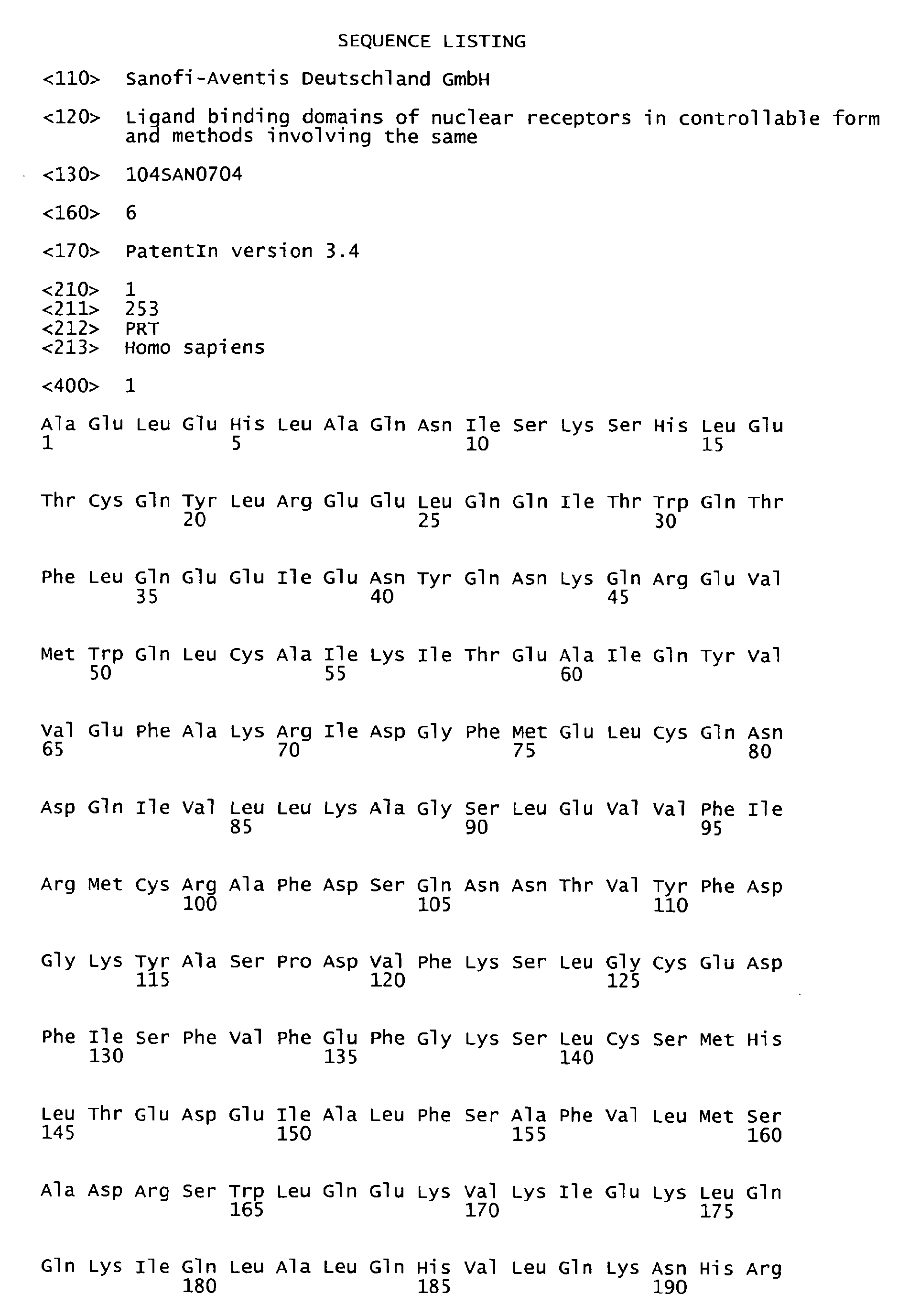


Claims (23)
- An isolated protein comprising a ligand binding domain of a nuclear receptor in controllable form.
- The isolated protein of claim 1, wherein the nuclear receptor is a Retinoic acid receptor-related Orphan Receptor (ROR), particularly RORα, RORβ or RORγ, especially RORα.
- The isolated protein of claim 1 or 2, wherein the ligand binding domain is activatable upon binding of an agonist to the ligand binding domain.
- The isolated protein of any of claims 1 to 3, wherein the protein further comprises a marker, particularly a tag.
- The isolated protein of claim 4, wherein the tag is selected from the group consisting of a His-tag, Arg-tag, Strep-tag, Flag-tag, T7-tag, V5-peptide-tag, c-Myc-tag, S-tag, HAT-tag, calmodulin-binding peptide-tag, chitin-binding peptide-tag, GST-tag and MBP-tag.
- The isolated protein of claim 4 or 5, wherein the marker or tag is removable from the protein by proteolytic cleavage at a specific cleavage site.
- The isolated protein of any of claims 1 to 6 comprising or consisting of the sequence of SEQ ID NO: 1 or 2.
- A method of producing the isolated protein of any of claims 1 to 7, comprising the steps of:a) culturing a cell comprising a nucleic acid coding for the protein of any of claims 1 to 7 under suitable conditions conducive to the production of the protein,b) isolating the protein from the cell culture, andc) contacting the isolated protein of step b) with an detergent, particularly with an alkaline salt of a saturated unbranched C6 to C20 alkyl sulphate or carbonate or with an isoprenyl salt.
- The method of claim 8, wherein the method further comprises removing the marker or tag after step b).
- The method of claim 9 or 10, wherein the alkaline salt of a saturated unbranched C6 to C20 alkyl sulphate or carbonate is an alkaline salt of a saturated unbranched C9 to C15 alkyl sulphate, preferably an alkaline salt of dodecyl sulphate, more preferably lithium dodecyl sulphate (LDS).
- The method of any of claims 8 to 10, wherein steps b) and/or c) are performed in the presence of an agonist for the ligand binding domain.
- The method of any of claims 8 to 11, wherein the cell is selected from the group consisting of an animal cell, a plant cells and a yeast cell, particularly wherein the cell is an insect cell or an E. coli cell.
- An isolated protein comprising a ligand binding domain of a nuclear receptor in controllable form produced according to the method of any of claims 8 to 12.
- Use of an isolated protein according to any of claims 1 to 7 and/or 13 for the identification of a ligand for a ligand binding domain of a nuclear receptor, particularly an agonist or antagonist, especially an agonist.
- A test system comprising- the isolated protein according to any of claims 1 to 7 and/or 13,- a co-factor, and- means for detecting the interaction between the protein and the co-factor upon binding of a ligand, especially an agonist, to the ligand binding domain of the nuclear receptor.
- The test system of claim 15, wherein the co-factor is glucocorticoid receptor-inactivating protein-1 (GRIP-1) or steroid receptor co-activator 1 (SRC1), optionally labeled with a marker, preferably a tag.
- The test system of claim 15 or 16, wherein the proximity of the protein to the co-activator induces a detectable signal.
- The test system of any of claims 15 to 17, wherein the means for detecting the interaction between the protein and the co-factor include at least one antibody specific for the protein or the co-factor.
- The test system of claim 18, wherein the test system comprises two antibodies, wherein the first antibody is specific for the protein and the second antibody is specific for the co-factor.
- The test system of claim 19, wherein(a) the first antibody is labeled with a donor moiety for fluorescence resonance energy transfer (FRET) and the second antibody is labeled with an acceptor moiety for FRET or vice versa; or(b) the first antibody is labeled with a donor moiety for time-resolved fluorescence resonance energy transfer (TR-FRET) and the second antibody is labeled with an acceptor moiety for TR-FRET or vice versa; or(c) the first antibody is labeled with a donor moiety for Amplified Luminescence Proximity Homogeneous Assay (ALPHA) and the second antibody is labeled with an acceptor moiety for ALPHA or vice versa.
- A method for screening for a ligand for a ligand binding domain of a nuclear receptor comprising the steps of:a) contacting the test system according to any of claims 15 to 20 with a substance andb) detecting a measurable signal upon binding of the substance to the ligand binding domain, thereby identifying the substance as a ligand for the ligand binding domain.
- The method of claim 21, wherein the test system is as defined in claim 20 and wherein the presence of FRET is indicative for an agonist.
- The method of claims 21 and 22, wherein the method is used for screening for an medicament for preventing and/or treating a coronary artery disease (CAD), atherosclerosis, dyslipidemia, a neurodegenerative disease, sleep disorder, a disease of circadian rhythmicity or osteoporosis.
Priority Applications (15)
| Application Number | Priority Date | Filing Date | Title |
|---|---|---|---|
| EP07291163A EP2042515A1 (en) | 2007-09-27 | 2007-09-27 | Ligand binding domains of nuclear receptors in controllable form and methods involving the same |
| KR1020107006453A KR20100075856A (en) | 2007-09-27 | 2008-09-13 | Ligand binding domains of nuclear receptors in controllable form and methods involving the same |
| JP2010526185A JP2010540474A (en) | 2007-09-27 | 2008-09-13 | Controllable form of the nuclear receptor ligand binding domain and methods comprising the same |
| AU2008303867A AU2008303867B2 (en) | 2007-09-27 | 2008-09-13 | Ligand binding domains of nuclear receptors in controllable form and methods involving the same |
| US12/675,804 US20110177611A1 (en) | 2007-09-27 | 2008-09-13 | Ligand binding domains of nuclear receptors in controllable form and methods involving the same |
| CN200880108831A CN101809031A (en) | 2007-09-27 | 2008-09-13 | Ligand binding domains of nuclear receptors in controllable form and methods involving the same |
| CA2700725A CA2700725A1 (en) | 2007-09-27 | 2008-09-13 | Ligand binding domains of nuclear receptors in controllable form and methods involving the same |
| MX2010002994A MX2010002994A (en) | 2007-09-27 | 2008-09-13 | Ligand binding domains of nuclear receptors in controllable form and methods involving the same. |
| PCT/EP2008/007596 WO2009040003A1 (en) | 2007-09-27 | 2008-09-13 | Ligand binding domains of nuclear receptors in controllable form and methods involving the same |
| EP08802147.2A EP2195334B1 (en) | 2007-09-27 | 2008-09-13 | Ligand binding domains of nuclear receptors in controllable form and methods involving the same |
| SG2012071916A SG185258A1 (en) | 2007-09-27 | 2008-09-13 | Ligand binding domains of nuclear receptors in controllable form and methods involving the same |
| TW097136778A TW200931020A (en) | 2007-09-27 | 2008-09-25 | Ligand binding domains of nuclear receptors in controllable form and methods involving the same |
| ARP080104166A AR068549A1 (en) | 2007-09-27 | 2008-09-25 | FIXING DOMAINS OF THE BINDING OF NUCLEAR RECEPTORS IN CONTROLLABLE FORM AND METHODS THAT CONCLUDE THE SAME |
| IL204667A IL204667A0 (en) | 2007-09-27 | 2010-03-22 | Ligand binding domains of nuclear receptors in controllable form and methods involving the same |
| US13/550,865 US20120282710A1 (en) | 2007-09-27 | 2012-07-17 | Ligand binding domains of nuclear receptors in controllable form and methods involving the same |
Applications Claiming Priority (1)
| Application Number | Priority Date | Filing Date | Title |
|---|---|---|---|
| EP07291163A EP2042515A1 (en) | 2007-09-27 | 2007-09-27 | Ligand binding domains of nuclear receptors in controllable form and methods involving the same |
Publications (1)
| Publication Number | Publication Date |
|---|---|
| EP2042515A1 true EP2042515A1 (en) | 2009-04-01 |
Family
ID=38738943
Family Applications (2)
| Application Number | Title | Priority Date | Filing Date |
|---|---|---|---|
| EP07291163A Ceased EP2042515A1 (en) | 2007-09-27 | 2007-09-27 | Ligand binding domains of nuclear receptors in controllable form and methods involving the same |
| EP08802147.2A Active EP2195334B1 (en) | 2007-09-27 | 2008-09-13 | Ligand binding domains of nuclear receptors in controllable form and methods involving the same |
Family Applications After (1)
| Application Number | Title | Priority Date | Filing Date |
|---|---|---|---|
| EP08802147.2A Active EP2195334B1 (en) | 2007-09-27 | 2008-09-13 | Ligand binding domains of nuclear receptors in controllable form and methods involving the same |
Country Status (13)
| Country | Link |
|---|---|
| US (2) | US20110177611A1 (en) |
| EP (2) | EP2042515A1 (en) |
| JP (1) | JP2010540474A (en) |
| KR (1) | KR20100075856A (en) |
| CN (1) | CN101809031A (en) |
| AR (1) | AR068549A1 (en) |
| AU (1) | AU2008303867B2 (en) |
| CA (1) | CA2700725A1 (en) |
| IL (1) | IL204667A0 (en) |
| MX (1) | MX2010002994A (en) |
| SG (1) | SG185258A1 (en) |
| TW (1) | TW200931020A (en) |
| WO (1) | WO2009040003A1 (en) |
Cited By (1)
| Publication number | Priority date | Publication date | Assignee | Title |
|---|---|---|---|---|
| CN115356320A (en) * | 2022-10-20 | 2022-11-18 | 上海诚益生物科技有限公司 | In-vitro RIG-I activation detection method based on homogeneous phase time-resolved fluorescence technology |
Families Citing this family (8)
| Publication number | Priority date | Publication date | Assignee | Title |
|---|---|---|---|---|
| EP2181710A1 (en) * | 2008-10-28 | 2010-05-05 | Phenex Pharmaceuticals AG | Ligands for modulation of orphan receptor-gamma (NR1F3) activity |
| ES2587795T3 (en) * | 2009-05-06 | 2016-10-26 | Opko Ireland Global Holdings, Limited | Method and composition of measuring the amount of vitamin D derivatives |
| US8187871B2 (en) | 2009-08-11 | 2012-05-29 | Janssen Pharmaceutica N.V. | Co-crystallization of ERR-α with a ligand that forms a reversible covalent bond |
| CN102262158A (en) * | 2010-05-27 | 2011-11-30 | 戴国祯 | Direct application of recombinant fusion protein to quick diagnostic detection |
| US20140017260A1 (en) * | 2012-07-16 | 2014-01-16 | Mayo Foundation For Medical Education And Research | Methods and materials for reducing bone loss |
| CN112689674B (en) * | 2018-10-29 | 2024-05-28 | 财团法人农业科技研究院 | Dextran affinity tags and their applications |
| CN113461809A (en) * | 2020-07-17 | 2021-10-01 | 南京诺唯赞生物科技股份有限公司 | Method for screening binding inhibitors and/or binding domains |
| CN120424229B (en) * | 2025-03-20 | 2026-03-31 | 重庆医科大学附属第二医院 | A fluorescent fusion protein and its application |
Citations (2)
| Publication number | Priority date | Publication date | Assignee | Title |
|---|---|---|---|---|
| WO2003000732A2 (en) * | 2001-05-07 | 2003-01-03 | Centre National De La Recherche Scientifique | Fragments of the retinoic acid-related orphan receptor (ror) comprising the lig and binding domain (lbd), crystal structure of the lbd of ror-beta and their applications |
| WO2003093312A1 (en) * | 2002-04-29 | 2003-11-13 | Novartis Ag | Crystal structure of the ligand binding domain of the retinoic acid-related orphan receptor alpha (ror-alpha) |
Family Cites Families (2)
| Publication number | Priority date | Publication date | Assignee | Title |
|---|---|---|---|---|
| US20050048539A1 (en) * | 2003-06-13 | 2005-03-03 | The General Hospital Corporation | Methods to monitor molecule conformation and molecule/molecule proximity |
| CA2633790C (en) * | 2005-12-22 | 2016-08-09 | Henry M. Krause | Methods and compositions for the detection and isolation of ligands |
-
2007
- 2007-09-27 EP EP07291163A patent/EP2042515A1/en not_active Ceased
-
2008
- 2008-09-13 US US12/675,804 patent/US20110177611A1/en not_active Abandoned
- 2008-09-13 KR KR1020107006453A patent/KR20100075856A/en not_active Ceased
- 2008-09-13 SG SG2012071916A patent/SG185258A1/en unknown
- 2008-09-13 MX MX2010002994A patent/MX2010002994A/en active IP Right Grant
- 2008-09-13 AU AU2008303867A patent/AU2008303867B2/en not_active Ceased
- 2008-09-13 EP EP08802147.2A patent/EP2195334B1/en active Active
- 2008-09-13 CN CN200880108831A patent/CN101809031A/en active Pending
- 2008-09-13 CA CA2700725A patent/CA2700725A1/en not_active Abandoned
- 2008-09-13 JP JP2010526185A patent/JP2010540474A/en active Pending
- 2008-09-13 WO PCT/EP2008/007596 patent/WO2009040003A1/en not_active Ceased
- 2008-09-25 AR ARP080104166A patent/AR068549A1/en not_active Application Discontinuation
- 2008-09-25 TW TW097136778A patent/TW200931020A/en unknown
-
2010
- 2010-03-22 IL IL204667A patent/IL204667A0/en unknown
-
2012
- 2012-07-17 US US13/550,865 patent/US20120282710A1/en not_active Abandoned
Patent Citations (2)
| Publication number | Priority date | Publication date | Assignee | Title |
|---|---|---|---|---|
| WO2003000732A2 (en) * | 2001-05-07 | 2003-01-03 | Centre National De La Recherche Scientifique | Fragments of the retinoic acid-related orphan receptor (ror) comprising the lig and binding domain (lbd), crystal structure of the lbd of ror-beta and their applications |
| WO2003093312A1 (en) * | 2002-04-29 | 2003-11-13 | Novartis Ag | Crystal structure of the ligand binding domain of the retinoic acid-related orphan receptor alpha (ror-alpha) |
Cited By (1)
| Publication number | Priority date | Publication date | Assignee | Title |
|---|---|---|---|---|
| CN115356320A (en) * | 2022-10-20 | 2022-11-18 | 上海诚益生物科技有限公司 | In-vitro RIG-I activation detection method based on homogeneous phase time-resolved fluorescence technology |
Also Published As
| Publication number | Publication date |
|---|---|
| CN101809031A (en) | 2010-08-18 |
| KR20100075856A (en) | 2010-07-05 |
| US20110177611A1 (en) | 2011-07-21 |
| MX2010002994A (en) | 2010-04-01 |
| EP2195334A1 (en) | 2010-06-16 |
| SG185258A1 (en) | 2012-11-29 |
| AR068549A1 (en) | 2009-11-18 |
| WO2009040003A1 (en) | 2009-04-02 |
| AU2008303867A1 (en) | 2009-04-02 |
| US20120282710A1 (en) | 2012-11-08 |
| CA2700725A1 (en) | 2009-04-02 |
| JP2010540474A (en) | 2010-12-24 |
| TW200931020A (en) | 2009-07-16 |
| IL204667A0 (en) | 2010-11-30 |
| EP2195334B1 (en) | 2013-07-31 |
| AU2008303867B2 (en) | 2013-05-16 |
Similar Documents
| Publication | Publication Date | Title |
|---|---|---|
| US20120282710A1 (en) | Ligand binding domains of nuclear receptors in controllable form and methods involving the same | |
| Whitelaw et al. | Ligand-dependent recruitment of the Arnt coregulator determines DNA recognition by the dioxin receptor | |
| Miner et al. | The basic region of AP-1 specifies glucocorticoid receptor activity at a composite response element. | |
| Kliewer et al. | Retinoid X receptor-COUP-TF interactions modulate retinoic acid signaling. | |
| Kato et al. | Widely spaced, directly repeated PuGGTCA elements act as promiscuous enhancers for different classes of nuclear receptors | |
| Haussler et al. | New understanding of the molecular mechanism of receptor-mediated genomic actions of the vitamin D hormone | |
| Lazennec et al. | Mechanistic aspects of estrogen receptor activation probed with constitutively active estrogen receptors: correlations with DNA and coregulator interactions and receptor conformational changes | |
| Raman et al. | Characterization of the fatty acid-responsive transcription factor FadR: biochemical and genetic analyses of the native conformation and functional domains | |
| US20030083469A1 (en) | Allosteric control of nuclear hormone receptors | |
| Huang et al. | Targeting estrogen responsive elements (EREs): design of potent transactivators for ERE-containing genes | |
| Kersten et al. | The tetramerization region of the retinoid X receptor is important for transcriptional activation by the receptor | |
| Molbaek et al. | High yield purification of full-length functional hERG K+ channels produced in Saccharomyces cerevisiae | |
| US6635429B1 (en) | Heterodimeric nuclear receptors proteins, genes encoding same, and usage thereof | |
| Baniahmad et al. | Co-operative binding of the glucocorticoid receptor DNA binding domain is one of at least two mechanisms for synergism | |
| Mackem et al. | A glucocorticoid/retinoic acid receptor chimera that displays cytoplasmic/nuclear translocation in response to retinoic acid: a real time sensing assay for nuclear receptor ligands | |
| Wei et al. | Constitutive activation of retinoic acid receptor β2 promoter by orphan nuclear receptor TR2 | |
| Lechtken et al. | Overexpression, refolding, and purification of polyhistidine-tagged human retinoic acid related orphan receptor RORα4 | |
| Lee et al. | Mutations in retinoid X receptor that impair heterodimerization with specific nuclear hormone receptor | |
| JP4172332B2 (en) | Nuclear receptor ligand detection agent, recombinant protein, gene for expression, recombinant vector, transformant, nuclear receptor ligand detection method, and nuclear receptor ligand detection kit | |
| Allegretto et al. | [3] Expression and characterization of retinoid receptors in yeast | |
| WO2000008052A1 (en) | A novel retinal nuclear hormone receptor | |
| Repa et al. | One-step immunoaffinity purification of recombinant human retinoic acid receptor γ | |
| Faris | Characterization of the DNA binding properties of the thyroid hormone receptor | |
| JP2004519227A (en) | Nurr1 transcription factor I-box mutant with monomeric transcription activation activity | |
| WO2001072969A1 (en) | Effector-specific protein assembly and uses thereof |
Legal Events
| Date | Code | Title | Description |
|---|---|---|---|
| PUAI | Public reference made under article 153(3) epc to a published international application that has entered the european phase |
Free format text: ORIGINAL CODE: 0009012 |
|
| AK | Designated contracting states |
Kind code of ref document: A1 Designated state(s): AT BE BG CH CY CZ DE DK EE ES FI FR GB GR HU IE IS IT LI LT LU LV MC MT NL PL PT RO SE SI SK TR |
|
| AX | Request for extension of the european patent |
Extension state: AL BA HR MK RS |
|
| STAA | Information on the status of an ep patent application or granted ep patent |
Free format text: STATUS: THE APPLICATION HAS BEEN REFUSED |
|
| 18R | Application refused |
Effective date: 20090418 |