EP2016098B1 - Antibody-targeted cytokines for therapy - Google Patents
Antibody-targeted cytokines for therapy Download PDFInfo
- Publication number
- EP2016098B1 EP2016098B1 EP07724968.8A EP07724968A EP2016098B1 EP 2016098 B1 EP2016098 B1 EP 2016098B1 EP 07724968 A EP07724968 A EP 07724968A EP 2016098 B1 EP2016098 B1 EP 2016098B1
- Authority
- EP
- European Patent Office
- Prior art keywords
- fusion protein
- antibody
- short
- functional
- protein according
- Prior art date
- Legal status (The legal status is an assumption and is not a legal conclusion. Google has not performed a legal analysis and makes no representation as to the accuracy of the status listed.)
- Not-in-force
Links
Images


Classifications
-
- C—CHEMISTRY; METALLURGY
- C07—ORGANIC CHEMISTRY
- C07K—PEPTIDES
- C07K16/00—Immunoglobulins [IG], e.g. monoclonal or polyclonal antibodies
- C07K16/18—Immunoglobulins [IG], e.g. monoclonal or polyclonal antibodies against material from animals or humans
-
- C—CHEMISTRY; METALLURGY
- C07—ORGANIC CHEMISTRY
- C07K—PEPTIDES
- C07K19/00—Hybrid peptides, i.e. peptides covalently bound to nucleic acids, or non-covalently bound protein-protein complexes
-
- A—HUMAN NECESSITIES
- A61—MEDICAL OR VETERINARY SCIENCE; HYGIENE
- A61P—SPECIFIC THERAPEUTIC ACTIVITY OF CHEMICAL COMPOUNDS OR MEDICINAL PREPARATIONS
- A61P17/00—Drugs for dermatological disorders
- A61P17/06—Antipsoriatics
-
- A—HUMAN NECESSITIES
- A61—MEDICAL OR VETERINARY SCIENCE; HYGIENE
- A61P—SPECIFIC THERAPEUTIC ACTIVITY OF CHEMICAL COMPOUNDS OR MEDICINAL PREPARATIONS
- A61P19/00—Drugs for skeletal disorders
- A61P19/02—Drugs for skeletal disorders for joint disorders, e.g. arthritis, arthrosis
-
- A—HUMAN NECESSITIES
- A61—MEDICAL OR VETERINARY SCIENCE; HYGIENE
- A61P—SPECIFIC THERAPEUTIC ACTIVITY OF CHEMICAL COMPOUNDS OR MEDICINAL PREPARATIONS
- A61P29/00—Non-central analgesic, antipyretic or antiinflammatory agents, e.g. antirheumatic agents; Non-steroidal antiinflammatory drugs [NSAID]
-
- A—HUMAN NECESSITIES
- A61—MEDICAL OR VETERINARY SCIENCE; HYGIENE
- A61P—SPECIFIC THERAPEUTIC ACTIVITY OF CHEMICAL COMPOUNDS OR MEDICINAL PREPARATIONS
- A61P35/00—Antineoplastic agents
-
- A—HUMAN NECESSITIES
- A61—MEDICAL OR VETERINARY SCIENCE; HYGIENE
- A61P—SPECIFIC THERAPEUTIC ACTIVITY OF CHEMICAL COMPOUNDS OR MEDICINAL PREPARATIONS
- A61P9/00—Drugs for disorders of the cardiovascular system
- A61P9/10—Drugs for disorders of the cardiovascular system for treating ischaemic or atherosclerotic diseases, e.g. antianginal drugs, coronary vasodilators, drugs for myocardial infarction, retinopathy, cerebrovascula insufficiency, renal arteriosclerosis
-
- C—CHEMISTRY; METALLURGY
- C07—ORGANIC CHEMISTRY
- C07K—PEPTIDES
- C07K14/00—Peptides having more than 20 amino acids; Gastrins; Somatostatins; Melanotropins; Derivatives thereof
- C07K14/435—Peptides having more than 20 amino acids; Gastrins; Somatostatins; Melanotropins; Derivatives thereof from animals; from humans
- C07K14/52—Cytokines; Lymphokines; Interferons
-
- C—CHEMISTRY; METALLURGY
- C07—ORGANIC CHEMISTRY
- C07K—PEPTIDES
- C07K14/00—Peptides having more than 20 amino acids; Gastrins; Somatostatins; Melanotropins; Derivatives thereof
- C07K14/435—Peptides having more than 20 amino acids; Gastrins; Somatostatins; Melanotropins; Derivatives thereof from animals; from humans
- C07K14/52—Cytokines; Lymphokines; Interferons
- C07K14/53—Colony-stimulating factor [CSF]
- C07K14/535—Granulocyte CSF; Granulocyte-macrophage CSF
-
- C—CHEMISTRY; METALLURGY
- C07—ORGANIC CHEMISTRY
- C07K—PEPTIDES
- C07K14/00—Peptides having more than 20 amino acids; Gastrins; Somatostatins; Melanotropins; Derivatives thereof
- C07K14/435—Peptides having more than 20 amino acids; Gastrins; Somatostatins; Melanotropins; Derivatives thereof from animals; from humans
- C07K14/52—Cytokines; Lymphokines; Interferons
- C07K14/54—Interleukins [IL]
-
- C—CHEMISTRY; METALLURGY
- C07—ORGANIC CHEMISTRY
- C07K—PEPTIDES
- C07K14/00—Peptides having more than 20 amino acids; Gastrins; Somatostatins; Melanotropins; Derivatives thereof
- C07K14/435—Peptides having more than 20 amino acids; Gastrins; Somatostatins; Melanotropins; Derivatives thereof from animals; from humans
- C07K14/52—Cytokines; Lymphokines; Interferons
- C07K14/54—Interleukins [IL]
- C07K14/5428—IL-10
-
- C—CHEMISTRY; METALLURGY
- C07—ORGANIC CHEMISTRY
- C07K—PEPTIDES
- C07K14/00—Peptides having more than 20 amino acids; Gastrins; Somatostatins; Melanotropins; Derivatives thereof
- C07K14/435—Peptides having more than 20 amino acids; Gastrins; Somatostatins; Melanotropins; Derivatives thereof from animals; from humans
- C07K14/52—Cytokines; Lymphokines; Interferons
- C07K14/54—Interleukins [IL]
- C07K14/5443—IL-15
-
- C—CHEMISTRY; METALLURGY
- C07—ORGANIC CHEMISTRY
- C07K—PEPTIDES
- C07K16/00—Immunoglobulins [IG], e.g. monoclonal or polyclonal antibodies
-
- C—CHEMISTRY; METALLURGY
- C07—ORGANIC CHEMISTRY
- C07K—PEPTIDES
- C07K2317/00—Immunoglobulins specific features
- C07K2317/60—Immunoglobulins specific features characterized by non-natural combinations of immunoglobulin fragments
- C07K2317/62—Immunoglobulins specific features characterized by non-natural combinations of immunoglobulin fragments comprising only variable region components
- C07K2317/622—Single chain antibody (scFv)
-
- C—CHEMISTRY; METALLURGY
- C07—ORGANIC CHEMISTRY
- C07K—PEPTIDES
- C07K2319/00—Fusion polypeptide
Definitions
- the present invention relates to fusion proteins comprising an antibody, functional fragment or functional derivative thereof having specific binding affinity to either the extracellular domain of oncofetal fibronectin (ED-B) or to at least one of the extracellular domains of oncofetal tenascin fused to cytokinelL-10, functional fragments and functional derivatives thereof.
- the invention is also directed to the use of at least one of said fusion proteins for the manufacture of a medicament.
- the invention concerns the use of said medicament for the treatment of tumors or chronic inflammatory diseases such as atherosclerosis, arthritis and psoriasis.
- Cytokines are immunomodulatory proteins, some of which have been used preclinically and clinically not only to fight cancer, but also to interfere with chronic inflammatory conditions and with infectious disease.
- cytokine fusion proteins have already been investigated for application in cancer therapy, often with impressive results.
- human antibody L19 specific to the ED-B domain of fibronectin a marker of angiogenesis
- pro-inflammatory cytokines such as IL-2, IL-12 or TNF
- cytokines have a history of clinical failure, both, when used as a single agent or as fusion partners with monoclonal antibodies.
- IL-2 Proleukin
- Chiron recombinant IL-2
- response rates are typically low (generally below 20 %) for this indication and even lower for other types of cancer.
- Other cytokines (such as interleukin-12 or interleukin-10, see below) have failed to demonstrate substantial efficacy in a series of clinical studies which has slowed clinical development programs. These cytokines are not yet approved as biopharmaceuticals.
- Interferon gamma is another example of a cytokine approved for a very narrow indication (treatment of chronic granulomatous disease, Genentech) which has failed to demonstrate substantial clinical benefits for other indications.
- Interleukin-10 is a homodimeric cytokine produced by activated monocytes and T cells that is deeply involved in the regulation of inflammatory responses and immune reactions. Its main overall function is best described as dampener of immune responses, but IL-10 also possesses stimulatory activities. IL-10 was first described as cytokine synthesis inhibitory factor (CSIF), an activity produced by mouse Th2 cells that inhibited activation of and cytokine production by Th1 cells [ Fiorentino et al., J. Exp. Med. 170(6): 2081-95 (1989 )]. The gene encoding human IL-10 is located on chromosome 1 [ Kim et al., J. Immunol.
- CCF cytokine synthesis inhibitory factor
- Human IL-10 is active as a non-disulfide-linked homodimer of 37 kDa [ Syto et al., Biochemistry 37(48): 16943-51 (1998 )].
- IL-10 has been considered an attractive candidate for therapeutic use based on its potent in vitro immunomodulating activities and proven effects in animal models of acute and chronic inflammation, autoimmunitiy, cancer and infectious disease.
- Schering-Plough developed recombinant human IL-10 (ilodecakin, Tenovil ® ) for clinical trials.
- the protein is produced in E. coli and consists of 161 amino acids, identical with the endogenous human protein except for a methionine residue at the amino-terminus.
- Interleukin-15 is a 14 to 15 kDa member of the 4 ⁇ -helix bundle family of cytokines composed of 114 amino acids.
- IL-15 protein is posttranscriptionally regulated by multiple controlling elements that inhibit translation, including 12 upstream AUGs of the 5' untranslated region (UTR), 2 unusual signal peptides (the short peptide with 21 amino acids stays intracellularly, the long peptide with 48 amino acids is for secretion) and the C-terminus of the mature-protein [ Bamford et al., J. Immunol., 160(9): 4418-26 (1998 )].
- UTR 5' untranslated region
- 2 unusual signal peptides the short peptide with 21 amino acids stays intracellularly, the long peptide with 48 amino acids is for secretion
- C-terminus of the mature-protein [ Bamford et al., J. Immunol., 160(9): 4418-26 (1998 )].
- IL-15 uses two distinct receptors and signalling pathways: A high affinity IL-15R system consisting of IL-2/15 ⁇ , ⁇ c and IL-15R ⁇ subunits is expressed on T and NK cells. The IL-2/15R ⁇ and the ⁇ c subunits are shared with IL-2 receptor [ Giri et al., EMBO J., 3(12):2822-30 (1994 )]. Mast cells respond to IL-15 with a receptor system that does not share elements with the IL-2 receptor but uses a novel 60 to 65 kDa IL-15RX subunit. A variety of tissues such as placenta, skeletal muscles, kidney, fibroblasts, epithelial cells, dendritic cells and monocytes express IL-15.
- IL-15 stimulates the production of proinflammatory cytokines (e.g. TNF ⁇ , IL-1, IFN ⁇ ), the proliferation and lg synthesis of activated B cells, the activation of T H 1, monocytes and lymphokine activated killer cells, the proliferation of mast cells and T cells and inhibits the apoptosis of T and B cells.
- cytokines e.g. TNF ⁇ , IL-1, IFN ⁇
- T H 1 monocytes and lymphokine activated killer cells
- mast cells e.g., mast cells
- IL-15 plays a pivotal role in the development, survival and function of NK cells [ Joost J. Oppenheim et al., Cytokine Reference; 213-221, (2002 )].
- exogenous IL-15 enhances the antitumor activity of tumor reactive CD8 + T cells [ Fehniger et al., Cytokine Growth Factor Rev.,13(2):169-83 (2002 )].
- IL-2 and IL-15 use the same receptor subunits they share many features. The major differences are their sites of synthesis and secretion. IL-2 is produced by activated T-cells. In contrast, IL-15 is expressed in a variety of tissues as mentioned above. While IL-2 can promote apoptosis and limited CD8 + memory T-cell survival and proliferation, IL-15 helps maintain memory CD8 + population and can inhibit apoptosis. IL-15, initially thought to mediate similar biological effects as IL-2, has been shown to have unique properties in basic and pre-clinical studies that may be of benefit in the immunotherapy of cancer [ Fehniger et al., Cytokine Growth Factor Rev., (2):169-83 (2002 )].
- IL-15 resembles that of IL-2 very closely [ Munger et al., Cell Immunol., 5(2):289-93 (1995 )], thus suggesting targeted delivery of IL-15 to be superior to systemic delivery in terms of therapeutic index.
- IL-15 mutants that showed either agonist or antagonist properties which may be useful as therapeutic agents [ Bernard et al., J. Biol. Chem. , 279(23): 24313-22 (2004 )].
- the IL-15 mutants IL-15D8S and IL-15Q108S were inactive in a CTLL-2 bioassay, but were able to competitively inhibit the biological activity of unmodified IL-15 [ Pettit et al., J. Biol. Chem, 272(4): 2312-8 (1997 )].
- the IL-24 gene cDNA encodes a 206 amino acid protein with 23.8 kDa.
- the secreted protein has a significantly higher molecular weight (40 kDa) due to heavy N-glycosylation compared to the intracellular protein (30/23 kDa).
- the homology of human IL-24 to the rat counterpart (MOB-5) is 68% and to the mouse one (FISP) 69%.
- IL-20R1/IL-20R2 There are two functional heterodimeric receptors for IL-24: IL-20R1/IL-20R2 and IL-22R1/IL-20R2 [ Wang et al., Genes Immun., 5(5):363-70 (2004 )], [ Chada et al., Mol. Ther., 10(6):1085-95 (2004 )].
- IL-20R1 and IL-22R1 receptor chains are widely expressed the restricted expression of the common IL-20R2 in certain non-haemopoietic tissues suggests a pleotropic role of IL-24 outside the haemopoietic system [ Wolk et al., J. Immunol., 168(11): 5397-402 (2002 )].
- IL-24 is expressed by monocytes, T cells, dendritic cells and melanocytes. IL-24 induces the secretion of IFN ⁇ , IL-6, TNF ⁇ , IL-1- ⁇ and GM-CSF indicating its function as a pro-Th1 cytokine. IL-10 (Th2 cytokine) inhibits the IL-24 activity.
- IL-24 may promote antigen presentation by activation or stimulation of immune accessory and effector cells [ Chada et al., Mol. Ther., 10(6):1085-95 (2004 )] .
- Ad-mda-7 adenovirus mda-7
- Granulocyte-macrophage colony-stimulating factor is a 141 amino acid (mouse) / 144 amino acid (human) protein containing a 17 amino acid secretion sequence.
- the apparent molecular weight of the mature glycosylated protein is 14 - 33 kDa, which is very resistant to denaturing and proteolytic conditions.
- the in vivo activities of GM-CSF are mediated by binding to high-affinity receptors comprising a GM-CSF-specific a chain and, for humans, a signal transducing ⁇ subunit that is shared with the IL-3 and the IL-5 receptors [ Joost J. Oppenheim et al., Cytokine Reference, 899-908, 2002 ].
- GM-CSF is a major regulator of granulocyte and macrophage lineage. It stimulates the survival, proliferation and differentiation of hematopoietic colony-forming cells of the neutrophil, macrophage and eosinophil lineages. In addition, it maintains the survival of hematopoietic colony-forming cells of the megakaryocytic and erythroid cell lineages [ Joost J. Oppenheim et al., Cytokine Reference, 899-908, 2002 ]. It is also a potent immunostimulator with pleiotropic effects, including the augmentation of Ag presentation in a variety of cells, increased expression of MHC class II on monocytes and amplification of T cell proliferation [ Fischer et al., J.
- GM-CSF GM-CSF
- inflammatory reactions e.g rheumatoid arthritis
- toxic shock e.g rheumatoid arthritis
- blindness e.g rheumatoid arthritis
- autoimmunity e.g rheumatoid arthritis
- subphysiological levels may be involved in some cases of alveolar proteinosis.
- Alveolar proteinosis is a fatal lung disease where surfactant proteins accumulate in the lung due to a defect in macrophage-mediated clearance [ Joost J. Oppenheim et al., Cytokine Reference; 899-908, 2002 ].
- mice bearing B16 melanoma with additional irradiated tumor cells expressing murine granulocyte-macrophage colony-stimulating factor (GM-CSF) stimulated a potent, long-lasting and specific anti-tumor immunity by increasing the immunogenicity of the tumors [ Dranoff et al., Proc. Natl. Acad. Sci. USA., 90(8):3539-43 (1993 )].
- GM-CSF murine granulocyte-macrophage colony-stimulating factor
- GM-CSF is widely used in oncology to reduce chemotherapy-related neutropenia, a reduction of neutrophils caused by chemotherapeutic drugs [ Danova et al., Haematologica., 82(5):622-9 (1997 )], [ Vose et al., J. Clin. Oncol., 13(4):1023-35 (1995 )].
- a GM-CSF based vaccine not only loses its efficacy but more importantly results in substantial immunosupression in vivo.
- the dual effects of GM-CSF are mediated by the systemic and not the local concentration of this cytokine [ Serafini et al., Cancer Res., 64(17):6337-43 (2004 )]. Serious adverse events are seen at doses of 16 ⁇ g/kg per day for humans [ Joost J. Oppenheim et al., Cytokine Reference; 899-908 (2002 )].
- Fibronectins are high molecular weight adhesive glycoproteins present in soluble form in plasma and other body fluids and in insoluble form in the extracellular matrix.
- EDB is a 91-amino-acid type III homology domain that is inserted into the fibronectin molecule by a mechanism of alternative splicing at the level of the primary transcript whenever tissue remodelling takes place [ Zardi et al., Embo J. 6(8): 2337-42 (1987 )].
- EDB is essentially undetectable in healthy adult tissues. Its expression is strongly associated with the remodelling of the extracellular matrix and angiogenesis. The domain is abundant in many aggressive tumors and depending on the tumor type displays either predominantly vascular or diffuse stromal patterns of expression [ Carnemolla et al., J. Cell Biol. 108(3): 1139-48 (1989 )]. Despite its very restricted expression in normal tissues and its strong expression in many solid tumors the function of EDB does not seem to be indispensable because mice lacking the EDB exon develop normally, are fertile and heal bone fractions. Furthermore, double knock-out mice lacking the EDB exon and p53 did not show any difference in the duration of survival compared to animals expressing EDB [ Fukuda et al., Cancer Res 62(19): 5603-10 (2002 )].
- L19 is able to stain tumor blood vessels in a wide range of experimental tumor models and on sections of human tumors and other angiogenic disorders [ Carnemolla et al., J. Cell Biol. 108(3): 1139-48 (1989 ); Kaczmarek et al., Int. J. Cancer 59(1): 11-6 (1994 ); Berndt et al., Histochem. Cell Biol. 109(3): 249-55 (1998 )]. Castellani et al.
- the EDB domain of fibronectin is a good-quality marker of angiogenesis, which is overexpressed in a variety of solid tumors (e.g., renal cell carcinoma, colorectal carcinoma, hepatocellular carcinoma, high-grade astrocytomas, head and neck tumors, bladder cancer, etc.) but is virtually undetectable in normal adult tissues (exception made for the endometrium in the proliferative phase and some vessels in the ovaries).
- solid tumors e.g., renal cell carcinoma, colorectal carcinoma, hepatocellular carcinoma, high-grade astrocytomas, head and neck tumors, bladder cancer, etc.
- EDB is only weakly expressed in most forms of breast cancer, prostate cancer and some types of lung cancer, thus stimulating the search for novel vascular tumor antigens, which could be used for the antibody-mediated targeted delivery of therapeutic cytokines to these neoplasias.
- Tenascin-C is a glycoprotein of the extracellular matrix. It comprises several fibronectin type 3 homology repeats that can be either included or omitted in the primary transcript by alternative splicing, leading to small and to large isoforms that have distinct biological functions. While the small isoform is expressed in several tissues the large isoform of tenascin-C exhibits a more restricted expression pattern.
- a fusion protein comprising:
- binding affinity is to be understood to mean that the antibody, functional fragment or functional derivative thereof specifically binds to the target protein with significant affinity and not to other proteins with significant affinity that are also located in the same environment, i.e. assay system or body, organ, etc., in vivo or in vitro, and under the same conditions, e.g. pH, temperature, buffer, etc.
- a binding specificity is tested by performing a binding assay with a specific target molecule and with a large number of non-related substances.
- functional tests, immunohistochemistry and other procedures can be used to assess the binding specificity of a specified antibody.
- a dissociation constant of 1 micromolar or lower is required to yield detectable binding signals which are often associated with a specific binding mode.
- the antibodies, functional fragments or functional derivatives for use in the present invention have a specific binding affinity corresponding to a dissociation constant of less than about 5, preferably about 1 or less micromolar ( ⁇ M), more preferably about 0,1 ⁇ M or less, most preferably about 1 nM or less or even 1 pM or less.
- Antibodies, functional fragments and functional derivatives thereof for practicing the invention are routinely available by hybridoma technology ( Kohler and Milstein, Nature 256, 495-497, 1975 ), antibody phage display ( Winter et al., Annu. Rev. Immunol. 12, 433-455, 1994 ), ribosome display ( Schaffitzel et al., J. Immunol. Methods, 231, 119-135, 1999 ) and iterative colony filter screening ( Giovannoni et al., Nucleic Acids Res. 29, E27, 2001 ) once the target antigen is available.
- Typical proteases for fragmenting antibodies into functional products are well-known. Other fragmentation techniques can be used as well as long as the resulting fragment has a specific high affinity and, preferably a dissociation constant in the micromolar to picomolar range.
- the vascular tumour targeting performance of antibody fragments in scFv format has been shown to crucially depend (at least for a micromolar to picomolar dissociation constant) on the affinity of the antibody to the target.
- the high affinity antibody fragment scFv(L19) specific for the ED-B domain of fibronectin, a marker of angiogenesis, was shown to target tumour neo-vasculature more efficiently than the parental antibody fragment scFv(E1), with a lower affinity for the antigen [ Viti et al., Cancer Res. 15;59(2):347-52 (1999 )].
- binding avidity e.g., associated with certain homobivalent antibody formats
- a very convenient antibody fragment for targeting applications is the single-chain Fv fragment, in which a variable heavy and a variable light domain are joined together by a polypeptide linker.
- Other antibody fragments for vascular targeting applications include Fab fragments, Fab 2 fragments, miniantibodies (also called small immune proteins), tandem scFv-scFv fusions as well as scFv fusions with suitable domains (e.g. with the Fc portion of an immunoglobulin).
- suitable domains e.g. with the Fc portion of an immunoglobulin.
- the term "functional derivative" of an antibody for use in the present invention is meant to include any antibody or fragment thereof that has been chemically or genetically modified in its amino acid sequence, e.g. by addition, substitution and/or deletion of amino acid residue(s) and/or has been chemically modified in at least one of its atoms and/or functional chemical groups, e.g. by additions, deletions, rearrangement, oxidation, reduction, etc. as long as the derivative has substantially the same binding affinity as to its original antigen and, preferably, has a dissociation constant in the micro-, nano- or picomolar range.
- a most preferred derivative of the antibodies for use in the present invention is an antibody fusion protein that will be defined in more detail below.
- the antibody, fragment or functional derivative thereof for use in the invention is one that is selected from the group consisting of polyclonal antibodies, monoclonal antibodies, chimeric antibodies, humanized antibodies, CDR-grafted antibodies, Fv-fragments, Fab-fragments and Fab 2 -fragments and antibody-like binding proteins, e.g. affilines, anticalines and aptamers .
- aptamer describes nucleic acids that bind to a polypeptide with high affinity. Aptamers can be isolated from a large pool of different single-stranded RNA molecules by selection methods such as SELEX (see, e.g., Jayasena, Clin. Chem., 45, p. 1628 - 1650, (1999 ); Klug and Famulok, M. Mol. Biol. Rep., 20, p. 97 - 107 (1994 ); US 5,582,981 ).
- Aptamers can also be synthesized and selected in their mirror form, for example, as the L-ribonucleotide ( Nolte et al., Nat. Biotechnol., 14, pp.1116 - 1119, (1996 ); Klussmann et al., Nat. Biotechnol., 14, p. 1112 - 1115, (1996 )).
- L-ribonucleotide Nolte et al., Nat. Biotechnol., 14, pp.1116 - 1119, (1996 ); Klussmann et al., Nat. Biotechnol., 14, p. 1112 - 1115, (1996 )
- Another antibody-like binding protein and alternative to classical antibodies are the so-called "protein scaffolds", for example, anticalines, that are based on lipocaline ( Beste et al., Proc. Natl. Acad. Sci. USA, 96, p. 1898 - 1903, (1999 )).
- the natural ligand binding sites of lipocalines, for example, of the retinol-binding protein or bilin-binding protein can be changed, for example, by employing a "combinatorial protein design” approach, and in such a way that they bind selected haptens ( Skerra, Biochem. Biophys. Acta, 1482, pp. 337 - 350, (2000 )).
- the term functional antibody derivative is meant to include said protein-derived alternatives for antibodies, i.e. antibody-like binding proteins, e.g. affilines, anticalines and aptamers that specifically recognize at least one extracellular domain of oncofetal fibronectin or oncofetal tenascin.
- antibody-like binding proteins e.g. affilines, anticalines and aptamers that specifically recognize at least one extracellular domain of oncofetal fibronectin or oncofetal tenascin.
- antibody functional fragment and functional derivative thereof denote all substances that have the same or similar specific binding affinity to any one of the extracellular domains of oncofetal fibronectin or oncofetal tenascin as a complete antibody having specific binding affinity to these targets.
- tenascin there are a number of isoforms available, e.g. tenascin-C, tenascin-R and tenascin-X.
- the extracellular domains of the large isoform of tenascin-C are most preferred as specific targets for the antibody, functional fragment or functional derivative thereof that is part of the fusion proteins of the present invention.
- the antibody, functional fragment or functional derivative thereof, that is part of a fusion protein of the invention has a specific binding affinity to at least one of the extracellular domains of oncofetal tenascin-C, more preferably to at least one of the extracellular domains of the large isoform of tenascin-C.
- the extracellular domains of tenascin-C are denoted domains A1, A2, A3, A4, B, C and D.
- domains A1, A2, A3, A4, B, C and D There are already a number of antibodies available that are directed against one of these domains (see Siri A. et al., Different susceptibility of small and large human tenascin-C isoforms to degradation by matrix metalloproteinases. J. Biol. Chem., Apr. 14, 1995, 270(15):8650-4 ; Carnemolla B. et al., Identification of a glioblastoma-associated tenascin-C isoform by a high affinity recombinant antibody. Am. J. Pathol.
- the present invention relates to fusion proteins of the invention comprising an antibody, functional fragment or functional derivative thereof having specific binding affinity to any one of the extracellular domains of tenascin-C, i.e. A1, A2, A3, A4, B, C and D, preferably to any one of the domains A1, C or D, more preferably to the domain C of tenascin-C.
- fusion protein as it is used in the context of the present invention is meant to encompass all conjugates, wherein said antibody, fragment or functional derivative is somehow bound to cytokine IL-10, functional fragments and functional derivatives thereof, by, e.g. covalent and/or non-covalent, e.g. ionic bonds.
- the term encompasses both binding arrangements, i.e. antibody - cytokine or cytokine - antibody.
- cytokines The terms functional fragment and functional derivative with regard to said cytokines are to be interpreted essentially in analogy to the same terms for antibodies.
- Functional fragments and derivatives of cytokines are those that essentially have the same physiological function/activity as the naturally occurring cytokines.
- preferred assays for determining the function/activity of the cytokines, fragments and derivatives thereof for preparing fusion proteins according to the present invention are:
- the fusion protein according to the invention comprises the diabody scFv L19 (long) having the amino acid sequence set forth in SEQ ID NO: 6.
- the fusion protein according to the invention comprises the diabody L19 (short) having the amino sequence set forth in SEQ ID NO: 7.
- the fusion protein is one, wherein the antibody, functional fragment or functional derivative thereof having specific binding affinity to at least one of the extracellular domains of oncofetal tenascin is selected from the group consisting of F16 (long), F16 (short), F16 (A34M) (long), F16 (A34M) (short), G11 (long) and G11 (short) having the amino sequences set forth in SEQ ID NO: 8 to 13, respectively.
- the fusion protein according to the invention is one, wherein a member of the group consisting of L19 (long), L19 (short), F16 (long), F16 (short), F16 (A34M) (long), F16 (A34M) (short), G11 (long) and G11 (short) is fused to cytokine IL-10, functional fragments and functional derivatives thereof.
- the cytokine is a murine or human, preferably a human cytokine, functional fragment or functional derivative thereof.
- the fusion proteins according to the invention may be arranged such that the cytokine, functional fragment or functional derivative thereof is fused N-terminally or C-terminally to the antibody, functional fragment or functional derivative thereof.
- those fusion proteins according to the invention are preferred, wherein the antibody fragment or functional derivative thereof is selected from the group consisting of L 19 (short), F16 (short), F16 (A34M) (short) and G11 (short).
- Fusion proteins comprising the F16 (A34M) variant (long or short) are more preferred and those comprising the short F16 (A34M) variant are most preferred.
- the present invention relates to a fusion protein comprising:
- the fusion protein is L19-IL-10.
- the fusion proteins according to the invention has the amino acid sequence set forth in SEQ ID NO: 14.
- the present invention relates to the use of a fusion protein according to the invention for the manufacture of a medicament.
- the present invention relates to the use of the above fusion proteins for the treatment of cancer in a mammal, preferably in a human.
- the present invention relates to the use of the above fusion proteins for the treatment of inflammatory diseases, preferably chronic inflammatory diseases in a mammal, preferably in a human.
- the inflammatory disease is selected from the group consisting of psoriasis, atherosclerosis, arthritis, preferably rheumatoid arthritis.
- a further aspect of the present invention relates to a pharmaceutical composition
- a pharmaceutical composition comprising at least one fusion protein of the invention and optionally a pharmaceutically acceptable excipient.
- compositions of the present invention typically comprise a therapeutically effective amount of a fusion protein according to the invention and optionally auxiliary substances such as pharmaceutically acceptable excipient(s).
- Said pharmaceutical compositions are prepared in a manner well known in the pharmaceutical art.
- a carrier or excipient may be a liquid material which can serve as a vehicle or medium for the active ingredient.
- Suitable carriers or excipients are well known in the art and include, for example, stabilizers, antioxidants, pH-regulating substances, controlled-release excipients.
- the pharmaceutical preparation of the invention may be adapted, for example, for parenteral use and may be administered to the patient in the form of solutions or the like.
- a fusion protein of the present invention can be administered in any form or mode which makes the therapeutic compound bioavailable in an effective amount, including oral or parenteral routes.
- compositions of the present invention can be administered subcutaneously, intramuscularly, intravenously and the like.
- One skilled in the art in the field of preparing formulations can readily select the proper form and mode of administration depending upon the particular characteristics of the product selected, the disease or condition to be treated, the stage of the disease or condition and other relevant circumstances (see. e.g. Remington's Pharmaceutical Sciences, Mack Publishing Co. (1990 )).
- compositions of the present invention can be administered alone or in the form of a pharmaceutical preparation in combination with pharmaceutically acceptable carriers or excipients, the proportion and nature of which are determined by the solubility and chemical properties of the product selected, the chosen route of administration and standard pharmaceutical practice.
- pharmaceutically acceptable salts such as acid addition salts or base addition salts, for purposes of stability, convenience of crystallization, increased solubility and the like.
- the cytokines were genetically fused to either the C- or the N-terminus of the scFv antibody fragments separated by a 15 amino acid linker.
- the resulting fragments preceded by a secretion sequence required for secretion of recombinant proteins, were cloned in a mammalian expression vector and the fusion proteins were expressed in stably transfected HEK 293 cells.
- the constructs were purified from the culture medium by affinity chromatography on antigen columns at yields of 1-2 mg / l. Quality control was performed by SDS-PAGE and gelfiltration.
- Fusion proteins are solved in physiological solutions and administered intravenously to animals.
- the proteins are stored in one of the following buffers depending on their isoelectric point and the desired storage time. Proteins are kept for long time storage (over one month) at minus 80 °C. To prevent aggregation by repeated thaw and freeze cycles 1% Glycerol and 0.04% Tween 80 may be added.
- Injections are typically administered 3-5 times, daily or every second day.
- the dosage is selected according to literature values following routine experimentation.
- Example 3 Targeting efficacy of the fusion protein in 129Sv mice grafted with subcutaneous F9 tumors
- the in vivo targeting properties of a radioiodinated preparation of L19-IL10 were evaluated in a biodistribution experiment in 129SvEv mice carrying subcutaneous F9 teratocarcinomas
- Favorable tumor/organ ratios (ranging between 7:1 and 128:1) were observed 24 hours after intravenous administration.
- the in vivo targeting properties of radioiodinated preparations of L19-IL15, IL 15-L19, IL 24-L19 and L19-GM-CSF were evaluated in a biodistribution experiment in 129SvEv mice carrying subcutaneous F9 teratocarcinomas.
- Favorable tumor:organ ratios were observed 24 hours after intravenous administration.
- Example 4 The human monoclonal antibodies L19 and G11 selectively accumulate at sites of arthritis
- mice were injected with SIP(L19), SIP(G11) or control SIP labelled with the near-infrared dye Alexa 750. Twenty-four hours after intravenous injection animals were imaged using an infrared fluorescence imager, revealing a strong and selective antibody accumulation in the lesions present in the arthritic limb in the case of SIP(L19) and SIP(G11) [ Figure 2 ].
- mice injected with control SIP an antibody of irrelevant specificity in the mouse which was used as negative control, displayed only a faint fluorescence signal, due to non-specific extravasation of the labelled antibody through the leaky vessels in the inflamed extremity.
- mice Arthritic mice were injected with radioactively labelled SIP(L19) and SIP(G11). After 24 h mice were sacrificed and paws imaged by autoradiography. A preferential accumulation of radioactivity was observed in the inflamed extremities of mice injected with SIP(L19) and SIP(G11), whereas no preferential antibody accumulation could be detected in mice exhibiting comparable grades of inflammation which had been injected with SIP antibody of irrelevant specificity in the mouse [ Figure 3 ].
- Example 5 Therapeutic efficacy of the fusion protein L19-IL10 in the collagen-induced mouse model of arthritis
- rheumatoid arthritis The widest used and best known animal model for rheumatoid arthritis is the type II collagen induced arthritis (CIA) in either mouse or rat [ Bliven et al., Arthritis Rheum. 29(9): 1131-8 ((1986 )].
- This model has been reported to have a number of characteristics in common with rheumatoid arthritis (RA) in humans, including humoral and cellular immunologic responses to collagen, linkage to genes residing in the major histocompatibility locus and some similar histologic manifestations.
- Maini and Feldmann have performed most of their pioneering work, such as the investigation of anti-tumor necrosis factor antibodies as a therapeutic strategy for RA, using this animal model [ Williams et al., Proc. Natl. Acad. Sci. USA, 89(20): 9784-8 (1992 ); Williams et al., J. Immunol. 165(12): 7240-5 (2000 )].
- L19-IL10 the therapeutic potential of L19-IL10 was compared to that of L19-IL2 and L19-TNF using mice with CIA.
- Saline-injected mice were used as a control group.
- Mice received three injections every 48 h starting on day 1 after onset of arthritis.
- the cumulative doses which were equal to the ones previously used for tumor therapy experiments, were 60 ⁇ g of L19-IL2 and 15 ⁇ g of L19-TNF.
- 450 ⁇ g of L19-IL10 per mouse were used in this experiment and in subsequent experiments with antibody-IL10 fusion proteins, in line with IL10 doses previously found to be active and not toxic in mice.
- L19-IL10 had a clear therapeutic effect on arthritic score and on paw swelling (see Fig.4 ). The magnitude of this effect was comparable to that observed for TNF-neutralizing antibodies in the same animal model.
- L19-IL2 and L19-TNF led to a rapid and pronounced swelling of the affected limbs, which was more severe than in the saline control group. None of the treated animals died or exhibited a weight loss of more than 15% and arthritic parameters did not significantly worsen after the third antibody administration ( Fig. 4 ).
- L19-IL10 demonstrated a therapeutic advantage of a targeted version of IL 10
- the two fusion proteins L19-IL10 and HyHel10-IL10 were investigated in the CIA model of arthritis.
- groups of 6 arthritic mice were treated with three injections of L19-IL10, HyHel10-IL 10 or saline every second day starting on the first day of arthritis onset.
- the cumulative dose administered to each mouse was 450 ⁇ g.
- L19-IL10 demonstrated a significant therapeutic response when compared to the saline control group, with arthritic score and paw swelling remaining low until day 9 after arthritis onset (i.e., 4 days after the last injection).
- Example 6 Therapeutic efficacy of the fusion proteins L19-IL15. IL24-L19 and L19-GM-CSF in 129Sv mice grafted with subcutaneous F9 tumors.
Landscapes
- Health & Medical Sciences (AREA)
- Chemical & Material Sciences (AREA)
- Organic Chemistry (AREA)
- Life Sciences & Earth Sciences (AREA)
- Medicinal Chemistry (AREA)
- General Health & Medical Sciences (AREA)
- Biochemistry (AREA)
- Molecular Biology (AREA)
- Genetics & Genomics (AREA)
- Biophysics (AREA)
- Proteomics, Peptides & Aminoacids (AREA)
- Gastroenterology & Hepatology (AREA)
- Zoology (AREA)
- Toxicology (AREA)
- Immunology (AREA)
- Animal Behavior & Ethology (AREA)
- Veterinary Medicine (AREA)
- Chemical Kinetics & Catalysis (AREA)
- General Chemical & Material Sciences (AREA)
- Nuclear Medicine, Radiotherapy & Molecular Imaging (AREA)
- Pharmacology & Pharmacy (AREA)
- Public Health (AREA)
- Engineering & Computer Science (AREA)
- Bioinformatics & Cheminformatics (AREA)
- Rheumatology (AREA)
- Vascular Medicine (AREA)
- Orthopedic Medicine & Surgery (AREA)
- Urology & Nephrology (AREA)
- Dermatology (AREA)
- Physical Education & Sports Medicine (AREA)
- Pain & Pain Management (AREA)
- Heart & Thoracic Surgery (AREA)
- Cardiology (AREA)
- Medicines That Contain Protein Lipid Enzymes And Other Medicines (AREA)
- Peptides Or Proteins (AREA)
- Medicines Containing Antibodies Or Antigens For Use As Internal Diagnostic Agents (AREA)
- Medicinal Preparation (AREA)
Description
- The present invention relates to fusion proteins comprising an antibody, functional fragment or functional derivative thereof having specific binding affinity to either the extracellular domain of oncofetal fibronectin (ED-B) or to at least one of the extracellular domains of oncofetal tenascin fused to cytokinelL-10, functional fragments and functional derivatives thereof. The invention is also directed to the use of at least one of said fusion proteins for the manufacture of a medicament. In particular, the invention concerns the use of said medicament for the treatment of tumors or chronic inflammatory diseases such as atherosclerosis, arthritis and psoriasis.
- Cytokines are immunomodulatory proteins, some of which have been used preclinically and clinically not only to fight cancer, but also to interfere with chronic inflammatory conditions and with infectious disease.
- The therapeutic potential of recombinant cytokines is often limited by severe side effects even at low concentrations, thus preventing sufficient cytokine concentrations at the target tissues. Recently, monoclonal antibodies have been employed to target and deliver cytokines to sites of disease for increasing their potency and sparing normal tissue from toxic effects. Indeed, a number of antibody-cytokine fusion proteins have already been investigated for application in cancer therapy, often with impressive results. For example, the human antibody L19 specific to the ED-B domain of fibronectin (a marker of angiogenesis) has been used to deliver pro-inflammatory cytokines (such as IL-2, IL-12 or TNF) to solid tumors, sometimes with striking therapeutic benefits [for a review and corresponding references see Neri & Bicknell, Nat. Rev. Cancer (2005) 5: 436-446, and also
WO 01/62298 - Even when fused to antibodies a striking gain in therapeutic index is unpredictable. For example, the anti-GD2 antibody-IL2 fusion EMD273063 failed to demonstrate substantial therapeutic benefits in a number of clinical trials, last but not least a trial in children with neuroblastoma (Osenga et al., Clin. Cancer Res. Mar 15; 12(6):1750-9 (2006)).
- Interleukin-10 (IL-10) is a homodimeric cytokine produced by activated monocytes and T cells that is deeply involved in the regulation of inflammatory responses and immune reactions. Its main overall function is best described as dampener of immune responses, but IL-10 also possesses stimulatory activities. IL-10 was first described as cytokine synthesis inhibitory factor (CSIF), an activity produced by mouse Th2 cells that inhibited activation of and cytokine production by Th1 cells [Fiorentino et al., J. Exp. Med. 170(6): 2081-95 (1989)]. The gene encoding human IL-10 is located on chromosome 1 [Kim et al., J. Immunol. 148(11): 3618-23 (1992)] and is translated into a protein composed of 160 amino acids with a molecular mass of 18.5 kDa. Human IL-10 is active as a non-disulfide-linked homodimer of 37 kDa [Syto et al., Biochemistry 37(48): 16943-51 (1998)].
- IL-10 has been considered an attractive candidate for therapeutic use based on its potent in vitro immunomodulating activities and proven effects in animal models of acute and chronic inflammation, autoimmunitiy, cancer and infectious disease. Schering-Plough developed recombinant human IL-10 (ilodecakin, Tenovil®) for clinical trials. The protein is produced in E. coli and consists of 161 amino acids, identical with the endogenous human protein except for a methionine residue at the amino-terminus. Phase I and II clinical trials investigating safety, tolerance, pharmacokinetics, pharmacodynamics, immunological and hematological effects of single or multiple doses of IL-10 administered by intravenous or subcutaneous routes have been performed in various settings on healthy volunteers and specific patient populations [Moore et al., Annu Rev. Immunol. 19: 683-765 (2001)]. Clinical development though has been discontinued due to lack of efficacy of the compound. Recently, data has been presented which may explain, at least in part, the dilemma of IL-10 therapy. Tilg et al. found that high doses of IL-10 upregulate the production of IFN-gamma and neopterin, thereby counterbalancing its immunosuppressive properties. The authors concluded that the therapeutic action of systemically administered hulL-10 is limited by proinflammatory effects of the cytokine and suggest that this problem may be circumvented by approaches that result in effective mucosal delivery without causing an increase in systemic IL-10 concentrations [Tilg et al., Gut 50(2): 191-5 (2002)].
- Interleukin-15 (IL-15) is a 14 to 15 kDa member of the 4α-helix bundle family of cytokines composed of 114 amino acids. In particular, IL-15 protein is posttranscriptionally regulated by multiple controlling elements that inhibit translation, including 12 upstream AUGs of the 5' untranslated region (UTR), 2 unusual signal peptides (the short peptide with 21 amino acids stays intracellularly, the long peptide with 48 amino acids is for secretion) and the C-terminus of the mature-protein [Bamford et al., J. Immunol., 160(9): 4418-26 (1998)]. There is 97% sequence identity between human and simian IL-15 and 73% between human and mouse. This appears to be sufficient for hulL-15 to render it biologically active on simian and murine cells. IL-15 uses two distinct receptors and signalling pathways: A high affinity IL-15R system consisting of IL-2/15β, γc and IL-15Rα subunits is expressed on T and NK cells. The IL-2/15R β and the γc subunits are shared with IL-2 receptor [Giri et al., EMBO J., 3(12):2822-30 (1994)]. Mast cells respond to IL-15 with a receptor system that does not share elements with the IL-2 receptor but uses a novel 60 to 65 kDa IL-15RX subunit. A variety of tissues such as placenta, skeletal muscles, kidney, fibroblasts, epithelial cells, dendritic cells and monocytes express IL-15.
- IL-15 stimulates the production of proinflammatory cytokines (e.g. TNFα, IL-1, IFNγ), the proliferation and lg synthesis of activated B cells, the activation of
T H1, monocytes and lymphokine activated killer cells, the proliferation of mast cells and T cells and inhibits the apoptosis of T and B cells. In addition to the mentioned functional activities IL-15 plays a pivotal role in the development, survival and function of NK cells [Joost J. Oppenheim et al., Cytokine Reference; 213-221, (2002)]. In vivo studies demonstrated that exogenous IL-15 enhances the antitumor activity of tumor reactive CD8+ T cells [Fehniger et al., Cytokine Growth Factor Rev.,13(2):169-83 (2002)]. - Abnormal high levels of IL-15 expression have been reported in inflammatory, neoplastic diseases and autoimmune diseases, e.g. rheumatoid arthritis, ulcerative colitis, Crohn's disease and multiple sclerosis [Joost J. Oppenheim et al., Cytokine Reference; 213-221, (2002)].
- Because IL-2 and IL-15 use the same receptor subunits they share many features. The major differences are their sites of synthesis and secretion. IL-2 is produced by activated T-cells. In contrast, IL-15 is expressed in a variety of tissues as mentioned above. While IL-2 can promote apoptosis and limited CD8+ memory T-cell survival and proliferation, IL-15 helps maintain memory CD8+ population and can inhibit apoptosis. IL-15, initially thought to mediate similar biological effects as IL-2, has been shown to have unique properties in basic and pre-clinical studies that may be of benefit in the immunotherapy of cancer [Fehniger et al., Cytokine Growth Factor Rev., (2):169-83 (2002)]. Also, the toxicity profile of IL-15 resembles that of IL-2 very closely [Munger et al., Cell Immunol., 5(2):289-93 (1995)], thus suggesting targeted delivery of IL-15 to be superior to systemic delivery in terms of therapeutic index.
- Studies to identify the epitopes of IL-15 that are responsible for binding to the IL-15 receptor revealed IL-15 mutants that showed either agonist or antagonist properties which may be useful as therapeutic agents [Bernard et al., J. Biol. Chem. , 279(23): 24313-22 (2004)]. The IL-15 mutants IL-15D8S and IL-15Q108S were inactive in a CTLL-2 bioassay, but were able to competitively inhibit the biological activity of unmodified IL-15 [Pettit et al., J. Biol. Chem, 272(4): 2312-8 (1997)].
- The melanoma differentiation associated gene-7 (mda-7 = IL-24) was first identified in the 1990's as a consequence of its property of being induced during melanoma differentiation. It is a member of the IL-10 family of cytokines. The IL-24 gene cDNA encodes a 206 amino acid protein with 23.8 kDa. In human cells the secreted protein has a significantly higher molecular weight (40 kDa) due to heavy N-glycosylation compared to the intracellular protein (30/23 kDa). The homology of human IL-24 to the rat counterpart (MOB-5) is 68% and to the mouse one (FISP) 69%. There are two functional heterodimeric receptors for IL-24: IL-20R1/IL-20R2 and IL-22R1/IL-20R2 [Wang et al., Genes Immun., 5(5):363-70 (2004)], [Chada et al., Mol. Ther., 10(6):1085-95 (2004)]. Although IL-20R1 and IL-22R1 receptor chains are widely expressed the restricted expression of the common IL-20R2 in certain non-haemopoietic tissues suggests a pleotropic role of IL-24 outside the haemopoietic system [Wolk et al., J. Immunol., 168(11): 5397-402 (2002)]. IL-24 is expressed by monocytes, T cells, dendritic cells and melanocytes. IL-24 induces the secretion of IFNγ, IL-6, TNFα, IL-1-β and GM-CSF indicating its function as a pro-Th1 cytokine. IL-10 (Th2 cytokine) inhibits the IL-24 activity.
- The amount of IL-24 deposit is inversely correlated with melanoma progression. These findings lead to the hypothesis that mda-7 production is lost during melanoma invasion suggesting a role of IL-24 as a tumor suppressor [Chada et al., Mol. Ther., 10(6):1085-95 (2004)] .
- Expression of IL-24 in tumors may promote antigen presentation by activation or stimulation of immune accessory and effector cells [Chada et al., Mol. Ther., 10(6):1085-95 (2004)] .
- A large body of data demonstrates that overexpression of the IL-24 gene using either plasmid vectors or a replication defective adenovirus results in growth suppression and induction of apoptosis through activation of intracellular signalling pathways in a broad range of cancer cells. This kind of gene transfer exhibits minimal toxicity on normal cells while inducing potent apoptosis in a variety of cancer cells [Sieger et al., Mol. Ther. , 9(3):355-67 (2004)]. A phase I dose escalation clinical trial, where adenoviral constructs expressing the IL-24 were administrated to 22 patients with advanced cancer, resulted in IL-24 expression, induction of apoptosis in all tumors and patients showed increases in CD3+CD8+ T cells after treatment. [Tong et al., Mol. Ther., 11(1):160-72 (2005)]. Different gene transfer studies of IL-24 noted that the tumors were smaller und appeared less vascularized compared to control tumors, which indicates antiangiogenic activity of IL-24 [Saeki et al., Oncogene., 21(29): 4558-66 (2002)]. When using adenovirus mda-7 (Ad-mda7) it is to be noted that there are potential drawbacks for its application in a clinical setting: first of all, ex vivo transduction of human cancer cells obtained from cancer patients with Ad-Mda7 followed by reintroduction into cancer patients is not practical; secondly, intratumoral administration of Ad-mda7 to generate a potent antitumor immune response is applicable only to localized tumors and not for disseminated tumors. Thus, alternative approaches need to be developed [Miyahara et al., Cancer Gene Ther. 2006].
- Granulocyte-macrophage colony-stimulating factor (GM-CSF) is a 141 amino acid (mouse) / 144 amino acid (human) protein containing a 17 amino acid secretion sequence. The apparent molecular weight of the mature glycosylated protein is 14 - 33 kDa, which is very resistant to denaturing and proteolytic conditions. The in vivo activities of GM-CSF are mediated by binding to high-affinity receptors comprising a GM-CSF-specific a chain and, for humans, a signal transducing β subunit that is shared with the IL-3 and the IL-5 receptors [Joost J. Oppenheim et al., Cytokine Reference, 899-908, 2002].
- GM-CSF is a major regulator of granulocyte and macrophage lineage. It stimulates the survival, proliferation and differentiation of hematopoietic colony-forming cells of the neutrophil, macrophage and eosinophil lineages. In addition, it maintains the survival of hematopoietic colony-forming cells of the megakaryocytic and erythroid cell lineages [Joost J. Oppenheim et al., Cytokine Reference, 899-908, 2002]. It is also a potent immunostimulator with pleiotropic effects, including the augmentation of Ag presentation in a variety of cells, increased expression of MHC class II on monocytes and amplification of T cell proliferation [Fischer et al., J. Immunol., 141(11):3882-8 (1988), Smith et al., J. Immunol., 144(10):3829-34 (1990), Morrissey et al., J. Immunol., 139(4):1113-9 (1987)].
- In pathology overexpression of GM-CSF may lead to inflammatory reactions (e.g rheumatoid arthritis), toxic shock, blindness and autoimmunity while subphysiological levels may be involved in some cases of alveolar proteinosis. Alveolar proteinosis is a fatal lung disease where surfactant proteins accumulate in the lung due to a defect in macrophage-mediated clearance [Joost J. Oppenheim et al., Cytokine Reference; 899-908, 2002].
- In animal models vaccination of mice bearing B16 melanoma with additional irradiated tumor cells expressing murine granulocyte-macrophage colony-stimulating factor (GM-CSF) stimulated a potent, long-lasting and specific anti-tumor immunity by increasing the immunogenicity of the tumors [Dranoff et al., Proc. Natl. Acad. Sci. USA., 90(8):3539-43 (1993)]. Additionally, GM-CSF is widely used in oncology to reduce chemotherapy-related neutropenia, a reduction of neutrophils caused by chemotherapeutic drugs [Danova et al., Haematologica., 82(5):622-9 (1997)], [Vose et al., J. Clin. Oncol., 13(4):1023-35 (1995)]. There is a threshold above which a GM-CSF based vaccine not only loses its efficacy but more importantly results in substantial immunosupression in vivo. The dual effects of GM-CSF are mediated by the systemic and not the local concentration of this cytokine [Serafini et al., Cancer Res., 64(17):6337-43 (2004)]. Serious adverse events are seen at doses of 16 µg/kg per day for humans [Joost J. Oppenheim et al., Cytokine Reference; 899-908 (2002)].
- Fibronectins are high molecular weight adhesive glycoproteins present in soluble form in plasma and other body fluids and in insoluble form in the extracellular matrix. EDB is a 91-amino-acid type III homology domain that is inserted into the fibronectin molecule by a mechanism of alternative splicing at the level of the primary transcript whenever tissue remodelling takes place [Zardi et al., Embo J. 6(8): 2337-42 (1987)].
- EDB is essentially undetectable in healthy adult tissues. Its expression is strongly associated with the remodelling of the extracellular matrix and angiogenesis. The domain is abundant in many aggressive tumors and depending on the tumor type displays either predominantly vascular or diffuse stromal patterns of expression [Carnemolla et al., J. Cell Biol. 108(3): 1139-48 (1989)]. Despite its very restricted expression in normal tissues and its strong expression in many solid tumors the function of EDB does not seem to be indispensable because mice lacking the EDB exon develop normally, are fertile and heal bone fractions. Furthermore, double knock-out mice lacking the EDB exon and p53 did not show any difference in the duration of survival compared to animals expressing EDB [Fukuda et al., Cancer Res 62(19): 5603-10 (2002)].
- Because the EDB sequence is identical in mouse, rat, rabbit, dog, monkey and man it has not yet been possible to raise antibodies against this domain by hybridoma technology due to natural tolerance. A few years ago high affinity scFv antibody fragments (L19) against EDB were isolated by phage display technology [Carnemolla et al., Int. J. Cancer 68(3): 397-405 (1996); Neri et al., Nat. Biotechnol. 15(12): 1271-5. (1997); Pini et al., J. Biol. Chem. 273(34): 21769-76 (1998)]. L19 is able to stain tumor blood vessels in a wide range of experimental tumor models and on sections of human tumors and other angiogenic disorders [Carnemolla et al., J. Cell Biol. 108(3): 1139-48 (1989); Kaczmarek et al., Int. J. Cancer 59(1): 11-6 (1994); Berndt et al., Histochem. Cell Biol. 109(3): 249-55 (1998)]. Castellani et al. have shown that L19 stains tumor blood vessels in grade III-IV astrocytomas but less than 10% of the vessels in grade I-II astrocytomas, suggesting that the expression of EDB in these lesions could be used for grading of the tumors [Castellani et al., Am. J. Pathol. 161(5): 1695-700 (2002)].
- Due to the conservation of the antigen the targeting performance of L19 could be investigated in immunocompetent syngeneic animal models. Biodistribution studies with different radiolabelled antibody formats (scFv, small immuno protein / SIP and IgG) showed a preferential accumulation of up to 20 % injected dose per gram of tissue (% ID/g) of L19 at the tumor site [Borsi et al., Blood 102(13): 4384-92 (2003)]. First immunoscintigraphy studies in human cancer patients with L19-diabody labelled with 123I confirmed that the antibody also localizes to human solid tumors and metastases [Santimaria et al., Clin. Cancer Res. 9(2): 571-9 (2003)].
- The EDB domain of fibronectin is a good-quality marker of angiogenesis, which is overexpressed in a variety of solid tumors (e.g., renal cell carcinoma, colorectal carcinoma, hepatocellular carcinoma, high-grade astrocytomas, head and neck tumors, bladder cancer, etc.) but is virtually undetectable in normal adult tissues (exception made for the endometrium in the proliferative phase and some vessels in the ovaries). However, EDB is only weakly expressed in most forms of breast cancer, prostate cancer and some types of lung cancer, thus stimulating the search for novel vascular tumor antigens, which could be used for the antibody-mediated targeted delivery of therapeutic cytokines to these neoplasias.
- In addition to EDB the extracellular domains of oncofetal tenascin have been established as an interesting target in therapy. Splice isoforms of tenascin-C are considered targets for antibody-based therapeutic strategies, particularly for those tumor classes in which low levels of EDB can be detected. Tenascin-C is a glycoprotein of the extracellular matrix. It comprises several
fibronectin type 3 homology repeats that can be either included or omitted in the primary transcript by alternative splicing, leading to small and to large isoforms that have distinct biological functions. While the small isoform is expressed in several tissues the large isoform of tenascin-C exhibits a more restricted expression pattern. It is virtually undetectable in healthy adult tissues but is expressed during embryogenesis and is again expressed in adult tissues undergoing tissue remodelling including neoplasia. Its expression is localized around vascular structures in the tumor stroma of a variety of different tumors including breast carcinoma, oral squamous cell carcinoma, lung cancer, prostatic adenocarcinoma, colorectal cancer or astrocytoma and other brains tumors. Traditionally, the scientific community referred to the large isoform of tenascin-C for tenascin molecules, which would putatively comprise all alternatively spliced domains, and to the small isoform of tenascin-C whenever these domains were absent. Carnemolla and colleagues reported that the alternatively spliced domain C of tenascin-C exhibited a more restricted pattern of expression when compared to other alternatively spliced domains. It remained unclear at that time whether other alternatively spliced domains of tenascin-C also exhibited restricted incorporation into the tenascin molecule, and whether it would be more appropriate to evaluate the individual spliced domains separately as targets for antibody-based therapeutic strategies. Radiolabelled antibodies specific for domains A1 and D of tenascin-C were successfully employed in the clinic for the treatment of glioma and lymphoma. Furthermore, efficient tumor targeting by anti-tenascin antibodies has been demonstrated clinically using an avidin/biotin-based pre-targeting approach or, more recently, with monoclonal antibodies specific for the small isoform of tenascin-C. However, all these antibodies are of murine origin and, therefore, are most probably not suitable for repetitive administration to human patients and the development of biopharmaceuticals. For these reasons human antibodies specific to domains A1, C and D of tenascin-C were generated using antibody phage technology [PCT/EP2005/011624 - As demonstrated above, there is still a high uncertainty involved in the field regarding the therapeutic utility of cytokines in general, in particular the therapeutic utility of cytokines for treating tumours and/or inflammatory diseases. Although the prior art sporadically indicates that some specific antibody-cytokine fusion proteins might allow for target-directed therapeutic treatment, there is still no reasonable expectation of success because the results are not predictable. The skilled person is left guessing with respect to the nature of a therapeutically useful cytokine and the effect that its combination with an antibody or derivative thereof would have. Therefore, the skilled person requires inventive skill to select the right combination of the many known cytokines and the many known targeting antibodies because the outcome cannot be predicted.
- It is the object of the present invention to provide novel therapeutic substances for the treatment of cancer and/or inflammatory diseases, in particular for treating psoriasis, atherosclerosis and arthritis, that allow for the targeted delivery of the therapeutic substance to the sites of disease, which in turn allows for concentrating the medicament and reducing the toxic load for the remaining healthy tissues.
- It was surprisingly found that the specific combination of an antibody targeting either the extracellular domain of oncofetal fibronectin (ED-B) or the extracellular domains of oncofetal tenascin fused to cytokine IL-10, provides for a new and therapeutically effective fusion protein.
- Therefore, the above object is solved by providing a fusion protein comprising:
- (i) an antibody, functional fragment or functional derivative thereof having a specific binding affinity to either the extracellular domain of oncofetal fibronectin (ED-B) or to at least one of the extracellular domains of oncofetal tenascin fused to
- (ii) cytokine IL-10, functional fragments and functional derivatives thereof.
- The term "specific binding affinity" as it is used herein is to be understood to mean that the antibody, functional fragment or functional derivative thereof specifically binds to the target protein with significant affinity and not to other proteins with significant affinity that are also located in the same environment, i.e. assay system or body, organ, etc., in vivo or in vitro, and under the same conditions, e.g. pH, temperature, buffer, etc. In general, a binding specificity is tested by performing a binding assay with a specific target molecule and with a large number of non-related substances. Furthermore, functional tests, immunohistochemistry and other procedures can be used to assess the binding specificity of a specified antibody.
- For many bioassays (e.g. ELISA) based on antibodies, functional fragments or functional derivatives thereof capable of specific binding, a dissociation constant of 1 micromolar or lower is required to yield detectable binding signals which are often associated with a specific binding mode. Preferably, the antibodies, functional fragments or functional derivatives for use in the present invention have a specific binding affinity corresponding to a dissociation constant of less than about 5, preferably about 1 or less micromolar (µM), more preferably about 0,1 µM or less, most preferably about 1 nM or less or even 1 pM or less.
- Antibodies, functional fragments and functional derivatives thereof for practicing the invention are routinely available by hybridoma technology (Kohler and Milstein, Nature 256, 495-497, 1975), antibody phage display (Winter et al., Annu. Rev. Immunol. 12, 433-455, 1994), ribosome display (Schaffitzel et al., J. Immunol. Methods, 231, 119-135, 1999) and iterative colony filter screening (Giovannoni et al., Nucleic Acids Res. 29, E27, 2001) once the target antigen is available. Typical proteases for fragmenting antibodies into functional products are well-known. Other fragmentation techniques can be used as well as long as the resulting fragment has a specific high affinity and, preferably a dissociation constant in the micromolar to picomolar range.
- The vascular tumour targeting performance of antibody fragments in scFv format has been shown to crucially depend (at least for a micromolar to picomolar dissociation constant) on the affinity of the antibody to the target. For example, the high affinity antibody fragment scFv(L19), specific for the ED-B domain of fibronectin, a marker of angiogenesis, was shown to target tumour neo-vasculature more efficiently than the parental antibody fragment scFv(E1), with a lower affinity for the antigen [Viti et al., Cancer Res. 15;59(2):347-52 (1999)]. In certain cases binding avidity (e.g., associated with certain homobivalent antibody formats) can compensate for a moderate monomeric binding affinity [Nielsen et al., Cancer Res., 60(22):6434-40 (2000)].
- A very convenient antibody fragment for targeting applications is the single-chain Fv fragment, in which a variable heavy and a variable light domain are joined together by a polypeptide linker. Other antibody fragments for vascular targeting applications include Fab fragments, Fab2 fragments, miniantibodies (also called small immune proteins), tandem scFv-scFv fusions as well as scFv fusions with suitable domains (e.g. with the Fc portion of an immunoglobulin). For a review on certain antibody formats, see Holliger P, Hudson PJ.; Engineered antibody fragments and the rise of single domains. Nat Biotechnol. 2005 Sep., 23(9):1126-36.).
- The term "functional derivative" of an antibody for use in the present invention is meant to include any antibody or fragment thereof that has been chemically or genetically modified in its amino acid sequence, e.g. by addition, substitution and/or deletion of amino acid residue(s) and/or has been chemically modified in at least one of its atoms and/or functional chemical groups, e.g. by additions, deletions, rearrangement, oxidation, reduction, etc. as long as the derivative has substantially the same binding affinity as to its original antigen and, preferably, has a dissociation constant in the micro-, nano- or picomolar range. A most preferred derivative of the antibodies for use in the present invention is an antibody fusion protein that will be defined in more detail below.
- In a preferred embodiment, the antibody, fragment or functional derivative thereof for use in the invention is one that is selected from the group consisting of polyclonal antibodies, monoclonal antibodies, chimeric antibodies, humanized antibodies, CDR-grafted antibodies, Fv-fragments, Fab-fragments and Fab2-fragments and antibody-like binding proteins, e.g. affilines, anticalines and aptamers .
- For a review of antibody-like binding proteins see Binz et al. on engineering binding proteins from non-immunoglobulin domains in Nature Biotechnology, Vol. 23, No. 10, October 2005, 12571268. The term "aptamer" describes nucleic acids that bind to a polypeptide with high affinity. Aptamers can be isolated from a large pool of different single-stranded RNA molecules by selection methods such as SELEX (see, e.g., Jayasena, Clin. Chem., 45, p. 1628 - 1650, (1999); Klug and Famulok, M. Mol. Biol. Rep., 20, p. 97 - 107 (1994);
US 5,582,981 ). Aptamers can also be synthesized and selected in their mirror form, for example, as the L-ribonucleotide (Nolte et al., Nat. Biotechnol., 14, pp.1116 - 1119, (1996); Klussmann et al., Nat. Biotechnol., 14, p. 1112 - 1115, (1996)). Forms isolated in this way have the advantage that they are not degraded by naturally occurring ribonucleases and, therefore, have a greater stability. - Another antibody-like binding protein and alternative to classical antibodies are the so-called "protein scaffolds", for example, anticalines, that are based on lipocaline (Beste et al., Proc. Natl. Acad. Sci. USA, 96, p. 1898 - 1903, (1999)). The natural ligand binding sites of lipocalines, for example, of the retinol-binding protein or bilin-binding protein, can be changed, for example, by employing a "combinatorial protein design" approach, and in such a way that they bind selected haptens (Skerra, Biochem. Biophys. Acta, 1482, pp. 337 - 350, (2000)). For other protein scaffolds it is also known that they are alternatives for antibodies (Skerra, J. Mol. Recognit, 13, pp. 167 - 287, (2000)). (Hey, Trends in Biotechnology, 23, pp. 514-522, (2005)).
- According to the invention the term functional antibody derivative is meant to include said protein-derived alternatives for antibodies, i.e. antibody-like binding proteins, e.g. affilines, anticalines and aptamers that specifically recognize at least one extracellular domain of oncofetal fibronectin or oncofetal tenascin.
- In summary, the terms antibody, functional fragment and functional derivative thereof denote all substances that have the same or similar specific binding affinity to any one of the extracellular domains of oncofetal fibronectin or oncofetal tenascin as a complete antibody having specific binding affinity to these targets.
- For tenascin there are a number of isoforms available, e.g. tenascin-C, tenascin-R and tenascin-X. For practicing the present invention the extracellular domains of the large isoform of tenascin-C are most preferred as specific targets for the antibody, functional fragment or functional derivative thereof that is part of the fusion proteins of the present invention.
- In a preferred embodiment the antibody, functional fragment or functional derivative thereof, that is part of a fusion protein of the invention, has a specific binding affinity to at least one of the extracellular domains of oncofetal tenascin-C, more preferably to at least one of the extracellular domains of the large isoform of tenascin-C.
- The extracellular domains of tenascin-C are denoted domains A1, A2, A3, A4, B, C and D. There are already a number of antibodies available that are directed against one of these domains (see Siri A. et al., Different susceptibility of small and large human tenascin-C isoforms to degradation by matrix metalloproteinases. J. Biol. Chem., Apr. 14, 1995, 270(15):8650-4; Carnemolla B. et al., Identification of a glioblastoma-associated tenascin-C isoform by a high affinity recombinant antibody. Am. J. Pathol. May 1999, 154(5):1345-52; Silacci M. et al, Human monoclonal antibodies to domain C of tenascin-C selectively target solid tumors in vivo. Protein Eng. Des. Sel. Oct.2006,19(10):471-8).
- In a more preferred embodiment the present invention relates to fusion proteins of the invention comprising an antibody, functional fragment or functional derivative thereof having specific binding affinity to any one of the extracellular domains of tenascin-C, i.e. A1, A2, A3, A4, B, C and D, preferably to any one of the domains A1, C or D, more preferably to the domain C of tenascin-C.
- The term "fusion protein" as it is used in the context of the present invention is meant to encompass all conjugates, wherein said antibody, fragment or functional derivative is somehow bound to cytokine IL-10, functional fragments and functional derivatives thereof, by, e.g. covalent and/or non-covalent, e.g. ionic bonds. The term encompasses both binding arrangements, i.e. antibody - cytokine or cytokine - antibody.
- The terms functional fragment and functional derivative with regard to said cytokines are to be interpreted essentially in analogy to the same terms for antibodies. Functional fragments and derivatives of cytokines are those that essentially have the same physiological function/activity as the naturally occurring cytokines. For example, preferred assays for determining the function/activity of the cytokines, fragments and derivatives thereof for preparing fusion proteins according to the present invention are:
- The cytokine activity/function of IL-10 or functional derivatives thereof can be determined by performing a proliferation assay on murine mast cells MC/9. For example, said cells are cultured in DMEM medium containing 10% FBS, 10% Rat T-Stim (Becton Dickinson), 1% antibiotic, 2 mM glutamine and 0.05 mM ß-mercaptoethanol [Thompson-Snipes et al., J. Exp. Med. 173(2): 507-10 (1991)]. For preparing the assay 100 µl medium without Rat T-Stim are placed in each well of a 96-well flat-bottom tissue culture plate with ultra low attachment (Costar® 3474) except for the first row. 200 µl recombinant human (rhu) IL-10 (100 ng/ml) or an equivalent molar amount of the sample to be tested are placed in the wells of the first row. 1:2 serial dilutions across the rows of the microtiter plate are prepared by transferring 100 µl of the sample to the next well in the row and mixing starting form the first row. One row of wells contains only 100 µl assay medium (no cytokine) as a negative control. MC/9 cells are then counted and diluted to a concentration of 5 x 10⁵ cells/ml. To remove residual cytokine the cells are washed twice with culture medium without Rat T-Stim by centrifuging the cells, aspirating the medium and resuspending them again in fresh medium. 100 µl of this cell suspension are added to the wells of the 96 well plate (5 x 10⁴ cells/well). After 48 - 72
hours 20 µl of 5 mg/ml MTT solution (in PBS, filtered), a substrate for the mitochondrial dehydrogenase, are added to the cells. 4 hours later, the plate is centrifuged at 2400 g for 10 minutes. The medium is aspirated and cells are lysed by adding 100 µl DMSO (Fluka 41641). Finally, the plates are read at 570 nm. Each concentration is performed in triplicates. - In a preferred embodiment of the invention the fusion protein according to the invention comprises the diabody scFv L19 (long) having the amino acid sequence set forth in SEQ ID NO: 6.
- In another preferred embodiment of the invention the fusion protein according to the invention comprises the diabody L19 (short) having the amino sequence set forth in SEQ ID NO: 7.
- In a further preferred embodiment of the invention the fusion protein is one, wherein the antibody, functional fragment or functional derivative thereof having specific binding affinity to at least one of the extracellular domains of oncofetal tenascin is selected from the group consisting of F16 (long), F16 (short), F16 (A34M) (long), F16 (A34M) (short), G11 (long) and G11 (short) having the amino sequences set forth in SEQ ID NO: 8 to 13, respectively.
- More preferably, the fusion protein according to the invention is one, wherein a member of the group consisting of L19 (long), L19 (short), F16 (long), F16 (short), F16 (A34M) (long), F16 (A34M) (short), G11 (long) and G11 (short) is fused to cytokine IL-10, functional fragments and functional derivatives thereof.
- For all embodiments and aspects of the present invention it is preferred that the cytokine is a murine or human, preferably a human cytokine, functional fragment or functional derivative thereof.
- The fusion proteins according to the invention may be arranged such that the cytokine, functional fragment or functional derivative thereof is fused N-terminally or C-terminally to the antibody, functional fragment or functional derivative thereof.
- It was surprisingly found that the short linker functional derivatives of L19, F16 and G11 result in an increased formation of diabodies when compared to the long linker variants. Furthermore, it was surprisingly noted that fusion proteins comprising scFv F16 (long or short) with a mutation at position 34 in the amino acid sequence (A->M) [SEQ ID NOS: 10 & 11] demonstrated much higher expression rates compared to the regular scFv F16 sequence.
- Because of the above advantages of short variants, those fusion proteins according to the invention are preferred, wherein the antibody fragment or functional derivative thereof is selected from the group consisting of L 19 (short), F16 (short), F16 (A34M) (short) and G11 (short).
- Fusion proteins comprising the F16 (A34M) variant (long or short) are more preferred and those comprising the short F16 (A34M) variant are most preferred.
- As a matter of fact, in an independent aspect the present invention relates to a fusion protein comprising:
- (i) F16 (A34M) (short or long, preferably short) having specific binding affinity to at least one of the extracellular domains of oncofetal tenascin fused to any cytokine, functional fragments and functional derivatives thereof.
- In a preferred embodiment the fusion protein is L19-IL-10.
- In another preferred embodiment the fusion proteins according to the invention has the amino acid sequence set forth in SEQ ID NO: 14.
- In another aspect the present invention relates to the use of a fusion protein according to the invention for the manufacture of a medicament.
- In a preferred embodiment the present invention relates to the use of the above fusion proteins for the treatment of cancer in a mammal, preferably in a human.
- In another preferred embodiment the present invention relates to the use of the above fusion proteins for the treatment of inflammatory diseases, preferably chronic inflammatory diseases in a mammal, preferably in a human.
- Preferably, the inflammatory disease is selected from the group consisting of psoriasis, atherosclerosis, arthritis, preferably rheumatoid arthritis.
- A further aspect of the present invention relates to a pharmaceutical composition comprising at least one fusion protein of the invention and optionally a pharmaceutically acceptable excipient.
- Pharmaceutical compositions of the present invention typically comprise a therapeutically effective amount of a fusion protein according to the invention and optionally auxiliary substances such as pharmaceutically acceptable excipient(s). Said pharmaceutical compositions are prepared in a manner well known in the pharmaceutical art. A carrier or excipient may be a liquid material which can serve as a vehicle or medium for the active ingredient. Suitable carriers or excipients are well known in the art and include, for example, stabilizers, antioxidants, pH-regulating substances, controlled-release excipients. The pharmaceutical preparation of the invention may be adapted, for example, for parenteral use and may be administered to the patient in the form of solutions or the like.
- In effecting treatment of a subject suffering from diseases or conditions described above, a fusion protein of the present invention can be administered in any form or mode which makes the therapeutic compound bioavailable in an effective amount, including oral or parenteral routes. For example, compositions of the present invention can be administered subcutaneously, intramuscularly, intravenously and the like. One skilled in the art in the field of preparing formulations can readily select the proper form and mode of administration depending upon the particular characteristics of the product selected, the disease or condition to be treated, the stage of the disease or condition and other relevant circumstances (see. e.g. Remington's Pharmaceutical Sciences, Mack Publishing Co. (1990)). The compositions of the present invention can be administered alone or in the form of a pharmaceutical preparation in combination with pharmaceutically acceptable carriers or excipients, the proportion and nature of which are determined by the solubility and chemical properties of the product selected, the chosen route of administration and standard pharmaceutical practice. The products of the present invention, while effective themselves, may be formulated and administered in the form of their pharmaceutically acceptable salts, such as acid addition salts or base addition salts, for purposes of stability, convenience of crystallization, increased solubility and the like.
-
- SEQ ID NO: 1 shows the amino acid sequence of human IL-10; accession no: P22301 (SwissProt); Vieira et al., Proc. Natl. Acad. Sci. U.S.A. 88(4), 1172-1176 (1991).

- SEQ ID NO: 2 shows the amino acid sequence of human IL-15; accession no: P40933 (SwissProt); Grabstein et al., Science 264 (5161), 965-968 (1994).

- SEQ ID NO: 3 shows the amino acid sequence of human IL-24; accession no: Q13007 (SwissProt); Jiang et al., Oncogene 11 (12), 2477-2486 (1995).

- SEQ ID NO: 4 shows the amino acid sequence of human GM-CSF; accession no: P04141 (SwissProt); Lee et al., Proc. Natl. Acad. Sci. U.S.A. 82 (13), 4360-4364 (1985).

- SEQ ID NO: 5 shows the amino acid sequence of murine GM-CSF; accession no: P01587 (SwissProt); Miyatake et al., EMBO J. 4 (10), 2561-2568 (1985).

- SEQ ID NO: 6 shows the amino acid sequence of L19 (long); Viti et al., Cancer Res., 59(2): 347-52 (1999).
(Bold letters indicate the 14 amino acid linker)

- SEQ ID NO: 7 shows the amino acid sequence of L19 (short) (not yet published).
 (Bold letters indicate the 5 amino acid linker.)
- SEQ ID NO: 8 shows the amino acid sequence of F16 (long).

- SEQ ID NO: 9 shows the amino acid sequence of F16 (short).
(Bold letters indicate the 5 amino acid linker.)

- SEQ ID NO: 10 shows the amino acid sequence of F16(A34M) (long).
(The underlined amino acid indicates the substitution of A to M.)

- SEQ ID NO: 11 shows the amino acid sequence of F16(A34M) (short).
(The underlined amino acid indicates the substitution of A to M. Bold letters indicate the 5 amino acid linker.)

- SEQ ID NO: 12 shows the amino acid sequence of G11 (long).

- SEQ ID NO: 13 shows the amino acid sequence of G11 (short).

- SEQ ID NO: 14 shows the amino acid sequence of fusion protein L19 (long) - hulL-10:

- SEQ ID NO: 15 shows the amino acid sequence of fusion protein L19 (short) - huIL15:

- SEQ ID NO: 16 shows the amino acid sequence of fusion protein huIL15 - L19 (short):

- SEQ ID NO: 17 shows the amino acid sequence of fusion protein huIL24-L19 (short):

- SEQ ID NO: 18 shows the amino acid sequence of fusion protein L19 (short) huGM-CSF:

- SEQ ID NO: 19 shows the amino acid sequence of fusion protein L19 (short) - murine GM-CSF:

-
-
Fig. 1 illustrates the accumulation of the fusion proteins in subcutaneous F9 tumors in 129Sv mice. The biodistribution data demonstrate that all four fusion proteins have a higher uptake in the tumor compared to normal organs. Data at 24 h after injection of the radiolabelled proteins are shown for: A) L19-IL10, B) IL15-L19, C) IL24-L19, D) L19-GMCSF and E) L19-IL15 -
Fig. 2 Antibody-mediated Near-Infrared-Imaging of arthritic mice. Animals were injected with SIP(L19)-Alexa750 (a), SIP(G11)-Alexa750 (b) or control SIP-Alexa750(c). Pictures were taken 24 h after injection of the fluorescently labelled antibodies. Arrows indicategrade 2 swelling at front paws of the mice. -
Fig. 3 Accumulation of radiolabelled SIP(L19) and SIP(G11) in arthritic paws. Panel A shows the arthritic extremities of a mouse injected with SIP(L19)-25I. The left paw was classified asgrade 2, the right paw asgrade 1 arthritis. Panel B displays the same experiment with SIP(G11)-125I. Here the left paw was classified asgrade 1, the right paw asgrade 2 arthritis. Panel C shows a mouse injected with control SIP-125I, an antibody that does not bind to any structure in the mouse. Here the left paw was classified asgrade 2, the right paw asgrade 1 arthritis. -
Fig. 4 illustrates the targeting of cytokines to arthritic lesions. Arthritic mice were injected intravenously (i.v.) in the lateral tail vein with saline (black circles), with L19-IL2 (black triangles, dashed line), with L19-TNFalpha (crosses, dashed line) or with L19-IL10 (open squares) diluted in a volume of 200 µl of saline. Injections were started atday 1 after arthritis onset and then repeated every second day for 3 injections per animal as indicated by arrows. The cumulative doses for the fusion proteins were: 20 µg equivalents of IL2, 6 µg equivalents of TNFalpha and 150 µg equivalents ofIL 10 per mouse, respectively. Arthritic score was evaluated daily and was expressed as means ± SEM. The swelling of the paws was measured every second day and the mean of all 4 paws was assigned as paw thickness to each animal. Results displayed are means ± SEM of each group. Each group consisted of 7 mice. -
Fig. 5 demonstrates that the targeted delivery of IL10 to sites of inflammation is superior to systemic IL10 treatment. Arthritic mice were injected intravenously (i.v.) in the lateral tail vein with saline (black circles), with L19-IL10 (open squares) or with HyHel10-IL10 (crosses, dashed line) diluted in a volume of 200 µI of saline. Injections were started atday 1 of arthritis onset and then repeated every second day for 3 injections per animal as indicated by arrows. The cumulative doses for the fusion proteins were 150 µg equivalents ofIL 10 per mouse. Arthritic score was evaluated daily and was expressed as means ± SEM. The swelling of the paws was measured every second day and the mean of all 4 paws was assigned as paw thickness to each animal. Results displayed are means ± SEM of each group. Each group contained 6 mice. -
Fig. 6 illustrates the therapy of s.c. F9 tumors with different amounts of L19-GM-CSF. Daily i.v. injections for four consecutive days (arrows) with 60 µg of L19-GM-CSF demonstrated significant tumor growth retardation compared to saline (PBS) treated group. -
Fig. 7 illustrates the therapy of s.c F9 tumors with L19-IL15. Daily i.v. injections for four consecutive days (arrows) with 50 µg L19-IL15 demonstrated significant tumor growth retardation compared to control (PBS) group. -
Fig. 8 illustrates the therapy of s.c F9 tumors with IL24-L19. Daily i.v. injections for four consecutive days (arrows) with 50 µg IL24-L19 showed significant tumor growth retardation compared to control (PBS) group. - The cytokines were genetically fused to either the C- or the N-terminus of the scFv antibody fragments separated by a 15 amino acid linker. The resulting fragments, preceded by a secretion sequence required for secretion of recombinant proteins, were cloned in a mammalian expression vector and the fusion proteins were expressed in stably transfected HEK 293 cells. The constructs were purified from the culture medium by affinity chromatography on antigen columns at yields of 1-2 mg / l. Quality control was performed by SDS-PAGE and gelfiltration.
- Fusion proteins are solved in physiological solutions and administered intravenously to animals. The proteins are stored in one of the following buffers depending on their isoelectric point and the desired storage time. Proteins are kept for long time storage (over one month) at minus 80 °C. To prevent aggregation by repeated thaw and
freeze cycles 1% Glycerol and 0.04% Tween 80 may be added. - PBS (Phosphate buffered saline): 100 mM NaCl, 30 mM Na2HPO4 x 2 H2O, 20 mM NaH2PO4 x 2 H2O, pH 7.4
- K-PBS: 137 mM NaCl, 8 mM Na2HPO4 x 2 H2O, 2.7 mM KCI, 1.5 mM KH2PO4, pH 7.4 PBS Siena: 20 mM NaCl, 6.7 mM Na2HPO4 x 2 H2O, 1.8 mM KCI, 133 mM Mannitol, pH 6.3
- TBS (Tris buffered saline): 20 mM Tris, 130 mM NaCl, pH 8.2
- Injections are typically administered 3-5 times, daily or every second day. The dosage is selected according to literature values following routine experimentation.
- The in vivo targeting properties of a radioiodinated preparation of L19-IL10 were evaluated in a biodistribution experiment in 129SvEv mice carrying subcutaneous F9 teratocarcinomas Favorable tumor/organ ratios (ranging between 7:1 and 128:1) were observed 24 hours after intravenous administration. The in vivo targeting properties of radioiodinated preparations of L19-IL15, IL 15-L19, IL 24-L19 and L19-GM-CSF were evaluated in a biodistribution experiment in 129SvEv mice carrying subcutaneous F9 teratocarcinomas. Favorable tumor:organ ratios were observed 24 hours after intravenous administration.
- The in vivo targeting performance of L19 and G11 in mini-antibody format (Borsi et al., lnt. J. Cancer, 102(1): 79-85 (2002)) was studied in arthritic mice using both fluorescence and radioactivity for antibody detection.
- Arthritic mice were injected with SIP(L19), SIP(G11) or control SIP labelled with the near-infrared dye Alexa 750. Twenty-four hours after intravenous injection animals were imaged using an infrared fluorescence imager, revealing a strong and selective antibody accumulation in the lesions present in the arthritic limb in the case of SIP(L19) and SIP(G11) [
Figure 2 ]. By contrast, mice injected with control SIP, an antibody of irrelevant specificity in the mouse which was used as negative control, displayed only a faint fluorescence signal, due to non-specific extravasation of the labelled antibody through the leaky vessels in the inflamed extremity. - Arthritic mice were injected with radioactively labelled SIP(L19) and SIP(G11). After 24 h mice were sacrificed and paws imaged by autoradiography. A preferential accumulation of radioactivity was observed in the inflamed extremities of mice injected with SIP(L19) and SIP(G11), whereas no preferential antibody accumulation could be detected in mice exhibiting comparable grades of inflammation which had been injected with SIP antibody of irrelevant specificity in the mouse [
Figure 3 ]. - The widest used and best known animal model for rheumatoid arthritis is the type II collagen induced arthritis (CIA) in either mouse or rat [Bliven et al., Arthritis Rheum. 29(9): 1131-8 ((1986)]. This model has been reported to have a number of characteristics in common with rheumatoid arthritis (RA) in humans, including humoral and cellular immunologic responses to collagen, linkage to genes residing in the major histocompatibility locus and some similar histologic manifestations. Maini and Feldmann have performed most of their pioneering work, such as the investigation of anti-tumor necrosis factor antibodies as a therapeutic strategy for RA, using this animal model [Williams et al., Proc. Natl. Acad. Sci. USA, 89(20): 9784-8 (1992); Williams et al., J. Immunol. 165(12): 7240-5 (2000)].
- In a first experiment, the therapeutic potential of L19-IL10 was compared to that of L19-IL2 and L19-TNF using mice with CIA. Saline-injected mice were used as a control group. Mice received three injections every 48 h starting on
day 1 after onset of arthritis. The cumulative doses, which were equal to the ones previously used for tumor therapy experiments, were 60 µg of L19-IL2 and 15 µg of L19-TNF. 450 µg of L19-IL10 per mouse were used in this experiment and in subsequent experiments with antibody-IL10 fusion proteins, in line with IL10 doses previously found to be active and not toxic in mice. - L19-IL10 had a clear therapeutic effect on arthritic score and on paw swelling (see
Fig.4 ). The magnitude of this effect was comparable to that observed for TNF-neutralizing antibodies in the same animal model. By contrast, L19-IL2 and L19-TNF led to a rapid and pronounced swelling of the affected limbs, which was more severe than in the saline control group. None of the treated animals died or exhibited a weight loss of more than 15% and arthritic parameters did not significantly worsen after the third antibody administration (Fig. 4 ). - In order to demonstrate a therapeutic advantage of a targeted version of
IL 10, when compared to the untargeted cytokine, the two fusion proteins L19-IL10 and HyHel10-IL10 were investigated in the CIA model of arthritis. As in the previous experiment groups of 6 arthritic mice were treated with three injections of L19-IL10, HyHel10-IL 10 or saline every second day starting on the first day of arthritis onset. For both fusion proteins the cumulative dose administered to each mouse was 450 µg. As expected, L19-IL10 demonstrated a significant therapeutic response when compared to the saline control group, with arthritic score and paw swelling remaining low untilday 9 after arthritis onset (i.e., 4 days after the last injection). Consistent with previous observations of a therapeutic activity of IL10 in this model the non-targeted HyHEL10-IL 10 fusion protein displayed a therapeutic benefit compared to the saline control, which was, however, not as efficient as in the case of L19-IL 10 (Figure 5 ). - In a first experiment, the therapeutic potential of L19-GM-CSF, L19-IL15 and IL24-L19 was evaluated using mice with s.c. F9 tumors. Saline-injected mice were used as a control group. Mice received a total of four injections every 24 h starting on
day 4 after tumor cell implantation when tumors were already visible and measurable. The cumulative doses which were equal to the ones previously used for tumor therapy experiments were 240 µg for L19-GM-CSF, 200 µg for L19-IL15 and 200 µg for IL24-L19. All three fusion proteins were non-toxic in this setting and demonstrated significant tumor growth retardation compared to the control group (Figs. 6-8 ). -
- <110> Philogen S.p.A.
- <120> Antibody Targeted Cytokines for Therapy
- <130> RP50020PCT
- <160> 19
- <170> Patent In version 3.3
- <210> 1
<211> 178
<212> PRT
<213> Homo sapiens - <400> 1


- <210> 2
<211> 114
<212> PRT
<213> Homo sapiens - <400> 2

- <210> 3
<211> 158
<212> PRT
<213> Homo sapiens - <400> 3


- <210> 4
<211> 127
<212> PRT
<213> Homo sapiens - <400> 4


- <210> 5
<211> 124
<212> PRT
<213> Mus musculus - <400> 5

- <210> 6
<211> 238
<212> PRT
<213> Artificial - <220>
<223> L19 (long) - <400> 6


- <210> 7
<211> 229
<212> PRT
<213> Artificial - <220>
<223> L19 (short) - <400> 7


- <210> 8
<211> 239
<212> PRT
<213> Artificial - <220>
<223> F16 (long) - <400> 8

- <210> 9
<211> 230
<212> PRT
<213> Artificial - <220>
<223> F16 (short) - <400> 9


- <210> 10
<211> 239
<212> PRT
<213> Artificial - <220>
<223> F16(A34M) (long) - <400> 10


- <210> 11
<211> 230
<212> PRT
<213> Artificial - <220>
<223> F16(A34M) (short) - <400> 11


- <210> 12
<211> 241
<212> PRT
<213> Artificial - <220>
<223> G11 (long) - <400> 12


- <210> 13
<211> 232
<212> PRT
<213> Artificial - <220>
<223> G11 (short) - <400> 13


- <210> 14
<211> 413
<212> PRT
<213> Artificial - <220>
<223>Fusion protein 1,19(log) - huIL-10 - <400> 14


- <210> 15
<211> 358
<212> PRT
<213> Artificial - <220>
<223>Fusion protein 1,19(shorn) - huIL15 - <400> 15


- <210> 16
<211> 358
<212> PRT
<213> Artificial - <220>
<223> Fusion protein huIL15 - 1,19(shorn) - <400> 16


- <210> 17
<211> 402
<212> PRT
<213> Artificial - <220>
<223> Fusion protein huIL24 - 1,19(shorn) - <400> 17
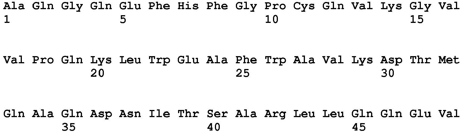


- <210> 18
<211> 371
<212> PRT
<213> Artificial - <220>
<223> Fusion Protein L19 (short) - huGM-CSF - <400> 18


- <210> 19
<211> 368
<212> PRT
<213> Artificial - <220>
<223> Fusion protein L19 (short) - murine GM-CSF - <400> 19


Claims (15)
- A fusion protein comprising:(i) an antibody, functional fragment or functional derivative thereof having specific binding affinity to either the extracellular domain of oncofetal fibronectin (ED-B) or to at least one of the extracellular domains of oncofetal tenascin fused to(ii) cytokine IL-10, or functional fragments thereof.
- The fusion protein of claim 1, wherein the antibody, functional fragment or functional derivative thereof is selected from the group consisting of polyclonal antibodies, monoclonal antibodies, chimeric antibodies, humanized antibodies, CDR-grafted antibodies, Fv-fragments, Fab-fragments, Fab2-fragments and antibody-like binding proteins.
- The fusion protein according to claim 1 or 2, wherein the functional derivative of the antibody is the diabody L19 (long) having the amino acid sequence set forth in SEQ ID NO: 6.
- The fusion protein according to claim 1 or 2, wherein the functional derivative of the antibody is the diabody L19 (short) having the amino sequence set forth in SEQ ID NO: 7.
- The fusion protein according to any one of claims 1 to 4, wherein a member of the group consisting of L19 (long), L19 (short), F16 (long), F16 (short), F16 (A34M) (long), F16 (A34M) (short), G11 (long) and G11 (short) having the amino sequences set forth in SEQ ID NO: 6 to 13, respectively, is fused to cytokine IL-10, or functional fragments thereof.
- The fusion protein according to any one of claims 1 to 5, wherein the cytokine is a murine or human, preferably a human cytokine, or functional fragment thereof.
- The fusion protein according to any one of claims 1 to 6, wherein the cytokine, or functional fragment thereof is fused N-terminally or C-terminally, preferably N-terminally, to the antibody, functional fragment or functional derivative thereof.
- The fusion protein according to any one of claims 1 to 7, wherein the antibody fragment or functional derivative thereof is selected from the group consisting of , L 19 (short), F16 (short), F16 (A34M) (short) and G11 as set forth in SEQ ID NO: 7, 9, 11 and 13, respectively, preferably F16 (A34M) (short).
- The fusion protein according to any one of claims 1 to 8, wherein the fusion protein is L19-IL-10.
- The fusion protein according to any one of claims 1 to 7 and 9 having the amino acid sequence set forth in SEQ ID NO: 14.
- The fusion protein according to any one of claims 1 to 10 for use in therapy.
- The fusion protein according to any of claims 1 to 10 for use in the treatment of cancer in a mammal, preferably in a human.
- The fusion protein according to any of claims 1 to 10 for use in the treatment of inflammatory diseases, preferably chronic inflammatory diseases in a mammal, preferably in a human.
- The fusion protein for use according to claim 13, wherein the inflammatory disease is selected from the group consisting of psoriasis, atherosclerosis, arthritis, preferably rheumatoid arthritis.
- A pharmaceutical composition comprising at least one fusion protein according to any one of claims 1 to 10 and optionally a pharmaceutically acceptable excipient.
Priority Applications (1)
| Application Number | Priority Date | Filing Date | Title |
|---|---|---|---|
| EP07724968.8A EP2016098B1 (en) | 2006-05-08 | 2007-05-08 | Antibody-targeted cytokines for therapy |
Applications Claiming Priority (3)
| Application Number | Priority Date | Filing Date | Title |
|---|---|---|---|
| EP06009456 | 2006-05-08 | ||
| EP07724968.8A EP2016098B1 (en) | 2006-05-08 | 2007-05-08 | Antibody-targeted cytokines for therapy |
| PCT/EP2007/004044 WO2007128563A1 (en) | 2006-05-08 | 2007-05-08 | Antibody-targeted cytokines for therapy |
Publications (2)
| Publication Number | Publication Date |
|---|---|
| EP2016098A1 EP2016098A1 (en) | 2009-01-21 |
| EP2016098B1 true EP2016098B1 (en) | 2016-09-07 |
Family
ID=38372334
Family Applications (1)
| Application Number | Title | Priority Date | Filing Date |
|---|---|---|---|
| EP07724968.8A Not-in-force EP2016098B1 (en) | 2006-05-08 | 2007-05-08 | Antibody-targeted cytokines for therapy |
Country Status (12)
| Country | Link |
|---|---|
| US (1) | US8404814B2 (en) |
| EP (1) | EP2016098B1 (en) |
| JP (1) | JP2009536170A (en) |
| KR (1) | KR20090008467A (en) |
| CN (1) | CN101437847A (en) |
| AU (1) | AU2007247380B2 (en) |
| BR (1) | BRPI0709787A2 (en) |
| CA (1) | CA2648484A1 (en) |
| EA (1) | EA200802289A1 (en) |
| ES (1) | ES2606490T3 (en) |
| MX (1) | MX2008013575A (en) |
| WO (1) | WO2007128563A1 (en) |
Families Citing this family (67)
| Publication number | Priority date | Publication date | Assignee | Title |
|---|---|---|---|---|
| DE19932688B4 (en) | 1999-07-13 | 2009-10-08 | Scil Proteins Gmbh | Design of beta-sheet proteins of gamma-II-crystalline antibody-like |
| DE10324447A1 (en) | 2003-05-28 | 2004-12-30 | Scil Proteins Gmbh | Generation of artificial binding proteins based on ubiquitin |
| KR20090008467A (en) * | 2006-05-08 | 2009-01-21 | 필로겐 에스.피.에이. | Therapeutic antibody-target cytokines |
| EP1925664A1 (en) | 2006-11-15 | 2008-05-28 | Scil proteins GmbH | Artificial binding proteins based on a modified alpha helical region of ubiquitin |
| JP5221641B2 (en) | 2007-04-02 | 2013-06-26 | フィロゲン エスピーエー | ED-A antigen of fibronectin associated with neovascularization of tumor metastasis |
| US10202442B2 (en) | 2007-07-25 | 2019-02-12 | Philogen S.P.A. | Antigen associated with lung cancers and lymphomas |
| SI2209805T1 (en) | 2007-10-30 | 2017-11-30 | Philogen S.P.A. | An antigen associated with rheumatoid arthritis |
| WO2009089998A1 (en) * | 2008-01-15 | 2009-07-23 | Philochem Ag | Binding members for tenascin-c domain a2 |
| WO2010005389A1 (en) * | 2008-07-11 | 2010-01-14 | Forskarpatent I Syd Ab | Oxidized ldl specific antibody-fusion and conjugated proteins |
| US8580267B2 (en) | 2008-12-19 | 2013-11-12 | Philogen S.P.A. | Immunocytokines for tumour therapy with chemotherapeutic agents |
| EP2379116B1 (en) | 2009-01-07 | 2015-08-26 | Philogen S.p.A. | Antigens associated with endometriosis |
| EP2947096B1 (en) | 2009-08-05 | 2018-11-28 | Philogen S.p.A. | Targeting of bone marrow neovasculature |
| PE20120630A1 (en) | 2009-08-17 | 2012-05-26 | Roche Glycart Ag | TARGETED IMMUNOCONJUGATES |
| KR101436222B1 (en) | 2009-12-14 | 2014-09-01 | 쉴 프로테인즈 게엠베하 | A method for identifying hetero-multimeric modified ubiquitin proteins with binding capability to ligands |
| RU2013139267A (en) | 2011-02-10 | 2015-03-20 | Рош Гликарт Аг | IMPROVED IMMUNOTHERAPY |
| HUE029139T2 (en) | 2011-02-10 | 2017-02-28 | Roche Glycart Ag | Mutant interleukin-2 polypeptides |
| EA201892619A1 (en) | 2011-04-29 | 2019-04-30 | Роше Гликарт Аг | IMMUNOCONJUGATES CONTAINING INTERLEUKIN-2 MUTANT POLYPETIPS |
| WO2012171541A1 (en) | 2011-06-15 | 2012-12-20 | Scil Proteins Gmbh | Human fusion proteins comprising interferons and hetero-dimeric modified ubiquitin proteins |
| EP2721152B1 (en) | 2011-06-15 | 2019-03-27 | Navigo Proteins GmbH | Dimeric binding proteins based on modified ubiquitins |
| EP2537933A1 (en) | 2011-06-24 | 2012-12-26 | Institut National de la Santé et de la Recherche Médicale (INSERM) | An IL-15 and IL-15Ralpha sushi domain based immunocytokines |
| PL2776460T3 (en) * | 2011-11-08 | 2018-10-31 | Umc Utrecht Holding B.V. | Fusion protein containing interleukin 4 and interleukin 10 |
| WO2013186329A1 (en) | 2012-06-13 | 2013-12-19 | Scil Proteins Gmbh | Human fusion proteins comprising single chain tnfalpha and targeting domains |
| BR112015002085A2 (en) * | 2012-08-08 | 2017-12-19 | Roche Glycart Ag | protein, polynucleotide, vector, host cell, method for producing protein, pharmaceutical composition, protein use, method of treatment and invention |
| US9695232B2 (en) | 2012-10-03 | 2017-07-04 | Philogen S.P.A. | Anti-ED-A immunoconjugates for inflammatory bowel disease |
| WO2014094799A1 (en) | 2012-12-19 | 2014-06-26 | Scil-Proteins-Gmbh | Ubiquitin moieties as a means for prolonging serum half-life |
| JP6295458B2 (en) * | 2013-03-08 | 2018-03-20 | たいまつ食品株式会社 | Granulocyte colony-stimulating factor production induction / promotion product |
| KR102539359B1 (en) | 2013-04-19 | 2023-06-02 | 싸이튠 파마 | Cytokine derived treatment with reduced vascular leak syndrome |
| EP3102594A1 (en) * | 2014-02-06 | 2016-12-14 | F. Hoffmann-La Roche AG | Interleukine 10 immunoconjugates |
| EP2915569A1 (en) | 2014-03-03 | 2015-09-09 | Cytune Pharma | IL-15/IL-15Ralpha based conjugates purification method |
| JP6836500B2 (en) | 2014-05-16 | 2021-03-03 | ベイラー リサーチ インスティテュートBaylor Research Institute | Methods and compositions for treating autoimmune and inflammatory conditions |
| CN104403004B (en) | 2014-11-24 | 2017-10-13 | 苏州丁孚靶点生物技术有限公司 | Preparation and use of antibody-interferon heterodimer |
| EP3253785B1 (en) | 2015-02-06 | 2019-04-10 | Navigo Proteins Gmbh | Novel egfr binding proteins |
| CN107922483B (en) | 2015-07-16 | 2021-07-30 | 纳维格蛋白质有限公司 | Novel immunoglobulin binding proteins and their use in affinity purification |
| WO2017013136A1 (en) | 2015-07-20 | 2017-01-26 | Scil Proteins Gmbh | Novel binding proteins based on di-ubiquitin muteins and methods for generation |
| WO2017087768A1 (en) * | 2015-11-18 | 2017-05-26 | Duke University | Targeted delivery of interleukin-10 to tumor infiltrating lymphocytes |
| US20190309060A1 (en) | 2016-01-15 | 2019-10-10 | Philogen S.P.A. | Intestinal antigens for pharmacodelivery applications |
| AU2017260274A1 (en) | 2016-05-04 | 2018-11-01 | Navigo Proteins Gmbh | Targeted compounds for the site-specific coupling of chemical moieties comprising a peptide linker |
| EP3257862A1 (en) | 2016-06-16 | 2017-12-20 | ETH Zürich | Fibronectin-binding peptides for use in tumor or fibrosis diagnosis and therapy |
| GB201612317D0 (en) | 2016-07-15 | 2016-08-31 | Philogen Spa | Antibody compositions |
| CA3030208A1 (en) | 2016-08-11 | 2018-02-15 | Navigo Proteins Gmbh | Novel alkaline stable immunoglobulin binding proteins |
| BR112019007281A2 (en) | 2016-10-14 | 2019-07-09 | Xencor Inc | heterodimeric protein, nucleic acid and expression vector compositions, expression vector, host cell, and methods for producing heterodimeric protein and for treating cancer in a patient |
| AU2018291497A1 (en) | 2017-06-30 | 2020-01-16 | Xencor, Inc. | Targeted heterodimeric Fc fusion proteins containing IL-15/IL-15Ra and antigen binding domains |
| TWI901940B (en) | 2017-08-03 | 2025-10-21 | 美商安進公司 | Interleukin-21 muteins and methods of treatment |
| WO2019062790A1 (en) * | 2017-09-29 | 2019-04-04 | Lan Keng Li | Novel conjugates and uses thereof |
| US11414466B2 (en) | 2017-11-07 | 2022-08-16 | Navigo Proteins Gmbh | Fusion proteins with specificity for ED-B and long serum half-life for diagnosis or treatment of cancer |
| JP2019122373A (en) | 2018-01-12 | 2019-07-25 | アムジエン・インコーポレーテツド | Anti-pd-1 antibodies and methods of treatment |
| WO2019185792A1 (en) | 2018-03-29 | 2019-10-03 | Philogen S.P.A | Cancer treatment using immunoconjugates and immune check-point inhibitors |
| CA3097593A1 (en) | 2018-04-18 | 2019-10-24 | Xencor, Inc. | Pd-1 targeted heterodimeric fusion proteins containing il-15/il-15ra fc-fusion proteins and pd-1 antigen binding domains and uses thereof |
| JP2021520829A (en) | 2018-04-18 | 2021-08-26 | ゼンコア インコーポレイテッド | TIM-3 targeted heterodimer fusion protein containing IL-15 / IL-15RA Fc fusion protein and TIM-3 antigen binding domain |
| JP7516254B2 (en) | 2018-04-18 | 2024-07-16 | ゼンコア インコーポレイテッド | IL-15/IL-15RA HETERODIMERIC FC FUSION PROTEINS AND USES THEREOF |
| KR20210003813A (en) | 2018-04-18 | 2021-01-12 | 젠코어 인코포레이티드 | IL-15/IL-15Rα Fc-fusion protein and LAG-3 targeting heterodimer fusion protein containing the LAG-3 antigen binding domain |
| EP3584258A1 (en) * | 2018-06-19 | 2019-12-25 | IEO - Istituto Europeo di Oncologia Srl | Antibodies anti tumor associated antigens and method for obtaining them |
| CR20210239A (en) | 2018-10-12 | 2021-12-15 | Xencor Inc | IL-15/IL-15RALFA-FC FUSION PROTEINS TARGETING PD-1 AND USES THEREOF IN COMBINATION THERAPIES |
| CA3123986A1 (en) | 2018-12-18 | 2020-06-25 | Navigo Proteins Gmbh | Novel folr1 specific binding proteins for cancer diagnosis and treatment |
| CN113438961A (en) | 2018-12-20 | 2021-09-24 | Xencor股份有限公司 | Targeting heterodimeric Fc fusion proteins containing IL-15/IL-15R α and NKG2D antigen binding domains |
| WO2020172631A2 (en) | 2019-02-21 | 2020-08-27 | Xencor, Inc. | Untargeted and targeted il-10 fc-fusion proteins |
| CN121652287A (en) * | 2019-03-06 | 2026-03-13 | 德卡生物科学公司 | IL-10 variant molecules and methods of treating inflammatory diseases and tumors |
| EP3955955B1 (en) | 2019-04-19 | 2025-02-26 | Synerkine Pharma B.V. | A fusion protein comprising il13 |
| WO2020249757A1 (en) | 2019-06-14 | 2020-12-17 | Philogen S.P.A | Immunoconjugates comprising a single chain diabody and interleukin-15 or interleukin-15 and a sushi domain of interleukin-15 receptor alpha |
| TW202128757A (en) | 2019-10-11 | 2021-08-01 | 美商建南德克公司 | Pd-1 targeted il-15/il-15ralpha fc fusion proteins with improved properties |
| WO2021142349A1 (en) * | 2020-01-09 | 2021-07-15 | Deka Biosciences, Inc. | Method of using genetic markers, snps and/or indels to determine responsiveness to il-10 treatment |
| JP7438396B2 (en) * | 2020-04-14 | 2024-02-26 | フィロジェン ソチエタ ペル アツィオーニ | Dosage units and regimens, uses, methods or formulations of compositions comprising a recombinant protein comprising interleukin-12 and an antibody that binds extra domain B of fibronectin |
| HRP20241274T1 (en) | 2020-05-22 | 2024-12-06 | Philogen S.P.A. | THERAPY BASED ON TNF-A IMMUNOCONJUGATE FOR THE TREATMENT OF BRAIN TUMORS |
| EP4181957A4 (en) | 2020-07-20 | 2024-08-07 | Deka Biosciences, Inc. | IL-10 CONTAINING DUAL CYTOKINE FFUSIONS PROTEINS |
| AU2021393752A1 (en) | 2020-12-04 | 2023-05-18 | F. Hoffmann-La Roche Ag | Ph-dependent mutant interleukin-2 polypeptides |
| AU2021467463A1 (en) | 2021-10-05 | 2024-04-04 | ETH Zürich | Fibronectin-binding peptides for use in tumor or fibrosis diagnosis and therapy |
| WO2026003203A1 (en) | 2024-06-27 | 2026-01-02 | Philogen S.P.A. | Combination of an immunocytokine comprising targeted il2 and a jak inhibitor |
Family Cites Families (3)
| Publication number | Priority date | Publication date | Assignee | Title |
|---|---|---|---|---|
| PT1257297E (en) * | 2000-02-24 | 2006-12-29 | Philogen Spa | Compositions and methods for treatemnt of angiogenesis in pathological lesions |
| KR101281208B1 (en) * | 2005-05-11 | 2013-07-26 | 필로겐 에스.피.에이. | Fusion protein of antibody l19 against fibronectin ed-b and interleukin 12 |
| KR20090008467A (en) * | 2006-05-08 | 2009-01-21 | 필로겐 에스.피.에이. | Therapeutic antibody-target cytokines |
-
2007
- 2007-05-08 KR KR1020087029943A patent/KR20090008467A/en not_active Ceased
- 2007-05-08 CN CNA2007800166749A patent/CN101437847A/en active Pending
- 2007-05-08 CA CA002648484A patent/CA2648484A1/en not_active Abandoned
- 2007-05-08 JP JP2009508239A patent/JP2009536170A/en active Pending
- 2007-05-08 EA EA200802289A patent/EA200802289A1/en unknown
- 2007-05-08 EP EP07724968.8A patent/EP2016098B1/en not_active Not-in-force
- 2007-05-08 ES ES07724968.8T patent/ES2606490T3/en active Active
- 2007-05-08 AU AU2007247380A patent/AU2007247380B2/en not_active Ceased
- 2007-05-08 US US12/227,060 patent/US8404814B2/en not_active Expired - Fee Related
- 2007-05-08 WO PCT/EP2007/004044 patent/WO2007128563A1/en not_active Ceased
- 2007-05-08 BR BRPI0709787-5A patent/BRPI0709787A2/en not_active IP Right Cessation
- 2007-05-08 MX MX2008013575A patent/MX2008013575A/en active IP Right Grant
Also Published As
| Publication number | Publication date |
|---|---|
| US20110256091A1 (en) | 2011-10-20 |
| ES2606490T3 (en) | 2017-03-24 |
| EP2016098A1 (en) | 2009-01-21 |
| AU2007247380B2 (en) | 2011-03-10 |
| CN101437847A (en) | 2009-05-20 |
| MX2008013575A (en) | 2008-11-04 |
| KR20090008467A (en) | 2009-01-21 |
| US8404814B2 (en) | 2013-03-26 |
| BRPI0709787A2 (en) | 2011-03-29 |
| CA2648484A1 (en) | 2007-11-15 |
| EA200802289A1 (en) | 2009-04-28 |
| WO2007128563A1 (en) | 2007-11-15 |
| JP2009536170A (en) | 2009-10-08 |
| AU2007247380A1 (en) | 2007-11-15 |
Similar Documents
| Publication | Publication Date | Title |
|---|---|---|
| AU2007247380B2 (en) | Antibody-targeted cytokines for therapy | |
| US12006361B2 (en) | Albumin binding domain fusion proteins | |
| RU2711979C2 (en) | Interleukin 15 protein complex and use thereof | |
| US7141651B2 (en) | Multiple cytokine protein complexes | |
| Ongaro et al. | A novel anti-cancer L19-interleukin-12 fusion protein with an optimized peptide linker efficiently localizes in vivo at the site of tumors | |
| JP6925431B2 (en) | Immunoconjugates of IL2 and TNF mutants | |
| JP2008528039A (en) | Homogeneous preparation of IL-31 | |
| JP7830450B2 (en) | Modified IL-2 molecules and their uses | |
| US7863018B2 (en) | G-CSF polypeptides and uses thereof | |
| EP1731531B1 (en) | Multiple cytokine-antibody complexes | |
| RU2786444C2 (en) | Fused proteins with albumin-binding domains | |
| HK40126456A (en) | Albumin binding domain fusion proteins | |
| CN117683140A (en) | Tumor-targeted fusion protein-based drug prodrugs with interleukin 2 as the active ingredient | |
| Pasche | Novel immunocytokines for cancer therapy |
Legal Events
| Date | Code | Title | Description |
|---|---|---|---|
| PUAI | Public reference made under article 153(3) epc to a published international application that has entered the european phase |
Free format text: ORIGINAL CODE: 0009012 |
|
| 17P | Request for examination filed |
Effective date: 20080927 |
|
| AK | Designated contracting states |
Kind code of ref document: A1 Designated state(s): AT BE BG CH CY CZ DE DK EE ES FI FR GB GR HU IE IS IT LI LT LU LV MC MT NL PL PT RO SE SI SK TR |
|
| AX | Request for extension of the european patent |
Extension state: AL BA HR MK RS |
|
| RIN1 | Information on inventor provided before grant (corrected) |
Inventor name: KASPAR, MANUELA Inventor name: NERI, DARIO Inventor name: TRACHSEL, EVELINE |
|
| 17Q | First examination report despatched |
Effective date: 20100825 |
|
| DAX | Request for extension of the european patent (deleted) | ||
| GRAP | Despatch of communication of intention to grant a patent |
Free format text: ORIGINAL CODE: EPIDOSNIGR1 |
|
| GRAJ | Information related to disapproval of communication of intention to grant by the applicant or resumption of examination proceedings by the epo deleted |
Free format text: ORIGINAL CODE: EPIDOSDIGR1 |
|
| GRAP | Despatch of communication of intention to grant a patent |
Free format text: ORIGINAL CODE: EPIDOSNIGR1 |
|
| INTG | Intention to grant announced |
Effective date: 20160323 |
|
| INTC | Intention to grant announced (deleted) | ||
| INTG | Intention to grant announced |
Effective date: 20160408 |
|
| GRAS | Grant fee paid |
Free format text: ORIGINAL CODE: EPIDOSNIGR3 |
|
| GRAA | (expected) grant |
Free format text: ORIGINAL CODE: 0009210 |
|
| AK | Designated contracting states |
Kind code of ref document: B1 Designated state(s): AT BE BG CH CY CZ DE DK EE ES FI FR GB GR HU IE IS IT LI LT LU LV MC MT NL PL PT RO SE SI SK TR |
|
| REG | Reference to a national code |
Ref country code: GB Ref legal event code: FG4D |
|
| REG | Reference to a national code |
Ref country code: CH Ref legal event code: EP Ref country code: CH Ref legal event code: NV Representative=s name: KASCHE AND PARTNER AG, CH |
|
| REG | Reference to a national code |
Ref country code: IE Ref legal event code: FG4D |
|
| REG | Reference to a national code |
Ref country code: AT Ref legal event code: REF Ref document number: 826766 Country of ref document: AT Kind code of ref document: T Effective date: 20161015 |
|
| REG | Reference to a national code |
Ref country code: DE Ref legal event code: R096 Ref document number: 602007047816 Country of ref document: DE |
|
| REG | Reference to a national code |
Ref country code: NL Ref legal event code: FP |
|
| REG | Reference to a national code |
Ref country code: LT Ref legal event code: MG4D |
|
| PG25 | Lapsed in a contracting state [announced via postgrant information from national office to epo] |
Ref country code: FI Free format text: LAPSE BECAUSE OF FAILURE TO SUBMIT A TRANSLATION OF THE DESCRIPTION OR TO PAY THE FEE WITHIN THE PRESCRIBED TIME-LIMIT Effective date: 20160907 Ref country code: LT Free format text: LAPSE BECAUSE OF FAILURE TO SUBMIT A TRANSLATION OF THE DESCRIPTION OR TO PAY THE FEE WITHIN THE PRESCRIBED TIME-LIMIT Effective date: 20160907 |
|
| REG | Reference to a national code |
Ref country code: AT Ref legal event code: MK05 Ref document number: 826766 Country of ref document: AT Kind code of ref document: T Effective date: 20160907 |
|
| PG25 | Lapsed in a contracting state [announced via postgrant information from national office to epo] |
Ref country code: GR Free format text: LAPSE BECAUSE OF FAILURE TO SUBMIT A TRANSLATION OF THE DESCRIPTION OR TO PAY THE FEE WITHIN THE PRESCRIBED TIME-LIMIT Effective date: 20161208 Ref country code: LV Free format text: LAPSE BECAUSE OF FAILURE TO SUBMIT A TRANSLATION OF THE DESCRIPTION OR TO PAY THE FEE WITHIN THE PRESCRIBED TIME-LIMIT Effective date: 20160907 Ref country code: SE Free format text: LAPSE BECAUSE OF FAILURE TO SUBMIT A TRANSLATION OF THE DESCRIPTION OR TO PAY THE FEE WITHIN THE PRESCRIBED TIME-LIMIT Effective date: 20160907 |
|
| PG25 | Lapsed in a contracting state [announced via postgrant information from national office to epo] |
Ref country code: EE Free format text: LAPSE BECAUSE OF FAILURE TO SUBMIT A TRANSLATION OF THE DESCRIPTION OR TO PAY THE FEE WITHIN THE PRESCRIBED TIME-LIMIT Effective date: 20160907 Ref country code: RO Free format text: LAPSE BECAUSE OF FAILURE TO SUBMIT A TRANSLATION OF THE DESCRIPTION OR TO PAY THE FEE WITHIN THE PRESCRIBED TIME-LIMIT Effective date: 20160907 |
|
| REG | Reference to a national code |
Ref country code: FR Ref legal event code: PLFP Year of fee payment: 11 |
|
| PG25 | Lapsed in a contracting state [announced via postgrant information from national office to epo] |
Ref country code: CZ Free format text: LAPSE BECAUSE OF FAILURE TO SUBMIT A TRANSLATION OF THE DESCRIPTION OR TO PAY THE FEE WITHIN THE PRESCRIBED TIME-LIMIT Effective date: 20160907 Ref country code: BE Free format text: LAPSE BECAUSE OF FAILURE TO SUBMIT A TRANSLATION OF THE DESCRIPTION OR TO PAY THE FEE WITHIN THE PRESCRIBED TIME-LIMIT Effective date: 20160907 Ref country code: IS Free format text: LAPSE BECAUSE OF FAILURE TO SUBMIT A TRANSLATION OF THE DESCRIPTION OR TO PAY THE FEE WITHIN THE PRESCRIBED TIME-LIMIT Effective date: 20170107 Ref country code: BG Free format text: LAPSE BECAUSE OF FAILURE TO SUBMIT A TRANSLATION OF THE DESCRIPTION OR TO PAY THE FEE WITHIN THE PRESCRIBED TIME-LIMIT Effective date: 20161207 Ref country code: PT Free format text: LAPSE BECAUSE OF FAILURE TO SUBMIT A TRANSLATION OF THE DESCRIPTION OR TO PAY THE FEE WITHIN THE PRESCRIBED TIME-LIMIT Effective date: 20170109 Ref country code: AT Free format text: LAPSE BECAUSE OF FAILURE TO SUBMIT A TRANSLATION OF THE DESCRIPTION OR TO PAY THE FEE WITHIN THE PRESCRIBED TIME-LIMIT Effective date: 20160907 Ref country code: SK Free format text: LAPSE BECAUSE OF FAILURE TO SUBMIT A TRANSLATION OF THE DESCRIPTION OR TO PAY THE FEE WITHIN THE PRESCRIBED TIME-LIMIT Effective date: 20160907 Ref country code: PL Free format text: LAPSE BECAUSE OF FAILURE TO SUBMIT A TRANSLATION OF THE DESCRIPTION OR TO PAY THE FEE WITHIN THE PRESCRIBED TIME-LIMIT Effective date: 20160907 |
|
| REG | Reference to a national code |
Ref country code: DE Ref legal event code: R097 Ref document number: 602007047816 Country of ref document: DE |
|
| PGFP | Annual fee paid to national office [announced via postgrant information from national office to epo] |
Ref country code: NL Payment date: 20170525 Year of fee payment: 11 |
|
| PLBE | No opposition filed within time limit |
Free format text: ORIGINAL CODE: 0009261 |
|
| STAA | Information on the status of an ep patent application or granted ep patent |
Free format text: STATUS: NO OPPOSITION FILED WITHIN TIME LIMIT |
|
| PG25 | Lapsed in a contracting state [announced via postgrant information from national office to epo] |
Ref country code: DK Free format text: LAPSE BECAUSE OF FAILURE TO SUBMIT A TRANSLATION OF THE DESCRIPTION OR TO PAY THE FEE WITHIN THE PRESCRIBED TIME-LIMIT Effective date: 20160907 |
|
| PGFP | Annual fee paid to national office [announced via postgrant information from national office to epo] |
Ref country code: GB Payment date: 20170522 Year of fee payment: 11 Ref country code: DE Payment date: 20170525 Year of fee payment: 11 Ref country code: FR Payment date: 20170526 Year of fee payment: 11 Ref country code: CH Payment date: 20170525 Year of fee payment: 11 |
|
| 26N | No opposition filed |
Effective date: 20170608 |
|
| PG25 | Lapsed in a contracting state [announced via postgrant information from national office to epo] |
Ref country code: LU Free format text: LAPSE BECAUSE OF NON-PAYMENT OF DUE FEES Effective date: 20170531 Ref country code: SI Free format text: LAPSE BECAUSE OF FAILURE TO SUBMIT A TRANSLATION OF THE DESCRIPTION OR TO PAY THE FEE WITHIN THE PRESCRIBED TIME-LIMIT Effective date: 20160907 |
|
| PGFP | Annual fee paid to national office [announced via postgrant information from national office to epo] |
Ref country code: ES Payment date: 20170612 Year of fee payment: 11 Ref country code: IT Payment date: 20170530 Year of fee payment: 11 |
|
| PG25 | Lapsed in a contracting state [announced via postgrant information from national office to epo] |
Ref country code: MC Free format text: LAPSE BECAUSE OF FAILURE TO SUBMIT A TRANSLATION OF THE DESCRIPTION OR TO PAY THE FEE WITHIN THE PRESCRIBED TIME-LIMIT Effective date: 20160907 |
|
| REG | Reference to a national code |
Ref country code: IE Ref legal event code: MM4A |
|
| PG25 | Lapsed in a contracting state [announced via postgrant information from national office to epo] |
Ref country code: LU Free format text: LAPSE BECAUSE OF NON-PAYMENT OF DUE FEES Effective date: 20170508 |
|
| PG25 | Lapsed in a contracting state [announced via postgrant information from national office to epo] |
Ref country code: IE Free format text: LAPSE BECAUSE OF NON-PAYMENT OF DUE FEES Effective date: 20170508 |
|
| PG25 | Lapsed in a contracting state [announced via postgrant information from national office to epo] |
Ref country code: MT Free format text: LAPSE BECAUSE OF NON-PAYMENT OF DUE FEES Effective date: 20170508 |
|
| REG | Reference to a national code |
Ref country code: DE Ref legal event code: R119 Ref document number: 602007047816 Country of ref document: DE |
|
| REG | Reference to a national code |
Ref country code: CH Ref legal event code: PL |
|
| REG | Reference to a national code |
Ref country code: NL Ref legal event code: MM Effective date: 20180601 |
|
| GBPC | Gb: european patent ceased through non-payment of renewal fee |
Effective date: 20180508 |
|
| PG25 | Lapsed in a contracting state [announced via postgrant information from national office to epo] |
Ref country code: CH Free format text: LAPSE BECAUSE OF NON-PAYMENT OF DUE FEES Effective date: 20180531 Ref country code: LI Free format text: LAPSE BECAUSE OF NON-PAYMENT OF DUE FEES Effective date: 20180531 |
|
| PG25 | Lapsed in a contracting state [announced via postgrant information from national office to epo] |
Ref country code: FR Free format text: LAPSE BECAUSE OF NON-PAYMENT OF DUE FEES Effective date: 20180531 Ref country code: NL Free format text: LAPSE BECAUSE OF NON-PAYMENT OF DUE FEES Effective date: 20180601 Ref country code: GB Free format text: LAPSE BECAUSE OF NON-PAYMENT OF DUE FEES Effective date: 20180508 Ref country code: IT Free format text: LAPSE BECAUSE OF NON-PAYMENT OF DUE FEES Effective date: 20180508 Ref country code: DE Free format text: LAPSE BECAUSE OF NON-PAYMENT OF DUE FEES Effective date: 20181201 |
|
| PG25 | Lapsed in a contracting state [announced via postgrant information from national office to epo] |
Ref country code: HU Free format text: LAPSE BECAUSE OF FAILURE TO SUBMIT A TRANSLATION OF THE DESCRIPTION OR TO PAY THE FEE WITHIN THE PRESCRIBED TIME-LIMIT; INVALID AB INITIO Effective date: 20070508 |
|
| REG | Reference to a national code |
Ref country code: ES Ref legal event code: FD2A Effective date: 20190913 |
|
| PG25 | Lapsed in a contracting state [announced via postgrant information from national office to epo] |
Ref country code: ES Free format text: LAPSE BECAUSE OF NON-PAYMENT OF DUE FEES Effective date: 20180509 Ref country code: CY Free format text: LAPSE BECAUSE OF NON-PAYMENT OF DUE FEES Effective date: 20160907 |
|
| PG25 | Lapsed in a contracting state [announced via postgrant information from national office to epo] |
Ref country code: TR Free format text: LAPSE BECAUSE OF FAILURE TO SUBMIT A TRANSLATION OF THE DESCRIPTION OR TO PAY THE FEE WITHIN THE PRESCRIBED TIME-LIMIT Effective date: 20160907 |