EP1994173B1 - Verfahren für die diagnose und/oder die prognosestellung bei eierstockkrebs - Google Patents
Verfahren für die diagnose und/oder die prognosestellung bei eierstockkrebs Download PDFInfo
- Publication number
- EP1994173B1 EP1994173B1 EP07723318A EP07723318A EP1994173B1 EP 1994173 B1 EP1994173 B1 EP 1994173B1 EP 07723318 A EP07723318 A EP 07723318A EP 07723318 A EP07723318 A EP 07723318A EP 1994173 B1 EP1994173 B1 EP 1994173B1
- Authority
- EP
- European Patent Office
- Prior art keywords
- tusc3
- pcr
- expression
- ovarian cancer
- methylation
- Prior art date
- Legal status (The legal status is an assumption and is not a legal conclusion. Google has not performed a legal analysis and makes no representation as to the accuracy of the status listed.)
- Not-in-force
Links
- 206010061535 Ovarian neoplasm Diseases 0.000 title claims description 59
- 206010033128 Ovarian cancer Diseases 0.000 title claims description 54
- 238000000034 method Methods 0.000 title claims description 23
- 101000762128 Homo sapiens Tumor suppressor candidate 3 Proteins 0.000 claims description 79
- 102100024248 Tumor suppressor candidate 3 Human genes 0.000 claims description 79
- 230000006607 hypermethylation Effects 0.000 claims description 36
- 230000011987 methylation Effects 0.000 claims description 19
- 238000007069 methylation reaction Methods 0.000 claims description 19
- 238000003745 diagnosis Methods 0.000 claims description 18
- 238000001514 detection method Methods 0.000 claims description 17
- 239000000523 sample Substances 0.000 claims description 16
- 238000007855 methylation-specific PCR Methods 0.000 claims description 14
- 238000004393 prognosis Methods 0.000 claims description 12
- 239000012472 biological sample Substances 0.000 claims description 11
- 239000003550 marker Substances 0.000 claims description 10
- 238000004458 analytical method Methods 0.000 claims description 6
- 239000013060 biological fluid Substances 0.000 claims description 6
- 238000002955 isolation Methods 0.000 claims description 5
- 208000037821 progressive disease Diseases 0.000 claims description 4
- 108700039691 Genetic Promoter Regions Proteins 0.000 claims description 3
- 238000009396 hybridization Methods 0.000 claims description 3
- 201000010099 disease Diseases 0.000 claims description 2
- 208000037265 diseases, disorders, signs and symptoms Diseases 0.000 claims description 2
- 230000014509 gene expression Effects 0.000 description 65
- 206010028980 Neoplasm Diseases 0.000 description 48
- 201000011510 cancer Diseases 0.000 description 23
- 210000004027 cell Anatomy 0.000 description 23
- 210000001519 tissue Anatomy 0.000 description 23
- 108020004414 DNA Proteins 0.000 description 21
- 108090000623 proteins and genes Proteins 0.000 description 16
- 230000004083 survival effect Effects 0.000 description 14
- 230000002611 ovarian Effects 0.000 description 13
- XAUDJQYHKZQPEU-KVQBGUIXSA-N 5-aza-2'-deoxycytidine Chemical compound O=C1N=C(N)N=CN1[C@@H]1O[C@H](CO)[C@@H](O)C1 XAUDJQYHKZQPEU-KVQBGUIXSA-N 0.000 description 12
- 238000002560 therapeutic procedure Methods 0.000 description 11
- 238000012360 testing method Methods 0.000 description 9
- 108091032973 (ribonucleotides)n+m Proteins 0.000 description 8
- HEMHJVSKTPXQMS-UHFFFAOYSA-M Sodium hydroxide Chemical compound [OH-].[Na+] HEMHJVSKTPXQMS-UHFFFAOYSA-M 0.000 description 6
- 238000002512 chemotherapy Methods 0.000 description 6
- 238000011518 platinum-based chemotherapy Methods 0.000 description 6
- 230000004044 response Effects 0.000 description 6
- 238000012353 t test Methods 0.000 description 6
- 230000035131 DNA demethylation Effects 0.000 description 5
- 102000003964 Histone deacetylase Human genes 0.000 description 5
- 108090000353 Histone deacetylase Proteins 0.000 description 5
- 239000003112 inhibitor Substances 0.000 description 5
- 190000008236 Carboplatin Chemical compound 0.000 description 4
- 108091029523 CpG island Proteins 0.000 description 4
- 208000000236 Prostatic Neoplasms Diseases 0.000 description 4
- 238000000692 Student's t-test Methods 0.000 description 4
- 102000044209 Tumor Suppressor Genes Human genes 0.000 description 4
- 108700025716 Tumor Suppressor Genes Proteins 0.000 description 4
- 239000011543 agarose gel Substances 0.000 description 4
- 229960004562 carboplatin Drugs 0.000 description 4
- 210000000349 chromosome Anatomy 0.000 description 4
- 239000002299 complementary DNA Substances 0.000 description 4
- 229960003603 decitabine Drugs 0.000 description 4
- ZMMJGEGLRURXTF-UHFFFAOYSA-N ethidium bromide Chemical compound [Br-].C12=CC(N)=CC=C2C2=CC=C(N)C=C2[N+](CC)=C1C1=CC=CC=C1 ZMMJGEGLRURXTF-UHFFFAOYSA-N 0.000 description 4
- 229960005542 ethidium bromide Drugs 0.000 description 4
- 239000003147 molecular marker Substances 0.000 description 4
- 102000004169 proteins and genes Human genes 0.000 description 4
- 238000003753 real-time PCR Methods 0.000 description 4
- 108091008146 restriction endonucleases Proteins 0.000 description 4
- 230000009466 transformation Effects 0.000 description 4
- 239000012650 DNA demethylating agent Substances 0.000 description 3
- 229940045805 DNA demethylating agent Drugs 0.000 description 3
- 230000007067 DNA methylation Effects 0.000 description 3
- 101000623901 Homo sapiens Mucin-16 Proteins 0.000 description 3
- 102100023123 Mucin-16 Human genes 0.000 description 3
- 229930012538 Paclitaxel Natural products 0.000 description 3
- 206010060862 Prostate cancer Diseases 0.000 description 3
- 238000011529 RT qPCR Methods 0.000 description 3
- 102000015736 beta 2-Microglobulin Human genes 0.000 description 3
- 108010081355 beta 2-Microglobulin Proteins 0.000 description 3
- 238000006243 chemical reaction Methods 0.000 description 3
- 238000012217 deletion Methods 0.000 description 3
- 230000037430 deletion Effects 0.000 description 3
- 108020004999 messenger RNA Proteins 0.000 description 3
- 239000000203 mixture Substances 0.000 description 3
- 229960001592 paclitaxel Drugs 0.000 description 3
- 238000007619 statistical method Methods 0.000 description 3
- RCINICONZNJXQF-MZXODVADSA-N taxol Chemical compound O([C@@H]1[C@@]2(C[C@@H](C(C)=C(C2(C)C)[C@H](C([C@]2(C)[C@@H](O)C[C@H]3OC[C@]3([C@H]21)OC(C)=O)=O)OC(=O)C)OC(=O)[C@H](O)[C@@H](NC(=O)C=1C=CC=CC=1)C=1C=CC=CC=1)O)C(=O)C1=CC=CC=C1 RCINICONZNJXQF-MZXODVADSA-N 0.000 description 3
- 102100027314 Beta-2-microglobulin Human genes 0.000 description 2
- LSNNMFCWUKXFEE-UHFFFAOYSA-M Bisulfite Chemical compound OS([O-])=O LSNNMFCWUKXFEE-UHFFFAOYSA-M 0.000 description 2
- 108010089072 Dolichyl-diphosphooligosaccharide-protein glycotransferase Proteins 0.000 description 2
- LFQSCWFLJHTTHZ-UHFFFAOYSA-N Ethanol Chemical compound CCO LFQSCWFLJHTTHZ-UHFFFAOYSA-N 0.000 description 2
- 238000000729 Fisher's exact test Methods 0.000 description 2
- QIGBRXMKCJKVMJ-UHFFFAOYSA-N Hydroquinone Chemical compound OC1=CC=C(O)C=C1 QIGBRXMKCJKVMJ-UHFFFAOYSA-N 0.000 description 2
- 238000002123 RNA extraction Methods 0.000 description 2
- 238000001369 bisulfite sequencing Methods 0.000 description 2
- 238000012512 characterization method Methods 0.000 description 2
- 239000003795 chemical substances by application Substances 0.000 description 2
- 230000002759 chromosomal effect Effects 0.000 description 2
- 210000001072 colon Anatomy 0.000 description 2
- 208000029742 colonic neoplasm Diseases 0.000 description 2
- 230000004732 colorectal carcinogenesis Effects 0.000 description 2
- 208000031513 cyst Diseases 0.000 description 2
- 230000034994 death Effects 0.000 description 2
- 231100000517 death Toxicity 0.000 description 2
- 230000004069 differentiation Effects 0.000 description 2
- 239000003814 drug Substances 0.000 description 2
- 238000005516 engineering process Methods 0.000 description 2
- 230000030279 gene silencing Effects 0.000 description 2
- 239000000463 material Substances 0.000 description 2
- 230000035772 mutation Effects 0.000 description 2
- 210000001672 ovary Anatomy 0.000 description 2
- BASFCYQUMIYNBI-UHFFFAOYSA-N platinum Chemical compound [Pt] BASFCYQUMIYNBI-UHFFFAOYSA-N 0.000 description 2
- 238000012216 screening Methods 0.000 description 2
- 210000002966 serum Anatomy 0.000 description 2
- 238000001356 surgical procedure Methods 0.000 description 2
- 208000011580 syndromic disease Diseases 0.000 description 2
- 238000011521 systemic chemotherapy Methods 0.000 description 2
- 208000005623 Carcinogenesis Diseases 0.000 description 1
- 206010009944 Colon cancer Diseases 0.000 description 1
- 102100024462 Cyclin-dependent kinase 4 inhibitor B Human genes 0.000 description 1
- 102100024458 Cyclin-dependent kinase inhibitor 2A Human genes 0.000 description 1
- 102000053602 DNA Human genes 0.000 description 1
- 238000007399 DNA isolation Methods 0.000 description 1
- 108010063593 DNA modification methylase SssI Proteins 0.000 description 1
- 102000016911 Deoxyribonucleases Human genes 0.000 description 1
- 108010053770 Deoxyribonucleases Proteins 0.000 description 1
- 108700039887 Essential Genes Proteins 0.000 description 1
- 229920002527 Glycogen Polymers 0.000 description 1
- 208000033640 Hereditary breast cancer Diseases 0.000 description 1
- 101000733249 Homo sapiens Tumor suppressor ARF Proteins 0.000 description 1
- 208000034179 Neoplasms, Glandular and Epithelial Diseases 0.000 description 1
- 238000009004 PCR Kit Methods 0.000 description 1
- ZYFVNVRFVHJEIU-UHFFFAOYSA-N PicoGreen Chemical compound CN(C)CCCN(CCCN(C)C)C1=CC(=CC2=[N+](C3=CC=CC=C3S2)C)C2=CC=CC=C2N1C1=CC=CC=C1 ZYFVNVRFVHJEIU-UHFFFAOYSA-N 0.000 description 1
- 108010029485 Protein Isoforms Proteins 0.000 description 1
- 102000001708 Protein Isoforms Human genes 0.000 description 1
- 238000011530 RNeasy Mini Kit Methods 0.000 description 1
- 101150099625 STT3 gene Proteins 0.000 description 1
- DWAQJAXMDSEUJJ-UHFFFAOYSA-M Sodium bisulfite Chemical compound [Na+].OS([O-])=O DWAQJAXMDSEUJJ-UHFFFAOYSA-M 0.000 description 1
- XSQUKJJJFZCRTK-UHFFFAOYSA-N Urea Chemical compound NC(N)=O XSQUKJJJFZCRTK-UHFFFAOYSA-N 0.000 description 1
- 208000007097 Urinary Bladder Neoplasms Diseases 0.000 description 1
- 208000002495 Uterine Neoplasms Diseases 0.000 description 1
- 238000000246 agarose gel electrophoresis Methods 0.000 description 1
- 230000032683 aging Effects 0.000 description 1
- 238000000540 analysis of variance Methods 0.000 description 1
- 238000000137 annealing Methods 0.000 description 1
- 230000000692 anti-sense effect Effects 0.000 description 1
- 230000004791 biological behavior Effects 0.000 description 1
- 239000000090 biomarker Substances 0.000 description 1
- 210000004369 blood Anatomy 0.000 description 1
- 239000008280 blood Substances 0.000 description 1
- 210000001124 body fluid Anatomy 0.000 description 1
- 239000010839 body fluid Substances 0.000 description 1
- 239000000872 buffer Substances 0.000 description 1
- 238000010804 cDNA synthesis Methods 0.000 description 1
- 238000004364 calculation method Methods 0.000 description 1
- 230000036952 cancer formation Effects 0.000 description 1
- 238000005251 capillar electrophoresis Methods 0.000 description 1
- 239000004202 carbamide Substances 0.000 description 1
- 231100000504 carcinogenesis Toxicity 0.000 description 1
- 230000003197 catalytic effect Effects 0.000 description 1
- 230000001364 causal effect Effects 0.000 description 1
- 239000006143 cell culture medium Substances 0.000 description 1
- 238000011500 cytoreductive surgery Methods 0.000 description 1
- 238000011334 debulking surgery Methods 0.000 description 1
- 239000012649 demethylating agent Substances 0.000 description 1
- 230000001419 dependent effect Effects 0.000 description 1
- 238000011161 development Methods 0.000 description 1
- 230000018109 developmental process Effects 0.000 description 1
- 230000002255 enzymatic effect Effects 0.000 description 1
- 230000001973 epigenetic effect Effects 0.000 description 1
- 230000004049 epigenetic modification Effects 0.000 description 1
- 238000002474 experimental method Methods 0.000 description 1
- 238000010195 expression analysis Methods 0.000 description 1
- 238000000605 extraction Methods 0.000 description 1
- 230000002349 favourable effect Effects 0.000 description 1
- 239000000499 gel Substances 0.000 description 1
- 229960005277 gemcitabine Drugs 0.000 description 1
- SDUQYLNIPVEERB-QPPQHZFASA-N gemcitabine Chemical compound O=C1N=C(N)C=CN1[C@H]1C(F)(F)[C@H](O)[C@@H](CO)O1 SDUQYLNIPVEERB-QPPQHZFASA-N 0.000 description 1
- 229940096919 glycogen Drugs 0.000 description 1
- PCHJSUWPFVWCPO-UHFFFAOYSA-N gold Chemical compound [Au] PCHJSUWPFVWCPO-UHFFFAOYSA-N 0.000 description 1
- 239000010931 gold Substances 0.000 description 1
- 229910052737 gold Inorganic materials 0.000 description 1
- 208000025581 hereditary breast carcinoma Diseases 0.000 description 1
- 230000002055 immunohistochemical effect Effects 0.000 description 1
- 238000000126 in silico method Methods 0.000 description 1
- 230000000415 inactivating effect Effects 0.000 description 1
- 230000003993 interaction Effects 0.000 description 1
- 238000011835 investigation Methods 0.000 description 1
- 238000002032 lab-on-a-chip Methods 0.000 description 1
- 231100000518 lethal Toxicity 0.000 description 1
- 230000001665 lethal effect Effects 0.000 description 1
- 210000004185 liver Anatomy 0.000 description 1
- 208000014018 liver neoplasm Diseases 0.000 description 1
- 210000004072 lung Anatomy 0.000 description 1
- 208000020816 lung neoplasm Diseases 0.000 description 1
- 230000036210 malignancy Effects 0.000 description 1
- 230000003211 malignant effect Effects 0.000 description 1
- 238000013507 mapping Methods 0.000 description 1
- 238000005259 measurement Methods 0.000 description 1
- 239000002609 medium Substances 0.000 description 1
- 208000010658 metastatic prostate carcinoma Diseases 0.000 description 1
- 125000002496 methyl group Chemical group [H]C([H])([H])* 0.000 description 1
- 239000002480 mineral oil Substances 0.000 description 1
- 235000010446 mineral oil Nutrition 0.000 description 1
- 239000003068 molecular probe Substances 0.000 description 1
- 230000000877 morphologic effect Effects 0.000 description 1
- 210000004877 mucosa Anatomy 0.000 description 1
- 238000000491 multivariate analysis Methods 0.000 description 1
- 239000013642 negative control Substances 0.000 description 1
- 230000001613 neoplastic effect Effects 0.000 description 1
- 238000001543 one-way ANOVA Methods 0.000 description 1
- 238000009806 oophorectomy Methods 0.000 description 1
- 210000002381 plasma Anatomy 0.000 description 1
- 229910052697 platinum Inorganic materials 0.000 description 1
- 239000013641 positive control Substances 0.000 description 1
- 230000002980 postoperative effect Effects 0.000 description 1
- 238000002360 preparation method Methods 0.000 description 1
- 230000000069 prophylactic effect Effects 0.000 description 1
- 210000002307 prostate Anatomy 0.000 description 1
- 230000004853 protein function Effects 0.000 description 1
- 238000000746 purification Methods 0.000 description 1
- 239000011535 reaction buffer Substances 0.000 description 1
- 230000009467 reduction Effects 0.000 description 1
- 230000028617 response to DNA damage stimulus Effects 0.000 description 1
- 238000003757 reverse transcription PCR Methods 0.000 description 1
- 238000012163 sequencing technique Methods 0.000 description 1
- 238000009097 single-agent therapy Methods 0.000 description 1
- 235000010267 sodium hydrogen sulphite Nutrition 0.000 description 1
- 238000011895 specific detection Methods 0.000 description 1
- 238000010186 staining Methods 0.000 description 1
- 238000009121 systemic therapy Methods 0.000 description 1
- 210000004881 tumor cell Anatomy 0.000 description 1
- 239000000439 tumor marker Substances 0.000 description 1
- 206010046766 uterine cancer Diseases 0.000 description 1
Images

Classifications
-
- C—CHEMISTRY; METALLURGY
- C12—BIOCHEMISTRY; BEER; SPIRITS; WINE; VINEGAR; MICROBIOLOGY; ENZYMOLOGY; MUTATION OR GENETIC ENGINEERING
- C12Q—MEASURING OR TESTING PROCESSES INVOLVING ENZYMES, NUCLEIC ACIDS OR MICROORGANISMS; COMPOSITIONS OR TEST PAPERS THEREFOR; PROCESSES OF PREPARING SUCH COMPOSITIONS; CONDITION-RESPONSIVE CONTROL IN MICROBIOLOGICAL OR ENZYMOLOGICAL PROCESSES
- C12Q1/00—Measuring or testing processes involving enzymes, nucleic acids or microorganisms; Compositions therefor; Processes of preparing such compositions
- C12Q1/68—Measuring or testing processes involving enzymes, nucleic acids or microorganisms; Compositions therefor; Processes of preparing such compositions involving nucleic acids
- C12Q1/6876—Nucleic acid products used in the analysis of nucleic acids, e.g. primers or probes
- C12Q1/6883—Nucleic acid products used in the analysis of nucleic acids, e.g. primers or probes for diseases caused by alterations of genetic material
- C12Q1/6886—Nucleic acid products used in the analysis of nucleic acids, e.g. primers or probes for diseases caused by alterations of genetic material for cancer
-
- C—CHEMISTRY; METALLURGY
- C12—BIOCHEMISTRY; BEER; SPIRITS; WINE; VINEGAR; MICROBIOLOGY; ENZYMOLOGY; MUTATION OR GENETIC ENGINEERING
- C12Q—MEASURING OR TESTING PROCESSES INVOLVING ENZYMES, NUCLEIC ACIDS OR MICROORGANISMS; COMPOSITIONS OR TEST PAPERS THEREFOR; PROCESSES OF PREPARING SUCH COMPOSITIONS; CONDITION-RESPONSIVE CONTROL IN MICROBIOLOGICAL OR ENZYMOLOGICAL PROCESSES
- C12Q2523/00—Reactions characterised by treatment of reaction samples
- C12Q2523/10—Characterised by chemical treatment
- C12Q2523/125—Bisulfite(s)
-
- C—CHEMISTRY; METALLURGY
- C12—BIOCHEMISTRY; BEER; SPIRITS; WINE; VINEGAR; MICROBIOLOGY; ENZYMOLOGY; MUTATION OR GENETIC ENGINEERING
- C12Q—MEASURING OR TESTING PROCESSES INVOLVING ENZYMES, NUCLEIC ACIDS OR MICROORGANISMS; COMPOSITIONS OR TEST PAPERS THEREFOR; PROCESSES OF PREPARING SUCH COMPOSITIONS; CONDITION-RESPONSIVE CONTROL IN MICROBIOLOGICAL OR ENZYMOLOGICAL PROCESSES
- C12Q2600/00—Oligonucleotides characterized by their use
- C12Q2600/106—Pharmacogenomics, i.e. genetic variability in individual responses to drugs and drug metabolism
-
- C—CHEMISTRY; METALLURGY
- C12—BIOCHEMISTRY; BEER; SPIRITS; WINE; VINEGAR; MICROBIOLOGY; ENZYMOLOGY; MUTATION OR GENETIC ENGINEERING
- C12Q—MEASURING OR TESTING PROCESSES INVOLVING ENZYMES, NUCLEIC ACIDS OR MICROORGANISMS; COMPOSITIONS OR TEST PAPERS THEREFOR; PROCESSES OF PREPARING SUCH COMPOSITIONS; CONDITION-RESPONSIVE CONTROL IN MICROBIOLOGICAL OR ENZYMOLOGICAL PROCESSES
- C12Q2600/00—Oligonucleotides characterized by their use
- C12Q2600/118—Prognosis of disease development
-
- C—CHEMISTRY; METALLURGY
- C12—BIOCHEMISTRY; BEER; SPIRITS; WINE; VINEGAR; MICROBIOLOGY; ENZYMOLOGY; MUTATION OR GENETIC ENGINEERING
- C12Q—MEASURING OR TESTING PROCESSES INVOLVING ENZYMES, NUCLEIC ACIDS OR MICROORGANISMS; COMPOSITIONS OR TEST PAPERS THEREFOR; PROCESSES OF PREPARING SUCH COMPOSITIONS; CONDITION-RESPONSIVE CONTROL IN MICROBIOLOGICAL OR ENZYMOLOGICAL PROCESSES
- C12Q2600/00—Oligonucleotides characterized by their use
- C12Q2600/154—Methylation markers
Definitions
- the invention relates to a method and the use of PCR primers for the diagnosis of ovarian cancer and / or the prognosis in ovarian cancer. Also described is a method of estimating the efficacy of therapeutics in the art and a method of therapy planning for the treatment of ovarian cancer.
- Malignant ovarian cancer is the most lethal gynecological cancer and is the fourth most common cause of death among women with cancer in western countries, accounting for about 6%. Estimates indicate that one in every 70 women suffer from ovarian cancer in their lifetime, with a median survival rate of 4.5 years. A recent cancer statistic indicates an estimated 22,200 new cases and 16,210 deaths per year in the United States ( Jemal A, Murray T, Ward E, Samuel A. Cancer Statistics, 2005 (vol 55, pg 10, 2005 ). Ca-A Cancer Journal for Clinicians 2005; 55 (4): 259 ).
- prognostic markers include the initial staging, grading and size of the remaining non-removable tumor mass after cytoreductive surgery.
- Molecular marker for early detection and prognosis in addition to the tumor marker "Cancer Antigen 125" (CA-125) Makar AP, Kristensen GB, Kaern J, Bormer OP, Abeler VM, Trope CG.
- the object of the present invention is the diagnosis and prognosis of ovarian cancer, as well as the establishment of a diagnosis and prognosis method for ovarian cancer and a test kit therefor, and the provision of a method for estimating the efficacy of therapeutics in and a therapy planning method for the treatment of ovarian cancer ,
- TUSC3 originally named N33 , has been identified as a potential tumor suppressor gene from the chromosome segment 8p22 in prostate cancer in the mid-1990s ( Bova GS, MacGrogan D, Levy A, Pin SS, Bookstein R, Isaacs WB. Physical mapping of chromosome 8p22 markers and their homozygous deletion in a metastatic prostate cancer. Genomics 1996; 35 (1): 46-54 ; MacGrogan D, Levy A, Bova GS, Isaacs WB, Bookstein R. Structure and methylation-associated silencing of a gene within a homozygously deleted region of human chromosomal band 8p22. Genomics 1996; 35 (1): 55-65 ).
- LHO heterozygosity
- TUSC3 N33
- Quantitative expression analysis by real-time RT-PCR confirmed the significant reduction of TUSC3 in ovarian cancer tissues .
- Statistical analysis of the correlation of TUSC3 expression with clinical parameters showed that patients with reduced TUSC3 expression at the time of diagnosis have a shorter overall survival compared to patients whose TUSC3 levels in their tumors were equal to those in their normal ovarian tissues .
- the PCR-based methylation detection can be done with methyl-sensitive restriction endonuclease reaction and PCR.
- methylated sequences For the specific detection of methylated sequences, 500 ng of genomic DNA with 40 units of the restriction enzyme Hpa II (New England Biolabs, Ipswich, Ma, USA), using the reaction buffer NEBuffer 1 (New England Biolabs, Ipswich, Ma, USA), in a Volume of 50 ⁇ l mixed with mineral oil overlaid and treated for 18 hrs at 37 ° C. Subsequently, the digested DNA is purified with the Invisorb Spin PCR kit Kit (Invitek, Berlin, Germany) and taken up in a volume of 50 ⁇ l.
- Hpa II New England Biolabs, Ipswich, Ma, USA
- the PCR mixture comprises 25 ⁇ l and is composed of 0.3 ⁇ M each of the two biotinylated primers (forward: 5'-bio-GCCTTTCCAGGTCTTCTCC-3 ', reverse: 5'-bio-CCACGCGCGGTAGCCGTG-3'), 50 ng of the digested and purified DNA, 1 unit of SuperTaq (HT Biotech, Cambridge, UK), 1 x SuperTaq buffer (HT Biotech, Cambridge, UK) and 0.2 mM of dNTPs (Larova, Teltow, Germany).
- PCR is performed under the following conditions: 94 ° C for 2 min, 40 cycles - 94 ° C for 15 sec, 58 ° C for 30 sec, 72 ° C for 30 sec - and finally 72 min for 3 min ° C.
- the detection of resulting PCR products is carried out on 2% agarose gels and ethidium bromide staining.
- biotinylated primer allows analysis of the PCR products also by alternative methods z.
- a test kit also described herein for diagnosing and / or predicting ovarian cancer and / or estimating the efficacy of platinum-based chemotherapy and / or therapy based on DNA demethylating agents and / or based on inhibitors of histone deacetylases in ovarian cancer PCR primers suitable for performing methylation- specific PCR in the TUSC3 promoter region.
- test kit may contain a restriction enzyme and PCR primers flanking the corresponding restriction site within the TUSC3 promoter region.
- test kit may contain means for detecting resulting PCR products, such as a hybridization probe.
- a method for estimating the efficacy of platinum-based chemotherapy and / or therapy based on DNA demethylating agents such as decitabine (5-aza-2'deoxycytidine) or zebularin, and or based on inhibitors of histone deacetylases in ovarian cancer, in which the promoter-hypermethylation of the marker TUSC3 is measured in a biological sample to be examined, preferably in a tissue sample or biological fluid to be examined, and upon detection of hypermethylation of the TUSC3 promoter
- a therapy based on DNA demethylating agents such as decitabine (5-aza-2'-deoxycytidine) or zebularin, and / or based on inhibitors of histone deacetylases is proposed.
- a method of estimating the efficacy of platinum-based chemotherapy and / or DNA demethylation agent-based therapy such as decitabine (5-aza-2'-deoxycytidine) or zebularin, and / or based on inhibitors of histone deacetylases in ovarian cancer by measuring the promoter hypermethylation of the marker TUSC3 in a biological sample to be examined, preferably in a tissue sample or biological fluid to be examined, and suggesting platinum-based chemotherapy if no hypermethylation or the TUSC3 promoter.
- DNA demethylation agent-based therapy such as decitabine (5-aza-2'-deoxycytidine) or zebularin
- TSGs tumor suppressor genes
- Hypermethylation of the TUSC3- containing chromosome segment has been studied in prostate, colon, liver, lung and bladder cancers ( MacGrogan D, Levy A, Bova GS, Isaacs WB, Bookstein R. Structure and methylation-associated silencing of a gene within a homozygously deleted region of human chromosomal band 8p22. Genomics 1996; 35 (1): 55-65 , Ahuja N, Li Q, Mohan AL, Baylin SB, Issa JP. Aging and DNA methylation in colorectal mucosa and cancer. Cancer. Res 1998; 58 (23): 5489-5494 .). Age-related hypermethylation was found only in normal and neoplastic colon tissue but not in other cancers.
- Hypermethylation has recently been studied in a wide range of cancer-related genes in ovarian cancer. The selection of genes was mainly based on randomness and availability of established detection systems based on methylation-specific PCR ( Makarla PB, Saboorian MH, Ashfaq R et al. Promoter hypermethylation profile of ovarian epithelial neoplasms. Clin Cancer Res 2005; 11 (15): 5365-5369 ; Teodoridis JM, Hall J, Marsh S et al. CpG Island Methylation of DNA Damage Response Genes in Advanced Ovarian Cancer. Cancer Res 2005; 65 (19): 8961-8967 ).
- the inventors have found that the percentage of patients resistant to platinum-based chemotherapy was significantly higher in the group with hypermethylated TUSC3 promoters, which implies a certain level of TUSC3 expression necessary for a favorable response to chemotherapy.
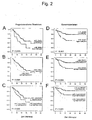
- 1C shows the restoration of TUSC3 expression in ovarian cancer cell lines with 5-aza-deoxycytidine and Fig.2 of Kaplan-Meier plots for progression-free and overall survival of patients whose tumor samples showed different expression intensities and methylation states.
- Tumor specimens were obtained from ovarian cancer patients at the time of primary diagnosis of ovarian cancer undergoing surgery. Epithelially enriched normal ovarian specimens and samples of benign cysts were from patients diagnosed without malignancy. A total of 20 benign ovarian samples and 102 primary tumor samples from 102 patients were assessed. For the tumor samples, as expected for a representative ovarian cancer population, a majority of patients (72%) were diagnosed with advanced stages (III and IV). 84% of the patients were after one on platinum treated chemo-based scheme. The corresponding data are summarized in Table 1. Table 1.
- ovarian cancer cell lines were constructed from ovarian cancer tissue specimens of 28 patrons and cultured as described ( Mobus V, Gerharz CD, Press U, et al., Morphological, immunohistochemical and biochemical characterization of 6 newly established human ovarian carcinoma cell lines. Int J Cancer. 1992; 52: 76-84 ).
- Genomic DNA of about 15 mg of frozen tissue was isolated using the DNeasy Tissue Kit (Qiagen, Hilden, Germany) as described by the manufacturer. The amount of DNA was measured using the PicoGreen dsDNA Quantitation Kit (Molecular Probes, Inc., Eugene, OR, USA), and the DNA quality was randomized by 0.3% agarose gel electrophoresis. DNA from ovarian cancer cell lines was isolated using the FlexiGen DNA Kit (Qiagen, Hilden, Germany).
- RNA from frozen tissues was extracted with the Agilent Total RNA Isolation Mini Kit (Agilent Technologies, Palo Alto, Calif., USA) according to the manufacturer's instructions.
- Total RNA from cancer cell lines was isolated using the RNeasy Mini Kit (Qiagen, Hilden, Germany). RNA quality and quantity were assessed by capillary electrophoresis using RNA Nano Chips (Lab-on-a-Chip, Agilent Technologies, Palo Alto, CA, USA).
- MSP Methylation-specific PCR
- DNA was digested with the restriction enzymes Hind III and Xba I, precipitated, dissolved and denatured for 20 minutes in 0.35 M NaOH. Thereafter, the DNA was treated with 1.75 M sodium bisulfite, 5.23 M urea, and 0.52 mM hydroquinone in a PCR cycler in 8 cycles (2 hrs. 75 ° C and 1 min. 95 ° C). The DNA was purified using the QIAEX II Gel Extraction Kit (Qiagen, Hilden, Germany) and the reaction was stopped with 0.32 M NaOH (20 min. 37 ° C).
- PCR was performed with the following primers (sense: 5'-GAGGTTGGTTGGGTAGGTGTGGTGTG-3 'and antisense: 5'-CACAACCACCACCCCACTTACCTACAACAT-3') and similar PCR conditions as above (except: 1x Optimized Buffer F (Invitrogen , Carlsbad, CA, USA) and 56 ° C annealing temperature).
- the PCR products were analyzed on 3% agarose gels stained with ethidium bromide.
- Bisulfite sequencing was performed directly with the PCR products purified with the QIAquick PCR Purification Kit (Quiagen, Hilden, Germany).
- DNA demethylation of the cancer cell lines was performed by 72-hour treatment with 1 and 2.5 ⁇ M 5-aza-deoxycytidine (Sigma-Aldrich, St. Louis, MO, USA) in cell culture medium and relative TUSC3 expression was determined by Real-Time RT -PCR rated as described below.
- cDNA was amplified from 1 ⁇ g of DNase I-digested total RNA using the DuraScript RT-PCR kit (Sigma-Aldrich, Saint Louis, Mo., USA) and a 1: 1 mixture of the provided oligo- (dT) 23 and Random nonamer oligo synthesized essentially as described in the manual.
- TaqMan real-time PCR was performed and the relative expression (compared to a calibrator, a multi-cell line cDNA mix) was determined from the threshold cycles (Ct) performed with the GeneAmp 5700 SDS v1.3 (Applied Biosystems) as follows Calculated: 2 ⁇ - [(Ct gene mean of duplicated probes - Ct gene means of duplicated calibrator) - (Ct B2M means of duplicated probes - Ct B2M means of duplicated calibrator)] (and given as arbitrary "ratio units") where the following Assay-on-Demand TM probes were used: TUSC3 , Hs00185147_m1 and beta 2- microglobulin, Hs99999907_m1 (Applied Biosystems, Foster City, CA, USA).
- Continuous variables are represented as mean and standard deviation, category variables as absolute and relative frequencies.
- a Fisher-Exact test and a Fisher-Freeman-Halton test, respectively, were performed.
- Univariate cox models were used to study the influence of known prognostic factors and the three potential new prognostic factors described above were to show. For each of the new factors, a multivariate Cox model, with known prognostic factors, was used as a setting variable.
- TUSC3 mRNA expression is reduced in ovarian cancer
- FIG. 1A Figure 10 shows box plots of expression of TUSC3 in arbitrary units (ratio units) based on the expression of ⁇ -2 microglobulin as internal reference (housekeeping gene control) of benign ovarian tissue samples (normal ovaries), ovarian cancer tissue samples (primary tumors) and ovarian cancer cell lines (tumor cell lines).
- ratio units based on the expression of ⁇ -2 microglobulin as internal reference (housekeeping gene control) of benign ovarian tissue samples (normal ovaries), ovarian cancer tissue samples (primary tumors) and ovarian cancer cell lines (tumor cell lines).
- U non-methylated
- M hypermethylated
- the TUSC3 promoter is hypermethylated and TUSC3 expression can be restored by DNA demethylation in ovarian cancer cell lines
- TUSC3 was undetectable in 5 out of 38 ovarian cancer cell lines, and that in three other cell lines expression was more than 10-fold lower than the mean expression of all cell lines (not shown here).
- MSP methylation- specific PCR
- Fig. 1B Figure 9 shows exemplary ethidium bromide agarose gel photos of methylation-specific PCR products from nine benign ovarian tissue samples (N1-N9), nine ovarian tumor samples (P1-P9), and nine ovarian cancer cell lines (MZ1-OVCAR3).
- a Sss I (CpG methylase) methylated human DNA was used and as a negative control H 2 O.
- M denotes the methylated PCR product and the unmethylated PCR product.
- Expression of TUSC3 depended significantly (P ⁇ 0.001) on hypermethylation status ( Fig. 1A ).
- the methylation status of individual CpG islands located near the putative TUSC3 promoter was assessed by bisulfite sequencing of three hypermethylated cell lines and two controls. Sequencing of the PCR products obtained showed that more than 75% of almost all CpGs in the PCR products of the three ovarian cancer cell lines were methylated by more than 10%, but no CpGs of either control (data not shown).
- TUSC3 hypermethylation correlates closely with TUSC3 expression
- TUSC3 expression is shown in the hypermethylated and non- methylated ovarian tumor tissues .
- Table 3 3.
- FIGS. 2A and 2B Table 4
- the hazard ratios for progression-free survival were comparable to the FIGO stage (relative risk of 2.821, P ⁇ 0.001) and the remaining tumor after surgery (relative risk of 2.867, P ⁇ 0.001) the only other significant prognostic factors available at the time are when options for systemic therapy need to be explored.
- FIGO stage 2,821 (1,867-4,260) ⁇ 0001 grading 0.978 (0.581-1.646) 0933 remaining tumor (yes vs no) ⁇ 2,867 (1,615-5,091) ⁇ 0001 Therapy Response (no vs yes) ⁇ 4,907 (2,644-9,104) ⁇ 0001 Methylation of TUSC3 (yes vs no) 1,972 (1,099-3,533) 0023 2,024 (1,025-4,000) 0042 Expression of TUSC3 (low vs high) ⁇ 1,661 (0.908-3.304) 0099 1,610 (0.726-3.571) 0242 L combined (methylation and expression) ⁇ 1,565 (1,072-2,283) 0020 1,510 (0.915-2.494) 0107 Overall survival univariate multivariate Relative risk (95% CI) ⁇ P Relative risk (95% CI) ⁇ P Age at diagnosis 1,056
Landscapes
- Chemical & Material Sciences (AREA)
- Life Sciences & Earth Sciences (AREA)
- Health & Medical Sciences (AREA)
- Organic Chemistry (AREA)
- Proteomics, Peptides & Aminoacids (AREA)
- Engineering & Computer Science (AREA)
- Immunology (AREA)
- Pathology (AREA)
- Analytical Chemistry (AREA)
- Zoology (AREA)
- Genetics & Genomics (AREA)
- Wood Science & Technology (AREA)
- Physics & Mathematics (AREA)
- Biotechnology (AREA)
- Microbiology (AREA)
- Molecular Biology (AREA)
- Hospice & Palliative Care (AREA)
- Biophysics (AREA)
- Oncology (AREA)
- Biochemistry (AREA)
- Bioinformatics & Cheminformatics (AREA)
- General Engineering & Computer Science (AREA)
- General Health & Medical Sciences (AREA)
- Measuring Or Testing Involving Enzymes Or Micro-Organisms (AREA)
- Investigating Or Analysing Biological Materials (AREA)
Description
- Die Erfindung betrifft ein Verfahren und die Verwendung von PCR-Primern für die Diagnose bei Eierstockkrebs und/oder die Prognogestellung bei Eierstockkrebs. Ebenfalls beschrieben ist ein Verfahren zur Abschätzung der Wirksamkeit von Therapeutika bei der und ein Verfahren zur Therapieplanung für die Behandlung von Eierstockkrebs.
- Bösartiger Eierstockkrebs ist die letalste gynäkologische Krebserkrankung und mit etwa 6% die vierthäufigste Todesursache bei krebskranken Frauen in westlichen Ländern. Schätzungen geben an, dass eine von 70 Frauen in ihrer Lebenszeit an Eierstockkrebs erkrankt, mit einer mittleren Überlebensrate von 4,5 Jahren. Eine neuere Krebsstatistik gibt geschätzte 22.200 neue Fälle und 16.210 Tote pro Jahr in den Vereinigten Staaten an (Jemal A, Murray T, Ward E, Samuels A. Cancer statistics, 2005 (vol 55, pg 10, 2005). Ca-A Cancer Journal for Clinicians 2005; 55(4):259).
- 5% bis 10% aller Eierstockkrebspatientinnen haben eine erbliche Prädisposition und leiden hauptsächlich am sogenannten "Erblichen Brust/Eierstockkrebssyndrom" oder am "Lynch II-Syndrom". Die meisten Fälle sind aber sporadische Eierstockkrebserkrankungen. Bei genetisch bedingten Hochrisikopatientinnen reduzieren prophylaktische Ovariektomien das Risiko und intensiviertes Screenen kann frühere Diagnosen in der Zukunft erlauben. In der allgemeinen Bevölkerung stellt die frühere Diagnose eine Hauptaufgabe dar, denn mehr als drei Viertel der Fälle werden in einem späteren Stadium (FIGO III und IV) diagnostiziert. Alle diese Patientinnen benötigen eine multimodale Therapie, bestehend aus einer optimalen Debulking-Operation und systemischer Chemotherapie. Molekularmarker, die mit dem Behandlungsausgang korrelieren und daher Behandlungsentscheidungen unterstützen sind erforderlich.
- Derzeit anerkannte prognostische Marker beinhalten die anfängliche Stadieneinteilung, Grading und Größe der verbleibenden nicht entfernbaren Tumormasse nach der zytoreduktiven Operation. (van der Burg ME. Advanced ovarian cancer. Curr Treat Options Oncol 2001; 2(2):109-118; Holschneider CH, Berek JS. Ovarian cancer: Epidemiology, biology, and prognostic factors. Seminars in Surgical Oncology 2000; 19(1):3-10). Molekularmarker für die frühe Auffindung und Prognose neben dem Tumormarker "Cancer Antigen 125" (CA-125) (Makar AP, Kristensen GB, Kaern J, Bormer OP, Abeler VM, Trope CG. Prognostic value of pre- and postoperative serum CA 125 levels in ovarian cancer: new aspects and multivariate analysis. Obstet Gynecol 1992; 79(6):1002-1010) existieren in der klinischen Praxis nicht (Ludwig JA, Weinstein JN. Biomarkers in Cancer Staging, Prognosis and Treatment Selection Nat Rev Cancer 2005).
- Aufgabe der vorliegenden Erfindung ist die Diagnose und die Prognosesstellung von Eierstockkrebs, sowie die Schaffung eines Diagnose- und Prognosestellungsverfahrens für Eierstockkrebs und eines Testkits dafür sowie die Schaffung eines Verfahren zur Abschätzung der Wirksamkeit von Therapeutika bei der und eines Verfahren zur Therapieplanung für die Behandlung von Eierstockkrebs.
- Die Erfinder der vorliegenden Erfindung haben herausgefunden, dass TUSC3 als Molekularmarker bei Eierstockkrebs geeignet ist. Die Messung der Expression des Markers TUSC3 in einer biologischen Probe, vorzugsweise einer Gewebeprobe, und das Vergleichen mit der Expression des Markers in biologischen Proben gesunder Frauen kann zur Diagnose von Eierstockkrebs beitragen. Ein entsprechendes Verfahren kann die folgenden Schritte umfassen:
- Isolation der RNA aus der zu untersuchenden biologischen Probe, vorzugsweise dem zu untersuchenden Gewebe,
- Herstellung von cDNA mittels der isolierten RNA,
- Durchführung einer Echtzeit-PCR für den Marker TUSC3,
- Vergleich des Expressionslevels des Markers aus der zu untersuchenden biologischen Probe mit dem Level in biologischen Proben gesunder Frauen.
- TUSC3, ursprünglich N33 genannt, wurde als ein potenzielles Tumorsuppressorgen aus dem Chromosomenabschnitt 8p22 in Prostatakrebs in der Mitte der 90er Jahre identifiziert (Bova GS, MacGrogan D, Levy A, Pin SS, Bookstein R, Isaacs WB. Physical mapping of chromosome 8p22 markers and their homozygous deletion in a metastatic prostate cancer. Genomics 1996; 35(1):46-54; MacGrogan D, Levy A, Bova GS, Isaacs WB, Bookstein R. Structure and methylation-associated silencing of a gene within a homozygously deleted region of human chromosome band 8p22. Genomics 1996; 35(1):55-65). Die Basis für diese Hypothese war die Identifizierung von mehreren homozygoten Deletionen auf dem Chromosomenabschnitt 8p22 in Prostatakrebszellen. Die Proteinvorhersage ergab, dass das kodierte Protein eine mutmaßliche Untereinheit des Oligosaccharyltransferase-Komplexes ist (Kelleher DJ, Karaoglu D, Mandon EC, Gilmore R. Oligosaccharyltransferase isoforms that contain different catalytic STT3 subunits have distinct enzymatic properties. Molecular Cell 2003; 12(1):101-111). TUSC3 wurde in einigen Krebszelllinien homozygot deletiert, aber selten mutiert gefunden. Mutationsanalysen, die bei Prostatakrebs und anderen Tumorformen durchgeführt wurden, die häufig Verlust der Heterozygotie (LOH) und/oder homozygote Deletionen auf 8p22 zeigen, zeigten keine signifikante Rate an inaktivierenden Mutationen. Missensmutationen waren aufgrund des Fehlens bekannter Proteinfunktionen schwer zu interpretieren und das Interesse an TUSC3 und seinem kodierten Protein verschwand daher.
- Einsicht über Haploinsuffizienz und epigenetische Modifikationen von Tumorsuppressorgenen haben das Interesse an alten Kandidatenmarkern wieder geweckt. Kürzlich verfügbar gemachte Datenbanken und In-Silico-Tools unterstützen diese Bemühungen.
- Unter Verwendung öffentlich erhältlicher Genexpressionsdaten, haben die Erfinder systematisch nach reduzierter Expression von Genen auf 8p22 (nicht veröffentlichte Daten) gesucht und TUSC3 (N33) als eines der Gene identifiziert, deren Expression signifikant bei Eierstockkrebs herabgesetzt ist. Eine quantitative Expressionsanalyse durch Real-Time RT-PCR bestätigte die signifikante Herabsetzung von TUSC3 in Eierstockkrebsgeweben. Die statistische Analyse der Korrelation der TUSC3-Expression mit klinischen Parametern zeigte, dass Patientinnen mit reduzierter TUSC3-Expression zum Zeitpunkt der Diagnose ein kürzeres Gesamtüberleben verglichen mit Patientinnen haben, deren TUSC3 Niveaus in ihren Tumoren gleich jenen in ihren normalen Eierstockgeweben waren. Diesen Ergebnissen wurde nachgegangen und an einer großen unabhängigen Patientenpopulation nochmals getestet. Die anfänglichen Beobachtungen einer möglichen prognostischen Bedeutung der TUSC3-Expression konnten bestätigt werden.
- Die Erfindung ist in den Ansprüchen definiert.
- Die Erfinder der vorliegenden Erfindung haben herausgefunden, dass TUSC3 auch als Molekularmarker bei der Diagnose von und/oder Prognosestellung bei Eierstockkrebs, sowie zur Abschätzung der Wirksamkeit einer platinbasierenden Chemotherapie und/oder einer Therapie auf Basis von DNA-Demethylierungsagenzien, wie Decitabin (5-Aza-2'-deoxycytidin) oder Zebularin, und/oder auf Basis von Inhibitoren der Histon-Deacetylasen bei Eierstockkrebs dienen kann, indemdie Promotor-Hypermethylierung des Markers TUSC3 in einer biologischen Probe, vorzugsweise in einer Gewebeprobe oder biologischen Flüssigkeit, wie Vollblut, Serum oder Plasma, gemessen wird. Vorzugsweise umfasst das Verfahren folgende Schritte:
- Isolation der DNA aus der biologischen Probe, vorzugsweise der Gewebeprobe oder biologischen Flüssigkeit
- Durchführung eines PCR-basierenden Methylierungsnachweises
- Analyse der gewonnenen PCR-Produkte.
- Vorteilhafterweise erfolgt der PCR-basierende Methylierungsnachweis mit methylierungsspezifischer PCR. Dabei kann für den Nachweis methylierter DNA eine PCR z.B. mit einem der folgenden Primerpaare durchgeführt werden:
- 5'-TTTTCGGTGAATCGGATG-3' und
5'-TACGCGCCCAACTCCTA-3', - 5'-GGTTTAGTTAGCGGGTTTTCGG-3' und
5'-AAACAAATACCGCAACCGCCG-3' oder - 5'-GGTCGGGTAGGCGTGGTGCG-3' und
5'-CCGCCCCGCTTACCTACGACGT-3'. - Zusätzlich kann für den Nachweis nicht methylierter DNA eine PCR mit einem der folgenden Primerpaare durchgeführt werden:
- - 5'-GGGTTTAGTTAGTGGGTTTTfGGAG-3' und
5'-AAACAAATACCACAACCACCACCC-3' oder - - 5'-GAGGTTGGTTGGGTAGGTGTGGTGTG-3' und
5'-CACAACCACCACCCCACTTACCTACAACAT-3'. - Alternativ kann der PCR-basierende Methylierungsnachweis mit methylsensitiver Restriktionsendonuklease-Reaktion und PCR erfolgen.
- Dazu kann z.B. das folgende Verfahren verwendet werden:
- Zum spezifischen Nachweis methylierter Sequenzen werden 500 ng einer genomischen DNA mit 40 Units des Restriktionsenzyms Hpa II (New England Biolabs, Ipswich, Ma, USA), unter Verwendung des Reaktionspuffers NEBuffer 1 (New England Biolabs, Ipswich, Ma, USA), in einem Volumen von 50 µl gemischt mit Mineralöl überschichtet und 18 Std. bei 37 °C behandelt. Darauffolgend wird die verdaute DNA mit dem Invisorb Spin PCRapid Kit (Invitek, Berlin, Deutschland) gereinigt und in einem Volumen von 50 µl aufgenommen. Der PCR-Ansatz umfasst 25 µl und setzte sich aus je 0.3 µM der beiden biotinilierten Primer (vorwärts: 5'-bio-GCCTTTCCAGGTCTTCTCC-3'; rückwärts: 5'-bio-CCACGCGCGGTAGCCGTG-3'), 50 ng der verdauten und gereinigten DNA, 1 Unit SuperTaq (HT Biotech, Cambridge, UK), 1 x SuperTaq Puffer (HT Biotech, Cambridge, UK) und 0,2 mM der dNTPs (Larova, Teltow, Deutschland) zusammen. Die PCR wird unter folgenden Bedingungen durchgeführt: 2 Min. bei 94 °C, 40 Zyklen - 15 Sek. bei 94 °C, 30 Sek. bei 58 °C, 30 Sek. bei 72 °C - und schließlich 3 Min. bei 72 °C. Der Nachweis entstandener PCR-Produkte erfolgt auf 2 % Agarose-Gelen und Ethidiumbromid-Färbung. Darüberhinaus ermöglicht die Verwendung biotinilierter Primer eine Analyse der PCR-Produkte auch durch alternative Methoden z. B. sequenz-spezifisches Capturing im Zuge der reversen Hybridisierung und der anschließenden Detektion der gebundenen biotinilierten Sequenzen mittels Streptavidin-Enzym-Konjugat.
- Ein ebenfalls hier beschriebener Testkit zur Diagnose von und/oder Prognosestellung bei Eierstockkrebs und/oder Abschätzung der Wirksamkeit einer platinbasierenden Chemotherapie und/oder einer Therapie auf Basis von DNA-Demethylierungsagenzien und/oder auf Basis von Inhibitoren der Histon-Deacetylasen bei Eierstockkrebs enthält PCR-Primer, die für die Durchführung einer methylierungsspezifischen PCR in der TUSC3-Promotorregion geeignet sind.
- Alternativ kann der Testkit ein Restriktionsenzym und PCR-Primer, die die entsprechende Restriktionsschnittstelle innerhalb der TUSC3-Promotorregion flankieren, enthalten.
- Zusätzlich kann der Testkit Mittel zum Nachweis entstandener PCR-Produkte, wie eine Hybridisierungssonde, enthalten.
- Beim ebenfalls hier beschriebenen Verfahren zur Therapieplanung für die Behandlung von Eierstockkrebs, wird ein Verfahren zur Abschätzung der Wirksamkeit einer platinbasierenden Chemotherapie und/oder einer Therapie auf Basis von DNA-Demethylierungsagenzien, wie Decitabin (5-Aza-2'deoxycytidin) oder Zebularin, und/oder auf Basis von Inhibitoren der Histon-Deacetylasen bei Eierstockkrebs durchgeführt, bei dem die Promotor-Hypermethylierung des Markers TUSC3 in einer zu untersuchenden biologischen Probe, vorzugsweise in einer zu untersuchenden Gewebeprobe oder biologischen Flüssigkeit gemessen wird und bei Feststellung einer Hypermethylierung des TUSC3-Promotors wird eine Therapie auf Basis von DNA-Demethylierungsagenzien, wie Decitabin (5-Aza-2'-deoxycytidin) oder Zebularin, und/oder auf Basis von Inhibitoren der Histon-Deacetylasen vorgeschlagen.
- Bei einem Verfahren zur Therapieplanung für die Behandlung von Eierstockkrebs, wird ein Verfahren zur Abschätzung der Wirksamkeit einer platinbasierenden Chemotherapie und/oder einer Therapie auf Basis von DNA-Demethylierungsagenzien, wie Decitabin (5-Aza-2'-deoxycytidin) oder Zebularin, und/oder auf Basis von Inhibitoren der Histon-Deacetylasen bei Eierstockkrebs durchgeführt indem die Promotor-Hypermethylierung des Markers TUSC3 in einer zu untersuchenden biologischen Probe, vorzugsweise in einer zu untersuchenden Gewebeprobe oder biologischen Flüssigkeit gemessen wird, und es wird eine platinbasierende Chemotherapie vorgeschlagen, wenn keine Hypermethylierung oder des TUSC3-Promotors festgestellt wird.
- Promotor-Hypermethylierung von Tumorsuppressorgenen (TSGs) ist als Molekularmarker vielversprechend. Es kann Einblick in die Biologie eines Tumors geben, und kann im Gegensatz zur Gen- oder Proteinexpression leicht durch auf PCR basierende Methoden nicht nur aus dem Tumormaterial sondern auch in Körperflüssigkeiten nachgewiesen werden (Das PM, Singal R. DNA methylation and cancer. J Clin Oncol 2004; 22(22):4632-4642; Jones PA, Baylin SB. The fundamental role of epigenetic events in cancer. Nat Rev Genet 2002; 3(6):415-428; Laird PW. The power and the promise of DNA methylation markers. Nat Rev Cancer 2003; 3(4):253-266).
- Hypermethylierung des TUSC3 beinhaltenden Chromosomenabschnitts wurde in Prostata-, Dickdarm-, Leber-, Lungen- und Blasenkrebs untersucht (MacGrogan D, Levy A, Bova GS, Isaacs WB, Bookstein R. Structure and methylation-associated silencing of a gene within a homozygously deleted region of human chromosome band 8p22. Genomics 1996; 35(1):55-65. Ahuja N, Li Q, Mohan AL, Baylin SB, Issa JP. Aging and DNA methylation in colorectal mucosa and cancer. Cancer. Res 1998; 58(23):5489-5494.). Altersabhängige Hypermethylierung wurde nur in normalem und neoplastischem Dickdarmgewebe, nicht aber bei anderen Krebsarten gefunden. Vor kurzem hat eine andere Gruppe keine Hypermethylierung von TUDC3 in 65 untersuchten Dickdarmkrebsproben gefunden (Xu XL, Yu J, Zhang HY et al. Methylation profile of the promoter CpG islands of 31 genes that may contribute to colorectal carcinogenesis. World J Gastroenterol 2004; 10(23):3441-3454).
- Hypermethylierung wurde kürzlich in einem breiten Bereich von krebsbezogenen Genen in Eierstockkrebs untersucht. Die Auswahl der Gene basierte hauptsächlich auf Zufälligkeit und Erhältlichkeit etablierter Nachweissysteme basierend auf methylierungsspezifischer PCR (Makarla PB, Saboorian MH, Ashfaq R et al. Promoter hypermethylation profile of ovarian epithelial neoplasms. Clin Cancer Res 2005; 11(15):5365-5369; Teodoridis JM, Hall J, Marsh S et al. CpG Island Methylation of DNA Damage Response Genes in Advanced Ovarian Cancer. Cancer Res 2005; 65(19):8961-8967). Obwohl diese Ergebnisse für die Entwicklung von Screeningtests für Krebserkrankungen im Allgemeinen wertvoll sind, können Promotorhypermethylierungen relativ späte und unspezifische Ereignisse in der Karzinogenese darstellen. Ein idealer Marker würde auch Hinweise auf das biologische Verhalten geben. Kürzlich konnte p15INK4b nach Korrektur in Bezug auf etablierte prognostische Faktoren in einem multivariaten Cox Regressionsmodel (Liu Z, Wang LE, Wang L et al. Methylation and Messenger RNA Expression of p15INK4b but Not p16INK4a Are Independent Risk Factors for Ovarian Cancer. Clin Cancer Res 2005; 11(13):4968-4976) als Risikofaktor für Eierstockkrebs identifiziert werden.
- Die Erfinder haben festgestellt, dass der Prozentsatz der Patientinnen, die gegen Platin-basierende Chemotherapie resistent waren, in der Gruppe mit hypermethylierten TUSC3-Promotoren signifikant höher war, was eine bestimmte notwendige Expressionsstärke von TUSC3 für ein günstiges Ansprechen auf Chemotherapie impliziert.
- Im Folgenden sollen Untersuchungen, die die Erkenntnisse stützen, auf denen die vorliegende Erfindung basiert, beschrieben werden. Dabei wird auch auf die beiliegenden Zeichnungen Bezug genommen, in denen
Fig.1A Boxplots der TUSC3-Gesamtexpression bzw. der Expression der nicht methylierten und hypermethylierten Fraktion von gutartigen Eierstockgewebeproben, Eierstockkrebsgewebeproben und Eierstockkrebszelllinen zeigt,Fig.1B Ethidiumbromide-Agarosegel-Fotos von methylierungsspezifischen PCR-Produkten von gutartigen Eierstockgewebeproben, Eierstockkrebsgewebeproben und Eierstockkrebszelllinien zeigt,Fig. 1C die Wiederherstellung von TUSC3-Expression in Eierstockkrebszelllinien mit 5-aza-deoxycytidin zeigt undFig.2 von Kaplan-Meier-Plots für progressionsfreies und Gesamtüberleben von Patientinnen, deren Tumorproben unterschiedliche Expressionsintensitäten und Methylierungszustände zeigten, zeigt. - Tumorproben wurden von Eierstockkrebspatientinnen zum Zeitpunkt der Primärdiagnose von Eierstockkrebs erhalten, die sich einer Operation unterzogen hatten. Epithelial angereicherte normale Eierstockproben und Proben gutartiger Zysten kamen von Patientinnen, die ohne maligne Erkrankung diagnostiziert wurden. Insgesamt wurden 20 gutartige Eierstockproben und 102 Primärtumorproben von 102 Patientinnen beurteilt. Bei den Tumorproben wurde, wie für eine repräsentative Eierstockkrebspopulation erwartet, eine Mehrheit der Patientinnen (72%) mit fortgeschrittenen Stadien (III und IV) diagnostiziert. 84% der Patientinnen wurden nach einem auf Platin basierenden Chemotherapie-Schema behandelt. Die entsprechenden Daten sind in Tabelle 1 zusammengefasst.
Tabelle 1. Klinisch-pathologische Merkmale von Eierstockkrebspatientinnen A) Gutartige Eierstockproben (entsprechende mRNA-Proben) 20 (20) Alter bei Operation [Jahren] Mittel ± Standardabweichung 49.9 ± 11.8 7 normale Eierstöcke, mittleres Alter [Jahre]: 51.6 13 gutartige Zysten, mittleres Alter [Jahre]: 49.0 B) Tumorproben (entsprechende mRNA-Proben) 102 (99) Alter bei Diagnose [Jahre] Mittel ± Standardabweichung 58.3 ± 11.0 Histoloqie (1 (1.0%) fehlend) serös 77 (75.5%∞) endometrioid 10 (9.8%) muzinös 5 (4.9%) klarzellig 2 (2.0%) andere 7 (6.9%) FIGO (2(2.0%) fehlend) I 17 (16.7%) II 10 (9.8%) III 51 (50.0%) IV 22 (21.6%) Grading (1 (1.0%) fehlend) 1 2 (2.0%) 2 48 (46.1%) 3 51 (50.0%) Chemotherapie (3 (2.9%) fehlend) Carboplatin und Taxol 63 (61.8%) Carboplatin, Taxol und Gemcitabin 17 (16.7%) Carboplatin oder Taxol Monotherapie 3 (2.9%) Carboplatin und andere 8 (7.8%) andere 3 (2.9%) keine 5 (4.9%) Ansprechen auf Chemotherapie (16 (15.7%) fehlend) pNED‡ 6 (5.9%) pNC† 3 (2.9%) pPD† 12 (11.8%) pPR† 5 (4.9%) pCR‡ 43 (42.2%) Dauer des Ansprechens 0 - 6 Monate†,‡ 6 (5.9%) 6 - 12 Monate‡ 5 (4.9%) > 12 Monate‡ 6 (5.9%) Remissionen/progressive Erkrankunq (3 (2.9%) fehlend) ohne Remission 49 (48.0%) mit Remission 33 (32.4%) progressive Erkrankung 17 (16.7%) ∞Prozentsatz basierend auf 102 Tumorproben. †Nichtanprechende auf Chemotherapie. ‡Ansprechende auf Chemotherapie. - Zusätzlich wurden 32 Eierstockkrebszelllinien untersucht, die aus Eierstockkrebsgewebeproben von 28 Patentinnen angelegt und wie beschrieben kultiviert wurden (Mobus V, Gerharz CD, Press U, et al., Morphological, immunohistochemical and biochemical characterization of 6 newly established human ovarian carcinoma cell lines. Int J Cancer.1992; 52:76-84).
- Genomische DNA von etwa 15 mg gefrorenem Gewebe wurde mit dem DNeasy Tissue Kit (Qiagen, Hilden, Deutschland) wie vom Hersteller beschrieben, isoliert. Die DNA-Menge wurde unter Verwendung des PicoGreen dsDNA Quantitation Kit (Molecular Probes, Inc., Eugene, OR, USA) gemessen und die DNA-Qualität wurde stichprobenartig mit 0,3% Agarose-Gel-Elektrophorese bewertet. DNA aus Eierstockkrebszelllinien wurde unter Verwendung des FlexiGen DNA Kit (Qiagen, Hilden, Deutschland) isoliert.
- Gesamt-RNA aus gefrorenen Geweben wurde mit dem Agilent Total RNA Isolation Mini Kit (Agilent Technologies, Palo Alto, CA, USA) gemäß den Angaben des Herstellers, extrahiert. Gesamt-RNA aus Krebszelllinien wurde mit dem RNeasy Mini Kit (Qiagen, Hilden, Deutschland) isoliert. RNA Qualität und Quantität wurden mittels Kapillarelektrophorese unter Verwendung von RNA Nano Chips (Lab-on-a-Chip, Agilent Technologies, Palo Alto, CA, USA) beurteilt.
- Zur Bisulfit-Behandlung wurde 1 µg genomische DNA mit den Restriktionsenzymen Hind III und Xba I verdaut, präzipitiert, gelöst und für 20 Minuten in 0,35 M NaOH denaturiert. Danach wurde die DNA in 8 Zyklen (2 Std. 75°C und 1 Min. 95°C) mit 1,75 M Natiumbisulfit, 5,23 M Harnstoff und 0,52 mM Hydroquinon in einem PCR-Cycler behandelt. Die DNA wurde unter Verwendung des QIAEX II Gel Extraction Kit (Qiagen, Hilden, Deutschland) gereinigt, und die Reaktion wurde mit 0,32 M NaOH (20 Min. 37°C) beendet. Schließlich wurde die DNA mit 1 µl Glycogen als Träger NH4Acetat-EtOH-präzipitiert mit 70% EtOH gewaschen und in 40 µl H2O gelöst. Für die methylierungsspezifische Reaktion wurde PCR mit 25 ng Bisulfit behandelter DNA, Primern die von Xu et al. publiziert worden waren (Xu XL, Yu J, Zhang HY et al. Methylation profile of the promoter CpG islands of 31 genes that may contribute to colorectal carcinogenesis. World J Gastroenterol 2004; 10(23):3441-3454), AmpliTaq Gold (Applied Biosystems, Foster City, CA, USA), 1x Optimized Buffer B (Invitrogen, Carlsbad, CA, USA) unter den folgenden PCR Bedingungen durchgeführt: 10 Min. bei 94°C, 50 Zyklen von 30 Sek. bei 94°C - 30 Sek. bei 50°C - 20 plus 1 Sek. pro Zyklus bei 72°C und schließlich 7 Min. bei 72°C. Für den Nachweis nicht methylierter DNA wurde PCR mit den folgenden Primern (sense: 5'-GAGGTTGGTTGGGTAGGTGTGGTGTG-3' und antisense: 5'-CACAACCACCACCCCACTTACCTACAACAT-3') und ähnlichen PCR-Bedingungen wie oben durchgeführt (außer: 1x Optimized Buffer F (Invitrogen, Carlsbad, CA, USA) und 56°C Annealing Temperatur). Die PCR-Produkte wurden auf 3% Agarose-Gelen, gefärbt mit Ethidiumbromid analysiert. Bisulfitsequenzieren wurde direkt mit den PCR-Produkten, die mit dem QIAquick PCR Purification Kit (Quiagen, Hilden, Deutschland) gereinigt wurden, durchgeführt. DNA-Demethylierung der Krebszelllinien wurde durch eine 72 Stunden Behandlung mit 1 und 2,5 µM 5-Aza-Deoxycytidin (Sigma-Aldrich, St. Louis, MO, USA) in Zellkulturmedium durchgeführt und die relative TUSC3 Expression wurde durch Real-Time RT-PCR wie unten beschrieben bewertet.
- cDNA wurde von 1 µg DNase I-verdauter Gesamt-RNA unter Verwendung des DuraScript RT-PCR Kits (Sigma-Aldrich, Saint Louis, MO, USA) und einer 1:1-Mischung des zur Verfügung gestellten Oligo-(dT)23 und Random-Nonamer Oligo, im Wesentlichen wie im Handbuch beschrieben, synthetisiert. TaqMan Real-Time PCR wurde durchgeführt und die relative Expression (verglichen mit einem Kalibrator, einer cDNA-Mischung aus mehreren Zelllinien) wurde aus den Schwellenwert-Zyklen (Ct), die mit der GeneAmp 5700 SDS v1.3 (Applied Biosystems) wie folgt errechnet: 2^-[(Ctgene Mittel von duplizierten Sonden - Ctgene Mittel von dupliziertem Kalibrator) - (CtB2M Mittel von duplizierten Sonden - CtB2M Mittel von dupliziertem Kalibrator)] (und als willkürliche "Verhältniseinheiten" angegeben) wobei die folgenden Assay-on-Demand™ Sonden verwendet wurden: TUSC3, Hs00185147_ m1 und beta 2-microglobulin, Hs99999907_m1 (Applied Biosystems, Foster City, CA, USA).
- Kontinuierliche Variable sind als Mittelwert und Standardabweichung dargestellt, Kategorievariable als absolute und relative Frequenzen. Um Frequenzen zwischen zwei oder mehreren Gruppen zu vergleichen, wurde ein Fisher-Exakt-Test bzw. ein Fisher-Freeman-Halton-Test jeweils durchgeführt.
- Im Falle einer ausreichenden Annäherung an eine Normalverteilung wurden Mittelwerte und 95% Vertrauensintervalle für TUSC3 Expression in einem logarithmischen Maßstab berechnet und dann in den Originalmaßstab zurücktransformiert. Um die TUSC3 Expression zwischen zwei oder mehr Gruppen zu vergleichen, wurde ein t-Test bzw. ein one-way ANOVA durchgeführt, und zwar unter Verwendung der logtransformierten Expression als unabhängige Variable. Analog wurde der mögliche Einfluss von Hypermethylierung auf TUSC3 Expression unter Verwendung eines t-Tests im logarithmischen Maßstab untersucht.
- Der mögliche Einfluss der TUSC3 Expression und von Hypermethylierung auf das progressionsfreie und Gesamtüberleben ist in entsprechenden Kaplan-Meier-Plots dargestellt, und unter Verwendung von Cox Proportional Hazard-Regressionsmodellen quantifiziert. Zu diesem Zweck wurde die Expression an der 33,33 % Perzentile dichotomisiert, da ungefähr 30% der Proben hypermethyliert waren und eine starke Korrelation von Hypermethylierungszustand und Expression beobachtet wurde; ferner wurde eine Kombination dieser dichotomen Variablen und des Hypermethylierungsstatus in drei Niveaus (3 = geringe Expression und hypermethyliert, 2 = entweder geringe Expression oder hypermethyliert, 1 = hohe Expression und nicht methyliert) ermittelt und als linearer Faktor in den Cox-Regressionsmodellen berücksichtigt; daher lässt sich das entsprechende Hazard-Verhältnis auf beide Vergleiche von benachbarten Gruppen (3 vs 2 und 2 vs 1) anwenden. Als Progressionszeitpunkt wurde der Tag der Dokumentation des ersten Rückfalls oder die erste Dokumentation der Progression der Erkrankung, beginnend mit dem Zeitpunkt der ersten Diagnose, verwendet.
- Univariate Coxmodelle wurden verwendet, um den Einfluss bekannter prognostischer Faktoren und der drei möglichen neuen prognostischen Faktoren, die oben beschrieben wurden, zu zeigen. Für jeden der neuen Faktoren wurde ein multivariates Coxmodell, mit bekannten prognostischen Faktoren, als Einstellungsvariable verwendet.
- P-Werte ≤0,05 wurden als statistisch signifikant betrachtet. Alle Berechnungen wurden unter Verwendung von SAS Software Version 9.1 (SAS Institute Inc., Cary, NC, USA, 2001) durchgeführt; graphische Darstellungen wurden unter Verwendung von SPSS Software Version 12.0 (SPSS Inc. Headquarters, Chicago, Illinois, USA) hergestellt.
- Quantitative Real-Time RT-PCR von 99 Eierstockkrebsgeweben und 20 gutartigen Eierstockgeweben wurde durchgeführt. Die
Fig. 1A zeigt Boxplots der Expression von TUSC3 in beliebigen Einheiten (Verhältniseinheiten) basierend auf der Expression von ß-2-Mikroglobulin als interne Referenz (Housekeepinggenkontrolle) von gutartigen Eierstockgewebeproben (normale Eierstöcke), Eierstockkrebsgewebeproben (Primärtumore) und Eierstockkrebszelllinen (Tumorzelllinien). Im rechten Teil der Grafik wurde die Expression von TUSC3 in nicht methylierte (U) und hypermethylierte (M) Fraktionen unterteilt. Für die statistische Analyse wurde ein t-Test im logarithmischen Maßstab verwendet. Verglichen mit Kontrollen (0,465, CI 0,347-0,623 Verhältniseinheiten) war die Expression in Tumoren signifikant (P < 0,001) niedriger (0,105, CI 0,082-0,135 Verhältniseinheiten). Signifikante Unterschiede in der TUSC3 Expression zwischen Tumorproben von Patientinnen verschiedener Altersgruppen (≤50 und >50), Proben von verschiedenen FIGO-Stadien und Proben unterschiedlicher Differenzierungsgrade (Grading) wurden nicht gefunden (Tabelle 2A). Nur die Expression in nichtserösen Tumoren war signifikant geringer (P = 0,037, nicht korrigiert für mehrfaches Testen) verglichen mit serösen Tumoren (Tabelle 2A).Tabelle 2. Vergleich der klinisch-pathologischen Merkmale mit TUSC3 Expression und Hypermethylierung A) TUSC3 Expression Klinisch-pathologische Merkmale Expression von TUSC3 Vergleich (P) Gutartige Eierstockproben Median 0.465 CI 0.347-0.623 Tumorproben (Vergleich der gutartigen Eierstockproben) <0.001† Median 0.105 CI 0.082-0.135 Alter ≤50 0.085 0.299† > 50 0.114 Histologie serös 0.121 0.037† nicht-serös 0.064 FIGO I 0.084 0.764‡ II 0.124 III 0.104 IV 0.126 Grading 1 0.068 0.188‡ 2 0.135 3 0.086 B) TUSC3 Hypermethylierung Klinisch-pathologische Merkmale Hypermethylierung von TUSC3 Vergleich (P)# Gutartige Eierstockproben 0 / 20 (0%) Primärtumorproben Alter 30 / 102 (29.4%) 0.003 ≤50 0.218 > 50 5 / 26 (19.2%) Histologie 25 / 75 (33.3%) serös 0.200 nicht-serös 21 / 79 (26.6%) FIGO 9 / 22 (40.9%) I 0.948 II 5 / 17 (29.4%) III 2 / 10 (20.0%) IV 15 / 51 (29.4%) Grading 7 / 22 (31.8%) 1 0.102 2 2 / 2 (100%) 3 15 / 48 (31.2%) ‡ANOVA (berechnet nach logarithmischer Transformation, Mittel und CI sind nach Rücktransformation in den Originalmaßstab angegeben). 13 / 51 (25.5%) † t-Test (wie oben berechnet) # Fisher-Exakt-Test und Fisher-Freeman-Halton-Test, entsprechend. - Die Erfinder haben gezeigt, dass die Expression von TUSC3 in 5 von 38 Eierstockkrebszelllinien nicht nachweisbar war, und dass in drei weiteren Zelllinien die Expression mehr als 10fach geringer war, als die mittlere Expression aller Zelllinien (hier nicht dargestellt). Unter Verwendung von methylierungsspezifischer PCR (MSP) wurde gefunden, dass der Promotor von TUSC3 in sieben von diesen acht Zelllinien mit geringer oder fehlender TUSC3 Expression hypermethyliert war. Die
Fig. 1B zeigt beispielhafte Ethidiumbromid-Agarosegel-Fotos von methylierungsspezifischen PCR-Produkten von neun gutartigen Eierstockgewebeproben (N1-N9), neun Eierstocktumorproben (P1-P9) und neun Eierstockkrebszelllinien (MZ1-OVCAR3). Als positive Kontrolle wurde eine Sss I (CpG-Methylase) methylierte humane DNA und als negative Kontrolle H2O verwendet. M bezeichnet das methylierte PCR-Produkt und das nicht methylierte PCR-Produkt. Die Expression von TUSC3 hing signifikant (P < 0,001) vom Hypermethylierungsstatus ab (Fig. 1A ). - Eine konzentrationsabhängige (1 und 2,5 µM) Wiederherstellung der TUSC3 Expression wurde durch eine 72 Stunden Behandlung mit dem demethylierenden Mittel 5-aza-deoxycytidin in der hypermethylierten Zelllinie MZ6 erzielt. In den scheinbehandelten Proben war die TUSC3-Expression unter der Nachweisgrenze. Die TUSC3-Expression war bei Verwendung von 2,5 µM 5-aza-deoxycytidine mindestens 9,5 mal höher. Die Versuche wurden dreifach durchgeführt und die Mittelwerte der relativen TUSC3-Expression (basierend auf der Expression von ß-2-mikroglobulin) mit Standardabweichungen wurden dargestellt.
- Um das Methylierungsniveau weiter zu bestimmen und die Spezifität des verwendeten MSP-Systems zu testen, wurde der Methylierungsstatus von einzelnen nahe dem vermeintlichen TUSC3 Promotor lokalisierten CpG-Inseln durch Bisulfit-Sequenzierung von drei hypermethylierten Zelllinien und von zwei Kontrollen bewertet. Das Sequenzieren der erhaltenen PCR-Produkte zeigte, das fast alle CpGs in den PCR-Produkten der drei Eierstockkrebszelllinien zu mehr als 75%, aber keine CpGs der beiden Kontrollen zu mehr als 10% methyliert waren (Daten nicht gezeigt).
- In einem nächsten Schritt wurde die Analyse des Hypermethylierungsstatus des TUSC3 Promotors durch methylierungsspezifische PCR auf eine Liste von 102 Eierstocktumorproben, die von Patientinnen bei Primärdiagnose erhalten wurden, erweitert (
Fig. 1 B) . Hypermethylierung von TUSC3 wurde in 29,8% (30/102) dieser Eierstockkrebsfälle gefunden, aber in keiner von 20 Kontrollen (P = 0,003). - In
Fig. 1 ist die TUSC3 Expression in den hypermethylierten und nichtmetylierten Eierstocktumorgeweben gezeigt. Die mittlere Expression von TUSC3 in der hypermethylierten Gruppe war signifikant geringer (0,065, CI 0,038-0,081 Verhältniseinheiten) als in der nicht methylierten Gruppe (0,135; CI 0,100-0,182 Verhältniseinheiten, P = 0,001), was eine ähnliche Wechselbeziehung des TUSC3 Hypermethylierungsstatus und der TUSC3 Expression, wie sie oben für Eierstockkrebszelllinien gezeigt wurde, bestätigt und einen kausalen Zusammenhang zwischen dem Hypermethylierungsstatus und der Expression in Primärtumorproben nahe legt (Fig. 1A und Tabelle 3).Tabelle 3. Signifikante Unterschiede von klinisch-pathologischen Merkmalen von hypermethylierten Tumorproben TUSC3 Hypermethylierung Klinisch-pathologische Merkmale nicht methyliert methyliert Vergleich (P) Alter n=72 n=30 0.023† Mittel ± Standardabweichung 56.7 ± 10.8 62.1 ± 10.7 Bereich 35-81 41-86 Expression von TUSC3 (qRT-PCR) n=71 n=28 0.001‡ Mittel 0.135 0.056 CI 0.100-0.182 0.038-0.081 †t-Test. ‡t-Test (berechnet nach logarithmischer Transformation, Mittel und CI sind nach Rücktransformation in den Originalmaßstab angegeben). - Es gab keinen signifikanten Unterschied in der Hypermethylierungshäufigkeit zwischen Tumorproben von Patientinnen verschiedener Altersgruppen (≤50 und >50), Proben mit unterschiedlicher histologischer Charakterisierung (serös v nicht-serös), Proben von verschiedenen FIGO-Stadien und Proben von verschiedenen Differenzierungsgraden (Grading) (siehe Tabelle 2B). Interessanterweise war das mittlere Alter in der hypermethylierten Gruppe bei Diagnose um 5,4 Jahre höher (P = 0,023) als in der nicht metylierten Gruppe (Tabelle 3). In der Gruppe der 30 Patienten mit hypermethylierten TUSC3 Promotoren, hatten mehr Patientinnen eine progressive Erkrankung (32,1%) verglichen mit der Gruppe der 72 Patientinnen ohne TUSC3 Hypermethylierung (11,3%), was den möglichen Einfluss der TUSC3 Hypermethylierung und Expression auf die Prognose von Patientinnen mit Eterstockkrebs nahe legt.
- Es wurde ein signifikanter Zusammenhang zwischen der TUSC3 Hypermethylierung und dem progressionsfreien Überleben (relatives Risiko von 1,972, P = 0,023) und dem Gesamtüberleben (relatives Risiko von 2,915, P = 0,007) gefunden (
Fig. 2A und 2B , Tabelle 4). Die Hazardverhältnisse für progressionsfreies Überleben waren vergleichbar mit dem FIGO-Stadium (relatives Risiko von 2,821, P < 0,001) und dem verbleibenden Tumor nach Operation (relatives Risiko von 2,867, P < 0,001) den einzig anderen signifikanten prognostischen Faktoren, die zu dem Zeitpunkt verfügbar sind, wenn Optionen für systemische Therapie sondiert werden müssen. - Das mittlere progressionsfreie Überleben war 11,10 (CI 2,58-19,64) Monate in der hypermethylierten Gruppe verglichen mit 24,55 (CI 11,55-37,55) Monate in der nichtmethylierten Gruppe (
Fig. 2A ). - Der Zusammenhang zwischen der TUSC3 Hypermethylierung und der Prognose der Patientinnen war unabhängig von anderen Risikofaktoren einschließlich dem Ansprechen auf systemische Chemotherapie (interaction term nicht signifikant) für das progressionsfreie Überleben (relatives Risiko von 2,024, P = 0.042) und für das Gesamtüberleben (relatives Risiko von 3,817, P = 0,013) (Tabelle 4).
- Kaplan-Meier-Schätzungen und Hazardverhältnisse, die mit den dichotomisierten TUSC3 Expressionsniveaus erhalten wurden, waren ähnlich zu jenen, die unter Verwendung des TUSC3 Hypermethylierungsstatus erhalten wurden (
Fig. 2B und 2E , Tabelle 4). In Fällen, wo der Hypermethylierungsstatus und das Expressionsniveau übereinstimmten, trat der Einfluss auf das progressionsfreie und Gesamtüberleben sogar noch signifikanter hervor (Fig. 2C und 2F , Tabelle 4).Tabelle 4. Univariate und multivariate Cox - Hazardverhältnisanalyse progressionsfreies Überleben univariat multivariat Relatives Risiko (95% CI) ‡ P Relatives Risiko (95% CI) ‡ P Alter bei Diagnose 1.018 (0.992-1.045) 0.184 Histologie (nicht serös vs serös)† 1.156 (0.576-2.323) 0.683 FIGO-Stadium 2.821 (1.867-4.260) <0.001 Grading 0.978 (0.581-1.646) 0.933 verbleibender Tumor (ja vs nein)† 2.867 (1.615-5.091) <0.001 Therapie Ansprechen (nein vs ja)† 4.907 (2.644-9.104) <0.001 Methylierung von TUSC3 (ja vs nein) 1.972 (1.099-3.533) 0.023 2.024 (1.025-4.000) 0.042 Expression von TUSC3 (gering vs hoch)†† 1.661 (0.908-3.304) 0.099 1.610 (0.726-3.571) 0.242 L kombiniert (Methylierung und Expression)± 1.565 (1.072-2.283) 0.020 1.510 (0.915-2.494) 0.107 Gesamtüberleben univariat multivariat Relatives Risiko (95% CI) ‡ P Relatives Risiko (95% CI) ‡ P Alter bei Diagnose 1.056 (1.018-1.095) 0.003 Histologie (nicht-serös vs serös)† 2.369 (1.051-5.319) 0.037 FIGO-Stadium 2.518 (1.411-4.493) 0.002 Grading 0.762 (0.378-1.534) 0.446 verbleibender Tumor (ja vs nein)† 5.278 (2.193-12.702) <0.001 Therapie Ansprechen (nein vs ja)† 9.560 (3.562-25.654) <0.001 Methylierung von TUSC3 (ja vs nein) 2.915 (1.348-6.289) 0.007 3.817 (1.326-10.989) 0.013 Expression von TUSC3 (gering vs hoch)†† 2.985 (1.333-6.711) 0.008 2.079 (0.642-6.757) 0.222 L kombiniert (Methylierung und Expression)± 2.267 (1.390-3.690) 0.001 2.150 (1.047-4.405) 0.037 ‡CI, Vertrauensbereich. †Kategorievariable. ††Kategorievariable (Schnittpunkt 33.33 Perzentile). ±methyliert und geringe Expression vs methyliert oder geringe Expression vs unmethylierte und hohe Expression.
Claims (7)
- Verfahren zur Diagnose von oder zur Prognosestellung bei oder zur gleichzeitigen Diagnose von und Prognosestellung bei Eierstockkrebs, dadurch gekennzeichnet, dass die Promotor-Hypermethylierung des Markers TUSC3 in einer zu untersuchenden biologischen Probe, vorzugsweise in einer zu untersuchenden Gewebeprobe oder biologischen Flüssigkeit gemessen wird, und bei Feststellung der Hypermethylierung eine Erkrankung bzw. eine progressive Erkrankung diagnostiziert wird.
- Verfahren nach Anspruch 1, dadurch gekennzeichnet, dass es folgende Schritte umfasst:- Isolation der DNA aus der biologischen Probe, vorzugsweise der Gewebeprobe oder der biologischen Flüssigkeit- Durchführung eines PCR-basierenden Methylierungsnachweises- Analyse der gewonnenen PCR-Produkte.
- Verfahren nach Anspruch 2, dadurch gekennzeichnet, dass der PCR-basierende Methylierungsnachweis mit methylierungsspezifischer PCR erfolgt.
- Verfahren nach Anspruch 3, dadurch gekennzeichnet, dass bei der methylierungsspezifischen PCR für den Nachweis methylierter DNA eine PCR mit einem der folgenden Primerpaare durchgeführt wird:- 5'-TTTTCGGTGAATCGGATG-3' und
5'-TACGCGCCCAACTCCTA-3',- 5'-GGTTTAGTTAGCGGGTTTTCGG-3' und
5'-AAACAAATACCGCAACCGCCG-3' oder- 5'-GGTCGGGTAGGCGTGGTGCG-3' und
5'-CCGCCCCGCTTACCTACGACGT-3'. - Verfahren nach Anspruch 4, dadurch gekennzeichnet, dass zusätzlich für den Nachweis nicht methylierter DNA eine PCR mit einem der folgenden Primerpaare durchgeführt wird:- 5'-GGGTTTAGTTAGTGGGTTTTTGGAG-3' und
5'-AAACAAATACCACAACCACCACCC-3' oder- 5'-GAGGTTGGTTGGGTAGGTGTGGTGTG-3' und
5'-CACAACCACCACCCCACTTACCTACAACAT-3'. - Verwendung von PCR-Primer, die für die Durchführung einer methylierungsspezifischen PCR in der TUSC3-Promotorregion geeignet sind, in einem Verfahren zur Diagnose von oder Prognosestellung bei oder zur gleichzeitigen Diagnose von und Prognosestellung bei Eierstockkrebs nach Anspruch 1.
- Verwendung nach Anspruch 6, dadurch gekennzeichnet, dass Verfahren zusätzlich Mittel zum Nachweis entstandener PCR-Produkte, wie eine Hybridisierungssonde, enthält.
Applications Claiming Priority (2)
| Application Number | Priority Date | Filing Date | Title |
|---|---|---|---|
| AT0044106A AT503411B1 (de) | 2006-03-16 | 2006-03-16 | Verfahren zur diagnose und/oder prognose von eierstockkrebs |
| PCT/EP2007/002338 WO2007104571A2 (de) | 2006-03-16 | 2007-03-16 | Verfahren und testkit für die diagnose und/oder die prognosestellung und/oder zur abschätzung der wirksamkeit von therapeutika bei der und verfahren zur therapieplanung für die behandlung von eierstockkrebs |
Publications (2)
| Publication Number | Publication Date |
|---|---|
| EP1994173A2 EP1994173A2 (de) | 2008-11-26 |
| EP1994173B1 true EP1994173B1 (de) | 2012-08-29 |
Family
ID=38436807
Family Applications (1)
| Application Number | Title | Priority Date | Filing Date |
|---|---|---|---|
| EP07723318A Not-in-force EP1994173B1 (de) | 2006-03-16 | 2007-03-16 | Verfahren für die diagnose und/oder die prognosestellung bei eierstockkrebs |
Country Status (4)
| Country | Link |
|---|---|
| US (1) | US20090298068A1 (de) |
| EP (1) | EP1994173B1 (de) |
| AT (1) | AT503411B1 (de) |
| WO (1) | WO2007104571A2 (de) |
Families Citing this family (1)
| Publication number | Priority date | Publication date | Assignee | Title |
|---|---|---|---|---|
| US20130143210A1 (en) * | 2009-10-28 | 2013-06-06 | Fabian Model | Method for the prognosis of ovarian carcinoma |
Family Cites Families (2)
| Publication number | Priority date | Publication date | Assignee | Title |
|---|---|---|---|---|
| CN1303718C (zh) * | 1999-03-26 | 2007-03-07 | 松下电器产业株式会社 | 叠合片封装型电池 |
| AT501014A1 (de) * | 2004-08-19 | 2006-05-15 | Kury & Zeillinger Oeg | Verfahren und kit zur diagnose einer krebserkrankung, verfahren zum feststellen des ansprechens eines patienten auf die behandlung einer krebserkrankung, therapeutikum zur prophylaxe oder behandlung einer krebserkrankung |
-
2006
- 2006-03-16 AT AT0044106A patent/AT503411B1/de not_active IP Right Cessation
-
2007
- 2007-03-16 EP EP07723318A patent/EP1994173B1/de not_active Not-in-force
- 2007-03-16 US US12/293,275 patent/US20090298068A1/en not_active Abandoned
- 2007-03-16 WO PCT/EP2007/002338 patent/WO2007104571A2/de not_active Ceased
Non-Patent Citations (1)
| Title |
|---|
| XU X L ET AL: "Methylation profile of the promoter CpG islands of 31 genes that may contribute to colerectal carcinogenesis", WORLD JOURNAL OF GASTROENTEROLOGY, WJG PRESS, CN, vol. 10, 1 January 2004 (2004-01-01), pages 3441 - 3454, ISSN: 1007-9327 * |
Also Published As
| Publication number | Publication date |
|---|---|
| WO2007104571A2 (de) | 2007-09-20 |
| US20090298068A1 (en) | 2009-12-03 |
| EP1994173A2 (de) | 2008-11-26 |
| AT503411A1 (de) | 2007-10-15 |
| WO2007104571A3 (de) | 2007-11-15 |
| AT503411B1 (de) | 2011-08-15 |
Similar Documents
| Publication | Publication Date | Title |
|---|---|---|
| Yamashita et al. | Epigenetic inactivation of BRCA1 through promoter hypermethylation and its clinical importance in triple-negative breast cancer | |
| EP2322658A1 (de) | Signatur für die Diagnose der Brustkrebsaggressivität und genetischen Instabilität | |
| DE19736691A1 (de) | Verfahren zur Charakterisierung und Identifizierung disseminierter und metastasierter Krebszellen | |
| CN106834462A (zh) | 一组胃癌基因的应用 | |
| Bergman-Jungeström et al. | Catechol-O-Methyltransferase (COMT) gene polymorphism and breast cancer risk in young women | |
| EP1532444B1 (de) | Verfahren zum untersuchen von körperflüssikeiten auf krebszellen sowie entsprechende analysekits | |
| Park et al. | Topoisomerase II-alpha gene deletion is not frequent as its amplification in breast cancer | |
| Rattanapan et al. | EGFL7 and RASSF1 promoter hypermethylation in epithelial ovarian cancer | |
| Brunner et al. | Increased expression of the tumor suppressor PLZF is a continuous predictor of long-term survival in malignant melanoma patients | |
| EP2092087B1 (de) | Prognostische marker für die klassifizierung von kolorektalen karzinomen basierend auf expressionsprofilen von biologischen proben | |
| Ntzeros et al. | Expression of ZEB1 in different forms of endometriosis: A pilot study | |
| US8101352B2 (en) | Detection of ESR1 amplification in breast cancer | |
| Kim et al. | Loss of heterozygosity on chromosome 22q and 17p correlates with aggressiveness of meningiomas | |
| AT503411B1 (de) | Verfahren zur diagnose und/oder prognose von eierstockkrebs | |
| EP1786929B1 (de) | Verfahren zur diagnose von eierstockkrebs | |
| EP3414343B1 (de) | Verfahren zur vorhersage des ansprechens auf neoadjuvante chemotherapie von dreifach negativen brustkrebs mittels cpg methylierungsanalyse | |
| Zhao et al. | The Association between Deoxyribonucleic Acid Hypermethylation in Intron VII and Human Leukocyte Antigen‐C∗ 07 Expression in Patients with Endometriosis | |
| Balducci et al. | A new nested primer pair improves the specificity of CK-19 mRNA detection by RT-PCR in occult breast cancer cells | |
| De Bock et al. | Association between the CHEK2* 1100delC germ line mutation and estrogen receptor status | |
| DE102021127535B3 (de) | Verfahren und Kit zur Diagnostik von Tumoren | |
| DE10224534A1 (de) | Verfahren zum Diagnostizieren des colorektaien Karzinoms in einer Gewebeprobe und Mittel zur Behandlung | |
| DE102020102143B3 (de) | Verfahren zur Bestimmung, ob eine Behandlung einer Krebserkrankung begonnen oder fortgesetzt werden soll, ein Biomarker, der mindestens einem Markergen entspricht, und eine Verwendung des Biomarkers in dem erfindungsgemäßen Verfahren | |
| DE69713359T2 (de) | Verfahren zur vorhersage der entwicklung von normalen und hyperplasmatischen brustzellen bei einem karzinom | |
| DE10326773B4 (de) | Detektieren des wiederaufgetretenen und im fortgeschrittenen Stadium befindlichen Blasenkarzinoms | |
| Todorović-Raković et al. | Metastatic breast cancer survival according to HER2 and Topo2a gene status |
Legal Events
| Date | Code | Title | Description |
|---|---|---|---|
| PUAI | Public reference made under article 153(3) epc to a published international application that has entered the european phase |
Free format text: ORIGINAL CODE: 0009012 |
|
| 17P | Request for examination filed |
Effective date: 20080825 |
|
| AK | Designated contracting states |
Kind code of ref document: A2 Designated state(s): AT BE BG CH CY CZ DE DK EE ES FI FR GB GR HU IE IS IT LI LT LU LV MC MT NL PL PT RO SE SI SK TR |
|
| 17Q | First examination report despatched |
Effective date: 20090115 |
|
| GRAP | Despatch of communication of intention to grant a patent |
Free format text: ORIGINAL CODE: EPIDOSNIGR1 |
|
| RTI1 | Title (correction) |
Free format text: METHOD FOR DIAGNOSING AND/OR MAKING PREDICTIONS ABOUT OVARIAN CANCER |
|
| DAX | Request for extension of the european patent (deleted) | ||
| GRAS | Grant fee paid |
Free format text: ORIGINAL CODE: EPIDOSNIGR3 |
|
| GRAA | (expected) grant |
Free format text: ORIGINAL CODE: 0009210 |
|
| AK | Designated contracting states |
Kind code of ref document: B1 Designated state(s): AT BE BG CH CY CZ DE DK EE ES FI FR GB GR HU IE IS IT LI LT LU LV MC MT NL PL PT RO SE SI SK TR |
|
| REG | Reference to a national code |
Ref country code: GB Ref legal event code: FG4D Free format text: NOT ENGLISH |
|
| REG | Reference to a national code |
Ref country code: CH Ref legal event code: EP |
|
| REG | Reference to a national code |
Ref country code: AT Ref legal event code: REF Ref document number: 573140 Country of ref document: AT Kind code of ref document: T Effective date: 20120915 |
|
| REG | Reference to a national code |
Ref country code: IE Ref legal event code: FG4D Free format text: LANGUAGE OF EP DOCUMENT: GERMAN |
|
| REG | Reference to a national code |
Ref country code: DE Ref legal event code: R096 Ref document number: 502007010457 Country of ref document: DE Effective date: 20121025 |
|
| REG | Reference to a national code |
Ref country code: NL Ref legal event code: VDEP Effective date: 20120829 |
|
| REG | Reference to a national code |
Ref country code: LT Ref legal event code: MG4D Effective date: 20120829 |
|
| PG25 | Lapsed in a contracting state [announced via postgrant information from national office to epo] |
Ref country code: FI Free format text: LAPSE BECAUSE OF FAILURE TO SUBMIT A TRANSLATION OF THE DESCRIPTION OR TO PAY THE FEE WITHIN THE PRESCRIBED TIME-LIMIT Effective date: 20120829 Ref country code: CY Free format text: LAPSE BECAUSE OF FAILURE TO SUBMIT A TRANSLATION OF THE DESCRIPTION OR TO PAY THE FEE WITHIN THE PRESCRIBED TIME-LIMIT Effective date: 20120829 Ref country code: IS Free format text: LAPSE BECAUSE OF FAILURE TO SUBMIT A TRANSLATION OF THE DESCRIPTION OR TO PAY THE FEE WITHIN THE PRESCRIBED TIME-LIMIT Effective date: 20121229 Ref country code: LT Free format text: LAPSE BECAUSE OF FAILURE TO SUBMIT A TRANSLATION OF THE DESCRIPTION OR TO PAY THE FEE WITHIN THE PRESCRIBED TIME-LIMIT Effective date: 20120829 |
|
| PG25 | Lapsed in a contracting state [announced via postgrant information from national office to epo] |
Ref country code: GR Free format text: LAPSE BECAUSE OF FAILURE TO SUBMIT A TRANSLATION OF THE DESCRIPTION OR TO PAY THE FEE WITHIN THE PRESCRIBED TIME-LIMIT Effective date: 20121130 Ref country code: LV Free format text: LAPSE BECAUSE OF FAILURE TO SUBMIT A TRANSLATION OF THE DESCRIPTION OR TO PAY THE FEE WITHIN THE PRESCRIBED TIME-LIMIT Effective date: 20120829 Ref country code: PT Free format text: LAPSE BECAUSE OF FAILURE TO SUBMIT A TRANSLATION OF THE DESCRIPTION OR TO PAY THE FEE WITHIN THE PRESCRIBED TIME-LIMIT Effective date: 20121231 Ref country code: SI Free format text: LAPSE BECAUSE OF FAILURE TO SUBMIT A TRANSLATION OF THE DESCRIPTION OR TO PAY THE FEE WITHIN THE PRESCRIBED TIME-LIMIT Effective date: 20120829 Ref country code: SE Free format text: LAPSE BECAUSE OF FAILURE TO SUBMIT A TRANSLATION OF THE DESCRIPTION OR TO PAY THE FEE WITHIN THE PRESCRIBED TIME-LIMIT Effective date: 20120829 |
|
| PG25 | Lapsed in a contracting state [announced via postgrant information from national office to epo] |
Ref country code: ES Free format text: LAPSE BECAUSE OF FAILURE TO SUBMIT A TRANSLATION OF THE DESCRIPTION OR TO PAY THE FEE WITHIN THE PRESCRIBED TIME-LIMIT Effective date: 20121210 Ref country code: CZ Free format text: LAPSE BECAUSE OF FAILURE TO SUBMIT A TRANSLATION OF THE DESCRIPTION OR TO PAY THE FEE WITHIN THE PRESCRIBED TIME-LIMIT Effective date: 20120829 Ref country code: DK Free format text: LAPSE BECAUSE OF FAILURE TO SUBMIT A TRANSLATION OF THE DESCRIPTION OR TO PAY THE FEE WITHIN THE PRESCRIBED TIME-LIMIT Effective date: 20120829 Ref country code: NL Free format text: LAPSE BECAUSE OF FAILURE TO SUBMIT A TRANSLATION OF THE DESCRIPTION OR TO PAY THE FEE WITHIN THE PRESCRIBED TIME-LIMIT Effective date: 20120829 Ref country code: EE Free format text: LAPSE BECAUSE OF FAILURE TO SUBMIT A TRANSLATION OF THE DESCRIPTION OR TO PAY THE FEE WITHIN THE PRESCRIBED TIME-LIMIT Effective date: 20120829 Ref country code: RO Free format text: LAPSE BECAUSE OF FAILURE TO SUBMIT A TRANSLATION OF THE DESCRIPTION OR TO PAY THE FEE WITHIN THE PRESCRIBED TIME-LIMIT Effective date: 20120829 |
|
| PGFP | Annual fee paid to national office [announced via postgrant information from national office to epo] |
Ref country code: GB Payment date: 20130321 Year of fee payment: 7 Ref country code: DE Payment date: 20130321 Year of fee payment: 7 Ref country code: FR Payment date: 20130408 Year of fee payment: 7 Ref country code: CH Payment date: 20130322 Year of fee payment: 7 |
|
| PG25 | Lapsed in a contracting state [announced via postgrant information from national office to epo] |
Ref country code: PL Free format text: LAPSE BECAUSE OF FAILURE TO SUBMIT A TRANSLATION OF THE DESCRIPTION OR TO PAY THE FEE WITHIN THE PRESCRIBED TIME-LIMIT Effective date: 20120829 Ref country code: IT Free format text: LAPSE BECAUSE OF FAILURE TO SUBMIT A TRANSLATION OF THE DESCRIPTION OR TO PAY THE FEE WITHIN THE PRESCRIBED TIME-LIMIT Effective date: 20120829 Ref country code: SK Free format text: LAPSE BECAUSE OF FAILURE TO SUBMIT A TRANSLATION OF THE DESCRIPTION OR TO PAY THE FEE WITHIN THE PRESCRIBED TIME-LIMIT Effective date: 20120829 |
|
| PGFP | Annual fee paid to national office [announced via postgrant information from national office to epo] |
Ref country code: AT Payment date: 20130313 Year of fee payment: 7 |
|
| PLBE | No opposition filed within time limit |
Free format text: ORIGINAL CODE: 0009261 |
|
| STAA | Information on the status of an ep patent application or granted ep patent |
Free format text: STATUS: NO OPPOSITION FILED WITHIN TIME LIMIT |
|
| PG25 | Lapsed in a contracting state [announced via postgrant information from national office to epo] |
Ref country code: BG Free format text: LAPSE BECAUSE OF FAILURE TO SUBMIT A TRANSLATION OF THE DESCRIPTION OR TO PAY THE FEE WITHIN THE PRESCRIBED TIME-LIMIT Effective date: 20121129 |
|
| 26N | No opposition filed |
Effective date: 20130530 |
|
| REG | Reference to a national code |
Ref country code: DE Ref legal event code: R097 Ref document number: 502007010457 Country of ref document: DE Effective date: 20130530 |
|
| BERE | Be: lapsed |
Owner name: ONCOLAB DIAGNOSTICS GMBH Effective date: 20130331 |
|
| PG25 | Lapsed in a contracting state [announced via postgrant information from national office to epo] |
Ref country code: MC Free format text: LAPSE BECAUSE OF NON-PAYMENT OF DUE FEES Effective date: 20130331 |
|
| REG | Reference to a national code |
Ref country code: IE Ref legal event code: MM4A |
|
| PG25 | Lapsed in a contracting state [announced via postgrant information from national office to epo] |
Ref country code: BE Free format text: LAPSE BECAUSE OF NON-PAYMENT OF DUE FEES Effective date: 20130331 Ref country code: IE Free format text: LAPSE BECAUSE OF NON-PAYMENT OF DUE FEES Effective date: 20130316 |
|
| PG25 | Lapsed in a contracting state [announced via postgrant information from national office to epo] |
Ref country code: MT Free format text: LAPSE BECAUSE OF FAILURE TO SUBMIT A TRANSLATION OF THE DESCRIPTION OR TO PAY THE FEE WITHIN THE PRESCRIBED TIME-LIMIT Effective date: 20120829 |
|
| REG | Reference to a national code |
Ref country code: DE Ref legal event code: R119 Ref document number: 502007010457 Country of ref document: DE |
|
| REG | Reference to a national code |
Ref country code: CH Ref legal event code: PL |
|
| REG | Reference to a national code |
Ref country code: AT Ref legal event code: MM01 Ref document number: 573140 Country of ref document: AT Kind code of ref document: T Effective date: 20140316 |
|
| GBPC | Gb: european patent ceased through non-payment of renewal fee |
Effective date: 20140316 |
|
| REG | Reference to a national code |
Ref country code: FR Ref legal event code: ST Effective date: 20141128 |
|
| REG | Reference to a national code |
Ref country code: DE Ref legal event code: R119 Ref document number: 502007010457 Country of ref document: DE Effective date: 20141001 |
|
| PG25 | Lapsed in a contracting state [announced via postgrant information from national office to epo] |
Ref country code: CH Free format text: LAPSE BECAUSE OF NON-PAYMENT OF DUE FEES Effective date: 20140331 Ref country code: FR Free format text: LAPSE BECAUSE OF NON-PAYMENT OF DUE FEES Effective date: 20140331 Ref country code: GB Free format text: LAPSE BECAUSE OF NON-PAYMENT OF DUE FEES Effective date: 20140316 Ref country code: DE Free format text: LAPSE BECAUSE OF NON-PAYMENT OF DUE FEES Effective date: 20141001 Ref country code: LI Free format text: LAPSE BECAUSE OF NON-PAYMENT OF DUE FEES Effective date: 20140331 |
|
| PG25 | Lapsed in a contracting state [announced via postgrant information from national office to epo] |
Ref country code: AT Free format text: LAPSE BECAUSE OF NON-PAYMENT OF DUE FEES Effective date: 20140316 |
|
| PG25 | Lapsed in a contracting state [announced via postgrant information from national office to epo] |
Ref country code: TR Free format text: LAPSE BECAUSE OF FAILURE TO SUBMIT A TRANSLATION OF THE DESCRIPTION OR TO PAY THE FEE WITHIN THE PRESCRIBED TIME-LIMIT Effective date: 20120829 |
|
| PG25 | Lapsed in a contracting state [announced via postgrant information from national office to epo] |
Ref country code: HU Free format text: LAPSE BECAUSE OF FAILURE TO SUBMIT A TRANSLATION OF THE DESCRIPTION OR TO PAY THE FEE WITHIN THE PRESCRIBED TIME-LIMIT; INVALID AB INITIO Effective date: 20070316 Ref country code: LU Free format text: LAPSE BECAUSE OF NON-PAYMENT OF DUE FEES Effective date: 20130316 |