EP1982587A1 - Animal model for hyperpigmentation - Google Patents
Animal model for hyperpigmentation Download PDFInfo
- Publication number
- EP1982587A1 EP1982587A1 EP08007510A EP08007510A EP1982587A1 EP 1982587 A1 EP1982587 A1 EP 1982587A1 EP 08007510 A EP08007510 A EP 08007510A EP 08007510 A EP08007510 A EP 08007510A EP 1982587 A1 EP1982587 A1 EP 1982587A1
- Authority
- EP
- European Patent Office
- Prior art keywords
- skin
- animal
- hyperpigmentation
- animal model
- grafting
- Prior art date
- Legal status (The legal status is an assumption and is not a legal conclusion. Google has not performed a legal analysis and makes no representation as to the accuracy of the status listed.)
- Granted
Links
- 208000000069 hyperpigmentation Diseases 0.000 title claims abstract description 50
- 230000003810 hyperpigmentation Effects 0.000 title claims abstract description 49
- 238000010171 animal model Methods 0.000 title claims abstract description 22
- 230000015572 biosynthetic process Effects 0.000 claims abstract description 19
- 241001465754 Metazoa Species 0.000 claims description 16
- 230000014509 gene expression Effects 0.000 claims description 15
- 102100027467 Pro-opiomelanocortin Human genes 0.000 claims description 13
- 102000004127 Cytokines Human genes 0.000 claims description 10
- 108090000695 Cytokines Proteins 0.000 claims description 10
- 102100033902 Endothelin-1 Human genes 0.000 claims description 10
- 101800004490 Endothelin-1 Proteins 0.000 claims description 10
- 238000000034 method Methods 0.000 claims description 10
- 108010069820 Pro-Opiomelanocortin Proteins 0.000 claims description 9
- 108090000623 proteins and genes Proteins 0.000 claims description 9
- 238000012216 screening Methods 0.000 claims description 8
- 229920012196 Polyoxymethylene Copolymer Polymers 0.000 claims description 7
- 239000000049 pigment Substances 0.000 claims description 5
- 102100020880 Kit ligand Human genes 0.000 claims 1
- 101710177504 Kit ligand Proteins 0.000 claims 1
- 210000003491 skin Anatomy 0.000 description 61
- 210000003953 foreskin Anatomy 0.000 description 24
- 241000699666 Mus <mouse, genus> Species 0.000 description 16
- 238000011156 evaluation Methods 0.000 description 12
- LOKCTEFSRHRXRJ-UHFFFAOYSA-I dipotassium trisodium dihydrogen phosphate hydrogen phosphate dichloride Chemical compound P(=O)(O)(O)[O-].[K+].P(=O)(O)([O-])[O-].[Na+].[Na+].[Cl-].[K+].[Cl-].[Na+] LOKCTEFSRHRXRJ-UHFFFAOYSA-I 0.000 description 10
- 239000002953 phosphate buffered saline Substances 0.000 description 10
- 206010024217 lentigo Diseases 0.000 description 9
- 230000019612 pigmentation Effects 0.000 description 9
- 208000012641 Pigmentation disease Diseases 0.000 description 8
- 210000002752 melanocyte Anatomy 0.000 description 8
- 230000008099 melanin synthesis Effects 0.000 description 7
- XUMBMVFBXHLACL-UHFFFAOYSA-N Melanin Chemical class O=C1C(=O)C(C2=CNC3=C(C(C(=O)C4=C32)=O)C)=C2C4=CNC2=C1C XUMBMVFBXHLACL-UHFFFAOYSA-N 0.000 description 6
- 241000699670 Mus sp. Species 0.000 description 5
- 206010040865 Skin hyperpigmentation Diseases 0.000 description 5
- 210000002615 epidermis Anatomy 0.000 description 5
- 230000005855 radiation Effects 0.000 description 5
- 239000006144 Dulbecco’s modified Eagle's medium Substances 0.000 description 4
- 101800001751 Melanocyte-stimulating hormone alpha Proteins 0.000 description 4
- 238000011160 research Methods 0.000 description 4
- 239000000523 sample Substances 0.000 description 4
- 230000001629 suppression Effects 0.000 description 4
- WHNFPRLDDSXQCL-UAZQEYIDSA-N α-msh Chemical compound C([C@@H](C(=O)N[C@@H](CO)C(=O)N[C@@H](CCSC)C(=O)N[C@@H](CCC(O)=O)C(=O)N[C@@H](CC=1NC=NC=1)C(=O)N[C@@H](CC=1C=CC=CC=1)C(=O)N[C@@H](CCCNC(N)=N)C(=O)N[C@@H](CC=1C2=CC=CC=C2NC=1)C(=O)NCC(=O)N[C@@H](CCCCN)C(=O)N1[C@@H](CCC1)C(=O)N[C@@H](C(C)C)C(N)=O)NC(=O)[C@H](CO)NC(C)=O)C1=CC=C(O)C=C1 WHNFPRLDDSXQCL-UAZQEYIDSA-N 0.000 description 4
- 108091032973 (ribonucleotides)n+m Proteins 0.000 description 3
- 230000009471 action Effects 0.000 description 3
- 238000003556 assay Methods 0.000 description 3
- 239000002243 precursor Substances 0.000 description 3
- 238000010186 staining Methods 0.000 description 3
- 238000010561 standard procedure Methods 0.000 description 3
- 239000000126 substance Substances 0.000 description 3
- 238000012360 testing method Methods 0.000 description 3
- 230000000699 topical effect Effects 0.000 description 3
- 241000700198 Cavia Species 0.000 description 2
- 241000700199 Cavia porcellus Species 0.000 description 2
- RTZKZFJDLAIYFH-UHFFFAOYSA-N Diethyl ether Chemical compound CCOCC RTZKZFJDLAIYFH-UHFFFAOYSA-N 0.000 description 2
- MYMOFIZGZYHOMD-UHFFFAOYSA-N Dioxygen Chemical compound O=O MYMOFIZGZYHOMD-UHFFFAOYSA-N 0.000 description 2
- 208000019028 Epidermal thickening Diseases 0.000 description 2
- 241000282412 Homo Species 0.000 description 2
- ZDXPYRJPNDTMRX-VKHMYHEASA-N L-glutamine Chemical compound OC(=O)[C@@H](N)CCC(N)=O ZDXPYRJPNDTMRX-VKHMYHEASA-N 0.000 description 2
- 229930182816 L-glutamine Natural products 0.000 description 2
- 241000124008 Mammalia Species 0.000 description 2
- 239000000683 Pro-Opiomelanocortin Substances 0.000 description 2
- 241000283984 Rodentia Species 0.000 description 2
- 238000011579 SCID mouse model Methods 0.000 description 2
- SHGAZHPCJJPHSC-YCNIQYBTSA-N all-trans-retinoic acid Chemical compound OC(=O)\C=C(/C)\C=C\C=C(/C)\C=C\C1=C(C)CCCC1(C)C SHGAZHPCJJPHSC-YCNIQYBTSA-N 0.000 description 2
- 230000001857 anti-mycotic effect Effects 0.000 description 2
- 239000002543 antimycotic Substances 0.000 description 2
- 230000003115 biocidal effect Effects 0.000 description 2
- 230000008859 change Effects 0.000 description 2
- 230000002638 denervation Effects 0.000 description 2
- 108020004445 glyceraldehyde-3-phosphate dehydrogenase Proteins 0.000 description 2
- 239000008187 granular material Substances 0.000 description 2
- 239000001963 growth medium Substances 0.000 description 2
- 230000003993 interaction Effects 0.000 description 2
- 239000000463 material Substances 0.000 description 2
- 210000002780 melanosome Anatomy 0.000 description 2
- 238000010172 mouse model Methods 0.000 description 2
- 229910052760 oxygen Inorganic materials 0.000 description 2
- 239000001301 oxygen Substances 0.000 description 2
- 210000004694 pigment cell Anatomy 0.000 description 2
- 229930002330 retinoic acid Natural products 0.000 description 2
- 230000001953 sensory effect Effects 0.000 description 2
- 210000000130 stem cell Anatomy 0.000 description 2
- 238000012546 transfer Methods 0.000 description 2
- VZSRBBMJRBPUNF-UHFFFAOYSA-N 2-(2,3-dihydro-1H-inden-2-ylamino)-N-[3-oxo-3-(2,4,6,7-tetrahydrotriazolo[4,5-c]pyridin-5-yl)propyl]pyrimidine-5-carboxamide Chemical compound C1C(CC2=CC=CC=C12)NC1=NC=C(C=N1)C(=O)NCCC(N1CC2=C(CC1)NN=N2)=O VZSRBBMJRBPUNF-UHFFFAOYSA-N 0.000 description 1
- 206010002091 Anaesthesia Diseases 0.000 description 1
- 208000019300 CLIPPERS Diseases 0.000 description 1
- 241000282693 Cercopithecidae Species 0.000 description 1
- 102000008186 Collagen Human genes 0.000 description 1
- 108010035532 Collagen Proteins 0.000 description 1
- 241000699800 Cricetinae Species 0.000 description 1
- 206010012442 Dermatitis contact Diseases 0.000 description 1
- 206010014970 Ephelides Diseases 0.000 description 1
- 102100031181 Glyceraldehyde-3-phosphate dehydrogenase Human genes 0.000 description 1
- 101001066129 Homo sapiens Glyceraldehyde-3-phosphate dehydrogenase Proteins 0.000 description 1
- 102100034343 Integrase Human genes 0.000 description 1
- 208000003351 Melanosis Diseases 0.000 description 1
- 241000713869 Moloney murine leukemia virus Species 0.000 description 1
- 239000004677 Nylon Substances 0.000 description 1
- 241000283973 Oryctolagus cuniculus Species 0.000 description 1
- 239000012807 PCR reagent Substances 0.000 description 1
- 102000016971 Proto-Oncogene Proteins c-kit Human genes 0.000 description 1
- 108010014608 Proto-Oncogene Proteins c-kit Proteins 0.000 description 1
- 108010092799 RNA-directed DNA polymerase Proteins 0.000 description 1
- 238000010240 RT-PCR analysis Methods 0.000 description 1
- 241000700159 Rattus Species 0.000 description 1
- -1 SCF Proteins 0.000 description 1
- 241000282887 Suidae Species 0.000 description 1
- 108060008682 Tumor Necrosis Factor Proteins 0.000 description 1
- 102000000852 Tumor Necrosis Factor-alpha Human genes 0.000 description 1
- 230000004913 activation Effects 0.000 description 1
- 229920003231 aliphatic polyamide Polymers 0.000 description 1
- 208000002029 allergic contact dermatitis Diseases 0.000 description 1
- 150000001408 amides Chemical class 0.000 description 1
- 230000037005 anaesthesia Effects 0.000 description 1
- 230000003444 anaesthetic effect Effects 0.000 description 1
- 238000004458 analytical method Methods 0.000 description 1
- 238000011888 autopsy Methods 0.000 description 1
- HUCIWBPMHXGLFM-UHFFFAOYSA-N bupivacaine hydrochloride hydrate Chemical compound O.[Cl-].CCCC[NH+]1CCCCC1C(=O)NC1=C(C)C=CC=C1C HUCIWBPMHXGLFM-UHFFFAOYSA-N 0.000 description 1
- 230000015556 catabolic process Effects 0.000 description 1
- 210000004027 cell Anatomy 0.000 description 1
- 239000003795 chemical substances by application Substances 0.000 description 1
- 208000021930 chronic lymphocytic inflammation with pontine perivascular enhancement responsive to steroids Diseases 0.000 description 1
- 229920001436 collagen Polymers 0.000 description 1
- 239000002299 complementary DNA Substances 0.000 description 1
- 238000007796 conventional method Methods 0.000 description 1
- 239000002537 cosmetic Substances 0.000 description 1
- 238000004042 decolorization Methods 0.000 description 1
- 238000006731 degradation reaction Methods 0.000 description 1
- 230000008021 deposition Effects 0.000 description 1
- 210000004207 dermis Anatomy 0.000 description 1
- 238000001514 detection method Methods 0.000 description 1
- 239000003814 drug Substances 0.000 description 1
- 230000000694 effects Effects 0.000 description 1
- 210000001339 epidermal cell Anatomy 0.000 description 1
- 238000002474 experimental method Methods 0.000 description 1
- 230000002349 favourable effect Effects 0.000 description 1
- 238000003633 gene expression assay Methods 0.000 description 1
- 230000002068 genetic effect Effects 0.000 description 1
- 102000006602 glyceraldehyde-3-phosphate dehydrogenase Human genes 0.000 description 1
- 238000011554 guinea pig model Methods 0.000 description 1
- 102000047486 human GAPDH Human genes 0.000 description 1
- 230000001900 immune effect Effects 0.000 description 1
- 230000002757 inflammatory effect Effects 0.000 description 1
- 239000007924 injection Substances 0.000 description 1
- 238000002347 injection Methods 0.000 description 1
- 210000002510 keratinocyte Anatomy 0.000 description 1
- 208000032839 leukemia Diseases 0.000 description 1
- 108020004999 messenger RNA Proteins 0.000 description 1
- 230000004060 metabolic process Effects 0.000 description 1
- 239000000203 mixture Substances 0.000 description 1
- 229920001778 nylon Polymers 0.000 description 1
- 230000003076 paracrine Effects 0.000 description 1
- 230000001575 pathological effect Effects 0.000 description 1
- 239000002504 physiological saline solution Substances 0.000 description 1
- 238000002360 preparation method Methods 0.000 description 1
- 230000035755 proliferation Effects 0.000 description 1
- 238000007388 punch biopsy Methods 0.000 description 1
- 238000003757 reverse transcription PCR Methods 0.000 description 1
- 229940063629 sensorcaine Drugs 0.000 description 1
- 230000011664 signaling Effects 0.000 description 1
- 239000000243 solution Substances 0.000 description 1
- 241000894007 species Species 0.000 description 1
- 238000001356 surgical procedure Methods 0.000 description 1
- 230000008719 thickening Effects 0.000 description 1
Images



Classifications
-
- A—HUMAN NECESSITIES
- A01—AGRICULTURE; FORESTRY; ANIMAL HUSBANDRY; HUNTING; TRAPPING; FISHING
- A01K—ANIMAL HUSBANDRY; AVICULTURE; APICULTURE; PISCICULTURE; FISHING; REARING OR BREEDING ANIMALS, NOT OTHERWISE PROVIDED FOR; NEW BREEDS OF ANIMALS
- A01K67/00—Rearing or breeding animals, not otherwise provided for; New or modified breeds of animals
- A01K67/027—New or modified breeds of vertebrates
- A01K67/0271—Chimeric vertebrates, e.g. comprising exogenous cells
-
- A—HUMAN NECESSITIES
- A01—AGRICULTURE; FORESTRY; ANIMAL HUSBANDRY; HUNTING; TRAPPING; FISHING
- A01K—ANIMAL HUSBANDRY; AVICULTURE; APICULTURE; PISCICULTURE; FISHING; REARING OR BREEDING ANIMALS, NOT OTHERWISE PROVIDED FOR; NEW BREEDS OF ANIMALS
- A01K2267/00—Animals characterised by purpose
- A01K2267/03—Animal model, e.g. for test or diseases
Definitions
- the present invention relates to an animal model for the evaluation of skin hyperpigmentations.
- melanocytes When ultraviolet radiation reaches the skin, melanocytes are activated to promote the synthesis of melanin, and this melanin has a function of defending the skin against the harmful ultraviolet radiation.
- the synthesis of melanin leads to tanning of the skin, and if the metabolism of the skin is deteriorated, there occurs deposition of pigment, and formation of hyperpigmentations which are cosmetically troubling. Therefore, there is a demand for a model which can be used for research on the pigmentation system in the skin, or for the evaluation of a lightening agent for abolishing the formation of hyperpigmentation or removing it.
- the three-dimensionally cultured skin contains human-derived melanocytes and epidermal cells (keratinocytes) innoculated on a support corresponding to dermis, such as collagen or the like, and it is also commercially available ( Seiberg, M. (2001) Keratinocyte-melanocyte interactions during melanosome transfer. Pigment Cell Res. 4, 236-242 .).
- keratinocytes epidermal cells
- dermis such as collagen or the like
- the hairless SKH-2 mouse and the like are known and used as a model for ultraviolet-induced pigmentation in addition to brownish guinea pigs ( Welsh, B.M., Mason, R.S., and Halliday, G.M. (1999) Topical all-trans retinoic acid augments ultraviolet radiation-induced increases in activated melanocyte numbers in mice. J. Invest. Dermatol. 112, 271-278 ).
- the genetic information of the brownish guinea pig has not been known yet, when attempting to interpret the function of a factor, it is required to verify every time as to whether or not a gene probe or an antibody used in other species could be used, and perform the interpretation.
- the hairless SKH-2 mouse has problems, when compared with humans, that the thickness of the epidermis is obviously different, and the like.
- the present invention relates to the following.
- the present invention provides an animal model for evaluating hyperpigmentations, in which hyperpigmentation formation in human skin is faithfully simulated.
- the inventors of the present invention examined various skin hyperpigmentation models, and found that when a black person's skin is grafted to an immunodeficient mouse, pigmentation such as pigment spots is formed, and at the same time, increases in the expression of cytokines and the epidermal thickness are observed in the hyperpigmentation area, thus the animal being able to serve as an animal model for hyperpigmentations.
- the animal of the present invention allows hyperpigmentation formation to be authentically simulated and maintained stably for a long time, the animal is a potential model for evaluating hyperpigmentations in human skin. Thus, upon using the model, evaluation or screening of a hyperpigmentation formation suppressant or the like can be carried out.
- the term "hyperpigmentation" herein includes pigment spots.
- the animal model for evaluating hyperpigmentations of the present invention can be prepared by grafting a black person's skin onto a non-human animal.
- the black person's skin that can be used for the present invention may be the skin from any of an infant, a child and an adult, as long as it is the skin derived from a black person (African descent or dark skin) , and the site of use is also not limited.
- the foreskin of an adult or an infant, or the skin extracted during surgery or pathological autopsy can be used, while it is preferable to use the foreskin from the viewpoint of procurement.
- the non-human animal that can be used in the present invention is preferably, for example, mammals such as a rodent, rabbit, monkey and the like, and among the mammals, rodents such as, for example, a mouse, rat, hamster and the like are preferred from the viewpoint of the convenience in skin grafting.
- the non-human animal is preferably an animal with suppressed immunological competence or an immunodeficient animal, and more preferably an immunodeficient animal, so as to suppress detachment of the grafted skin.
- the immunodeficient mouse may be exemplified by an immunodeficient mouse such as SCID mouse, BALBcA-nu/Scid, B-17/Icr-Scid or the like, or an immunodeficient rat such as F344Jca-rnu or the like. Furthermore, in the case of raising an immunodeficient non-human animal, it is preferable to perform the raising under aseptic conditions.
- the grafting of a black person's skin may be performed using known methods. For example, it may be performed according to the method described below.
- a black person's skin is aseptically collected in its full-thickness or split-thickness.
- the collected skin can be preserved in sterilized physiological saline or in an appropriate culture medium, for example, in Dulbecco's Modified Eagle's Medium (DMEM) culture medium supplemented with L-glutamine and antibiotic/antimycotic (Invitrogen, CA), at 4 to 20°C until it is used for grafting to an animal.
- DMEM Dulbecco's Modified Eagle's Medium
- Invitrogen, CA antibiotic/antimycotic
- the recipient animal is anesthetized with ether or the like, and then the region in which the skin is removed in a size of, for example, 2 to 3 cm in diameter is produced on the dorsal side. It is favorable to shave hair, if necessary.
- a black person's skin graft which is slightly smaller than the region is grafted onto the region on the dorsal side of the animal.
- the graft is sutured with a thread of monofilament base, nylon or the like, preferably with a long-chained aliphatic nylon thread, at an interval of about 5 mm.
- the graft and the region are rendered identical in shape and sutured at a uniform tension
- the graft and the graft bed can adhere tightly to facilitate the engraftment of the graft.
- a topical anesthetic of amide type or the like is administered at the boundary of the grafted skin to induce sensory denervation, and the mouse is nursed.
- 3 to 12 weeks are required to complete engraftment of the graft.
- the grafted black person's skin graft undergoes decolorization of the black color of the skin in 7 to 15 days after the grafting, and after about 1 month, the darkly pigmented part is reduced to less than 20% of the total area ( Fig. 1 ).
- the darkly pigmented part stably increases, and at a point in time of 2 months after the grafting, 25 to 45%; at a point in time of 3 months after the grafting, 30 to 50%; at a point in time of 4 months after the grafting, 35 to 55%; at a point in time of 5 months after the grafting, 40 to 60%; and at a point in time of 6 months after the grafting, 50% or greater on average, typically 50 to 60%, of the area becomes darkly pigmented ( Fig. 1 ). Thereafter, the darkly pigmented part scarcely increases and remains relatively stable.
- the grafted skin is collected to include darkly pigmented parts and light parts, and the collected skin is subjected to Fontana-Masson staining.
- the darklypigmented part where melanin granules are confirmed exhibits a significant increase in thickness of the epidermis, as in the case of lentigo senilis ( Fig. 2 ).
- the darkly pigmented part shows closely similar characteristics regarding the melanin synthesis leading to the appearance of hyperpigmentations.
- a medicament, cosmetic product or the like intended to suppress the formation of skin hyperpigmentations or to remove formed hyperpigmentations
- it is possible to implement the evaluation or screening of a hyperpigmentation formation suppressant or a hyperpigmentation remover by analyzing the RNA derived from the epidermis of the darkly pigmented part (hyperpigmentation part) and the light part (non-pigment spot part) of the grafted skin in the animal model for hyperpigmentations of the present invention, measuring the amount of expression of genes of cytokines such as Endothelin-1, SCF, ⁇ Melanocyte Stimulating Hormone (POMC) and the like, and using the results as an index; or by measuring the skin color using a colorimeter, and using the results as an index; or comparing the skin color change by photographs or naked-eye observation, and using the results as index.
- the term "hyperpigmentation formation suppressant or a hyperpigmentation remover by analyzing the RNA derived from the epidermis of the
- the evaluation or screening of a hyperpigmenation formation suppressant or a hyperpigmenation remover can be performed by, specifically, administering an appropriate amount of test substance to the animal of the present invention via an administration route involving transepidermal, subepidermal, injection, oral or the like, and determining the effect of the test substance.
- the test substance is administered to the darkly pigmented part by the desired method as mentioned above. It is possible to evaluate or select, as a hyperpigmenation formation suppressant or a hyperpigmenation remover, the material to be tested which is low in the amount of expressed genes of cytokines or high in the skin lightness (L* value), by comparing the gene expression amount or skin color at this site with that of control (free of the material to be tested).
- Foreskin derived from African descent was obtained from the University of Cincinnati Hospital and Christ Hospital, and then immediately washed with phosphate buffered saline (PBS) . Then, the foreskin was trimmed to a suitable size (about 1.5 cm x 1.5 cm to about 2.0 cm x 2.0 cm). Until transferred to an animal facility, the foreskin was immersed in Dulbecco's Modified Eagle's Medium (DMEM) containing L-glutamine and antibiotic/antimycotic (Invitrogen, CA).
- DMEM Dulbecco's Modified Eagle's Medium
- mice Four to six-weeks old female SCID mice (Taconic, NY) were housed in an animal facility in Cincinnati Children's Hospital (Cincinnati Children's Hospital Medical Center, Cincinnati, OH) , in a specific pathogen-free condition from the beginning to the end of the experiment. After acclimation of about 1 week, the mice were anesthetized in a box containing isofluorane/oxygen (3%/0.8 liter)and the mice were shaved on the dorsal side with hair clippers and a shaver. During the grafting of foreskin, the mouse was allowed to inhale isofluorane/oxygen (2%/0.7 liter).
- the dorsal side skin of the mouse was excised to a size which was slightly larger than the trimmed foreskin (about 2 to 3 cm in diameter) , and the mouse skin and the foreskin were sutured according to a standard method.
- the boundary of the grafted foreskin and the mouse skin was subjected to a sensory denervation treatment with sensorcaine. After the grafting, the mouse was maintained at 37°C for 1 hour, or until it woke up from anesthesia.
- the grafted foreskin Since the grafted foreskin completely conglutinates with the mouse skin in about 1 month after grafting, the grafted foreskin was photographed from that point in time and at an interval of 1 month using a digital camera, and a relative ratio of the area of the darkly pigmented part to the entire area was calculated. As shown in Fig. 1 , the area of the darkly pigmented part at the time point of 1 month after the grafting was less than 20% of the entire area. But, the area stably increased thereafter, and at the point in time of 6 months after the grafting, 50% or larger area on average was found to be covered by the darkly pigmented part. After then, most of the grafted foreskins were not acknowledged to have such stable increases in the area.
- the grafted foreskin was collected, and a sample section was prepared to contain darkly pigmented parts and light parts. After the preparation of the section, Fontana-Masson staining was attempted according to a standard method, so that melanin granules could be clearly detected. The section was compared with a lentigo senilis (age spot) sample which was obtained from a contract laboratory (Stephens & Associates) in Dallas, the darkly pigmented part in the grafted foreskin was found to have significant thickening of the epidermis, as in the lentigo senilis ( Fig. 2 ).
- RNAlater Qiagen, Valencia, CA
- RNAlater Qiagen, Valencia, CA
- cDNA was synthesized according to a standard method using oligo dT and Moloney murine leukemia virus reverse transcriptase.
- a probe and primers which are specific to the genes of Endothelin-1 prepro-endothelin-1; Assay ID: Hs00174961_m1), SCF (Assay ID: Hs00241497_m1) and POMC (a precursor of ⁇ Melanocyte Stimulating Hormone; Assay ID: Hs00174947_m1) which are known as three major cytokines associated with melanin synthesis (TaqMan Gene Expression Assays), and PCR reagent (TaqMan Gene Expression Master Mix) were purchased from Applied Biosystems (Foster City, CA), and real-time quantitative RT-PCR was performed using an ABI PRISM 7300 sequence detection system (Applied Biosystems). Expression of glyceraldehyde-3-phosphate dehydrogenase (GAPDH; human GAPDH VIC-MGB No. 4326317E, Applied Biosystems) was taken as the internal standard.
- GPDH glyceraldehyde-3-
- the animal model for evaluating hyperpigmentation enables evaluation or screening of the hyperpigmentation formation suppressant or hyperpigmentation remover.
- the animal model for evaluating hyperpigmentations is grafted with human skin, an evaluation closely similar to that of real human skin can be conducted.
Landscapes
- Life Sciences & Earth Sciences (AREA)
- Environmental Sciences (AREA)
- Cell Biology (AREA)
- Animal Behavior & Ethology (AREA)
- Zoology (AREA)
- Animal Husbandry (AREA)
- Biodiversity & Conservation Biology (AREA)
- Investigating Or Analysing Biological Materials (AREA)
- Measuring Or Testing Involving Enzymes Or Micro-Organisms (AREA)
Abstract
Description
- The present invention relates to an animal model for the evaluation of skin hyperpigmentations.
- When ultraviolet radiation reaches the skin, melanocytes are activated to promote the synthesis of melanin, and this melanin has a function of defending the skin against the harmful ultraviolet radiation. However, the synthesis of melanin leads to tanning of the skin, and if the metabolism of the skin is deteriorated, there occurs deposition of pigment, and formation of hyperpigmentations which are cosmetically troubling.
Therefore, there is a demand for a model which can be used for research on the pigmentation system in the skin, or for the evaluation of a lightening agent for abolishing the formation of hyperpigmentation or removing it. - As the model for the evaluation of skinhyperpigmentations, a three-dimensionally cultured skin, a brownish guinea pig, a C57 black mouse, a hairless SKH-2 mouse, and the like have been reported (Imokawa, G., Kawai, M., Mishima, Y., and Motegi, I. (1986) Differential analysis of experimental hypermelanosis induced by UVB, PUVA, and allergic contact dermatitis using a brownish guinea pig model. Arch. Dermatol. Res. 278, 352-362.; Welsh, B.M., Mason, R.S., and Halliday, G.M. (1999) Topical all-trans retinoic acid augments ultraviolet radiation-induced increases in activated melanocyte numbers in mice. J. Invest. Dermatol. 112, 271-278.; Kawaguchi, Y., Mori, N., and Nakayama, A. (2001) Kit(+) melanocytes seem to contribute to melanocyte proliferation after UV exposure as precursor cells. J. Invest. Dermatol. 116, 920-925.; Seiberg, M. (2001) Keratinocyte-melanocyte interactions during melanosome transfer, Pigment Cell Res. 4, 236-242.).
- The three-dimensionally cultured skin contains human-derived melanocytes and epidermal cells (keratinocytes) innoculated on a support corresponding to dermis, such as collagen or the like, and it is also commercially available (Seiberg, M. (2001) Keratinocyte-melanocyte interactions during melanosome transfer. Pigment Cell Res. 4, 236-242.).
However, with regard to the three-dimensionally cultured skin, it is necessary to perform research under a presumption that the cultured skin clearly differs from the actual skin, and there is a problem that the evaluation is possible only for about 2 weeks at maximum. - Furthermore, in an animal model, since the finding that whenultraviolet radiation (UVB) is irradiatedtobrownishguinea pigs which have melanocytes present in the epidermis like humans, pigmentation is formed (Arch Dermatol Res 278:352-362, 1986), and research using the brownish guinea pigs have been actively conducted (Hachiya, A., Kobayashi, A., Ohuchi, A., Takema, Y., and Imokawa, G. (2001) The paracrine role of stem cell factor/c-kit signaling in the activation of human melanocytes in ultraviolet-B-induced pigmentation. J. Invest. Dermatol., 116, 578-586.).
Also, for the hairless SKH-2 mouse and the like are known and used as a model for ultraviolet-induced pigmentation in addition to brownish guinea pigs (Welsh, B.M., Mason, R.S., and Halliday, G.M. (1999) Topical all-trans retinoic acid augments ultraviolet radiation-induced increases in activated melanocyte numbers in mice. J. Invest. Dermatol. 112, 271-278).
However, since the genetic information of the brownish guinea pig has not been known yet, when attempting to interpret the function of a factor, it is required to verify every time as to whether or not a gene probe or an antibody used in other species could be used, and perform the interpretation. Also, the hairless SKH-2 mouse has problems, when compared with humans, that the thickness of the epidermis is obviously different, and the like. - Because of the above reasons, the creation of a model in which the pigmentation system of the human skin, particularly formation of hyperpigmentation, is simulated faithfully and maintained stably for a long time, is desired.
- The present invention relates to the following.
- (1) An animal model for evaluating hyperpigmentations, wherein a black person' s skin is grafted onto a non-human animal.
- (2) A method of producing an animal model for evaluating hyperpigmentations, which comprises grafting a black person's skin onto a non-human animal, and raising the animal for at least 6 months after the grafting.
- (3) Amethodof evaluating or screening a hyperpigmentation formation suppressant or a hyperpigmentation remover, wherein the animal model of (1) is used.
-
-
Fig. 1A is a photograph showing changes over time in the darkly pigmented area after grafting of a black person's foreskin; -
Fig. 1B is a graph showing changes over time in the ratio of the darkly pigmented area relative to the entire area of the grafted black person' s foreskin after grafting of the foreskin; -
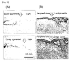
Fig. 2 is a set of photographs showing Fontana-Masson stainings of grafted foreskins and lentigo senilis. (A) Grafted foreskin. (B) Lentigo senilis; and -
Fig. 3 is a set of graphs showing the amount of expression of genes of cytokines for melanin synthesis (amount of mRNA) in the light part anddarklypigmentedpart of the grafted foreskin. (A) SCF (stem cell factor). (B) Endothelin-1. (C) POMC (Precursor of α melanocyte stimulating hormone, Pro-opiomelanocortin). -
Fig. 4 shows the evaluation of suppression of hyperpigmentation by the administration of s-kit in the animal model for evaluating hyperpigmentations. (A) From the lefthand, macro photographs at the time before administration, after 1 week, and after 2 weeks. Upper column; control (administration of PBS), lower column; administration of s-kit, (B) a graph showing color change of administration sites in the skin due to PBS or s-kit administration. - The present invention provides an animal model for evaluating hyperpigmentations, in which hyperpigmentation formation in human skin is faithfully simulated.
- The inventors of the present invention examined various skin hyperpigmentation models, and found that when a black person's skin is grafted to an immunodeficient mouse, pigmentation such as pigment spots is formed, and at the same time, increases in the expression of cytokines and the epidermal thickness are observed in the hyperpigmentation area, thus the animal being able to serve as an animal model for hyperpigmentations.
- Since the animal of the present invention allows hyperpigmentation formation to be authentically simulated and maintained stably for a long time, the animal is a potential model for evaluating hyperpigmentations in human skin. Thus, upon using the model, evaluation or screening of a hyperpigmentation formation suppressant or the like can be carried out. The term "hyperpigmentation" herein includes pigment spots.
- The animal model for evaluating hyperpigmentations of the present invention can be prepared by grafting a black person's skin onto a non-human animal.
The black person's skin that can be used for the present invention may be the skin from any of an infant, a child and an adult, as long as it is the skin derived from a black person (African descent or dark skin) , and the site of use is also not limited. For example, the foreskin of an adult or an infant, or the skin extracted during surgery or pathological autopsy can be used, while it is preferable to use the foreskin from the viewpoint of procurement. - The non-human animal that can be used in the present invention is preferably, for example, mammals such as a rodent, rabbit, monkey and the like, and among the mammals, rodents such as, for example, a mouse, rat, hamster and the like are preferred from the viewpoint of the convenience in skin grafting. In addition, the non-human animal is preferably an animal with suppressed immunological competence or an immunodeficient animal, and more preferably an immunodeficient animal, so as to suppress detachment of the grafted skin.
The immunodeficient mouse may be exemplified by an immunodeficient mouse such as SCID mouse, BALBcA-nu/Scid, B-17/Icr-Scid or the like, or an immunodeficient rat such as F344Jca-rnu or the like. Furthermore, in the case of raising an immunodeficient non-human animal, it is preferable to perform the raising under aseptic conditions. - The grafting of a black person's skin may be performed using known methods. For example, it may be performed according to the method described below.
A black person's skin is aseptically collected in its full-thickness or split-thickness. The collected skin can be preserved in sterilized physiological saline or in an appropriate culture medium, for example, in Dulbecco's Modified Eagle's Medium (DMEM) culture medium supplemented with L-glutamine and antibiotic/antimycotic (Invitrogen, CA), at 4 to 20°C until it is used for grafting to an animal.
Then, the recipient animal is anesthetized with ether or the like, and then the region in which the skin is removed in a size of, for example, 2 to 3 cm in diameter is produced on the dorsal side. It is favorable to shave hair, if necessary. Thereafter, a black person's skin graft which is slightly smaller than the region is grafted onto the region on the dorsal side of the animal.
Then, by use of a conventional method, the graft is sutured with a thread of monofilament base, nylon or the like, preferably with a long-chained aliphatic nylon thread, at an interval of about 5 mm. In this case, as the graft and the region are rendered identical in shape and sutured at a uniform tension, the graft and the graft bed can adhere tightly to facilitate the engraftment of the graft.
After the grafting, a topical anesthetic of amide type or the like is administered at the boundary of the grafted skin to induce sensory denervation, and the mouse is nursed. Typically, 3 to 12 weeks are required to complete engraftment of the graft. - Thus, the grafted black person's skin graft undergoes decolorization of the black color of the skin in 7 to 15 days after the grafting, and after about 1 month, the darkly pigmented part is reduced to less than 20% of the total area (
Fig. 1 ). Thereafter, the darkly pigmented part stably increases, and at a point in time of 2 months after the grafting, 25 to 45%; at a point in time of 3 months after the grafting, 30 to 50%; at a point in time of 4 months after the grafting, 35 to 55%; at a point in time of 5 months after the grafting, 40 to 60%; and at a point in time of 6 months after the grafting, 50% or greater on average, typically 50 to 60%, of the area becomes darkly pigmented (Fig. 1 ). Thereafter, the darkly pigmented part scarcely increases and remains relatively stable. - About 7 months after the grafting, the grafted skin is collected to include darkly pigmented parts and light parts, and the collected skin is subjected to Fontana-Masson staining. As the cross-section of the skin is observed, the darklypigmented part where melanin granules are confirmed exhibits a significant increase in thickness of the epidermis, as in the case of lentigo senilis (
Fig. 2 ).
Furthermore, in the darkly pigmented part, the expression of genes of cytokines associated with melanin synthesis (Endothelin-1 (prepro-endothelin-1), SCF, α Melanocyte Stimulating Hormone (pro-opiomelanocortin; POMC) gene) is increased (Fig. 3 ).
As such, the darkly pigmented part formed upon the skin grafting of the present invention shows closely similar characteristics regarding the melanin synthesis leading to the appearance of hyperpigmentations. - Therefore, by using the animal or the grafted skin of the present invention, evaluation or screening of a medicament, cosmetic product or the like intended to suppress the formation of skin hyperpigmentations or to remove formed hyperpigmentations can be implemented.
For example, it is possible to implement the evaluation or screening of a hyperpigmentation formation suppressant or a hyperpigmentation remover, by analyzing the RNA derived from the epidermis of the darkly pigmented part (hyperpigmentation part) and the light part (non-pigment spot part) of the grafted skin in the animal model for hyperpigmentations of the present invention, measuring the amount of expression of genes of cytokines such as Endothelin-1, SCF, α Melanocyte Stimulating Hormone (POMC) and the like, and using the results as an index; or by measuring the skin color using a colorimeter, and using the results as an index; or comparing the skin color change by photographs or naked-eye observation, and using the results as index. Inaddition, the term "hyperpigmentation" as used herein includeslentigosenilis,pigmentation afterinflammation, and darkening of the skin due to pigmentation such as freckles and the like. - The evaluation or screening of a hyperpigmenation formation suppressant or a hyperpigmenation remover can be performed by, specifically, administering an appropriate amount of test substance to the animal of the present invention via an administration route involving transepidermal, subepidermal, injection, oral or the like, and determining the effect of the test substance.
- Specifically, after 2-8 month raising after grafting the model mouse for evaluating hyperpigmenations of the present invention, the test substance is administered to the darkly pigmented part by the desired method as mentioned above. It is possible to evaluate or select, as a hyperpigmenation formation suppressant or a hyperpigmenation remover, the material to be tested which is low in the amount of expressed genes of cytokines or high in the skin lightness (L* value), by comparing the gene expression amount or skin color at this site with that of control (free of the material to be tested).
- Foreskin derived from African descent was obtained from the University of Cincinnati Hospital and Christ Hospital, and then immediately washed with phosphate buffered saline (PBS) . Then, the foreskin was trimmed to a suitable size (about 1.5 cm x 1.5 cm to about 2.0 cm x 2.0 cm). Until transferred to an animal facility, the foreskin was immersed in Dulbecco's Modified Eagle's Medium (DMEM) containing L-glutamine and antibiotic/antimycotic (Invitrogen, CA).
- Four to six-weeks old female SCID mice (Taconic, NY) were housed in an animal facility in Cincinnati Children's Hospital (Cincinnati Children's Hospital Medical Center, Cincinnati, OH) , in a specific pathogen-free condition from the beginning to the end of the experiment.
After acclimation of about 1 week, the mice were anesthetized in a box containing isofluorane/oxygen (3%/0.8 liter)and the mice were shaved on the dorsal side with hair clippers and a shaver. During the grafting of foreskin, the mouse was allowed to inhale isofluorane/oxygen (2%/0.7 liter). The dorsal side skin of the mouse was excised to a size which was slightly larger than the trimmed foreskin (about 2 to 3 cm in diameter) , and the mouse skin and the foreskin were sutured according to a standard method. The boundary of the grafted foreskin and the mouse skin was subjected to a sensory denervation treatment with sensorcaine. After the grafting, the mouse was maintained at 37°C for 1 hour, or until it woke up from anesthesia. - Since the grafted foreskin completely conglutinates with the mouse skin in about 1 month after grafting, the grafted foreskin was photographed from that point in time and at an interval of 1 month using a digital camera, and a relative ratio of the area of the darkly pigmented part to the entire area was calculated.
As shown inFig. 1 , the area of the darkly pigmented part at the time point of 1 month after the grafting was less than 20% of the entire area. But, the area stably increased thereafter, and at the point in time of 6 months after the grafting, 50% or larger area on average was found to be covered by the darkly pigmented part. After then, most of the grafted foreskins were not acknowledged to have such stable increases in the area. - After about 7 months of the foreskin grafting, at which time the increase in area of the darkly pigmented part became stable, the grafted foreskin was collected, and a sample section was prepared to contain darkly pigmented parts and light parts. After the preparation of the section, Fontana-Masson staining was attempted according to a standard method, so that melanin granules could be clearly detected.
The section was compared with a lentigo senilis (age spot) sample which was obtained from a contract laboratory (Stephens & Associates) in Dallas, the darkly pigmented part in the grafted foreskin was found to have significant thickening of the epidermis, as in the lentigo senilis (Fig. 2 ). - After about 7 months of the foreskin grafting, samples of 3 mm in diameter were collected from a darkly pigmented part and a light part in the grafted foreskin using a punch biopsy method. In order to prevent RNA from degradation, the samples were immediately immersed in RNAlater (Qiagen, Valencia, CA), and then total RNA was extractedusing anRNeasymicro kit (Qiagen). cDNA was synthesized according to a standard method using oligo dT and Moloney murine leukemia virus reverse transcriptase. Subsequently, a probe and primers which are specific to the genes of Endothelin-1 (prepro-endothelin-1; Assay ID: Hs00174961_m1), SCF (Assay ID: Hs00241497_m1) and POMC (a precursor of α Melanocyte Stimulating Hormone; Assay ID: Hs00174947_m1) which are known as three major cytokines associated with melanin synthesis (TaqMan Gene Expression Assays), and PCR reagent (TaqMan Gene Expression Master Mix) were purchased from Applied Biosystems (Foster City, CA), and real-time quantitative RT-PCR was performed using an ABI PRISM 7300 sequence detection system (Applied Biosystems). Expression of glyceraldehyde-3-phosphate dehydrogenase (GAPDH; human GAPDH VIC-MGB No. 4326317E, Applied Biosystems) was taken as the internal standard.
- It has already been reported that the above-mentioned cytokines show increased expression in lentigo senilis parts (J Invest Dermatol 116: 578-586, 2001, J Invest Dermatol 122: 1256-1265, 2004, J Dermatol Sci 2: 120-123, 2005). Also as a result of the quantitative RT-PCR analysis, it was found that the expression of all of the above-mentioned cytokines was higher in the darkly pigmented part compared to the light part (
Fig. 3 ). In particular, the expression of SCF was significantly (p < 0.05) higher in the darkly pigmented part. - From the above, in the darkly pigmented part of the grafted skin of the present invention, increased expression was observed for the genes of Endothelin-1 (prepro-endothelin-1), SCF and POMC, which have been actually observed to show increased expression in lentigo senilis parts. Furthermore, as it is being found that lentigo senilis parts are associated with increased expression of inflammatory cytokines such as tumor necrosis factor alpha and the like (J Invest Dermatol 116: 571-577, 2001) , or with epidermal thickening, the darkly pigmented parts of the grafted skin were also observed to have definite epidermal thickening as in lentigo senilis parts.
Therefore, it is contemplated that there is a high possibility that the characteristics of the grafted skin related to melanin synthesis is closely similar to the hyperpigmentations. - In accordance with the method mentioned above, evaluation of screening of hyperpigmentation formation suppressant or hyperpigmentation remover was conducted by use of mouse model for evaluating hyperpigmentations in which foreskin derived from African descent was grafted onto the back of imminodeficient mice and six months were passed thereafter.
A PBS solution containing 10 µM s-kit (R&I) systems) (hereinafter, referred to as s-kit) as the hyperpigmentation formation suppressant, and PBS free of s-kit (hereinafter, referred to as PBS) was used as control. It is known that the s-kit suppresses signal of SCF (D. D. Dahlen et al., Leukemia Research 25 (2001) 413-421) and has an action of hyperpigmentation formation suppression or hyperpigmentation removal.
To the darkly pigmented parts of the grown-up skin of model mice, 25 µL of 10 µM s-kit (R&D systems) by three times per one week was administered subepidermaly, and photography and skin lightness (L* value) of the skin using a colorimeter (Minolta CR-200) were measured atweek 0,week 1, andweek 2 from the start of the administration. Concurrently with this, to a darkly pigmented parts other than the portion to which the above mentioned suppressant or remover was administered, 25 µL of PBS was administered, and skin lightness was compared between the s-kit administered sites and PBS administration sites. The results by the colorimeter was shown by the variation from the initial value (ΔL* value: L* value at week 0 - L* value of each week). - As shown in
Fig. 4 , while the skin lightness was lowered (increase of ΔL* value) at PBS administration sites showing no action of hyperpigmentation formation suppression, the decrease in the skin lightness is suppressed at the s-kit administration sites showing hyperpigmentation formation suppression action.
Thus, using the animal model for evaluating hyperpigmentation enables evaluation or screening of the hyperpigmentation formation suppressant or hyperpigmentation remover. In addition, since the the animal model for evaluating hyperpigmentations is grafted with human skin, an evaluation closely similar to that of real human skin can be conducted.
Claims (8)
- An animal model for evaluating hyperpigmentations, wherein a black person's skin is grafted onto a non-human animal.
- The animal model according to claim 1, wherein the animal is an immunodeficient non-human animal.
- The animal model according to claim 1 or 2, wherein 50 to 60% of the area of the grafted skin becomes pigmented in about 6 months after grafting.
- The animal model according to claim 3, wherein the expression of a gene of at least one cytokine selected from Endothelin-1, SCF and POMC is increased in the darkly pigmented area.
- The animal model for evaluating hyperpigmentation according to claim 1, wherein the hyperpigmentation is a pigment spot.
- A method of preparing an animal model for evaluating hyperpigmentations, which comprises grafting a black person's skin onto a non-human animal, and raising the animal for at least 6 months after the grafting.
- A model skin for evaluating hyperpigmentations, prepared according to the method of claim 6.
- A method of evaluating or screening a hyperpigmentation formation suppressant or a hyperpigmentation remover, wherein the animal model of claim 1 is used.
Applications Claiming Priority (2)
| Application Number | Priority Date | Filing Date | Title |
|---|---|---|---|
| US11/785,549 US7897833B2 (en) | 2007-04-18 | 2007-04-18 | Animal model for pigment spots |
| US12/073,869 US7973211B2 (en) | 2007-04-18 | 2008-03-11 | Animal model for hyperpigmentation |
Publications (2)
| Publication Number | Publication Date |
|---|---|
| EP1982587A1 true EP1982587A1 (en) | 2008-10-22 |
| EP1982587B1 EP1982587B1 (en) | 2011-03-02 |
Family
ID=39689256
Family Applications (1)
| Application Number | Title | Priority Date | Filing Date |
|---|---|---|---|
| EP08007510A Ceased EP1982587B1 (en) | 2007-04-18 | 2008-04-17 | Animal model for hyperpigmentation |
Country Status (4)
| Country | Link |
|---|---|
| US (1) | US7973211B2 (en) |
| EP (1) | EP1982587B1 (en) |
| JP (1) | JP5435884B2 (en) |
| DE (1) | DE602008005203D1 (en) |
Families Citing this family (3)
| Publication number | Priority date | Publication date | Assignee | Title |
|---|---|---|---|---|
| US7897833B2 (en) * | 2007-04-18 | 2011-03-01 | Kao Corporation | Animal model for pigment spots |
| US20090226901A1 (en) * | 2008-03-06 | 2009-09-10 | Kao Corporation | Method of evaluating or selecting agents for preventing or improving pigmented spots |
| JP5357520B2 (en) * | 2008-11-13 | 2013-12-04 | 丸善製薬株式会社 | Endothelin-1 mRNA expression inhibitor, stem cell growth factor (SCF) mRNA expression inhibitor, basic fibroblast growth factor (bFGF) mRNA expression inhibitor, and proopiomelanocortin (POMC) mRNA expression inhibitor |
Citations (1)
| Publication number | Priority date | Publication date | Assignee | Title |
|---|---|---|---|---|
| US20060058252A1 (en) * | 2002-06-26 | 2006-03-16 | Clawson Gary A | Methods and materials for treating human papillomavirus infections |
Family Cites Families (5)
| Publication number | Priority date | Publication date | Assignee | Title |
|---|---|---|---|---|
| AU5609694A (en) * | 1992-11-24 | 1994-06-22 | University Of Cincinnati, The | Melanogenic inhibitor, and methods of producing and using the same |
| JPH09172909A (en) * | 1995-12-27 | 1997-07-08 | Kanagawa Kagaku Gijutsu Akad | Transplanting method of cultured human skin cell to non-human animal |
| US7285570B2 (en) * | 2003-04-17 | 2007-10-23 | The Procter & Gamble Company | Compositions and methods for regulating mammalian keratinous tissue |
| JP4022196B2 (en) * | 2003-12-17 | 2007-12-12 | 花王株式会社 | Method for constructing reconstructed skin |
| US7897833B2 (en) * | 2007-04-18 | 2011-03-01 | Kao Corporation | Animal model for pigment spots |
-
2008
- 2008-03-11 US US12/073,869 patent/US7973211B2/en not_active Expired - Fee Related
- 2008-04-15 JP JP2008105705A patent/JP5435884B2/en not_active Expired - Fee Related
- 2008-04-17 EP EP08007510A patent/EP1982587B1/en not_active Ceased
- 2008-04-17 DE DE602008005203T patent/DE602008005203D1/en active Active
Patent Citations (1)
| Publication number | Priority date | Publication date | Assignee | Title |
|---|---|---|---|---|
| US20060058252A1 (en) * | 2002-06-26 | 2006-03-16 | Clawson Gary A | Methods and materials for treating human papillomavirus infections |
Non-Patent Citations (16)
| Title |
|---|
| ARCH DERMATOL RES, vol. 278, 1986, pages 352 - 362 |
| BROOME-POWELL M ET AL: "Hyperpigmentation and melanocytic hyperplasia in transgenic mice expressing the human T24 Ha-ras gene regulated by a mouse tyrosinase promoter", MOLECULAR CARCINOGENESIS, ALAN LISS, NEW YORK, NY, US, vol. 12, 1 January 1995 (1995-01-01), pages 82 - 90, XP002466450, ISSN: 0899-1987 * |
| D. D. DAHLEN ET AL., LEUKEMIA RESEARCH, vol. 25, 2001, pages 413 - 421 |
| HACHIYA A. ET AL.: "An in vivo mouse model of human skin substitute containing spontaneously sorted melanocytes demonstrates physiological changes after UVB irradiation.", J INVEST DERMATOL, vol. 125, 2005, pages 364 - 372, XP002493073 * |
| HACHIYA, A. ET AL.: "The paracrine role of stem cell factor/c-kit signaling in the activation of human melanocytes in ultraviolet-B-induced pigmentation", J. INVEST. DERMATOL., vol. 116, 2001, pages 578 - 586 |
| IMOKAWA, G. ET AL.: "Differential analysis of experimental hypermelanosis induced by UVB, PUVA, and allergic contact dermatitis using a brownish guinea pig model", ARCH. DERMATOL. RES., vol. 278, 1986, pages 352 - 362 |
| J DERMATOL SCI, vol. 2, 2005, pages 120 - 123 |
| J INVEST DERMATOL, vol. 116, 2001, pages 571 - 577 |
| J INVEST DERMATOL, vol. 116, 2001, pages 578 - 586 |
| J INVEST DERMATOL, vol. 122, 2004, pages 1256 - 1265 |
| KAWAGUCHI, Y.; MORI, N.; NAKAYAMA, A.: "Kit(+) melanocytes seem to contribute to melanocyte proliferation after UV exposure as precursor cells", J. INVEST. DERMATOL, vol. 116, 2001, pages 920 - 925 |
| SEIBERG, M.: "Keratinocyte-melanocyte interactions during melanosome transfer", PIGMENT CELL RES, vol. 4, 2001, pages 236 - 242 |
| SEIBERG, M.: "Keratinocyte-melanocyte interactions during melanosome transfer", PIGMENT CELL RES., vol. 4, 2001, pages 236 - 242 |
| WELSH, B.M.; MASON, R.S.; HALLIDAY, G.M.: "Topical all-trans retinoic acid augments ultraviolet radiation-induced increases in activated melanocyte numbers in mice", J. INVEST. DERMATOL, vol. 112, 1999, pages 271 - 278 |
| WELSH, B.M.; MASON, R.S.; HALLIDAY, G.M.: "Topical all-trans retinoic acid augments ultraviolet radiation-induced increases in activated melanocyte numbers in mice", J. INVEST. DERMATOL., vol. 112, 1999, pages 271 - 278 |
| YOSHIDA Y ET AL.: "Functional analysis of keratinocytes in skin color using a human substitute model composed of cell derived from different skin pigmentation types.", FASEB J, vol. 21, 2007, pages 2829 - 2839, XP002493072 * |
Also Published As
| Publication number | Publication date |
|---|---|
| JP2008263980A (en) | 2008-11-06 |
| EP1982587B1 (en) | 2011-03-02 |
| DE602008005203D1 (en) | 2011-04-14 |
| US7973211B2 (en) | 2011-07-05 |
| US20080306409A1 (en) | 2008-12-11 |
| JP5435884B2 (en) | 2014-03-05 |
Similar Documents
| Publication | Publication Date | Title |
|---|---|---|
| Kunisada et al. | Transgene expression of steel factor in the basal layer of epidermis promotes survival, proliferation, differentiation and migration of melanocyte precursors | |
| Mooi et al. | Pathology of Melanocytic Disorders 2ed | |
| Kunisada et al. | Keratinocyte expression of transgenic hepatocyte growth factor affects melanocyte development, leading to dermal melanocytosis | |
| US9273338B2 (en) | Chimeric avian-based screening system containing mammalian grafts | |
| JP5872158B2 (en) | Skin whitening method and screening method for skin spot formation inhibition and / or removal factor | |
| Asaka et al. | Type XVII collagen is a key player in tooth enamel formation | |
| US20080109915A1 (en) | Hair follicle-reconstitution system and animal carrying the same | |
| EP1982587B1 (en) | Animal model for hyperpigmentation | |
| JP7456990B2 (en) | Methods for preventing or treating skin disorders and conditions | |
| Galanternik et al. | Anatomical and molecular characterization of the zebrafish meninges | |
| Hachiya et al. | An in vivo mouse model of human skin substitute containing spontaneously sorted melanocytes demonstrates physiological changes after UVB irradiation | |
| Chen et al. | Keratinocytes take part in the regulation of substance P in melanogenesis through the HPA axis | |
| US7897833B2 (en) | Animal model for pigment spots | |
| US6576812B1 (en) | Compound screening assays using a transgenic mouse model of human skin diseases | |
| JP7016533B2 (en) | Test method for vitiligo toxicity and melasma toxicity | |
| EP2617284B1 (en) | Model animal for studying hair growth cycle | |
| Norum et al. | A conditional transgenic mouse line for targeted expression of the stem cell marker LGR5 | |
| JP4022196B2 (en) | Method for constructing reconstructed skin | |
| US7736894B2 (en) | Method for constructing reconstructed skin | |
| Subburaj et al. | Animal models in disorders of skin color− utilizing evolution and demystifying mysteries | |
| KR100775333B1 (en) | Screening method of melanin synthesis regulator using zebrafish (Zebrafish) | |
| Flesher | Using In Vivo Models to Understand Melanogenesis | |
| JP5824784B2 (en) | Hair growth resting period shortening agent | |
| WO1997017982A1 (en) | Methods and compositions for hair growth promotion | |
| JP2012062294A (en) | Agent for shortening hair growth telogen |
Legal Events
| Date | Code | Title | Description |
|---|---|---|---|
| PUAI | Public reference made under article 153(3) epc to a published international application that has entered the european phase |
Free format text: ORIGINAL CODE: 0009012 |
|
| 17P | Request for examination filed |
Effective date: 20080417 |
|
| AK | Designated contracting states |
Kind code of ref document: A1 Designated state(s): AT BE BG CH CY CZ DE DK EE ES FI FR GB GR HR HU IE IS IT LI LT LU LV MC MT NL NO PL PT RO SE SI SK TR |
|
| AX | Request for extension of the european patent |
Extension state: AL BA MK RS |
|
| AKX | Designation fees paid |
Designated state(s): DE FR GB |
|
| GRAP | Despatch of communication of intention to grant a patent |
Free format text: ORIGINAL CODE: EPIDOSNIGR1 |
|
| GRAS | Grant fee paid |
Free format text: ORIGINAL CODE: EPIDOSNIGR3 |
|
| GRAA | (expected) grant |
Free format text: ORIGINAL CODE: 0009210 |
|
| AK | Designated contracting states |
Kind code of ref document: B1 Designated state(s): DE FR GB |
|
| REG | Reference to a national code |
Ref country code: GB Ref legal event code: FG4D |
|
| REF | Corresponds to: |
Ref document number: 602008005203 Country of ref document: DE Date of ref document: 20110414 Kind code of ref document: P |
|
| REG | Reference to a national code |
Ref country code: DE Ref legal event code: R096 Ref document number: 602008005203 Country of ref document: DE Effective date: 20110414 |
|
| PLBE | No opposition filed within time limit |
Free format text: ORIGINAL CODE: 0009261 |
|
| STAA | Information on the status of an ep patent application or granted ep patent |
Free format text: STATUS: NO OPPOSITION FILED WITHIN TIME LIMIT |
|
| 26N | No opposition filed |
Effective date: 20111205 |
|
| REG | Reference to a national code |
Ref country code: DE Ref legal event code: R097 Ref document number: 602008005203 Country of ref document: DE Effective date: 20111205 |
|
| REG | Reference to a national code |
Ref country code: FR Ref legal event code: PLFP Year of fee payment: 9 |
|
| REG | Reference to a national code |
Ref country code: FR Ref legal event code: PLFP Year of fee payment: 10 |
|
| REG | Reference to a national code |
Ref country code: FR Ref legal event code: PLFP Year of fee payment: 11 |
|
| PGFP | Annual fee paid to national office [announced via postgrant information from national office to epo] |
Ref country code: GB Payment date: 20180329 Year of fee payment: 11 |
|
| PGFP | Annual fee paid to national office [announced via postgrant information from national office to epo] |
Ref country code: FR Payment date: 20180315 Year of fee payment: 11 |
|
| PGFP | Annual fee paid to national office [announced via postgrant information from national office to epo] |
Ref country code: DE Payment date: 20180404 Year of fee payment: 11 |
|
| REG | Reference to a national code |
Ref country code: DE Ref legal event code: R119 Ref document number: 602008005203 Country of ref document: DE |
|
| GBPC | Gb: european patent ceased through non-payment of renewal fee |
Effective date: 20190417 |
|
| PG25 | Lapsed in a contracting state [announced via postgrant information from national office to epo] |
Ref country code: GB Free format text: LAPSE BECAUSE OF NON-PAYMENT OF DUE FEES Effective date: 20190417 Ref country code: DE Free format text: LAPSE BECAUSE OF NON-PAYMENT OF DUE FEES Effective date: 20191101 |
|
| PG25 | Lapsed in a contracting state [announced via postgrant information from national office to epo] |
Ref country code: FR Free format text: LAPSE BECAUSE OF NON-PAYMENT OF DUE FEES Effective date: 20190430 |