EP1980623A1 - A method for identifying a renal fibrosis process, use of snail-activity-inhibiting compounds in the production of pharmaceutical compositions, method for identifying said inhibiting compounds, said pharmaceutical compositions and applications thereof - Google Patents
A method for identifying a renal fibrosis process, use of snail-activity-inhibiting compounds in the production of pharmaceutical compositions, method for identifying said inhibiting compounds, said pharmaceutical compositions and applications thereof Download PDFInfo
- Publication number
- EP1980623A1 EP1980623A1 EP06807963A EP06807963A EP1980623A1 EP 1980623 A1 EP1980623 A1 EP 1980623A1 EP 06807963 A EP06807963 A EP 06807963A EP 06807963 A EP06807963 A EP 06807963A EP 1980623 A1 EP1980623 A1 EP 1980623A1
- Authority
- EP
- European Patent Office
- Prior art keywords
- snail
- protein
- renal fibrosis
- expression
- identification
- Prior art date
- Legal status (The legal status is an assumption and is not a legal conclusion. Google has not performed a legal analysis and makes no representation as to the accuracy of the status listed.)
- Withdrawn
Links
- 241000237858 Gastropoda Species 0.000 title claims abstract description 161
- 201000002793 renal fibrosis Diseases 0.000 title claims abstract description 76
- 238000000034 method Methods 0.000 title claims description 69
- 150000001875 compounds Chemical class 0.000 title claims description 39
- 230000000694 effects Effects 0.000 title claims description 28
- 239000008194 pharmaceutical composition Substances 0.000 title claims description 23
- 230000008569 process Effects 0.000 title claims description 23
- 230000002401 inhibitory effect Effects 0.000 title claims description 13
- 108090000623 proteins and genes Proteins 0.000 claims abstract description 128
- 102000004169 proteins and genes Human genes 0.000 claims abstract description 61
- 208000037265 diseases, disorders, signs and symptoms Diseases 0.000 claims abstract description 12
- 201000010099 disease Diseases 0.000 claims abstract description 8
- 230000014509 gene expression Effects 0.000 claims description 90
- 108020004999 messenger RNA Proteins 0.000 claims description 42
- 210000003734 kidney Anatomy 0.000 claims description 35
- 239000002773 nucleotide Substances 0.000 claims description 22
- 125000003729 nucleotide group Chemical group 0.000 claims description 22
- 108091032973 (ribonucleotides)n+m Proteins 0.000 claims description 16
- 238000013518 transcription Methods 0.000 claims description 16
- 230000035897 transcription Effects 0.000 claims description 16
- 102000040430 polynucleotide Human genes 0.000 claims description 14
- 108091033319 polynucleotide Proteins 0.000 claims description 14
- 239000002157 polynucleotide Substances 0.000 claims description 14
- 241001465754 Metazoa Species 0.000 claims description 13
- 239000003795 chemical substances by application Substances 0.000 claims description 13
- 230000001939 inductive effect Effects 0.000 claims description 12
- 239000012634 fragment Substances 0.000 claims description 11
- 230000009261 transgenic effect Effects 0.000 claims description 11
- 206010016654 Fibrosis Diseases 0.000 claims description 10
- 230000004761 fibrosis Effects 0.000 claims description 10
- 239000000203 mixture Substances 0.000 claims description 10
- 102000053642 Catalytic RNA Human genes 0.000 claims description 9
- 108090000994 Catalytic RNA Proteins 0.000 claims description 9
- 101000702691 Homo sapiens Zinc finger protein SNAI1 Proteins 0.000 claims description 9
- 238000003757 reverse transcription PCR Methods 0.000 claims description 9
- 108091092562 ribozyme Proteins 0.000 claims description 9
- 230000001225 therapeutic effect Effects 0.000 claims description 9
- 230000000692 anti-sense effect Effects 0.000 claims description 8
- 229940079593 drug Drugs 0.000 claims description 8
- 239000003814 drug Substances 0.000 claims description 8
- 102000044908 human SNAI1 Human genes 0.000 claims description 8
- 238000009396 hybridization Methods 0.000 claims description 8
- 230000004952 protein activity Effects 0.000 claims description 8
- 108091023037 Aptamer Proteins 0.000 claims description 7
- 239000012472 biological sample Substances 0.000 claims description 7
- 102000039446 nucleic acids Human genes 0.000 claims description 7
- 108020004707 nucleic acids Proteins 0.000 claims description 7
- 150000007523 nucleic acids Chemical class 0.000 claims description 7
- 102000008186 Collagen Human genes 0.000 claims description 6
- 108010035532 Collagen Proteins 0.000 claims description 6
- 229920001436 collagen Polymers 0.000 claims description 6
- 238000011065 in-situ storage Methods 0.000 claims description 6
- 201000002327 urinary tract obstruction Diseases 0.000 claims description 6
- 239000002671 adjuvant Substances 0.000 claims description 5
- 230000006378 damage Effects 0.000 claims description 5
- 230000004660 morphological change Effects 0.000 claims description 5
- 108010022452 Collagen Type I Proteins 0.000 claims description 4
- 206010018364 Glomerulonephritis Diseases 0.000 claims description 4
- 102000013127 Vimentin Human genes 0.000 claims description 4
- 108010065472 Vimentin Proteins 0.000 claims description 4
- 230000006866 deterioration Effects 0.000 claims description 4
- 206010012601 diabetes mellitus Diseases 0.000 claims description 4
- 208000035475 disorder Diseases 0.000 claims description 4
- 239000002243 precursor Substances 0.000 claims description 4
- 238000002360 preparation method Methods 0.000 claims description 4
- 230000009467 reduction Effects 0.000 claims description 4
- 231100000419 toxicity Toxicity 0.000 claims description 4
- 230000001988 toxicity Effects 0.000 claims description 4
- 210000005048 vimentin Anatomy 0.000 claims description 4
- 208000010159 IgA glomerulonephritis Diseases 0.000 claims description 3
- 206010021263 IgA nephropathy Diseases 0.000 claims description 3
- 241000124008 Mammalia Species 0.000 claims description 3
- 230000003321 amplification Effects 0.000 claims description 3
- 239000013068 control sample Substances 0.000 claims description 3
- 230000008021 deposition Effects 0.000 claims description 3
- 238000011534 incubation Methods 0.000 claims description 3
- 238000003199 nucleic acid amplification method Methods 0.000 claims description 3
- 230000007170 pathology Effects 0.000 claims description 3
- 239000003550 marker Substances 0.000 abstract description 9
- 230000005764 inhibitory process Effects 0.000 abstract description 6
- 238000003745 diagnosis Methods 0.000 abstract description 3
- 239000002547 new drug Substances 0.000 abstract description 2
- 101710196874 Cadherin-16 Proteins 0.000 description 64
- 102100024151 Cadherin-16 Human genes 0.000 description 60
- 210000004027 cell Anatomy 0.000 description 45
- 230000004913 activation Effects 0.000 description 20
- NKANXQFJJICGDU-QPLCGJKRSA-N Tamoxifen Chemical compound C=1C=CC=CC=1C(/CC)=C(C=1C=CC(OCCN(C)C)=CC=1)/C1=CC=CC=C1 NKANXQFJJICGDU-QPLCGJKRSA-N 0.000 description 16
- 102000000905 Cadherin Human genes 0.000 description 15
- 108050007957 Cadherin Proteins 0.000 description 14
- 238000011830 transgenic mouse model Methods 0.000 description 13
- 241000699666 Mus <mouse, genus> Species 0.000 description 12
- 210000001519 tissue Anatomy 0.000 description 11
- 239000013598 vector Substances 0.000 description 11
- 241000699660 Mus musculus Species 0.000 description 10
- 229960001603 tamoxifen Drugs 0.000 description 10
- 230000003176 fibrotic effect Effects 0.000 description 8
- 238000001727 in vivo Methods 0.000 description 8
- 102100031181 Glyceraldehyde-3-phosphate dehydrogenase Human genes 0.000 description 7
- 108091034117 Oligonucleotide Proteins 0.000 description 7
- 108020004445 glyceraldehyde-3-phosphate dehydrogenase Proteins 0.000 description 7
- 239000000523 sample Substances 0.000 description 7
- JLCPHMBAVCMARE-UHFFFAOYSA-N [3-[[3-[[3-[[3-[[3-[[3-[[3-[[3-[[3-[[3-[[3-[[5-(2-amino-6-oxo-1H-purin-9-yl)-3-[[3-[[3-[[3-[[3-[[3-[[5-(2-amino-6-oxo-1H-purin-9-yl)-3-[[5-(2-amino-6-oxo-1H-purin-9-yl)-3-hydroxyoxolan-2-yl]methoxy-hydroxyphosphoryl]oxyoxolan-2-yl]methoxy-hydroxyphosphoryl]oxy-5-(5-methyl-2,4-dioxopyrimidin-1-yl)oxolan-2-yl]methoxy-hydroxyphosphoryl]oxy-5-(6-aminopurin-9-yl)oxolan-2-yl]methoxy-hydroxyphosphoryl]oxy-5-(6-aminopurin-9-yl)oxolan-2-yl]methoxy-hydroxyphosphoryl]oxy-5-(6-aminopurin-9-yl)oxolan-2-yl]methoxy-hydroxyphosphoryl]oxy-5-(6-aminopurin-9-yl)oxolan-2-yl]methoxy-hydroxyphosphoryl]oxyoxolan-2-yl]methoxy-hydroxyphosphoryl]oxy-5-(5-methyl-2,4-dioxopyrimidin-1-yl)oxolan-2-yl]methoxy-hydroxyphosphoryl]oxy-5-(4-amino-2-oxopyrimidin-1-yl)oxolan-2-yl]methoxy-hydroxyphosphoryl]oxy-5-(5-methyl-2,4-dioxopyrimidin-1-yl)oxolan-2-yl]methoxy-hydroxyphosphoryl]oxy-5-(5-methyl-2,4-dioxopyrimidin-1-yl)oxolan-2-yl]methoxy-hydroxyphosphoryl]oxy-5-(6-aminopurin-9-yl)oxolan-2-yl]methoxy-hydroxyphosphoryl]oxy-5-(6-aminopurin-9-yl)oxolan-2-yl]methoxy-hydroxyphosphoryl]oxy-5-(4-amino-2-oxopyrimidin-1-yl)oxolan-2-yl]methoxy-hydroxyphosphoryl]oxy-5-(4-amino-2-oxopyrimidin-1-yl)oxolan-2-yl]methoxy-hydroxyphosphoryl]oxy-5-(4-amino-2-oxopyrimidin-1-yl)oxolan-2-yl]methoxy-hydroxyphosphoryl]oxy-5-(6-aminopurin-9-yl)oxolan-2-yl]methoxy-hydroxyphosphoryl]oxy-5-(4-amino-2-oxopyrimidin-1-yl)oxolan-2-yl]methyl [5-(6-aminopurin-9-yl)-2-(hydroxymethyl)oxolan-3-yl] hydrogen phosphate Polymers Cc1cn(C2CC(OP(O)(=O)OCC3OC(CC3OP(O)(=O)OCC3OC(CC3O)n3cnc4c3nc(N)[nH]c4=O)n3cnc4c3nc(N)[nH]c4=O)C(COP(O)(=O)OC3CC(OC3COP(O)(=O)OC3CC(OC3COP(O)(=O)OC3CC(OC3COP(O)(=O)OC3CC(OC3COP(O)(=O)OC3CC(OC3COP(O)(=O)OC3CC(OC3COP(O)(=O)OC3CC(OC3COP(O)(=O)OC3CC(OC3COP(O)(=O)OC3CC(OC3COP(O)(=O)OC3CC(OC3COP(O)(=O)OC3CC(OC3COP(O)(=O)OC3CC(OC3COP(O)(=O)OC3CC(OC3COP(O)(=O)OC3CC(OC3COP(O)(=O)OC3CC(OC3COP(O)(=O)OC3CC(OC3COP(O)(=O)OC3CC(OC3CO)n3cnc4c(N)ncnc34)n3ccc(N)nc3=O)n3cnc4c(N)ncnc34)n3ccc(N)nc3=O)n3ccc(N)nc3=O)n3ccc(N)nc3=O)n3cnc4c(N)ncnc34)n3cnc4c(N)ncnc34)n3cc(C)c(=O)[nH]c3=O)n3cc(C)c(=O)[nH]c3=O)n3ccc(N)nc3=O)n3cc(C)c(=O)[nH]c3=O)n3cnc4c3nc(N)[nH]c4=O)n3cnc4c(N)ncnc34)n3cnc4c(N)ncnc34)n3cnc4c(N)ncnc34)n3cnc4c(N)ncnc34)O2)c(=O)[nH]c1=O JLCPHMBAVCMARE-UHFFFAOYSA-N 0.000 description 6
- 239000012190 activator Substances 0.000 description 6
- 238000004458 analytical method Methods 0.000 description 6
- 210000000981 epithelium Anatomy 0.000 description 6
- 230000000754 repressing effect Effects 0.000 description 6
- 108020004414 DNA Proteins 0.000 description 5
- 208000001647 Renal Insufficiency Diseases 0.000 description 5
- 102000040945 Transcription factor Human genes 0.000 description 5
- 108091023040 Transcription factor Proteins 0.000 description 5
- 102000004887 Transforming Growth Factor beta Human genes 0.000 description 5
- 108090001012 Transforming Growth Factor beta Proteins 0.000 description 5
- 150000001413 amino acids Chemical class 0.000 description 5
- 239000002299 complementary DNA Substances 0.000 description 5
- 210000002257 embryonic structure Anatomy 0.000 description 5
- 210000002919 epithelial cell Anatomy 0.000 description 5
- 238000002474 experimental method Methods 0.000 description 5
- 230000006698 induction Effects 0.000 description 5
- 201000006370 kidney failure Diseases 0.000 description 5
- 230000007704 transition Effects 0.000 description 5
- 102000007469 Actins Human genes 0.000 description 4
- 108010085238 Actins Proteins 0.000 description 4
- 102000012046 Hepatocyte Nuclear Factor 1-beta Human genes 0.000 description 4
- 101100421767 Mus musculus Snai1 gene Proteins 0.000 description 4
- 230000000295 complement effect Effects 0.000 description 4
- 238000011161 development Methods 0.000 description 4
- 230000018109 developmental process Effects 0.000 description 4
- 230000008034 disappearance Effects 0.000 description 4
- 230000013020 embryo development Effects 0.000 description 4
- 210000004940 nucleus Anatomy 0.000 description 4
- 239000013612 plasmid Substances 0.000 description 4
- 238000003753 real-time PCR Methods 0.000 description 4
- ZRKFYGHZFMAOKI-QMGMOQQFSA-N tgfbeta Chemical compound C([C@H](NC(=O)[C@H](C(C)C)NC(=O)CNC(=O)[C@H](CCC(O)=O)NC(=O)[C@H](CCCNC(N)=N)NC(=O)[C@H](CC(N)=O)NC(=O)[C@H](CC(C)C)NC(=O)[C@H]([C@@H](C)O)NC(=O)[C@H](CCC(O)=O)NC(=O)[C@H]([C@@H](C)O)NC(=O)[C@H](CC(C)C)NC(=O)CNC(=O)[C@H](C)NC(=O)[C@H](CO)NC(=O)[C@H](CCC(N)=O)NC(=O)[C@@H](NC(=O)[C@H](C)NC(=O)[C@H](C)NC(=O)[C@@H](NC(=O)[C@H](CC(C)C)NC(=O)[C@@H](N)CCSC)C(C)C)[C@@H](C)CC)C(=O)N[C@@H]([C@@H](C)O)C(=O)N[C@@H](C(C)C)C(=O)N[C@@H](CC=1C=CC=CC=1)C(=O)N[C@@H](C)C(=O)N1[C@@H](CCC1)C(=O)N[C@@H]([C@@H](C)O)C(=O)N[C@@H](CC(N)=O)C(=O)N[C@@H](CCC(O)=O)C(=O)N[C@@H](C)C(=O)N[C@@H](CC=1C=CC=CC=1)C(=O)N[C@@H](CCCNC(N)=N)C(=O)N[C@@H](C)C(=O)N[C@@H](CC(C)C)C(=O)N1[C@@H](CCC1)C(=O)N1[C@@H](CCC1)C(=O)N[C@@H](CCCNC(N)=N)C(=O)N[C@@H](CCC(O)=O)C(=O)N[C@@H](CCCNC(N)=N)C(=O)N[C@@H](CO)C(=O)N[C@@H](CCCNC(N)=N)C(=O)N[C@@H](CC(C)C)C(=O)N[C@@H](CC(C)C)C(O)=O)C1=CC=C(O)C=C1 ZRKFYGHZFMAOKI-QMGMOQQFSA-N 0.000 description 4
- 238000002560 therapeutic procedure Methods 0.000 description 4
- 239000003981 vehicle Substances 0.000 description 4
- 102000040650 (ribonucleotides)n+m Human genes 0.000 description 3
- 108010010803 Gelatin Proteins 0.000 description 3
- 108010061414 Hepatocyte Nuclear Factor 1-beta Proteins 0.000 description 3
- 108060001084 Luciferase Proteins 0.000 description 3
- OKKJLVBELUTLKV-UHFFFAOYSA-N Methanol Chemical compound OC OKKJLVBELUTLKV-UHFFFAOYSA-N 0.000 description 3
- 101000762245 Mus musculus Cadherin-16 Proteins 0.000 description 3
- 101001045759 Mus musculus Hepatocyte nuclear factor 1-beta Proteins 0.000 description 3
- 241000699670 Mus sp. Species 0.000 description 3
- 206010028980 Neoplasm Diseases 0.000 description 3
- 108020004459 Small interfering RNA Proteins 0.000 description 3
- 108700019146 Transgenes Proteins 0.000 description 3
- 208000027418 Wounds and injury Diseases 0.000 description 3
- 238000010171 animal model Methods 0.000 description 3
- 230000015572 biosynthetic process Effects 0.000 description 3
- 210000000481 breast Anatomy 0.000 description 3
- 230000004069 differentiation Effects 0.000 description 3
- 239000008273 gelatin Substances 0.000 description 3
- 229920000159 gelatin Polymers 0.000 description 3
- 235000019322 gelatine Nutrition 0.000 description 3
- 235000011852 gelatine desserts Nutrition 0.000 description 3
- 238000003364 immunohistochemistry Methods 0.000 description 3
- 230000000670 limiting effect Effects 0.000 description 3
- 210000001161 mammalian embryo Anatomy 0.000 description 3
- 238000002493 microarray Methods 0.000 description 3
- 238000013059 nephrectomy Methods 0.000 description 3
- 210000000885 nephron Anatomy 0.000 description 3
- 239000000546 pharmaceutical excipient Substances 0.000 description 3
- 210000005084 renal tissue Anatomy 0.000 description 3
- 230000032258 transport Effects 0.000 description 3
- IJGRMHOSHXDMSA-UHFFFAOYSA-N Atomic nitrogen Chemical compound N#N IJGRMHOSHXDMSA-UHFFFAOYSA-N 0.000 description 2
- LFQSCWFLJHTTHZ-UHFFFAOYSA-N Ethanol Chemical compound CCO LFQSCWFLJHTTHZ-UHFFFAOYSA-N 0.000 description 2
- WSFSSNUMVMOOMR-UHFFFAOYSA-N Formaldehyde Chemical compound O=C WSFSSNUMVMOOMR-UHFFFAOYSA-N 0.000 description 2
- 241000282412 Homo Species 0.000 description 2
- 101000882584 Homo sapiens Estrogen receptor Proteins 0.000 description 2
- 101000633054 Homo sapiens Zinc finger protein SNAI2 Proteins 0.000 description 2
- 239000005089 Luciferase Substances 0.000 description 2
- 229930193140 Neomycin Natural products 0.000 description 2
- 108091034057 RNA (poly(A)) Proteins 0.000 description 2
- 108010052090 Renilla Luciferases Proteins 0.000 description 2
- 108010092935 Snail Family Transcription Factors Proteins 0.000 description 2
- 102000016845 Snail Family Transcription Factors Human genes 0.000 description 2
- 208000004608 Ureteral Obstruction Diseases 0.000 description 2
- 102100029570 Zinc finger protein SNAI2 Human genes 0.000 description 2
- 230000001594 aberrant effect Effects 0.000 description 2
- TXUZVZSFRXZGTL-QPLCGJKRSA-N afimoxifene Chemical compound C=1C=CC=CC=1C(/CC)=C(C=1C=CC(OCCN(C)C)=CC=1)/C1=CC=C(O)C=C1 TXUZVZSFRXZGTL-QPLCGJKRSA-N 0.000 description 2
- 230000003510 anti-fibrotic effect Effects 0.000 description 2
- 239000000074 antisense oligonucleotide Substances 0.000 description 2
- 238000012230 antisense oligonucleotides Methods 0.000 description 2
- 239000000512 collagen gel Substances 0.000 description 2
- 230000000052 comparative effect Effects 0.000 description 2
- 238000001514 detection method Methods 0.000 description 2
- 238000000605 extraction Methods 0.000 description 2
- 210000002950 fibroblast Anatomy 0.000 description 2
- 230000003328 fibroblastic effect Effects 0.000 description 2
- 230000006870 function Effects 0.000 description 2
- 238000001415 gene therapy Methods 0.000 description 2
- 238000009650 gentamicin protection assay Methods 0.000 description 2
- 239000000411 inducer Substances 0.000 description 2
- NOESYZHRGYRDHS-UHFFFAOYSA-N insulin Chemical compound N1C(=O)C(NC(=O)C(CCC(N)=O)NC(=O)C(CCC(O)=O)NC(=O)C(C(C)C)NC(=O)C(NC(=O)CN)C(C)CC)CSSCC(C(NC(CO)C(=O)NC(CC(C)C)C(=O)NC(CC=2C=CC(O)=CC=2)C(=O)NC(CCC(N)=O)C(=O)NC(CC(C)C)C(=O)NC(CCC(O)=O)C(=O)NC(CC(N)=O)C(=O)NC(CC=2C=CC(O)=CC=2)C(=O)NC(CSSCC(NC(=O)C(C(C)C)NC(=O)C(CC(C)C)NC(=O)C(CC=2C=CC(O)=CC=2)NC(=O)C(CC(C)C)NC(=O)C(C)NC(=O)C(CCC(O)=O)NC(=O)C(C(C)C)NC(=O)C(CC(C)C)NC(=O)C(CC=2NC=NC=2)NC(=O)C(CO)NC(=O)CNC2=O)C(=O)NCC(=O)NC(CCC(O)=O)C(=O)NC(CCCNC(N)=N)C(=O)NCC(=O)NC(CC=3C=CC=CC=3)C(=O)NC(CC=3C=CC=CC=3)C(=O)NC(CC=3C=CC(O)=CC=3)C(=O)NC(C(C)O)C(=O)N3C(CCC3)C(=O)NC(CCCCN)C(=O)NC(C)C(O)=O)C(=O)NC(CC(N)=O)C(O)=O)=O)NC(=O)C(C(C)CC)NC(=O)C(CO)NC(=O)C(C(C)O)NC(=O)C1CSSCC2NC(=O)C(CC(C)C)NC(=O)C(NC(=O)C(CCC(N)=O)NC(=O)C(CC(N)=O)NC(=O)C(NC(=O)C(N)CC=1C=CC=CC=1)C(C)C)CC1=CN=CN1 NOESYZHRGYRDHS-UHFFFAOYSA-N 0.000 description 2
- QWTDNUCVQCZILF-UHFFFAOYSA-N isopentane Chemical compound CCC(C)C QWTDNUCVQCZILF-UHFFFAOYSA-N 0.000 description 2
- 230000029795 kidney development Effects 0.000 description 2
- 239000000463 material Substances 0.000 description 2
- 229960004927 neomycin Drugs 0.000 description 2
- 108090000765 processed proteins & peptides Proteins 0.000 description 2
- 208000005069 pulmonary fibrosis Diseases 0.000 description 2
- 230000002441 reversible effect Effects 0.000 description 2
- 238000012552 review Methods 0.000 description 2
- UCSJYZPVAKXKNQ-HZYVHMACSA-N streptomycin Chemical compound CN[C@H]1[C@H](O)[C@@H](O)[C@H](CO)O[C@H]1O[C@@H]1[C@](C=O)(O)[C@H](C)O[C@H]1O[C@@H]1[C@@H](NC(N)=N)[C@H](O)[C@@H](NC(N)=N)[C@H](O)[C@H]1O UCSJYZPVAKXKNQ-HZYVHMACSA-N 0.000 description 2
- 238000001890 transfection Methods 0.000 description 2
- 238000012546 transfer Methods 0.000 description 2
- 210000005239 tubule Anatomy 0.000 description 2
- 210000002229 urogenital system Anatomy 0.000 description 2
- 230000003612 virological effect Effects 0.000 description 2
- FWBHETKCLVMNFS-UHFFFAOYSA-N 4',6-Diamino-2-phenylindol Chemical compound C1=CC(C(=N)N)=CC=C1C1=CC2=CC=C(C(N)=N)C=C2N1 FWBHETKCLVMNFS-UHFFFAOYSA-N 0.000 description 1
- 108020000948 Antisense Oligonucleotides Proteins 0.000 description 1
- 108091003079 Bovine Serum Albumin Proteins 0.000 description 1
- 102000003780 Clusterin Human genes 0.000 description 1
- 108090000197 Clusterin Proteins 0.000 description 1
- 102000012422 Collagen Type I Human genes 0.000 description 1
- 102000004266 Collagen Type IV Human genes 0.000 description 1
- 108010042086 Collagen Type IV Proteins 0.000 description 1
- 108020004635 Complementary DNA Proteins 0.000 description 1
- 239000003298 DNA probe Substances 0.000 description 1
- 102000016928 DNA-directed DNA polymerase Human genes 0.000 description 1
- 108010014303 DNA-directed DNA polymerase Proteins 0.000 description 1
- 238000003718 Dual-Luciferase Reporter Assay System Methods 0.000 description 1
- 239000006144 Dulbecco’s modified Eagle's medium Substances 0.000 description 1
- 108091035710 E-box Proteins 0.000 description 1
- 229940122880 Estrogen receptor agonist Drugs 0.000 description 1
- 241000237369 Helix pomatia Species 0.000 description 1
- 241000711549 Hepacivirus C Species 0.000 description 1
- 108010086512 Hepatocyte Nuclear Factor 1 Proteins 0.000 description 1
- 102000006754 Hepatocyte Nuclear Factor 1 Human genes 0.000 description 1
- 102100022123 Hepatocyte nuclear factor 1-beta Human genes 0.000 description 1
- 101001045758 Homo sapiens Hepatocyte nuclear factor 1-beta Proteins 0.000 description 1
- 101001043352 Homo sapiens Lysyl oxidase homolog 2 Proteins 0.000 description 1
- 102000004877 Insulin Human genes 0.000 description 1
- 108090001061 Insulin Proteins 0.000 description 1
- 241000254158 Lampyridae Species 0.000 description 1
- 241001529936 Murinae Species 0.000 description 1
- 101000984025 Mus musculus Cadherin-1 Proteins 0.000 description 1
- 206010061309 Neoplasm progression Diseases 0.000 description 1
- 238000000636 Northern blotting Methods 0.000 description 1
- 108091028043 Nucleic acid sequence Proteins 0.000 description 1
- 239000004677 Nylon Substances 0.000 description 1
- 229930040373 Paraformaldehyde Natural products 0.000 description 1
- 229930182555 Penicillin Natural products 0.000 description 1
- JGSARLDLIJGVTE-MBNYWOFBSA-N Penicillin G Chemical compound N([C@H]1[C@H]2SC([C@@H](N2C1=O)C(O)=O)(C)C)C(=O)CC1=CC=CC=C1 JGSARLDLIJGVTE-MBNYWOFBSA-N 0.000 description 1
- 108020004518 RNA Probes Proteins 0.000 description 1
- 108091008103 RNA aptamers Proteins 0.000 description 1
- 238000012228 RNA interference-mediated gene silencing Methods 0.000 description 1
- 239000003391 RNA probe Substances 0.000 description 1
- 206010061481 Renal injury Diseases 0.000 description 1
- 241000242743 Renilla reniformis Species 0.000 description 1
- 108010034634 Repressor Proteins Proteins 0.000 description 1
- 102000009661 Repressor Proteins Human genes 0.000 description 1
- 102100023345 Tyrosine-protein kinase ITK/TSK Human genes 0.000 description 1
- 230000009471 action Effects 0.000 description 1
- 230000001154 acute effect Effects 0.000 description 1
- 230000004075 alteration Effects 0.000 description 1
- 239000005557 antagonist Substances 0.000 description 1
- 238000013459 approach Methods 0.000 description 1
- 239000007900 aqueous suspension Substances 0.000 description 1
- 238000003556 assay Methods 0.000 description 1
- 238000000376 autoradiography Methods 0.000 description 1
- 230000001580 bacterial effect Effects 0.000 description 1
- 230000004071 biological effect Effects 0.000 description 1
- 230000008827 biological function Effects 0.000 description 1
- 230000033228 biological regulation Effects 0.000 description 1
- 230000037396 body weight Effects 0.000 description 1
- 210000000988 bone and bone Anatomy 0.000 description 1
- 150000001720 carbohydrates Chemical class 0.000 description 1
- 238000004113 cell culture Methods 0.000 description 1
- 210000003855 cell nucleus Anatomy 0.000 description 1
- 230000001413 cellular effect Effects 0.000 description 1
- 229920002678 cellulose Polymers 0.000 description 1
- 239000001913 cellulose Substances 0.000 description 1
- 230000008859 change Effects 0.000 description 1
- 238000012512 characterization method Methods 0.000 description 1
- 238000012761 co-transfection Methods 0.000 description 1
- 239000002285 corn oil Substances 0.000 description 1
- 235000005687 corn oil Nutrition 0.000 description 1
- 230000037416 cystogenesis Effects 0.000 description 1
- 210000004292 cytoskeleton Anatomy 0.000 description 1
- 238000012217 deletion Methods 0.000 description 1
- 230000037430 deletion Effects 0.000 description 1
- 238000013461 design Methods 0.000 description 1
- 238000002405 diagnostic procedure Methods 0.000 description 1
- 238000010586 diagram Methods 0.000 description 1
- 238000000502 dialysis Methods 0.000 description 1
- 239000003085 diluting agent Substances 0.000 description 1
- AFABGHUZZDYHJO-UHFFFAOYSA-N dimethyl butane Natural products CCCC(C)C AFABGHUZZDYHJO-UHFFFAOYSA-N 0.000 description 1
- 230000003828 downregulation Effects 0.000 description 1
- 230000005014 ectopic expression Effects 0.000 description 1
- 230000000408 embryogenic effect Effects 0.000 description 1
- 230000007275 epithelial homeostasis Effects 0.000 description 1
- 230000007705 epithelial mesenchymal transition Effects 0.000 description 1
- 229940011871 estrogen Drugs 0.000 description 1
- 239000000262 estrogen Substances 0.000 description 1
- 102000015694 estrogen receptors Human genes 0.000 description 1
- 108010038795 estrogen receptors Proteins 0.000 description 1
- 239000013613 expression plasmid Substances 0.000 description 1
- 239000013604 expression vector Substances 0.000 description 1
- 239000012091 fetal bovine serum Substances 0.000 description 1
- 239000012737 fresh medium Substances 0.000 description 1
- 108020001507 fusion proteins Proteins 0.000 description 1
- 102000037865 fusion proteins Human genes 0.000 description 1
- 239000000499 gel Substances 0.000 description 1
- 230000009368 gene silencing by RNA Effects 0.000 description 1
- 238000012252 genetic analysis Methods 0.000 description 1
- 238000010353 genetic engineering Methods 0.000 description 1
- ZDXPYRJPNDTMRX-UHFFFAOYSA-N glutamine Natural products OC(=O)C(N)CCC(N)=O ZDXPYRJPNDTMRX-UHFFFAOYSA-N 0.000 description 1
- 238000003119 immunoblot Methods 0.000 description 1
- 238000007901 in situ hybridization Methods 0.000 description 1
- 239000003112 inhibitor Substances 0.000 description 1
- 238000002347 injection Methods 0.000 description 1
- 239000007924 injection Substances 0.000 description 1
- 238000003780 insertion Methods 0.000 description 1
- 230000037431 insertion Effects 0.000 description 1
- 229940125396 insulin Drugs 0.000 description 1
- 230000010354 integration Effects 0.000 description 1
- 238000007912 intraperitoneal administration Methods 0.000 description 1
- 210000003292 kidney cell Anatomy 0.000 description 1
- 208000017169 kidney disease Diseases 0.000 description 1
- 239000003446 ligand Substances 0.000 description 1
- 239000007788 liquid Substances 0.000 description 1
- 210000005075 mammary gland Anatomy 0.000 description 1
- 230000035800 maturation Effects 0.000 description 1
- 239000002609 medium Substances 0.000 description 1
- 239000012528 membrane Substances 0.000 description 1
- 210000002783 mesonephros Anatomy 0.000 description 1
- 210000005033 mesothelial cell Anatomy 0.000 description 1
- 244000005700 microbiome Species 0.000 description 1
- 230000005012 migration Effects 0.000 description 1
- 238000010232 migration assay Methods 0.000 description 1
- 238000010369 molecular cloning Methods 0.000 description 1
- 230000000921 morphogenic effect Effects 0.000 description 1
- 230000004899 motility Effects 0.000 description 1
- 230000035772 mutation Effects 0.000 description 1
- 239000002105 nanoparticle Substances 0.000 description 1
- 210000002569 neuron Anatomy 0.000 description 1
- 201000001119 neuropathy Diseases 0.000 description 1
- 230000007823 neuropathy Effects 0.000 description 1
- 229910052757 nitrogen Inorganic materials 0.000 description 1
- 238000010899 nucleation Methods 0.000 description 1
- 229920001778 nylon Polymers 0.000 description 1
- 229920002866 paraformaldehyde Polymers 0.000 description 1
- 230000001575 pathological effect Effects 0.000 description 1
- 229940049954 penicillin Drugs 0.000 description 1
- 208000033808 peripheral neuropathy Diseases 0.000 description 1
- 230000004481 post-translational protein modification Effects 0.000 description 1
- 230000003389 potentiating effect Effects 0.000 description 1
- 230000000750 progressive effect Effects 0.000 description 1
- 238000004445 quantitative analysis Methods 0.000 description 1
- 238000012755 real-time RT-PCR analysis Methods 0.000 description 1
- 102000005962 receptors Human genes 0.000 description 1
- 108020003175 receptors Proteins 0.000 description 1
- 230000001105 regulatory effect Effects 0.000 description 1
- 238000011160 research Methods 0.000 description 1
- 230000028617 response to DNA damage stimulus Effects 0.000 description 1
- 230000001568 sexual effect Effects 0.000 description 1
- 230000019491 signal transduction Effects 0.000 description 1
- 230000011664 signaling Effects 0.000 description 1
- 238000002741 site-directed mutagenesis Methods 0.000 description 1
- 239000007787 solid Substances 0.000 description 1
- 239000000243 solution Substances 0.000 description 1
- 241000894007 species Species 0.000 description 1
- 238000010561 standard procedure Methods 0.000 description 1
- 229960005322 streptomycin Drugs 0.000 description 1
- 238000007920 subcutaneous administration Methods 0.000 description 1
- 238000006467 substitution reaction Methods 0.000 description 1
- 230000002123 temporal effect Effects 0.000 description 1
- 230000001052 transient effect Effects 0.000 description 1
- 238000013519 translation Methods 0.000 description 1
- 230000014621 translational initiation Effects 0.000 description 1
- 230000005945 translocation Effects 0.000 description 1
- 230000005751 tumor progression Effects 0.000 description 1
- 235000013311 vegetables Nutrition 0.000 description 1
- 239000013603 viral vector Substances 0.000 description 1
- 238000005406 washing Methods 0.000 description 1
- 238000001262 western blot Methods 0.000 description 1
Images


Classifications
-
- C—CHEMISTRY; METALLURGY
- C12—BIOCHEMISTRY; BEER; SPIRITS; WINE; VINEGAR; MICROBIOLOGY; ENZYMOLOGY; MUTATION OR GENETIC ENGINEERING
- C12Q—MEASURING OR TESTING PROCESSES INVOLVING ENZYMES, NUCLEIC ACIDS OR MICROORGANISMS; COMPOSITIONS OR TEST PAPERS THEREFOR; PROCESSES OF PREPARING SUCH COMPOSITIONS; CONDITION-RESPONSIVE CONTROL IN MICROBIOLOGICAL OR ENZYMOLOGICAL PROCESSES
- C12Q1/00—Measuring or testing processes involving enzymes, nucleic acids or microorganisms; Compositions therefor; Processes of preparing such compositions
- C12Q1/68—Measuring or testing processes involving enzymes, nucleic acids or microorganisms; Compositions therefor; Processes of preparing such compositions involving nucleic acids
- C12Q1/6876—Nucleic acid products used in the analysis of nucleic acids, e.g. primers or probes
- C12Q1/6883—Nucleic acid products used in the analysis of nucleic acids, e.g. primers or probes for diseases caused by alterations of genetic material
-
- C—CHEMISTRY; METALLURGY
- C12—BIOCHEMISTRY; BEER; SPIRITS; WINE; VINEGAR; MICROBIOLOGY; ENZYMOLOGY; MUTATION OR GENETIC ENGINEERING
- C12Q—MEASURING OR TESTING PROCESSES INVOLVING ENZYMES, NUCLEIC ACIDS OR MICROORGANISMS; COMPOSITIONS OR TEST PAPERS THEREFOR; PROCESSES OF PREPARING SUCH COMPOSITIONS; CONDITION-RESPONSIVE CONTROL IN MICROBIOLOGICAL OR ENZYMOLOGICAL PROCESSES
- C12Q1/00—Measuring or testing processes involving enzymes, nucleic acids or microorganisms; Compositions therefor; Processes of preparing such compositions
- C12Q1/68—Measuring or testing processes involving enzymes, nucleic acids or microorganisms; Compositions therefor; Processes of preparing such compositions involving nucleic acids
- C12Q1/6876—Nucleic acid products used in the analysis of nucleic acids, e.g. primers or probes
-
- A—HUMAN NECESSITIES
- A01—AGRICULTURE; FORESTRY; ANIMAL HUSBANDRY; HUNTING; TRAPPING; FISHING
- A01K—ANIMAL HUSBANDRY; AVICULTURE; APICULTURE; PISCICULTURE; FISHING; REARING OR BREEDING ANIMALS, NOT OTHERWISE PROVIDED FOR; NEW BREEDS OF ANIMALS
- A01K67/00—Rearing or breeding animals, not otherwise provided for; New or modified breeds of animals
- A01K67/027—New or modified breeds of vertebrates
- A01K67/0275—Genetically modified vertebrates, e.g. transgenic
-
- A—HUMAN NECESSITIES
- A61—MEDICAL OR VETERINARY SCIENCE; HYGIENE
- A61P—SPECIFIC THERAPEUTIC ACTIVITY OF CHEMICAL COMPOUNDS OR MEDICINAL PREPARATIONS
- A61P13/00—Drugs for disorders of the urinary system
- A61P13/12—Drugs for disorders of the urinary system of the kidneys
-
- G—PHYSICS
- G01—MEASURING; TESTING
- G01N—INVESTIGATING OR ANALYSING MATERIALS BY DETERMINING THEIR CHEMICAL OR PHYSICAL PROPERTIES
- G01N33/00—Investigating or analysing materials by specific methods not covered by groups G01N1/00 - G01N31/00
- G01N33/48—Biological material, e.g. blood, urine; Haemocytometers
- G01N33/50—Chemical analysis of biological material, e.g. blood, urine; Testing involving biospecific ligand binding methods; Immunological testing
- G01N33/5005—Chemical analysis of biological material, e.g. blood, urine; Testing involving biospecific ligand binding methods; Immunological testing involving human or animal cells
- G01N33/5091—Chemical analysis of biological material, e.g. blood, urine; Testing involving biospecific ligand binding methods; Immunological testing involving human or animal cells for testing the pathological state of an organism
-
- G—PHYSICS
- G01—MEASURING; TESTING
- G01N—INVESTIGATING OR ANALYSING MATERIALS BY DETERMINING THEIR CHEMICAL OR PHYSICAL PROPERTIES
- G01N33/00—Investigating or analysing materials by specific methods not covered by groups G01N1/00 - G01N31/00
- G01N33/48—Biological material, e.g. blood, urine; Haemocytometers
- G01N33/50—Chemical analysis of biological material, e.g. blood, urine; Testing involving biospecific ligand binding methods; Immunological testing
- G01N33/68—Chemical analysis of biological material, e.g. blood, urine; Testing involving biospecific ligand binding methods; Immunological testing involving proteins, peptides or amino acids
- G01N33/6893—Chemical analysis of biological material, e.g. blood, urine; Testing involving biospecific ligand binding methods; Immunological testing involving proteins, peptides or amino acids related to diseases not provided for elsewhere
-
- A—HUMAN NECESSITIES
- A01—AGRICULTURE; FORESTRY; ANIMAL HUSBANDRY; HUNTING; TRAPPING; FISHING
- A01K—ANIMAL HUSBANDRY; AVICULTURE; APICULTURE; PISCICULTURE; FISHING; REARING OR BREEDING ANIMALS, NOT OTHERWISE PROVIDED FOR; NEW BREEDS OF ANIMALS
- A01K2217/00—Genetically modified animals
- A01K2217/05—Animals comprising random inserted nucleic acids (transgenic)
-
- A—HUMAN NECESSITIES
- A01—AGRICULTURE; FORESTRY; ANIMAL HUSBANDRY; HUNTING; TRAPPING; FISHING
- A01K—ANIMAL HUSBANDRY; AVICULTURE; APICULTURE; PISCICULTURE; FISHING; REARING OR BREEDING ANIMALS, NOT OTHERWISE PROVIDED FOR; NEW BREEDS OF ANIMALS
- A01K2217/00—Genetically modified animals
- A01K2217/20—Animal model comprising regulated expression system
- A01K2217/206—Animal model comprising tissue-specific expression system, e.g. tissue specific expression of transgene, of Cre recombinase
-
- A—HUMAN NECESSITIES
- A01—AGRICULTURE; FORESTRY; ANIMAL HUSBANDRY; HUNTING; TRAPPING; FISHING
- A01K—ANIMAL HUSBANDRY; AVICULTURE; APICULTURE; PISCICULTURE; FISHING; REARING OR BREEDING ANIMALS, NOT OTHERWISE PROVIDED FOR; NEW BREEDS OF ANIMALS
- A01K2227/00—Animals characterised by species
- A01K2227/10—Mammal
- A01K2227/105—Murine
-
- A—HUMAN NECESSITIES
- A01—AGRICULTURE; FORESTRY; ANIMAL HUSBANDRY; HUNTING; TRAPPING; FISHING
- A01K—ANIMAL HUSBANDRY; AVICULTURE; APICULTURE; PISCICULTURE; FISHING; REARING OR BREEDING ANIMALS, NOT OTHERWISE PROVIDED FOR; NEW BREEDS OF ANIMALS
- A01K2267/00—Animals characterised by purpose
- A01K2267/03—Animal model, e.g. for test or diseases
- A01K2267/0306—Animal model for genetic diseases
-
- C—CHEMISTRY; METALLURGY
- C12—BIOCHEMISTRY; BEER; SPIRITS; WINE; VINEGAR; MICROBIOLOGY; ENZYMOLOGY; MUTATION OR GENETIC ENGINEERING
- C12Q—MEASURING OR TESTING PROCESSES INVOLVING ENZYMES, NUCLEIC ACIDS OR MICROORGANISMS; COMPOSITIONS OR TEST PAPERS THEREFOR; PROCESSES OF PREPARING SUCH COMPOSITIONS; CONDITION-RESPONSIVE CONTROL IN MICROBIOLOGICAL OR ENZYMOLOGICAL PROCESSES
- C12Q2600/00—Oligonucleotides characterized by their use
- C12Q2600/136—Screening for pharmacological compounds
-
- C—CHEMISTRY; METALLURGY
- C12—BIOCHEMISTRY; BEER; SPIRITS; WINE; VINEGAR; MICROBIOLOGY; ENZYMOLOGY; MUTATION OR GENETIC ENGINEERING
- C12Q—MEASURING OR TESTING PROCESSES INVOLVING ENZYMES, NUCLEIC ACIDS OR MICROORGANISMS; COMPOSITIONS OR TEST PAPERS THEREFOR; PROCESSES OF PREPARING SUCH COMPOSITIONS; CONDITION-RESPONSIVE CONTROL IN MICROBIOLOGICAL OR ENZYMOLOGICAL PROCESSES
- C12Q2600/00—Oligonucleotides characterized by their use
- C12Q2600/158—Expression markers
-
- G—PHYSICS
- G01—MEASURING; TESTING
- G01N—INVESTIGATING OR ANALYSING MATERIALS BY DETERMINING THEIR CHEMICAL OR PHYSICAL PROPERTIES
- G01N2333/00—Assays involving biological materials from specific organisms or of a specific nature
- G01N2333/435—Assays involving biological materials from specific organisms or of a specific nature from animals; from humans
- G01N2333/46—Assays involving biological materials from specific organisms or of a specific nature from animals; from humans from vertebrates
- G01N2333/47—Assays involving proteins of known structure or function as defined in the subgroups
- G01N2333/4701—Details
- G01N2333/4703—Regulators; Modulating activity
-
- G—PHYSICS
- G01—MEASURING; TESTING
- G01N—INVESTIGATING OR ANALYSING MATERIALS BY DETERMINING THEIR CHEMICAL OR PHYSICAL PROPERTIES
- G01N2500/00—Screening for compounds of potential therapeutic value
-
- G—PHYSICS
- G01—MEASURING; TESTING
- G01N—INVESTIGATING OR ANALYSING MATERIALS BY DETERMINING THEIR CHEMICAL OR PHYSICAL PROPERTIES
- G01N2800/00—Detection or diagnosis of diseases
- G01N2800/34—Genitourinary disorders
- G01N2800/347—Renal failures; Glomerular diseases; Tubulointerstitial diseases, e.g. nephritic syndrome, glomerulonephritis; Renovascular diseases, e.g. renal artery occlusion, nephropathy
Definitions
- This invention belongs to the field of biomedicine and, more specifically, to the application of biotechnological tools for the diagnosis and treatment of human diseases and, more specifically, of renal fibrosis.
- the renal epithelium originates from cells that suffer a mesenchyme-epithelium transition (MET).
- MET mesenchyme-epithelium transition
- EMT epithelium-mesenchyme transition
- Snail transcription factors induce both physiological and pathological EMTs by the repression of E-Cadherin transcription (amongst other targets) (Cano et al., 2000; Batlle et al., 2000; Bolos et al., 2003).
- Snail also suppresses the expression of kidney-specific cadherin, Cadherin 16 (Thompson et al., 1995), by repressing the transcription of the activator thereof, HNF-1beta (Hepatic nuclear factor-1 beta; Bai et al., 2002). This repression is active during early embryo development and it has been observed that the disappearance of Snail is concomitant with the appearance of HNF-1beta and, subsequently, of Cadherin 16, which are identified as signs of differentiation of the renal epithelium (Dressler, 2002). Snail activation in the mature kidney may be considered as a return to embryogenic properties and has been identified as sufficient to induce EMT and renal fibrosis in mice.
- HNF-1beta Hepatic nuclear factor-1 beta
- Snail expression is concomitant with the EMT suffered in the kidney following the unilateral ureteral obstruction that causes renal fibrosis (Sato et al., 2003), although a cause-effect has not been established, since the cause of renal fibrosis was the ureteral obstruction. Furthermore, Snail expression is concomitant with the EMT suffered by mesothelial cells, which constitutes a mesothelial fibrosis (Yá ⁇ ez-Mo et al., 2003), in the dialisates of patients subject to peritoneal dialysis and in culture cells treated with TGF-beta.
- BMP-7 bone morphogenic protein number 7
- TGF-beta an agent that induces renal fibrosis and is known to induce Snail
- Both TGF-beta and BMP-7 are extracellular signalling molecules which initiate a complex cascade of events. If the induction of Snail is sufficient to reproduce renal fibrosis, the specific inhibition of Snail is expected to be a much more specific therapy than the inhibition of the entire TGF-beta signalling cascade.
- Snail activity in the mature kidney is sufficient to induce EMT and renal fibrosis
- the presence of Snail could be considered to be a marker of renal fibrosis
- the inhibition thereof could be considered as a form of anti-fibrotic therapy and Snail could be of great utility in identifying new anti-fibrotic drugs.
- An object of this invention is a method of identifying a renal fibrosis process in humans, hereinafter renal fibrosis process identification method of the invention, based on the identification of the presence of Snail in a biological sample, which comprises the following steps:
- a particular object of the invention is the identification method of the invention, wherein the identification of Snail of a) relates to the human forms of Snail1 (hSnail1, SEQ ID NO 5 and 6) and (hSnail2, SEQ ID NO 7 and 8), whether the identification is in the form of a gene transcript (mRNA) or the protein form of both genes.
- Another object of this invention is a method of identifying and evaluating the activity of Snail protein inhibitory compounds which are useful to treat renal fibrosis, hereinafter compound identification method of this invention, which comprises the following steps:
- Another particular object of this invention is the identification method of the invention where the biological system of point a) is a transgenic animal where the expression of the Snail protein is inducible, in a constant or conditional manner, and where the expression thereof causes renal fibrosis.
- the transgenic animal is the transgenic mouse of this invention (Example 2, transgSnail1-ER mouse).
- Another object of this invention is the use of a compound or agent that inhibits Snail protein activity, hereinafter use of a compound of this invention, in the preparation of drugs or pharmaceutical compositions for the treatment of renal fibrosis, preferably human.
- said use of a compound is based on the fact that the inhibitory compound is a nucleic acid or polynucleotide which prevents or reduces the expression of the human Snail protein encoding gene and includes a nucleotide sequence selected from:
- a particular embodiment of the invention is the use of an iRNA which preferably binds to the Snail mRNA gatgcacatccgaagccac (SEQ ID NO 9) fragment sequence or to another fragment that comprises the latter.
- Another object of this invention is a pharmaceutical composition or a drug for the treatment of renal fibrosis, hereinafter pharmaceutical composition of this invention, which comprises a therapeutically effective quantity of a compound or agent that inhibits the Snail protein, jointly with, optionally, one or more pharmaceutically acceptable adjuvants and/or vehicles.
- Another object of this invention is the use of the pharmaceutical composition of the invention in a treatment method for a mammal, preferably a human being, suffering from renal fibrosis, hereinafter use of the pharmaceutical composition of this invention, which consists of administering said therapeutic composition that inhibits the fibrosis process.
- a particular object of this invention is the use of the pharmaceutical composition of this invention wherein the renal fibrosis is caused by a disease, disorder or pathology which, for illustrative purposes, and without this limiting the scope of the invention, belongs to the following group: glomerulonephritis, IgA neuropathy, diabetes, renal damage induced by toxicity, urinary obstruction and deterioration of kidney transplants.
- This invention is based on the fact that the inventors have observed that Snail genes repress in vivo expression of cadherin-16 by indirectly repressing the gene transcription of the activator thereof, HNF1beta, in both cellular and animal models.
- Snail genes repress in vivo expression of cadherin-16 by indirectly repressing the gene transcription of the activator thereof, HNF1beta, in both cellular and animal models.
- their relative expression patterns were studied during embryo development in mice, when different EMT and MET processes take place that lead to the formation of the mature kidney (Example 1), in transgenic mice with inducible expression of the Snail genes (Example 2) and in samples from patients with renal fibrosis.
- the presence of Snail may be considered to be a marker of renal fibrosis and, therefore, the identification thereof may be used as a diagnosis of renal fibrosis; and, on the other hand, the Snail protein may be of great utility in identifying new drugs for the treatment of renal fibrosis, and the gene inhibition thereof as a form of therapy.
- These therapeutic approaches to renal fibrosis are based on the use of compounds or agents that inhibit the activity of said Snail protein.
- an object of this invention is a method of identifying a renal fibrosis process, hereinafter renal fibrosis process identification method of the invention, based on the identification of the presence of Snail in a biological sample, which comprises the following steps:
- the term "Snail genes” or “Snail proteins” refers to both the Snail1 gene or protein (SEQ ID NO 1 and 2, respectively) and the Snail2 gene or protein (SEQ ID NO 3 and 4, respectively), as well as any nucleotide or amino acid (aa) sequence that is analogous to those of other species, respectively.
- the term “analogous” is intended to include any nucleotide or amino acid sequence that may be isolated or constructed on the basis of the nucleotide or aa. sequences shown in this specification, for example, by the introduction of conservative or non-conservative nucleotide or aa.
- substitutions including the insertion of one or more nucleotides or aa., the addition of one or more nucleotides or aa. at any of the ends of the molecule or the deletion of one or more nucleotides or aa. at any end or in the interior of the sequence, and which is an encoding sequence or peptide with an activity similar to that of the sequences of the invention, i.e., that it is capable of inducing renal fibrosis.
- an analogous nucleotide or amino acid sequence is substantially homologous to the amino acid sequence previously discussed.
- the expression "substantially homologous” means that the nucleotide or aa. sequences in question have a degree of identity of, at least, 40%, preferably of, at least, 85%, or more preferably of, at least, 95%.
- a particular object of the invention is the identification method of the invention wherein the identification of Snail of a) relates to the human forms of Snail1 (hSnail1, SEQ ID NO 5 and 6) and (hSnail2, SEQ ID NO 7 and 8), whether the identification is in the form of a gene transcript (mRNA) or the protein form of both genes.
- Snail1 hSnail1, SEQ ID NO 5 and 6
- mRNA gene transcript
- These analyses designed to identify Snail expression levels may be performed by a person skilled in the field of biomedicine, thanks to the information disclosed in this invention and in the state of the art, by different techniques ( Sambrook, J., Fritsch, E.F., and Maniatis, T. (1989). Molecular cloning: a laboratory manual, 2nd ed. Cold Spring Harbor Laboratory, Cold Spring Harbor, N.Y .).
- Another particular object of the invention is the renal fibrosis process identification method wherein the identification of Snail is performed using specific Snail antibodies.
- the antibodies may be monoclonal or polyclonal.
- Another particular object of the invention is the renal fibrosis process identification method wherein the identification of Snail is performed by in situ hybridisation with a Snail precursor.
- Another particular object of the invention is the renal fibrosis process identification method wherein the identification of Snail is performed by RT-PCR of a Snail gene precursor. This method is based on the extraction of polyA+ RNA from a biological sample of renal origin and a control tissue and the amplification of the Snail-encoding sequence with suitable primer oligonucleotides.
- this diagnostic method for renal fibrosis may be performed using Snail as the sole marker or jointly with other markers of renal fibrosis, for example as a part of a biological expression microarray, either in gene form -from mRNA— or in protein form, that defines a diagnostic marker or profile for pulmonary fibrosis.
- Another object of this invention is a method of identifying and evaluating the activity of Snail protein inhibitory compounds which are useful to treat renal fibrosis, hereinafter compound identification method of this invention, which comprises the following steps:
- Another particular object of this invention is the identification method of the invention where the biological system of point a) is a transgenic animal where the expression of the Snail protein is inducible, in a constant or conditional manner, and where the expression thereof causes renal fibrosis.
- the transgenic animal is the transgenic mouse of this invention (Example 2, transgSnail1-ER mouse).
- Another particular object of this invention is the identification method of the invention wherein the parameter related to the renal fibrosis process of a) belongs, for illustrative purposes, and without this limiting the scope of this invention, to the following group: a morphological change characteristic of an EMT, level of vimentin, Collagen I gene transcription and deposition of collagen fibres ( Figure 6 , Example 2).
- Another object of this invention is the use of a compound or agent that inhibits Snail protein activity, hereinafter use of a compound of this invention, in the preparation of drugs or pharmaceutical compositions for the treatment of renal fibrosis, preferably human.
- the term "inhibitory or antagonist compound/agent” refers to a molecule which, when it is bound to or interacts with the Snail protein (for example, SEQ ID NO 2, SEQ ID NO 4, SEQ ID NO 6 and SEQ ID NO 8), or with functional fragments thereof, reduces or eliminates the intensity or the duration of the biological activity of said protein.
- Snail protein for example, SEQ ID NO 2, SEQ ID NO 4, SEQ ID NO 6 and SEQ ID NO 8
- This definition includes, furthermore, those compounds which prevent or reduce the expression of the Snail protein encoding gene, i.e., which prevent or reduce gene transcription, mRNA maturation, mRNA translation and post-translational modification.
- An inhibitory agent may be composed of a peptide, a protein, a nucleic acid or polynucleotide, a carbohydrate, an antibody, a chemical compound or any other type of molecule that reduces or eliminates the effect and/or the function of the Snail protein.
- said polynucleotide may be a polynucleotide that encodes a Snail protein gene or mRNA sequence specific anti-sense nucleotide sequence, or a polynucleotide that encodes a Snail protein mRNA specific ribozyme, or a polynucleotide that encodes a Snail protein mRNA specific aptamer, or a polynucleotide that encodes a Snail protein mRNA specific interference RNA ("small interference RNA" or siRNA).
- small interference RNA small interference RNA
- the above-mentioned polynucleotides may be used in a gene therapy process which allows, by means of any technique or procedure, the integration thereof in the cells of a human patient.
- This objective may be achieved by administering a gene construct comprising one of the above-mentioned polynucleotides to these kidney cells in order to transform said cells, allowing for their expression in the interior thereof in such a way that Snail protein expression is inhibited.
- said gene construct may be included within a vector, such as, for example, an expression vector or a transfer vector.
- vector refers to systems used in the process of transferring an exogenous gene or an exogenous gene construct inside the cell, thereby allowing for the transport of exogenous genes and gene constructs.
- Said vectors may be non-viral or viral vectors ( Pfeifer A, Verma IM (2001) Gene therapy: promises and problems. Annu Rev Genomics Hum Genet 2: 177-211 ) and the administration thereof may be prepared by a person skilled in the art on the basis of the needs and specificities of each case.
- said use of a compound is based on the fact that the inhibitory compound is a nucleic acid or polynucleotide which prevents or reduces the expression of the human Snail protein encoding gene and includes a nucleotide sequence selected from:
- these gene inhibition techniques and, more specifically, transport of the compounds -antisense oligonucleotides, iRNA, ribozymes or aptamers—may be performed using nanoparticles, which increase the success rate of said transfer ( Lu PV and Woodle MC, Adv Genet 54: 117-42, 2005 ; Hawker CJ and Wooley KL, Science 19 (309): 1200-5, 2005 ).
- a particular embodiment of the invention is the use of an iRNA that preferably binds to the gatgcacatccgaagccac (SEQ ID NO 9) Snail mRNA fragment sequence or to another fragment which comprises the latter.
- Nucleotide sequences a)-d) mentioned above prevent mRNA gene expression or mRNA expression in the Snail protein, and, therefore, destroy its biological function, and may be developed by a person skilled in the field of genetic engineering on the basis of the existing knowledge about transgenesis and gene expression destruction in the state of the art (Clarke, A.R. (2002) Transgenesis Techniques. Principles and Protocols, 2nd Ed. Humana Press, Edinburgh University; Patent US20020128220 . Gleave, Martin. TRPM-2 antisense therapy; Puerta-Fernández E et al. (2003) Ribozymes: recent advances in the development of RNA tools.
- these compounds that inhibit Snail protein activity may be varied, such that they may be of natural origin (for example, vegetable, bacterial, viral, animal origin, or from eukaryotic microorganisms) or synthetic.
- Another object of this invention is a pharmaceutical composition or a drug for the treatment of renal fibrosis, hereinafter pharmaceutical composition of this invention, which comprises a therapeutically effective quantity of a compound or agent that inhibits the Snail protein, jointly with, optionally, one or more pharmaceutically acceptable adjuvants and/or vehicles.
- a particular embodiment of the invention is the pharmaceutical composition of the invention wherein the Snail inhibitor is an iRNA that preferably binds to the gatgcacatccgaagccac (SEQ ID NO 5) Snail mRNA fragment sequence or to another fragment that comprises the latter.
- compositions are the adjuvants and vehicles known by those skilled in the art and habitually used in the preparation of therapeutic compositions.
- therapeutically effective quantity refers to the quantity of the agent or compound that inhibits Snail protein activity calculated to produce the desired effect and, in general, will be determined, amongst other factors, by the compounds' characteristics, including the patient's age, condition, the severity of the alteration or disorder, and the administration route and frequency.
- said therapeutic composition is prepared in solid form or in aqueous suspension, in a pharmaceutically acceptable diluent.
- the therapeutic composition provided by this invention may be administered by any suitable administration route; to this end, said composition will be formulated in the pharmaceutical form suitable for the chosen administration route.
- the administration of the therapeutic composition provided by this invention is performed by parenteral route, by oral route, by intraperitoneal route, by subcutaneous route, etc.
- Another object of this invention is the use of the pharmaceutical composition of the invention in a treatment method for a mammal, preferably a human being, suffering from renal fibrosis, hereinafter use of the pharmaceutical composition of this invention, which consists of administering said therapeutic composition that inhibits the fibrosis process.
- a particular object of this invention is the use of the pharmaceutical composition of this invention wherein the renal fibrosis is caused by a disease, disorder or pathology which, for illustrative purposes, and without this limiting the scope of the invention, belongs to the following group: IgA nephropathy, glomerulonephritis, diabetes, renal damage induced by toxicity, urinary obstruction and deterioration of kidney transplants.
- FIG. 1 Snail induces EMT in NMuMG cells concomitant with Cadherin-16 repression.
- Phase-contrast images (a, b), expression of E-Cadherin (c, d) and F-actin (e, f) in cells transfected with the empty vector (Mock) or transfected with Snail1 (Snail).
- the motility was determined by a cultured wound assay (g, h). The invasive properties were analysed by the cells' capacity to go through collagen IV gels.
- the cells that express Snail have lost E-Cadherin, reorganised the actin fibres and acquired a fibroblastic morphology, all of which is indicative of an EMT. Moreover, they are capable of closing the wound in 24 hours and of invading the collagen gels. Bar scale 50 ⁇ m.
- FIG. 2 The renal epithelia that express Cadherin-16 originate from positive mesenchyme for Snail genes. In situ hybridisation for Cadherin-16, Snail1 and Snail2 on different days of the mouse's embryo development: 10.5 (a-i), 13.5 (j-or) and 17.5 (p-u). Cadherin-16 is expressed in the newly-formed nephric duct epithelium (nd, b), which no longer expresses Snail (e, h, inserts). Snail is observed in the undifferentiated metanephric mesenchyme (mm).
- the desiccated urogenital systems (see insert in j) or the sections thereof (j-or) were hybridised with probes in order to detect Cadherin-16 and the Snail genes.
- Cadherin-16 is also expressed in the mounds in formation (j, k), jointly with the sexual ducts (sd) and the transient mesonephros' epithelial mounds (ms), as shown in the insert in j.
- the Snail genes continue to be expressed in the remaining mesenchyme (mm, I-or).
- the nephrons' and the collecting tubules' epithelium expresses high Cadherin-16 levels when the entire mesenchyme has become differentiated into the epithelium and Snail gene expression has disappeared (r-u). This expression remains in the adult kidney. Bar scale 100 ⁇ m. v. Snail transcription factors inhibit Cadherin-16 gene promoter activity. Snail1 and Snail2 continue to repress promoter activity when the Snail binding sites have been eliminated.
- HNF-1beta precedes that of Cadherin-16 in the developing kidney's epithelial components. In situ hybridisation of embryos with 10.5, 13.5 and 17.5 days of development and their corresponding sections. HNF-1beta expression is observed as soon as mesenchyme-derived epithelia appear. At 10.5 days of development, the expression is observed in the nephric duct (b) and in the mesenchyme that is condensing in regions where Cadherin-16 expression has yet not appeared ( Figure 2c ). At 13.5 days of development, in addition to the nephric duct, many HNF-1beta expression sites are observed (d), some of which already express Cadherin-16. Snail2 is expressed in the metanephric mesenchyme. Bar scale 100 ⁇ m
- FIG. 4 Snail1 and Snail2 repress Cadherin-16 expression by repressing the transcription of the activator thereof, HNF-1beta.
- FIG. 5 Snail1 represses in vivo expression of HNF-1beta and cadherin-16.
- a b) The exogenous Snail protein was translocated to the nucleus following the administration of tamoxifen, as observed by the anti-human-estrogen receptor antibody.
- HNF-1beta (d, e), cadherin-16 (g, h) and Snail2 (j, k) in marrow sections of two-week-old transgenic kidneys and the corresponding expression values by real-time RT-PCR (f, i and I, respectively). Bar scale 25 ⁇ m.
- E-Cadherin a direct target of Snail shown in tissues with a different etiology, also disappears (reviewed in Barrallo-Gimeno and Nieto, 2005).
- FIG. 6 - Snail activation is sufficient to induce renal fibrosis in transgenic mice. Snail activation induces a morphological change characteristic of an EMT (a, b); activation of the mesenchymal marker vimentin (c, d); activation of Collagen I gene transcription (e, f) and deposition of collagen fibres, as observed by means of the Trichrome-Masson stain (g, h).
- FIG. 7 - Snail activation also produces fibrosis in the renal cortex.
- the same morphological changes indicative of EMT are observed as in the marrow; also observed is the disappearance of Cadherin-16 and E-Cadherin, and the appearance of Snail2 and of Collagen 1 deposits.
- Example 1 Snail genes repress cadherin-16 expression by repressing the gene transcription of the activator thereof, HNF1-beta.
- the nephric mesenchyme expresses both genes, Snail1 and Snail2 ( Figure 2f and 2i ), and, again, the epithelialisation thereof correlates with their repression and with the beginning of cadherin-16 expression ( Figure 2j-2o ).
- Snail1 and Snail2 Figure 2f and 2i
- the epithelialisation thereof correlates with their repression and with the beginning of cadherin-16 expression
- Figure 2j-2o On day 17.5 dpc, in addition to the facts mentioned regarding the collecting ducts, strong cadherin-16 expression can be observed in the neuron ( Figure 2p-2u ).
- the Snail genes are completely repressed in the kidney, without an undifferentiated mesenchyme being observed, a situation which remains throughout adult life.
- HNF-1beta is a known potent Cadherin-16 activator (Bai et al., 2002) and, furthermore, it exhibits a very interesting expression pattern during kidney development.
- Cadherin-16 a known potent Cadherin-16 activator (Bai et al., 2002) and, furthermore, it exhibits a very interesting expression pattern during kidney development.
- HNF-1beta is expressed in the differentiation of the nephric ducts in the posterior region (F igura 3c), where the Snail genes have been repressed and cadherin-16 is not expressed ( Figure 2c ).
- the metanephric mesenchyme expresses Snail1 and Snail2, whereas the HNF-1beta transcripts are detected in some areas of epithelialised mesenchyme which are still negative for cadherin-16 ( Figure 3d-f ).
- HNF-1beta The nephrons that differentiate from that mesenchyme to give rise to the functional epithelial structures of the mature kidney express both HNF-1beta and cadherin ( Figure 2 ). These data are consistent with the fact that Snail represses HNF -1beta and that this repression prevents the appearance of cadherin-16.
- an inducible construct that activates Snail1 by the administration of 4-OH-Tamoxifen was used (similar to that used in Locascio et al., 2002) in NMuMG cells. This method is used for the quick activation of transcription factors, since the protein is synthesised in inactive form and the inducing agent simply transports it to the cell nucleus so that it may act as a transcription factor, regulating the expression of other genes.
- transgenic mice with inducible Snail1 activity were generated (transgSnail1-ER mouse). The same system as for inducible expression in cell lines was used. Normal and transgenic mice were treated, after birth, with an injection of excipient or with tamoxifen for two weeks, when they were sacrificed and the expression ( Figure 5c ) and the location of the transgenic protein ( Figure 5a and 5b ) were analysed.
- Example 3 The Snail genes pathologically activate during the human renal fibrosis process.
- Samples of normal and fibrotic renal tissue from human kidneys obtained from patients subject to nephrectomy more specifically, of renal tissue obtained from non-tumour renal areas in patients subject to nephrectomy (4 cases), and of fibrotic and non-fibrotic areas in patients suffering from renal failure caused by urinary obstruction, were analysed.
- the samples of approximately 1 cm3, were fixated in 10% formalin or immediately frozen with isopentane in liquid nitrogen.
- the quantitative RT-PCR was performed using the ABI PRISM® 7000 detection system and TaqMan® probes.
- the oligos and the probes were obtained from Applied Biosystems Assays-on-Demand, from the catalogue: Hs 99999905-m1 (GAPDH), Hs 00193591-m1 (SNAl1), Hs 00161904-m1 (SNAI2), Hs 00187880-m1 (CADH16).
- the RNA expression was calculated using the comparative C t method normalised with the GAPDH levels. The data are expressed in relation to a calibrator (mock or wild mouse cells) using the formula 2 -( ⁇ Ct) ⁇ SD.
- the RNAs were extracted and the cDNA was synthesised from the human kidney samples.
- Snail1 activation in the adult kidney was sufficient to cause the epithelium-to-mesenchyme transition of the tubular cells and the collecting ducts, and to favour the deposit of collagen; therefore, Snail may be considered to be a good candidate for a therapeutic target or prognostic and diagnostic marker of fibrosis and the cyst formation that takes place in human renal diseases.
- pcDNA3-Snail1 corresponds to the complete mouse Snail1 cDNA sequence inserted into plasmid pcDNA3 (Invitrogen; Cano et al., 2000).
- pcDNA3-Snail-ER corresponds to the Snail1-encoding sequence joined to a mutated version of the binding domain to the human estrogen receptor agonist that recognises the 4'-OH-Tamoxifen synthetic ligand (Locascio et al., 2002).
- the complete mouse Snail2 cDNA sequence was also cloned in pcDNA3 (pcDNA3- Snail2 ). Reporter plasmids.
- the pKsp(1268F)-Luc reporter construct of the mouse cadherin-16 promoter was gently supplied by Doctor Peter Igarashi (University of Texas, Southwestern Medical Center, Dallas, TX).
- the mouse HNF-1 ⁇ promoter sequence, containing 1,072 pb from the ATG, was amplified by PCR from the genomic DNA of NMuMG cells using high-fidelity DNA polymerase ( PfuTurbo, Stratagene).
- the primer oligonucleotides used for the amplification were 5'-ggtaccATCTACACATTCACTACTAGA-3' (SEQ ID NO 10) and 5'-acgcgtTTTCCAAGGACGGAAAAAGAA-3' (SEQ ID NO 11), corresponding to the GenBank TM X55842 sequence and containing the Kpnl and Mlul restriction sites at the 5' ends, respectively.
- the purified PCR product was subcloned in vector pGL3-basic (Promega).
- the Quickchange Site Directed Mutagenesis kit (Stratagene) was used to introduce mutations inside the E-boxes present in the mouse cadherin-16 promoter.
- the 5'-CA(G/C)(G/C)TG-3' sequence was mutated by 5'-AA(G/C)(G/C)TA-3'.
- the sequences of the oligonucleotides used to eliminate the E-boxes in the cadherin-16 and HNF-1 ⁇ promoters are available upon request.
- Antibodies Anti-E-cadherin (ECCD2, 1:200, Takara), anti-vimentin (M0725, 1:200, Dako).
- the Snaill-ER fusion protein was detected by immunoblots or immunohistochemistry using an anti- ⁇ human estrogen receptor antibody (1:100, Santa-Cruz).
- F-actin was detected using phalloidin-FITC (1:10, Sigma).
- the NMuMG cell line comes from the epithelium of mouse mammary gland. This epithelial cell line expresses high E-cadherin levels and is very sensitive to the epithelium- mesenchyme transition induced by TGF ⁇ (Miettinen et al., 1994).
- the NMuMG cells were seeded in 6-well plates (5x10 4 cells per 3.5-cm well) in a 1:1 mixture of Ham's F12 medium and Dulbecco's Modified Eagle's medium supplemented with 100 IU/ml of penicillin, 0.1 mg/ml of streptomycin, 2 mM of glutamine and 2.5 ⁇ g/ml of amphothericin B, 10% of fetal bovine serum and 10 ⁇ g/ml of insulin. 24 hours after seeding the cells, 500 ng of DNA were added in the presence of LIPOFECTAMINE (Roche) and the cells were incubated with the DNA overnight.
- LIPOFECTAMINE LIPOFECTAMINE
- neomycin analogue G-418 Calbiochem, 400 ⁇ g/ml. Two independent clones transfected with Snail1 or Snail1-ER were isolated.
- the cells were seeded in 6-well plates at a density of 3x10 5 cells per well. After 24 hours, a wound was made in the central area of the confluent culture and the cells were incubated for an additional 24-hour period after washing the culture and adding fresh medium thereto. The cultures were observed at several times and photographs were taken of the wounded area using a Zeiss Axiovert inverted microscope.
- the invasion assays in type IV collagen gels were performed in Boyden chambers, as described in Cano et al., 2000. The cells in the lower compartment were collected and counted after 24 hours. Parallel to this, the nuclei of the cells on the lower part of the filter were stained with DAPI following fixation in methanol and after having removed all the cells from the upper part of the filter.
- the Affymetrix murine genome Chip U74Av2 was used to define the gene expression profile of the cells transfected with Snail1 ( Snail1 -transfectants). This profile was compared to that of the cells transfected with the empty vector (mock-transfectants). The biotinylated RNAs were analysed and hybridised to the Chip. Subsequently, the Chip was stained with streptavidin-phycoerithrin and scanned. The chips were analysed using the Affymetrix® Microarray Suite 5.0 programme.
- the Poly(A)+ mRNAs were extracted from the NMuMG cells using the Microfast Track kit (Invitrogen).
- the Snail1, E-cadherin, cadherin-16 and GAPDH DNA probes were amplified by RT-PCR from 25 ng of the corresponding purified cDNAs and the hybridisations thereof were visualised by autoradiography using a Hyperfilm MP (Amershan Biosciences).
- RNA expression was calculated using the comparative C t method normalised with GAPDH.
- RNA was extracted and the cDNA was synthesised from the cells transfected with Snail1-ER or with the empty vector at several times following treatment with 4'-OH-Tamoxifen and from the kidneys of the newborn normal or transgenic animals or two weeks after the administration of tamoxifen or of an excipient.
- In situ hybridisation The mouse embryos came from the Balb-C strain, and their ages, established in days post-coitum (dpc) were determined considering the day when the vaginal plug is seen as day 0.5.
- the urogenital systems or the kidneys were desiccated at 13.5 dpc and 17.5 dpc, respectively, and fixated in 4% paraformaldehyde in PBS/DEPC overnight. Subsequently, they were processed directly for in situ hybridisation (ISH) or soaked in gelatin and cut with a vibratome in order to obtain 50- ⁇ m sections.
- ISH in situ hybridisation
- ISHs in gelatin sections or in intact embryos were performed as described in Blanco et al., 2002 using the DIG-11-UTP-labelled mouse Snail1, Snail2 and E-cadherin RNA probes (Cano et al., 2000; Sefton et al., 1998).
- the probes for mouse Cadherin-16 and HNF-1 ⁇ were obtained by RT-PCR from the cDNA of the NMuMG cells using the oligonucleotides described above. Following the hybridisation, the embryos or the kidney sections were processed as described in Cano et al., 2000.
- the activity of the cadherin-16 and HNF-1 ⁇ promoters was determined by co-transfecting the NMuMG cells with 50 ng of pcDNA3-Snail, pcDNA3-mSnail2 or the empty vector, and with 300 ng of pKsp(1268F)-Luc or 400 ng of pmHNF-1 ⁇ - Luc .
- a plasmid with the Renilla reniformis luciferase gene (phRL -CMV-Luc, Promega) was co-transfected as an efficiency control.
- the activity of the firefly (Luc) and renilla luciferases was determined using the Dual Luciferase Reporter Assay system (Promega), following the supplier's instructions.
- the Luc activity was normalised to that of the renilla luciferase.
- the total quantity of transfected DNA was standardised by adding the empty vector. The results are represented as the percentage of luciferase activity relative to the controls (luciferase values in cells co-transfected with the empty vector).
- transgenic mice The Snail1-ER transgene was designed as previously described (Locascio et al., 2002) and a transgenic mouse (transgSnail1-ER mouse) was generated for this construct following standard procedures ( Hogan, B., Beddington, R. and Lacy, F. Manipulating the mouse embryo. A laboratory manual. Cold Spring Harbor Laboratory Press (1994 )). For this study, we selected an animal line that had a very high expression of the transgenic protein in the kidney. In this model, even though the Snail1-ER protein is constitutively expressed, its function as a transcription factor develops only when the protein is translocated in the nucleus following treatment with tamoxifen.
- the transgene is detected from the animal tails' DNA by PCR (the details about the oligonucleotides used are available upon request).
- the protein's subcellular location was analysed by immunohistochemistry using an anti-human estrogen receptor antibody. The same antibody was used to assess the quantity of Snail1-ER protein in the different tissues from the transgenic mice by Western Blots.
- the tamoxifen (Sigma) was first dissolved in ethanol (10% of the final volume) and, subsequently, in corn oil (Sigma) in order to obtain a final concentration of 30 mg/ml.
- the solution was sonicated in order to improve its solubility and 3 mg of Tamoxifen for every 20 grams of body weight were administered subcutaneously to the newborn animals every three days for two weeks. Once the treatment was concluded, the animals were sacrificed and the kidneys obtained were soaked in gelatin, cut with a vibratome for the ISH or immunohistochemistry or processed for the extraction of RNAs.
Landscapes
- Life Sciences & Earth Sciences (AREA)
- Health & Medical Sciences (AREA)
- Chemical & Material Sciences (AREA)
- Engineering & Computer Science (AREA)
- Proteomics, Peptides & Aminoacids (AREA)
- Immunology (AREA)
- Organic Chemistry (AREA)
- Molecular Biology (AREA)
- Analytical Chemistry (AREA)
- General Health & Medical Sciences (AREA)
- Zoology (AREA)
- Biotechnology (AREA)
- Urology & Nephrology (AREA)
- Wood Science & Technology (AREA)
- Physics & Mathematics (AREA)
- Biochemistry (AREA)
- Microbiology (AREA)
- Hematology (AREA)
- Biomedical Technology (AREA)
- Pathology (AREA)
- Genetics & Genomics (AREA)
- Medicinal Chemistry (AREA)
- Bioinformatics & Cheminformatics (AREA)
- Environmental Sciences (AREA)
- General Physics & Mathematics (AREA)
- Biophysics (AREA)
- Food Science & Technology (AREA)
- Cell Biology (AREA)
- General Engineering & Computer Science (AREA)
- Veterinary Medicine (AREA)
- Animal Behavior & Ethology (AREA)
- Animal Husbandry (AREA)
- Biodiversity & Conservation Biology (AREA)
- Tropical Medicine & Parasitology (AREA)
- Physiology (AREA)
- Public Health (AREA)
- Chemical Kinetics & Catalysis (AREA)
- Nuclear Medicine, Radiotherapy & Molecular Imaging (AREA)
- General Chemical & Material Sciences (AREA)
- Pharmacology & Pharmacy (AREA)
Abstract
Snail is a protein directly involved in the etiopathogeny of renal fibrosis as well as being a marker for this disease. Therefore, its identification may be used as a renal-fibrosis diagnosis. Also, furthermore, the Snail protein may be of great use in the identification of new drugs for the treatment of renal fibrosis, and its gene inhibition as a form of treatment.
Description
- This invention belongs to the field of biomedicine and, more specifically, to the application of biotechnological tools for the diagnosis and treatment of human diseases and, more specifically, of renal fibrosis.
- The renal epithelium originates from cells that suffer a mesenchyme-epithelium transition (MET). The reverse process, epithelium-mesenchyme transition (EMT) has been involved in the progression of epithelial tumours and in the fibrosis which ultimately leads to renal failure. Snail transcription factors induce both physiological and pathological EMTs by the repression of E-Cadherin transcription (amongst other targets) (Cano et al., 2000; Batlle et al., 2000; Bolos et al., 2003). It has been determined that Snail also suppresses the expression of kidney-specific cadherin, Cadherin 16 (Thompson et al., 1995), by repressing the transcription of the activator thereof, HNF-1beta (Hepatic nuclear factor-1 beta; Bai et al., 2002). This repression is active during early embryo development and it has been observed that the disappearance of Snail is concomitant with the appearance of HNF-1beta and, subsequently, of Cadherin 16, which are identified as signs of differentiation of the renal epithelium (Dressler, 2002). Snail activation in the mature kidney may be considered as a return to embryogenic properties and has been identified as sufficient to induce EMT and renal fibrosis in mice.
- Progressive renal fibrosis is a devastating disease that triggers renal failure in patients suffering from various diseases, such as glomerulonephritis, IgA nephropathy, diabetes, renal damage induced by toxicity, urinary obstruction or deterioration of kidney transplants, amongst others (Liu, 2004; Kalluri and Neilson, 2003, Zeisberg and Kalluri, 2004; Vongwiwatana et al., 2005). It used to be believed that renal fibrosis arose from the activation of interstitial fibroblasts, but there are conclusive data which suggest that it also arises as a result of an EMT suffered by the renal tubules' epithelial cells (Iwano et al., 2002). Signs of tubular EMT have been observed in the kidneys of patients with renal fibrosis (Jinde et al., 2001; Rastaldi et al., 2002) and, in animal models, it has been calculated that about 36% of the fibroblast population arises from a local EMT of epithelial cells (Kalluri and Neilson, 2003).
- On the other hand, Snail expression is concomitant with the EMT suffered in the kidney following the unilateral ureteral obstruction that causes renal fibrosis (Sato et al., 2003), although a cause-effect has not been established, since the cause of renal fibrosis was the ureteral obstruction. Furthermore, Snail expression is concomitant with the EMT suffered by mesothelial cells, which constitutes a mesothelial fibrosis (Yáñez-Mo et al., 2003), in the dialisates of patients subject to peritoneal dialysis and in culture cells treated with TGF-beta.
- The use of BMP-7 as an inhibitory agent with the capacity to reverse renal fibrosis is patented. BMP-7 (bone morphogenic protein number 7) exerts this effect due to its capacity to inhibit TGF-beta, an agent that induces renal fibrosis and is known to induce Snail (reviewed in Barrallo-Gimeno and Nieto, 2005;
United States Patent Application 20020173453 , Method of treating renal injury). Both TGF-beta and BMP-7 are extracellular signalling molecules which initiate a complex cascade of events. If the induction of Snail is sufficient to reproduce renal fibrosis, the specific inhibition of Snail is expected to be a much more specific therapy than the inhibition of the entire TGF-beta signalling cascade. - Finally, if Snail activity in the mature kidney is sufficient to induce EMT and renal fibrosis, the presence of Snail could be considered to be a marker of renal fibrosis, the inhibition thereof could be considered as a form of anti-fibrotic therapy and Snail could be of great utility in identifying new anti-fibrotic drugs.
- An object of this invention is a method of identifying a renal fibrosis process in humans, hereinafter renal fibrosis process identification method of the invention, based on the identification of the presence of Snail in a biological sample, which comprises the following steps:
- a) identification of the presence of Snail, in a biological sample of renal origin, and
- b) comparison of the presence of Snail observed in a) with the absence thereof in a control sample, where its presence is indicative of the existence of renal fibrosis.
- A particular object of the invention is the identification method of the invention, wherein the identification of Snail of a) relates to the human forms of Snail1 (hSnail1, SEQ ID NO 5 and 6) and (hSnail2, SEQ ID NO 7 and 8), whether the identification is in the form of a gene transcript (mRNA) or the protein form of both genes.
- Another object of this invention is a method of identifying and evaluating the activity of Snail protein inhibitory compounds which are useful to treat renal fibrosis, hereinafter compound identification method of this invention, which comprises the following steps:
- a) Bringing into contact a biological system wherein there is a Snail expression that produces renal fibrosis with the candidate compound that is the object of this method, and incubation under suitable conditions,
- b) determination of an indicative parameter of the renal fibrosis process, and
- c) identification of a compound that inhibits Snail protein activity when a reduction of said renal fibrosis parameter is observed.
- Another particular object of this invention is the identification method of the invention where the biological system of point a) is a transgenic animal where the expression of the Snail protein is inducible, in a constant or conditional manner, and where the expression thereof causes renal fibrosis. A particular embodiment is one wherein the transgenic animal is the transgenic mouse of this invention (Example 2, transgSnail1-ER mouse).
- Another object of this invention is the use of a compound or agent that inhibits Snail protein activity, hereinafter use of a compound of this invention, in the preparation of drugs or pharmaceutical compositions for the treatment of renal fibrosis, preferably human.
- Therefore, in another particular embodiment of the invention, said use of a compound is based on the fact that the inhibitory compound is a nucleic acid or polynucleotide which prevents or reduces the expression of the human Snail protein encoding gene and includes a nucleotide sequence selected from:
- a) a Snail protein gene or mRNA sequence specific anti-sense nucleotide sequence,
- b) a Snail protein mRNA specific ribozyme,
- c) a Snail protein mRNA specific aptamer, and
- d) a Snail protein mRNA specific interference RNA (iRNA).
- A particular embodiment of the invention is the use of an iRNA which preferably binds to the Snail mRNA gatgcacatccgaagccac (SEQ ID NO 9) fragment sequence or to another fragment that comprises the latter.
- Another object of this invention is a pharmaceutical composition or a drug for the treatment of renal fibrosis, hereinafter pharmaceutical composition of this invention, which comprises a therapeutically effective quantity of a compound or agent that inhibits the Snail protein, jointly with, optionally, one or more pharmaceutically acceptable adjuvants and/or vehicles.
- A particular embodiment of the invention is a pharmaceutical composition wherein the inhibitory compound is a nucleic acid or polynucleotide which prevents or reduces the expression of the human Snail protein encoding gene and includes a nucleotide sequence selected from:
- a) a Snail protein gene or mRNA sequence specific anti-sense nucleotide sequence,
- b) a Snail protein mRNA specific ribozyme,
- c) a Snail protein mRNA specific aptamer, and
- d) a Snail protein mRNA specific interference RNA (iRNA).
- Another object of this invention is the use of the pharmaceutical composition of the invention in a treatment method for a mammal, preferably a human being, suffering from renal fibrosis, hereinafter use of the pharmaceutical composition of this invention, which consists of administering said therapeutic composition that inhibits the fibrosis process.
- A particular object of this invention is the use of the pharmaceutical composition of this invention wherein the renal fibrosis is caused by a disease, disorder or pathology which, for illustrative purposes, and without this limiting the scope of the invention, belongs to the following group: glomerulonephritis, IgA neuropathy, diabetes, renal damage induced by toxicity, urinary obstruction and deterioration of kidney transplants.
- This invention is based on the fact that the inventors have observed that Snail genes repress in vivo expression of cadherin-16 by indirectly repressing the gene transcription of the activator thereof, HNF1beta, in both cellular and animal models. In order to determine whether both Snail genes may repress cadherin-16 in vivo, their relative expression patterns were studied during embryo development in mice, when different EMT and MET processes take place that lead to the formation of the mature kidney (Example 1), in transgenic mice with inducible expression of the Snail genes (Example 2) and in samples from patients with renal fibrosis.
- The data show that the expression patterns are complementary in the different embryo stages and that cadherin-16 only appears from the mesenchyme following the disappearance of expression of the Snail1 and Snail2 genes (Example 1). These data are consistent with the fact that Snail indirectly represses in vivo expression of the cadherin-16 gene (
Figure 2v ), through the repression of HNF-1beta and, more specifically, through direct action on the promoter thereof (binding to conserved consensus E-box identified in this invention), thereby inducing a complete EMT (Figure 4a and 4c ). - Moreover, in order to determine whether the Snail genes may repress HFN-1beta transcription and, consequently, Cadherin-16 expression, transgenic mice with inducible Snail1 activity were generated (Example 2). Thus, it was observed that Snail1 represses HNF-1beta in vivo, which induces repression of cadherin-16, and induces the loss of the cells' epithelial characteristics, which seem to acquire a morphology similar to the fibroblastic morphology that occurs in a complete EMT (
Figure 5b, e, h, k , inserts). These changes disclosed herein are reminiscent of those observed following the experimental induction of renal fibrosis under different conditions (Liu, 2003). Finally, the results in patients with renal fibrosis showed that Snail expression causes the epithelium-to-mesenchyme transition (Example 3,Figure 8 ). - In summary, these data suggest that the Snail1 and 2 genes act as repressors of the epithelial phenotype in the mature kidney and, moreover, that the activation thereof is sufficient to induce all the characteristics of EMT and of renal fibrosis, i.e., that there is a direct relationship -not only a temporal association—between the activity of the Snail genes and the etiopathogeny of this disease. Thus, the presence of Snail may be considered to be a marker of renal fibrosis and, therefore, the identification thereof may be used as a diagnosis of renal fibrosis; and, on the other hand, the Snail protein may be of great utility in identifying new drugs for the treatment of renal fibrosis, and the gene inhibition thereof as a form of therapy. These therapeutic approaches to renal fibrosis are based on the use of compounds or agents that inhibit the activity of said Snail protein.
- Therefore, an object of this invention is a method of identifying a renal fibrosis process, hereinafter renal fibrosis process identification method of the invention, based on the identification of the presence of Snail in a biological sample, which comprises the following steps:
- a) identification of the presence of Snail, in a biological sample of renal origin, and
- b) comparison of the presence of Snail observed in a) with the absence thereof in a control sample, and where its presence is indicative of the existence of renal fibrosis.
- As used in this invention, the term "Snail genes" or "Snail proteins" refers to both the Snail1 gene or protein (
SEQ ID NO SEQ ID NO - In general, an analogous nucleotide or amino acid sequence is substantially homologous to the amino acid sequence previously discussed. In the sense used in this description, the expression "substantially homologous" means that the nucleotide or aa. sequences in question have a degree of identity of, at least, 40%, preferably of, at least, 85%, or more preferably of, at least, 95%.
- A particular object of the invention is the identification method of the invention wherein the identification of Snail of a) relates to the human forms of Snail1 (hSnail1, SEQ ID NO 5 and 6) and (hSnail2, SEQ ID NO 7 and 8), whether the identification is in the form of a gene transcript (mRNA) or the protein form of both genes. These analyses designed to identify Snail expression levels may be performed by a person skilled in the field of biomedicine, thanks to the information disclosed in this invention and in the state of the art, by different techniques (Sambrook, J., Fritsch, E.F., and Maniatis, T. (1989). Molecular cloning: a laboratory manual, 2nd ed. Cold Spring Harbor Laboratory, Cold Spring Harbor, N.Y.).
- Another particular object of the invention is the renal fibrosis process identification method wherein the identification of Snail is performed using specific Snail antibodies. The antibodies may be monoclonal or polyclonal.
- Another particular object of the invention is the renal fibrosis process identification method wherein the identification of Snail is performed by in situ hybridisation with a Snail precursor.
- Another particular object of the invention is the renal fibrosis process identification method wherein the identification of Snail is performed by RT-PCR of a Snail gene precursor. This method is based on the extraction of polyA+ RNA from a biological sample of renal origin and a control tissue and the amplification of the Snail-encoding sequence with suitable primer oligonucleotides.
- On the other hand, this diagnostic method for renal fibrosis may be performed using Snail as the sole marker or jointly with other markers of renal fibrosis, for example as a part of a biological expression microarray, either in gene form -from mRNA— or in protein form, that defines a diagnostic marker or profile for pulmonary fibrosis.
- Another object of this invention is a method of identifying and evaluating the activity of Snail protein inhibitory compounds which are useful to treat renal fibrosis, hereinafter compound identification method of this invention, which comprises the following steps:
- a) Bringing into contact a biological system wherein there is a Snail expression that produces renal fibrosis with the candidate compound that is the object of this method, and incubation under suitable conditions,
- b) determination of an indicative parameter of the renal fibrosis process, and
- c) identification of a compound that inhibits Snail protein activity when a reduction of said renal fibrosis parameter is observed.
- Another particular object of this invention is the identification method of the invention where the biological system of point a) is a transgenic animal where the expression of the Snail protein is inducible, in a constant or conditional manner, and where the expression thereof causes renal fibrosis. A particular embodiment is one wherein the transgenic animal is the transgenic mouse of this invention (Example 2, transgSnail1-ER mouse).
- Another particular object of this invention is the identification method of the invention wherein the parameter related to the renal fibrosis process of a) belongs, for illustrative purposes, and without this limiting the scope of this invention, to the following group: a morphological change characteristic of an EMT, level of vimentin, Collagen I gene transcription and deposition of collagen fibres (
Figure 6 , Example 2). - Another object of this invention is the use of a compound or agent that inhibits Snail protein activity, hereinafter use of a compound of this invention, in the preparation of drugs or pharmaceutical compositions for the treatment of renal fibrosis, preferably human.
- As used in this invention, the term "inhibitory or antagonist compound/agent" refers to a molecule which, when it is bound to or interacts with the Snail protein (for example,
SEQ ID NO 2,SEQ ID NO 4, SEQ ID NO 6 and SEQ ID NO 8), or with functional fragments thereof, reduces or eliminates the intensity or the duration of the biological activity of said protein. This definition includes, furthermore, those compounds which prevent or reduce the expression of the Snail protein encoding gene, i.e., which prevent or reduce gene transcription, mRNA maturation, mRNA translation and post-translational modification. An inhibitory agent may be composed of a peptide, a protein, a nucleic acid or polynucleotide, a carbohydrate, an antibody, a chemical compound or any other type of molecule that reduces or eliminates the effect and/or the function of the Snail protein. - For illustrative purposes, said polynucleotide may be a polynucleotide that encodes a Snail protein gene or mRNA sequence specific anti-sense nucleotide sequence, or a polynucleotide that encodes a Snail protein mRNA specific ribozyme, or a polynucleotide that encodes a Snail protein mRNA specific aptamer, or a polynucleotide that encodes a Snail protein mRNA specific interference RNA ("small interference RNA" or siRNA).
- The above-mentioned polynucleotides may be used in a gene therapy process which allows, by means of any technique or procedure, the integration thereof in the cells of a human patient. This objective may be achieved by administering a gene construct comprising one of the above-mentioned polynucleotides to these kidney cells in order to transform said cells, allowing for their expression in the interior thereof in such a way that Snail protein expression is inhibited. Advantageously, said gene construct may be included within a vector, such as, for example, an expression vector or a transfer vector.
- As used in this invention, the term "vector" refers to systems used in the process of transferring an exogenous gene or an exogenous gene construct inside the cell, thereby allowing for the transport of exogenous genes and gene constructs. Said vectors may be non-viral or viral vectors (Pfeifer A, Verma IM (2001) Gene therapy: promises and problems. Annu Rev Genomics Hum Genet 2: 177-211) and the administration thereof may be prepared by a person skilled in the art on the basis of the needs and specificities of each case.
- Therefore, in another particular embodiment of the invention, said use of a compound is based on the fact that the inhibitory compound is a nucleic acid or polynucleotide which prevents or reduces the expression of the human Snail protein encoding gene and includes a nucleotide sequence selected from:
- a) a Snail protein gene or mRNA sequence specific anti-sense nucleotide sequence,
- b) a Snail protein mRNA specific ribozyme,
- c) a Snail protein mRNA specific aptamer, and
- d) a Snail protein mRNA specific interference RNA (iRNA).
- Previously, antisense oligonucleotides have been disclosed and even patent protected (
US20060003956 , Materials and methods for the derepression of the E-cadherin promoter; Kajita M, McClinic KN, Wade PA. Aberrant expression of the transcription factors snail and slug alters the response to genotoxic stress. Mol Cell Biol. 2004, 24(17): 7559-66); also disclosed and patent protected are siRNAs that inhibit the expression thereof (Peinado H, Del Carmen Iglesias-de la Cruz M, Olmeda D, Csiszar K, Fong KS, Vega S, Nieto MA, Cano A, Portillo F. A molecular role for lysyl oxidase-like 2 enzyme in snail regulation and tumor progression. EMBO J. 2005, 24(19): 3446-58; Tripathi MK, Misra S, Chaudhuri G. Negative regulation of the expressions of ). On the other hand, these gene inhibition techniques, and, more specifically, transport of the compounds -antisense oligonucleotides, iRNA, ribozymes or aptamers—may be performed using nanoparticles, which increase the success rate of said transfer (Lu PV and Woodle MC, Adv Genet 54: 117-42, 2005; Hawker CJ and Wooley KL, Science 19 (309): 1200-5, 2005). - Thus, a particular embodiment of the invention is the use of an iRNA that preferably binds to the gatgcacatccgaagccac (SEQ ID NO 9) Snail mRNA fragment sequence or to another fragment which comprises the latter.
- Nucleotide sequences a)-d) mentioned above prevent mRNA gene expression or mRNA expression in the Snail protein, and, therefore, destroy its biological function, and may be developed by a person skilled in the field of genetic engineering on the basis of the existing knowledge about transgenesis and gene expression destruction in the state of the art (Clarke, A.R. (2002) Transgenesis Techniques. Principles and Protocols, 2nd Ed. Humana Press, Cardiff University; Patent
US20020128220 . Gleave, Martin. TRPM-2 antisense therapy; Puerta-Fernández E et al. (2003) Ribozymes: recent advances in the development of RNA tools. FEMS Microbiology Reviews 27: 75-97; Kikuchi, et al., 2003. RNA aptamers targeted to domain II of Hepatitis C virus IRES that bind to its apical loop region. J. Biochem. 133, 263-270; Reynolds A. et al., 2004. Rational siRNA design for RNA interference. Nature Biotechnology 22 (3): 326-330). - On the other hand, the origin of these compounds that inhibit Snail protein activity may be varied, such that they may be of natural origin (for example, vegetable, bacterial, viral, animal origin, or from eukaryotic microorganisms) or synthetic.
- Another object of this invention is a pharmaceutical composition or a drug for the treatment of renal fibrosis, hereinafter pharmaceutical composition of this invention, which comprises a therapeutically effective quantity of a compound or agent that inhibits the Snail protein, jointly with, optionally, one or more pharmaceutically acceptable adjuvants and/or vehicles.
- A particular embodiment of this invention is a pharmaceutical composition wherein the inhibitory compound is a nucleic acid or polynucleotide which prevents or reduces the expression of the human Snail protein encoding gene and includes a nucleotide sequence selected from:
- a) a Snail protein gene or mRNA sequence specific anti-sense nucleotide sequence,
- b) a Snail protein mRNA specific ribozyme,
- c) a Snail protein mRNA specific aptamer, and
- d) a Snail protein mRNA specific interference RNA (iRNA).
- As previously discussed, a particular embodiment of the invention is the pharmaceutical composition of the invention wherein the Snail inhibitor is an iRNA that preferably binds to the gatgcacatccgaagccac (SEQ ID NO 5) Snail mRNA fragment sequence or to another fragment that comprises the latter.
- The pharmaceutically acceptable adjuvants and vehicles that may be used in said compositions are the adjuvants and vehicles known by those skilled in the art and habitually used in the preparation of therapeutic compositions.
- In the sense used in this description, the expression "therapeutically effective quantity" refers to the quantity of the agent or compound that inhibits Snail protein activity calculated to produce the desired effect and, in general, will be determined, amongst other factors, by the compounds' characteristics, including the patient's age, condition, the severity of the alteration or disorder, and the administration route and frequency.
- In a particular embodiment, said therapeutic composition is prepared in solid form or in aqueous suspension, in a pharmaceutically acceptable diluent. The therapeutic composition provided by this invention may be administered by any suitable administration route; to this end, said composition will be formulated in the pharmaceutical form suitable for the chosen administration route. In a particular embodiment, the administration of the therapeutic composition provided by this invention is performed by parenteral route, by oral route, by intraperitoneal route, by subcutaneous route, etc. A review of the different pharmaceutical forms to administer drugs and the necessary excipients to obtain them may be found, for example, in "Tratado de Farmacia Galénica", C. Faulí i Trillo, 1993, Luzán 5, S.A. Ediciones, Madrid.
- Another object of this invention is the use of the pharmaceutical composition of the invention in a treatment method for a mammal, preferably a human being, suffering from renal fibrosis, hereinafter use of the pharmaceutical composition of this invention, which consists of administering said therapeutic composition that inhibits the fibrosis process.
- A particular object of this invention is the use of the pharmaceutical composition of this invention wherein the renal fibrosis is caused by a disease, disorder or pathology which, for illustrative purposes, and without this limiting the scope of the invention, belongs to the following group: IgA nephropathy, glomerulonephritis, diabetes, renal damage induced by toxicity, urinary obstruction and deterioration of kidney transplants.
-
Figure 1 . Snail induces EMT in NMuMG cells concomitant with Cadherin-16 repression. Phase-contrast images (a, b), expression of E-Cadherin (c, d) and F-actin (e, f) in cells transfected with the empty vector (Mock) or transfected with Snail1 (Snail). The motility was determined by a cultured wound assay (g, h). The invasive properties were analysed by the cells' capacity to go through collagen IV gels. (k) Expression of E-Cadherin, cadherin-16 and Snail1 by RT-PCR in Mock and Snail cells. The cells that express Snail have lost E-Cadherin, reorganised the actin fibres and acquired a fibroblastic morphology, all of which is indicative of an EMT. Moreover, they are capable of closing the wound in 24 hours and of invading the collagen gels.Bar scale 50 µm. -
Figure 2 . The renal epithelia that express Cadherin-16 originate from positive mesenchyme for Snail genes. In situ hybridisation for Cadherin-16, Snail1 and Snail2 on different days of the mouse's embryo development: 10.5 (a-i), 13.5 (j-or) and 17.5 (p-u). Cadherin-16 is expressed in the newly-formed nephric duct epithelium (nd, b), which no longer expresses Snail (e, h, inserts). Snail is observed in the undifferentiated metanephric mesenchyme (mm). The desiccated urogenital systems (see insert in j) or the sections thereof (j-or) were hybridised with probes in order to detect Cadherin-16 and the Snail genes. Cadherin-16 is also expressed in the mounds in formation (j, k), jointly with the sexual ducts (sd) and the transient mesonephros' epithelial mounds (ms), as shown in the insert in j. The Snail genes continue to be expressed in the remaining mesenchyme (mm, I-or). The nephrons' and the collecting tubules' epithelium expresses high Cadherin-16 levels when the entire mesenchyme has become differentiated into the epithelium and Snail gene expression has disappeared (r-u). This expression remains in the adult kidney.Bar scale 100 µm. v. Snail transcription factors inhibit Cadherin-16 gene promoter activity. Snail1 and Snail2 continue to repress promoter activity when the Snail binding sites have been eliminated. -
Figure 3 .- The expression of HNF-1beta precedes that of Cadherin-16 in the developing kidney's epithelial components. In situ hybridisation of embryos with 10.5, 13.5 and 17.5 days of development and their corresponding sections. HNF-1beta expression is observed as soon as mesenchyme-derived epithelia appear. At 10.5 days of development, the expression is observed in the nephric duct (b) and in the mesenchyme that is condensing in regions where Cadherin-16 expression has yet not appeared (Figure 2c ). At 13.5 days of development, in addition to the nephric duct, many HNF-1beta expression sites are observed (d), some of which already express Cadherin-16. Snail2 is expressed in the metanephric mesenchyme.Bar scale 100 µm -
Figure 4 .- Snail1 and Snail2 repress Cadherin-16 expression by repressing the transcription of the activator thereof, HNF-1beta. a) The activation of Snail1 represses the expression of Cadherin-16 and HNF-1beta. The NMuMG cells stably transfected with an activatable version of Snail1 change their morphology 24 hours after the induction. b) Transgene expression by RT-PCR. c) Real-time RT-PCR of Cadherin-16 and HNF-1beta 24 hours after administration of the inducer, 4-OH-tamoxifen. And d) Diagram of the 1-kb region in front of the mouse HNF-1beta gene translation initiation site, showing a very conserved region with humans. Both Snail binding sites are indicated by black boxes. Snail1 and Snail2 repress the HNF-1beta native promoter activity, but did not affect the activity of a promoter whose conserved site was eliminated. -
Figure 5 .- Snail1 represses in vivo expression of HNF-1beta and cadherin-16. a, b) The exogenous Snail protein was translocated to the nucleus following the administration of tamoxifen, as observed by the anti-human-estrogen receptor antibody. c) Real-time RT-PCR of Snail1 in normal and transgenic mice in the absence or presence of tamoxifen. Expression of HNF-1beta (d, e), cadherin-16 (g, h) and Snail2 (j, k) in marrow sections of two-week-old transgenic kidneys and the corresponding expression values by real-time RT-PCR (f, i and I, respectively).Bar scale 25 µm. Although it is not shown, E-Cadherin, a direct target of Snail shown in tissues with a different etiology, also disappears (reviewed in Barrallo-Gimeno and Nieto, 2005). -
Figure 6 .- Snail activation is sufficient to induce renal fibrosis in transgenic mice. Snail activation induces a morphological change characteristic of an EMT (a, b); activation of the mesenchymal marker vimentin (c, d); activation of Collagen I gene transcription (e, f) and deposition of collagen fibres, as observed by means of the Trichrome-Masson stain (g, h). -
Figure 7 .- Snail activation also produces fibrosis in the renal cortex. The same morphological changes indicative of EMT are observed as in the marrow; also observed is the disappearance of Cadherin-16 and E-Cadherin, and the appearance of Snail2 and ofCollagen 1 deposits. -
Figure 8 .- Human kidneys with fibrosis show strong Snail expression. Tissues from normal human kidneys and from patients with pulmonary fibrosis subject to nephrectomy for renal tumour exeresis and for urinary obstruction and renal failure, respectively, were analysed. Shown is an RT-PCR quantitative analysis of Snail1 and Snail2 expression in normal human kidney tissue (C=control, n=4), non-fibrotic tissue from a kidney with fibrosis (1C) and fibrotic tissue from patient 1 (1F) and patient 2 (2F). The transcription levels were normalised with the GAPDH mRNA expression levels and the error bars represent the standard error from the mean. - The ectopic expression of Snail1 in the NMuMG cell line induced an EMT, as has been previously described in other epithelial cell lines (Cano et al., 2000; Batlle et al., 2000), with the acquisition of mesenchymal characteristics, such as the loss of E-cadherin expression (
Figure 1d ), the reorganisation of the F-actin cytoskeleton (Figure 1f ); moreover, they acquire mobility (Figure 1g and 1h ) and invasive properties (Figure 1i and 1j ). In addition to repressing known epithelial markers, it also repressed the expression of Cadherin-16 (Figure 1k ), a kidney-specific cadherin. Since the mouse breast and other cell lines from human breast did not express Cadherin-16, it was concluded that this expression is peculiar to the NMuMG cell line, but it induced us to study whether Snail can repress Cadherin-16 in the kidney. - In order to determine whether both Snail genes can repress Cadherin-16 in vivo, the relative expression patterns were studied during embryo development in mice, when different EMT and MET processes take place which lead to the formation of the mature kidney. As soon as the nephric ducts epithelialise, Snail2 expression is repressed and cadherin-16 clearly appears; it can be observed that the antero-posterior gradient differentiation in the ducts correlates with the complementary expression of the cadherin-16 and Senil genes (
Figure 2a-2i ). The collecting ducts induce the nephric mesenchyme to condense and differentiate in tubular structures which will give rise to the nephrons. The nephric mesenchyme expresses both genes, Snail1 and Snail2 (Figure 2f and 2i ), and, again, the epithelialisation thereof correlates with their repression and with the beginning of cadherin-16 expression (Figure 2j-2o ). On day 17.5 dpc, in addition to the facts mentioned regarding the collecting ducts, strong cadherin-16 expression can be observed in the neuron (Figure 2p-2u ). At this time, the Snail genes are completely repressed in the kidney, without an undifferentiated mesenchyme being observed, a situation which remains throughout adult life. The data show that the expression patterns are complementary in the different embryo stages and that Cadherin-16 only appears from mesenchyme following the disappearance of the expression of the Snail1 and Snail2 genes. These data are consistent with the fact that Snail represses Cadherin-16 gene expression in vivo. - In order to determine whether Snail directly repressed the cadherin-16 gene transcription, its promoter was analysed; there, two Snail transcription factor-binding consensus E-boxes, located on-site in positions -581 and -746 (
Figure 2v ), were found. When Snail1 or Snail2 were transfected jointly with a gene construct containing these two boxes, and capable of reproducing the Cadherin-16 expression pattern (Whyte DA et al., 1999; Shao X et al., 2002), a 61% and 56% reduction was observed, respectively, in the promoter activity. In order to confirm whether the Snail binding boxes were necessary to produce this effect, constructs which had these boxes mutated or eliminated were co-transfected with Snail1 or Snail2. These experiments produced the same result, which indicates that Snail represses the Cadherin-16 promoter activity indirectly (Figure 2v ). - Since the Snail genes are characterised as repressors, we studied whether their repressor effect took place through the repression of a Cadherin-16 activator. HNF-1beta is a known potent Cadherin-16 activator (Bai et al., 2002) and, furthermore, it exhibits a very interesting expression pattern during kidney development. Thus, we studied its expression pattern in relation to that of the Snail genes and Cadherin-16, and an expression complementary to that of the Snail genes was observed, which appears when the Snail genes disappear; it was also observed that the expression thereof occurs prior to that of Cadherin-16 and in the same territories. Thus, at 10 dpc, HNF-1beta is expressed in the differentiation of the nephric ducts in the posterior region (F igura 3c), where the Snail genes have been repressed and cadherin-16 is not expressed (
Figure 2c ). At 13.5 dpc, the metanephric mesenchyme expresses Snail1 and Snail2, whereas the HNF-1beta transcripts are detected in some areas of epithelialised mesenchyme which are still negative for cadherin-16 (Figure 3d-f ). The nephrons that differentiate from that mesenchyme to give rise to the functional epithelial structures of the mature kidney express both HNF-1beta and cadherin (Figure 2 ). These data are consistent with the fact that Snail represses HNF -1beta and that this repression prevents the appearance of cadherin-16. - In order to determine whether the acute activation of Snail is capable of repressing HNF-1beta and cadherin-16, an inducible construct that activates Snail1 by the administration of 4-OH-Tamoxifen was used (similar to that used in Locascio et al., 2002) in NMuMG cells. This method is used for the quick activation of transcription factors, since the protein is synthesised in inactive form and the inducing agent simply transports it to the cell nucleus so that it may act as a transcription factor, regulating the expression of other genes. These experiments made it possible to state that the activation of the Snail1 protein was sufficient to repress HNF-1beta and cadherin-16 transcription in 24 h, thereby inducing a complete EMT (
Figure 4a and 4c ). Interestingly, 6 hours after Snail activation, a 20% repression of HNF-1beta levels was observed, together with unaltered cadherin-16 values. These data indicate that Snail induces the sequential repression of HNF-1beta and cadherin-16, i.e., that Snail inhibits cadherin-16 expression through the repression of HNF-1beta. - In order to determine whether HNF-1beta repression by Snail takes place directly on the promoter, similar experiments to those described on the Cadherin-16 promoter were performed with Snail1 and Snail2. Two Snail-binding consensus E-boxes were identified, one of them conserved in the mouse and human promoter (
Figure 4d ). The co-transfection experiments with the construct of this intact HNF-1beta promoter showed that both genes, Snail1 and 2 -as previously described for the cadherin-16 promoter—, repress the promoter activity (Figure 4d ), but this is not the case with those wherein the two boxes, or simply the conserved box, had been eliminated (Figure 5d ). These data show that Snail is a direct repressor of HNF-1beta gene transcription by binding with the conserved box. - In order to determine whether the Snail genes can repress HNF-1beta transcription and, consequently, the expression of Cadherin-16 and also of E-Cadherin (its known target in other tissues (reviewed in Barrallo-Gimeno and Nieto, 2005) in the kidney, transgenic mice with inducible Snail1 activity were generated (transgSnail1-ER mouse). The same system as for inducible expression in cell lines was used. Normal and transgenic mice were treated, after birth, with an injection of excipient or with tamoxifen for two weeks, when they were sacrificed and the expression (
Figure 5c ) and the location of the transgenic protein (Figure 5a and 5b ) were analysed. The analysis of the corresponding kidneys showed the translocation of the exogenous protein to the nucleus (Figure 5b ) and the repression of E-Cadherin, Cadherin-16 and HNF-1beta gene expression only in the transgenic mice treated with tamoxifen (Figure 5d-5i ; supplementaryfigure 5 online). Also observed was induction of the expression of Snail2, the member of the family that has prominent expression during kidney development (Figure 5j-5l ; supplementaryfigure 5 online) and has been shown to be functionally equivalent to Snail1 in cell lines and in embryos as an EMT inducer (Bolós et al., 2003; del Barrio and Nieto, 2002). In summary, it was observed that Snail1 represses HNF-1beta in vivo, which induces the repression of cadherin-16. Jointly with this repression of E-cadherin, Snail1 expression has a great impact on the loss of epithelial characteristics, such that the marrow collecting duct cells seem to acquire a fibroblastic-like morphology reminiscent of EMT (Figure 5b, e, h, k , inserts). - In order to verify that the expression of the Snail genes not only induced the repression of the above-mentioned genes, but a complete EMT, the histological phenotype of these transgenic animals' kidneys was analysed, and it was observed that the activation of Snail1 induced a morphological change in the kidney marrow consistent with EMT, activation of the mesenchymal marker vimentin and activation of Collagen I transcription (
Figure 6e-f ), jointly with the deposits thereof (Figure 6g-h ), a prototypical marker of renal fibrosis (Alexakis et al., 2005). These changes also appeared in the renal cortex (Figure 7 ). The changes described herein are reminiscent of those observed following the experimental induction of renal fibrosis under different conditions (Liu, 2003). These data indicate that the Snail1 and Snail2 genes act as repressors of the epithelial phenotype in the kidney and, moreover, that the activation thereof is sufficient to induce all the characteristics of renal fibrosis. - Samples of normal and fibrotic renal tissue from human kidneys obtained from patients subject to nephrectomy, more specifically, of renal tissue obtained from non-tumour renal areas in patients subject to nephrectomy (4 cases), and of fibrotic and non-fibrotic areas in patients suffering from renal failure caused by urinary obstruction, were analysed. The samples, of approximately 1 cm3, were fixated in 10% formalin or immediately frozen with isopentane in liquid nitrogen. The Research Ethics Committee of the Sant Joan Hospital of Alicante, where the samples were obtained, approved the protocols to analyse the tissues, which were subject to real-time RT-PCR analysis.
- The quantitative RT-PCR was performed using the ABI PRISM® 7000 detection system and TaqMan® probes. The oligos and the probes were obtained from Applied Biosystems Assays-on-Demand, from the catalogue: Hs 99999905-m1 (GAPDH), Hs 00193591-m1 (SNAl1), Hs 00161904-m1 (SNAI2), Hs 00187880-m1 (CADH16). The RNA expression was calculated using the comparative Ct method normalised with the GAPDH levels. The data are expressed in relation to a calibrator (mock or wild mouse cells) using the
formula 2-(ΔΔCt)±SD. The RNAs were extracted and the cDNA was synthesised from the human kidney samples. - The histological characteristics of renal fibrosis were observed in the samples from patients suffering from renal failure (data not shown). On the contrary, no Snail1 or Snail2 transcript expression was observed in the normal human tissue (
Figure 7 ; n=4), whereas high Snail1 levels were detected in patients with renal fibrosis (Figure 7 ). - As in the case of the animal models, Snail2 was expressed in the fibrotic tissue. Interestingly, the presence of fibrotic and non-fibrotic tissue in the same kidney shows that Snail activation is only associated with fibrosis (
Figure 7 ). In conclusion, it may be affirmed that human renal fibrosis is accompanied by the aberrant activation of Snail expression, leading to significant effects in the kidney's epithelial homeostasis. Snail1 activation in the adult kidney was sufficient to cause the epithelium-to-mesenchyme transition of the tubular cells and the collecting ducts, and to favour the deposit of collagen; therefore, Snail may be considered to be a good candidate for a therapeutic target or prognostic and diagnostic marker of fibrosis and the cyst formation that takes place in human renal diseases. - Plasmids and antibodies. Expression plasmids. pcDNA3-Snail1 corresponds to the complete mouse Snail1 cDNA sequence inserted into plasmid pcDNA3 (Invitrogen; Cano et al., 2000). pcDNA3-Snail-ER corresponds to the Snail1-encoding sequence joined to a mutated version of the binding domain to the human estrogen receptor agonist that recognises the 4'-OH-Tamoxifen synthetic ligand (Locascio et al., 2002). The complete mouse Snail2 cDNA sequence was also cloned in pcDNA3 (pcDNA3-Snail2). Reporter plasmids. The pKsp(1268F)-Luc reporter construct of the mouse cadherin-16 promoter was gently supplied by Doctor Peter Igarashi (University of Texas, Southwestern Medical Center, Dallas, TX). The mouse HNF-1β promoter sequence, containing 1,072 pb from the ATG, was amplified by PCR from the genomic DNA of NMuMG cells using high-fidelity DNA polymerase (PfuTurbo, Stratagene). The primer oligonucleotides used for the amplification were 5'-ggtaccATCTACACATTCACTACTAGA-3' (SEQ ID NO 10) and 5'-acgcgtTTTCCAAGGACGGAAAAAGAA-3' (SEQ ID NO 11), corresponding to the GenBank™ X55842 sequence and containing the Kpnl and Mlul restriction sites at the 5' ends, respectively. The purified PCR product was subcloned in vector pGL3-basic (Promega). The Quickchange Site Directed Mutagenesis kit (Stratagene) was used to introduce mutations inside the E-boxes present in the mouse cadherin-16 promoter. The 5'-CA(G/C)(G/C)TG-3' sequence was mutated by 5'-AA(G/C)(G/C)TA-3'. The sequences of the oligonucleotides used to eliminate the E-boxes in the cadherin-16 and HNF-1β promoters are available upon request. Antibodies: Anti-E-cadherin (ECCD2, 1:200, Takara), anti-vimentin (M0725, 1:200, Dako). The Snaill-ER fusion protein was detected by immunoblots or immunohistochemistry using an anti-α human estrogen receptor antibody (1:100, Santa-Cruz). F-actin was detected using phalloidin-FITC (1:10, Sigma).
- Cell culture and generation of stably transfected cells with Snail1 or Snail1-ER. The NMuMG cell line comes from the epithelium of mouse mammary gland. This epithelial cell line expresses high E-cadherin levels and is very sensitive to the epithelium- mesenchyme transition induced by TGFβ (Miettinen et al., 1994). For the transfection, the NMuMG cells were seeded in 6-well plates (5x10⁴ cells per 3.5-cm well) in a 1:1 mixture of Ham's F12 medium and Dulbecco's Modified Eagle's medium supplemented with 100 IU/ml of penicillin, 0.1 mg/ml of streptomycin, 2 mM of glutamine and 2.5 µg/ml of amphothericin B, 10% of fetal bovine serum and 10 µg/ml of insulin. 24 hours after seeding the cells, 500 ng of DNA were added in the presence of LIPOFECTAMINE (Roche) and the cells were incubated with the DNA overnight. One day later, selection of the neomycin-resistant transfected cells began, using neomycin analogue G-418 (Calbiochem, 400 µg/ml). Two independent clones transfected with Snail1 or Snail1-ER were isolated.
- Migration and invasion assays. The cells were seeded in 6-well plates at a density of 3x10⁵ cells per well. After 24 hours, a wound was made in the central area of the confluent culture and the cells were incubated for an additional 24-hour period after washing the culture and adding fresh medium thereto. The cultures were observed at several times and photographs were taken of the wounded area using a Zeiss Axiovert inverted microscope. The invasion assays in type IV collagen gels were performed in Boyden chambers, as described in Cano et al., 2000. The cells in the lower compartment were collected and counted after 24 hours. Parallel to this, the nuclei of the cells on the lower part of the filter were stained with DAPI following fixation in methanol and after having removed all the cells from the upper part of the filter.
- Microarray genetic analysis. The Affymetrix murine genome Chip U74Av2 was used to define the gene expression profile of the cells transfected with Snail1 (Snail1-transfectants). This profile was compared to that of the cells transfected with the empty vector (mock-transfectants). The biotinylated RNAs were analysed and hybridised to the Chip. Subsequently, the Chip was stained with streptavidin-phycoerithrin and scanned. The chips were analysed using the Affymetrix® Microarray Suite 5.0 programme.
- Transcript analysis. The Poly(A)+ mRNAs were extracted from the NMuMG cells using the Microfast Track kit (Invitrogen). For the Northern blot analysis, 1.5-µg aliquots of Poly(A)+ mRNAs purified with oligo(dT) cellulose were transferred to nylon membranes that were hybridised with [α-32P]dCTP-radiolabelled probes (rediprime II, Amersham Biosciences). The Snail1, E-cadherin, cadherin-16 and GAPDH DNA probes were amplified by RT-PCR from 25 ng of the corresponding purified cDNAs and the hybridisations thereof were visualised by autoradiography using a Hyperfilm MP (Amershan Biosciences). The RT-PCR for Snail1 and GAPDH was described in Cano et al., 2000. The real-time RT-PCR was performed using the ABI PRISM® 7000 sequence detection system and TaqMan® probes. The oligonucleotides and the probes were obtained from Applied Biosystems Assays-on-Demand, as follows: Mm-99999915-g1 (GAPDH), Mm-00441533-g1 (Snail1), Mm00483196-m1 (Cadherin-16) and Mm-00447452-m1 (HNF-1β). The RNA expression was calculated using the comparative Ct method normalised with GAPDH. The final results are expressed in relation to a calibrator (mock or wild mouse cells) using the
formula 2-(ΔΔCt)±SD. The RNA was extracted and the cDNA was synthesised from the cells transfected with Snail1-ER or with the empty vector at several times following treatment with 4'-OH-Tamoxifen and from the kidneys of the newborn normal or transgenic animals or two weeks after the administration of tamoxifen or of an excipient. - In situ hybridisation. The mouse embryos came from the Balb-C strain, and their ages, established in days post-coitum (dpc) were determined considering the day when the vaginal plug is seen as day 0.5. The urogenital systems or the kidneys were desiccated at 13.5 dpc and 17.5 dpc, respectively, and fixated in 4% paraformaldehyde in PBS/DEPC overnight. Subsequently, they were processed directly for in situ hybridisation (ISH) or soaked in gelatin and cut with a vibratome in order to obtain 50-µm sections. The ISHs in gelatin sections or in intact embryos were performed as described in Blanco et al., 2002 using the DIG-11-UTP-labelled mouse Snail1, Snail2 and E-cadherin RNA probes (Cano et al., 2000; Sefton et al., 1998). The probes for mouse Cadherin-16 and HNF-1β were obtained by RT-PCR from the cDNA of the NMuMG cells using the oligonucleotides described above. Following the hybridisation, the embryos or the kidney sections were processed as described in Cano et al., 2000.
- Promoter analysis. The activity of the cadherin-16 and HNF-1β promoters was determined by co-transfecting the NMuMG cells with 50 ng of pcDNA3-Snail, pcDNA3-mSnail2 or the empty vector, and with 300 ng of pKsp(1268F)-Luc or 400 ng of pmHNF-1β-Luc. A plasmid with the Renilla reniformis luciferase gene (phRL -CMV-Luc, Promega) was co-transfected as an efficiency control. 24 hours after the transfection, the activity of the firefly (Luc) and renilla luciferases was determined using the Dual Luciferase Reporter Assay system (Promega), following the supplier's instructions. The Luc activity was normalised to that of the renilla luciferase. In all the experiments, the total quantity of transfected DNA was standardised by adding the empty vector. The results are represented as the percentage of luciferase activity relative to the controls (luciferase values in cells co-transfected with the empty vector).
- Transgenic mice. The Snail1-ER transgene was designed as previously described (Locascio et al., 2002) and a transgenic mouse (transgSnail1-ER mouse) was generated for this construct following standard procedures (Hogan, B., Beddington, R. and Lacy, F. Manipulating the mouse embryo. A laboratory manual. Cold Spring Harbor Laboratory Press (1994)). For this study, we selected an animal line that had a very high expression of the transgenic protein in the kidney. In this model, even though the Snail1-ER protein is constitutively expressed, its function as a transcription factor develops only when the protein is translocated in the nucleus following treatment with tamoxifen. The transgene is detected from the animal tails' DNA by PCR (the details about the oligonucleotides used are available upon request). The protein's subcellular location was analysed by immunohistochemistry using an anti-human estrogen receptor antibody. The same antibody was used to assess the quantity of Snail1-ER protein in the different tissues from the transgenic mice by Western Blots. The tamoxifen (Sigma) was first dissolved in ethanol (10% of the final volume) and, subsequently, in corn oil (Sigma) in order to obtain a final concentration of 30 mg/ml. The solution was sonicated in order to improve its solubility and 3 mg of Tamoxifen for every 20 grams of body weight were administered subcutaneously to the newborn animals every three days for two weeks. Once the treatment was concluded, the animals were sacrificed and the kidneys obtained were soaked in gelatin, cut with a vibratome for the ISH or immunohistochemistry or processed for the extraction of RNAs.
-
- Bai, Y., Pontoglio, M., Hiesberger, T., Sinclair, A.M. & Igarashi, P. Regulation of kidney-specific Kspcadherin gene promoter by hepatocyte nuclear factor-1 beta. Am. J. Physiol. Renal Physiol. 283, F839-51 (2002).
- Barrallo-Gimeno, A. and Nieto, M.A. The Snail genes as inducers of cell movement and survival: implications in development and cancer. Development 132, 3151-61 (2005).
- Batlle, E. et al. The transcription factor snail is a repressor of E-cadherin gene expression in epithelial tumour cells. Nat. Cell Biol. 2, 84-9 (2000).
- Bolos, V. et al. The transcription factor Slug represses E-cadherin expression and induces epithelial to mesenchymal transitions: a comparison with Snail and E47 repressors. J. Cell Sci. 116, 499-511 (2003).
- Cano, A. et al. The transcription factor snail controls mesenchymal-epithelial transitions by repressing Ecadherin expression. Nat. Cell Biol. 2, 76-83 (2000).
- Chilosi et al., 2003.
- del Barrio, M.G. and Nieto, M.A. Overexpression of Snail family members highlights their ability to promote chick neural crest formation. Development 129, 1583-93 (2002).
- Dressler, 2002. Book.
- Hawker, C.J. and Wooley, K.L. The convergence of synthetic organic and polymer chemistries. Science 19 (309): 1200-5 (2005).
- Huber et al. Current Op Cell Biol (2005).
- Iwano, M. et al. Evidence that fibroblasts derive from epithelium during tissue fibrosis. J. Clin. Invest. 110, 341-50 (2002).
- Jinde et al.
- Kalluri and Neilson, 2003.
- Li et al., 2005.
- Locascio, A., Vega, S., de Frutos, C.A., Manzanares, M. and Nieto, M.A. Biological potential of a functional human SNAIL retrogene. J. Biol. Chem. 277, 38803-9 (2002).
- Rastaldi et al., 2002.
- Lu, P.V. and Woodle, M.C. In vivo application of RNA interference: from functional genomics to therapeutics. Adv Genet 54: 117-42 (2005).
- Sato, M., Muragaki, Y., Saika, S., Roberts, A.B. and Ooshima, A. Targeted disruption of TGF-beta1/Smad3 signaling protects against renal tubulointerstitial fibrosis induced by unilateral ureteral obstruction. J. Clin. Invest. 112, 1486-94 (2003).
- Shao, X., Johnson, J.E., Richardson, J.A., Hiesberger, T. and Igarashi, P. A minimal Ksp-cadherin promoter linked to a green fluorescent protein reporter gene exhibits tissue-specific expression in the developing kidney and genitourinary tract. J. Am. Soc. Nephrol. 13, 1824-36 (2002).
- Thompson, R.B. et al. Isolation and cDNA cloning of Kspcadherin, a novel kidney-specific member of the cadherin multigene family. J. Biol. Chem. 270, 17594-601 (1995).
- Vongwiwatana et al., 2005.
- Whyte, D.A. et al. Ksp-cadherin gene promoter. I. Characterization and renal epithelial cell-specific activity. Am. J. Physiol. 277, F587-98 (1999).
- Yáñez-Mo et al., 2002.
- Zeisberg, M. and Kalluri, R. The role of epithelial-to-mesenchymal transition in renal fibrosis. J. Mol. Med. 82, 175-181 (2004).


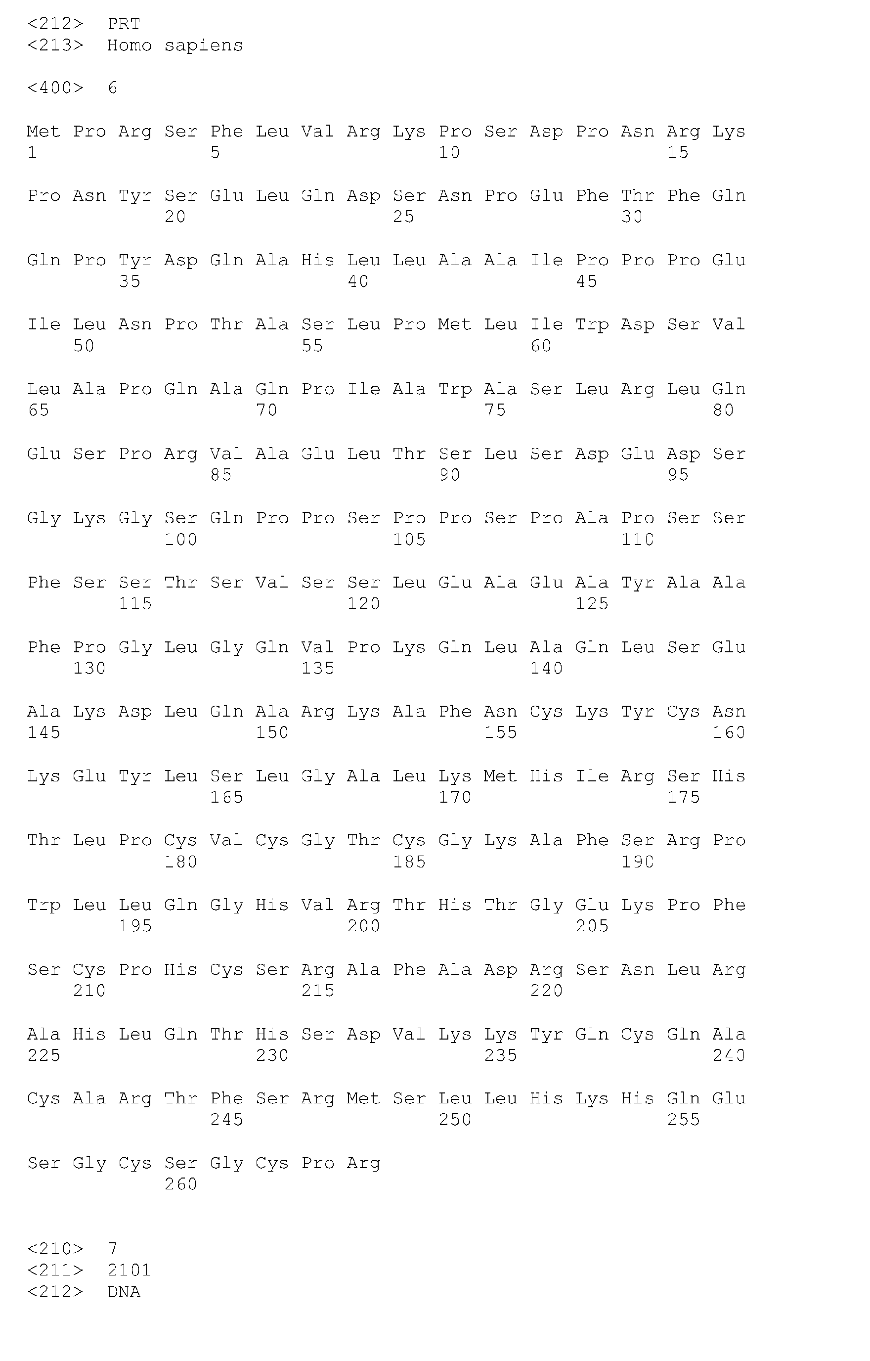
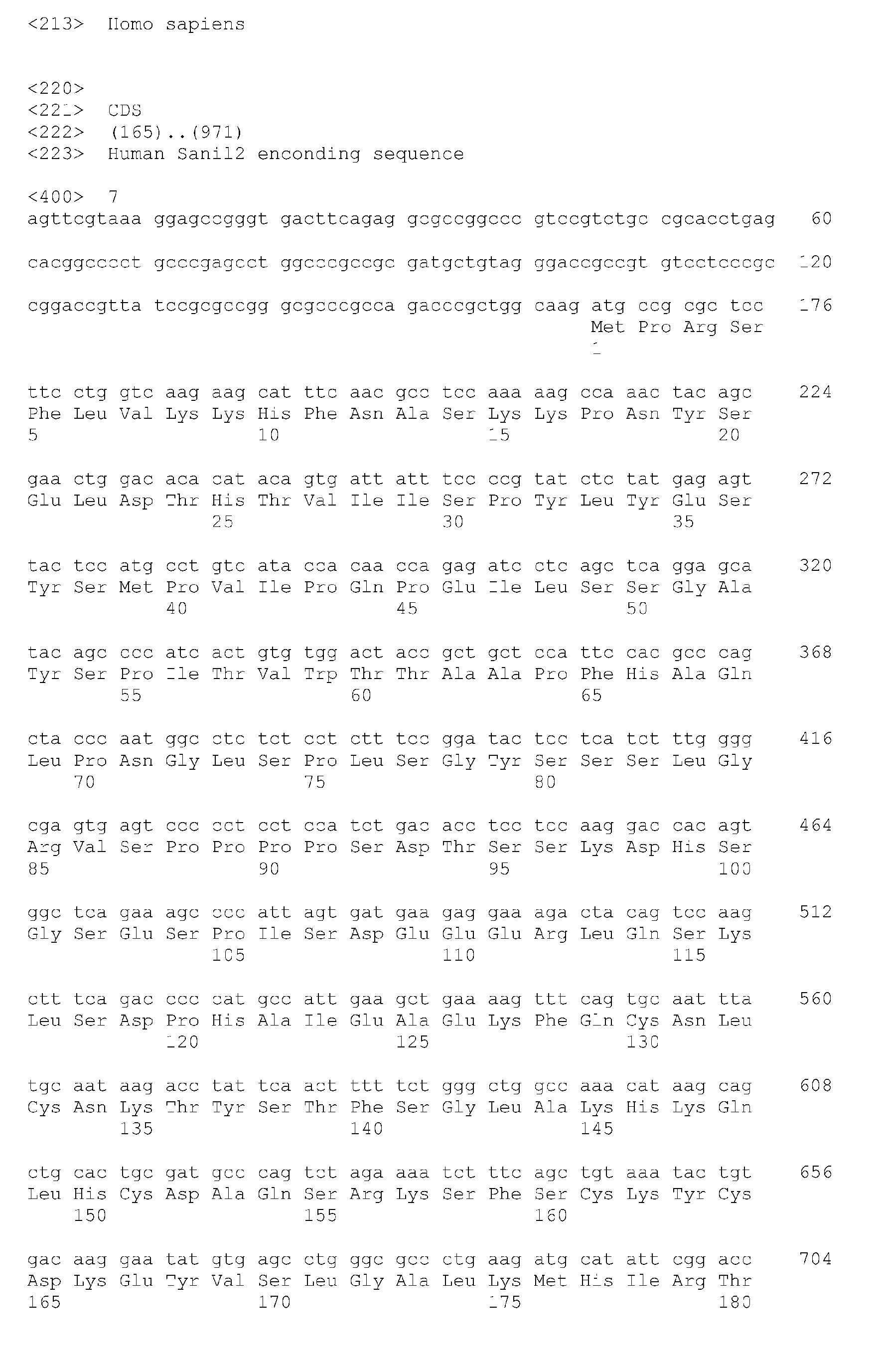


Claims (18)
- A method of identifying a renal fibrosis process characterised in that it is based on the identification of the presence of Snail in a biological sample and that it comprises the following steps:a) identification of the presence of Snail, in a biological sample of renal origin, andb) comparison of the presence of Snail observed in a) with the absence thereof in a control sample, and where its presence is indicative of the existence of renal fibrosis.
- A method according to claim 1, characterised in that the Snail of a) relates to the identification of a human Snail1 gene (SEQ ID NO 5) or Snail2 gene (SEQ ID NO 7) transcript.
- A method according to claim 1, characterised in that the Snail of a) relates to the identification of the human Snail1 protein (SEQ ID NO 6) or the human Snail2 protein (SEQ ID NO 8).
- A method according to claim 1, characterised in that the identification of the Snail protein of a) is performed using specific, monoclonal or polyclonal, Snail antibodies.
- A method according to claim 1, characterised in that the identification of the Snail protein of a) is performed by in situ hybridisation with a Snail precursor.
- A method according to claim 1, characterised in that the identification of the Snail protein of a) is performed by RT-PCR amplification of a Snail gene precursor.
- Method of identifying and evaluating the activity of Snail protein inhibitory compounds that are useful for the treatment of renal fibrosis, characterised in that it comprises the following steps:a) Bringing into contact a biological system wherein there is a Snail expression that produces renal fibrosis with the candidate compound that is the object of this method, and incubation under suitable conditions,b) determination of an indicative parameter of the renal fibrosis process, andc) identification of a compound that inhibits Snail protein activity when a reduction of said renal fibrosis parameter is observed.
- Identification method according to claim 7, characterised in that the biological system of point a) is a transgenic animal where the expression of the Snail protein is inducible, in a constant or conditional manner, and where the expression thereof causes renal fibrosis.
- Identification method according to claim 8, characterised in that the transgenic animal is the transgSnail1-ER mouse.
- Identification method according to claim 7, characterised in that the parameter related to the renal fibrosis process of b) belongs to the following group: a morphological change characteristic of an EMT, level of vimentin, Collagen I gene transcription and deposition of collagen fibres.
- Use of a compound or agent that inhibits Snail protein activity in the preparation of drugs or pharmaceutical compositions for the treatment of renal fibrosis, preferably human.
- Use of a compound according to claim 11, characterised in that the compound is a nucleic acid or polynucleotide which prevents or reduces the expression of the human Snail protein encoding gene, either Snail1 or Snail2, and includes a nucleotide sequence selected from:a) a Snail protein gene or mRNA sequence specific anti-sense nucleotide sequence,b) a Snail protein mRNA specific ribozyme,c) a Snail protein mRNA specific aptamer, andd) a Snail protein mRNA specific interference RNA (iRNA).
- Use of a compound according to claim 12, characterised in that the iRNA is preferably bound to the Snail mRNA fragment sequence (SEQ ID NO 9) or to another fragment that comprises the latter.
- Pharmaceutical composition or a drug for the treatment of renal fibrosis characterised in that it comprises a therapeutically effective quantity of a compound or agent that inhibits the Snail protein, either Snail1 or Snail2, jointly with, optionally, one or more pharmaceutically acceptable adjuvants and/or vehicles.
- Pharmaceutical composition according to claim 14, characterised in that the inhibitory compound is a nucleic acid or polynucleotide which prevents or reduces the expression of the human Snail protein encoding gene, either Snail1 or Snail2, and includes a nucleotide sequence selected from:a) a Snail protein gene or mRNA sequence specific anti-sense nucleotide sequence,b) a Snail protein mRNA specific ribozyme,c) a Snail protein mRNA specific aptamer, andd) a Snail protein mRNA specific interference RNA (iRNA).
- Pharmaceutical composition according to claim 15, characterised in that the iRNA is preferably bound to the Snail mRNA fragment sequence (SEQ ID NO 9) or to another fragment that comprises the latter.
- Use of the pharmaceutical composition according to claims 14 to 16, in a treatment method for a mammal, preferably a human being, suffering from renal fibrosis, which consists of administering said therapeutic composition that inhibits the fibrosis process.
- Use of the pharmaceutical composition according to claim 17, characterised in that the renal fibrosis is caused by a disease, disorder or pathology that belongs to the following group: glomerulonephritis, IgA nephropathy, diabetes, renal damage induced by toxicity, urinary obstruction and deterioration of kidney transplants.
Applications Claiming Priority (2)
| Application Number | Priority Date | Filing Date | Title |
|---|---|---|---|
| ES200600119A ES2310434B1 (en) | 2006-01-19 | 2006-01-19 | A PMTO OF IDENTIFICATION OF A RENAL FIBROSIS PROCESS, PMTO OF IDENTIFICATION OF INHIBITING COMPOUNDS, USE OF INHIBITING COMPOUNDS OF THE EXPRESSION OF THE GENE OF THE SNAIL TRANSCRIPTION FACTOR IN THE ELABORATION OF PHARMACEUTICAL COMPOSITIONS, SUCH COMPOSITIONS ..... |
| PCT/ES2006/070165 WO2007082967A1 (en) | 2006-01-19 | 2006-10-30 | A method for identifying a renal fibrosis process, use of snail-activity-inhibiting compounds in the production of pharmaceutical compositions, method for identifying said inhibiting compounds, said pharmaceutical compositions and applications thereof |
Publications (1)
| Publication Number | Publication Date |
|---|---|
| EP1980623A1 true EP1980623A1 (en) | 2008-10-15 |
Family
ID=38287291
Family Applications (1)
| Application Number | Title | Priority Date | Filing Date |
|---|---|---|---|
| EP06807963A Withdrawn EP1980623A1 (en) | 2006-01-19 | 2006-10-30 | A method for identifying a renal fibrosis process, use of snail-activity-inhibiting compounds in the production of pharmaceutical compositions, method for identifying said inhibiting compounds, said pharmaceutical compositions and applications thereof |
Country Status (6)
| Country | Link |
|---|---|
| US (1) | US20100286231A1 (en) |
| EP (1) | EP1980623A1 (en) |
| AU (1) | AU2006336009A1 (en) |
| CA (1) | CA2637609A1 (en) |
| ES (1) | ES2310434B1 (en) |
| WO (1) | WO2007082967A1 (en) |
Cited By (2)
| Publication number | Priority date | Publication date | Assignee | Title |
|---|---|---|---|---|
| CN101999658A (en) * | 2010-12-07 | 2011-04-06 | 龚泉福 | Nutrition particles with golden aster and snail and preparation method thereof |
| CN112424605A (en) * | 2018-05-11 | 2021-02-26 | 美国控股实验室公司 | Compositions and methods for detecting renal fibrosis |
Families Citing this family (2)
| Publication number | Priority date | Publication date | Assignee | Title |
|---|---|---|---|---|
| ES2310469B1 (en) * | 2007-03-08 | 2009-11-16 | Consejo Superior Investig. Cientificas | USE INHIBITING COMPOUNDS SNAIL1 ACTIVITY IN ELABORATION PHARMACEUTICAL COMPOSITIONS USEFUL FOR TREATMENT OF DRIVING DISPLAYS, PROCEDURE IDENTIFICATION INHIBITING COMPOUNDS, SUCH PHARMACEUTICAL COMPOSITIONS, PROCEDURE DIAGNOSIS CONDROPLIES AND APPLICATIONS. |
| KR101936799B1 (en) * | 2017-01-09 | 2019-01-11 | 주식회사 엠이티라이프사이언스 | Pharmaceutical Composition for Treating Oral Precancer and Method for Predicting or Detecting of Oral Precancer or Oral Cancer |
Family Cites Families (5)
| Publication number | Priority date | Publication date | Assignee | Title |
|---|---|---|---|---|
| JP4785252B2 (en) | 1999-02-26 | 2011-10-05 | ザ・ユニバーシティ・オブ・ブリティッシュ・コロンビア | TRPM-2 antisense therapy |
| US20020173453A1 (en) | 2000-12-15 | 2002-11-21 | Rama Akella | Method of treating renal injury |
| WO2004065602A1 (en) * | 2003-01-17 | 2004-08-05 | Universidad Autónoma de Madrid | Method of identifying the epithelial-mesenchymal transition of mesothelial cells (emtmc) and identifying emtmc-modulating compounds, pharmaceutical compositions and use thereof in the diagnosis and treatment of diseases associated with emtmc |
| US20090016963A1 (en) * | 2003-10-22 | 2009-01-15 | Dalia Cohen | Genes involved in neurodegenerative conditions |
| US20060003956A1 (en) * | 2004-03-03 | 2006-01-05 | Casadome David O | Materials and methods for the derepression of the E-cadherin promoter |
-
2006
- 2006-01-19 ES ES200600119A patent/ES2310434B1/en not_active Withdrawn - After Issue
- 2006-10-30 WO PCT/ES2006/070165 patent/WO2007082967A1/en not_active Ceased
- 2006-10-30 CA CA002637609A patent/CA2637609A1/en not_active Abandoned
- 2006-10-30 EP EP06807963A patent/EP1980623A1/en not_active Withdrawn
- 2006-10-30 US US12/161,450 patent/US20100286231A1/en not_active Abandoned
- 2006-10-30 AU AU2006336009A patent/AU2006336009A1/en not_active Abandoned
Non-Patent Citations (1)
| Title |
|---|
| See references of WO2007082967A1 * |
Cited By (2)
| Publication number | Priority date | Publication date | Assignee | Title |
|---|---|---|---|---|
| CN101999658A (en) * | 2010-12-07 | 2011-04-06 | 龚泉福 | Nutrition particles with golden aster and snail and preparation method thereof |
| CN112424605A (en) * | 2018-05-11 | 2021-02-26 | 美国控股实验室公司 | Compositions and methods for detecting renal fibrosis |
Also Published As
| Publication number | Publication date |
|---|---|
| ES2310434A1 (en) | 2009-01-01 |
| WO2007082967A1 (en) | 2007-07-26 |
| US20100286231A1 (en) | 2010-11-11 |
| AU2006336009A1 (en) | 2007-07-26 |
| ES2310434B1 (en) | 2009-11-12 |
| CA2637609A1 (en) | 2007-07-26 |
Similar Documents
| Publication | Publication Date | Title |
|---|---|---|
| Boutet et al. | Snail activation disrupts tissue homeostasis and induces fibrosis in the adult kidney | |
| KR101485071B1 (en) | Method for regulating muscle cell proliferation and differentiation, treating muscle cell, controling gene expression which are using MICRORNAS, and derepession vector of MICRORNAS molecule, Kit and Myocyt using them | |
| Thum et al. | MicroRNA-21 contributes to myocardial disease by stimulating MAP kinase signalling in fibroblasts | |
| Blichenberg et al. | Identification of a cis‐acting dendritic targeting element in the mRNA encoding the alpha subunit of Ca2+/calmodulin‐dependent protein kinase II | |
| Hatini et al. | Essential role of stromal mesenchyme in kidney morphogenesis revealed by targeted disruption of Winged Helix transcription factor BF-2. | |
| Chen et al. | miR-146a regulates glucose induced upregulation of inflammatory cytokines extracellular matrix proteins in the retina and kidney in diabetes | |
| Liu et al. | Overexpression of miR-142-3p improves mitochondrial function in cardiac hypertrophy | |
| CN106701900B (en) | Long-chain non-coding RNA HERC2P3 gene and application thereof in gastric cancer | |
| Ma et al. | Neuronal miR-29a protects from obesity in adult mice | |
| US20220095596A1 (en) | Use of ecm1 gene-knockout mouse in screening of anti-hepatic fibrosis drug | |
| Jin et al. | Cooperation between ectopic FGFR1 and depression of FGFR2 in induction of prostatic intraepithelial neoplasia in the mouse prostate | |
| Willet et al. | SOX9 governs gastric mucous neck cell identity and is required for injury-induced metaplasia | |
| EP2454372B1 (en) | Managing the treatment of inflammatory or autoimmune disorders using hom-1 expression | |
| US9309514B2 (en) | G protein-coupled purinergic receptor Gpr17 mediates orexigenic effects of FoxO1 in AgRP neurons | |
| EP1980623A1 (en) | A method for identifying a renal fibrosis process, use of snail-activity-inhibiting compounds in the production of pharmaceutical compositions, method for identifying said inhibiting compounds, said pharmaceutical compositions and applications thereof | |
| WO2011011061A2 (en) | Method of regulating angiogenesis and lymphangiogenesis, and a pharmaceutical composition for effecting anti-angiogenic and anti-lymphangiogenic cancer therapy | |
| JP2021534799A (en) | How to Target Kits Using Splice Switching Oligonucleotides to Induce Mast Cell Apoptosis | |
| CA2676946A1 (en) | Adam12 inhibitors and their use against inflammation-induced fibrosis | |
| Wu et al. | The ischemia‐enhanced myocardial infarction protection‐related lncRNA protects against acute myocardial infarction | |
| KR20210066758A (en) | Nucleic acids inducing rna interference including 8-oxoguanine, micro rna including 8-oxoguanine, and use thereof | |
| JP5567555B2 (en) | Head and neck tumor growth inhibitor | |
| O′ Shea et al. | Transplacental RNAi: Deciphering Gene Function in the Postimplantation‐Staged Embryo | |
| JP6230546B2 (en) | Method for screening compound having preventive and therapeutic action for obesity | |
| JP5737702B2 (en) | Arf6 gene function loss animal and method of using the same | |
| US20100218266A1 (en) | Methods of identifying and using snail1 inhibitory compounds in chondrodysplasia treatment and preparation of pharmaceutical compositions |
Legal Events
| Date | Code | Title | Description |
|---|---|---|---|
| PUAI | Public reference made under article 153(3) epc to a published international application that has entered the european phase |
Free format text: ORIGINAL CODE: 0009012 |
|
| 17P | Request for examination filed |
Effective date: 20080731 |
|
| AK | Designated contracting states |
Kind code of ref document: A1 Designated state(s): AT BE BG CH CY CZ DE DK EE ES FI FR GB GR HU IE IS IT LI LT LU LV MC NL PL PT RO SE SI SK TR |
|
| 17Q | First examination report despatched |
Effective date: 20090629 |
|
| STAA | Information on the status of an ep patent application or granted ep patent |
Free format text: STATUS: THE APPLICATION IS DEEMED TO BE WITHDRAWN |
|
| 18D | Application deemed to be withdrawn |
Effective date: 20091110 |