EP1947453A1 - Method for analyzing protein - Google Patents
Method for analyzing protein Download PDFInfo
- Publication number
- EP1947453A1 EP1947453A1 EP06822174A EP06822174A EP1947453A1 EP 1947453 A1 EP1947453 A1 EP 1947453A1 EP 06822174 A EP06822174 A EP 06822174A EP 06822174 A EP06822174 A EP 06822174A EP 1947453 A1 EP1947453 A1 EP 1947453A1
- Authority
- EP
- European Patent Office
- Prior art keywords
- group
- electrophoresis
- protein
- sample
- substituted
- Prior art date
- Legal status (The legal status is an assumption and is not a legal conclusion. Google has not performed a legal analysis and makes no representation as to the accuracy of the status listed.)
- Granted
Links
Images


Classifications
-
- G—PHYSICS
- G01—MEASURING; TESTING
- G01N—INVESTIGATING OR ANALYSING MATERIALS BY DETERMINING THEIR CHEMICAL OR PHYSICAL PROPERTIES
- G01N27/00—Investigating or analysing materials by the use of electric, electrochemical, or magnetic means
- G01N27/26—Investigating or analysing materials by the use of electric, electrochemical, or magnetic means by investigating electrochemical variables; by using electrolysis or electrophoresis
- G01N27/416—Systems
- G01N27/447—Systems using electrophoresis
- G01N27/44704—Details; Accessories
- G01N27/44717—Arrangements for investigating the separated zones, e.g. localising zones
- G01N27/44721—Arrangements for investigating the separated zones, e.g. localising zones by optical means
- G01N27/44726—Arrangements for investigating the separated zones, e.g. localising zones by optical means using specific dyes, markers or binding molecules
Definitions
- the present invention relates to a method for efficiently detecting, qualitatively analyzing, or quantitatively analyzing proteins by electrophoresis.
- Proteome research is underway to examine the states of diseases and to explore the causes of the diseases through extensive examination of in vivo proteins.
- research for determination of proteins that can serve as disease markers, such as cancer marker proteins, is underway.
- transcriptome analysis has been actively conducted for extensive analysis of RNA expression, as a result of improved DNA chip technology.
- RNA expression profiles do not always agree with the protein expression profiles. The correlation between the two is said to be 50% or less.
- extensive analysis of both protein(s) and RNA is also important. Attempts to elucidate protein functions have been made using a two-dimensional electrophoresis method that has currently been significantly developed, mass spectrometry, and high throughput analysis equivalent to genome analysis such as protein chip analysis.
- Electrophoresis has been broadly employed as a method for analyzing proteins. In proteome research, such electrophoresis is used for comparing protein components contained in normal tissues with those contained in disease tissues (differential display) or for preparing proteins to be used for protein identification, for example.
- Electrophoresis requires staining (labeling) proteins for detection. Staining methods are largely classified into two methods.
- One method is a pre-staining method that involves labeling a protein before electrophoresis.
- the other method is a post-staining method that involves labeling a protein after electrophoresis.
- An example of the former method is an Ettan DIGE method.
- Examples of the latter method include a Bio-Safe CBB staining method and a Sypro Ruby staining method. The characteristics of these methods are each listed in the following Table 1.
- the present inventors have reported a reagent capable of forming a complex with a protein to vary the resulting fluorescence or luminescence, so as to enable rapid and convenient analysis of the protein ( JP Patent Application No. 2005-167613 ).
- An object of the present invention is to provide a method for analyzing proteins using electrophoresis, by which problems associated with electrophoresis employed for protein analysis including:
- the present inventors have conducted concentrated studies in order to attain the above objects. As a result, the present inventors have discovered that proteins can be rapidly and conveniently analyzed with high sensitivity by setting a sample in gel for electrophoresis, performing electrophoresis of the sample using a buffer for electrophoresis in which a specific labeling compound is dissolved, and then detecting a complex of the labeling compound and the protein via spectrophotometrical measurement. Thus, the present inventors have completed the present invention.
- the present invention includes the following inventions.
- the present invention is a method for analyzing a protein, comprising setting a sample in gel for electrophoresis, performing electrophoresis for the sample using a buffer for electrophoresis in which a labeling compound represented by formula I: wherein R 1 is an aryl group or a heteroaryl group that may be substituted, R 2 is a heterocyclic group that may be substituted, and n is an integer between 1 and 5, is dissolved, and then detecting a complex of the compound of formula I and the protein via spectrophotometrical measurement.
- "protein” in the present invention is a concept including "peptide” comprising about dozens of amino acid residues.
- electrophoresis is performed using a labeling reagent for analyzing proteins as disclosed in the previous application ( JP Patent Application No. 2005-167613 ), so that staining (labeling) and separation of proteins are performed simultaneously.
- a labeling reagent is mixed in advance with a buffer for gel electrophoresis.
- the labeling reagent infiltrates the gel with electroosmotic flow and then forms a complex (CT complex) with proteins existing in a sample set in the gel. Formation of such complex causes a change in fluorescence, luminescence, or the like of the labeling reagent. Proteins are analyzed by measuring such change with spectroscopical measures (including checking by eye).
- Examples of carriers for electrophoresis used in the present invention are not particularly limited, as long as they are generally used as carriers for electrophoresis and include membranes (e.g., a cellulose acetate membrane, a nitrocellulose membrane, and a polyvinylidene difluoride (PVDF) membrane) and gel (e.g., polyacrylamide gel or agarose gel).
- membranes e.g., a cellulose acetate membrane, a nitrocellulose membrane, and a polyvinylidene difluoride (PVDF) membrane
- gel e.g., polyacrylamide gel or agarose gel.
- the compound represented by formula I is used for labeling (staining) a protein contained in an analytical sample.
- the compound forms a complex with the protein to show altered fluorescence, luminescence, coloration, absorption wavelength (e.g., absorption in the ultraviolet [UV] region, in the visible region, and in the infrared region) or to generate the same.
- absorption wavelength e.g., absorption in the ultraviolet [UV] region, in the visible region, and in the infrared region
- Compounds that enable detection of such changes using spectroscopical measures are preferable.
- R 1 is an aryl group or a heteroaryl group that may be substituted
- R 2 is a heterocyclic group that may be substituted
- n is an integer between 1 and 5.
- the aryl group examples include phenyl and a polycyclic aromatic group (e.g., naphthyl, anthryl, and pyrenyl) in which 2 to 4 benzene rings are fused.
- the heteroaryl group refers to an aryl group containing at least one heteroatom selected from the group consisting of N, O, and S as its ring-forming atom.
- Such aryl group or heteroaryl group may be an aryl group or heteroaryl group further fused with another aromatic ring or heteroaromatic ring.
- aryl group and heteroaryl group include a pyridyl group, a furyl group, a dansyl group, coumarin, benzothiazole, fluorescein, rhodamine, and azobenzene.
- the aryl group or the heteroaryl group may be substituted with one or more groups selected from the group consisting of a C1-10 (preferably C1-6) alkyl group, a C1-10 (preferably C1-6) alkoxy group, a phenyl group (and such phenyl group may be substituted with one or more groups selected from among an amino group, halogen, and a nitro group), an amino group, a cyano group, a nitro group, a carboxyl group (or a salt, ester, or amide thereof), sulfonic acid (or a salt, ester, or amide thereof), a thiol group, and a hydroxy group (or a salt thereof), a C1-10 (preferably C1-6) acyl group, halogen, and sugar.
- the aryl group or the heteroaryl group is preferably substituted with a hydroxy group for imparting of water solubility and electron-donating ability.
- a fluorescent group or a chromogenic group absorption and fluorescence in the ultraviolet [UV] region, those in the visible region, and those in the infrared region can be measured
- a chromogenic group absorption and fluorescence in the ultraviolet [UV] region, those in the visible region, and those in the infrared region can be measured
- Examples of such heterocyclic group include coumarin, benzothiazole, fluorescein, and rhodamine.
- R 3 , R 4 , R 5 , and R 6 are independently from each other selected from the group consisting of a hydrogen atom, a C1-10 alkyl group, a C1-10 alkoxy group, a phenyl group (and such phenyl group may be substituted with one or more groups selected from among an amino group, halogen, and a nitro group), an amino group, a cyano group, a nitro group, a carboxyl group (or a salt, ester, or amide thereof), sulfonic acid (or a salt, ester, or amide thereof), a thiol group, and a hydroxy group (or a salt thereof), and a C1-10 acyl group, halogen, and sugar;
- X is an -NR- group (wherein R is hydrogen or a C1-5 alkyl group), an oxygen atom, or a sulfur atom; and any one of R 3 , R 4 , R 5 , and R 6 are independently from each
- R 2 which serves as a fluorescent group or a chromogenic group
- a compound of formula I can be easily synthesized by a known method.
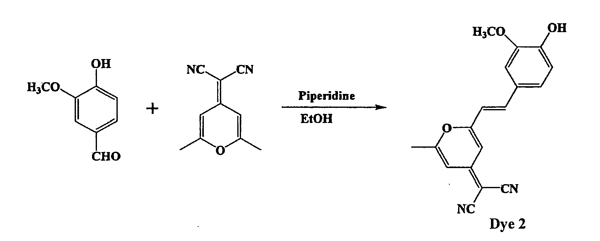
- a compound of the following formula: can be produced by reacting 4-hydroxybenzaldehyde with 4-(dicyanomethylene)-2,6-dimethyl-4H-pyran in ethanol in the presence of a nucleotide (e.g., amine such as piperidine).
- a nucleotide e.g., amine such as piperidine
- the method of the present invention is the same as a conventional method in that it employs electrophoresis, but is different therefrom in terms of the compound to be used for labeling and in that steps are simplified in the method of the present invention. Schemes are shown below for comparison of the method of the present invention with the conventional method.
- the conventional method requires a series of steps (electrophoresis-protein immobilization-staining-washing-detection) before detection of proteins is performed after completion of electrophoresis.
- the method of the present invention requires only short steps (staining-detection of protein) while performing electrophoresis because of the use of the compound of formula I as a labeling reagent, so that the time required for analysis can be drastically shortened.
- a buffer for electrophoresis may be a generally used buffer.
- a buffer in which a compound of formula I is dissolved is used.
- electrophoresis is performed in a manner similar to that of a general method, thereby fractionating the proteins in the sample.
- the compound of formula I contained in the buffer forms a charge-transfer complex with the proteins in the sample (that is, the proteins are labeled) through hydrophobic interaction and charge-transfer interaction while migrating with electroosmotic flow.
- absorption wavelength, absorption intensity, fluorescence wavelength, fluorescence intensity, and the like unique to the compound of formula I may vary. This variation may be checked by eye as a change in color tone.
- the carrier (gel) is removed.
- the carrier may be washed with a lavage solution, if desired.
- the thus removed carrier is observed by spectroscopical measures (e.g., a fluorometer or an absorptiometer; checking by eye is also included herein) capable of detecting fluorescence, luminescence, coloration, absorption wavelength (e.g., absorption in the ultraviolet [UV] region, the visible region, and the infrared region) and the like.
- spectroscopical measures e.g., a fluorometer or an absorptiometer; checking by eye is also included herein
- fluorescence, luminescence, coloration, absorption wavelength e.g., absorption in the ultraviolet [UV] region, the visible region, and the infrared region
- absorption wavelength e.g., absorption in the ultraviolet [UV] region, the visible region, and the infrared region
- protein spots can be easily detected in both cases.
- a protein in a sample can be detected or qualitatively analyzed.
- proteins in a sample can also be quantitatively analyzed by measuring the intensity of fluorescence, luminescence, coloration, or the like.
- the method of the present invention can also be applied not only to one-dimensional electrophoresis, but also to two-dimensional electrophoresis.
- Dye 3 prepared in Example 3 was used as a labeling reagent. Electrophoresis performed herein was one-dimensional electrophoresis using SDS-polyacrylamide gel as a carrier. Measurement conditions for the one-dimensional electrophoresis are as described below.
- a stock solution (DMSO solution at a concentration of 500 mg/mL) of dye 3 was diluted with an electrophoresis buffer attached to a Sure Blot F1 Gel system. The thus obtained 0.5 mg/mL, 0.1 mg/mL, and 0.05 mg/mL solutions were used.
- ProXpress imaging system produced by Perkin-Elmer
- LS400 scanner produced by Tecan
- An electrophoresis buffer (containing dye 3 at a concentration of 0.5 mg/mL) was added to an upper buffer (cathode side).
- An SDS-PAGE gel to which samples (BSA and IgG) had been applied was set, followed by electrophoresis. After completion of electrophoresis, the gel was removed and then observed using a fluorescence detector ( Fig. 1A ). Subsequently, the gel was washed for 30 minutes using a lavage solution and then protein spots were observed using the fluorescence detector ( Fig. 1B ).
- electrophoresis was performed similarly using an electrophoresis buffer containing dye 3 at a concentration of 0.1 mg/mL. After completion of electrophoresis, the gel was removed and then observed using a fluorescence detector ( Fig. 2A ). Subsequently, the gel was washed for 30 minutes using a lavage solution and then protein spots were observed using the fluorescence detector ( Fig. 2B ).
- Protein spots were clearly confirmed in all of these cases. It was understood that proteins had formed complexes with labeling reagents during electrophoresis such that they were altered to generate fluorescence. Moreover, as shown in Fig. 1B and Fig. 2B , the background was reduced via washing of the gel, allowing protein spots to be detected more clearly. Moreover, as shown in Fig. 2A and Fig. 2B , protein spots could also be clearly observed by adjusting the concentration of the labeling reagent in electrophoresis buffer to 0.1 mg/mL.
- Dye 3 prepared in Example 3 was used as a labeling reagent.
- An SDS-polyacrylamide gel carrier was used for two-dimensional electrophoresis. Measurement conditions employed herein are as described below.
- ProXpress imaging system produced by Perkin-Elmer
- LS400 scanner produced by Tecan
- the first dimension of two-dimensional electrophoresis was performed by subjecting each sample to isoelectric focusing. Subsequently, an electrophoresis buffer (containing dye 3 at a concentration of 0.5 mg/mL) was added to the upper buffer (cathode side). The sample was applied to an SDS-PAGE gel and then the SDS-PAGE gel was set, so that the second dimension of two-dimensional electrophoresis was performed. After completion of electrophoresis, the gel was removed and then observed using a fluorescence detector ( Fig. 3A ). Subsequently, the gel was washed for 30 minutes using a lavage solution and then protein spots were observed using the fluorescence detector ( Fig. 3B ). Protein spots were clearly confirmed in all of these cases.
- proteins had formed complexes with labeling reagents during electrophoresis such that they were altered to generate fluorescence. Moreover, as shown in Fig. 3B , the background was reduced via washing of the gel, allowing protein spots to be detected more clearly.
- the method for analyzing proteins of the present invention makes it possible to perform high throughput analysis of a great variety of proteins. Hence, the method of the present invention is useful for extensive and convenient protein analysis with high sensitivity in the fields of biochemistry, medicine, analytical chemistry, proteome research, and the like.
Landscapes
- Health & Medical Sciences (AREA)
- Life Sciences & Earth Sciences (AREA)
- Chemical & Material Sciences (AREA)
- Physics & Mathematics (AREA)
- Molecular Biology (AREA)
- Analytical Chemistry (AREA)
- Chemical Kinetics & Catalysis (AREA)
- Electrochemistry (AREA)
- Spectroscopy & Molecular Physics (AREA)
- Biochemistry (AREA)
- General Health & Medical Sciences (AREA)
- General Physics & Mathematics (AREA)
- Immunology (AREA)
- Pathology (AREA)
- Investigating Or Analysing Biological Materials (AREA)
- Investigating Or Analysing Materials By The Use Of Chemical Reactions (AREA)
- Investigating, Analyzing Materials By Fluorescence Or Luminescence (AREA)
Abstract

Description
- The present invention relates to a method for efficiently detecting, qualitatively analyzing, or quantitatively analyzing proteins by electrophoresis.
- Proteome research is underway to examine the states of diseases and to explore the causes of the diseases through extensive examination of in vivo proteins. For example, research for determination of proteins that can serve as disease markers, such as cancer marker proteins, is underway.
- Furthermore, in recent years, transcriptome analysis has been actively conducted for extensive analysis of RNA expression, as a result of improved DNA chip technology. However, RNA expression profiles do not always agree with the protein expression profiles. The correlation between the two is said to be 50% or less. To achieve the aforementioned purpose, extensive analysis of both protein(s) and RNA is also important. Attempts to elucidate protein functions have been made using a two-dimensional electrophoresis method that has currently been significantly developed, mass spectrometry, and high throughput analysis equivalent to genome analysis such as protein chip analysis.
- As described above, methods for analyzing proteins more efficiently are required with the advancement of protein research. Establishment of technology for conveniently and rapidly analyzing a great variety of proteins is important.
- Electrophoresis has been broadly employed as a method for analyzing proteins. In proteome research, such electrophoresis is used for comparing protein components contained in normal tissues with those contained in disease tissues (differential display) or for preparing proteins to be used for protein identification, for example.
- Electrophoresis requires staining (labeling) proteins for detection. Staining methods are largely classified into two methods.
- One method is a pre-staining method that involves labeling a protein before electrophoresis. The other method is a post-staining method that involves labeling a protein after electrophoresis. An example of the former method is an Ettan DIGE method. Examples of the latter method include a Bio-Safe CBB staining method and a Sypro Ruby staining method. The characteristics of these methods are each listed in the following Table 1.
Table 1 Staining method Binding mode with protein Pre-staining or post-staining Operational steps (time required: minute) Total operation time (minute) Detection method Defects Ettan DIGE Covalent binding Pre-staining Reaction (30)→ stop (10) 40 +α Fluorescence After electrophoresis, analysis using a mass spectrometer cannot be performed. Bio-Safe CBB Non-covalent binding (binding not mediated by SDS) Post-staining Immobilization (30)→ staining (60) → washing (30) 120 +α Visible Poor detection sensitivity Sypro Ruby Non-covalent binding (binding not mediated by SDS) Post-staining Immobilization (30) → staining (180) → washing (30) 240 +α Fluorescence Good detection sensitivity. However, staining and washing processes require long time (half a day to one day in some cases). - In the case of the Ettan DIGE method, proteins in a sample are labeled in advance with a fluorescent reagent via covalent binding before electrophoresis. This method requires a short total operation time and has high general versatility, but it is problematic in that a plurality of fluorescent reagents bind to one protein molecule and the number of such fluorescent reagent bound to each protein differs depending on the protein in question. Hence, proteins separated by electrophoresis cannot be subjected to mass spectrometry in proteomic analysis.
- In the cases of the Bio-Safe CBB method and the Sypro Ruby method, the post-staining methods, a series of complicated steps such as:
- a. immobilization of proteins on gel and removal of SDS;
- b. staining of proteins with a fluorescent reagent;
- c. washing off of excess fluorescent reagent; and
- d. detection
- The present inventors have reported a reagent capable of forming a complex with a protein to vary the resulting fluorescence or luminescence, so as to enable rapid and convenient analysis of the protein (
JP Patent Application No. 2005-167613 - An object of the present invention is to provide a method for analyzing proteins using electrophoresis, by which problems associated with electrophoresis employed for protein analysis including:
- many operational steps and lengthy required time;
- poor detection sensitivity;
- proteins that cannot be subjected to mass spectrometry after separation; and the like are addressed and rapid and convenient analysis of a great variety of proteins with high sensitivity is made possible.
- The present inventors have conducted concentrated studies in order to attain the above objects. As a result, the present inventors have discovered that proteins can be rapidly and conveniently analyzed with high sensitivity by setting a sample in gel for electrophoresis, performing electrophoresis of the sample using a buffer for electrophoresis in which a specific labeling compound is dissolved, and then detecting a complex of the labeling compound and the protein via spectrophotometrical measurement. Thus, the present inventors have completed the present invention.
- The present invention includes the following inventions.
- (1) A method for analyzing a protein in a sample, comprising setting a sample in a carrier for electrophoresis, performing electrophoresis for the sample using a buffer for electrophoresis in which a labeling compound represented by formula I:

- (2) The method according to (1) above, wherein R2 is a group represented by formula II:

- (3) The method according to (1) or (2) above, wherein R1 is a phenyl group, a naphthyl group, a pyridyl group, or a furyl group that may be substituted.
- Conventional protein analysis methods using electrophoresis have defects such as poor sensitivity, poor accuracy, complicated operation, and lengthy required time. However, the method of the present invention possesses the following characteristics such that:
- a labeling reagent and a protein form a complex via non-covalent binding, so as to enable further mass spectrometric analysis of the protein separated by electrophoresis;
- steps and operation can be simplified and operation time can be drastically shortened (particularly, time for staining and time for washing); and
- detection sensitivity is good.
- This description includes part or all of the contents as disclosed in the description and/or drawings of Japanese Patent Application No.
2005-304478 -
-
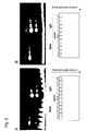
Fig. 1 shows fluorescence observed after electrophoresis performed in Example 4 (labeling reagent at a concentration of 0.5 mg/mL; A: before washing, B: after washing; excitation wavelength of 480 nm; detection wavelength of 530 nm). -
Fig. 2 shows fluorescence observed after electrophoresis performed in Example 4 (labeling reagent at a concentration of 0.1 mg/mL; A: before washing, B: after washing; excitation wavelength of 480 nm; detection wavelength of 530 nm). -
Fig. 3 shows fluorescence observed after two-dimensional electrophoresis performed in Example 5 (labeling reagent at a concentration of 0.1 mg/mL; A: before washing, B: after washing; excitation wavelength of 480 nm; detection wavelength of 530 nm). - The present invention is a method for analyzing a protein, comprising setting a sample in gel for electrophoresis, performing electrophoresis for the sample using a buffer for electrophoresis in which a labeling compound represented by formula I:

- According to the method of the present invention, electrophoresis is performed using a labeling reagent for analyzing proteins as disclosed in the previous application (
JP Patent Application No. 2005-167613 - Examples of carriers for electrophoresis used in the present invention are not particularly limited, as long as they are generally used as carriers for electrophoresis and include membranes (e.g., a cellulose acetate membrane, a nitrocellulose membrane, and a polyvinylidene difluoride (PVDF) membrane) and gel (e.g., polyacrylamide gel or agarose gel). In the present invention, it is preferable to use gel as a carrier and it is particularly preferable to use SDS-polyacrylamide gel.
- The compound represented by formula I is used for labeling (staining) a protein contained in an analytical sample. The compound forms a complex with the protein to show altered fluorescence, luminescence, coloration, absorption wavelength (e.g., absorption in the ultraviolet [UV] region, in the visible region, and in the infrared region) or to generate the same. Compounds that enable detection of such changes using spectroscopical measures (including checking by eye) are preferable.
- In formula I, R1 is an aryl group or a heteroaryl group that may be substituted, R2 is a heterocyclic group that may be substituted, and n is an integer between 1 and 5.
- Examples of the aryl group include phenyl and a polycyclic aromatic group (e.g., naphthyl, anthryl, and pyrenyl) in which 2 to 4 benzene rings are fused. Furthermore, the heteroaryl group refers to an aryl group containing at least one heteroatom selected from the group consisting of N, O, and S as its ring-forming atom. Such aryl group or heteroaryl group may be an aryl group or heteroaryl group further fused with another aromatic ring or heteroaromatic ring. Specific examples of such aryl group and heteroaryl group include a pyridyl group, a furyl group, a dansyl group, coumarin, benzothiazole, fluorescein, rhodamine, and azobenzene.
- The aryl group or the heteroaryl group may be substituted with one or more groups selected from the group consisting of a C1-10 (preferably C1-6) alkyl group, a C1-10 (preferably C1-6) alkoxy group, a phenyl group (and such phenyl group may be substituted with one or more groups selected from among an amino group, halogen, and a nitro group), an amino group, a cyano group, a nitro group, a carboxyl group (or a salt, ester, or amide thereof), sulfonic acid (or a salt, ester, or amide thereof), a thiol group, and a hydroxy group (or a salt thereof), a C1-10 (preferably C1-6) acyl group, halogen, and sugar. The aryl group or the heteroaryl group is preferably substituted with a hydroxy group for imparting of water solubility and electron-donating ability.
- The heterocyclic group is preferably a fluorescent group or a chromogenic group (absorption and fluorescence in the ultraviolet [UV] region, those in the visible region, and those in the infrared region can be measured) that has at least one heteroatom as an ring-forming atom and forms a conjugate system the an olefin portion -(CH=CH)n-represented by formula I. Examples of such heterocyclic group include coumarin, benzothiazole, fluorescein, and rhodamine. In the present invention, a group represented by formula II:

is preferable.
Furthermore, a group represented by the following formula:
- Connection of an alkenyl group and an aromatic group to R2, which serves as a fluorescent group or a chromogenic group, results in an elongated conjugate system and promotes lengthening of fluorescence and coloration wavelengths.
- A compound of formula I can be easily synthesized by a known method. For example, a compound of the following formula:

- Next, the procedures of the method of the present invention will be described below. The method of the present invention is the same as a conventional method in that it employs electrophoresis, but is different therefrom in terms of the compound to be used for labeling and in that steps are simplified in the method of the present invention. Schemes are shown below for comparison of the method of the present invention with the conventional method.


- Specifically, the conventional method requires a series of steps (electrophoresis-protein immobilization-staining-washing-detection) before detection of proteins is performed after completion of electrophoresis. However, the method of the present invention requires only short steps (staining-detection of protein) while performing electrophoresis because of the use of the compound of formula I as a labeling reagent, so that the time required for analysis can be drastically shortened.
- According to the method of the present invention, first, a sample containing proteins is applied to an electrophoretic carrier. A buffer for electrophoresis may be a generally used buffer. In the present invention, such a buffer in which a compound of formula I is dissolved is used. Subsequently, electrophoresis is performed in a manner similar to that of a general method, thereby fractionating the proteins in the sample.
- During electrophoresis, the compound of formula I contained in the buffer forms a charge-transfer complex with the proteins in the sample (that is, the proteins are labeled) through hydrophobic interaction and charge-transfer interaction while migrating with electroosmotic flow. Through the formation of such complex, absorption wavelength, absorption intensity, fluorescence wavelength, fluorescence intensity, and the like unique to the compound of formula I may vary. This variation may be checked by eye as a change in color tone.
- After completion of electrophoresis, the carrier (gel) is removed. The carrier may be washed with a lavage solution, if desired. The thus removed carrier is observed by spectroscopical measures (e.g., a fluorometer or an absorptiometer; checking by eye is also included herein) capable of detecting fluorescence, luminescence, coloration, absorption wavelength (e.g., absorption in the ultraviolet [UV] region, the visible region, and the infrared region) and the like. When a reagent by which fluorescence is generated is used, observation is performed using a fluorescence detector. When a reagent by which absorption wavelength is generated in the visible region is used, checking by eye is performed. Thus, protein spots can be easily detected in both cases. As described above, a protein in a sample can be detected or qualitatively analyzed. Moreover, proteins in a sample can also be quantitatively analyzed by measuring the intensity of fluorescence, luminescence, coloration, or the like.
- The method of the present invention can also be applied not only to one-dimensional electrophoresis, but also to two-dimensional electrophoresis.
- Hereafter, the present invention is described in greater detail with reference to the following examples, although the present invention is not limited to these examples.
-

-
-

- Dye 3 prepared in Example 3 was used as a labeling reagent. Electrophoresis performed herein was one-dimensional electrophoresis using SDS-polyacrylamide gel as a carrier. Measurement conditions for the one-dimensional electrophoresis are as described below.
- A stock solution (DMSO solution at a concentration of 500 mg/mL) of dye 3 was diluted with an electrophoresis buffer attached to a Sure Blot F1 Gel system. The thus obtained 0.5 mg/mL, 0.1 mg/mL, and 0.05 mg/mL solutions were used.
- [BSA] = 0.08 to 20 µg/well and [Sheep IgG] = 0.21 to 50 µg/well
- AcOH : MeOH : H2O = 3 : 10 : 87 v/v
- An electrophoresis buffer (containing dye 3 at a concentration of 0.5 mg/mL) was added to an upper buffer (cathode side). An SDS-PAGE gel to which samples (BSA and IgG) had been applied was set, followed by electrophoresis. After completion of electrophoresis, the gel was removed and then observed using a fluorescence detector (
Fig. 1A ). Subsequently, the gel was washed for 30 minutes using a lavage solution and then protein spots were observed using the fluorescence detector (Fig. 1B ). - Furthermore, electrophoresis was performed similarly using an electrophoresis buffer containing dye 3 at a concentration of 0.1 mg/mL. After completion of electrophoresis, the gel was removed and then observed using a fluorescence detector (
Fig. 2A ). Subsequently, the gel was washed for 30 minutes using a lavage solution and then protein spots were observed using the fluorescence detector (Fig. 2B ). - Protein spots were clearly confirmed in all of these cases. It was understood that proteins had formed complexes with labeling reagents during electrophoresis such that they were altered to generate fluorescence. Moreover, as shown in
Fig. 1B andFig. 2B , the background was reduced via washing of the gel, allowing protein spots to be detected more clearly. Moreover, as shown inFig. 2A and Fig. 2B , protein spots could also be clearly observed by adjusting the concentration of the labeling reagent in electrophoresis buffer to 0.1 mg/mL. - Dye 3 prepared in Example 3 was used as a labeling reagent. An SDS-polyacrylamide gel carrier was used for two-dimensional electrophoresis. Measurement conditions employed herein are as described below.
- One-dimensional: Isoelectric focusing; ZOOM IPG runner system (produced by Invitrogen)
Two-dimensional: SDS-PAGE Sure Blot F1 Gel system (produced by Astellas Pharma Inc.) - Stock solutions (DMSO solution at a concentration of 500 mg/mL) of dye 3 were diluted with electrophoresis buffers attached to a Sure Blot F1 Gel system. The thus obtained 0.5 mg/mL, 0.1 mg/mL, and 0.05 mg/mL solutions were used.
- [Mouse Brain Lysate] = 40 µg/strip
- AcOH : MeOH : H2O = 3 : 10 : 87 v/v
- ProXpress imaging system (produced by Perkin-Elmer) and LS400 scanner (produced by Tecan)
- The first dimension of two-dimensional electrophoresis was performed by subjecting each sample to isoelectric focusing. Subsequently, an electrophoresis buffer (containing dye 3 at a concentration of 0.5 mg/mL) was added to the upper buffer (cathode side). The sample was applied to an SDS-PAGE gel and then the SDS-PAGE gel was set, so that the second dimension of two-dimensional electrophoresis was performed. After completion of electrophoresis, the gel was removed and then observed using a fluorescence detector (
Fig. 3A ). Subsequently, the gel was washed for 30 minutes using a lavage solution and then protein spots were observed using the fluorescence detector (Fig. 3B ). Protein spots were clearly confirmed in all of these cases. It was understood that proteins had formed complexes with labeling reagents during electrophoresis such that they were altered to generate fluorescence. Moreover, as shown inFig. 3B , the background was reduced via washing of the gel, allowing protein spots to be detected more clearly. - The method for analyzing proteins of the present invention makes it possible to perform high throughput analysis of a great variety of proteins. Hence, the method of the present invention is useful for extensive and convenient protein analysis with high sensitivity in the fields of biochemistry, medicine, analytical chemistry, proteome research, and the like.
- All publications, patents, and patent applications cited herein are incorporated herein by reference in their entirety.
Claims (3)
- A method for analyzing a protein in a sample, comprising setting a sample in a carrier for electrophoresis, performing electrophoresis for the sample using a buffer for electrophoresis in which a labeling compound represented by formula I:

is dissolved, and then detecting a complex of the compound of formula I and the protein via spectrophotometrical measurement. - The method according to claim 1, wherein R2 is a group represented by formula II:

- The method according to claim 1 or 2, wherein R1 is a phenyl group, a naphthyl group, a pyridyl group, or a furyl group that may be substituted.
Applications Claiming Priority (2)
| Application Number | Priority Date | Filing Date | Title |
|---|---|---|---|
| JP2005304478A JP4822409B2 (en) | 2005-10-19 | 2005-10-19 | Protein analysis method |
| PCT/JP2006/321197 WO2007046537A1 (en) | 2005-10-19 | 2006-10-18 | Method for analyzing protein |
Publications (3)
| Publication Number | Publication Date |
|---|---|
| EP1947453A1 true EP1947453A1 (en) | 2008-07-23 |
| EP1947453A4 EP1947453A4 (en) | 2012-02-29 |
| EP1947453B1 EP1947453B1 (en) | 2013-10-09 |
Family
ID=37962621
Family Applications (1)
| Application Number | Title | Priority Date | Filing Date |
|---|---|---|---|
| EP06822174.6A Not-in-force EP1947453B1 (en) | 2005-10-19 | 2006-10-18 | Method for analyzing protein |
Country Status (4)
| Country | Link |
|---|---|
| US (1) | US8062492B2 (en) |
| EP (1) | EP1947453B1 (en) |
| JP (1) | JP4822409B2 (en) |
| WO (1) | WO2007046537A1 (en) |
Families Citing this family (6)
| Publication number | Priority date | Publication date | Assignee | Title |
|---|---|---|---|---|
| JP5182809B2 (en) | 2008-07-07 | 2013-04-17 | 独立行政法人産業技術総合研究所 | Protein analysis method and reagent |
| CN104508473B (en) * | 2012-05-29 | 2017-10-13 | 健康诊断实验室有限公司 | Composition and method for the gel electrophoresis with calibrated in situ |
| WO2014178107A1 (en) * | 2013-04-30 | 2014-11-06 | システム・インスツルメンツ株式会社 | Electrophoresis method and electrophoresis device |
| CN104178132B (en) * | 2014-07-30 | 2017-01-25 | 四川大学 | Fluorescent compound and application thereof in medicine |
| CN104803993B (en) * | 2015-03-01 | 2017-11-03 | 四川大学 | A kind of fluorescent chemicals with A β affinity and application thereof |
| CN111253398B (en) * | 2020-03-08 | 2021-06-22 | 温州医科大学附属第二医院、温州医科大学附属育英儿童医院 | A kind of fluorescent compound for detecting nerve damage and its application |
Family Cites Families (9)
| Publication number | Priority date | Publication date | Assignee | Title |
|---|---|---|---|---|
| JPS62502548A (en) | 1985-04-25 | 1987-10-01 | ポラロイド コ−ポレ−シヨン | Novel fluorescent compounds and biological testing devices |
| US4886744A (en) * | 1985-04-25 | 1989-12-12 | Polaroid Corporation | Fluorescent conjugates and biological diagnostic assay system |
| US4886774A (en) * | 1988-08-09 | 1989-12-12 | Alfred Doi | Ultraviolet protective overcoat for application to heat sensitive record materials |
| US5616502A (en) * | 1995-05-19 | 1997-04-01 | Molecular Probes, Inc. | Non-specific protein staining using merocyanine dyes |
| JP3674820B2 (en) * | 1998-03-25 | 2005-07-27 | 富士写真フイルム株式会社 | Novel methine compound, organic light emitting device material and organic light emitting device using the same |
| WO2005033687A1 (en) * | 2003-01-17 | 2005-04-14 | University Of Virginia Patent Foundation | Method for microchip and capillary detection of proteins in the sub-ug/ml range |
| JP2004317297A (en) * | 2003-04-16 | 2004-11-11 | Advance Co Ltd | Dye agent and dyeing method using the same |
| JP4256250B2 (en) | 2003-12-02 | 2009-04-22 | パイオニア株式会社 | DATA RECORDING SYSTEM, DATA RECORDING DEVICE, DATA TRANSMITTING DEVICE, DATA RECORDING METHOD, RECORDING PROGRAM, AND RECORDING MEDIUM RECORDING THE SAME |
| DE112005003114T5 (en) * | 2004-12-06 | 2008-05-08 | Katayanagi Institute, Hachioji | Method of separating protein, method of staining protein and liquid protein stain, and protein staining kit for use in these methods |
-
2005
- 2005-10-19 JP JP2005304478A patent/JP4822409B2/en not_active Expired - Fee Related
-
2006
- 2006-10-18 WO PCT/JP2006/321197 patent/WO2007046537A1/en not_active Ceased
- 2006-10-18 US US12/090,789 patent/US8062492B2/en not_active Expired - Fee Related
- 2006-10-18 EP EP06822174.6A patent/EP1947453B1/en not_active Not-in-force
Also Published As
| Publication number | Publication date |
|---|---|
| US8062492B2 (en) | 2011-11-22 |
| EP1947453B1 (en) | 2013-10-09 |
| JP2007114009A (en) | 2007-05-10 |
| US20090045060A1 (en) | 2009-02-19 |
| WO2007046537A1 (en) | 2007-04-26 |
| EP1947453A4 (en) | 2012-02-29 |
| JP4822409B2 (en) | 2011-11-24 |
Similar Documents
| Publication | Publication Date | Title |
|---|---|---|
| US8153827B2 (en) | Reagents for biomolecular labeling, detection and quantification employing Raman spectroscopy | |
| JP3130872B2 (en) | Dye for highly sensitive detection of double-stranded DNA | |
| US7863048B2 (en) | Coumarin-based cyanine dyes for non-specific protein binding | |
| US5783687A (en) | Dyes designed for high sensitivity detection of double-stranded DNA | |
| CN110143966B (en) | A kind of spiropyran-naphthalimide derivative and its synthesis method and application | |
| EP1200833B1 (en) | Difference detection methods using matched multiple dyes | |
| EP1947453B1 (en) | Method for analyzing protein | |
| Dadajová et al. | Fluorescein-based low-molecular-mass markers of isoelectric point for tracing pH gradient using high sensitivity capillary isoelectric focusing with laser-induced fluorescence detection | |
| Meier et al. | SDS-PAGE of proteins using a chameleon-type of fluorescent prestain | |
| JP4893964B2 (en) | Novel compound, reagent for analysis of peptide or protein containing the compound, and analysis method using the analysis reagent | |
| CN113646636B (en) | Advanced methods and systems based on novel fluorescent dyes for automated, high-performance identification of carbohydrate and carbohydrate mixture composition patterns. | |
| US8263791B2 (en) | Method and reagent for protein analysis | |
| Dusa et al. | Labeling of oligosaccharides and N-linked glycans by a rhodamine-based fluorescent tag for analysis by capillary electrophoresis with laser-induced fluorescence and mass spectrometry detection | |
| Sun et al. | Highly Selective Fluorescent 4-(4-(Diethylamino)-2-Hydroxystyryl)-1-Methylpyridine iodide and nitrobenzofurazan based probe for cysteine with application in living cells | |
| US20110186434A1 (en) | Qualitative and/or quantitative determination of a proteinaceous molecule in a plurality of samples | |
| JPWO2010064638A1 (en) | Novel ClearNative Electrophoresis Using Aromatic Sulfonic Acid Compound | |
| JP7779495B2 (en) | Fluorescent dyes for nucleic acid detection | |
| EP3001197A1 (en) | Fluorescent protein stains | |
| EP1738177B1 (en) | Method for analyzing n-terminal protein adducts | |
| JP2012031082A (en) | Analytical reagent for detecting protein | |
| AU740831B2 (en) | Difference gel electrophoresis using matched multiple dyes |
Legal Events
| Date | Code | Title | Description |
|---|---|---|---|
| PUAI | Public reference made under article 153(3) epc to a published international application that has entered the european phase |
Free format text: ORIGINAL CODE: 0009012 |
|
| 17P | Request for examination filed |
Effective date: 20080502 |
|
| AK | Designated contracting states |
Kind code of ref document: A1 Designated state(s): AT BE BG CH CY CZ DE DK EE ES FI FR GB GR HU IE IS IT LI LT LU LV MC NL PL PT RO SE SI SK TR |
|
| A4 | Supplementary search report drawn up and despatched |
Effective date: 20120127 |
|
| RIC1 | Information provided on ipc code assigned before grant |
Ipc: G01N 27/447 20060101AFI20120123BHEP |
|
| DAX | Request for extension of the european patent (deleted) | ||
| 17Q | First examination report despatched |
Effective date: 20121009 |
|
| GRAP | Despatch of communication of intention to grant a patent |
Free format text: ORIGINAL CODE: EPIDOSNIGR1 |
|
| INTG | Intention to grant announced |
Effective date: 20130423 |
|
| RIN1 | Information on inventor provided before grant (corrected) |
Inventor name: SUZUKI, YOSHIO Inventor name: YOKOYAMA, KENJI Inventor name: NAMATAME, ICHIJI |
|
| GRAS | Grant fee paid |
Free format text: ORIGINAL CODE: EPIDOSNIGR3 |
|
| GRAA | (expected) grant |
Free format text: ORIGINAL CODE: 0009210 |
|
| AK | Designated contracting states |
Kind code of ref document: B1 Designated state(s): AT BE BG CH CY CZ DE DK EE ES FI FR GB GR HU IE IS IT LI LT LU LV MC NL PL PT RO SE SI SK TR |
|
| RAP1 | Party data changed (applicant data changed or rights of an application transferred) |
Owner name: SHARP KABUSHIKI KAISHA Owner name: KATAYANAGI INSTITUTE Owner name: TOPPAN PRINTING CO., LTD. Owner name: NATIONAL INSTITUTE OF ADVANCED INDUSTRIAL SCIENCE |
|
| REG | Reference to a national code |
Ref country code: GB Ref legal event code: FG4D |
|
| REG | Reference to a national code |
Ref country code: CH Ref legal event code: EP Ref country code: AT Ref legal event code: REF Ref document number: 635769 Country of ref document: AT Kind code of ref document: T Effective date: 20131015 |
|
| REG | Reference to a national code |
Ref country code: IE Ref legal event code: FG4D |
|
| REG | Reference to a national code |
Ref country code: DE Ref legal event code: R096 Ref document number: 602006038801 Country of ref document: DE Effective date: 20131205 |
|
| REG | Reference to a national code |
Ref country code: AT Ref legal event code: MK05 Ref document number: 635769 Country of ref document: AT Kind code of ref document: T Effective date: 20131009 |
|
| REG | Reference to a national code |
Ref country code: NL Ref legal event code: VDEP Effective date: 20131009 |
|
| PG25 | Lapsed in a contracting state [announced via postgrant information from national office to epo] |
Ref country code: SI Free format text: LAPSE BECAUSE OF FAILURE TO SUBMIT A TRANSLATION OF THE DESCRIPTION OR TO PAY THE FEE WITHIN THE PRESCRIBED TIME-LIMIT Effective date: 20131009 |
|
| REG | Reference to a national code |
Ref country code: LT Ref legal event code: MG4D |
|
| PG25 | Lapsed in a contracting state [announced via postgrant information from national office to epo] |
Ref country code: LT Free format text: LAPSE BECAUSE OF FAILURE TO SUBMIT A TRANSLATION OF THE DESCRIPTION OR TO PAY THE FEE WITHIN THE PRESCRIBED TIME-LIMIT Effective date: 20131009 Ref country code: FI Free format text: LAPSE BECAUSE OF FAILURE TO SUBMIT A TRANSLATION OF THE DESCRIPTION OR TO PAY THE FEE WITHIN THE PRESCRIBED TIME-LIMIT Effective date: 20131009 Ref country code: IS Free format text: LAPSE BECAUSE OF FAILURE TO SUBMIT A TRANSLATION OF THE DESCRIPTION OR TO PAY THE FEE WITHIN THE PRESCRIBED TIME-LIMIT Effective date: 20140209 Ref country code: BE Free format text: LAPSE BECAUSE OF FAILURE TO SUBMIT A TRANSLATION OF THE DESCRIPTION OR TO PAY THE FEE WITHIN THE PRESCRIBED TIME-LIMIT Effective date: 20131009 Ref country code: SE Free format text: LAPSE BECAUSE OF FAILURE TO SUBMIT A TRANSLATION OF THE DESCRIPTION OR TO PAY THE FEE WITHIN THE PRESCRIBED TIME-LIMIT Effective date: 20131009 Ref country code: NL Free format text: LAPSE BECAUSE OF FAILURE TO SUBMIT A TRANSLATION OF THE DESCRIPTION OR TO PAY THE FEE WITHIN THE PRESCRIBED TIME-LIMIT Effective date: 20131009 |
|
| PG25 | Lapsed in a contracting state [announced via postgrant information from national office to epo] |
Ref country code: CY Free format text: LAPSE BECAUSE OF FAILURE TO SUBMIT A TRANSLATION OF THE DESCRIPTION OR TO PAY THE FEE WITHIN THE PRESCRIBED TIME-LIMIT Effective date: 20131009 Ref country code: AT Free format text: LAPSE BECAUSE OF FAILURE TO SUBMIT A TRANSLATION OF THE DESCRIPTION OR TO PAY THE FEE WITHIN THE PRESCRIBED TIME-LIMIT Effective date: 20131009 Ref country code: PL Free format text: LAPSE BECAUSE OF FAILURE TO SUBMIT A TRANSLATION OF THE DESCRIPTION OR TO PAY THE FEE WITHIN THE PRESCRIBED TIME-LIMIT Effective date: 20131009 Ref country code: ES Free format text: LAPSE BECAUSE OF FAILURE TO SUBMIT A TRANSLATION OF THE DESCRIPTION OR TO PAY THE FEE WITHIN THE PRESCRIBED TIME-LIMIT Effective date: 20131009 Ref country code: LV Free format text: LAPSE BECAUSE OF FAILURE TO SUBMIT A TRANSLATION OF THE DESCRIPTION OR TO PAY THE FEE WITHIN THE PRESCRIBED TIME-LIMIT Effective date: 20131009 |
|
| REG | Reference to a national code |
Ref country code: CH Ref legal event code: PL |
|
| PG25 | Lapsed in a contracting state [announced via postgrant information from national office to epo] |
Ref country code: PT Free format text: LAPSE BECAUSE OF FAILURE TO SUBMIT A TRANSLATION OF THE DESCRIPTION OR TO PAY THE FEE WITHIN THE PRESCRIBED TIME-LIMIT Effective date: 20140210 |
|
| REG | Reference to a national code |
Ref country code: DE Ref legal event code: R097 Ref document number: 602006038801 Country of ref document: DE |
|
| REG | Reference to a national code |
Ref country code: IE Ref legal event code: MM4A |
|
| PG25 | Lapsed in a contracting state [announced via postgrant information from national office to epo] |
Ref country code: EE Free format text: LAPSE BECAUSE OF FAILURE TO SUBMIT A TRANSLATION OF THE DESCRIPTION OR TO PAY THE FEE WITHIN THE PRESCRIBED TIME-LIMIT Effective date: 20131009 Ref country code: MC Free format text: LAPSE BECAUSE OF FAILURE TO SUBMIT A TRANSLATION OF THE DESCRIPTION OR TO PAY THE FEE WITHIN THE PRESCRIBED TIME-LIMIT Effective date: 20131009 Ref country code: LI Free format text: LAPSE BECAUSE OF NON-PAYMENT OF DUE FEES Effective date: 20131031 Ref country code: CH Free format text: LAPSE BECAUSE OF NON-PAYMENT OF DUE FEES Effective date: 20131031 |
|
| PLBE | No opposition filed within time limit |
Free format text: ORIGINAL CODE: 0009261 |
|
| STAA | Information on the status of an ep patent application or granted ep patent |
Free format text: STATUS: NO OPPOSITION FILED WITHIN TIME LIMIT |
|
| PG25 | Lapsed in a contracting state [announced via postgrant information from national office to epo] |
Ref country code: RO Free format text: LAPSE BECAUSE OF FAILURE TO SUBMIT A TRANSLATION OF THE DESCRIPTION OR TO PAY THE FEE WITHIN THE PRESCRIBED TIME-LIMIT Effective date: 20131009 Ref country code: CZ Free format text: LAPSE BECAUSE OF FAILURE TO SUBMIT A TRANSLATION OF THE DESCRIPTION OR TO PAY THE FEE WITHIN THE PRESCRIBED TIME-LIMIT Effective date: 20131009 Ref country code: SK Free format text: LAPSE BECAUSE OF FAILURE TO SUBMIT A TRANSLATION OF THE DESCRIPTION OR TO PAY THE FEE WITHIN THE PRESCRIBED TIME-LIMIT Effective date: 20131009 Ref country code: IT Free format text: LAPSE BECAUSE OF FAILURE TO SUBMIT A TRANSLATION OF THE DESCRIPTION OR TO PAY THE FEE WITHIN THE PRESCRIBED TIME-LIMIT Effective date: 20131009 |
|
| REG | Reference to a national code |
Ref country code: FR Ref legal event code: ST Effective date: 20140804 |
|
| 26N | No opposition filed |
Effective date: 20140710 |
|
| PG25 | Lapsed in a contracting state [announced via postgrant information from national office to epo] |
Ref country code: DK Free format text: LAPSE BECAUSE OF FAILURE TO SUBMIT A TRANSLATION OF THE DESCRIPTION OR TO PAY THE FEE WITHIN THE PRESCRIBED TIME-LIMIT Effective date: 20131009 |
|
| REG | Reference to a national code |
Ref country code: DE Ref legal event code: R097 Ref document number: 602006038801 Country of ref document: DE Effective date: 20140710 |
|
| PG25 | Lapsed in a contracting state [announced via postgrant information from national office to epo] |
Ref country code: IE Free format text: LAPSE BECAUSE OF NON-PAYMENT OF DUE FEES Effective date: 20131018 |
|
| PG25 | Lapsed in a contracting state [announced via postgrant information from national office to epo] |
Ref country code: FR Free format text: LAPSE BECAUSE OF NON-PAYMENT OF DUE FEES Effective date: 20131209 |
|
| REG | Reference to a national code |
Ref country code: DE Ref legal event code: R082 Ref document number: 602006038801 Country of ref document: DE Representative=s name: LEINWEBER & ZIMMERMANN, DE |
|
| REG | Reference to a national code |
Ref country code: GB Ref legal event code: 732E Free format text: REGISTERED BETWEEN 20150312 AND 20150318 |
|
| REG | Reference to a national code |
Ref country code: DE Ref legal event code: R081 Ref document number: 602006038801 Country of ref document: DE Owner name: SHARP LIFE SCIENCE CORP., KOBE CITY, JP Free format text: FORMER OWNERS: KATAYANAGI INSTITUTE, HACHIOJI, TOKIO/TOKYO, JP; NATIONAL INSTITUTE OF ADVANCED INDUSTRIAL SCIENCE AND TECHNOLOGY, TOKIO/TOKYO, JP; SHARP K.K., OSAKA, JP; TOPPAN PRINTING CO., LTD., TOKYO, JP Effective date: 20150319 Ref country code: DE Ref legal event code: R081 Ref document number: 602006038801 Country of ref document: DE Owner name: KATAYANAGI INSTITUTE, JP Free format text: FORMER OWNERS: KATAYANAGI INSTITUTE, HACHIOJI, TOKIO/TOKYO, JP; NATIONAL INSTITUTE OF ADVANCED INDUSTRIAL SCIENCE AND TECHNOLOGY, TOKIO/TOKYO, JP; SHARP K.K., OSAKA, JP; TOPPAN PRINTING CO., LTD., TOKYO, JP Effective date: 20150319 Ref country code: DE Ref legal event code: R082 Ref document number: 602006038801 Country of ref document: DE Representative=s name: LEINWEBER & ZIMMERMANN, DE Effective date: 20150319 Ref country code: DE Ref legal event code: R081 Ref document number: 602006038801 Country of ref document: DE Owner name: KATAYANAGI INSTITUTE, HACHIOJI, JP Free format text: FORMER OWNER: KATAYANAGI INSTITUTE, NATIONAL INSTITUTE OF ADVANCED , SHARP K.K., TOPPAN PRINTING CO., LTD., , JP Effective date: 20150319 Ref country code: DE Ref legal event code: R081 Ref document number: 602006038801 Country of ref document: DE Owner name: NATIONAL INSTITUTE OF ADVANCED INDUSTRIAL SCIE, JP Free format text: FORMER OWNER: KATAYANAGI INSTITUTE, NATIONAL INSTITUTE OF ADVANCED , SHARP K.K., TOPPAN PRINTING CO., LTD., , JP Effective date: 20150319 Ref country code: DE Ref legal event code: R081 Ref document number: 602006038801 Country of ref document: DE Owner name: SHARP K.K., JP Free format text: FORMER OWNER: KATAYANAGI INSTITUTE, NATIONAL INSTITUTE OF ADVANCED , SHARP K.K., TOPPAN PRINTING CO., LTD., , JP Effective date: 20150319 Ref country code: DE Ref legal event code: R081 Ref document number: 602006038801 Country of ref document: DE Owner name: SHARP K.K., JP Free format text: FORMER OWNERS: KATAYANAGI INSTITUTE, HACHIOJI, TOKIO/TOKYO, JP; NATIONAL INSTITUTE OF ADVANCED INDUSTRIAL SCIENCE AND TECHNOLOGY, TOKIO/TOKYO, JP; SHARP K.K., OSAKA, JP; TOPPAN PRINTING CO., LTD., TOKYO, JP Effective date: 20150319 Ref country code: DE Ref legal event code: R081 Ref document number: 602006038801 Country of ref document: DE Owner name: NATIONAL INSTITUTE OF ADVANCED INDUSTRIAL SCIE, JP Free format text: FORMER OWNERS: KATAYANAGI INSTITUTE, HACHIOJI, TOKIO/TOKYO, JP; NATIONAL INSTITUTE OF ADVANCED INDUSTRIAL SCIENCE AND TECHNOLOGY, TOKIO/TOKYO, JP; SHARP K.K., OSAKA, JP; TOPPAN PRINTING CO., LTD., TOKYO, JP Effective date: 20150319 Ref country code: DE Ref legal event code: R081 Ref document number: 602006038801 Country of ref document: DE Owner name: KATAYANAGI INSTITUTE, HACHIOJI, JP Free format text: FORMER OWNERS: KATAYANAGI INSTITUTE, HACHIOJI, TOKIO/TOKYO, JP; NATIONAL INSTITUTE OF ADVANCED INDUSTRIAL SCIENCE AND TECHNOLOGY, TOKIO/TOKYO, JP; SHARP K.K., OSAKA, JP; TOPPAN PRINTING CO., LTD., TOKYO, JP Effective date: 20150319 |
|
| PG25 | Lapsed in a contracting state [announced via postgrant information from national office to epo] |
Ref country code: TR Free format text: LAPSE BECAUSE OF FAILURE TO SUBMIT A TRANSLATION OF THE DESCRIPTION OR TO PAY THE FEE WITHIN THE PRESCRIBED TIME-LIMIT Effective date: 20131009 |
|
| PG25 | Lapsed in a contracting state [announced via postgrant information from national office to epo] |
Ref country code: LU Free format text: LAPSE BECAUSE OF NON-PAYMENT OF DUE FEES Effective date: 20131018 Ref country code: HU Free format text: LAPSE BECAUSE OF FAILURE TO SUBMIT A TRANSLATION OF THE DESCRIPTION OR TO PAY THE FEE WITHIN THE PRESCRIBED TIME-LIMIT; INVALID AB INITIO Effective date: 20061018 Ref country code: BG Free format text: LAPSE BECAUSE OF FAILURE TO SUBMIT A TRANSLATION OF THE DESCRIPTION OR TO PAY THE FEE WITHIN THE PRESCRIBED TIME-LIMIT Effective date: 20131009 |
|
| PG25 | Lapsed in a contracting state [announced via postgrant information from national office to epo] |
Ref country code: GR Free format text: LAPSE BECAUSE OF NON-PAYMENT OF DUE FEES Effective date: 20131009 |
|
| PG25 | Lapsed in a contracting state [announced via postgrant information from national office to epo] |
Ref country code: GR Free format text: LAPSE BECAUSE OF FAILURE TO SUBMIT A TRANSLATION OF THE DESCRIPTION OR TO PAY THE FEE WITHIN THE PRESCRIBED TIME-LIMIT Effective date: 20140110 |
|
| REG | Reference to a national code |
Ref country code: DE Ref legal event code: R082 Ref document number: 602006038801 Country of ref document: DE Representative=s name: LEINWEBER & ZIMMERMANN, DE Ref country code: DE Ref legal event code: R081 Ref document number: 602006038801 Country of ref document: DE Owner name: NATIONAL INSTITUTE OF ADVANCED INDUSTRIAL SCIE, JP Free format text: FORMER OWNERS: KATAYANAGI INSTITUTE, HACHIOJI, TOKIO/TOKYO, JP; NATIONAL INSTITUTE OF ADVANCED INDUSTRIAL SCIENCE AND TECHNOLOGY, TOKIO/TOKYO, JP; SHARP K.K., OSAKA, JP Ref country code: DE Ref legal event code: R081 Ref document number: 602006038801 Country of ref document: DE Owner name: KATAYANAGI INSTITUTE, JP Free format text: FORMER OWNERS: KATAYANAGI INSTITUTE, HACHIOJI, TOKIO/TOKYO, JP; NATIONAL INSTITUTE OF ADVANCED INDUSTRIAL SCIENCE AND TECHNOLOGY, TOKIO/TOKYO, JP; SHARP K.K., OSAKA, JP Ref country code: DE Ref legal event code: R081 Ref document number: 602006038801 Country of ref document: DE Owner name: SHARP LIFE SCIENCE CORP., KOBE CITY, JP Free format text: FORMER OWNERS: KATAYANAGI INSTITUTE, HACHIOJI, TOKIO/TOKYO, JP; NATIONAL INSTITUTE OF ADVANCED INDUSTRIAL SCIENCE AND TECHNOLOGY, TOKIO/TOKYO, JP; SHARP K.K., OSAKA, JP |
|
| REG | Reference to a national code |
Ref country code: GB Ref legal event code: 732E Free format text: REGISTERED BETWEEN 20170727 AND 20170802 |
|
| PGFP | Annual fee paid to national office [announced via postgrant information from national office to epo] |
Ref country code: DE Payment date: 20171011 Year of fee payment: 12 |
|
| PGFP | Annual fee paid to national office [announced via postgrant information from national office to epo] |
Ref country code: GB Payment date: 20171018 Year of fee payment: 12 |
|
| REG | Reference to a national code |
Ref country code: DE Ref legal event code: R119 Ref document number: 602006038801 Country of ref document: DE |
|
| GBPC | Gb: european patent ceased through non-payment of renewal fee |
Effective date: 20181018 |
|
| PG25 | Lapsed in a contracting state [announced via postgrant information from national office to epo] |
Ref country code: DE Free format text: LAPSE BECAUSE OF NON-PAYMENT OF DUE FEES Effective date: 20190501 |
|
| PG25 | Lapsed in a contracting state [announced via postgrant information from national office to epo] |
Ref country code: GB Free format text: LAPSE BECAUSE OF NON-PAYMENT OF DUE FEES Effective date: 20181018 |