EP1947188A1 - Methods of inducing and treating vascular disease - Google Patents
Methods of inducing and treating vascular disease Download PDFInfo
- Publication number
- EP1947188A1 EP1947188A1 EP08250173A EP08250173A EP1947188A1 EP 1947188 A1 EP1947188 A1 EP 1947188A1 EP 08250173 A EP08250173 A EP 08250173A EP 08250173 A EP08250173 A EP 08250173A EP 1947188 A1 EP1947188 A1 EP 1947188A1
- Authority
- EP
- European Patent Office
- Prior art keywords
- mice
- sirolimus
- apoe
- adventitia
- inducing
- Prior art date
- Legal status (The legal status is an assumption and is not a legal conclusion. Google has not performed a legal analysis and makes no representation as to the accuracy of the status listed.)
- Withdrawn
Links
- 238000000034 method Methods 0.000 title claims abstract description 15
- 230000001939 inductive effect Effects 0.000 title claims abstract description 6
- 208000019553 vascular disease Diseases 0.000 title claims abstract description 6
- HVYWMOMLDIMFJA-DPAQBDIFSA-N cholesterol Chemical compound C1C=C2C[C@@H](O)CC[C@]2(C)[C@@H]2[C@@H]1[C@@H]1CC[C@H]([C@H](C)CCCC(C)C)[C@@]1(C)CC2 HVYWMOMLDIMFJA-DPAQBDIFSA-N 0.000 claims abstract description 32
- 101150037123 APOE gene Proteins 0.000 claims abstract description 19
- 235000005911 diet Nutrition 0.000 claims abstract description 17
- 230000037213 diet Effects 0.000 claims abstract description 17
- 235000012000 cholesterol Nutrition 0.000 claims abstract description 14
- 241000124008 Mammalia Species 0.000 claims abstract description 5
- 102000005862 Angiotensin II Human genes 0.000 claims abstract description 4
- 101800000733 Angiotensin-2 Proteins 0.000 claims abstract description 4
- CZGUSIXMZVURDU-JZXHSEFVSA-N Ile(5)-angiotensin II Chemical compound C([C@@H](C(=O)N[C@@H]([C@@H](C)CC)C(=O)N[C@@H](CC=1NC=NC=1)C(=O)N1[C@@H](CCC1)C(=O)N[C@@H](CC=1C=CC=CC=1)C([O-])=O)NC(=O)[C@@H](NC(=O)[C@H](CCCNC(N)=[NH2+])NC(=O)[C@@H]([NH3+])CC([O-])=O)C(C)C)C1=CC=C(O)C=C1 CZGUSIXMZVURDU-JZXHSEFVSA-N 0.000 claims abstract description 4
- 108010001831 LDL receptors Proteins 0.000 claims abstract description 4
- 208000035868 Vascular inflammations Diseases 0.000 claims abstract description 4
- 229950006323 angiotensin ii Drugs 0.000 claims abstract description 4
- 238000012385 systemic delivery Methods 0.000 claims abstract description 4
- 210000005166 vasculature Anatomy 0.000 claims abstract description 4
- 201000010099 disease Diseases 0.000 abstract description 9
- 208000037265 diseases, disorders, signs and symptoms Diseases 0.000 abstract description 9
- 241001465754 Metazoa Species 0.000 abstract description 8
- 239000003814 drug Substances 0.000 abstract description 4
- 238000012360 testing method Methods 0.000 abstract description 4
- 229940124597 therapeutic agent Drugs 0.000 abstract description 2
- 241000699670 Mus sp. Species 0.000 description 43
- ZAHRKKWIAAJSAO-UHFFFAOYSA-N rapamycin Natural products COCC(O)C(=C/C(C)C(=O)CC(OC(=O)C1CCCCN1C(=O)C(=O)C2(O)OC(CC(OC)C(=CC=CC=CC(C)CC(C)C(=O)C)C)CCC2C)C(C)CC3CCC(O)C(C3)OC)C ZAHRKKWIAAJSAO-UHFFFAOYSA-N 0.000 description 42
- 229960002930 sirolimus Drugs 0.000 description 42
- QFJCIRLUMZQUOT-HPLJOQBZSA-N sirolimus Chemical compound C1C[C@@H](O)[C@H](OC)C[C@@H]1C[C@@H](C)[C@H]1OC(=O)[C@@H]2CCCCN2C(=O)C(=O)[C@](O)(O2)[C@H](C)CC[C@H]2C[C@H](OC)/C(C)=C/C=C/C=C/[C@@H](C)C[C@@H](C)C(=O)[C@H](OC)[C@H](O)/C(C)=C/[C@@H](C)C(=O)C1 QFJCIRLUMZQUOT-HPLJOQBZSA-N 0.000 description 42
- 230000003902 lesion Effects 0.000 description 38
- 210000002808 connective tissue Anatomy 0.000 description 27
- 210000000709 aorta Anatomy 0.000 description 16
- 101100216294 Danio rerio apoeb gene Proteins 0.000 description 15
- 238000004458 analytical method Methods 0.000 description 13
- 210000002540 macrophage Anatomy 0.000 description 13
- 230000003187 abdominal effect Effects 0.000 description 12
- 101000934372 Homo sapiens Macrosialin Proteins 0.000 description 10
- 102100025136 Macrosialin Human genes 0.000 description 10
- 241000699666 Mus <mouse, genus> Species 0.000 description 7
- 238000010186 staining Methods 0.000 description 7
- 210000001519 tissue Anatomy 0.000 description 7
- OKKJLVBELUTLKV-UHFFFAOYSA-N Methanol Chemical compound OC OKKJLVBELUTLKV-UHFFFAOYSA-N 0.000 description 6
- 230000003143 atherosclerotic effect Effects 0.000 description 6
- 230000015572 biosynthetic process Effects 0.000 description 6
- 239000008280 blood Substances 0.000 description 6
- 210000004369 blood Anatomy 0.000 description 6
- 230000002757 inflammatory effect Effects 0.000 description 6
- 238000001543 one-way ANOVA Methods 0.000 description 6
- 102000016942 Elastin Human genes 0.000 description 5
- 108010014258 Elastin Proteins 0.000 description 5
- NPGIHFRTRXVWOY-UHFFFAOYSA-N Oil red O Chemical compound Cc1ccc(C)c(c1)N=Nc1cc(C)c(cc1C)N=Nc1c(O)ccc2ccccc12 NPGIHFRTRXVWOY-UHFFFAOYSA-N 0.000 description 5
- 229920002549 elastin Polymers 0.000 description 5
- 238000011282 treatment Methods 0.000 description 5
- 201000001320 Atherosclerosis Diseases 0.000 description 4
- 102100024616 Platelet endothelial cell adhesion molecule Human genes 0.000 description 4
- 208000007536 Thrombosis Diseases 0.000 description 4
- 210000000702 aorta abdominal Anatomy 0.000 description 4
- 231100000673 dose–response relationship Toxicity 0.000 description 4
- 238000002513 implantation Methods 0.000 description 4
- 230000008595 infiltration Effects 0.000 description 4
- 238000001764 infiltration Methods 0.000 description 4
- 150000002632 lipids Chemical class 0.000 description 4
- 239000003550 marker Substances 0.000 description 4
- 230000000207 pro-atherogenic effect Effects 0.000 description 4
- 238000004445 quantitative analysis Methods 0.000 description 4
- 238000001356 surgical procedure Methods 0.000 description 4
- 230000037396 body weight Effects 0.000 description 3
- 230000003247 decreasing effect Effects 0.000 description 3
- 230000008021 deposition Effects 0.000 description 3
- 238000001802 infusion Methods 0.000 description 3
- 239000000243 solution Substances 0.000 description 3
- 241000283074 Equus asinus Species 0.000 description 2
- LFQSCWFLJHTTHZ-UHFFFAOYSA-N Ethanol Chemical compound CCO LFQSCWFLJHTTHZ-UHFFFAOYSA-N 0.000 description 2
- WZUVPPKBWHMQCE-UHFFFAOYSA-N Haematoxylin Chemical compound C12=CC(O)=C(O)C=C2CC2(O)C1C1=CC=C(O)C(O)=C1OC2 WZUVPPKBWHMQCE-UHFFFAOYSA-N 0.000 description 2
- 241000282412 Homo Species 0.000 description 2
- 238000008214 LDL Cholesterol Methods 0.000 description 2
- FAPWRFPIFSIZLT-UHFFFAOYSA-M Sodium chloride Chemical compound [Na+].[Cl-] FAPWRFPIFSIZLT-UHFFFAOYSA-M 0.000 description 2
- 210000002376 aorta thoracic Anatomy 0.000 description 2
- 238000000065 atmospheric pressure chemical ionisation Methods 0.000 description 2
- 210000001168 carotid artery common Anatomy 0.000 description 2
- 229940079593 drug Drugs 0.000 description 2
- 210000002889 endothelial cell Anatomy 0.000 description 2
- 210000000497 foam cell Anatomy 0.000 description 2
- 230000000977 initiatory effect Effects 0.000 description 2
- 238000002347 injection Methods 0.000 description 2
- 239000007924 injection Substances 0.000 description 2
- 238000000622 liquid--liquid extraction Methods 0.000 description 2
- 239000000203 mixture Substances 0.000 description 2
- 238000010172 mouse model Methods 0.000 description 2
- 238000002552 multiple reaction monitoring Methods 0.000 description 2
- 230000003204 osmotic effect Effects 0.000 description 2
- 108090000623 proteins and genes Proteins 0.000 description 2
- 230000009467 reduction Effects 0.000 description 2
- 230000004044 response Effects 0.000 description 2
- 238000004366 reverse phase liquid chromatography Methods 0.000 description 2
- 238000000638 solvent extraction Methods 0.000 description 2
- 210000001604 vasa vasorum Anatomy 0.000 description 2
- VFWCMGCRMGJXDK-UHFFFAOYSA-N 1-chlorobutane Chemical compound CCCCCl VFWCMGCRMGJXDK-UHFFFAOYSA-N 0.000 description 1
- USFZMSVCRYTOJT-UHFFFAOYSA-N Ammonium acetate Chemical compound N.CC(O)=O USFZMSVCRYTOJT-UHFFFAOYSA-N 0.000 description 1
- 239000005695 Ammonium acetate Substances 0.000 description 1
- 238000011740 C57BL/6 mouse Methods 0.000 description 1
- KCXVZYZYPLLWCC-UHFFFAOYSA-N EDTA Chemical compound OC(=O)CN(CC(O)=O)CCN(CC(O)=O)CC(O)=O KCXVZYZYPLLWCC-UHFFFAOYSA-N 0.000 description 1
- 206010014486 Elevated triglycerides Diseases 0.000 description 1
- HKVAMNSJSFKALM-GKUWKFKPSA-N Everolimus Chemical compound C1C[C@@H](OCCO)[C@H](OC)C[C@@H]1C[C@@H](C)[C@H]1OC(=O)[C@@H]2CCCCN2C(=O)C(=O)[C@](O)(O2)[C@H](C)CC[C@H]2C[C@H](OC)/C(C)=C/C=C/C=C/[C@@H](C)C[C@@H](C)C(=O)[C@H](OC)[C@H](O)/C(C)=C/[C@@H](C)C(=O)C1 HKVAMNSJSFKALM-GKUWKFKPSA-N 0.000 description 1
- 206010018691 Granuloma Diseases 0.000 description 1
- 208000031226 Hyperlipidaemia Diseases 0.000 description 1
- 108010016648 Immunophilins Proteins 0.000 description 1
- 102000000521 Immunophilins Human genes 0.000 description 1
- PIWKPBJCKXDKJR-UHFFFAOYSA-N Isoflurane Chemical compound FC(F)OC(Cl)C(F)(F)F PIWKPBJCKXDKJR-UHFFFAOYSA-N 0.000 description 1
- 108090001030 Lipoproteins Proteins 0.000 description 1
- 102000004895 Lipoproteins Human genes 0.000 description 1
- 208000034827 Neointima Diseases 0.000 description 1
- 241000283973 Oryctolagus cuniculus Species 0.000 description 1
- 229930040373 Paraformaldehyde Natural products 0.000 description 1
- 208000037273 Pathologic Processes Diseases 0.000 description 1
- 102100027913 Peptidyl-prolyl cis-trans isomerase FKBP1A Human genes 0.000 description 1
- 238000000692 Student's t-test Methods 0.000 description 1
- RCTGMCJBQGBLKT-UHFFFAOYSA-N Sudan IV Chemical compound CC1=CC=CC=C1N=NC(C=C1C)=CC=C1N=NC1=C(O)C=CC2=CC=CC=C12 RCTGMCJBQGBLKT-UHFFFAOYSA-N 0.000 description 1
- 102000013530 TOR Serine-Threonine Kinases Human genes 0.000 description 1
- 108010065917 TOR Serine-Threonine Kinases Proteins 0.000 description 1
- 108010006877 Tacrolimus Binding Protein 1A Proteins 0.000 description 1
- 235000021068 Western diet Nutrition 0.000 description 1
- 238000009825 accumulation Methods 0.000 description 1
- DPXJVFZANSGRMM-UHFFFAOYSA-N acetic acid;2,3,4,5,6-pentahydroxyhexanal;sodium Chemical compound [Na].CC(O)=O.OCC(O)C(O)C(O)C(O)C=O DPXJVFZANSGRMM-UHFFFAOYSA-N 0.000 description 1
- 229940043376 ammonium acetate Drugs 0.000 description 1
- 235000019257 ammonium acetate Nutrition 0.000 description 1
- 238000010171 animal model Methods 0.000 description 1
- 238000013459 approach Methods 0.000 description 1
- 210000001367 artery Anatomy 0.000 description 1
- ZDQSOHOQTUFQEM-PKUCKEGBSA-N ascomycin Chemical compound C/C([C@H]1OC(=O)[C@@H]2CCCCN2C(=O)C(=O)[C@]2(O)O[C@@H]([C@H](C[C@H]2C)OC)[C@@H](OC)C[C@@H](C)C\C(C)=C/[C@H](C(C[C@H](O)[C@H]1C)=O)CC)=C\[C@@H]1CC[C@@H](O)[C@H](OC)C1 ZDQSOHOQTUFQEM-PKUCKEGBSA-N 0.000 description 1
- ZDQSOHOQTUFQEM-XCXYXIJFSA-N ascomycin Natural products CC[C@H]1C=C(C)C[C@@H](C)C[C@@H](OC)[C@H]2O[C@@](O)([C@@H](C)C[C@H]2OC)C(=O)C(=O)N3CCCC[C@@H]3C(=O)O[C@H]([C@H](C)[C@@H](O)CC1=O)C(=C[C@@H]4CC[C@@H](O)[C@H](C4)OC)C ZDQSOHOQTUFQEM-XCXYXIJFSA-N 0.000 description 1
- 238000003556 assay Methods 0.000 description 1
- 230000003115 biocidal effect Effects 0.000 description 1
- 238000004364 calculation method Methods 0.000 description 1
- 239000001768 carboxy methyl cellulose Substances 0.000 description 1
- 230000015556 catabolic process Effects 0.000 description 1
- 210000004027 cell Anatomy 0.000 description 1
- 238000005119 centrifugation Methods 0.000 description 1
- BNIILDVGGAEEIG-UHFFFAOYSA-L disodium hydrogen phosphate Chemical compound [Na+].[Na+].OP([O-])([O-])=O BNIILDVGGAEEIG-UHFFFAOYSA-L 0.000 description 1
- 229910000397 disodium phosphate Inorganic materials 0.000 description 1
- 238000002651 drug therapy Methods 0.000 description 1
- 230000002255 enzymatic effect Effects 0.000 description 1
- YQGOJNYOYNNSMM-UHFFFAOYSA-N eosin Chemical compound [Na+].OC(=O)C1=CC=CC=C1C1=C2C=C(Br)C(=O)C(Br)=C2OC2=C(Br)C(O)=C(Br)C=C21 YQGOJNYOYNNSMM-UHFFFAOYSA-N 0.000 description 1
- 229960005167 everolimus Drugs 0.000 description 1
- 238000002474 experimental method Methods 0.000 description 1
- 239000000835 fiber Substances 0.000 description 1
- 239000006260 foam Substances 0.000 description 1
- 230000003862 health status Effects 0.000 description 1
- 238000003384 imaging method Methods 0.000 description 1
- 238000002991 immunohistochemical analysis Methods 0.000 description 1
- 230000002055 immunohistochemical effect Effects 0.000 description 1
- 238000011532 immunohistochemical staining Methods 0.000 description 1
- 238000003364 immunohistochemistry Methods 0.000 description 1
- 229960003444 immunosuppressant agent Drugs 0.000 description 1
- 230000001861 immunosuppressant effect Effects 0.000 description 1
- 239000003018 immunosuppressive agent Substances 0.000 description 1
- 238000001727 in vivo Methods 0.000 description 1
- 230000004054 inflammatory process Effects 0.000 description 1
- 230000005764 inhibitory process Effects 0.000 description 1
- 230000003601 intercostal effect Effects 0.000 description 1
- 229960002725 isoflurane Drugs 0.000 description 1
- 239000007788 liquid Substances 0.000 description 1
- 238000004811 liquid chromatography Methods 0.000 description 1
- 238000001294 liquid chromatography-tandem mass spectrometry Methods 0.000 description 1
- 238000004949 mass spectrometry Methods 0.000 description 1
- 230000007246 mechanism Effects 0.000 description 1
- 238000010339 medical test Methods 0.000 description 1
- 230000003562 morphometric effect Effects 0.000 description 1
- 238000013425 morphometry Methods 0.000 description 1
- 239000002674 ointment Substances 0.000 description 1
- 108010071584 oxidized low density lipoprotein Proteins 0.000 description 1
- 239000012188 paraffin wax Substances 0.000 description 1
- 229920002866 paraformaldehyde Polymers 0.000 description 1
- 230000009054 pathological process Effects 0.000 description 1
- 230000000144 pharmacologic effect Effects 0.000 description 1
- 235000010482 polyoxyethylene sorbitan monooleate Nutrition 0.000 description 1
- 229920000053 polysorbate 80 Polymers 0.000 description 1
- 230000006920 protein precipitation Effects 0.000 description 1
- 102000004169 proteins and genes Human genes 0.000 description 1
- 230000001105 regulatory effect Effects 0.000 description 1
- 230000000979 retarding effect Effects 0.000 description 1
- 210000003291 sinus of valsalva Anatomy 0.000 description 1
- 229940034224 sirolimus 0.5 mg Drugs 0.000 description 1
- 235000019812 sodium carboxymethyl cellulose Nutrition 0.000 description 1
- 229920001027 sodium carboxymethylcellulose Polymers 0.000 description 1
- 239000011780 sodium chloride Substances 0.000 description 1
- 210000003270 subclavian artery Anatomy 0.000 description 1
- 238000007920 subcutaneous administration Methods 0.000 description 1
- 238000012384 transportation and delivery Methods 0.000 description 1
- 238000011277 treatment modality Methods 0.000 description 1
- 150000003626 triacylglycerols Chemical class 0.000 description 1
- UFTFJSFQGQCHQW-UHFFFAOYSA-N triformin Chemical compound O=COCC(OC=O)COC=O UFTFJSFQGQCHQW-UHFFFAOYSA-N 0.000 description 1
- 230000029663 wound healing Effects 0.000 description 1
- NWONKYPBYAMBJT-UHFFFAOYSA-L zinc sulfate Chemical compound [Zn+2].[O-]S([O-])(=O)=O NWONKYPBYAMBJT-UHFFFAOYSA-L 0.000 description 1
- 229960001763 zinc sulfate Drugs 0.000 description 1
- 229910000368 zinc sulfate Inorganic materials 0.000 description 1
Images




Classifications
-
- A—HUMAN NECESSITIES
- A01—AGRICULTURE; FORESTRY; ANIMAL HUSBANDRY; HUNTING; TRAPPING; FISHING
- A01K—ANIMAL HUSBANDRY; AVICULTURE; APICULTURE; PISCICULTURE; FISHING; REARING OR BREEDING ANIMALS, NOT OTHERWISE PROVIDED FOR; NEW BREEDS OF ANIMALS
- A01K67/00—Rearing or breeding animals, not otherwise provided for; New or modified breeds of animals
- A01K67/027—New or modified breeds of vertebrates
- A01K67/0275—Genetically modified vertebrates, e.g. transgenic
- A01K67/0276—Knock-out vertebrates
-
- A—HUMAN NECESSITIES
- A61—MEDICAL OR VETERINARY SCIENCE; HYGIENE
- A61P—SPECIFIC THERAPEUTIC ACTIVITY OF CHEMICAL COMPOUNDS OR MEDICINAL PREPARATIONS
- A61P43/00—Drugs for specific purposes, not provided for in groups A61P1/00-A61P41/00
-
- A—HUMAN NECESSITIES
- A01—AGRICULTURE; FORESTRY; ANIMAL HUSBANDRY; HUNTING; TRAPPING; FISHING
- A01K—ANIMAL HUSBANDRY; AVICULTURE; APICULTURE; PISCICULTURE; FISHING; REARING OR BREEDING ANIMALS, NOT OTHERWISE PROVIDED FOR; NEW BREEDS OF ANIMALS
- A01K2207/00—Modified animals
- A01K2207/10—Animals modified by protein administration, for non-therapeutic purpose
-
- A—HUMAN NECESSITIES
- A01—AGRICULTURE; FORESTRY; ANIMAL HUSBANDRY; HUNTING; TRAPPING; FISHING
- A01K—ANIMAL HUSBANDRY; AVICULTURE; APICULTURE; PISCICULTURE; FISHING; REARING OR BREEDING ANIMALS, NOT OTHERWISE PROVIDED FOR; NEW BREEDS OF ANIMALS
- A01K2217/00—Genetically modified animals
- A01K2217/05—Animals comprising random inserted nucleic acids (transgenic)
-
- A—HUMAN NECESSITIES
- A01—AGRICULTURE; FORESTRY; ANIMAL HUSBANDRY; HUNTING; TRAPPING; FISHING
- A01K—ANIMAL HUSBANDRY; AVICULTURE; APICULTURE; PISCICULTURE; FISHING; REARING OR BREEDING ANIMALS, NOT OTHERWISE PROVIDED FOR; NEW BREEDS OF ANIMALS
- A01K2227/00—Animals characterised by species
- A01K2227/10—Mammal
- A01K2227/105—Murine
-
- A—HUMAN NECESSITIES
- A01—AGRICULTURE; FORESTRY; ANIMAL HUSBANDRY; HUNTING; TRAPPING; FISHING
- A01K—ANIMAL HUSBANDRY; AVICULTURE; APICULTURE; PISCICULTURE; FISHING; REARING OR BREEDING ANIMALS, NOT OTHERWISE PROVIDED FOR; NEW BREEDS OF ANIMALS
- A01K2267/00—Animals characterised by purpose
- A01K2267/03—Animal model, e.g. for test or diseases
- A01K2267/035—Animal model for multifactorial diseases
- A01K2267/0375—Animal model for cardiovascular diseases
Definitions
- the present invention relates to medical test procedures, and more particularly to methods of inducing and treating vascular disease in mammals.
- Determining the efficacy of medical devices and/or therapeutic agents initially requires animal testing, wherein the animals preferably have disease state characteristics identical to or approximate the disease state characteristics found in humans.
- Human disease state characteristics are not typically found in animals. Accordingly, these disease state characteristics may be induced in the animals by various means, including gene manipulation, diet, drug therapy and/or various combinations thereof. The precise control of these inducement means allows researchers the opportunity to approximate many human disease state characteristics.
- the present invention is directed to a method of inducing one or more predetermined vascular diseases in a mammal having at least one of an apoE gene and LDL-receptor gene deleted.
- the method comprises accelerating athereosclerosis in the mammalian vasculature through the introduction of a high fat, high cholesterol diet and introducing, via systemic delivery, angiotensin II at a predetermined time to induce and accelerate vascular inflammation.
- the present invention is directed to a model for creating disease states and determining the efficacy of treatments for these disease states in mammals. More specifically, in this invention artheriosclerosis and aortic lesions are created utilizing a combination of diet and angll in apoE - / - mice. Once the disease states are created, various treatments may be utilized. In this exemplary embodiment, rapamycin is utilized as the treatment modality. For the data generated, it appears that treatment with rapamycin reduces atherosclerotic lesions, retards inflammatory macrophage infiltration which is responsible for decreased foam call formation which in turn leads to reduced lipid deposition, and reduces the incidence of adventitial lesions.
- rapamycin includes rapamycin, sirolimus, everolimus and all analogs, derivatives and conjugates that bind FKBP12, and other immunophilins and possesses the same pharmacologic properties as rapamycin including inhibition of mTOR.
- ApoE -/- mice (backcrossed ten times to the C57BL/6 background) were purchased from The Jackson Laboratory. The mice were kept on a twelve-hour light/ twelve-hour dark regimen upon arrival. All animal experiments were approved by the IACUC of the Consumer & Personal Product Worldwide, in accordance with AAALAC guidelines. Eight week old, male apoE -/- mice were fed a normal mouse-chow diet (RP5001; PMI Feeds Inc., St. Louis, MO) and acclimated for one week before switching to a pro-atherogenic high fat, high cholesterol, diet (" Western diet", 21 % fat, 0.2 % cholesterol, diet No. 88137, Harlan-Teklad, Madison, WI) for four weeks.
- RP5001 normal mouse-chow diet
- PMI Feeds Inc. St. Louis, MO
- Western diet 21 % fat, 0.2 % cholesterol, diet No. 88137, Harlan-Teklad, Madison, WI
- Alzet osmotic mini-pumps (Model 2004; ALZA Scientific Products, Mountain View, CA) were implanted into the apoE -/- mice subcutaneously.
- the mini-pumps were loaded with angII (EMD Biosciences, San Diego, CA) at the delivery rate of 1,000 ng/min/kg for twenty-eight days.
- the mice were anesthetized using isoflurane for the implantation of the mini-pumps. Small incisions in the lower back of the neck were made and the mini-pumps were inserted into the subcutaneous space of the mice. The incisions were then closed using sterile staplers. Triple antibiotic ointment was applied to each incision post surgery.
- the mice on the high fat, high cholesterol diet were sacrificed by CO 2 inhalation twenty-eight days post mini-pump implantation as is explained in detail subsequently.
- mice Upon initiation of the pro-atherogenic diet and osmotic mini-pump implantation, mice were randomly assigned to four groups; namely, vehicle control, rapamycin or sirolimus 0.5 mg/kg, sirolimus 1.0 mg/kg and sirolimus 4.0 mg/kg body weight. The defined amount of sirolimus was dissolved in ethanol, brought up in a vehicle comprising 0.2 percent Sodium carboxymethylcellulose and 0.25 percent tween 80) and administrated to mice ( i.p., daily).
- Sirolimus was quantitated using liquid-liquid extraction and reverse-phase liquid chromatography mass spectrometry (LC-MS/MS). Briefly, 100 ⁇ l of mouse whole blood sample underwent protein precipitation with 5 percent Methanol/Zinc sulfate solution containing the internal standard ascomycin. After centrifugation of the resulting solution, the liquid mixture proceeded with liquid-liquid extraction with 1-chlorobutane. The combined organic extract was then blown down to dryness and reconstituted with 50:50 Methanol:20 mM ammonium acetate solution. The samples were analyzed on API-5000 using C18 reverse-phase chromatography in Multiple Reaction Monitoring (MRM) mode using atmospheric pressure chemical ionization (ApCl) approach. The calibration standard curve results for sirolimus in mouse whole blood were acceptable from 0.5 ng/ml to 10,000 ng/ml with correlation coefficient of 0.998. The percent bias for the calibration standards ranged from about 10.9 percent to about 8.0 percent.
- MRM Multiple Reaction Monitoring
- mice were sacrificed by CO 2 inhalation and bled from the abdominal vena cava.
- the aortas were perfusion-fixed and post-fixed in 4 percent paraformaldehyde.
- Atherosclerosis quantitation was assessed using two independent methods: " en face " surface lesion analysis and aortic root analysis.
- the aortic arch and thoracic aorta (defined as above the last intercostal artery) was subjected to " en face " analysis. Briefly, the aorta with major branches (left common carotid artery, right common carotid artery, left subclavian artery) was opened up. The lipid deposition in the inner aortic wall was stained in Sudan IV buffer.
- the abdominal aorta was subjected to abdominal aortic adventitia lesion analysis. Mice with an increase in aortic diameter greater than 50 percent were considered to have an adventitia lesion. Tissue affected by adventitia lesion was categorized independently by two observers. Classification of the adventitia lesions was determined according to the scale based on the gross appearance of the aorta reported by Daugherty et al.
- I Dilated lumen in the suprarenal region of the aorta with no thrombus
- II Remodeled tissue in the suprarenal region that frequently contains thrombus
- III A pronounced bulbous form of type II that contains thrombus
- IV A form in which there are multiple adventitia lesions containing thrombus, some overlapping, in the suprarenal area of the aorta.
- the paraformaldehyde-perfused aortas covering the adventitia lesional area were embedded in paraffin.
- Sections (1 mm) of the abdominal aortas were stained with Hematoxylin & Eosin (H & E), Verhoeffs and Van Gisson staining for elastin, CD68 (FA-11, Serotec Ltd., Kidlington, U.K.) for macrophage and CD31 (Santa Cruz Biotechnology, Santa Cruz, CA) for endothelial cells.
- the macrophage composition at the aortic root was analyzed on fresh frozen tissues by immunohistochemistry using anti-CD68 primary antibody. Secondary antibodies were donkey anti rabbit, HRP-conjugated; donkey anti rat, HRP conjugated (Jackson Immunoresearch Laboratories, West Grove, PA).
- Plasma triglyceride and total cholesterol levels were determined via an automated enzymatic technique (Equal Diagnostic Inc., Exton, PA).
- Mouse lipoproteins were separated by fast protein liquid chromatography (FPLC) analysis of plasma using 2 Superose 6 columns in series (Pharmacia Biotech Inc., Piscataway, NJ). Aliquots of mouse plasma (100 ⁇ l) were injected onto the column, separated with a buffer containing 0.15 M NaCl, 0.01 M Na 2 HPO 4 , and 0.1 mM EDTA (pH 7.5) at a flow rate of 0.5 mL/min, and analyzed for lipids.
- FPLC fast protein liquid chromatography
- the regimen of sirolimus administration results in significant increase of whole blood sirolimus levels in the apoE -/- mice within 24 hours post injection.
- the blood drug levels reached peak level at 1 hour post injection, dropped to approximately 30 to 40 percent at 6 hours and remained at trace level at 24 hours.
- a clear drug dose curve was observed in this whole blood pharmacokinetic study as illustrated by the data in the tabular form (Table 1) illustrated in Figure 1 .
- sirolimus administration is associated with increased incidence of re-suturing post the surgery for mini-pump implantation in a clear dose dependent manner as illustrated in Figure 3 , suggesting a role of sirolimus in retarding wound healing in this particular animal model.
- the data is depicted as re-suturing incedince post surgery per group size. A clear increase of the suture index is observed in response to sirolimus administration in a dose dependent manner.
- High fat, high cholesterol diet and angll infusion resulted in marked hyperlipidemia in the vehicle control group, with total cholesterol approaching ⁇ 1000 mg/dl (see Table 2 illustrated in Figure 2 ).
- Sirolimus administration was associated with about 44 to 50 percent increase of plasma total cholesterol level as compared with the vehicle control group.
- LDL cholesterol was elevated greater than approximately 60 percent in all sirolimus treated mice. It should be noted that an approximately 55 to 81 percent increase of HDL cholesterol was also observed in the mice receiving sirolimus.
- a trend of elevated triglyceride in mice receiving this immunosuppressant was observed, regardless of the dosage employed.
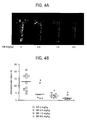
- FIG. 4A illustrates a Sudan IV staining of apoE -/- mouse aortas with or without simolimus treatment.
- Each aorta is representative of the mean lesion percentage in the respective groups in Figure 4A , and each point represents percentage lesion even to total aorta area from an individual mouse and bars depict average in Figure 4B .
- Significant differences P ⁇ 0.0001, one-way ANOVA
- P ⁇ 0.05, one-way ANOVA were observed in the sirolimus treated group as compared to the vehicle control groups. This reduction in atherosclerotic lesions was confirmed and extended by aortic root analysis.
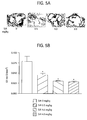
- lesion area was significantly ( P ⁇ 0.05, one-way ANOVA) reduced in sirolimus treated groups as compared with vehicle control as illustrated in Figures 5A and 5B.
- Figure 5A represents oil red O staining of atherosclerotic lesions at the aortic root in apoE - / - mice with or without sirolimus treatment. Each section is representative of the mean lesion percentage in the respective groups.
- Figure 5B represents the quantitative analysis of oil red O positive staining area at the aortic root. Significant differences ( P ⁇ 0.05, one-way ANOVA) were observed in the sirolimus treated groups as compared to the vehicle control groups.
- FIG. 6B illustrates quantitative analysis of CD68 positive staining at the aortic root. Significant differences ( P ⁇ 0.05, one-way ANOVA) were observed in the sirolimus treated groups as compared to the vehicle control groups.
- Angll infusion has been demonstrated to induce abdominal aortic adventitia lesion formation in apoE -/- mice on chow diet.
- angll was infused to apoE -/- mice receiving a high fat, high cholesterol diet.
- 30 percent incidence of abdominal aortic adventitia lesion was observed in the animals of the vehicle control group.
- Sirolimus administration exhibited a marked decrease in the incidence of abdominal aortic adventitia lesion (4.5 percent, 1 of 22 mice) compared to vehicle control (30 percent, 3 of 10 mice, abdominal aortic adventitia lesion positive).
- Representative abdominal aortas from mice with and without sirolimus administration are depicted in Figure 7A .
- FIG. 7A illustrates representative abdominal aortas from mice with and without sirolimus. Incidence of abdominal aortic adventitia lesion was reduced in mice receiving sirolimus (4.5 percent) as compared with mice on aortic control (30 percent).
- Figure 7B illustrates that sirolimus reduced the severity of abdominal aortic adventitia lesions in apoE -/- mice.
- CD68 a marker of macrophages, in adventitia lesions induced by angll infusion in apoE -/- mice on a pro-atherogenic diet.
- CD68 + macrophages were observed in the adventitia lesions.
- CD68 expression largely co-localized with elastin disruption at the "shoulder area" of the adventitia lesions as illustrated in Figure 8C , suggesting macrophages may play an active role in extracellular matix disruption.
- Macrophage infiltration into the adventitia lesional tissue was verified using a second marker for macrophage, F4/80 (data not shown).
- sirolimus administration prevents inflammatory macrophages accumulation in the granulomas as illustrated in Figure 8C . It has been reported that adventitia lesional tissue contained many vasa vasorum in humans, however, as best understood, this has never been documented in mouse models of adventitia lesion formation.
- the expression of CD31 was explored, an active marker for endothelial cells, in the aortic adventitia lesional tissues. CD31 expression was detected not only in the intima area as expected, but also in the adventitia lesions. CD31 + cells preferentially accumulated in the small blood vessel-shape area, indicating vasa vasorum in the current mouse model.
- Sirolimus administration obviously prevents this event in the vessel wall as illustrated in Figure 8D .
Landscapes
- Life Sciences & Earth Sciences (AREA)
- Environmental Sciences (AREA)
- Health & Medical Sciences (AREA)
- Engineering & Computer Science (AREA)
- Veterinary Medicine (AREA)
- General Health & Medical Sciences (AREA)
- Animal Behavior & Ethology (AREA)
- Biotechnology (AREA)
- Zoology (AREA)
- Biodiversity & Conservation Biology (AREA)
- Animal Husbandry (AREA)
- Medicinal Chemistry (AREA)
- General Chemical & Material Sciences (AREA)
- Public Health (AREA)
- Chemical Kinetics & Catalysis (AREA)
- Chemical & Material Sciences (AREA)
- Organic Chemistry (AREA)
- Nuclear Medicine, Radiotherapy & Molecular Imaging (AREA)
- Pharmacology & Pharmacy (AREA)
- Bioinformatics & Cheminformatics (AREA)
- Pharmaceuticals Containing Other Organic And Inorganic Compounds (AREA)
- Medicines That Contain Protein Lipid Enzymes And Other Medicines (AREA)
Abstract
Description
- The present invention relates to medical test procedures, and more particularly to methods of inducing and treating vascular disease in mammals.
- Determining the efficacy of medical devices and/or therapeutic agents initially requires animal testing, wherein the animals preferably have disease state characteristics identical to or approximate the disease state characteristics found in humans. Human disease state characteristics are not typically found in animals. Accordingly, these disease state characteristics may be induced in the animals by various means, including gene manipulation, diet, drug therapy and/or various combinations thereof. The precise control of these inducement means allows researchers the opportunity to approximate many human disease state characteristics.
- In accordance with one aspect, the present invention is directed to a method of inducing one or more predetermined vascular diseases in a mammal having at least one of an apoE gene and LDL-receptor gene deleted. The method comprises accelerating athereosclerosis in the mammalian vasculature through the introduction of a high fat, high cholesterol diet and introducing, via systemic delivery, angiotensin II at a predetermined time to induce and accelerate vascular inflammation.
- The foregoing and other features and advantages of the invention will be apparent from the following, more particular description of preferred embodiments of the invention, as illustrated in the accompanying drawings.
- Embodiments of the invention will now be described by way of example with reference to the accompanying drawings, in which:
-
Figure 1 is a tabular representation of sirolimus pharmacokinetics in apoE -/- mice in accordance with the present invention. -
Figure 2 is a tabular representation of body weight, plasma total cholesterol, triglycerides, LDL cholesterol and HDL cholesterol in apoE -/ - mice in accordance with the present invention. -
Figure 3 is a graphical representation of re-suturing incidence post surgery in apoE -/- mice in accordance with the present invention. -
Figure 4A are first images of aortas of apoE -/ - mice in accordance with the present invention. -
Figure 4B is a graphical representation of percentage lesion area to total aorta area in apoE -/ - mice in accordance with the present invention. -
Figure 5A are second images of aortas of apoE -/ - mice in accordance with the present invention. -
Figure 5B is a graphical representation of quantitative analysis of oil red O positive staining area at the aortic root of apoE -/- mice in accordance with the present invention. -
Figure 6A are third images of aortas of apoE -/ - mice in accordance with the present invention. -
Figure 6B is a graphical representation of quantitative analysis of CD68 position staining at the aortic root of apoE -/ - mice in accordance with the present invention. -
Figure 7A are third images of aortas of apoE -/- mice in accordance with the present invention. -
Figure 7B is a graphical representation of the severity of abdominal aortic adventitia lesions in apoE -/ - mice in accordance with the present invention. -
Figures 8A-8D are histological and immunohistochemical images of adventitia lesions in apoE -/- mice in accordance with the present invention. - The present invention is directed to a model for creating disease states and determining the efficacy of treatments for these disease states in mammals. More specifically, in this invention artheriosclerosis and aortic lesions are created utilizing a combination of diet and angll in apoE -/ - mice. Once the disease states are created, various treatments may be utilized. In this exemplary embodiment, rapamycin is utilized as the treatment modality. For the data generated, it appears that treatment with rapamycin reduces atherosclerotic lesions, retards inflammatory macrophage infiltration which is responsible for decreased foam call formation which in turn leads to reduced lipid deposition, and reduces the incidence of adventitial lesions.
- As used herein, rapamycin includes rapamycin, sirolimus, everolimus and all analogs, derivatives and conjugates that bind FKBP12, and other immunophilins and possesses the same pharmacologic properties as rapamycin including inhibition of mTOR.
- ApoE-/- mice (backcrossed ten times to the C57BL/6 background) were purchased from The Jackson Laboratory. The mice were kept on a twelve-hour light/ twelve-hour dark regimen upon arrival. All animal experiments were approved by the IACUC of the Consumer & Personal Product Worldwide, in accordance with AAALAC guidelines. Eight week old, male apoE-/- mice were fed a normal mouse-chow diet (RP5001; PMI Feeds Inc., St. Louis, MO) and acclimated for one week before switching to a pro-atherogenic high fat, high cholesterol, diet ("Western diet", 21 % fat, 0.2 % cholesterol, diet No. 88137, Harlan-Teklad, Madison, WI) for four weeks.
- Upon initiation of the pro-atherogenic diet, Alzet osmotic mini-pumps (Model 2004; ALZA Scientific Products, Mountain View, CA) were implanted into the apoE-/- mice subcutaneously. The mini-pumps were loaded with angII (EMD Biosciences, San Diego, CA) at the delivery rate of 1,000 ng/min/kg for twenty-eight days. The mice were anesthetized using isoflurane for the implantation of the mini-pumps. Small incisions in the lower back of the neck were made and the mini-pumps were inserted into the subcutaneous space of the mice. The incisions were then closed using sterile staplers. Triple antibiotic ointment was applied to each incision post surgery. The mice on the high fat, high cholesterol diet were sacrificed by CO2 inhalation twenty-eight days post mini-pump implantation as is explained in detail subsequently.
- Upon initiation of the pro-atherogenic diet and osmotic mini-pump implantation, mice were randomly assigned to four groups; namely, vehicle control, rapamycin or sirolimus 0.5 mg/kg, sirolimus 1.0 mg/kg and sirolimus 4.0 mg/kg body weight. The defined amount of sirolimus was dissolved in ethanol, brought up in a vehicle comprising 0.2 percent Sodium carboxymethylcellulose and 0.25 percent tween 80) and administrated to mice (i.p., daily).
- Sirolimus was quantitated using liquid-liquid extraction and reverse-phase liquid chromatography mass spectrometry (LC-MS/MS). Briefly, 100 µl of mouse whole blood sample underwent protein precipitation with 5 percent Methanol/Zinc sulfate solution containing the internal standard ascomycin. After centrifugation of the resulting solution, the liquid mixture proceeded with liquid-liquid extraction with 1-chlorobutane. The combined organic extract was then blown down to dryness and reconstituted with 50:50 Methanol:20 mM ammonium acetate solution. The samples were analyzed on API-5000 using C18 reverse-phase chromatography in Multiple Reaction Monitoring (MRM) mode using atmospheric pressure chemical ionization (ApCl) approach. The calibration standard curve results for sirolimus in mouse whole blood were acceptable from 0.5 ng/ml to 10,000 ng/ml with correlation coefficient of 0.998. The percent bias for the calibration standards ranged from about 10.9 percent to about 8.0 percent.
- As stated above, the mice were sacrificed by CO2 inhalation and bled from the abdominal vena cava. The aortas were perfusion-fixed and post-fixed in 4 percent paraformaldehyde. Atherosclerosis quantitation was assessed using two independent methods: "en face" surface lesion analysis and aortic root analysis. The aortic arch and thoracic aorta (defined as above the last intercostal artery) was subjected to "en face" analysis. Briefly, the aorta with major branches (left common carotid artery, right common carotid artery, left subclavian artery) was opened up. The lipid deposition in the inner aortic wall was stained in Sudan IV buffer. Images of the Sudan-IV stained aortas were captured with a video camera, and morphometric imaging carried out using Image-Pro 6.0 analysis software (Media Cybernetics, Inc. Carlsbad, CA). The extent of atherosclerosis is expressed as the percent of the aortic surface area covered by lesions. Aortic root analysis provided a second method verifying atherosclerosis quantitation. Briefly, alternate 8-µm frozen sections covering 300 µm of the proximal aorta, starting at the aortic sinus, were stained for atherosclerotic lesions with oil red O. Images of oil red O-stained aortic root sections were captured and analyzed using Image-Pro 6.0 analysis software.
All samples were coded by one investigator and analyzed by another investigator who is completely blinded to the codes. - The abdominal aorta was subjected to abdominal aortic adventitia lesion analysis. Mice with an increase in aortic diameter greater than 50 percent were considered to have an adventitia lesion. Tissue affected by adventitia lesion was categorized independently by two observers. Classification of the adventitia lesions was determined according to the scale based on the gross appearance of the aorta reported by Daugherty et al. Briefly, I: Dilated lumen in the suprarenal region of the aorta with no thrombus; II: Remodeled tissue in the suprarenal region that frequently contains thrombus; III: A pronounced bulbous form of type II that contains thrombus; IV: A form in which there are multiple adventitia lesions containing thrombus, some overlapping, in the suprarenal area of the aorta.
- The paraformaldehyde-perfused aortas covering the adventitia lesional area were embedded in paraffin. Sections (1 mm) of the abdominal aortas were stained with Hematoxylin & Eosin (H & E), Verhoeffs and Van Gisson staining for elastin, CD68 (FA-11, Serotec Ltd., Kidlington, U.K.) for macrophage and CD31 (Santa Cruz Biotechnology, Santa Cruz, CA) for endothelial cells. The macrophage composition at the aortic root was analyzed on fresh frozen tissues by immunohistochemistry using anti-CD68 primary antibody. Secondary antibodies were donkey anti rabbit, HRP-conjugated; donkey anti rat, HRP conjugated (Jackson Immunoresearch Laboratories, West Grove, PA).
- Plasma triglyceride and total cholesterol levels were determined via an automated enzymatic technique (Equal Diagnostic Inc., Exton, PA). Mouse lipoproteins were separated by fast protein liquid chromatography (FPLC) analysis of plasma using 2
Superose 6 columns in series (Pharmacia Biotech Inc., Piscataway, NJ). Aliquots of mouse plasma (100 µl) were injected onto the column, separated with a buffer containing 0.15 M NaCl, 0.01 M Na2HPO4, and 0.1 mM EDTA (pH 7.5) at a flow rate of 0.5 mL/min, and analyzed for lipids. - Initial analyses were performed by the Student's t test. If the data did not fit the constraints of this parametric test, data were analyzed with the one-way ANOVA test. P <0.05 was considered significant. Prism 4.0 software (GraphPad Software Inc., Lake Forest, CA) was used for all calculations. All data are presented as mean ± SEM.
- The regimen of sirolimus administration (i.p., daily, 0.5, 1.0, and 4.0 mg/kg) results in significant increase of whole blood sirolimus levels in the apoE-/- mice within 24 hours post injection. The blood drug levels reached peak level at 1 hour post injection, dropped to approximately 30 to 40 percent at 6 hours and remained at trace level at 24 hours. A clear drug dose curve was observed in this whole blood pharmacokinetic study as illustrated by the data in the tabular form (Table 1) illustrated in
Figure 1 . - The health status of apoE-/- mice in the current study was not overtly affected by dosing with sirolimus. A trend of decreased body weight was observed in all the 3 sirolimus treated groups but no statistical significance was reached as illustrated by the data in tabular form (Table 2) in
Figure 2 . sirolimus administration is associated with increased incidence of re-suturing post the surgery for mini-pump implantation in a clear dose dependent manner as illustrated inFigure 3 , suggesting a role of sirolimus in retarding wound healing in this particular animal model. The data is depicted as re-suturing incedince post surgery per group size. A clear increase of the suture index is observed in response to sirolimus administration in a dose dependent manner. - High fat, high cholesterol diet and angll infusion resulted in marked hyperlipidemia in the vehicle control group, with total cholesterol approaching ~1000 mg/dl (see Table 2 illustrated in
Figure 2 ). Sirolimus administration was associated with about 44 to 50 percent increase of plasma total cholesterol level as compared with the vehicle control group. LDL cholesterol was elevated greater than approximately 60 percent in all sirolimus treated mice. It should be noted that an approximately 55 to 81 percent increase of HDL cholesterol was also observed in the mice receiving sirolimus. In addition, a trend of elevated triglyceride in mice receiving this immunosuppressant was observed, regardless of the dosage employed. - Atherosclerosis quantitation was assessed using two independent methods: "en face" analysis and aortic root analysis. Strikingly, at 13 weeks of age in mice infused with angll on a high fat, high cholesterol diet, significantly (P < 0.0001, one-way ANOVA) reduced atherosclerotic lesions were seen in mice administrated with sirolimus in a dose dependent manner using "en face" analysis as illustrated in
Figure 4A and 4B. Figure 4A illustrates a Sudan IV staining of apoE -/- mouse aortas with or without simolimus treatment. Each aorta is representative of the mean lesion percentage in the respective groups inFigure 4A , and each point represents percentage lesion even to total aorta area from an individual mouse and bars depict average inFigure 4B .
Significant differences (P< 0.0001, one-way ANOVA) were observed in the sirolimus treated group as compared to the vehicle control groups. This reduction in atherosclerotic lesions was confirmed and extended by aortic root analysis. In this assay, lesion area was significantly (P < 0.05, one-way ANOVA) reduced in sirolimus treated groups as compared with vehicle control as illustrated inFigures 5A and 5B. Figure 5A represents oil red O staining of atherosclerotic lesions at the aortic root in apoE -/ - mice with or without sirolimus treatment. Each section is representative of the mean lesion percentage in the respective groups.Figure 5B represents the quantitative analysis of oil red O positive staining area at the aortic root. Significant differences (P < 0.05, one-way ANOVA) were observed in the sirolimus treated groups as compared to the vehicle control groups. - Inflammatory macrophages infiltration into the neo-intima and the subsequent uptake of ox-LDL lead to the formation of foam cells in atherosclerotic lesions. To determine if sirolimus plays an active role in regulating this inflammatory process, immunohistochemical analysis was performed for CD68, a marker of inflammatory macrophages, at the aortic root. Significant reduction in CD68 positive area was clearly apparent in this analysis as illustrated in
Figures 6A and 6B . These data suggest that retarded inflammatory macrophage infiltration is responsible for decreased foam cells formation, which in turn leads to reduced lipid deposition in response to sirolimus administration.Figure 6A illustrates immunohistochemical staining for CD68. Sirolimus suppresses CD68 positive signal (brown) in a dose dependent manner.Figure 6B illustrates quantitative analysis of CD68 positive staining at the aortic root. Significant differences (P < 0.05, one-way ANOVA) were observed in the sirolimus treated groups as compared to the vehicle control groups. - Angll infusion has been demonstrated to induce abdominal aortic adventitia lesion formation in apoE-/- mice on chow diet. In the current methology, angll was infused to apoE-/- mice receiving a high fat, high cholesterol diet. 30 percent incidence of abdominal aortic adventitia lesion was observed in the animals of the vehicle control group. Sirolimus administration exhibited a marked decrease in the incidence of abdominal aortic adventitia lesion (4.5 percent, 1 of 22 mice) compared to vehicle control (30 percent, 3 of 10 mice, abdominal aortic adventitia lesion positive). Representative abdominal aortas from mice with and without sirolimus administration are depicted in
Figure 7A . The abdominal aortic adventitia lesions were further classified using previously described criteria described herein. Much less advanced abdominal aortic adventitia lesion was observed in the one positive mouse receiving sirolimus than the ones in the vehicle control group (Figure 7B). Figure 7A illustrates representative abdominal aortas from mice with and without sirolimus. Incidence of abdominal aortic adventitia lesion was reduced in mice receiving sirolimus (4.5 percent) as compared with mice on aortic control (30 percent).Figure 7B illustrates that sirolimus reduced the severity of abdominal aortic adventitia lesions in apoE-/- mice. - To further characterize the aortic adventitia lesions, histological and immunohistological analyses were performed as illustrated in
Figures 8A, 8B, 8C and 8D . Media lamina and elastin disruption has been established as a major pathological process during abdominal aortic adventitia lesion formation. In the vehicle control group, adventitia lesional tissue exhibited complete disruption of media lamina and elastin fibers, while in apoE-/- mice receiving sirolimus, the extent of media disruption was obviously reduced as illustrated inFigure 8B . These data provide the first piece of the mechanism underlying the anti-adventitia lesion capacity of sirolimus in vivo. To explore the participation of inflammatory macrophages in media and elastin breakdown, the expression of CD68 was examined, a marker of macrophages, in adventitia lesions induced by angll infusion in apoE-/- mice on a pro-atherogenic diet. Populations of CD68+ macrophages were observed in the adventitia lesions. Interestingly, CD68 expression largely co-localized with elastin disruption at the "shoulder area" of the adventitia lesions as illustrated inFigure 8C , suggesting macrophages may play an active role in extracellular matix disruption. Macrophage infiltration into the adventitia lesional tissue was verified using a second marker for macrophage, F4/80 (data not shown). Notably, sirolimus administration prevents inflammatory macrophages accumulation in the granulomas as illustrated inFigure 8C . It has been reported that adventitia lesional tissue contained many vasa vasorum in humans, however, as best understood, this has never been documented in mouse models of adventitia lesion formation. The expression of CD31 was explored, an active marker for endothelial cells, in the aortic adventitia lesional tissues. CD31 expression was detected not only in the intima area as expected, but also in the adventitia lesions. CD31+ cells preferentially accumulated in the small blood vessel-shape area, indicating vasa vasorum in the current mouse model. Sirolimus administration obviously prevents this event in the vessel wall as illustrated inFigure 8D .
Claims (2)
- A method of inducing one or more vascular diseases in a mammal comprising:having at least one of an apoE gene and LDL-receptor gene deleted, the method comprising:accelerating athereosclorosis in the mammalian vasculature through the introduction of a high fat, high cholesterol diet; andintroducing, via systemic delivery, angiotensin II at a predetermined time to induce and accelerate vascular inflammation.
- A method of inducing one or more vascular diseases in a mouse comprising:having at least one of an apoE gene and LDL-receptor gene deleted, the method comprising:accelerating athereosclorosis in the mammalian vasculature through the introduction of a high fat, high cholesterol diet; andintroducing, via systemic delivery, angiotensin II at a predetermined time to induce and accelerate vascular inflammation.
Applications Claiming Priority (2)
| Application Number | Priority Date | Filing Date | Title |
|---|---|---|---|
| US88515507P | 2007-01-16 | 2007-01-16 | |
| US11/954,381 US20080171705A1 (en) | 2007-01-16 | 2007-12-12 | Methods of inducing and treating vascular disease |
Publications (1)
| Publication Number | Publication Date |
|---|---|
| EP1947188A1 true EP1947188A1 (en) | 2008-07-23 |
Family
ID=39304796
Family Applications (1)
| Application Number | Title | Priority Date | Filing Date |
|---|---|---|---|
| EP08250173A Withdrawn EP1947188A1 (en) | 2007-01-16 | 2008-01-14 | Methods of inducing and treating vascular disease |
Country Status (3)
| Country | Link |
|---|---|
| US (1) | US20080171705A1 (en) |
| EP (1) | EP1947188A1 (en) |
| JP (1) | JP2008228726A (en) |
Citations (1)
| Publication number | Priority date | Publication date | Assignee | Title |
|---|---|---|---|---|
| EP1709974A2 (en) * | 2000-06-16 | 2006-10-11 | Wyeth | Method of treating cardiovascular disease using rapamycin |
-
2007
- 2007-12-12 US US11/954,381 patent/US20080171705A1/en not_active Abandoned
-
2008
- 2008-01-14 EP EP08250173A patent/EP1947188A1/en not_active Withdrawn
- 2008-01-15 JP JP2008005744A patent/JP2008228726A/en not_active Withdrawn
Patent Citations (1)
| Publication number | Priority date | Publication date | Assignee | Title |
|---|---|---|---|---|
| EP1709974A2 (en) * | 2000-06-16 | 2006-10-11 | Wyeth | Method of treating cardiovascular disease using rapamycin |
Non-Patent Citations (7)
| Title |
|---|
| DAUGHERTY A ET AL: "Chronic angiotensin II infusion promotes atherogenesis in low density lipoprotein receptor -/- mice.", ANNALS OF THE NEW YORK ACADEMY OF SCIENCES 18 NOV 1999, vol. 892, 18 November 1999 (1999-11-18), pages 108 - 118, XP002478695, ISSN: 0077-8923 * |
| DAUGHERTY ALAN ET AL: "Angiotensin II promotes atherosclerotic lesions and aneurysms in apolipoprotein E-deficient mice", JOURNAL OF CLINICAL INVESTIGATION, vol. 105, no. 11, June 2000 (2000-06-01), pages 1605 - 1612, XP002478696, ISSN: 0021-9738 * |
| DAUGHERTY ALAN ET AL: "Mouse models of abdominal aortic aneurysms.", ARTERIOSCLEROSIS, THROMBOSIS, AND VASCULAR BIOLOGY MAR 2004, vol. 24, no. 3, March 2004 (2004-03-01), pages 429 - 434, XP002467953, ISSN: 1524-4636 * |
| ELLOSO M MERLE ET AL: "Protective effect of the immunosuppressant sirolimus against aortic atherosclerosis in apo E-deficient mice.", AMERICAN JOURNAL OF TRANSPLANTATION : OFFICIAL JOURNAL OF THE AMERICAN SOCIETY OF TRANSPLANTATION AND THE AMERICAN SOCIETY OF TRANSPLANT SURGEONS MAY 2003, vol. 3, no. 5, May 2003 (2003-05-01), pages 562 - 569, XP002478697, ISSN: 1600-6135 * |
| LANGHEINRICH ALEXANDER C ET AL: "Correlation of vasa vasorum neovascularization and plaque progression in aortas of apolipoprotein E(-/-)/low-density lipoprotein(-/-) double knockout mice.", ARTERIOSCLEROSIS, THROMBOSIS, AND VASCULAR BIOLOGY FEB 2006, vol. 26, no. 2, February 2006 (2006-02-01), pages 347 - 352, XP002478698, ISSN: 1524-4636 * |
| MANNING MICHAEL W ET AL: "Differential effects of doxycycline, a broad-spectrum matrix metalloproteinase inhibitor, on angiotensin II-induced atherosclerosis and abdominal aortic aneurysms.", ARTERIOSCLEROSIS, THROMBOSIS, AND VASCULAR BIOLOGY 1 MAR 2003, vol. 23, no. 3, 1 March 2003 (2003-03-01), pages 483 - 488, XP002478693, ISSN: 1524-4636 * |
| WEISS D ET AL: "Angiotensin II-induced hypertension accelerates the development of atherosclerosis in apoE-deficient mice.", CIRCULATION 23 JAN 2001, vol. 103, no. 3, 23 January 2001 (2001-01-23), pages 448 - 454, XP002478694, ISSN: 1524-4539 * |
Also Published As
| Publication number | Publication date |
|---|---|
| US20080171705A1 (en) | 2008-07-17 |
| JP2008228726A (en) | 2008-10-02 |
Similar Documents
| Publication | Publication Date | Title |
|---|---|---|
| Choi et al. | Peripheral inflammation induces tumor necrosis factor dependent AMPA receptor trafficking and Akt phosphorylation in spinal cord in addition to pain behavior | |
| Lin et al. | α1-Anti-trypsin improves function of porcine donor lungs during ex-vivo lung perfusion | |
| Gan et al. | Celastrol attenuates bone erosion in collagen-Induced arthritis mice and inhibits osteoclast differentiation and function in RANKL-induced RAW264. 7 | |
| Li et al. | Nicotinic acetylcholine receptor α7 subunit improves energy homeostasis and inhibits inflammation in nonalcoholic fatty liver disease | |
| Yu et al. | Combining TGF-β inhibition and angiotensin II blockade results in enhanced antifibrotic effect | |
| Iannello et al. | Low-dose enalapril in the treatment of surgical cutaneous hypertrophic scar and keloid-two case reports and literature review | |
| Roksnoer et al. | Optimum AT1 receptor-neprilysin inhibition has superior cardioprotective effects compared with AT1 receptor blockade alone in hypertensive rats | |
| Lee et al. | Enalapril inhibits nuclear factor-κB signaling in intestinal epithelial cells and peritoneal macrophages and attenuates experimental colitis in mice | |
| US9808501B2 (en) | Compositions and methods for treating and preventing hyperlipidemia, fatty liver, atherosclerosis and other disorders associated with metabolic syndrome | |
| Zhao et al. | Low‐dose oral sirolimus reduces atherogenesis, vascular inflammation and modulates plaque composition in mice lacking the LDL receptor | |
| D'souza et al. | Suppressing hyperinsulinemia prevents obesity but causes rapid onset of diabetes in leptin-deficient Lepob/ob mice | |
| Neumann et al. | Lipid nanoparticle delivery of glucagon receptor siRNA improves glucose homeostasis in mouse models of diabetes | |
| Schulz et al. | Effect of chronic treatment with acetylsalicylic acid and clopidogrel on atheroprogression and atherothrombosis in ApoE-deficient mice in vivo | |
| French et al. | The angiotensin receptor blocker, azilsartan medoxomil (TAK-491), suppresses vascular wall expression of plasminogen activator inhibitor type-I protein potentially facilitating the stabilization of atherosclerotic plaques | |
| Li et al. | Endocannabinoids regulate cocaine-associated memory through brain AEA–CB1R signalling activation | |
| Zhang et al. | Intracerebroventricular administration of morphine confers remote cardioprotection—Role of opioid receptors and calmodulin | |
| Singha et al. | Leptin receptors in RIP-Cre25Mgn neurons mediate anti-dyslipidemia effects of leptin in insulin-deficient mice | |
| US20080171764A1 (en) | Methods for preventing, managing, and reversing disease caused by inflammation mediated vascular lesions | |
| US20080171705A1 (en) | Methods of inducing and treating vascular disease | |
| EP1949897A1 (en) | Inflammation mediated vascular lesions | |
| CA2618541A1 (en) | Methods of inducing and treating vascular disease | |
| US9186421B2 (en) | Establishment of rhesus monkey model of autoimmunity type 1 diabetes | |
| US20080267969A1 (en) | Treatment of Pulmonary Hypertension Using an Agent That Inhibits a Tissue Factor Pathway | |
| US11733232B2 (en) | Method for producing disease modeling non-human animal, disease modeling non-human animal, and method for screening drug and method for determining risk of disease using the same | |
| Jablonka‐Shariff et al. | FK506 Enhancement of Neuromuscular Junction Recovery After Nerve Injury Is Macrophage‐Dependent |
Legal Events
| Date | Code | Title | Description |
|---|---|---|---|
| PUAI | Public reference made under article 153(3) epc to a published international application that has entered the european phase |
Free format text: ORIGINAL CODE: 0009012 |
|
| AK | Designated contracting states |
Kind code of ref document: A1 Designated state(s): AT BE BG CH CY CZ DE DK EE ES FI FR GB GR HR HU IE IS IT LI LT LU LV MC MT NL NO PL PT RO SE SI SK TR |
|
| AX | Request for extension of the european patent |
Extension state: AL BA MK RS |
|
| 17P | Request for examination filed |
Effective date: 20080819 |
|
| 17Q | First examination report despatched |
Effective date: 20081016 |
|
| AKX | Designation fees paid |
Designated state(s): AT BE BG CH CY CZ DE DK EE ES FI FR GB GR HR HU IE IS IT LI LT LU LV MC MT NL NO PL PT RO SE SI SK TR |
|
| STAA | Information on the status of an ep patent application or granted ep patent |
Free format text: STATUS: THE APPLICATION IS DEEMED TO BE WITHDRAWN |
|
| 18D | Application deemed to be withdrawn |
Effective date: 20090428 |