EP1945760B1 - Productivity augmenting protein factor, novel cell lines and uses thereof - Google Patents
Productivity augmenting protein factor, novel cell lines and uses thereof Download PDFInfo
- Publication number
- EP1945760B1 EP1945760B1 EP06807777A EP06807777A EP1945760B1 EP 1945760 B1 EP1945760 B1 EP 1945760B1 EP 06807777 A EP06807777 A EP 06807777A EP 06807777 A EP06807777 A EP 06807777A EP 1945760 B1 EP1945760 B1 EP 1945760B1
- Authority
- EP
- European Patent Office
- Prior art keywords
- cell
- pix
- cells
- virus
- protein
- Prior art date
- Legal status (The legal status is an assumption and is not a legal conclusion. Google has not performed a legal analysis and makes no representation as to the accuracy of the status listed.)
- Active
Links
Images


Classifications
-
- C—CHEMISTRY; METALLURGY
- C12—BIOCHEMISTRY; BEER; SPIRITS; WINE; VINEGAR; MICROBIOLOGY; ENZYMOLOGY; MUTATION OR GENETIC ENGINEERING
- C12N—MICROORGANISMS OR ENZYMES; COMPOSITIONS THEREOF; PROPAGATING, PRESERVING, OR MAINTAINING MICROORGANISMS; MUTATION OR GENETIC ENGINEERING; CULTURE MEDIA
- C12N7/00—Viruses; Bacteriophages; Compositions thereof; Preparation or purification thereof
-
- C—CHEMISTRY; METALLURGY
- C07—ORGANIC CHEMISTRY
- C07K—PEPTIDES
- C07K14/00—Peptides having more than 20 amino acids; Gastrins; Somatostatins; Melanotropins; Derivatives thereof
- C07K14/005—Peptides having more than 20 amino acids; Gastrins; Somatostatins; Melanotropins; Derivatives thereof from viruses
- C07K14/01—DNA viruses
-
- C—CHEMISTRY; METALLURGY
- C07—ORGANIC CHEMISTRY
- C07K—PEPTIDES
- C07K14/00—Peptides having more than 20 amino acids; Gastrins; Somatostatins; Melanotropins; Derivatives thereof
- C07K14/005—Peptides having more than 20 amino acids; Gastrins; Somatostatins; Melanotropins; Derivatives thereof from viruses
-
- C—CHEMISTRY; METALLURGY
- C07—ORGANIC CHEMISTRY
- C07K—PEPTIDES
- C07K14/00—Peptides having more than 20 amino acids; Gastrins; Somatostatins; Melanotropins; Derivatives thereof
- C07K14/005—Peptides having more than 20 amino acids; Gastrins; Somatostatins; Melanotropins; Derivatives thereof from viruses
- C07K14/01—DNA viruses
- C07K14/075—Adenoviridae
-
- C—CHEMISTRY; METALLURGY
- C07—ORGANIC CHEMISTRY
- C07K—PEPTIDES
- C07K14/00—Peptides having more than 20 amino acids; Gastrins; Somatostatins; Melanotropins; Derivatives thereof
- C07K14/435—Peptides having more than 20 amino acids; Gastrins; Somatostatins; Melanotropins; Derivatives thereof from animals; from humans
- C07K14/705—Receptors; Cell surface antigens; Cell surface determinants
- C07K14/70567—Nuclear receptors, e.g. retinoic acid receptor [RAR], RXR, nuclear orphan receptors
-
- C—CHEMISTRY; METALLURGY
- C12—BIOCHEMISTRY; BEER; SPIRITS; WINE; VINEGAR; MICROBIOLOGY; ENZYMOLOGY; MUTATION OR GENETIC ENGINEERING
- C12N—MICROORGANISMS OR ENZYMES; COMPOSITIONS THEREOF; PROPAGATING, PRESERVING, OR MAINTAINING MICROORGANISMS; MUTATION OR GENETIC ENGINEERING; CULTURE MEDIA
- C12N5/00—Undifferentiated human, animal or plant cells, e.g. cell lines; Tissues; Cultivation or maintenance thereof; Culture media therefor
- C12N5/10—Cells modified by introduction of foreign genetic material
-
- C—CHEMISTRY; METALLURGY
- C12—BIOCHEMISTRY; BEER; SPIRITS; WINE; VINEGAR; MICROBIOLOGY; ENZYMOLOGY; MUTATION OR GENETIC ENGINEERING
- C12N—MICROORGANISMS OR ENZYMES; COMPOSITIONS THEREOF; PROPAGATING, PRESERVING, OR MAINTAINING MICROORGANISMS; MUTATION OR GENETIC ENGINEERING; CULTURE MEDIA
- C12N5/00—Undifferentiated human, animal or plant cells, e.g. cell lines; Tissues; Cultivation or maintenance thereof; Culture media therefor
- C12N5/10—Cells modified by introduction of foreign genetic material
- C12N5/12—Fused cells, e.g. hybridomas
- C12N5/16—Animal cells
-
- C—CHEMISTRY; METALLURGY
- C07—ORGANIC CHEMISTRY
- C07K—PEPTIDES
- C07K2319/00—Fusion polypeptide
-
- C—CHEMISTRY; METALLURGY
- C07—ORGANIC CHEMISTRY
- C07K—PEPTIDES
- C07K2319/00—Fusion polypeptide
- C07K2319/01—Fusion polypeptide containing a localisation/targetting motif
- C07K2319/09—Fusion polypeptide containing a localisation/targetting motif containing a nuclear localisation signal
-
- C—CHEMISTRY; METALLURGY
- C07—ORGANIC CHEMISTRY
- C07K—PEPTIDES
- C07K2319/00—Fusion polypeptide
- C07K2319/60—Fusion polypeptide containing spectroscopic/fluorescent detection, e.g. green fluorescent protein [GFP]
-
- C—CHEMISTRY; METALLURGY
- C07—ORGANIC CHEMISTRY
- C07K—PEPTIDES
- C07K2319/00—Fusion polypeptide
- C07K2319/70—Fusion polypeptide containing domain for protein-protein interaction
- C07K2319/71—Fusion polypeptide containing domain for protein-protein interaction containing domain for transcriptional activaation, e.g. VP16
- C07K2319/715—Fusion polypeptide containing domain for protein-protein interaction containing domain for transcriptional activaation, e.g. VP16 containing a domain for ligand dependent transcriptional activation, e.g. containing a steroid receptor domain
-
- C—CHEMISTRY; METALLURGY
- C12—BIOCHEMISTRY; BEER; SPIRITS; WINE; VINEGAR; MICROBIOLOGY; ENZYMOLOGY; MUTATION OR GENETIC ENGINEERING
- C12N—MICROORGANISMS OR ENZYMES; COMPOSITIONS THEREOF; PROPAGATING, PRESERVING, OR MAINTAINING MICROORGANISMS; MUTATION OR GENETIC ENGINEERING; CULTURE MEDIA
- C12N2510/00—Genetically modified cells
- C12N2510/02—Cells for production
-
- C—CHEMISTRY; METALLURGY
- C12—BIOCHEMISTRY; BEER; SPIRITS; WINE; VINEGAR; MICROBIOLOGY; ENZYMOLOGY; MUTATION OR GENETIC ENGINEERING
- C12N—MICROORGANISMS OR ENZYMES; COMPOSITIONS THEREOF; PROPAGATING, PRESERVING, OR MAINTAINING MICROORGANISMS; MUTATION OR GENETIC ENGINEERING; CULTURE MEDIA
- C12N2710/00—MICROORGANISMS OR ENZYMES; COMPOSITIONS THEREOF; PROPAGATING, PRESERVING, OR MAINTAINING MICROORGANISMS; MUTATION OR GENETIC ENGINEERING; CULTURE MEDIA dsDNA viruses
- C12N2710/00011—Details
- C12N2710/10011—Adenoviridae
- C12N2710/10311—Mastadenovirus, e.g. human or simian adenoviruses
- C12N2710/10322—New viral proteins or individual genes, new structural or functional aspects of known viral proteins or genes
-
- C—CHEMISTRY; METALLURGY
- C12—BIOCHEMISTRY; BEER; SPIRITS; WINE; VINEGAR; MICROBIOLOGY; ENZYMOLOGY; MUTATION OR GENETIC ENGINEERING
- C12N—MICROORGANISMS OR ENZYMES; COMPOSITIONS THEREOF; PROPAGATING, PRESERVING, OR MAINTAINING MICROORGANISMS; MUTATION OR GENETIC ENGINEERING; CULTURE MEDIA
- C12N2710/00—MICROORGANISMS OR ENZYMES; COMPOSITIONS THEREOF; PROPAGATING, PRESERVING, OR MAINTAINING MICROORGANISMS; MUTATION OR GENETIC ENGINEERING; CULTURE MEDIA dsDNA viruses
- C12N2710/00011—Details
- C12N2710/16011—Herpesviridae
- C12N2710/16611—Simplexvirus, e.g. human herpesvirus 1, 2
- C12N2710/16622—New viral proteins or individual genes, new structural or functional aspects of known viral proteins or genes
-
- C—CHEMISTRY; METALLURGY
- C12—BIOCHEMISTRY; BEER; SPIRITS; WINE; VINEGAR; MICROBIOLOGY; ENZYMOLOGY; MUTATION OR GENETIC ENGINEERING
- C12N—MICROORGANISMS OR ENZYMES; COMPOSITIONS THEREOF; PROPAGATING, PRESERVING, OR MAINTAINING MICROORGANISMS; MUTATION OR GENETIC ENGINEERING; CULTURE MEDIA
- C12N2710/00—MICROORGANISMS OR ENZYMES; COMPOSITIONS THEREOF; PROPAGATING, PRESERVING, OR MAINTAINING MICROORGANISMS; MUTATION OR GENETIC ENGINEERING; CULTURE MEDIA dsDNA viruses
- C12N2710/00011—Details
- C12N2710/16011—Herpesviridae
- C12N2710/16611—Simplexvirus, e.g. human herpesvirus 1, 2
- C12N2710/16651—Methods of production or purification of viral material
- C12N2710/16652—Methods of production or purification of viral material relating to complementing cells and packaging systems for producing virus or viral particles
Definitions
- the present invention provides a method for preparing a non-adenoviral target virus or target proteins utilizing a potent expression cell line having stably integrated into its genome a gene encoding a PIX protein.
- Biopharmaceutical products from eukaryotic cells are an integral part of modern medicine. However, increased productivity and increased safety are important parameters that still require substantial optimization. The most crucial bottleneck in the efforts towards optimization is the cellular substrate itself. A safe transgene that can be introduced into cell lines already released for use in biopharmaceutical processes to increase product yields from the such manipulated cells is extremely valuable.
- Adenoviruses are unenveloped (naked) double-stranded DNA viruses that infect a broad spectrum of animals. Among the best characterized members is, the human adenovirus serotype 5 (Ad5). This virus is a frequent cause of common cold symptoms and infection often occurs in childhood.
- Adenoviruses are amenable to genetic manipulation and replication incompetent adenoviruses, including Ad5 (GenBank accession number for sequence: AC_000008), are used as vectors in gene therapy and for therapeutic vaccination.
- Ad5 GeneBank accession number for sequence: AC_000008
- To obtain replication incompetent viruses large regions of the genomic DNA are substituted with non-viral sequences.
- the missing viral functions are provided in trans by host cell lines that have been stably transfected with genes that are deleted in the vector.
- One of the earlier systems that were developed consist of adenoviral vectors deleted in the regulatory E1 region produced on cell lines providing the corresponding E1 proteins.
- the most common cell line for this purpose is the 293 cell line. This line was generated in 1977 by transfection of fragmented adenoviral genomic DNA into primary human cells ( Graham et al., J. Gen. Virol. 36, 59-74 (1977 )), well before it was recognized that adenoviruses may serve as gene therapy vectors. The resulting cell line was subsequently shown to contain nucleotides 1 to 4344 of the genomic DNA ( Louis et al., Virology 233, 423-429 (1997 )) which includes the E1 region; this characterization demonstrated that the E1 region can be used to immortalize and transform primary cells.
- Adjacent to the E1 region follows the gene for pIX ("protein 9"; nucleotides 3609 to 4031 on the genomic DNA).
- the promoter for pIX is embedded within the E1B component of the E1 region.
- a mechanism called promoter occlusion allows expression of pIX at a delayed-early time In the viral infection cycle at the onset of viral DNA replication with increasing copy number of viral DNA ( Fessler and Young, J. Virol. 72, 4049-4056 (1998 )).
- the gene is present within the 4344 nucleotides integrated into 293 cells, pIX expression cannot be detected even with sensitive radioactive labeling methods ( Spector et al., J. Virol. 36, 860-871 (1980 )).
- replication deficient adenoviral vectors have been generated by deletion of the E1 region from the viral genome.
- E1 products are essential for viral replication and the function of the E1 region therefore had to be provided in trans via stable E1 transgenes in the adenovirus packaging cell (such as the 293 cell line).
- the pIX gene caused frequent recombination between deleted adenoviral vectors and E1 in the host genome.
- This recombination event generated replication competent adenovirus (RCA), a serious contamination in vector preparations. To suppress this recombination event pIX was deleted from the adenovirus genome.
- pIX stabilizes the adenoviral capsid against thermal and steric stress by increasing the interaction of the main building blocks, the hexons ( Colby and Shenk, J. Virol. 39, 977-980 (1981 ); Ghosh-Choudhury et al., EMBO J. 6, 1733-1739 (1987 )).
- Morphogenesis In the absence of pIX yields temperature-sensitive viruses that cannot deliver genomic DNA molecules larger than 105% of the wild type 35938 base pairs. To still allow a normal packaging capacity and thermal stability the pIX protein was stably or episomally introduced into cell lines intended as packaging cells for adenovirus vectors ( Krougllak and Graham, Hum.
- pIX-fusion proteins have been generated with the purpose to expand the host range of adenovirus vectors or to follow morphogenesis and the intracellular migration of virus particles (reviewed by Parks, Mol. Ther. 11, 19-25 (2005 )).
- PIX Although clearly a structural protein, pIX also is implicated as regulatory protein for adenovirus replication. PIX is assumed to function as transactivator of transcription to augment E1A expression, a function possibly even excerted as a virokine by incoming PIX protein from the capsid at the infection step (reviewed by Parks, Mol. Ther. 11, 19-25 (2005 )). PIX, together with early protein E4 Orf3, also is described to interact with sub-nuclear inclusions called PML bodies ( Puvion-Dutilleul et al., Exp. Cell. Res. 218, 9-16 (1995 ); Leppard and Everett, J. Gen. Virol. 80, 997-1008 (1999 )).
- PIX protein Because of its pleotropic effects we introduced the PIX protein into cell lines to examine whether PIX augments cell proliferation or production properties for biopharmaceutical products that are not related to adenovirus or adenoviral vectors. There is a general need for factors that modulate these properties in established cell lines.
- Attenuated (weakened) viruses are promising vaccine candidates: upon inoculation they mimick a natural infection but allow more time for establishment of the desired protective immune response by the vaccinee.
- highly attenuated strains are desirable.
- attenuated strains still proceed towards (usually benign) infection highly attenuated strains are blocked at a cellular level even in absence of a functional immune system.
- a frequently used method to establish and maintain attenuation is to passage viruses on different host tissue. For example, measles and mumps viruses intended for human vaccination are passaged in primary cells, either in embryonated chicken eggs or cultures derived thereof.
- a new generation of vaccines is based on highly attenuated pox viruses that also depend on primary chicken cells for production.
- a cell line that can substitute for primary chicken cells and at the same time that even less efficiently protects itself against viral infection due to secondary manipulations such as introduction of the PIX transgene therefore provides a highly desirable substrate.
- a mammalian (rather than avian) cell line may be preferred.
- Such preferred cell lines already exist and have passed the examination by health authorities with respect to safety and risks posed by derived biopharmaceutical products.
- a secondary manipulation such as introduction of the PIX transgene to increase the spectrum of available applications or production efficiencies without compromising safety features is highly desirable.
- pIX excerts a phenotypical effect in avian and human cells.
- avian cells this is especially surprising because avian cells cannot be infected by human adenoviruses and aviadenoviruses (such as CELO or fowl adenovirus type 8) do not encode a PIX homologue ( Ojkic and Nagy, J. Gen. Virol. 81, 1833-1837 (2000 )).
- the method of embodiment (1) of the invention utilizes an expression cell having integrated into its genome a gene coding for, pIX which possesses the following properties:
- the heterologous regulator protein is the adenovirus serotype 5 protein pIX in other embodiments the protein is obtained from an adenovirus of a different serotype.
- fusion proteins namly fusion proteins comprising at least one first domain comprising a PIX protein as defined hereinbefore and at least one second domain comprising a protein or peptide acting as a transcription modulator.
- said transcription modulator is a transcription factor including the retinoic acid receptor alpha, which may be present in complete or incomplete (i.e. truncated) form.
- the modulator may also be a transit peptide, which includes NLF sequences, e.g. that shown in SEQ ID NO:21.
- the first and second domain(s) are either directly or via a peptide linker covalently attached to each other.
- Suitable peptide linkers include flexible and hydrophilic structures such as poly gly-ser.
- cell and “cell line” as used in the following detailed description refer to expression cells/expression cell lines and host cells/host cell lines.
- the PIX protein is expressed in the cell in an amount of at least 1 pg/ ⁇ g cellular protein, preferably at an amount of at least 10 pg/ ⁇ g cellular protein so that its expression can be determined by Western-blotting.
- Suitable promoters are constitutive cellular promoter or variants thereof such as the human translocation elongation factor 2 promoter.
- a particular variant of the human translocation elongation factor 2 promoter utilized within the invention has the sequence shown in SEQ ID NO:12. The sequence shown represents a "short" version of the promoter which is located on human chromosome 19:3,935,325-3,936,638 (human genome assembly May 2004) and provides for a stable medium level expression. For a stronger expression a "longer" version of the promoter located on chr19:3,935,349-3,938,957 (human genome assembly May 2004) may be used.
- the cell is a vertebrate cell including mammalian cells, and avian cells.
- Suitable mammalian cells are human cells, and rodent cells including mouse, rat, hamster, etc.
- Particularly preferred mammalian cells according to the invention are cells derived from human brain, particularly from human foetal brain, a mouse NSO or Sp2/0 cell, a BHk or CHO cell.
- Suitable avian cells are duck, chicken quail and goose cells.
- Particularly preferred avian cells according to the invention are cells derived from a duck retina-derived or somite-derived cell.
- the cell of the invention may be derived from a primary cell or from a previously immortalized cell. Moreover, the cell may carry further immortalizing (viral) genes including an E1 protein of an adenovirus, such as the E1 protein of mastadenovirus group C type 5, and the like. Particularly preferred according to the present invention is that the cell further carries the adenovirus E1A and/or E1B gene shown In SEQ ID NO:5.
- the cell utilized in the method of embodiment (1) of the invention may further carry functional sequences, e.g. sequences required for its application as expression cell such as selection marker sequences, splice donor/acceptor sites and/or recombinase recognition sequences allowing integration of a target nucleic add sequence to be expressed in the cell, etc.
- functional sequences e.g. sequences required for its application as expression cell such as selection marker sequences, splice donor/acceptor sites and/or recombinase recognition sequences allowing integration of a target nucleic add sequence to be expressed in the cell, etc.
- the target virus includes from wild-type, mutated or deleted virus, cold adapted or attenuated virus, vaccine strains, viral vectors carrying heterologous gene(s), viral vectors such as lentivirus, poxvirus, adeno associated virus (aav), herpesvirus, and flavivirus.
- the one or more target proteins include antibodies, recombinant proteins such as erythropoetin, alpha-1-antitrypsin, blood clotting factors VIII and IX and interferons, viral antigens such as influenza HA and NA, and M, HBV-S, herpes G protein and rabies G protein, peptid hormones and the like.
- recombinant proteins such as erythropoetin, alpha-1-antitrypsin, blood clotting factors VIII and IX and interferons
- viral antigens such as influenza HA and NA
- M HBV-S
- herpes G protein and rabies G protein peptid hormones and the like.
- the culturing and isolation of the method of embodiment (1) of the invention may be performed according to standard procedures readily available to the skilled person. Said method may further include standard purification steps as well as subsequent modification steps of the target virus or target protein(s).
- the cell line NCST11#34 was deposited with the DSMZ-Deutsche Sammlung von Mikroorganismen und Zellkulturen GmbH, Mascheroder Weg 1b, 38124 Braunschweig, Germany on November 4, 2005 under accession number DSM ACC2744.
- the cell line CR.PIX (17a11b) was deposited with the DSMZ-Deutsche Sammlung von Mikroorganismen und Zellkulturen GmbH, Mascheroder Weg 1b, 38124 Braunschweig, Germany on November 24, 2005 under accession number DSM ACC2749.
- the cell line was developed from a mixture of cells from foetal brain by immortalisation with adenovirus 5 E1A and B genes by nonviral transfection.
- a tissue sample was taken from the periventricular zone of the brain of a foetus after induced abortion. It was dissected with scissors and homogenised by aspiration with a tissue culture pipette in neuronal culture medium based on DEMEM/F12 containing 20 ng/ml hFGF (Invitrogen, Carlsbad, CA 92008, USA), 20 ng/ml hEGF (Invitrogen), 1x N2 supplement (Invitrogen) and 8 ⁇ g/ml heparin (Sigma Aldrich, St. Louis, USA). Cells were sedimented at 200g for 3 min.
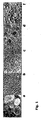
- Viability of cells was assessed using trypan blue and propidium jodide using a flow cytometer (BD Biosciences, Jose, CA 95131, USA). The viability was 75%. 0.5x10 6 cells were seeded into T25 flasks. Cells were incubated at 37°C and 5% CO 2 . The cells formed neurospheres by day 5 of the culture. Neurospheres are shown in Figure 1A . On day 8 cells were transferred to DMEM/F12 supplemented with 5% FCS to allow for attachment and to stimulate proliferation. Cells formed a homogenous monolayer as shown in Figure 1B and were passaged 1:5 once per week. This primary neural cell culture was named NC5.
- the plasmid p79 contains the following elements: The pBluescript (Stratagene, USA) serves as plasmid backbone, in which the ampicillin resistance marker was replaced by the kanamycin resistance gene driven by a bacterial promoter which allows for growth and selection in E.coli.
- the vector harbours a fragment form wild type adenovirus type 5 (SEQ ID NO: 5) containing the open reading frames for E1A (splice variants 13S and 12S with or without the CR3 domain respectively) and E1B 55k and 19k as well as sequences upstream of E1A.
- E1A gene is preceded by the phosphoglycerate kinase promoter (mouse).
- the adenovirus sequences are followed by the polyadenylation signal from the Herpes simplex thymidine kinase gene, serving as a replacement for the E1B polyadenylation signal.
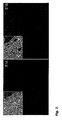
- Clones T11a.1 and T11a.6 showed the fastest growth and were cryopreserved in DMEM/F12, 10% DMSO, 25% serum 8 weeks post transfection. At this time all clones still contained a fraction of cells with enlarged cytoplasma resembling primary phenotype. However, with a split ratio of 1:5 for T11a.1 and T11a.6 they were overgrown and eliminated approximately 3 month after transfection. Immunofluerescence experiments were carried out to associate the changed morphology with expression of E1 A and E1B. After fixation with methanol cells were treated with rat antibodies directed against E1A and E1B 55k respectively followed by texas red conjugated anti rat antibodies. As shown in Fig. 2 all cells in the sample show nuclear staining typical for E1A and cytoplasmic staining for E1B.
- T11a.1 and T11a.6 were transferred to serumfree medium ProPER (Cambrex, Belgium) by seeding 1.6x10 6 cells after trypsin treatment. Cells were harvested by centrifugation and medium was exchanged by centrifugation once a week. The cell population survived but even six month after transfection cells still showed a low viability and grew with a high doubling time of 60-80 h.
- T11a.1 was chosen as the more most robust cell clone for further experiments and named NC5T11 ( Fig. 1D )
- Protein production in a cell line depends on efficient transfection and selection methods. The suitability of commonly used selection markers G418, puromycin, hygromycin, blasticidin, MTX, histidinol for NC5T11 was unknown.
- different commercially available transfection reagents Lipofectamine (Invitrogene), Fugene (Roche, Germany), Polyfect (Qiagen) and Effectene were tested following the manufacturers protocols.
- the plasmid pEGFP-N1 (Clonetech, USA) expressing gfp from the human CMV promoter was used to determine transient transfection efficacy. The highest efficiency was obtained with Effectene (Qiagen) reaching 20-50% depending on cell density when applied to adherent cells growing in DMEM/F12 10% FCS.
- expression vectors were constructed containing the human alpha-1-antitrypsin (aat) gene under control of the human CMV promoter followed by the bovine growth hormone PolyA signal and the respective selection marker controlled by a weak promoter. More specifically, to test for puromycin selection, vector C55 was used which contains the puromycin resistance gene driven by the human PGK promoter followed by the SV40 early polyadenylation signal.
- transfection cells were seeded in DMEM/F12 5% FCS containing 0.75 ⁇ g/ml puromycin and selected for 3 weeks with weekly medium exchange.
- a clone pool was generated and expression was verified by immunofluoroescense using a goat polyclonal anti- alpha-1-antitrypsin antibody (Innogenetics, USA) followed by a biotin labelled secondary rabbit anti-goat antibody and a streptavidin-Texas Red conjugate.
- Example 3 Shift of NC5T11 and NC5T11puro#8 to production medium and expression analysis in shaker batch culture
- NC5T11 and NC5T11puro#8 in ProPER medium were approximately two times reduced compared to serum containing medium. Therefore, Xcell VPRO (JRH Biosciences) was tested as an alternative. Adaptation was done by direct seeding of 4.5x10 6 cells in 5 ml EXCELL VPRO in a T25 flask. After 2 weeks cell growth reinitiated and a weekly split ratio of 1:3 was established. As a next step both clones were adapted to growth in the presence of shear stress.
- Example 4 Construction of the fusion Protein pIX-RARA and integration vectors for both pIX-RARA and pIX
- adenovirus PIX and retinoic acid receptor alpha were obtained by PCR from adenovirus type 5 and human genomic DNA respectively using primers
- pEFpromhyg the EF2 sequence is followed by the unique restriction site (AfeI) used for insertion of pIX-RARA.
- AfeI unique restriction site
- An internal ribosome binding site following the AF1 site allows expression of the hygromycin resistance gene as a second open reading frame in a joined RNA with pIX-RARA. This configuration supports pIX-RARA expression in all hygromycin resistant cells.
- the gene for wt pIX was amplified using primers AACCAGCGCTACCATGAGCACCAACTCGT (SEQ ID NO:10) and ACCGAGCGCTTGTTTTAAACCGCATTGG (SEQ ID NO:11) and integrated into the Afe site of pEFpromhyg instead of pIX-RARA to yield pEFpIX.
- Example 5 Modification of the cell lines NCST11 and NC5T11puro#8 with the PIX gene and the PIX-retinoic acid receptor fusion protein
- Plasmid F67 pEFpIXRARA
- plasmid F76 pEFpIX
- Asp700 Asp700
- Effectene Qiagen: DNA was mixed with 16 ⁇ l Enhancer and 200 ⁇ l EC Buffer. After 2 min at room temperature, 18 ⁇ l Effectene were added and liposome formation allowed for 10 min at room temperature.
- the transfection mix was added to 1 ml culture volume on cells seeded to 80% confluency in 6-well plates on the previous day. High transfection efficiency was confirmed with a parallel transfection of pEGFP-N1 (Clontech, Palo Alto, CA 94303-4230, USA).
- pEGFP-N1 Clontech, Palo Alto, CA 94303-4230, USA.
- One day after transfection medium was exchanged to DMEM/F12 with 10% FCS and 75 or 100 ⁇ g/ml (NC5T11) and 50 ⁇ g/ml ⁇ g/ml (NC5T11puro#8) hygromycin was added. Selection medium war replaced two times per week. After 18 days 15-20 clear clones became visible in each of the wells. Clone pools were treated with trypsin and selection was continued for two more weeks.
- Clone pools were tested for expression of PIXRARA by RTPCR. A strong signal was detected with primers land 2, a weaker signal with primers 1 and 4 in most of the clone pools.
- To isolate individual cell clones 10000 and 1000 cells of the clone pool were seeded into 15 cm dishes. After 4 weeks clones were isolated using clone cylinders (Coming) For clones resulting from transfection of F76 a different strategy was applied.
- Clone pools were treated with trypsin, transferred to a 50 ml shaker tube in EXCELL VPRO containing 75 ⁇ g/ml hygromycin and after 2 weeks of suspension culture 2000 viable cells were mixed with 12 ml clone matrix (Genetix,UK), seeded into a six-well plate (Greiner)(2 ml/well) and subjected aoutomated colony picking (clonepixFL, Genetics, UK). Clones originating from NC5T11puro#8 were given numbers #10-21. Clones originating from NC5T11 were given # 34-45. Clones transfected with pEF2PIX were named NC5T11PIXA- NC5T11PIXE.
- Clones were tested for the presence of the respective vector by PCR. An initial test using primers 1 and 2 is provided in Fig. 6 . All clones showing a positive signal were shifted to growth in ExCell VPRO. Readaptation was done by direct seeding of 4.5x10 6 cells in 5 ml EXCELL VPRO in a T25 flask. After 6 weeks in stationary culture cells were transferred to 50 ml polypropylene shaker tubes (TPP, Switzerland) at a seeding density of 6x10 4 cells/ml in EXCELL VPRO with a total volume of 12 ml and subjected to rotation with a radius of 1 cm and a speed of 160 rpm.
- TPP polypropylene shaker tubes
- PIX and E1B DNA levels were determined by Real Time PCR (ABI 7000, CYBR Green) at 2 DNA concentrations Examples of this evaluation are shown in Fig. 7
- the vector pEF2PIXRARA is stably maintained in subclones of #34 and #43 with approximately 0.4-0.6 copies of PIXRARA per E1B.
- Clone NCST11PIXC originally contains 4 copies of pIX per E1B. This number is reduced twofold during the cause of the experiment.
- Retinoic acid is an inducer of cell differentiation. Differentiation is typically associated with reduced cell growth. The response to RA depends on expression of retinoic receptor alpha, a transcription factor activated by RA. RA has a profound effect on PML (promyelocytic leukaemia ) reverting the transformed phenotype by growth arrest.
- NC5T11puro#8pIXRARA N-(NC5T11puro#8pIXRARA)
- #34 NC5T11pIXRARA
- Example 7 PIXRARA prevents inhibition of virus replication by interferon
- NC5 T11 and NC5T11#34 cells were seeded into 6 well plates at 8.0x10 5 cells/well in DMEM/F12 5% FCS. After 12 h interferon beta (rebif 44 of Serono) was added to wells at a concentration of 1 and 8 IU/ml. After 30 min interferon treated cells and control cells of both NC5 T11 and NC5T11#34 were infected with Encephalomyocarditis virus (EMCV) at a multiplicity of infection (MOI) of 0.004. After 24 h, cultures were harvested and frozen at -80 °C to disrupt the cells. This suspension was thawed and cleared by centrifugation with 800 x g for 10 min.
- EMCV Encephalomyocarditis virus
- Lysates were subjected to virus titeration by plaque assay on A549 cells:
- A549 cells were seeded to 24 well plates and grown to reach confluency, incubated for 30 minutes with cleared lysates diluted 10 to 2x10 8 fold, overlayed with 0.2 % low gelling agarose type VII in RPMI 10% FCS and incubated at 37 °C for 24 h.
- the agarose layer was aspirated, cells were fixed with 2% glutaraldeyhde in PBS for 20 min at 20 °C, washed with water and incubated for 30 min at room temperature with 1% Kristallviolett solution in 50% ethanol to count virus plaques. Results shown in Fig.
- NC5T11#34 demonstrate that EMCV replication is rather ineffective in NC5T11 cells and that it is highly sensitive to interferon treatment before infection. At 8 IU/ml no viable virus can be recovered from the culture. In contrast virus replication in NC5T11#34 is only slightly affected by interferon treatment. Moreover, even in the absence of exogenous IFN, higher titers are observed for NC5T11#34.
- the phenomena shown model typical vaccine processes because many vaccine strains induce an IFN response in producer cells with an intact IFN pathway, infection at a low multiplicity of infection (MOI) preferred in production processes can cause IFN secretion resulting in a block of virus replication of cells that did not get infected initially. Therefore, expression of PIX may allow for high titer virus production starting with low MOI.
- MOI multiplicity of infection
- Example 8 Stimulation of protein production by pIX-RARA
- NCST11puro#8pIXRARA carrying vector F67 as well as alpha-1-antitrypsin were compared to the starter clone NC5T11puro#8 in there capacity to produce alpha-1 antitrypsin in a shaker batch assay.
- Cells were seeded in EXCELL VPRO (JRH Biosciences) at 6x10 4 cells/ml in 50 ml polypropylene shaker tubes (TPP, Switzerland) with a total volume of 12 ml and subjected to rotation with a radius of 1cm and a speed of 160 rpm. Culture was continued until day 22. Besides suspension cells, a ring of cell clusters adhering to the tube wall was formed.
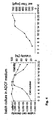
- the cells in these clusters preserved a viability above 70% throughout the process. Because dissociation of these clusters by Trypsin or Acutase (PAA) was not successful, growth kinetics and maximal cell densities were not determined. Samples were taken at day 9, 13, and 22 and titers were determined using the alpha-1-antitrypsin ELISA . Results are shown in Fig. 10 .
- Example 9 Generation of duck retina cells expressing pIX
- the cell line CR derived from primary duck retina cells and immortalized according to defined risk guidelines is described elsewhere (patent application WO05042728 , plasmid 60E-transfected retina cells).
- the cells were cultivated in DMEM:F12 medium (Invitrogen, Carlsbad, CA 92008, USA) supplemented to 5% fetal calf serum (Biochrom AG, 12213 Berlin, Germany) at 39°C and 7.5% CO 2 .
- DMEM:F12 medium Invitrogen, Carlsbad, CA 92008, USA
- fetal calf serum Biochrom AG, 12213 Berlin, Germany
- the .cells were briefly treated with TrypLE Express (Invitrogen).
- Plasmid 76F pEFPIX1 was linearized with Ssp I and Xmn I restriction enzymes (both from New England Biolabs, Beverly, MA 01915-5599, USA) and purified to 400 ng/ ⁇ l by affinity chromatography (gel extraction kit from Qiagen, 40724 Hilden, Germany). 5 ⁇ l (2 ⁇ g) of the purified DNA was transfected into CR cells with the Effectene reagents (Qiagen): DNA was mixed with 16 ⁇ l Enhancer and 200 ⁇ l EC Buffer. After 2 min at room temperature, 18 ⁇ l Effectene were added and liposome formation allowed for 10 min at room temperature.
- the transfection mix was added to 1 ml culture volume on cells seeded to 80% confluency in 6-well plates on the previous day. High transfection efficiency was confirmed with a parallel transfection of pEGFP-N1 (Clontech, Palo Alto, CA 94303-4230, USA).
- the pIX-transfected cultures were expanded into T75 flasks three days after transfection and selection was initiated with 25 ⁇ g/ml hygromycin B (Invitrogen). Medium was replaced once per week with hygromycin B raised to 50 Ng/ml. After three weeks, a total of four large foci survived and were re-seeded into a T25 flask: cells were detached with TrypLE and collected with 100 x g for 10 min, resuspended in fresh medium and plated into a T25 flask.
- hygromycin B Invitrogen
- PIX protein was confirmed by Western blotting: 6x10 5 cells were disrupted by boiling in (20 mM Tris pH 7.4, 300 mM NaCl, 1% Na-desoxycholat, 1% Triton ® X-100, 0.1% SDS) and protein was separated by gel electrophoresis, then transfered to a nylon membrane. PIX was detected with pIX primary antibody (a generous gift from Dr. W. Seidel, Ernst-Moritz-Arndt-Universmaschine Greifswald, Germany), then reacted with secondary antibody directed against the former antibody and labeled with alkaline phosphatase.
- pIX primary antibody a generous gift from Dr. W. Seidel, Ernst-Moritz-Arndt-Universmaschineifswald, Germany
- Fig. 11 (right panel) confirms expression of pIX in CRpIX cells. No signal was present in the negative control from 293 cells prepared in a parallel reaction.
- the cells were cultivated in medium #63032-1000M for two additional passages, then transfered to medium # 14561-1000M (also from JRH Biosciences) for two additional passages. All JRH media were supplemented to 1 x with Glutamax I (Invitrogen) and 100 ⁇ g/ml hygromycin B.
- genomic DNA was isolated from 1 x 10 6 cells with the QIAamp DNA Blood kit (Qiagen) and number of E1B and PIX molecules was determined in 25 ng and 50 ng of genomic DNA in an ABI 7000 TaqMan reaction with SYBR Green (ABI) detection chemistry.
- Primers used for quantification were gTggTTgCTTCATgCTAgTg ((SEQ ID NO:13) and TCTTCAgCAggTgACAgTTg (SEQ ID NO:14) for E1B, ACCTACgAgACCgTgTCTg (SEQ ID NO:15) and gAgCCgTCAACTTgTCATC (SEQ ID NO:16) for PIX.
- E1B was introduced independently and must be maintained for the cells to survive this gene serves as an internal marker to standardize PIX copy number and gene expression strength.
- Fig. 12 demonstrates stable maintenance of PIX transgene in CR suspension cells even in absence of selection pressure. It also demonstrates that PIX-positive cells suffer from a decrease in growth rate. We consistently observed decreased proliferation for all of our cells (avian and human cells) upon stable transfection with a PIX-expression plasmid demonstrating an impact of PIX on the biochemistry of the host cell. Furthermore, we observed a tendency in PIX-positive, adherent CR cells to avoid growth into confluent layers (increased contact inhibition).
- the cell line CS is derived from embryonal somites, also described in patent application W005042728 .
- Generation of PIX-positive CS cells was performed in parallel to the above described procedure for the CR line. Contrary to CRpIX, no suspension cultures were established for CSpIX. Strict anchorage dependency is a feature of the CS cells. A cell that does not proliferate in suspension can be considered to be less tumorigenic as the potential to metastasize is severly impaired. PIX protein does not change this property in CS cells. Although pleotropic this protein therefore appears not to impact on the transformation phenotype. This observation supports our assumption that PIX can safely be applied in biopharmaceutical processes.
- CS cells stably transfected for PIX expression and parental CS cells as reference were seeded into a 6-well plate at 2x10 6 cells/cavity and infected with MVA at an m.o.i. of 0.1 on the following day.
- Pictures to document the differences in the advance of CPE were taken 48 h and 72 h post infection.
- Virus yield in the supernantant 48 h post infection and complete yield (supernatant together with lysed cell pellet) 72 h post infection was determined in a microfocus assay. The results are shown in Fig. 13 : the presence of PIX in the CS cells appears to delay the onset of CPE at 48 h post infection when compared to the parental reference.
- 72 h post infection both cultures are lysed completely suggesting that the differences in the extent of CPE is not due to a mixture of MVA-refractory and MVA-susceptible cells in the CS.PIX population.
- the positive effect of the PIX protein was also quantified on CR cells. Contrary to CS cells, the CR cells are adapted to suspension. Suspension cultures are preferred for many industrial applications. We therefore determined the PIX effect in such a system as opposed to adherent culture.
- Fig. 14 shows a comparision of suspension clone CR.HS (selected out of several clones as the cell line with greatest MVA yields) and PIX(+) cell line CR.MCX.
- the abscissa indicates m.o.i. (0.01, 0.05 or 0.1, respectively), the ordinate indicates burst and the size of the bubbles indicates cell densities (8, 2, or 0.8x10 6 cells/ml, respectively).
- Burst is the ration of output virus (or yield) to input virus (the inoculum dependent on cell number and m.o.i.) and thus a measurement of amplification efficiency.
- the PIX(+) cell lines exceeds the PIX(-) line in performance.
- Plasmid 76F pEFPIX1 was treated with Acc I und Dra I restriction enzymes and termini were blunted with Klenow polymerase (all from New England Biolabs, Beverly, MA 01915-5599, USA).
- Klenow polymerase all from New England Biolabs, Beverly, MA 01915-5599, USA.
- the desired fragment of 447 bp containing the PIX coding sequence was isolated by agarose gel extraction (gel extraction kit from Qiagen, 40724 Hilden, Germany).
- the Dra I restriction enzyme recognizes the sequence "ttTAAa” to cut after the last thymidine residue, wherein the central "TAA" triplett is the stop codon of the PIX open reading frame.
- the 447 bp fragment therefore is devoid of a stop codon. Fusion of this fragment to the gene for EGFP in the plasmid pEGFP-N1 opened with Sma I (also New England Biolabs) generates a continuous fusion gene of PIX followed by EGFP.
- Sma I also New England Biolabs
- the resulting plasmid was named:p9GFP, the expressed fusion protein will be named PIX-GFP in this text.
- CS and CR cells where transfected with p9GFP and selected with 300 ⁇ g/mL geneticin (Invitrogen) for stable PIX-GFP expression.
- Two populations of different PIX-GFP expression were observed within 2 weeks: one population had usually 2 to 5 bright spots of PIX-GFP in the cytoplasm, the other population had a more uniform expression of PIX-GFP in the cytoplasm and more diffuse accumulation of GFP signal in a region proximal to the nucleus. In both cases, the cell nuclei appeared as a dark region suggesting that no or very little chimeric protein enters the nucleus. Representative examples for both types of clones in CRpIXGFP are shown in Fig. 15 .
- NLS nuclear localisation sequence
- PIX-GFP The cytoplasmic localisation of PIX-GFP in our duck cells therefore is consistent with the primary sequence of PIX.
- a PIX-GFP-NLS expression plasmid was derived from the above described p9GFP construct by insertion of synthetic oligonucleotides i185 and i186. These oligos were denatured at 80°C for 5 min and allowed to anneal in 10 mM MgCl 2 by gradual cooling to room temperature to yield:
- This double stranded oligonucleotide was digested with BsrG I and Not I and inserted into the same sites of p9GFP. Translated, the insertion adds a NLS sequence (PKKNRK; SEQ ID NO:21) to the PIX-GFP fusion protein that resembles the simian virus 40 NLS. A new Hpa I site introduced with this insert serves as diagnostic marker to confirm successful cloning.
- the resulting plasmid is called p9 GFP NLS, the expressed protein PIX-GFP-NLS and the open reading frame coding for the protein PIX-GFP-NLS are shown in SEQ ID NOs:23 and 22, respectively.
- a GFP-tagged PIX-RARA fusion protein was generated via PCR amplification of a fragment containing PIX-RARA with primers EBR 44A (5'-GGATCCTTCCTCCTCGGGCGGGTGT-3'; (SEQ ID NO:24), V293 (5'-AACCAGCGCTACCATGAGCACCAACTCGT 3'; SEQ ID NO:25) and plasmid #67F pEF PIX RARA NEO as template.
- the amplicon of 1926 bp was treated with polynucleotide kinase and inserted into pEGFP-C2 (Clontech) linearized with EcoR I and blunted with Klenow enzyme (all enzymes from New England Biolabs). The resulting plasmid is called p9 GFP RARA, the expressed protein PIX GFP RARA.
- Fig. 16 shows the intracellular distribution of the various GFP-tagged proteins in transiently transfected CHO cells.
- TLR-3 recognizes double stranded RNA, a hallmark pattern of viral replication.
- NFkB and type I interferon Alexopoulou et al., Nature 413, 732-738 (2001 )
- Interferon mediates an antiviral state in the host cell.
- poly I:poly C polyinosinic-polycytidylic acid, Sigma
- CR and CRpIX cells displayed very little poly I:poly C sensitivity whereas CS cells clearly responded to the inductor.
- PIX-positive CS cells responded faster and at lower concentrations than parental CS cells ( Fig. 17 ). To our knowledge, a connection between PIX and interferon has not been described in the current literature.
- the PIX-supportive effect for MVA was stronger in CS cells than CR cells, and the CS cells responded to double stranded RNA surrogate better than CR cells. It is known that MVA induces interferon and that this virus also is equipped with proteins that alleviate the interferon response.
- the effect of poly I:poly C inductor on MVA in CRp9GFP and CSp9GFP cells therefore determined.
- Fig. 18 suggests that MVA infected cells suffer less from poly I:poly C than uninfected control.
Landscapes
- Health & Medical Sciences (AREA)
- Chemical & Material Sciences (AREA)
- Life Sciences & Earth Sciences (AREA)
- Organic Chemistry (AREA)
- Genetics & Genomics (AREA)
- General Health & Medical Sciences (AREA)
- Zoology (AREA)
- Biochemistry (AREA)
- Engineering & Computer Science (AREA)
- Medicinal Chemistry (AREA)
- Virology (AREA)
- Wood Science & Technology (AREA)
- Bioinformatics & Cheminformatics (AREA)
- Biophysics (AREA)
- Gastroenterology & Hepatology (AREA)
- Molecular Biology (AREA)
- Proteomics, Peptides & Aminoacids (AREA)
- Biotechnology (AREA)
- Biomedical Technology (AREA)
- Immunology (AREA)
- Cell Biology (AREA)
- Microbiology (AREA)
- General Engineering & Computer Science (AREA)
- High Energy & Nuclear Physics (AREA)
- Physics & Mathematics (AREA)
- Toxicology (AREA)
- Micro-Organisms Or Cultivation Processes Thereof (AREA)
- Preparation Of Compounds By Using Micro-Organisms (AREA)
- Peptides Or Proteins (AREA)
Abstract
Description
- The present invention provides a method for preparing a non-adenoviral target virus or target proteins utilizing a potent expression cell line having stably integrated into its genome a gene encoding a PIX protein.
- Biopharmaceutical products from eukaryotic cells are an integral part of modern medicine. However, increased productivity and increased safety are important parameters that still require substantial optimization. The most crucial bottleneck in the efforts towards optimization is the cellular substrate itself. A safe transgene that can be introduced into cell lines already released for use in biopharmaceutical processes to increase product yields from the such manipulated cells is extremely valuable.
- Adenoviruses are unenveloped (naked) double-stranded DNA viruses that infect a broad spectrum of animals. Among the best characterized members is, the human adenovirus serotype 5 (Ad5). This virus is a frequent cause of common cold symptoms and infection often occurs in childhood.
- Adenoviruses are amenable to genetic manipulation and replication incompetent adenoviruses, including Ad5 (GenBank accession number for sequence: AC_000008), are used as vectors in gene therapy and for therapeutic vaccination. To obtain replication incompetent viruses large regions of the genomic DNA are substituted with non-viral sequences. The missing viral functions are provided in trans by host cell lines that have been stably transfected with genes that are deleted in the vector. One of the earlier systems that were developed consist of adenoviral vectors deleted in the regulatory E1 region produced on cell lines providing the corresponding E1 proteins.
- The most common cell line for this purpose is the 293 cell line. This line was generated in 1977 by transfection of fragmented adenoviral genomic DNA into primary human cells (Graham et al., J. Gen. Virol. 36, 59-74 (1977)), well before it was recognized that adenoviruses may serve as gene therapy vectors. The resulting cell line was subsequently shown to contain
nucleotides 1 to 4344 of the genomic DNA (Louis et al., Virology 233, 423-429 (1997)) which includes the E1 region; this characterization demonstrated that the E1 region can be used to immortalize and transform primary cells. - Adjacent to the E1 region follows the gene for pIX ("
protein 9"; nucleotides 3609 to 4031 on the genomic DNA). The promoter for pIX is embedded within the E1B component of the E1 region. A mechanism called promoter occlusion allows expression of pIX at a delayed-early time In the viral infection cycle at the onset of viral DNA replication with increasing copy number of viral DNA (Fessler and Young, J. Virol. 72, 4049-4056 (1998)). Although the gene is present within the 4344 nucleotides integrated into 293 cells, pIX expression cannot be detected even with sensitive radioactive labeling methods (Spector et al., J. Virol. 36, 860-871 (1980)). - As described above, replication deficient adenoviral vectors have been generated by deletion of the E1 region from the viral genome. E1 products are essential for viral replication and the function of the E1 region therefore had to be provided in trans via stable E1 transgenes in the adenovirus packaging cell (such as the 293 cell line). Because of the proximity to the E1 region the pIX gene caused frequent recombination between deleted adenoviral vectors and E1 in the host genome. This recombination event generated replication competent adenovirus (RCA), a serious contamination in vector preparations. To suppress this recombination event pIX was deleted from the adenovirus genome. In the course of these experiments it was recognized that pIX stabilizes the adenoviral capsid against thermal and steric stress by increasing the interaction of the main building blocks, the hexons (Colby and Shenk, J. Virol. 39, 977-980 (1981); Ghosh-Choudhury et al., EMBO J. 6, 1733-1739 (1987)). Morphogenesis In the absence of pIX yields temperature-sensitive viruses that cannot deliver genomic DNA molecules larger than 105% of the wild type 35938 base pairs. To still allow a normal packaging capacity and thermal stability the pIX protein was stably or episomally introduced into cell lines intended as packaging cells for adenovirus vectors (Krougllak and Graham, Hum. Gene Ther. 6, 1575-1586 (1995); Carovokyri, C. and Leppard, K.N., J. Virol. 69, 6627-6633 (1995)
WO 99/57296 - Also with recognition that pIX decorates the surface of virions pIX-fusion proteins have been generated with the purpose to expand the host range of adenovirus vectors or to follow morphogenesis and the intracellular migration of virus particles (reviewed by Parks, Mol. Ther. 11, 19-25 (2005)).
- Although clearly a structural protein, pIX also is implicated as regulatory protein for adenovirus replication. PIX is assumed to function as transactivator of transcription to augment E1A expression, a function possibly even excerted as a virokine by incoming PIX protein from the capsid at the infection step (reviewed by Parks, Mol. Ther. 11, 19-25 (2005)). PIX, together with early protein E4 Orf3, also is described to interact with sub-nuclear inclusions called PML bodies (Puvion-Dutilleul et al., Exp. Cell. Res. 218, 9-16 (1995); Leppard and Everett, J. Gen. Virol. 80, 997-1008 (1999)). These are dynamic aggregates of 250 nm to 500 nm in size and proposed to be involved in regulation of cell differentiation (Wang et al., Science 279, 1547-1551 (1998)), control of apoptosis (Quignon et al., Nature Gen. 20, 259-265 (1998)), and response to viral infection (Möller and Schmitz, Arch Immunol Ther Exp (Warsz) 51, 295-300 (2003)).
- Because of its pleotropic effects we introduced the PIX protein into cell lines to examine whether PIX augments cell proliferation or production properties for biopharmaceutical products that are not related to adenovirus or adenoviral vectors. There is a general need for factors that modulate these properties in established cell lines.
- For example, attenuated (weakened) viruses are promising vaccine candidates: upon inoculation they mimick a natural infection but allow more time for establishment of the desired protective immune response by the vaccinee. With increasing numbers of immunocompromised patients (for example, due to HIV infection) highly attenuated strains are desirable. Whereas attenuated strains still proceed towards (usually benign) infection highly attenuated strains are blocked at a cellular level even in absence of a functional immune system. A frequently used method to establish and maintain attenuation is to passage viruses on different host tissue. For example, measles and mumps viruses intended for human vaccination are passaged in primary cells, either in embryonated chicken eggs or cultures derived thereof. A new generation of vaccines is based on highly attenuated pox viruses that also depend on primary chicken cells for production. A cell line that can substitute for primary chicken cells and at the same time that even less efficiently protects itself against viral infection due to secondary manipulations such as introduction of the PIX transgene therefore provides a highly desirable substrate.
- For other purposes a mammalian (rather than avian) cell line may be preferred. Such preferred cell lines already exist and have passed the examination by health authorities with respect to safety and risks posed by derived biopharmaceutical products. Here too, a secondary manipulation (such as introduction of the PIX transgene) to increase the spectrum of available applications or production efficiencies without compromising safety features is highly desirable.
- When preparing cell lines that have been stably transfected with adenoviral gene pIX (or a chimaeric fusion analogue thereof), we unexpectedly observed that pIX excerts a phenotypical effect in avian and human cells. For avian cells this is especially surprising because avian cells cannot be infected by human adenoviruses and aviadenoviruses (such as CELO or fowl adenovirus type 8) do not encode a PIX homologue (Ojkic and Nagy, J. Gen. Virol. 81, 1833-1837 (2000)). Furthermore, we observed that stable presence of PIX increases susceptibility of the cell to induction by double stranded RNA analogue, probably via toll like
receptor 3. Probably in this context, we also unexpectedly observed that the presence of PIX protein increases yields of highly attenuated poxvirus in avian host cells. As poxvirus infected cells suffered less from induction by double stranded RNA analogue we may have found an interaction between PIX protein and anti-interferon genes of poxvirus that has not been described previously. We also observed a surprising increase in the yield of proteinaceous product (not only virus) released by a stably transfected cell line. - Thus, the present invention provides
- (1) a method for preparing a non-adenoviral target virus or one or more target proteins, which method comprises
- (a) culturing an expression cell being obtainable by infecting or transfecting a host cell, having stably integrated into its genome a gene coding for a Adenovirus PIX and stably expressing said PIX protein with said target virus, or with a vector carrying nucleic acid sequences encoding said target virus, or with a vector carrying nucleic acid sequences encoding said one or more target proteins (wherein the non-adenoviral target virus does not contain said PIX protein and the target protein(s) differs from said protein PIX, and
- (b) isolating the target virus or the target protein(s);
- (2) a preferred embodiment of (1) above, wherein said PIX protein enhances the productivity of the cell In the production of a virus not containing said PIX protein and in the production of a protein differing from said regulator protein;
- (3) a preferred embodiment of (1) and (2) above, where the adenovirus pIX is provided as a fusion protein with retinoic acid receptor alpha, preferably is a fusion protein having the sequence shown In SEQ ID NO:4, or is fused to a GFP and contains an NLS, preferably is a fusion protein having the sequence shown in SEQ ID NO:23;
- (4) a preferred embodiment of (1) to (3) above, wherein the host and expression cell is a vertebrate cell including mammalian cells and avian cells, preferably the mammalian cell is a human brain derived cell, and the avian cell is a duck retina derived cell or a duck somite derived cell;
- (5) a preferred embodiment of (1) to (4) above, wherein the host and expression cell is derived from a human brain, preferably from human foetal brain, and most preferably is a NC5T11 cell, and said cell carries a nucleic acid sequence encoding Adenovirus PIX preferably said PIX protein is encoded by a nucleic acid shown in SEQ ID NO:1, SEQ ID NO:3 or SEQ ID NO: 22;
- (6) a preferred embodiment of (5) above, wherein the host cell is a
NC5T11# 34 cell and the expression cell is derived from theNC5T11# 34 cell, saidNC5T11# 34 cell being deposited with the DSMZ under Accession Number DSM ACC2744; - (7) a preferred embodiment of (1) to (4) above, wherein the host and expression cell is an avian cell, preferably is derived from duck retina or duck somite, and said cell carries a nucleic acid sequence encoding Adenovirus PIX, preferably said PIX protein is encoded by a nucleic acid shown in SEQ ID NO:1, SEQ ID NO:3 or SEQ ID NO: 22;
- (8) a preferred embodiment of (7) above, wherein the host cell is a CR.PIX (17a11b) cell and the expression cell is derived from the CR.PIX (17a11b) cell, said CR.PIX (17a11b) cell being deposited with the DSMZ under accession number DSM ACC2749;
- (9) an immortalized vertebrate expression cell for producing a target virus 375.5.2011 as defined in (1) to (8) above, preferably the expression is as defined in (5) to (8) above; wherein the expression cell has stably integrated into its genome a gene according for adenovirus PIX and stably expressing said PIX and infected or transfected with a non-adenoviral target virus or with a vector carrying nucleic acid sequences encoding said virus;
- (70) the use of the expression cell as defined in (7) above for preparing a target virus 5.5.2011.
-
-
Figure 1 : History of the cell line NC5T11. A neurospheres of primary cells grown under serumfree conditions. B monolayer of primary cells grown in DMEM/F12 with 5% FCS. C primary focus two weeks after transfection with p79. the arrows indicate the border between primary and immortal cells. D homogenous adherent culture of immortalised cells in DMEM/F12 with 5% FCS. E suspension culture of NC5T11 in shaker tubes in EXcell VPRO. -
Figure 2 : Immunofluorescense detecting E1A and E1B in NC5T11. After fixation with methanol cells were treated with rat antibodies directed against E1A and E1B 55k respectivly followed by texas red conjugated anti rat antibodies. As shown infig. 2 all cells in the sample show nuclear staining typical for E1A and cytoplasmic staining for E1B. After fixation with methanol cells were treated with rat antibodies directed against E1A and E1B 55k respectivly followed by texas red conjugated anti rat antibodies. As shown here, all cells in the sample show nuclear staining typical for E1A and cytoplasmic staining for E1B. -
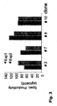
Figure 3 : Specific productivity of selected aat clones. Cell clones of NC5T11 transfected with C55 and selected with puromycin were seeded at 7x10⁴ cells in 12 well plates, cell number and aat titer was determined onday -
Figure 4 : Expression analysis ofNC5Tllpuro# 8 in shaker batch culture. Cells were seeded in 12 ml of EXCELL VPRO at 6x10⁴ cells/ml in 50 ml polypropylene shaker tubes (TPP, Switzerland) with and subjected to rotation with a radius of 1 cm and a speed of 160 rpm. Samples of 200 µl were taken ondays -
Figure 5 : Detection of the pIX-RARA cDNA by PCR in a clone pool. A clone pool carrying the pIXRARA gene was generated fromNC5T11puro# 8 by transfection with F67 and hygromycin selection. RNA was extracted using an RNA extraction kit (Machery Nagel, Germany) and cDNA was syntesized using AMV Reverse Transcriptase (Invitrogen). cDNAs for the pIX fragment alone or the whole pIXRARA gene were amplified withprimer -
Figure 6 : Detection of the pIXs sequences as part of the fusion protein in hygromycin resistant subclones ofNC5T11puro# 8 and NC5T11(#34, 35, 36, 37, 38) by PCR. DNA was isolated from cell clones grown in 6 wells By lysis in SDS followed by phenol extraction and precipitation. DNA was amplified withPrimers -
Figure 7 : Stability of the pIXRARA and pIX genes in stably transfected NC5T11 was determined relative to E1B: E1A+E1B and pIXRARA were introduced in separate transfections and E1 genes and have been maintained in the absence of selection for > 2 years and are therefore considered stably integrated. Genomic DNA was isolated from selected cell clones at 2 time points: immediately after hyg selection stop (early); after 2 month in the absence of selection pressure (late). PIX and E1B DNA levels were determined by Real Time PCR. -
Figure 8 : Growth retardation induced by RA treatment in pIXRARA recombinant clones of NC5T11 andNC5T11puro# 8. Cells were seeded at 2x10⁵ in 6 well plates in the presence or absence of 6 µg/ml retinoic acid. Photos showing growth retardation in RA treatedclones # 12 and #34 but not in NC5T11 were taken 4 days after seeding using phase contrast imaging at a 4x magnification. -
Figure 9 : PIXRARA prevents inhibition of virus replication by interferon. NC5T11 andNC5T11# 34 cells were infected with EMCV an interferon sensitiv virus at an MOI of 0.004 in cells treated with interferon beta. Cell lysates were subjected to titration by plaque formation on A549 cells. -
Figure 10 : Expression analysis of the cellline NC5T11puro# 8 expressing alpha-1-antitrypsin from vector C55 andsubclones # 10, #12, #14 thereof, carrying the vector F67 (pEFpIX-RARA) in addition to C55. Cells were seeded in EXCELL VPRO (JRH Biosciences) at 6x10⁴ cells/ml in 50 ml polypropylene shaker tubes (TPP, Switzerland) with a total volume of 12 ml and subjected to rotation with a radius of 1 cm and a speed of 160 rpm. -
Figure 11 : Expression of PIX in duck retina CR cells. Left Panel: PCR reaction against PIX gene in genomic DNA of pIX cells. Gel loading from left to right: 1 kb-marker (Invitrogen); PCR on genomic DNA from CRpIX; non-template control; positive control with plasmid used in transfection of CRpIX. Right panel: Western blot for detection of pIX protein in CRpIX cells. Lane 1: 293 cells; lane 2: pIX protein in CRpIX cells. -
Figure 12 : Stable maintenance of PIX transgene in duck retina cells. Top-left panel: MCX and DXS are independent aliquots of a CRpIX clone cultivated for > 3 months in parallel and without selection. After three months, TaqMan PCR was used to count the copy numbers of E1B and PIX transgene in MCX and DXS. As E1B transgene is maintained independent of PIX the ratio of the two genes is an indication of maintenance of PIX transgene. The ratio did not change between MCX and DXS. Furthermore, the ratio of PIX mRNA to E1B mRNA also did not change and PIX mRNA was in excess of E1B mRNA indicating expression of PIX. Right panel: MCX and parental (PIX-negative) CR.HS cells were cultivated with or without hygromycin selective pressure for 2 weeks (indicated by black bar on x-axis). At various time points genomic DNA was isolated (indicated by "A", "B", and "C") and - subjected to TaqMan quantification (bottom-left). Whereas parental cells where killed by hygromycin MCX cells survived. The ratio of transgenes remained constant, again indicating stable maintenance of PIX transgene also in presence of selective pressure. Doubling time for parental cells is approx. 32 h but only 41 hours for MCX cells.
-
Figure 13 : PIX effect on MVA replication in CS cells. CS and CSpIX cells were infected with MVA and assayed forMVA replication - The yield of MVA was significantly higher in PIX-positive CS cells. Also, as evident in the phase contrast microimage, cytopathic effect appeared delayed for CSpIX cells at 48 h. However, both cultures are susceptible to virus to similar degrees as complete lysis is evident at 72 h post infection.
-
Figure 14 : PIX effect on MVA replication in CR cells. CR and CRpIX cells in suspension were infected with various multiplicities and at various seed cell densities. MVA yield was assayed 48 h post infection and is shown on the y-axis. - The size of the bubbles displays seed cell densities. The filled bubbles show values for CRpIX cells, white bubbles show values for parental cells. In all configurations,
- PIX confers a clear increase in amplification rates of MVA.
-
Figure 15 : Subcellular distribution of PIX-GFP in duck cells. GFP-tagged PIX appears in few cytoplasmic bright spots and diffuse cytoplasmatic staining largely excluding the nucleus. Other clones exhibit stronger, diffuse cytoplasmatic staining and more intense accumulation just outside the nucleus. -
Figure 16 : Subcellular distribution of PIX-GFP variants in CHO cells. To investigate induced changes of PIX distribution within cells fusion variants were generated, including insertion of a nuclear localization site (NLS) and fusion to retinoic receptor alpha, a cellular protein that already contains an NLS. Shown are CHO cells transiently transfected with expression plasmids for PIX-GFP variants. -
Figure 17 : Effect of interferon induction in presence of PIX. CSpIX and CRpIX cells were treated with poly I: poly C, a known inductor of type I interferon response and compared to parental cells. CSpIX cells reacted with greater sensitivity to poly I:poly C than CS cells. CRpIX and CR cells reacted comparably and to a lower extent than CS cells. -
Figure 18 : Effect of MVA infection and interferon induction in presence of PIX-GFP. CSp9GFP and CRp9GFP cells were treated with poly I:poly C and infected with MVA at an M.O.I. of 0.1 as indicated. Again, CS-derived cells reacted more strongly to interferon induction. Surprisingly, bright PIX bodies appear to decrease in number upon induction or infection in both cell lines whereas overall signal intensity of PIX-GFP increases. Furthermore, the effect of poly I:poly C induction is ameliorated upon parallel infection with MVA as indicated by a greater number of CS cells still adherent 22 h after treatment. - The method of embodiment (1) of the invention utilizes an expression cell having integrated into its genome a gene coding for, pIX which possesses the following properties:
- 1. It modulates transcription and, especially if linked to appropriate modulators or regulators, also influences cell growth.
- 2. It enhances productivity of the cell line with regard to the production of a virus not containing said PIX protein (i.e. the PIX protein is not a substitute of 5.5.2011 for a protein deleted from the virus) and/or with regard to the production of a protein differing from said regulator protein.
- According to an embodiment of the invention the heterologous regulator protein is the
adenovirus serotype 5 protein pIX in other embodiments the protein is obtained from an adenovirus of a different serotype. - Particularly preferred are fusion proteins, namly fusion proteins comprising at least one first domain comprising a PIX protein as defined hereinbefore and at least one second domain comprising a protein or peptide acting as a transcription modulator. In a preferred embodiment of the invention said transcription modulator is a transcription factor including the retinoic acid receptor alpha, which may be present in complete or incomplete (i.e. truncated) form. The modulator may also be a transit peptide, which includes NLF sequences, e.g. that shown in SEQ ID NO:21. According to the invention the first and second domain(s) are either directly or via a peptide linker covalently attached to each other. Suitable peptide linkers include flexible and hydrophilic structures such as poly gly-ser.
- The terms "cell" and "cell line" as used in the following detailed description refer to expression cells/expression cell lines and host cells/host cell lines.
- According to the invention it is preferred that the PIX protein is expressed in the cell in an amount of at least 1 pg/µg cellular protein, preferably at an amount of at least 10 pg/µg cellular protein so that its expression can be determined by Western-blotting.
- In the cell of the PIX protein is preferably under control of a stabile homologous or heterologous promoter. Suitable promoters are constitutive cellular promoter or variants thereof such as the human
translocation elongation factor 2 promoter. A particular variant of the humantranslocation elongation factor 2 promoter utilized within the invention has the sequence shown in SEQ ID NO:12. The sequence shown represents a "short" version of the promoter which is located on human chromosome 19:3,935,325-3,936,638 (human genome assembly May 2004) and provides for a stable medium level expression. For a stronger expression a "longer" version of the promoter located on chr19:3,935,349-3,938,957 (human genome assembly May 2004) may be used. - According to the invention the cell is a vertebrate cell including mammalian cells, and avian cells. Suitable mammalian cells are human cells, and rodent cells including mouse, rat, hamster, etc. Particularly preferred mammalian cells according to the invention are cells derived from human brain, particularly from human foetal brain, a mouse NSO or Sp2/0 cell, a BHk or CHO cell. Suitable avian cells are duck, chicken quail and goose cells. Particularly preferred avian cells according to the invention are cells derived from a duck retina-derived or somite-derived cell.
- On the other hand, the cell of the invention may be derived from a primary cell or from a previously immortalized cell. Moreover, the cell may carry further immortalizing (viral) genes including an E1 protein of an adenovirus, such as the E1 protein of mastadenovirus
group C type 5, and the like. Particularly preferred according to the present invention is that the cell further carries the adenovirus E1A and/or E1B gene shown In SEQ ID NO:5. - The cell utilized in the method of embodiment (1) of the invention may further carry functional sequences, e.g. sequences required for its application as expression cell such as selection marker sequences, splice donor/acceptor sites and/or recombinase recognition sequences allowing integration of a target nucleic add sequence to be expressed in the cell, etc.
- The "infecting", "transfecting" and "transforming" effected within the method of embodiments (1), (10) and (12) of the invention may be performed according to standard procedures known to the skilled person. Said methods may further encompass suitable selection, isolation and expansion steps, if required.
- In the method of embodiment (1) the target virus includes from wild-type, mutated or deleted virus, cold adapted or attenuated virus, vaccine strains, viral vectors carrying heterologous gene(s), viral vectors such as lentivirus, poxvirus, adeno associated virus (aav), herpesvirus, and flavivirus.
- Further, in the method of embodiment (1) the one or more target proteins include antibodies, recombinant proteins such as erythropoetin, alpha-1-antitrypsin, blood clotting factors VIII and IX and interferons, viral antigens such as influenza HA and NA, and M, HBV-S, herpes G protein and rabies G protein, peptid hormones and the like. Although the method allows the production of cells capable of contemporary expression of more than one target proteins, it is particularly preferred that it encodes only one target protein.
- The culturing and isolation of the method of embodiment (1) of the invention may be performed according to standard procedures readily available to the skilled person. Said method may further include standard purification steps as well as subsequent modification steps of the target virus or target protein(s).
- Concerning the preferred embodiments of the expression and host cell lines of embodiment (9) it is referred to the detailed discussion provided hereinbefore In connection with embodiment (1).
- The cell
line NCST11# 34 was deposited with the DSMZ-Deutsche Sammlung von Mikroorganismen und Zellkulturen GmbH, Mascheroder Weg 1b, 38124 Braunschweig, Germany on November 4, 2005 under accession number DSM ACC2744. The cell line CR.PIX (17a11b) was deposited with the DSMZ-Deutsche Sammlung von Mikroorganismen und Zellkulturen GmbH, Mascheroder Weg 1b, 38124 Braunschweig, Germany on November 24, 2005 under accession number DSM ACC2749. - The invention will be explained in more detail by means of the following examples, which are, however, not to be construed to limit the invention.
- The cell line was developed from a mixture of cells from foetal brain by immortalisation with
adenovirus 5 E1A and B genes by nonviral transfection. - A tissue sample was taken from the periventricular zone of the brain of a foetus after induced abortion. It was dissected with scissors and homogenised by aspiration with a tissue culture pipette in neuronal culture medium based on DEMEM/F12 containing 20 ng/ml hFGF (Invitrogen, Carlsbad, CA 92008, USA), 20 ng/ml hEGF (Invitrogen), 1x N2 supplement (Invitrogen) and 8 µg/ml heparin (Sigma Aldrich, St. Louis, USA). Cells were sedimented at 200g for 3 min. Viability of cells was assessed using trypan blue and propidium jodide using a flow cytometer (BD Biosciences, Jose, CA 95131, USA). The viability was 75%. 0.5x10⁶ cells were seeded into T25 flasks. Cells were incubated at 37°C and 5% CO2. The cells formed neurospheres by
day 5 of the culture. Neurospheres are shown inFigure 1A . Onday 8 cells were transferred to DMEM/F12 supplemented with 5% FCS to allow for attachment and to stimulate proliferation. Cells formed a homogenous monolayer as shown inFigure 1B and were passaged 1:5 once per week. This primary neural cell culture was named NC5. - After 2 weeks in serum containing medium cells were transfected with vector p79 in subconfluent 6-well plates using Effectene (Qiagen, 40724 Hilden, Germany) as a transfection reagent following the manufacturers instructions. The plasmid p79 contains the following elements: The pBluescript (Stratagene, USA) serves as plasmid backbone, in which the ampicillin resistance marker was replaced by the kanamycin resistance gene driven by a bacterial promoter which allows for growth and selection in E.coli. The vector harbours a fragment form wild type adenovirus type 5 (SEQ ID NO: 5) containing the open reading frames for E1A (splice variants 13S and 12S with or without the CR3 domain respectively) and E1B 55k and 19k as well as sequences upstream of E1A. The E1A gene is preceded by the phosphoglycerate kinase promoter (mouse). The adenovirus sequences are followed by the polyadenylation signal from the Herpes simplex thymidine kinase gene, serving as a replacement for the E1B polyadenylation signal. Elements were obtained from the respective organisms or from donor plasmids by PCR and cloned using conventional recombinant DNA techniques and verified by sequencing. Two days after transfection cells were trypsinized and transferred to a 10 cm dish. Two weeks later foci of small cells with a high nucleus/Cytoplasma ratio and clearly distinguishable borders were formed (
Figure 1C ) in transfected but not in mock treated cells. From the 11th transfection eight independent foci were isolated with and trypsin using cloning cylinders (Corning, USA), seeded to wells of a 24 well plate and expanded via 12 well plate, 6 well plate to T25 flasks. All isolated foci contained two types of cells: small cells with sharp borders and extended fibroblast like cells. - Three weeks posttransfection 15 additional clones became visible in transfection T11 some of which may originate from remaining cells of already isolated primary clones.
- Clones T11a.1 and T11a.6 showed the fastest growth and were cryopreserved in DMEM/F12, 10% DMSO, 25
% serum 8 weeks post transfection. At this time all clones still contained a fraction of cells with enlarged cytoplasma resembling primary phenotype. However, with a split ratio of 1:5 for T11a.1 and T11a.6 they were overgrown and eliminated approximately 3 month after transfection. Immunofluerescence experiments were carried out to associate the changed morphology with expression of E1 A and E1B. After fixation with methanol cells were treated with rat antibodies directed against E1A and E1B 55k respectively followed by texas red conjugated anti rat antibodies. As shown inFig. 2 all cells in the sample show nuclear staining typical for E1A and cytoplasmic staining for E1B. - Three month after transfection clone T11a.1 and T11a.6 were transferred to serumfree medium ProPER (Cambrex, Belgium) by seeding 1.6x10⁶ cells after trypsin treatment. Cells were harvested by centrifugation and medium was exchanged by centrifugation once a week. The cell population survived but even six month after transfection cells still showed a low viability and grew with a high doubling time of 60-80 h.
- When the cells were transferred back to DMEM/F12 with 5% FCS after 3 month in ProPER medium, a homogenous and highly viable culture with a doubling time of 40 h has formed for both clones. T11a.1 was chosen as the more most robust cell clone for further experiments and named NC5T11 (
Fig. 1D ) - Protein production in a cell line depends on efficient transfection and selection methods. The suitability of commonly used selection markers G418, puromycin, hygromycin, blasticidin, MTX, histidinol for NC5T11 was unknown. In a first evaluation step different commercially available transfection reagents Lipofectamine (Invitrogene), Fugene (Roche, Germany), Polyfect (Qiagen) and Effectene were tested following the manufacturers protocols. The plasmid pEGFP-N1 (Clonetech, USA) expressing gfp from the human CMV promoter was used to determine transient transfection efficacy. The highest efficiency was obtained with Effectene (Qiagen) reaching 20-50% depending on cell density when applied to adherent cells growing in DMEM/
F12 10% FCS. - To test selection marker genes, expression vectors were constructed containing the human alpha-1-antitrypsin (aat) gene under control of the human CMV promoter followed by the bovine growth hormone PolyA signal and the respective selection marker controlled by a weak promoter. More specifically, to test for puromycin selection, vector C55 was used which contains the puromycin resistance gene driven by the human PGK promoter followed by the SV40 early polyadenylation signal.
- After transfection cells were seeded in DMEM/
F12 5% FCS containing 0.75 µg/ml puromycin and selected for 3 weeks with weekly medium exchange. A clone pool was generated and expression was verified by immunofluoroescense using a goat polyclonal anti- alpha-1-antitrypsin antibody (Innogenetics, USA) followed by a biotin labelled secondary rabbit anti-goat antibody and a streptavidin-Texas Red conjugate. - To isolate
individual cell clones 10000 cells of the clone pool were seeded in a 15 cm dish. After 4 weeks clones were isolated using clone cylinders (Corning) andclones # Figure 3 .Clone NC5T11puro# 8 shows a specific productivity above 100pg/cell·day and was chosen for further development. - The growth rate of both NC5T11 and
NC5T11puro# 8 in ProPER medium (Cambrex) was approximately two times reduced compared to serum containing medium. Therefore, Xcell VPRO (JRH Biosciences) was tested as an alternative. Adaptation was done by direct seeding of 4.5x10⁶ cells in 5 ml EXCELL VPRO in a T25 flask. After 2 weeks cell growth reinitiated and a weekly split ratio of 1:3 was established. As a next step both clones were adapted to growth in the presence of shear stress. Cells were seeded in 12 ml of EXCELL VPRO at 6x10⁴ cells/ml in 50 ml polypropylene shaker tubes (TPP, Switzerland) with and subjected to rotation with a radius of 1 cm and a speed of 160 rpm. After repeated passages batch assays were performed o determine maximal cell densities and product accumulation during exponential and stationary growth phases. The following observations were made: Clone NC511puro showed a high viability which was maintained above 70% untilday 18. Maximal cell density did not exceed 1x10⁶. Product accumulation continued during stationary phase (Fig. 5 ). It was concluded that specific media development or further cell improvement will be required to achieve a high cell density comparable to those obtained with CHO cells. This will likely result in a substantial increased product titers. - Sequences for adenovirus PIX and retinoic acid receptor alpha (RARA) were obtained by PCR from
adenovirus type 5 and human genomic DNA respectively using primers - 1. AACCAGCGCTACCATGAGCACCAACTCGT (SEQ ID NO:6)
- 2. AATGGTGGCAACCGCATTGGGAGGGGAGG (SEQ ID NO:7) for PIX and
- 3.CCAATGCGGTTGCCACCATTGAGACCCAGA(SEQ ID NO:8)
- 4. AAGGAGCGCTGGCGAGGGCTGTGTCCAT (SEQ ID NO:9) for RARA.
- Both Fragments were amplified separately. An overlap between
primers PCR using primers human elongation factor 2 promoter located on chr19:3,935,325-3,936,638 (human genome assembly May 2004; SEQ ID NO:12). It contains upstream elements and the first intron of the EF2gene. The natural start codon of EF2, located 5' to the first intron, was removed by mutagenesis. In the vector, pEFpromhyg, the EF2 sequence is followed by the unique restriction site (AfeI) used for insertion of pIX-RARA. An internal ribosome binding site following the AF1 site allows expression of the hygromycin resistance gene as a second open reading frame in a joined RNA with pIX-RARA. This configuration supports pIX-RARA expression in all hygromycin resistant cells. - The gene for wt pIX was amplified using primers AACCAGCGCTACCATGAGCACCAACTCGT (SEQ ID NO:10) and ACCGAGCGCTTGTTTTAAACCGCATTGG (SEQ ID NO:11) and integrated into the Afe site of pEFpromhyg instead of pIX-RARA to yield pEFpIX.
- Cell clones NC5T11 and
NC5T11puro# 8 were prepared for transfection in 6-well plates by seeding 1.3x10⁶ cells/well in DMEM/F12, 5% FCS. Plasmid F67 (pEFpIXRARA) and plasmid F76 (pEFpIX) were linearised with restriction enzyme Asp700 (Roche) and transfected after ethanol precipitation using Effectene (Qiagen): DNA was mixed with 16 µl Enhancer and 200 µl EC Buffer. After 2 min at
room temperature, 18 µl Effectene were added and liposome formation allowed for 10 min at room temperature. The transfection mix was added to 1 ml culture volume on cells seeded to 80% confluency in 6-well plates on the previous day. High transfection efficiency was confirmed with a parallel transfection of pEGFP-N1 (Clontech, Palo Alto, CA 94303-4230, USA). One day after transfection medium was exchanged to DMEM/F12 with 10% FCS and 75 or 100 µg/ml (NC5T11) and 50 µg/ml µg/ml (NC5T11puro#8) hygromycin was added. Selection medium war replaced two times per week. After 18 days 15-20 clear clones became visible in each of the wells. Clone pools were treated with trypsin and selection was continued for two more weeks. Clone pools were tested for expression of PIXRARA by RTPCR. A strong signal was detected withprimers land 2, a weaker signal withprimers individual cell clones suspension culture 2000 viable cells were mixed with 12 ml clone matrix (Genetix,UK), seeded into a six-well plate (Greiner)(2 ml/well) and subjected aoutomated colony picking (clonepixFL, Genetics, UK). Clones originating fromNC5T11puro# 8 were given numbers #10-21. Clones originating from NC5T11 were given # 34-45. Clones transfected with pEF2PIX were named NC5T11PIXA- NC5T11PIXE. Clones were tested for the presence of the respective vector by PCR. An initialtest using primers Fig. 6 . All clones showing a positive signal were shifted to growth in ExCell VPRO. Readaptation was done by direct seeding of 4.5x10⁶ cells in 5 ml EXCELL VPRO in a T25 flask. After 6 weeks in stationary culture cells were transferred to 50 ml polypropylene shaker tubes (TPP, Switzerland) at a seeding density of 6x10⁴ cells/ml in EXCELL VPRO with a total volume of 12 ml and subjected to rotation with a radius of 1 cm and a speed of 160 rpm.Clones # 10, #12, #14 (NC5T11puro#8pIXRARA) and #34, #43 (NC5T11pIXRARA) and NC5T11PIXB, NC5T11PIXC were chosen for there highest viability under shear stress.Clones # 34, 43 and NC5T11PIXC were recloned and analysed for stability of the integrated DNA. Stability was assessed determining the gene ratio between PIX and E1B because E1A+E1B and pIXRARA were introduced in separate transfections and E1 genes were introduced and have been maintained in the absence of selection for > 2 years and are therefore considered stably integrated. Genomic DNA was isolated from selected cell clones at 2 time points: immediately after hyg selection stop (early); after 2 month in the absence of selection pressure (late). PIX and E1B DNA levels were determined by Real Time PCR (ABI 7000, CYBR Green) at 2 DNA concentrations Examples of this evaluation are shown inFig. 7 The vector pEF2PIXRARA is stably maintained in subclones of #34 and #43 with approximately 0.4-0.6 copies of PIXRARA per E1B. Clone NCST11PIXC originally contains 4 copies of pIX per E1B. This number is reduced twofold during the cause of the experiment. - Retinoic acid (RA) is an inducer of cell differentiation. Differentiation is typically associated with reduced cell growth. The response to RA depends on expression of retinoic receptor alpha, a transcription factor activated by RA. RA has a profound effect on PML (promyelocytic leukaemia ) reverting the transformed phenotype by growth arrest.
- As shown in
Fig. 8 RA treatment at a concentration of 6 µg/ml did not have any effect on growth of either NC5T11 or the alpha-1-antitrypsin expressingclone NC5T11puro# 8. However, growth of clones #14 (NC5T11puro#8pIXRARA) and #34 (NC5T11pIXRARA) were arrested by treatment with 6 µg/ml for 5 days in adherent culture in DMEM/F12 with 5% FCS. - NC5 T11 and
NC5T11# 34 cells were seeded into 6 well plates at 8.0x10⁵ cells/well in DMEM/F12 5% FCS. After 12 h interferon beta (rebif 44 of Serono) was added to wells at a concentration of 1 and 8 IU/ml. After 30 min interferon treated cells and control cells of both NC5 T11 andNC5T11# 34 were infected with Encephalomyocarditis virus (EMCV) at a multiplicity of infection (MOI) of 0.004. After 24 h, cultures were harvested and frozen at -80 °C to disrupt the cells. This suspension was thawed and cleared by centrifugation with 800 x g for 10 min. Lysates were subjected to virus titeration by plaque assay on A549 cells: In brief, A549 cells were seeded to 24 well plates and grown to reach confluency, incubated for 30 minutes with cleared lysates diluted 10 to 2x10⁸ fold, overlayed with 0.2 % low gelling agarose type VII in RPMI 10% FCS and incubated at 37 °C for 24 h. The agarose layer was aspirated, cells were fixed with 2% glutaraldeyhde in PBS for 20 min at 20 °C, washed with water and incubated for 30 min at room temperature with 1% Kristallviolett solution in 50% ethanol to count virus plaques. Results shown inFig. 9 demonstrate that EMCV replication is rather ineffective in NC5T11 cells and that it is highly sensitive to interferon treatment before infection. At 8 IU/ml no viable virus can be recovered from the culture. In contrast virus replication inNC5T11# 34 is only slightly affected by interferon treatment. Moreover, even in the absence of exogenous IFN, higher titers are observed forNC5T11# 34. The phenomena shown model typical vaccine processes because many vaccine strains induce an IFN response in producer cells with an intact IFN pathway, infection at a low multiplicity of infection (MOI) preferred in production processes can cause IFN secretion resulting in a block of virus replication of cells that did not get infected initially. Therefore, expression of PIX may allow for high titer virus production starting with low MOI. - NCST11puro#8pIXRARA carrying vector F67 as well as alpha-1-antitrypsin were compared to the starter
clone NC5T11puro# 8 in there capacity to produce alpha-1 antitrypsin in a shaker batch assay. Cells were seeded in EXCELL VPRO (JRH Biosciences) at 6x10⁴ cells/ml in 50 ml polypropylene shaker tubes (TPP, Switzerland) with a total volume of 12 ml and subjected to rotation with a radius of 1cm and a speed of 160 rpm. Culture was continued untilday 22. Besides suspension cells, a ring of cell clusters adhering to the tube wall was formed. The cells in these clusters preserved a viability above 70% throughout the process. Because dissociation of these clusters by Trypsin or Acutase (PAA) was not successful, growth kinetics and maximal cell densities were not determined. Samples were taken atday Fig. 10 . - The cell line CR, derived from primary duck retina cells and immortalized according to defined risk guidelines is described elsewhere (patent application
WO05042728 - The pIX-transfected cultures were expanded into T75 flasks three days after transfection and selection was initiated with 25 µg/ml hygromycin B (Invitrogen). Medium was replaced once per week with hygromycin B raised to 50 Ng/ml. After three weeks, a total of four large foci survived and were re-seeded into a T25 flask: cells were detached with TrypLE and collected with 100 x g for 10 min, resuspended in fresh medium and plated into a T25 flask.
- Four weeks after transfection, cells from a healthy, sub-confluent culture in a T25 flask were detached with TrypLE into 5 ml of DMEM:F12, 5% FCS. 2 ml thereof were mixed with medium #63032-1000M (JRH Biosciences, KS 66215, USA) a medium free of animal-derived components intended for maintenance of suspension cultures. Hygromycin B was added to 50 µg/ml. Genomic DNA was isolated from 2 ml of the culture and subjected to PCR against the pIX transgene with primers V293 and V294. Negative control was provided by a parallel reaction without DNA, positive control by a parallel reaction with plasmid 76F. The expected PCR product in
Fig. 11 (left panel) demonstrates stable insertion of the pIX transgene into the CR cells, now termed "CRpIX". - Expression of the PIX protein was confirmed by Western blotting: 6x10⁵ cells were disrupted by boiling in (20 mM Tris pH 7.4, 300 mM NaCl, 1% Na-desoxycholat, 1% Triton® X-100, 0.1% SDS) and protein was separated by gel electrophoresis, then transfered to a nylon membrane. PIX was detected with pIX primary antibody (a generous gift from Dr. W. Seidel, Ernst-Moritz-Arndt-Universität Greifswald, Germany), then reacted with secondary antibody directed against the former antibody and labeled with alkaline phosphatase.
- The signal of expected size in
Fig. 11 (right panel) confirms expression of pIX in CRpIX cells. No signal was present in the negative control from 293 cells prepared in a parallel reaction. - The cells were cultivated in medium #63032-1000M for two additional passages, then transfered to medium # 14561-1000M (also from JRH Biosciences) for two additional passages. All JRH media were supplemented to 1 x with Glutamax I (Invitrogen) and 100 µg/ml hygromycin B.
- With the second passage in medium #14561-1000M a small fraction (approx. 2% of a 5 ml suspension culture) was resuspended thoroughly into DMEM:F12, FCS medium and plated into a 15 cm-diameter petri dish with. After 6 days, eleven foci were removed to individual cavities of a 12-well plate and maintained in DMEM:F12, FCS medium. For clone transfer, the medium was aspirated and cloning disks (Sigma, MO, USA) soaked in TrypLE, briefly applied directly to the clones, then transfered to the cavities filled with medium. Individual clones were expanded for further experiments and determination of growth properties.
- To analyze maintenance of PIX in the stably transfected cells genomic DNA was isolated from 1 x 10⁶ cells with the QIAamp DNA Blood kit (Qiagen) and number of E1B and PIX molecules was determined in 25 ng and 50 ng of genomic DNA in an ABI 7000 TaqMan reaction with SYBR Green (ABI) detection chemistry. Primers used for quantification were gTggTTgCTTCATgCTAgTg ((SEQ ID NO:13) and TCTTCAgCAggTgACAgTTg (SEQ ID NO:14) for E1B, ACCTACgAgACCgTgTCTg (SEQ ID NO:15) and gAgCCgTCAACTTgTCATC (SEQ ID NO:16) for PIX. As E1B was introduced independently and must be maintained for the cells to survive this gene serves as an internal marker to standardize PIX copy number and gene expression strength.
-
Fig. 12 demonstrates stable maintenance of PIX transgene in CR suspension cells even in absence of selection pressure. It also demonstrates that PIX-positive cells suffer from a decrease in growth rate. We consistently observed decreased proliferation for all of our cells (avian and human cells) upon stable transfection with a PIX-expression plasmid demonstrating an impact of PIX on the biochemistry of the host cell. Furthermore, we observed a tendency in PIX-positive, adherent CR cells to avoid growth into confluent layers (increased contact inhibition). - The cell line CS is derived from embryonal somites, also described in patent application
W005042728 - CS cells stably transfected for PIX expression and parental CS cells as reference were seeded into a 6-well plate at 2x10⁶ cells/cavity and infected with MVA at an m.o.i. of 0.1 on the following day. Pictures to document the differences in the advance of CPE were taken 48 h and 72 h post infection. Virus yield in the
supernantant 48 h post infection and complete yield (supernatant together with lysed cell pellet) 72 h post infection was determined in a microfocus assay. The results are shown inFig. 13 : the presence of PIX in the CS cells appears to delay the onset of CPE at 48 h post infection when compared to the parental reference. 72 h post infection both cultures are lysed completely suggesting that the differences in the extent of CPE is not due to a mixture of MVA-refractory and MVA-susceptible cells in the CS.PIX population. - To our knowledge, no PIX-positive cell has ever been tested against viruses other than cognate adenovirus.
- The positive effect of the PIX protein was also quantified on CR cells. Contrary to CS cells, the CR cells are adapted to suspension. Suspension cultures are preferred for many industrial applications. We therefore determined the PIX effect in such a system as opposed to adherent culture.
- For CR cells the PIX-supportive effect is not as pronounced as in CS cells but we consistently observe higher titers for MVA. MVA yields were optimized in a series of experiments and found to be greatest 48 h post infection at medium cell densities.
Fig. 14 shows a comparision of suspension clone CR.HS (selected out of several clones as the cell line with greatest MVA yields) and PIX(+) cell line CR.MCX. The abscissa indicates m.o.i. (0.01, 0.05 or 0.1, respectively), the ordinate indicates burst and the size of the bubbles indicates cell densities (8, 2, or 0.8x10⁶ cells/ml, respectively). Burst is the ration of output virus (or yield) to input virus (the inoculum dependent on cell number and m.o.i.) and thus a measurement of amplification efficiency. In all configurations, the PIX(+) cell lines exceeds the PIX(-) line in performance. - A PIX-GFP fusion gene ways generated to visualize distribution of the PIX protein in life cells and to overcome weak binding activity of the available antibody: Plasmid 76F pEFPIX1 was treated with Acc I und Dra I restriction enzymes and termini were blunted with Klenow polymerase (all from New England Biolabs, Beverly, MA 01915-5599, USA). Out of the complex banding pattern, the desired fragment of 447 bp containing the PIX coding sequence was isolated by agarose gel extraction (gel extraction kit from Qiagen, 40724 Hilden, Germany). The Dra I restriction enzyme recognizes the sequence "ttTAAa" to cut after the last thymidine residue, wherein the central "TAA" triplett is the stop codon of the PIX open reading frame. The 447 bp fragment therefore is devoid of a stop codon. Fusion of this fragment to the gene for EGFP in the plasmid pEGFP-N1 opened with Sma I (also New England Biolabs) generates a continuous fusion gene of PIX followed by EGFP. The resulting plasmid was named:p9GFP, the expressed fusion protein will be named PIX-GFP in this text.
- CS and CR cells where transfected with p9GFP and selected with 300 µg/mL geneticin (Invitrogen) for stable PIX-GFP expression. Two populations of different PIX-GFP expression were observed within 2 weeks: one population had usually 2 to 5 bright spots of PIX-GFP in the cytoplasm, the other population had a more uniform expression of PIX-GFP in the cytoplasm and more diffuse accumulation of GFP signal in a region proximal to the nucleus. In both cases, the cell nuclei appeared as a dark region suggesting that no or very little chimeric protein enters the nucleus. Representative examples for both types of clones in CRpIXGFP are shown in
Fig. 15 . - Consistent with this result we found a nuclear export sequence in the PIX open reading frame by applying the search algorithm NetNES (Cour et al., Protein Eng. Des. Sel. 17, 527-536 (2004)) to the PIX primary sequence. The NES identified by this program is 313-GCACAATTGGATTCTTTGACCCGGGAACTT-342 (SEQ ID NO:17) (translated: AQLDSLTREL; SEQ ID NO:18). To our knowledge, it is the first time that such a signal has been described in the PIX protein. Conversely, a nuclear localisation sequence (NLS) has not been described in the literature for PIX and we cannot detect such a signal (for example, using the algorithm provided by the Columbia University at http://cubic.bioc.columbia.edu/cgi/var/nair/resonline.pl).
- The cytoplasmic localisation of PIX-GFP in our duck cells therefore is consistent with the primary sequence of PIX.
- The surprising nuclear exclusion of PIX GFP was investigated with additional PIX-fusion constructs. A PIX-GFP-NLS expression plasmid was derived from the above described p9GFP construct by insertion of synthetic oligonucleotides i185 and i186. These oligos were denatured at 80°C for 5 min and allowed to anneal in 10 mM MgCl2 by gradual cooling to room temperature to yield:
- 5'-GATGTACAAAGATCCGAAGAAGAACCGCAAAGGTTAACGCGGCCGCAC-3' (SEQ ID NO:19)
- 3'-CTACATGTTTCTAGGCTTCTTCTTGGCGTTTCCAATTGCGCCGGCGTG-5' (SEQ ID NO:20)
- This double stranded oligonucleotide was digested with BsrG I and Not I and inserted into the same sites of p9GFP. Translated, the insertion adds a NLS sequence (PKKNRK; SEQ ID NO:21) to the PIX-GFP fusion protein that resembles the simian virus 40 NLS. A new Hpa I site introduced with this insert serves as diagnostic marker to confirm successful cloning. The resulting plasmid is called p9 GFP NLS, the expressed protein PIX-GFP-NLS and the open reading frame coding for the protein PIX-GFP-NLS are shown in SEQ ID NOs:23 and 22, respectively.
- A GFP-tagged PIX-RARA fusion protein was generated via PCR amplification of a fragment containing PIX-RARA with primers EBR 44A (5'-GGATCCTTCCTCCTCGGGCGGGTGT-3'; (SEQ ID NO:24), V293 (5'-AACCAGCGCTACCATGAGCACCAACTCGT 3'; SEQ ID NO:25) and plasmid #67F pEF PIX RARA NEO as template. The amplicon of 1926 bp was treated with polynucleotide kinase and inserted into pEGFP-C2 (Clontech) linearized with EcoR I and blunted with Klenow enzyme (all enzymes from New England Biolabs). The resulting plasmid is called p9 GFP RARA, the expressed protein PIX GFP RARA.
-
Fig. 16 shows the intracellular distribution of the various GFP-tagged proteins in transiently transfected CHO cells. - Many viruses induce the innate cellular immune response via TLR-3 (toll like receptor 3). TLR-3 recognizes double stranded RNA, a hallmark pattern of viral replication. Among the functions of TLR-3 is activation of NFkB and type I interferon (Alexopoulou et al., Nature 413, 732-738 (2001)). Interferon mediates an antiviral state in the host cell. The effect of interferon induction via an artificial double stranded RNA, poly I:poly C (polyinosinic-polycytidylic acid, Sigma) was examined. Surprisingly the CR and CRpIX cells displayed very little poly I:poly C sensitivity whereas CS cells clearly responded to the inductor. Unexpectedly, PIX-positive CS cells responded faster and at lower concentrations than parental CS cells (
Fig. 17 ). To our knowledge, a connection between PIX and interferon has not been described in the current literature. - The PIX-supportive effect for MVA was stronger in CS cells than CR cells, and the CS cells responded to double stranded RNA surrogate better than CR cells. It is known that MVA induces interferon and that this virus also is equipped with proteins that alleviate the interferon response. The effect of poly I:poly C inductor on MVA in CRp9GFP and CSp9GFP cells therefore determined.
Fig. 18 suggests that MVA infected cells suffer less from poly I:poly C than uninfected control. This observation is consistent with the fact that MVA both induces and interferes with the cellular innate immune response and connects with our unexpected observation that PIX from a human adenovirus in avian cells cooperates with antiviral proteins of a highly attenuated pox virus to increase yields of the latter virus. -
- <110> Probiogen
- <120> Productivity Augmenting Protein Factors, Novel Cell Lines and Uses Thereof
- <130> 062069wo
- <150>
EP05110453.7 <151> 2005-11-08 - <160> 25
- <170> PatentIn version 3.3
- <210> 1
<211> 423
<212> DNA
<213> Adenovirus type 37 - <220>
<221> CDS
<222> (1)..(423) - <400> 1

- <210> 2
<211> 140
<212> PRT
<213> Adenovirus type 37 - <400> 2

- <210> 3
<211> 1635
<212> DNA
<213> Artificial - <220>
<223> fusion protein pIX-RARA - <220>
<221> CDS
<222> (1)..(1635) - <400> 3


- <210> 4
<211> 544
<212> PRT
<213> Artificial - <220>
<223> Synthetic Construct - <400> 4


- <210> 5
<211> 3069
<212> DNA
<213> Adenovirus type 37 - <400> 5


- <210> 6
<211> 29
<212> DNA
<213> Artificial - <220>
<223> primer - <400> 6
aaccagcgct accatgagca ccaactcgt 29 - <210> 7
<211> 29
<212> DNA
<213> Artificial - <220>
<223> primer - <400> 7
aatggtggca accgcattgg gaggggagg 29 - <210> 8
<211> 30
<212> DNA
<213> Artificial - <220>
<223> primer - <400> 8
ccaatgcggt tgccaccatt gagacccaga 30 - <210> 9
<211> 28
<212> DNA
<213> Artificial - <220>
<223> primer - <400> 9
aaggagcgct ggcgagggct gtgtccat 28 - <210> 10
<211> 29
<212> DNA
<213> Artificial - <220>
<223> primer - <400> 10
aaccagcgct accatgagca ccaactcgt 29 - <210> 11
<211> 28
<212> DNA
<213> Artificial - <220>
<223> primer - <400> 11
accgagcgct tgttttaaac cgcattgg 28 - <210> 12
<211> 1313
<212> DNA
<213> Homo sapiens - <400> 12


- <210> 13
<211> 20
<212> DNA
<213> Artificial - <220>
<223> primer - <400> 13
gtggttgctt catgctagtg 20 - <210> 14
<211> 20
<212> DNA
<213> Artificial - <220>
<223> primer - <400> 14
tcttcagcag gtgacagttg 20 - <210> 15
<211> 19
<212> DNA
<213> Artificial - <220>
<223> primer - <400> 15
acctacgaga ccgtgtctg 19 - <210> 16
<211> 19
<212> DNA
<213> Artificial - <220>
<223> primer - <400> 16
gagccgtcaa cttgtcatc 19 - <210> 17
<211> 30
<212> DNA
<213> Artificial - <220>
<223> NES sequence - <400> 17
gcacaattgg attctttgac ccgggaactt 30 - <210> 18
<211> 10
<212> PRT
<213> Artificial - <220>
<223> NES sequence - <400> 18

- <210> 19
<211> 48
<212> DNA
<213> Artificial - <220>
<223> NLS sequence - <400> 19
gatgtacaaa gatccgaaga agaaccgcaa aggttaacgc ggccgcac 48 - <210> 20
<211> 48
<212> DNA
<213> Artificial - <220>
<223> NLS sequence - <400> 20
ctacatgttt ctaggcttct tcttggcgtt tccaattgcg ccggcgtg 48 - <210> 21
<211> 6
<212> PRT
<213> Artificial - <220>
<223> NLS sequence - <400> 21

- <210> 22
<211> 1185
<212> DNA
<213> Artificial - <220>
<223> fusion protein pIX-GFP_NLS - <220>
<221> CDS
<222> (1)..(1185) - <400> 22


- <210> 23
<211> 394
<212> PRT
<213> Artificial - <220>
<223> Synthetic Construct - <400> 23


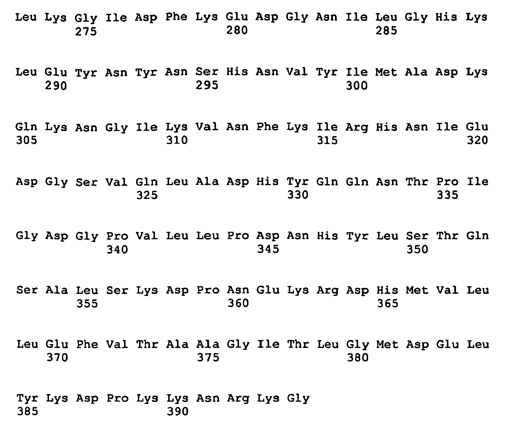
- <210> 24
<211> 25
<212> DNA
<213> Artificial - <220>
<223> primer - <400> 24
ggatccttcc tcctcgggcg ggtgt 25 - <210> 25
<211> 29
<212> DNA
<213> Artificial - <220>
<223> primer - <400> 25
aaccagcgct accatgagca ccaactcgt 29
Claims (23)
- A method for preparing a non-adenoviral target virus or one or more target protein(s) which is (are) not related to Adenovirus, which method comprises(a) culturing an immortalized vertebrate expression cell, being obtainable by infecting or transfecting an immortalized vertebrate host cell, with said target virus, with a vector carrying nucleic acid sequences encoding said virus, or with a vector carrying nucleic acid sequences encoding said one or more target protein(s), wherein said immortalized vertebrate expression cell has stably integrated into its genome a gene coding for Adenovirus PIX and stably expressing said Adenovirus PIX and(b) isolating said non-adenoviral target virus or said one or more target protein(s).
- The method of claim 1, wherein(i) Adenovirus PIX is modified by fusion with a transcription factor, a fluorescence marker protein, an enzyme, or a transit peptide; and/or(ii) Adenovirus PIX is expressed in the host cell and the expression cell in an amount of at least 1 pg/µg cellular protein; and/ or(iii) the gene encoding Adenovirus PIX is under control of a stable homologous or heterologous promoter.
- The method of claim 2, wherein(i) said transcription factor is retinoic acid receptor alpha, said fluorescence marker protein is GFP or DsRed, said enzyme is LacZ, or said transit peptide is a NLS, and/or(ii) said promoter is a constitutive cellular promoter.
- The method of claims 1 to 3, wherein Adenovirus PIX(i) has the sequence as shown in SEQ ID NO: 2;(ii) is fused with retinoic acid receptor alpha (RARA);(iii) is fused with GFP and a NLS sequence;(iv) is fused with GFP; or(v) is fused with an isolated NLS sequence.
- The method of claim 4, wherein Adenovirus PIX fused with retinoic acid receptor alpha has the sequence as shown in SEQ ID NO: 4, or wherein Adenovirus PIX fused with GFP and a NLS sequence has the sequence as shown in SEQ ID NO: 23.
- The method of any one of claims 1 to 5, wherein(i) the host and expression cell carries further immortalizing (viral) genes including an E1 protein of an adenovirus; and/or(ii) the cell further carrying functional sequences such as selection marker sequences, splice donor/acceptor sites and/or recombinase recognition sequences allowing integration, of a target nucleic acid sequence to be expressed in the cell.
- The method of any one of claims 1 to 6, wherein(i) the target virus is selected from a wild-type, mutated or deleted virus, a cold adapted or attenuated virus, a vaccine strain, and a viral vector; or(ii) the one or more target protein(s) is (are) selected from antibodies, recombinant proteins, viral antigens, and peptide hormones.
- The method of claim 7, wherein the viral vector is selected from a lentivirus, a poxvirus, an adeno associated virus (aav), a herpesvirus and a flavivirus.
- The method of claim 8, wherein the poxvirus is selected from a cowpox virus, a fowlpox and a canarypox virus.
- The method of claim 9, wherein the cowpox virus is a Modified Vaccinia Ankara (MVA) virus.
- The method of claim 7, wherein(i) the recombinant proteins are selected from erythropoetin, alpha-1-antitrypsin, blood clotting factors VIII and IX, and interferons, or(ii) the viral antigens are selected from influenza HA and NA, HBV-S, herpes G protein and rabies G protein.
- The method of any one of claims 1 to 11, wherein the host and expression cell is derived from human brain.
- The method of claim 12, wherein the host and expression cell is derived from human foetal brain.
- The method of claim 13, wherein the host cell is a NC5T11#34 cell and the expression cell is derived from the NCST11#34 cell, said NCSTI1#34 cell being deposited with the DSMZ under accession number DSM ACC2744.
- The method of any one of claims 1 to 11, wherein the host and expression cell is an avian cell.
- The method of claim 15, wherein the host cell and expression cell is an avian cell derived from duck retina or duck somite.
- The method of claim 16, wherein the host cell is a CR.PIX (17a11b) cell and the expression cell is derived from the CR.PIX (17a11b) cell, said CR.PIX (17a11b) cell being deposited with the DSMZ under accession number DSM ACC2749.
- An immortalized vertebrate expression cell for producing a non-adenoviral target virus, which(i) has stably integrated into its genome a gene coding for Adenovirus PIX and stably expressing said Adenovirus PIX, and(ii) is infected or transfected with a non-adenoviral target virus or with a vector carrying nucleic acid sequences encoding said virus.
- The immortalized vertebrate expression cell of claim 18, wherein(i) Adenovirus PIX is modified by fusion with a transcription factor, a fluorescence marker protein, an enzyme, or a transit peptide; and/or(ii) Adenovirus PIX is expressed in the host cell and the expression cell in an amount of at least 1 pg/µg cellular protein; and/or(iii) the gene encoding Adenovirus PIX is under control of a stable homologous or heterologous promoter.
- The immortalized vertebrate expression cell of claim 19, wherein(i) said transcription factor is retinoic acid receptor alpha, said fluorescence marker protein is GFP or DsRed, said enzyme is LacZ, or said transit peptide is a NLS, and/or(ii) said promoter is a constitutive cellular promoter.
- The immortalized vertebrate expression cell of claims 18 to 20, wherein the viral vector is selected from a lentivirus, a poxvirus, an adeno associated virus (aav), a herpesvirus and a flavivirus.
- The immortalized vertebrate expression cell of claim 18, wherein the expression cell is
derived from human brain. - Use of the immortalized vertebrate expression cell of any of claims 18 to 22 for preparing a non-adenoviral target virus.
Priority Applications (3)
| Application Number | Priority Date | Filing Date | Title |
|---|---|---|---|
| EP06807777A EP1945760B1 (en) | 2005-11-08 | 2006-11-08 | Productivity augmenting protein factor, novel cell lines and uses thereof |
| PL06807777T PL1945760T3 (en) | 2005-11-08 | 2006-11-08 | Productivity augmenting protein factor, novel cell lines and uses thereof |
| CY20121100350T CY1112734T1 (en) | 2005-11-08 | 2012-04-09 | PROTEIN PRODUCTIVITY INCREASE FACTOR, ORIGINAL CELL SERIES AND USES |
Applications Claiming Priority (4)
| Application Number | Priority Date | Filing Date | Title |
|---|---|---|---|
| EP05110453A EP1783210A1 (en) | 2005-11-08 | 2005-11-08 | Productivity augmenting protein factors, novel cell lines and uses thereof |
| US75015605P | 2005-12-14 | 2005-12-14 | |
| EP06807777A EP1945760B1 (en) | 2005-11-08 | 2006-11-08 | Productivity augmenting protein factor, novel cell lines and uses thereof |
| PCT/EP2006/068234 WO2007054516A1 (en) | 2005-11-08 | 2006-11-08 | Productivity augmenting protein factor, novel cell lines and uses thereof |
Publications (2)
| Publication Number | Publication Date |
|---|---|
| EP1945760A1 EP1945760A1 (en) | 2008-07-23 |
| EP1945760B1 true EP1945760B1 (en) | 2012-01-25 |
Family
ID=35985267
Family Applications (2)
| Application Number | Title | Priority Date | Filing Date |
|---|---|---|---|
| EP05110453A Withdrawn EP1783210A1 (en) | 2005-11-08 | 2005-11-08 | Productivity augmenting protein factors, novel cell lines and uses thereof |
| EP06807777A Active EP1945760B1 (en) | 2005-11-08 | 2006-11-08 | Productivity augmenting protein factor, novel cell lines and uses thereof |
Family Applications Before (1)
| Application Number | Title | Priority Date | Filing Date |
|---|---|---|---|
| EP05110453A Withdrawn EP1783210A1 (en) | 2005-11-08 | 2005-11-08 | Productivity augmenting protein factors, novel cell lines and uses thereof |
Country Status (20)
| Country | Link |
|---|---|
| US (1) | US8815540B2 (en) |
| EP (2) | EP1783210A1 (en) |
| JP (1) | JP5468780B2 (en) |
| KR (1) | KR101311973B1 (en) |
| CN (1) | CN101356265B (en) |
| AT (1) | ATE542890T1 (en) |
| AU (1) | AU2006310999B2 (en) |
| BR (1) | BRPI0618369B1 (en) |
| CA (1) | CA2628689C (en) |
| CY (1) | CY1112734T1 (en) |
| DK (1) | DK1945760T3 (en) |
| ES (1) | ES2380975T3 (en) |
| IL (1) | IL191327A (en) |
| MY (1) | MY147959A (en) |
| NZ (1) | NZ568482A (en) |
| PL (1) | PL1945760T3 (en) |
| PT (1) | PT1945760E (en) |
| RU (1) | RU2449015C2 (en) |
| SI (1) | SI1945760T1 (en) |
| WO (1) | WO2007054516A1 (en) |
Families Citing this family (5)
| Publication number | Priority date | Publication date | Assignee | Title |
|---|---|---|---|---|
| EP2265712B1 (en) * | 2008-03-04 | 2014-07-23 | ProBioGen AG | Cell line from rousettus as host cell for pathogen amplification |
| EP2098590A1 (en) * | 2008-03-04 | 2009-09-09 | ProBioGen AG | Cell line from Rousettus as host cell for pathogen amplification |
| EP2199385A1 (en) * | 2008-12-16 | 2010-06-23 | ProBioGen AG | Specific and persistent activation of heat shock response in cell lines using a viral factor |
| CN108795983A (en) * | 2018-06-28 | 2018-11-13 | 深圳市疾病预防控制中心(深圳市卫生检验中心、深圳市预防医学研究所) | A kind of protein stabilized high-expression cell lines of RXR α and preparation method thereof |
| WO2026041811A1 (en) | 2024-08-23 | 2026-02-26 | Transgene | Recombinant poxvirus and method for regulating the expression of toxic conditional gene products of said recombinant poxvirus in a producing cell |
Family Cites Families (7)
| Publication number | Priority date | Publication date | Assignee | Title |
|---|---|---|---|---|
| US5804413A (en) * | 1992-07-31 | 1998-09-08 | University Of Pittsburgh Of The Commonwealth System Of Higher Education | Herpes simplex virus strains for gene transfer |
| US5989805A (en) * | 1995-10-27 | 1999-11-23 | Board Of Trustees Operating Michigan State University | Immortal avian cell line to grow avian and animal viruses to produce vaccines |
| EP1075532A1 (en) * | 1998-05-01 | 2001-02-14 | Genzyme Corporation | Partially deleted adenoviral vectors |
| WO1999057296A1 (en) * | 1998-05-01 | 1999-11-11 | Genzyme Corporation | Partially deleted adenoviral vectors |
| CA2448908C (en) * | 2001-05-30 | 2008-03-18 | Transgene S.A. | Adenovirus protein ix, its domains involved in capsid assembly, transcriptional activity and nuclear reorganization |
| EP1528101A1 (en) * | 2003-11-03 | 2005-05-04 | ProBioGen AG | Immortalized avian cell lines for virus production |
| AU2006310341B2 (en) * | 2005-11-04 | 2011-01-06 | Novartis Vaccines And Diagnostics Limited | Influenza vaccines extemporaneously adsorbed to aluminium adjuvants |
-
2005
- 2005-11-08 EP EP05110453A patent/EP1783210A1/en not_active Withdrawn
-
2006
- 2006-11-08 SI SI200631265T patent/SI1945760T1/en unknown
- 2006-11-08 CA CA2628689A patent/CA2628689C/en active Active
- 2006-11-08 EP EP06807777A patent/EP1945760B1/en active Active
- 2006-11-08 RU RU2008122920/10A patent/RU2449015C2/en active
- 2006-11-08 MY MYPI20081500A patent/MY147959A/en unknown
- 2006-11-08 WO PCT/EP2006/068234 patent/WO2007054516A1/en not_active Ceased
- 2006-11-08 AU AU2006310999A patent/AU2006310999B2/en active Active
- 2006-11-08 DK DK06807777.5T patent/DK1945760T3/en active
- 2006-11-08 JP JP2008539429A patent/JP5468780B2/en active Active
- 2006-11-08 NZ NZ568482A patent/NZ568482A/en unknown
- 2006-11-08 PL PL06807777T patent/PL1945760T3/en unknown
- 2006-11-08 ES ES06807777T patent/ES2380975T3/en active Active
- 2006-11-08 PT PT06807777T patent/PT1945760E/en unknown
- 2006-11-08 KR KR1020087013574A patent/KR101311973B1/en active Active
- 2006-11-08 CN CN2006800506905A patent/CN101356265B/en active Active
- 2006-11-08 AT AT06807777T patent/ATE542890T1/en active
- 2006-11-08 BR BRPI0618369-7A patent/BRPI0618369B1/en active IP Right Grant
-
2008
- 2008-05-07 IL IL191327A patent/IL191327A/en active IP Right Grant
- 2008-05-08 US US12/117,475 patent/US8815540B2/en active Active
-
2012
- 2012-04-09 CY CY20121100350T patent/CY1112734T1/en unknown
Also Published As
| Publication number | Publication date |
|---|---|
| CN101356265B (en) | 2013-04-03 |
| PT1945760E (en) | 2012-03-28 |
| EP1945760A1 (en) | 2008-07-23 |
| KR101311973B1 (en) | 2013-09-27 |
| IL191327A (en) | 2013-03-24 |
| AU2006310999A1 (en) | 2007-05-18 |
| JP5468780B2 (en) | 2014-04-09 |
| PL1945760T3 (en) | 2012-05-31 |
| ATE542890T1 (en) | 2012-02-15 |
| CA2628689A1 (en) | 2007-05-18 |
| WO2007054516A1 (en) | 2007-05-18 |
| CN101356265A (en) | 2009-01-28 |
| BRPI0618369A2 (en) | 2011-08-30 |
| RU2449015C2 (en) | 2012-04-27 |
| RU2008122920A (en) | 2009-12-20 |
| SI1945760T1 (en) | 2012-03-30 |
| NZ568482A (en) | 2011-06-30 |
| DK1945760T3 (en) | 2012-03-05 |
| EP1783210A1 (en) | 2007-05-09 |
| BRPI0618369B1 (en) | 2022-04-12 |
| CA2628689C (en) | 2014-02-25 |
| MY147959A (en) | 2013-02-15 |
| KR20080068104A (en) | 2008-07-22 |
| JP2009514544A (en) | 2009-04-09 |
| AU2006310999B2 (en) | 2012-04-12 |
| ES2380975T3 (en) | 2012-05-22 |
| US8815540B2 (en) | 2014-08-26 |
| CY1112734T1 (en) | 2016-02-10 |
| US20120164727A1 (en) | 2012-06-28 |
Similar Documents
| Publication | Publication Date | Title |
|---|---|---|
| Benedict et al. | Three adenovirus E3 proteins cooperate to evade apoptosis by tumor necrosis factor-related apoptosis-inducing ligand receptor-1 and-2 | |
| Gage et al. | Syncytium-inducing mutations localize to two discrete regions within the cytoplasmic domain of herpes simplex virus type 1 glycoprotein B | |
| CN1934243B (en) | Immortalized avian cell lines for virus production | |
| Lafemina et al. | Expression of the acidic nuclear immediate-early protein (IE1) of human cytomegalovirus in stable cell lines and its preferential association with metaphase chromosomes | |
| RS58080B1 (en) | Therapeutic hpv16 vaccines | |
| KR102405246B1 (en) | Recombinant Herpes Simplex Virus Having Expression Cassette Expressing Fused Protein of Cancer Cell-Targeting Domain and Extracellular Damain of HVEM and Use Thereof | |
| CN107190013B (en) | Zika virus vaccine taking human Ad5 replication-defective adenovirus as vector | |
| US8815540B2 (en) | Productivity augmenting protein factor, novel cell lines and uses thereof | |
| CN114560915B (en) | Modified high-titer SARS-CoV-2 pseudovirus | |
| Perry et al. | The DNA binding domain of herpes simplex virus type 1 origin binding protein is a transdominant inhibitor of virus replication | |
| ES2330826A1 (en) | System for packaging of high-capacity adenoviruses | |
| US8318662B2 (en) | Protein increasing cell infectivity of herpes simplex virus and use thereof | |
| WO2024153120A1 (en) | Mutated pd1 ectodomain fragment and car containing same, and nk cell | |
| MX2008005987A (en) | Productivity augmenting protein factor, novel cell lines and uses thereof | |
| JP2009514544A5 (en) | ||
| EP2199385A1 (en) | Specific and persistent activation of heat shock response in cell lines using a viral factor | |
| Sunstrom | Specific DNA binding by polyomavirus large T antigen | |
| KR20080007832A (en) | Recombinant shuttle vector for adenovirus containing CAV promoter | |
| Dore | Production andin vitromanipulation of oncogenic animal papillomaviruses | |
| Yang | Analysis of the regulation of a bi-directional varicella-zoster virus (VZV) promoter: The ORF 28/29 promoter | |
| Cytomegalovirus | Cutting Edge: A Novel Viral TNF Receptor | |
| Busha | A Novel Selection Technology for the Discovery of High-Affinity Human Proteins | |
| Pardo-Mateos | A genetic analysis of the structure and function of the human adenovirus IVa2 protein | |
| KR20030079214A (en) | Expression vector containing the restricted E1 gene of wild type adenoviurs serotype 5 and the animal cell line thereof |
Legal Events
| Date | Code | Title | Description |
|---|---|---|---|
| PUAI | Public reference made under article 153(3) epc to a published international application that has entered the european phase |
Free format text: ORIGINAL CODE: 0009012 |
|
| 17P | Request for examination filed |
Effective date: 20080513 |
|
| AK | Designated contracting states |
Kind code of ref document: A1 Designated state(s): AT BE BG CH CY CZ DE DK EE ES FI FR GB GR HU IE IS IT LI LT LU LV MC NL PL PT RO SE SI SK TR |
|
| 17Q | First examination report despatched |
Effective date: 20090313 |
|
| GRAP | Despatch of communication of intention to grant a patent |
Free format text: ORIGINAL CODE: EPIDOSNIGR1 |
|
| DAX | Request for extension of the european patent (deleted) | ||
| GRAS | Grant fee paid |
Free format text: ORIGINAL CODE: EPIDOSNIGR3 |
|
| GRAA | (expected) grant |
Free format text: ORIGINAL CODE: 0009210 |
|
| AK | Designated contracting states |
Kind code of ref document: B1 Designated state(s): AT BE BG CH CY CZ DE DK EE ES FI FR GB GR HU IE IS IT LI LT LU LV MC NL PL PT RO SE SI SK TR |
|
| REG | Reference to a national code |
Ref country code: GB Ref legal event code: FG4D |
|
| REG | Reference to a national code |
Ref country code: CH Ref legal event code: EP |
|
| REG | Reference to a national code |
Ref country code: AT Ref legal event code: REF Ref document number: 542890 Country of ref document: AT Kind code of ref document: T Effective date: 20120215 |
|
| REG | Reference to a national code |
Ref country code: IE Ref legal event code: FG4D |
|
| REG | Reference to a national code |
Ref country code: DK Ref legal event code: T3 |
|
| REG | Reference to a national code |
Ref country code: NL Ref legal event code: T3 |
|
| REG | Reference to a national code |
Ref country code: DE Ref legal event code: R096 Ref document number: 602006027349 Country of ref document: DE Effective date: 20120322 |
|
| REG | Reference to a national code |
Ref country code: RO Ref legal event code: EPE |
|
| REG | Reference to a national code |
Ref country code: SE Ref legal event code: TRGR |
|
| REG | Reference to a national code |
Ref country code: PT Ref legal event code: SC4A Free format text: AVAILABILITY OF NATIONAL TRANSLATION Effective date: 20120315 |
|
| REG | Reference to a national code |
Ref country code: ES Ref legal event code: FG2A Ref document number: 2380975 Country of ref document: ES Kind code of ref document: T3 Effective date: 20120522 |
|
| REG | Reference to a national code |
Ref country code: PL Ref legal event code: T3 Ref country code: CH Ref legal event code: NV Representative=s name: HANS ULRICH SEIFERT SEIFERT & PARTNER |
|
| REG | Reference to a national code |
Ref country code: EE Ref legal event code: FG4A Ref document number: E006584 Country of ref document: EE Effective date: 20120323 |
|
| REG | Reference to a national code |
Ref country code: GR Ref legal event code: EP Ref document number: 20120400927 Country of ref document: GR Effective date: 20120518 |
|
| REG | Reference to a national code |
Ref country code: SK Ref legal event code: T3 Ref document number: E 11630 Country of ref document: SK |
|
| REG | Reference to a national code |
Ref country code: HU Ref legal event code: AG4A Ref document number: E013669 Country of ref document: HU |
|
| PLBE | No opposition filed within time limit |
Free format text: ORIGINAL CODE: 0009261 |
|
| STAA | Information on the status of an ep patent application or granted ep patent |
Free format text: STATUS: NO OPPOSITION FILED WITHIN TIME LIMIT |
|
| 26N | No opposition filed |
Effective date: 20121026 |
|
| REG | Reference to a national code |
Ref country code: DE Ref legal event code: R097 Ref document number: 602006027349 Country of ref document: DE Effective date: 20121026 |
|
| PG25 | Lapsed in a contracting state [announced via postgrant information from national office to epo] |
Ref country code: PL Free format text: LAPSE BECAUSE OF NON-PAYMENT OF DUE FEES Effective date: 20121108 Ref country code: RO Free format text: LAPSE BECAUSE OF NON-PAYMENT OF DUE FEES Effective date: 20120125 |
|
| REG | Reference to a national code |
Ref country code: PL Ref legal event code: LAPE |
|
| REG | Reference to a national code |
Ref country code: FR Ref legal event code: PLFP Year of fee payment: 10 |
|
| REG | Reference to a national code |
Ref country code: FR Ref legal event code: PLFP Year of fee payment: 11 |
|
| REG | Reference to a national code |
Ref country code: FR Ref legal event code: PLFP Year of fee payment: 12 |
|
| PGFP | Annual fee paid to national office [announced via postgrant information from national office to epo] |
Ref country code: LU Payment date: 20171122 Year of fee payment: 12 |
|
| PGFP | Annual fee paid to national office [announced via postgrant information from national office to epo] |
Ref country code: SK Payment date: 20171030 Year of fee payment: 12 Ref country code: EE Payment date: 20171122 Year of fee payment: 12 Ref country code: FI Payment date: 20171117 Year of fee payment: 12 Ref country code: MC Payment date: 20171121 Year of fee payment: 12 Ref country code: CZ Payment date: 20171027 Year of fee payment: 12 Ref country code: TR Payment date: 20171107 Year of fee payment: 12 |
|
| PGFP | Annual fee paid to national office [announced via postgrant information from national office to epo] |
Ref country code: SI Payment date: 20171030 Year of fee payment: 12 Ref country code: GR Payment date: 20171121 Year of fee payment: 12 Ref country code: IS Payment date: 20171121 Year of fee payment: 12 Ref country code: LV Payment date: 20171122 Year of fee payment: 12 Ref country code: PT Payment date: 20171026 Year of fee payment: 12 Ref country code: BG Payment date: 20171122 Year of fee payment: 12 |
|
| PGFP | Annual fee paid to national office [announced via postgrant information from national office to epo] |
Ref country code: CY Payment date: 20171030 Year of fee payment: 12 |
|
| REG | Reference to a national code |
Ref country code: EE Ref legal event code: MM4A Ref document number: E006584 Country of ref document: EE Effective date: 20181130 |
|
| PG25 | Lapsed in a contracting state [announced via postgrant information from national office to epo] |
Ref country code: CY Free format text: LAPSE BECAUSE OF NON-PAYMENT OF DUE FEES Effective date: 20181108 Ref country code: LU Free format text: LAPSE BECAUSE OF NON-PAYMENT OF DUE FEES Effective date: 20181108 Ref country code: CZ Free format text: LAPSE BECAUSE OF NON-PAYMENT OF DUE FEES Effective date: 20181108 Ref country code: FI Free format text: LAPSE BECAUSE OF NON-PAYMENT OF DUE FEES Effective date: 20181108 Ref country code: PT Free format text: LAPSE BECAUSE OF NON-PAYMENT OF DUE FEES Effective date: 20190508 Ref country code: MC Free format text: LAPSE BECAUSE OF NON-PAYMENT OF DUE FEES Effective date: 20181130 |
|
| REG | Reference to a national code |
Ref country code: SK Ref legal event code: MM4A Ref document number: E 11630 Country of ref document: SK Effective date: 20181108 |
|
| PG25 | Lapsed in a contracting state [announced via postgrant information from national office to epo] |
Ref country code: EE Free format text: LAPSE BECAUSE OF NON-PAYMENT OF DUE FEES Effective date: 20181130 Ref country code: GR Free format text: LAPSE BECAUSE OF NON-PAYMENT OF DUE FEES Effective date: 20190605 Ref country code: LV Free format text: LAPSE BECAUSE OF NON-PAYMENT OF DUE FEES Effective date: 20181108 Ref country code: SK Free format text: LAPSE BECAUSE OF NON-PAYMENT OF DUE FEES Effective date: 20181108 Ref country code: IS Free format text: LAPSE BECAUSE OF FAILURE TO SUBMIT A TRANSLATION OF THE DESCRIPTION OR TO PAY THE FEE WITHIN THE PRESCRIBED TIME-LIMIT Effective date: 20190531 Ref country code: BG Free format text: LAPSE BECAUSE OF NON-PAYMENT OF DUE FEES Effective date: 20190531 Ref country code: SI Free format text: LAPSE BECAUSE OF NON-PAYMENT OF DUE FEES Effective date: 20181109 |
|
| REG | Reference to a national code |
Ref country code: SI Ref legal event code: KO00 Effective date: 20190708 |
|
| PG25 | Lapsed in a contracting state [announced via postgrant information from national office to epo] |
Ref country code: TR Free format text: LAPSE BECAUSE OF NON-PAYMENT OF DUE FEES Effective date: 20181108 |
|
| P01 | Opt-out of the competence of the unified patent court (upc) registered |
Effective date: 20230516 |
|
| REG | Reference to a national code |
Ref country code: CH Ref legal event code: U11 Free format text: ST27 STATUS EVENT CODE: U-0-0-U10-U11 (AS PROVIDED BY THE NATIONAL OFFICE) Effective date: 20251201 |
|
| PGFP | Annual fee paid to national office [announced via postgrant information from national office to epo] |
Ref country code: HU Payment date: 20251031 Year of fee payment: 20 |
|
| PGFP | Annual fee paid to national office [announced via postgrant information from national office to epo] |
Ref country code: NL Payment date: 20251119 Year of fee payment: 20 |
|
| PGFP | Annual fee paid to national office [announced via postgrant information from national office to epo] |
Ref country code: DE Payment date: 20251118 Year of fee payment: 20 |
|
| PGFP | Annual fee paid to national office [announced via postgrant information from national office to epo] |
Ref country code: GB Payment date: 20251120 Year of fee payment: 20 Ref country code: LT Payment date: 20251023 Year of fee payment: 20 |
|
| PGFP | Annual fee paid to national office [announced via postgrant information from national office to epo] |
Ref country code: AT Payment date: 20251117 Year of fee payment: 20 |
|
| PGFP | Annual fee paid to national office [announced via postgrant information from national office to epo] |
Ref country code: IT Payment date: 20251128 Year of fee payment: 20 Ref country code: DK Payment date: 20251119 Year of fee payment: 20 |
|
| PGFP | Annual fee paid to national office [announced via postgrant information from national office to epo] |
Ref country code: FR Payment date: 20251125 Year of fee payment: 20 |
|
| PGFP | Annual fee paid to national office [announced via postgrant information from national office to epo] |
Ref country code: BE Payment date: 20251118 Year of fee payment: 20 |
|
| PGFP | Annual fee paid to national office [announced via postgrant information from national office to epo] |
Ref country code: CH Payment date: 20251201 Year of fee payment: 20 Ref country code: SE Payment date: 20251119 Year of fee payment: 20 |
|
| PGFP | Annual fee paid to national office [announced via postgrant information from national office to epo] |
Ref country code: IE Payment date: 20251120 Year of fee payment: 20 |
|
| PGFP | Annual fee paid to national office [announced via postgrant information from national office to epo] |
Ref country code: ES Payment date: 20251216 Year of fee payment: 20 |