EP1932548A1 - Reinforcing agent for biological tissue, method of producing the same, utilization of the same and method of culturing cells - Google Patents
Reinforcing agent for biological tissue, method of producing the same, utilization of the same and method of culturing cells Download PDFInfo
- Publication number
- EP1932548A1 EP1932548A1 EP06781124A EP06781124A EP1932548A1 EP 1932548 A1 EP1932548 A1 EP 1932548A1 EP 06781124 A EP06781124 A EP 06781124A EP 06781124 A EP06781124 A EP 06781124A EP 1932548 A1 EP1932548 A1 EP 1932548A1
- Authority
- EP
- European Patent Office
- Prior art keywords
- biological tissue
- reinforcing material
- vivo
- material according
- derived substance
- Prior art date
- Legal status (The legal status is an assumption and is not a legal conclusion. Google has not performed a legal analysis and makes no representation as to the accuracy of the status listed.)
- Withdrawn
Links
Images



Classifications
-
- A—HUMAN NECESSITIES
- A61—MEDICAL OR VETERINARY SCIENCE; HYGIENE
- A61L—METHODS OR APPARATUS FOR STERILISING MATERIALS OR OBJECTS IN GENERAL; DISINFECTION, STERILISATION OR DEODORISATION OF AIR; CHEMICAL ASPECTS OF BANDAGES, DRESSINGS, ABSORBENT PADS OR SURGICAL ARTICLES; MATERIALS FOR BANDAGES, DRESSINGS, ABSORBENT PADS OR SURGICAL ARTICLES
- A61L27/00—Materials for grafts or prostheses or for coating grafts or prostheses
- A61L27/50—Materials characterised by their function or physical properties, e.g. injectable or lubricating compositions, shape-memory materials, surface modified materials
- A61L27/54—Biologically active materials, e.g. therapeutic substances
-
- A—HUMAN NECESSITIES
- A61—MEDICAL OR VETERINARY SCIENCE; HYGIENE
- A61L—METHODS OR APPARATUS FOR STERILISING MATERIALS OR OBJECTS IN GENERAL; DISINFECTION, STERILISATION OR DEODORISATION OF AIR; CHEMICAL ASPECTS OF BANDAGES, DRESSINGS, ABSORBENT PADS OR SURGICAL ARTICLES; MATERIALS FOR BANDAGES, DRESSINGS, ABSORBENT PADS OR SURGICAL ARTICLES
- A61L27/00—Materials for grafts or prostheses or for coating grafts or prostheses
-
- A—HUMAN NECESSITIES
- A61—MEDICAL OR VETERINARY SCIENCE; HYGIENE
- A61L—METHODS OR APPARATUS FOR STERILISING MATERIALS OR OBJECTS IN GENERAL; DISINFECTION, STERILISATION OR DEODORISATION OF AIR; CHEMICAL ASPECTS OF BANDAGES, DRESSINGS, ABSORBENT PADS OR SURGICAL ARTICLES; MATERIALS FOR BANDAGES, DRESSINGS, ABSORBENT PADS OR SURGICAL ARTICLES
- A61L24/00—Surgical adhesives or cements; Adhesives for colostomy devices
- A61L24/0005—Ingredients of undetermined constitution or reaction products thereof
-
- A—HUMAN NECESSITIES
- A61—MEDICAL OR VETERINARY SCIENCE; HYGIENE
- A61L—METHODS OR APPARATUS FOR STERILISING MATERIALS OR OBJECTS IN GENERAL; DISINFECTION, STERILISATION OR DEODORISATION OF AIR; CHEMICAL ASPECTS OF BANDAGES, DRESSINGS, ABSORBENT PADS OR SURGICAL ARTICLES; MATERIALS FOR BANDAGES, DRESSINGS, ABSORBENT PADS OR SURGICAL ARTICLES
- A61L24/00—Surgical adhesives or cements; Adhesives for colostomy devices
- A61L24/001—Use of materials characterised by their function or physical properties
- A61L24/0015—Medicaments; Biocides
-
- A—HUMAN NECESSITIES
- A61—MEDICAL OR VETERINARY SCIENCE; HYGIENE
- A61L—METHODS OR APPARATUS FOR STERILISING MATERIALS OR OBJECTS IN GENERAL; DISINFECTION, STERILISATION OR DEODORISATION OF AIR; CHEMICAL ASPECTS OF BANDAGES, DRESSINGS, ABSORBENT PADS OR SURGICAL ARTICLES; MATERIALS FOR BANDAGES, DRESSINGS, ABSORBENT PADS OR SURGICAL ARTICLES
- A61L24/00—Surgical adhesives or cements; Adhesives for colostomy devices
- A61L24/0047—Composite materials, i.e. containing one material dispersed in a matrix of the same or different material
- A61L24/0052—Composite materials, i.e. containing one material dispersed in a matrix of the same or different material with an inorganic matrix
- A61L24/0063—Phosphorus containing materials, e.g. apatite
-
- A—HUMAN NECESSITIES
- A61—MEDICAL OR VETERINARY SCIENCE; HYGIENE
- A61L—METHODS OR APPARATUS FOR STERILISING MATERIALS OR OBJECTS IN GENERAL; DISINFECTION, STERILISATION OR DEODORISATION OF AIR; CHEMICAL ASPECTS OF BANDAGES, DRESSINGS, ABSORBENT PADS OR SURGICAL ARTICLES; MATERIALS FOR BANDAGES, DRESSINGS, ABSORBENT PADS OR SURGICAL ARTICLES
- A61L27/00—Materials for grafts or prostheses or for coating grafts or prostheses
- A61L27/02—Inorganic materials
- A61L27/12—Phosphorus-containing materials, e.g. apatite
-
- A—HUMAN NECESSITIES
- A61—MEDICAL OR VETERINARY SCIENCE; HYGIENE
- A61L—METHODS OR APPARATUS FOR STERILISING MATERIALS OR OBJECTS IN GENERAL; DISINFECTION, STERILISATION OR DEODORISATION OF AIR; CHEMICAL ASPECTS OF BANDAGES, DRESSINGS, ABSORBENT PADS OR SURGICAL ARTICLES; MATERIALS FOR BANDAGES, DRESSINGS, ABSORBENT PADS OR SURGICAL ARTICLES
- A61L27/00—Materials for grafts or prostheses or for coating grafts or prostheses
- A61L27/36—Materials for grafts or prostheses or for coating grafts or prostheses containing ingredients of undetermined constitution or reaction products thereof, e.g. transplant tissue, natural bone, extracellular matrix
- A61L27/38—Materials for grafts or prostheses or for coating grafts or prostheses containing ingredients of undetermined constitution or reaction products thereof, e.g. transplant tissue, natural bone, extracellular matrix containing added animal cells
-
- A—HUMAN NECESSITIES
- A61—MEDICAL OR VETERINARY SCIENCE; HYGIENE
- A61L—METHODS OR APPARATUS FOR STERILISING MATERIALS OR OBJECTS IN GENERAL; DISINFECTION, STERILISATION OR DEODORISATION OF AIR; CHEMICAL ASPECTS OF BANDAGES, DRESSINGS, ABSORBENT PADS OR SURGICAL ARTICLES; MATERIALS FOR BANDAGES, DRESSINGS, ABSORBENT PADS OR SURGICAL ARTICLES
- A61L27/00—Materials for grafts or prostheses or for coating grafts or prostheses
- A61L27/36—Materials for grafts or prostheses or for coating grafts or prostheses containing ingredients of undetermined constitution or reaction products thereof, e.g. transplant tissue, natural bone, extracellular matrix
- A61L27/38—Materials for grafts or prostheses or for coating grafts or prostheses containing ingredients of undetermined constitution or reaction products thereof, e.g. transplant tissue, natural bone, extracellular matrix containing added animal cells
- A61L27/3804—Materials for grafts or prostheses or for coating grafts or prostheses containing ingredients of undetermined constitution or reaction products thereof, e.g. transplant tissue, natural bone, extracellular matrix containing added animal cells characterised by specific cells or progenitors thereof, e.g. fibroblasts, connective tissue cells, kidney cells
- A61L27/3834—Cells able to produce different cell types, e.g. hematopoietic stem cells, mesenchymal stem cells, marrow stromal cells, embryonic stem cells
-
- C—CHEMISTRY; METALLURGY
- C12—BIOCHEMISTRY; BEER; SPIRITS; WINE; VINEGAR; MICROBIOLOGY; ENZYMOLOGY; MUTATION OR GENETIC ENGINEERING
- C12N—MICROORGANISMS OR ENZYMES; COMPOSITIONS THEREOF; PROPAGATING, PRESERVING, OR MAINTAINING MICROORGANISMS; MUTATION OR GENETIC ENGINEERING; CULTURE MEDIA
- C12N5/00—Undifferentiated human, animal or plant cells, e.g. cell lines; Tissues; Cultivation or maintenance thereof; Culture media therefor
- C12N5/06—Animal cells or tissues; Human cells or tissues
-
- C—CHEMISTRY; METALLURGY
- C12—BIOCHEMISTRY; BEER; SPIRITS; WINE; VINEGAR; MICROBIOLOGY; ENZYMOLOGY; MUTATION OR GENETIC ENGINEERING
- C12N—MICROORGANISMS OR ENZYMES; COMPOSITIONS THEREOF; PROPAGATING, PRESERVING, OR MAINTAINING MICROORGANISMS; MUTATION OR GENETIC ENGINEERING; CULTURE MEDIA
- C12N5/00—Undifferentiated human, animal or plant cells, e.g. cell lines; Tissues; Cultivation or maintenance thereof; Culture media therefor
- C12N5/06—Animal cells or tissues; Human cells or tissues
- C12N5/0602—Vertebrate cells
- C12N5/0652—Cells of skeletal and connective tissues; Mesenchyme
- C12N5/0654—Osteocytes, Osteoblasts, Odontocytes; Bones, Teeth
-
- A—HUMAN NECESSITIES
- A61—MEDICAL OR VETERINARY SCIENCE; HYGIENE
- A61L—METHODS OR APPARATUS FOR STERILISING MATERIALS OR OBJECTS IN GENERAL; DISINFECTION, STERILISATION OR DEODORISATION OF AIR; CHEMICAL ASPECTS OF BANDAGES, DRESSINGS, ABSORBENT PADS OR SURGICAL ARTICLES; MATERIALS FOR BANDAGES, DRESSINGS, ABSORBENT PADS OR SURGICAL ARTICLES
- A61L2300/00—Biologically active materials used in bandages, wound dressings, absorbent pads or medical devices
- A61L2300/40—Biologically active materials used in bandages, wound dressings, absorbent pads or medical devices characterised by a specific therapeutic activity or mode of action
- A61L2300/412—Tissue-regenerating or healing or proliferative agents
- A61L2300/414—Growth factors
-
- A—HUMAN NECESSITIES
- A61—MEDICAL OR VETERINARY SCIENCE; HYGIENE
- A61L—METHODS OR APPARATUS FOR STERILISING MATERIALS OR OBJECTS IN GENERAL; DISINFECTION, STERILISATION OR DEODORISATION OF AIR; CHEMICAL ASPECTS OF BANDAGES, DRESSINGS, ABSORBENT PADS OR SURGICAL ARTICLES; MATERIALS FOR BANDAGES, DRESSINGS, ABSORBENT PADS OR SURGICAL ARTICLES
- A61L2300/00—Biologically active materials used in bandages, wound dressings, absorbent pads or medical devices
- A61L2300/40—Biologically active materials used in bandages, wound dressings, absorbent pads or medical devices characterised by a specific therapeutic activity or mode of action
- A61L2300/426—Immunomodulating agents, i.e. cytokines, interleukins, interferons
Definitions
- the present invention relates to a biological tissue-reinforcing material, a method of producing the same, use of the same, and a method of culturing cells.
- hydroxyapatite (HAP) and ⁇ -tricalcium phosphate ( ⁇ TCP) are known as artificial bone-reinforcing materials, which are a type of biological tissue-reinforcing material (for example, refer to Non-patent Document 1). Recently, it has been reported that calcium sulfate can also be used as a bone-reinforcing material (for example, refer to Non-patent Document 2).
- calcium sulfate is a powder and can not be formed into a sintered compact as calcium phosphate can be, thus causing an inconvenience of being inapplicable to a site required to be reinforced.
- calcium sulfate is a powder, there is a problem in that the working properties are unsatisfactory for transplantation.
- the present invention takes the above problems into consideration, with an object of providing a biological tissue-reinforcing material which is applicable to a site required to be reinforced and which enables to improve the working properties for transplantation, a method of producing the same, use of the same, and a method of culturing cells.
- the present invention employs the following solutions in order to achieve the above object.
- the present invention provides a biological tissue-reinforcing material comprising sulfate group-containing calcium phosphate.
- the preparation is preferably performed so that the ratio of sulfur (S) to phosphorus (P) satisfies 1/2 ⁇ P/(S+P) ⁇ 5/6.
- the biological reinforcing material may additionally contain a vivo-derived substance.
- the present invention provides a method of producing a biological tissue-reinforcing material, comprising the step of coprecipitating ammonium hydrogen phosphate with calcium nitrate in the presence of sulfuric acid.
- the present invention provides a method of culturing cells, comprising the step of combining the biological tissue-reinforcing material with a vivo-derived substance. Moreover, the present invention provides use of the biological tissue-reinforcing material as a cell culture substrate in combination with a vivo-derived substance.
- the vivo-derived substance may include at least either one of a growth factor and a cytokine. Moreover, in the present invention, the vivo-derived substance may include a tissue-derived cell. Furthermore, in the present invention, the vivo-derived substance may include a cell group containing stem cells.
- the biological tissue-reinforcing material and the method of culturing cells of the present invention demonstrate effects of being applicable to a site required to be reinforced while enabling to improve the working properties for transplantation. Moreover, according to the production method of the present invention, effects in which phosphorus and sulfur can be homogenously mixed and readily prepared at a determined mixture ratio, are demonstrated.
- the biological tissue-reinforcing material according to the present embodiment comprises sulfate group-containing calcium phosphate.
- this biological tissue-reinforcing material can be produced using the following production method. First, 0.5M sulfuric acid H 2 SO 4 , 0.5M ammonium hydrogen phosphate (NH 4 ) 2 HPO 4 aqueous solution, and 0.5M calcium nitrate Ca(NO 3 ) 2 aqueous solution are prepared. Next, thus prepared 0.5M sulfuric acid and 0.5M ammonium hydrogen phosphate aqueous solution are mixed. Then, the obtained mixture is adjusted to pH 10 using 1.0N sodium hydroxide.
- step S1 prepared mixture is added with 0.5M calcium nitrate aqueous solution gradually dropwise (step S1). At this time, over monitoring the pH of the mixture, 1.0N sodium hydroxide is added to adjust the pH to 10. After the dropwise addition, the mixture is stirred over day and night (24 hours) (step S2). The precipitate is separated by centrifugation and washed with distilled water (step S3). The washed precipitate is filtered (step S4), dried (step S5), and retained in an atmospheric air at 800°C for 2 hours to thereby effect sintering (step S6). By so ding, the biological tissue-reinforcing material of the present embodiment is produced.
- a biological tissue-reinforcing material which homogenously contains phosphorus and sulfur at a determined mixture ratio can be readily produced.
- the action of the homogenously contained sulfate group brings superior proliferation potency and differentiation potency of osteoblasts to those of conventional HAP, while enabling to retain characteristics of conventional HAP.
- the ratio of sulfur S to phosphorus P preferably satisfies 1/2 ⁇ P/(S+P) ⁇ 5/6.
- the biological tissue-reinforcing material according to the present embodiment is surely applicable as a conventional bone-reinforcing material, as well as being applicable to the following directions because of its high osteoblast-activating action.
- Osteoblasts can be cultured at a highly activated state.
- a cultured bone having a high osteoblast activity or a cultured bone rich in the extracellular matrix can be provided.
- a bone-reinforcing material which activates osteoblasts can be provided.
- Therapeutic effects can be expected due to the activation of osteoblasts and the recalcification effect of the extracellular matrix.
- Therapeutic effects can be expected due to the activation of osteoblasts and the recalcification effect of the extracellular matrix.
- Example of the biological tissue-reinforcing material is a description of Example of the biological tissue-reinforcing material and the method of producing the same according to the above embodiment.
- a biological tissue-reinforcing material comprising a sulfuric acid-HAP was produced in accordance with the flowchart shown in Fig. 1 .
- a pellet comprising the biological tissue-reinforcing material according to the present embodiment was produced.
- the pellet was produced by the following manner.
- a powder of sulfuric acid-HAP obtained in the drying step S5 of FIG. 1 was put into, for example, a stainless mold, and was press-formed at 30MPa.
- the formed product was sintered in an atmospheric air at 800°C for 2 hours in the sintering step S6.
- normal human osteoblasts were seeded at 4 ⁇ 10 4 cells/well/mL, and were cultured for a week.
- the proliferation potency of the resultant osteoblasts is shown in FIG. 3
- the differentiation potency thereof is shown in FIG. 4 , respectively in ratios with respect to the control.
- the control respectively means cells which were directly cultured in a culture dish without seeding.
- the proliferation potency was measured with Tetra Color One (450 nm), and the differentiation potency was measured using alkaline phosphatase (ALP) activity through the color reaction of PNP (405nm).
- ALP alkaline phosphatase
- the sulfuric acid-HAP powder that had been heat-treated in the sintering step S6 was put in a medium (100 mg/mL), and agitated with a shaker for 72 hours (150 rpm). Then, the obtained liquid was placed in a centrifugal separator and subjected to centrifugation at 3000 rpm for 10 minutes. Thus obtained supernatant was added with osteoblasts (4 ⁇ 10 4 cells/well/mL).
- the proliferation potency of the resultant cultured osteoblasts is shown in FIG. 5
- the differentiation potency thereof is shown in FIG. 6 , respectively in ratios with respect to the control.
- the control respectively means cells which were directly cultured in a culture dish without seeding.
- hMSC human mesenchymal stem cells
- the hMSC proliferation was quantified through the production of a cell lysate and following measurement of DNA amount in the lysate.
- the cell lysate was produced as follows. First, on completion of culture, the medium was discarded and cells were washed with a phosphate buffered saline solution (PBS) twice. Then, trypsin was added thereto (200 ⁇ l/well, 37°C, 1 minute) to peel off these cells. PBS was added at 800 ⁇ l per each well, and the cell solution was collected and centrifuged (4°C, 1000 rpm, 2 minutes). The supernatant was discarded and the cells were collected. The collected cells were added with 600 ⁇ l of a solution containing 2% nonionic surfactant Nonidet (registered trademark) P-40. The solution was cooled in ice and treated with ultrasonic waves for 10 seconds to disrupt these cells.
- PBS phosphate buffered saline solution
- FIG. 7 shows the proliferation potency resulting from the culture in the extract
- FIG. 8 shows the proliferation potency resulting from the culture on the pellet, respectively using SO 4 -HAp and HAp.
- the differentiation potency was assessed by the production amounts of alkaline phosphatase and osteocalcin.
- the production amount of alkaline phosphatase was measured as follows. On completion of culture, the medium in each well was discarded and cells were washed PBS. Then, 0.1M glycine-NaCl-NaOH buffer solution containing 4 mM p-nitrophosphoric acid, 10 mM MgCl 2 , and 0.1 mM ZnCl 2 was added at 500 ⁇ l per each well. After leaving at a room temperature for 5 minutes, the absorbance of the buffer solution (optical density O.D. at the wavelength of 405 nm) was measured.
- the production amount of osteocalcin was measured by placing 100 ⁇ l of the cell lysate in an antibody plate, and following measurement of the osteocalcin concentration with use of a commercially available ELISA kit (Gla-OC EIA Kit (manufactured by TAKARA BIO INC.)).
- FIG. 9 shows the production amount of alkaline phosphatase resulting from the culture in the extract
- FIG. 10 shows the production amount of alkaline phosphatase resulting from the culture on the pellet
- FIG. 11 shows the production amount of osteocalcin resulting from the culture in the extract
- FIG. 12 shows the production amount of osteocalcin resulting from the culture on the pellet, respectively using SO 4 -HAp and HAp.
- hMSCs are said to be immature cells as compared to mature cells such as osteoblasts and are superior in the proliferation potency to mature cells, they attract a lot of attention as a cell source to be used for regenerative medicines.
- the results of Experimental Example 3 suggest that the combination of the biological tissue-reinforcing material of the present invention with hMSCs or stem cells would optimize the combined cell potency, and the effect thereof is expected to improve the therapeutic effect.
- bFGF basic fibroblast growth factor
- the bFGF amount absorbed onto the pellet was measured using a reagent included in a commercially available ELISA kit (Quantikine (registered trademark), Human FGF Basic Immunoassay, R&D Systems Inc., Minneapolis, MN, USA).
- the incubated pellet was washed with a cleaning solution included in the ELISA kit, and added with 800 ⁇ l of bFGF conjugate. After 2 hours, a substrate solution and a stop solution were added in accordance with the protocol of the ELISA kit. The colored solution was transferred in a new well, and the absorbance of the solution at 450 nm was measured. At this time, the bFGF amount absorbed on the well containing no pellet was used as a control value. The measurement results are shown in FIG. 14 .
Landscapes
- Health & Medical Sciences (AREA)
- Life Sciences & Earth Sciences (AREA)
- Chemical & Material Sciences (AREA)
- Engineering & Computer Science (AREA)
- Biomedical Technology (AREA)
- General Health & Medical Sciences (AREA)
- Epidemiology (AREA)
- Animal Behavior & Ethology (AREA)
- Public Health (AREA)
- Veterinary Medicine (AREA)
- Medicinal Chemistry (AREA)
- Zoology (AREA)
- Dermatology (AREA)
- Transplantation (AREA)
- Oral & Maxillofacial Surgery (AREA)
- Cell Biology (AREA)
- Wood Science & Technology (AREA)
- Surgery (AREA)
- Organic Chemistry (AREA)
- Biotechnology (AREA)
- Bioinformatics & Cheminformatics (AREA)
- Genetics & Genomics (AREA)
- Chemical Kinetics & Catalysis (AREA)
- Materials Engineering (AREA)
- Inorganic Chemistry (AREA)
- Botany (AREA)
- Biochemistry (AREA)
- Microbiology (AREA)
- General Engineering & Computer Science (AREA)
- Molecular Biology (AREA)
- Rheumatology (AREA)
- Composite Materials (AREA)
- Orthopedic Medicine & Surgery (AREA)
- Urology & Nephrology (AREA)
- Hematology (AREA)
- Developmental Biology & Embryology (AREA)
- Materials For Medical Uses (AREA)
- Micro-Organisms Or Cultivation Processes Thereof (AREA)
- Compositions Of Oxide Ceramics (AREA)
Abstract
Description
- The present invention relates to a biological tissue-reinforcing material, a method of producing the same, use of the same, and a method of culturing cells.
- Conventionally, hydroxyapatite (HAP) and β-tricalcium phosphate (βTCP) are known as artificial bone-reinforcing materials, which are a type of biological tissue-reinforcing material (for example, refer to Non-patent Document 1). Recently, it has been reported that calcium sulfate can also be used as a bone-reinforcing material (for example, refer to Non-patent Document 2).
Non-patent Document 1: - Uemura et. al., "Tissue engineering in bone using biodegradable βTCP porous material - A new material strengthened in vivo: Osferion", Medical Asahi, The Asahi Shimbun Company, October 1, 2001, Vol. 30, No. 10, p.46-49.
- Raffy Mirzayan et al, "The use of calcium sulfate in the treatment of benign bone lesions", The Journal of bone and joint surgery, vol. 83-A, No. 3, March 2001, p.355-358.
- However, calcium sulfate is a powder and can not be formed into a sintered compact as calcium phosphate can be, thus causing an inconvenience of being inapplicable to a site required to be reinforced. Moreover, since calcium sulfate is a powder, there is a problem in that the working properties are unsatisfactory for transplantation.
- The present invention takes the above problems into consideration, with an object of providing a biological tissue-reinforcing material which is applicable to a site required to be reinforced and which enables to improve the working properties for transplantation, a method of producing the same, use of the same, and a method of culturing cells.
- The present invention employs the following solutions in order to achieve the above object.
The present invention provides a biological tissue-reinforcing material comprising sulfate group-containing calcium phosphate.
In the invention, the preparation is preferably performed so that the ratio of sulfur (S) to phosphorus (P) satisfies 1/2 ≤ P/(S+P) ≤ 5/6.
The biological reinforcing material may additionally contain a vivo-derived substance.
Moreover, the present invention provides a method of producing a biological tissue-reinforcing material, comprising the step of coprecipitating ammonium hydrogen phosphate with calcium nitrate in the presence of sulfuric acid. - Further, the present invention provides a method of culturing cells, comprising the step of combining the biological tissue-reinforcing material with a vivo-derived substance.
Moreover, the present invention provides use of the biological tissue-reinforcing material as a cell culture substrate in combination with a vivo-derived substance. - In the present invention, the vivo-derived substance may include at least either one of a growth factor and a cytokine.
Moreover, in the present invention, the vivo-derived substance may include a tissue-derived cell.
Furthermore, in the present invention, the vivo-derived substance may include a cell group containing stem cells. - The biological tissue-reinforcing material and the method of culturing cells of the present invention demonstrate effects of being applicable to a site required to be reinforced while enabling to improve the working properties for transplantation. Moreover, according to the production method of the present invention, effects in which phosphorus and sulfur can be homogenously mixed and readily prepared at a determined mixture ratio, are demonstrated.
-
-
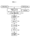
FIG. 1 is a flowchart showing a method of producing a biological tissue-reinforcing material according to an embodiment of the present invention. -
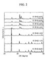
FIG. 2 shows X-ray analysis results of the biological tissue-reinforcing material according to the present embodiment produced by the production method ofFIG. 1 . -
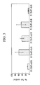
FIG. 3 is a graph showing the proliferation potency of osteoblasts seeded and cultured on a pellet of the biological tissue-reinforcing material according to the present embodiment produced by the production method ofFIG. 1 . -
FIG. 4 is a graph showing the differentiation potency of osteoblasts seeded and cultured on a pellet of the biological tissue-reinforcing material according to the present embodiment produced by the production method ofFIG. 1 . -
FIG. 5 is a graph showing the proliferation potency of osteoblasts cultured using a medium containing the biological tissue-reinforcing material according to the present embodiment produced by the production method ofFIG. 1 . -
FIG. 6 is a graph showing the differentiation potency of osteoblasts cultured using a medium containing the biological tissue-reinforcing material according to the present embodiment produced by the production method ofFIG. 1 . -
FIG. 7 is a graph showing results of the proliferation potency assessed by DNA amount when hMSCs were cultured in an extract in Experimental Example 3-1. -
FIG. 8 is a graph showing results of the proliferation potency assessed by DNA amount when hMSCs were cultured on a pellet in Experimental Example 3-1. -
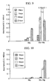
FIG. 9 is a graph showing results of the differentiation potency assessed by the production amount of alkaline phosphatase when hMSCs were cultured in an extract in Experimental Example 3-2. -
FIG. 10 is a graph showing results of the differentiation potency assessed by the production amount of alkaline phosphatase when hMSCs were cultured on a pellet in Experimental Example 3-2. -
FIG. 11 is a graph showing results of the differentiation potency assessed by the production amount of osteocalcin when hMSCs were cultured the extract in Experimental Example 3-2. -
FIG. 12 is a graph showing results of the differentiation potency assessed by the production amount of osteocalcin when hMSCs were cultured on the pellet in Experimental Example 3-2. -
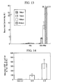
FIG. 13 is a graph showing results of the results of the degree of calcification assessed by the deposition amount of calcium when hMSCs were cultured in an extract in Experimental Example 4. -
FIG. 14 is a graph showing the assessment results of the absorption amount of bFGF in Experimental Example 5. - Hereunder is a description of a biological tissue-reinforcing material and a method of producing the same according to an embodiment according to the present invention, with reference to
FIG. 1 to FIG. 6 .
The biological tissue-reinforcing material according to the present embodiment comprises sulfate group-containing calcium phosphate. - As shown in
FIG. 1 , this biological tissue-reinforcing material can be produced using the following production method.
First, 0.5M sulfuric acid H2SO4, 0.5M ammonium hydrogen phosphate (NH4)2HPO4 aqueous solution, and 0.5M calcium nitrate Ca(NO3)2 aqueous solution are prepared. Next, thus prepared 0.5M sulfuric acid and 0.5M ammonium hydrogen phosphate aqueous solution are mixed. Then, the obtained mixture is adjusted topH 10 using 1.0N sodium hydroxide. - Thus prepared mixture is added with 0.5M calcium nitrate aqueous solution gradually dropwise (step S1). At this time, over monitoring the pH of the mixture, 1.0N sodium hydroxide is added to adjust the pH to 10. After the dropwise addition, the mixture is stirred over day and night (24 hours) (step S2). The precipitate is separated by centrifugation and washed with distilled water (step S3). The washed precipitate is filtered (step S4), dried (step S5), and retained in an atmospheric air at 800°C for 2 hours to thereby effect sintering (step S6). By so ding, the biological tissue-reinforcing material of the present embodiment is produced.
- In this manner, according to the method of producing a biological tissue-reinforcing material of the present embodiment, since the step S1 of coprecipitating ammonium hydrogen phosphate with calcium nitrate in the presence of sulfuric acid, is included, then a biological tissue-reinforcing material which homogenously contains phosphorus and sulfur at a determined mixture ratio can be readily produced. Moreover, according to thus produced biological tissue-reinforcing material of the present embodiment, the action of the homogenously contained sulfate group brings superior proliferation potency and differentiation potency of osteoblasts to those of conventional HAP, while enabling to retain characteristics of conventional HAP.
- In particular, the ratio of sulfur S to phosphorus P preferably satisfies 1/2 ≤ P/(S+P) ≤ 5/6. By so doing, equivalent physical properties to those of conventional HAP can be provided.
Moreover, according to the biological tissue-reinforcing material and the method of producing the same of the present embodiment, advantages are provided in which the production is carried out without a need for special raw materials, at an approximately comparative cost to the production cost using conventional HAP. - The biological tissue-reinforcing material according to the present embodiment is surely applicable as a conventional bone-reinforcing material, as well as being applicable to the following directions because of its high osteoblast-activating action.
- Osteoblasts can be cultured at a highly activated state.
- A cultured bone having a high osteoblast activity or a cultured bone rich in the extracellular matrix can be provided.
- A bone-reinforcing material which activates osteoblasts can be provided.
- Therapeutic effects can be expected due to the activation of osteoblasts and the recalcification effect of the extracellular matrix.
- Therapeutic effects can be expected due to the activation of osteoblasts and the recalcification effect of the extracellular matrix.
- Next is a description of Example of the biological tissue-reinforcing material and the method of producing the same according to the above embodiment.
In the present Example, a biological tissue-reinforcing material comprising a sulfuric acid-HAP was produced in accordance with the flowchart shown inFig. 1 . - 0.5M sulfuric acid and 0.5M ammonium hydrogen phosphate aqueous solution were mixed so that the ratio of sulfate ions SO4 2- to phosphate ions PO4 3-, that is, X = PO4 3-/(SO4 2-+PO4 3-) = P/ (S+P), satisfies X = 0, 1/6, 2/6, 3/6, 4/6, 5/6, or 6/6, and the obtained mixture was adjusted to
pH 10 using 1.0N sodium hydroxide. -
FIG. 2 shows X-ray analysis results of thus obtained biological tissue-reinforcing material of the present embodiment having ratios of sulfur to phosphorus X = 1/6 to 5/6, conventional HAP having X = 6/6, and calcium sulfate having X = 0. According to thisFIG. 2 , it was confirmed that, in cases of X = 3/6 to 5/6, the biological tissue-reinforcing material according to the present embodiment takes an approximately same structure as that of conventional HAP (X = 6/6). - Next, a pellet comprising the biological tissue-reinforcing material according to the present embodiment was produced. The pellet was produced by the following manner. A powder of sulfuric acid-HAP obtained in the drying step S5 of
FIG. 1 was put into, for example, a stainless mold, and was press-formed at 30MPa. The formed product was sintered in an atmospheric air at 800°C for 2 hours in the sintering step S6. - On thus produced pellet of the biological tissue-reinforcing material according to the present embodiment, normal human osteoblasts were seeded at 4 × 10⁴ cells/well/mL, and were cultured for a week. The proliferation potency of the resultant osteoblasts is shown in
FIG. 3 , and the differentiation potency thereof is shown inFIG. 4 , respectively in ratios with respect to the control. The control respectively means cells which were directly cultured in a culture dish without seeding.
The proliferation potency was measured with Tetra Color One (450 nm), and the differentiation potency was measured using alkaline phosphatase (ALP) activity through the color reaction of PNP (405nm). - According to this, the pellet of the biological tissue-reinforcing material according to the present embodiment was confirmed to have significantly higher proliferation potency and differentiation potency of osteoblasts as compared to those of conventional HAP (X = 6/6) and calcium sulfate (X = 0/6).
- Next, the sulfuric acid-HAP powder that had been heat-treated in the sintering step S6 was put in a medium (100 mg/mL), and agitated with a shaker for 72 hours (150 rpm). Then, the obtained liquid was placed in a centrifugal separator and subjected to centrifugation at 3000 rpm for 10 minutes. Thus obtained supernatant was added with osteoblasts (4 × 10⁴ cells/well/mL). The proliferation potency of the resultant cultured osteoblasts is shown in
FIG. 5 , and the differentiation potency thereof is shown inFIG. 6 , respectively in ratios with respect to the control. The control respectively means cells which were directly cultured in a culture dish without seeding. - According to this, it was found that even the mere addition of the biological tissue-reinforcing material according to the present embodiment in the medium of osteoblasts was able to increase the proliferation potency and the differentiation potency.
- A pellet (12 mmϕ × 1 mm) comprising the biological tissue-reinforcing material having the ratio of sulfur to phosphorus X = 4/6 (referred to as "SO4-HAp" in the following description and drawings) was produced in the same production method as that of the above embodiment and was used as a sample. Moreover, a pellet (12 mmϕ × 1 mm) comprising conventional hydroxyapatite (X = 6/6; abbreviated as "HAp" in the following description and drawings) was used as a comparative sample.
- In order to examine the use of SO4-HAp as a scaffold material (culture substrate) in regenerative medicines, using the above samples as scaffold materials (culture substrates), human mesenchymal stem cells (hereunder, abbreviated as "hMSC") were seeded in a bone differentiation-inducing medium containing differentiation inducing factors (dexamethasone, ascorbic acid, and β-glycerophosphoric acid) in a 24-well plate at 2 × 10⁴ cells/ml/well, and cultured (7 days, 14 days, and 21 days) to examine the proliferation and the differentiation of hMSCs.
- Moreover, in order to examine the influence of runoff components of SO4-HAp, extraction treatment (100 mg/ml, 37°C, 3 days) was performed and hMSCs were cultured in the obtained extract (7 days, 14 days, and 21 days).
- The hMSC proliferation was quantified through the production of a cell lysate and following measurement of DNA amount in the lysate.
The cell lysate was produced as follows. First, on completion of culture, the medium was discarded and cells were washed with a phosphate buffered saline solution (PBS) twice. Then, trypsin was added thereto (200 µl/well, 37°C, 1 minute) to peel off these cells. PBS was added at 800 µl per each well, and the cell solution was collected and centrifuged (4°C, 1000 rpm, 2 minutes). The supernatant was discarded and the cells were collected. The collected cells were added with 600 µl of a solution containing 2% nonionic surfactant Nonidet (registered trademark) P-40. The solution was cooled in ice and treated with ultrasonic waves for 10 seconds to disrupt these cells. - In the DNA quantification, PicoGreen (registered trademark) ds DNA quantification kit (excitation wavelength (Ex): 485 nm; detection wavelength (Em): 530 nm) manufactured by Molecular Probes was used.
FIG. 7 shows the proliferation potency resulting from the culture in the extract, andFIG. 8 shows the proliferation potency resulting from the culture on the pellet, respectively using SO4-HAp and HAp. - The differentiation potency was assessed by the production amounts of alkaline phosphatase and osteocalcin.
The production amount of alkaline phosphatase was measured as follows. On completion of culture, the medium in each well was discarded and cells were washed PBS. Then, 0.1M glycine-NaCl-NaOH buffer solution containing 4 mM p-nitrophosphoric acid, 10 mM MgCl2, and 0.1 mM ZnCl2 was added at 500 µl per each well. After leaving at a room temperature for 5 minutes, the absorbance of the buffer solution (optical density O.D. at the wavelength of 405 nm) was measured. - The production amount of osteocalcin was measured by placing 100 µl of the cell lysate in an antibody plate, and following measurement of the osteocalcin concentration with use of a commercially available ELISA kit (Gla-OC EIA Kit (manufactured by TAKARA BIO INC.)).
-
FIG. 9 shows the production amount of alkaline phosphatase resulting from the culture in the extract,FIG. 10 shows the production amount of alkaline phosphatase resulting from the culture on the pellet,FIG. 11 shows the production amount of osteocalcin resulting from the culture in the extract, andFIG. 12 shows the production amount of osteocalcin resulting from the culture on the pellet, respectively using SO4-HAp and HAp. - Since hMSCs are said to be immature cells as compared to mature cells such as osteoblasts and are superior in the proliferation potency to mature cells, they attract a lot of attention as a cell source to be used for regenerative medicines. The results of Experimental Example 3 suggest that the combination of the biological tissue-reinforcing material of the present invention with hMSCs or stem cells would optimize the combined cell potency, and the effect thereof is expected to improve the therapeutic effect.
- On completion of culture in the extract of Experimental Example 3, cells were collected, and then 0.1N hydrochloric acid was added at 500 µl per each well to solve deposited calcium. Subsequently, the calcium concentration of this solution was quantified using Calcium C-Test Wako (Wako Chemicals), to be used as the measured value of the degree of calcification. The measurement results are shown in
FIG. 13 . - A pellet comprising the same SO4-HAp as that of Experimental Example 3 (ratio of sulfur to phosphorus X = 4/6) and a pellet comprising HAp (ratio of sulfur to phosphorus X = 6/6) were respectively put in a serum-free medium containing 500 pg/ml of a basic fibroblast growth factor (hereunder, referred to as "bFGF"; manufactured by Pepro Tech EC Ltd. (London, UK)). These solutions were incubated at 37°C for 7 days, and thereby the absorption of bFGF was examined. The bFGF amount absorbed onto the pellet was measured using a reagent included in a commercially available ELISA kit (Quantikine (registered trademark), Human FGF Basic Immunoassay, R&D Systems Inc., Minneapolis, MN, USA).
- First, the incubated pellet was washed with a cleaning solution included in the ELISA kit, and added with 800 µl of bFGF conjugate. After 2 hours, a substrate solution and a stop solution were added in accordance with the protocol of the ELISA kit. The colored solution was transferred in a new well, and the absorbance of the solution at 450 nm was measured. At this time, the bFGF amount absorbed on the well containing no pellet was used as a control value. The measurement results are shown in
FIG. 14 . - The measurement results showed that SO4-HAp had a higher bFGF adsorption capacity than that of HAp. Growth factors or cytokines have stimulating and activating effects on related cells, and are said to be associated with bone remodeling and actions of inducing stem cells or immunocytes to necessary sites, for example. The results of the present Experimental Examples suggest that the combination of SO4-HAp with a growth factor or the like would provide a long-time sustaining effect on the efficacy of the combined growth factor (sustained-release effect of the growth factor over a long time), and the effect thereof is expected to improve the therapeutic effect.
Claims (16)
- A biological tissue-reinforcing material, comprising sulfate group-containing calcium phosphate.
- A biological tissue-reinforcing material according to claim 1, wherein the preparation is performed so that a ratio of sulfur (S) to phosphorus (P) satisfies

- A method of producing a biological tissue-reinforcing material, comprising the step of coprecipitating ammonium hydrogen phosphate with calcium nitrate in the presence of sulfuric acid.
- A biological tissue-reinforcing material, having a vivo-derived substance and a substrate which comprises sulfate group-containing calcium phosphate.
- A biological tissue-reinforcing material according to claim 4, wherein the preparation is performed so that a ratio of sulfur (S) to phosphorus (P) satisfies

- A biological tissue-reinforcing material according to either one of claim 4 and claim 5, wherein the vivo-derived substance includes at least either one of a growth factor and a cytokine.
- A biological tissue-reinforcing material according to either one of claim 4 and claim 5, wherein the vivo-derived substance includes a tissue-derived cell.
- A biological tissue-reinforcing material according to either one of claim 4 and claim 5, wherein the vivo-derived substance includes a cell group containing stem cells.
- A method of culturing cells, comprising the step of combining the biological tissue-reinforcing material according to either one of claim 1 and claim 2 with a vivo-derived substance.
- A method of culturing cells according to claim 9, wherein the vivo-derived substance includes at least either one of a growth factor and a cytokine.
- A method of culturing cells according to claim 9, wherein the vivo-derived substance includes a tissue-derived cell.
- A method of culturing cells according to claim 9, wherein the vivo-derived substance includes a cell group containing stem cells.
- Use of the biological tissue-reinforcing material according to either one of claim 1 and claim 2 as a cell culture substrate in combination with a vivo-derived substance.
- The use of a biological tissue-reinforcing material according to claim 13, wherein the vivo-derived substance includes at least either one of a growth factor and a cytokine.
- The use of a biological tissue-reinforcing material according to claim 13, wherein the vivo-derived substance includes a tissue-derived cell.
- The use of a biological tissue-reinforcing material according to claim 13, wherein the vivo-derived substance includes a cell group containing stem cells.
Applications Claiming Priority (2)
| Application Number | Priority Date | Filing Date | Title |
|---|---|---|---|
| JP2005294058A JP2007097956A (en) | 2005-10-06 | 2005-10-06 | Biological tissue filling material and its manufacturing method |
| PCT/JP2006/314096 WO2007039973A1 (en) | 2005-10-06 | 2006-07-14 | Reinforcing agent for biological tissue, method of producing the same, utilization of the same and method of culturing cells |
Publications (2)
| Publication Number | Publication Date |
|---|---|
| EP1932548A1 true EP1932548A1 (en) | 2008-06-18 |
| EP1932548A4 EP1932548A4 (en) | 2011-10-12 |
Family
ID=37906019
Family Applications (1)
| Application Number | Title | Priority Date | Filing Date |
|---|---|---|---|
| EP06781124A Withdrawn EP1932548A4 (en) | 2005-10-06 | 2006-07-14 | Reinforcing agent for biological tissue, method of producing the same, utilization of the same and method of culturing cells |
Country Status (6)
| Country | Link |
|---|---|
| US (1) | US20090291499A1 (en) |
| EP (1) | EP1932548A4 (en) |
| JP (1) | JP2007097956A (en) |
| KR (1) | KR20080051155A (en) |
| CN (1) | CN101277728B (en) |
| WO (1) | WO2007039973A1 (en) |
Family Cites Families (7)
| Publication number | Priority date | Publication date | Assignee | Title |
|---|---|---|---|---|
| US5462722A (en) * | 1991-04-17 | 1995-10-31 | Liu; Sung-Tsuen | Calcium phosphate calcium sulfate composite implant material |
| DE69739540D1 (en) * | 1996-04-19 | 2009-10-01 | Osiris Therapeutics Inc | THE RECONSTRUCTION AND REINFORCEMENT OF BONES BY MEANS OF MESENCHYMAL STEM CELLS |
| WO2000007639A1 (en) * | 1998-08-07 | 2000-02-17 | Tissue Engineering, Inc. | Bone precursor compositions |
| US6224635B1 (en) * | 1998-11-06 | 2001-05-01 | Hospital For Joint Diseases | Implantation of surgical implants with calcium sulfate |
| US7220571B2 (en) * | 2000-09-28 | 2007-05-22 | Archer-Daniels-Midland Company | Escherichia coli strains which over-produce L-threonine and processes for the production of L-threonine by fermentation |
| JP5007476B2 (en) * | 2001-03-23 | 2012-08-22 | オリンパス株式会社 | Artificial aggregate |
| ES2402651T3 (en) * | 2005-09-09 | 2013-05-07 | Agnovos Healthcare, Llc | Cement substitute for bone graft of composite material and articles produced from it |
-
2005
- 2005-10-06 JP JP2005294058A patent/JP2007097956A/en active Pending
-
2006
- 2006-07-14 WO PCT/JP2006/314096 patent/WO2007039973A1/en not_active Ceased
- 2006-07-14 CN CN2006800366948A patent/CN101277728B/en not_active Expired - Fee Related
- 2006-07-14 EP EP06781124A patent/EP1932548A4/en not_active Withdrawn
- 2006-07-14 KR KR1020087007993A patent/KR20080051155A/en not_active Withdrawn
- 2006-07-14 US US12/066,475 patent/US20090291499A1/en not_active Abandoned
Also Published As
| Publication number | Publication date |
|---|---|
| EP1932548A4 (en) | 2011-10-12 |
| KR20080051155A (en) | 2008-06-10 |
| JP2007097956A (en) | 2007-04-19 |
| CN101277728B (en) | 2012-03-07 |
| US20090291499A1 (en) | 2009-11-26 |
| CN101277728A (en) | 2008-10-01 |
| WO2007039973A1 (en) | 2007-04-12 |
Similar Documents
| Publication | Publication Date | Title |
|---|---|---|
| Wang et al. | Bioresponsive phosphoester hydrogels for bone tissue engineering | |
| JP3808900B2 (en) | Biological material composed of a three-dimensional biocompatible and biodegradable matrix comprising an effective culture of bone marrow stem cells partially or fully differentiated into connective tissue cells and a hyaluronic acid derivative | |
| JP3310291B2 (en) | Bioactive substance template for in vitro synthesis of bone tissue | |
| Sun et al. | Proliferation and osteoblastic differentiation of human bone marrow-derived stromal cells on akermanite-bioactive ceramics | |
| Shelton et al. | Bone marrow cell gene expression and tissue construct assembly using octacalcium phosphate microscaffolds | |
| Baykan et al. | Evaluation of a biomimetic poly (ε-caprolactone)/β-tricalcium phosphate multispiral scaffold for bone tissue engineering: in vitro and in vivo studies | |
| Kim et al. | Bone tissue engineering supported by bioprinted cell constructs with endothelial cell spheroids | |
| CN103721292A (en) | Novel multifunctional mesoporous glass bracket with biological activity as well as preparation method and purpose thereof | |
| Suzuki et al. | Growth and adhesion of osteoblast-like cells derived from neonatal rat calvaria on calcium phosphate ceramics | |
| Manferdini et al. | Mineralization behavior with mesenchymal stromal cells in a biomimetic hyaluronic acid-based scaffold | |
| JP2003509021A (en) | Cell growth substrate | |
| Cordonnier et al. | Osteoblastic differentiation and potent osteogenicity of three‐dimensional hBMSC‐BCP particle constructs | |
| CN120346383A (en) | Biphasic hydrogel scaffold of bionic bone cartilage matrix and preparation method and application thereof | |
| Li et al. | Osteogenic induction of adipose‐derived stromal cells: not a requirement for bone formation in vivo | |
| Zeimaran et al. | Osteogenic differentiation of mesenchymal stem cells on a poly (octanediol citrate)/bioglass composite scaffold in vitro | |
| RU2530622C2 (en) | Biotransplant for recovery of bone tissue volume in case of degenerative diseases and traumatic injuries of bones and method of obtaining thereof | |
| Sun et al. | Dual mechanism design to enhance bone formation by overexpressed SDF-1 ADSCs in magnesium doped calcium phosphate scaffolds | |
| JP5007476B2 (en) | Artificial aggregate | |
| WO2001082773A2 (en) | Structures useful for bone engineering and methods | |
| Walther et al. | Development of novel scaffolds for tissue engineering by flock technology | |
| Zhou et al. | Biological effects of apatite nanoparticle-constructed ceramic surfaces in regulating behaviours of mesenchymal stem cells | |
| US20090291499A1 (en) | Biological tissue-reinforcing material, method of producing the same, use of the same, and method of culturing cells | |
| US20050100533A1 (en) | Structures useful for bone engineering and methods | |
| JP3650734B2 (en) | Methods for inducing osteoblast differentiation of human extramedullary adipose tissue cells | |
| Viale-Bouroncle et al. | β-tricalcium-phosphate stimulates the differentiation of dental follicle cells |
Legal Events
| Date | Code | Title | Description |
|---|---|---|---|
| PUAI | Public reference made under article 153(3) epc to a published international application that has entered the european phase |
Free format text: ORIGINAL CODE: 0009012 |
|
| 17P | Request for examination filed |
Effective date: 20080319 |
|
| AK | Designated contracting states |
Kind code of ref document: A1 Designated state(s): DE FR GB |
|
| RBV | Designated contracting states (corrected) |
Designated state(s): DE FR GB |
|
| RIN1 | Information on inventor provided before grant (corrected) |
Inventor name: TAMAI, MASATO Inventor name: HAKAMATSUKA, YASUHARU Inventor name: TSUCHIYA, TOSHIE Inventor name: TAMURA, TOMOAKI |
|
| A4 | Supplementary search report drawn up and despatched |
Effective date: 20110908 |
|
| RIC1 | Information provided on ipc code assigned before grant |
Ipc: A61L 27/00 20060101AFI20110920BHEP |
|
| STAA | Information on the status of an ep patent application or granted ep patent |
Free format text: STATUS: THE APPLICATION HAS BEEN WITHDRAWN |
|
| 18W | Application withdrawn |
Effective date: 20120404 |