EP1917528B1 - Biomarkers and methods for determining sensitivity to epidermal growth factor receptor modulators - Google Patents
Biomarkers and methods for determining sensitivity to epidermal growth factor receptor modulators Download PDFInfo
- Publication number
- EP1917528B1 EP1917528B1 EP06813702A EP06813702A EP1917528B1 EP 1917528 B1 EP1917528 B1 EP 1917528B1 EP 06813702 A EP06813702 A EP 06813702A EP 06813702 A EP06813702 A EP 06813702A EP 1917528 B1 EP1917528 B1 EP 1917528B1
- Authority
- EP
- European Patent Office
- Prior art keywords
- homo sapiens
- egfr
- dna
- mrna
- biomarkers
- Prior art date
- Legal status (The legal status is an assumption and is not a legal conclusion. Google has not performed a legal analysis and makes no representation as to the accuracy of the status listed.)
- Not-in-force
Links
Images


Classifications
-
- C—CHEMISTRY; METALLURGY
- C12—BIOCHEMISTRY; BEER; SPIRITS; WINE; VINEGAR; MICROBIOLOGY; ENZYMOLOGY; MUTATION OR GENETIC ENGINEERING
- C12Q—MEASURING OR TESTING PROCESSES INVOLVING ENZYMES, NUCLEIC ACIDS OR MICROORGANISMS; COMPOSITIONS OR TEST PAPERS THEREFOR; PROCESSES OF PREPARING SUCH COMPOSITIONS; CONDITION-RESPONSIVE CONTROL IN MICROBIOLOGICAL OR ENZYMOLOGICAL PROCESSES
- C12Q1/00—Measuring or testing processes involving enzymes, nucleic acids or microorganisms; Compositions therefor; Processes of preparing such compositions
- C12Q1/68—Measuring or testing processes involving enzymes, nucleic acids or microorganisms; Compositions therefor; Processes of preparing such compositions involving nucleic acids
- C12Q1/6876—Nucleic acid products used in the analysis of nucleic acids, e.g. primers or probes
- C12Q1/6883—Nucleic acid products used in the analysis of nucleic acids, e.g. primers or probes for diseases caused by alterations of genetic material
- C12Q1/6886—Nucleic acid products used in the analysis of nucleic acids, e.g. primers or probes for diseases caused by alterations of genetic material for cancer
-
- A—HUMAN NECESSITIES
- A61—MEDICAL OR VETERINARY SCIENCE; HYGIENE
- A61P—SPECIFIC THERAPEUTIC ACTIVITY OF CHEMICAL COMPOUNDS OR MEDICINAL PREPARATIONS
- A61P35/00—Antineoplastic agents
-
- G—PHYSICS
- G01—MEASURING; TESTING
- G01N—INVESTIGATING OR ANALYSING MATERIALS BY DETERMINING THEIR CHEMICAL OR PHYSICAL PROPERTIES
- G01N33/00—Investigating or analysing materials by specific methods not covered by groups G01N1/00 - G01N31/00
- G01N33/48—Biological material, e.g. blood, urine; Haemocytometers
- G01N33/50—Chemical analysis of biological material, e.g. blood, urine; Testing involving biospecific ligand binding methods; Immunological testing
- G01N33/53—Immunoassay; Biospecific binding assay; Materials therefor
- G01N33/575—Immunoassay; Biospecific binding assay; Materials therefor for cancer
- G01N33/57535—Immunoassay; Biospecific binding assay; Materials therefor for cancer of the large intestine, e.g. colon, rectum or anus
-
- C—CHEMISTRY; METALLURGY
- C12—BIOCHEMISTRY; BEER; SPIRITS; WINE; VINEGAR; MICROBIOLOGY; ENZYMOLOGY; MUTATION OR GENETIC ENGINEERING
- C12Q—MEASURING OR TESTING PROCESSES INVOLVING ENZYMES, NUCLEIC ACIDS OR MICROORGANISMS; COMPOSITIONS OR TEST PAPERS THEREFOR; PROCESSES OF PREPARING SUCH COMPOSITIONS; CONDITION-RESPONSIVE CONTROL IN MICROBIOLOGICAL OR ENZYMOLOGICAL PROCESSES
- C12Q2600/00—Oligonucleotides characterized by their use
- C12Q2600/106—Pharmacogenomics, i.e. genetic variability in individual responses to drugs and drug metabolism
-
- C—CHEMISTRY; METALLURGY
- C12—BIOCHEMISTRY; BEER; SPIRITS; WINE; VINEGAR; MICROBIOLOGY; ENZYMOLOGY; MUTATION OR GENETIC ENGINEERING
- C12Q—MEASURING OR TESTING PROCESSES INVOLVING ENZYMES, NUCLEIC ACIDS OR MICROORGANISMS; COMPOSITIONS OR TEST PAPERS THEREFOR; PROCESSES OF PREPARING SUCH COMPOSITIONS; CONDITION-RESPONSIVE CONTROL IN MICROBIOLOGICAL OR ENZYMOLOGICAL PROCESSES
- C12Q2600/00—Oligonucleotides characterized by their use
- C12Q2600/136—Screening for pharmacological compounds
-
- G—PHYSICS
- G01—MEASURING; TESTING
- G01N—INVESTIGATING OR ANALYSING MATERIALS BY DETERMINING THEIR CHEMICAL OR PHYSICAL PROPERTIES
- G01N2333/00—Assays involving biological materials from specific organisms or of a specific nature
- G01N2333/435—Assays involving biological materials from specific organisms or of a specific nature from animals; from humans
- G01N2333/475—Assays involving growth factors
- G01N2333/485—Epidermal growth factor [EGF] (urogastrone)
-
- G—PHYSICS
- G01—MEASURING; TESTING
- G01N—INVESTIGATING OR ANALYSING MATERIALS BY DETERMINING THEIR CHEMICAL OR PHYSICAL PROPERTIES
- G01N2333/00—Assays involving biological materials from specific organisms or of a specific nature
- G01N2333/435—Assays involving biological materials from specific organisms or of a specific nature from animals; from humans
- G01N2333/705—Assays involving receptors, cell surface antigens or cell surface determinants
- G01N2333/70596—Molecules with a "CD"-designation not provided for elsewhere in G01N2333/705
-
- G—PHYSICS
- G01—MEASURING; TESTING
- G01N—INVESTIGATING OR ANALYSING MATERIALS BY DETERMINING THEIR CHEMICAL OR PHYSICAL PROPERTIES
- G01N2333/00—Assays involving biological materials from specific organisms or of a specific nature
- G01N2333/435—Assays involving biological materials from specific organisms or of a specific nature from animals; from humans
- G01N2333/705—Assays involving receptors, cell surface antigens or cell surface determinants
- G01N2333/71—Assays involving receptors, cell surface antigens or cell surface determinants for growth factors; for growth regulators
-
- G—PHYSICS
- G01—MEASURING; TESTING
- G01N—INVESTIGATING OR ANALYSING MATERIALS BY DETERMINING THEIR CHEMICAL OR PHYSICAL PROPERTIES
- G01N2800/00—Detection or diagnosis of diseases
- G01N2800/52—Predicting or monitoring the response to treatment, e.g. for selection of therapy based on assay results in personalised medicine; Prognosis
Definitions
- the present invention relates generally to the field of pharmacogenomics, and more specifically to methods and procedures to determine drug sensitivity in patients to allow the identification of individualized genetic profiles which will aid in treating diseases and disorders.
- Cancer is a disease with extensive histoclinical heterogeneity. Although conventional histological and clinical features have been correlated to prognosis, the same apparent prognostic type of tumors varies widely in its responsiveness to therapy and consequent survival of the patient.
- New prognostic and predictive markers which would facilitate an individualization of therapy for each patient, are needed to accurately predict patient response to treatments, such as small molecule or biological molecule drugs, in the clinic.
- the problem may be solved by the identification of new parameters that could better predict the patient's sensitivity to treatment.
- the classification of patient samples is a crucial aspect of cancer diagnosis and treatment.
- the association of a patient's response to a treatment with molecular and genetic markers can open up new opportunities for treatment development in non-responding patients, or distinguish a treatment's indication among other treatment choices because of higher confidence in the efficacy.
- the pre-selection of patients who are likely to respond well to a medicine, drug, or combination therapy may reduce the number of patients needed in a clinical study or accelerate the time needed to complete a clinical development program ( Cockett et al., Current Opinion in Biotechnology, 11:602-609 (2000 )).
- the epidermal growth factor receptor (EGFR) and its downstream signaling effectors notably members of the Ras/Raf/MAP kinase pathway, play an important role in both normal and malignant epithelial cell biology ( Normanno et al., Gene 366, 2-16 (2006 )) and have therefore become established targets for therapeutic development.
- EGFR antibody cetuximab and the EGFR small molecular tyrosine kinase inhibitors (TKIs) gefitinib and erlotinib have demonstrated activity in a subset of patients ( Baselga and Arteaga, J. Clin. Oncol.
- cetuximab has been attributed to its ability to block EGFR ligand binding and ligand-dependent EGFR activation.
- Clinical activity of cetuximab has been shown in multiple epithelial tumor types ( Bonner et al., N. Engl. J. Med., 354, 567-578 (2006 ); Cunningham et al., N. Engl. J. Med., 351, 337-345 (2004 )), however responses continue to be seen in only a fraction of patients.
- Previous attempts to identify predictors of sensitivity or resistance as described above have focused on specific biomarkers rather than using genomic discovery approaches.
- RNA-, DNA- and protein-based markers have rarely been examined in the same patient population in a single study, making comparisons challenging.
- Biomarkers useful for determining sensitivity to EGFR modulators have been described in published PCT applications US 2005/019785 , WO2004/063709 , WO2005/067667 , and WO2005/094332 .
- the present invention relates to an in vitro method for predicting the likelihood a mammal will respond therapeutically to a method of treating colorectal cancer comprising administering an EGFR modulator that inhibits binding of EGF to EGFR or the kinase activity of EGFR, wherein the method comprises:
- the present invention also relates to an vitro method for predicting the likelihood a mammal will respond therapeutically to a method of treating colorectal cancer comprising administering an EGFR modulator that inhibits binding of EGF to EGFR or the kinase activity of EGFR, wherein the method comprises measuring the mRNA expression level of both epiregulin and amphiregulin biomarkers in a biological sample of said mammal, wherein an elevated expression level of said biomarkers relative to a predetermined level indicates an increased likelihood the mammal will respond therapeutically to said method of treating colorectal cancer.
- the invention provides methods and procedures for determining patient sensitivity to one or more Epidermal Growth Factor Receptor (EGFR) modulators that inhibit binding of EGF to EGFR or the kinase activity of EGFR.
- EGFR Epidermal Growth Factor Receptor
- the invention also provides methods of determining or predicting whether an individual requiring therapy for a disease state such as cancer will or will not respond to treatment, prior to administration of the treatment, wherein the treatment comprises administration of one or more EGFR modulators.
- the one or more EGFR modulators that inhibit binding of EGF to EGFR or the kinase activity of EGFR are compounds that can be selected from, for example, one or more EGFR-specific ligands, one or more small molecule EGFR inhibitors, or one or more EGFR binding monoclonal antibodies.
- the biological sample can be, for example, a tissue sample comprising cancer cells and the tissue is fixed, paraffin-embedded, fresh, or frozen.
- the EGFR modulator is cetuximab.
- a difference in the level of said biomarkers that is sufficient to predict the likelihood that the mammal will or will not respond therapeutically to the method of treating colorectal cancer can be readily determined by one of skill in the art using known techniques.
- the increase or decrease in the mRNA expression level of the biomarkers can be correlated to determine whether the difference is sufficient to predict the likelihood that a mammal will respond therapeutically.
- the difference in the level of the biomarkers that is sufficient can, in one aspect, be predetermined prior to predicting the likelihood that the mammal will respond therapeutically to the treatment.
- the difference in the level of the biomarker is a difference in the mRNA level (measured, for example, by RT-PCR or a microarray), such as at least a two-fold difference, at least a three-fold difference, or at least a four-fold difference in the level of expression.
- the difference in the level of the biomarker refers to a p-value of ⁇ 0.05 in Anova (t test) analysis.
- respond therapeutically refers to the alleviation or abrogation of the cancer. This means that the life expectancy of an individual affected with the cancer will be increased or that one or more of the symptoms of the cancer will be reduced or ameliorated.
- the term encompasses a reduction in cancerous cell growth or tumor volume. Whether a mammal responds therapeutically can be measured by many methods well known in the art, such as PET imaging.
- the mammal can be, for example, a human, rat, mouse, dog, rabbit, pig sheep, cow, horse, cat, primate, or monkey.
- the method of the invention is an in vitro method which comprises measuring the level of the biomarkers in the biological sample.
- the biological sample is a biological sample taken from the mammal and measuring the level of the biomarkers in the biological sample is for measuring the level of the biomarkers in the mammal.
- the biological sample can comprise, for example, at least one of serum, whole fresh blood, peripheral blood mononuclear cells, frozen whole blood, fresh plasma, frozen plasma, urine, saliva, skin, hair follicle, bone marrow, or tumor tissue.
- the level of the at least one additional biomarker can be, for example, the level of protein and/or mRNA transcript of the biomarker.
- the level of the biomarker can be determined, for example, by RT-PCR or another PCR-based method, immunohistochemistry, proteomics techniques, or any other methods known in the art, or their combination.
- the invention provides an in vitro method for identifying a mammal that will respond therapeutically to a method of treating colorectal cancer comprising administering of an EGFR modulator that inhibits binding of EGF to EGFR or the kinase activity of EGFR, wherein the method comprises: (a) measuring in a biological sample of said mammal the mRNA expression level of bothepiregulin and amphiregulin biomarkers; (b) exposing a biological sample from the mammal to the EGFR modulator, (c) following the exposing in step (b), measuring in said biological sample the mRNA expression level of said biomarkers, wherein a difference in the mRNA expression level of said biomarkers measured in step (c) compared to the mRNA expression level of said biomarkers measured in step (a) indicates that the mammal will respond therapeutically to the said method of treating colorectal cancer.
- the invention provides an in vitro method for identifying a mammal that will respond therapeutically to a method of treating colorectal cancer comprising administering an EGFR modulator that inhibits binding of EGF to EGFR or the kinase activity of EGFR, wherein the method comprises: (a) exposing a biological sample from the mammal to the EGFR modulator, (b) following the exposing of step (a), measuring in said biological sample the mRNA expression level of both epiregulin and amphiregulinbiomarkers of Table 1, wherein a difference in the mRNA expression level of saidbiomarkers measured in step (b), compared to the mRNA expression level of said biomarkers in a mammal that has not been exposed to said EGFR modulator, indicates that the mammal will respond therapeutically to said method of treating colorectal cancer.
- the invention provides an in vitro method for testing or predicting whether a mammal will respond therapeutically to a method of treating colorectal cancer comprising administering an EGFR modulator that inhibits binding of EGF to EGFR or the kinase activity of EGFR, wherein the method comprises: (a) measuring in a biological sample of said mammal the mRNA expression level of both epiregulin and amphiregulin biomarkers; (b) exposing the mammal to the EGFR modulator; (c) following the exposing of step (b), measuring in the mammal the mRNA expression level of said biomarkers, wherein a difference in the mRNA expression level of said biomarkers measured in step (c) compared to the mRNA expression level of said biomarkers measured in step (a) indicates that the mammal will respond therapeutically to said method of treating colorectal cancer.
- responding encompasses responding by way of a biological and cellular response, as well as a clinical response (such as improved symptoms, a therapeutic effect, or an adverse event), in a mammal.
- biomarkers selected from the biomarkers of Table 1.
- the biomarkers comprise sequences selected from the nucleotide and amino acid sequences provided in Table 1 and the Sequence Listing, as well as fragments and variants thereof.
- biomarker sets comprising epiregulin and amphiregulin and optionally one or more additional biomarkers selected from the biomarkers of Table 1.
- the description also provides kits for carrying out the methods of the present invention.
- the kit comprises a suitable container that comprises one or more specialized microarrays of the invention, one or more EGFR modulators that inhibits binding of EGF to EGFR or the kinase activity of EGFR for use in testing cells from patient tissue specimens or patient samples, and instructions for use.
- the kit may further comprise reagents or materials for monitoring the expression of a biomarker set at the level of mRNA or protein.
- the description provides a kit comprising epiregulin and amphiregulin and optionally one or more biomarkers selected from the biomarkers of Table 1.
- the description provides a kit comprising a nucleic acid for detecting the presence of the epiregulin and amphiregulin biomarkers.
- the kit further comprises instructions for determining whether or not a mammal will respond therapeutically to a method of treating colorectal cancer comprising administering a compound that inhibits EGFR activity.
- the instructions comprise the steps of the methods of the present invention.
- the description also provides screening assays for determining if a patient will be susceptible or resistant to treatment with one or more EGFR modulators that inhibit binding of EGF to EGFR or the kinase activity of EGFR.
- the description also provides a method of monitoring the treatment of a patient having colorectal cancer, wherein said colorectal cancer is treated by a method comprising administering one or more EGFR modulators that inhibit binding of EGF to EGFR or the kinase activity of EGFR.
- the description also provides individualized genetic profiles which are necessary to treat colorectal cancerbased on patient response at a molecular level.
- microarrays e.g., oligonucleotide microarrays or cDNA microarrays, comprising one or more biomarkers of the invention having expression profiles that correlate with either sensitivity or resistance to one or more EGFR modulators that inhibit binding of EGF to EGFR or the kinase activity of EGF.
- the description also provides antibodies, including polyclonal or monoclonal, directed against one or more biomarkers of the invention.
- Embodiments of the invention include measuring changes in the levels of secreted proteins, or plasma biomarkers, which represent one category of biomarker.
- plasma samples which represent a readily accessible source of material, serve as surrogate tissue for biomarker analysis.
- the invention provides biomarkers that respond to the modulation of a specific signal transduction pathway and also correlate with EGFR modulator sensitivity or resistance. These biomarkers can be employed for predicting response to one or more EGFR modulators.
- the biomarkers of the invention are epiregulin and amphiregulin mRNA and optionally at least one additional biomarker selected from table 1.
- Affymetrix Probe Set NT5E: 5'-nucleotidase, ecto (CD73) (LOC4907) SEQ ID NOS: 1 (DNA) and 129 (amino acid) gb:NM_002526.1/DEF Homo sapiens 5 nucleotidase (CD73) (NT5), mRNA.
- PRKA kinase anchor protein
- LOC9590 anchor protein
- SEQ ID NOS: 11 DNA
- 139 amino acid
- MD-2 Homo sapiens MD-2 protein
- brain /FL gb:L47647.1 gb:BC001190.1 gb:BC004914.1
- gb:NM_005531.1 /DEF Homo sapiens interferon, gamma-inducible protein (IFI16), mRNA.
- gamma-inducible protein 16 /FL gb:M63838.1 gb:NM_005531.1 206332_s_at 16
- PRSS8: protease, serine, 8 (prostasin) (LOC5652) SEQ ID NOS: 18 (DNA) and 146 (amino acid) gb:NM_002773.1 /DEF Homo sapiens protease, serine, 8 (prostasin) (PRSS8), mRNA.
- SEQ ID NOS: 23 (DNA) and 151 (amino acid) gb:NM_004244.1 /DEF Homo sapiens CD163 antigen (CD163), mRNA.
- CKTSF1B1 BMP antagonist 1
- PIGR polymeric immunoglobulin receptor
- alpha-inducible protein 27 /FL gb:NM 005532.1 202411_at
- gb:D31661.1 /DEF Human mRNA for tyrosine kinase, complete cds.
- DRM Homo sapiens DRM
- MIG gamma interferon
- MMP1 matrix metalloproteinas
- ALOX5: arachidonate 5-lipoxygenase (LOC240) SEQ ID NOS: 71 (DNA) and 199 (amino acid) gb:NM_000698.1 /DEF Homo sapiens arachidonate 5-lipoxygenase (ALOX5), mRNA.
- SEQ ID NOS: 83 DNA
- 211 amino acid
- gb NM_001129.2
- DEF Homo sapiens AE-binding protein 1 (AEBP1), mRNA.
- SECT Human proteolytic serine esterase-like protein
- MAFB v-maf musculoaponeurotic fibrosarcoma oncogene homolog B (avian) (LOC9935) SEQ ID NOS: 120 (DNA) and 248 (amino acid) gb:NM_005461.1 /
- biomarkers provided in Table 1 which include the nucleotide sequences of SEQ ID NOS:1-128 and the amino acid sequences of SEQ ID NOS:129-256, are referred to herein as a total of 128 biomarkers with reference to the Unigene Title.
- the biomarkers have expression levels in cells that may be dependent on the activity of the EGFR signal transduction pathway, and that are also highly correlated with EGFR modulator sensitivity exhibited by the cells. Biomarkers serve as useful molecular tools for predicting the likelihood of a response to EGFR modulators, preferably biological molecules, small molecules, and the like that affect EGFR kinase activity via direct or indirect inhibition or antagonism of EGFR kinase function or activity.
- wild type K-Ras can be selected from the K-Ras variant a and variant b nucleotide and amino acid sequences.
- Wild type K-Ras variant a has a nucleotide sequence that is 5436 nucleotides (GenBank Accession No. NM_033360.2) and encodes a protein that is 189 amino acids (GenBank Accession No. NP_203524.1).
- Wild type K-Ras variant b has a nucleotide sequence that is 5312 nucleotides (GenBank Accession No. NM_004985.3) and encodes a protein that is 188 amino acids (GenBank Accession No. NP_004976.2).
- a mutated form of K-Ras is a nucleotide or amino acid sequence that differs from wild type K-Ras at least at one position, preferably at least one nucleotide position that encodes an amino acid that differs from wild type K-Ras.
- the mutated form of K-Ras includes at least one mutation in exon 2.
- the mutated form of K-RAS includes at least one of the following mutations in exon 2 (base change (amino acid change)): 200G>A (V7M); 216G>C (G12A); 215G>T (G12C); 216G>A (G12D); 215G>C (G12R); 215G>A (G12S); 216G>T (G12V); 218G>T (G13C); 219G>A (G13D).
- base change amino acid change
- Methods for detecting K-Ras mutations are well known in the art and include, for example, the methods described in PCT Publication No. Wo2005/118876 .
- EGFR modulator is intended to mean a compound or drug that is a biological molecule or a small molecule that directly or indirectly modulates EGFR activity or the EGFR signal transduction pathway by inhibiting binding of EGF to EGFR or the kinase activity of EGFR.
- EGFR modulators include, for example, EGFR-specific ligands, small molecule EGFR inhibitors, and EGFR monoclonal antibodies.
- the EGFR modulator inhibits EGFR activity and/or inhibits the EGFR signal transduction pathway.
- the EGFR modulator is an EGFR monoclonal antibody that inhibits EGFR activity and/or inhibits the EGFR signal transduction pathway.
- EGFR modulators include biological molecules or small molecules.
- Biological molecules include all lipids and polymers of monosaccharides, amino acids, and nucleotides having a molecular weight greater than 450.
- biological molecules include, for example, oligosaccharides and polysaccharides; oligopeptides, polypeptides, peptides, and proteins; and oligonucleotides and polynucleotides.
- Oligonucleotides and polynucleotides include, for example, DNA and RNA.
- Biological molecules further include derivatives of any of the molecules described above.
- derivatives of biological molecules include lipid and glycosylation derivatives of oligopeptides, polypeptides, peptides, and proteins.
- Derivatives of biological molecules further include lipid derivatives of oligosaccharides and polysaccharides, e.g., lipopolysaccharides.
- biological molecules are antibodies, or functional equivalents of antibodies.
- Functional equivalents of antibodies have binding characteristics comparable to those of antibodies, and inhibit the growth of cells that express EGFR.
- Such functional equivalents include, for example, chimerized, humanized, and single chain antibodies as well as fragments thereof.
- Functional equivalents of antibodies also include polypeptides with amino acid sequences substantially the same as the amino acid sequence of the variable or hypervariable regions of the antibodies.
- An amino acid sequence that is substantially the same as another sequence, but that differs from the other sequence by means of one or more substitutions, additions, and/or deletions, is considered to be an equivalent sequence.
- Preferably, less than 50%, more preferably less than 25%, and still more preferably less than 10%, of the number of amino acid residues in a sequence are substituted for, added to, or deleted from the protein.
- the functional equivalent of an antibody is preferably a chimerized or humanized antibody.
- a chimerized antibody comprises the variable region of a non-human antibody and the constant region of a human antibody.
- a humanized antibody comprises the hypervariable region (CDRs) of a non-human antibody.
- the variable region other than the hypervariable region, e.g., the framework variable region, and the constant region of a humanized antibody are those of a human antibody.
- Suitable variable and hypervariable regions of non-human antibodies may be derived from antibodies produced by any non-human mammal in which monoclonal antibodies are made.
- Suitable examples of mammals other than humans include, for example, rabbits, rats, mice, horses, goats, or primates.
- Functional equivalents further include fragments of antibodies that have binding characteristics that are the same as, or are comparable to, those of the whole antibody.
- Suitable fragments of the antibody include any fragment that comprises a sufficient portion of the hypervariable (i.e., complementarity determining) region to bind specifically, and with sufficient affinity, to EGFR tyrosine kinase to inhibit growth of cells that express such receptors.
- Such fragments may, for example, contain one or both Fab fragments or the F(ab') 2 fragment.
- the antibody fragments may contain all six complementarity determining regions of the whole antibody, although functional fragments containing fewer than all of such regions, such as three, four, or five CDRs, are also included.
- the fragments are single chain antibodies, or Fv fragments.
- Single chain antibodies are polypeptides that comprise at least the variable region of the heavy chain of the antibody linked to the variable region of the light chain, with or without an interconnecting linker.
- Fv fragment comprises the entire antibody combining site.
- These chains may be produced in bacteria or in eukaryotic cells.
- the antibodies and functional equivalents may be members of any class of immunoglobulins, such as IgG, IgM, IgA, IgD, or IgE, and the subclasses thereof. In one aspect, the antibodies are members of the IgG1 subclass.
- the functional equivalents may also be equivalents of combinations of any of the above classes and subclasses.
- EGFR antibodies can be selected from chimerized, humanized, fully human, and single chain antibodies derived from the murine antibody 225 described in U.S. Patent No. 4,943,533 .
- the EGFR antibody is cetuximab (IMC-C225) which is a chimeric (human/mouse) IgG monoclonal antibody, also known under the tradename ERBITUX.
- Cetuximab Fab contains the Fab fragment of cetuximab, i.e., the heavy and light chain variable region sequences of murine antibody M225 ( U.S. Application No. 2004/0006212 , incorporated herein by reference) with human IgG1 C H 1 heavy and kappa light chain constant domains. Cetuximab includes all three IgG1 heavy chain constant domains.
- the EGFR antibody can be selected from the antibodies described in U.S. Patent No. 6,235,883 , U.S. Patent No. 5,558,864 , and U.S. Patent No. 5,891,996 .
- the EGFR antibody can be, for example, AGX-EGF (Amgen Inc.) (also known as panitumumab) which is a fully human IgG2 monoclonal antibody.
- AGX-EGF Amgen Inc.
- panitumumab also known as panitumumab
- the sequence and characterization of ABX-EGF, which was formerly known as clone E7.6.3, is disclosed in U.S. Patent No. 6,235,883 at column 28, line 62 through column 29, line 36 and Figures 29-34, which is incorporated by reference herein.
- the EGFR antibody can also be, for example, EMD72000 (Merck KGaA), which is a humanized version of the murine EGFR antibody EMD 55900.
- the EGFR antibody can also be, for example: h-R3 (TheraCIM), which is a humanized EGFR monoclonal antibody; Y10 which is a murine monoclonal antibody raised against a murine homologue of the human EGFRvIII mutation; or MDX-447 (Medarex Inc.).
- the EGFR modulators useful in the invention may also be small molecules. Any molecule that is not a biological molecule is considered herein to be a small molecule. Some examples of small molecules include organic compounds, organometallic compounds, salts of organic and organometallic compounds, saccharides, amino acids, and nucleotides. Small molecules further include molecules that would otherwise be considered biological molecules, except their molecular weight is not greater than 450. Thus, small molecules may be lipids, oligosaccharides, oligopeptides, and oligonucleotides and their derivatives, having a molecular weight of 450 or less.
- small molecules can have any molecular weight. They are merely called small molecules because they typically have molecular weights less than 450. Small molecules include compounds that are found in nature as well as synthetic compounds.
- the EGFR modulator is a small molecule that inhibits the growth of tumor cells that express EGFR. In another embodiment, the EGFR modulator is a small molecule that inhibits the growth of refractory tumor cells that express EGFR.
- IRESSA a small molecule EGFR antagonist
- TARCEVA 4-(substitutedphenylamino)quinozaline derivative [6,7-Bis(2-methoxy-ethoxy)-quinazolin-4-yl]-(3-ethynyl-1-phenyl)amine hydrochloride] EGFR inhibitor.
- TARCEVA may function by inhibiting phosphorylation of EGFR and its downstream PI3/Akt and MAP (mitogen activated protein) kinase signal transduction pathways resulting in p27-mediated cell-cycle arrest. See Hidalgo et al., Abstract 281 presented at the 37th Annual Meeting of ASCO, San Francisco, CA, 12-15 May 2001.
- small molecules are also reported to inhibit EGFR, many of which are thought to be specific to the tyrosine kinase domain of an EGFR.
- Some examples of such small molecule EGFR antagonists are described in WO 91/116051 , WO96/30347 , WO96/33980 , WO97/27199 .
- Examples of specific small molecule EGFR antagonists include C1-1033 (Pfizer Inc.), which is a quinozaline (N-[4-(3-monocyclic ring with one or two heteroatoms, or a bicyclic ring with 1 to about 4 heteroatoms, the compound being optionally substituted or polysubstituted.
- tyrphostins that inhibit EGFR/HER1 and HER 2, particularly those in Tables I, II, III, and IV described in Osherov et al., J. Biol. Chem., 25;268(15):11134-42 (1993 ).
- PD 166285 is identified as 6-(2,6-dichlorophenyl)-2-(4-(2-diethylaminoethyoxy)phenylamino)-8-methyl-8H-pyrido(2,3-d)pyrimidin-7-one having the structure shown in Figure 1 on page 1436 of Panek et al., Journal of Pharmacology and Experimental Therapeutics 283, 1433-1444 (1997 ).
- useful small molecule to be used in the invention are inhibitors of EGFR, but need not be completely specific for EGFR.
- biomarkers and biomarker sets having both diagnostic and prognostic value in disease areas in which signaling through EGFR or the EGFR pathway is of importance, e.g., in cancers or tumors, in immunological disorders, conditions or dysfunctions, or in disease states in which cell signaling and/or cellular proliferation controls are abnormal or aberrant are disclosed.
- the biomarker sets comprise a plurality of biomarkers such as, for example, a plurality of the biomarkers provided in Table 1, that highly correlate with resistance or sensitivity to one or more EGFR modulators.
- the biomarkers and biomarker sets of the invention enable one to predict or reasonably foretell the likely effect of one or more EGFR modulators that inhibit binding of EGF to EGFR or the kinase activity of EGFR in different biological systems or for cellular responses.
- the biomarkers and biomarker sets can be used in in vitro assays of EGFR modulator response by test cells to predict in vivo outcome.
- the various biomarkers and biomarker sets described herein, or the combination of these biomarker sets with other biomarkers or markers can be used, for example, to predict how patients with cancer might respond to therapeutic intervention with one or more EGFR modulators that inhibit binding of EGF to EGFR or the kinase activity of EGFR.
- a biomarker and biomarker set of cellular gene expression patterns correlating with sensitivity or resistance of cells following exposure of the cells to one or more EGFR modulators that inhibit binding of EGF to EGFR or the kinase activity of EGFR provides a useful tool for screening one or more tumor samples before treatment with the EGFR modulator.
- the screening allows a prediction of cells of a tumor sample exposed to one or more EGFR modulators that inhibit binding of EGF to EGFR or the kinase activity of EGFR, based on the expression results of the biomarker and biomarker set, as to whether or not the tumor, and hence a patient harboring the tumor, will or will not respond to treatment with the EGFR modulator.
- the biomarker or biomarker set can also be used as described herein for monitoring the progress of disease treatment or therapy in those patients undergoing treatment for a disease involving an EGFR modulator that inhibits binding of EGF to EGFR or the kinase activity of EGFR.
- the biomarkers also serve as targets for the development of therapies for disease treatment. Such targets may be particularly applicable to treatment of colorectal cancer. Indeed, because these biomarkers are differentially expressed in sensitive and resistant cells, their expression patterns are correlated with relative intrinsic sensitivity of cells to treatment with EGFR modulators that inhibit binding of EGF to EGFR or the kinase activity of EGFR. Accordingly, the biomarkers highly expressed in resistant cells may serve as targets for the development of new therapies for the tumors which are resistant to EGFR modulators that inhibit binding of EGF to EGFR or the kinase activity of EGFR, particularly EGFR inhibitors.
- the level of biomarker protein and/or mRNA can be determined using methods well known to those skilled in the art. For example, quantification of protein can be carried out using methods such as ELISA, 2-dimensional SDS PAGE, Western blot, immunopreciptation, immunohistochemistry, fluorescence activated cell sorting (FACS), or flow cytometry. Quantification of mRNA can be carried out using methods such as PCR, array hybridization, Northern blot, in-situ hybridization, dot-blot, Taqman, or RNAse protection assay.
- the disclosure also includes specialized microarrays, e.g., oligonucleotide microarrays or cDNA microarrays, comprising one or more biomarkers, showing expression profiles that correlate with either sensitivity or resistance to one or more EGFR modulators that inhibit binding of EGF to EGFR or the kinase activity of EGFR.
- microarrays can be employed in in vitro assays for assessing the expression level of the biomarkers in the test cells from tumor biopsies, and determining whether these test cells are likely to be resistant or sensitive to EGFR modulators that inhibit binding of EGF to EGFR or the kinase activity of EGFR.
- a specialized microarray can be prepared using all the biomarkers, or subsets thereof, as described herein and shown in Table 1.
- Cells from a tissue or organ biopsy can be isolated and exposed to one or more of the EGFR modulators.
- the pattern of gene expression of the tested cells can be determined and compared with that of the biomarker pattern from the control panel of cells used to create the biomarker set on the microarray. Based upon the gene expression pattern results from the cells that underwent testing, it can be determined if the cells show a resistant or a sensitive profile of gene expression.
- the tested cells from a tissue or organ biopsy will respond to one or more of the EGFR modulators that inhibit binding of EGF to EGFR or the kinase activity of EGFR and the course of treatment or therapy can then be determined or evaluated based on the information gleaned from the results of the specialized microarray analysis.
- the disclosure also includes antibodies, including polyclonal or monoclonal, directed against one or more of the polypeptide biomarkers.
- Such antibodies can be used in a variety of ways, for example, to purify, detect, and target the biomarkers of the invention, including both in vitro and in vivo diagnostic, detection, screening, and/or therapeutic methods.
- kits for determining or predicting whether a patient would be susceptible or resistant to a treatment that comprises one or more EGFR modulators that inhibit binding of EGF to EGFR or the kinase activity of EGFR may have colorectal cancer.
- kits for determining or predicting whether a patient would be susceptible or resistant to a treatment that comprises one or more EGFR modulators that inhibit binding of EGF to EGFR or the kinase activity of EGFR.
- the patient may have colorectal cancer.
- kits would be useful in a clinical setting for use in testing a patient's biopsied tumor or other cancer samples, for example, to determine or predict if the patient's tumor or cancer will be resistant or sensitive to a given treatment or therapy with an EGFR modulator that inhibits binding of EGF to EGFR or the kinase activity of EGFR.
- the kit comprises a suitable container that comprises: one or more microarrays, e.g., oligonucleotide microarrays or cDNA microarrays, that comprise those biomarkers that correlate with resistance and sensitivity to EGFR modulators that inhibit binding of EGF to EGFR or the kinase activity of EGFR, particularly EGFR inhibitors; one or more of the EGFR modulators for use in testing cells from patient tissue specimens or patient samples; and instructions for use.
- microarrays e.g., oligonucleotide microarrays or cDNA microarrays
- biomarkers that correlate with resistance and sensitivity to EGFR modulators that inhibit binding of EGF to EGFR or the kinase activity of EGFR, particularly EGFR inhibitors
- one or more of the EGFR modulators for use in testing cells from patient tissue specimens or patient samples; and instructions for use.
- kits contemplated by the invention can further include, for example, reagents or materials for monitoring the expression of biomarkers of the invention at the level of mRNA or protein, using other techniques and systems practiced in the art such as, for example, RT-PCR assays, which employ primers designed on the basis of one or more of the biomarkers described herein, immunoassays, such as enzyme linked immunosorbent assays (ELISAs), immunoblotting, e.g., Western blots, or in situ hybridization, and the like.
- ELISAs enzyme linked immunosorbent assays
- immunoblotting e.g., Western blots, or in situ hybridization, and the like.
- Biomarkers and biomarker sets may be used in different applications.
- Biomarker sets can be built from any combination of epiregulin and amphiregulin, and optionally biomarkers listed in Table 1 to make predictions about the effect of an EGFR modulator in different biological systems.
- the various biomarkers and biomarkers sets described herein can be used, for example, as diagnostic or prognostic indicators in disease management, to predict how patients with colorectal cancer might respond to therapeutic intervention with compounds that modulate the EGFR, and to predict how patients might respond to therapeutic intervention that modulates signaling through the entire EGFR regulatory pathway.
- the biomarkers have both diagnostic and prognostic value in diseases areas in which signaling through EGFR or the EGFR pathway is of importance, e.g., in immunology, or in cancers or tumors in which cell signaling and/or proliferation controls have gone awry.
- cells from a patient tissue sample e.g., a colorectal tumor or cancer biopsy
- a patient tissue sample e.g., a colorectal tumor or cancer biopsy
- test cells e.g., tumor or cancer biopsy
- test cells show a biomarker expression profile which corresponds to that of the biomarkers in the control panel of cells which are sensitive to the EGFR modulator, it is highly likely or predicted that the individual's cancer or tumor will respond favorably to treatment with the EGFR modulator.
- test cells show a biomarker expression pattern corresponding to that of the biomarkers of the control panel of cells which are resistant to the EGFR modulator, it is highly likely or predicted that the individual's cancer or tumor will not respond to treatment with the EGFR modulator.
- the disclosure also provides a method of monitoring the treatment of a patient having a colorectal tumor or cancer treatable by one or more EGFR modulators that inhibit binding of EGF to EGFR or the kinase activity of EGFR.
- the isolated test cells from the patient's tissue sample e.g., a tumor biopsy or tumor sample, can be assayed to determine the expression pattern of one or more biomarkers before and after exposure to an EGFR modulator that inhibits binding of EGF to EGFR or the kinase activity of EGFR
- the EGFR modulator is an EGFR biomarker expression profile of the test cells before and after treatment is compared with that of one or more biomarkers as described and shown herein to be highly expressed in the control panel of cells that are either resistant or sensitive to an EGFR modulator that inhibits binding of EGF to EGFR or the kinase activity of EGFR.
- a patient's response is sensitive to treatment by an EGFR modulator that inhibits binding of EGF to EGFR or the kinase activity of EGFR, based on correlation of the expression profile of the one or biomarkers, the patient's treatment prognosis can be qualified as favorable and treatment can continue.
- the test cells don't show a change in the biomarker expression profile corresponding to the control panel of cells that are sensitive to the EGFR modulator, it can serve as an indicator that the current treatment should be modified, changed, or even discontinued.
- This monitoring process can indicate success or failure of a patient's treatment with an EGFR modulator that inhibits binding of EGF to EGFR or the kinase activity of EGFR and such monitoring processes can be repeated as necessary or desired.
- the CA225-045 pharmacogenomics trial is a phase II randomized exploratory study of ERBITUX (cetuximab) monotherapy in patients with refractory metastatic colorectal cancer (mCRC). An interim analysis of data from samples obtained from this trial was performed to examine the preclinically discovered markers in the clinical samples and to identify response prediction markers de novo.
- RNA patient samples isolated from pre-treatment tumor biopsies of the metastatic site were randomized into five blocks and profiled on U133A v2.0 chips (Affymetrix, Santa Clara, California).
- Profiling data from 30/49 patients were included in the analysis based on meeting the following criteria: completion of at least two cycles of therapy, availability of sufficient clinical data to evaluate response; presence of tumor cells in biopsy sample; and good quality profiling data from chip.
- the 30 patient expression profiles consisted of 24 liver metastases and 6 other tissue types.
- the Best Clinical Response information from the 30 patients identified 4 partial responders (PR), 5 stable disease (SD) and 21 progressive disease (PD) patients.
- Assessment of response was performed according to a modified version of the World Health Organization (WHO) criteria ( Miller et al., Cancer, 47: 207-214 (1981 )).
- WHO World Health Organization
- Overall response was determined based on evaluation of target, non-target, and new lesions.
- Partial response (PR) was defined as at least a 50% decrease in the sum of the product of diameters (SPD) of target lesions, taking as reference the baseline SPD.
- PD Progressive disease
- SD Stable disease
- the first step was to examine 42 probesets that were identified preclinically ( FIG. 1 ) in the transcriptional profiles of 30 metastatic tumors. This was done to examine whether the preclinical markers are differentially expressed between patients who derive clinical benefit (PR and SD) from ERBITUX treatment and those who do not (PD).
- PR and SD clinical benefit
- PD do not
- a two-sided unequal variance t test was performed between the 9 patients who derive clinical benefit and the 21 patents who have progressive disease.
- Three probesets out of 42 are differentially expressed between 9 (PR+SD) patients and 21 (PD) patients (p ⁇ 0.05). These probesets represent the mRNA expression of Annexin A1 (ANXA1 201012_at), serine proteinase inhibitor clade B member 5 ( SERPINB5 204855_at), and fibroblast growth factor receptor 3 ( FGFR3 204379_s_at).
- the 635 probesets were identified as being highly variably expressed in transcriptional profiles of 164 primary untreated CRC tumors. They expressed at a moderate to high level in colon tumors (at least one expression value of two times the mean value for the array, i.e., 3000 expression units) and with a population variance value of >0.1.
- the 635 probe sets were examined in transcriptional profiles of 30 metastatic tumors from the CA225-045 trial. 39 out of 635 probesets were found to be differentially expressed between 9 (PR+SD) and 21 (PD), p ⁇ 0.05 and are described in Table 2. 19 of the 39 probe sets are resistance markers for ERBITUX and 20 of these are sensitivity markers for ERBITUX ( FIG. 2 ).
- epiregulin EREG, 205767_at
- annexin A1 ANXA1 201012_at
- amphiregulin AREG , 205239_at
- epiregulin and amphiregulin are ligands for EGFR. Examination of their individual mRNA expression profiles indicates that they appear to be more highly expressed in patients who derive clinical benefit from ERBITUX treatment ( FIGS. 3A and 3B ). This suggests that patients who have high levels of epiregulin and amphiregulin have tumors that are addicted to the EGFR signaling pathway that is being driven by these two ligands.
- epidermal growth factor (EGF, 206254_at), transforming growth factor alpha (TGF ⁇ , 205016_at), betacellulin (BTC, 207326_at), and heparin binding-EGF (HB-EGF, 203821_ at), which are the other known ligands for EGFR, were also examined. Their expression levels showed no correlation with response to ERBITUX.
- the Ingenuity Pathway Analysis web-based application (Ingenuity Systems Inc., Mountain View, California) was used to test the biological relationship between the 39 biomarkers of Table 2.
- This application makes use of the Ingenuity Knowledge Base, a curated database consisting of millions of individually modeled relationships between proteins, genes, complexes, cells, tissues, drugs, and diseases.
- the 39 genes were inputted into the Pathway Analysis application.
- the Ingenuity Knowledge base had information on 25 of the 39 genes. Strikingly, of the 25 "network eligible" genes, 17 mapped to the EGFR network ( FIG. 4 , 17 genes are shaded) indicating a strong link between the EGFR signaling status in the tumors and response to ERBITUX. No other network emerged from the analysis of the 39 genes.
- DUSP6 is a member of the ERK/MAPK signaling pathway
- SFN is a member of the PI3K/AKT signaling pathway, which are the two key pathways downstream of EGFR signaling.
- the t test and ANOVA analysis was used to assess the ability of individual biomarkers to separate PR/SD patients from PD patients.
- Multivariate discriminant analysis was used to assess the prediction power of the 39 markers on patient response, and identify the set of variables/biomarkers that would be the best predictors of response to ERBITUX treatment.
- SAS discriminant function analysis (SAS Scientific Discovery Solutions, release 8.02, SAS Institute Inc., Cary, North Carolina) was applied to the data set of 39 markers. Discriminant function analysis was broken into a 2-step process: (1) testing the significance of a set of discriminant functions; and (2) using these functions to classify the sample objects to the corresponding response groups. The first step was accomplished by a SAS "stepwise" procedure using the forward variable selection method. The derived discriminant functions were passed on to the second SAS procedure, called the "discrim" procedure, for classification of the given samples.
- the single gene predictor model was found to have 0.7037 prediction power as measured by AUC coverage (area under the R eceiver O perating C haracteristic (ROC) curve which shows the tradeoff between sensitivity and specificity).
- AUC coverage area under the R eceiver O perating C haracteristic (ROC) curve which shows the tradeoff between sensitivity and specificity.
- An area of 1 represents completely accurate prediction.
- the prediction power increases to 0.9.
- the number of predictors included in the model exceeds three, there tends to be a decrease in prediction power.
- Ingenuity Pathway analysis suggested that at least 17 of the 39 biomarkers identified belong to a single interaction network.
- a correlation analysis using SAS "corr" procedure was applied to investigate the correlation of genes identified from the discriminant analysis.
- Table 3 shows an example of a correlation matrix of some of the top predictors selected by the SAS procedure. Some of the genes show very high correlation coefficient values which suggests they are highly correlated. For example, 205767_at (EREG) and 205239_at (AREG), or 205767_at (EREG) and 218807_at (VAV3), or 206754_s_at (CYP2B6) and 209260_at (SFN) were found to be highly correlated.
- the best prediction models were determined using the SAS stepwise procedure. 205767_at (EREG) was always picked first. This suggests that the expression of the EGFR ligand epiregulin can explain most of the variation that exists between the group of patients that are PR/SD and the group of patients who are PD.
- the second predictor aids in picking up the largest proportion of the unexplained variation from the first variable function (predictor) and so on.
- the CA225-045 pharmacogenomics trial is a phase II randomized exploratory study of ERBITUX (cetuximab) monotherapy in patients with refractory metastatic colorectal cancer (mCRC).
- This trial enrolled 111 patients.
- a standard cetuximab dosing regimen was followed for the first 3 weeks of therapy, thereafter patients were eligible for dose escalation every 3 weeks to a maximum dose of 400 mg/m 2 provided they had not experienced a > grade 2 skin rash.
- all patients underwent a tumor biopsy procedure involving three passes with an 18-gauge needle of a single metastatic lesion.
- RNALater Two pre-treatment core needle biopsies were stored in a single tube of RNALater at room temperature and one core needle biopsy was formalin-fixed and embedded in paraffin for subsequent analyses. All subjects also underwent a pre-treatment blood draw. All specimens were obtained from patients with appropriate informed consent and IRB approval.
- Tumor response was evaluated every nine weeks (one cycle of therapy) according to the modified World Health Organization criteria ( Miller et al., Cancer, 47, 207-214 (1981 )). Overall response was determined based on evaluation of target, non-target and new lesions. For this analysis, subjects experiencing a complete (CR) or partial response (PR), or stable disease (SD), were grouped as the disease control group; progressive disease (PD) and select unable to determine (UTD) subjects were grouped as non-responders. The UTD subjects that were included in the non-responder group for analysis were those that died prior to the response assessment. All other UTD subjects were excluded from the analysis.
- RNA and DNA were isolated from two pre-treatment core needle biopsies provided in a single tube of RNALater at room temperature within seven days from the date of the biopsy procedure.
- RNA was isolated using the RNeasy mini kit (Qiagen, Valencia, California). The quality of RNA was checked by measuring the 28S:18S ribosomal RNA ratio using an Agilent 2100 Bioanalyzer (Agilent Technologies, Rockville, Maryland). DNA was isolated from the flow-through collected during the RNA isolation procedure using the DNeasy mini kit (Qiagen). Concentration of RNA and DNA was determined spectrophotometrically.
- RNA was used to prepare biotinylated probes according to the Affymetrix GeneChip Expression Analysis Technical Manual. Targets were hybridized to human HG-U133A v2.0 GeneChips according to the manufacturer's instructions. Data were preprocessed using the MAS 5.0 software (Affymetrix, Santa Clara, California) and statistical analyses were performed using quantile normalized values for signal intensity. Univariate analysis was done by using a two-sided unequal variance t-test. For multivariate analysis samples were randomly partitioned 50-50 into a training set and a test set. Top candidate predictors were selected from the training set using a t-test. This was followed by model construction using stepwise discriminant analysis (v8.2, SAS, Cary, North Carolina). Class prediction was assessed using 10-fold cross validation. The models developed from the training set were evaluated using a test set.
- RNA was converted into cDNA by the random priming method using MultiScribe Reverse Transcriptase according to the manufacturer's instructions (TaqMan Reverse Transcription Reagents, Applied Biosystems Inc. ((ABI), Foster City, California).
- the resulting cDNA was measured on the ABI 7900HT Sequence Detection System using ABI Assay-on-Demand primer/probe sets directed against the amphiregulin (Hs00155832_ml) and epiregulin (Hs00154995_ml) genes.
- Relative expression levels were calculated using the ⁇ Ct method in which average values of duplicate reactions were compared, with GAPDH (Hs001266705_g1) serving as the internal reference. In this experimental design, low ⁇ Ct values correspond to high levels of expression.
- K-RAS F 5'-TAAGGCCTGCTGAAAATGACTG-3' SEQ ID NO:257
- K-RAS R 5'-TGGTCCTGCACCAGTAA TATGC-3' SEQ ID NO:258
- BRAF F 5'-TCATAATGCTTGCTCTGATAGGA-3' SEQ ID NO:259
- BRAF R 5'-GGCCAAAAATTTAATCAGTGGA-3' SEQ ID NO:260
- PCR fragments were cleaned with QIAquick PCR Purification Kit (Qiagen), sequenced on an ABI 3100A Capillary Genetic Analyzer (Applied Biosystems Inc.) and analyzed in both sense and antisense directions for the presence of heterozygous mutations. Analysis of the DNA sequence was performed using SEQUENCHER v4.2 (Gene Codes, Ann Arbor, Michigan) followed by visual analysis of each electropherogram by two independent reviewers. Appropriate positive and negative controls were included for each of the exons evaluated. Mutational analyses were done without knowledge of clinical outcome including tumor response.
- the primary objective of this study was to identify predictive markers of response to cetuximab therapy in CRC. Evaluable RNA and/or DNA and/or plasma samples were available for 103 out of 111 subjects. The objective response determination for these 103 subjects were: one complete response (CR), six partial response (PR), twenty-eight stable disease (SD), fifty-six progressive disease (PD), and twelve patients who died prior to their first radiographic assessment and are therefore unable to determine (UTD). Thirty-four percent of the subjects either responded or had disease stabilization whereas the remaining 66% were classified as non-responders.
- RNA isolated from 95 pre-treatment biopsies Seventy percent of the biopsies were taken from the liver metastatic tissue, and 30% of the biopsies were taken from non-hepatic tissue sites. 91 out of the 95 samples yielded > 500 ng RNA and were randomized into ten blocks and profiled on U133A v2.0 chips (Affymetrix). High quality transcriptional profiling data were obtained from 87 patients.
- An initial candidate set of genes was identified that were variably expressed in an independent set of 164 primary colorectal tumors by filtering transcriptional data from all 22,215 probe sets. This filtering yielded 640 probe sets that were expressed at a moderate to high level in colon tumors (at least one expression value of two times the mean value for the array i.e. 3000 expression units) and with a population variance value of >0.1. It was proposed that these 640 probe sets that had a highly dynamic range of expression across a population of CRC tumors were most likely to yield markers that would be useful for patient selection.
- Unsupervised hierarchical clustering of the 640 probe sets across the 164 primary colon tumors showed that biologically interesting genes that might be predictive of response to cetuximab were preferentially expressed in a subset of colorectal tumors ( FIG. 6 ).
- the 164 tumors were divided into 3 major classes (Class 1, 2 and 3).
- the 640 probe sets were divided into 5 clusters (labeled A through E).
- Cluster A which contains cancer antigens such as CEACAM 6 and CD24, also contains the EGFR ligands EREG and AREG.
- Cluster A is most highly expressed in Class 1a, which represents approximately 25% of the 164 colorectal tumor specimens.
- CD73 5'nucleotidase ecto (CD73, 203939_at), epiregulin (EREG, 205767_at) and amphiregulin (AREG, 205239_at).
- CD73 is a purine metabolizing enzyme that may have prognostic value in colorectal and pancreatic cancer ( Eroglu et al., Med. Oncol., 17, 319-324 (2000 ); Giovannetti et al., Cancer Res., 66, 3928-3935 (2006 )). Examination of its mRNA profile showed that it is expressed at higher levels in the non-responder group.
- Epiregulin and amphiregulin are ligands for EGFR ( Singh and Harris, Cell Signal, 17, 1183-1193 (2005 )). Examination of their individual mRNA expression profiles revealed that they were more highly expressed in patients in the disease control group ( FIG. 7A and 7B ).
- FIG. 7A and 7B provide mRNA levels of EGFR ligands epiregulin and amphiregulin. Affymetrix mRNA levels of Epiregulin (EREG, 205767_at) and Amphiregulin (AREG, 205239_at) are plotted on the y axis.
- CD73 dual specificity phosphatase 4 (DUSP4, 204015_s_at and 204014_at), and pleckstrin homology like domain A1 (PHLDA1, 217999_s_at) were found to be top resistance markers.
- AREG and EREG transcript levels were measured using quantitative RT-PCR TaqMan assays. Expression levels of these genes were obtained for tumor samples from 73 of the subjects using both array-based and qRT-PCR methods (Table 6).
- Somatic mutations in the EGFR tyrosine kinase domain are found to be strongly associated with sensitivity to gefitinib and erlotinib in NSCLC ( Janne et al., J. Clin. Oncol., 23, 3227-3234 (2005 )). It has been reported that somatic mutations in the EGFR TK domain are not required for response to cetuximab, nor do they appear to be predictive of response to cetuximab ( Tsuchihashi et al., N. Engl. J. Med., 353, 208-209 (2005 )).
- K-RAS Somatic mutations in K-RAS are associated with a lack of sensitivity to gefitinib and erlotinib in NSCLC but their role in cetuximab sensitivity in CRC is unclear ( Moroni et al., Lancet Oncol., 6, 279-286 (2005 ); Pao et al., PLoS Med., 2, e17 (2005 )).
- DNA from 80 tumor biopsies was evaluated for mutations in EGFR, K-RAS and BRAF. Not a single heterozygous mutation was detected in either the EGFR kinase domain or in exon 15 of the BRAF gene.
- K-RAS exon 2 mutations affecting codon 12 and 13 were detected in 30 out of 80 (38%) analyzed samples (Table 6).
- K-RAS mutations were detected in only 3 Stable Disease patients out of the 27 Disease Control Group (5 PR and 22 SD) patients tested (11%). On the other hand, K-RAS mutations were detected in 27 out of 53 non-responders (51%). The data clearly show that the presence of a K-RAS mutation correlates with a lack of response to cetuximab therapy.
- Elevated expression of epiregulin and/or amphiregulin may play an important role in tumor growth and survival by stimulating an autocrine loop through EGFR. This may characterize a tumor that is "EGFR-dependent" and therefore sensitive to the ability of cetuximab to block ligand-receptor interaction.
- mRNA transcripts may code for the membrane-anchored precursor forms that are eventually cleaved to generate soluble forms.
- amphiregulin it has been shown that the membrane-anchored isoform, as well as the soluble form, are biologically active and may induce juxtacrine, autocrine or paracrine signaling ( Singh and Harris, Cell Signal, 17, 1183-1193 (2005 )). It is interesting to note that in contrast to these findings, elevated serum levels of amphiregulin and TGF ⁇ have been reported to predict poor response to gefitinib in patients with advanced NSCLC.
- Epiregulin and amphiregulin can be used to identify other tumor types that might be sensitive to cetuximab.
- Epiregulin and amphiregulin expression is increased in androgen-independent prostate cancer cells and after castration in an androgen-sensitive prostate cancer xenograft ( Torring et al., Prostate, 64, 1-8 (2005 ); Torring et al., Anticancer Res., 20, 91-95 (2000 )).
- Epiregulin expression is higher in pancreatic cancer where it stimulates cell growth ( Zhu et al., Biochem. Biophys. Res.
- K-RAS plays a crucial role in the RAS/MAPK pathway, which is located downstream of EGFR and other growth factor receptors, and is involved in cell proliferation.
- the presence of activating mutations in K-RAS might be expected to circumvent the inhibitory activity of cetuximab.
- K-RAS mutations have also been found to be associated with resistance to gefitinib and erlotinib in NSCLC ( Pao et al., PLoS Med., 2, e17 (2005 )).
- Antibodies against the biomarkers can be prepared by a variety of methods. For example, cells expressing a biomarker polypeptide can be administered to an animal to induce the production of sera containing polyclonal antibodies directed to the expressed polypeptides.
- the biomarker protein is prepared and isolated or otherwise purified to render it substantially free of natural contaminants, using techniques commonly practiced in the art. Such a preparation is then introduced into an animal in order to produce polyclonal antisera of greater specific activity for the expressed and isolated polypeptide.
- the antibodies of the invention are monoclonal antibodies (or protein binding fragments thereof).
- Cells expressing the biomarker polypeptide can be cultured in any suitable tissue culture medium, however, it is preferable to culture cells in Earle's modified Eagle's medium supplemented to contain 10% fetal bovine serum (inactivated at about 56 °C), and supplemented to contain about 10 g/l nonessential amino acids, about 1,00 U/ml penicillin, and about 100 ⁇ g/ml streptomycin.
- the splenocytes of immunized (and boosted) mice can be extracted and fused with a suitable myeloma cell line.
- a suitable myeloma cell line can be employed in accordance with the invention, however, it is preferable to employ the parent myeloma cell line (SP2/0), available from the ATCC (Manassas, VA).
- SP2/0 parent myeloma cell line
- the resulting hybridoma cells are selectively maintained in HAT medium, and then cloned by limiting dilution as described by Wands et al. (1981, Gastroenterology, 80:225-232 ).
- the hybridoma cells obtained through such a selection are then assayed to identify those cell clones that secrete antibodies capable of binding to the polypeptide immunogen, or a portion thereof.
- additional antibodies capable of binding to the biomarker polypeptide can be produced in a two-step procedure using anti-idiotypic antibodies.
- a method makes use of the fact that antibodies are themselves antigens and, therefore, it is possible to obtain an antibody that binds to a second antibody.
- protein specific antibodies can be used to immunize an animal, preferably a mouse.
- the splenocytes of such an immunized animal are then used to produce hybridoma cells, and the hybridoma cells are screened to identify clones that produce an antibody whose ability to bind to the protein-specific antibody can be blocked by the polypeptide.
- Such antibodies comprise anti-idiotypic antibodies to the protein-specific antibody and can be used to immunize an animal to induce the formation of further protein-specific antibodies.
- the following immunofluorescence protocol may be used, for example, to verify EGFR biomarker protein expression on cells or, for example, to check for the presence of one or more antibodies that bind EGFR biomarkers expressed on the surface of cells.
- Lab-Tek II chamber slides are coated overnight at 4 °C with 10 micrograms/milliliter ( ⁇ g/ml) of bovine collagen Type II in DPBS containing calcium and magnesium (DPBS++-). The slides are then washed twice with cold DPBS++ and seeded with 8000 CHO-CCR5 or CHO pC4 transfected cells in a total volume of 125 ⁇ l and incubated at 37°C in the presence of 95% oxygen / 5% carbon dioxide.
- the culture medium is gently removed by aspiration and the adherent cells are washed twice with DPBS++ at ambient temperature.
- the slides are blocked with DPBS++ containing 0.2% BSA (blocker) at 0-4 °C for one hour.
- the blocking solution is gently removed by aspiration, and 125 ⁇ l of antibody containing solution (an antibody containing solution may be, for example, a hybridoma culture supernatant which is usually used undiluted, or serum/plasma which is usually diluted, e.g., a dilution of about 1/100 dilution).
- the slides are incubated for 1 hour at 0-4 °C.
- Antibody solutions are then gently removed by aspiration and the cells are washed five times with 400 ⁇ l of ice cold blocking solution. Next, 125 ⁇ l of 1 ⁇ g/ml rhodamine labeled secondary antibody (e.g., anti-human IgG) in blocker solution is added to the cells. Again, cells are incubated for 1 hour at 0-4 °C.
- rhodamine labeled secondary antibody e.g., anti-human IgG
- the secondary antibody solution is then gently removed by aspiration and the cells are washed three times with 400 ⁇ l of ice cold blocking solution, and five times with cold DPBS++.
- the cells are then fixed with 125 ⁇ l of 3.7% formaldehyde in DPBS++ for 15 minutes at ambient temperature. Thereafter, the cells are washed five times with 400 ⁇ l of DPBS++ at ambient temperature. Finally, the cells are mounted in 50% aqueous glycerol and viewed in a fluorescence microscope using rhodamine filters.
Landscapes
- Health & Medical Sciences (AREA)
- Life Sciences & Earth Sciences (AREA)
- Chemical & Material Sciences (AREA)
- Immunology (AREA)
- Engineering & Computer Science (AREA)
- Organic Chemistry (AREA)
- Molecular Biology (AREA)
- Proteomics, Peptides & Aminoacids (AREA)
- Analytical Chemistry (AREA)
- Pathology (AREA)
- General Health & Medical Sciences (AREA)
- Physics & Mathematics (AREA)
- Hematology (AREA)
- Urology & Nephrology (AREA)
- Biomedical Technology (AREA)
- Biochemistry (AREA)
- Zoology (AREA)
- Genetics & Genomics (AREA)
- Wood Science & Technology (AREA)
- Microbiology (AREA)
- Biotechnology (AREA)
- Medicinal Chemistry (AREA)
- General Engineering & Computer Science (AREA)
- General Physics & Mathematics (AREA)
- Food Science & Technology (AREA)
- Hospice & Palliative Care (AREA)
- Biophysics (AREA)
- Cell Biology (AREA)
- Oncology (AREA)
- Bioinformatics & Cheminformatics (AREA)
- Pharmacology & Pharmacy (AREA)
- Public Health (AREA)
- Animal Behavior & Ethology (AREA)
- Veterinary Medicine (AREA)
- Chemical Kinetics & Catalysis (AREA)
- Nuclear Medicine, Radiotherapy & Molecular Imaging (AREA)
- General Chemical & Material Sciences (AREA)
- Measuring Or Testing Involving Enzymes Or Micro-Organisms (AREA)
- Investigating Or Analysing Biological Materials (AREA)
- Medicines That Contain Protein Lipid Enzymes And Other Medicines (AREA)
Abstract
Description
- The present invention relates generally to the field of pharmacogenomics, and more specifically to methods and procedures to determine drug sensitivity in patients to allow the identification of individualized genetic profiles which will aid in treating diseases and disorders.
- Cancer is a disease with extensive histoclinical heterogeneity. Although conventional histological and clinical features have been correlated to prognosis, the same apparent prognostic type of tumors varies widely in its responsiveness to therapy and consequent survival of the patient.
- New prognostic and predictive markers, which would facilitate an individualization of therapy for each patient, are needed to accurately predict patient response to treatments, such as small molecule or biological molecule drugs, in the clinic. The problem may be solved by the identification of new parameters that could better predict the patient's sensitivity to treatment. The classification of patient samples is a crucial aspect of cancer diagnosis and treatment. The association of a patient's response to a treatment with molecular and genetic markers can open up new opportunities for treatment development in non-responding patients, or distinguish a treatment's indication among other treatment choices because of higher confidence in the efficacy. Further, the pre-selection of patients who are likely to respond well to a medicine, drug, or combination therapy may reduce the number of patients needed in a clinical study or accelerate the time needed to complete a clinical development program (Cockett et al., Current Opinion in Biotechnology, 11:602-609 (2000)).
- The ability to predict drug sensitivity in patients is particularly challenging because drug responses reflect not only properties intrinsic to the target cells, but also a host's metabolic properties. Efforts to use genetic information to predict drug sensitivity have primarily focused on individual genes that have broad effects, such as the multidrug resistance genes, mdr1 and mrp1 (Sonneveld, J. Intern. Med., 247:521-534 (2000)).
- The development of microarray technologies for large scale characterization of gene mRNA expression pattern has made it possible to systematically search for molecular markers and to categorize cancers into distinct subgroups not evident by traditional histopathological methods (Khan et al., Cancer Res., 58:5009-5013 (1998); Alizadeh et al., Nature, 403:503-511 (2000); Bittner et al., Nature, 406:536-540 (2000); Khan et al., Nature Medicine, 7(6):673-679 (2001); and Golub et al., Science, 286:531-537 (1999); Alon et al., P. N. A. S. USA, 96:6745-6750 (1999)). Such technologies and molecular tools have made it possible to monitor the expression level of a large number of transcripts within a cell population at any given time (see, e.g., Schena et al., Science, 270:467-470 (1995); Lockhart et al., Nature Biotechnology, 14:1675-1680 (1996); Blanchard et al., Nature Biotechnology, 14:1649 (1996);
U.S. Patent No. 5,569,588 ). - Recent studies demonstrate that gene expression information generated by microarray analysis of human tumors can predict clinical outcome (van't Veer et al., Nature, 415:530-536 (2002); Sorlie et al., P. N. A. S. USA, 98:10869-10874 (2001); M. Shipp et al., Nature Medicine, 8(1):68-74 (2002): Glinsky et al., The Journal of Clin. Invest., 113(6):913-923 (2004)). These findings bring hope that cancer treatment will be vastly improved by better predicting the response of individual tumors to therapy.
- The epidermal growth factor receptor (EGFR) and its downstream signaling effectors, notably members of the Ras/Raf/MAP kinase pathway, play an important role in both normal and malignant epithelial cell biology (Normanno et al., Gene 366, 2-16 (2006)) and have therefore become established targets for therapeutic development. Whereas the anti-EGFR antibody cetuximab and the EGFR small molecular tyrosine kinase inhibitors (TKIs) gefitinib and erlotinib have demonstrated activity in a subset of patients (Baselga and Arteaga, J. Clin. Oncol. 23, 2445-2459 (2005)), their initial clinical development has not benefited from an accompanying strategy for identifying the patient populations that would most likely derive benefit. The hypothesis that only a relatively small number of tumors are "EGFR-pathway dependent" and therefore likely to respond to EGFR inhibitors might explain the limited clinical activity that is observed with this class of therapeutics. For example, in patients with refractory metastatic colorectal cancer clinical response rates with cetuximab consistently range from 11 % in a monotherapy setting to 23% in a combination setting with chemotherapy (Cunningham et al., N. Engl. J. Med 351, 337-345 (2004)). To date, significant efforts have been focused on elucidating the mechanisms of sensitivity or resistance to EGFR inhibition, particularly through evaluation of EGFR protein expression, kinase domain mutations, and gene copy number.
- While relative protein expression of the EGFR as measured by immunohistochemistry (IHC) has been demonstrated in many solid tumors (Ciardiello and Tortora, Eur. J. Cancer 399, 1348-1354 (2003)), no consistent association between EGFR expression and response to EGFR inhibitors has been established. Clinical studies of cetuximab in a monotherapy setting and in combination with irinotecan in patients with mCRC failed to reveal an association between radiographic response and EGFR protein expression as measured by IHC (Cunningham et al., N. Engl. J. Med 351, 337-345 (2004); Saltz et al., J. Clin. Oncol. 22, 1201-1208 (2004)). Furthermore, clinical responses have been demonstrated in patients with undetectable EGFR protein expression (Chung et al., J. Clin. Oncol., 23, 1803-1810 (2005); Lenz et al., Activity of cetuximab in patients with colorectal cancer refractory to both irinotecan and oxaliplatin. Paper presented at: 2004 ASCO Annual Meeting Proceedings; Saltz, Clin Colorectal Cancer, 5 Suppl. 2, S98-100 (2005)). In comparison, clinical studies of erlotinib in NSCLC patients and gefitinib in ovarian cancer did demonstrate an association between EGFR expression, response, and survival (Schilder et al., Clin. Cancer Res., 11, 5539-5548 (2005); Tsao et al., N. Engl. J. Med., 353, 133-144 (2005)). The presence of somatic mutations in the tyrosine kinase domain, particularly in NSCLC has been extensively described (Janne et al., J. Clin. Oncol., 23, 3227-3234 (2005)). In both preclinical and clinical settings, these mutations are found to correlate with sensitivity to gefitinib and erlotinib but not to cetuximab (Janne et al., J. Clin. Oncol., 23, 3227-3234 (2005); Tsuchihashi et al., N. Engl. J. Med., 353, 208-209 (2005)). In addition, the lack of EGFR kinase domain mutations in CRC patients suggests that such mutations do not underlie the response to cetuximab. EGFR gene copy number has also been evaluated as a potential predictor of response to EGFR inhibitors. Clinical studies of gefitinib demonstrated an association between increased EGFR copy number, mutational status, and clinical response (Cappuzzo et al., J. Natl. Cancer Inst., 97, 643-655 (2005)). A similar association was identified in a small number of patients treated with the anti-EGFR monoclonal antibodies cetuximab and panitumumab (Moroni et al., Lancet Oncol., 6, 279-286 (2005)). Additional potential predictive biomarkers have also been evaluated. For example, in glioblastoma patients, a significant association between co-expression of EGFRvIII and PTEN and response to EGFR small molecule inhibitors was found (Mellinghoff et al., N. Engl. J. Med., 353, 2012-2024 (2005)).
- The anti-tumor activity of cetuximab has been attributed to its ability to block EGFR ligand binding and ligand-dependent EGFR activation. Clinical activity of cetuximab has been shown in multiple epithelial tumor types (Bonner et al., N. Engl. J. Med., 354, 567-578 (2006); Cunningham et al., N. Engl. J. Med., 351, 337-345 (2004)), however responses continue to be seen in only a fraction of patients.
Previous attempts to identify predictors of sensitivity or resistance as described above have focused on specific biomarkers rather than using genomic discovery approaches. In addition, RNA-, DNA- and protein-based markers have rarely been examined in the same patient population in a single study, making comparisons challenging. - Biomarkers useful for determining sensitivity to EGFR modulators have been described in published
PCT applications US 2005/019785 ,WO2004/063709 ,WO2005/067667 , andWO2005/094332 . - Needed are new and alternative methods and procedures to determine drug sensitivity in patients to allow the development of individualized genetic profiles which are necessary to treat diseases and disorders based on patient response at a molecular level.
- The present invention relates to an in vitro method for predicting the likelihood a mammal will respond therapeutically to a method of treating colorectal cancer comprising administering an EGFR modulator that inhibits binding of EGF to EGFR or the kinase activity of EGFR, wherein the method comprises:
- (a) measuring the mRNA expression level of both epiregulin and amphiregulin biomarkers in a biological sample of said mammal;
- (b) following the exposure of the mammal to the EGFR modulator, measuring in a biological sample of said mammal the mRNA expression level of said biomarkers,
- The present invention also relates to an vitro method for predicting the likelihood a mammal will respond therapeutically to a method of treating colorectal cancer comprising administering an EGFR modulator that inhibits binding of EGF to EGFR or the kinase activity of EGFR, wherein the method comprises measuring the mRNA expression level of both epiregulin and amphiregulin biomarkers in a biological sample of said mammal, wherein an elevated expression level of said biomarkers relative to a predetermined level indicates an increased likelihood the mammal will respond therapeutically to said method of treating colorectal cancer.
- Thus, the invention provides methods and procedures for determining patient sensitivity to one or more Epidermal Growth Factor Receptor (EGFR) modulators that inhibit binding of EGF to EGFR or the kinase activity of EGFR. The invention also provides methods of determining or predicting whether an individual requiring therapy for a disease state such as cancer will or will not respond to treatment, prior to administration of the treatment, wherein the treatment comprises administration of one or more EGFR modulators. The one or more EGFR modulators that inhibit binding of EGF to EGFR or the kinase activity of EGFR are compounds that can be selected from, for example, one or more EGFR-specific ligands, one or more small molecule EGFR inhibitors, or one or more EGFR binding monoclonal antibodies.
- The biological sample can be, for example, a tissue sample comprising cancer cells and the tissue is fixed, paraffin-embedded, fresh, or frozen.
- In another aspect, the EGFR modulator is cetuximab.
- A difference in the level of said biomarkers that is sufficient to predict the likelihood that the mammal will or will not respond therapeutically to the method of treating colorectal cancer can be readily determined by one of skill in the art using known techniques. The increase or decrease in the mRNA expression level of the biomarkers can be correlated to determine whether the difference is sufficient to predict the likelihood that a mammal will respond therapeutically. The difference in the level of the biomarkers that is sufficient can, in one aspect, be predetermined prior to predicting the likelihood that the mammal will respond therapeutically to the treatment. In one aspect, the difference in the level of the biomarker is a difference in the mRNA level (measured, for example, by RT-PCR or a microarray), such as at least a two-fold difference, at least a three-fold difference, or at least a four-fold difference in the level of expression. In another aspect, the difference in the level of the biomarker refers to a p-value of <0.05 in Anova (t test) analysis. As used herein, respond therapeutically refers to the alleviation or abrogation of the cancer. This means that the life expectancy of an individual affected with the cancer will be increased or that one or more of the symptoms of the cancer will be reduced or ameliorated. The term encompasses a reduction in cancerous cell growth or tumor volume. Whether a mammal responds therapeutically can be measured by many methods well known in the art, such as PET imaging.
- The mammal can be, for example, a human, rat, mouse, dog, rabbit, pig sheep, cow, horse, cat, primate, or monkey.
- The method of the invention is an in vitro method which comprises measuring the level of the biomarkers in the biological sample. The biological sample is a biological sample taken from the mammal and measuring the level of the biomarkers in the biological sample is for measuring the level of the biomarkers in the mammal. The biological sample can comprise, for example, at least one of serum, whole fresh blood, peripheral blood mononuclear cells, frozen whole blood, fresh plasma, frozen plasma, urine, saliva, skin, hair follicle, bone marrow, or tumor tissue.
- The level of the at least one additional biomarker can be, for example, the level of protein and/or mRNA transcript of the biomarker. The level of the biomarker can be determined, for example, by RT-PCR or another PCR-based method, immunohistochemistry, proteomics techniques, or any other methods known in the art, or their combination.
- In another aspect, the invention provides an in vitro method for identifying a mammal that will respond therapeutically to a method of treating colorectal cancer comprising administering of an EGFR modulator that inhibits binding of EGF to EGFR or the kinase activity of EGFR, wherein the method comprises: (a) measuring in a biological sample of said mammal the mRNA expression level of bothepiregulin and amphiregulin biomarkers; (b) exposing a biological sample from the mammal to the EGFR modulator, (c) following the exposing in step (b), measuring in said biological sample the mRNA expression level of said biomarkers, wherein a difference in the mRNA expression level of said biomarkers measured in step (c) compared to the mRNA expression level of said biomarkers measured in step (a) indicates that the mammal will respond therapeutically to the said method of treating colorectal cancer.
- In another aspect, the invention provides an in vitro method for identifying a mammal that will respond therapeutically to a method of treating colorectal cancer comprising administering an EGFR modulator that inhibits binding of EGF to EGFR or the kinase activity of EGFR, wherein the method comprises: (a) exposing a biological sample from the mammal to the EGFR modulator, (b) following the exposing of step (a), measuring in said biological sample the mRNA expression level of both epiregulin and amphiregulinbiomarkers of Table 1, wherein a difference in the mRNA expression level of saidbiomarkers measured in step (b), compared to the mRNA expression level of said biomarkers in a mammal that has not been exposed to said EGFR modulator, indicates that the mammal will respond therapeutically to said method of treating colorectal cancer.
- In yet another aspect, the invention provides an in vitro method for testing or predicting whether a mammal will respond therapeutically to a method of treating colorectal cancer comprising administering an EGFR modulator that inhibits binding of EGF to EGFR or the kinase activity of EGFR, wherein the method comprises: (a) measuring in a biological sample of said mammal the mRNA expression level of both epiregulin and amphiregulin biomarkers; (b) exposing the mammal to the EGFR modulator; (c) following the exposing of step (b), measuring in the mammal the mRNA expression level of said biomarkers, wherein a difference in the mRNA expression level of said biomarkers measured in step (c) compared to the mRNA expression level of said biomarkers measured in step (a) indicates that the mammal will respond therapeutically to said method of treating colorectal cancer.
- As used herein, "responding" encompasses responding by way of a biological and cellular response, as well as a clinical response (such as improved symptoms, a therapeutic effect, or an adverse event), in a mammal.
- The description also discloses isolated biomarkers selected from the biomarkers of Table 1. The biomarkers comprise sequences selected from the nucleotide and amino acid sequences provided in Table 1 and the Sequence Listing, as well as fragments and variants thereof.
- The description also discloses biomarker sets comprising epiregulin and amphiregulin and optionally one or more additional biomarkers selected from the biomarkers of Table 1.
- The description also provides kits for carrying out the methods of the present invention.
- In one aspect, the kit comprises a suitable container that comprises one or more specialized microarrays of the invention, one or more EGFR modulators that inhibits binding of EGF to EGFR or the kinase activity of EGFR for use in testing cells from patient tissue specimens or patient samples, and instructions for use. The kit may further comprise reagents or materials for monitoring the expression of a biomarker set at the level of mRNA or protein.
- In another aspect, the description provides a kit comprising epiregulin and amphiregulin and optionally one or more biomarkers selected from the biomarkers of Table 1.
- In yet another aspect, the description provides a kit comprising a nucleic acid for detecting the presence of the epiregulin and amphiregulin biomarkers. In one aspect, the kit further comprises instructions for determining whether or not a mammal will respond therapeutically to a method of treating colorectal cancer comprising administering a compound that inhibits EGFR activity. In another aspect, the instructions comprise the steps of the methods of the present invention.
- The description also provides screening assays for determining if a patient will be susceptible or resistant to treatment with one or more EGFR modulators that inhibit binding of EGF to EGFR or the kinase activity of EGFR.
- The description also provides a method of monitoring the treatment of a patient having colorectal cancer, wherein said colorectal cancer is treated by a method comprising administering one or more EGFR modulators that inhibit binding of EGF to EGFR or the kinase activity of EGFR.
- The description also provides individualized genetic profiles which are necessary to treat colorectal cancerbased on patient response at a molecular level.
- The description also provides specialized microarrays, e.g., oligonucleotide microarrays or cDNA microarrays, comprising one or more biomarkers of the invention having expression profiles that correlate with either sensitivity or resistance to one or more EGFR modulators that inhibit binding of EGF to EGFR or the kinase activity of EGF.
- The description also provides antibodies, including polyclonal or monoclonal, directed against one or more biomarkers of the invention.
- The invention will be better understood upon a reading of the detailed description of the invention when considered in connection with the accompanying figures.
-
-
FIG. 1 illustrates a scheme used for identifying the biomarkers described herein. -
FIG. 2 illustrates the expression profiling of the biomarkers described herein. -
FIG. 3 (FIGS. 3A and3B ) illustrates the mRNA expression profiles of epiregulin and amphiregulin in 30 patients. -
FIG. 4 illustrates the biological relationship of biomarkers described herein using Ingenuity Pathway Analysis. -
FIG. 5 illustrates a comparison of a single biomarker model to multiple biomarker models. -
FIG. 6 illustrates the filtering of candidate markers for cetuximab response. Expression data on 640 probe sets from 164 primary colorectal tumors was subjected to an unsupervised hierarchical clustering. The 164 tumors were divided into 3 major classes (Class CEACAM 6 and CD24, also contains EREG and AREG. Cluster A is most highly expressed inClass 1a, which represents approximately 25% of the 164 colorectal tumor specimens. -
FIG. 7 (FIGS. 7A and7B ) illustrates the mRNA levels of epiregulin and amphiregulin in 80 patients. Affymetrix mRNA levels of epiregulin (EREG, 205767_at) and amphiregulin (AREG, 205239_at) are plotted on the y axis. Subjects are ordered by best clinical response. There is a statistically significant difference in gene expression levels between the disease control group (CR, PR and SD) and the non-responder group (EREG p = 1.474e-05, AREG p = 2.489e-05). -
FIG. 8 (FIGS. 8A and8B ) illustrates receiver operating characteristic (ROC) curves for prediction of patient response.FIG. 8A provides ROC using EREG to predict on test samples. EREG was the top single gene predictor using the discriminant function analysis, and has an area under the ROC curve (AUC) of 0.845 on the test set, indicating a high performance for prediction.FIG. 8B provides ROC using AREG to predict on the test set The AREG gene, which was found to be coordinately regulated with the EREG gene, has an AUC of 0.815 on the test set, indicating that it too has a good prediction power as a single gene predictor. -
FIG. 9 illustrates the results obtained from validation of AREG and EREG Affymetrix expression by qRT-PCR. A good correlation between the two methods (Pearson > 0.85, R2 > 0.7) was seen. High expression on Affymetrix arrays (y axis) corresponds to low ΔCt values from TaqMan qPCR assays for both AREG and EREG (x axis). - Identification of biomarkers that provide rapid and accessible readouts of efficacy, drug exposure, or clinical response is increasingly important in the clinical development of drug candidates. Embodiments of the invention include measuring changes in the levels of secreted proteins, or plasma biomarkers, which represent one category of biomarker. In one aspect, plasma samples, which represent a readily accessible source of material, serve as surrogate tissue for biomarker analysis.
- The invention provides biomarkers that respond to the modulation of a specific signal transduction pathway and also correlate with EGFR modulator sensitivity or resistance. These biomarkers can be employed for predicting response to one or more EGFR modulators. The biomarkers of the invention are epiregulin and amphiregulin mRNA and optionally at least one additional biomarker selected from table 1.
TABLE 1 - Biomarkers Unigene title and SEQ ID NO: Affymetrix Description Affymetrix Probe Set NT5E: 5'-nucleotidase, ecto (CD73) (LOC4907)
SEQ ID NOS: 1 (DNA) and 129 (amino acid)gb:NM_002526.1/DEF= Homo sapiens 5 nucleotidase (CD73) (NT5), mRNA. /FEA=mRNA /GEN=NT5 /PROD=5 nucleotidase /DB_XREF=gi:4505466 /UG=Hs.153952 5 nucleotidase (CD73) /FL=gb:NM_002526.1203939_at EREG: epiregulin (LOC2069)
SEQ ID NOS: 2 (DNA) and 130 (amino acid)gb:NM_001432.1 /DEF=Homo sapiens epiregulin (EREG), mRNA. /FEA=mRNA /GEN=EREG /PROD=epiregulin precursor /DB_XREF=gi:4557566 /UG=Hs.115263 epiregulin /FL=gb:D30783.1 gb:NM_001432.1 205767_at AREG: amphiregulin (schwannoma-derived growth factor) (LOC374)
SEQ ID NOS: 3 (DNA) and 131 (amino acid)gb:NM_001657.1 /DEF=Homo sapiens amphiregulin (schwannoma-derived growth factor) (AREG), mRNA. /FEA=mRNA /GEN=AREG /PROD=amphiregulin (schwannoma-derived growth factor) /DB_XREF=gi:4502198 /UG=Hs.270833 amphiregulin (schwannoma-derived growth factor)/FL=gb:M30704.1 gb:NM 001657.1 205239_at LYZ: lysozyme (renal amyloidosis) (LOC4069)
SEQ ID NOS: 4 (DNA) and 132 (amino acid)Consensus includes gb:AV711904 /FEA=EST /DB_XREF=gi:10731210 /DB_XREF=est:AV711904 /CLONE=DCAAIE08 /UG=Hs.277431 Homo sapiens cDNA: FLJ23356 fis, clone HEP14919 213975_s_at BST2: bone marrow stromal cell antigen 2 (LOC684)
SEQ ID NOS: 5 (DNA) and 133 (amino acid)gb:NM_004335.2 /DEF=Homo sapiens bone marrow stromal cell antigen (BST2), mRNA. /FEA=mRNA /GEN=BST2 /PROD=bone marrow stromal cell antigen 2 /DB_XREF=gi:7262372 /UG=Hs.118110 bone marrowstromal cell antigen 2 /FL=gb:D28137.1 gb:NM_004335.2201641_at 2DUSP6: dual specificity phosphatase 6 (LOC1848)
SEQ ID NOS: 6 (DNA) and 134 (amino acid)gb:BC005047.1 /DEF=Homo sapiens, clone MGC: 12852, mRNA, complete cds. /FEA=mRNA /PROD=Unknown (protein for MGC:12852) /DB_XREF=gi:13477170 /UG=Hs.180383 dual specificity phosphatase 6 /FL=gb:BC003562.1 gb:BC003143.1 gb:BC005047.1 gb:AB013382.1 gb:NM 001946.1208893_s_at VAV3: vav 3 oncogene (LOC10451)
SEQ ID NOS: 7 (DNA) and 135 (amino acid)gb:NM_006113.2 /DEF=Homo sapiens vav 3 oncogene (VAV3), mRNA. /FEA=mRNA /GEN=VAV3 /PROD= vav 3 oncogene /DB_XREF=gi:7262390 /UG=Hs.267659vav 3 oncogene /FL=gb:AF067817.1 gb:AF118887.1 gb:NM_006113.2218807_at VAV3: vav 3 oncogene (LOC10451)
SEQ ID NOS: 8 (DNA) and 136 (amino acid)gb:AF118887.1 /DEF=Homo sapiens VAV-3 protein (VAV-3) mRNA, alternatively spliced, complete cds. /FEA=mRNA /GEN=VAV-3 /PROD=VAV-3 protein /DB_XREF=gi:4416407 /UG=Hs.267659 vav 3 oncogene /FL=gb:AF067817.1 gb:AF118887.1 gb:NM 006113.2218806_s_at CCL2: chemokine (C-C motif) ligand 2 (LOC6347)
SEQ ID NOS: 9 (DNA) and 137 (amino acid)Consensus includes gb:S69738.1 /DEF=MCP-1=monocyte chemotactic protein human, aortic endothelial cells, mRNA, 661 nt. /FEA=mRNA /GEN=MCP-1 /PROD=MCP-1 /DB_XREF=gi:545464/UG=Hs.303649 small inducible cytokine A2 (monocyte chemotactic protein 1, homologous to mouse Sig-je)216598_s_at SATB2: SATB family member 2 (LOC23314)
SEQ ID NOS: 10 (DNA) and 138 (amino acid)Consensus includes gb:AB028957.1 /DEF=Homo sapiens mRNA for KIAA1034 protein, partial cds. /FEA=mRNA /GEN=KIAA1034 /PROD=KIAA1034 protein /DB_XREF=gi:5689404 /UG=Hs.12896 KIAA1034 protein 213435_at AKAP12: A kinase (PRKA) anchor protein (gravin) 12 (LOC9590)
SEQ ID NOS: 11 (DNA) and 139 (amino acid)gb:AB003476.1 /DEF=Homo sapiens mRNA for gravin, complete cds. /FEA=mRNA /PROD=gravin /DB_XREF=gi:2081606 /UG=Hs.788 A kinase (PRKA) anchor protein (gravin) 12 /FL=gb:AB003476.1 210517_s_at GCNT3: glucosaminyl (N- acetyl) transferase 3, mucin type (LOC9245)
SEQ ID NOS: 12 (DNA) and 140 (amino acid)gb:NM_004751.1 /DEF=Homo sapiens glucosaminyl (N-acetyl) transferase 3, mucin type (GCNT3), mRNA. /FEA=mRNA /GEN=GCNT3 /PROD=glucosaminyl (N-acetyl)transferase 3, mucintype /DB_XREF=gi:4758421 /UG=Hs.194710 glucosaminyl (N-acetyl)transferase 3, mucin type /FL=gb:AF102542.1 gb:AF038650.1 gb:NM 004751.1219508_at SCRN1: secernin 1 (LOC9805)
SEQ ID NOS: 13 (DNA) and 141 (amino acid)gb:NM_014766.1 /DEF=Homo sapiens KIAA0193 gene product (KIAA0193), mRNA. /FEA=mRNA /GEN=hIAA0193 /PROD=kIAA0193 gene product /DB_XREF=gi:7661983 /UG=Hs.75137 KIAA0193 gene product /FL=gb:D83777.1 gb:NM 014766.1 201462_at FGFR3: fibroblast growth factor receptor 3 (achondroplasia, thanatophoric dwarfism) (LOC2261)
SEQ ID NOS: 14 (DNA) and 142 (amino acid)gb:NM_000142.2 /DEF=Homo sapiens fibroblast growth factor receptor (achondroplasia, thanatophoric dwarfism) (FGFR3), transcript variant 1, mRNA. /FEA=mRNA /GEN=FGFR3 /PROD=fibroblastgrowth factor receptor 3, isoform 1precursor /DB_XREF=gi:13112046 /UG=Hs.1420 fibroblast growth factor receptor 3 (achondroplasia, thanatophoric dwarfism) /FL=gb:NM 000142.2 gb:M58051.1204379_s_at 3LY96: lymphocyte antigen 96 (LOC23643)
SEQ ID NOS: 15 (DNA) and 143 (amino acid)gb:NM 015364.1 /DEF=Homo sapiens MD-2 protein (MD-2), mRNA. /FEA=mRNA /GEN=MD-2 /PROD=MD-2 protein /DB_XREF=gi:7662503 /UG=Hs.69328 MD-2 protein /FL=gb:AB018549.1 gb:NM_015364.1 gb:AF168121.1 206584_at CKB: creatine kinase, brain (LOC1152)
SEQ ID NOS: 16 (DNA) and 144 (amino acid)gb:NM_001823.1 /DEF=Homo sapiens creatine kinase, brain (CKB), mRNA. /FEA=mRNA /GEN=CKB /PROD=creatine kinase, brain /DB_XREF=gi:4502850 /UG=Hs.173724 creatine kinase, brain /FL=gb:L47647.1 gb:BC001190.1 gb:BC004914.1 gb:M16364.1 gb:M16451.1 gb:NM 001823.1 200884_at IFI16: interferon, gamma-inducible protein 16 (LOC3428)
SEQ ID NOS: 17 (DNA) and 145 (amino acid)gb:NM_005531.1 /DEF=Homo sapiens interferon, gamma-inducible protein (IFI16), mRNA. /FEA=mRNA /GEN=IFI16 /PROD=interferon, gamma- inducible protein 16 /DB_XREF=gi:5031778 /UG=Hs.155530 interferon, gamma-inducible protein 16 /FL=gb:M63838.1 gb:NM_005531.1206332_s_at 16PRSS8: protease, serine, 8 (prostasin) (LOC5652)
SEQ ID NOS: 18 (DNA) and 146 (amino acid)gb:NM_002773.1 /DEF=Homo sapiens protease, serine, 8 (prostasin) (PRSS8), mRNA. /FEA=mRNA /GEN=PRSS8 /PROD=protease, serine, 8 (prostasin) /DB_XREF=gi:4506152 /UG=Hs.75799 protease, serine, 8 (prostasin) /FL=gb:BC001462.1 gb:NM_002773.1 gb:L41351.1 202525_at IL1R2: interleukin 1 receptor, type II (LOC7850)
SEQ ID NOS: 19 (DNA) and 147 (amino acid)gb:NM_004633.1 /DEF=Homo sapiens interleukin 1 receptor, type II (IL1R2), mRNA. /FEA=mRNA /GEN=IL1R2 /PROD=interleukin 1 receptor, type II /DB_XREF=gi:4758597 /UG=Hs.25333interleukin 1 receptor, type II /FL=gb:U74649.1 gb:NM_004633.1205403_at BHLHB3: basic helix-loop-helix domain containing, class B, 3 (LOC79365)
SEQ ID NOS: 20 (DNA) and 148 (amino acid)Consensus includes gb:BE857425 /FEA=EST /DB_XREF=gi:10371439 /DB_XREF=est:7f97a11.x1 /CLONE=IMAGE:3304892 /UG=Hs.33829 bHLH protein DEC2 /FL=gb:AB044088.1 221530_s_at HLA-DRB4: major histocompatibility complex, class II, DR beta 4 (LOC3126)
SEQ ID NOS: 21 (DNA) and 149 (amino acid)gb:BC005312.1 /DEF=Homo sapiens, clone MGC:12387, mRNA, complete cds. /FEA=mRNA /PROD=Unknown (protein for MGC:12387) /DB_XREF=gi:13529055 /UG=Hs.318720 Homo sapiens, clone MGC:12387, mRNA, complete cds /FL=gb:BC005312.1 gb:M16942.1 209728 at CD163: CD163 antigen (LOC9332)
SEQ ID NOS: 22 (DNA) and 150 (amino acid)Consensus includes gb:Z22969.1 /DEF=H.sapiens mRNA for antigen cytoplasmic variant 1. /FEA=mRNA /PROD=M130 antigencytoplasmic variant 1 /DB_XREF=gi:312143 /UG=Hs.74076 CD163 antigen215049_x_at M130 CD163: CD163 antigen (LOC9332) .
SEQ ID NOS: 23 (DNA) and 151 (amino acid)gb:NM_004244.1 /DEF=Homo sapiens CD163 antigen (CD163), mRNA. /FEA=mRNA /GEN=CD 163 /PROD=CD163 antigen /DB_XREF=gi:47S8721 /UG=Hs.74076 CD163 antigen /FL=gb:NM_004244.1 203645_s_at C13orf18: chromosome 13 open reading frame 18 (LOC80183)
SEQ ID NOS: 24 (DNA) and 152 (amino acid)gb:NM_025113.1 /DEF=Homo sapiens hypothetical protein FLJ21562 (FLJ21562), mRNA. /FEA=mRNA /GEN=FLJ21562 /PROD=hypothetical protein FLJ21562 /DB_XREF=gi:13376686 /UG=Hs.288708 hypothetical protein FLJ21562 /FL=gb:NM 025113.1 219471_at CCL11: chemokine (C-C motif) ligand 11 (LOC6356)
SEQ ID NOS: 25 (DNA) and 153 (amino acid)gb:D49372.1 /DEF=Human mRNA for eotaxin, complete cds. /FEA=mRNA /PROD=eotaxin /DB_XREF=gi:1552240 /UG=Hs.54460 small inducible cytokine subfamily A (Cys-Cys), member 11 (eotaxin) /FL=gb:U46573.1 gb:D49372.1 gb:NM 002986.1 210133_at SLC26A2: solute carrier family 26 (sulfate transporter), member 2 (LOC1836)
SEQ ID NOS: 26 (DNA) and 154 (amino acid)Consensus includes gb:AI025519 /FEA=EST /DB_XREF=gi:3241132 /DB_XREF=est:ov75c04.x1 /CLONE=IMAGE:1643142 /UG=Hs.29981 solute carrier family 26 (sulfate transporter), member 2 /FL=gb:NM 000112.1 gb:U14528.1205097_at HLA-DQB1: major histocompatibility complex, class II, DQ beta 1 (LOC3119)
SEQ ID NOS: 27 (DNA) and 155 (amino acid)gb:M32577.1 /DEF=Human MHC HLA-DQ beta mRNA, complete cds. /FEA=mRNA /GEN=HLA- DQB 1 /DB_XREF=gi:188194/FL=gb:M32577.1211656_x_at ENPP2: ectonucleotide pyrophosphatase/phosphodiester ase 2 (autotaxin) (LOC5168)
SEQ ID NOS: 28 (DNA) and 156 (amino acid)gb:L35594.1 /DEF=Human autotaxin mRNA, complete cds. /FEA=mRNA /PROD=autotaxin /DB_XREF=gi:537905 /UG=Hs.174185 ectonucleotide pyrophosphatasephosphodiesterase 2 (autotaxin) /FL=gb:L35594.1 209392_at PRSS3: protease, serine, 3 (mesotrypsin) (LOC5646)
SEQ ID NOS: 29 (DNA) and 157 (amino acid)gb:NM_002770.1 /DEF=Homo sapiens protease, serine, 2 (trypsin 2) (PRSS2), mRNA. /FEA=mRNA /GEN=PRSS2 /PROD=protease, serine, 2 (trypsin 2) /DB_XREF=gi:4506146 /UG=Hs.241561 protease, serine, 2 (trypsin 2) /FL=gb:NM_002770.1 gb:M27602.1 205402_x_at CXCR4: chemokine (C-X-C motif) receptor 4 (LOC7852)
SEQ ID NOS: 30 (DNA) and 158 (amino acid)Consensus includes gb:AJ224869 /DEF=Homo sapiens CXCR4 gene encoding receptor CXCR4 /FEA=mRNA /DB_XREF=gi:3059119 /UG=Hs.89414 chemokine (C-X-C motif), receptor 4 (fusin) 217028_at SERPINB5: serine (or cysteine) proteinase inhibitor, clade B (ovalbumin), member 5 (LOC5268)
SEQ ID NOS: 31 (DNA) and 159 (amino acid)gb:NM_002639.1 /DEF=Homo sapiens serine (or cysteine) proteinase inhibitor, clade B (ovalbumin), member 5 (SERPINB5), mRNA. /FEA=mRNA /GEN=SERPINB5 /PROD=serine (or cysteine) proteinase inhibitor, cladeB (ovalbumin), member 5 /DB_XREF=gi:4505788 /UG=Hs.55279 serine (or cysteine) proteinase inhibitor, clade B (ovalbumin),member 5 /FL=gb:NM 002639.1 gb:U04313.1204855_at HLA-DPB1: major histocompatibility complex, class II, DP beta 1 (LOC3115)
SEQ ID NOS: 32 (DNA) and 160 (amino acid)gb:NM_002121.1 /DEF=Homo sapiens major histocompatibility complex, class II, DP beta 1 (HLA-DPB1), mRNA. /FEA=mRNA /GEN=HLA-DPB1 /PROD=major histocompatibility complex, class II, DPbeta 1 /DB_XREF=gi:4504404 /LTG=Hs.814 major histocompatibility complex, class II,DP beta 1 /FL=gb:J03041.1 gb:M57466.1 gb:M83664.1 gb:NM_002121.1 gb:M28200.1 gb:M28202.1201137_s_at AIF1: allograft inflammatory factor 1 (LOC199)
SEQ ID NOS: 33 (DNA) and 161 (amino acid)Consensus includes gb:BF213829 /FEA=EST /DB_XREF=gi:11107415 /DB_XREF=est:601848003F1 /CLONE=IMAGE:4078849 /UG=Hs.76364 allograft inflammatory factor 1215051_x_at IL8: interleukin 8 (LOC3576)
SEQ ID NOS: 34 (DNA) and 162 (amino acid)gb:NM_000584.1 /DEF=Homo sapiens interleukin 8 (IL8), mRNA. /FEA=mRNA /GEN=IL8 /PROD= interleukin 8 /DB_XREF=gi:10834977 /UG=Hs.624interleukin 8 /FL=gb:NM_000584.1 gb:M17017.1 gb:M26383.1202859_x_at IL8: interleukin 8 (LOC3576)
SEQ ID NOS: 35 (DNA) and 163 (amino acid)gb:AF043337.1 /DEF=Homo sapiens interleukin 8 C-terminal variant mRNA, complete cds. /FEA=mRNA /GEN=IL8 /PROD=interleukin 8 C-terminal variant /DB_XREF=gi:12641914 /UG=Hs.624 interleukin 8 /FL=gb:AF043337.1211506_s-at (IL8) LY6G6D: lymphocyte antigen 6 complex, locus G6D (LOC58530)
SEQ ID NOS: 36 (DNA) and 164 (amino acid)gb:NM_021246.1 /DEF=Homo sapiens megakaryocyte-enhanced gene transcript 1 protein (MEGT1), mRNA. /FEA=mRNA /GEN=MEGT1 /PROD=megakaryocyte-enhanced gene transcript 1protein /DB_XREF=gi:10864054 /UG=Hs.241587 megakaryocyte-enhancedgene transcript 1 protein /FL=gb:NM_021246.1 gb:AF195764.1207457_s_at CYP3A5: cytochrome P450, family 3, subfamily A, polypeptide 5 (LOC1577)
SEQ ID NOS: 37 (DNA) and 165 (amino acid)gb:NM_000777.1 /DEF=Homo sapiens cytochrome P450, subfamily IIIA (niphedipine oxidase), polypeptide 5 (CYP3A5), mRNA. /FEA=mRNA /GEN=CYP3A5 /PROD=cytochrome P450, subfamily IIIA, polypeptide 5 /DB_XREF=gi:4503230 /LJG=Hs.104117 cytochrome P450, subfamily IIIA (niphedipine oxidase),polypeptide 5 /FL=gb:J04813.1 gb:NM 000777.1205765_at CSPG2: chondroitin sulfate proteoglycan 2 (versican) (LOC 1462)
SEQ ID NOS: 38 (DNA) and 166 (amino acid)Consensus includes gb:BF590263 /FEA=EST /DB_XREF=gi:11682587 /DB_XEF=est:nab22b12.x1 /CLONE=IMAGE:3266638 /UG=Hs.81800 chondroitin sulfate proteoglycan 2 (versican) /FL=gb:NM 004385.1 204619_s_at CA9: carbonic anhydrase IX (LOC768)
SEQ ID NOS: 39 (DNA) and 167 (amino acid)gb:NM_001216.1 /DEF=Homo sapiens carbonic anhydrase IX (CA9), mRNA. /FEA=mRNA /GEN=CA9 /PROD=carbonic anhydrase LX precursor /DB_XREF=gi:9955947 /UG=Hs.63287 carbonic anhydrase IX /FL=gb:NM_001216.1 205199 at ACE2: angiotensin I converting enzyme (peptidyl-dipeptidase A) 2 (LOC59272)
SEQ ID NOS: 40 (DNA) and 168 (amino acid)gb:NM_021804.1 /DEF=Homo sapiens angiotensin I converting enzyme (peptidyl-dipeptidase A) 2 (ACE2), mRNA. /FEA=mRNA /GEN=ACE2 /PROD=angiotensin I converting enzyme(peptidyl-dipeptidase A) 2 /DB_XREF=gi:11225608 /UG=Hs.178098 angiotensin I converting enzyme (peptidyldipeptidase A) 2 /FL=gb:NM_021804.1 gb:AB046569.1 gb:AF241254.1 gb:AF291820.1 219962_at CXCL13: chemokine (C-X-C motif) ligand 13 (B-cell chemoattractant) (LOC10563)
SEQ ID NOS: 41 (DNA) and 169 (amino acid)gb:NM_006419.1 /DEF=Homo sapiens small inducible cytokine B subfamily (Cys-X-Cys motif), member 13 (B-cell chemoattractant) (SCYB 13), mRNA. /FEA=mRNA /GEN=SCYB13 /PROD=small inducible cytokine B subfamily (Cys-X-Cysmotif), member 13 (B-cell chemoattractant) /DB_XREF=gi:5453576 /UG=Hs.100431 small inducible cytokine B subfamily (Cys-X-Cys motif), member 13 (B-cell chemoattractant) /FL=gb:AF044197.1 gb:AF029894.1 gb:NM_006419.1 205242_at COL10A1: collagen, type X, alpha 1(Schmid metaphyseal chondrodysplasia) (LOC1300)
SEQ ID NOS: 42 (DNA) and 170 (amino acid)Consensus includes gb:X98568 /DEF=H.sapiens type X collagen gene /FEA=mRNA /DB_XREF=gi:1405722 /UG=Hs.179729 collagen, type X, alpha 1 (Schmid metaphyseal chondrodysplasia) 217428_s_at CPNE1: copine I (LOC8904)
SEQ ID NOS: 43 (DNA) and 171 (amino acid)gb:NM_003915.1 /DEF=Homo sapiens copine I (CPNE1), mRNA. /FEA=mRNA /GEN=CPNE1 /PROD=copine I /DB_XREF=gi:4503012 /UG=Hs.166887 copine I /FL=gb:U83246.1 gb:NM 003915.1 206918_s_at C13orf18: chromosome 13 open reading frame 18 (LOC80183)
SEQ ID NOS: 44 (DNA) and 172 (amino acid)Cluster Incl. AI129310:qc48a05.x1 Homo sapiens cDNA, 3 end /clone=IMAGE-1712816 /clone end=3'/gb=AI129310/gi=3597824 /ug=Hs.234923 /len=811 44790_s_at GREM1: gremlin 1 homolog, cysteine knot superfamily (Xenopus laevis) (LOC26585)
SEQ ID NOS: 45 (DNA) and 173 (amino acid)gb:NM_013372.1 /DEF=Homo sapiens cysteine knot superfamily 1, BMP antagonist 1 (CKTSF1B1), mRNA. /FEA=mRNA /GEN=CKTSF1B1 /PROD=cysteine knot superfamily 1,BMP antagonist 1 /DB_XREF=gi:7019348 /UG=Hs.40098cysteine knot superfamily 1,BMP antagonist 1 /FL=gb:AF154054.1 gb:AF045800.1 gb:AF110137.2 gb:NM_013372.1218469_at HLA-DQB1: major histocompatibility complex, class II, DQ beta 1 (LOC3119)
SEQ ID NOS: 46 (DNA) and 174 (amino acid)gb:M17955.1 /DEF=Human MHC class II HLA-DQ-beta mRNA, complete cds. /FEA=mRNA /DB_XREF=gi:188178 /UG=Hs.73931 major histocompatibility complex, class II, DQ beta 1 /FL=gb:M33907.1 gb:M17955.1 gb:M17563.1 gb:M26042.1 gb:M20432.1 gb:M16996.1209823_x_at TCN1: transcobalamin I ( vitamin B 12 binding protein, R binder family) (LOC6947)
SEQ ID NOS: 47 (DNA) and 175 (amino acid)gb:NM_001062.1 /DEF=Homo sapiens transcobalamin I ( vitamin B 12 binding protein, R binder family) (TCN1), mRNA. /FEA=mRNA /GEN=TCN1 /PROD=transcobalamin I (vitamin B12 binding protein, Rbinder family) /DB_XREF=gi:4507406 /UG=Hs.2012 transcobalamin I (vitamin B12 binding protein, R binder family) /FL=gb:J05068.1 gb:NM 001062.1205513_at PIGR: polymeric immunoglobulin receptor (LOC5284)
SEQ ID NOS: 48 (DNA) and 176 (amino acid)gb:NM_002644.1 /DEF=Homo sapiens polymeric immunoglobulin receptor (PIGR), mRNA. /FEA=mRNA /GEN=PIGR /PROD=polymeric immunoglobulin receptor /DB_XREF=gi:11342673 /UG=Hs.288579 polymeric immunoglobulin receptor /FL=gb:NM 002644.1 204213_at COL10A1: collagen, type X, alpha 1(Schmid metaphyseal chondrodysplasia) (LOC1300)
SEQ ID NOS: 49 (DNA) and 177 (amino acid)Consensus includes gb:AI376003 /FEA=EST /DB_XREF=gi:4175993 /DB_XREF=est:tc30d11.x1 /CLONE=IMAGE:2066133 /UG=Hs.179729 collagen, type X, alpha 1 (Schmid metaphyseal chondrodysplasia)/FL=gb:NM 000493.1 205941_s_at KCTD12: potassium channel tetramerisation domain containing 12 (LOC115207)
SEQ ID NOS: 50 (DNA) and 178 (amino acid)Consensus includes gb:AI718937 /FEA=EST /DB_XREF=gi:5036193 /DB_XREF=est:as50b04.x1 /CLONE=IMAGE:2320591 /UG=Hs.109438 Homo sapiens clone 24775 mRNA sequence 212192_at LCK: lymphocyte-specific protein tyrosine kinase (LOC3932)
SEQ ID NOS: 51 (DNA) and 179 (amino acid)gb:NM_005356.1 /DEF=Homo sapiens lymphocyte-specific protein tyrosine kinase (LCK), mRNA. /FEA=mRNA /GEN=LCK /PROD=lymphocyte-specific protein tyrosine kinase /DB_XREF=gi:4885448 /UG=Hs.1765 lymphocyte-specific protein tyrosine kinase /FL=gb:M36881.1 gb:U07236.1 gb:NM_005356.1 204891_s_at LAPTM4B: lysosomal associated protein transmembrane 4 beta (LOC55353)
SEQ ID NOS: 52 (DNA) and 180 (amino acid)gb:NM_018407.1 /DEF=Homo sapiens putative integral membrane transporter (LC27), mRNA. /FEA=mRNA /GEN=LC27 /PROD=putative integral membrane transporter /DB_XREF=gi:8923827 /FL=gb:NM 018407.1 208029_s_at CEACAM5: carcinoembryonic antigen-related cell adhesion molecule 5 (LOC1048)
SEQ ID NOS: 53 (DNA) and 181 (amino acid)gb:NM_004363.1 /DEF=Homo sapiens carcinoembryonic antigen-related cell adhesion molecule 5 (CEACAM5), mRNA. /FEA=mRNA /GEN=CEACAM5 /PROD=carcinoembryonic antigen-related cell adhesionmolecule 5 /DB_XREF=gi:11386170 /UG=Hs.220529 carcinoembryonic antigen-relatedcell adhesion molecule 5 /FL=gb:NM 004363.1 gb:M29540.1201884_at LDHB: lactate dehydrogenase B (LOC3945)
SEQ ID NOS: 54 (DNA) and 182 (amino acid)gb:NM_002300.1 /DEF=Homo sapiens lactate dehydrogenase B (LDHB), mRNA. /FEA=mRNA /GEN=LDHB /PROD=lactate dehydrogenase B /DB_XREF=gi:4557031 /UG=Hs.234489 lactate dehydrogenase B /FL=gb:BC002362.1 gb:NM 002300.1 201030_x_at IFI27: interferon, alpha-inducible protein 27 (LOC3429)
SEQ ID NOS: 55 (DNA) and 183 (amino acid)gb:NM_005532.1 /DEF=Homo sapiens interferon, alpha-inducible protein 27 (IFI27), mRNA. /FEA=mRNA /GEN=IFI27 /PROD=interferon, alpha- inducible protein 27 /DB_XREF=gi:5031780/UG=Hs.278613 interferon, alpha-inducible protein 27 /FL=gb:NM 005532.1202411_at EPHB2: EphB2 (LOC2048)
SEQ ID NOS: 56 (DNA) and 184 (amino acid)gb:D31661.1 /DEF=Human mRNA for tyrosine kinase, complete cds. /FEA=mRNA /GEN=ERK /PROD=tyrosine kinase precursor /DB_XREF=gi:495677 /UG=Hs.125124 EphB2 /FL=gb:D31661.1 211165_x_at ACACA: acetyl-Coenzyme A carboxylase alpha (LOC31)
SEQ ID NOS: 57 (DNA) and 185 (amino acid)Consensus includes gb:BE855983 /FEA=EST /DB_XREF=gi:10368561 /DB_XREF=est:7f85g11.x1 /CLONE=IMAGE:3303812 /UG=Hs.7232 acetyl-Coenzyme A carboxylase alpha /FL=gb:NM_000664.1 gb:U19822.1 212186_at CD14: CD14 antigen (LOC929)
SEQ ID NOS: 58 (DNA) and 186 (amino acid)gb:NM_000591.1 /DEF=Homo sapiens CD14 antigen (CD14), mRNA. /FEA=mRNA /GEN=CD14 /PROD= CD 14 antigen precursor /DB_XREF=gi:4557416 /UG=Hs.75627 CD14 antigen /FL=gb:M86511.1 gb:AF097942.1 gb:NM_000591.1201743_at ABHD2: abhydrolase domain containing 2 (LOC11057)
SEQ ID NOS: 59 (DNA) and 187 (amino acid)Cluster Incl. AI832249:td14g10.x1 Homo sapiens cDNA, 3 end /clone=IMAGE-2075682 /clone_end=3' /gb=AI832249 /gi=5452920 /ug=Hs.211522 /len=545 87100_at TNFRSF6B: tumor necrosis factor receptor superfamily, member 6b, decoy (LOC8771)
SEQ ID NOS: 60 (DNA) and 188 (amino acid)gb:NM_003823.1 /DEF=Homo sapiens tumor necrosis factor receptor superfamily, member 6b, decoy (TNFRSF6B), mRNA. /FEA=mRNA /GEN=TNFRSF6B /PROD= decoy receptor 3 /DB_XREF=gi:4507584 /UG=Hs.278556 tumor necrosis factor receptor superfamily, member 6b, decoy /FL=gb:AF104419.1 gb:NM_003823.1 gb:AF134240.1 gb:AF217794.1206467_x_at GREM1: gremlin 1 homolog, cysteine knot superfamily (Xenopus laevis) (LOC26585)
SEQ ID NOS: 61 (DNA) and 189 (amino acid)gb:AF154054.1 /DEF=Homo sapiens DRM (DRM) mRNA, complete cds. /FEA=mRNA /GEN=DRM /PROD=DRM /DB_XREF=gi:10863087 /UG=Hs.40098 cysteine knot superfamily 1,BMP antagonist 1/FL=gb:AF154054.1 gb:AF045800.1 gb:AF110137.2 gb:NM_013372.1218468_s_at ACE2: angiotensin I converting enzyme (peptidyl-dipeptidase A) 2 (LOC59272)
SEQ ID NOS: 62 (DNA) and 190 (amino acid)Consensus includes gb:AK026461.1 /DEF=Homo sapiens cDNA: FLJ22808 fis, clone KAIA2925. /FEA=mRNA /DB_XREF=gi:10439331 /UG=Hs.178098 angiotensin I converting enzyme (peptidyldipeptidase A) 2 222257_s_at COL5A2: collagen, type V, alpha 2 (LOC1290)
SEQ ID NOS: 63 (DNA) and 191 (amino acid)Consensus includes gb:NM_000393.1 /DEF=Homo sapiens collagen, type V, alpha 2 (COL5A2), mRNA. /FEA=CDS /GEN=COL5A2 /PROD=collagen, type V, alpha 2 /DB_XREF=gi:4502958 /UG=Hs.82985 collagen, type V,alpha 2 /FL=gb:NM_000393.1221730_at CXCL9: chemokine (C-X-C motif) ligand 9 (LOC4283)
SEQ ID NOS: 64 (DNA) and 192 (amino acid)gb:NM_002416.1 /DEF=Homo sapiens monokine induced by gamma interferon (MIG), mRNA. /FEA=mRNA /GEN=MIG /PROD=monokine induced by gamma interferon /DB_XREF=gi:4505186 /UG=Hs.77367 monokine induced by gamma interferon /FL=gb:NM_002416.1 203915_at HOXC6: homeo box C6 (LOC3223)
SEQ ID NOS: 65 (DNA) and 193 (amino acid)gb:NM_004503.1 /DEF=Homo sapiens homeo box C6 (HOXC6), mRNA. /FEA=mRNA /GEN=HOXC6 /PROD=homeo box C6 /DB_XREF=gi:4758553 /UG=Hs.820 homeo box C6 /FL=gb:NM 004503.1 206858_s_at KCNMA1: potassium large conductance calcium-activated channel, subfamily M, alpha member 1 (LOC3778)
SEQ ID NOS: 66 (DNA) and 194 (amino acid)gb:U11058.2 /DEF=Homo sapiens large conductance calcium- and voltage-dependent potassium channel alpha subunit (MaxiK) mRNA, complete cds. /FEA=mRNA /GEN=MaxiK /PROD=large conductance calcium- and voltage-dependentpotassium channel alpha subunit /DB_XREF=gi:7914977 /UG=Hs.89463 potassium large conductance calcium-activated channel, subfamily M, alpha member 1 /FL=gb:AF025999.1 gb:NM_002247.1 gb:AF118141.1 gb:U13913.1 gb:U11717.1 gb:U23767.1 gb:U11058.2221584_s_at MMP1: matrix metalloproteinase 1 (interstitial collagenase) (LOC4312)
SEQ ID NOS: 67 (DNA) and 195 (amino acid)gb:NM_002421.2 /DEF=Homo sapiens matrix metalloproteinase 1 (interstitial collagenase) (MMP1), mRNA. /FEA=mRNA /GEN=MMP1 /PROD= matrix metalloproteinase 1 preproprotein /DB_XREF=gi:13027798 /UG=Hs.83169 matrix metalloproteinase 1 (interstitial collagenase) /FL=gb:NM_002421.2 gb:M13509.1204475_at PLCB4: phospholipase C, beta 4 (LOC5332)
SEQ ID NOS: 68 (DNA) and 196 (amino acid)Consensus includes gb:AL535113 /FEA=EST /DB_XREF=gi:12798606 /DB_XREF=est:AL535113 /CLONE=CS0DF008YC23 (3 prime) /UG=Hs.283006 phospholipase C, beta 4 /FL=gb:NM 000933.1 gb:L41349.1203895_at PTPRD: protein tyrosine phosphatase, receptor type, D (LOC5789)
SEQ ID NOS: 69 (DNA) and 197 (amino acid)Consensus includes gb:BF062299 /FEA=EST /DB_XREF=gi:10821197 /DB_XREF=est:7k76c03.x1 /CLONE=IMAGE:3481325 /UG=Hs.323079 Homo sapiens mRNA; cDNA DKFZp564P116 (from clone DKFZp564P116) 214043_at KCNK1: potassium channel, subfamily K, member 1 (LOC3775)
SEQ ID NOS: 70 (DNA) and 198 (amino acid)gb:U90065.1/DEF=Human potassium channel KCNO1 mRNA, complete cds. /FEA=mRNA /PROD=potassium channel KCNO1 /DB_XREF=gi:1916294 /UG=Hs.79351 potassium channel, subfamily K, member 1 (TWIK-1) /FL=gb:U33632.1 gb:U90065.1 gb:U76996.1 gb:NM_002245.1 204678_s_at ALOX5: arachidonate 5-lipoxygenase (LOC240)
SEQ ID NOS: 71 (DNA) and 199 (amino acid)gb:NM_000698.1 /DEF=Homo sapiens arachidonate 5-lipoxygenase (ALOX5), mRNA. /FEA=mRNA /GEN=ALOX5 /PROD=arachidonate 5-lipoxygenase /DB_XREF=gi:4502056 /UG=Hs.89499 arachidonate 5-lipoxygenase /FL=gb:J03600.1 gb:J03571.1 gb:NM 000698.1 204446_s_at CXCL10: chemokine (C-X-C motif) ligand 10 (LOC3627)
SEQ ID NOS: 72 (DNA) and 200 (amino acid)gb:NM_001565.1 /DEF=Homo sapiens small inducible cytokine subfamily B (Cys-X-Cys), member 10 (SCYB10), mRNA. /FEA=mRNA /GEN=SCYB 10 /PROD=interferon gamma-induced precursor /DB XREF=gi:4504700 /UG=Hs.2248 small inducible cytokine subfamily B (Cys-X-Cys), member 10 /FL=gb:NM 001565.1204533_at TMPRSS2: transmembrane protease, serine 2 (LOC7113)
SEQ ID NOS: 73 (DNA) and 201 (amino acid)gb:AF270487.1 /DEF=Homo sapiens androgen-regulated serine protease TMPRSS2 precursor (TMPRSS2) mRNA, complete cds. /FEA=mRNA /GEN=TMPRSS2 /PROD=androgen-regulated serine protease TMPRSS2precursor /DB_XREF=gi:13 540003 /FL=gb:AF270487.1 211689_s_at PRG1: proteoglycan 1, secretory granule (LOC5552)
SEQ ID NOS: 74 (DNA) and 202 (amino acid)gb:J03223.1 /DEF=Human secretory granule proteoglycan peptide core mRNA, complete cds. /FEA=mRNA /GEN=PRG1 /DB_XREF=gi:190419 /UG=Hs.1908 proteoglycan 1, secretory granule /FL=gb:J03223.1 gb:NM_002727.1201858_s_at HLA-DQA1: major histocompatibility complex, class II, DQ alpha 1 (LOC3117)
SEQ ID NOS: 75 (DNA) and 203 (amino acid)Consensus includes gb:BG397856 /FEA=EST /DB_XREF=gi:13291304 /DB_XREF=est:602438950F1 /CLONE=IMAGE:4564956 /UG=Hs.198253 major histocompatibility complex, class II, DQ alpha 1212671_s_at NR4A2: nuclear receptor subfamily 4, group A, member 2 (LOC4929)
SEQ ID NOS: 76 (DNA) and 204 (amino acid)Consensus includes gb:S77154.1 /DEF=TINUR= NGFI-Bnur77 beta-type transcription factor homolog human, T lymphoid cell line, PEER, mRNA, 2469 nt. /FEA=mRNA /GEN=TINUR /DB_XREF=gi:913966 /UG=Hs.82120 nuclear receptor subfamily 4, group A,member 2216248_s_at KCTD12: potassium channel tetramerisation domain containing 12 (LOC115207)
SEQ ID NOS: 77 (DNA) and 205 (amino acid)Consensus includes gb:AA551075 /FEA=EST /DB_XREF=gi:2321327 /DB_XREF=est:nk74h06.s1 /CLONE=IMAGE:1019291 /UG=Hs.109438 Homo sapiens clone 24775 mRNA sequence 212188_art RARRES3: retinoic acid receptor responder (tazarotene induced) 3 (LOC5920)
SEQ ID NOS: 78 (DNA) and 206 (amino acid)gb:NM_004585.2 /DEF=Homo sapiens retinoic acid receptor responder (tazarotene induced) 3 (RARRES3), mRNA. /FEA=mRNA /GEN=RARRES3 /PROD=retinoic acid receptor responder (tazaroteneinduced) 3 /DB XREF=gi:8051633/UG=Hs.17466 retinoic acid receptor responder (tazarotene induced) 3 /FL=gb:AF060228.1 gb:AF092922.1 gb:NM 004585.2 gb:AB030815.1 204070_at LDHB: lactate dehydrogenase B (LOC3945)
SEQ ID NOS: 79 (DNA) and 207 (amino acid)Consensus includes gb:BE042354 /FEA=EST /DB_XREF=gi:8359407 /DB_XREF=est:ho19b09.x1 /CLONE=IMAGE:3037817 /UG=Hs.234489 lactate dehydrogenase B 213564_x_at CLECSF2: C-type (calcium dependent, carbohydrate-recognition domain) lectin, superfamily member 2 (activation-induced) (LOC9976)
SEQ ID NOS: 80 (DNA) and 208 (amino acid)gb:BC005254.1 /DEF=Homo sapiens, Similar to C-type (calcium dependent, carbohydrate-recognition domain) lectin, superfamily member 2 (activation-induced), clone MGC:12289, mRNA, complete cds. /FEA=mRNA /PROD=Similar to C-type (calcium dependent,carbohydrate-recognition domain) lectin, superfamilymember 2 (activation-induced) /DB_XREF=gi:13528920 /UG=Hs.85201 C-type (calcium dependent, carbohydrate-recognition domain) lectin, superfamily member 2 (activation-induced) /FL=gb:BC005254.1 gb:AB015628.1 gb:NM 005127.1 209732_at FLNA: filamin A, alpha (actin binding protein 280) (LOC2316)
SEQ ID NOS: 81 (DNA) and 209 (amino acid)Consensus includes gb:AW051856 /FEA=EST /DB_XREF=gi:5914215 /DB_XREF=est:wz04a05.x 1 /CLONE=IMAGE:2557040 /UG=Hs.195464 filamin A, alpha (actin-binding protein-280) 213746_s_at CXCL5: chemokine (C-X-C motif) ligand 5 (LOC6374)
SEQ ID NOS: 82 (DNA) and 210 (amino acid)Consensus includes gb:AK026546.1 /DEF=Homo sapiens cDNA: FLJ22893 fis, clone KAT04792. /FEA=mRNA /DB_XREF=gi:10439427 /UG=Hs.287716 Homo sapiens cDNA: FLJ22893 fis, clone Kart04792 214974_x_at AEBP1: AE binding protein 1 (LOC165)
SEQ ID NOS: 83 (DNA) and 211 (amino acid)gb:NM_001129.2 /DEF=Homo sapiens AE-binding protein 1 (AEBP1), mRNA. /FEA=mRNA /GEN=AEBP1 /PROD=adipocyte enhancer binding protein 1 precursor /DB_XREF=gi:4755145 /UG=Hs.118397 AE-bindingprotein 1 /FL=gb:DS6479.1 gb:AF053944.1 gb:NM 001129.2201792-at BGN: biglycan (LOC633)
SEQ ID NOS: 84 (DNA) and 212 (amino acid)Consensus includes gb:AA845258 /FEA=EST /DB_XREF=gi:2931709 /DB_XREF=est:ak84a11.s1 /CLONE=IMAGE:1414556 /UG=Hs.821 biglycan 213905_x_at SULF1: sulfatase 1 (LOC23213)
SEQ ID NOS: 85 (DNA) and 213 (amino acid)Consensus includes gb:AI479175 /FEA=EST /DB_XREF=gi:4372343 /DB_XREF=est:tm55c05.x1 /CLONE=IMAGE:2162024 /UG=Hs.70823 KIAA1077 protein 212353_at COL6A2: collagen, type VI, alpha 2 (LOC1292)
SEQ ID NOS: 86 (DNA) and 214 (amino acid)gb:AY029208.1 /DEF=Homo sapiens type VI collagen alpha 2 chain precursor (COL6A2) mRNA, complete cds, alternatively spliced. /FEA=mRNA /GEN=COL6A2 /PROD=type VIcollagen alpha 2 chain precursor /DB_XREF=gi:13603393 /UG=Hs.159263 collagen, type VI,alpha 2 /FL=gb:AY029208.1209156_s_at THBS2: thrombospondin 2 (LOC7058)
SEQ ID NOS: 87 (DNA) and 215 (amino acid)gb:NM_003247.1 /DEF=Homo sapiens thrombospondin 2 (THBS2), mRNA. /FEA=mRNA /GEN=THBS2 /PROD= thrombospondin 2 /DB XREF=gi:4507486 /UG=Hs.108623thrombospondin 2 /FL=gb:L12350.1 gb:NM_003247.1203083_at PLCB4: phospholipase C, beta 4 (LOC5332)
SEQ ID NOS: 88 (DNA) and 216 (amino acid)gb:NM_000933.1 /DEF=Homo sapiens phospholipase C, beta 4 (PLCB4), mRNA. /FEA=mRNA /GEN=PLCB4 /PROD=phospholipase C, beta 4 /DB_XREF=gi:4505866 /UG=Hs.283006 phospholipase C,beta 4/FL=gb:NM_000933.1 gb:L41349.1203896_s_at CALD1: caldesmon 1 (LOC800)
SEQ ID NOS: 89 (DNA) and 217 (amino acid)gb:NM_004342.2 /DEF=Homo sapiens caldesmon 1 (CALD1), mRNA. /FEA=mRNA /GEN=CALD1 /PROD= caldesmon 1 /DB_XREF=gi:11091984 /UG=Hs.325474caldesmon 1 /FL=gb:NM 004342.2 gb:M64110.1201617_x_at NGFRAP1: nerve growth factor receptor (TNFRSF16) associated protein 1 (LOC27018)
SEQ ID NOS: 90 (DNA) and 218 (amino acid)gb:NM_014380.1/DEF=Homo sapiens p75NTR-associated cell death executor; ovarian granulosa cell protein (13kD) (DXS6984E), mRNA. /FEA=mRNA /GEN=DXS6984E /PROD=p75NTR-associated cell death executor; ovariangranulosa cell protein (13kD) /DB_XREF=gi:7657043/UG=Hs.17775 p75NTR-associated cell death executor; ovarian granulosa cell protein (13kD) /FL=gb:NM-014380.1 gb:AF187064.1 217963_s_at IFI16: interferon, gamma-inducible protein 16 (LOC3428)
SEQ ID NOS: 91 (DNA) and 219 (amino acid)Consensus includes gb:BG256677 /FEA=EST /DB_XREF=gi:12766493 /DB_XREF=est:602370865F1 /CLONE=IMAGE:4478872 /UG=Hs.155530 interferon, gamma- inducible protein 16 /FL=gb:AF208043.1208965_s_at RAB31: RAB31, member RAS oncogene family (LOC11031)
SEQ ID NOS: 92 (DNA) and 220 (amino acid)gb:NM_006868.1 /DEF=Homo sapiens RAB31, member RAS oncogene family (RAB31), mRNA. /FEA=mRNA /GEN=RAB31 /PROD=RAB31, member RAS oncogene family /DB _XREF=gi:5803130 /UG=Hs.223025 RAB31, member RAS oncogene family /FL=gb:AF234995.1 gb:BC001148.1 gb:U59877.1 gb:US7091.1 gb:NM_006868.1 gb:AF183421.1 217763_s_at COLSA1: collagen, type V, alpha 1 (LOC1289)
SEQ ID NOS: 93 (DNA) and 221 (amino acid)Consensus includes gb:AI130969 /FEA=EST /DB_XREF=gi:3600985 /DB_XREF=est:qc15e01.x1 /CLONE=IMAGE:1709688 /UG=Hs.146428 collagen, type V, alpha 1 /FL=gb:M76729.1 gb:D90279.1 gb:NM_000093.1203325_s_at KLK10: kallikrein 10 (LOC5655)
SEQ ID NOS: 94 (DNA) and 222 (amino acid)gb:BC002710.1 /DEF=Homo sapiens, kallikrein 10, clone MGC:3667, mRNA, complete cds. /FEA=mRNA /PROD= kallikrein 10 /DB_XREF=gi:12803744 /UG=Hs.69423kallikrein 10 /FL=gb:BC002710.1209792_s_at PCP4: Purkinje cell protein 4 (LOC5121)
SEQ ID NOS: 95 (DNA) and 223 (amino acid)gb:NM_006198.1 /DEF=Homo sapiens Purkinje cell protein 4 (PCP4), mRNA. /FEA=mRNA /GEN=PCP4 /PROD= Purkinje cell protein 4 /DB_XREF=gi:5453857 /UG=Hs.80296Purkinje cell protein 4 /FL=gb:U52969.1 gb:NM 006198.1205549_at NR4A2: nuclear receptor subfamily 4, group A, member 2 (LOC4929)
SEQ ID NOS: 96 (DNA) and 224 (amino acid)gb:NM_006186.1 /DEF=Homo sapiens nuclear receptor subfamily 4, group A, member 2 (NR4A2), mRNA. /FEA=mRNA /GEN=NR4A2 /PROD=nuclear receptor subfamily 4, group A,member 2 /DB_XREF=gi:5453821 /UG=Hs.82120nuclear receptor subfamily 4, group A,member 2 /FL=gb:NM_006186.1204622_x_at IGFBP3: insulin-like growth factor binding protein 3 (LOC3486)
SEQ ID NOS: 97 (DNA) and 225 (amino acid)gb:M31159.1 /DEF=Human growth hormone-dependent insulin-like growth factor-binding protein mRNA, complete cds. /FEA=mRNA /GEN=IGFBP1 /DB_XREF=gi:183115 /UG=Hs.77326 insulin-like growth factor binding protein 3 /FL=gb:BC000013.1 gb:M31159.1210095_s_at STAT1: signal transducer and activator of transcription 1, 91kDa (LOC6772)
SEQ ID NOS: 98 (DNA) and 226 (amino acid)gb:BC002704.1 /DEF=Homo sapiens, Similar to signal transducer activator of transcription 1, 91kD, clone MGC:3493, mRNA, complete cds. /FEA=mRNA /PROD=Similar to signal transducer andactivator oftranscription 1, 91kD /DB_XREF=gi:12803734 /UG=Hs.21486 signal transducer and activator oftranscription 1,91kD /FL=gb:BC002704.1209969_s_at and CYP1B1: cytochrome P450, family 1, subfamily B, polypeptide 1 (LOC1545)
SEQ ID NOS: 99 (DNA) and 227 (amino acid)Consensus includes gb:AU144855 /FEA=EST /DB_XREF=gi:11006376 /DB_XREF=est:AU144855 /CLONE=HEMBA1003161 /UG=Hs.154654 cytochrome P450, subfamily I (dioxin-inducible), polypeptide 1 ( glaucoma 3, primary infantile) /FL=gb:NM_000104.2 gb:U03688.1202436_s_at COL1A1: collagen, type I, alpha 1 (LOC1277)
SEQ ID NOS: 100 (DNA) and 228 (amino acid)Consensus includes gb:AI743621 /FEA=EST /DB_XREF=gi:5111909 /DB_XREF=est:wg51h09.x1 /CLONE=IMAGE:2368673 /UG=Hs.172928 collagen, type I, alpha 1 /FL=gb:NM 000088.1202311_s_at DKFZP434F0318: hypothetical protein DKFZp434F0318 (LOC81575)
SEQ ID NOS: 101 (DNA) and 229 (amino acid)gb:NM_030817.1 /DEF=Homo sapiens hypothetical protein DKFZp434F0318 (DKFZP434F0318), mRNA. /FEA=mRNA /GEN=DKFZP434F0318 /PROD=hypothetical protein DKFZp434F0318 /DB_XREF=gi:13540611 /FL=gb:NM_030817.1 221031_s_at TUBA3: tubulin, alpha 3 (LOC7846)
SEQ ID NOS: 102 (DNA) and 230 (amino acid)gb:AF141347.1 /DEF=Homo sapiens hum-a-tub2 alpha-tubulin mRNA, complete cds. /FEA=mRNA /PROD=alpha-tubulin /DB_XREF=gi:4929133 /UG=Hs.272897 Tubulin, alpha, brain-specific /FL=gb:AF141347.1 gb:NM_006009.1 209118 s at GZMB: granzyme B ( granzyme 2, cytotoxic T-lymphocyte-associated serine esterase 1) (LOC3002)
SEQ ID NOS: 103 (DNA) and 231 (amino acid)gb:J03189.1 /DEF=Human proteolytic serine esterase-like protein (SECT) gene, complete cds. /FEA=mRNA /DB_XREF=gi:338010 /UG=Hs.1051 granzyme B ( granzyme 2, cytotoxic T-lymphocyte-associated serine esterase 1) /FL=gb:J04071.1 gb:J03189.1 gb:M17016.1 gb:NM_004131.2210164_at ROBO1: roundabout, axon guidance receptor, homolog 1 (Drosophila) (LOC6091)
SEQ ID NOS: 104 (DNA) and 232 (amino acid)Consensus includes gb:BF059159 /FEA=EST /DB_XREF=gi:10813055 /DB_XREF=est:7k66g04.x1 /CLONE=IMAGE:3480391 /UG=Hs.301198 roundabout (axon guidance receptor, Drosophila) homolog 1 /FL=gb:AF040990.1 gb:NM_002941.1213194_at CHGA: chromogranin A (parathyroid secretory protein 1) (LOC1113)
SEQ ID NOS: 105 (DNA) and 233 (amino acid)gb:NM_001275.2 /DEF=Homo sapiens chromogranin A (parathyroid secretory protein 1) (CHGA), mRNA. /FEA=mRNA /GEN=CHGA /PROD=chromogranin A /DB_XREF=gi:10800418 /UG=Hs.172216 chromogranin A (parathyroid secretory protein 1) /FL=gb:NM_001275.2 gb:BC001059.1 gb:J03483.1 gb:J03915.1 204697_s_at SLC7A8: solute carrier family 7 (cationic amino acid transporter, y+ system), member 8 (LOC23428)
SEQ ID NOS: 106 (DNA) and 234 (amino acid)gb:NM_012244.1 /DEF=Homo sapiens solute carrier family 7 (cationic amino acid transporter, y+ system), member 8 (SLC7A8), mRNA. /FEA=mRNA /GEN=SLC7A8 /PROD=solute carrier family 7 (cationic amino acidtransporter, y+ system), member 8 /DB_XREF=gi:6912669 /UG=Hs.22891 solute carrier family 7 (cationic amino acid transporter, y+ system),member 8 /FL=gb:AB037669.1 gb:AF171669.1 gb:NM 012244.1202752_x_at GPA33: glycoprotein A33 (transmembrane) (LOC10223)
SEQ ID NOS: 107 (DNA) and 235 (amino acid)gb:NM_005814.1 /DEF=Homo sapiens glycoprotein A33 (transmembrane)(GPA33), mRNA. /FEA=mRNA /GEN=GPA33 /PROD=transmembrane glycoprotein A33 precursor /DB XREF=gi:5031560 /UG=Hs.13131 glycoprotein A33 (transmembrane) /FL=gb:U79725.1 gb:NM 005814.1 205929_at QPRT:quinolinate phosphoribosyltransferase (nicotinate-nucleotide pyrophosphorylase (carboxylating)) (LOC23475)
SEQ ID NOS: 108 (DNA) and 236 (amino acid)gb:NM_014298.2 /DEF=Homo sapiens quinolinate phosphoribosyltransferase (nicotinate-nucleotide pyrophosphorylase (carboxylating)) (QPRT), mRNA. /FEA=mRNA /GEN=QPRT /PROD=quinolinate phosphoribosyltransferase /DB_XREF=gi:9257236 /UG=Hs.8935 quinolinate phosphoribosyltransferase (nicotinate-nucleotide pyrophosphorylase (carboxylating)) /FL=gb:D78177.1 gb:BC005060.1 gb:NM 014298.2 204044_at DDC: dopa decarboxylase (aromatic L-amino acid decarboxylase) (LOC1644)
SEQ ID NOS: 109 (DNA) and 237 (amino acid)gb:NM_000790.1 /DEF=Homo sapiens dopa decarboxylase (aromatic L-amino acid decarboxylase) (DDC), mRNA. /FEA=mRNA /GEN=DDC /PROD=dopa decarboxylase (aromatic L-amino aciddecarboxylase) /DB_XREF=gi:4503280 /UG=Hs.150403 dopa decarboxylase (aromatic L-amino acid decarboxylase) /FL=gb:BC000485.1 gb:M76180.1 gb:M88700.1 gb:NM_000790.1 205311_at COL11A1: collagen, type XI, alpha 1 (LOC1301)
SEQ ID NOS: 110 (DNA) and 238 (amino acid)gb:NM_001854.1 /DEF=Homo sapiens collagen, type XI, alpha 1 (COL11A1), mRNA. /FEA=mRNA /GEN=COL11A1 /PROD=collagen, type XI, alpha 1 /DB_XREF=gi:4502938 /UG=Hs.82772 collagen, type XI,alpha 1 /FL=gb:J04177.1 gb:NM_001854.1204320_at C2orf23: chromosome 2 open reading frame 23 (LOC65055)
SEQ ID NOS: 111 (DNA) and 239 (amino acid)Consensus includes gb:BE535746 /FEA=EST /DB_XREF=gi:9764391 /DB_XREF=est:601060419F1 /CLONE=IMAGE:3446788 /UG=Hs.7358 hypothetical protein FLJ13110/FL=gb:NM 022912.1 204364_s_at SULF1: sulfatase 1 (LOC23213)
SEQ ID NOS: 112 (DNA) and 240 (amino acid)Consensus includes gb:BE500977 /FEA=EST /DB_XREF=gi:9703385 /DB XREF=est:7a33h02.x1/CLONE=IMAGE:3220563 /UG=Hs.70823 KIAA1077 protein 212354_at PCOLCE: procollagen C-endopeptidase enhancer (LOC5118)
SEQ ID NOS: 113 (DNA) and 241 (amino acid)gb:NM_002593.2 /DEF=Homo sapiens procollagen C-endopeptidase enhancer (PCOLCE), mRNA. /FEA=mRNA /GEN=PCOLCE /PROD=procollagen C-endopeptidase enhancer /DB_XREF=gi:7262388 /UG=Hs.202097 procollagen C-endopeptidase enhancer /FL=gb:BC000574.1 gb:AB008549.1 gb:L33799.1 gb:NM 002593.2 202465_at C14orf78: chromosome 14 open reading frame 78 (LOC113146)
SEQ ID NOS: 114 (DNA) and 242 (amino acid)Consensus includes gb:AI935123 /FEA=EST /DB_XREF=gi:5673993 /DB_XREF=est:wp13h09.x1 /CLONE=IMAGE:2464769 /UG=Hs.57548 ESTs 212992_at CXCR4: chemokine (C-X-C motif) receptor 4 (LOC7852)
SEQ ID NOS: 115 (DNA) and 243 (amino acid)gb:L01639.1 /DEF=Human (clone HSY3RR) neuropeptide Y receptor (NPYR) mRNA, complete cds. /FEA=mRNA /GEN=NPYR /PROD=neuropeptide Y receptor /DB_XREF=gi:189313 /UG=Hs.89414 chemokine (C-X-C motif), receptor 4 (fusin) /FL=gb:L01639.1 gb:AF025375.1 gb:M99293.1 gb:L06797.1 gb:NM_003467.1 gb:AF147204.1 209201_x_at CSPG2: chondroitin sulfate proteoglycan 2 (versican) (LOC1462)
SEQ ID NOS: 116 (DNA) and 244 (amino acid)Consensus includes gb:R94644 /FEA=EST /DB_XREF=gi:970039 /DB_XREF=est:yq42a12.r1 /CLONE=IMAGE:198430 /UG=Hs.306542 Homo sapiens versican Vint isoform, mRNA, partial cds 215646_s_at SERPINF1: serine (or cysteine) proteinase inhibitor, clade F (alpha-2 antiplasmin, pigment epithelium derived factor), member 1 (LOC5176)
SEQ ID NOS: 117 (DNA) and 245 (amino acid)gb:NM_002615.1 /DEF=Homo sapiens serine (or cysteine) proteinase inhibitor, clade F (alpha-2 antiplasmin, pigment epithelium derived factor), member 1 (SERPINF1), mRNA. /FEA=mRNA /GEN=SERPINF1 /PROD=serine (or cysteine) proteinase inhibitor, cladeF (alpha-2 antiplasmin, pigment epithelium derivedfactor), member 1 /DB_XREF=gi:4505708 /UG=Hs.173354 serine (or cysteine) proteinase inhibitor, clade F (alpha-2 antiplasmin, pigment epithelium derived factor),member 1 /FL=gb:M90439.1 gb:BC000522.1 gb:M76979.1 gb:NM_002615.1202283_at SPON1: spondin 1, extracellular matrix protein (LOC10418)
SEQ ID NOS: 118 (DNA) and 246 (amino acid)Consensus includes gb:AB018305.1 /DEF=Homo sapiens mRNA for KIAA0762 protein, partial cds. /FEA=mRNA /GEN=KIAA0762 /PROD=KIAA0762 protein /DB_XREF=gi:3882244 /UG=Hs.5378 spondin 1, (f-spondin) extracellular matrix protein /FL=gb:AB051390.1209436_at COL11A1: collagen, type XI, alpha 1 (LOC1301)
SEQ ID NOS: 119 (DNA) and 247 (amino acid)Cluster Incl. J04177:Human alpha-1 type XI collagen (COL11A1) mRNA, complete cds /cds=(161,5581) /gb=J04177 /gi=179729 /ug=Hs.82772 /len=6158 37892_at MAFB: v-maf musculoaponeurotic fibrosarcoma oncogene homolog B (avian) (LOC9935)
SEQ ID NOS: 120 (DNA) and 248 (amino acid)gb:NM_005461.1 /DEF=Homo sapiens Kreisler (mouse) maf-related leucine zipper homolog (KRML), mRNA. /FEA=mRNA /GEN=KRML /PROD=Kreisler (mouse) maf-related leucine zipperhomolog /DB_XREF=gi:4885446 /UG=Hs.169487 Kreisler (mouse) maf-related leucine zipper homolog /FL=gb:AF134157.1 gb:NM 005461.1 218559_s_at DDX17: DEAD (Asp-Glu-Ala-Asp) box polypeptide 17 (LOC10521)
SEQ ID NOS: 121 (DNA) and 249 (amino acid)Consensus includes gb:AW188131 /FEA=EST /DB XREF=gi:6462567 /DB_XREF=est:xj92f11.x1 /CLONE=IMAGE:2664717 /UG=Hs.6179 DEADH (Asp-Glu-Ala-AspHis) box polypeptide 17 (72kD) 213998_s_at PHLDA1: pleckstrin homology-like domain, family A, member 1 (LOC22822)
SEQ ID NOS: 122 (DNA) and 250 (amino acid)Consensus includes gb:NM_007350.1 /DEF=Homo sapiens pleckstrin homology-like domain, family A, member 1 (PHLDA1), mRNA. /FEA=mRNA /GEN=PHLDA1 /PROD=pleckstrin homology-like domain, family A, member 1 /DB_XREF=gi:6679302 /UG=Hs.82101 pleckstrin homology-like domain, family A,member 1 /FL=gb:NM 007350.1217999_s_at ETV5: ets variant gene 5 (ets-related molecule) (LOC2119)
SEQ ID NOS: 123 (DNA) and 251 (amino acid)gb:NM_004454.1 /DEF=Homo sapiens ets variant gene 5 (ets-related molecule) (ETV5), mRNA. /FEA=mRNA /GEN=ETV5 /PROD=ets variant gene 5 (ets-related molecule) /DB_XREF=gi:4758315 /UG=Hs.43697 ets variant gene 5 (ets-related molecule) /FL=gb:NM_004454.1 203349_s_at DUSP4: dual specificity phosphatase 4 (LOC1846)
SEQ ID NOS: 124 (DNA) and 252 (amino acid)gb:BC002671.1 /DEF=Homo sapiens, dual specificity phosphatase 4, clone MGC:3713, mRNA, complete cds. /FEA=mRNA /PROD=dual specificity phosphatase 4 /DB_XREF=gi:12803670 /UG=Hs.2359dual specificity phosphatase 4 /FL=gb:U48807.1 gb:NM_001394.2 gb:BC002671.1 gb:U21108.1204015_s_at DUSP4: dual specificity phosphatase 4 (LOC 1846)
SEQ ID NOS: 125 (DNA) and 253 (amino acid)gb:NM_001394.2 /DEF=Homo sapiens dual specificity phosphatase 4 (DUSP4), mRNA. /FEA=mRNA /GEN=DUSP4 /PROD= dual specificity phosphatase 4 /DB_XREF=gi:12707552 /UG=Hs.2359dual specificity phosphatase 4 /FL=gb:U48807.1 gb:NM_001394.2 gb:BC002671.1 gb:U21108.1204014_at POFUT1: protein O-fucosyltransferase 1 (LOC23509)
SEQ ID NOS: 126 (DNA) and 254 (amino acid)Consensus includes gb:AL045513 /FEA=EST /DB_XREF=gi:5433649 /DB_XREF=est:DKFZp434J015_r1 /CLONE=DKFZp434J015 /UG=Hs.178292 KIAA0180 protein 212349 at TBXAS1: thromboxane A synthase 1 (platelet, cytochrome P450, family 5, subfamily A) (LOC6916)
SEQ ID NOS: 127 (DNA) and 255 (amino acid)gb:NM_030984.1 /DEF=Homo sapiens thromboxane A synthase 1 (platelet, cytochrome P450, subfamily V) (TBXAS1), transcript variant TXS-II, mRNA. /FEA=mRNA /GEN=TBXAS1 /PROD=thromboxane A synthase 1 (platelet, cytochromeP450, subfamily V), isoform TXS-II /DB_XREF=gi:13699839 /FL=gb:NM_030984.1 20813_s_at KCNK5: potassium channel, subfamily K, member 5 (LOC8645)
SEQ ID NOS: 128 (DNA) and 256 (amino acid)gb:NM_003740.1 /DEF=Homo sapiens potassium channel, subfamily K, member 5 (TASK-2) (KCNK5), mRNA. /FEA=mRNA /GEN=KCNK5 /PROD=potassium channel, subfamily K, member 5(TASK-2) /DB XREF=gi:4504850/UG=Hs.127007 potassium channel, subfamily K, member 5 (TASK-2) /FL=gb:AF084830.1 gb:NM 003740.1 219615_s_at - The biomarkers provided in Table 1, which include the nucleotide sequences of SEQ ID NOS:1-128 and the amino acid sequences of SEQ ID NOS:129-256, are referred to herein as a total of 128 biomarkers with reference to the Unigene Title.
- The biomarkers have expression levels in cells that may be dependent on the activity of the EGFR signal transduction pathway, and that are also highly correlated with EGFR modulator sensitivity exhibited by the cells. Biomarkers serve as useful molecular tools for predicting the likelihood of a response to EGFR modulators, preferably biological molecules, small molecules, and the like that affect EGFR kinase activity via direct or indirect inhibition or antagonism of EGFR kinase function or activity.
- As used herein, wild type K-Ras can be selected from the K-Ras variant a and variant b nucleotide and amino acid sequences. Wild type K-Ras variant a has a nucleotide sequence that is 5436 nucleotides (GenBank Accession No. NM_033360.2) and encodes a protein that is 189 amino acids (GenBank Accession No. NP_203524.1). Wild type K-Ras variant b has a nucleotide sequence that is 5312 nucleotides (GenBank Accession No. NM_004985.3) and encodes a protein that is 188 amino acids (GenBank Accession No. NP_004976.2).
- A mutated form of K-Ras is a nucleotide or amino acid sequence that differs from wild type K-Ras at least at one position, preferably at least one nucleotide position that encodes an amino acid that differs from wild type K-Ras. In one aspect, the mutated form of K-Ras includes at least one mutation in
exon 2. In another aspect, the mutated form of K-RAS includes at least one of the following mutations in exon 2 (base change (amino acid change)): 200G>A (V7M); 216G>C (G12A); 215G>T (G12C); 216G>A (G12D); 215G>C (G12R); 215G>A (G12S); 216G>T (G12V); 218G>T (G13C); 219G>A (G13D). - Methods for detecting K-Ras mutations are well known in the art and include, for example, the methods described in
PCT Publication No. Wo2005/118876 . - As used herein, the term "EGFR modulator" is intended to mean a compound or drug that is a biological molecule or a small molecule that directly or indirectly modulates EGFR activity or the EGFR signal transduction pathway by inhibiting binding of EGF to EGFR or the kinase activity of EGFR.
- EGFR modulators include, for example, EGFR-specific ligands, small molecule EGFR inhibitors, and EGFR monoclonal antibodies. In one aspect, the EGFR modulator inhibits EGFR activity and/or inhibits the EGFR signal transduction pathway. In another aspect, the EGFR modulator is an EGFR monoclonal antibody that inhibits EGFR activity and/or inhibits the EGFR signal transduction pathway.
- EGFR modulators include biological molecules or small molecules. Biological molecules include all lipids and polymers of monosaccharides, amino acids, and nucleotides having a molecular weight greater than 450. Thus, biological molecules include, for example, oligosaccharides and polysaccharides; oligopeptides, polypeptides, peptides, and proteins; and oligonucleotides and polynucleotides. Oligonucleotides and polynucleotides include, for example, DNA and RNA.
- Biological molecules further include derivatives of any of the molecules described above. For example, derivatives of biological molecules include lipid and glycosylation derivatives of oligopeptides, polypeptides, peptides, and proteins.
- Derivatives of biological molecules further include lipid derivatives of oligosaccharides and polysaccharides, e.g., lipopolysaccharides. Most typically, biological molecules are antibodies, or functional equivalents of antibodies.
Functional equivalents of antibodies have binding characteristics comparable to those of antibodies, and inhibit the growth of cells that express EGFR. Such functional equivalents include, for example, chimerized, humanized, and single chain antibodies as well as fragments thereof. - Functional equivalents of antibodies also include polypeptides with amino acid sequences substantially the same as the amino acid sequence of the variable or hypervariable regions of the antibodies. An amino acid sequence that is substantially the same as another sequence, but that differs from the other sequence by means of one or more substitutions, additions, and/or deletions, is considered to be an equivalent sequence. Preferably, less than 50%, more preferably less than 25%, and still more preferably less than 10%, of the number of amino acid residues in a sequence are substituted for, added to, or deleted from the protein.
- The functional equivalent of an antibody is preferably a chimerized or humanized antibody. A chimerized antibody comprises the variable region of a non-human antibody and the constant region of a human antibody. A humanized antibody comprises the hypervariable region (CDRs) of a non-human antibody. The variable region other than the hypervariable region, e.g., the framework variable region, and the constant region of a humanized antibody are those of a human antibody.
- Suitable variable and hypervariable regions of non-human antibodies may be derived from antibodies produced by any non-human mammal in which monoclonal antibodies are made. Suitable examples of mammals other than humans include, for example, rabbits, rats, mice, horses, goats, or primates.
- Functional equivalents further include fragments of antibodies that have binding characteristics that are the same as, or are comparable to, those of the whole antibody. Suitable fragments of the antibody include any fragment that comprises a sufficient portion of the hypervariable (i.e., complementarity determining) region to bind specifically, and with sufficient affinity, to EGFR tyrosine kinase to inhibit growth of cells that express such receptors.
- Such fragments may, for example, contain one or both Fab fragments or the F(ab')2 fragment. Preferably, the antibody fragments contain all six complementarity determining regions of the whole antibody, although functional fragments containing fewer than all of such regions, such as three, four, or five CDRs, are also included.
- In one aspect, the fragments are single chain antibodies, or Fv fragments. Single chain antibodies are polypeptides that comprise at least the variable region of the heavy chain of the antibody linked to the variable region of the light chain, with or without an interconnecting linker. Thus, Fv fragment comprises the entire antibody combining site. These chains may be produced in bacteria or in eukaryotic cells.
- The antibodies and functional equivalents may be members of any class of immunoglobulins, such as IgG, IgM, IgA, IgD, or IgE, and the subclasses thereof. In one aspect, the antibodies are members of the IgG1 subclass. The functional equivalents may also be equivalents of combinations of any of the above classes and subclasses.
- In one aspect, EGFR antibodies can be selected from chimerized, humanized, fully human, and single chain antibodies derived from the murine antibody 225 described in
U.S. Patent No. 4,943,533 . - In another aspect, the EGFR antibody is cetuximab (IMC-C225) which is a chimeric (human/mouse) IgG monoclonal antibody, also known under the tradename ERBITUX. Cetuximab Fab contains the Fab fragment of cetuximab, i.e., the heavy and light chain variable region sequences of murine antibody M225 (
U.S. Application No. 2004/0006212 , incorporated herein by reference) withhuman IgG1 C H1 heavy and kappa light chain constant domains. Cetuximab includes all three IgG1 heavy chain constant domains. - In another aspect, the EGFR antibody can be selected from the antibodies described in
U.S. Patent No. 6,235,883 ,U.S. Patent No. 5,558,864 , andU.S. Patent No. 5,891,996 . The EGFR antibody can be, for example, AGX-EGF (Amgen Inc.) (also known as panitumumab) which is a fully human IgG2 monoclonal antibody. The sequence and characterization of ABX-EGF, which was formerly known as clone E7.6.3, is disclosed inU.S. Patent No. 6,235,883 atcolumn 28, line 62 throughcolumn 29, line 36 and Figures 29-34, which is incorporated by reference herein. The EGFR antibody can also be, for example, EMD72000 (Merck KGaA), which is a humanized version of the murine EGFR antibody EMD 55900. The EGFR antibody can also be, for example: h-R3 (TheraCIM), which is a humanized EGFR monoclonal antibody; Y10 which is a murine monoclonal antibody raised against a murine homologue of the human EGFRvIII mutation; or MDX-447 (Medarex Inc.). - In addition to the biological molecules discussed above, the EGFR modulators useful in the invention may also be small molecules. Any molecule that is not a biological molecule is considered herein to be a small molecule. Some examples of small molecules include organic compounds, organometallic compounds, salts of organic and organometallic compounds, saccharides, amino acids, and nucleotides. Small molecules further include molecules that would otherwise be considered biological molecules, except their molecular weight is not greater than 450. Thus, small molecules may be lipids, oligosaccharides, oligopeptides, and oligonucleotides and their derivatives, having a molecular weight of 450 or less.
- It is emphasized that small molecules can have any molecular weight. They are merely called small molecules because they typically have molecular weights less than 450. Small molecules include compounds that are found in nature as well as synthetic compounds. In one embodiment, the EGFR modulator is a small molecule that inhibits the growth of tumor cells that express EGFR. In another embodiment, the EGFR modulator is a small molecule that inhibits the growth of refractory tumor cells that express EGFR.
- Numerous small molecules have been described as being useful to inhibit EGFR.
- One example of a small molecule EGFR antagonist is IRESSA(ZD1939), which is a quinozaline derivative that functions as an ATP-mimetic to inhibit EGFR. See,
U.S. Patent No. 5,616,582 ;WO 96/33980 page 4. Another example of a small molecule EGFR antagonist is TARCEVA (OSI-774), which is a 4-(substitutedphenylamino)quinozaline derivative [6,7-Bis(2-methoxy-ethoxy)-quinazolin-4-yl]-(3-ethynyl-1-phenyl)amine hydrochloride] EGFR inhibitor. SeeWO 96/30347 page 2,line 12 throughpage 4, line 34 andpage 19, lines 14-17. TARCEVA may function by inhibiting phosphorylation of EGFR and its downstream PI3/Akt and MAP (mitogen activated protein) kinase signal transduction pathways resulting in p27-mediated cell-cycle arrest. See Hidalgo et al., Abstract 281 presented at the 37th Annual Meeting of ASCO, San Francisco, CA, 12-15 May 2001. - Other small molecules are also reported to inhibit EGFR, many of which are thought to be specific to the tyrosine kinase domain of an EGFR. Some examples of such small molecule EGFR antagonists are described in
WO 91/116051 WO96/30347 WO96/33980 WO97/27199 WO97/30034 WO97/42187 WO97/49688 WO98/33798 WO00/18761 WO00/31048 - Further small molecules reported to inhibit EGFR and that are therefore within the scope of the present invention are bis mono and/or bicyclic aryl heteroaryl, carbocyclic, and heterocarbocyclic compounds described in
U.S. Patent No. 5,646,153 . - Further small molecules reported to inhibits EGFR and that are therefore within the scope of the present invention is the compound provided
Figure 1 of Fry et al., Science 265, 1093-1095 (1994) that inhibits EGFR. - Further small molecules reported to inhibit EGFR and that are therefore within the scope of the present invention are tyrphostins that inhibit EGFR/HER1 and HER 2, particularly those in Tables I, II, III, and IV described in Osherov et al., J. Biol. Chem., 25;268(15):11134-42 (1993).
- Further small molecules reported to inhibit EGFR and that are therefore within the scope of the present invention is a compound identified as PD 166285 that inhibits the EGFR, PDGFR, and FGFR families of receptors. PD166285 is identified as 6-(2,6-dichlorophenyl)-2-(4-(2-diethylaminoethyoxy)phenylamino)-8-methyl-8H-pyrido(2,3-d)pyrimidin-7-one having the structure shown in
Figure 1 on page 1436 of Panek et al., Journal of Pharmacology and Experimental Therapeutics 283, 1433-1444 (1997). - It should be appreciated that useful small molecule to be used in the invention are inhibitors of EGFR, but need not be completely specific for EGFR.
- Individual biomarkers and biomarker sets having both diagnostic and prognostic value in disease areas in which signaling through EGFR or the EGFR pathway is of importance, e.g., in cancers or tumors, in immunological disorders, conditions or dysfunctions, or in disease states in which cell signaling and/or cellular proliferation controls are abnormal or aberrant are disclosed. The biomarker sets comprise a plurality of biomarkers such as, for example, a plurality of the biomarkers provided in Table 1, that highly correlate with resistance or sensitivity to one or more EGFR modulators.
- The biomarkers and biomarker sets of the invention enable one to predict or reasonably foretell the likely effect of one or more EGFR modulators that inhibit binding of EGF to EGFR or the kinase activity of EGFR in different biological systems or for cellular responses. The biomarkers and biomarker sets can be used in in vitro assays of EGFR modulator response by test cells to predict in vivo outcome. In accordance with the invention, the various biomarkers and biomarker sets described herein, or the combination of these biomarker sets with other biomarkers or markers, can be used, for example, to predict how patients with cancer might respond to therapeutic intervention with one or more EGFR modulators that inhibit binding of EGF to EGFR or the kinase activity of EGFR.
- A biomarker and biomarker set of cellular gene expression patterns correlating with sensitivity or resistance of cells following exposure of the cells to one or more EGFR modulators that inhibit binding of EGF to EGFR or the kinase activity of EGFR provides a useful tool for screening one or more tumor samples before treatment with the EGFR modulator. The screening allows a prediction of cells of a tumor sample exposed to one or more EGFR modulators that inhibit binding of EGF to EGFR or the kinase activity of EGFR, based on the expression results of the biomarker and biomarker set, as to whether or not the tumor, and hence a patient harboring the tumor, will or will not respond to treatment with the EGFR modulator.
- The biomarker or biomarker set can also be used as described herein for monitoring the progress of disease treatment or therapy in those patients undergoing treatment for a disease involving an EGFR modulator that inhibits binding of EGF to EGFR or the kinase activity of EGFR.
- The biomarkers also serve as targets for the development of therapies for disease treatment. Such targets may be particularly applicable to treatment of colorectal cancer. Indeed, because these biomarkers are differentially expressed in sensitive and resistant cells, their expression patterns are correlated with relative intrinsic sensitivity of cells to treatment with EGFR modulators that inhibit binding of EGF to EGFR or the kinase activity of EGFR. Accordingly, the biomarkers highly expressed in resistant cells may serve as targets for the development of new therapies for the tumors which are resistant to EGFR modulators that inhibit binding of EGF to EGFR or the kinase activity of EGFR, particularly EGFR inhibitors.
- The level of biomarker protein and/or mRNA can be determined using methods well known to those skilled in the art. For example, quantification of protein can be carried out using methods such as ELISA, 2-dimensional SDS PAGE, Western blot, immunopreciptation, immunohistochemistry, fluorescence activated cell sorting (FACS), or flow cytometry. Quantification of mRNA can be carried out using methods such as PCR, array hybridization, Northern blot, in-situ hybridization, dot-blot, Taqman, or RNAse protection assay.
- The disclosure also includes specialized microarrays, e.g., oligonucleotide microarrays or cDNA microarrays, comprising one or more biomarkers, showing expression profiles that correlate with either sensitivity or resistance to one or more EGFR modulators that inhibit binding of EGF to EGFR or the kinase activity of EGFR. Such microarrays can be employed in in vitro assays for assessing the expression level of the biomarkers in the test cells from tumor biopsies, and determining whether these test cells are likely to be resistant or sensitive to EGFR modulators that inhibit binding of EGF to EGFR or the kinase activity of EGFR. For example, a specialized microarray can be prepared using all the biomarkers, or subsets thereof, as described herein and shown in Table 1. Cells from a tissue or organ biopsy can be isolated and exposed to one or more of the EGFR modulators. In one aspect, following application of nucleic acids isolated from both untreated and treated cells to one or more of the specialized microarrays, the pattern of gene expression of the tested cells can be determined and compared with that of the biomarker pattern from the control panel of cells used to create the biomarker set on the microarray. Based upon the gene expression pattern results from the cells that underwent testing, it can be determined if the cells show a resistant or a sensitive profile of gene expression. Whether or not the tested cells from a tissue or organ biopsy will respond to one or more of the EGFR modulators that inhibit binding of EGF to EGFR or the kinase activity of EGFR and the course of treatment or therapy can then be determined or evaluated based on the information gleaned from the results of the specialized microarray analysis.
- The disclosure also includes antibodies, including polyclonal or monoclonal, directed against one or more of the polypeptide biomarkers. Such antibodies can be used in a variety of ways, for example, to purify, detect, and target the biomarkers of the invention, including both in vitro and in vivo diagnostic, detection, screening, and/or therapeutic methods.
- The disclosure also includes kits for determining or predicting whether a patient would be susceptible or resistant to a treatment that comprises one or more EGFR modulators that inhibit binding of EGF to EGFR or the kinase activity of EGFR. The patient may have colorectal cancer. Such kits would be useful in a clinical setting for use in testing a patient's biopsied tumor or other cancer samples, for example, to determine or predict if the patient's tumor or cancer will be resistant or sensitive to a given treatment or therapy with an EGFR modulator that inhibits binding of EGF to EGFR or the kinase activity of EGFR. The kit comprises a suitable container that comprises: one or more microarrays, e.g., oligonucleotide microarrays or cDNA microarrays, that comprise those biomarkers that correlate with resistance and sensitivity to EGFR modulators that inhibit binding of EGF to EGFR or the kinase activity of EGFR, particularly EGFR inhibitors; one or more of the EGFR modulators for use in testing cells from patient tissue specimens or patient samples; and instructions for use. In addition, kits contemplated by the invention can further include, for example, reagents or materials for monitoring the expression of biomarkers of the invention at the level of mRNA or protein, using other techniques and systems practiced in the art such as, for example, RT-PCR assays, which employ primers designed on the basis of one or more of the biomarkers described herein, immunoassays, such as enzyme linked immunosorbent assays (ELISAs), immunoblotting, e.g., Western blots, or in situ hybridization, and the like.
- The biomarkers and biomarker sets may be used in different applications. Biomarker sets can be built from any combination of epiregulin and amphiregulin, and optionally biomarkers listed in Table 1 to make predictions about the effect of an EGFR modulator in different biological systems. The various biomarkers and biomarkers sets described herein can be used, for example, as diagnostic or prognostic indicators in disease management, to predict how patients with colorectal cancer might respond to therapeutic intervention with compounds that modulate the EGFR, and to predict how patients might respond to therapeutic intervention that modulates signaling through the entire EGFR regulatory pathway.
- The biomarkers have both diagnostic and prognostic value in diseases areas in which signaling through EGFR or the EGFR pathway is of importance, e.g., in immunology, or in cancers or tumors in which cell signaling and/or proliferation controls have gone awry.
- In one aspect, cells from a patient tissue sample, e.g., a colorectal tumor or cancer biopsy, can be assayed to determine the expression pattern of one or more biomarkers prior to treatment with one or more EGFR modulators. Success or failure of a treatment can be determined based on the biomarker expression pattern of the cells from the test tissue (test cells), e.g., tumor or cancer biopsy, as being relatively similar or different from the expression pattern of a control set of the one or more biomarkers. Thus, if the test cells show a biomarker expression profile which corresponds to that of the biomarkers in the control panel of cells which are sensitive to the EGFR modulator, it is highly likely or predicted that the individual's cancer or tumor will respond favorably to treatment with the EGFR modulator. By contrast, if the test cells show a biomarker expression pattern corresponding to that of the biomarkers of the control panel of cells which are resistant to the EGFR modulator, it is highly likely or predicted that the individual's cancer or tumor will not respond to treatment with the EGFR modulator.
- The disclosure also provides a method of monitoring the treatment of a patient having a colorectal tumor or cancer treatable by one or more EGFR modulators that inhibit binding of EGF to EGFR or the kinase activity of EGFR. The isolated test cells from the patient's tissue sample, e.g., a tumor biopsy or tumor sample, can be assayed to determine the expression pattern of one or more biomarkers before and after exposure to an EGFR modulator that inhibits binding of EGF to EGFR or the kinase activity of EGFR wherein, preferably, the EGFR modulator is an EGFR biomarker expression profile of the test cells before and after treatment is compared with that of one or more biomarkers as described and shown herein to be highly expressed in the control panel of cells that are either resistant or sensitive to an EGFR modulator that inhibits binding of EGF to EGFR or the kinase activity of EGFR.
Thus, if a patient's response is sensitive to treatment by an EGFR modulator that inhibits binding of EGF to EGFR or the kinase activity of EGFR, based on correlation of the expression profile of the one or biomarkers, the patient's treatment prognosis can be qualified as favorable and treatment can continue. Also, if, after treatment with an EGFR modulator that inhibits binding of EGF to EGFR or the kinase activity of EGFR, the test cells don't show a change in the biomarker expression profile corresponding to the control panel of cells that are sensitive to the EGFR modulator, it can serve as an indicator that the current treatment should be modified, changed, or even discontinued. This monitoring process can indicate success or failure of a patient's treatment with an EGFR modulator that inhibits binding of EGF to EGFR or the kinase activity of EGFR and such monitoring processes can be repeated as necessary or desired. - The CA225-045 pharmacogenomics trial is a phase II randomized exploratory study of ERBITUX (cetuximab) monotherapy in patients with refractory metastatic colorectal cancer (mCRC). An interim analysis of data from samples obtained from this trial was performed to examine the preclinically discovered markers in the clinical samples and to identify response prediction markers de novo.
- 49 RNA patient samples isolated from pre-treatment tumor biopsies of the metastatic site were randomized into five blocks and profiled on U133A v2.0 chips (Affymetrix, Santa Clara, California). Profiling data from 30/49 patients were included in the analysis based on meeting the following criteria: completion of at least two cycles of therapy, availability of sufficient clinical data to evaluate response; presence of tumor cells in biopsy sample; and good quality profiling data from chip.
- The 30 patient expression profiles consisted of 24 liver metastases and 6 other tissue types. The Best Clinical Response information from the 30 patients identified 4 partial responders (PR), 5 stable disease (SD) and 21 progressive disease (PD) patients. Assessment of response was performed according to a modified version of the World Health Organization (WHO) criteria (Miller et al., Cancer, 47: 207-214 (1981)). Overall response was determined based on evaluation of target, non-target, and new lesions. Partial response (PR) was defined as at least a 50% decrease in the sum of the product of diameters (SPD) of target lesions, taking as reference the baseline SPD. Progressive disease (PD) was defined as a 25% or greater increase in the SPD of target lesions, taking as reference the smallest SPD recorded since the treatment started or the appearance of new lesions. Stable disease (SD) was defined as neither sufficient shrinkage to qualify for PR nor sufficient increase to qualify for PD.
- Pre-treatment biopsies were obtained from the metastatic site for RNA isolation. RNA was isolated from the pre-treatment biopsies using the RNeasy mini kit (Qiagen, Valencia, California). The quality of RNA was checked by measuring the 28S:18S ribosomal RNA ratio using an Agilent 2100 Bioanalyzer (Agilent Technologies, Rockville, Maryland). Concentration of total RNA was determined spectrophotometrically. 1 µg of total RNA was used to prepare biotinylated probes according to the Affymetrix Genechip Expression Analysis Technical Manual. Targets were hybridized to human HG-U133A v2.0 gene chips according to the manufacturer's instructions. Data were preprocessed using the MAS 5.0 software (Affymetrix, Santa Clara, California).
- Of the 22,215 probesets present on the U133A v2.0 chip, 17,261 probesets that had present calls in at least two liver metastatic tissues were included for data analysis. Data was analyzed by performing a two-sided unequal variance t test with Microsoft Excel or Anova analysis using PartekPro Pattern Recognition Software (Partek, St. Charles, Missouri). The statistical analyses were performed using MAS 5.0 quantile normalized values for signal intensity for 17,261 probe sets.
- The first step was to examine 42 probesets that were identified preclinically (
FIG. 1 ) in the transcriptional profiles of 30 metastatic tumors. This was done to examine whether the preclinical markers are differentially expressed between patients who derive clinical benefit (PR and SD) from ERBITUX treatment and those who do not (PD). - A two-sided unequal variance t test was performed between the 9 patients who derive clinical benefit and the 21 patents who have progressive disease. Three probesets out of 42 are differentially expressed between 9 (PR+SD) patients and 21 (PD) patients (p<0.05). These probesets represent the mRNA expression of Annexin A1 (ANXA1 201012_at), serine proteinase inhibitor clade B member 5 (SERPINB5 204855_at), and fibroblast growth factor receptor 3 (FGFR3 204379_s_at).
- Next, a broader list of 640 genes from which the 42 probe set list had been derived (
FIG. 1 ) was examined. 635 out of the 640 probesets were present in the 17,261 probe sets that are included in the analysis. The 635 probesets were identified as being highly variably expressed in transcriptional profiles of 164 primary untreated CRC tumors. They expressed at a moderate to high level in colon tumors (at least one expression value of two times the mean value for the array, i.e., 3000 expression units) and with a population variance value of >0.1. - The 635 probe sets were examined in transcriptional profiles of 30 metastatic tumors from the CA225-045 trial. 39 out of 635 probesets were found to be differentially expressed between 9 (PR+SD) and 21 (PD), p<0.05 and are described in Table 2. 19 of the 39 probe sets are resistance markers for ERBITUX and 20 of these are sensitivity markers for ERBITUX (
FIG. 2 ).TABLE 2 - 39 Markers for Response Prediction to ERBITUX Affymetrix ID p value Gene name Symbol 1 205767_at 0.0002 epiregulin EREG 2 201012_at 0.006 annexin A1 ANXA1 3 205239_at 0.0068 amphiregulin AREG 4 213435_at 0.0098 SATB family member 2 SATB2 5 209260_at 0.0122 stratifin SFN 6 204379_s_at 0.0129 fibroblast growth factor receptor 3 FGFR3 7 205295_at 0.0143 creatine kinase, mitochondrial 2 CKMT2 8 204678_s_at 0.0148 potassium channel, subfamily K, memb.1 KCNK1 9 204044_at 0.0151 quinolinate phosphoribosyltransferase QPRT 10 203726_s_at 0.0154 laminin, alpha 3 LAMA3 11 219555_s_at 0.0165 uncharacterized bone marrow prtn BM039 BM039 12 216598_s_at 0.0188 chemokine (C-C motif) ligand 2 CCL2 13 209425_at 0.0195 alpha-methylacyl-CoA racemase AMACR 14 204855_at 0.0207 serine proteinase inhibitor, clade B, memb. 5 SERPINB 5 15 218807_at 0.0213 vav 3 oncogene VAV3 16 210764_s_at 0.0261 cysteine-rich, angiogenic inducer, 61 CYR61 17 210511_s_at 0.0265 inhibin, beta A INHBA 18 220834_at 0.0266 membrane-spanning 4-domains, subtly A, 12 MS4A12 19 210809_s_at 0.0268 periostin, osteoblast specific factor POSTN 20 213385_at 0.0304 chimerin 2 CHN2 21 218468_s_at 0.0323 gremlin 1 homolog, cysteine knot superfamily GREM1 22 202859_x_ at 0.033 interleukin 8 IL8 23 206754_s_at 0.0337 cytochrome P450, 2B6 CYP2B6 24 218806_s_at 0.034 vav 3 oncogene VAV3 25 218469_at 0.0342 gremlin 1 homolog, cysteine knot superfamily GREM1 26 219508_at 0.0347 glucosaminyl (N-acetyl) transferase 3, mucin type GCNT3 27 204364_s_at 0.0367 chromosome 2 open reading frame 23 C2orf23 28 219471_at 0.0376 chromosome 13 open reading frame 18 C13orf18 29 219014_at 0.0396 placenta-specific 8 PLAC8 30 203939_at 0.04 5'-nucleotidase, ecto (CD73) NT5E 31 211506_s_at 0.0401 interleukin 8 IL8 32 206143_at 0.0404 solute carrier family 26, member 3 SLC26A3 33 44790_s_at 0.0425 chromosome 13 open reading frame 18 C13orf18 34 202075_s_at 0.0427 phospholipid transfer protein PLTP 35 201650_at 0.0436 keratin 19 KRT19 36 205259_at 0.046 nuclear receptor subfamily 3, C2 NR3C2 37 208893_s_at 0.0466 dual specificity phosphatase 6 DUSP6 38 209436_at 0.048 spondin 1, extracellular matrix protein SPON1 39 218087_s_at 0.0496 sorbin and SH3 domain containing 1 SORBS 1 - The top 3 markers based on lowest p value were epiregulin (EREG, 205767_at), annexin A1 (ANXA1 201012_at), and amphiregulin (AREG, 205239_at). Interestingly, epiregulin and amphiregulin are ligands for EGFR. Examination of their individual mRNA expression profiles indicates that they appear to be more highly expressed in patients who derive clinical benefit from ERBITUX treatment (
FIGS. 3A and3B ). This suggests that patients who have high levels of epiregulin and amphiregulin have tumors that are addicted to the EGFR signaling pathway that is being driven by these two ligands. - The expression levels of epidermal growth factor (EGF, 206254_at), transforming growth factor alpha (TGFα, 205016_at), betacellulin (BTC, 207326_at), and heparin binding-EGF (HB-EGF, 203821_ at), which are the other known ligands for EGFR, were also examined. Their expression levels showed no correlation with response to ERBITUX.
- The Ingenuity Pathway Analysis web-based application (Ingenuity Systems Inc., Mountain View, California) was used to test the biological relationship between the 39 biomarkers of Table 2. This application makes use of the Ingenuity Knowledge Base, a curated database consisting of millions of individually modeled relationships between proteins, genes, complexes, cells, tissues, drugs, and diseases. The 39 genes were inputted into the Pathway Analysis application. The Ingenuity Knowledge base had information on 25 of the 39 genes. Strikingly, of the 25 "network eligible" genes, 17 mapped to the EGFR network (
FIG. 4 , 17 genes are shaded) indicating a strong link between the EGFR signaling status in the tumors and response to ERBITUX. No other network emerged from the analysis of the 39 genes. Of the 17 genes, DUSP6 is a member of the ERK/MAPK signaling pathway and SFN is a member of the PI3K/AKT signaling pathway, which are the two key pathways downstream of EGFR signaling. - The t test and ANOVA analysis was used to assess the ability of individual biomarkers to separate PR/SD patients from PD patients. Multivariate discriminant analysis was used to assess the prediction power of the 39 markers on patient response, and identify the set of variables/biomarkers that would be the best predictors of response to ERBITUX treatment.
- SAS discriminant function analysis (SAS Scientific Discovery Solutions, release 8.02, SAS Institute Inc., Cary, North Carolina) was applied to the data set of 39 markers. Discriminant function analysis was broken into a 2-step process: (1) testing the significance of a set of discriminant functions; and (2) using these functions to classify the sample objects to the corresponding response groups. The first step was accomplished by a SAS "stepwise" procedure using the forward variable selection method. The derived discriminant functions were passed on to the second SAS procedure, called the "discrim" procedure, for classification of the given samples.
- Given the small sample size of 30 patients, the samples were not partitioned into separate training and test data sets. Instead a single data set was used, and the leave-one-out cross-validation method was applied to test the prediction power of the identified biomarker predictors. A SAS cross-validation protocol was developed, which implemented leave-one-out cross-validation method in a SAS program, and was run on this data set to define the number of predictors that could be used for building the discriminant function models. This method allowed a comparison of a single biomarker model to multiple biomarker models (up to 15 biomarkers) (
FIG. 5 ). The single gene predictor model was found to have 0.7037 prediction power as measured by AUC coverage (area under the Receiver Operating Characteristic (ROC) curve which shows the tradeoff between sensitivity and specificity). An area of 1 represents completely accurate prediction. When the number of predictors included in the model goes up to three biomarkers, the prediction power increases to 0.9. When the number of predictors included in the model exceeds three, there tends to be a decrease in prediction power. These results indicate that the best prediction power is achieved by building a discriminant function model with 3 out of the 39 biomarkers. - Ingenuity Pathway analysis suggested that at least 17 of the 39 biomarkers identified belong to a single interaction network. A correlation analysis using SAS "corr" procedure was applied to investigate the correlation of genes identified from the discriminant analysis. Table 3 shows an example of a correlation matrix of some of the top predictors selected by the SAS procedure. Some of the genes show very high correlation coefficient values which suggests they are highly correlated. For example, 205767_at (EREG) and 205239_at (AREG), or 205767_at (EREG) and 218807_at (VAV3), or 206754_s_at (CYP2B6) and 209260_at (SFN) were found to be highly correlated. The highly correlated genes could replace each other to explain a certain proportion of the variation between the groups of patients who derive clinical benefit and those that do not. These results show excellent agreement between the possible biological mechanism as elucidated by Ingenuity Pathway Analysis and literature, and the statistical prediction as determined by the SAS procedure.
TABLE 3 - Pearson Correlation Co-Efficients on 7 Most Frequent Probesets That Were Identified As Top Variables For Discriminant Analysis Affymetrix ID 205767_at 201012_at 205239_at 206754_at 209260_at 205259_at 218807_at 205767_a t 1 -0.28587 0.84089 -0.16409 -0.04261 -0.02338 0.64133 201012_a -t 0.28587 1 -0.16652 -0.41722 0.31615 -0.45851 0.28141 205239_a t 0.84089 -0.16652 1 -0.21894 0.07064 -0.19815 0.60752 206754_s at -0.16409 -0.41722 -0.21894 1 -0.47769 0.53511 -0.21663 209260_a -t 0.04261 0.31615 0.07064 -0.47769 1 -0.26621 0.26204 205259_a -t 0.02338 -0.45851 -0.19815 0.53511 -0.26621 1 -0.02668 218807_a t 0.64133 -0.28141 0.60752 -0.21663 0.26204 -0.02668 1 - The best prediction models were determined using the SAS stepwise procedure. 205767_at (EREG) was always picked first. This suggests that the expression of the EGFR ligand epiregulin can explain most of the variation that exists between the group of patients that are PR/SD and the group of patients who are PD. The second predictor aids in picking up the largest proportion of the unexplained variation from the first variable function (predictor) and so on. The misclassification rates of the best SAS selected models were:
Model Error rate 205767_at (EREG) 0.2143 205767_at (EREG), 206754_s_at (CYP2B6) 0.127 205767_at (EREG), 206754_s_at (CYP2B6), 201650_at (KRT19) 0.1032 205767_at (EREG), 206754_s_at (CYP2B6), 201650_at (KRT19), 204678_at (KCNK1) 0.1032 Model Error rate 205767_at (EREG), 206754_s_at (CYP2B6) 201650_at (KRT19) 0.1032 205767_at (EREG), 209260_at (SFN), 205259_at (NR3C2) 0.079 201012_at (ANXA1), 205239_at (AREG), 209260_at (SFN), 205259_at (NR3C2), 218807_at (VAV3) 0.07 209260_at (SFN), 218807_at (VAV3) 0.1270 - As mentioned above, the CA225-045 pharmacogenomics trial is a phase II randomized exploratory study of ERBITUX (cetuximab) monotherapy in patients with refractory metastatic colorectal cancer (mCRC). This trial enrolled 111 patients. A standard cetuximab dosing regimen was followed for the first 3 weeks of therapy, thereafter patients were eligible for dose escalation every 3 weeks to a maximum dose of 400 mg/m2 provided they had not experienced a >
grade 2 skin rash. During the pre-treatment phase, all patients underwent a tumor biopsy procedure involving three passes with an 18-gauge needle of a single metastatic lesion. Two pre-treatment core needle biopsies were stored in a single tube of RNALater at room temperature and one core needle biopsy was formalin-fixed and embedded in paraffin for subsequent analyses. All subjects also underwent a pre-treatment blood draw. All specimens were obtained from patients with appropriate informed consent and IRB approval. - Tumor response was evaluated every nine weeks (one cycle of therapy) according to the modified World Health Organization criteria (Miller et al., Cancer, 47, 207-214 (1981)). Overall response was determined based on evaluation of target, non-target and new lesions. For this analysis, subjects experiencing a complete (CR) or partial response (PR), or stable disease (SD), were grouped as the disease control group; progressive disease (PD) and select unable to determine (UTD) subjects were grouped as non-responders. The UTD subjects that were included in the non-responder group for analysis were those that died prior to the response assessment. All other UTD subjects were excluded from the analysis.
- For each subject's tumor sample, RNA and DNA were isolated from two pre-treatment core needle biopsies provided in a single tube of RNALater at room temperature within seven days from the date of the biopsy procedure. RNA was isolated using the RNeasy mini kit (Qiagen, Valencia, California). The quality of RNA was checked by measuring the 28S:18S ribosomal RNA ratio using an Agilent 2100 Bioanalyzer (Agilent Technologies, Rockville, Maryland). DNA was isolated from the flow-through collected during the RNA isolation procedure using the DNeasy mini kit (Qiagen). Concentration of RNA and DNA was determined spectrophotometrically.
- For each sample from which sufficient RNA was available, 1 µg of total RNA was used to prepare biotinylated probes according to the Affymetrix GeneChip Expression Analysis Technical Manual. Targets were hybridized to human HG-U133A v2.0 GeneChips according to the manufacturer's instructions. Data were preprocessed using the MAS 5.0 software (Affymetrix, Santa Clara, California) and statistical analyses were performed using quantile normalized values for signal intensity. Univariate analysis was done by using a two-sided unequal variance t-test. For multivariate analysis samples were randomly partitioned 50-50 into a training set and a test set. Top candidate predictors were selected from the training set using a t-test. This was followed by model construction using stepwise discriminant analysis (v8.2, SAS, Cary, North Carolina). Class prediction was assessed using 10-fold cross validation. The models developed from the training set were evaluated using a test set.
- In addition to the profiling of RNA from the clinical study, an expression database of 164 primary colorectal tumors (Banerjea et al., Mol. Cancer, 3, 21 (2004)) was examined to identify potential predictive markers. Data from the 640 probe sets that passed the filtering steps described above in the results were then subjected to an unsupervised average linkage hierarchical clustering using CLUSTER and the results were displayed by using TREEVIEW.
- For each sample from which RNA was available, approximately 100 ng RNA was converted into cDNA by the random priming method using MultiScribe Reverse Transcriptase according to the manufacturer's instructions (TaqMan Reverse Transcription Reagents, Applied Biosystems Inc. ((ABI), Foster City, California). The resulting cDNA was measured on the ABI 7900HT Sequence Detection System using ABI Assay-on-Demand primer/probe sets directed against the amphiregulin (Hs00155832_ml) and epiregulin (Hs00154995_ml) genes. Relative expression levels were calculated using the ΔCt method in which average values of duplicate reactions were compared, with GAPDH (Hs001266705_g1) serving as the internal reference. In this experimental design, low ΔCt values correspond to high levels of expression.
- Mutational analyses of EGFR, K-RAS, and BRAF were performed using available genomic DNAs isolated from tumor specimens. Primers used for EGFR exons 18-21, coding for the TK domain, were published previously (Lynch et al., N. Engl. J. Med., 350, 2129-2139 (2004)). The primers used to evaluate
exon 2 of K-RAS andexon 15 of BRAF were as follows: K-RAS F 5'-TAAGGCCTGCTGAAAATGACTG-3' (SEQ ID NO:257) and K-RAS R 5'-TGGTCCTGCACCAGTAA TATGC-3' (SEQ ID NO:258); BRAF F 5'-TCATAATGCTTGCTCTGATAGGA-3' (SEQ ID NO:259) and BRAF R 5'-GGCCAAAAATTTAATCAGTGGA-3' (SEQ ID NO:260). PCR was performed using conditions as previously described (Chen et al., Hum. Mutat., 27, 427-435 (2006)). PCR fragments were cleaned with QIAquick PCR Purification Kit (Qiagen), sequenced on an ABI 3100A Capillary Genetic Analyzer (Applied Biosystems Inc.) and analyzed in both sense and antisense directions for the presence of heterozygous mutations. Analysis of the DNA sequence was performed using SEQUENCHER v4.2 (Gene Codes, Ann Arbor, Michigan) followed by visual analysis of each electropherogram by two independent reviewers. Appropriate positive and negative controls were included for each of the exons evaluated. Mutational analyses were done without knowledge of clinical outcome including tumor response. - The primary objective of this study was to identify predictive markers of response to cetuximab therapy in CRC. Evaluable RNA and/or DNA and/or plasma samples were available for 103 out of 111 subjects. The objective response determination for these 103 subjects were: one complete response (CR), six partial response (PR), twenty-eight stable disease (SD), fifty-six progressive disease (PD), and twelve patients who died prior to their first radiographic assessment and are therefore unable to determine (UTD). Thirty-four percent of the subjects either responded or had disease stabilization whereas the remaining 66% were classified as non-responders.
- In order to identify genes that were differentially expressed between the disease control and non-responder groups, gene expression profiling was carried out on RNA isolated from 95 pre-treatment biopsies. Seventy percent of the biopsies were taken from the liver metastatic tissue, and 30% of the biopsies were taken from non-hepatic tissue sites. 91 out of the 95 samples yielded > 500 ng RNA and were randomized into ten blocks and profiled on U133A v2.0 chips (Affymetrix). High quality transcriptional profiling data were obtained from 87 patients. Seven patients were excluded from further analysis either because they withdrew from the study prior to the first assessment, experienced hypersensitivity or withdrew their consent Final data analysis was carried out using best clinical response assessments for the remaining 80 patients and expression profiles from these patients were included in the statistical analysis. These 80 patients included 1 CR, 5 PR, 19 SD, 43 PD, and 12 UTD.
- An initial candidate set of genes was identified that were variably expressed in an independent set of 164 primary colorectal tumors by filtering transcriptional data from all 22,215 probe sets. This filtering yielded 640 probe sets that were expressed at a moderate to high level in colon tumors (at least one expression value of two times the mean value for the array i.e. 3000 expression units) and with a population variance value of >0.1. It was proposed that these 640 probe sets that had a highly dynamic range of expression across a population of CRC tumors were most likely to yield markers that would be useful for patient selection. Unsupervised hierarchical clustering of the 640 probe sets across the 164 primary colon tumors showed that biologically interesting genes that might be predictive of response to cetuximab were preferentially expressed in a subset of colorectal tumors (
FIG. 6 ). InFIG. 6 , the 164 tumors were divided into 3 major classes (Class CEACAM 6 and CD24, also contains the EGFR ligands EREG and AREG. Cluster A is most highly expressed inClass 1a, which represents approximately 25% of the 164 colorectal tumor specimens. - Out of 22,215 probe sets, data analysis was conducted on 17,137 probe sets that were found to be expressed in at least 10% of the liver metastases patient samples. 629 of the previously identified 640 probe sets were present in the 17,137 probe set list. Their gene expression profiles were examined in the data from 80 patients and were correlated with response assessments. 121 out of the 629 probe sets were found to be differentially expressed between 25 patients with disease control and 55 non-responders, p<0.05 (t test of the disease group (CR, PR, SD) vs. non-responders) as shown in Table 4.
TABLE 4 - 121 Probe Sets Differentially Expressed Between 25 patients with disease control and 55 non-responders, p<0.05 Affymetrix ID p value Gene name Symbol 203939_at 3.787E-07 5'-nucleotidase, ecto (CD73) NT5E 205767_at 1.474E-05 epiregulin EREG 205239_at 2.489E-05 derived amphiregulin (schwannoma-growth factor) AREG 213975_s_at 3.617E-05 leukocyte lysozyme (renal amyloidosis) /// immunoglobulin-like receptor, subfamily B (with TM and ITIM domains), member 1 LYZ /// LILRB1 201641_at 9.146E-05 bone marrow stromal cell antigen 2 BST2 208893_s_at 0.000257 dual specificity phosphatase 6 DUSP6 218807_at 0.000507 vav 3 oncogene VAV3 218806_s_at 0.000513 vav 3 oncogene VAV3 216598_s_at 0.000680 chemokine (C-C motif) ligand 2 CCL2 213435_at 0.000909 SATB family member 2 SATB2 210517_s_at 0.001636 A kinase (PRKA) anchor protein (gravin) 12 AKAP12 219508_at 0.001935 3, mucin type glucosaminyl (N-acetyl) transferase GCNT3 201462_at 0.001937 secernin 1 SCRN1 204379_s_at 0.002008 fibroblast growth factor receptor 3 (achondroplasia, thanatophoric dwarfism) FGFR3 206584_at 0.002018 lymphocyte antigen 96 LY96 200884_at 0.002042 creatine kinase, brain CKB 206332_s_at 0.002612 interferon, gamma-inducible protein 16 IFI16 202525_at 0.002630 protease, serine, 8 (prostasin) PRSS8 205403_at 0.002869 interleukin 1 receptor, type II IL1R2 221530_s_at 0.002881 basic helix-loop-helix domain containing, class B, 3 BHLHB3 209728_at 0.003260 major histocompatibility complex, class II, DR beta 4 /// major histocompatibility complex, class II, DR beta 4 HLA-DRB4 215049_x_at 0.004039 CD163 antigen CD163 203645_s_at 0.004182 CD163 antigen CD163 219471_at 0.004627 chromosome 13 open reading frame 18 C13orf18 210133_at 0.004790 chemokine (C-C motif) ligand 11 CCL11 205097_at 0.005553 solute carrier family 26 (sulfate transporter), member 2 SLC26A2 211656_x_at 0.006050 major histocompatibility complex, class II, DQ beta 1 /// major histocompatibility complex, class II, DQ beta 1 HLA-DQB1 209392_at 0.006150 ectonucleotide pyrophosphatase/phosphodiesterase 2 (autotaxin) ENPP2 205402_x_at 0.006181 protease, serine, 2 (trypsin 2) PRSS2 217028_at 0.006582 chemokine (C-X-C motif) receptor 4 CXCR4 204855_at 0.006615 serpin peptidase inhibitor, clade B (ovalbumin), member 5 SERPINBS 201137_s_at 0.007369 major histocompatibility complex, class II, DP beta 1 HLA-DPB1 215051_x_at 0.007563 allograft inflammatory factor 1 AIF1 202859_x_at 0.007872 interleukin 8 IL8 211506_s_at 0.008119 interleukin 8 IL8 207457_s_at 0.008600 lymphocyte antigen 6 complex, locus G6D LY6G6D 205765_at 0.009101 cytochrome P450, family 3, subfamily A, polypeptide 5 CYP3A5 204619_s_at 0.009733 (versican) chondroitin sulfate proteoglycan 2 CSPG2 205199_at 0.010621 carbonic anhydrase IX CA9 219962_at 0.010751 angiotensin I converting enzyme (peptidyl-dipeptidase A) 2 ACE2 205242_at 0.011022 13 chemokine (C-X-C motif) ligand (B-cell chemoattractant) CXCL13 217428_s_at 0.011274 collagen, type X, alpha 1(Schmid metaphyseal chondrodysplasia) COL10A1 206918_s_at 0.011540 copine I CPNE1 44790_s_at 0.011645 frame chromosome 13 open reading 18 C13orf18 218469_at 0.011704 gremlin 1, cysteine knot superfamily, homolog (Xenopus laevis) GREM1 209823_x_at 0.011862 major histocompatibility complex, class II, DQ beta 1 HLA-DQB1 205513_at 0.011867 binding transcobalamin I (vitamin B12 protein, R binder family) TCN1 204213_at 0.012198 polymeric immunoglobulin receptor PIGR 205941_s_at 0.012335 collagen, type X, alpha 1(Schmid metaphyseal chondrodysplasia) COL10A1 212192_at 0.012522 domain potassium channel tetramerisation containing 12 KCTD12 204891_s_at 0.012755 lymphocyte-specific protein tyrosine kinase LCK 208029_s_at 0.012800 lysosomal associated protein transmembrane 4 beta /// lysosomal associated protein transmembrane 4 beta LAPTM4B 201884_at 0.013032 carcinoembryonic antigen-related cell adhesion molecule 5 CEACAM5 201030_x_at 0.013074 lactate dehydrogenase B LDHB 202411_at 0.013302 27 interferon, alpha-inducible protein IFI27 211165_x_at 0.013671 EPH receptor B2 EPHB2 212186_at 0.014902 acetyl-Coenzyme A carboxylase alpha ACACA 201743_at 0.015156 CD14 antigen /// CD14 antigen CD14 87100_at 0.015861 --- --- 206467_x_at 0.015975 tumor necrosis factor receptor superfamily, member 6b, decoy /// regulator of telomere elongation helicase 1 TNFRSF6B /// RTEL1 218468_s_at 0.016329 gremlin 1, cysteine knot superfamily, homolog (Xenopus laevis) GREM1 222257_s_at 0.016397 angiotensin I converting enzyme (peptidyl-dipeptidase A) 2 ACE2 221730_at 0.016992 collagen, type V, alpha 2 COL5A2 203915_at 0.017412 chemokine (C-X-C motif) ligand 9 CXCL9 206858_s_at 0.017492 homeo box C6 HOXC6 221584_s_at 0.017554 potassium large conductance calcium-activated channel, subfamily M, alpha member 1 KCNMA1 204475_at 0.018085 matrix metallopeptidase 1 (interstitial collagenase) MMP1 203895_at 0.018353 phospholipase C, beta 4 PLCB4 214043_at 0.018926 Protein tyrosine phosphatase, receptor type, D PTPRD 204678_s_at 0.019645 potassium channel, subfamily K, member 1 KCNK1 204446_s_at 0.019912 arachidonate 5-lipoxygenase ALOX5 204533_at 0.020226 chemokine (C-X-C motif) ligand 10 CXCL10 211689_s_at 0.020262 transmembrane protease, serine 2 /// transmembrane protease, serine 2 TMPRSS2 201858_s_at 0.020471 proteoglycan 1, secretory granule PRG1 212671_s_at 0.020852 major histocompatibility complex, class II, DQ alpha 1 /// major histocompatibility complex, class II, DQ alpha 2 HLA-DQA1 /// HLA-DQA2 216248_s_at 0.021062 A, nuclear receptor subfamily 4, group member 2 NR4A2 212188_at 0.021225 domain potassium channel tetramerisation containing 12 /// potassium channel tetramerisation domain containing 12 KCTD12 204070_at 0.021833 (tazarotene retinoic acid receptor responder induced) 3 RARRES3 213564_x_at 0.022061 lactate dehydrogenase B LDHB 209732_at 0.022699 C-type lectin domain family 2, member B CLEC2B 213746_s_at 0.023141 filamin A, alpha (actin binding protein 280) FLNA 214974_x_at 0.023351 chemokine (C-X-C motif) ligand 5 CXCL5 201792_at 0.023592 AE binding protein 1 AEBP1 213905_x_at 0.023638 biglycan /// serologically defined colon cancer antigen 33 BGN /// SDCCAG33 212353_at 0.024175 sulfatase 1 SULF1 209156_s_at 0.024926 collagen, type VI, alpha 2 COL6A2 203083_at 0.025140 thrombospondin 2 THBS2 203896_s_at 0.025311 phospholipase C, beta 4 PLCB4 201617_x_at 0.025316 caldesmon 1 CALD1 217963_s_at 0.025667 nerve growth factor receptor (TNFRSF16) associated protein 1 NGFRAP1 208965_s_at 0.025706 interferon, gamma-inducible protein 16 IFI16 217763_s_at 0.026315 RAB31, member RAS oncogene family RAB31 203325_s_at 0.026698 collagen, type V, alpha 1 COL5A1 209792_s_at 0.026893 kallikrein 10 KLK10 205549_at 0.027028 Purkinje cell protein 4 PCP4 204622_x_at 0.028026 A, nuclear receptor subfamily 4, group member 2 NR4A2 210095_s_at 0.030712 insulin-like growth factor binding protein 3 IGFBP3 209969_s_at 0.031010 signal transducer and activator of transcription 1, 91kDa STAT1 202436_s_at 0.031792 cytochrome P450, family 1, subfamily B, polypeptide 1 CYP1B1 202311_ s_at 0.032306 collagen, type I, alpha 1 COL1A1 221031_s_at 0.032415 DKFZp434F0318 hypothetical protein /// hypothetical protein DKFZp434F0318 DKFZP434F0318 209118_s_at 0.032949 tubulin, alpha 3 TUBA3 210164_at 0.033266 granzyme B (granzyme 2, cytotoxic T-lymphocyte-associated serine esterase 1) /// granzyme B (granzyme 2, cytotoxic T-lymphocyte-associated serine esterase 1) GZMB 213194_at 0.034686 roundabout, axon guidance receptor, homolog 1 (Drosophila) ROBO1 204697_s_at 0.034934 chromogranin A (parathyroid secretory protein 1) CHGA 202752_x_at 0.035921 solute carrier family 7 (cationic amino acid transporter, y+ system), member 8 SLC7A8 205929_at 0.037216 glycoprotein A33 (transmembrane) GPA33 204044_at 0.037293 quinolinate phosphoribosyltransferase (nicotinate-nucleotide pyrophosphorylase (carboxylating)) QPRT 205311_at 0.037673 dopa decarboxylase (aromatic L-amino acid decarboxylase) DDC 204320_at 0.038710 collagen, type XI, alpha 1 COL11A1 204364_s_at 0.040104 chromosome 2 open reading frame 23 C2orf23 212354_at 0.040347 sulfatase 1 SULF1 202465_at 0.040639 procollagen C-endopeptidase enhancer PCOLCE 212992_at 0.041178 frame chromosome 14 open reading 78 C14orf78 209201_x_at 0.042126 4 chemokine (C-X-C motif) receptor CXCR4 215646_s_at 0.043050 chondroitin sulfate proteoglycan 2 (versican) /// chondroitin sulfate proteoglycan 2 (versican) CSPG2 202283_at 0.045795 (alpha-2 serpin peptidase inhibitor, clade F antiplasmin, pigment epithelium derived factor), member 1 SERPINF1 209436_at 0.046099 spondin 1, extracellular matrix protein SPON1 37892_at 0.048675 collagen, type XI, alpha 1 COL11A1 218559_s_at 0.048679 v-maf musculoaponeurotic fibrosarcoma oncogene homolog B (avian) MAFB 213998_s_at 0.049742 DEAD (Asp-Glu-Ala-Asp) box polypeptide 17 DDX17 - The top three candidate markers based on lowest p value were 5'nucleotidase ecto (CD73, 203939_at), epiregulin (EREG, 205767_at) and amphiregulin (AREG, 205239_at). CD73 is a purine metabolizing enzyme that may have prognostic value in colorectal and pancreatic cancer (Eroglu et al., Med. Oncol., 17, 319-324 (2000); Giovannetti et al., Cancer Res., 66, 3928-3935 (2006)). Examination of its mRNA profile showed that it is expressed at higher levels in the non-responder group. Epiregulin and amphiregulin are ligands for EGFR (Singh and Harris, Cell Signal, 17, 1183-1193 (2005)). Examination of their individual mRNA expression profiles revealed that they were more highly expressed in patients in the disease control group (
FIG. 7A and7B ).FIG. 7A and7B provide mRNA levels of EGFR ligands epiregulin and amphiregulin. Affymetrix mRNA levels of Epiregulin (EREG, 205767_at) and Amphiregulin (AREG, 205239_at) are plotted on the y axis. There is a statistically significant difference in gene expression levels between the disease control group (CR, PR and SD) and the non-responder group (EREG p = 1.474e-05, AREG p = 2.489e-05). These results suggest that patients who have high levels of EREG and AREG have tumors that are addicted to the EGFR signaling pathway and are therefore most likely to experience disease control on treatment with cetuximab. - In addition to the gene filtering approach described above, a de novo analysis was performed on the transcriptional profiles of the same 80 patients. A two-sided unequal-variance t-test was done on all 17,137 probe sets. The top 10 genes are provided in Table 5.
TABLE 5 - Top 10 Genes from De Novo AnalysisAffymetrix ID p value Gene name Symbol 203939_at 07 3.787E- 5'-nucleotidase, ecto (CD73) NT5E 217999_s_at 06 7.056E- Pleckstrin homology-like domain, family A, member 1PHLDA1 205767_at 05 1.474E- epiregulin EREG 203349_s_at 05 1.704E- ets variant gene 5 (ets-related molecule) ETV5 204015_s_at 1.812E-05 dual specificity phosphatase 4DUSP4 204014_at 1.856E-05 dual specificity phosphatase 4DUSP4 212349_at 05 2.395E- protein O- fucosyltransferase 1POFUT1 205239_at 05 2.489E- amphiregulin (schwannoma-derived growth factor) AREG 208130_s_at 2.646E-05 thromboxane A synthase 1 (platelet, cytochrome P450, family 5, subfamily A) /// thromboxane A synthase 1 (platelet, cytochrome P450,family 5, subfamily A)TBXAS1 219615_s_at 3.153E-05 potassium channel, subfamily K, member 5KCNK5 - From the t-test analyses, the ability of individual biomarkers to separate the disease control group from the non-responders could be assessed. Using discriminant function analysis, the prediction power of a set of the 100 top candidate markers for patient response was assessed in order to identify the set of variables that would be the best predictors of disease control with cetuximab treatment. The AUC (area under the receiver operating characteristic curve) values of the different multi-gene models showed that as the number of genes in the model increased from one to fifteen the predictive power of the model did not improve. The AUC value of a single gene model was >0.8. An independent test was done to assess the performance of the most frequently identified gene, EREG, and also of AREG, as individual predictors. EREG has an AUC value of 0.845, and AREG has an AUC value of 0.815, indicating that they are both highly powerful predictive markers for patient selection (
FIG. 8A and8B ). - In order to independently verify gene expression with a different technology platform that may ultimately be more easily transferable into a diagnostic test, AREG and EREG transcript levels were measured using quantitative RT-PCR TaqMan assays. Expression levels of these genes were obtained for tumor samples from 73 of the subjects using both array-based and qRT-PCR methods (Table 6).
TABLE 6 - Expression Levels of Amphiregulin and Epiregulin by quantitative RT-PCR TaqMan Assays Best Clinical Response Assessment AffyQ AREG expression AffyQ EREG expression qRT-PCR AREG dCt qRT-PCR EREG dCt KRAS Mutation codon base change KRAS Mutation amino acid change Order of sample on FIG.7 CR 2573.74 1659.91 5.80 5.32 1 PD 949.81 450.25 7.79 7.20 WT 36 SD 3353.93 2336.8 9.58 8.89 c.35G>T G12V 7 SD 105.82 89.23 9.35 9.31 WT 8 UTD 1581.54 603.27 6.48 6.20 c.35G>A G12D 73 SD PD 1626.87 668.84 5.40 5.48 c.35G>T G12V 32 PD 122.3 46.36 58 UTD 321.51 56.59 9.20 9.31 c.35G>A G12D 69 SD SD PD 177.95 128.85 9.01 8.76 c.35G>A G12D 67 PD 2550.49 655.04 4.57 5.64 WT 30 PR 3974.98 1108.91 3.23 4.38 WT 2 PD 1084.91 622.01 5.35 5.46 WT 26 PD 611.84 573.66 6.17 5.60 WT 47 SD 955.24 292.33 6.22 7.30 WT 11 PR 5083.12 1166.18 WT 5 PD SD 2481.22 1154.9 4.56 4.99 WT 12 SD 2527.86 1395.95 5.37 4.35 WT 13 SD WT PD c.35G>A G12D PD 402.53 419.27 9.34 6.14 c.35G>A G12D 62 PR 3395.09 1447.49 3.76 4.14 WT 3 PD 2134.23 906.03 7.11 6.45 c.35G>T G12V 37 PD 1163.17 100.48 6.39 9.52 c.35G>T G12V 27 UTD 1086.48 113.14 UTD UTD WT 70 UTD 301.36 241.05 8.82 8.30 WT 74 SD 4414.67 1331.61 3.77 4.67 WT 14 SD 609.57 62.96 c.35G>A G12D 15 PD WT PD 901.86 459.6 8.30 7.43 WT 68 PD WT PR 3332.21 2042.92 5.17 3.47 WT 6 PD 42.03 78.71 11.81 9.19 WT 48 SD WT PD c.35G>C G12A PR 1418.75 2411.15 4.91 3.40 WT 4 UTD 872.72 469.76 6.32 5.55 c.35G>A G12D 71 SD 1384.71 632.61 5.75 5.60 na 9 PD 503.53 206.2 6.83 7.10 na 59 PD 75.64 50.98 10.33 9.52 61 PD 1879.09 587.4 7.50 7.25 na 41 PD 471.68 36.46 5.60 4.77 34 PD 39.27 8.15 12.33 13.18 WT 55 PD 111.94 107.83 10.02 8.30 WT 43 PD na PR na PD 1464.45 298.7 5.94 7.16 WT 51 SD 5533.18 2232.8 na 10 PD 236.8 42.59 8.96 UTD 54 SD 1416.68 819.85 WT 16 PD 719.16 550.72 6.38 5.90 c.35G>A G12D 42 PD UTD 127.95 12.85 9.86 10.64 c.35G>A G12D 72 PD 331.54 307.55 8.22 6.83 WT 33 PD 936.71 64.49 8.28 10.95 WT 65 PD 132.01 28.72 10.55 12.04 c.35G>A G12D 35 UTD 760.08 221.16 6.27 8.55 . 75 PD 162.74 71.16 10.21 11.17 WT 28 UTD 865.02 258.5 7.95 8.94 c.34G>A G12S 76 PD 489.57 224.81 8.17 7.70 c.35G>T G12V 46 PD 813.24 529.95 7.16 6.79 c.35G>A G12D 38 PD PD PD 1556.84 703.23 5.70 5.40 c.35G>C G12A 60 SD PD 1646.55 1127.43 6.44 5.39 WT 57 PD PD 27.71 1.05 13.23 UTD WT 56 PD 1182.47 76.66 7.48 10.91 c.34G>A G12S 50 PD PD 532.55 171.22 8.87 8.79 c.35G>C G12A 45 PD 12.43 13.62 UTD 13.67 WT 63 SD 2809.16 804.93 6.13 5.20 WT 17 UTD 1656.76 665.01 6.14 5.07 c.38G>A G13D 77 SD 18.88 2.2 10.67 12.31 WT 18 SD 1479.28 799.93 5.74 6.28 WT 19 PD 1034.32 384.07 6.64 7.29 WT 53 UTD 24.18 15.47 UTD UTD WT 78 UTD 54.13 11.49 9.44 11.32 WT 79 SD 1554.57 646.2 5.23 5.86 WT 20 SD 3536.88 1764.91 5.82 3.45 WT 21 SD WT SD 6390.33 3078.94 3.47 4.02 WT 22 PD PD 801.39 486.2 6.81 7.14 WT 40 SD c.35G>A G12D UTD 1945.99 240.5 8.21 10.16 c.38G>A G13D 80 PD 1984.72 897.89 4.21 4.31 c.35G>T G12V 64 SD 5830.27 1980.37 2.58 3.11 WT 23 PD 2321 784.77 5.41 5.21 c.35G>T G12V 29 PD WT PD 1095.66 468.77 9.03 7.75 c.38G>A G13D 66 PD 442.29 77.8 9.84 10.39 c.35G>A G12D 49 SD 1610.75 442.09 5.25 6.21 WT 24 SD 2615.62 1113.89 5.67 7.03 WT 25 PD 1737.75 694.22 6.05 7.01 WT 44 SD WT PD 2271.37 634.05 5.32 5.61 c.35G>A G12D 39 PD 1858.06 870.14 6.27 6.34 c.35G>A G12D 52 PD 1018.25 859.41 8.08 5.91 WT 31 FIG. 9 ). - Somatic mutations in the EGFR tyrosine kinase domain are found to be strongly associated with sensitivity to gefitinib and erlotinib in NSCLC (Janne et al., J. Clin. Oncol., 23, 3227-3234 (2005)). It has been reported that somatic mutations in the EGFR TK domain are not required for response to cetuximab, nor do they appear to be predictive of response to cetuximab (Tsuchihashi et al., N. Engl. J. Med., 353, 208-209 (2005)). Somatic mutations in K-RAS are associated with a lack of sensitivity to gefitinib and erlotinib in NSCLC but their role in cetuximab sensitivity in CRC is unclear (Moroni et al., Lancet Oncol., 6, 279-286 (2005); Pao et al., PLoS Med., 2, e17 (2005)). DNA from 80 tumor biopsies was evaluated for mutations in EGFR, K-RAS and BRAF. Not a single heterozygous mutation was detected in either the EGFR kinase domain or in
exon 15 of the BRAF gene. K-RAS exon 2mutations affecting codon - The key findings from the analysis of pre-treatment biopsies are that patients whose tumors express high levels of the EGFR ligands epiregulin and amphiregulin are most likely to benefit from cetuximab therapy. In addition, it was found that patients whose tumors do not have K-RAS mutations have a significantly higher disease control rate than those with K-RAS mutations.
- The genes for the EGFR ligands epiregulin and amphiregulin are co-localized on chromosome 4q13.3 (Conti et al., Mol. Endocrinol., 20, 715-723 (2006)). It was observed that the expression of epiregulin and amphiregulin was coordinately regulated (Pearson correlation = 0.85). Epiregulin is known to bind more weakly to EGFR and ERBB4 than the EGF ligand, but is a much more potent mitogen than EGF and leads to a prolonged state of receptor activation (Shelly et al., J. Biol. Chem., 273, 10496-10505 (1998)). Elevated expression of epiregulin and/or amphiregulin may play an important role in tumor growth and survival by stimulating an autocrine loop through EGFR. This may characterize a tumor that is "EGFR-dependent" and therefore sensitive to the ability of cetuximab to block ligand-receptor interaction. The observations that constitutive epiregulin and amphiregulin expression in L2987 cells is decreased upon EGFR inhibitor treatment, is stimulated by EGF treatment, and that cetuximab treatment blocks L2987 cell growth, support the hypothesis that these EGFR ligands are beacons of an activated EGFR pathway and perhaps autocrine stimulators. This hypothesis is also supported by results in a lung cancer mouse model in which high expression of epiregulin and amphiregulin, as well as ERBB3, was dependent on EGFR activation (Fujimoto et al., Cancer Res., 65, 11478-11485 (2005)).
- It is not surprising that the findings of epiregulin and amphiregulin RNA expression was not translated into protein-based assays. The mRNA transcripts may code for the membrane-anchored precursor forms that are eventually cleaved to generate soluble forms. In the case of amphiregulin, it has been shown that the membrane-anchored isoform, as well as the soluble form, are biologically active and may induce juxtacrine, autocrine or paracrine signaling (Singh and Harris, Cell Signal, 17, 1183-1193 (2005)). It is interesting to note that in contrast to these findings, elevated serum levels of amphiregulin and TGFα have been reported to predict poor response to gefitinib in patients with advanced NSCLC. (Ishikawa et al., Cancer Res., 65, 9176-9184 (2005)). It remains to be determined whether the tumors of the patients with high serum levels of amphiregulin and TGFα described in that study may have other genetic aberrations such as K-RAS mutation that may allow bypass of their dependence on EGFR signaling for growth and survival.
- Epiregulin and amphiregulin can be used to identify other tumor types that might be sensitive to cetuximab. Epiregulin and amphiregulin expression is increased in androgen-independent prostate cancer cells and after castration in an androgen-sensitive prostate cancer xenograft (Torring et al., Prostate, 64, 1-8 (2005); Torring et al., Anticancer Res., 20, 91-95 (2000)). Epiregulin expression is higher in pancreatic cancer where it stimulates cell growth (Zhu et al., Biochem. Biophys. Res. Commun., 273, 1019-1024 (2000)) and in bladder cancer patients where it is correlated with survival (Thogersen et al., Cancer Res., 61, 6227-6233 (2001)). The enhanced expression of amphiregulin is found to be significantly correlated with overall survival in non-small cell lung cancer (NSCLC) (Fontanini et al., Clin. Cancer Res., 4, 241-249 (1998)). Amphiregulin expression is higher in multiple myeloma cells expressing ERBB receptors and promotes their growth (Mahtouk et al., Oncogene, 24, 3512-3524 (2005)). Recently, it has been found that high levels of lutenizing hormone may elevate the risk of ovarian and breast cancers through the stimulation of epiregulin and amphiregulin which in turn could stimulate mitogenic EGFR signaling (Freimann et al., Biochem. Pharmacol., 68, 989-996 (2004)). Finally, the observation that EGFR and estrogen receptor (ERα) mediate expression of amphiregulin (Britton et al., Breast Cancer Res. Treat., 96, 131-146 (2006)) suggests that a subset of breast cancer patients (EGFR+, ER+, amphiregulin+) may benefit from cetuximab therapy. It is notable that among metastatic breast cancer patients treated with the EGFR inhibitor gefitinib in combination with taxotere, significantly better response rates were seen in ER positive than in ER negative tumors (Ciardiello et al., Br. J. Cancer, 94, 1604-1609 (2006)).
- In addition to the observation that the two EGFR ligands are predictive of response to cetuximab, it was found that patients without K-RAS mutations have a higher disease control rate (48%) than those with K-RAS mutations (10%). This result confirms findings from a recently reported study that shows that patients without K-RAS mutations have a higher disease control rate (76%) than those with K-RAS mutations (31%) (Lievre et al., Cancer Res., 66, 3992-3995 (2006)). Interestingly, a majority of the patients described in the previous study were treated with a combination of cetuximab and chemotherapy, suggesting that the K-RAS mutations are predictive of disease progression in both the monotherapy and combination therapy settings. K-RAS plays a crucial role in the RAS/MAPK pathway, which is located downstream of EGFR and other growth factor receptors, and is involved in cell proliferation. The presence of activating mutations in K-RAS might be expected to circumvent the inhibitory activity of cetuximab. K-RAS mutations have also been found to be associated with resistance to gefitinib and erlotinib in NSCLC (Pao et al., PLoS Med., 2, e17 (2005)). These data consistently support the role of K-RAS mutations in predicting response to cetuximab and/or other EGFR inhibitors, and should continue to be evaluated in cancers where RAS mutations are prevalent such as CRC, NSCLC and pancreatic cancer (Minamoto et al., Cancer Detect. Prev., 24, 1-12 (2000)).
- In contrast to what has been observed in patients with NSCLC (Janne et al., J. Clin. Oncol., 23, 3227-3234 (2005)), mutations in the EGFR gene (exons 18-21) in the patients enrolled in this CRC study were not detected, confirming the paucity of mutations in patients with CRC (Tsuchihashi et al., N. Engl. J. Med., 353, 208-209 (2005)). Mutations in BRAF (exon 15) were not detected, though such mutations have been observed at a low frequency (<5%) in other studies (Moroni et al., Lancet Oncol., 6,279-286 (2005)). An increase in EGFR gene copy number was observed in less than 10% of the patients evaluated in this study and while there was a trend towards higher copy number in the patients with disease control, the result was more in line with that of Lievre et al (10% of patients had amplification) than with Moroni et al (31% of patients had amplification). Assessment of the performance of a model using the combination of K-RAS mutation status and epiregulin mRNA expression levels showed excellent prediction power (AUC value of 0.89).
- Antibodies against the biomarkers can be prepared by a variety of methods. For example, cells expressing a biomarker polypeptide can be administered to an animal to induce the production of sera containing polyclonal antibodies directed to the expressed polypeptides. In one aspect, the biomarker protein is prepared and isolated or otherwise purified to render it substantially free of natural contaminants, using techniques commonly practiced in the art. Such a preparation is then introduced into an animal in order to produce polyclonal antisera of greater specific activity for the expressed and isolated polypeptide.
- In one aspect, the antibodies of the invention are monoclonal antibodies (or protein binding fragments thereof). Cells expressing the biomarker polypeptide can be cultured in any suitable tissue culture medium, however, it is preferable to culture cells in Earle's modified Eagle's medium supplemented to contain 10% fetal bovine serum (inactivated at about 56 °C), and supplemented to contain about 10 g/l nonessential amino acids, about 1,00 U/ml penicillin, and about 100 µg/ml streptomycin.
- The splenocytes of immunized (and boosted) mice can be extracted and fused with a suitable myeloma cell line. Any suitable myeloma cell line can be employed in accordance with the invention, however, it is preferable to employ the parent myeloma cell line (SP2/0), available from the ATCC (Manassas, VA). After fusion, the resulting hybridoma cells are selectively maintained in HAT medium, and then cloned by limiting dilution as described by Wands et al. (1981, Gastroenterology, 80:225-232). The hybridoma cells obtained through such a selection are then assayed to identify those cell clones that secrete antibodies capable of binding to the polypeptide immunogen, or a portion thereof.
- Alternatively, additional antibodies capable of binding to the biomarker polypeptide can be produced in a two-step procedure using anti-idiotypic antibodies. Such a method makes use of the fact that antibodies are themselves antigens and, therefore, it is possible to obtain an antibody that binds to a second antibody. In accordance with this method, protein specific antibodies can be used to immunize an animal, preferably a mouse. The splenocytes of such an immunized animal are then used to produce hybridoma cells, and the hybridoma cells are screened to identify clones that produce an antibody whose ability to bind to the protein-specific antibody can be blocked by the polypeptide. Such antibodies comprise anti-idiotypic antibodies to the protein-specific antibody and can be used to immunize an animal to induce the formation of further protein-specific antibodies.
- The following immunofluorescence protocol may be used, for example, to verify EGFR biomarker protein expression on cells or, for example, to check for the presence of one or more antibodies that bind EGFR biomarkers expressed on the surface of cells. Briefly, Lab-Tek II chamber slides are coated overnight at 4 °C with 10 micrograms/milliliter (µg/ml) of bovine collagen Type II in DPBS containing calcium and magnesium (DPBS++-). The slides are then washed twice with cold DPBS++ and seeded with 8000 CHO-CCR5 or CHO pC4 transfected cells in a total volume of 125 µl and incubated at 37°C in the presence of 95% oxygen / 5% carbon dioxide.
- The culture medium is gently removed by aspiration and the adherent cells are washed twice with DPBS++ at ambient temperature. The slides are blocked with DPBS++ containing 0.2% BSA (blocker) at 0-4 °C for one hour. The blocking solution is gently removed by aspiration, and 125 µl of antibody containing solution (an antibody containing solution may be, for example, a hybridoma culture supernatant which is usually used undiluted, or serum/plasma which is usually diluted, e.g., a dilution of about 1/100 dilution). The slides are incubated for 1 hour at 0-4 °C. Antibody solutions are then gently removed by aspiration and the cells are washed five times with 400 µl of ice cold blocking solution. Next, 125 µl of 1 µg/ml rhodamine labeled secondary antibody (e.g., anti-human IgG) in blocker solution is added to the cells. Again, cells are incubated for 1 hour at 0-4 °C.
- The secondary antibody solution is then gently removed by aspiration and the cells are washed three times with 400 µl of ice cold blocking solution, and five times with cold DPBS++. The cells are then fixed with 125 µl of 3.7% formaldehyde in DPBS++ for 15 minutes at ambient temperature. Thereafter, the cells are washed five times with 400 µl of DPBS++ at ambient temperature. Finally, the cells are mounted in 50% aqueous glycerol and viewed in a fluorescence microscope using rhodamine filters.
- <110> Bristol-Myers Squibb Co.
Ford, Shirin K.
Clark, Edwin A.
Huang, Xin - <120> BIOMARKERS AND METHODS FOR DETERMINING SENSITIVITY TO EPIDERMAL GROWTH FACTOR RECEPTOR MODULATORS
- <130> 10646 PCT
- <150>
US 60/711, 054
<151> 2005-08-24 - <160> 260
- <170> PatentIn version 3.3
- <210> 1
<211> 3547
<212> DNA
<213> Homo sapiens - <400> 1


- <210> 2
<211> 4627
<212> DNA
<213> Homo sapiens - <400> 2


- <210> 3
<211> 1270
<212> DNA
<213> Homo sapiens - <400> 3


- <210> 4
<211> 1487
<212> DNA
<213> Homo sapiens - <400> 4


- <210> 5
<211> 983
<212> DNA
<213> Homo sapiens - <400> 5


- <210> 6
<211> 2842
<212> DNA
<213> Homo sapiens - <400> 6


- <210> 7
<211> 4768
<212> DNA
<213> Homo sapiens - <400> 7


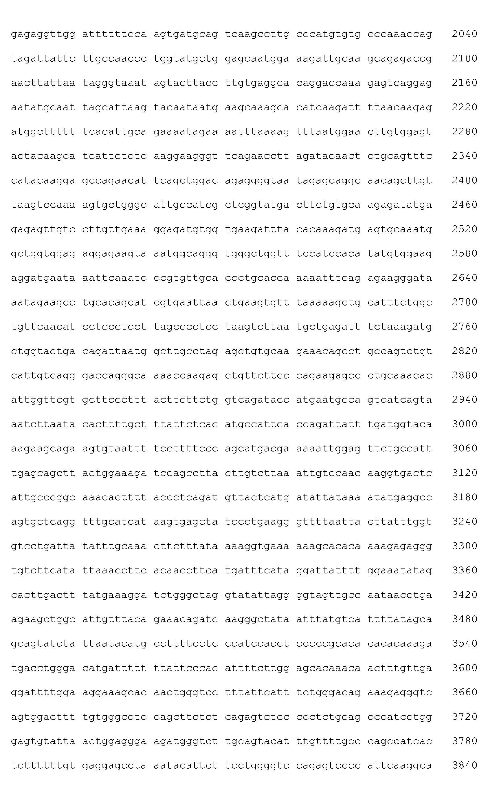

- <210> 8
<211> 4768
<212> DNA
<213> Homo sapiens - <400> 8


- <210> 9
<211> 760
<212> DNA
<213> Homo sapiens - <400> 9

- <210> 10
<211> 4999
<212> DNA
<213> Homo sapiens - <400> 10


- <210> 11
<211> 6609
<212> DNA
<213> Homo sapiens - <400> 11


- <210> 12
<211> 2128
<212> DNA
<213> Homo sapiens - <400> 12

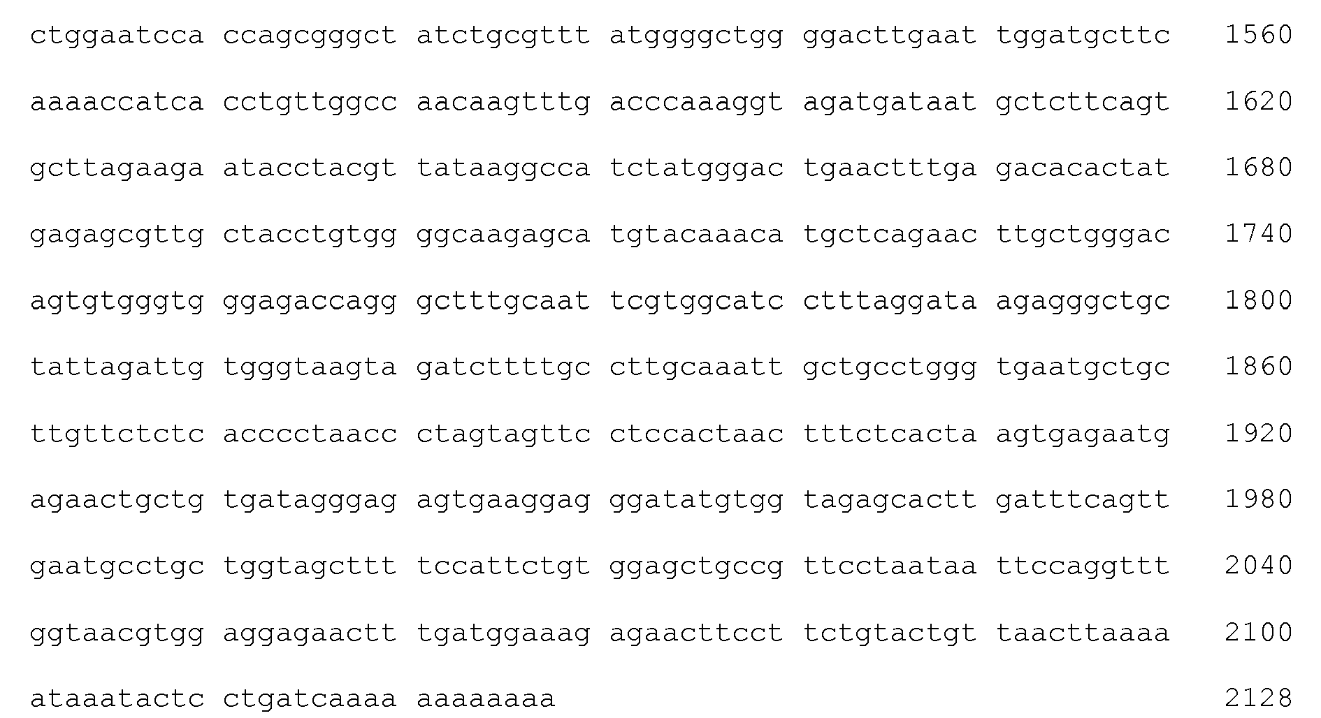
- <210> 13
<211> 5228
<212> DNA
<213> Homo sapiens - <400> 13


- <210> 14
<211> 4093
<212> DNA
<213> Homo sapiens - <400> 14


- <210> 15
<211> 587
<212> DNA
<213> Homo sapiens - <400> 15

- <210> 16
<211> 1431
<212> DNA
<213> Homo sapiens - <400> 16

- <210> 17
<211> 2709
<212> DNA
<213> Homo sapiens - <400> 17


- <210> 18
<211> 1938
<212> DNA
<213> Homo sapiens - <400> 18
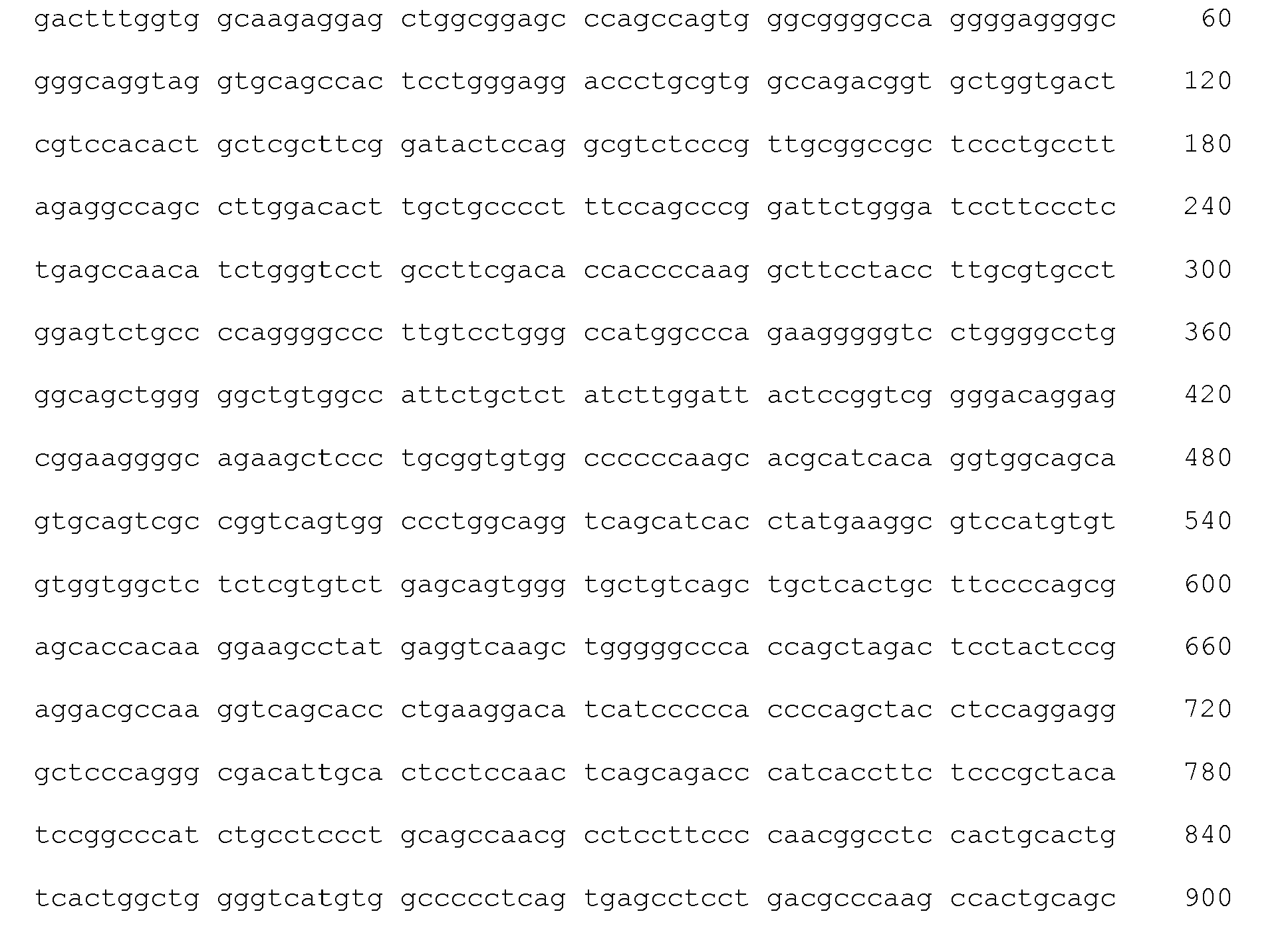

- <210> 19
<211> 1484
<212> DNA
<213> Homo sapiens - <400> 19


- <210> 20
<211> 3641
<212> DNA
<213> Homo sapiens - <400> 20


- <210> 21
<211> 1201
<212> DNA
<213> Homo sapiens - <400> 21


- <210> 22
<211> 3806
<212> DNA
<213> Homo sapiens - <400> 22


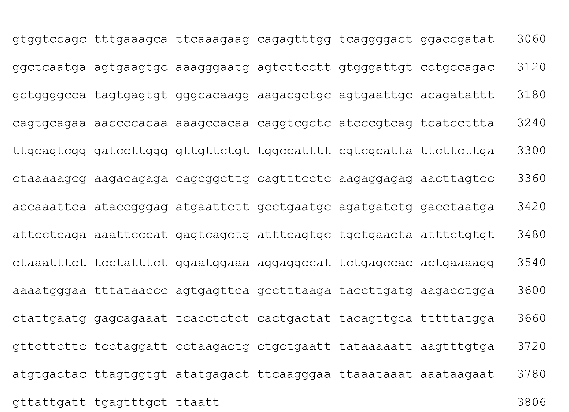
- <210> 23
<211> 3806
<212> DNA
<213> Homo sapiens - <400> 23


- <210> 24
<211> 2508
<212> DNA
<213> Homo sapiens - <400> 24


- <210> 25
<211> 925
<212> DNA
<213> Homo sapiens - <400> 25

- <210> 26
<211> 8082
<212> DNA
<213> Homo sapiens - <400> 26
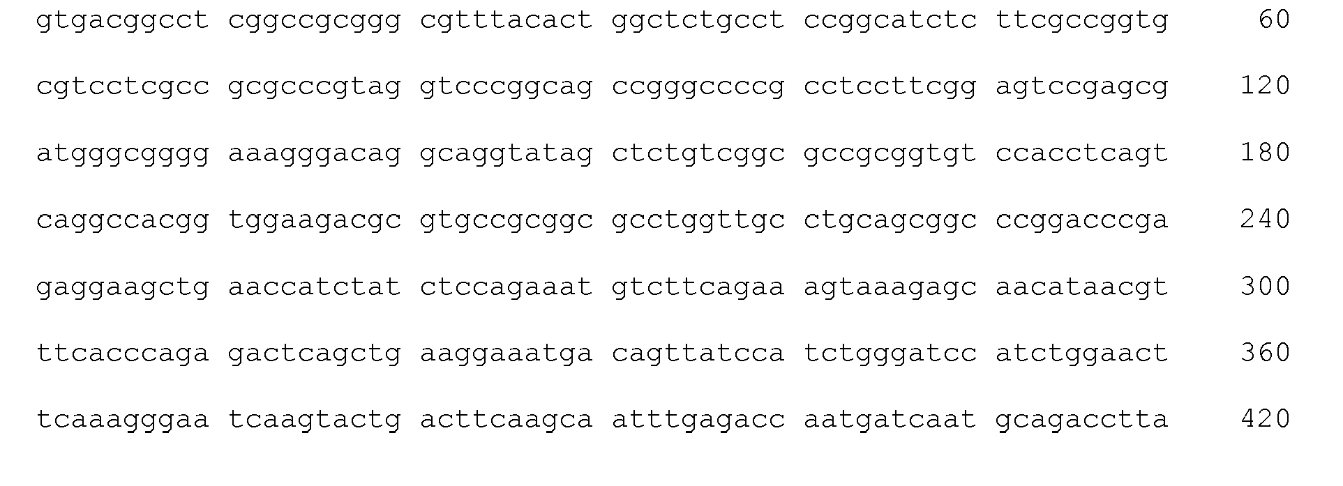


- <210> 27
<211> 1190
<212> DNA
<213> Homo sapiens - <400> 27

- <210> 28
<211> 3276
<212> DNA
<213> Homo sapiens - <400> 28


- <210> 29
<211> 802
<212> DNA
<213> Homo sapiens - <400> 29


- <210> 30
<211> 1691
<212> DNA
<213> Homo sapiens - <400> 30


- <210> 31
<211> 2558
<212> DNA
<213> Homo sapiens - <400> 31


- <210> 32
<211> 1501
<212> DNA
<213> Homo sapiens - <400> 32


- <210> 33
<211> 503
<212> DNA
<213> Homo sapiens - <400> 33


- <210> 34
<211> 1666
<212> DNA
<213> Homo sapiens - <400> 34


- <210> 35
<211> 1666
<212> DNA
<213> Homo sapiens - <400> 35


- <210> 36
<211> 402
<212> DNA
<213> Homo sapiens - <400> 36

- <210> 37
<211> 1707
<212> DNA
<213> Homo sapiens - <400> 37


- <210> 38
<211> 11185
<212> DNA
<213> Homo sapiens - <400> 38


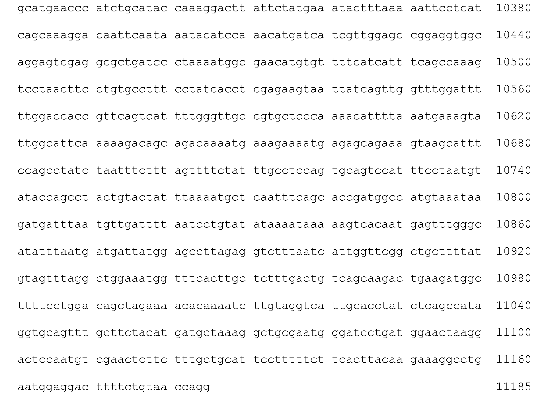
- <210> 39
<211> 1552
<212> DNA
<213> Homo sapiens - <400> 39


- <210> 40
<211> 3405
<212> DNA
<213> Homo sapiens - <400> 40
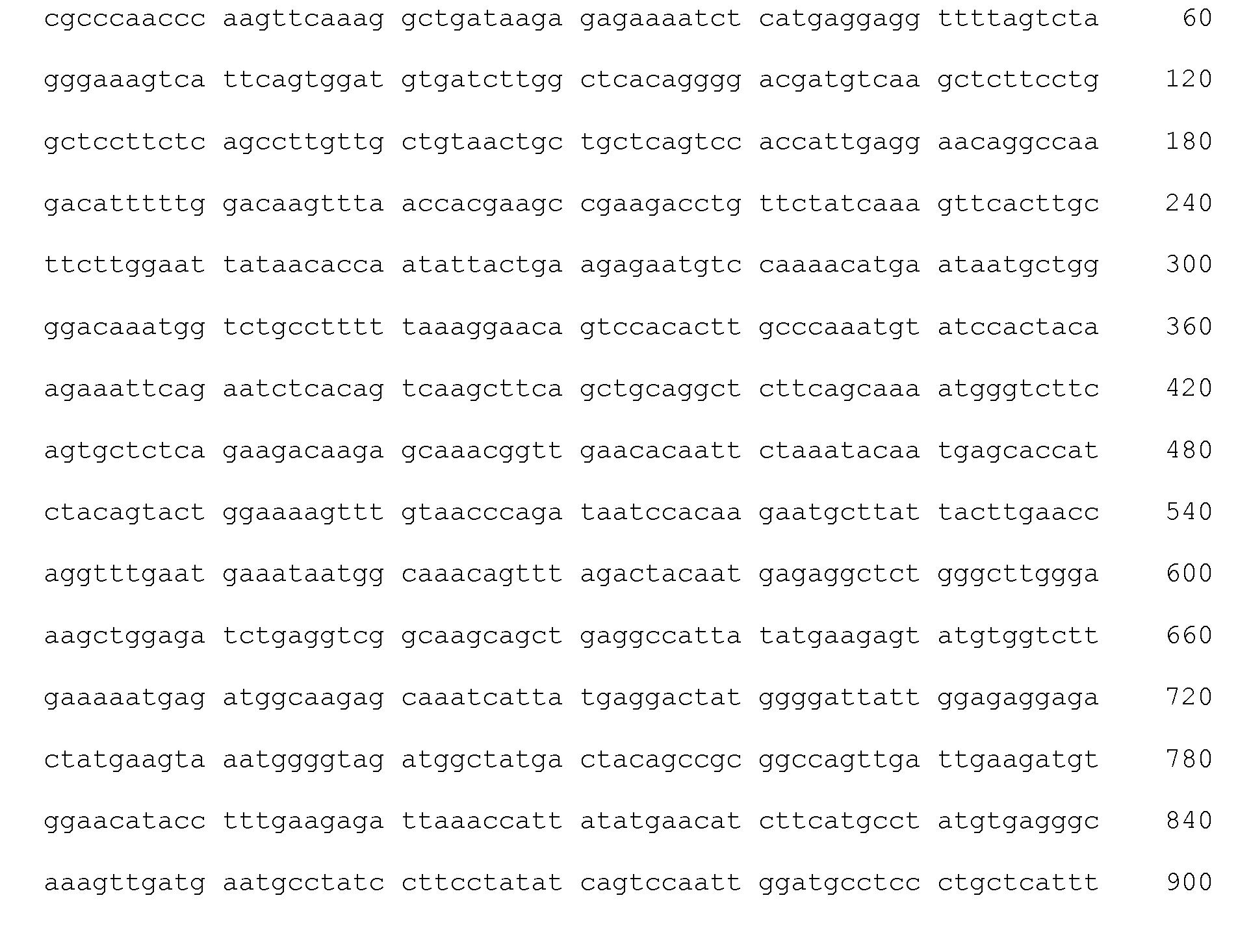


- <210> 41
<211> 1216
<212> DNA
<213> Homo sapiens - <400> 41


- <210> 42
<211> 3307
<212> DNA
<213> Homo sapiens - <400> 42


- <210> 43
<211> 2211
<212> DNA
<213> Homo sapiens - <400> 43


- <210> 44
<211> 2508
<212> DNA
<213> Homo sapiens - <400> 44


- <210> 45
<211> 4175
<212> DNA
<213> Homo sapiens - <400> 45


- <210> 46
<211> 1190
<212> DNA
<213> Homo sapiens - <400> 46

- <210> 47
<211> 1543
<212> DNA
<213> Homo sapiens - <400> 47


- <210> 48
<211> 4266
<212> DNA
<213> Homo sapiens - <400> 48


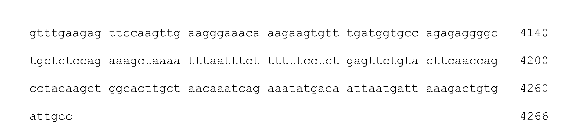
- <210> 49
<211> 3307
<212> DNA
<213> Homo sapiens - <400> 49


- <210> 50
<211> 6216
<212> DNA
<213> Homo sapiens - <400> 50


- <210> 51
<211> 2135
<212> DNA
<213> Homo sapiens - <400> 51


- <210> 52
<211> 2242
<212> DNA
<213> Homo sapiens - <400> 52


- <210> 53
<211> 3600
<212> DNA
<213> Homo sapiens - <400> 53
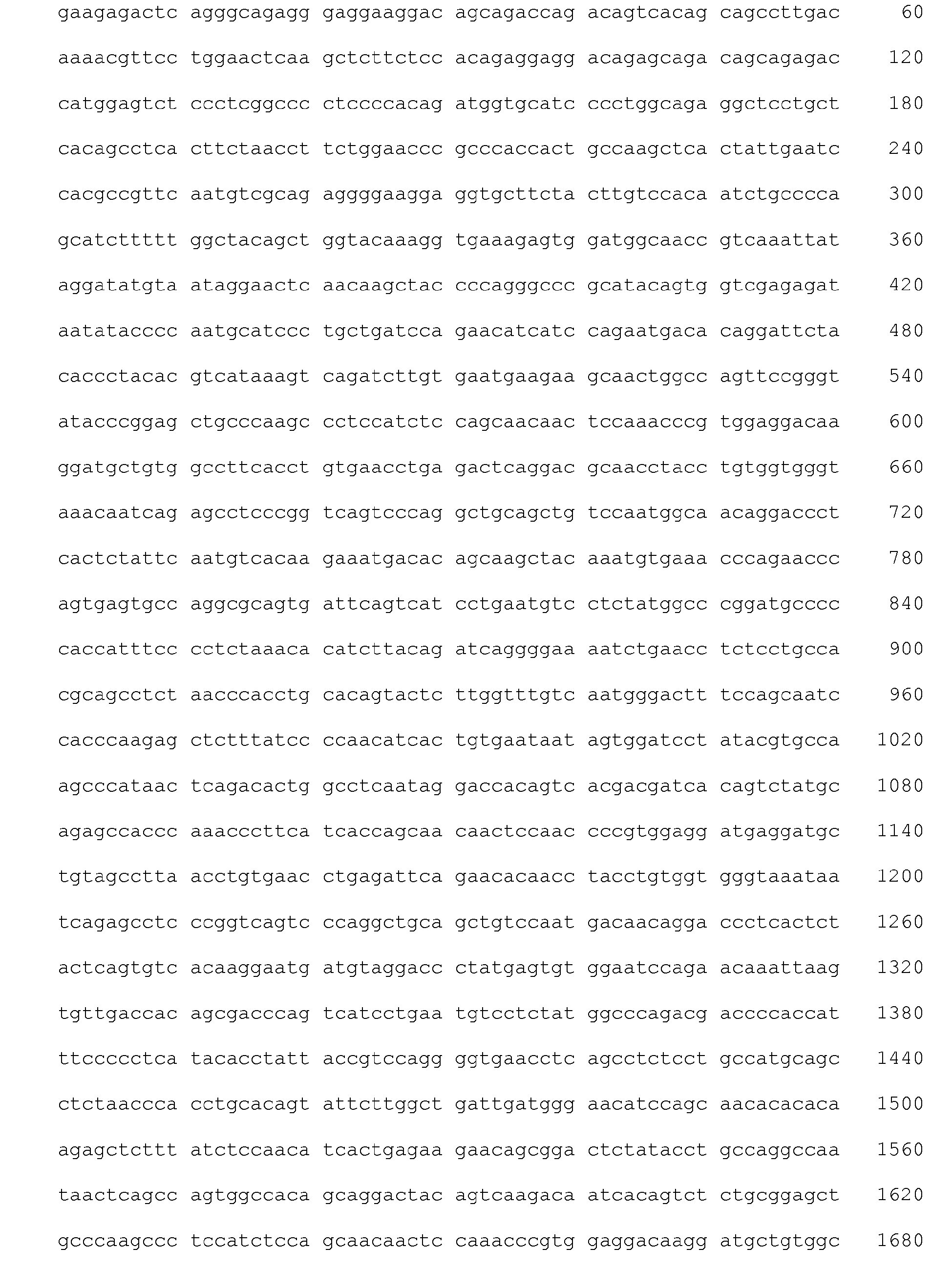


- <210> 54
<211> 1336
<212> DNA
<213> Homo sapiens - <400> 54

- <210> 55
<211> 656
<212> DNA
<213> Homo sapiens - <400> 55


- <210> 56
<211> 4869
<212> DNA
<213> Homo sapiens - <400> 56


- <210> 57
<211> 10128
<212> DNA
<213> Homo sapiens - <400> 57


- <210> 58
<211> 1623
<212> DNA
<213> Homo sapiens - <400> 58


- <210> 59
<211> 9085
<212> DNA
<213> Homo sapiens - <400> 59


- <210> 60
<211> 1124
<212> DNA
<213> Homo sapiens - <400> 60


- <210> 61
<211> 4175
<212> DNA
<213> Homo sapiens - <400> 61


- <210> 62
<211> 3405
<212> DNA
<213> Homo sapiens - <400> 62


- <210> 63
<211> 6930
<212> DNA
<213> Homo sapiens - <400> 63


- <210> 64
<211> 2545
<212> DNA
<213> Homo sapiens - <400> 64


- <210> 65
<211> 1681
<212> DNA
<213> Homo sapiens - <400> 65


- <210> 66
<211> 6103
<212> DNA
<213> Homo sapiens - <400> 66


- <210> 67
<211> 1973
<212> DNA
<213> Homo sapiens - <400> 67


- <210> 68
<211> 5546
<212> DNA
<213> Homo sapiens - <400> 68


- <210> 69
<211> 10078
<212> DNA
<213> Homo sapiens - <400> 69


- <210> 70
<211> 1901
<212> DNA
<213> Homo sapiens - <400> 70


- <210> 71
<211> 2568
<212> DNA
<213> Homo sapiens - <400> 71


- <210> 72
<211> 1172
<212> DNA
<213> Homo sapiens - <400> 72


- <210> 73
<211> 3226
<212> DNA
<213> Homo sapiens - <400> 73


- <210> 74
<211> 1270
<212> DNA
<213> Homo sapiens - <400> 74

- <210> 75
<211> 1542
<212> DNA
<213> Homo sapiens - <400> 75


- <210> 76
<211> 3447
<212> DNA
<213> Homo sapiens - <400> 76


- <210> 77
<211> 6216
<212> DNA
<213> Homo sapiens - <400> 77


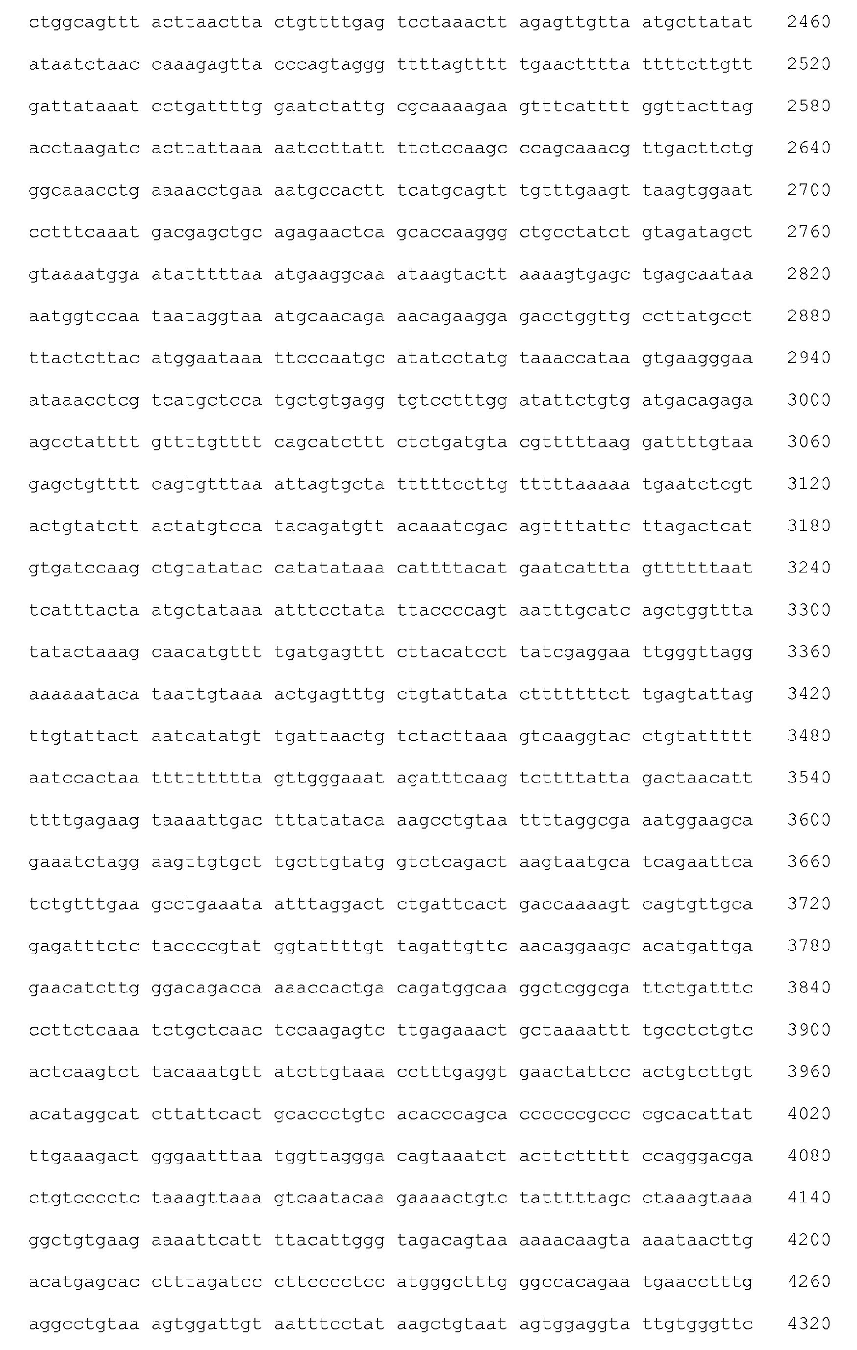


- <210> 78
<211> 768
<212> DNA
<213> Homo sapiens - <400> 78

- <210> 79
<211> 1336
<212> DNA
<213> Homo sapiens - <400> 79


- <210> 80
<211> 2052
<212> DNA
<213> Homo sapiens - <400> 80


- <210> 81
<211> 8368
<212> DNA
<213> Homo sapiens - <400> 81

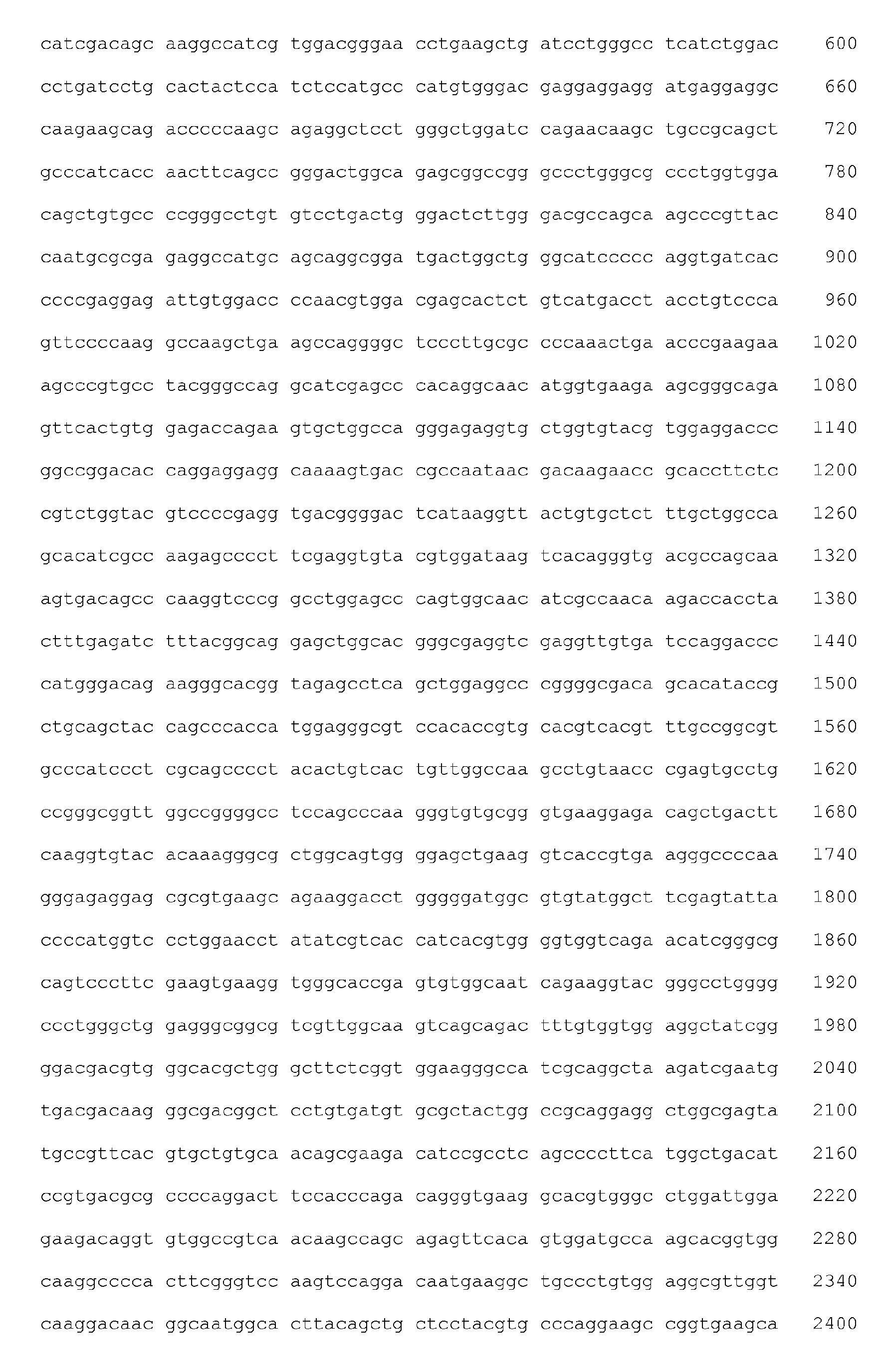


- <210> 82
<211> 2475
<212> DNA
<213> Homo sapiens - <400> 82


- <210> 83
<211> 4125
<212> DNA
<213> Homo sapiens - <400> 83


- <210> 84
<211> 2401
<212> DNA
<213> Homo sapiens - <400> 84


- <210> 85
<211> 5699
<212> DNA
<213> Homo sapiens - <400> 85


- <210> 86
<211> 3439
<212> DNA
<213> Homo sapiens - <400> 86


- <210> 87
<211> 5826
<212> DNA
<213> Homo sapiens - <400> 87


- <210> 88
<211> 5546
<212> DNA
<213> Homo sapiens - <400> 88


- <210> 89
<211> 4468
<212> DNA
<213> Homo sapiens - <400> 89


- <210> 90
<211> 891
<212> DNA
<213> Homo sapiens - <400> 90

- <210> 91
<211> 2750
<212> DNA
<213> Homo sapiens - <400> 91


- <210> 92
<211> 3969
<212> DNA
<213> Homo sapiens - <400> 92


- <210> 93
<211> 8439
<212> DNA
<213> Homo sapiens - <400> 93


- <210> 94
<211> 1580
<212> DNA
<213> Homo sapiens - <400> 94


- <210> 95
<211> 566
<212> DNA
<213> Homo sapiens - <400> 95

- <210> 96
<211> 3447
<212> DNA
<213> Homo sapiens - <400> 96


- <210> 97
<211> 2620
<212> DNA
<213> Homo sapiens - <400> 97


- <210> 98
<211> 2762
<212> DNA
<213> Homo sapiens - <400> 98


- <210> 99
<211> 5128
<212> DNA
<213> Homo sapiens - <400> 99


- <210> 100
<211> 5927
<212> DNA
<213> Homo sapiens - <400> 100


- <210> 101
<211> 4608
<212> DNA
<213> Homo sapiens - <400> 101


- <210> 102
<211> 1677
<212> DNA
<213> Homo sapiens - <400> 102


- <210> 103
<211> 896
<212> DNA
<213> Homo sapiens - <400> 103

- <210> 104
<211> 6629
<212> DNA
<213> Homo sapiens - <400> 104


- <210> 105
<211> 1994
<212> DNA
<213> Homo sapiens - <400> 105


- <210> 106
<211> 4237
<212> DNA
<213> Homo sapiens - <400> 106


- <210> 107
<211> 2793
<212> DNA
<213> Homo sapiens - <400> 107


- <210> 108
<211> 1575
<212> DNA
<213> Homo sapiens - <400> 108

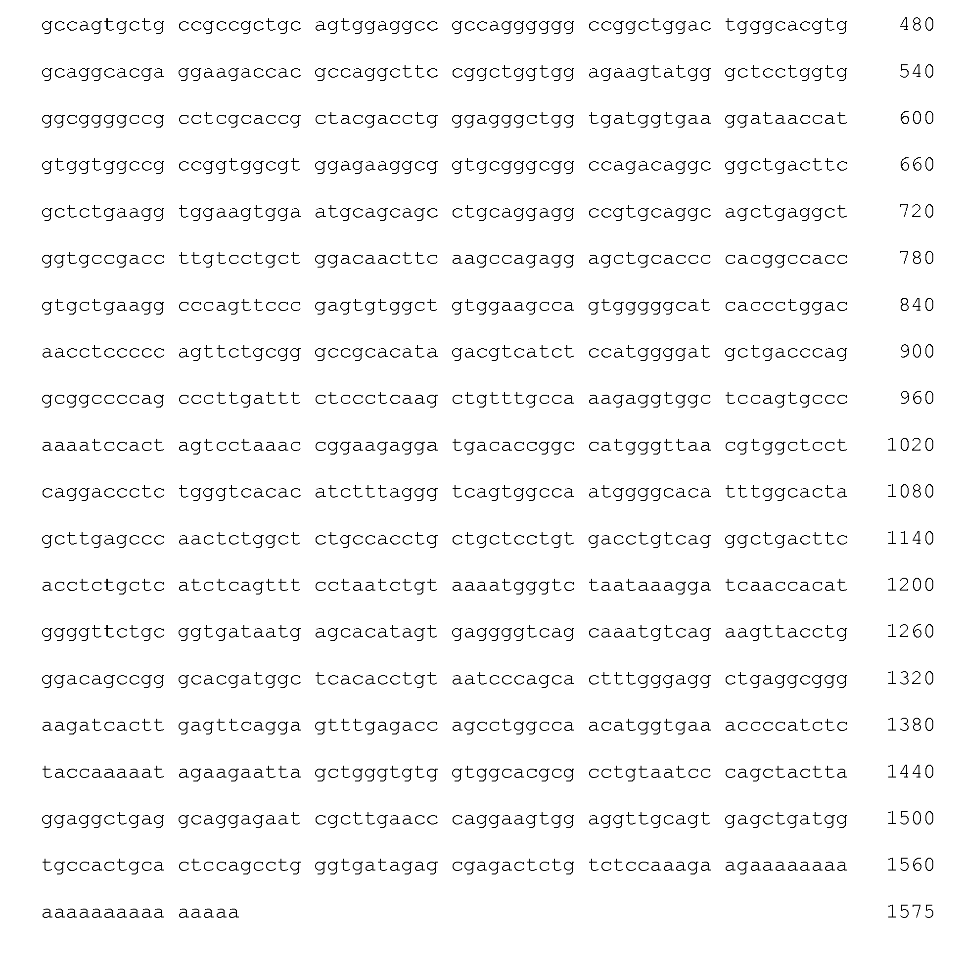
- <210> 109
<211> 1953
<212> DNA
<213> Homo sapiens - <400> 109


- <210> 110
<211> 7291
<212> DNA
<213> Homo sapiens - <400> 110


- <210> 111
<211> 3856
<212> DNA
<213> Homo sapiens - <400> 111


- <210> 112
<211> 5699
<212> DNA
<213> Homo sapiens - <400> 112


- <210> 113
<211> 1510
<212> DNA
<213> Homo sapiens - <400> 113

- <210> 114
<211> 9217
<212> DNA
<213> Homo sapiens - <400> 114


- <210> 115
<211> 1691
<212> DNA
<213> Homo sapiens - <400> 115


- <210> 116
<211> 11185
<212> DNA
<213> Homo sapiens - <400> 116


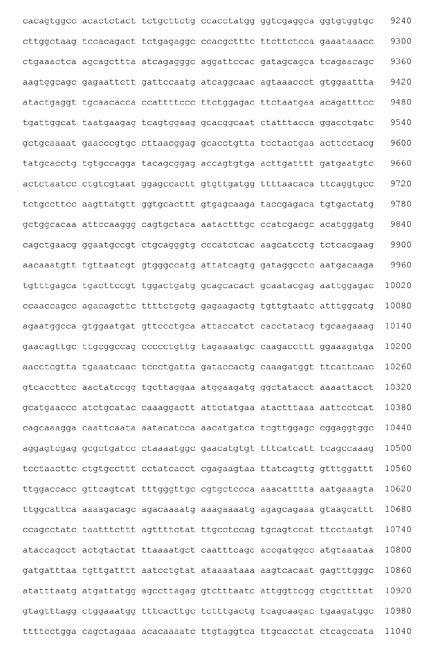

- <210> 117
<211> 1542
<212> DNA
<213> Homo sapiens - <400> 117


- <210> 118
<211> 5400
<212> DNA
<213> Homo sapiens - <400> 118


- <210> 119
<211> 7291
<212> DNA
<213> Homo sapiens - <400> 119


- <210> 120
<211> 3378
<212> DNA
<213> Homo sapiens - <400> 120


- <210> 121
<211> 4761
<212> DNA
<213> Homo sapiens - <400> 121


- <210> 122
<211> 5913
<212> DNA
<213> Homo sapiens - <400> 122


- <210> 123
<211> 4071
<212> DNA
<213> Homo sapiens - <400> 123


- <210> 124
<211> 3404
<212> DNA
<213> Homo sapiens - <400> 124


- <210> 125
<211> 2498
<212> DNA
<213> Homo sapiens - <400> 125


- <210> 126
<211> 5249
<212> DNA
<213> Homo sapiens - <400> 126

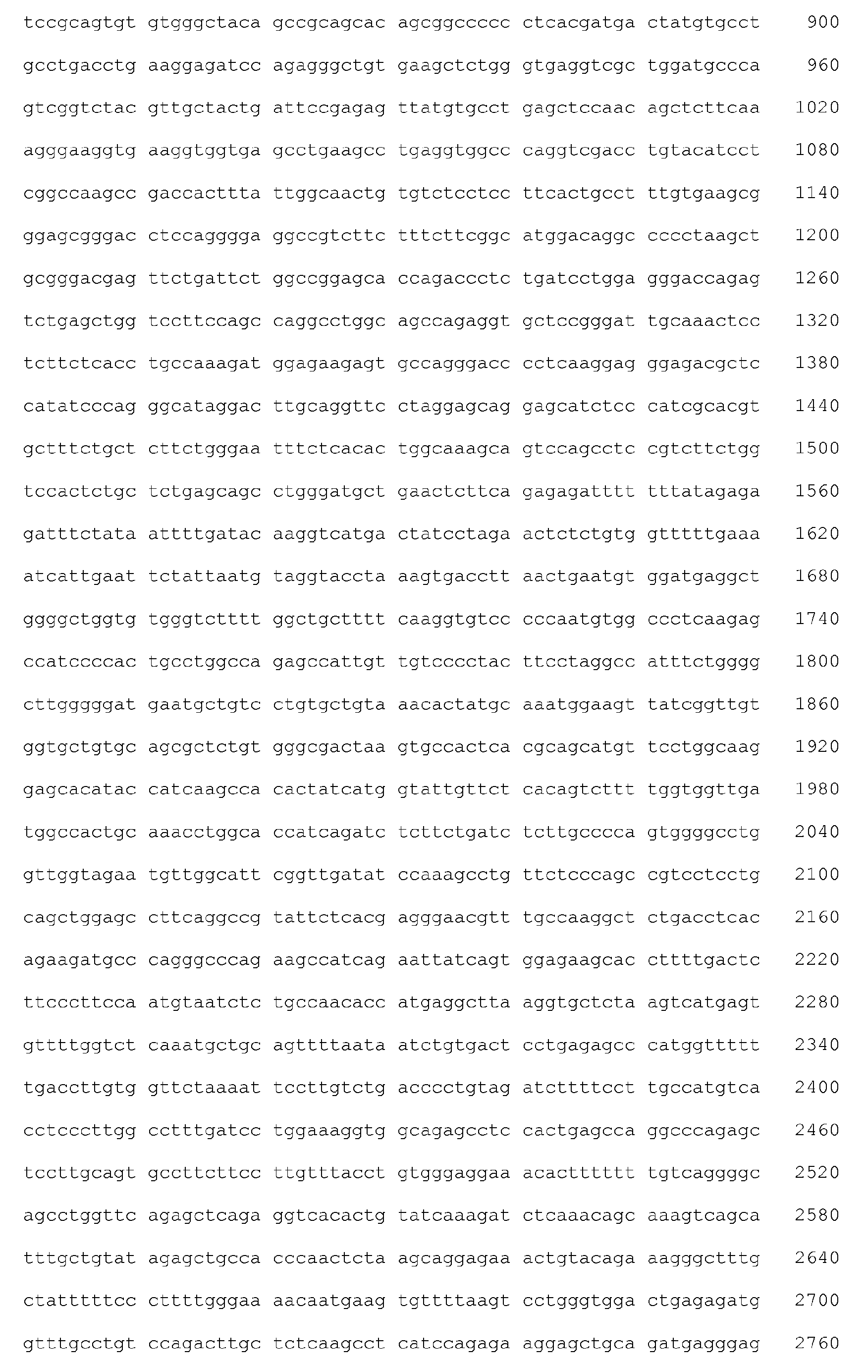


- <210> 127
<211> 1783
<212> DNA
<213> Homo sapiens - <400> 127


- <210> 128
<211> 3800
<212> DNA
<213> Homo sapiens - <400> 128

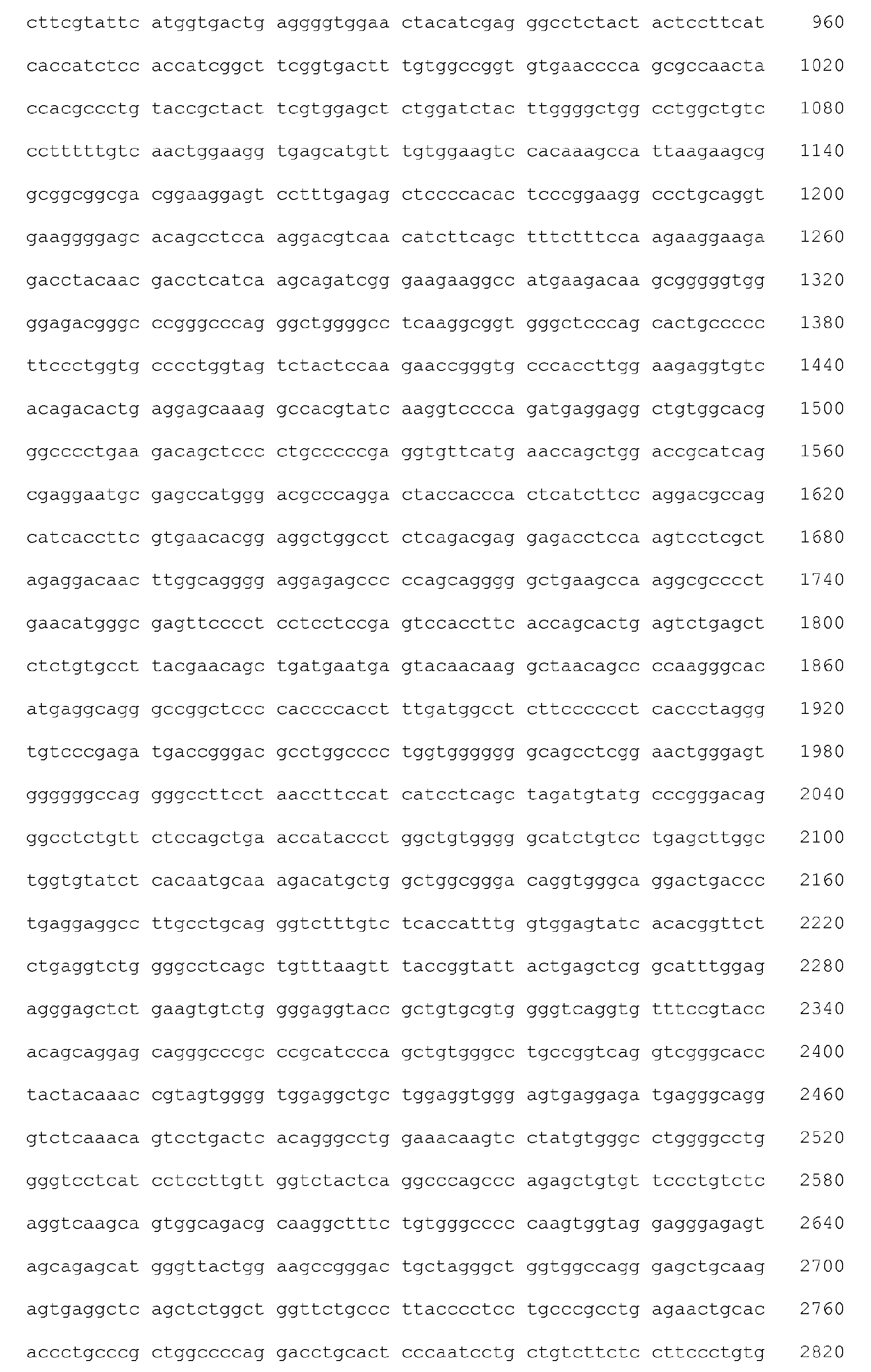

- <210> 129
<211> 574
<212> PRT
<213> Homo sapiens - <400> 129


- <210> 130
<211> 169
<212> PRT
<213> Homo sapiens - <400> 130

- <210> 131
<211> 252
<212> PRT - <213> Homo sapiens
- <400> 131


- <210> 132
<211> 148
<212> PRT
<213> Homo sapiens - <400> 132

- <210> 133
<211> 180
<212> PRT
<213> Homo sapiens - <400> 133


- <210> 134
<211> 381
<212> PRT
<213> Homo sapiens - <400> 134


- <210> 135
<211> 847
<212> PRT
<213> Homo sapiens - <400> 135


- <210> 136
<211> 847
<212> PRT
<213> Homo sapiens - <400> 136


- <210> 137
<211> 99
<212> PRT
<213> Homo sapiens - <400> 137

- <210> 138
<211> 733
<212> PRT
<213> Homo sapiens - <400> 138


- <210> 139
<211> 1782
<212> PRT
<213> Homo sapiens <400> 139


- <210> 140
<211> 438
<212> PRT
<213> Homo sapiens - <400> 140
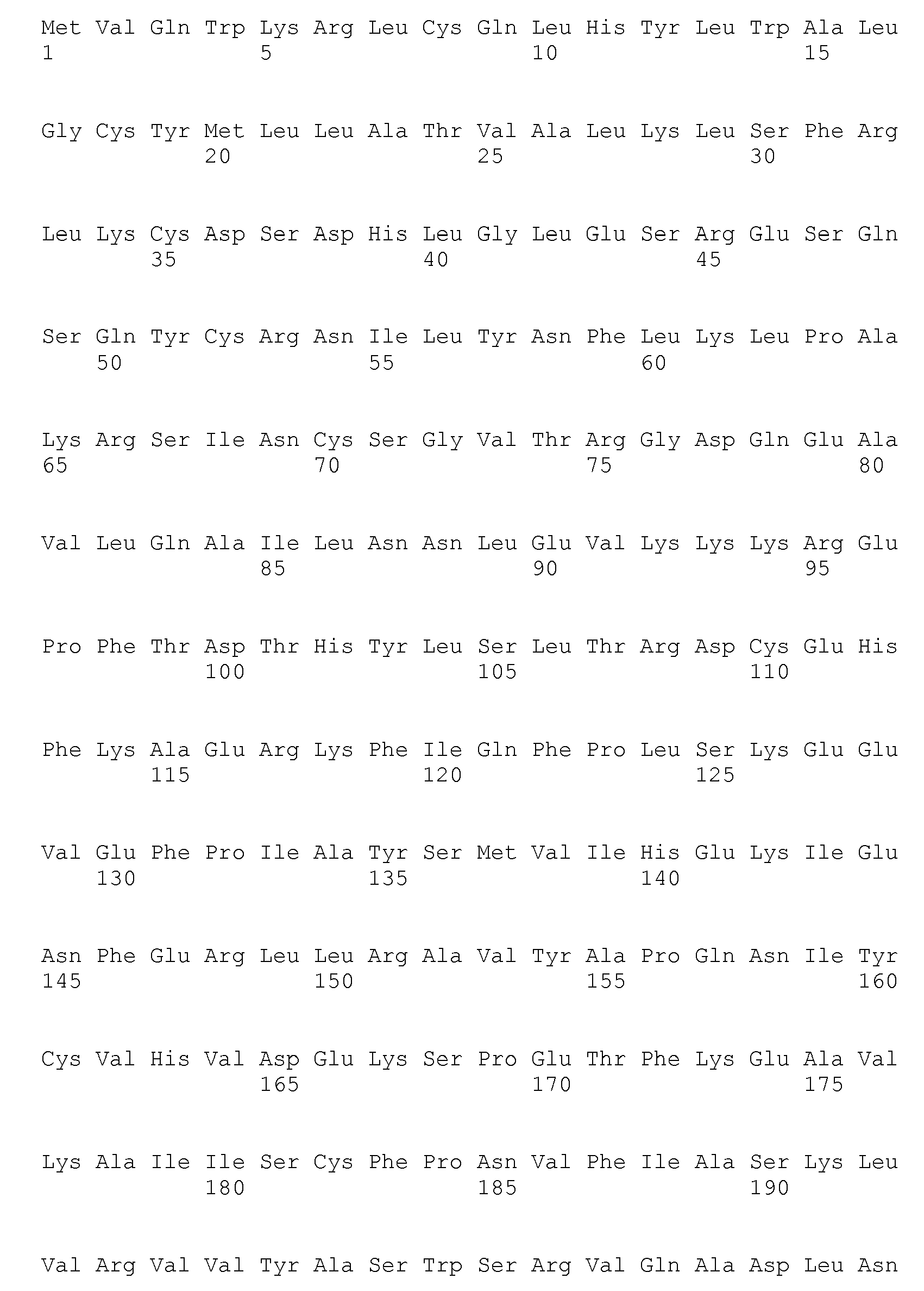

- <210> 141
<211> 414
<212> PRT
<213> Homo sapiens - <400> 141


- <210> 142
<211> 806
<212> PRT
<213> Homo sapiens - <400> 142


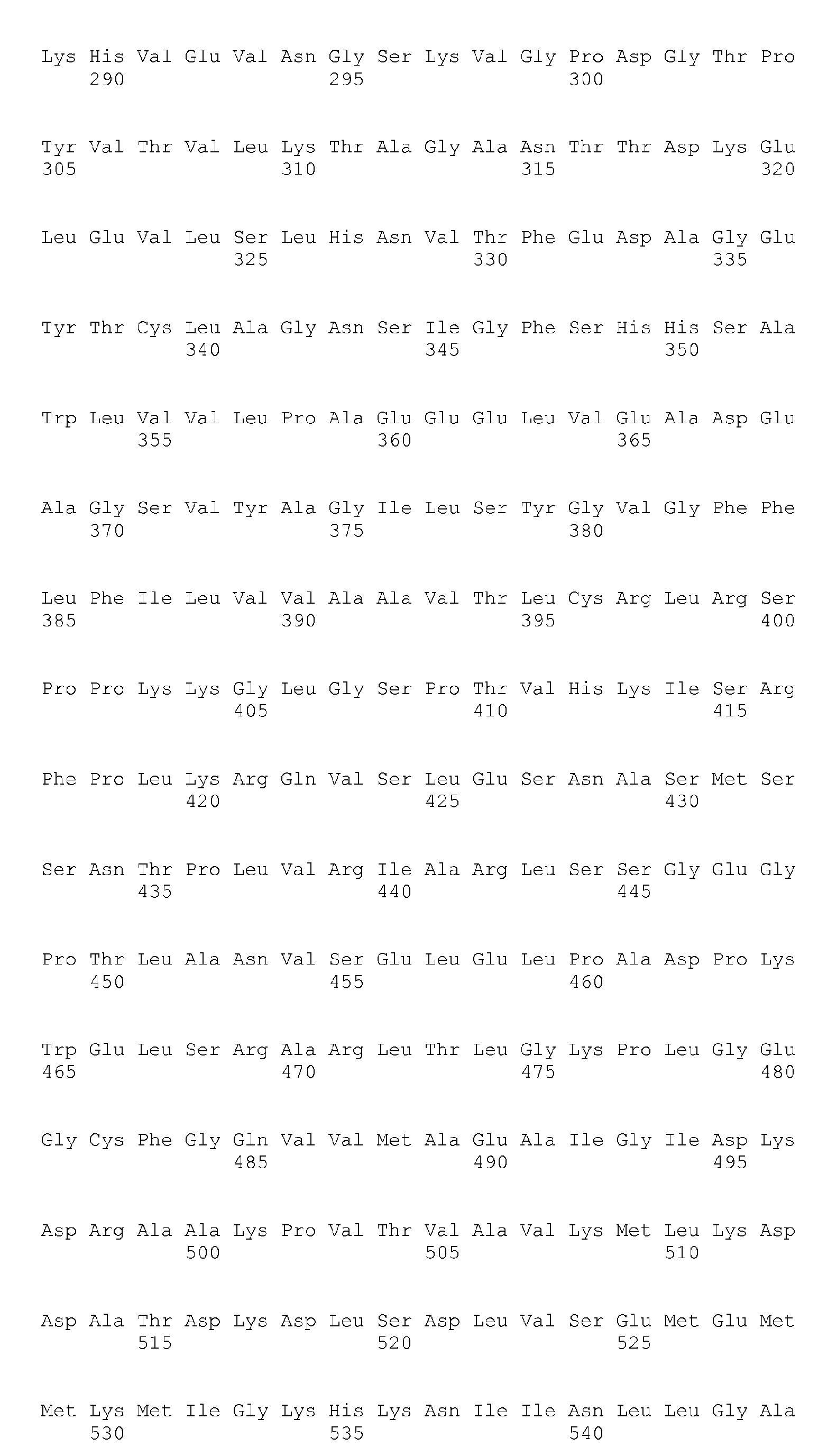


- <210> 143
<211> 160
<212> PRT
<213> Homo sapiens - <400> 143

- <210> 144
<211> 381
<212> PRT
<213> Homo sapiens - <400> 144


- <210> 145
<211> 729
<212> PRT
<213> Homo sapiens - <400> 145


- <210> 146
<211> 343
<212> PRT
<213> Homo sapiens - <400> 146


- <210> 147
<211> 398
<212> PRT
<213> Homo sapiens - <400> 147


- <210> 148
<211> 482
<212> PRT
<213> Homo sapiens - <400> 148


- <210> 149
<211> 266
<212> PRT
<213> Homo sapiens - <400> 149


- <210> 150
<211> 1156
<212> PRT
<213> Homo sapiens - <400> 150


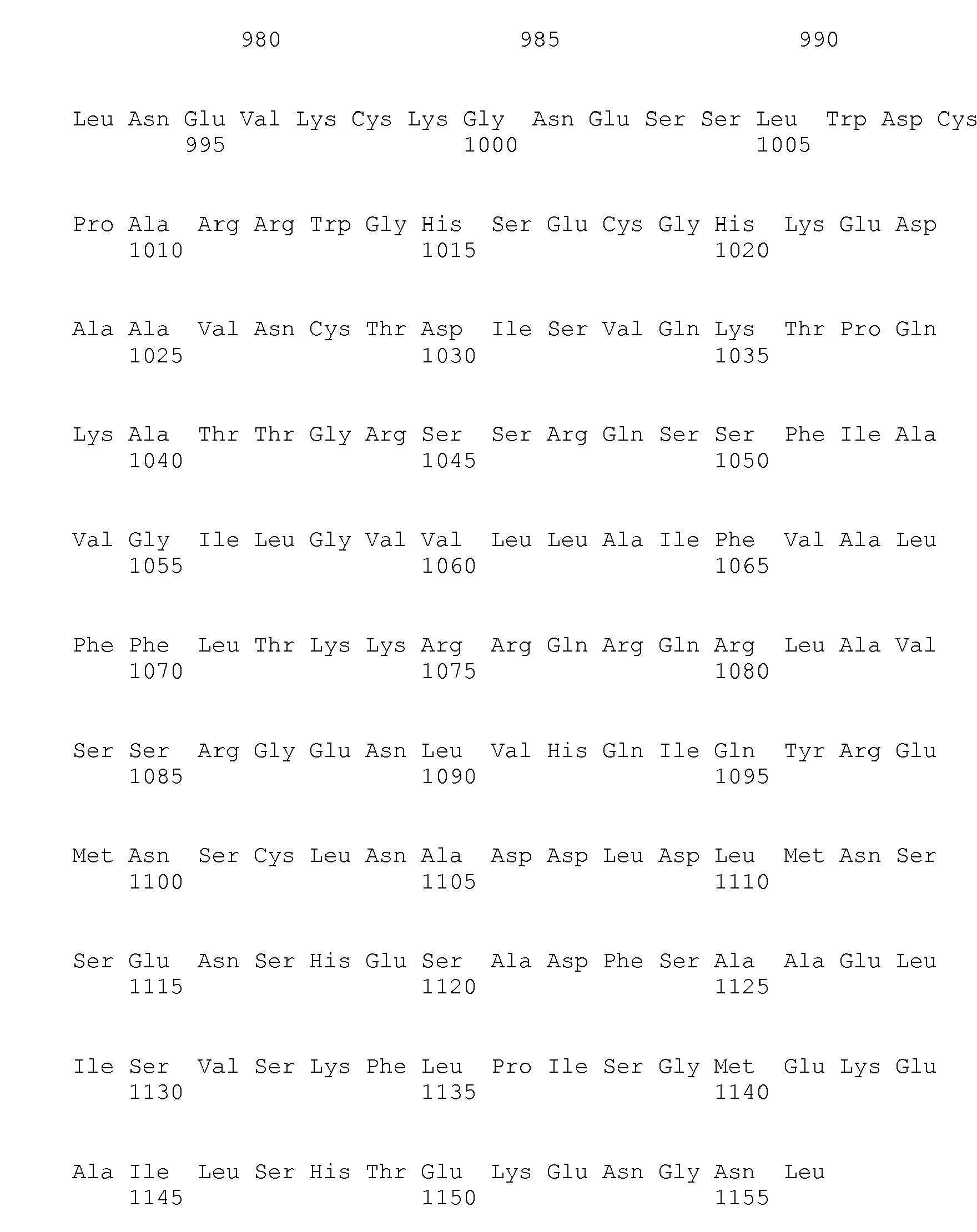
- <210> 151
<211> 1156
<212> PRT
<213> Homo sapiens - <400> 151


- <210> 152
<211> 635
<212> PRT
<213> Homo sapiens - <400> 152


- <210> 153
<211> 97
<212> PRT
<213> Homo sapiens - <400> 153

- <210> 154
<211> 739
<212> PRT
<213> Homo sapiens - <400> 154


- <210> 155
<211> 261
<212> PRT
<213> Homo sapiens - <400> 155


- <210> 156
<211> 915
<212> PRT
<213> Homo sapiens - <400> 156
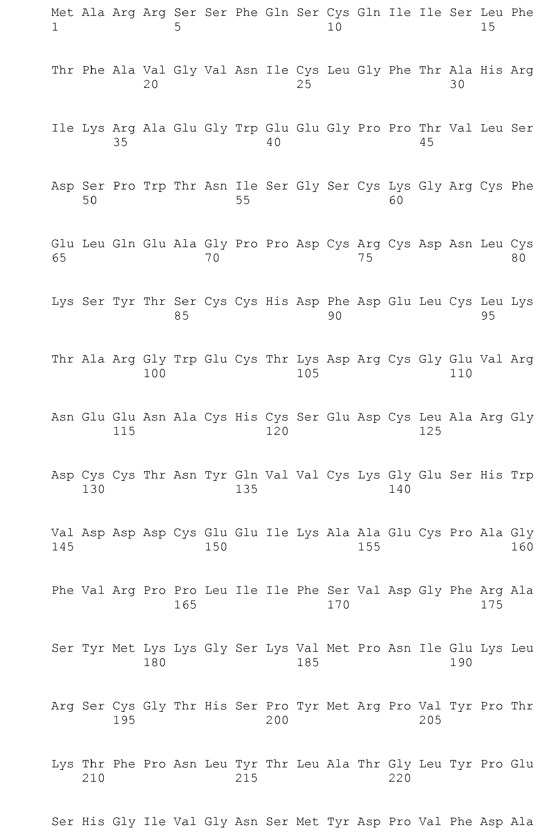


- <210> 157
<211> 247
<212> PRT
<213> Homo sapiens - <400> 157
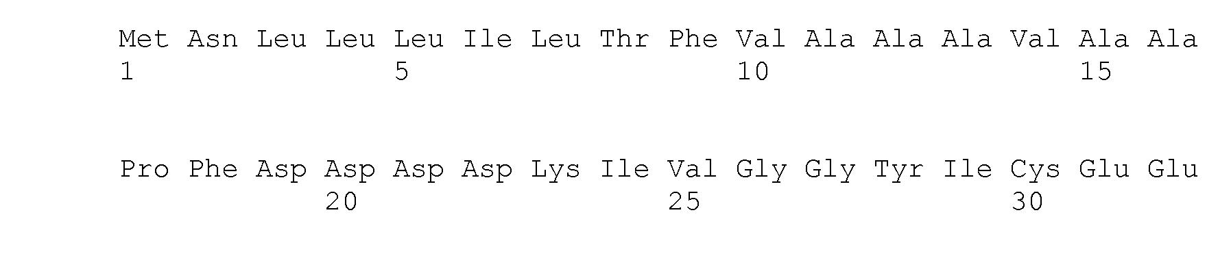

- <210> 158
<211> 352
<212> PRT
<213> Homo sapiens - <400> 158


- <210> 159
<211> 375
<212> PRT
<213> Homo sapiens - <400> 159


- <210> 160
<211> 258
<212> PRT
<213> Homo sapiens - <400> 160


- <210> 161
<211> 93
<212> PRT
<213> Homo sapiens - <400> 161

- <210> 162
<211> 99
<212> PRT
<213> Homo sapiens - <400> 162


- <210> 163
<211> 99
<212> PRT
<213> Homo sapiens - <400> 163

- <210> 164
<211> 133
<212> PRT
<213> Homo sapiens - <400> 164


- <210> 165
<211> 502
<212> PRT
<213> Homo sapiens - <400> 165
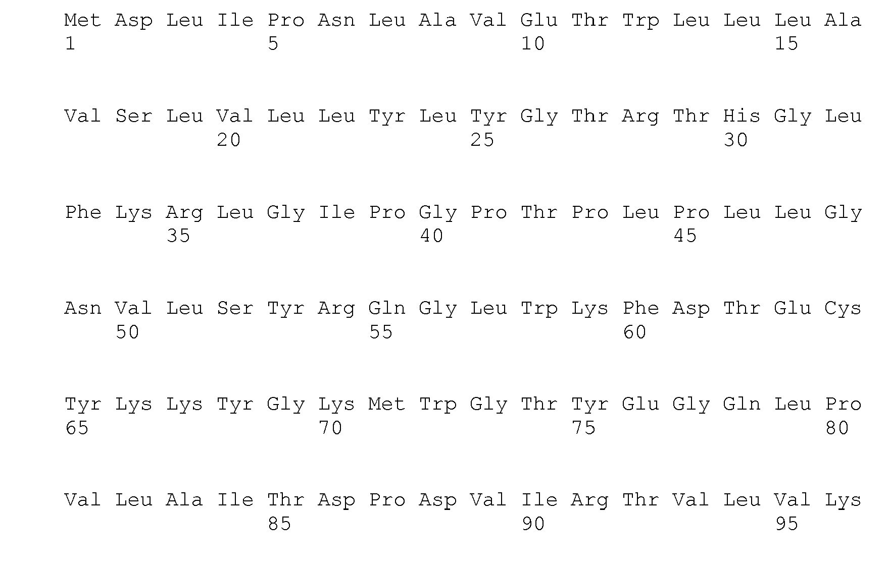


- <210> 166
<211> 3396
<212> PRT
<213> Homo sapiens - <400> 166


- <210> 167
<211> 459
<212> PRT
<213> Homo sapiens - <400> 167

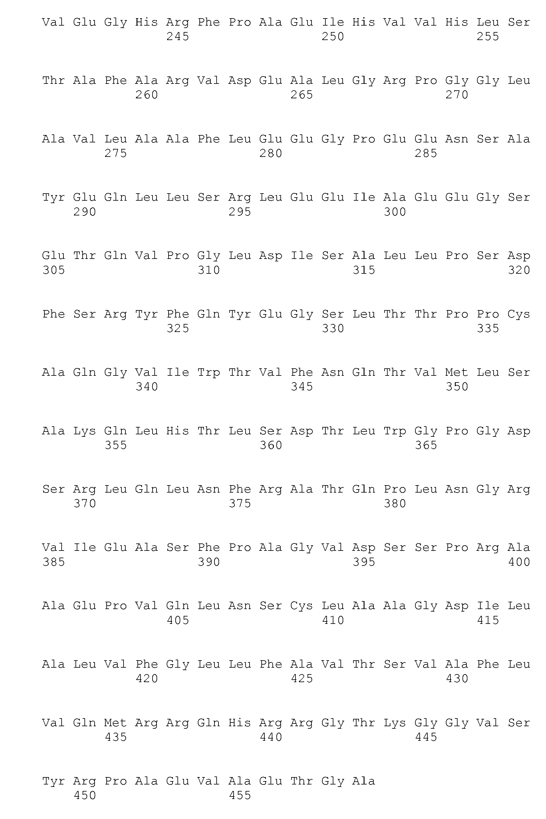
- <210> 168
<211> 805
<212> PRT
<213> Homo sapiens - <400> 168


- <210> 169
<211> 109
<212> PRT
<213> Homo sapiens - <400> 169

- <210> 170
<211> 680
<212> PRT
<213> Homo sapiens - <400> 170


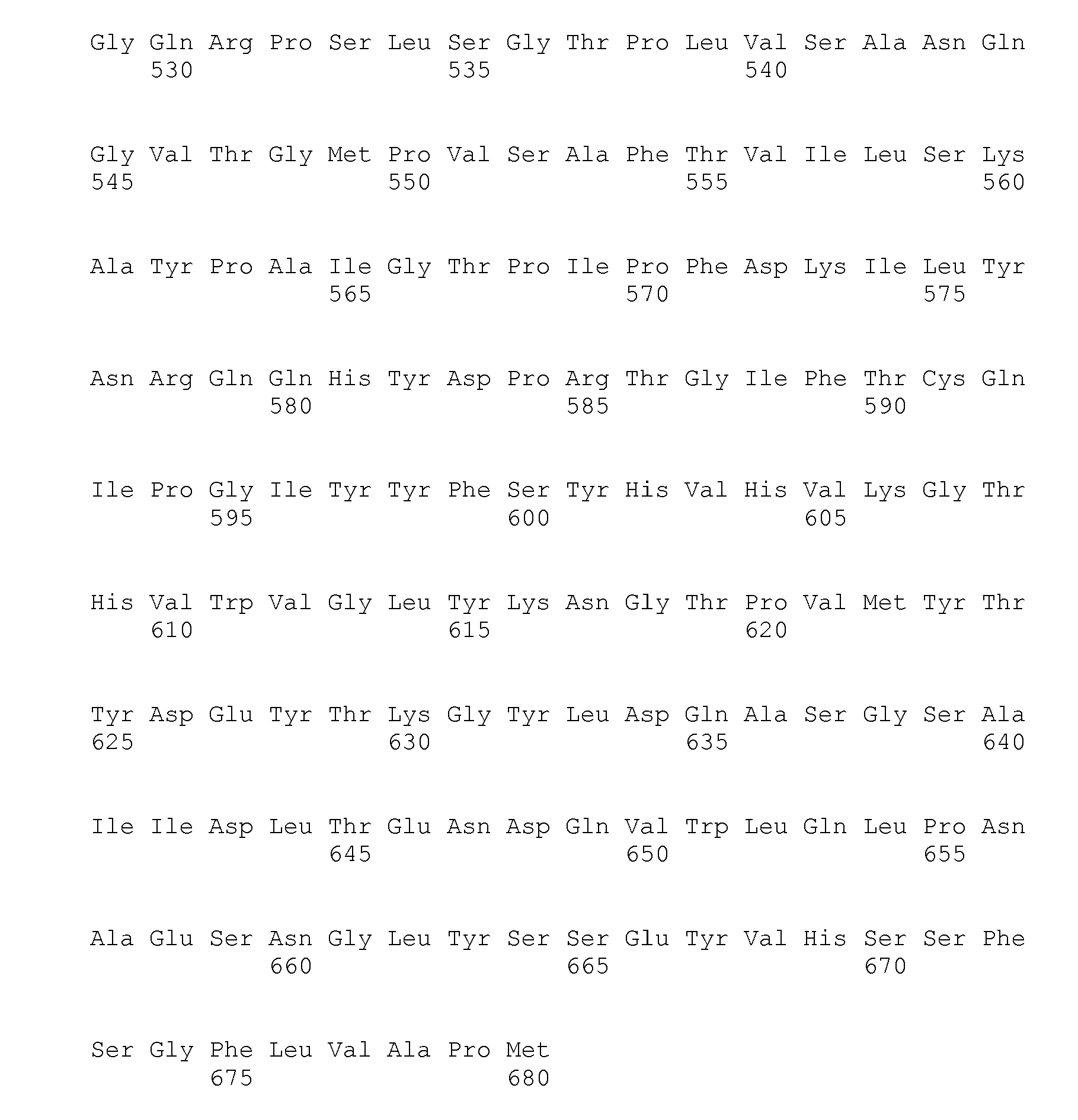
- <210> 171
<211> 537
<212> PRT
<213> Homo sapiens - <400> 171
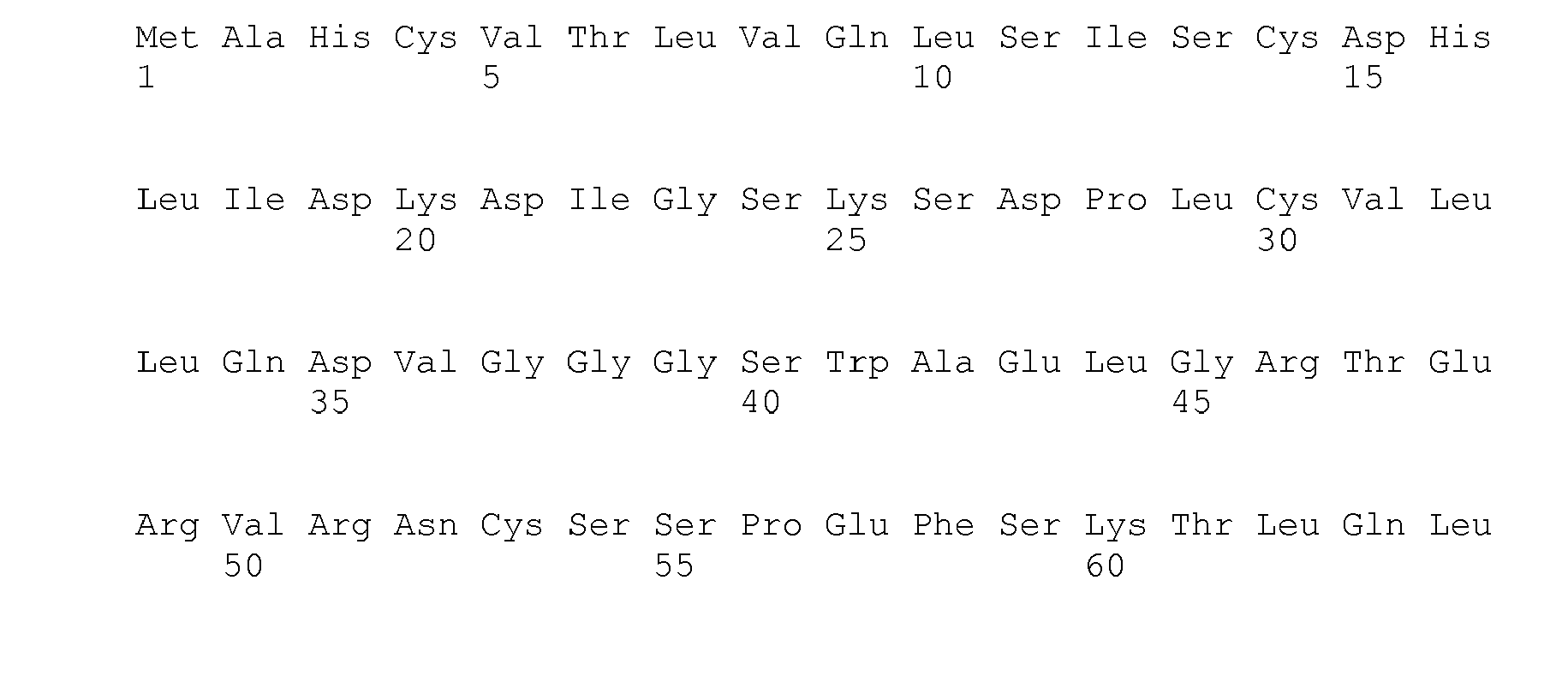


- <210> 172
<211> 635
<212> PRT
<213> Homo sapiens - <400> 172


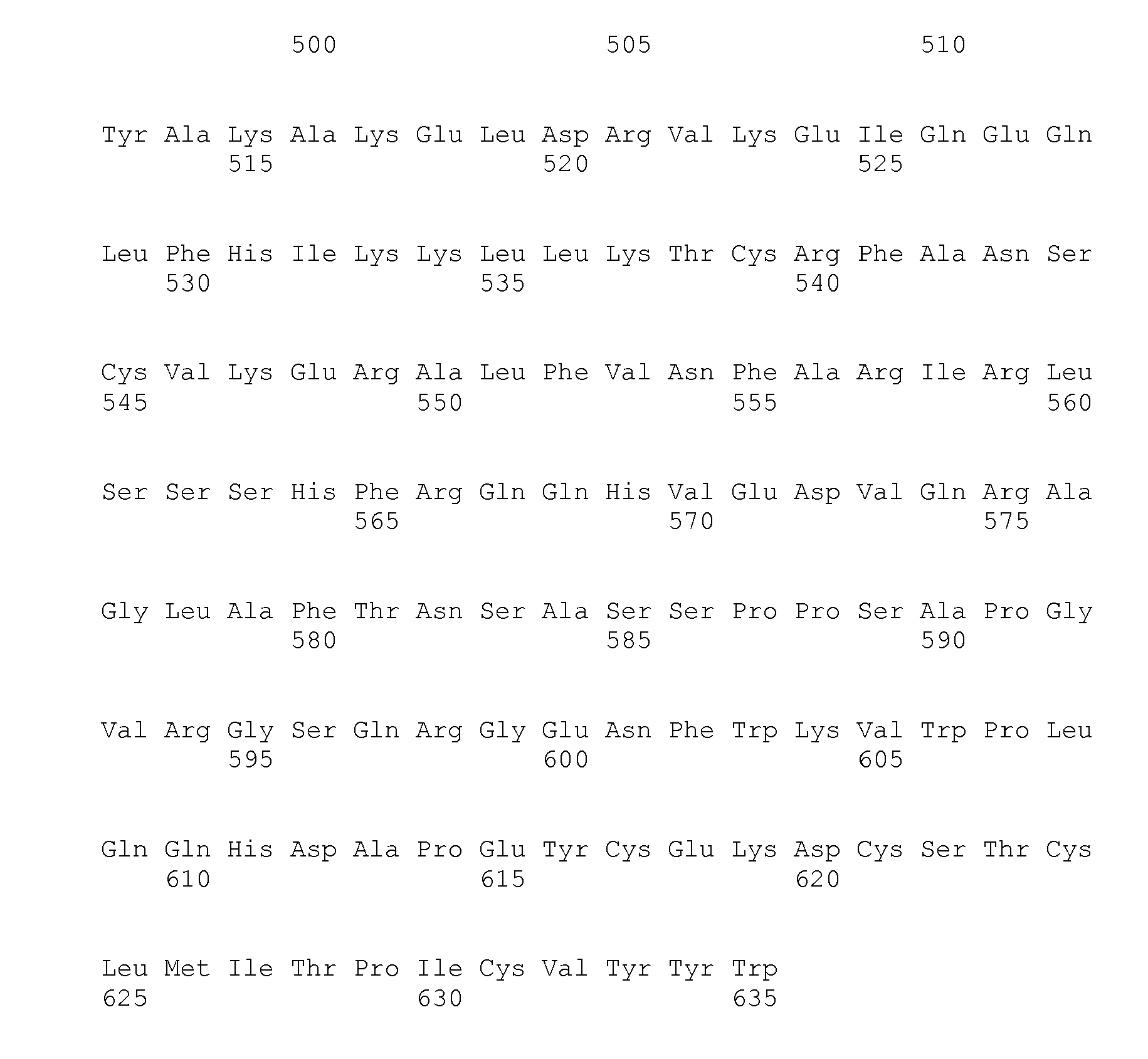
- <210> 173
<211> 184
<212> PRT
<213> Homo sapiens - <400> 173


- <210> 174
<211> 261
<212> PRT
<213> Homo sapiens - <400> 174


- <210> 175
<211> 433
<212> PRT
<213> Homo sapiens - <400> 175


- <210> 176
<211> 764
<212> PRT
<213> Homo sapiens - <400> 176


- <210> 177
<211> 680
<212> PRT
<213> Homo sapiens - <400> 177
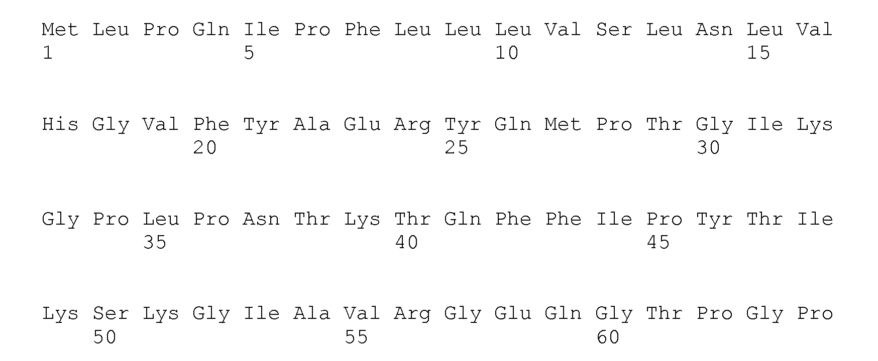
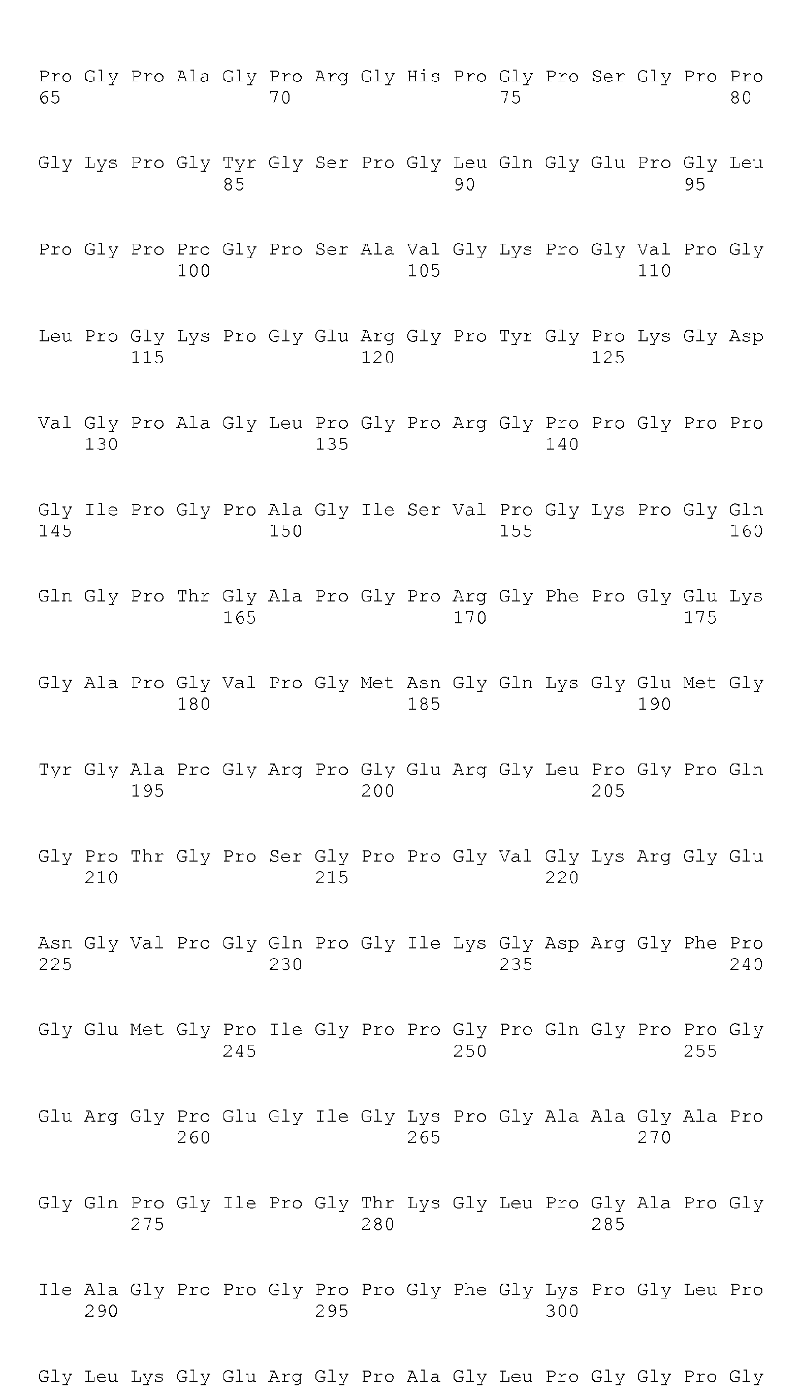


- <210> 178
<211> 325
<212> PRT
<213> Homo sapiens - <400> 178


- <210> 179
<211> 509
<212> PRT
<213> Homo sapiens - <400> 179


- <210> 180
<211> 317
<212> PRT
<213> Homo sapiens - <400> 180


- <210> 181
<211> 702
<212> PRT
<213> Homo sapiens - <400> 181


- <210> 182
<211> 334
<212> PRT
<213> Homo sapiens - <400> 182
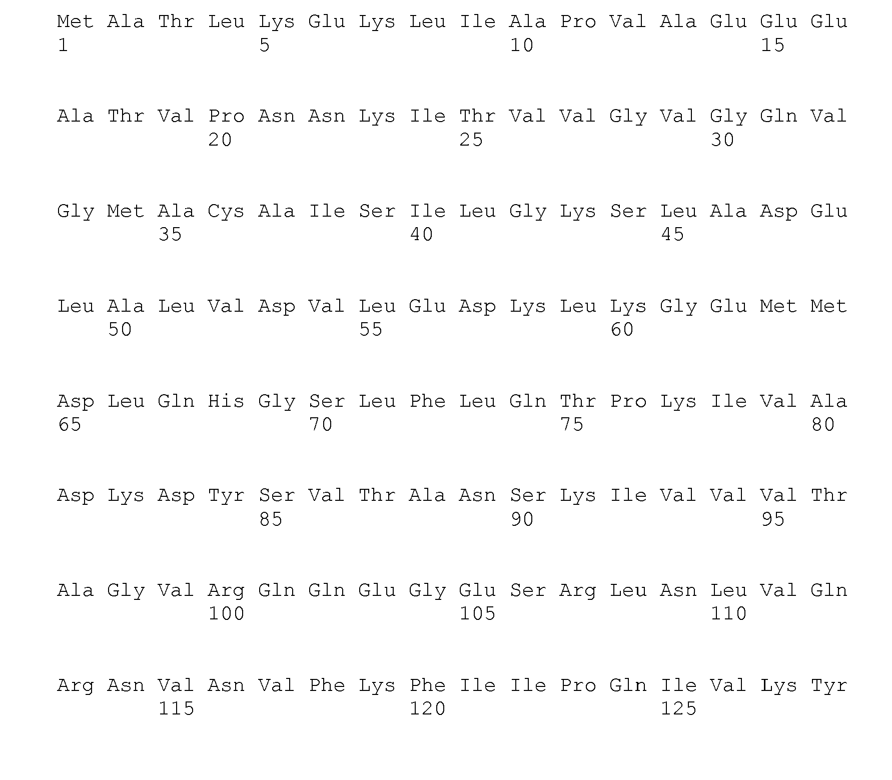

- <210> 183
<211> 119
<212> PRT
<213> Homo sapiens - <400> 183


- <210> 184
<211> 987
<212> PRT
<213> Homo sapiens - <400> 184


- <210> 185
<211> 114
<212> PRT
<213> Homo sapiens - <400> 185


- <210> 186
<211> 375
<212> PRT
<213> Homo sapiens - <400> 186


- <210> 187
<211> 425
<212> PRT
<213> Homo sapiens - <400> 187
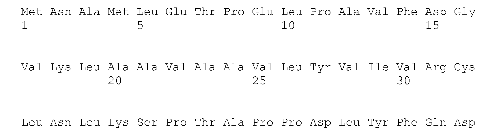

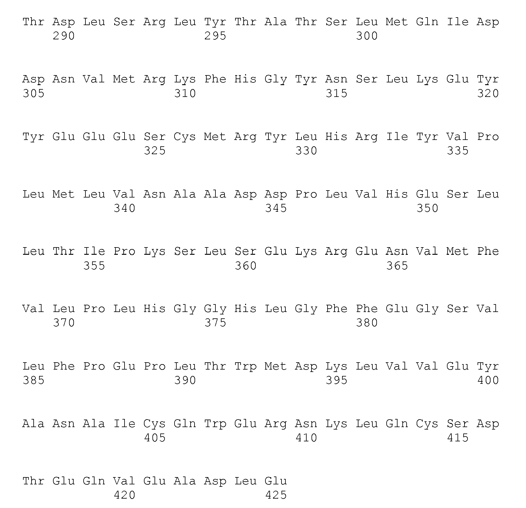
- <210> 188
<211> 300
<212> PRT
<213> Homo sapiens - <400> 188


- <210> 189
<211> 184
<212> PRT
<213> Homo sapiens - <400> 189

- <210> 190
<211> 805
<212> PRT
<213> Homo sapiens - <400> 190


- <210> 191
<211> 1499
<212> PRT
<213> Homo sapiens - <400> 191


- <210> 193
<211> 235
<212> PRT
<213> Homo sapiens - <400> 193

- <210> 194
<211> 1178
<212> PRT
<213> Homo sapiens - <400> 194


- <210> 195
<211> 469
<212> PRT
<213> Homo sapiens - <400> 195


- <210> 196
<211> 1194
<212> PRT
<213> Homo sapiens - <400> 196


- <210> 197
<211> 1912
<212> PRT
<213> Homo sapiens - <400> 197


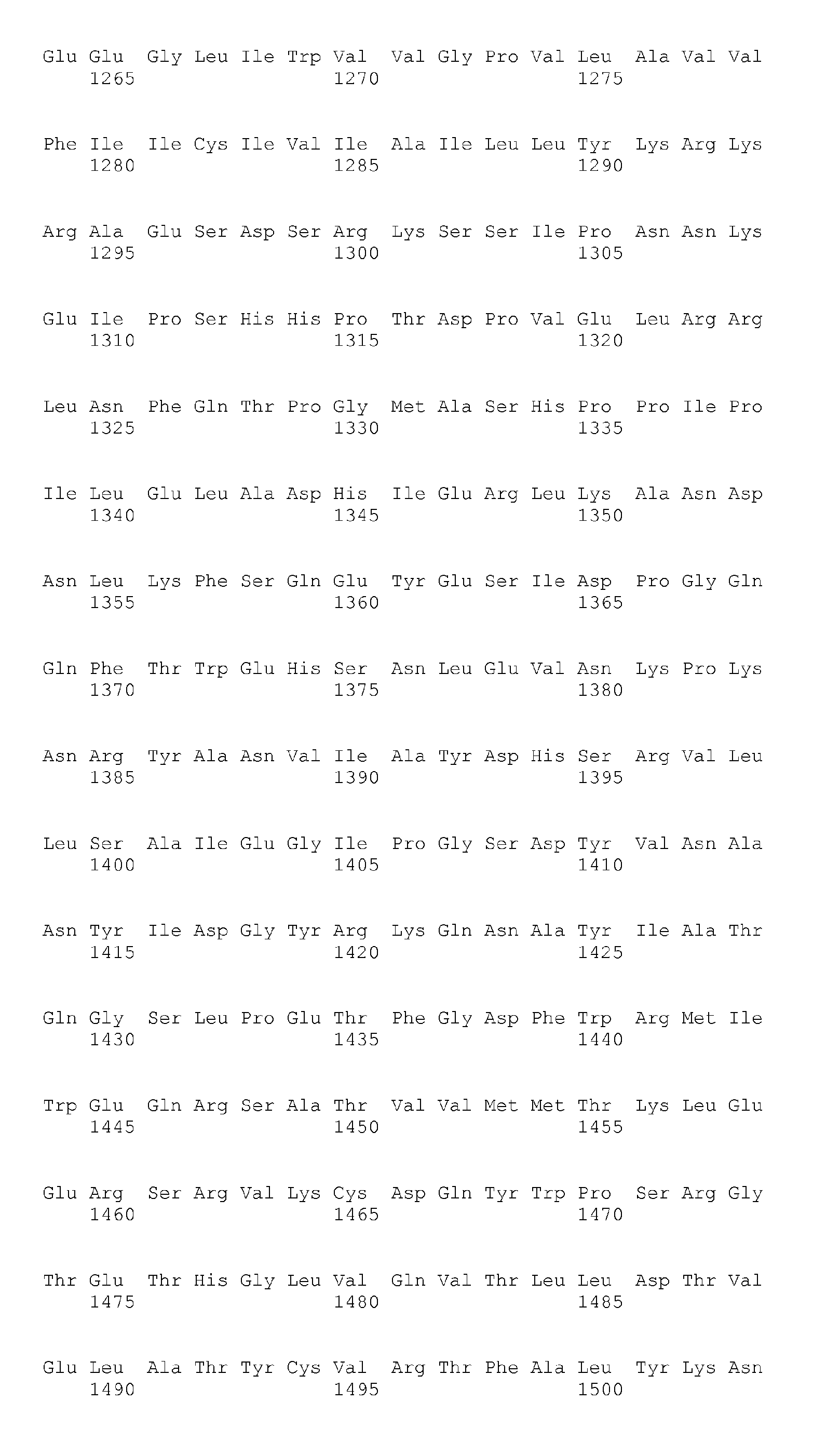

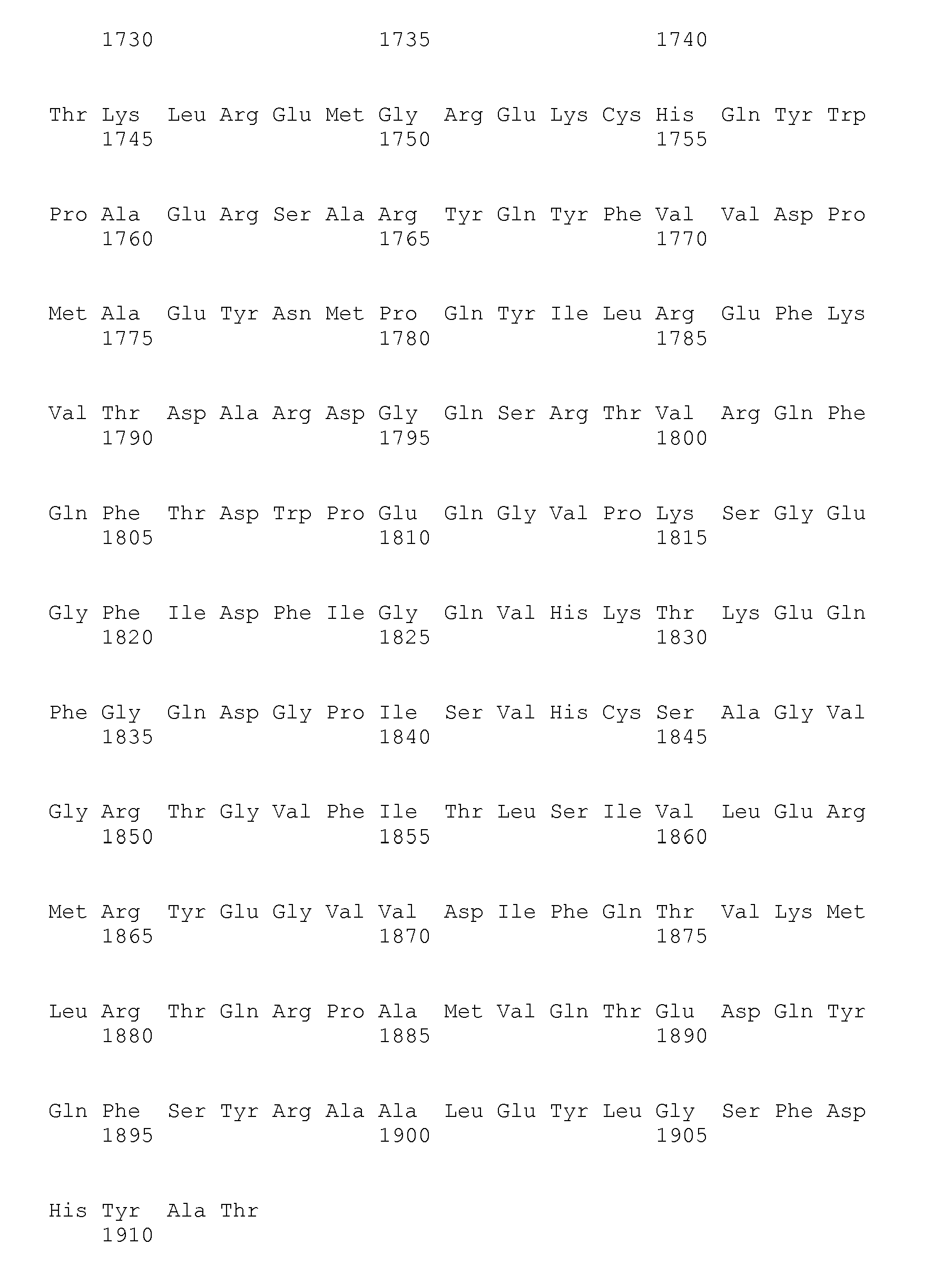
- <210> 198
<211> 336
<212> PRT
<213> Homo sapiens - <400> 198


- <210> 199
<211> 674
<212> PRT
<213> Homo sapiens - <400> 199


- <210> 200
<211> 98
<212> PRT
<213> Homo sapiens - <400> 200

- <210> 201
<211> 492
<212> PRT
<213> Homo sapiens - <400> 201


- <210> 202
<211> 158
<212> PRT
<213> Homo sapiens - <400> 202


- <210> 203
<211> 255
<212> PRT
<213> Homo sapiens - <400> 203
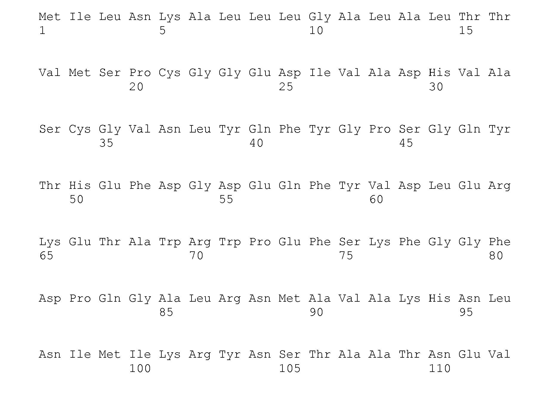
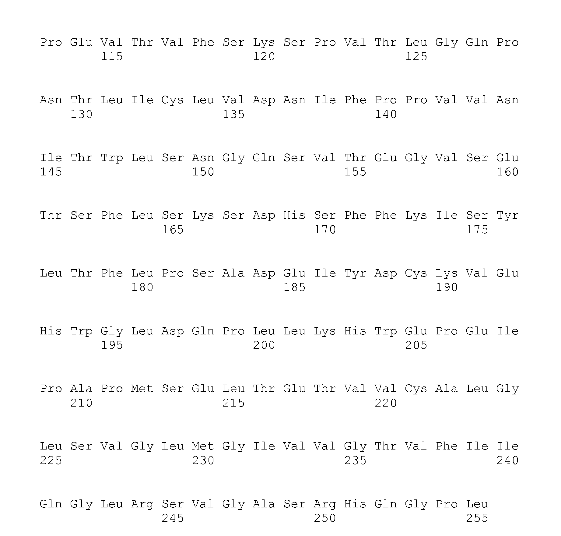
- <210> 204
<211> 598
<212> PRT
<213> Homo sapiens - <400> 204


- <210> 205
<211> 325
<212> PRT
<213> Homo sapiens - <400> 205


- <210> 206
<211> 164
<212> PRT
<213> Homo sapiens - <400> 206


- <210> 207
<211> 334
<212> PRT
<213> Homo sapiens - <400> 207


- <210> 208
<211> 149
<212> PRT
<213> Homo sapiens - <400> 208


- <210> 209
<211> 2647
<212> PRT
<213> Homo sapiens - <400> 209


- <210> 210
<211> 114
<212> PRT
<213> Homo sapiens - <400> 210


- <210> 211
<211> 1158
<212> PRT
<213> Homo sapiens - <400> 211


- <210> 212
<211> 368
<212> PRT
<213> Homo sapiens - <400> 212


- <210> 213
<211> 871
<212> PRT
<213> Homo sapiens - <400> 213


- <210> 214
<211> 1019
<212> PRT
<213> Homo sapiens - <400> 214


- <210> 215
<211> 1172
<212> PRT
<213> Homo sapiens - <400> 215
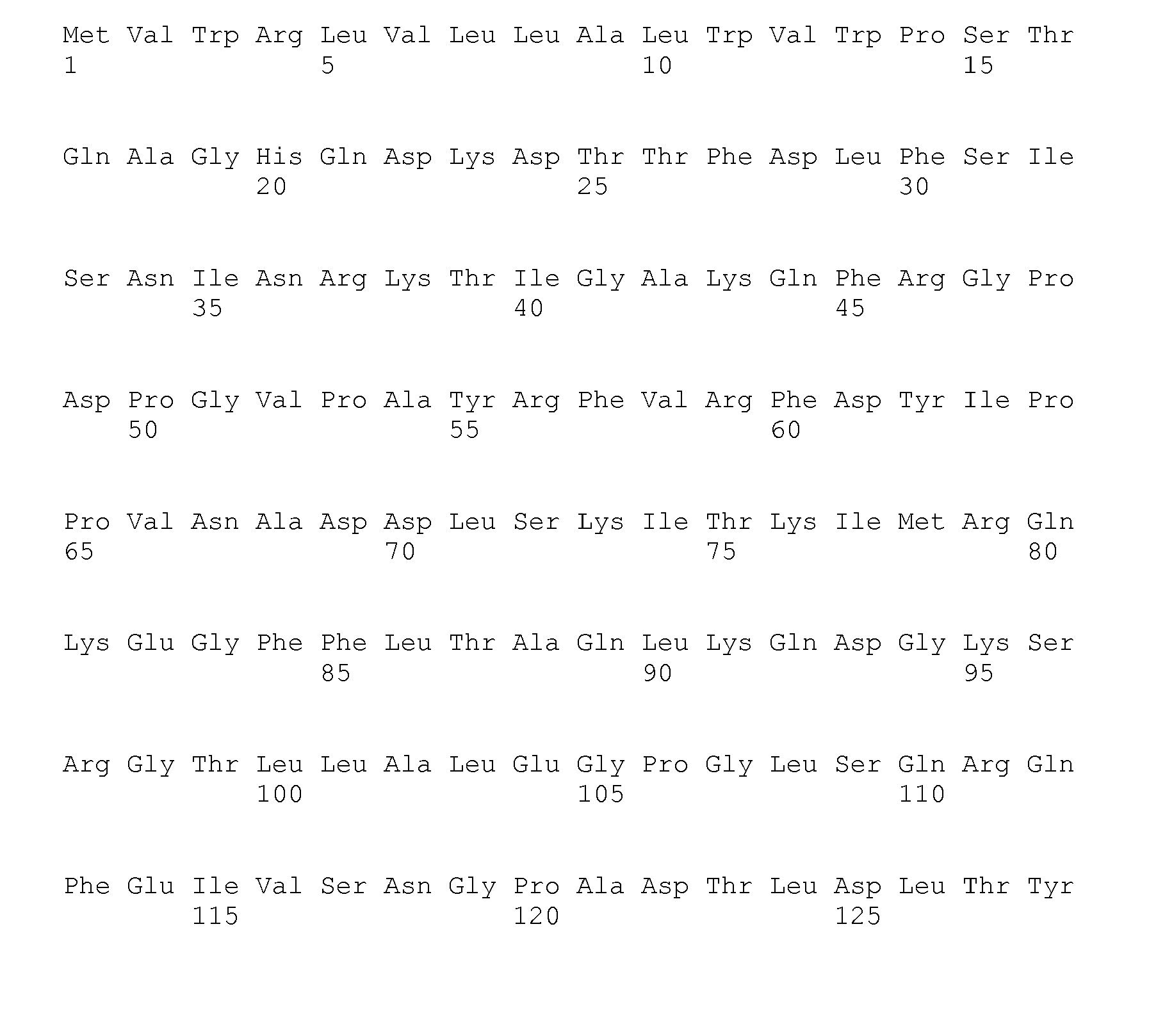


- <210> 216
<211> 1194
<212> PRT
<213> Homo sapiens - <400> 216


- <210> 217
<211> 538
<212> PRT
<213> Homo sapiens - <400> 217

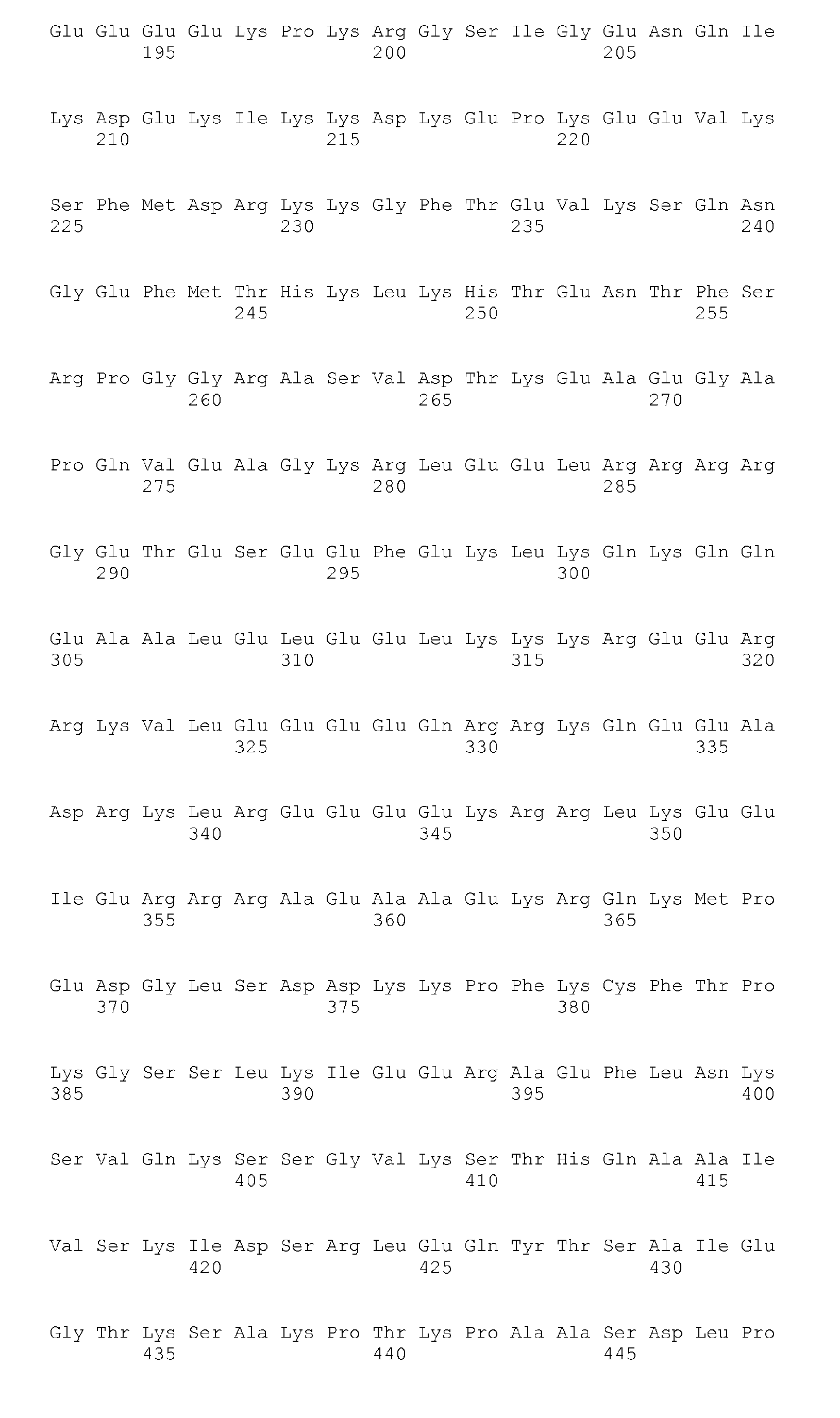

- <210> 218
<211> 111
<212> PRT
<213> Homo sapiens - <400> 218

- <210> 219
<211> 729
<212> PRT
<213> Homo sapiens - <400> 219


- <210> 220
<211> 195
<212> PRT
<213> Homo sapiens - <400> 220

- <210> 221
<211> 1838
<212> PRT
<213> Homo sapiens - <400> 221


- <210> 222
<211> 276
<212> PRT
<213> Homo sapiens - <400> 222
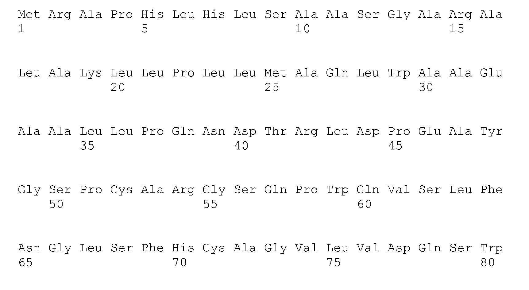
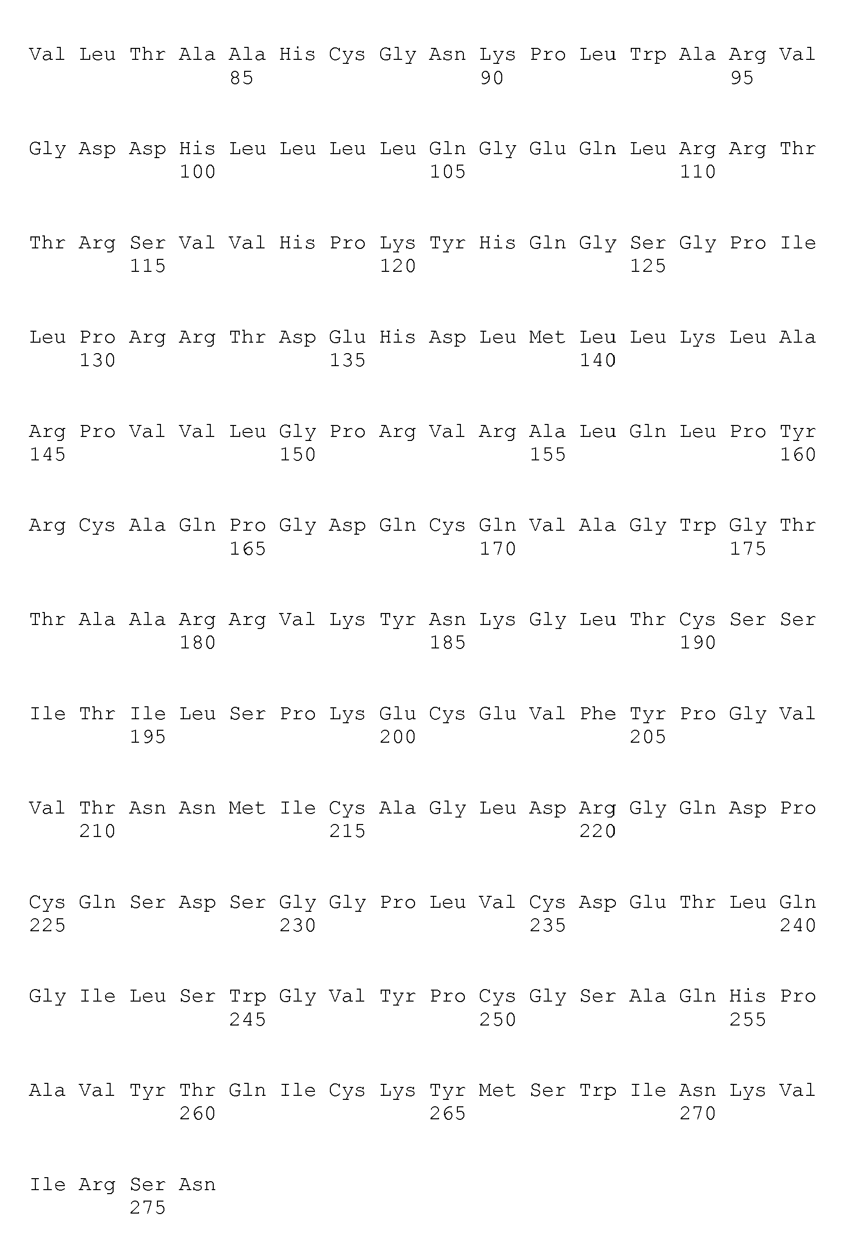
- <210> 223
<211> 62
<212> PRT
<213> Homo sapiens - <400> 223


- <210> 224
<211> 598
<212> PRT
<213> Homo sapiens - <400> 224


- <210> 225
<211> 291
<212> PRT
<213> Homo sapiens - <400> 225



- <210> 226
<211> 712
<212> PRT
<213> Homo sapiens - <400> 226


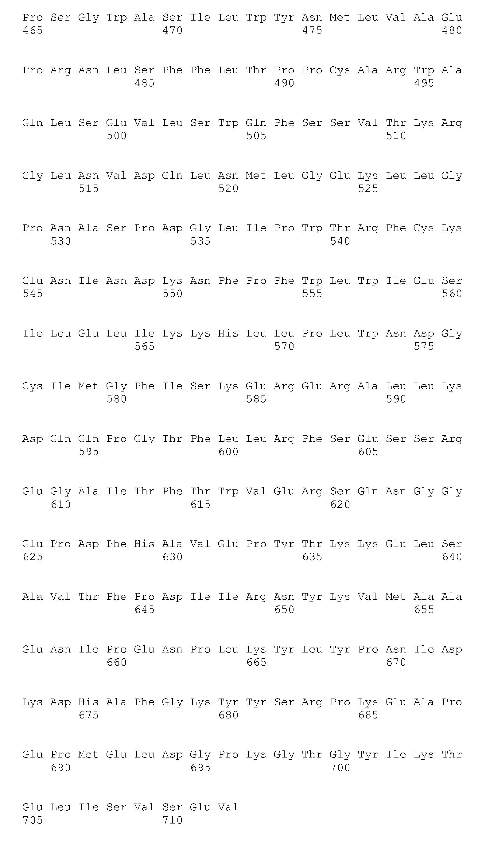
- <210> 227
<211> 543
<212> PRT
<213> Homo sapiens - <400> 227


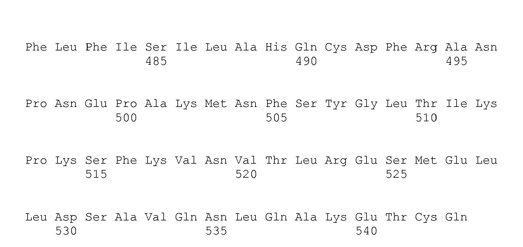
- <210> 228
<211> 1464
<212> PRT
<213> Homo sapiens - <400> 228


- <210> 229
<211> 248
<212> PRT
<213> Homo sapiens - <400> 229

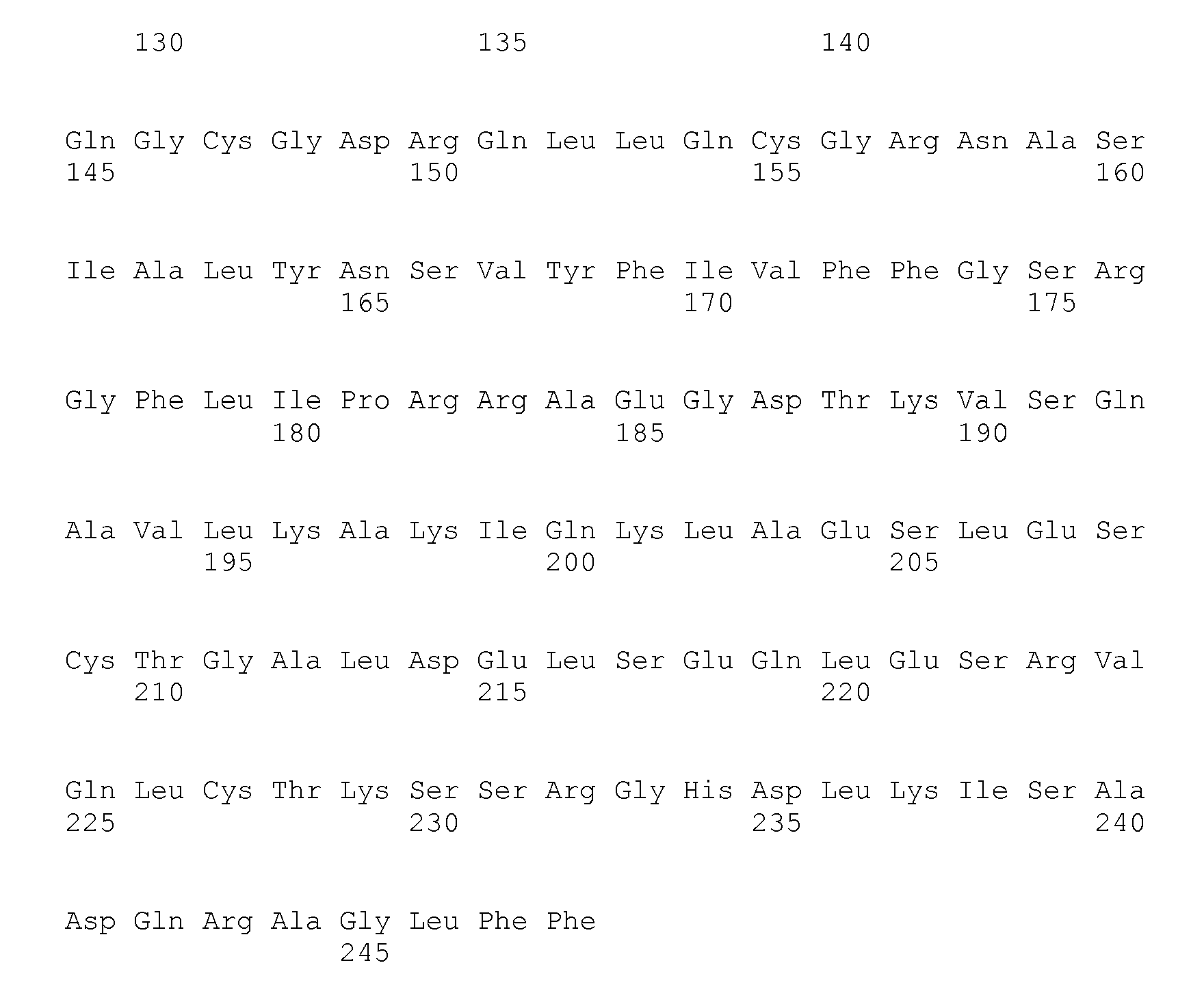
- <210> 230
<211> 451
<212> PRT
<213> Homo sapiens - <400> 230


- <210> 231
<211> 247
<212> PRT
<213> Homo sapiens - <400> 231


- <210> 232
<211> 1651
<212> PRT
<213> Homo sapiens - <400> 232


- <210> 233
<211> 457
<212> PRT
<213> Homo sapiens - <400> 233


- <210> 234
<211> 535
<212> PRT
<213> Homo sapiens - <400> 234


- <210> 235
<211> 319
<212> PRT
<213> Homo sapiens - <400> 235


- <210> 236
<211> 297
<212> PRT
<213> Homo sapiens - <400> 236

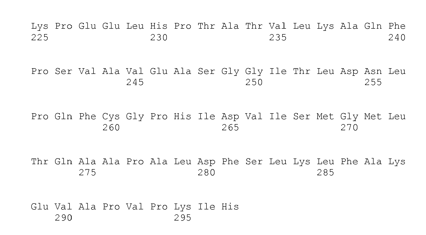
- <210> 237
<211> 480
<212> PRT
<213> Homo sapiens - <400> 237


- <210> 238
<211> 1806
<212> PRT
<213> Homo sapiens - <400> 238


- <210> 239
<211> 201
<212> PRT
<213> Homo sapiens - <400> 239

- <210> 240
<211> 871
<212> PRT
<213> Homo sapiens - <400> 240


- <210> 241
<211> 449
<212> PRT
<213> Homo sapiens - <400> 241


- <210> 242
<211> 2797
<212> PRT
<213> Homo sapiens - <400> 242


- <210> 243
<211> 352
<212> PRT
<213> Homo sapiens - <400> 243


- <210> 244
<211> 3396
<212> PRT
<213> Homo sapiens - <400> 244


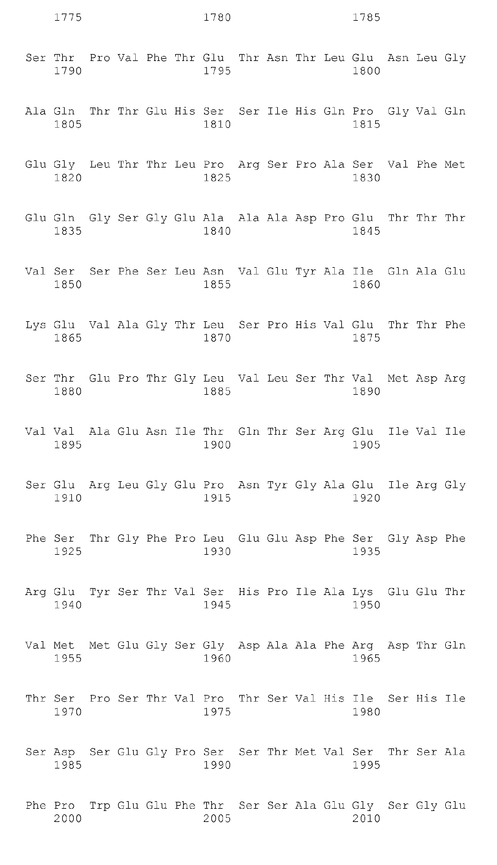

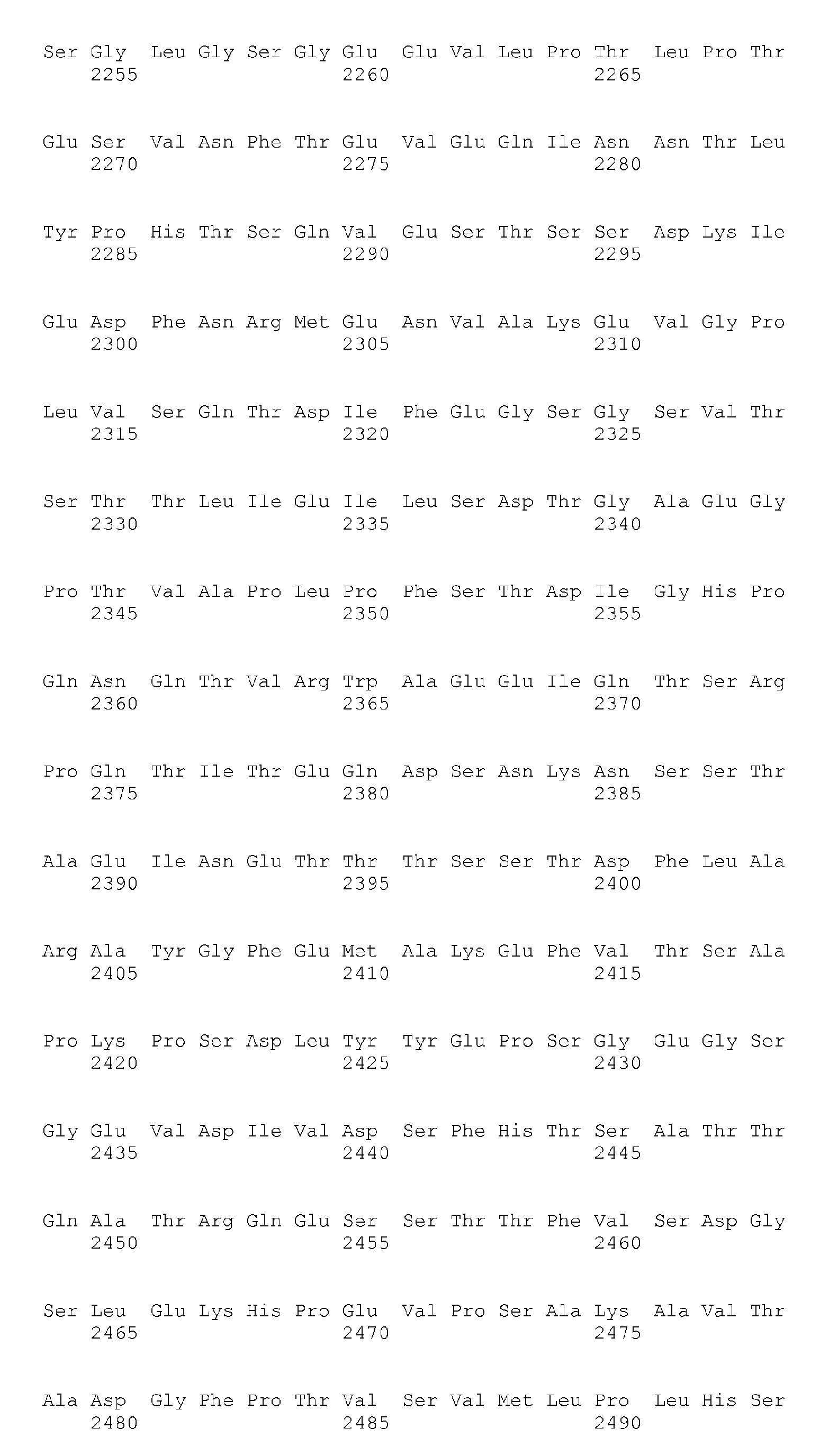


- <210> 245
<211> 418
<212> PRT
<213> Homo sapiens - <400> 245


- <210> 246
<211> 807
<212> PRT
<213> Homo sapiens - <400> 246


- <210> 247
<211> 1806
<212> PRT
<213> Homo sapiens - <400> 247


- <210> 248
<211> 323
<212> PRT
<213> Homo sapiens - <400> 248


- <210> 249
<211> 729
<212> PRT
<213> Homo sapiens - <400> 249


- <210> 250
<211> 259
<212> PRT
<213> Homo sapiens - <400> 250


- <210> 251
<211> 510
<212> PRT
<213> Homo sapiens - <400> 251

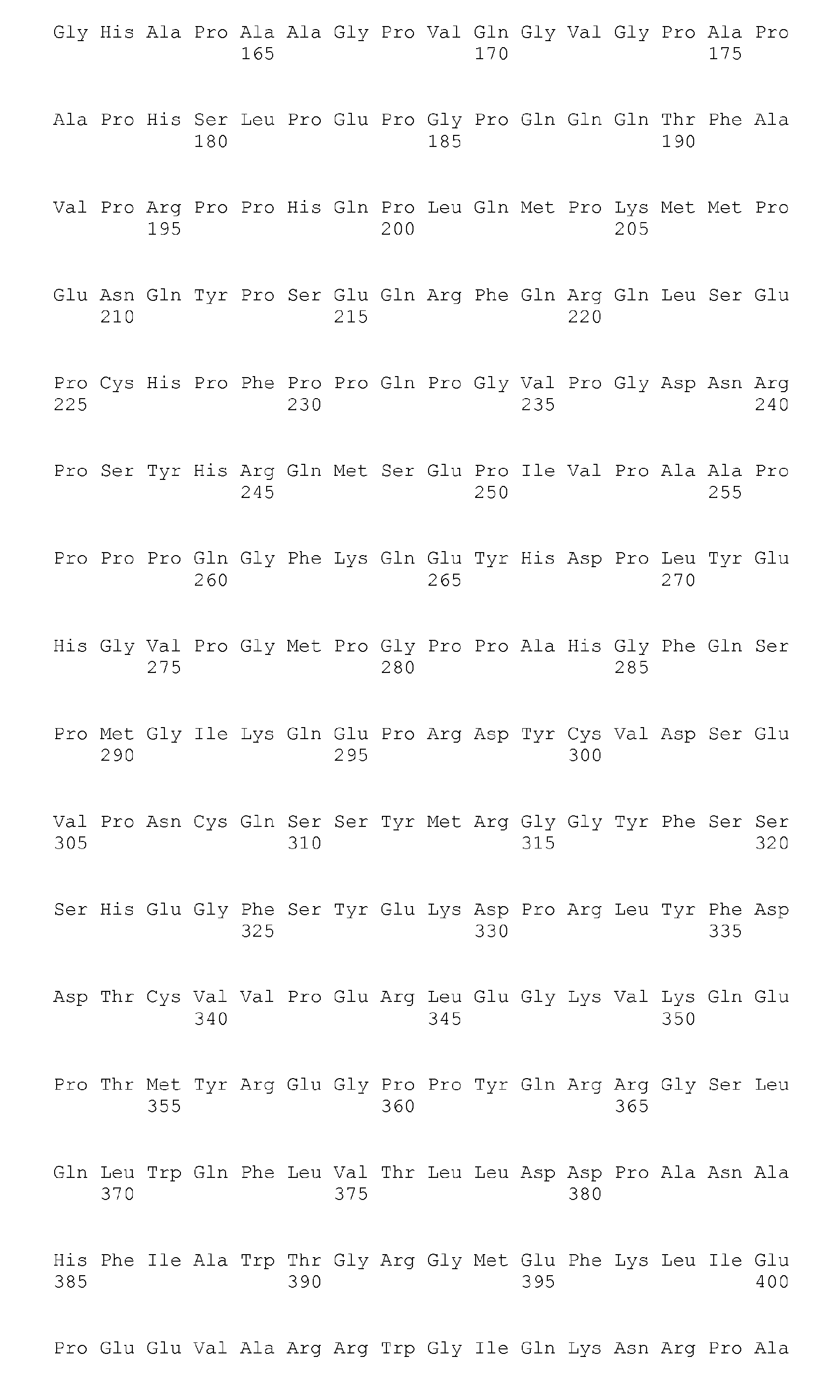

- <210> 252
<211> 303
<212> PRT
<213> Homo sapiens - <400> 252

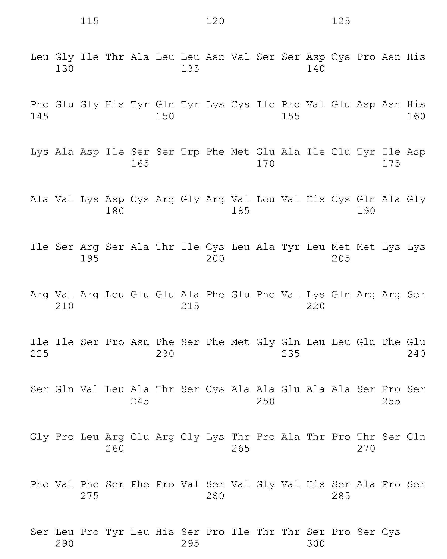
- <210> 253
<211> 394
<212> PRT
<213> Homo sapiens - <400> 253
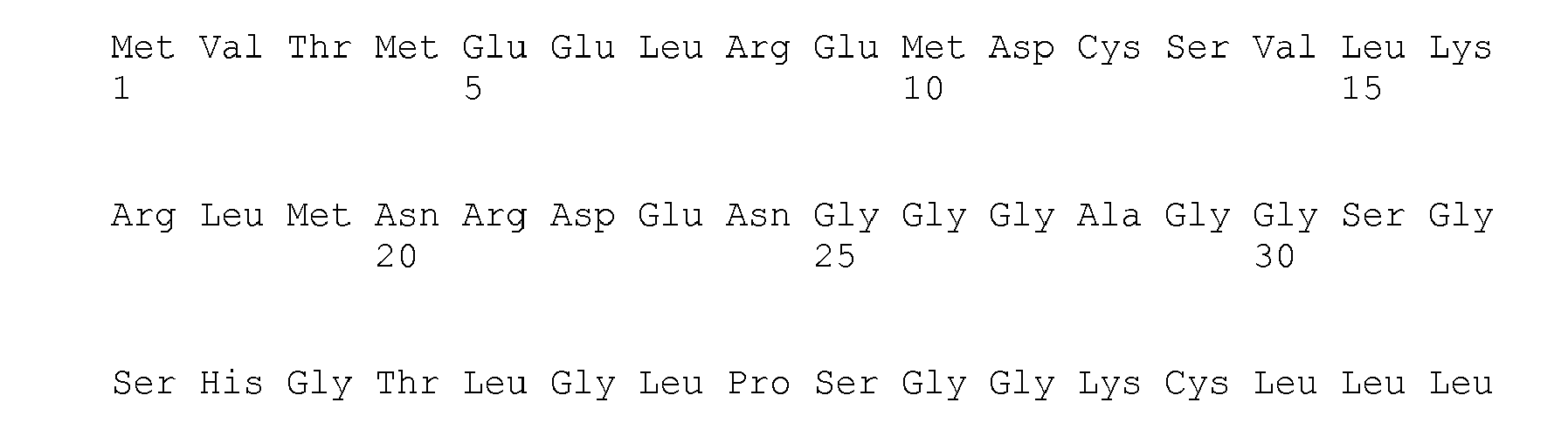


- <210> 254
<211> 388
<212> PRT
<213> Homo sapiens - <400> 254


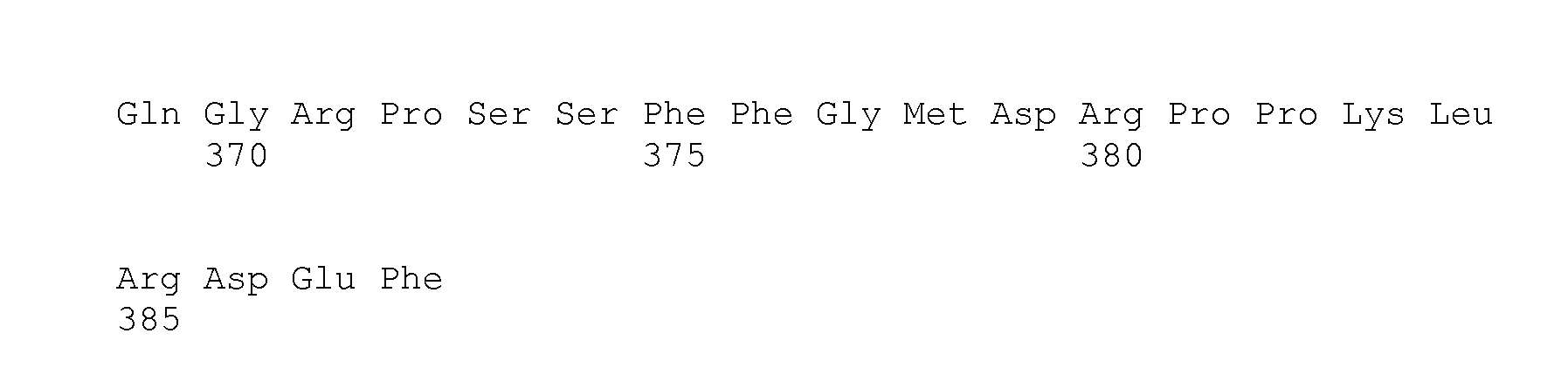
- <210> 255
<211> 460
<212> PRT
<213> Homo sapiens - <400> 255
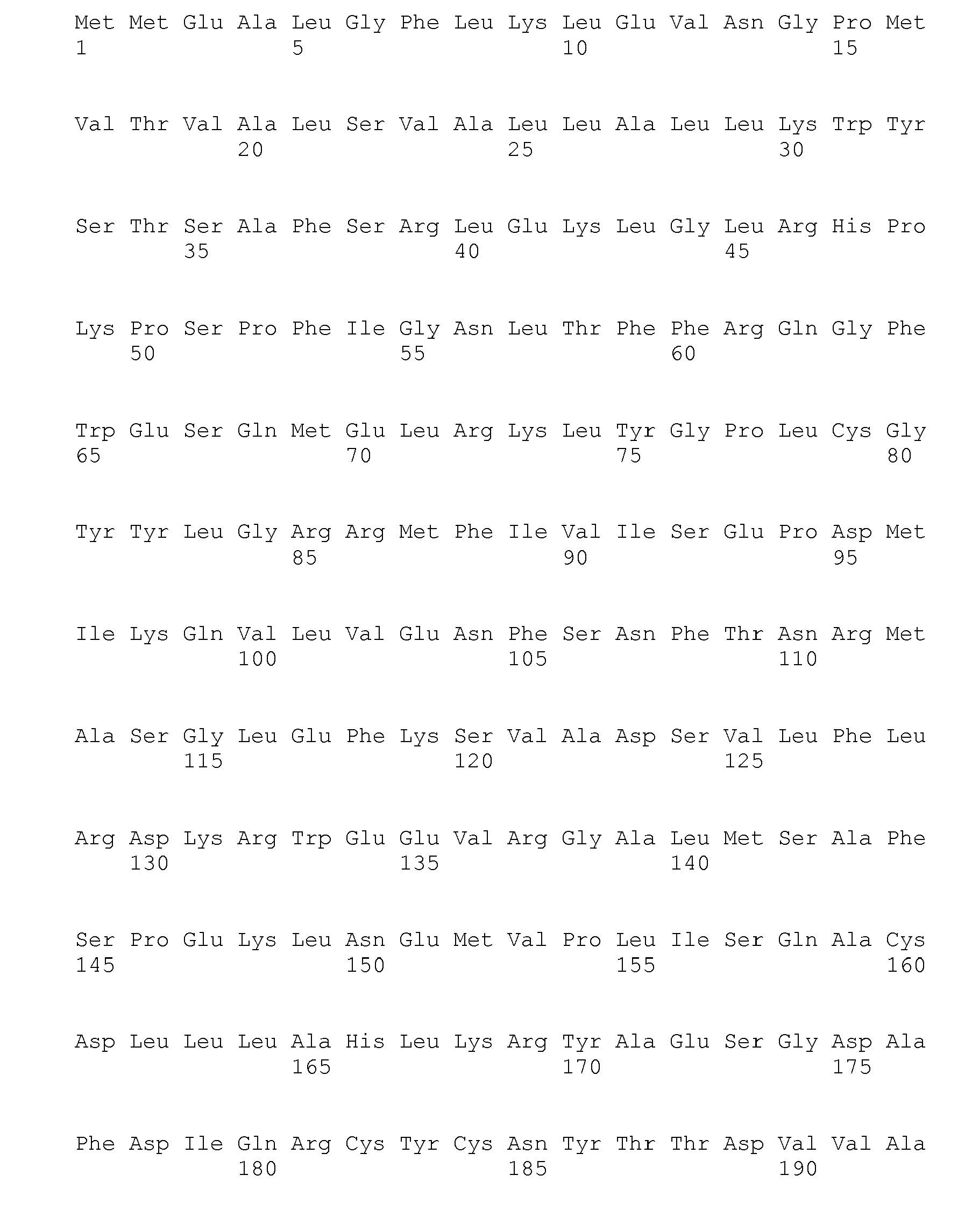


- <210> 256
<211> 499
<212> PRT
<213> Homo sapiens - <400> 256


- <210> 257
<211> 22
<212> DNA
<213> Artificial - <220>
<223> primer - <400> 257
taaggcctgc tgaaaatgac tg 22 - <210> 258
<211> 22
<212> DNA
<213> Artificial - <220>
<223> primer - <400> 258
tggtcctgca ccagtaatat gc 22 - <210> 259
<211> 23
<212> DNA
<213> Artificial - <220>
<223> primer - <400> 259
tcataatgct tgctctgata gga 23 - <210> 260
<211> 22
<212> DNA
<213> Artificial - <220>
<223> primer - <400> 260
ggccaaaaat ttaatcagtgga 22
Claims (13)
- An in vitro method for predicting the likelihood a mammal will respond therapeutically to a method of treating colorectal cancer comprising administering an EGFR modulator that inhibits binding of EGF to EGFR or the kinase activity of EGFR, wherein the method comprises measuring the mRNA expression level of both epiregulin and amphiregulin biomarkers in a biological sample of said mammal, wherein an elevated expression level of said biomarkers relative to a predetermined level indicates an increased likelihood the mammal will respond therapeutically to said method of treating colorectal cancer.
- The method of claim 1 wherein said measuring is performed following exposure of the mammal to the EGFR modulator.
- The method of claim 1 wherein the method comprises:(a) measuring the mRNA expression level of both epiregulin and amphiregulin biomarkers in a biological sample of said mammal;(b) following the exposure of the mammal to the EGFR modulator, measuring in a biological sample of said mammal the mRNA expression level of said biomarkers,wherein an elevated expression level of said biomarkers measured in step (b) relative to the level of said biomarkers measured in step (a) indicates an increased likelihood the mammal will respond therapeutically to said method of treating colorectal cancer.
- The method of any one of claims 1 to 3 further comprising measuring at least one additional biomarker selected from Table 1.
- The method of any one of claim 1 to 3 wherein said biological sample is a tissue sample comprising cancer cells and said tissue is fixed, paraffin-embedded, fresh, or frozen.
- The method of claim 5 that further comprises the step of determining whether said cancer cells have the presence of a mutated K-RAS, wherein detection of a mutated K-RAS indicates a decreased likelihood the mammal will respond therapeutically to said method of treating colorectal cancer.
- The method of any one of claims 1 to 6 wherein said mammal is a human.
- The method of any one of claims 1 to 6 wherein said mammal is selected from the group consisting of: rat, mouse, dog, rabbit, pig, sheep, cow, horse, cat, primate, and monkey.
- The method of any one of claims 1 to 8 wherein said EGFR modulator is an anti-EGFR monoclonal antibody.
- The method of any one of claims 1 to 8 wherein said EGFR modulator is an anti-EGFR single chain antibody.
- The method of any one of claims 1 to 8 wherein said EGFR modulator is an anti-EGFR humanized or chimeric antibody.
- The method of any one of claims 1 to 8 wherein said EGFR modulator is cetuximab.
- The method of any one of claims 1 to 8 wherein said EGFR modulator is panitumumab.
Applications Claiming Priority (2)
| Application Number | Priority Date | Filing Date | Title |
|---|---|---|---|
| US71105405P | 2005-08-24 | 2005-08-24 | |
| PCT/US2006/033073 WO2007025044A2 (en) | 2005-08-24 | 2006-08-24 | Biomarkers and methods for determining sensitivity to epidermal growth factor receptor modulators |
Publications (3)
| Publication Number | Publication Date |
|---|---|
| EP1917528A2 EP1917528A2 (en) | 2008-05-07 |
| EP1917528A4 EP1917528A4 (en) | 2009-03-11 |
| EP1917528B1 true EP1917528B1 (en) | 2011-08-17 |
Family
ID=37772388
Family Applications (1)
| Application Number | Title | Priority Date | Filing Date |
|---|---|---|---|
| EP06813702A Not-in-force EP1917528B1 (en) | 2005-08-24 | 2006-08-24 | Biomarkers and methods for determining sensitivity to epidermal growth factor receptor modulators |
Country Status (7)
| Country | Link |
|---|---|
| US (2) | US8129114B2 (en) |
| EP (1) | EP1917528B1 (en) |
| JP (1) | JP2009505658A (en) |
| AT (1) | ATE520979T1 (en) |
| CA (1) | CA2620195A1 (en) |
| ES (1) | ES2370054T3 (en) |
| WO (1) | WO2007025044A2 (en) |
Families Citing this family (32)
| Publication number | Priority date | Publication date | Assignee | Title |
|---|---|---|---|---|
| WO2004046386A1 (en) | 2002-11-15 | 2004-06-03 | Genomic Health, Inc. | Gene expression profiling of egfr positive cancer |
| WO2005070020A2 (en) | 2004-01-23 | 2005-08-04 | The Regents Of The University Of Colorado | Gefitinib sensitivity-related gene expression and products and methods related thereto |
| EP2527460B1 (en) | 2004-05-27 | 2014-12-24 | The Regents of The University of Colorado | Methods for prediction of clinical outcome to epidermal growth factor receptor inhibitors by cancer patients |
| DK2465950T3 (en) | 2007-03-13 | 2017-10-09 | Amgen Inc | K-RAS MUTATIONS AND ANTI-EGFR ANTIBODY TREATMENT |
| AU2016250348A1 (en) * | 2007-03-13 | 2016-11-10 | Amgen Inc. | K-ras mutations and anti-EGFr antibody therapy field |
| US8975081B2 (en) | 2007-10-24 | 2015-03-10 | Faron Pharmaceuticals Oy | Biomarker for monitoring development of diseases and assessing the efficacy of therapies |
| FI20070795A0 (en) * | 2007-10-24 | 2007-10-24 | Faron Pharmaceuticals Oy | A new biomarker for controlling disease development and assessing the efficacy of therapies |
| WO2009068621A1 (en) * | 2007-11-30 | 2009-06-04 | Glaxosmithkline Biologicals S.A. | Method for classifying cancer patients as responder or non-responder to immunotherapy |
| US8273534B2 (en) | 2008-05-14 | 2012-09-25 | Genomic Health, Inc. | Predictors of patient response to treatment with EGF receptor inhibitors |
| WO2010065626A1 (en) * | 2008-12-02 | 2010-06-10 | Globeimmune, Inc. | Genotyping tools, methods and kits |
| US20110269139A1 (en) * | 2009-01-06 | 2011-11-03 | Bristol-Myers Squibb Company | Biomarkers and methods for determining sensitivity to epidermal growth factor receptor modulators |
| US8765383B2 (en) | 2009-04-07 | 2014-07-01 | Genomic Health, Inc. | Methods of predicting cancer risk using gene expression in premalignant tissue |
| BRPI1015179A2 (en) * | 2009-06-19 | 2016-04-19 | Merck Patent Gmbh | Biomarkers and Methods for Determining Efficacy of Anti-Egfr Antibodies in Cancer Therapy |
| MY165154A (en) * | 2009-08-24 | 2018-02-28 | Genentech Inc | Determining sensitivity of cells to b-raf inhibitor treatment by detecting kras mutation and rtk expression levels |
| EP2507628A4 (en) * | 2009-12-01 | 2013-04-24 | Medimmune Llc | IMPROVED METHODS AND COMPOSITIONS FOR THE DETECTION AND TREATMENT OF CANCERS EXPRESSING CARCINO-EMBRYONIC ANTIGEN (ACE) |
| NZ602920A (en) * | 2010-04-12 | 2014-07-25 | Response Genetics Inc | Kras primers and probes |
| MX385227B (en) | 2012-01-20 | 2025-03-14 | Del Mar Pharmaceuticals | USE OF SUBSTITUTED HEXITOLS INCLUDING DIANHYDROGALACTITOL AND ANALOGUES TO TREAT NEOPLASTIC DISEASE AND CANCER STEM CELLS INCLUDING GLIOBLASTOMA MULTIFORME AND MEDULOBLASTOMA. |
| EP2823307A4 (en) * | 2012-03-08 | 2015-07-29 | Bioscale Inc | METHODS AND KITS FOR ANALYZING BIOMARKERS IN A SIGNAL TRANSDUCTION PATH |
| US20150293105A1 (en) * | 2012-10-26 | 2015-10-15 | The University Of Queensland | Methods for classifying tumors and uses therefor |
| US10071109B2 (en) | 2013-11-06 | 2018-09-11 | Molecular Templates, Inc. | Predictive biomarker for hypoxia-activated prodrug therapy |
| CN104762371B (en) | 2014-01-07 | 2021-03-09 | 三星电子株式会社 | Biomarkers for predicting or monitoring the efficacy of c-Met inhibitors |
| US20150254433A1 (en) * | 2014-03-05 | 2015-09-10 | Bruce MACHER | Methods and Models for Determining Likelihood of Cancer Drug Treatment Success Utilizing Predictor Biomarkers, and Methods of Diagnosing and Treating Cancer Using the Biomarkers |
| US9753036B2 (en) | 2014-04-29 | 2017-09-05 | Edp Biotech Corporation | Methods and compositions for screening and detecting biomarkers |
| US20160024585A1 (en) * | 2014-05-02 | 2016-01-28 | Duke University | Methods of predicting responsiveness of a cancer to an agent and methods of determining a prognosis for a cancer patient |
| KR20160037666A (en) | 2014-09-29 | 2016-04-06 | 삼성전자주식회사 | Prediction of Effect of a c-Met Inhibitor using mutation of KRAS or BRAF |
| EP3073268A1 (en) | 2015-03-27 | 2016-09-28 | Deutsches Krebsforschungszentrum Stiftung des Öffentlichen Rechts | Biomarker panel for diagnosing cancer |
| CN108474040B (en) * | 2015-10-09 | 2023-05-16 | 夸登特健康公司 | Population-based treatment recommendations using cell-free DNA |
| JP6936221B2 (en) | 2015-11-02 | 2021-09-15 | ファイブ プライム セラピューティクス, インコーポレイテッド | CD80 extracellular domain polypeptides and their use in cancer treatment |
| WO2018078143A1 (en) | 2016-10-28 | 2018-05-03 | MAX-PLANCK-Gesellschaft zur Förderung der Wissenschaften e.V. | Means and methods for determining efficacy of anti-egfr inhibitors in colorectal cancer (crc) therapy |
| CN109457052A (en) * | 2018-12-18 | 2019-03-12 | 苏州德思普生物科技有限公司 | Detect primer, probe, kit and the detection method of human immunodeficiency virus nucleic acid |
| EP4217393A1 (en) * | 2020-09-22 | 2023-08-02 | Ventana Medical Systems, Inc. | Prediction of response to epidermal growth factor receptor-directed therapies using epiregulin and amphiregulin |
| CN117964690B (en) * | 2024-02-04 | 2024-07-23 | 北京榕树堂生物科技有限公司 | Bird's nest peptide III with skin elasticity protecting and anti-inflammatory effects and application thereof |
Family Cites Families (46)
| Publication number | Priority date | Publication date | Assignee | Title |
|---|---|---|---|---|
| CU22545A1 (en) * | 1994-11-18 | 1999-03-31 | Centro Inmunologia Molecular | OBTAINING A CHEMICAL AND HUMANIZED ANTIBODY AGAINST THE RECEPTOR OF THE EPIDERMAL GROWTH FACTOR FOR DIAGNOSTIC AND THERAPEUTIC USE |
| US6713619B1 (en) * | 1980-08-29 | 2004-03-30 | Massachusetts Institute Of Technology | Oncogenes and methods for their detection |
| US4535058A (en) * | 1982-10-01 | 1985-08-13 | Massachusetts Institute Of Technology | Characterization of oncogenes and assays based thereon |
| US4943533A (en) * | 1984-03-01 | 1990-07-24 | The Regents Of The University Of California | Hybrid cell lines that produce monoclonal antibodies to epidermal growth factor receptor |
| US5591582A (en) * | 1985-07-23 | 1997-01-07 | The Board Of Rijks Universiteit Leiden | Methods for detecting activated RAS oncogenes |
| WO1991016051A1 (en) | 1990-04-16 | 1991-10-31 | Rhône-Poulenc Rorer International (Holdings) Inc. | Styryl-substituted monocyclic and bicyclic heteroaryl compounds which inhibit egf receptor tyrosine kinase |
| US5196446A (en) * | 1990-04-16 | 1993-03-23 | Yissum Research Development Company Of The Hebrew University Of Jerusalem | Certain indole compounds which inhibit EGF receptor tyrosine kinase |
| SK281143B6 (en) * | 1991-03-06 | 2000-12-11 | Merck Patent Gesellschaft Mit Beschr�Nkter Haftung | Chimeric monoclonal antibodies, expression vectors and pharmaceutical compoisitons |
| US5480883A (en) * | 1991-05-10 | 1996-01-02 | Rhone-Poulenc Rorer Pharmaceuticals Inc. | Bis mono- and bicyclic aryl and heteroaryl compounds which inhibit EGF and/or PDGF receptor tyrosine kinase |
| GB9300059D0 (en) * | 1992-01-20 | 1993-03-03 | Zeneca Ltd | Quinazoline derivatives |
| US5654307A (en) * | 1994-01-25 | 1997-08-05 | Warner-Lambert Company | Bicyclic compounds capable of inhibiting tyrosine kinases of the epidermal growth factor receptor family |
| US5656655A (en) * | 1994-03-17 | 1997-08-12 | Rhone-Poulenc Rorer Pharmaceuticals, Inc. | Styryl-substituted heteroaryl compounds which inhibit EGF receptor tyrosine kinase |
| DE69536015D1 (en) | 1995-03-30 | 2009-12-10 | Pfizer Prod Inc | Quinazolinone derivatives |
| GB9508538D0 (en) | 1995-04-27 | 1995-06-14 | Zeneca Ltd | Quinazoline derivatives |
| US7060808B1 (en) * | 1995-06-07 | 2006-06-13 | Imclone Systems Incorporated | Humanized anti-EGF receptor monoclonal antibody |
| US5569588A (en) * | 1995-08-09 | 1996-10-29 | The Regents Of The University Of California | Methods for drug screening |
| AU1441497A (en) | 1996-01-23 | 1997-08-20 | Novartis Ag | Pyrrolopyrimidines and processes for their preparation |
| GB9603095D0 (en) | 1996-02-14 | 1996-04-10 | Zeneca Ltd | Quinazoline derivatives |
| US6156504A (en) * | 1996-03-15 | 2000-12-05 | The Penn State Research Foundation | Detection of extracellular tumor-associated nucleic acid in blood plasma or serum using nucleic acid amplification assays |
| GB9707800D0 (en) | 1996-05-06 | 1997-06-04 | Zeneca Ltd | Chemical compounds |
| EP0907642B1 (en) | 1996-06-24 | 2005-11-02 | Pfizer Inc. | Phenylamino-substituted tricyclic derivatives for treatment of hyperproliferative diseases |
| KR20000070751A (en) | 1997-02-05 | 2000-11-25 | 로즈 암스트롱, 크리스틴 에이. 트러트웨인 | Pyrido[2,3-D]pyrimidines and 4-Aminopyrimidines as Inhibitors of Cellular Proliferation |
| US6235883B1 (en) * | 1997-05-05 | 2001-05-22 | Abgenix, Inc. | Human monoclonal antibodies to epidermal growth factor receptor |
| HUP0103520A3 (en) | 1998-09-29 | 2003-01-28 | Wyeth Holdings Corp Madison | Substituted 3-cyanoquinolines as protein tyrosine kinases inhibitors and pharmaceutical compositions containing them |
| ES2188254T3 (en) | 1998-11-19 | 2003-06-16 | Warner Lambert Co | N- (4- (3-CHLORO-4-FLUORO-PHENYLAMINE) -7- (3-MORFOLIN-4-IL-PROPOXI) -QUIN AZOLIN-6-IL) -ACRILAMADA, AN IRREVERSIBLE THYROSINE KINASE INHIBITOR. |
| AU2001247616B2 (en) * | 2000-04-11 | 2007-06-14 | Genentech, Inc. | Multivalent antibodies and uses therefor |
| US6503914B1 (en) * | 2000-10-23 | 2003-01-07 | Board Of Regents, The University Of Texas System | Thienopyrimidine-based inhibitors of the Src family |
| DK2258872T3 (en) * | 2002-03-13 | 2013-11-18 | Genomic Health Inc | Gene expression profiling in tumor tissue biopsies |
| WO2004046386A1 (en) * | 2002-11-15 | 2004-06-03 | Genomic Health, Inc. | Gene expression profiling of egfr positive cancer |
| JP2007520995A (en) | 2003-01-08 | 2007-08-02 | ブリストル−マイヤーズ スクイブ カンパニー | Biomarkers and methods for determining susceptibility to epidermal growth factor receptor modulators |
| US20040231909A1 (en) * | 2003-01-15 | 2004-11-25 | Tai-Yang Luh | Motorized vehicle having forward and backward differential structure |
| WO2004071572A2 (en) * | 2003-02-06 | 2004-08-26 | Genomic Health, Inc. | Gene expression markers for response to egfr inhibitor drugs |
| CA2516553C (en) * | 2003-02-20 | 2013-04-16 | Genomic Health, Inc. | Use of intronic rna to measure gene expression |
| AU2004291709C1 (en) | 2003-05-30 | 2010-03-11 | Astrazeneca Uk Limited | Process |
| EP1759010A4 (en) * | 2004-01-07 | 2008-12-24 | Bristol Myers Squibb Co | BIOMARKERS AND METHODS FOR DETERMINING SENSITIVITY TO EPIDERMAL GROWTH FACTOR RECEPTOR MODULATORS |
| CA2561111A1 (en) | 2004-03-26 | 2005-10-13 | Bristol-Myers Squibb Company | Biomarkers and methods for determining sensitivity to epidermal growth factor receptor modulators in non-small cell lung cancer |
| EP3042964A1 (en) * | 2004-06-04 | 2016-07-13 | Genentech, Inc. | Egfr mutations |
| ZA200704796B (en) * | 2004-12-07 | 2008-11-26 | Genentech Inc | Selecting patients for therapy with a HER inhibitor |
| US20060204505A1 (en) * | 2005-03-08 | 2006-09-14 | Sliwkowski Mark X | Methods for identifying tumors responsive to treatment with HER dimerization inhibitors (HDIs) |
| EP1913157B2 (en) * | 2005-06-28 | 2016-10-26 | Genentech, Inc. | Egfr and kras mutations for prediction of patient response to egfr inhibitor treatment |
| US7700299B2 (en) * | 2005-08-12 | 2010-04-20 | Hoffmann-La Roche Inc. | Method for predicting the response to a treatment |
| US7908091B2 (en) * | 2006-03-17 | 2011-03-15 | Prometheus Laboratories Inc. | Methods of predicting and monitoring tyrosine kinase inhibitor therapy |
| US7906342B2 (en) * | 2006-03-31 | 2011-03-15 | Biodesix, Inc. | Monitoring treatment of cancer patients with drugs targeting EGFR pathway using mass spectrometry of patient samples |
| US7858389B2 (en) * | 2006-03-31 | 2010-12-28 | Biodesix, Inc. | Selection of non-small-cell lung cancer patients for treatment with monoclonal antibody drugs targeting EGFR pathway |
| EP2412828B1 (en) * | 2007-03-13 | 2013-06-05 | Amgen Inc. | K-ras and B-raf mutations and anti-EGFr antibody therapy |
| DK2465950T3 (en) * | 2007-03-13 | 2017-10-09 | Amgen Inc | K-RAS MUTATIONS AND ANTI-EGFR ANTIBODY TREATMENT |
-
2006
- 2006-08-24 AT AT06813702T patent/ATE520979T1/en not_active IP Right Cessation
- 2006-08-24 EP EP06813702A patent/EP1917528B1/en not_active Not-in-force
- 2006-08-24 WO PCT/US2006/033073 patent/WO2007025044A2/en not_active Ceased
- 2006-08-24 JP JP2008528150A patent/JP2009505658A/en not_active Withdrawn
- 2006-08-24 US US11/990,713 patent/US8129114B2/en active Active
- 2006-08-24 ES ES06813702T patent/ES2370054T3/en active Active
- 2006-08-24 CA CA002620195A patent/CA2620195A1/en not_active Abandoned
-
2012
- 2012-02-14 US US13/372,824 patent/US20120195889A1/en not_active Abandoned
Non-Patent Citations (1)
| Title |
|---|
| ODA KANAE; MATSUOKA YUKIKO; FUNAHASHI AKIRA; KITANO HIROAKI: "A comprehensive pathway map of epidermal growth factor receptor signaling", MOLECULAR SYSTEMS BIOLOGY, vol. 1, 25 May 2005 (2005-05-25), pages ARTICLE NUMBER 0010 * |
Also Published As
| Publication number | Publication date |
|---|---|
| JP2009505658A (en) | 2009-02-12 |
| ES2370054T3 (en) | 2011-12-12 |
| ATE520979T1 (en) | 2011-09-15 |
| US8129114B2 (en) | 2012-03-06 |
| WO2007025044A2 (en) | 2007-03-01 |
| US20120195889A1 (en) | 2012-08-02 |
| WO2007025044A8 (en) | 2007-07-05 |
| CA2620195A1 (en) | 2007-03-01 |
| EP1917528A4 (en) | 2009-03-11 |
| WO2007025044A3 (en) | 2007-09-20 |
| EP1917528A2 (en) | 2008-05-07 |
| US20100221754A1 (en) | 2010-09-02 |
Similar Documents
| Publication | Publication Date | Title |
|---|---|---|
| US8129114B2 (en) | Biomarkers and methods for determining sensitivity to epidermal growth factor receptor modulators | |
| Khambata-Ford et al. | Expression of epiregulin and amphiregulin and K-ras mutation status predict disease control in metastatic colorectal cancer patients treated with cetuximab | |
| JP5422120B2 (en) | Methods for predicting clinical outcome for epidermal growth factor receptor inhibitors by cancer patients | |
| AU2010262133B2 (en) | Biomarkers and methods for determining efficacy of anti-EGFR antibodies in cancer therapy | |
| WO2008144345A2 (en) | Biomarkers and methods for determining sensitivity to insulin growth factor-1 receptor modulators | |
| KR20110018930A (en) | Identification and use of prognostic and predictive markers in cancer treatment | |
| JP2007530954A (en) | Biomarkers and methods for determining sensitivity to epidermal growth factor receptor modulators in non-small cell lung cancer | |
| WO2011107819A1 (en) | Methods for predicting outcome of breast cancer, and/or risk of relapse, response or survival of a patient suffering therefrom | |
| JP2016535079A (en) | Nucleic acid biomarkers and uses thereof | |
| EP2527467A2 (en) | Biomarkers and methods for determining sensitivity to vascular endothelial growth factor receptor-2 modulators | |
| WO2007067476A2 (en) | Methods for predicting treatment response based on the expression profiles of protein and transcription biomakers | |
| EP2281063B1 (en) | A method for predicting a clinical response of a patient suffering from or at risk of developing cancer towards a given mode of treatment | |
| EP3294748A1 (en) | Novel methylation site regulating expression of mda-9/syntenin | |
| CN111433375A (en) | Approaches to choosing treatments for cancer patients | |
| AU2008286407A1 (en) | Predictive markers for EGFR inhibitor treatment | |
| KR20070103001A (en) | Identification and use of prognostic and predictive markers in cancer treatment | |
| KR102759983B1 (en) | Gene markers representing the acquired resistance to EGFR-Targeting Agent in colorectal cancer patients and method for predicting resistance acquisition using the same | |
| US20140127689A1 (en) | Cxcr1 as a predictor of response to treatment with epidermal growth factor receptor therapeutic | |
| Mauro | Expression of Epiregulin and Amphiregulin andK-rasMutation Status Predict Disease Control in Metastatic Colorectal Cancer Patients Treated With Cetuximab |
Legal Events
| Date | Code | Title | Description |
|---|---|---|---|
| PUAI | Public reference made under article 153(3) epc to a published international application that has entered the european phase |
Free format text: ORIGINAL CODE: 0009012 |
|
| 17P | Request for examination filed |
Effective date: 20080225 |
|
| AK | Designated contracting states |
Kind code of ref document: A2 Designated state(s): AT BE BG CH CY CZ DE DK EE ES FI FR GB GR HU IE IS IT LI LT LU LV MC NL PL PT RO SE SI SK TR |
|
| A4 | Supplementary search report drawn up and despatched |
Effective date: 20090209 |
|
| RIC1 | Information provided on ipc code assigned before grant |
Ipc: G01N 33/574 20060101ALI20090130BHEP Ipc: G01N 33/50 20060101ALI20090130BHEP Ipc: G01N 33/48 20060101AFI20080325BHEP |
|
| 17Q | First examination report despatched |
Effective date: 20090625 |
|
| GRAP | Despatch of communication of intention to grant a patent |
Free format text: ORIGINAL CODE: EPIDOSNIGR1 |
|
| TPAC | Observations filed by third parties |
Free format text: ORIGINAL CODE: EPIDOSNTIPA |
|
| GRAS | Grant fee paid |
Free format text: ORIGINAL CODE: EPIDOSNIGR3 |
|
| GRAA | (expected) grant |
Free format text: ORIGINAL CODE: 0009210 |
|
| DAX | Request for extension of the european patent (deleted) | ||
| AK | Designated contracting states |
Kind code of ref document: B1 Designated state(s): AT BE BG CH CY CZ DE DK EE ES FI FR GB GR HU IE IS IT LI LT LU LV MC NL PL PT RO SE SI SK TR |
|
| REG | Reference to a national code |
Ref country code: GB Ref legal event code: FG4D |
|
| REG | Reference to a national code |
Ref country code: CH Ref legal event code: EP |
|
| REG | Reference to a national code |
Ref country code: IE Ref legal event code: FG4D |
|
| REG | Reference to a national code |
Ref country code: DE Ref legal event code: R096 Ref document number: 602006023892 Country of ref document: DE Effective date: 20111013 |
|
| REG | Reference to a national code |
Ref country code: NL Ref legal event code: VDEP Effective date: 20110817 |
|
| REG | Reference to a national code |
Ref country code: ES Ref legal event code: FG2A Ref document number: 2370054 Country of ref document: ES Kind code of ref document: T3 Effective date: 20111212 |
|
| LTIE | Lt: invalidation of european patent or patent extension |
Effective date: 20110817 |
|
| PG25 | Lapsed in a contracting state [announced via postgrant information from national office to epo] |
Ref country code: NL Free format text: LAPSE BECAUSE OF FAILURE TO SUBMIT A TRANSLATION OF THE DESCRIPTION OR TO PAY THE FEE WITHIN THE PRESCRIBED TIME-LIMIT Effective date: 20110817 Ref country code: SE Free format text: LAPSE BECAUSE OF FAILURE TO SUBMIT A TRANSLATION OF THE DESCRIPTION OR TO PAY THE FEE WITHIN THE PRESCRIBED TIME-LIMIT Effective date: 20110817 Ref country code: FI Free format text: LAPSE BECAUSE OF FAILURE TO SUBMIT A TRANSLATION OF THE DESCRIPTION OR TO PAY THE FEE WITHIN THE PRESCRIBED TIME-LIMIT Effective date: 20110817 Ref country code: IS Free format text: LAPSE BECAUSE OF FAILURE TO SUBMIT A TRANSLATION OF THE DESCRIPTION OR TO PAY THE FEE WITHIN THE PRESCRIBED TIME-LIMIT Effective date: 20111217 Ref country code: PT Free format text: LAPSE BECAUSE OF FAILURE TO SUBMIT A TRANSLATION OF THE DESCRIPTION OR TO PAY THE FEE WITHIN THE PRESCRIBED TIME-LIMIT Effective date: 20111219 Ref country code: LT Free format text: LAPSE BECAUSE OF FAILURE TO SUBMIT A TRANSLATION OF THE DESCRIPTION OR TO PAY THE FEE WITHIN THE PRESCRIBED TIME-LIMIT Effective date: 20110817 |
|
| REG | Reference to a national code |
Ref country code: AT Ref legal event code: MK05 Ref document number: 520979 Country of ref document: AT Kind code of ref document: T Effective date: 20110817 |
|
| PG25 | Lapsed in a contracting state [announced via postgrant information from national office to epo] |
Ref country code: AT Free format text: LAPSE BECAUSE OF FAILURE TO SUBMIT A TRANSLATION OF THE DESCRIPTION OR TO PAY THE FEE WITHIN THE PRESCRIBED TIME-LIMIT Effective date: 20110817 Ref country code: PL Free format text: LAPSE BECAUSE OF FAILURE TO SUBMIT A TRANSLATION OF THE DESCRIPTION OR TO PAY THE FEE WITHIN THE PRESCRIBED TIME-LIMIT Effective date: 20110817 Ref country code: CY Free format text: LAPSE BECAUSE OF FAILURE TO SUBMIT A TRANSLATION OF THE DESCRIPTION OR TO PAY THE FEE WITHIN THE PRESCRIBED TIME-LIMIT Effective date: 20110817 Ref country code: LV Free format text: LAPSE BECAUSE OF FAILURE TO SUBMIT A TRANSLATION OF THE DESCRIPTION OR TO PAY THE FEE WITHIN THE PRESCRIBED TIME-LIMIT Effective date: 20110817 Ref country code: SI Free format text: LAPSE BECAUSE OF FAILURE TO SUBMIT A TRANSLATION OF THE DESCRIPTION OR TO PAY THE FEE WITHIN THE PRESCRIBED TIME-LIMIT Effective date: 20110817 Ref country code: GR Free format text: LAPSE BECAUSE OF FAILURE TO SUBMIT A TRANSLATION OF THE DESCRIPTION OR TO PAY THE FEE WITHIN THE PRESCRIBED TIME-LIMIT Effective date: 20111118 |
|
| PG25 | Lapsed in a contracting state [announced via postgrant information from national office to epo] |
Ref country code: MC Free format text: LAPSE BECAUSE OF NON-PAYMENT OF DUE FEES Effective date: 20110831 Ref country code: BE Free format text: LAPSE BECAUSE OF FAILURE TO SUBMIT A TRANSLATION OF THE DESCRIPTION OR TO PAY THE FEE WITHIN THE PRESCRIBED TIME-LIMIT Effective date: 20110817 |
|
| REG | Reference to a national code |
Ref country code: CH Ref legal event code: PL |
|
| PG25 | Lapsed in a contracting state [announced via postgrant information from national office to epo] |
Ref country code: CZ Free format text: LAPSE BECAUSE OF FAILURE TO SUBMIT A TRANSLATION OF THE DESCRIPTION OR TO PAY THE FEE WITHIN THE PRESCRIBED TIME-LIMIT Effective date: 20110817 Ref country code: SK Free format text: LAPSE BECAUSE OF FAILURE TO SUBMIT A TRANSLATION OF THE DESCRIPTION OR TO PAY THE FEE WITHIN THE PRESCRIBED TIME-LIMIT Effective date: 20110817 Ref country code: CH Free format text: LAPSE BECAUSE OF NON-PAYMENT OF DUE FEES Effective date: 20110831 Ref country code: LI Free format text: LAPSE BECAUSE OF NON-PAYMENT OF DUE FEES Effective date: 20110831 |
|
| REG | Reference to a national code |
Ref country code: IE Ref legal event code: MM4A |
|
| PG25 | Lapsed in a contracting state [announced via postgrant information from national office to epo] |
Ref country code: RO Free format text: LAPSE BECAUSE OF FAILURE TO SUBMIT A TRANSLATION OF THE DESCRIPTION OR TO PAY THE FEE WITHIN THE PRESCRIBED TIME-LIMIT Effective date: 20110817 Ref country code: EE Free format text: LAPSE BECAUSE OF FAILURE TO SUBMIT A TRANSLATION OF THE DESCRIPTION OR TO PAY THE FEE WITHIN THE PRESCRIBED TIME-LIMIT Effective date: 20110817 |
|
| PLBE | No opposition filed within time limit |
Free format text: ORIGINAL CODE: 0009261 |
|
| STAA | Information on the status of an ep patent application or granted ep patent |
Free format text: STATUS: NO OPPOSITION FILED WITHIN TIME LIMIT |
|
| PG25 | Lapsed in a contracting state [announced via postgrant information from national office to epo] |
Ref country code: DK Free format text: LAPSE BECAUSE OF FAILURE TO SUBMIT A TRANSLATION OF THE DESCRIPTION OR TO PAY THE FEE WITHIN THE PRESCRIBED TIME-LIMIT Effective date: 20110817 |
|
| 26N | No opposition filed |
Effective date: 20120521 |
|
| PG25 | Lapsed in a contracting state [announced via postgrant information from national office to epo] |
Ref country code: IE Free format text: LAPSE BECAUSE OF NON-PAYMENT OF DUE FEES Effective date: 20110824 |
|
| REG | Reference to a national code |
Ref country code: DE Ref legal event code: R097 Ref document number: 602006023892 Country of ref document: DE Effective date: 20120521 |
|
| PG25 | Lapsed in a contracting state [announced via postgrant information from national office to epo] |
Ref country code: LU Free format text: LAPSE BECAUSE OF NON-PAYMENT OF DUE FEES Effective date: 20110824 |
|
| PG25 | Lapsed in a contracting state [announced via postgrant information from national office to epo] |
Ref country code: BG Free format text: LAPSE BECAUSE OF FAILURE TO SUBMIT A TRANSLATION OF THE DESCRIPTION OR TO PAY THE FEE WITHIN THE PRESCRIBED TIME-LIMIT Effective date: 20111117 |
|
| PG25 | Lapsed in a contracting state [announced via postgrant information from national office to epo] |
Ref country code: TR Free format text: LAPSE BECAUSE OF FAILURE TO SUBMIT A TRANSLATION OF THE DESCRIPTION OR TO PAY THE FEE WITHIN THE PRESCRIBED TIME-LIMIT Effective date: 20110817 |
|
| PG25 | Lapsed in a contracting state [announced via postgrant information from national office to epo] |
Ref country code: HU Free format text: LAPSE BECAUSE OF FAILURE TO SUBMIT A TRANSLATION OF THE DESCRIPTION OR TO PAY THE FEE WITHIN THE PRESCRIBED TIME-LIMIT Effective date: 20110817 |
|
| REG | Reference to a national code |
Ref country code: FR Ref legal event code: PLFP Year of fee payment: 11 |
|
| REG | Reference to a national code |
Ref country code: FR Ref legal event code: PLFP Year of fee payment: 12 |
|
| REG | Reference to a national code |
Ref country code: FR Ref legal event code: PLFP Year of fee payment: 13 |
|
| PGFP | Annual fee paid to national office [announced via postgrant information from national office to epo] |
Ref country code: GB Payment date: 20220630 Year of fee payment: 17 |
|
| PGFP | Annual fee paid to national office [announced via postgrant information from national office to epo] |
Ref country code: IT Payment date: 20220712 Year of fee payment: 17 Ref country code: ES Payment date: 20220905 Year of fee payment: 17 Ref country code: DE Payment date: 20220705 Year of fee payment: 17 |
|
| PGFP | Annual fee paid to national office [announced via postgrant information from national office to epo] |
Ref country code: FR Payment date: 20220709 Year of fee payment: 17 |
|
| P01 | Opt-out of the competence of the unified patent court (upc) registered |
Effective date: 20230417 |
|
| REG | Reference to a national code |
Ref country code: DE Ref legal event code: R119 Ref document number: 602006023892 Country of ref document: DE |
|
| GBPC | Gb: european patent ceased through non-payment of renewal fee |
Effective date: 20230824 |
|
| PG25 | Lapsed in a contracting state [announced via postgrant information from national office to epo] |
Ref country code: GB Free format text: LAPSE BECAUSE OF NON-PAYMENT OF DUE FEES Effective date: 20230824 |
|
| PG25 | Lapsed in a contracting state [announced via postgrant information from national office to epo] |
Ref country code: IT Free format text: LAPSE BECAUSE OF NON-PAYMENT OF DUE FEES Effective date: 20230824 Ref country code: GB Free format text: LAPSE BECAUSE OF NON-PAYMENT OF DUE FEES Effective date: 20230824 Ref country code: FR Free format text: LAPSE BECAUSE OF NON-PAYMENT OF DUE FEES Effective date: 20230831 Ref country code: DE Free format text: LAPSE BECAUSE OF NON-PAYMENT OF DUE FEES Effective date: 20240301 |
|
| REG | Reference to a national code |
Ref country code: ES Ref legal event code: FD2A Effective date: 20241001 |
|
| PG25 | Lapsed in a contracting state [announced via postgrant information from national office to epo] |
Ref country code: ES Free format text: LAPSE BECAUSE OF NON-PAYMENT OF DUE FEES Effective date: 20230825 |
|
| PG25 | Lapsed in a contracting state [announced via postgrant information from national office to epo] |
Ref country code: ES Free format text: LAPSE BECAUSE OF NON-PAYMENT OF DUE FEES Effective date: 20230825 |