EP1916306A1 - Recombinant vectors based on the modified vaccinia ankara (mva) virus as vaccines against leishmaniasis - Google Patents
Recombinant vectors based on the modified vaccinia ankara (mva) virus as vaccines against leishmaniasis Download PDFInfo
- Publication number
- EP1916306A1 EP1916306A1 EP06778475A EP06778475A EP1916306A1 EP 1916306 A1 EP1916306 A1 EP 1916306A1 EP 06778475 A EP06778475 A EP 06778475A EP 06778475 A EP06778475 A EP 06778475A EP 1916306 A1 EP1916306 A1 EP 1916306A1
- Authority
- EP
- European Patent Office
- Prior art keywords
- lack
- vaccination
- mva
- dose
- dna
- Prior art date
- Legal status (The legal status is an assumption and is not a legal conclusion. Google has not performed a legal analysis and makes no representation as to the accuracy of the status listed.)
- Granted
Links
- 239000013598 vector Substances 0.000 title claims abstract description 144
- 206010046865 Vaccinia virus infection Diseases 0.000 title claims abstract description 29
- 208000007089 vaccinia Diseases 0.000 title claims abstract description 29
- 208000004554 Leishmaniasis Diseases 0.000 title claims abstract description 20
- 241000700605 Viruses Species 0.000 title abstract description 66
- 229960005486 vaccine Drugs 0.000 title abstract description 8
- 108090000623 proteins and genes Proteins 0.000 claims abstract description 90
- 238000002255 vaccination Methods 0.000 claims abstract description 85
- 102000004169 proteins and genes Human genes 0.000 claims abstract description 68
- 208000015181 infectious disease Diseases 0.000 claims abstract description 36
- 230000014509 gene expression Effects 0.000 claims abstract description 26
- 230000028993 immune response Effects 0.000 claims abstract description 25
- 201000010099 disease Diseases 0.000 claims abstract description 22
- 208000037265 diseases, disorders, signs and symptoms Diseases 0.000 claims abstract description 22
- 206010047505 Visceral leishmaniasis Diseases 0.000 claims description 46
- 241000222697 Leishmania infantum Species 0.000 claims description 43
- 108020004414 DNA Proteins 0.000 claims description 37
- 238000000034 method Methods 0.000 claims description 36
- 239000013612 plasmid Substances 0.000 claims description 34
- 108091026890 Coding region Proteins 0.000 claims description 28
- 241000222722 Leishmania <genus> Species 0.000 claims description 27
- 241001183012 Modified Vaccinia Ankara virus Species 0.000 claims description 27
- 239000012634 fragment Substances 0.000 claims description 27
- 239000002671 adjuvant Substances 0.000 claims description 22
- 229940079593 drug Drugs 0.000 claims description 22
- 239000003814 drug Substances 0.000 claims description 22
- 108010029697 CD40 Ligand Proteins 0.000 claims description 20
- 102100032937 CD40 ligand Human genes 0.000 claims description 20
- 239000000203 mixture Substances 0.000 claims description 20
- 230000002163 immunogen Effects 0.000 claims description 17
- 108091061960 Naked DNA Proteins 0.000 claims description 13
- 241000282414 Homo sapiens Species 0.000 claims description 12
- 241000124008 Mammalia Species 0.000 claims description 12
- 238000010276 construction Methods 0.000 claims description 12
- 230000000694 effects Effects 0.000 claims description 12
- 241000222732 Leishmania major Species 0.000 claims description 11
- 238000004458 analytical method Methods 0.000 claims description 11
- 238000011282 treatment Methods 0.000 claims description 11
- 238000003780 insertion Methods 0.000 claims description 10
- 230000037431 insertion Effects 0.000 claims description 10
- 241000894007 species Species 0.000 claims description 10
- 206010011668 Cutaneous leishmaniasis Diseases 0.000 claims description 9
- 230000002238 attenuated effect Effects 0.000 claims description 6
- 108010005774 beta-Galactosidase Proteins 0.000 claims description 6
- 239000000499 gel Substances 0.000 claims description 6
- 150000001413 amino acids Chemical class 0.000 claims description 4
- 238000002360 preparation method Methods 0.000 claims description 4
- 229920001817 Agar Polymers 0.000 claims description 3
- 229960000723 ampicillin Drugs 0.000 claims description 3
- AVKUERGKIZMTKX-NJBDSQKTSA-N ampicillin Chemical compound C1([C@@H](N)C(=O)N[C@H]2[C@H]3SC([C@@H](N3C2=O)C(O)=O)(C)C)=CC=CC=C1 AVKUERGKIZMTKX-NJBDSQKTSA-N 0.000 claims description 3
- WQZGKKKJIJFFOK-FPRJBGLDSA-N beta-D-galactose Chemical compound OC[C@H]1O[C@@H](O)[C@H](O)[C@@H](O)[C@H]1O WQZGKKKJIJFFOK-FPRJBGLDSA-N 0.000 claims description 3
- 230000008569 process Effects 0.000 claims description 3
- 102000002260 Alkaline Phosphatase Human genes 0.000 claims description 2
- 108020004774 Alkaline Phosphatase Proteins 0.000 claims description 2
- 239000008272 agar Substances 0.000 claims description 2
- 108091008146 restriction endonucleases Proteins 0.000 claims description 2
- 241000894006 Bacteria Species 0.000 claims 2
- 230000029087 digestion Effects 0.000 claims 2
- 102000005936 beta-Galactosidase Human genes 0.000 claims 1
- 210000000805 cytoplasm Anatomy 0.000 claims 1
- 230000030609 dephosphorylation Effects 0.000 claims 1
- 238000006209 dephosphorylation reaction Methods 0.000 claims 1
- 230000002265 prevention Effects 0.000 claims 1
- 241000282472 Canis lupus familiaris Species 0.000 abstract description 42
- 241001465754 Metazoa Species 0.000 abstract description 36
- 101710154606 Hemagglutinin Proteins 0.000 abstract 1
- 101710093908 Outer capsid protein VP4 Proteins 0.000 abstract 1
- 101710135467 Outer capsid protein sigma-1 Proteins 0.000 abstract 1
- 101710176177 Protein A56 Proteins 0.000 abstract 1
- 239000000185 hemagglutinin Substances 0.000 abstract 1
- 241000699670 Mus sp. Species 0.000 description 87
- 210000004027 cell Anatomy 0.000 description 62
- 238000002649 immunization Methods 0.000 description 61
- 241000699666 Mus <mouse, genus> Species 0.000 description 53
- 230000003902 lesion Effects 0.000 description 53
- 235000018102 proteins Nutrition 0.000 description 49
- 102100037850 Interferon gamma Human genes 0.000 description 48
- 108010074328 Interferon-gamma Proteins 0.000 description 48
- 244000045947 parasite Species 0.000 description 47
- 239000000427 antigen Substances 0.000 description 33
- 102000036639 antigens Human genes 0.000 description 33
- 108091007433 antigens Proteins 0.000 description 33
- 210000004988 splenocyte Anatomy 0.000 description 25
- 238000012360 testing method Methods 0.000 description 25
- 230000004044 response Effects 0.000 description 21
- 240000005528 Arctium lappa Species 0.000 description 17
- 102000006601 Thymidine Kinase Human genes 0.000 description 16
- 108020004440 Thymidine kinase Proteins 0.000 description 16
- 210000000952 spleen Anatomy 0.000 description 16
- 102000004127 Cytokines Human genes 0.000 description 14
- 108090000695 Cytokines Proteins 0.000 description 14
- 230000000875 corresponding effect Effects 0.000 description 13
- 238000011081 inoculation Methods 0.000 description 13
- 108060008682 Tumor Necrosis Factor Proteins 0.000 description 12
- 102000000852 Tumor Necrosis Factor-alpha Human genes 0.000 description 12
- 238000003556 assay Methods 0.000 description 12
- 238000002474 experimental method Methods 0.000 description 12
- MZOFCQQQCNRIBI-VMXHOPILSA-N (3s)-4-[[(2s)-1-[[(2s)-1-[[(1s)-1-carboxy-2-hydroxyethyl]amino]-4-methyl-1-oxopentan-2-yl]amino]-5-(diaminomethylideneamino)-1-oxopentan-2-yl]amino]-3-[[2-[[(2s)-2,6-diaminohexanoyl]amino]acetyl]amino]-4-oxobutanoic acid Chemical compound OC[C@@H](C(O)=O)NC(=O)[C@H](CC(C)C)NC(=O)[C@H](CCCN=C(N)N)NC(=O)[C@H](CC(O)=O)NC(=O)CNC(=O)[C@@H](N)CCCCN MZOFCQQQCNRIBI-VMXHOPILSA-N 0.000 description 11
- 102000004388 Interleukin-4 Human genes 0.000 description 11
- 108090000978 Interleukin-4 Proteins 0.000 description 11
- 238000003114 enzyme-linked immunosorbent spot assay Methods 0.000 description 11
- 230000009467 reduction Effects 0.000 description 11
- 239000006228 supernatant Substances 0.000 description 11
- 238000002965 ELISA Methods 0.000 description 10
- 230000024932 T cell mediated immunity Effects 0.000 description 10
- FAPWRFPIFSIZLT-UHFFFAOYSA-M Sodium chloride Chemical compound [Na+].[Cl-] FAPWRFPIFSIZLT-UHFFFAOYSA-M 0.000 description 9
- 238000011161 development Methods 0.000 description 9
- 230000003053 immunization Effects 0.000 description 9
- 210000002966 serum Anatomy 0.000 description 8
- 230000003612 virological effect Effects 0.000 description 8
- WSFSSNUMVMOOMR-UHFFFAOYSA-N Formaldehyde Chemical compound O=C WSFSSNUMVMOOMR-UHFFFAOYSA-N 0.000 description 7
- 210000000609 ganglia Anatomy 0.000 description 7
- 239000013642 negative control Substances 0.000 description 7
- 239000013641 positive control Substances 0.000 description 7
- 238000011156 evaluation Methods 0.000 description 6
- 210000004185 liver Anatomy 0.000 description 6
- 239000013600 plasmid vector Substances 0.000 description 6
- 108090000765 processed proteins & peptides Proteins 0.000 description 6
- 241000282412 Homo Species 0.000 description 5
- 230000002596 correlated effect Effects 0.000 description 5
- 230000012010 growth Effects 0.000 description 5
- 230000001965 increasing effect Effects 0.000 description 5
- 238000007912 intraperitoneal administration Methods 0.000 description 5
- 230000001926 lymphatic effect Effects 0.000 description 5
- 238000010172 mouse model Methods 0.000 description 5
- 210000000056 organ Anatomy 0.000 description 5
- 230000037452 priming Effects 0.000 description 5
- 239000000243 solution Substances 0.000 description 5
- 108091003079 Bovine Serum Albumin Proteins 0.000 description 4
- 241000178949 Leishmania chagasi Species 0.000 description 4
- 108060001084 Luciferase Proteins 0.000 description 4
- 241001529936 Murinae Species 0.000 description 4
- MWUXSHHQAYIFBG-UHFFFAOYSA-N Nitric oxide Chemical compound O=[N] MWUXSHHQAYIFBG-UHFFFAOYSA-N 0.000 description 4
- 238000010222 PCR analysis Methods 0.000 description 4
- 241000700618 Vaccinia virus Species 0.000 description 4
- JLCPHMBAVCMARE-UHFFFAOYSA-N [3-[[3-[[3-[[3-[[3-[[3-[[3-[[3-[[3-[[3-[[3-[[5-(2-amino-6-oxo-1H-purin-9-yl)-3-[[3-[[3-[[3-[[3-[[3-[[5-(2-amino-6-oxo-1H-purin-9-yl)-3-[[5-(2-amino-6-oxo-1H-purin-9-yl)-3-hydroxyoxolan-2-yl]methoxy-hydroxyphosphoryl]oxyoxolan-2-yl]methoxy-hydroxyphosphoryl]oxy-5-(5-methyl-2,4-dioxopyrimidin-1-yl)oxolan-2-yl]methoxy-hydroxyphosphoryl]oxy-5-(6-aminopurin-9-yl)oxolan-2-yl]methoxy-hydroxyphosphoryl]oxy-5-(6-aminopurin-9-yl)oxolan-2-yl]methoxy-hydroxyphosphoryl]oxy-5-(6-aminopurin-9-yl)oxolan-2-yl]methoxy-hydroxyphosphoryl]oxy-5-(6-aminopurin-9-yl)oxolan-2-yl]methoxy-hydroxyphosphoryl]oxyoxolan-2-yl]methoxy-hydroxyphosphoryl]oxy-5-(5-methyl-2,4-dioxopyrimidin-1-yl)oxolan-2-yl]methoxy-hydroxyphosphoryl]oxy-5-(4-amino-2-oxopyrimidin-1-yl)oxolan-2-yl]methoxy-hydroxyphosphoryl]oxy-5-(5-methyl-2,4-dioxopyrimidin-1-yl)oxolan-2-yl]methoxy-hydroxyphosphoryl]oxy-5-(5-methyl-2,4-dioxopyrimidin-1-yl)oxolan-2-yl]methoxy-hydroxyphosphoryl]oxy-5-(6-aminopurin-9-yl)oxolan-2-yl]methoxy-hydroxyphosphoryl]oxy-5-(6-aminopurin-9-yl)oxolan-2-yl]methoxy-hydroxyphosphoryl]oxy-5-(4-amino-2-oxopyrimidin-1-yl)oxolan-2-yl]methoxy-hydroxyphosphoryl]oxy-5-(4-amino-2-oxopyrimidin-1-yl)oxolan-2-yl]methoxy-hydroxyphosphoryl]oxy-5-(4-amino-2-oxopyrimidin-1-yl)oxolan-2-yl]methoxy-hydroxyphosphoryl]oxy-5-(6-aminopurin-9-yl)oxolan-2-yl]methoxy-hydroxyphosphoryl]oxy-5-(4-amino-2-oxopyrimidin-1-yl)oxolan-2-yl]methyl [5-(6-aminopurin-9-yl)-2-(hydroxymethyl)oxolan-3-yl] hydrogen phosphate Polymers Cc1cn(C2CC(OP(O)(=O)OCC3OC(CC3OP(O)(=O)OCC3OC(CC3O)n3cnc4c3nc(N)[nH]c4=O)n3cnc4c3nc(N)[nH]c4=O)C(COP(O)(=O)OC3CC(OC3COP(O)(=O)OC3CC(OC3COP(O)(=O)OC3CC(OC3COP(O)(=O)OC3CC(OC3COP(O)(=O)OC3CC(OC3COP(O)(=O)OC3CC(OC3COP(O)(=O)OC3CC(OC3COP(O)(=O)OC3CC(OC3COP(O)(=O)OC3CC(OC3COP(O)(=O)OC3CC(OC3COP(O)(=O)OC3CC(OC3COP(O)(=O)OC3CC(OC3COP(O)(=O)OC3CC(OC3COP(O)(=O)OC3CC(OC3COP(O)(=O)OC3CC(OC3COP(O)(=O)OC3CC(OC3COP(O)(=O)OC3CC(OC3CO)n3cnc4c(N)ncnc34)n3ccc(N)nc3=O)n3cnc4c(N)ncnc34)n3ccc(N)nc3=O)n3ccc(N)nc3=O)n3ccc(N)nc3=O)n3cnc4c(N)ncnc34)n3cnc4c(N)ncnc34)n3cc(C)c(=O)[nH]c3=O)n3cc(C)c(=O)[nH]c3=O)n3ccc(N)nc3=O)n3cc(C)c(=O)[nH]c3=O)n3cnc4c3nc(N)[nH]c4=O)n3cnc4c(N)ncnc34)n3cnc4c(N)ncnc34)n3cnc4c(N)ncnc34)n3cnc4c(N)ncnc34)O2)c(=O)[nH]c1=O JLCPHMBAVCMARE-UHFFFAOYSA-N 0.000 description 4
- 230000004913 activation Effects 0.000 description 4
- 238000001574 biopsy Methods 0.000 description 4
- 210000004369 blood Anatomy 0.000 description 4
- 239000008280 blood Substances 0.000 description 4
- 238000010790 dilution Methods 0.000 description 4
- 239000012895 dilution Substances 0.000 description 4
- 239000012894 fetal calf serum Substances 0.000 description 4
- 230000001524 infective effect Effects 0.000 description 4
- 230000003834 intracellular effect Effects 0.000 description 4
- 238000004519 manufacturing process Methods 0.000 description 4
- 230000010076 replication Effects 0.000 description 4
- UCSJYZPVAKXKNQ-HZYVHMACSA-N streptomycin Chemical compound CN[C@H]1[C@H](O)[C@@H](O)[C@H](CO)O[C@H]1O[C@@H]1[C@](C=O)(O)[C@H](C)O[C@H]1O[C@@H]1[C@@H](NC(N)=N)[C@H](O)[C@@H](NC(N)=N)[C@H](O)[C@H]1O UCSJYZPVAKXKNQ-HZYVHMACSA-N 0.000 description 4
- 210000001519 tissue Anatomy 0.000 description 4
- 241000255925 Diptera Species 0.000 description 3
- 241000287828 Gallus gallus Species 0.000 description 3
- 241000238631 Hexapoda Species 0.000 description 3
- 108010002350 Interleukin-2 Proteins 0.000 description 3
- 241000222712 Kinetoplastida Species 0.000 description 3
- 241000222727 Leishmania donovani Species 0.000 description 3
- 108091034117 Oligonucleotide Proteins 0.000 description 3
- 241000251539 Vertebrata <Metazoa> Species 0.000 description 3
- 238000002835 absorbance Methods 0.000 description 3
- 230000004520 agglutination Effects 0.000 description 3
- 238000010171 animal model Methods 0.000 description 3
- 230000036755 cellular response Effects 0.000 description 3
- 238000012512 characterization method Methods 0.000 description 3
- 239000001963 growth medium Substances 0.000 description 3
- 238000010166 immunofluorescence Methods 0.000 description 3
- 238000012744 immunostaining Methods 0.000 description 3
- 238000011534 incubation Methods 0.000 description 3
- 230000006698 induction Effects 0.000 description 3
- 238000007918 intramuscular administration Methods 0.000 description 3
- 238000002955 isolation Methods 0.000 description 3
- 238000005259 measurement Methods 0.000 description 3
- 239000002609 medium Substances 0.000 description 3
- 239000000523 sample Substances 0.000 description 3
- 239000000725 suspension Substances 0.000 description 3
- 230000009278 visceral effect Effects 0.000 description 3
- 208000030507 AIDS Diseases 0.000 description 2
- 208000004998 Abdominal Pain Diseases 0.000 description 2
- KRKNYBCHXYNGOX-UHFFFAOYSA-K Citrate Chemical compound [O-]C(=O)CC(O)(CC([O-])=O)C([O-])=O KRKNYBCHXYNGOX-UHFFFAOYSA-K 0.000 description 2
- 241000588724 Escherichia coli Species 0.000 description 2
- 206010061218 Inflammation Diseases 0.000 description 2
- 241000222738 Leishmania aethiopica Species 0.000 description 2
- 241000222724 Leishmania amazonensis Species 0.000 description 2
- 241000222740 Leishmania braziliensis Species 0.000 description 2
- 241000222696 Leishmania guyanensis Species 0.000 description 2
- 241000222734 Leishmania mexicana Species 0.000 description 2
- 241000222695 Leishmania panamensis Species 0.000 description 2
- 241000222704 Leishmania peruviana Species 0.000 description 2
- 241000222736 Leishmania tropica Species 0.000 description 2
- 208000008771 Lymphadenopathy Diseases 0.000 description 2
- 229930182555 Penicillin Natural products 0.000 description 2
- JGSARLDLIJGVTE-MBNYWOFBSA-N Penicillin G Chemical compound N([C@H]1[C@H]2SC([C@@H](N2C1=O)C(O)=O)(C)C)C(=O)CC1=CC=CC=C1 JGSARLDLIJGVTE-MBNYWOFBSA-N 0.000 description 2
- 239000006146 Roswell Park Memorial Institute medium Substances 0.000 description 2
- UIIMBOGNXHQVGW-UHFFFAOYSA-M Sodium bicarbonate Chemical compound [Na+].OC([O-])=O UIIMBOGNXHQVGW-UHFFFAOYSA-M 0.000 description 2
- 210000001744 T-lymphocyte Anatomy 0.000 description 2
- 241000222714 Trypanosomatidae Species 0.000 description 2
- 208000025865 Ulcer Diseases 0.000 description 2
- 238000007818 agglutination assay Methods 0.000 description 2
- 230000000890 antigenic effect Effects 0.000 description 2
- 238000005119 centrifugation Methods 0.000 description 2
- 238000011109 contamination Methods 0.000 description 2
- 230000007423 decrease Effects 0.000 description 2
- 239000003085 diluting agent Substances 0.000 description 2
- 238000009826 distribution Methods 0.000 description 2
- 239000000284 extract Substances 0.000 description 2
- 210000002950 fibroblast Anatomy 0.000 description 2
- 210000003495 flagella Anatomy 0.000 description 2
- 238000000684 flow cytometry Methods 0.000 description 2
- 230000005847 immunogenicity Effects 0.000 description 2
- 239000000411 inducer Substances 0.000 description 2
- 230000001939 inductive effect Effects 0.000 description 2
- 230000004054 inflammatory process Effects 0.000 description 2
- 210000000936 intestine Anatomy 0.000 description 2
- 210000001165 lymph node Anatomy 0.000 description 2
- 210000002540 macrophage Anatomy 0.000 description 2
- 210000001161 mammalian embryo Anatomy 0.000 description 2
- 230000003071 parasitic effect Effects 0.000 description 2
- 229940049954 penicillin Drugs 0.000 description 2
- 238000011809 primate model Methods 0.000 description 2
- 230000001681 protective effect Effects 0.000 description 2
- 230000001012 protector Effects 0.000 description 2
- 238000009589 serological test Methods 0.000 description 2
- 239000011780 sodium chloride Substances 0.000 description 2
- 239000001509 sodium citrate Substances 0.000 description 2
- NLJMYIDDQXHKNR-UHFFFAOYSA-K sodium citrate Chemical compound O.O.[Na+].[Na+].[Na+].[O-]C(=O)CC(O)(CC([O-])=O)C([O-])=O NLJMYIDDQXHKNR-UHFFFAOYSA-K 0.000 description 2
- 238000002415 sodium dodecyl sulfate polyacrylamide gel electrophoresis Methods 0.000 description 2
- 230000000638 stimulation Effects 0.000 description 2
- 229960005322 streptomycin Drugs 0.000 description 2
- 239000000758 substrate Substances 0.000 description 2
- 238000004448 titration Methods 0.000 description 2
- 230000001960 triggered effect Effects 0.000 description 2
- 239000003981 vehicle Substances 0.000 description 2
- 230000001018 virulence Effects 0.000 description 2
- 230000003442 weekly effect Effects 0.000 description 2
- MIAKOEWBCMPCQR-YBXAARCKSA-N (2s,3r,4s,5r,6r)-2-(4-aminophenoxy)-6-(hydroxymethyl)oxane-3,4,5-triol Chemical compound C1=CC(N)=CC=C1O[C@H]1[C@H](O)[C@@H](O)[C@@H](O)[C@@H](CO)O1 MIAKOEWBCMPCQR-YBXAARCKSA-N 0.000 description 1
- QFVHZQCOUORWEI-UHFFFAOYSA-N 4-[(4-anilino-5-sulfonaphthalen-1-yl)diazenyl]-5-hydroxynaphthalene-2,7-disulfonic acid Chemical compound C=12C(O)=CC(S(O)(=O)=O)=CC2=CC(S(O)(=O)=O)=CC=1N=NC(C1=CC=CC(=C11)S(O)(=O)=O)=CC=C1NC1=CC=CC=C1 QFVHZQCOUORWEI-UHFFFAOYSA-N 0.000 description 1
- FWMNVWWHGCHHJJ-SKKKGAJSSA-N 4-amino-1-[(2r)-6-amino-2-[[(2r)-2-[[(2r)-2-[[(2r)-2-amino-3-phenylpropanoyl]amino]-3-phenylpropanoyl]amino]-4-methylpentanoyl]amino]hexanoyl]piperidine-4-carboxylic acid Chemical compound C([C@H](C(=O)N[C@H](CC(C)C)C(=O)N[C@H](CCCCN)C(=O)N1CCC(N)(CC1)C(O)=O)NC(=O)[C@H](N)CC=1C=CC=CC=1)C1=CC=CC=C1 FWMNVWWHGCHHJJ-SKKKGAJSSA-N 0.000 description 1
- OPIFSICVWOWJMJ-AEOCFKNESA-N 5-bromo-4-chloro-3-indolyl beta-D-galactoside Chemical compound O[C@@H]1[C@@H](O)[C@@H](O)[C@@H](CO)O[C@H]1OC1=CNC2=CC=C(Br)C(Cl)=C12 OPIFSICVWOWJMJ-AEOCFKNESA-N 0.000 description 1
- 201000004384 Alopecia Diseases 0.000 description 1
- 235000017060 Arachis glabrata Nutrition 0.000 description 1
- 244000105624 Arachis hypogaea Species 0.000 description 1
- 235000010777 Arachis hypogaea Nutrition 0.000 description 1
- 235000018262 Arachis monticola Nutrition 0.000 description 1
- 208000034048 Asymptomatic disease Diseases 0.000 description 1
- 238000009010 Bradford assay Methods 0.000 description 1
- 210000001266 CD8-positive T-lymphocyte Anatomy 0.000 description 1
- 241000283707 Capra Species 0.000 description 1
- 241000256135 Chironomus thummi Species 0.000 description 1
- 208000002881 Colic Diseases 0.000 description 1
- 208000035473 Communicable disease Diseases 0.000 description 1
- 241000701022 Cytomegalovirus Species 0.000 description 1
- 206010012735 Diarrhoea Diseases 0.000 description 1
- 208000000655 Distemper Diseases 0.000 description 1
- 238000011510 Elispot assay Methods 0.000 description 1
- 101150039660 HA gene Proteins 0.000 description 1
- 206010019233 Headaches Diseases 0.000 description 1
- 206010019842 Hepatomegaly Diseases 0.000 description 1
- 108010033040 Histones Proteins 0.000 description 1
- 102000006947 Histones Human genes 0.000 description 1
- 241000701109 Human adenovirus 2 Species 0.000 description 1
- 241000725303 Human immunodeficiency virus Species 0.000 description 1
- 241001034085 Leishmania aristidesi Species 0.000 description 1
- 241001232212 Leishmania donovani archibaldi Species 0.000 description 1
- 241000122139 Leishmania gerbilli Species 0.000 description 1
- 241000012072 Leishmania mexicana venezuelensis Species 0.000 description 1
- 206010024238 Leptospirosis Diseases 0.000 description 1
- 241000255134 Lutzomyia <genus> Species 0.000 description 1
- 229930193140 Neomycin Natural products 0.000 description 1
- 239000000020 Nitrocellulose Substances 0.000 description 1
- 241000283973 Oryctolagus cuniculus Species 0.000 description 1
- 229910019142 PO4 Inorganic materials 0.000 description 1
- 206010033546 Pallor Diseases 0.000 description 1
- 208000002606 Paramyxoviridae Infections Diseases 0.000 description 1
- 208000030852 Parasitic disease Diseases 0.000 description 1
- 108010046016 Peanut Agglutinin Proteins 0.000 description 1
- 241001494479 Pecora Species 0.000 description 1
- 206010057249 Phagocytosis Diseases 0.000 description 1
- 241000722350 Phlebotomus <genus> Species 0.000 description 1
- 108091000080 Phosphotransferase Proteins 0.000 description 1
- 241000125945 Protoparvovirus Species 0.000 description 1
- 206010037660 Pyrexia Diseases 0.000 description 1
- 239000012979 RPMI medium Substances 0.000 description 1
- 239000012980 RPMI-1640 medium Substances 0.000 description 1
- 102000006438 Receptors for Activated C Kinase Human genes 0.000 description 1
- 108010044157 Receptors for Activated C Kinase Proteins 0.000 description 1
- 108020004511 Recombinant DNA Proteins 0.000 description 1
- 108010008281 Recombinant Fusion Proteins Proteins 0.000 description 1
- 102000007056 Recombinant Fusion Proteins Human genes 0.000 description 1
- 206010041660 Splenomegaly Diseases 0.000 description 1
- 229930006000 Sucrose Natural products 0.000 description 1
- CZMRCDWAGMRECN-UGDNZRGBSA-N Sucrose Chemical compound O[C@H]1[C@H](O)[C@@H](CO)O[C@@]1(CO)O[C@@H]1[C@H](O)[C@@H](O)[C@H](O)[C@@H](CO)O1 CZMRCDWAGMRECN-UGDNZRGBSA-N 0.000 description 1
- 102000004142 Trypsin Human genes 0.000 description 1
- 108090000631 Trypsin Proteins 0.000 description 1
- 241001522283 Viannia Species 0.000 description 1
- 108020005202 Viral DNA Proteins 0.000 description 1
- 108010067390 Viral Proteins Proteins 0.000 description 1
- 238000010521 absorption reaction Methods 0.000 description 1
- 208000013228 adenopathy Diseases 0.000 description 1
- 230000002411 adverse Effects 0.000 description 1
- 231100000360 alopecia Toxicity 0.000 description 1
- 235000001014 amino acid Nutrition 0.000 description 1
- 208000007502 anemia Diseases 0.000 description 1
- 230000000507 anthelmentic effect Effects 0.000 description 1
- 230000002141 anti-parasite Effects 0.000 description 1
- 230000030741 antigen processing and presentation Effects 0.000 description 1
- 239000004599 antimicrobial Substances 0.000 description 1
- 239000007900 aqueous suspension Substances 0.000 description 1
- 206010003549 asthenia Diseases 0.000 description 1
- 230000004993 binary fission Effects 0.000 description 1
- 230000015572 biosynthetic process Effects 0.000 description 1
- 210000001185 bone marrow Anatomy 0.000 description 1
- KQNZDYYTLMIZCT-KQPMLPITSA-N brefeldin A Chemical compound O[C@@H]1\C=C\C(=O)O[C@@H](C)CCC\C=C\[C@@H]2C[C@H](O)C[C@H]21 KQNZDYYTLMIZCT-KQPMLPITSA-N 0.000 description 1
- JUMGSHROWPPKFX-UHFFFAOYSA-N brefeldin-A Natural products CC1CCCC=CC2(C)CC(O)CC2(C)C(O)C=CC(=O)O1 JUMGSHROWPPKFX-UHFFFAOYSA-N 0.000 description 1
- 210000005056 cell body Anatomy 0.000 description 1
- 230000007910 cell fusion Effects 0.000 description 1
- 210000003855 cell nucleus Anatomy 0.000 description 1
- 238000010367 cloning Methods 0.000 description 1
- 238000004040 coloring Methods 0.000 description 1
- 230000016396 cytokine production Effects 0.000 description 1
- 230000006378 damage Effects 0.000 description 1
- 230000034994 death Effects 0.000 description 1
- 230000005860 defense response to virus Effects 0.000 description 1
- 238000001514 detection method Methods 0.000 description 1
- 238000003745 diagnosis Methods 0.000 description 1
- 235000013681 dietary sucrose Nutrition 0.000 description 1
- 238000001085 differential centrifugation Methods 0.000 description 1
- 239000003937 drug carrier Substances 0.000 description 1
- 235000013601 eggs Nutrition 0.000 description 1
- 210000002257 embryonic structure Anatomy 0.000 description 1
- 239000013604 expression vector Substances 0.000 description 1
- 210000003414 extremity Anatomy 0.000 description 1
- 210000000720 eyelash Anatomy 0.000 description 1
- GNBHRKFJIUUOQI-UHFFFAOYSA-N fluorescein Chemical compound O1C(=O)C2=CC=CC=C2C21C1=CC=C(O)C=C1OC1=CC(O)=CC=C21 GNBHRKFJIUUOQI-UHFFFAOYSA-N 0.000 description 1
- 230000037406 food intake Effects 0.000 description 1
- 239000008098 formaldehyde solution Substances 0.000 description 1
- 238000007499 fusion processing Methods 0.000 description 1
- ZDXPYRJPNDTMRX-UHFFFAOYSA-N glutamine Natural products OC(=O)C(N)CCC(N)=O ZDXPYRJPNDTMRX-UHFFFAOYSA-N 0.000 description 1
- 230000009036 growth inhibition Effects 0.000 description 1
- 231100000869 headache Toxicity 0.000 description 1
- 230000036541 health Effects 0.000 description 1
- 230000005802 health problem Effects 0.000 description 1
- 208000006454 hepatitis Diseases 0.000 description 1
- 231100000283 hepatitis Toxicity 0.000 description 1
- HNDVDQJCIGZPNO-UHFFFAOYSA-N histidine Natural products OC(=O)C(N)CC1=CN=CN1 HNDVDQJCIGZPNO-UHFFFAOYSA-N 0.000 description 1
- 230000006801 homologous recombination Effects 0.000 description 1
- 238000002744 homologous recombination Methods 0.000 description 1
- 230000001900 immune effect Effects 0.000 description 1
- 230000036039 immunity Effects 0.000 description 1
- 238000011997 immunoflourescence assay Methods 0.000 description 1
- 230000007365 immunoregulation Effects 0.000 description 1
- 229960001438 immunostimulant agent Drugs 0.000 description 1
- 239000003022 immunostimulating agent Substances 0.000 description 1
- 230000003308 immunostimulating effect Effects 0.000 description 1
- 230000001976 improved effect Effects 0.000 description 1
- 230000002779 inactivation Effects 0.000 description 1
- 230000006759 inflammatory activation Effects 0.000 description 1
- 230000028709 inflammatory response Effects 0.000 description 1
- 230000000977 initiatory effect Effects 0.000 description 1
- 230000000968 intestinal effect Effects 0.000 description 1
- 244000000056 intracellular parasite Species 0.000 description 1
- 230000000670 limiting effect Effects 0.000 description 1
- 230000005923 long-lasting effect Effects 0.000 description 1
- 210000003141 lower extremity Anatomy 0.000 description 1
- 210000003563 lymphoid tissue Anatomy 0.000 description 1
- 239000012528 membrane Substances 0.000 description 1
- 108020004999 messenger RNA Proteins 0.000 description 1
- 210000004877 mucosa Anatomy 0.000 description 1
- 229960004927 neomycin Drugs 0.000 description 1
- 229920001220 nitrocellulos Polymers 0.000 description 1
- 239000002773 nucleotide Substances 0.000 description 1
- 125000003729 nucleotide group Chemical group 0.000 description 1
- 210000004940 nucleus Anatomy 0.000 description 1
- 208000013441 ocular lesion Diseases 0.000 description 1
- 230000003287 optical effect Effects 0.000 description 1
- 239000002245 particle Substances 0.000 description 1
- 235000020232 peanut Nutrition 0.000 description 1
- 230000008782 phagocytosis Effects 0.000 description 1
- 210000003800 pharynx Anatomy 0.000 description 1
- NBIIXXVUZAFLBC-UHFFFAOYSA-K phosphate Chemical compound [O-]P([O-])([O-])=O NBIIXXVUZAFLBC-UHFFFAOYSA-K 0.000 description 1
- 239000010452 phosphate Substances 0.000 description 1
- 102000020233 phosphotransferase Human genes 0.000 description 1
- 239000000047 product Substances 0.000 description 1
- 230000002035 prolonged effect Effects 0.000 description 1
- 238000002731 protein assay Methods 0.000 description 1
- 238000000746 purification Methods 0.000 description 1
- 230000001105 regulatory effect Effects 0.000 description 1
- 230000002787 reinforcement Effects 0.000 description 1
- 238000011160 research Methods 0.000 description 1
- 239000004576 sand Substances 0.000 description 1
- 231100000241 scar Toxicity 0.000 description 1
- 230000028327 secretion Effects 0.000 description 1
- 210000002955 secretory cell Anatomy 0.000 description 1
- 239000013049 sediment Substances 0.000 description 1
- 238000012163 sequencing technique Methods 0.000 description 1
- 238000013207 serial dilution Methods 0.000 description 1
- 239000002356 single layer Substances 0.000 description 1
- 229910000030 sodium bicarbonate Inorganic materials 0.000 description 1
- 230000002269 spontaneous effect Effects 0.000 description 1
- 238000013112 stability test Methods 0.000 description 1
- 238000010186 staining Methods 0.000 description 1
- 229960004793 sucrose Drugs 0.000 description 1
- 208000024891 symptom Diseases 0.000 description 1
- 238000003786 synthesis reaction Methods 0.000 description 1
- 238000010257 thawing Methods 0.000 description 1
- 206010043554 thrombocytopenia Diseases 0.000 description 1
- 230000009466 transformation Effects 0.000 description 1
- 239000012588 trypsin Substances 0.000 description 1
- 231100000397 ulcer Toxicity 0.000 description 1
- 230000036269 ulceration Effects 0.000 description 1
- 108700026220 vif Genes Proteins 0.000 description 1
- 239000013603 viral vector Substances 0.000 description 1
- 230000004580 weight loss Effects 0.000 description 1
- 206010048282 zoonosis Diseases 0.000 description 1
Images







Classifications
-
- A—HUMAN NECESSITIES
- A61—MEDICAL OR VETERINARY SCIENCE; HYGIENE
- A61K—PREPARATIONS FOR MEDICAL, DENTAL OR TOILETRY PURPOSES
- A61K39/00—Medicinal preparations containing antigens or antibodies
- A61K39/002—Protozoa antigens
- A61K39/008—Leishmania antigens
-
- C—CHEMISTRY; METALLURGY
- C12—BIOCHEMISTRY; BEER; SPIRITS; WINE; VINEGAR; MICROBIOLOGY; ENZYMOLOGY; MUTATION OR GENETIC ENGINEERING
- C12N—MICROORGANISMS OR ENZYMES; COMPOSITIONS THEREOF; PROPAGATING, PRESERVING, OR MAINTAINING MICROORGANISMS; MUTATION OR GENETIC ENGINEERING; CULTURE MEDIA
- C12N15/00—Mutation or genetic engineering; DNA or RNA concerning genetic engineering, vectors, e.g. plasmids, or their isolation, preparation or purification; Use of hosts therefor
- C12N15/09—Recombinant DNA-technology
- C12N15/63—Introduction of foreign genetic material using vectors; Vectors; Use of hosts therefor; Regulation of expression
- C12N15/79—Vectors or expression systems specially adapted for eukaryotic hosts
- C12N15/85—Vectors or expression systems specially adapted for eukaryotic hosts for animal cells
- C12N15/86—Viral vectors
- C12N15/863—Poxviral vectors, e.g. entomopoxvirus
- C12N15/8636—Vaccina virus vectors
-
- A—HUMAN NECESSITIES
- A61—MEDICAL OR VETERINARY SCIENCE; HYGIENE
- A61P—SPECIFIC THERAPEUTIC ACTIVITY OF CHEMICAL COMPOUNDS OR MEDICINAL PREPARATIONS
- A61P31/00—Antiinfectives, i.e. antibiotics, antiseptics, chemotherapeutics
- A61P31/04—Antibacterial agents
-
- A—HUMAN NECESSITIES
- A61—MEDICAL OR VETERINARY SCIENCE; HYGIENE
- A61P—SPECIFIC THERAPEUTIC ACTIVITY OF CHEMICAL COMPOUNDS OR MEDICINAL PREPARATIONS
- A61P33/00—Antiparasitic agents
- A61P33/02—Antiprotozoals, e.g. for leishmaniasis, trichomoniasis, toxoplasmosis
-
- A—HUMAN NECESSITIES
- A61—MEDICAL OR VETERINARY SCIENCE; HYGIENE
- A61P—SPECIFIC THERAPEUTIC ACTIVITY OF CHEMICAL COMPOUNDS OR MEDICINAL PREPARATIONS
- A61P37/00—Drugs for immunological or allergic disorders
- A61P37/02—Immunomodulators
- A61P37/04—Immunostimulants
-
- C—CHEMISTRY; METALLURGY
- C07—ORGANIC CHEMISTRY
- C07K—PEPTIDES
- C07K14/00—Peptides having more than 20 amino acids; Gastrins; Somatostatins; Melanotropins; Derivatives thereof
- C07K14/435—Peptides having more than 20 amino acids; Gastrins; Somatostatins; Melanotropins; Derivatives thereof from animals; from humans
- C07K14/44—Peptides having more than 20 amino acids; Gastrins; Somatostatins; Melanotropins; Derivatives thereof from animals; from humans from protozoa
-
- C—CHEMISTRY; METALLURGY
- C12—BIOCHEMISTRY; BEER; SPIRITS; WINE; VINEGAR; MICROBIOLOGY; ENZYMOLOGY; MUTATION OR GENETIC ENGINEERING
- C12N—MICROORGANISMS OR ENZYMES; COMPOSITIONS THEREOF; PROPAGATING, PRESERVING, OR MAINTAINING MICROORGANISMS; MUTATION OR GENETIC ENGINEERING; CULTURE MEDIA
- C12N15/00—Mutation or genetic engineering; DNA or RNA concerning genetic engineering, vectors, e.g. plasmids, or their isolation, preparation or purification; Use of hosts therefor
- C12N15/09—Recombinant DNA-technology
- C12N15/63—Introduction of foreign genetic material using vectors; Vectors; Use of hosts therefor; Regulation of expression
- C12N15/79—Vectors or expression systems specially adapted for eukaryotic hosts
- C12N15/85—Vectors or expression systems specially adapted for eukaryotic hosts for animal cells
- C12N15/86—Viral vectors
-
- C—CHEMISTRY; METALLURGY
- C12—BIOCHEMISTRY; BEER; SPIRITS; WINE; VINEGAR; MICROBIOLOGY; ENZYMOLOGY; MUTATION OR GENETIC ENGINEERING
- C12N—MICROORGANISMS OR ENZYMES; COMPOSITIONS THEREOF; PROPAGATING, PRESERVING, OR MAINTAINING MICROORGANISMS; MUTATION OR GENETIC ENGINEERING; CULTURE MEDIA
- C12N7/00—Viruses; Bacteriophages; Compositions thereof; Preparation or purification thereof
-
- A—HUMAN NECESSITIES
- A61—MEDICAL OR VETERINARY SCIENCE; HYGIENE
- A61K—PREPARATIONS FOR MEDICAL, DENTAL OR TOILETRY PURPOSES
- A61K39/00—Medicinal preparations containing antigens or antibodies
- A61K2039/51—Medicinal preparations containing antigens or antibodies comprising whole cells, viruses or DNA/RNA
- A61K2039/525—Virus
- A61K2039/5256—Virus expressing foreign proteins
-
- A—HUMAN NECESSITIES
- A61—MEDICAL OR VETERINARY SCIENCE; HYGIENE
- A61K—PREPARATIONS FOR MEDICAL, DENTAL OR TOILETRY PURPOSES
- A61K39/00—Medicinal preparations containing antigens or antibodies
- A61K2039/51—Medicinal preparations containing antigens or antibodies comprising whole cells, viruses or DNA/RNA
- A61K2039/53—DNA (RNA) vaccination
-
- A—HUMAN NECESSITIES
- A61—MEDICAL OR VETERINARY SCIENCE; HYGIENE
- A61K—PREPARATIONS FOR MEDICAL, DENTAL OR TOILETRY PURPOSES
- A61K39/00—Medicinal preparations containing antigens or antibodies
- A61K2039/545—Medicinal preparations containing antigens or antibodies characterised by the dose, timing or administration schedule
-
- C—CHEMISTRY; METALLURGY
- C12—BIOCHEMISTRY; BEER; SPIRITS; WINE; VINEGAR; MICROBIOLOGY; ENZYMOLOGY; MUTATION OR GENETIC ENGINEERING
- C12N—MICROORGANISMS OR ENZYMES; COMPOSITIONS THEREOF; PROPAGATING, PRESERVING, OR MAINTAINING MICROORGANISMS; MUTATION OR GENETIC ENGINEERING; CULTURE MEDIA
- C12N2710/00—MICROORGANISMS OR ENZYMES; COMPOSITIONS THEREOF; PROPAGATING, PRESERVING, OR MAINTAINING MICROORGANISMS; MUTATION OR GENETIC ENGINEERING; CULTURE MEDIA dsDNA viruses
- C12N2710/00011—Details
- C12N2710/24011—Poxviridae
- C12N2710/24111—Orthopoxvirus, e.g. vaccinia virus, variola
- C12N2710/24141—Use of virus, viral particle or viral elements as a vector
- C12N2710/24143—Use of virus, viral particle or viral elements as a vector viral genome or elements thereof as genetic vector
-
- C—CHEMISTRY; METALLURGY
- C12—BIOCHEMISTRY; BEER; SPIRITS; WINE; VINEGAR; MICROBIOLOGY; ENZYMOLOGY; MUTATION OR GENETIC ENGINEERING
- C12N—MICROORGANISMS OR ENZYMES; COMPOSITIONS THEREOF; PROPAGATING, PRESERVING, OR MAINTAINING MICROORGANISMS; MUTATION OR GENETIC ENGINEERING; CULTURE MEDIA
- C12N2710/00—MICROORGANISMS OR ENZYMES; COMPOSITIONS THEREOF; PROPAGATING, PRESERVING, OR MAINTAINING MICROORGANISMS; MUTATION OR GENETIC ENGINEERING; CULTURE MEDIA dsDNA viruses
- C12N2710/00011—Details
- C12N2710/24011—Poxviridae
- C12N2710/24111—Orthopoxvirus, e.g. vaccinia virus, variola
- C12N2710/24171—Demonstrated in vivo effect
-
- Y—GENERAL TAGGING OF NEW TECHNOLOGICAL DEVELOPMENTS; GENERAL TAGGING OF CROSS-SECTIONAL TECHNOLOGIES SPANNING OVER SEVERAL SECTIONS OF THE IPC; TECHNICAL SUBJECTS COVERED BY FORMER USPC CROSS-REFERENCE ART COLLECTIONS [XRACs] AND DIGESTS
- Y02—TECHNOLOGIES OR APPLICATIONS FOR MITIGATION OR ADAPTATION AGAINST CLIMATE CHANGE
- Y02A—TECHNOLOGIES FOR ADAPTATION TO CLIMATE CHANGE
- Y02A50/00—TECHNOLOGIES FOR ADAPTATION TO CLIMATE CHANGE in human health protection, e.g. against extreme weather
- Y02A50/30—Against vector-borne diseases, e.g. mosquito-borne, fly-borne, tick-borne or waterborne diseases whose impact is exacerbated by climate change
Definitions
- the present invention refers to the use of recombinant viruses based on the modified vaccinia Ankara (MVA) virus in a vaccination. More specifically, the invention refers to recombinant viruses derived from MVA which act as systems for the expression of the LACK protein or immunogenic fragments of the same and their use in the vaccination against leishmaniasis both in human beings as well as in other affected mammals, as dogs.
- MVA modified vaccinia Ankara
- Leishmaniasis is an anthropozoonosis which includes a complex group of clinical pictures produced by protozoa of the Leishmania genus, which act as parasites on cells of the monocyte-macrophage system.
- Leishmania is a flagellated protozoon which belongs to the Kinetoplastida order and the Trypanosomatidae family.
- the classification of the Leishmania genus into its different species is complex and currently it is carried out by using analysis of the restriction fragments of the DNA of Kinetoplast. It is as follows:
- the life cycle is developed in two hosts, one vertebrate (mammal) and an invertebrate vector (female mosquito of the Phlebotomidae family, a diptera of the Phlebotomus genus in the Old World and Lutzomyia in the New World, also known as sand fly).
- Leishmania is an obligatory intracellular parasite which is found in macrophages in amastigote form.
- the amastigotes are round in shape and measure 3-5 ⁇ m x 2-3 ⁇ m, with rudimentary flagella which do not protrude from the soma and are reproduced by binary fission.
- amastigotes are passed on by insects, with the bite while sucking blood from the parasite stricken mammal.
- the amastigotes are transformed into promastygotes, a mobile and elongated form with a single flagellum on its front pole, which are actively multiplied in the middle intestine of the mosquito. 15-20 days after their ingestion they start to detach from the intestinal cuticles and invade the lower pharynx.
- the insect By biting a new host, the insect inoculates the promastygotes, called metacyclics, which once inside the vertebrate host will be subjected to phagocytosis by the cells of the monocyte-macrophage system, where they will be transformed and multiplied as amastigotes.
- the clinical signs of the disease vary depending on the immune response of the host, the strain, and the virulence of the parasite, with cutaneous lesions which cure spontaneously, to the visceral form of the disease, which can lead to death if treatment is not received, being observed.
- Cutaneous leishmaniasis is typically caused by Leishmania tropica, Leishmania major and Leishmania aethiopica (species from the "Old World") and Leishmania mexicana, Leishmania amazonensis, Leishmania braziliensis, Leishmania panamensis, Leishmania guyanensis, Leishmania peruviana, Leishmania chagasi and other species in the New World. It is often present in the form of superficial ulcers with elevated edges, which usually arise in localised or exposed areas of the face and limbs and which can be accompanied by cutaneous lesions and regional adenopathy. The clinical signs of cutaneous leishmaniasis are similar in the Old and New World. It takes years for the spontaneous resolution of the lesions and it usually leaves an atrophic flat scar.
- Visceral leishmaniasis also known as Kala-azar or Dumdum fever
- L. donovani L. chagasi and the L. infantum species.
- Its characteristic symptoms in humans consist of, headaches, intermittent fever, asthenia, diarrhoea, abdominal pains, colic, adenopathies, hepatomegaly, splenomegaly, anaemia, leucopoenia, thrombocytopenia, ocular lesions, excessive growth of the nails and eyelashes as well as the appearance of opportunist infections.
- leishmaniasis is a zoonosis, dogs being its main reservoir. Epidemiological studies show that up to 80% of dogs in endemic areas are infected (3, 31), of which 50% develop the visceral form of the disease (3, 17).
- Leishmania infantum The species traditionally known as the cause of visceral leishmaniasis in the Mediterranean area is Leishmania infantum. However, its distribution extends to Eastern Europe and countries in Asia. It is currently considered as a synonym of Leishmania chagasi (22), as its distribution could extend to Latin America and possibly to the area south of the United States (10).
- the World Health Organization has estimated that the world prevalence of the disease in humans is around 12 million people, with an annual incidence of 1.5 to 2 million new cases of cutaneous leishmaniasis and 500,000 new cases of visceral leishmaniasis (WHO 2000). There is also a high incidence in patients with AIDS, since infection with HIV increases the risk of developing leishmaniasis by 100 to 1000 times (21), which causes increased mortality. WHO estimates that between 2% and 9% of all AIDS patients in the southern area of Europe develop visceral leishmaniasis (WHO 1995).
- the LACK antigen (Leishmania homologue of receptors for Activated C Kinase) is a protein with a molecular weight of 36 Kda, highly conserved among the different Leishmania species, and which is expressed in the promastigote as well as in the amastigote.
- the LACK protein is a preferential target of the early anti-parasite response, as it controls the expansion of IL-4 secretory cells which lead to the disease. Studies indicate that the administration of naked DNA vectors which code the LACK antigen are capable of providing protection against L.
- VL murine visceral leishmaniasis
- 36 intravenously
- LACK can be a useful antigen when its presence in the cell is determined by the inclusion of its coding sequence in a vector which may make its expression possible that may not be naked DNA.
- the vectors based on the Vaccinia virus have shown to be good for providing antigens for the control of infectious diseases in studies with animal models (37) and, in particular, they have demonstrated a large capacity for increasing the specific cellular immune response when the animals are administered a first immunising dose (priming) which contains different recombinant vectors (e.g.
- the protocols which combine the recombinant Vaccinia vectors in the second dose with the administration of the recombinant vectors of naked DNA in the first dose enable the expected results to be achieved in different animal models, protection being obtained which correlates with the activation of a cellular immune response, particularly the activation of CD8 T cells + IFN- ⁇ secretors.
- Immunisation with DNA promotes the humoral, as well as the cellular immune response, providing protection in experimental models (34). This method offers the possibility of manipulating the immune response induced with concomitant administration of adjuvants, such as cytokines, to increase the efficiency of the vaccination (11, 16).
- MVA Modified Vaccinia Ankara Virus
- Ankara strain obtained by more than 500 passes in chicken embryo fibroblasts, which has been used in around 120,000 Caucasian individuals without adverse effects (23).
- Vaccinia derived from the Ankara strain obtained by more than 500 passes in chicken embryo fibroblasts, which has been used in around 120,000 Caucasian individuals without adverse effects (23).
- This virus is incapable of being replicated in cell lines and in human primary cultures (6).
- changes are not produced in the levels of expression of the viral or recombinant proteins (32). Its low virulence and its good capability in triggering cell responses (27) makes it a good candidate to be used for the generation of recombinant forms which may enable the expression of antigens against which an immune response needs to be triggered.
- the present invention has decided to choose this virus as a basis to develop a recombinant capable of expressing a coding sequence of the LACK protein of L. infantum or an immunogenic fragment of the same.
- the haemagglutinin (HA) locus has been chosen instead of the thymidine kinase (TK) locus for the insertion of the LACK coding sequence, as the inactivation of the haemagglutinin gene in the MVA vector makes the cell-cell fusion process easier (38), which increases the antigen presentation after an intramuscular or intradermal inoculation.
- the pE/L (early/late) (39) synthetic promoter has been chosen as a promoter that regulates the expression of the sequence, instead of the promoter p7.5 which is used in the vectors described in the cited applications, which ensures the continued synthesis of the antigen during the infection process and gives rise to high production levels of the same, facilitating the generation of an immune response against the aforementioned antigen.
- the infection with MVA produces less cell destruction than the WR wild-type strain, increasing the antigenic presentation. With all this, a vector has been obtained which gives rise to a higher immunological stimulation together with a good protection against leishmaniasis compared to the known vectors in the state of the technique used against the aforementioned disease.
- the vector of the invention has shown to be stable and maintains the insert which it contains after subjecting it to successive passes.
- the invention provides a recombinant vector derived from the MVA virus which involves inserting a coding sequence of the LACK protein or an immunogenic fragment of the same, a sequence which is under the control of a promoter which allows its expression during the infection process of the MVA virus.
- a coding sequence of LACK is inserted, in the vector derived from MVA, in the haemagglutinin locus, in a way that the said gene is inactivated.
- the recombinant vectors derived from the MVA virus in which the LACK coding sequence or an immunogenic fragment of the same is inserted in other insertion places of the vector such as the thymidine kinase locus.
- the expression of the LACK coding sequence is regulated by the synthetic pE/L promoter, which enables the expression in early as well as in later periods of the infection with MVA virus, although any other pox virus promoter could be used.
- the LACK coding sequence present in the recombinant vector of the invention can code the complete protein or an immunogenic fragment of the same.
- immunogenic fragment refers to a fragment of the protein which comprises, at least, 20% or, preferably, 50% of the amino acid sequence of the protein and which is capable of triggering an immune response against the same.
- the invention also refers to compositions which include at least a recombinant vector of the invention and, optionally, at least an adjuvant or an acceptable pharmaceutical vehicle.
- the pharmaceutically acceptable adjuvants and vehicles that can be used to form part of a composition which includes at least a recombinant vector of the invention are adjuvants and vehicles known by experts on the subject, which will be chosen depending on the administration route which is intended to be used in such a way that a composition suitable for administering by this route may be obtained.
- the administration route is chosen between the intraperitoneal route, the intradermal route or the intramuscular route, the intramuscular route being particularly preferred.
- compositions in solution form or aqueous suspension is preferred, therefore the composition will contain a pharmaceutically acceptable diluent such as a saline solution, a saline solution buffered with phosphate (PBS) or any other pharmaceutically acceptable diluent.
- a pharmaceutically acceptable diluent such as a saline solution, a saline solution buffered with phosphate (PBS) or any other pharmaceutically acceptable diluent.
- the vector of the invention is safe and stable, which gives rise to a powerful cellular immune response against the LACK antigen and which, as is subsequently shown in the examples, is capable of inducing protection against leishmaniasis in the murine model. It is for this reason that another aspect of the invention establishes the use of the vector of the invention for the preparation of a drug destined to protect from leishmaniasis a mammal susceptible to developing it.
- the vector of the invention When the studies were carried out to evaluate the protection generated against Leishmania infantum, the use of the vector of the invention in immunisation protocols in which a recombinant naked DNA which also expresses the LACK protein is administered in the first dose, the vector of the invention forming part of the second booster dose, gave rise to a protection similar to that observed when the virus used in the booster dose is a recombinant virus which also expresses the LACK protein, derived from the Vaccinia Western Reserve strain, while in studies in which the protection generated against Leishmania major is evaluated the vector of the invention gives rise to a higher protection in general to that generated by recombinant viruses derived from the Western Reserve strain and in particular superior to the already known recombinant virus in which the LACK coding sequence is inserted in the TK locus.
- the drug can be used as protection against visceral leishmaniasis or against cutaneous leishmaniasis.
- the safety of the vector of the invention and the results of the protection observed in the murine model which are considered predictive of the responses which can be observed on carrying out tests in non-human primates, make the vector of the invention a good candidate to be used in human beings for protection against leishmaniasis, although it is also an option to be administered in combination with or substituting for recombinant viruses derived from the Vaccinia Western Reserve strain which also express the LACK protein, the use of which has been proposed for the protection of other animals such as dogs.
- An additional aspect of the invention consists of the vaccination methods by which the vector of the invention is administered.
- the administration of a single dose of the vector of the invention enables a cellular immune response to be observed, methods are preferred in which more than one dose, with a time gap, is administered.
- heterologous vaccination methods are preferred in which the virus of the invention is administered in the second and/or subsequent booster doses, while another different recombinant vector is administered in the first dose which, preferably, also constitutes a system for the expression of the LACK protein or an immunogenic fragment of the same.
- the recombinant vector which is administered in the first dose is a naked DNA capable of expressing the LACK protein of Leishmania infantum, which has been shown to give rise to a good protection in the examples which are described later.
- a particularly preferred realisation of the vaccination methods of the invention is that in which the recombinant DNA vector used is the plasmid that is mentioned in the report as DNA-LACK (pCl-neo-LACK), which when combined with the vector of the invention has given rise to good results of protection against Leishmania major and against Leishmania infantum, although it is possible to use other different plasmids such as, for example, pMOK.
- DNA-LACK pCl-neo-LACK
- Another possible realisation of the vaccination method of the invention is that in which, in addition to the vaccination vectors, which could correspond to protein CD40L, an adjuvant is also added.
- Figure 1 shows a scheme of the construction of the vector pHLZ-LACK (lower part of the figure) from pHLZ plasmids (left upper part) and pUC-LACK (right upper part).
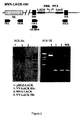
- Figure 2 shows, in its upper part, a scheme of the MVA-LACK virus and the location of the areas where the oligonucleotides used are paired as primers in the PCR analysis of the HA and TK locus (for the identification of the oligonucleotides see Table 1). Photographs of the gels obtained on performing the aforementioned PCR are shown in the lower part; the left part corresponds to the analysis of the HA locus and the right part to the analysis of the TK locus. In both, the lanes correspond to the following samples: + : pHLZ LACK; 1: VV-LACK HA - ; 2: MVA-LACK; 3: VV-LACK TK-; WR: Western Reserve strain.
- Figure 3 shows the photographs obtained after the immunostaining of the plates generated by infection of the CEF cells with the P4 stock of MVA-LACK, using an anti-LACK polyclonal antibody (photograph on the left) or anti-WR (photograph on the right).
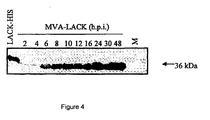
- Figure 4 shows the pattern of bands obtained after reacting an anti-LACK polyclonal antibody with the bands obtained by fractionating cell extracts in an SDS-PAGE gel harvested at the post-infection times indicated over each one of the lanes.
- M sample taken 48 hours after the infection had been simulated.
- LACK-HIS positive control sample obtained from a strain of E. coli which expresses a LACK protein which has a histidine tail.
- Figure 5 shows the number of secretor cells of IFN- ⁇ for every 10 6 splenocytes from Balb/c mice stimulated with the vectors indicated in the abscissa and detected by using ELISPOT, which are: VV p36: VV-LACK HA - ; MVAp36: MVA-LACK; VV-Luc: vector derived from the WR strain which contains the luciferase gene.
- P first dose
- B booster dose.
- Part A corresponds to the secretor cells of IFN- ⁇ specific for the LACK protein and part B to the secretor cells specific for viral antigens.
- Figure 6 shows the concentration of IFN- ⁇ , in ng/ml, detected in the supernatant of the culture of splenocytes isolated from mice inoculated with the vectors indicated on the abscissa and re-stimulated with the LACK protein (first band which appears shadowed) or with an immunostimulant class II peptide (second band, which appears filled in black).
- the names of the vectors correspond to: Wp36: VV-LACK HA-; MVAp36: MVA-LACK; VV-Luc: vector derived from the WR strain which contains the luciferase gene.
- P first dose
- B booster dose.
- Figure 7 shows the optical density values, measured at 450 nm, obtained on carrying out the detection of antibodies specific for LACK in serum of Balb/c mice diluted 1/10, collected 8 days after the second dose of a vaccination protocol with two immunisation doses, each one of which included the vectors indicated on the abscissa, P indicating the vector inoculated in the first dose and B the one inoculated in the booster dose.
- the names of the vectors correspond to: VVp36: VV-LACK HA - ; MVAp36: MVA-LACK; VV-Luc: vector derived from the WR strain which contains the luciferase gene.
- the first band of each vaccination protocol, marked with sloping lines corresponds to the antibodies to the IgG1 isotype; the second band, filled in black, to the antibodies to the IgG2a isotype.
- Figure 8 shows the structure of the pCI-neo-LACK (DNA-LACK) plasmid.
- Figure 9 shows the concentration of IFN- ⁇ for every 10 6 splenocytes of Balb/c mice stimulated with the vectors indicated on the abscissa, and detected using ELISPOT.
- P first dose
- B booster dose.
- Part A corresponds to the secretor cells of IFN- ⁇ specific for the LACK protein and part B to the secretor cells specific for viral antigens.
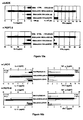
- Figure 10 shows the pattern of cytokines secreted after restimulation of splenocytes isolated from Balb/c mice immunised with combinations of recombinant vectors, in which the first vector referred to is administered in the first dose and the second vector in the second dose.
- the graphs in the upper part of the figure correspond to the results obtained after restimulating splenocytes with the LACK protein, while graphs in the lower part correspond to restimulation with a class II immunodominant peptide (PEPT-II).
- the vector combinations used to immunize the mice are those indicated next to the bands, in which DNA-CTRL: control DNA lacking the insert with the LACK encoding sequence.
- the graphs at the left show the concentration of cytokines associated with type Th2 (IL-4) responses in the case of Figure 10a and both IL-4 (shaded bands, scale on each graph) and IL-10 (empty bands, scale under each graph) in the case of Figure 10b ;
- the graphs at the right show the concentration of cytokines associated with Th1 type response detected in each case (only IFN- ⁇ in the case of Figure 10a and both IFN- ⁇ (shaded bands, scale on each graph) and TNF- ⁇ (empty bands; scale on each graph) in the case of Figure 10b .
- the data obtained after restimulation of splenocytes obtained from mice immunised with the combination of vectors DNA-LACK/VV-LACK TK are only present in Figure 10a , and have not been included in Figure 10b .
- Figure 11 corresponds to the evaluation of the protection against a challenge by L. major to Balb/c mice immunised with different vaccination protocols.
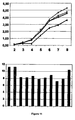
- the upper part A shows a graph in which, in order, the size of the lesion is shown, expressed in millimetres, detected after the weeks that are indicated on the abscissa after the challenge with promastygotes of L. major to Balb/c mice immunised with DNA-LACK (100 micrograms) in the first dose and, in the second dose with 1 x 10 7 pfu/mouse of: ( ⁇ ): VV-LACK TK-; ( ⁇ ): VV-LACK HA-; ( ⁇ ): MVA-LACK.
- the points marked with the (•) symbol correspond to the data of the mice that were administered a control DNA in the first dose and VV-Luc HA- in the second.
- part B the logarithm to the base 10 of the parasite load corresponding to the same experiment, detected in the popliteal ganglia of Balb/c mice, is shown.
- band 1 corresponds to the spleen mixture of 8 mice immunised with DNA-LACK/VV-LACK-TK-; band 3; the spleen mixture of 8 mice immunised with DNA-LACK/VV-LACK-HA-; bands 3 to 10 represents a mouse spleen immunised with DNA-LACK/MVA-LACK HA-; band 11, mixture of 7 spleens from mice immunised with a DNA-control (empty plasmid)/VV-Luc HA-.
- Figure 12 shows, in part A, a graph in which, on the ordinates, the size of the lesion is indicated, expressed in millimetres, detected after the weeks that are indicated on the abscissa after the challenge with promastygotes of L. major to Balb/c mice immunised with: pMOK in the first and in the second dose (data indicated by a circle, •); MIDGE-NLS in the first dose and VV-Luc in the second dose (data indicated by the symbol ); pMOK-LACK in the first dose and MVA-LACK in the second dose (data indicated by the symbol X).
- the parasite load is indicated, expressed as the number of parasites/mg, detected in the same animals; band 1 corresponds to the immunisation with pMOK-LACK and MVA-LACK, band 2 to the immunisation with MIDGE-NLS and VV-Luc and band 3 to the immunisation with pMOK in the first and in the second dose.
- Figure 13 shows a graph in which are indicated, on the ordinates, the number of IL-2 secretor cells, specific for the LACK protein, detected for every 10 6 splenocytes of Balb/c mice immunised with DNA-LACK in the first dose and 1 x 10 7 pfu/mouse of MVA-LACK in the second dose (first band); DNA-LACK in the first dose and 5 x 10 7 pfu/mouse of MVA-LACK in the second dose (second band); DNA-LACK in the first dose and 5 x 10 7 pfu/mouse of VV-LACK in the second dose (third band); Control DNA in the first dose and 5 x 10 7 pfu/mouse of VV-Luc in the second (fourth band); PBS in the first dose and in the second dose (fifth band).
- Figure 14 shows a graph in which, on the ordinates, the size of the lesion is indicated, expressed in millimetres, detected after the weeks indicated on the abscissa after the challenge with promastygotes (5 x 10 4 ) of L. major to Balb/c mice immunised with DNA-LACK in the first dose and in the second dose with: ( ⁇ ): 1 x 10 7 pfu/mouse of MVA-LACK; ( ⁇ ): 5 x 10 7 pfu/mouse of MVA-LACK; ( ⁇ ): 5 x 10 7 pfu/mouse of VV-LACK; (X): 5 x 10 7 pfu/mouse of VV-Luc.
- the points marked with the symbol ( ) correspond to the data from mice that were administered PBS in the first dose and in the second dose.
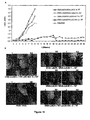
- Figure 15 shows photographs taken of the mice subjected to different immunisation protocols, 8 weeks after the challenge with Leishmania major.
- the "+” signs indicate the paws in which the promastygotes (5 x 10 4 ) were inoculated whilst the "-” signs correspond to the paws where promastygotes were not inoculated, which served as controls.
- Panel A DNA-LACK in the first dose and 1 x 10 7 pfu/mouse of MVA-LACK in the second dose
- panel B DNA-LACK in the first dose and 5 x 10 7 pfu/mouse of MVA-LACK in the second dose
- panel C DNA-LACK in the first dose and 5 x 10 7 pfu/mouse of W-LACK in the second dose
- panel D Control DNA in the first dose and 5 x 10 7 pfu/mouse of W-Luc in the second dose
- panel E PBS in the first dose and in the second dose (group N: naive ).
- Figure 16 shows graphs which represent the parasite loads detected in different organs of Balb/c mice, immunised with different treatments, detected one month after an intradermal challenge with 1 x 10 7 metacyclic promastygotes of L. infantum.
- Graph A load detected in spleen
- graph B load detected in liver
- graph C load detected in lymphatic drainage nodule.
- the bands correspond to the following vaccination protocols: first band (shaded), DNA-LACK in the first dose and 1 x 10 7 pfu/mouse of W-LACK in the second dose; second band (with reticle), DNA-LACK in the first dose and 5 x 10 7 pfu/mouse of W-LACK in the second dose; third band (with sloped lines), DNA-LACK in the first dose and 1 x 10 7 pfu/mouse of MVA-LACK in the second dose; fourth band, DNA-LACK in the first dose and 5 x 10 7 pfu/mouse of MVA-LACK in the second dose; fifth band (filled in black), pCI-neo plasmid in the first dose and 5 x 10 7 pfu/mouse of W-Luc in the second dose; last band (with vertical lines), PBS in both doses.
- the asterisks on the bands indicate there are statistically significant differences between the data: ***: p ⁇
- Figure 17 shows graphs which correspond to the immune response detected in the spleens of Balb/c mice subjected to different immunisation treatments, to those who had not been inoculated with L. infantum.
- Graph A corresponds to the levels of IFN- ⁇ , in ng/ml, detected in the supernatant of cultures of splenocytes re-stimulated with the LACK protein.
- Graph B corresponds to the number of IFN- ⁇ producer cells specific for LACK, detected using ELISPOT, per 10 6 splenocytes.
- Graph C corresponds to the levels of TNF ⁇ /LT, in ng/ml, detected in the supernatant of cultures of splenocytes re-stimulated with the LACK protein.
- the bands correspond to the following vaccination protocols: first band, DNA-LACK in the first dose and 1 x 10 7 pfu/mouse of W-LACK in the second dose; second band, DNA-LACK in the first dose and 5 x 10 7 pfu/mouse of W-LACK in the second dose; third band, DNA-LACK in the first dose and 1 x 10 7 pfu/mouse of MVA-LACK in the second dose; fourth band, DNA-LACK in the first dose and 5 x 10 7 pfu/mouse of MVA-LACK in the second dose; fifth band, pCl-neo plasmid in the first dose and VV-Luc in the second dose; last band, PBS in both doses.
- Figure 18 shows both graphs, relating to the growth efficiency of the recombinant virus MVA-LACK, graphs which represent, on the ordinates, the logarithm of the concentration of the MVA-LACK virus (data represented by shaded squares: ⁇ ) and of the wild type virus MVA-WT (data represented by ⁇ ) detected or associated with cells (Intracell: intracellular, upper graph: A) or in the growth medium (Extracell: extracellular, lower graph B) by immunostaining carried out in BHK-21 cells, after times (t) post-infection indicated in hours (h) on the abscissa.
- Figure 19 shows the lesions developed by mice inoculated with L. major promastygotes, in relation to the previous immunisation treatment.
- the higher part of the Figure, A depicts a graph in which the ordinates represent the size of the lesion (Lesn), in millimetres observed in mice (mm), after a time, expressed in weeks, indicated on the abscissa, from the moment when the mice were inoculated with L. major promastygotes; the combinations of immunisation vectors administered to each group of mice previously inoculated with L. major are indicated on the graph.
- the lower part, B shows photographs of the legs of mice submitted to different immunisation protocols, 9 weeks after challenge with Leishmania major, photographs labelled with the letter "I" indicate the legs in which the promastygotes were inoculated (5 x 10 4 ), while the letters N/I indicate those used as control, in which no promastygotes were inoculated.
- DNA-LACK/MVA-LACK 1_10 7 DNA-LACK in the first dose and 5 X 10 7 ufp/mouse of MVA-LACK in the second dose
- DNA-LACK/MVA-LACK 5_10 7 DNA-LACK in the first dose and 5 X 10 7 ufp/mouse of MVA-LACK in the second dose
- DNA-LACK/VV-LACK-HA DNA-LACK in the first dose and 5 X 10 7 ufp/mouse of VV-LACK in the second dose
- DNA-Control/VV-LUC-HA control plasmid lacking LACK encoding insert in the first dose and 5 X 10 7 ufp/mouse of VV-Luc in the second dose
- PBS/PBS PBS both in the first and the second dose.
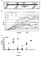
- Figure 20 corresponds to the assay to study the increased protection conferred by administering an adjuvant, CD40L, to Balb/c mice submitted to different immunisation treatments and to a challenge of L. major promastygotes.
- the upper part, A shows a schematic representation of the immunisation protocol followed.
- the intermediate part, B shows a graph representing on the ordinates the size of the lesion (lesn), in millimetres (mm) observed in the mice, after a given time period, expressed in weeks, indicated on the abscissa, from the time the mice were inoculated promastygotes of L.
- the abbreviations G1 to G7 for each line of data indicate the assay group referred to (Group 1 to Group 7), the signs ⁇ indicate the times when the mice were sacrificed and the arrow marked as B2 indicate the times when mice from groups 1,3 and 4 received their third dose of immunisation vector (2 nd booster dose "booster 2").
- the sizes of the lesions are shown in millimetres, observed in each of the surviving mice from groups 1,2,3,4,5 and 7, 27 weeks after the challenge; each circle represents a mouse, and the height at which this circle is found corresponds to the size of the lesion observed, and the group it belongs to is indicated by the reading on the abscissa.
- the signs ⁇ together with a circle from part C indicate that the mouse was sacrificed before 27 weeks.
- the groups correspond to the following immunisation combinations: Group 1 (G1): DNA-LACK/MVA-LACK/MVA-LACK; Group 2 (G2): DNA-LACK + MegaCD40L(1 ⁇ g)/MVA-LACK + MegaCD40L (1 ⁇ g); Group 3 (G3) DNA-LACK + MegaCD40L(10 ⁇ g)/MVA-LACK + MegaCD40L (10 ⁇ g)/MVA-LACK; Group 4 (G4) DNA-LACK + MegaCD40L(20 ⁇ g)/MVA-LACK + MegaCD40L (20 ⁇ g)/ MVA-LACK + MegaCD40L (20 ⁇ g); Group 5 (G5): DNA-Control/MVA-WT; Group 6 (G6): PBS/PBS ; Group 7 (G7): PBS + MegaCD40L (10 ⁇ g)/PBS + MegaCD40L (10 ⁇ g).
- the graphs depicted in Figure 21 represent the evolution over time of the concentration of anti-SLA antibodies (soluble Leishmania antigen) IgG1 type (upper part, A) or IgG2 type (upper part, B) in Beagle dogs inoculated with promastygotes of Leishmania infantum. This is done by indicating on each graph, on the ordinate axis, the absorbance value corresponding to transformation of the substrate OPD produced by plasma samples from dogs submitted to ELISA, N, the value expressed as per 1 relative to the value obtained on day 30, the day the sample was taken is indicated on the abscissa, with day 0 corresponding to the day of inoculation with the promastygotes.
- anti-SLA antibodies soluble Leishmania antigen
- Each curve corresponds to a different previous immunisation group: shaded squares ⁇ : Negative control (C(-)); shaded triangles ⁇ : Positive control (C(+)); shaded rhombi ⁇ : the group immunised with DNA-LACK/rVV-LACK before the challenge with promastygotes; six point asterisk (*): the group immunised with DNA-LACK/MVA-LACK before challenge with promastygotes.
- a plasmid pHLZ- LACK
- This plasmid contains regions flanking left and right of the haemagglutinin gene (HA), and the gene resistant to ampicillin. Between the flanking regions of the HA gene, there are two Vaccinia promoters in the opposite direction: the viral p7.5 promoter which directs the expression of the ⁇ -gal gene, and the early/late (pE/L) synthetic promoter, which directs the expression the gene of the LACK protein of L. infantum.
- pE/L early/late
- the pHLZ-LACK vector is generated from these two plasmids in the following way:
- a 942 bp fragment of DNA which contains the coding sequence of the LACK protein of L. infantum is purified from the pUC LACK plasmid.
- the pUC LACK vector is directed with the EcoRI restriction enzyme, and the fragment corresponding to the LACK gene is purified in agar gels.
- the ends of the aforementioned fragment were made blunt by treatment with Klenow and is cloned in the plasmid by insertion into pHLZ Vaccinia (previously directed with Smal and dephosphorylated by incubation with alkaline phosphatase (CIP)), thus generating the pHLZ-LACK (8342 bp) insertion vector.
- the selection of the recombinant plasmids is carried out by analysing the ⁇ -gal activity.
- CEF chicken embryo fibroblast
- MVA specifically MVA-F6, pass 586, provided by Gerd Sutter
- IVITROGEN the instructions of the manufacturer
- the recombinant viruses that contained the LACK gene of L. infantum and the ⁇ -gal gene were selected by consecutive purification passes in CEF cells and stained with 5-bromo-4-chloro-3-indolyl- ⁇ -galactosidase (300 ⁇ g/ml). After 6 purifying cycles the purified recombinant virus was obtained, without contamination of the wild MVA virus.
- the recombinant, named 3.111.2.1.1.2 (MVA pass 592) was used to prepare a second stock, P2, with a titration of 3 x 10 7 pfu/ml.
- the P3 stock was prepared from the CEF cells infected to a multiplicity of 0.05 pfu/cell and was purified through two 36% saccharose matrices. The titration of this stock is 0.975 x 10 9 pfu/ml. The sequencing of the insert present in its genome gave rise to the nucleotide sequence which is shown in SEQ ID NO:6.
- the viral DNA was purified from CEF cells infected by the MVA LACK virus (stock P2) to an infection multiplicity of 5 pfu/cell. After verifying the integrity of the DNA by analysis in agar gel, PCR analysis was carried out for the haemagglutinin and thymidine kinase locus, using the oligonucleotides shown in Table 1 as primers, and their location in relation to the haemagglutinin and thymidine kinase locus are shown in the upper part of Figure 2 .
- TK-L 5' TGATTAGTTTGATGCGATTC 3' (SEQ ID NO: 1 TK-R 5' TGTCCTTGATACGGCAG 3' (SEQ ID NO: 2) HA-1 5' GTCACGTGTTACCACGCA 3' (SEQ ID NO: 3) HA-2 5' GATCCGCATCATCGGTGG 3' (SEQ ID NO: 4) HA-MVA 5' TGACACGATTACCAATAC 3' (SEQ ID NO: 5)
- the DNA extracted from CEF cells infected with the Western Reserve (WR) wild strain was used as a negative control and the pHLZ-LACK plasmid as a positive control.
- the VV-LACK HA - recombinant virus was also included in the analysis, which was prepared in a form analogous to MVA-LACK and which contained the same LACK coding sequence equally inserted in the HA locus although, unlike MVA-LACK, the Western Reserve strain of Vaccinia, competent for replication, was used for its construction.
- VV-LACK virus whenever the VV-LACK virus is mentioned, it will refer to this recombinant virus, calling it VV-LACK-HA - in those cases where it is required to distinguish it from the VV-LACK-TK - virus, in which the LACK sequence is inserted in the thymidine kinase locus.
- the middle or low hyphen used in the names of vectors is indiscriminate and does not signify any difference between them, such that "MVA-LACK" and "MVA_LACK" correspond to the same vector.
- VV_LACK HA /"VV-LACK HA
- VV_LACK TK /VV-LACK TK
- VV_LACK /"VV-LACK
- MVA-LACK (lane 2) as well as VV-LACK-HA- (lane 1) give rise to bands of identical size to those generated from the DNA extracted from cells infected with WR wild strain, while the W-LACK-TK - vector (lane 3), which contains the same sequence but inserted in the TK locus, gives rise to a much bigger band.
- the recombinant virus was amplified from the P2 to the P4 stock in CEF cells to a multiplicity of infection of 0.05 pfu/cell.
- the plaques generated with the P4 stock were analysed by immunomarking with an anti-WR and anti-LACK polyclonal antibody.
- the results, shown in Figure 3 demonstrated that 100% of the plaques recognised by the anti-WR antibody, were also positive for LACK.
- the LACK gene therefore, remained in stable form in the recombinant MVA-LACK virus.
- mice of the susceptible Balb/C species were immunised with one (1 x 10 7 pfu/mouse) or two (5 x 10 7 pfu/mouse) doses of W-LACK HA - ; MVA-LACK or W-Luc as control. 8 days after the second immunisation, the mice were sacrificed and the spleen were processed using the ELISPOT technique.
- the results of the ELISPOT show that the number of CD8+ IFN- ⁇ secretor cells specific for LACK is 3.15 times less in the animals inoculated with MVA-LACK compared to those who received W-LACK when the received a single dose.
- the quantity of IFN- ⁇ secreted by the splenocytes re-stimulated with the protein was 5 times less.
- the number of CD8+ IFN- ⁇ secretor cells specific for the viral antigens was 5 times higher in the animals inoculated with W-LACK. The fact is that the immune response induced against the vector in the case of MVA produces a booster effect after a second immunisation dose.
- IgG2a is associated with asymptomatic conditions while the IgG1 isotype is related to disease.
- 8 days after the second immunisation serum was collected from the animals and it was evaluated for the presence of antibodies specific for LACK and its isotypes.
- the results obtained in a dilution of 1/10 of the sera are shown in Figure 7 . In them, it can be seen that the highest levels of IgG2a (associated with a Th1 response) were found after two doses with MVA-LACK.
- Example 5 Immune response generated in heterologous immunisation protocols with naked DNA in the first dose and MVA-LACK in the booster dose.
- the immune response generated by the MVA-LACK virus was also evaluated in a heterologous immunisation protocol based on a first immunisation dose with a recombinant vector of naked DNA which enables the expression of the LACK protein in mammal cells and a second dose with recombinant viruses derived from Vaccinia.
- pCI-neo-LACK plasmid was used in the first immunisations, which hereinafter will be referred to as DNA-LACK, which is an expression vector in mammal cells which like MVA-LACK expresses the LACK protein of L. infantum.
- This plasmid was generated by insertion of the LACK gene in the Smal location of pCl-neo, and it differs from that patented by the Consejo Superior de Investigativations Cientificas (Higher Council of Scientific Research) (CSIC) as a DNA-LACK vector in the LACK insertion site, as the vector of the CSIC was cloned in the EcoRI/Xbal location of pCl-neo. It contains the cytomegalovirus promoter and the genes resistant to ampicillin and neomycin, arranged as shown in Figure 8 .
- Th1 types associated with protection
- Th2 types associated with susceptibility
- the type of cellular response induced was evaluated after the immunisation described earlier.
- the splenocytes isolated from the immunised mice were re-stimulated with the LACK protein (upper part of Figure 10 ) or with a class II immunodominant peptide of the same (lower part of Figure 10 ).
- the supernatants were harvested and the presence of type Th1 (IFN- ⁇ ) or Th2 (IL-4) cytokines in these supernatants was determined using ELISA.
- the graphs of Figure 10 show that the splenocytes produced more IFN- ⁇ and IL-4 than the rest of the groups.
- Th2 type cytokines inclusion of data for IL-10 are in agreement with the highest levels detected in the groups immunized with DNA-LACK/MVA-LACK.
- the Th1:Th2 ratio corresponded to groups receiving MVA-LACK in the second dose, independently of IL-4 and IL-10 levels, confirming a clear tendency of the immune response towards Th1 type in this group.
- the next step was to determine what population of T-cells (CD4+ or CD8+) was involved in the secretion of Th1-type cytokines (IFN- ⁇ and TFN- ⁇ ).
- animals were immunised with immunisation regimes analogous to those used to obtain the results shown in Figure 8 , that is, a first response priming dose with DNA-LACK and a second booster dose with MVA-LACK (dose of 1 x 10 7 pfu or 5 x 10 7 pfu) or W-LACK-HA - (dose of 1 x 10 7 pfu or 5 x 10 7 pfu), while the animals used as controls received DNA-CONTROL (the pCl-neo plasmid vector, without the LACK sequence) in the first response priming dose and W-Luc-HA- in the second booster dose.
- DNA-CONTROL the pCl-neo plasmid vector, without the LACK sequence
- the spleens and splenocytes were collected and were re-stimulated with LACK protein or with RPMI as a control. During the last 5 hours of restimulation a new stimulation together with Brefeldin A was added. Next, the intracellular cytokines were stained and analysed by flow cytometry. The results are shown below in Table 2.
- mice that received DNA-LACK/MVA-LACK had a greater reduction in parasite load (up to 1000 times) than the groups immunised with DNA-LACK/VV-LACK.
- the animals were immunised with a DNA vector that expresses a different LACK protein of DNA-LACK, pMOK-LACK, and as controls, the minimalistic MIDGE vector (minimalistic, immunologically defined gene expression) MIDGE-NLS (MOLGEN®) or an empty vector without a pMOK insert. 15 days later, the group that had been inoculated with pMOK received a second dose of pMOK, the group that had received MIDGE-NLS received VV-Luc in the second booster response dose and the group that received pMOK-LACK received MVA-LACK.
- MIDGE vector minimalistic, immunologically defined gene expression
- the group immunised with pMOK-LACK/MVA-LACK were protected against leishmaniasis produced by L. Major , since the mice that were immunised with it hardly developed a lesion. This measurement is correlated with a decrease in the parasite load: they had 4 times less parasites than the control groups.
- Balb/c mice were again used as an animal model, on being a strain highly susceptible to infection by Leishmania.
- Nine weeks old females were inoculated intradermally with 100 ⁇ g of the DNA plasmid vector that expresses the DNA-LACK protein(pCINeo-DNA-LACK) or control DNA.
- a control group was included that received PBS in the first dose as well as in the second immunisation dose, which is called N ( naive, usual name in molecular biology to designate that which has not had contact with anything).
- the immunised groups are represented in Table 3.
- the groups immunised with 1 X 10 7 pfu of MVA-LACK in the booster dose did not develop a lesion.
- the mice immunised with 5 X 10 7 pfu of MVA-LACK one in 5 mice developed a small lesion at the inoculation site from the 7th week after the challenge. From the fourth week after the challenge the lesion presented was significantly different in the groups immunised with recombinant vectors derived from Vaccinia as compared to the controls.
- the group immunised with 5 X 10 7 pfu of VV-LACK had a lesion to a certain degree, although this was significantly different to the control group from the 8th week after the challenge.
- the lesion in the control groups continued increasing exponentially while in the group immunised with DNA-LACK/VV-LACK the lesion began to stabilise.
- the animals of the control groups were sacrificed for ethical reasons 9 weeks after the challenge. At the same time the animals of the groups immunised with DNA-LACK and VV-LACK were also sacrificed. The animals of the groups that received MVA-LACK remained with lesions for at least 11 weeks, and are under observation to analyse whether the protection is maintained over time.
- Tests were also carried out to evaluate whether the powerful induction of cellular immune response triggered by MVA-LACK when it is administered in the second booster dose of the immunisation, after the subjects had been immunised with a DNA vector which equally expresses the LACK antigen in the first dose, also correlated with the generation of protection against Leishmania infantum, which mainly causes visceral leishmaniasis, using a new heterologous immunisation protocol based on DNA-LACK/MVA-LACK.
- the murine model can be used to predict the results that might be obtained in vaccination trials carried out on non-human primate models (40, 41, 42), the intradermal murine model was used to test the potential of heterologous vaccination protocols with DNA vectors and recombinant viruses derived from Vaccinia.
- mice from 4 to 6 weeks old were inoculated intradermally with 100 ⁇ g of the DNA plasmid vector that expressed the LACK protein, DNA-LACK (pCINeo-DNA-LACK) or Control DNA. Fifteen days later they received the second immunisation dose, by the intraperitoneal route, with 1 x 10 7 pfu/mouse or 5 x 10 7 pfu/mouse of MVA-LACK or the recombinant virus derived from the Western Reserve wild strain which equally had the LACK antigen inserted in the haemagglutinin locus (VV-LACK). The group that received Control DNA were administered 5 x 10 7 pfu/mouse of VV-Luc. A control group was included which received PBS in the first and second immunisation dose.
- DNA-LACK pCINeo-DNA-LACK
- Control DNA Fifteen days later they received the second immunisation dose, by the intraperitoneal route, with 1 x 10 7
- mice were infected intradermally in the ear pavilion using 1 x 10 7 L. infantum metacyclic promastygotes as has been described previously (43).
- the parasite loads were evaluated using the analysis by limiting dilution in the immunised and control mouse groups. The evaluations were carried on the spleen, liver and lymphatic drainage nodule. The results obtained, which are shown in Figure 16 , in which the mean values obtained from at least 4 mice per group are displayed, demonstrating that the mice immunised following a vaccination protocol with a first response priming dose and a second booster dose using vectors capable of expressing the LACK antigen were protected significantly against infection.
- the level of protection in each tissue was comparable between the different groups of mice who had received vectors capable of expressing the antigen and it did not differ statistically between the mice that received VV-LACK or MVA-LACK.
- variations were observed in the levels of protection between the different organs, with higher levels of protection being observed in the lymphatic drainage nodule (part C of Figure 16 ).
- the level of protection in this organ varied between a reduction factor of 144 to 244 times in the parasite loads compared with control mice.
- the spleen part A of Figure 15
- the liver part B of Figure 16
- lower levels of protection were observed, which varied between reduction factors of 6 to 9 times in the parasite loads in the liver and 9 to 30 times in the spleen.
- Part A corresponding to the concentration of IFN- ⁇ , in ng/ml, detected in the supernatants of re-stimulated splenocyte cultures, shows that the mice that received 1 x 10 7 pfu of VV-LACK or 5 x 10 7 pfu of MVA-LACK seem to produce somewhat higher levels of IFN- ⁇ (100-113 ng/ml), compared with the mice that received 5 x 10 7 pfu of VV-LACK or 1 x 10 7 pfu of MVA-LACK (55-67 ng/ml).
- IFN- ⁇ producer cells specific for LACK carried out by ELISPOT, which is shown in part B of Figure 17 , indicates that the number IFN- ⁇ secretor cells correlates with the levels of IFN- ⁇ detected by ELISA, the frequency of IFN- ⁇ producer cells varying between 380 and 640 cells/10 6 splenocytes.
- Significant levels of TNF- ⁇ /LT (58 and 134 pg/ml, respectively) were also observed in the mice that had received W-LACK, while the TNF- ⁇ /LT levels produced in response to the LACK antigen by the mice that had received MVA-LACK were lower (27 pg/ml and 8 pg/ml, respectively).
- TNF- ⁇ induction can reflect, partly, the different capacity of the WR and MVA to induce inflammatory responses and activation of NF- K B, which results in different cytokine profiles: MVA increases the activation of NF- K B while WR appears to inhibit it.
- the quantities of IL-10 produced by the splenocytes isolated before the challenge shown in part D of Figure 17 , also varied depending on the immunisation protocol used, varying between 0.1 ng/ml in mice who had received 5 x 10 7 pfu of VV-LACK in the booster dose and 0.7 ng/ml in those that had received 1 x 10 7 of VV-LACK or MVA-LACK.
- cytokine responses detected in splenocytes isolated one month after the mice were subjected to a challenge with L. infantum showed variations between samples parallel to those found before the challenge, although they were somewhat higher.
- nitric oxide is critical for the leishmanicide activity of murine macrophages (44, 45, 46). It was observed that, in the mice that had received vectors capable of expressing LACK, significant quantities of this antimicrobial agent were detected, which varied between 6 and 7 ⁇ M, whilst in the control groups the NO/nitric levels were much lower, varying between 0.5 ⁇ M and 1 ⁇ M. Therefore, the vaccinated mice, in keeping with the levels of IFN- ⁇ detected, had higher levels of inducing the production of nitric oxide and a higher leishmanicide potential. These results are consistent with the protection found in the mice vaccinated with DNA-LACK and VV-LACK or MVA-LACK.
- the tests described in this example demonstrate that the administration of recombinant viruses derived from Vaccinia capable of expressing the LACK antigen as part of immunisation protocols where a second booster dose is administered, while a DNA vector, which expresses the same antigen, is highly immunogenic and provides protection against L. infantum in mice is administered in the first dose, while the protector effect is not achieved with vaccinations with DNA vectors in the two doses.
- the vector derived from the highly attenuated MVA strain and that derived from the virulent strain of Vaccinia competent for Western Reserve replication gave rise to comparable levels of protection.
- the highly attenuated MVA strain guarantees higher safety for its use in human beings makes this vector a good candidate to be used for the protection of human beings against visceral leishmaniasis.
- An additional assay was performed to determine the growth efficiency of the recombinant virus MVA-LACK.
- permissive cells were infected (BHK-21) at a multiplicity of infection of 0.01, either with the recombinant virus MVA-LACK or with the parental virus lacking inserts, MVA-WT.
- the virus present in the growth medium was titrated (extracellular) and the virus associated to cells (intracellular) by immunostaining of the virus.
- Table 6 Innmunization groups of protection duration test INITIAL DOSE BOOSTER DOSE GROUP 1 DNA-LACK 100 ⁇ g MVA-LACK 1 x 10 7 ufp GROUP 2 DNA-LACK 100 ⁇ g MVA-LACK 5 x 10 7 ufp GROUP 3 DNA-LACK 100 ⁇ g VV-LACK 5 x 10 7 ufp GROUP 4 DNA-CONTROL 100 ⁇ g VV-LUC 5 x 10 7 ufp GROUP 5 PBS PBS
- the immune response was challenged by inoculation of 5 x 10 4 metacyclic promastygotes of Leishmania major, isolated from stationary cultures of Leishmania after incubation with peanut aglutinine.
- the promastygotes were inoculated subcutaneously in the plantar pad of the right hind leg.
- the evolution of the lesions in the plantar pad where the inoculate had been introduced was followed, as a parameter to evaluate protection, taking measurements weekly.
- the assay was prolonged for 30 weeks after the challenge, to establish whether the protection conferred by immunisation was extended in time.
- the upper part (A) of Figure 19 depicts a graph which represents the evolution of the size of the lesions with time after the challenge. Corroborating the results obtained in the analogous experiments performed, all the animals in the control groups, Group 4 (DNA-control/VV-LUC) and Group 5 (PBS/PBS)developed severe lesions, as can be observed in the photographs shown in part B of Figure 19 , taken 8 weeks after the challenge. In week 9, the control animals had to be sacrificed for ethical reasons. The animals that received vectors containing LACK inserts, mice immunized with MVA-LACK, showed, once again, a higher degree of protection than mice immunized with W-LACK (W-LACK-HA- in Figure 19 ).
- mice were immunized with a first dose of DNA-LACK, followed by a dose of MVA-LACK, inoculating animals one day after each of the doses of vaccination vector, with different amounts of adjuvant, namely the protein CD40L(Mega CD40L;m-ACRP30m-CD40L, from APOXIS).
- adjuvant namely the protein CD40L(Mega CD40L;m-ACRP30m-CD40L, from APOXIS).
- mice The animals, Balb/c mice, were immunised intradermically on day 0 with 100 ⁇ g of plasmid DNA-LACK or of the plasmid DNA-Control, without the LACK insert, and one day later the animals received the adjuvant by the same route (1, 10 or 20 ⁇ g of Mega CD40L, according to the group). Fifteen days later, the mice received the booster dose with 1 x 10 7 ufp/mouse of MVA-LACK or MVA-WT intraperitoneally, and one day later they were inoculated by the same route with different amounts of adjuvant (1, 10 or 20 ⁇ g of Mega CD40L, depending on the group).
- mice were inoculated with 5 x 10 4 metacyclic promastygotes of L. major.
- Table 7 ten weeks after the challenge (15 weeks after having been administered the initial dose), the mice from groups 1, 3 and 4 received a second booster dose with 5 x 10 7 ufp of MVA-LACK; in the case of group 4, together with this second booster dose, the mice also received 20 ⁇ g of adjuvant, also intraperitoneally.
- the upper part A of Figure 20 shows a schematic representation of the immunization protocol followed.
- the size of the lesions was monitored weekly, obtaining the results shown in the graph appearing in the middle of Figure 20 , labelled "B".
- the graph of the lower part of Figure 20 labelled “C” shows the size of the lesions measured in each of the mice surviving from groups 1,2,3,4,5 and 7, 27 weeks after the challenge. Mice from group 6 that had only received PBS before the challenge, had to be sacrificed 12 weeks after the challenge, as also occurred with one mouse from group 2 after the same period of time, and also with one mouse from group 5 a week later.
- mice 1 and 3 For the groups immunized with DNA-LACK and MVA-LACK without adjuvant (groups 1 and 3), only one mouse from each group developed a lesion; in group 3, the mouse had to be sacrificed before the 27 week period after challenge had elapsed because of the large size of the lesion. The mice from these two groups that did not develop a lesion (75%), parasitic growth was controlled, as shown by the fact that they did not develop lesions 27 weeks after the challenge and also because they did not present any inflammation of the draining lymphatic nodule. Group 1, to which adjuvant was not administered, administration of the second booster dose after the challenge helped to control parasite replication.
- Group 1 Dogs of all the groups, except Group 1 (negative control), were inoculated intravenously, with 10 8 promastygotes of Leishmania infantum in 0.5 ml saline solution, obtained as specified in section 11.2. The day of the inoculation of the parasites was considered as day 0. Table 8 gives a summary of the inoculations received by each group. Table 8: Groups for dog vaccination test INITIAL DOSE (Day 30) BOOSTER DOSE (Day 15) CHALLENGE (Day 0) GROUP 1 (5 dogs) - - 0,5 ml Saline solution GROUP 2 (3 dogs) - - L.
- the promastygotes of L.infantum used to trigger the experimental infection were obtained from a dog in Zaragoza, infected naturally with L.infantum, which had not received any treatment (MON 1/MCAN/ES/01/LLM 996, Parasitology Reference Service, Majadahonda, Spain).
- the parasites were obtained by aspiration of bone marrow and popliteal lymph nodes, they were grown in NNN medium (Novy-Nicolle-McNeal), a medium prepared in two steps, after which they were multiplied in RPMI (Sigma, United Kingdom) supplemented with 2 mM glutamine, 100 ⁇ g/ml of streptomycin and 100 U/ml of penicillin containing 10% heat-inactivated fetal calf serum (FCS)(Sigma-United Kingdom).
- FCS heat-inactivated fetal calf serum
- L.infantum promastygotes were also grown in RPMI medium containing 10% fetal calf serum at 26°C, to be used in the direct agglutination and in the immunofluorescence assay.
- the promastygotes were collected at 3600 x g for 10 minutes at 4°C. After rinsing 5 times, 20 volumes of trypsin were added to the sediment (0.4% p/v, from Difco), the mixture was incubated at 37°C for 45 minutes and then rinsed 5 more times in cold Locke solution (NaCl 154 mM, KCI 6 mM, NaHCO 3 2 mM pH 7.7). The cells were counted and resuspended to reach a final concentration of 1 x 10 8 cells/ml.
- the soluble Leishmania antigen was prepared by growing the promastygotes in RPMI 1640 medium (Gibco) supplemented with 10% heat-inactivated FCS, 100 ⁇ g/ml of streptomycin and 100 UI/ml of penicillin (Gibco).
- the parasites were collected at the end of the logarithmic phase, rinsed in PBS and fragmented by three freezing-thawing cycles. Then, the parasites were sonicated and centrifuged at 16000 x g for 3 minutes at 4°C, collecting the supernatant. Then, the concentration of proteins was determined using the Bradford method with the Bio-Rad Protein Assay kit (BioRad Laboratories).
- the dogs were checked regularly for signs of parasitic infection and development of the disease by carrying out routine examinations to search for classical clinical signs such as cutaneous lesions, alopecia, the presence of popliteal lymphodenopathies, weight loss, ulceration of the skin and pallor of the mucosa.
- blood samples were taken from the animals every two weeks for biochemical and serology tests and for cytokine mRNA tests. After this, and until the end of the experiment, blood samples were taken monthly.
- DAT direct agglutination test
- IIF indirect immunofluorescence serology test
- test was performed on a V-shaped 96-well microtitre plate (Costar, US). A total of 50 ⁇ l of parasite suspension were added to each well, which had been previously loaded with serial dilutions of dog serum, with an initial dilution of 1:100. The plates were shaken gently for 1 minute and were then incubated at RT in a moist chamber overnight. The presence of blue aggregates was detected by direct observation (two independent measurements), correlated with serum dilutions. Values higher than 1:800 were considered as positive.
- the dog serum to be tested is made to react with a preparation of Leishmania on a slide, incubated at 4°C for 30-45 minutes and then the preparation is incubated with an anti-dog serum conjugated with fluoresceine, for at least an hour, so that if there is a specific antibody present in the dog serum that can recognise the Leishmania parasite, this will be labelled green.
- Sera with a value of 1/80 or above will be given a positive value (55,56).
- a test was performed to determine IgG1 and IgG2 in the plasma samples obtained from the blood samples extracted over the study period. Specifically, determination of specific antibodies was carried out by ELISA of the specific antibodies against the soluble Leishmania antigen (SLA), obtained as described in section 11.2. Briefly, the ELISA plates were coated with 10 ⁇ g/ml of SLA blocked with PBS containing 1% BSA and afterwards incubated with 100 ⁇ l of dog serum diluted 1:100.
- SLA soluble Leishmania antigen
- HRP-conjugated antibodies 100 ⁇ l/well of the following HRP-conjugated antibodies were added:goat anti-dog IgG1 antibody (1:15000) or a sheep anti-dog IgG1 antibody (1:20000) both from Bethyl Laboratories (Montgomery, TX, US).
- the antibodies were incubated with the OPD substrate (Zymed laboratories, Invitrogen Immunidetection). Absorbance was read at 450 nm, arbitrarily assigning a value of 1 in each group to the mean value of the samples obtained on day 30 of the experiment and calculating the value n for the remaining samples, corresponding to an increase per one in absorbance compared to the value obtained on day 30.
- the results indicate that vaccination with DNA-LACK and MVA-LACK confer, to the dogs they are administered to, protection against the development of the disease similar to that conferred by vaccination with DNA-LACK and rVV-LACK, obtaining in both cases experimental protection of at least 75% compared to positive controls, of which 100% presented the infection and clear clinical signs of the disease.
- the experiment confirms the validity of MVA-LACK as an alternative to the recombinant vectors derived from virulent strains of Vaccinia (as is the case of rVV-LACK) for the vaccination of mammals suspectible to being infected with Leishmania, especially for dogs.
- MVA-LACK is an attenuated virus it represents a much safer alternative to these other vectors.
Landscapes
- Health & Medical Sciences (AREA)
- Life Sciences & Earth Sciences (AREA)
- Chemical & Material Sciences (AREA)
- Genetics & Genomics (AREA)
- Organic Chemistry (AREA)
- Engineering & Computer Science (AREA)
- Zoology (AREA)
- General Health & Medical Sciences (AREA)
- Bioinformatics & Cheminformatics (AREA)
- Wood Science & Technology (AREA)
- Medicinal Chemistry (AREA)
- Biotechnology (AREA)
- General Engineering & Computer Science (AREA)
- Biomedical Technology (AREA)
- Biochemistry (AREA)
- Microbiology (AREA)
- Immunology (AREA)
- Veterinary Medicine (AREA)
- Biophysics (AREA)
- Virology (AREA)
- Molecular Biology (AREA)
- Public Health (AREA)
- Pharmacology & Pharmacy (AREA)
- Tropical Medicine & Parasitology (AREA)
- Animal Behavior & Ethology (AREA)
- Physics & Mathematics (AREA)
- Plant Pathology (AREA)
- Chemical Kinetics & Catalysis (AREA)
- General Chemical & Material Sciences (AREA)
- Nuclear Medicine, Radiotherapy & Molecular Imaging (AREA)
- Gastroenterology & Hepatology (AREA)
- Proteomics, Peptides & Aminoacids (AREA)
- Epidemiology (AREA)
- Toxicology (AREA)
- Mycology (AREA)
- Communicable Diseases (AREA)
- Oncology (AREA)
- Medicines Containing Antibodies Or Antigens For Use As Internal Diagnostic Agents (AREA)
- Medicines Containing Material From Animals Or Micro-Organisms (AREA)
- Micro-Organisms Or Cultivation Processes Thereof (AREA)
Abstract
Description
- The present invention refers to the use of recombinant viruses based on the modified vaccinia Ankara (MVA) virus in a vaccination. More specifically, the invention refers to recombinant viruses derived from MVA which act as systems for the expression of the LACK protein or immunogenic fragments of the same and their use in the vaccination against leishmaniasis both in human beings as well as in other affected mammals, as dogs.
- Leishmaniasis is an anthropozoonosis which includes a complex group of clinical pictures produced by protozoa of the Leishmania genus, which act as parasites on cells of the monocyte-macrophage system. Leishmania is a flagellated protozoon which belongs to the Kinetoplastida order and the Trypanosomatidae family. The classification of the Leishmania genus into its different species is complex and currently it is carried out by using analysis of the restriction fragments of the DNA of Kinetoplast. It is as follows:
      ORDER: Kinetoplastida
      FAMILY: Trypanosomatidae
      GENUS: Leishmania
            SUB-GENUS: Leishmania
            Complex: L. donovani
                   L. donovani; L. infantum; L. chagasi: L. archibaldi
            Species outside the L. donovani complex
                   L. tropica; L. aethiopica; L. major; L. gerbilli
            Complex: L. mexicana
                   L. mexicana; L. amazonensis; L. venezuelensis; L.
 aristidesi
            SUB-GENUS: Viannia
            Complex: L. braziliensis
                   L. braziliensis; L. panamensis; L. guyanensis; L.
 peruviana


- pUC LACK: a plasmid derived form the pUC19 vector which contains the gene of the LACK protein of L. infantum in the position of the EcoRI cut, besides the elements required for the replication and selection in E. coli. This vector was generated by Dr. Gloria González-Aseguinolaza from a mould of genomic DNA of L. infantum using PCR. The PCR product has a length of 942 base pairs (bp) and the codifying region 312 amino acids (7).
- pHLZ: (7400 bp). A plasmid generated in the laboratory of the inventors which enables the insertion of genes into the Vaccinia genome by homologous recombination in the encoding region of HA. After the addition of the β- galactosidase substrate, X-gal, the development of blue plaques enables the recombinant viruses to be selected.
| Oligonucleotides | Sequence | |
| TK-L | 5' TGATTAGTTTGATGCGATTC 3' | (SEQ ID NO: 1 |
| TK-R | 5' TGTCCTTGATACGGCAG 3' | (SEQ ID NO: 2) |
| HA-1 | 5' GTCACGTGTTACCACGCA 3' | (SEQ ID NO: 3) |
| HA-2 | 5' GATCCGCATCATCGGTGG 3' | (SEQ ID NO: 4) |
| HA-MVA | 5' TGACACGATTACCAATAC 3' | (SEQ ID NO: 5) |
| INITIAL DOSE | BOOSTER DOSE | IFN-γ CD8+ PRODUCING CELLS | TNF-α CD8+ PRODUCING CELLS | IFN-γ CD4+ PRODUCING CELLS | TNF-α CD4+ PRODUCING CELLS |
| DNA-LACK | W-LACK 1 | 10.1 | 10.4 | 1.8 | 1.6 |
| DNA-LACK | VV-LACK 5 | 8.3 | 7.5 | 1.9 | 1.9 |
| DNA-LACK | MVA-LACK 1 | 23.4 | 15.7 | 2.9 | 1.9 |
| DNA-LACK | MVA-LACK 5 | 12.7 | 11.2 | 2.5 | 1.9 |
| DNA CONTROL | W-Luc 5 | 1.7 | 1.5 | 0.7 | 1.0 |
| PBS | PBS | 1.5 | 1.2 | 0.3 | 0.4 |
| INITIAL DOSE | | |
| GROUP | ||
| 1 | DNA- | MVA-LACK 1 X 10⁷ |
| GROUP | ||
| 2 | DNA- | MVA-LACK 5 X 10⁷ |
| GROUP | ||
| 3 | DNA- | VV-LACK 5 X 10⁷ |
| GROUP | ||
| 4 | | VV-LUC 5 X 10⁷ pfu |
| GROUP N | PBS | PBS |
| INITIAL DOSE | BOOSTER DOSE | SIZE OF LESION | % REDUCTION |
| DNA-LACK | MVA-LACK 1 | 0.10 ± 0.05 | 97.4 % |
| DNA-LACK | MVA-LACK 5 | 0.36 ± 0.33 | 90.3 % |
| DNA-LACK | W-LACK 5 | 2.17 ± 1.64 | 41.8 % |
| DNA CONTROL | VV-Luc 5 | 3.73 ± 0.23 | 0 % |
| Experimental Group | IFN-γ/IL-10 Ratio | IFN-γ (ng/ml ) | Reduction factor of parasite load Spleen Liver Lymphatic nodule | ||
| DNA-LACK + 5 X 10⁷ MVA-LACK | 4633 | 204 | 31 | 9 | 244 |
| DNA-LACK + 1 X 10⁷ VV-LACK | 720 | 153 | 18 | 6.5 | 244 |
| DNA-LACK + 1 X 10⁷ MVA-LACK | 271 | 147 | 12.8 | 6.7 | 203 |
| DNA-LACK + 5 X 10⁷ W-LACK | 92 | 20 | 9.2 | 6 | 144 |
| INITIAL DOSE | | |
| GROUP | ||
| 1 | DNA- | MVA-LACK 1 x 10⁷ |
| GROUP | ||
| 2 | DNA- | MVA-LACK 5 x 10⁷ |
| GROUP | ||
| 3 | DNA- | VV-LACK 5 x 10⁷ |
| GROUP | ||
| 4 | DNA- | VV-LUC 5 x 10⁷ |
| GROUP | ||
| 5 | PBS | PBS |
| INITIAL DOSE (Week 0) | BOOSTER DOSE (Week 2) | 2nd BOOSTER DOSE (Week 15) | |
| | DNA-LACK (100 µg) | MVA-LACK (1 × 10⁷ ufp) | MVA-LACK (5 x 10⁷ ufp) |
| | DNA-LACK (100 µg) Mega CD40L (1 µg) | MVA-LACK (1 x 10⁷ ufp) Mega CD40L (1 µg) | - |
| | DNA-LACK (100 µg) Mega CD40L (10 µg) | MVA-LACK (1 × 10⁷ ufp) Mega CD40L (10 µg) | MVA-LACK (5 x 10⁷ ufp) |
| | DNA-LACK (100 µg) Mega CD40L (20 µg) | MVA-LACK (1 x 10⁷ ufp) Mega CD40L (20 µg) | MVA-LACK (5 x 10⁷ ufp) Mega CD40L (20 µg) |
| | DNA-CONTROL (100 µg) Mega CD40L (10 µg) | MVA-WT(1 x 10⁷ ufp) Mega CD40L (10 µg) | - |
| | PBS | PBS | - |
| | PBS Mega CD40L (10 µg) | PBS Mega CD40L (10 µg) | - |
- The first group, used as a negative control, were neither vaccinated nor inoculated with L. infantum. Dogs from this group received 0.05 ml of saline solution intravenously in one day (47,48).
- The second group was used as a positive control. The dogs were inoculated with L. infantum but were not vaccinated.
- The third group was vaccinated with two doses of the LACK encoding gene, once on
day 30 with the DNA-LACK plasmid (100µg) and the second time onday 15 with the recombinant virus derived from vaccinia rVV-LACK (10 8ufp/dog). - The fourth group was inoculated with a dose of DNA-LACK (100µg)on
day 30 and a second dose of the LACK encoding gene by the MVA-LACK virus (108 ufp/dog).
| INITIAL DOSE (Day 30) | BOOSTER DOSE (Day 15) | CHALLENGE (Day 0) | |
| GROUP 1 (5 dogs) | - | - | 0,5 ml Saline solution |
| GROUP 2 (3 dogs) | - | - | L. infantum (1 x 10⁸ ufp) |
| GROUP 3 (4 dogs) | DNA-LACK (100 µg) | rVV-LACK (1 x 10⁸ ufp) | L. infantum (1 x10⁸ ufp) |
| GROUP 4 (4 dogs) | DNA-LACK (100 µg) | MVA-LACK (1 × 10⁸ ufp) | L. infantum (1 x10⁸ ufp) |

| GROUP | Dog | Serology | Presence of parasites | Infection/ Disease | ||||
| DAT | IIF | Isolation | Biopsy* | |||||
| Positive Control (2) | I8 | 1/6400 (406) | 1/1280(406) | (426) | (406) | Yes | ||
| D9 | 1/1600(320) | 1/5120 (320) | (436) | (330) | Yes | |||
| O1 | 1/800(260) | 1/5120 (260) | (436) | (290) | Yes | |||
| O2 | 1/6400 (289) | 1/640(320) | (426) | (426) | Yes | |||
| O3 | 1/3200 (320) | 1/1240(320) | (436) | (426) | Yes | |||
| Negative Control (1) | O4 | - | - | - | - | No | ||
| O5 | - | - | - | - | No | |||
| O6 | - | - | - | - | No | |||
| DNA-LACK/ rW-LACK (3) | I9 | 1/1600(410) | 1/160(350) | (-) | (436) | Yes | ||
| D4 | - | - | - | - | No | |||
| D5 | - | - | - | - | No | |||
| D7 | - | - | - | - | No | |||
| DNA-LACK/ MVA-LACK (4) | I2 | - | - | - | - | No | ||
| I3 | - | - | - | - | No | |||
| I7 | - | - | - | - | No | |||
| D3 | 1/1600 (320) | 1/640(320) | - | (436) | Yes | |||
- 1. Ahmed, S. B., C. Bahloul, C. Robbana, S. Askri, and K. Dellagi. 2004. A comparative evaluation of different DNA vaccine candidates against experimental murine leishmaniasis due to L. major. Vaccine 22:1631-9.
- 2. Altenburger, W., C. P. Suter, and J. Altenburger. 1989. Partial deletion of the human host range gene in the attenuated vaccinia virus MVA. Arch Virol 105:15-27.
- 3. Berrahal, F., C. Mary, M. Roze, A. Berenger, K. Escoffier, D. Lamouroux, and S. Dunan. 1996. Canine leishmaniasis: identification of asymptomatic carriers by polymerase chain reaction and immunoblotting. Am J Trop Med Hyg 55:273-7.
- 4. Blanchard, T. J., A. Alcami, P. Andrea, and G. L. Smith. 1998. Modified vaccinia virus Ankara undergoes limited replication in human cells and lacks several immunomodulatory proteins: implications for use as a human vaccine. J Gen Virol 79 (Pt 5): 1159-67.
- 5. Caver, T. E., T. D. Lockey, R. V. Srinivas, R. G. Webster, and J. L. Hurvitz. 1999. A novel vaccine regimen utilizing DNA, vaccinia virus and protein immunizations for HIV-1 envelope presentation. Vaccine 17:1567-72.
- 6. Drexler, I., K. Heller, B. Wahren, V. Erfle, and G. Sutter. 1998. Highly attenuated modified vaccinia virus Ankara replicates in baby hamster kidney cells, a potential host for virus propagation, but not in various human transformed and primary cells. J Gen Virol 79 (Pt 2):347-52.
- 7. Gonzalez-Aseguinolaza, G., S. Taladriz, A. Marquet, and V. Larraga. 1999. Molecular cloning, cell localization and binding affinity to DNA replication proteins of the p36/LACK protective antigen from Leishmania infantum. Eur J Biochem 259:909-16.
- 8. Gonzalo, R. M., G. del Real, J. R. Rodriguez, D. Rodriguez, R. Heljasvaara, P. Lucas, V. Larraga, and M. Esteban. 2002. A heterologous prime-boost regime using DNA and recombinant vaccinia virus expressing the Leishmania infantum P36/LACK antigen protects BALB/c mice from cutaneous leishmaniasis. Vaccine 20:1226-31.
- 9. Gonzalo, R. M., J. R. Rodriguez, D. Rodriguez, G. Gonzalez-Aseguinolaza, V. Larraga, and M. Esteban. 2001. Protective immune response against cutaneous leishmaniasis by prime/booster immunization regimens with vaccinia virus recombinants expressing Leishmania infantum p36/LACK and IL-12 in combination with purified p36. Microbes Infect 3:701 - 11.
- 10. Gradoni, L. 2001. An update on antileishmanial vaccine candidates and prospects for a canine Leishmania vaccine. Vet Parasitol 100:87-103.
- 11. Gurunathan, S., D. L. Sacks, D. R. Brown, S. L. Reiner, H. Charest, N. Glaichenhaus, and R. A. Seder. 1997. Vaccination with DNA encoding the immunodominant LACK parasite antigen confers protective immunity to mice infected with Leishmania major. J Exp Med 186:1137-47.
- 12. Hanke, T., T. J. Blanchard, J. Schneider, C. M. Hannan, M. Becker, S. C. Gilbert, A. V. Hill, G. L. Smith, and A. McMichael. 1998. Enhancement of MHC class I-restricted peptide-specific T cell induction by a DNA prime/MVA boost vaccination regime. Vaccine 16:439-45.
- 13. Hanke, T., and A. McMichael. 1999. Pre-clinical development of a multi-CTL epitope-based DNA prime MVA boost vaccine for AIDS. immune) Lett 66:177-81.
- 14. Hanke, T., V. C. Neumann, T. J. Blanchard, P. Sweeney, A. V. Hill, G. L. Smith, and A. McMichael. 1999. Effective induction of HIV-specific CTL by multi-epitope using gene gun in a combined vaccination regime. Vaccine 17:589-96.
- 15. Kent, S. J., A. Zhao, S. J. Best, J. D. Chandler, D. B. Boyle, and I. A. Ramshaw. 1998. Enhanced T-cell immunogenicity and protective efficacy of a human .
- 16. Kim, J. J., M. L. Bagarazzi, N. Trivedi, Y. Hu, K. Kazahaya, D. M. Wilson, R. Ciccarelli, M. A. Chattergoon, K. Dang, S. Mahalingam, A. A. Chalian, M. G. Agadjanyan, J. D. Boyer, B. Wang, and D. B. Weiner. 1997. Engineering of in vivo immune responses to DNA immunization via codelivery of costimulatory molecule genes. Nat Biotechnol 15:641-6.
- 17. Lanotte, G., J. A. Rioux, J. Perieres, and Y. Vollhardt. 1979. [Ecology of leishmaniasis in the south of France. 10. Developmental stages and clinical characterization of canine leishmaniasis in relation to epidemiology. (author's transl)]. Ann Parasitol Hum Comp 54:277-95.
- 18. Li, S., M. Rodrigues, D. Rodriguez, J. R. Rodriguez, M. Esteban, P. Palese, R. S. Nussenzweig, and F. Zavala. 1993. Priming with recombinant influenza virus followed by administration of recombinant vaccinia virus induces CD8+ T-cell-mediated protective immunity against malaria. Proc Natl Acad Sci U S A 90:5214-8.
- 19. Liew, F. Y., and C. A. O'Donnell. 1993. Immunology of leishmaniasis. Adv Parasitol 32:161-259.
- 20. Lopez-Fuertes, L., E. Perez-Jimenez, A. J. Vila-Coro, F. Sack, S. Moreno, S. A. Konig, C. Junghans, B. Wittig, M. Timon, and M. Esteban. 2002. DNA vaccination with linear minimalistic (MIDGE) vectors confers protection against Leishmania major infection in mice. Vaccine 21:247-57.
- 21. Lopez-Velez, R., J. A. Perez-Molina, A. Guerrero, F. Baquero, J. Villarrubia, L. Escribano, C. Bellas, F. Perez-Corral, and J. Alvar. 1998. Clinicoepidemiologic characteristics, prognostic factors, and survival analysis of patients coinfected with human immunodeficiency virus and Leishmania in an area of Madrid, Spain. Am J Trop Med Hyg 58:436-43.
- 22. Mauricio, I. L., J.R Stothard, and M. A. Miles. 2000. The strange case of Leishmania chagasi. Parasitol Today 16:188-9.
- 23. Mayr, A., H. Stickl, H. K. Muller, K. Danner, and H. Singer. 1978. [The smallpox vaccination strain MVA: marker, genetic structure, experience gained with the parenteral vaccination and behavior in organisms with a debilitated defence mechanism (author's transl)]. Zentralbl Bakteriol [B] 167:375-90.
- 24. McMahon-Pratt, D., D. Rodriguez, J. R. Rodriguez, Y. Zhang, K. Manson, C. Bergman, L. Rivas, J. F. Rodriguez, K. L. Lohman, N. H. Ruddle, et al. 1993. Recombinant vaccinia viruses expressing GP46/M-2 protect against Leishmania infection. Infect Immun 61:3351-9.
- 25. Meyer, H., G. Sutter, and A. Mayr. 1991. Mapping of deletions in the genome of the highly attenuated vaccinia virus MVA and their influence on virulence. J Gen Virol 72 (Pt 5):1031-8.
- 26. Paoletti, E. 1996. Applications of pox virus vectors to vaccination: an update. Proc Natl Acad Sci U S A 93:11349-53.
- 27. Ramirez, J. C., M. M. Gherardi, and M. Esteban. 2000. Biology of attenuated modified vaccinia virus Ankara recombinant vector in mice: virus fate and activation of B- and T-cell immune responses in comparison with the Western Reserve strain and advantages as a vaccine. J Virol 74:923-33.
- 28. Robinson, H. L., D. C. Montefiori, R. P. Johnson, K. H. Manson, M. L. Kalish, J. D. Lifson, T. A. Rizvi, S. Lu, S. L. Hu, G. P. Mazzara, D. L. Panicali, J. G. Hemdon, R. Glickman, M. A. Candido, S. L. Lydy, M. S. Wyand, and H. M. McClure. 1999. Neutralizing antibody-independent containment of immunodeficiency virus challenges by DNA priming and recombinant pox virus booster immunizations. Nat Med 5:526-34.
- 29. Sacks, D., and N. Noben-Trauth. 2002. The immunology of susceptibility and resistance to Leishmania major in mice. Nat Rev Immunol 2:845-58.
- 30. Sedegah, M., T. R. Jones, M. Kaur, R. Hedstrom, P. Hobart, J. A. Tine, and S. L. Hoffman. 1998. Boosting with recombinant vaccinia increases immunogenicity and protective efficacy of malaria DNA vaccine. Proc Natl Acad Sci U S A 95:7648-53.
- 31. Solano-Gallego, L., J. Llull, G. Ramos, C. Riera, M. Arboix, J. Alberola, and L. Ferrer. 2000. The Ibizian hound presents a predominantly cellular immune response against natural Leishmania infection. Vet Parasitol 90:37-45.
- 32. Sutter, G., and B. Moss. 1992. Nonreplicating vaccinia vector efficiently expresses recombinant genes. Proc Natl Acad Sci U S A 89:10847-51.
- 33. Tapia, E., E. Perez-Jimenez, L. Lopez-Fuertes, R. Gonzalo, M. M. Gherardi, and M. Esteban. 2003. The combination of DNA vectors expressing IL- 12 + IL- 18 elicits high protective immune response against cutaneous leishmaniasis after priming with DNA-p36/LACK and the cytokines, followed by a booster with a vaccinia virus recombinant expressing p36/LACK. Microbes Infect 5:73-84.
- 34. Xu, D., and F. Y. Liew. 1994. Genetic vaccination against leishmaniasis. Vaccine 12:1534-6.
- 35. Zavala, F., M. Rodrigues, D. Rodriguez, J. R. Rodriguez, R. S. Nussenzweig, and M. Esteban. 2001. A striking property of recombinant poxviruses: efficient inducers of in vivo expansion of primed CD8(+) T cells. Virology 280:155-9.
- 36. Melby, P.C., J. Yang, W. Zhao, L.E. Pérez and J. Cheng. 2001. Leishmania donovani p36(LACK) DNA vaccine is highly immunogenic but not protective against experimental visceral leishmaniasis. Infect Immun 69:4719-4725.
- 37. Moss, B. 1996. Genetically engineered poxviruses for recombinant gene expression, vaccination, and safety. Proc. Natl. Acad. Sci USA 93:11341-11348.
- 38. Seki, M., M. Oie, Y. Ichihashi, and H. Shida. 1990. Hemadsoption and fusion inhibition activities of hemagglutinin analyzed by vaccinia virus mutants. Virology 175, 372-384.
- 39. Chakrabarti, S. J.R. Sisler, B. Moss. 1997. Compact, synthetic, vaccina virus early/late promoter for protein expression. BioTechniques 23:1094-1097.
- 40. Belli, S., A. Formenton, T. Noll, A. Ivens, R. Jacquet, C. Desponds, D. Hofer, and N. Fasel. 1999. Leishmania major: histone H1 gene expression from the sw3 locus. Exp. Parasitol 91:151-160.
- 41. Campos-Neto, A., R. Porozzi, K. Greeson, R.M. Coler, J.R. Webb, Y.A. Seiky, S.G. Reed, and G. Grimaldi, Jr. 2001. Protection against cutaneous leishmaniasis induced by recombinant antigens in murine and nonhuman primate models of the human disease. Infect. Immun. 69:4103-4108.
- 42. Masina, S., M.G.M., S.O. Demotz, and N.J. Fasel. 2003. Protection against cutaneous leishmaniasis in outbred brevet monkeys using a recombinat histone H1 antigen. J. Infect. Dis. 188:1250-1257.
- 43. Ahmed, S., M. Colmenares, L. Soong, K. Goldsmith-Pestana, L. Munstermann, R. Molina and D. McMahon-Pratt. 2003. Intradermal infection model for patogénesis and vaccine studies of murine visceral leishmaniasis. Infect Immun 71 :401-10.
- 44. Bhakuni, V., U.K Singha, G.P. Dutta, H.B. Levy, and R.K. Maheshawari. 1996. Killing of Leishmania donovani amastigotes by poly ICLC in hamsters. J. Interferon Cytokine Res. 16:321-325.
- 45. Green, S.J., M.S. Meltzer, J.B. Hibbs, Jr., and C.A, Nacy. 1990. Activated macrophages destroy intracellular Leishmania major amastigotes by and L-arginine-dependent killing mechanism. J. Immunol 144:278-283.
- 46. Liew, F., Y.S. Millot, C. Parkinson, R.M. Palmer, and S. Moncada. 1990. Macropage killing of Leishmania parasite in vivo is mediated by nitric oxide from L-arginine. J. Immunol. 144:4791-4797.
- 47.Martínez-Moreno, A, Martinez-Cruz MS, Blanco A, Hernández-Rodriguez S. 1993. Inmunological histological study of T-and- B-lymphocyte activity in canine visceral leishmaniasis. Vet. Parasitol 51:49-59.
- 48.Riera, C, Valladares JE, Gállego M, et al.1999. Serological and parasitological follow-up in dogs experimentally infected with Leishmania infantum and treated with menglubine antimoniate. Vet Parasitol 84:33-47.
- 49.Evans D. 1989. Handbook on isolation characterization and cryopreservation of Leishmania. En: UNDP/world Bank/WHO special program for research and training in tropical diseases.
- 50. WHO Manual on visceral leishmaniasis control. WHO/LEISH/96.40. Ginebra (Suiza):1996; p. 51-70.
- 51. Boelaert M; El Safi S, Mousa H, et al. 1999. Multi-centre evaluation of repeatability and reproducibility of the direct agglutination test for visceral leishmaniasis. Trop Med Int Health 4(1):31-7.
- 52. Boelaert M; EI Safi S, Jacquet D, Van der Stuyf P, Le Ray D, De Muynck D. 1999.Operational validation of the direct agglutination test for diagnosis of visceral leishmaniasis.Am J Trop Med Hyg 4(1):129-34.
- 53. Boelaert M, EI Safi S, Goethebeur E, Gomes-Pereira S, Le Ray D, van der Stuyf P. 1999. Latent class analysis permits unbiased estimates of the validity of DAT for the diagnosis of visceral leishmaniasis. Trop Med Int Health4(5):395-401.
- 54. Baumann RJ, Hanson WL, McCaan PP, Sjodersma A, Bionti AJ.1990. Suppression of both antimony-susceptible and antimony-resistant Leishmania donovani by a bis(benzyl)polyamine analogue. Antimicrob Agents Chemother 34:722-7
- 55. Tesouro MA et al 1992. Información Veterinari 127:34-40.
- 56.Ashford DA et al. 1995. Am. J. Trop. Med. Hyg. 53:251-255

Claims (43)
- A recombinant vector derived from the attenuated MVA virus which inserts a coding sequence of the LACK protein or an immunogenic fragment of the same sequence under the control of a promoter which enables its expression during the infection process of the MVA virus.
- A recombinant vector derived from the MVA virus according to claim 1, in which the coding sequence of the LACK protein or an immunogenic fragment of the same is inserted in the haemagglutinin locus, in such a way that the said gene is rendered inactive.
- A recombinant vector derived from the MVA virus according to claim 2, in which the coding sequence of the LACK protein or an immunogenic fragment of the same is under the control of the synthetic pE/L promoter.
- A recombinant vector derived from the MVA virus according to claim 3, in which the coding sequence of the LACK protein or an immunogenic fragment of the same is derived from the Leishmania infantum species.
- A recombinant vector derived from the MVA virus according to claim 4, in which the coding sequence of the Lack protein gives rise to the expression of a form of LACK protein which contains all its amino acids and is located in the cell cytoplasm.
- A composition which comprises a recombinant vector derived from the MVA virus according to any one of claims 1 to 5 and, optionally, at least one pharmaceutically acceptable adjuvant or vehicle.
- Use of a recombinant vector derived from the MVA virus according to any one of claims 1 to 5 for the preparation of a drug destined to be administered to a mammal susceptible to being infected by a species of the Leishmania genus, for the prevention or treatment in the said mammal of a disease caused by a species of the Leishmania genus.
- Use according to claim 7, in which the disease corresponds to visceral leishmaniasis.
- Use according to claim 8, in which the visceral leishmaniasis is caused by Leishmania infantum.
- Use according to claim 9, in which the mammal susceptible to being infected by the said species of the Leishmania genus for which the drug is destined is a human being.
- Use according to claim 7, in which the disease corresponds to cutaneous leishmaniasis.
- Use according to claim 11, in which the cutaneous leishmaniasis is caused by Leishmania major.
- Use according to claim 12, in which the mammal susceptible to being infected by the said species of the Leishmania genus for which the drug is destined is a human being.
- Use according to any one of claims 7 to 13 in which the drug is destined to be administered in a single vaccination dose.
- Use according to any one of claims 7 to 13 in which the drug is destined to be administered in at least one of the vaccination doses which constitutes a vaccination protocol constituted by at least two vaccination doses, each one of which is administered separated by a time period.
- Use according to claim 15 in which the drug is destined to be administered in the first vaccination dose which triggers the immune response as well as in at least one of the vaccination doses after the first.
- Use according to claim 15 in which the drug is destined to be administered in only the first vaccination dose which triggers the immune response, vaccination doses after the first one being absent.
- Use according to claim 17 in that the drug is destined to be administered in only the first vaccination dose which triggers the immune response, at least one of the vaccination doses subsequent to the first of which the drug is absent containing a different recombinant vector that is also a system of LACK protein expression or an immunogenic fragment of the same.
- Use according to claim 15 in which the drug is destined to be administered in at least one of the vaccination doses subsequent to the first, being absent from the first vaccination dose.
- Use according to claim 19 in which the drug is destined to be administered in at least one of the vaccination doses subsequent to the first, the first vaccination dose from which the drug is absent containing a different recombinant vector that is also a system of LACK protein expression or an immunogenic fragment of the same.
- Use according to claim 20 in which the drug is destined to be administered in at least one of the vaccination doses subsequent to the first, the first vaccination dose from which the drug is absent containing a naked DNA that is also a system of LACK protein expression or an immunogenic fragment of the same.
- Use according to claim 21 in which the drug is destined to be administered in at least one of the vaccination doses subsequent to the first, the first vaccination dose from which the drug is absent containing a DNA-LACK vector.
- A vaccination method in which a recombinant vector derived from MVA according to any one of claims 1 to 5 or a composition according to claim 6 is administrated.
- A vaccination method according to claim 23 in which a single vaccination dose is administered which contains at least a recombinant vector derived from MVA according to any one of claims 1 to 5 or a composition according to claim 6 is administered.
- A vaccination method according to claim 23 in which several vaccination doses are administered, at least one of which contains at least a recombinant vector derived from MVA according to any one of claims 1 to 5 or a composition according to claim 6.
- A vaccination method according to claim 25 in which the first vaccination dose and at least one of the vaccination doses subsequent to the first contain at least a recombinant vector derived from MVA according to claims 1 to 4 or a composition according to claim 5.
- A vaccination method according to claim 23 in which only the first vaccination dose contains at least a recombinant vector derived from MVA according to claims 1 to 4 or a composition according to claim 6.
- A vaccination method according to claim 23 in which any recombinant vector derived from MVA according to claims 1 to 5 or any composition according to claim 6 is absent from the first vaccination dose, at least one of the vaccination doses subsequent to the first containing a recombinant vector derived from MVA according to claims 1 to 5 or a composition according to claim 6.
- A vaccination method according to claim 28 in which the first vaccination dose contains at least a recombinant vector which is a system for expressing the LACK protein or an immunogenic fragment of the same and which is different from any recombinant vector derived from MVA according to claims 1 to 5.
- A vaccination method according to claim 29 in which the first vaccination dose contains at least a naked DNA which is a system for expressing the LACK protein or an immunogenic fragment of the same.
- A vaccination method according to claim 30 in which the first vaccination dose contains the LACK-DNA vector.
- A procedure for obtaining a plasmid to be used for the construction of a recombinant vector derived from the MVA virus according to claim 5, where the stages include:a) obtaining a pure DNA fragment which contains the coding sequence of the LACK protein of L. infantum by splitting the pUC LACK plasmid by means of digestion with the restriction enzyme EcoRI and purifying it in agar gels;b) blunting the ends of the fragment obtained in the previous stage by treatment with Klenow;c) obtaining a fragment of the plasmid by insertion into pHLZ Vaccinia that enables it to be joined to the blunt-ended DNA fragment by means of digestion with Smal and dephosphorylation with alkaline phosphatase (CIP);d) binding the fragment of the pHLZ plasmid to the fragment of pure blunt-ended DNA that contains the coding sequence of the LACK protein of L. infantum;e) selecting the recombinant plasmids by means of analysis of the β-gal activity, verifying that the bacteria in those that are present show β-galactosidase enzyme activity, while the bacteria in those where the plasmid is not found are negative for the said activity.
- pHLZ-LACK plasmid represented in Figure 1, obtainable according to the procedure of claim 32, characterised in that it includes a gene resistant to ampicillin and the right and left flanking regions of the gene of haemagglutinin (HA) flanking a sequence in which, in a part further from the ends, in the opposite direction, the Vaccinia p7.5 promoters are found, to which is joined a gene of β-gal in a way that the p7.5 promoter directs its expression, and the early/late pE/L synthetic promoter, to which is joined a coding sequence of the LACK protein of Leishmania infantum in a way that the promoter directs the expression of the said coding sequence to give rise to the LACK protein.
- Use of the plasmid of claim 33 in the construction of recombinant vectors derived from the MVA virus which will insert into the haemagglutinin locus a sequence which codes a form of the LACK protein which contains all its amino acids, a sequence which is under the control of the synthetic pE/L promoter.
- A composition according to claim 6, in which the protein CD40L is present.
- Use according to claim 7, in which the mammal susceptible to infection by a species of the Leishmania genus for which the drug is destined is a dog.
- Use according to claim 36, in which the leishmaniasis is caused by Leishmania infantum.
- A vaccination method according to claim 37, in which the DNA-LACK is administered intradermically and the recombinant vector according to claims 1 to 5 is administered intraperitoneally.
- Vaccination method according to claim 38 in which an adjuvant is administered in the 24 hours after administration of the first vaccination dose.
- Vaccination method according to claim 39 in which an adjuvant is administered in the 24 hours after administration of the second vaccination dose.
- Vaccination method according to claims 39 and 40, in which the adjuvant administered is the protein CD40L.
- Vaccination method according to claim 41, in which the amount of CD40L protein administered in each dose is at least 20µg.
- Vaccination method according to claim 28, in which both the second and the third vaccination dose contain a recombinant vector derived from MVA according to claims 1 to 5 or a composition according to claim 6.
Applications Claiming Priority (2)
| Application Number | Priority Date | Filing Date | Title |
|---|---|---|---|
| ES200501886A ES2313807B1 (en) | 2005-07-30 | 2005-07-30 | RECOMBINANT VECTORS BASED ON ANKARA MODIFIED VACCINIA VIRUS (MVA) AS VACCINES AGAINST LEISHMANIASIS. |
| PCT/ES2006/070122 WO2007014977A1 (en) | 2005-07-30 | 2006-07-28 | Recombinant vectors based on the modified vaccinia ankara (mva) virus as vaccines against leishmaniasis |
Publications (2)
| Publication Number | Publication Date |
|---|---|
| EP1916306A1 true EP1916306A1 (en) | 2008-04-30 |
| EP1916306B1 EP1916306B1 (en) | 2012-02-29 |
Family
ID=37708555
Family Applications (1)
| Application Number | Title | Priority Date | Filing Date |
|---|---|---|---|
| EP06778475A Not-in-force EP1916306B1 (en) | 2005-07-30 | 2006-07-28 | Recombinant vectors based on the modified vaccinia ankara (mva) virus as vaccines against leishmaniasis |
Country Status (5)
| Country | Link |
|---|---|
| US (1) | US20100172933A1 (en) |
| EP (1) | EP1916306B1 (en) |
| AT (1) | ATE547530T1 (en) |
| ES (2) | ES2313807B1 (en) |
| WO (1) | WO2007014977A1 (en) |
Family Cites Families (8)
| Publication number | Priority date | Publication date | Assignee | Title |
|---|---|---|---|---|
| WO1988006182A1 (en) * | 1987-02-10 | 1988-08-25 | Toa Nenryo Kogyo Kabushiki Kaisha | Bovine leukemia virus vaccine prepared by using recombinant vaccinia virus |
| US5212057A (en) * | 1988-10-26 | 1993-05-18 | University Of Florida | Biological system for constructing and testing viral vaccines |
| US5718902A (en) * | 1991-06-17 | 1998-02-17 | The Regents Of The University Of California | Double recombinant vaccinia virus vaccines |
| JP4409097B2 (en) * | 1998-12-09 | 2010-02-03 | アメリカ合衆国 | Recombinant vector expressing multiple costimulatory molecules and use thereof |
| EP1366167A2 (en) * | 2001-01-26 | 2003-12-03 | Transgene S.A. | Biological organism for preparing pharmaceutical compositions for treating mammals |
| ES2171360B1 (en) * | 2001-02-21 | 2003-12-16 | Consejo Superior Investigacion | VACCINE FOR THE PROTECTION OF ANIMALS AGAINST LEISHMANIA. |
| CA2467486A1 (en) * | 2001-11-30 | 2003-06-12 | Isis Innovation Limited | Vaccine |
| GB0215621D0 (en) * | 2002-07-05 | 2002-08-14 | Imp College Innovations Ltd | Improved poxvirus vaccines |
-
2005
- 2005-07-30 ES ES200501886A patent/ES2313807B1/en not_active Expired - Fee Related
-
2006
- 2006-07-28 EP EP06778475A patent/EP1916306B1/en not_active Not-in-force
- 2006-07-28 US US11/989,614 patent/US20100172933A1/en not_active Abandoned
- 2006-07-28 ES ES06778475T patent/ES2383324T3/en active Active
- 2006-07-28 AT AT06778475T patent/ATE547530T1/en active
- 2006-07-28 WO PCT/ES2006/070122 patent/WO2007014977A1/en not_active Ceased
Non-Patent Citations (1)
| Title |
|---|
| See references of WO2007014977A1 * |
Also Published As
| Publication number | Publication date |
|---|---|
| ATE547530T1 (en) | 2012-03-15 |
| ES2383324T3 (en) | 2012-06-20 |
| US20100172933A1 (en) | 2010-07-08 |
| ES2313807A1 (en) | 2009-03-01 |
| ES2313807B1 (en) | 2009-12-23 |
| EP1916306B1 (en) | 2012-02-29 |
| WO2007014977A1 (en) | 2007-02-08 |
Similar Documents
| Publication | Publication Date | Title |
|---|---|---|
| Schneider et al. | Enhanced immunogenicity for CD8+ T cell induction and complete protective efficacy of malaria DNA vaccination by boosting with modified vaccinia virus Ankara | |
| CA2293692C (en) | Methods and reagents for vaccination which generate a cd8 t cell immune response | |
| Lopez-Fuertes et al. | DNA vaccination with linear minimalistic (MIDGE) vectors confers protection against Leishmania major infection in mice | |
| CN101959420A (en) | Immunogenic compositions | |
| EP1916306B1 (en) | Recombinant vectors based on the modified vaccinia ankara (mva) virus as vaccines against leishmaniasis | |
| US20060127413A1 (en) | Recombinant mva strains as potential vaccines against p. falciparum malaria | |
| US8986711B2 (en) | Vaccine to protect animals against leishmania | |
| EP1371375B1 (en) | Vaccine to protect animals against leishmania | |
| Zadeh-Vakili et al. | Bivalent DNA vaccination with genes encoding Leishmania major cysteine proteinases type I and II protects mice against infectious challenge | |
| Aseginolaza González et al. | Vaccine to protect animals against leishmania | |
| KR20230153047A (en) | Recombinant Vaccinia virus vaccines comprising antigen ROP4 of Toxoplasma gondii | |
| AU2004203141B2 (en) | Methods and reagents for vaccination which generate CD8 T cell immune response | |
| AU775973B2 (en) | Methods and reagents for vaccination which generate a CD8 T cell immune response | |
| Akiba et al. | Immune Responses against a Single CD8 |
Legal Events
| Date | Code | Title | Description |
|---|---|---|---|
| PUAI | Public reference made under article 153(3) epc to a published international application that has entered the european phase |
Free format text: ORIGINAL CODE: 0009012 |
|
| 17P | Request for examination filed |
Effective date: 20080214 |
|
| AK | Designated contracting states |
Kind code of ref document: A1 Designated state(s): AT BE BG CH CY CZ DE DK EE ES FI FR GB GR HU IE IS IT LI LT LU LV MC NL PL PT RO SE SI SK TR |
|
| RAP1 | Party data changed (applicant data changed or rights of an application transferred) |
Owner name: CONSEJO SUPERIOR DE INVESTIGACIONES CIENTIFICAS |
|
| 17Q | First examination report despatched |
Effective date: 20090401 |
|
| GRAP | Despatch of communication of intention to grant a patent |
Free format text: ORIGINAL CODE: EPIDOSNIGR1 |
|
| DAX | Request for extension of the european patent (deleted) | ||
| RIN1 | Information on inventor provided before grant (corrected) |
Inventor name: LARRAGA RODRIGUEZ DE VERA, VICENTE EMILIO Inventor name: ESTEBAN RODRIGUEZ, MARIANO,CTRO NACIONAL DE BIOTEC Inventor name: PEREZ JIMENEZ, EVA,CTRO NACIONAL DE BIOTECNOLOGIA |
|
| GRAS | Grant fee paid |
Free format text: ORIGINAL CODE: EPIDOSNIGR3 |
|
| RIN1 | Information on inventor provided before grant (corrected) |
Inventor name: PEREZ JIMENEZ, EVA Inventor name: LARRAGA RODRIGUEZ DE VERA, VICENTE EMILIO Inventor name: ESTEBAN RODRIGUEZ, MARIANO,CTRO NACIONAL DE BIOTEC |
|
| GRAA | (expected) grant |
Free format text: ORIGINAL CODE: 0009210 |
|
| AK | Designated contracting states |
Kind code of ref document: B1 Designated state(s): AT BE BG CH CY CZ DE DK EE ES FI FR GB GR HU IE IS IT LI LT LU LV MC NL PL PT RO SE SI SK TR |
|
| REG | Reference to a national code |
Ref country code: GB Ref legal event code: FG4D Ref country code: CH Ref legal event code: EP |
|
| REG | Reference to a national code |
Ref country code: AT Ref legal event code: REF Ref document number: 547530 Country of ref document: AT Kind code of ref document: T Effective date: 20120315 |
|
| REG | Reference to a national code |
Ref country code: IE Ref legal event code: FG4D |
|
| REG | Reference to a national code |
Ref country code: DE Ref legal event code: R096 Ref document number: 602006027945 Country of ref document: DE Effective date: 20120426 |
|
| REG | Reference to a national code |
Ref country code: ES Ref legal event code: FG2A Ref document number: 2383324 Country of ref document: ES Kind code of ref document: T3 Effective date: 20120620 |
|
| REG | Reference to a national code |
Ref country code: NL Ref legal event code: VDEP Effective date: 20120229 |
|
| RIN2 | Information on inventor provided after grant (corrected) |
Inventor name: LARRAGA RODRIGUEZ DE VERA, VICENTE EMILIO Inventor name: PEREZ JIMENEZ, EVA Inventor name: ESTEBAN RODRIGUEZ, MARIANO, CENTRO NACIONAL DE BIO |
|
| LTIE | Lt: invalidation of european patent or patent extension |
Effective date: 20120229 |
|
| PG25 | Lapsed in a contracting state [announced via postgrant information from national office to epo] |
Ref country code: IS Free format text: LAPSE BECAUSE OF FAILURE TO SUBMIT A TRANSLATION OF THE DESCRIPTION OR TO PAY THE FEE WITHIN THE PRESCRIBED TIME-LIMIT Effective date: 20120629 Ref country code: NL Free format text: LAPSE BECAUSE OF FAILURE TO SUBMIT A TRANSLATION OF THE DESCRIPTION OR TO PAY THE FEE WITHIN THE PRESCRIBED TIME-LIMIT Effective date: 20120229 Ref country code: LT Free format text: LAPSE BECAUSE OF FAILURE TO SUBMIT A TRANSLATION OF THE DESCRIPTION OR TO PAY THE FEE WITHIN THE PRESCRIBED TIME-LIMIT Effective date: 20120229 |
|
| PG25 | Lapsed in a contracting state [announced via postgrant information from national office to epo] |
Ref country code: GR Free format text: LAPSE BECAUSE OF FAILURE TO SUBMIT A TRANSLATION OF THE DESCRIPTION OR TO PAY THE FEE WITHIN THE PRESCRIBED TIME-LIMIT Effective date: 20120530 Ref country code: FI Free format text: LAPSE BECAUSE OF FAILURE TO SUBMIT A TRANSLATION OF THE DESCRIPTION OR TO PAY THE FEE WITHIN THE PRESCRIBED TIME-LIMIT Effective date: 20120229 Ref country code: PT Free format text: LAPSE BECAUSE OF FAILURE TO SUBMIT A TRANSLATION OF THE DESCRIPTION OR TO PAY THE FEE WITHIN THE PRESCRIBED TIME-LIMIT Effective date: 20120629 Ref country code: BE Free format text: LAPSE BECAUSE OF FAILURE TO SUBMIT A TRANSLATION OF THE DESCRIPTION OR TO PAY THE FEE WITHIN THE PRESCRIBED TIME-LIMIT Effective date: 20120229 Ref country code: LV Free format text: LAPSE BECAUSE OF FAILURE TO SUBMIT A TRANSLATION OF THE DESCRIPTION OR TO PAY THE FEE WITHIN THE PRESCRIBED TIME-LIMIT Effective date: 20120229 |
|
| REG | Reference to a national code |
Ref country code: AT Ref legal event code: MK05 Ref document number: 547530 Country of ref document: AT Kind code of ref document: T Effective date: 20120229 |
|
| PG25 | Lapsed in a contracting state [announced via postgrant information from national office to epo] |
Ref country code: CY Free format text: LAPSE BECAUSE OF FAILURE TO SUBMIT A TRANSLATION OF THE DESCRIPTION OR TO PAY THE FEE WITHIN THE PRESCRIBED TIME-LIMIT Effective date: 20120229 |
|
| PG25 | Lapsed in a contracting state [announced via postgrant information from national office to epo] |
Ref country code: DK Free format text: LAPSE BECAUSE OF FAILURE TO SUBMIT A TRANSLATION OF THE DESCRIPTION OR TO PAY THE FEE WITHIN THE PRESCRIBED TIME-LIMIT Effective date: 20120229 Ref country code: CZ Free format text: LAPSE BECAUSE OF FAILURE TO SUBMIT A TRANSLATION OF THE DESCRIPTION OR TO PAY THE FEE WITHIN THE PRESCRIBED TIME-LIMIT Effective date: 20120229 Ref country code: EE Free format text: LAPSE BECAUSE OF FAILURE TO SUBMIT A TRANSLATION OF THE DESCRIPTION OR TO PAY THE FEE WITHIN THE PRESCRIBED TIME-LIMIT Effective date: 20120229 Ref country code: SE Free format text: LAPSE BECAUSE OF FAILURE TO SUBMIT A TRANSLATION OF THE DESCRIPTION OR TO PAY THE FEE WITHIN THE PRESCRIBED TIME-LIMIT Effective date: 20120229 Ref country code: PL Free format text: LAPSE BECAUSE OF FAILURE TO SUBMIT A TRANSLATION OF THE DESCRIPTION OR TO PAY THE FEE WITHIN THE PRESCRIBED TIME-LIMIT Effective date: 20120229 Ref country code: RO Free format text: LAPSE BECAUSE OF FAILURE TO SUBMIT A TRANSLATION OF THE DESCRIPTION OR TO PAY THE FEE WITHIN THE PRESCRIBED TIME-LIMIT Effective date: 20120229 Ref country code: SI Free format text: LAPSE BECAUSE OF FAILURE TO SUBMIT A TRANSLATION OF THE DESCRIPTION OR TO PAY THE FEE WITHIN THE PRESCRIBED TIME-LIMIT Effective date: 20120229 |
|
| PG25 | Lapsed in a contracting state [announced via postgrant information from national office to epo] |
Ref country code: SK Free format text: LAPSE BECAUSE OF FAILURE TO SUBMIT A TRANSLATION OF THE DESCRIPTION OR TO PAY THE FEE WITHIN THE PRESCRIBED TIME-LIMIT Effective date: 20120229 |
|
| PLBE | No opposition filed within time limit |
Free format text: ORIGINAL CODE: 0009261 |
|
| STAA | Information on the status of an ep patent application or granted ep patent |
Free format text: STATUS: NO OPPOSITION FILED WITHIN TIME LIMIT |
|
| PG25 | Lapsed in a contracting state [announced via postgrant information from national office to epo] |
Ref country code: AT Free format text: LAPSE BECAUSE OF FAILURE TO SUBMIT A TRANSLATION OF THE DESCRIPTION OR TO PAY THE FEE WITHIN THE PRESCRIBED TIME-LIMIT Effective date: 20120229 |
|
| 26N | No opposition filed |
Effective date: 20121130 |
|
| PG25 | Lapsed in a contracting state [announced via postgrant information from national office to epo] |
Ref country code: MC Free format text: LAPSE BECAUSE OF NON-PAYMENT OF DUE FEES Effective date: 20120731 |
|
| REG | Reference to a national code |
Ref country code: CH Ref legal event code: PL |
|
| GBPC | Gb: european patent ceased through non-payment of renewal fee |
Effective date: 20120728 |
|
| REG | Reference to a national code |
Ref country code: DE Ref legal event code: R097 Ref document number: 602006027945 Country of ref document: DE Effective date: 20121130 |
|
| PG25 | Lapsed in a contracting state [announced via postgrant information from national office to epo] |
Ref country code: LI Free format text: LAPSE BECAUSE OF NON-PAYMENT OF DUE FEES Effective date: 20120731 Ref country code: CH Free format text: LAPSE BECAUSE OF NON-PAYMENT OF DUE FEES Effective date: 20120731 Ref country code: GB Free format text: LAPSE BECAUSE OF NON-PAYMENT OF DUE FEES Effective date: 20120728 |
|
| REG | Reference to a national code |
Ref country code: IE Ref legal event code: MM4A |
|
| PG25 | Lapsed in a contracting state [announced via postgrant information from national office to epo] |
Ref country code: BG Free format text: LAPSE BECAUSE OF FAILURE TO SUBMIT A TRANSLATION OF THE DESCRIPTION OR TO PAY THE FEE WITHIN THE PRESCRIBED TIME-LIMIT Effective date: 20120529 Ref country code: IE Free format text: LAPSE BECAUSE OF NON-PAYMENT OF DUE FEES Effective date: 20120728 |
|
| PG25 | Lapsed in a contracting state [announced via postgrant information from national office to epo] |
Ref country code: TR Free format text: LAPSE BECAUSE OF FAILURE TO SUBMIT A TRANSLATION OF THE DESCRIPTION OR TO PAY THE FEE WITHIN THE PRESCRIBED TIME-LIMIT Effective date: 20120229 |
|
| PG25 | Lapsed in a contracting state [announced via postgrant information from national office to epo] |
Ref country code: LU Free format text: LAPSE BECAUSE OF NON-PAYMENT OF DUE FEES Effective date: 20120728 |
|
| PG25 | Lapsed in a contracting state [announced via postgrant information from national office to epo] |
Ref country code: HU Free format text: LAPSE BECAUSE OF FAILURE TO SUBMIT A TRANSLATION OF THE DESCRIPTION OR TO PAY THE FEE WITHIN THE PRESCRIBED TIME-LIMIT Effective date: 20060728 |
|
| PGFP | Annual fee paid to national office [announced via postgrant information from national office to epo] |
Ref country code: ES Payment date: 20140520 Year of fee payment: 9 |
|
| PGFP | Annual fee paid to national office [announced via postgrant information from national office to epo] |
Ref country code: DE Payment date: 20140721 Year of fee payment: 9 |
|
| PGFP | Annual fee paid to national office [announced via postgrant information from national office to epo] |
Ref country code: FR Payment date: 20140721 Year of fee payment: 9 |
|
| PGFP | Annual fee paid to national office [announced via postgrant information from national office to epo] |
Ref country code: IT Payment date: 20140723 Year of fee payment: 9 |
|
| REG | Reference to a national code |
Ref country code: DE Ref legal event code: R119 Ref document number: 602006027945 Country of ref document: DE |
|
| PG25 | Lapsed in a contracting state [announced via postgrant information from national office to epo] |
Ref country code: IT Free format text: LAPSE BECAUSE OF NON-PAYMENT OF DUE FEES Effective date: 20150728 Ref country code: DE Free format text: LAPSE BECAUSE OF NON-PAYMENT OF DUE FEES Effective date: 20160202 |
|
| REG | Reference to a national code |
Ref country code: FR Ref legal event code: ST Effective date: 20160331 |
|
| PG25 | Lapsed in a contracting state [announced via postgrant information from national office to epo] |
Ref country code: FR Free format text: LAPSE BECAUSE OF NON-PAYMENT OF DUE FEES Effective date: 20150731 |
|
| REG | Reference to a national code |
Ref country code: ES Ref legal event code: FD2A Effective date: 20160826 |
|
| PG25 | Lapsed in a contracting state [announced via postgrant information from national office to epo] |
Ref country code: ES Free format text: LAPSE BECAUSE OF NON-PAYMENT OF DUE FEES Effective date: 20150729 |