EP1901066B1 - Method of diagnosing alzheimer's disease using serum glycoprotein as biomarker - Google Patents
Method of diagnosing alzheimer's disease using serum glycoprotein as biomarker Download PDFInfo
- Publication number
- EP1901066B1 EP1901066B1 EP06746259A EP06746259A EP1901066B1 EP 1901066 B1 EP1901066 B1 EP 1901066B1 EP 06746259 A EP06746259 A EP 06746259A EP 06746259 A EP06746259 A EP 06746259A EP 1901066 B1 EP1901066 B1 EP 1901066B1
- Authority
- EP
- European Patent Office
- Prior art keywords
- glycoprotein
- antibody
- disease
- lectin
- sialyl
- Prior art date
- Legal status (The legal status is an assumption and is not a legal conclusion. Google has not performed a legal analysis and makes no representation as to the accuracy of the status listed.)
- Expired - Lifetime
Links
Images

Classifications
-
- G—PHYSICS
- G01—MEASURING; TESTING
- G01N—INVESTIGATING OR ANALYSING MATERIALS BY DETERMINING THEIR CHEMICAL OR PHYSICAL PROPERTIES
- G01N33/00—Investigating or analysing materials by specific methods not covered by groups G01N1/00 - G01N31/00
- G01N33/48—Biological material, e.g. blood, urine; Haemocytometers
- G01N33/50—Chemical analysis of biological material, e.g. blood, urine; Testing involving biospecific ligand binding methods; Immunological testing
- G01N33/68—Chemical analysis of biological material, e.g. blood, urine; Testing involving biospecific ligand binding methods; Immunological testing involving proteins, peptides or amino acids
- G01N33/6893—Chemical analysis of biological material, e.g. blood, urine; Testing involving biospecific ligand binding methods; Immunological testing involving proteins, peptides or amino acids related to diseases not provided for elsewhere
- G01N33/6896—Neurological disorders, e.g. Alzheimer's disease
-
- G—PHYSICS
- G01—MEASURING; TESTING
- G01N—INVESTIGATING OR ANALYSING MATERIALS BY DETERMINING THEIR CHEMICAL OR PHYSICAL PROPERTIES
- G01N2800/00—Detection or diagnosis of diseases
- G01N2800/28—Neurological disorders
- G01N2800/2814—Dementia; Cognitive disorders
- G01N2800/2821—Alzheimer
Definitions
- the present invention relates to the use of an agent and a method for diagnosing Alzheimer's disease. More specifically, the present invention relates to the use of an agent and a method for diagnosing Alzheimer's disease, in which an a 2,6-sialyl residue-containing glycoprotein is used as a marker.
- Alzheimer's disease is a disease attended with a wide range of symptoms of neurodegenerative dementia.
- Generation of amyloid ⁇ peptides (A ⁇ ) is considered to be a cause of Alzheimer's disease ( Selkoe, D. J. (2001) Physiol. Rev. 81, 741-766 ; and Iwata, N. et al., (2001) Science 292, 1550-1552 ).
- An amyloid precursor protein (APP) is cleaved with ⁇ -secretase, and as a result, a soluble NH 2 -terminal fragment (APPs ⁇ ) and a 12-kDa COOH-terminal fragment (C99) are generated. The latter fragment remains bound to a membrane.
- C99 is further cleaved with ⁇ -secretase, and then pathogenic A ⁇ is generated ( De Strooper, B. et al., (1998) Nature (London) 391, 387-390 ; and Wolfe, M. S. et al., (1990) Nature (London) 398, 513-517 .)
- APP is cleaved with ⁇ -secretase in an A ⁇ sequence, and as a result, a soluble NH 2 -terminal fragment (APPs ⁇ ) and a 10-kDa membrane-bound COOH-terminal fragment (C83) are generated ( Buxbaum, J. D. et al., (1998) J. Biol. Chem. 273, 27765-27767 ; and Lammich, S. et al., (1999) Proc. Natl. Acad. Sci. USA 96, 3922-3927 ).
- ⁇ -secretase cleaves not only an amyloid precursor protein, but also ⁇ 2,6-sialyltransferase. Based on this discovery, the inventors have measured the cleaved free ⁇ 2,6-sialyltransferase. Thereafter, regarding the fact that such ⁇ 2,6-sialyltransferase can be used in diagnoses as a marker for an increase in ⁇ -secretase activity, the inventors have previously filed a patent application (Japanese Patent Application No. 2003-382374 , published as WO 2005/047897 ).
- an antibody that specifically recognizes the cleavage site of human soluble ⁇ 2,6-sialyltransferase is used.
- Use of this antibody has enabled quantification of soluble ⁇ 2,6-sialyltransferase in human blood or cerebrospinal fluid.
- a method for monitoring ⁇ -secretase activity has been provided.
- a trace amount of glycosyltransferase is contained in body fluid, and thus the detection efficiency is not necessarily high.
- a cleavage site-recognizing antibody has been required for detection of such glycosyltransferase.
- a given haptoglobin molecule may comprise ⁇ 2,6-sialyl residues or may not.
- Alzheimer's disease In sporadic Alzheimer's disease, which accounts for 95% or more of all types of Alzheimer's diseases, Alzheimer's disease ⁇ -secretase activity is increased, and the cleavage of an amyloid precursor protein with this enzyme increasingly takes place. As a result, generation of an amyloid ⁇ peptide (A ⁇ ) that is a final product is increased, and it is then precipitated in the brain. This precipitate is called senile plaque, which is considered to be a pathologic change in the early stage of Alzheimer's disease. That is to say, an increase in the activity of ⁇ -secretase triggers sporadic Alzheimer's disease.
- a ⁇ amyloid ⁇ peptide
- ⁇ -secretase cleaves not only an amyloid precursor protein, but also ⁇ 2,6-sialyltransferase, and that the metabolism of a glycoprotein having an ⁇ 2,6-sialyl residue that is an enzyme product thereof is changed.
- the inventors have used lectins specifically binding to ⁇ 2,6-sialyl residues (SNA, SSA, and TJA lectins, etc.) to examine changes in such glycoproteins contained in the blood of a patient suffering from Alzheimer's disease.
- SNA ⁇ 2,6-sialyl residues
- the present invention provides:
- the use and method for diagnosing Alzheimer's disease of the present invention is characterized in that an a 2,6-sialyl residue-containing glycoprotein is used as a marker. More specifically, a 180-kD glycoprotein, a 95-kD glycoprotein, or a 40-kD glycoprotein, which contains sialic acid, can be used as a marker. All of such glycoproteins can be detected using lectin. In addition, the 40-kD glycoprotein can be detected using an anti-haptoglobin antibody.
- the amount of an a 2,6-sialyl residue-containing glycoprotein existing in a subject-derived sample is compared with the amount of an ⁇ 2,6-sialyl residue-containing glycoprotein existing in a sample derived from a control subject, so as to diagnose Alzheimer's disease.
- Such an ⁇ 2,6-sialyl residue-containing glycoprotein (preferably, a 180-kD glycoprotein, a 95-kD glycoprotein, or a 40-kD glycoprotein) can be detected or measured by an assay using lectin or an antibody that specifically recognizes the a 2,6-sialyl residue-containing glycoprotein (for example, an anti-haptoglobin antibody that can be used for the 40-kD glycoprotein).
- the binding of an ⁇ 2,6-sialyl residue-containing glycoprotein with lectin or the aforementioned antibody can be analyzed by binding a labeling substance used for enzyme labeling, color labeling, radiolabeling, or luminescent labeling, etc., to lectin, the aforementioned antibody, or a secondary antibody, and then detecting or measuring such a labeling substance.
- a human-derived sample can be used in the diagnostic method of the present invention.
- the type of such a human-derived sample is not particularly limited, as long as an a 2,6-sialyl residue-containing glycoprotein (preferably, a 180-kD glycoprotein, a 95-kD glycoprotein, or a 40-kD glycoprotein) can be detected or measured in the sample.
- Examples of such a human-derived sample used herein include blood (serum, plasma, etc.), urine, saliva, and a brain-derived sample.
- a preferred example is blood (serum or plasma).
- the type of lectin used in the present invention is not particularly limited, as long as it recognizes such an a 2,6-sialyl residue-containing glycoprotein and detects it.
- Examples of such lectin used herein include SNA lectin ( Sambucus nigra lectin), SSA lectin ( Sambucus sieboldiana lectin), and TJA lectin ( Trichosanthes japonica lectin).
- the lectin used in the present invention may be labeled, as necessary.
- As the labeling substance a labeling substance for a labeled antibody as described later in the present specification can be used.
- An example of the antibody used in the present invention is an anti-haptoglobin antibody. It has been known that haptoglobin is fluctuated when acute inflammation or hemolytic anemia occurs. Accordingly, it is necessary to make the differential diagnosis between the aforementioned diseases and Alzheimer's disease. Acute inflammation can be differentiated by a common biochemical examination of blood such as the use of a C reactive protein value. Hemolytic anemia can be differentiated by a common examination used for icterus, hyperbilirubinemia. In the case of hemolytic anemia, advanced examinations such as the measurement of erythrocyte life span or the measurement of iron metabolism cycle may be necessary in some cases. However, such advanced examinations are carried out only in extremely exceptional cases. That is, Alzheimer's disease can be easily differentiated from the aforementioned diseases by a common biochemical examination with blood specimen, and thus the use of an anti-haptoglobin antibody does not become an impediment to the diagnosis of Alzheimer's disease.
- an antibody in the case of a 180-kD glycoprotein and a 95-kD glycoprotein, such a glycoprotein is purified, and thereafter, an animal is immunized with the glycoprotein or a partial peptide thereof used as an antigen, so as to obtain an antibody.
- the antibody used in the present invention can be either a polyclonal antibody or a monoclonal antibody. Such an antibody can be produced by common methods.
- a mammal is immunized with the aforementioned protein or a partial peptide thereof used as an antigen, and blood is then collected from the mammal. Thereafter, an antibody can be separated and purified from the collected blood.
- mammals such as a mouse, a hamster, a guinea pig, a chicken, a rat, a rabbit, a dog, a goat, a sheep, or a bovine can be immunized.
- an immunization method a common immunization method known to persons skilled in the art may be used, and an antigen may be administered one or more times, for example.
- Such an antigen can be administered two or three times at intervals of 7 to 30 days, and particularly 12 to 16 days. As a single dose, approximately 0.05 to 2 mg of antigen can be administered, for example. Also, an administration route is not particularly limited. Subcutaneous administration, intracutaneous administration, intraperitoneal administration, intravenous administration, intramuscular administration can be selected, as appropriate. It is preferable to administer an antigen via an intravenous, intraperitoneal, or subcutaneous administration route.
- an antigen can be dissolved in a suitable buffer that contains a commonly used adjuvant such as a complete Freund's adjuvant, RAS [MPL (Monophosphoryl Lipid A) + TDM (Synthetic Trehalose Dicorynomycolate) + CWS (Cell Wall Skeleton) adjuvant system], or aluminum hydroxide, before use.
- a commonly used adjuvant such as a complete Freund's adjuvant, RAS [MPL (Monophosphoryl Lipid A) + TDM (Synthetic Trehalose Dicorynomycolate) + CWS (Cell Wall Skeleton) adjuvant system]
- adjuvant is used herein to mean a substance, which is administered together with an antigen and which non-specifically enhances an immune reaction to the antigen.
- An immunized mammal is raised for 0.5 to 4 months. Thereafter, a small amount of serum is collected as a sample from the ear vein of the mammal, and an antibody titer thereof can be then measured. If such an antibody titer is increased, an antigen is administered for a suitable number of times depending on the situation. A booster can be carried out with 10 to 1,000 ⁇ g of antigen, for example.
- blood is collected from the immunized mammal according to an ordinary method, and it is then treated at 56°C for 30 minutes to inactivate a complement system. Thereafter, a specific antibody is purified by affinity chromatography.
- an antigen peptide that has been immobilized on Affigel can be used, for example.
- the aforementioned blood is separated and purified by an ordinary method such as centrifugation, precipitation using ammonium sulfate or polyethylene glycol, or chromatography such as gel filtration chromatography, ion exchange chromatography or affinity chromatography, so as to obtain a polyclonal antibody in the form of a polyclonal antiserum.
- an ordinary method such as centrifugation, precipitation using ammonium sulfate or polyethylene glycol, or chromatography such as gel filtration chromatography, ion exchange chromatography or affinity chromatography, so as to obtain a polyclonal antibody in the form of a polyclonal antiserum.
- chromatography such as gel filtration chromatography, ion exchange chromatography or affinity chromatography
- a monoclonal antibody can be produced using a hybridoma obtained by the cell fusion between an antibody-generating cell and a myeloma cell line.
- a hybridoma that generates a monoclonal antibody can be obtained by the following cell fusion method.
- splenic cells lymph node cells, B lymphocytes, or other types of cells, obtained from the immunized animal, can be used.
- an antigen the same protein or peptide as in the case of a polyclonal antibody can be used.
- an animal to be immunized a mouse, a rat can be used.
- An antigen is administered to such animals according to common methods. For example, a suspension or an emulsion is prepared by mixing an adjuvant such as a complete Freund's adjuvant or an incomplete Freund adjuvant with an antigen peptide.
- the thus prepared suspension or emulsion is administered intravenously, subcutaneously, intracutaneously, intraperitoneally, etc., to an animal to be immunized several times, so that the animal can be immunized.
- antibody-generating cells such as splenic cells are obtained from the thus immunized animal, and the obtained splenic cells are then fused with myeloma cells according to a known method ( G. Kohler et al., Nature, 256 495 (1975 )), so as to produce hybridomas.
- Examples of a myeloma cell line used in cell fusion include P3X63Ag8, P3U1 line, and Sp2/0 line, in the case of mice.
- a fusion promoter such as polyethylene glycol or Sendai virus is used.
- a hypoxanthine-aminopterin-thymidine (HAT) medium can be used according to common methods.
- Hybridomas obtained as a result of the cell fusion can be cloned by the limiting dilution method or the like. Further, the obtained cells are screened by enzyme immunoassay, so as to obtain a cell line that generates a monoclonal antibody capable of specifically recognizing an ⁇ 2,6-sialyl residue-containing glycoprotein.
- the hybridomas may be cultured by a common cell culture method or an ascites formation method, and the monoclonal antibody may be purified from the culture supernatant or ascites.
- Such monoclonal antibody can be purified from such culture supernatant or ascites according to common methods. For example, methods such as ammonium sulfate fractionation, gel filtration, ion exchange chromatography, and affinity chromatography can be used in combination, as appropriate.
- the antibody used in the present invention can also be used as a labeled antibody.
- an ⁇ 2,6-sialyl residue-containing glycoprotein By producing such a labeled antibody, an ⁇ 2,6-sialyl residue-containing glycoprotein can be easily detected or measured.
- the antibody used in the present invention, or a secondary antibody that binds to an ⁇ 2,6-sialyl residue-containing glycoprotein as an antigen thereof can also be labeled before use.
- the type of labeling substance for the antibody of the present invention or the secondary antibody thereof, and a labeling method thereof, can be appropriately selected from those known to persons skilled in the art.
- an enzyme When an enzyme is used as a labeling substance, horseradish peroxidase, alkaline phosphatase, glucose oxidase, ⁇ -galactosidase, glucoamylase, carbonic anhydrase, acetylcholine esterase, lysozyme, malate dehydrogenase, glucose-6-phosphate dehydrogenase, can be used as such a labeling substance.
- Examples of a method of labeling the antibody of the present invention, the secondary antibody thereof, or the fragment thereof (F(ab')2 fragment, Fab' fragment, etc.) with such enzyme include: a method comprising oxidizing a sugar chain of enzyme with periodic acid and allowing the amino acid of the aforementioned antibody to bind to the generated aldehyde group; and a method of introducing a maleimide group, a pyridyl sulfide group into the enzyme and allowing such a group to bind to a thiol group existing in the Fab' fragment of the aforementioned antibody.
- a test sample and a labeled antibody are subjected to incubation. Thereafter, a free labeled antibody is eliminated by washing, and the substrate of the aforementioned labeling enzyme is then allowed to act thereon. The reaction is measured based on color development, so that the labeled antibody can be detected.
- an antibody is labeled with peroxidase for example, hydrogen peroxide used as a substrate is combined with diaminobenzidine or O-phenylenediamine used as a coloring agent, so as to generate a brown or yellow color.
- ABTS 2,2'-azino-di-(3-ethylbenzothiazolin-6-sulfonate
- the antibody used in the present invention or a secondary antibody thereof can be labeled with a fluorochrome such as FITC (fluorescein isothiocyanate) or TRITC (tetramethylrhodamine B isothiocyanate).
- a fluorochrome such as FITC (fluorescein isothiocyanate) or TRITC (tetramethylrhodamine B isothiocyanate).
- FITC fluorescein isothiocyanate
- TRITC tetramethylrhodamine B isothiocyanate
- colloidal metal or color latex can be used as a labeling substance, for example.
- colloidal metal include colloidal metal particles that are the disperse particles of gold sol, silver sol, selenium sol, tellurium sol, platinum sol, etc. With regard to the size of such a colloidal metal particle, the diameter thereof is generally between approximately 3 and 60 nm.
- color latex include synthetic latexes such as polystyrene latex colored with a pigment of red, blue, or other colors. As such latex, natural latex such as natural rubber latex can be used.
- the diameter thereof can be selected from the size ranging from approximately several tens of to several hundreds of nm.
- Commercially available products can be directly used as such coloring substances. However, there may also be cases where such a commercially available product is further processed, or where a coloring substance can also be produced by a known method.
- the antibody of the present invention or a secondary antibody thereof can be bound to a coloring substance by a common method.
- a coloring substance is a gold colloidal particle that is the disperse particle of gold sol, for example, it is generally possible that the aforementioned antibody will be physically bound to the gold sol by mixing them at room temperature.
- affinity labeling substances e.g. biotin, etc.
- isotopic labeling substances e.g. 125 I, etc.
- an ⁇ 2,6-sialyl residue-containing glycoprotein can be detected or measured by ELISA, Western blotting, immunoprecipitation, slot or dot blot assay, immunohistological staining, radio immune assay (RIA), " fluoroimmunoassay, an immunoassay using an avidin-biotin or streptoavidin-biotin system, etc.
- ELISA Western blotting
- immunoprecipitation immunoprecipitation
- slot or dot blot assay immunohistological staining
- radio immune assay RIA
- fluoroimmunoassay an immunoassay using an avidin-biotin or streptoavidin-biotin system, etc.
- a glycoprotein can be detected by the method described in Example 2 of the present specification.
- quantification can be carried out by the following sandwich lectin ELISA method or a common sandwich ELISA method.
- a plate used in ELISA has previously been coated with an anti-haptoglobin antibody. Thereafter, haptoglobin contained in a sample is trapped on the plate.
- the sandwich lectin ELISA method the trapped haptoglobin is allowed to react with biotinylated lectin (SNA, SSA, TJA, etc.). For detection, the resultant is allowed to react with streptavidin-horseradish peroxidase (HRP).
- an antibody which differs from the antibody that has been applied onto a plate, is used for detection.
- an antibody used for detection or a Fab fragment thereof
- horseradish peroxidase can be used in the reaction.
- a secondary antibody labeled with horseradish peroxidase can be used.
- a coloring substrate is added to develop a color, and the concentration of the color is then measured using a spectrophotometer.
- a calibration curve has previously been produced with a standard sample containing a known concentration of haptoglobin. Using such a calibration curve, a quantitative value can be calculated.
- the present invention also provides a diagnostic kit for diagnosing Alzheimer's disease.
- a diagnostic kit comprises: (1) lectin for detecting an ⁇ 2,6-sialyl residue-containing glycoprotein; and (2) an antibody against a 180-kD glycoprotein, 95-kD glycoprotein or 40-kD glycoprotein that contains ⁇ 2,6-sialyl residue (e.g. an anti-haptoglobin antibody for detecting a 40-kD glycoprotein, etc.).
- the diagnostic kit may further comprise a secondary antibody to which a labeling substance capable of generating signals has bound, as well as the aforementioned components.
- Examples of a secondary antibody used herein include an antibody capable of binding to the aforementioned primary antibody and an antibody capable of recognizing an ⁇ 2,6-sialyl residue-containing glycoprotein and binding thereto (however, which recognizes a site differing from the binding site of the primary antibody and binds thereto).
- sandwich immunoassay e.g. sandwich ELISA, etc.
- the primary antibody of the present invention may have previously been solid-phased, or it may have previously been labeled with a labeling substance.
- the type of a solid phase used in the diagnostic kit of the present invention is not particularly limited. Examples of such a solid phase include polymers such as polystyrene, and insoluble carriers such as glass beads, magnetic particles, a microplate, a filter used in immunochromatography, or a glass filter.
- the diagnostic kit of the present invention may further comprise other types of any given components.
- examples of such other types of any given components include an enzyme used in labeling, a substrate thereof, a radioisotope, a luminescent substance, a fluorescent substance, a coloring substance, a buffer, and a plate, but examples are not limited thereto.
- the form of the diagnostic kit of the present invention is not particularly limited.
- the diagnostic kit of the present invention can be provided in the form of an integral-type diagnostic kit, wherein constituents for the diagnostic kit of the present invention are integrated.
- the form of such an integral-type diagnostic kit is not particularly limited. Examples of the form of such an integral-type diagnostic kit include a cassette-type kit used in immunochromatography and a cartridge-type kit used in competitive immunoassay.
- Example 1 Detection of ⁇ 2,6-sialyl residue-containing glycoprotein by lectin-blotting method
- a human serum or plasma sample (corresponding to 20 ⁇ g of protein) was mixed with a Laemmli sample buffer, and the mixture was then heated. Thereafter, the mixture was subjected to SDS/PAGE using 4/20% gradient polyacrylamide gel. Electrophoresis was performed at a constant current of 40 mA for 55 minutes. The separated protein was electrically transcribed onto a PVDF membrane at a constant current of 260 mA for 50 minutes. Thereafter, the membrane was blocked in 1% bovine serum albumin (BSA)-phosphate buffered saline (PBS) for one or more hours. Thereafter, biotinylated lectin (SSA) diluted with 1% BSA-PBS was allowed to react with the PVDF membrane for 1 hour.
- BSA bovine serum albumin
- PBS phosphate buffered saline
- biotin-SSA (#300442; Seikagaku Corp.) was used in a concentration of 1 ⁇ g/ml.
- the membrane was washed three times with 0.05% Tween 20 (surfactant) in PBS for 15 minutes for each time. Thereafter, the resultant was allowed to react with streptavidin-horseradish peroxidase conjugate (HRP) (#RPN 1231V; Amersham) diluted with 1% BSA-PBS for 1 hour. Thereafter, the membrane was washed again with a washing solution three times for 15 minutes for each time.
- HRP streptavidin-horseradish peroxidase conjugate
- a chemoluminescent substrate (PierceSuperSignal West DuraExtended Duration Substrate) was used, and the band of an ⁇ 2,6-sialyl residue-containing glycoprotein was detected by a lumino-image analyzer (LAS-1000plus; Fuji Film, Tokyo), followed by quantification.
- LAS-1000plus a lumino-image analyzer
- Figure 1 The results demonstrated that a 180-kD glycoprotein, a 95-kD glycoprotein, and a 40-kD glycoprotein, which contain ⁇ 2,6-sialyl residue, were increased in patients suffering from Alzheimer's disease.
- a human serum or plasma sample (corresponding to 0.04 ⁇ g of protein) was mixed with a Laemmli sample buffer, and the mixture was then heated. Thereafter, the mixture was subjected to SDS/PAGE using 4/20% gradient polyacrylamide gel. Electrophoresis was performed at a constant current of 40 mA for 55 minutes. The separated protein was electrically transcribed onto a PVDF membrane at a constant current of 260 mA for 50 minutes. Thereafter, the membrane was blocked in 5% skimmed milk-0.1% Tween 20 in Tris buffered saline (TBS) for one or more hours.
- TBS Tris buffered saline
- a rabbit anti-human haptoglobin antibody (Sigma H-8636) (12 ⁇ g/ml) diluted with 3% skimmed milk-TBS at a ratio of 1 : 1,000 was allowed to react with a nitrocellulose membrane for 1 hour. Thereafter, the membrane was washed three times with 0.1% Tween 20 (surfactant) in TBS for 15 minutes for each time. Thereafter, the resultant was allowed to react with anti-rabbit IgG, horseradish peroxidase (#NA934V; Amersham) (1 ⁇ g/ml) diluted with 3% skimmed milk-TBS at a ratio of 1 : 1,000 for 1 hour.
- Tween 20 surfactant
- the membrane was washed again with a washing solution three times for 15 minutes for each time.
- a chemoluminescent substrate (PierceSuperSignal West DuraExtended Duration Substrate) was used, and the band of an ⁇ 2,6-sialyl residue-containing glycoprotein was detected by a lumino-image analyzer (LAS-1000plus; Fuji Film, Tokyo), followed by quantification.
- the results of the Western blotting using an anti-haptoglobin antibody are shown in Figure 2 .
- An increase in haptoglobin was confirmed in Alzheimer's disease.
- a calibration curve has previously been produced with a standard sample containing normal human serum or a known concentration of haptoglobin (#H0138; Sigma). Using such a calibration curve, a quantitative value can be calculated.
- an ⁇ 2,6-sialyl residue-containing glycoprotein which exists in a large amount in blood, is used as a marker.
- a marker When compared with the conventional diagnostic methods using cerebrospinal fluid or a large amount of blood, a lesser burden is imposed upon a subject.
- a special antibody is not required, and lectin that can be prepared in a large amount in an inexpensive way or antibodies that can be easily produced (e.g. anti-haptoglobin antibody, etc.) are used. Accordingly, a method for screening many analytes with high sensitivity can be easily established.
Landscapes
- Life Sciences & Earth Sciences (AREA)
- Health & Medical Sciences (AREA)
- Engineering & Computer Science (AREA)
- Biomedical Technology (AREA)
- Hematology (AREA)
- Chemical & Material Sciences (AREA)
- Urology & Nephrology (AREA)
- Molecular Biology (AREA)
- Immunology (AREA)
- Proteomics, Peptides & Aminoacids (AREA)
- Medicinal Chemistry (AREA)
- Microbiology (AREA)
- Biotechnology (AREA)
- Neurosurgery (AREA)
- Neurology (AREA)
- Food Science & Technology (AREA)
- Cell Biology (AREA)
- Physics & Mathematics (AREA)
- Analytical Chemistry (AREA)
- Biochemistry (AREA)
- General Health & Medical Sciences (AREA)
- General Physics & Mathematics (AREA)
- Pathology (AREA)
- Peptides Or Proteins (AREA)
- Investigating Or Analysing Biological Materials (AREA)
Abstract
Description
- The present invention relates to the use of an agent and a method for diagnosing Alzheimer's disease. More specifically, the present invention relates to the use of an agent and a method for diagnosing Alzheimer's disease, in which an a 2,6-sialyl residue-containing glycoprotein is used as a marker.
- Alzheimer's disease is a disease attended with a wide range of symptoms of neurodegenerative dementia. Generation of amyloid β peptides (Aβ) is considered to be a cause of Alzheimer's disease (Selkoe, D. J. (2001) Physiol. Rev. 81, 741-766; and Iwata, N. et al., (2001) Science 292, 1550-1552). An amyloid precursor protein (APP) is cleaved with β -secretase, and as a result, a soluble NH2-terminal fragment (APPsβ) and a 12-kDa COOH-terminal fragment (C99) are generated. The latter fragment remains bound to a membrane. C99 is further cleaved with γ-secretase, and then pathogenic Aβ is generated (De Strooper, B. et al., (1998) Nature (London) 391, 387-390; and Wolfe, M. S. et al., (1990) Nature (London) 398, 513-517.) Via another pathway, APP is cleaved with α-secretase in an Aβ sequence, and as a result, a soluble NH2-terminal fragment (APPsα) and a 10-kDa membrane-bound COOH-terminal fragment (C83) are generated (Buxbaum, J. D. et al., (1998) J. Biol. Chem. 273, 27765-27767; and Lammich, S. et al., (1999) Proc. Natl. Acad. Sci. USA 96, 3922-3927).
- It has been reported that there are a large number of markers showing a correlation with Alzheimer's disease. The relationship of a majority of such markers with the pathological changes is unknown, and the diagnostic value has not been necessarily established. Markers that are directly associated with such pathological changes include a decrease in one type of amyloid β peptide (Aβ 1-42) consisting of 42 amino acids contained in cerebrospinal fluid. However, since such Aβ peptide is easily aggregated to create a precipitate, it becomes a cause of diseases. Accordingly, the amount of free Aβ is very small, and thus it is theoretically difficult to use such a trace amount of Aβ as a diagnostic marker. As a matter of fact, it has been known that the amount of such Aβ peptide is fluctuated only after the symptoms of Alzheimer's disease become severe. Thus, such Aβ peptide cannot be used as an early diagnostic marker. On the other hand, an increase of the phosphorylated tau protein in cerebrospinal fluid is observed even at an early stage of the disease, and thus it is considered the most excellent biomarker. However, when such phosphorylated tau is increased, neuron death has already progressed. Accordingly, even if a treatment is initiated at such stage, a complete recovery of nerve function cannot be expected. In addition, when such markers are used in measurements using cerebrospinal fluid (lumbar puncture fluid). In order to collect such material, a special technique is necessary. Since it puts a burden upon patients, a method using the aforementioned markers cannot become a mass screening method.
- The present inventors have found that β -secretase cleaves not only an amyloid precursor protein, but also α2,6-sialyltransferase. Based on this discovery, the inventors have measured the cleaved free α2,6-sialyltransferase. Thereafter, regarding the fact that such α2,6-sialyltransferase can be used in diagnoses as a marker for an increase in β -secretase activity, the inventors have previously filed a patent application (Japanese Patent Application No.
2003-382374 WO 2005/047897 ). In the aforementioned patent application, an antibody that specifically recognizes the cleavage site of human soluble α 2,6-sialyltransferase is used. Use of this antibody has enabled quantification of soluble α 2,6-sialyltransferase in human blood or cerebrospinal fluid. Thus, a method for monitoring β -secretase activity has been provided. However, in general, only a trace amount of glycosyltransferase is contained in body fluid, and thus the detection efficiency is not necessarily high. Further, a cleavage site-recognizing antibody has been required for detection of such glycosyltransferase. - The document Johnson et al. (Applied and Theoretical Electrophoresis, Vol.3, No. 2, pages 47-53 (1992)) discloses a study showing that haptoglobin is increased in Alzheimer patients.
- The document Yasukawa et al. (Glycobiology, Vol. 15, No. 9, pages 827-837 (2005)) discloses that that in sera of inflamed mice α 2,6-sialyl glycotopes on glycoproteins are increased. The document further discloses the use of lectin for the detection of the α 2,6-sialyl residue-containing glycoproteins.
- Further, from this document the skilled person also learns that a given haptoglobin molecule may comprise α 2,6-sialyl residues or may not.
- None of the prior art document discloses a correlation of the presence of α 2,6-sialyl residues on glycoproteins as haptoglobin with Alzheimer's disease
- In sporadic Alzheimer's disease, which accounts for 95% or more of all types of Alzheimer's diseases, Alzheimer's disease β -secretase activity is increased, and the cleavage of an amyloid precursor protein with this enzyme increasingly takes place. As a result, generation of an amyloid β peptide (Aβ) that is a final product is increased, and it is then precipitated in the brain. This precipitate is called senile plaque, which is considered to be a pathologic change in the early stage of Alzheimer's disease. That is to say, an increase in the activity of β -secretase triggers sporadic Alzheimer's disease. If changes in such β -secretase activity can be detected, the aforementioned disease can be diagnosed before the development thereof, and thus it becomes possible to effectively prevent such disease. However, to date, such a method for easily detecting such activity increase with high sensitivity has not previously existed.
- It is an object of the present invention to detect a change in the metabolism of a glycoprotein having an α 2,6-sialyl residue, which is contained in blood, so as to provide an agent and a method for diagnosing sporadic Alzheimer's disease.
- As a result of intensive studies directed towards achieving the aforementioned object, the present inventors have found that β -secretase cleaves not only an amyloid precursor protein, but also α 2,6-sialyltransferase, and that the metabolism of a glycoprotein having an α 2,6-sialyl residue that is an enzyme product thereof is changed. Thereafter, the inventors have used lectins specifically binding to α 2,6-sialyl residues (SNA, SSA, and TJA lectins, etc.) to examine changes in such glycoproteins contained in the blood of a patient suffering from Alzheimer's disease. As a result, the inventors have revealed that the quantities of several glycoproteins had been changed (refer to Example 1). This result demonstrated that an α 2,6-sialyl residue-containing glycoprotein can be used as a marker in the diagnosis of Alzheimer's disease.
- The present invention provides:
- (1) In vitro use of an agent for diagnosing Alzheimer's disease, said agent comprising lectin used for detecting an α 2,6-sialyl residue-containing glycoprotein, wherein said lectin specifically binds to α 2,6-sialyl residues of an α 2,6-sialyl residue-containing glycoprotein.
- (2) The use according to (1), wherein the α 2,6-sialyl residue-containing glycoprotein is a 180-kD glycoprotein, a 95-kD glycoprotein, or a 40-kD glycoprotein.
- (3) The use according to (1), wherein said agent further comprises an anti-haptoglobin antibody used for detecting a 40-kD glycoprotein that contains α 2,6-sialyl residue.
- (4) A kit for diagnosing Alzheimer's disease, which comprises lectin and an anti-haptoglobin antibody, wherein said lectin specifically binds to α 2,6-sialyl residues of an α 2,6-sialyl residue-containing glycoprotein.
- (5) A method for diagnosing Alzheimer's disease, which comprises detecting or measuring an α 2,6-sialyl residue-containing glycoprotein contained in a human-derived sample, using a lectin that specifically binds said α 2,6-sialyl residue.
- (6) The method according to (5), wherein the α 2,6-sialyl residue-containing glycoprotein is a 180-kD glycoprotein, a 95-kD glycoprotein, or a 40-kD glycoprotein.
- (7) the method according to (5) or (6), wherein a 40-kD glycoprotein that contains α 2,6-sialyl residue is detected or measured by using an anti-haptoglobin antibody.
- (8) The method according to (5), wherein a 40-kD glycoprotein that contains α 2,6-sialyl residue is detected or measured by sandwich assay using lectin and an anti-haptoglobin antibody, wherein said lectin specifically binds to α 2,6-sialyl residues of an α 2,6-sialyl residue-containing glycoprotein.
- (9) The method, according to any of (5) to (8), wherein the human-derived sample is a blood sample.
- The embodiments of the present invention will be described in detail below.
- The use and method for diagnosing Alzheimer's disease of the present invention is characterized in that an a 2,6-sialyl residue-containing glycoprotein is used as a marker. More specifically, a 180-kD glycoprotein, a 95-kD glycoprotein, or a 40-kD glycoprotein, which contains sialic acid, can be used as a marker. All of such glycoproteins can be detected using lectin. In addition, the 40-kD glycoprotein can be detected using an anti-haptoglobin antibody.
- As an example of the diagnostic method of the present invention, the amount of an a 2,6-sialyl residue-containing glycoprotein existing in a subject-derived sample is compared with the amount of an α 2,6-sialyl residue-containing glycoprotein existing in a sample derived from a control subject, so as to diagnose Alzheimer's disease.
- Such an α 2,6-sialyl residue-containing glycoprotein (preferably, a 180-kD glycoprotein, a 95-kD glycoprotein, or a 40-kD glycoprotein) can be detected or measured by an assay using lectin or an antibody that specifically recognizes the a 2,6-sialyl residue-containing glycoprotein (for example, an anti-haptoglobin antibody that can be used for the 40-kD glycoprotein).
- That is, the binding of an α 2,6-sialyl residue-containing glycoprotein with lectin or the aforementioned antibody can be analyzed by binding a labeling substance used for enzyme labeling, color labeling, radiolabeling, or luminescent labeling, etc., to lectin, the aforementioned antibody, or a secondary antibody, and then detecting or measuring such a labeling substance. Examples of an immunoassay that can be carried out in the present invention include ELISA, Western blotting, immunoprecipitation, slot or dot blot assay, immunohistological staining, radio immuno assay (RIA), fluoroimmunoassay, and an immunoassay using an avidin-biotin or streptoavidin-biotin system, but examples are not limited thereto.
- A human-derived sample can be used in the diagnostic method of the present invention. The type of such a human-derived sample is not particularly limited, as long as an a 2,6-sialyl residue-containing glycoprotein (preferably, a 180-kD glycoprotein, a 95-kD glycoprotein, or a 40-kD glycoprotein) can be detected or measured in the sample. Examples of such a human-derived sample used herein include blood (serum, plasma, etc.), urine, saliva, and a brain-derived sample. A preferred example is blood (serum or plasma).
- The type of lectin used in the present invention is not particularly limited, as long as it recognizes such an a 2,6-sialyl residue-containing glycoprotein and detects it. Examples of such lectin used herein include SNA lectin (Sambucus nigra lectin), SSA lectin (Sambucus sieboldiana lectin), and TJA lectin (Trichosanthes japonica lectin). The lectin used in the present invention may be labeled, as necessary. As the labeling substance, a labeling substance for a labeled antibody as described later in the present specification can be used.
- An example of the antibody used in the present invention is an anti-haptoglobin antibody. It has been known that haptoglobin is fluctuated when acute inflammation or hemolytic anemia occurs. Accordingly, it is necessary to make the differential diagnosis between the aforementioned diseases and Alzheimer's disease. Acute inflammation can be differentiated by a common biochemical examination of blood such as the use of a C reactive protein value. Hemolytic anemia can be differentiated by a common examination used for icterus, hyperbilirubinemia. In the case of hemolytic anemia, advanced examinations such as the measurement of erythrocyte life span or the measurement of iron metabolism cycle may be necessary in some cases. However, such advanced examinations are carried out only in extremely exceptional cases. That is, Alzheimer's disease can be easily differentiated from the aforementioned diseases by a common biochemical examination with blood specimen, and thus the use of an anti-haptoglobin antibody does not become an impediment to the diagnosis of Alzheimer's disease.
- Moreover, in the case of a 180-kD glycoprotein and a 95-kD glycoprotein, such a glycoprotein is purified, and thereafter, an animal is immunized with the glycoprotein or a partial peptide thereof used as an antigen, so as to obtain an antibody. The antibody used in the present invention can be either a polyclonal antibody or a monoclonal antibody. Such an antibody can be produced by common methods.
- In the case of a polyclonal antibody, for example, a mammal is immunized with the aforementioned protein or a partial peptide thereof used as an antigen, and blood is then collected from the mammal. Thereafter, an antibody can be separated and purified from the collected blood. For example, mammals such as a mouse, a hamster, a guinea pig, a chicken, a rat, a rabbit, a dog, a goat, a sheep, or a bovine can be immunized. As an immunization method, a common immunization method known to persons skilled in the art may be used, and an antigen may be administered one or more times, for example.
- Such an antigen can be administered two or three times at intervals of 7 to 30 days, and particularly 12 to 16 days. As a single dose, approximately 0.05 to 2 mg of antigen can be administered, for example. Also, an administration route is not particularly limited. Subcutaneous administration, intracutaneous administration, intraperitoneal administration, intravenous administration, intramuscular administration can be selected, as appropriate. It is preferable to administer an antigen via an intravenous, intraperitoneal, or subcutaneous administration route. Furthermore, such an antigen can be dissolved in a suitable buffer that contains a commonly used adjuvant such as a complete Freund's adjuvant, RAS [MPL (Monophosphoryl Lipid A) + TDM (Synthetic Trehalose Dicorynomycolate) + CWS (Cell Wall Skeleton) adjuvant system], or aluminum hydroxide, before use. However, there may also be cases where the aforementioned adjuvants cannot be used depending on an administration route, other administration conditions, etc. The term "adjuvant" is used herein to mean a substance, which is administered together with an antigen and which non-specifically enhances an immune reaction to the antigen.
- An immunized mammal is raised for 0.5 to 4 months. Thereafter, a small amount of serum is collected as a sample from the ear vein of the mammal, and an antibody titer thereof can be then measured. If such an antibody titer is increased, an antigen is administered for a suitable number of times depending on the situation. A booster can be carried out with 10 to 1,000 µg of antigen, for example. One to two months after the final administration, blood is collected from the immunized mammal according to an ordinary method, and it is then treated at 56°C for 30 minutes to inactivate a complement system. Thereafter, a specific antibody is purified by affinity chromatography. As an affinity carrier, an antigen peptide that has been immobilized on Affigel can be used, for example. The aforementioned blood is separated and purified by an ordinary method such as centrifugation, precipitation using ammonium sulfate or polyethylene glycol, or chromatography such as gel filtration chromatography, ion exchange chromatography or affinity chromatography, so as to obtain a polyclonal antibody in the form of a polyclonal antiserum. It is to be noted that such serum may be treated at 56°C for 30 minutes to inactivate a complement system.
- A monoclonal antibody can be produced using a hybridoma obtained by the cell fusion between an antibody-generating cell and a myeloma cell line. A hybridoma that generates a monoclonal antibody can be obtained by the following cell fusion method.
- As antibody-generating cells, splenic cells, lymph node cells, B lymphocytes, or other types of cells, obtained from the immunized animal, can be used. As an antigen, the same protein or peptide as in the case of a polyclonal antibody can be used. As an animal to be immunized, a mouse, a rat can be used. An antigen is administered to such animals according to common methods. For example, a suspension or an emulsion is prepared by mixing an adjuvant such as a complete Freund's adjuvant or an incomplete Freund adjuvant with an antigen peptide. Thereafter, the thus prepared suspension or emulsion is administered intravenously, subcutaneously, intracutaneously, intraperitoneally, etc., to an animal to be immunized several times, so that the animal can be immunized. Thereafter, antibody-generating cells such as splenic cells are obtained from the thus immunized animal, and the obtained splenic cells are then fused with myeloma cells according to a known method (G. Kohler et al., Nature, 256 495 (1975)), so as to produce hybridomas.
- Examples of a myeloma cell line used in cell fusion include P3X63Ag8, P3U1 line, and Sp2/0 line, in the case of mice. When cell fusion is carried out, a fusion promoter such as polyethylene glycol or Sendai virus is used. In order to select hybridomas after completion of the cell fusion, a hypoxanthine-aminopterin-thymidine (HAT) medium can be used according to common methods. Hybridomas obtained as a result of the cell fusion can be cloned by the limiting dilution method or the like. Further, the obtained cells are screened by enzyme immunoassay, so as to obtain a cell line that generates a monoclonal antibody capable of specifically recognizing an α 2,6-sialyl residue-containing glycoprotein.
- In order to produce a monoclonal antibody of interest from the thus obtained hybridomas, the hybridomas may be cultured by a common cell culture method or an ascites formation method, and the monoclonal antibody may be purified from the culture supernatant or ascites. Such monoclonal antibody can be purified from such culture supernatant or ascites according to common methods. For example, methods such as ammonium sulfate fractionation, gel filtration, ion exchange chromatography, and affinity chromatography can be used in combination, as appropriate.
- The antibody used in the present invention can also be used as a labeled antibody.
- By producing such a labeled antibody, an α 2,6-sialyl residue-containing glycoprotein can be easily detected or measured. In addition, the antibody used in the present invention, or a secondary antibody that binds to an α 2,6-sialyl residue-containing glycoprotein as an antigen thereof, can also be labeled before use. The type of labeling substance for the antibody of the present invention or the secondary antibody thereof, and a labeling method thereof, can be appropriately selected from those known to persons skilled in the art.
- When an enzyme is used as a labeling substance, horseradish peroxidase, alkaline phosphatase, glucose oxidase, β-galactosidase, glucoamylase, carbonic anhydrase, acetylcholine esterase, lysozyme, malate dehydrogenase, glucose-6-phosphate dehydrogenase, can be used as such a labeling substance. Examples of a method of labeling the antibody of the present invention, the secondary antibody thereof, or the fragment thereof (F(ab')2 fragment, Fab' fragment, etc.) with such enzyme include: a method comprising oxidizing a sugar chain of enzyme with periodic acid and allowing the amino acid of the aforementioned antibody to bind to the generated aldehyde group; and a method of introducing a maleimide group, a pyridyl sulfide group into the enzyme and allowing such a group to bind to a thiol group existing in the Fab' fragment of the aforementioned antibody.
- When an enzyme is used as a labeling substance, a test sample and a labeled antibody are subjected to incubation. Thereafter, a free labeled antibody is eliminated by washing, and the substrate of the aforementioned labeling enzyme is then allowed to act thereon. The reaction is measured based on color development, so that the labeled antibody can be detected. When an antibody is labeled with peroxidase for example, hydrogen peroxide used as a substrate is combined with diaminobenzidine or O-phenylenediamine used as a coloring agent, so as to generate a brown or yellow color. When an antibody is labeled with glucoseoxidase, 2,2'-azino-di-(3-ethylbenzothiazolin-6-sulfonate) (ABTS) can be used as a substrate, for example.
- When a fluorochrome is used as a labeling substance, the antibody used in the present invention or a secondary antibody thereof can be labeled with a fluorochrome such as FITC (fluorescein isothiocyanate) or TRITC (tetramethylrhodamine B isothiocyanate). The antibody used in the present invention or a secondary antibody thereof can be bound to a fluorochrome by a common method.
- When a coloring substance is used as a labeling substance, colloidal metal or color latex can be used as a labeling substance, for example. Representative examples of colloidal metal include colloidal metal particles that are the disperse particles of gold sol, silver sol, selenium sol, tellurium sol, platinum sol, etc. With regard to the size of such a colloidal metal particle, the diameter thereof is generally between approximately 3 and 60 nm. Representative examples of color latex include synthetic latexes such as polystyrene latex colored with a pigment of red, blue, or other colors. As such latex, natural latex such as natural rubber latex can be used. With regard to the size of such color latex, the diameter thereof can be selected from the size ranging from approximately several tens of to several hundreds of nm. Commercially available products can be directly used as such coloring substances. However, there may also be cases where such a commercially available product is further processed, or where a coloring substance can also be produced by a known method.
- The antibody of the present invention or a secondary antibody thereof can be bound to a coloring substance by a common method. When such a coloring substance is a gold colloidal particle that is the disperse particle of gold sol, for example, it is generally possible that the aforementioned antibody will be physically bound to the gold sol by mixing them at room temperature.
- As a labeling substance, affinity labeling substances (e.g. biotin, etc.) or isotopic labeling substances (e.g. 125I, etc.) can be used, as well as the aforementioned substances.
- In the method of the present invention, an α 2,6-sialyl residue-containing glycoprotein can be detected or measured by ELISA, Western blotting, immunoprecipitation, slot or dot blot assay, immunohistological staining, radio immune assay (RIA), " fluoroimmunoassay, an immunoassay using an avidin-biotin or streptoavidin-biotin system, etc. These methods are well known to persons skilled in the art.
- When Western blotting is applied, for example, a glycoprotein can be detected by the method described in Example 2 of the present specification.
- In addition, when an antibody such as anti-haptoglobin can be used, quantification can be carried out by the following sandwich lectin ELISA method or a common sandwich ELISA method. A plate used in ELISA has previously been coated with an anti-haptoglobin antibody. Thereafter, haptoglobin contained in a sample is trapped on the plate. According to the sandwich lectin ELISA method, the trapped haptoglobin is allowed to react with biotinylated lectin (SNA, SSA, TJA, etc.). For detection, the resultant is allowed to react with streptavidin-horseradish peroxidase (HRP). On the other hand, according to a common sandwich ELISA method, an antibody, which differs from the antibody that has been applied onto a plate, is used for detection. In this case, an antibody used for detection (or a Fab fragment thereof) that is directly labeled with horseradish peroxidase can be used in the reaction. (Otherwise, a secondary antibody labeled with horseradish peroxidase can be used.) In both types of ELISA methods, a coloring substrate is added to develop a color, and the concentration of the color is then measured using a spectrophotometer. A calibration curve has previously been produced with a standard sample containing a known concentration of haptoglobin. Using such a calibration curve, a quantitative value can be calculated.
- The present invention also provides a diagnostic kit for diagnosing Alzheimer's disease. Such a diagnostic kit comprises: (1) lectin for detecting an α 2,6-sialyl residue-containing glycoprotein; and (2) an antibody against a 180-kD glycoprotein, 95-kD glycoprotein or 40-kD glycoprotein that contains α 2,6-sialyl residue (e.g. an anti-haptoglobin antibody for detecting a 40-kD glycoprotein, etc.). The diagnostic kit may further comprise a secondary antibody to which a labeling substance capable of generating signals has bound, as well as the aforementioned components. Examples of a secondary antibody used herein include an antibody capable of binding to the aforementioned primary antibody and an antibody capable of recognizing an α 2,6-sialyl residue-containing glycoprotein and binding thereto (however, which recognizes a site differing from the binding site of the primary antibody and binds thereto). When an antibody capable of recognizing an α 2,6-sialyl residue-containing glycoprotein and binding thereto is used as a secondary antibody, sandwich immunoassay (e.g. sandwich ELISA, etc.) can be carried out.
- In the diagnostic kit of the present invention, the primary antibody of the present invention may have previously been solid-phased, or it may have previously been labeled with a labeling substance. The type of a solid phase used in the diagnostic kit of the present invention is not particularly limited. Examples of such a solid phase include polymers such as polystyrene, and insoluble carriers such as glass beads, magnetic particles, a microplate, a filter used in immunochromatography, or a glass filter.
- The diagnostic kit of the present invention may further comprise other types of any given components. Examples of such other types of any given components include an enzyme used in labeling, a substrate thereof, a radioisotope, a luminescent substance, a fluorescent substance, a coloring substance, a buffer, and a plate, but examples are not limited thereto.
- Also, the form of the diagnostic kit of the present invention is not particularly limited. For the purpose of quickly and simply making diagnoses, the diagnostic kit of the present invention can be provided in the form of an integral-type diagnostic kit, wherein constituents for the diagnostic kit of the present invention are integrated. The form of such an integral-type diagnostic kit is not particularly limited. Examples of the form of such an integral-type diagnostic kit include a cassette-type kit used in immunochromatography and a cartridge-type kit used in competitive immunoassay.
- The present invention will be more specifically described in the following examples. EXAMPLES
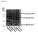
- A human serum or plasma sample (corresponding to 20 µg of protein) was mixed with a Laemmli sample buffer, and the mixture was then heated. Thereafter, the mixture was subjected to SDS/PAGE using 4/20% gradient polyacrylamide gel. Electrophoresis was performed at a constant current of 40 mA for 55 minutes. The separated protein was electrically transcribed onto a PVDF membrane at a constant current of 260 mA for 50 minutes. Thereafter, the membrane was blocked in 1% bovine serum albumin (BSA)-phosphate buffered saline (PBS) for one or more hours. Thereafter, biotinylated lectin (SSA) diluted with 1% BSA-PBS was allowed to react with the PVDF membrane for 1 hour. (In this experiment, biotin-SSA (#300442; Seikagaku Corp.) was used in a concentration of 1 µg/ml.) The membrane was washed three times with 0.05% Tween 20 (surfactant) in PBS for 15 minutes for each time. Thereafter, the resultant was allowed to react with streptavidin-horseradish peroxidase conjugate (HRP) (#RPN 1231V; Amersham) diluted with 1% BSA-PBS for 1 hour. Thereafter, the membrane was washed again with a washing solution three times for 15 minutes for each time. A chemoluminescent substrate (PierceSuperSignal West DuraExtended Duration Substrate) was used, and the band of an α 2,6-sialyl residue-containing glycoprotein was detected by a lumino-image analyzer (LAS-1000plus; Fuji Film, Tokyo), followed by quantification. The results are shown in
Figure 1 . The results demonstrated that a 180-kD glycoprotein, a 95-kD glycoprotein, and a 40-kD glycoprotein, which contain α 2,6-sialyl residue, were increased in patients suffering from Alzheimer's disease. - Taking into consideration the fact that such glycoproteins contain α 2,6-sialyl residue and the molecular weights thereof, an attempt was made to identify a 180-kD glycoprotein, a 95-kD glycoprotein, and a 40-kD glycoprotein. An antibody reacting with a human serum glycoprotein used as a candidate was purchased and examined. As a result, it was found that the 40-kD glycoprotein is haptoglobin. A detection method thereof will be described below.
- A human serum or plasma sample (corresponding to 0.04 µg of protein) was mixed with a Laemmli sample buffer, and the mixture was then heated. Thereafter, the mixture was subjected to SDS/PAGE using 4/20% gradient polyacrylamide gel. Electrophoresis was performed at a constant current of 40 mA for 55 minutes. The separated protein was electrically transcribed onto a PVDF membrane at a constant current of 260 mA for 50 minutes. Thereafter, the membrane was blocked in 5% skimmed milk-0.1% Tween 20 in Tris buffered saline (TBS) for one or more hours. Subsequently, a rabbit anti-human haptoglobin antibody (Sigma H-8636) (12 µg/ml) diluted with 3% skimmed milk-TBS at a ratio of 1 : 1,000 was allowed to react with a nitrocellulose membrane for 1 hour. Thereafter, the membrane was washed three times with 0.1% Tween 20 (surfactant) in TBS for 15 minutes for each time. Thereafter, the resultant was allowed to react with anti-rabbit IgG, horseradish peroxidase (#NA934V; Amersham) (1 µg/ml) diluted with 3% skimmed milk-TBS at a ratio of 1 : 1,000 for 1 hour. Thereafter, the membrane was washed again with a washing solution three times for 15 minutes for each time. A chemoluminescent substrate (PierceSuperSignal West DuraExtended Duration Substrate) was used, and the band of an α 2,6-sialyl residue-containing glycoprotein was detected by a lumino-image analyzer (LAS-1000plus; Fuji Film, Tokyo), followed by quantification. The results of the Western blotting using an anti-haptoglobin antibody are shown in
Figure 2 . An increase in haptoglobin was confirmed in Alzheimer's disease. A calibration curve has previously been produced with a standard sample containing normal human serum or a known concentration of haptoglobin (#H0138; Sigma). Using such a calibration curve, a quantitative value can be calculated. -
-
Figure 1 shows human serum that was stained with SSA lectin. -
Figure 2 shows detection of a 40kD-glycoprotein with a haptoglobin antibody. - In the present invention, an α 2,6-sialyl residue-containing glycoprotein, which exists in a large amount in blood, is used as a marker. Thus, it is possible to make diagnoses using only a trace amount of blood sample. When compared with the conventional diagnostic methods using cerebrospinal fluid or a large amount of blood, a lesser burden is imposed upon a subject. In addition, in the present invention, a special antibody is not required, and lectin that can be prepared in a large amount in an inexpensive way or antibodies that can be easily produced (e.g. anti-haptoglobin antibody, etc.) are used. Accordingly, a method for screening many analytes with high sensitivity can be easily established.
Claims (9)
- In vitro use of an agent for diagnosing Alzheimer's disease, said agent comprising lectin used for detecting an α 2,6-sialyl residue-containing glycoprotein, wherein said lectin specifically binds to α 2,6-sialyl residues of an α 2,6-sialyl residue-containing glycoprotein.
- The use according to claim 1, wherein the α 2,6-sialyl residue-containing glycoprotein is a 180-kD glycoprotein, a 95-kD glycoprotein, or a 40-kD glycoprotein.
- The use according to claim 1, wherein said agent further comprises an anti-haptoglobin antibody used for detecting a 40-kD glycoprotein that contains α 2,6-sialyl residue.
- A kit for diagnosing Alzheimer's disease, which comprises lectin and an anti-haptoglobin antibody, wherein said lectin specifically binds to α 2,6-sialyl residues of an α 2,6-sialyl residue-containing glycoprotein.
- A method for diagnosing Alzheimer's disease, which comprises detecting or measuring an α 2,6-sialyl residue-containing glycoprotein contained in a human-derived sample, using a lectin that specifically binds said α 2,6-sialyl residue.
- The method according to claim 5, wherein the α 2,6-sialyl residue-containing glycoprotein is a 180-kD glycoprotein, a 95-kD glycoprotein, or a 40-kD glycoprotein.
- The method according to claim 5 or 6, wherein a 40-kD glycoprotein that contains α 2,6-sialyl residue is detected or measured by using an anti-haptoglobin antibody.
- The method according to claim 5, wherein a 40-kD glycoprotein that contains α 2,6-sialyl residue is detected or measured by sandwich assay using lectin and an anti-haptoglobin antibody, wherein said lectin specifically binds to α 2,6-sialyl residues of an α 2,6-sialyl residue-containing glycoprotein.
- The method according to any of claims 5 to 8, wherein the human-derived sample is a blood sample.
Applications Claiming Priority (2)
| Application Number | Priority Date | Filing Date | Title |
|---|---|---|---|
| JP2005138070A JP4385149B2 (en) | 2005-05-11 | 2005-05-11 | A diagnostic agent for Alzheimer's disease using serum glycoprotein as a biomarker |
| PCT/JP2006/309450 WO2006121099A1 (en) | 2005-05-11 | 2006-05-11 | Method of diagnosing alzheimer’s disease using serum glycoprotein as biomarker |
Publications (3)
| Publication Number | Publication Date |
|---|---|
| EP1901066A1 EP1901066A1 (en) | 2008-03-19 |
| EP1901066A4 EP1901066A4 (en) | 2008-08-27 |
| EP1901066B1 true EP1901066B1 (en) | 2011-07-20 |
Family
ID=37396607
Family Applications (1)
| Application Number | Title | Priority Date | Filing Date |
|---|---|---|---|
| EP06746259A Expired - Lifetime EP1901066B1 (en) | 2005-05-11 | 2006-05-11 | Method of diagnosing alzheimer's disease using serum glycoprotein as biomarker |
Country Status (4)
| Country | Link |
|---|---|
| US (1) | US8609346B2 (en) |
| EP (1) | EP1901066B1 (en) |
| JP (1) | JP4385149B2 (en) |
| WO (1) | WO2006121099A1 (en) |
Families Citing this family (8)
| Publication number | Priority date | Publication date | Assignee | Title |
|---|---|---|---|---|
| DK2223108T3 (en) * | 2007-12-27 | 2016-07-18 | Baxalta GmbH | Methods for differentiation of plasma-derived protein from the recombinant protein in a sample |
| AU2009256601A1 (en) * | 2008-06-11 | 2009-12-17 | Universiteit Gent | Blood serum marker for detection and diagnosis of Alzheimer's disease |
| JP2010085364A (en) * | 2008-10-02 | 2010-04-15 | Nationa Hospital Organization | Method, composition and kit for examining tissue disorder caused by atrophy of collagen fiber |
| US8778622B2 (en) | 2009-12-01 | 2014-07-15 | Shire Human Genetic Therapies, Inc. | Enzymatic activity-based detection |
| EP2653476B1 (en) | 2010-12-16 | 2017-04-26 | National Institute of Advanced Industrial Science And Technology | Method for enrichment and separation of spinal fluid glycoprotein, method for searching for marker for central nervous system diseases which utilizes the aforementioned method, and marker for central nervous system diseases |
| CA2838746C (en) | 2011-06-08 | 2020-08-25 | Shire Human Genetic Therapies, Inc. | Competition based-detection assays |
| FI20155280A7 (en) | 2015-04-15 | 2016-10-16 | Medicortex Finland Oy | Prognostic and diagnostic glycan-based biomarkers of brain damage |
| KR101989383B1 (en) | 2017-04-04 | 2019-06-14 | 조선대학교산학협력단 | A peptide probe for early diagnosis of alzheimer's disease |
Family Cites Families (3)
| Publication number | Priority date | Publication date | Assignee | Title |
|---|---|---|---|---|
| US20090166224A1 (en) * | 2004-05-05 | 2009-07-02 | Ziping Yang | Multi-lectin affinity chromatography and uses thereof |
| US7838634B2 (en) * | 2005-04-15 | 2010-11-23 | Van Andel Research Institute | Methods for measuring glycan levels of proteins |
| US7575876B2 (en) * | 2005-10-27 | 2009-08-18 | The University Of Washington | Biomarkers for neurodegenerative disorders |
-
2005
- 2005-05-11 JP JP2005138070A patent/JP4385149B2/en not_active Expired - Fee Related
-
2006
- 2006-05-11 US US11/920,167 patent/US8609346B2/en not_active Expired - Fee Related
- 2006-05-11 WO PCT/JP2006/309450 patent/WO2006121099A1/en not_active Ceased
- 2006-05-11 EP EP06746259A patent/EP1901066B1/en not_active Expired - Lifetime
Also Published As
| Publication number | Publication date |
|---|---|
| EP1901066A4 (en) | 2008-08-27 |
| US20110076704A1 (en) | 2011-03-31 |
| JP4385149B2 (en) | 2009-12-16 |
| JP2006317210A (en) | 2006-11-24 |
| US8609346B2 (en) | 2013-12-17 |
| EP1901066A1 (en) | 2008-03-19 |
| WO2006121099A1 (en) | 2006-11-16 |
Similar Documents
| Publication | Publication Date | Title |
|---|---|---|
| JP6096813B2 (en) | Multi-biomarker set for breast cancer diagnosis, detection method thereof, and breast cancer diagnosis kit including antibody thereto | |
| US6762032B1 (en) | Compositions, assay kits, and methods for use related to a disease condition comprising multiple sclerosis and/or a pro-MS immune response | |
| US8105839B2 (en) | Diagnostic kit for Alzheimer's disease, diagnostic marker, and detection method for indicator of pathological state thereof | |
| WO2008063369A2 (en) | Biomarkers for neurological conditions | |
| CN104884958B (en) | Sugar chain isomers detection method and sugar chain isomers detection means | |
| US20060188951A1 (en) | Method for measuring the level of anti-beta-amyloid antibody in body fluids and diagnostic kit for alzheimer's disease using same | |
| WO2011070174A1 (en) | Methods and reagents for improved detection of amyloid beta peptides | |
| EP0444856A2 (en) | Diagnostic assay for Alzheimer's disease | |
| US11719697B2 (en) | Immunoassay and antibodies for the detection of chromogranin A | |
| JP2023024532A (en) | Alzheimer's disease biomarker | |
| US8609346B2 (en) | Method for diagnosing alzheimer's disease using serum glycoprotein as biomarker | |
| US20150285804A1 (en) | Diagnostic method for colorectal cancer | |
| JP5892169B2 (en) | Diagnostic kit, diagnostic marker and detection method for Alzheimer type dementia by measuring sugar chain of complement C3 protein | |
| US10228373B2 (en) | Kit and method for diagnosis, prognosis or monitoring of liver disease through measurement of amount of AST | |
| EP1371986A1 (en) | Diagnosis of Alzheimer's disease based on the hAbeta42:hAbeta40 ratio | |
| JP2005145837A (en) | Diagnostic method for Alzheimer's disease | |
| KR101431063B1 (en) | Protein marker apolipoprotein c-1 for breast cancer diagnosis, method of detecting the same, and diagnosis kit for breast cancer using antibody against the same | |
| US20040248200A1 (en) | Method of diagnosing cerebral infarction | |
| JP4993065B2 (en) | Diagnostic method for hepatocellular carcinoma | |
| WO2025182761A1 (en) | Gfap detection method, method for assisting diagnosis of alzheimer 's disease, and kit used therefor | |
| JP2024070581A (en) | Biomarkers for als testing and testing method | |
| AU718732B2 (en) | Diagnostic assay for Alzheimer's disease | |
| JP2020139770A (en) | Diagnostic index for Parkinson's disease | |
| WO2007042897A1 (en) | Method for the differential diagnosis and the monitoring of alzheimer-type dementia | |
| JPH07209300A (en) | Method for measuring factor of rheumatism |
Legal Events
| Date | Code | Title | Description |
|---|---|---|---|
| PUAI | Public reference made under article 153(3) epc to a published international application that has entered the european phase |
Free format text: ORIGINAL CODE: 0009012 |
|
| 17P | Request for examination filed |
Effective date: 20071210 |
|
| AK | Designated contracting states |
Kind code of ref document: A1 Designated state(s): CH DE ES FR GB IT LI |
|
| RIN1 | Information on inventor provided before grant (corrected) |
Inventor name: SAIDO, TAKAOMI Inventor name: KITAZUME, SHINOBU Inventor name: OKA, RITSUKO Inventor name: SATO, YUJI Inventor name: ARAI, HIROYUKI Inventor name: HASHIMOTO, YASUHIRO Inventor name: ENDO, TAMAO Inventor name: HIGUCHI, MASATO |
|
| RIN1 | Information on inventor provided before grant (corrected) |
Inventor name: SAIDO, TAKAOMI Inventor name: OKA, RITSUKO Inventor name: SATO, YUJI Inventor name: HIGUCHI, MASATO Inventor name: ARAI, HIROYUKI Inventor name: ENDO, TAMAOC/O TOKYO MET. INST. OF GERONTOLOGY Inventor name: KITAZUME, SHINOBU Inventor name: HASHIMOTO, YASUHIROC/O FUKUSHIMA MEDICAL UNIVERSIT |
|
| DAX | Request for extension of the european patent (deleted) | ||
| RBV | Designated contracting states (corrected) |
Designated state(s): CH DE ES FR GB IT LI |
|
| A4 | Supplementary search report drawn up and despatched |
Effective date: 20080728 |
|
| 17Q | First examination report despatched |
Effective date: 20081008 |
|
| RIN1 | Information on inventor provided before grant (corrected) |
Inventor name: SAIDO, TAKAOMI Inventor name: ARAI, HIROYUKI Inventor name: HIGUCHI, MAKOTO Inventor name: ENDO, TAMAOC/O TOKYO MET. INST. OF GERONTOLOGY Inventor name: SATO, YUJI Inventor name: HASHIMOTO, YASUHIROC/O FUKUSHIMA MEDICAL UNIVERSIT Inventor name: OKA, RITSUKO Inventor name: KITAZUME, SHINOBU |
|
| GRAP | Despatch of communication of intention to grant a patent |
Free format text: ORIGINAL CODE: EPIDOSNIGR1 |
|
| RTI1 | Title (correction) |
Free format text: METHOD OF DIAGNOSING ALZHEIMER'S DISEASE USING SERUM GLYCOPROTEIN AS BIOMARKER |
|
| GRAS | Grant fee paid |
Free format text: ORIGINAL CODE: EPIDOSNIGR3 |
|
| GRAA | (expected) grant |
Free format text: ORIGINAL CODE: 0009210 |
|
| AK | Designated contracting states |
Kind code of ref document: B1 Designated state(s): CH DE ES FR GB IT LI |
|
| REG | Reference to a national code |
Ref country code: GB Ref legal event code: FG4D |
|
| REG | Reference to a national code |
Ref country code: CH Ref legal event code: EP |
|
| REG | Reference to a national code |
Ref country code: DE Ref legal event code: R096 Ref document number: 602006023184 Country of ref document: DE Effective date: 20110908 |
|
| PLBE | No opposition filed within time limit |
Free format text: ORIGINAL CODE: 0009261 |
|
| STAA | Information on the status of an ep patent application or granted ep patent |
Free format text: STATUS: NO OPPOSITION FILED WITHIN TIME LIMIT |
|
| PG25 | Lapsed in a contracting state [announced via postgrant information from national office to epo] |
Ref country code: IT Free format text: LAPSE BECAUSE OF FAILURE TO SUBMIT A TRANSLATION OF THE DESCRIPTION OR TO PAY THE FEE WITHIN THE PRESCRIBED TIME-LIMIT Effective date: 20110720 |
|
| 26N | No opposition filed |
Effective date: 20120423 |
|
| REG | Reference to a national code |
Ref country code: DE Ref legal event code: R097 Ref document number: 602006023184 Country of ref document: DE Effective date: 20120423 |
|
| REG | Reference to a national code |
Ref country code: CH Ref legal event code: PL |
|
| PG25 | Lapsed in a contracting state [announced via postgrant information from national office to epo] |
Ref country code: LI Free format text: LAPSE BECAUSE OF NON-PAYMENT OF DUE FEES Effective date: 20120531 Ref country code: CH Free format text: LAPSE BECAUSE OF NON-PAYMENT OF DUE FEES Effective date: 20120531 |
|
| PG25 | Lapsed in a contracting state [announced via postgrant information from national office to epo] |
Ref country code: ES Free format text: LAPSE BECAUSE OF FAILURE TO SUBMIT A TRANSLATION OF THE DESCRIPTION OR TO PAY THE FEE WITHIN THE PRESCRIBED TIME-LIMIT Effective date: 20111031 |
|
| REG | Reference to a national code |
Ref country code: DE Ref legal event code: R082 Ref document number: 602006023184 Country of ref document: DE Representative=s name: GODEMEYER BLUM LENZE PARTNERSCHAFT, PATENTANWA, DE Ref country code: DE Ref legal event code: R082 Ref document number: 602006023184 Country of ref document: DE Representative=s name: GODEMEYER BLUM LENZE PATENTANWAELTE, PARTNERSC, DE |
|
| PGFP | Annual fee paid to national office [announced via postgrant information from national office to epo] |
Ref country code: GB Payment date: 20140507 Year of fee payment: 9 |
|
| PGFP | Annual fee paid to national office [announced via postgrant information from national office to epo] |
Ref country code: FR Payment date: 20140509 Year of fee payment: 9 Ref country code: DE Payment date: 20140507 Year of fee payment: 9 |
|
| REG | Reference to a national code |
Ref country code: DE Ref legal event code: R119 Ref document number: 602006023184 Country of ref document: DE |
|
| GBPC | Gb: european patent ceased through non-payment of renewal fee |
Effective date: 20150511 |
|
| REG | Reference to a national code |
Ref country code: FR Ref legal event code: ST Effective date: 20160129 |
|
| PG25 | Lapsed in a contracting state [announced via postgrant information from national office to epo] |
Ref country code: DE Free format text: LAPSE BECAUSE OF NON-PAYMENT OF DUE FEES Effective date: 20151201 Ref country code: GB Free format text: LAPSE BECAUSE OF NON-PAYMENT OF DUE FEES Effective date: 20150511 |
|
| PG25 | Lapsed in a contracting state [announced via postgrant information from national office to epo] |
Ref country code: FR Free format text: LAPSE BECAUSE OF NON-PAYMENT OF DUE FEES Effective date: 20150601 |