EP1871805B1 - Antigen binding molecules that bind egfr, vectors encoding same, and uses thereof - Google Patents
Antigen binding molecules that bind egfr, vectors encoding same, and uses thereof Download PDFInfo
- Publication number
- EP1871805B1 EP1871805B1 EP06710336.6A EP06710336A EP1871805B1 EP 1871805 B1 EP1871805 B1 EP 1871805B1 EP 06710336 A EP06710336 A EP 06710336A EP 1871805 B1 EP1871805 B1 EP 1871805B1
- Authority
- EP
- European Patent Office
- Prior art keywords
- antibody
- seq
- egfr
- cancer
- cells
- Prior art date
- Legal status (The legal status is an assumption and is not a legal conclusion. Google has not performed a legal analysis and makes no representation as to the accuracy of the status listed.)
- Active
Links
- 230000027455 binding Effects 0.000 title claims description 212
- 239000013598 vector Substances 0.000 title claims description 12
- 108060006698 EGF receptor Proteins 0.000 title description 151
- 239000000427 antigen Substances 0.000 title description 88
- 108091007433 antigens Proteins 0.000 title description 88
- 102000036639 antigens Human genes 0.000 title description 88
- 210000004027 cell Anatomy 0.000 claims description 293
- 108090000765 processed proteins & peptides Proteins 0.000 claims description 172
- 102000004196 processed proteins & peptides Human genes 0.000 claims description 171
- 229920001184 polypeptide Polymers 0.000 claims description 169
- 241000282414 Homo sapiens Species 0.000 claims description 144
- 230000001965 increasing effect Effects 0.000 claims description 141
- 238000000034 method Methods 0.000 claims description 128
- 206010028980 Neoplasm Diseases 0.000 claims description 104
- 150000002482 oligosaccharides Chemical class 0.000 claims description 82
- 230000000694 effects Effects 0.000 claims description 79
- 229920001542 oligosaccharide Polymers 0.000 claims description 77
- 108091033319 polynucleotide Proteins 0.000 claims description 68
- 102000040430 polynucleotide Human genes 0.000 claims description 68
- 239000002157 polynucleotide Substances 0.000 claims description 68
- 201000011510 cancer Diseases 0.000 claims description 57
- 238000011282 treatment Methods 0.000 claims description 49
- 239000012636 effector Substances 0.000 claims description 44
- 125000003275 alpha amino acid group Chemical group 0.000 claims description 42
- 230000004927 fusion Effects 0.000 claims description 36
- 150000007523 nucleic acids Chemical class 0.000 claims description 35
- 238000004519 manufacturing process Methods 0.000 claims description 34
- 102000039446 nucleic acids Human genes 0.000 claims description 34
- 108020004707 nucleic acids Proteins 0.000 claims description 34
- 230000001404 mediated effect Effects 0.000 claims description 29
- 108010087819 Fc receptors Proteins 0.000 claims description 23
- 102000009109 Fc receptors Human genes 0.000 claims description 23
- 239000003814 drug Substances 0.000 claims description 19
- 230000004807 localization Effects 0.000 claims description 19
- 230000025545 Golgi localization Effects 0.000 claims description 18
- 239000008194 pharmaceutical composition Substances 0.000 claims description 14
- 101000851181 Homo sapiens Epidermal growth factor receptor Proteins 0.000 claims description 13
- 230000005889 cellular cytotoxicity Effects 0.000 claims description 13
- 230000002829 reductive effect Effects 0.000 claims description 12
- 239000003937 drug carrier Substances 0.000 claims description 11
- 210000002540 macrophage Anatomy 0.000 claims description 11
- 108010083819 mannosyl-oligosaccharide 1,3 - 1,6-alpha-mannosidase Proteins 0.000 claims description 11
- 210000001616 monocyte Anatomy 0.000 claims description 10
- 230000006907 apoptotic process Effects 0.000 claims description 9
- 102000045108 human EGFR Human genes 0.000 claims description 8
- 206010006187 Breast cancer Diseases 0.000 claims description 7
- 208000026310 Breast neoplasm Diseases 0.000 claims description 7
- 206010058467 Lung neoplasm malignant Diseases 0.000 claims description 7
- OVRNDRQMDRJTHS-RTRLPJTCSA-N N-acetyl-D-glucosamine Chemical group CC(=O)N[C@H]1C(O)O[C@H](CO)[C@@H](O)[C@@H]1O OVRNDRQMDRJTHS-RTRLPJTCSA-N 0.000 claims description 7
- 230000001939 inductive effect Effects 0.000 claims description 7
- 210000000822 natural killer cell Anatomy 0.000 claims description 7
- 230000011664 signaling Effects 0.000 claims description 7
- 230000037453 T cell priming Effects 0.000 claims description 6
- 230000004041 dendritic cell maturation Effects 0.000 claims description 6
- 208000014829 head and neck neoplasm Diseases 0.000 claims description 6
- 108010019236 Fucosyltransferases Proteins 0.000 claims description 5
- 102000006471 Fucosyltransferases Human genes 0.000 claims description 5
- SHZGCJCMOBCMKK-DHVFOXMCSA-N L-fucopyranose Chemical group C[C@@H]1OC(O)[C@@H](O)[C@H](O)[C@@H]1O SHZGCJCMOBCMKK-DHVFOXMCSA-N 0.000 claims description 5
- 206010033128 Ovarian cancer Diseases 0.000 claims description 5
- 206010061535 Ovarian neoplasm Diseases 0.000 claims description 5
- 206010061902 Pancreatic neoplasm Diseases 0.000 claims description 5
- 206010060862 Prostate cancer Diseases 0.000 claims description 5
- 208000000236 Prostatic Neoplasms Diseases 0.000 claims description 5
- 208000007097 Urinary Bladder Neoplasms Diseases 0.000 claims description 5
- 201000005202 lung cancer Diseases 0.000 claims description 5
- 208000020816 lung neoplasm Diseases 0.000 claims description 5
- 208000015486 malignant pancreatic neoplasm Diseases 0.000 claims description 5
- 208000008443 pancreatic carcinoma Diseases 0.000 claims description 5
- 206010005003 Bladder cancer Diseases 0.000 claims description 4
- 206010009944 Colon cancer Diseases 0.000 claims description 4
- 208000008839 Kidney Neoplasms Diseases 0.000 claims description 4
- 208000000453 Skin Neoplasms Diseases 0.000 claims description 4
- 208000029742 colonic neoplasm Diseases 0.000 claims description 4
- 238000012258 culturing Methods 0.000 claims description 4
- 108010009689 mannosyl-oligosaccharide 1,2-alpha-mannosidase Proteins 0.000 claims description 4
- 238000011321 prophylaxis Methods 0.000 claims description 4
- 201000000849 skin cancer Diseases 0.000 claims description 4
- 201000005112 urinary bladder cancer Diseases 0.000 claims description 4
- 208000003174 Brain Neoplasms Diseases 0.000 claims description 3
- 206010038389 Renal cancer Diseases 0.000 claims description 3
- 201000010982 kidney cancer Diseases 0.000 claims description 3
- 201000010536 head and neck cancer Diseases 0.000 claims description 2
- 102000001301 EGF receptor Human genes 0.000 description 150
- 108020004414 DNA Proteins 0.000 description 74
- 241001465754 Metazoa Species 0.000 description 69
- NFGXHKASABOEEW-UHFFFAOYSA-N 1-methylethyl 11-methoxy-3,7,11-trimethyl-2,4-dodecadienoate Chemical compound COC(C)(C)CCCC(C)CC=CC(C)=CC(=O)OC(C)C NFGXHKASABOEEW-UHFFFAOYSA-N 0.000 description 64
- 210000002966 serum Anatomy 0.000 description 62
- 108090000623 proteins and genes Proteins 0.000 description 61
- 241000700159 Rattus Species 0.000 description 58
- 230000010056 antibody-dependent cellular cytotoxicity Effects 0.000 description 56
- 230000014509 gene expression Effects 0.000 description 53
- 208000037265 diseases, disorders, signs and symptoms Diseases 0.000 description 49
- 108010047041 Complementarity Determining Regions Proteins 0.000 description 42
- 235000001014 amino acid Nutrition 0.000 description 39
- 239000012634 fragment Substances 0.000 description 39
- 239000000203 mixture Substances 0.000 description 38
- 230000006870 function Effects 0.000 description 37
- 235000018102 proteins Nutrition 0.000 description 33
- 102000004169 proteins and genes Human genes 0.000 description 33
- 238000001990 intravenous administration Methods 0.000 description 32
- 238000006467 substitution reaction Methods 0.000 description 31
- 241000282693 Cercopithecidae Species 0.000 description 30
- 241000282567 Macaca fascicularis Species 0.000 description 28
- 239000003446 ligand Substances 0.000 description 28
- 239000000523 sample Substances 0.000 description 27
- 125000000539 amino acid group Chemical group 0.000 description 26
- 150000001413 amino acids Chemical class 0.000 description 26
- 208000035475 disorder Diseases 0.000 description 26
- 239000013604 expression vector Substances 0.000 description 26
- 210000004602 germ cell Anatomy 0.000 description 26
- 230000013595 glycosylation Effects 0.000 description 26
- 238000006206 glycosylation reaction Methods 0.000 description 26
- 238000003556 assay Methods 0.000 description 25
- 239000000243 solution Substances 0.000 description 25
- 229940024606 amino acid Drugs 0.000 description 24
- 201000010099 disease Diseases 0.000 description 23
- 230000001225 therapeutic effect Effects 0.000 description 22
- 238000001727 in vivo Methods 0.000 description 21
- 210000001519 tissue Anatomy 0.000 description 21
- 238000004458 analytical method Methods 0.000 description 20
- 239000002773 nucleotide Substances 0.000 description 19
- DHMQDGOQFOQNFH-UHFFFAOYSA-N Glycine Chemical compound NCC(O)=O DHMQDGOQFOQNFH-UHFFFAOYSA-N 0.000 description 18
- 108060003951 Immunoglobulin Proteins 0.000 description 18
- 241001529936 Murinae Species 0.000 description 18
- 102000018358 immunoglobulin Human genes 0.000 description 18
- 210000004072 lung Anatomy 0.000 description 18
- 125000003729 nucleotide group Chemical group 0.000 description 18
- 101710117290 Aldo-keto reductase family 1 member C4 Proteins 0.000 description 17
- 108091026890 Coding region Proteins 0.000 description 17
- 239000008280 blood Substances 0.000 description 17
- 238000000338 in vitro Methods 0.000 description 17
- 210000003734 kidney Anatomy 0.000 description 17
- 102000005962 receptors Human genes 0.000 description 17
- 108020003175 receptors Proteins 0.000 description 17
- 238000012360 testing method Methods 0.000 description 17
- 230000004913 activation Effects 0.000 description 16
- 210000004369 blood Anatomy 0.000 description 16
- 210000001672 ovary Anatomy 0.000 description 16
- 108091028043 Nucleic acid sequence Proteins 0.000 description 15
- 238000013459 approach Methods 0.000 description 15
- 239000000872 buffer Substances 0.000 description 15
- 238000012217 deletion Methods 0.000 description 15
- 230000037430 deletion Effects 0.000 description 15
- 238000009472 formulation Methods 0.000 description 15
- 210000004185 liver Anatomy 0.000 description 15
- 230000035772 mutation Effects 0.000 description 14
- 210000004978 chinese hamster ovary cell Anatomy 0.000 description 13
- 210000001072 colon Anatomy 0.000 description 13
- 210000003491 skin Anatomy 0.000 description 13
- 230000009885 systemic effect Effects 0.000 description 13
- 206010035226 Plasma cell myeloma Diseases 0.000 description 12
- 229940127089 cytotoxic agent Drugs 0.000 description 12
- 201000000050 myeloid neoplasm Diseases 0.000 description 12
- 230000002018 overexpression Effects 0.000 description 12
- 210000000496 pancreas Anatomy 0.000 description 12
- 210000003819 peripheral blood mononuclear cell Anatomy 0.000 description 12
- 230000004044 response Effects 0.000 description 12
- 210000004881 tumor cell Anatomy 0.000 description 12
- 241000124008 Mammalia Species 0.000 description 11
- 241000699666 Mus <mouse, genus> Species 0.000 description 11
- 241000283984 Rodentia Species 0.000 description 11
- 230000037396 body weight Effects 0.000 description 11
- 210000003128 head Anatomy 0.000 description 11
- 230000004048 modification Effects 0.000 description 11
- 238000012986 modification Methods 0.000 description 11
- 241000196324 Embryophyta Species 0.000 description 10
- 102000003886 Glycoproteins Human genes 0.000 description 10
- 108090000288 Glycoproteins Proteins 0.000 description 10
- DCXYFEDJOCDNAF-REOHCLBHSA-N L-asparagine Chemical compound OC(=O)[C@@H](N)CC(N)=O DCXYFEDJOCDNAF-REOHCLBHSA-N 0.000 description 10
- XSQUKJJJFZCRTK-UHFFFAOYSA-N Urea Chemical compound NC(N)=O XSQUKJJJFZCRTK-UHFFFAOYSA-N 0.000 description 10
- 230000002159 abnormal effect Effects 0.000 description 10
- 230000015572 biosynthetic process Effects 0.000 description 10
- 229940079593 drug Drugs 0.000 description 10
- 210000004379 membrane Anatomy 0.000 description 10
- 239000012528 membrane Substances 0.000 description 10
- 230000001105 regulatory effect Effects 0.000 description 10
- 108090000695 Cytokines Proteins 0.000 description 9
- 102000004127 Cytokines Human genes 0.000 description 9
- 108010021625 Immunoglobulin Fragments Proteins 0.000 description 9
- 102000008394 Immunoglobulin Fragments Human genes 0.000 description 9
- FAPWRFPIFSIZLT-UHFFFAOYSA-M Sodium chloride Chemical compound [Na+].[Cl-] FAPWRFPIFSIZLT-UHFFFAOYSA-M 0.000 description 9
- 238000007792 addition Methods 0.000 description 9
- 210000000481 breast Anatomy 0.000 description 9
- 210000000621 bronchi Anatomy 0.000 description 9
- 208000035269 cancer or benign tumor Diseases 0.000 description 9
- 239000003599 detergent Substances 0.000 description 9
- 108020001507 fusion proteins Proteins 0.000 description 9
- 102000037865 fusion proteins Human genes 0.000 description 9
- 239000002596 immunotoxin Substances 0.000 description 9
- 238000002347 injection Methods 0.000 description 9
- 239000007924 injection Substances 0.000 description 9
- 210000003739 neck Anatomy 0.000 description 9
- 241000894007 species Species 0.000 description 9
- 239000003053 toxin Substances 0.000 description 9
- 231100000765 toxin Toxicity 0.000 description 9
- 108700012359 toxins Proteins 0.000 description 9
- DCXYFEDJOCDNAF-UHFFFAOYSA-N Asparagine Natural products OC(=O)C(N)CC(N)=O DCXYFEDJOCDNAF-UHFFFAOYSA-N 0.000 description 8
- 201000009030 Carcinoma Diseases 0.000 description 8
- 238000002965 ELISA Methods 0.000 description 8
- 102000004190 Enzymes Human genes 0.000 description 8
- 108090000790 Enzymes Proteins 0.000 description 8
- AYFVYJQAPQTCCC-GBXIJSLDSA-N L-threonine Chemical compound C[C@@H](O)[C@H](N)C(O)=O AYFVYJQAPQTCCC-GBXIJSLDSA-N 0.000 description 8
- 108010076504 Protein Sorting Signals Proteins 0.000 description 8
- 230000035508 accumulation Effects 0.000 description 8
- 238000009825 accumulation Methods 0.000 description 8
- 239000005557 antagonist Substances 0.000 description 8
- 230000005888 antibody-dependent cellular phagocytosis Effects 0.000 description 8
- 235000009582 asparagine Nutrition 0.000 description 8
- 229960001230 asparagine Drugs 0.000 description 8
- 210000004556 brain Anatomy 0.000 description 8
- 230000001472 cytotoxic effect Effects 0.000 description 8
- 238000001943 fluorescence-activated cell sorting Methods 0.000 description 8
- 230000003993 interaction Effects 0.000 description 8
- 239000003550 marker Substances 0.000 description 8
- 238000000816 matrix-assisted laser desorption--ionisation Methods 0.000 description 8
- 239000002609 medium Substances 0.000 description 8
- 239000002953 phosphate buffered saline Substances 0.000 description 8
- 238000012545 processing Methods 0.000 description 8
- 239000000047 product Substances 0.000 description 8
- 210000002307 prostate Anatomy 0.000 description 8
- 230000028327 secretion Effects 0.000 description 8
- 230000008685 targeting Effects 0.000 description 8
- 210000003932 urinary bladder Anatomy 0.000 description 8
- XLYOFNOQVPJJNP-UHFFFAOYSA-N water Chemical compound O XLYOFNOQVPJJNP-UHFFFAOYSA-N 0.000 description 8
- 230000003442 weekly effect Effects 0.000 description 8
- MTCFGRXMJLQNBG-REOHCLBHSA-N (2S)-2-Amino-3-hydroxypropansäure Chemical compound OC[C@H](N)C(O)=O MTCFGRXMJLQNBG-REOHCLBHSA-N 0.000 description 7
- 108091006020 Fc-tagged proteins Proteins 0.000 description 7
- 239000004471 Glycine Substances 0.000 description 7
- OVRNDRQMDRJTHS-UHFFFAOYSA-N N-acelyl-D-glucosamine Natural products CC(=O)NC1C(O)OC(CO)C(O)C1O OVRNDRQMDRJTHS-UHFFFAOYSA-N 0.000 description 7
- MBLBDJOUHNCFQT-LXGUWJNJSA-N N-acetylglucosamine Natural products CC(=O)N[C@@H](C=O)[C@@H](O)[C@H](O)[C@H](O)CO MBLBDJOUHNCFQT-LXGUWJNJSA-N 0.000 description 7
- 108700008625 Reporter Genes Proteins 0.000 description 7
- 238000011123 anti-EGFR therapy Methods 0.000 description 7
- 230000004071 biological effect Effects 0.000 description 7
- 239000006143 cell culture medium Substances 0.000 description 7
- 150000001875 compounds Chemical class 0.000 description 7
- 238000004590 computer program Methods 0.000 description 7
- 230000000875 corresponding effect Effects 0.000 description 7
- 230000009089 cytolysis Effects 0.000 description 7
- 231100000433 cytotoxic Toxicity 0.000 description 7
- 239000002254 cytotoxic agent Substances 0.000 description 7
- 230000001419 dependent effect Effects 0.000 description 7
- 238000003364 immunohistochemistry Methods 0.000 description 7
- 230000002637 immunotoxin Effects 0.000 description 7
- 229940051026 immunotoxin Drugs 0.000 description 7
- 231100000608 immunotoxin Toxicity 0.000 description 7
- 229910052740 iodine Inorganic materials 0.000 description 7
- 230000003902 lesion Effects 0.000 description 7
- 239000002502 liposome Substances 0.000 description 7
- 210000004962 mammalian cell Anatomy 0.000 description 7
- 239000011159 matrix material Substances 0.000 description 7
- 210000000056 organ Anatomy 0.000 description 7
- 239000013612 plasmid Substances 0.000 description 7
- 239000000092 prognostic biomarker Substances 0.000 description 7
- 230000019491 signal transduction Effects 0.000 description 7
- 238000002560 therapeutic procedure Methods 0.000 description 7
- 230000001988 toxicity Effects 0.000 description 7
- 231100000419 toxicity Toxicity 0.000 description 7
- 231100000041 toxicology testing Toxicity 0.000 description 7
- 229910052727 yttrium Inorganic materials 0.000 description 7
- ZCYVEMRRCGMTRW-UHFFFAOYSA-N 7553-56-2 Chemical compound [I] ZCYVEMRRCGMTRW-UHFFFAOYSA-N 0.000 description 6
- 206010003694 Atrophy Diseases 0.000 description 6
- LFQSCWFLJHTTHZ-UHFFFAOYSA-N Ethanol Chemical compound CCO LFQSCWFLJHTTHZ-UHFFFAOYSA-N 0.000 description 6
- 208000032843 Hemorrhage Diseases 0.000 description 6
- 241000238631 Hexapoda Species 0.000 description 6
- 241000282412 Homo Species 0.000 description 6
- 101000989062 Homo sapiens Immunoglobulin heavy variable 5-51 Proteins 0.000 description 6
- 102100029414 Immunoglobulin heavy variable 5-51 Human genes 0.000 description 6
- OUYCCCASQSFEME-QMMMGPOBSA-N L-tyrosine Chemical compound OC(=O)[C@@H](N)CC1=CC=C(O)C=C1 OUYCCCASQSFEME-QMMMGPOBSA-N 0.000 description 6
- 206010062049 Lymphocytic infiltration Diseases 0.000 description 6
- 238000011887 Necropsy Methods 0.000 description 6
- 239000002033 PVDF binder Substances 0.000 description 6
- 241000288906 Primates Species 0.000 description 6
- 230000003213 activating effect Effects 0.000 description 6
- 239000002246 antineoplastic agent Substances 0.000 description 6
- 230000037444 atrophy Effects 0.000 description 6
- -1 bisected Chemical class 0.000 description 6
- 230000000295 complement effect Effects 0.000 description 6
- 238000000684 flow cytometry Methods 0.000 description 6
- 210000004408 hybridoma Anatomy 0.000 description 6
- 238000003018 immunoassay Methods 0.000 description 6
- 239000011630 iodine Substances 0.000 description 6
- 230000003211 malignant effect Effects 0.000 description 6
- 239000000463 material Substances 0.000 description 6
- 108020004999 messenger RNA Proteins 0.000 description 6
- 239000003094 microcapsule Substances 0.000 description 6
- 229920002981 polyvinylidene fluoride Polymers 0.000 description 6
- 235000002639 sodium chloride Nutrition 0.000 description 6
- 210000000278 spinal cord Anatomy 0.000 description 6
- 239000000126 substance Substances 0.000 description 6
- 210000001550 testis Anatomy 0.000 description 6
- VBEQCZHXXJYVRD-GACYYNSASA-N uroanthelone Chemical compound C([C@@H](C(=O)N[C@H](C(=O)N[C@@H](CS)C(=O)N[C@@H](CC(N)=O)C(=O)N[C@@H](CS)C(=O)N[C@H](C(=O)N[C@@H]([C@@H](C)CC)C(=O)NCC(=O)N[C@@H](CC=1C=CC(O)=CC=1)C(=O)N[C@@H](CO)C(=O)NCC(=O)N[C@@H](CC(O)=O)C(=O)N[C@@H](CCCNC(N)=N)C(=O)N[C@@H](CS)C(=O)N[C@@H](CCC(N)=O)C(=O)N[C@@H]([C@@H](C)O)C(=O)N[C@@H](CCCNC(N)=N)C(=O)N[C@@H](CC(O)=O)C(=O)N[C@@H](CC(C)C)C(=O)N[C@@H](CCCNC(N)=N)C(=O)N[C@@H](CC=1C2=CC=CC=C2NC=1)C(=O)N[C@@H](CC=1C2=CC=CC=C2NC=1)C(=O)N[C@@H](CCC(O)=O)C(=O)N[C@@H](CC(C)C)C(=O)N[C@@H](CCCNC(N)=N)C(O)=O)C(C)C)[C@@H](C)O)NC(=O)[C@H](CO)NC(=O)[C@H](CC(O)=O)NC(=O)[C@H](CC(C)C)NC(=O)[C@H](CO)NC(=O)[C@H](CCC(O)=O)NC(=O)[C@@H](NC(=O)[C@H](CC=1NC=NC=1)NC(=O)[C@H](CCSC)NC(=O)[C@H](CS)NC(=O)[C@@H](NC(=O)CNC(=O)CNC(=O)[C@H](CC(N)=O)NC(=O)[C@H](CC(C)C)NC(=O)[C@H](CS)NC(=O)[C@H](CC=1C=CC(O)=CC=1)NC(=O)CNC(=O)[C@H](CC(O)=O)NC(=O)[C@H](CC=1C=CC(O)=CC=1)NC(=O)[C@H](CO)NC(=O)[C@H](CO)NC(=O)[C@H]1N(CCC1)C(=O)[C@H](CS)NC(=O)CNC(=O)[C@H]1N(CCC1)C(=O)[C@H](CC=1C=CC(O)=CC=1)NC(=O)[C@H](CO)NC(=O)[C@@H](N)CC(N)=O)C(C)C)[C@@H](C)CC)C1=CC=C(O)C=C1 VBEQCZHXXJYVRD-GACYYNSASA-N 0.000 description 6
- 238000001262 western blot Methods 0.000 description 6
- VWQVUPCCIRVNHF-UHFFFAOYSA-N yttrium atom Chemical compound [Y] VWQVUPCCIRVNHF-UHFFFAOYSA-N 0.000 description 6
- 102100022622 Alpha-1,3-mannosyl-glycoprotein 2-beta-N-acetylglucosaminyltransferase Human genes 0.000 description 5
- 241000252983 Caecum Species 0.000 description 5
- 239000006144 Dulbecco’s modified Eagle's medium Substances 0.000 description 5
- 101800003838 Epidermal growth factor Proteins 0.000 description 5
- 108700023372 Glycosyltransferases Proteins 0.000 description 5
- 101000972916 Homo sapiens Alpha-1,3-mannosyl-glycoprotein 2-beta-N-acetylglucosaminyltransferase Proteins 0.000 description 5
- ZDXPYRJPNDTMRX-VKHMYHEASA-N L-glutamine Chemical compound OC(=O)[C@@H](N)CCC(N)=O ZDXPYRJPNDTMRX-VKHMYHEASA-N 0.000 description 5
- 101710099301 Low affinity immunoglobulin gamma Fc region receptor III-A Proteins 0.000 description 5
- 102100029193 Low affinity immunoglobulin gamma Fc region receptor III-A Human genes 0.000 description 5
- 108091034117 Oligonucleotide Proteins 0.000 description 5
- 102100033237 Pro-epidermal growth factor Human genes 0.000 description 5
- 101710100969 Receptor tyrosine-protein kinase erbB-3 Proteins 0.000 description 5
- KZSNJWFQEVHDMF-UHFFFAOYSA-N Valine Natural products CC(C)C(N)C(O)=O KZSNJWFQEVHDMF-UHFFFAOYSA-N 0.000 description 5
- 239000003146 anticoagulant agent Substances 0.000 description 5
- 229940127219 anticoagulant drug Drugs 0.000 description 5
- 230000000903 blocking effect Effects 0.000 description 5
- 210000004899 c-terminal region Anatomy 0.000 description 5
- 239000004202 carbamide Substances 0.000 description 5
- 150000001720 carbohydrates Chemical group 0.000 description 5
- 210000004534 cecum Anatomy 0.000 description 5
- 238000005119 centrifugation Methods 0.000 description 5
- 210000003679 cervix uteri Anatomy 0.000 description 5
- 230000004186 co-expression Effects 0.000 description 5
- 238000001514 detection method Methods 0.000 description 5
- 229940116977 epidermal growth factor Drugs 0.000 description 5
- 210000003743 erythrocyte Anatomy 0.000 description 5
- 206010020718 hyperplasia Diseases 0.000 description 5
- 210000004969 inflammatory cell Anatomy 0.000 description 5
- 210000000265 leukocyte Anatomy 0.000 description 5
- 150000002632 lipids Chemical class 0.000 description 5
- 210000004698 lymphocyte Anatomy 0.000 description 5
- 230000036210 malignancy Effects 0.000 description 5
- 230000007246 mechanism Effects 0.000 description 5
- 238000011275 oncology therapy Methods 0.000 description 5
- 238000002864 sequence alignment Methods 0.000 description 5
- 239000011780 sodium chloride Substances 0.000 description 5
- 206010041823 squamous cell carcinoma Diseases 0.000 description 5
- OUYCCCASQSFEME-UHFFFAOYSA-N tyrosine Natural products OC(=O)C(N)CC1=CC=C(O)C=C1 OUYCCCASQSFEME-UHFFFAOYSA-N 0.000 description 5
- 235000002374 tyrosine Nutrition 0.000 description 5
- 210000005253 yeast cell Anatomy 0.000 description 5
- 206010002536 Anisocytosis Diseases 0.000 description 4
- BPYKTIZUTYGOLE-IFADSCNNSA-N Bilirubin Chemical compound N1C(=O)C(C)=C(C=C)\C1=C\C1=C(C)C(CCC(O)=O)=C(CC2=C(C(C)=C(\C=C/3C(=C(C=C)C(=O)N\3)C)N2)CCC(O)=O)N1 BPYKTIZUTYGOLE-IFADSCNNSA-N 0.000 description 4
- 101100123850 Caenorhabditis elegans her-1 gene Proteins 0.000 description 4
- 206010011732 Cyst Diseases 0.000 description 4
- KCXVZYZYPLLWCC-UHFFFAOYSA-N EDTA Chemical compound OC(=O)CN(CC(O)=O)CCN(CC(O)=O)CC(O)=O KCXVZYZYPLLWCC-UHFFFAOYSA-N 0.000 description 4
- 102000006395 Globulins Human genes 0.000 description 4
- 108010044091 Globulins Proteins 0.000 description 4
- WQZGKKKJIJFFOK-GASJEMHNSA-N Glucose Natural products OC[C@H]1OC(O)[C@H](O)[C@@H](O)[C@@H]1O WQZGKKKJIJFFOK-GASJEMHNSA-N 0.000 description 4
- 102000051366 Glycosyltransferases Human genes 0.000 description 4
- 101001002931 Homo sapiens Immunoglobulin heavy variable 1-58 Proteins 0.000 description 4
- 206010050808 Hyperchromasia Diseases 0.000 description 4
- 206010050789 Hypochromasia Diseases 0.000 description 4
- 102100020774 Immunoglobulin heavy variable 1-58 Human genes 0.000 description 4
- AGPKZVBTJJNPAG-WHFBIAKZSA-N L-isoleucine Chemical compound CC[C@H](C)[C@H](N)C(O)=O AGPKZVBTJJNPAG-WHFBIAKZSA-N 0.000 description 4
- 102000003855 L-lactate dehydrogenase Human genes 0.000 description 4
- 108700023483 L-lactate dehydrogenases Proteins 0.000 description 4
- COLNVLDHVKWLRT-QMMMGPOBSA-N L-phenylalanine Chemical compound OC(=O)[C@@H](N)CC1=CC=CC=C1 COLNVLDHVKWLRT-QMMMGPOBSA-N 0.000 description 4
- QIVBCDIJIAJPQS-VIFPVBQESA-N L-tryptophane Chemical compound C1=CC=C2C(C[C@H](N)C(O)=O)=CNC2=C1 QIVBCDIJIAJPQS-VIFPVBQESA-N 0.000 description 4
- 206010027540 Microcytosis Diseases 0.000 description 4
- 108010029485 Protein Isoforms Proteins 0.000 description 4
- 102000001708 Protein Isoforms Human genes 0.000 description 4
- 102000004022 Protein-Tyrosine Kinases Human genes 0.000 description 4
- 108090000412 Protein-Tyrosine Kinases Proteins 0.000 description 4
- 241000700162 Rattus sp. Species 0.000 description 4
- 108020004511 Recombinant DNA Proteins 0.000 description 4
- 108010039491 Ricin Proteins 0.000 description 4
- MTCFGRXMJLQNBG-UHFFFAOYSA-N Serine Natural products OCC(N)C(O)=O MTCFGRXMJLQNBG-UHFFFAOYSA-N 0.000 description 4
- AYFVYJQAPQTCCC-UHFFFAOYSA-N Threonine Natural products CC(O)C(N)C(O)=O AYFVYJQAPQTCCC-UHFFFAOYSA-N 0.000 description 4
- 239000004473 Threonine Substances 0.000 description 4
- 102400001320 Transforming growth factor alpha Human genes 0.000 description 4
- 101800004564 Transforming growth factor alpha Proteins 0.000 description 4
- QIVBCDIJIAJPQS-UHFFFAOYSA-N Tryptophan Natural products C1=CC=C2C(CC(N)C(O)=O)=CNC2=C1 QIVBCDIJIAJPQS-UHFFFAOYSA-N 0.000 description 4
- 244000126002 Ziziphus vulgaris Species 0.000 description 4
- 235000008529 Ziziphus vulgaris Nutrition 0.000 description 4
- 230000004075 alteration Effects 0.000 description 4
- 210000000612 antigen-presenting cell Anatomy 0.000 description 4
- 210000003651 basophil Anatomy 0.000 description 4
- 210000001185 bone marrow Anatomy 0.000 description 4
- 239000011575 calcium Substances 0.000 description 4
- 230000003197 catalytic effect Effects 0.000 description 4
- 230000022534 cell killing Effects 0.000 description 4
- 230000004663 cell proliferation Effects 0.000 description 4
- 238000002512 chemotherapy Methods 0.000 description 4
- HVYWMOMLDIMFJA-DPAQBDIFSA-N cholesterol Chemical compound C1C=C2C[C@@H](O)CC[C@]2(C)[C@@H]2[C@@H]1[C@@H]1CC[C@H]([C@H](C)CCCC(C)C)[C@@]1(C)CC2 HVYWMOMLDIMFJA-DPAQBDIFSA-N 0.000 description 4
- 239000013068 control sample Substances 0.000 description 4
- 238000012937 correction Methods 0.000 description 4
- DDRJAANPRJIHGJ-UHFFFAOYSA-N creatinine Chemical compound CN1CC(=O)NC1=N DDRJAANPRJIHGJ-UHFFFAOYSA-N 0.000 description 4
- 210000004748 cultured cell Anatomy 0.000 description 4
- 208000031513 cyst Diseases 0.000 description 4
- 231100000599 cytotoxic agent Toxicity 0.000 description 4
- 230000007423 decrease Effects 0.000 description 4
- 230000029087 digestion Effects 0.000 description 4
- 230000033581 fucosylation Effects 0.000 description 4
- 238000002873 global sequence alignment Methods 0.000 description 4
- 230000012010 growth Effects 0.000 description 4
- 238000009396 hybridization Methods 0.000 description 4
- 210000000987 immune system Anatomy 0.000 description 4
- 238000010166 immunofluorescence Methods 0.000 description 4
- 230000001976 improved effect Effects 0.000 description 4
- 238000011534 incubation Methods 0.000 description 4
- 230000002401 inhibitory effect Effects 0.000 description 4
- 230000002147 killing effect Effects 0.000 description 4
- 206010025382 macrocytosis Diseases 0.000 description 4
- 201000002528 pancreatic cancer Diseases 0.000 description 4
- 230000037361 pathway Effects 0.000 description 4
- 238000002360 preparation method Methods 0.000 description 4
- 230000008569 process Effects 0.000 description 4
- 229940002612 prodrug Drugs 0.000 description 4
- 239000000651 prodrug Substances 0.000 description 4
- 230000005855 radiation Effects 0.000 description 4
- 238000001959 radiotherapy Methods 0.000 description 4
- 230000009467 reduction Effects 0.000 description 4
- 235000004400 serine Nutrition 0.000 description 4
- 210000000952 spleen Anatomy 0.000 description 4
- 238000003860 storage Methods 0.000 description 4
- 235000008521 threonine Nutrition 0.000 description 4
- 210000001541 thymus gland Anatomy 0.000 description 4
- 238000001890 transfection Methods 0.000 description 4
- 230000009261 transgenic effect Effects 0.000 description 4
- OOIBFPKQHULHSQ-UHFFFAOYSA-N (3-hydroxy-1-adamantyl) 2-methylprop-2-enoate Chemical compound C1C(C2)CC3CC2(O)CC1(OC(=O)C(=C)C)C3 OOIBFPKQHULHSQ-UHFFFAOYSA-N 0.000 description 3
- 108091032973 (ribonucleotides)n+m Proteins 0.000 description 3
- QTBSBXVTEAMEQO-UHFFFAOYSA-N Acetic acid Chemical compound CC(O)=O QTBSBXVTEAMEQO-UHFFFAOYSA-N 0.000 description 3
- 108010088751 Albumins Proteins 0.000 description 3
- 102000009027 Albumins Human genes 0.000 description 3
- 239000004475 Arginine Substances 0.000 description 3
- WVDDGKGOMKODPV-UHFFFAOYSA-N Benzyl alcohol Chemical compound OCC1=CC=CC=C1 WVDDGKGOMKODPV-UHFFFAOYSA-N 0.000 description 3
- QNAYBMKLOCPYGJ-REOHCLBHSA-N L-alanine Chemical compound C[C@H](N)C(O)=O QNAYBMKLOCPYGJ-REOHCLBHSA-N 0.000 description 3
- WHUUTDBJXJRKMK-VKHMYHEASA-N L-glutamic acid Chemical compound OC(=O)[C@@H](N)CCC(O)=O WHUUTDBJXJRKMK-VKHMYHEASA-N 0.000 description 3
- KDXKERNSBIXSRK-YFKPBYRVSA-N L-lysine Chemical compound NCCCC[C@H](N)C(O)=O KDXKERNSBIXSRK-YFKPBYRVSA-N 0.000 description 3
- KDXKERNSBIXSRK-UHFFFAOYSA-N Lysine Natural products NCCCCC(N)C(O)=O KDXKERNSBIXSRK-UHFFFAOYSA-N 0.000 description 3
- OKKJLVBELUTLKV-UHFFFAOYSA-N Methanol Chemical compound OC OKKJLVBELUTLKV-UHFFFAOYSA-N 0.000 description 3
- OVRNDRQMDRJTHS-FMDGEEDCSA-N N-acetyl-beta-D-glucosamine Chemical compound CC(=O)N[C@H]1[C@H](O)O[C@H](CO)[C@@H](O)[C@@H]1O OVRNDRQMDRJTHS-FMDGEEDCSA-N 0.000 description 3
- 108091007491 NSP3 Papain-like protease domains Proteins 0.000 description 3
- 238000000636 Northern blotting Methods 0.000 description 3
- 108091000080 Phosphotransferase Proteins 0.000 description 3
- 239000006146 Roswell Park Memorial Institute medium Substances 0.000 description 3
- 241000700605 Viruses Species 0.000 description 3
- JLCPHMBAVCMARE-UHFFFAOYSA-N [3-[[3-[[3-[[3-[[3-[[3-[[3-[[3-[[3-[[3-[[3-[[5-(2-amino-6-oxo-1H-purin-9-yl)-3-[[3-[[3-[[3-[[3-[[3-[[5-(2-amino-6-oxo-1H-purin-9-yl)-3-[[5-(2-amino-6-oxo-1H-purin-9-yl)-3-hydroxyoxolan-2-yl]methoxy-hydroxyphosphoryl]oxyoxolan-2-yl]methoxy-hydroxyphosphoryl]oxy-5-(5-methyl-2,4-dioxopyrimidin-1-yl)oxolan-2-yl]methoxy-hydroxyphosphoryl]oxy-5-(6-aminopurin-9-yl)oxolan-2-yl]methoxy-hydroxyphosphoryl]oxy-5-(6-aminopurin-9-yl)oxolan-2-yl]methoxy-hydroxyphosphoryl]oxy-5-(6-aminopurin-9-yl)oxolan-2-yl]methoxy-hydroxyphosphoryl]oxy-5-(6-aminopurin-9-yl)oxolan-2-yl]methoxy-hydroxyphosphoryl]oxyoxolan-2-yl]methoxy-hydroxyphosphoryl]oxy-5-(5-methyl-2,4-dioxopyrimidin-1-yl)oxolan-2-yl]methoxy-hydroxyphosphoryl]oxy-5-(4-amino-2-oxopyrimidin-1-yl)oxolan-2-yl]methoxy-hydroxyphosphoryl]oxy-5-(5-methyl-2,4-dioxopyrimidin-1-yl)oxolan-2-yl]methoxy-hydroxyphosphoryl]oxy-5-(5-methyl-2,4-dioxopyrimidin-1-yl)oxolan-2-yl]methoxy-hydroxyphosphoryl]oxy-5-(6-aminopurin-9-yl)oxolan-2-yl]methoxy-hydroxyphosphoryl]oxy-5-(6-aminopurin-9-yl)oxolan-2-yl]methoxy-hydroxyphosphoryl]oxy-5-(4-amino-2-oxopyrimidin-1-yl)oxolan-2-yl]methoxy-hydroxyphosphoryl]oxy-5-(4-amino-2-oxopyrimidin-1-yl)oxolan-2-yl]methoxy-hydroxyphosphoryl]oxy-5-(4-amino-2-oxopyrimidin-1-yl)oxolan-2-yl]methoxy-hydroxyphosphoryl]oxy-5-(6-aminopurin-9-yl)oxolan-2-yl]methoxy-hydroxyphosphoryl]oxy-5-(4-amino-2-oxopyrimidin-1-yl)oxolan-2-yl]methyl [5-(6-aminopurin-9-yl)-2-(hydroxymethyl)oxolan-3-yl] hydrogen phosphate Polymers Cc1cn(C2CC(OP(O)(=O)OCC3OC(CC3OP(O)(=O)OCC3OC(CC3O)n3cnc4c3nc(N)[nH]c4=O)n3cnc4c3nc(N)[nH]c4=O)C(COP(O)(=O)OC3CC(OC3COP(O)(=O)OC3CC(OC3COP(O)(=O)OC3CC(OC3COP(O)(=O)OC3CC(OC3COP(O)(=O)OC3CC(OC3COP(O)(=O)OC3CC(OC3COP(O)(=O)OC3CC(OC3COP(O)(=O)OC3CC(OC3COP(O)(=O)OC3CC(OC3COP(O)(=O)OC3CC(OC3COP(O)(=O)OC3CC(OC3COP(O)(=O)OC3CC(OC3COP(O)(=O)OC3CC(OC3COP(O)(=O)OC3CC(OC3COP(O)(=O)OC3CC(OC3COP(O)(=O)OC3CC(OC3COP(O)(=O)OC3CC(OC3CO)n3cnc4c(N)ncnc34)n3ccc(N)nc3=O)n3cnc4c(N)ncnc34)n3ccc(N)nc3=O)n3ccc(N)nc3=O)n3ccc(N)nc3=O)n3cnc4c(N)ncnc34)n3cnc4c(N)ncnc34)n3cc(C)c(=O)[nH]c3=O)n3cc(C)c(=O)[nH]c3=O)n3ccc(N)nc3=O)n3cc(C)c(=O)[nH]c3=O)n3cnc4c3nc(N)[nH]c4=O)n3cnc4c(N)ncnc34)n3cnc4c(N)ncnc34)n3cnc4c(N)ncnc34)n3cnc4c(N)ncnc34)O2)c(=O)[nH]c1=O JLCPHMBAVCMARE-UHFFFAOYSA-N 0.000 description 3
- 210000000577 adipose tissue Anatomy 0.000 description 3
- 230000002411 adverse Effects 0.000 description 3
- 235000004279 alanine Nutrition 0.000 description 3
- 230000003321 amplification Effects 0.000 description 3
- 230000000259 anti-tumor effect Effects 0.000 description 3
- 239000007864 aqueous solution Substances 0.000 description 3
- ODKSFYDXXFIFQN-UHFFFAOYSA-N arginine Natural products OC(=O)C(N)CCCNC(N)=N ODKSFYDXXFIFQN-UHFFFAOYSA-N 0.000 description 3
- 239000012298 atmosphere Substances 0.000 description 3
- 238000006243 chemical reaction Methods 0.000 description 3
- 239000003153 chemical reaction reagent Substances 0.000 description 3
- 239000003795 chemical substances by application Substances 0.000 description 3
- 208000003167 cholangitis Diseases 0.000 description 3
- 238000011210 chromatographic step Methods 0.000 description 3
- 229960004316 cisplatin Drugs 0.000 description 3
- DQLATGHUWYMOKM-UHFFFAOYSA-L cisplatin Chemical compound N[Pt](N)(Cl)Cl DQLATGHUWYMOKM-UHFFFAOYSA-L 0.000 description 3
- 238000002648 combination therapy Methods 0.000 description 3
- 229920001577 copolymer Polymers 0.000 description 3
- 238000004132 cross linking Methods 0.000 description 3
- 238000009826 distribution Methods 0.000 description 3
- 238000012377 drug delivery Methods 0.000 description 3
- 238000002509 fluorescent in situ hybridization Methods 0.000 description 3
- 230000005714 functional activity Effects 0.000 description 3
- 239000008103 glucose Substances 0.000 description 3
- ZDXPYRJPNDTMRX-UHFFFAOYSA-N glutamine Natural products OC(=O)C(N)CCC(N)=O ZDXPYRJPNDTMRX-UHFFFAOYSA-N 0.000 description 3
- 235000004554 glutamine Nutrition 0.000 description 3
- 230000036541 health Effects 0.000 description 3
- HNDVDQJCIGZPNO-UHFFFAOYSA-N histidine Natural products OC(=O)C(N)CC1=CN=CN1 HNDVDQJCIGZPNO-UHFFFAOYSA-N 0.000 description 3
- 239000003018 immunosuppressive agent Substances 0.000 description 3
- 229940125721 immunosuppressive agent Drugs 0.000 description 3
- 230000008676 import Effects 0.000 description 3
- 229910052738 indium Inorganic materials 0.000 description 3
- APFVFJFRJDLVQX-UHFFFAOYSA-N indium atom Chemical compound [In] APFVFJFRJDLVQX-UHFFFAOYSA-N 0.000 description 3
- 230000006698 induction Effects 0.000 description 3
- 230000002757 inflammatory effect Effects 0.000 description 3
- 238000001802 infusion Methods 0.000 description 3
- 238000003780 insertion Methods 0.000 description 3
- 230000037431 insertion Effects 0.000 description 3
- 238000007912 intraperitoneal administration Methods 0.000 description 3
- 239000007788 liquid Substances 0.000 description 3
- 208000026037 malignant tumor of neck Diseases 0.000 description 3
- 238000005259 measurement Methods 0.000 description 3
- 238000002156 mixing Methods 0.000 description 3
- 230000007935 neutral effect Effects 0.000 description 3
- 238000003199 nucleic acid amplification method Methods 0.000 description 3
- 229960001972 panitumumab Drugs 0.000 description 3
- 201000010315 pericholangitis Diseases 0.000 description 3
- 102000020233 phosphotransferase Human genes 0.000 description 3
- 235000010482 polyoxyethylene sorbitan monooleate Nutrition 0.000 description 3
- 229920000053 polysorbate 80 Polymers 0.000 description 3
- 238000000159 protein binding assay Methods 0.000 description 3
- 238000000746 purification Methods 0.000 description 3
- 238000003908 quality control method Methods 0.000 description 3
- 238000003127 radioimmunoassay Methods 0.000 description 3
- 238000011160 research Methods 0.000 description 3
- 239000011734 sodium Substances 0.000 description 3
- 230000002269 spontaneous effect Effects 0.000 description 3
- 210000002784 stomach Anatomy 0.000 description 3
- 238000007920 subcutaneous administration Methods 0.000 description 3
- 239000006228 supernatant Substances 0.000 description 3
- 238000003786 synthesis reaction Methods 0.000 description 3
- 229940124597 therapeutic agent Drugs 0.000 description 3
- 231100000607 toxicokinetics Toxicity 0.000 description 3
- 238000013518 transcription Methods 0.000 description 3
- 230000035897 transcription Effects 0.000 description 3
- 210000004291 uterus Anatomy 0.000 description 3
- WXTMDXOMEHJXQO-UHFFFAOYSA-N 2,5-dihydroxybenzoic acid Chemical compound OC(=O)C1=CC(O)=CC=C1O WXTMDXOMEHJXQO-UHFFFAOYSA-N 0.000 description 2
- VPFUWHKTPYPNGT-UHFFFAOYSA-N 3-(3,4-dihydroxyphenyl)-1-(5-hydroxy-2,2-dimethylchromen-6-yl)propan-1-one Chemical compound OC1=C2C=CC(C)(C)OC2=CC=C1C(=O)CCC1=CC=C(O)C(O)=C1 VPFUWHKTPYPNGT-UHFFFAOYSA-N 0.000 description 2
- IZZIWIAOVZOBLF-UHFFFAOYSA-N 5-methoxysalicylic acid Chemical compound COC1=CC=C(O)C(C(O)=O)=C1 IZZIWIAOVZOBLF-UHFFFAOYSA-N 0.000 description 2
- 244000118350 Andrographis paniculata Species 0.000 description 2
- CIWBSHSKHKDKBQ-JLAZNSOCSA-N Ascorbic acid Chemical compound OC[C@H](O)[C@H]1OC(=O)C(O)=C1O CIWBSHSKHKDKBQ-JLAZNSOCSA-N 0.000 description 2
- OYPRJOBELJOOCE-UHFFFAOYSA-N Calcium Chemical compound [Ca] OYPRJOBELJOOCE-UHFFFAOYSA-N 0.000 description 2
- UXVMQQNJUSDDNG-UHFFFAOYSA-L Calcium chloride Chemical compound [Cl-].[Cl-].[Ca+2] UXVMQQNJUSDDNG-UHFFFAOYSA-L 0.000 description 2
- KRKNYBCHXYNGOX-UHFFFAOYSA-K Citrate Chemical compound [O-]C(=O)CC(O)(CC([O-])=O)C([O-])=O KRKNYBCHXYNGOX-UHFFFAOYSA-K 0.000 description 2
- NYNKCGWJPNZJMI-UHFFFAOYSA-N Clebopride malate Chemical compound [O-]C(=O)C(O)CC(O)=O.COC1=CC(N)=C(Cl)C=C1C(=O)NC1CC[NH+](CC=2C=CC=CC=2)CC1 NYNKCGWJPNZJMI-UHFFFAOYSA-N 0.000 description 2
- 108091033380 Coding strand Proteins 0.000 description 2
- 108020004705 Codon Proteins 0.000 description 2
- 108091035707 Consensus sequence Proteins 0.000 description 2
- 241000699800 Cricetinae Species 0.000 description 2
- 241000699802 Cricetulus griseus Species 0.000 description 2
- 101150074155 DHFR gene Proteins 0.000 description 2
- 230000004544 DNA amplification Effects 0.000 description 2
- AOJJSUZBOXZQNB-TZSSRYMLSA-N Doxorubicin Chemical compound O([C@H]1C[C@@](O)(CC=2C(O)=C3C(=O)C=4C=CC=C(C=4C(=O)C3=C(O)C=21)OC)C(=O)CO)[C@H]1C[C@H](N)[C@H](O)[C@H](C)O1 AOJJSUZBOXZQNB-TZSSRYMLSA-N 0.000 description 2
- 101150029707 ERBB2 gene Proteins 0.000 description 2
- 208000009774 Follicular Cyst Diseases 0.000 description 2
- WSFSSNUMVMOOMR-UHFFFAOYSA-N Formaldehyde Chemical compound O=C WSFSSNUMVMOOMR-UHFFFAOYSA-N 0.000 description 2
- ZHNUHDYFZUAESO-UHFFFAOYSA-N Formamide Chemical compound NC=O ZHNUHDYFZUAESO-UHFFFAOYSA-N 0.000 description 2
- 102000005744 Glycoside Hydrolases Human genes 0.000 description 2
- 108010031186 Glycoside Hydrolases Proteins 0.000 description 2
- HTTJABKRGRZYRN-UHFFFAOYSA-N Heparin Chemical compound OC1C(NC(=O)C)C(O)OC(COS(O)(=O)=O)C1OC1C(OS(O)(=O)=O)C(O)C(OC2C(C(OS(O)(=O)=O)C(OC3C(C(O)C(O)C(O3)C(O)=O)OS(O)(=O)=O)C(CO)O2)NS(O)(=O)=O)C(C(O)=O)O1 HTTJABKRGRZYRN-UHFFFAOYSA-N 0.000 description 2
- 208000017095 Hereditary nonpolyposis colon cancer Diseases 0.000 description 2
- 206010020751 Hypersensitivity Diseases 0.000 description 2
- DGAQECJNVWCQMB-PUAWFVPOSA-M Ilexoside XXIX Chemical class C[C@@H]1CC[C@@]2(CC[C@@]3(C(=CC[C@H]4[C@]3(CC[C@@H]5[C@@]4(CC[C@@H](C5(C)C)OS(=O)(=O)[O-])C)C)[C@@H]2[C@]1(C)O)C)C(=O)O[C@H]6[C@@H]([C@H]([C@@H]([C@H](O6)CO)O)O)O.[Na+] DGAQECJNVWCQMB-PUAWFVPOSA-M 0.000 description 2
- 108010054477 Immunoglobulin Fab Fragments Proteins 0.000 description 2
- 102000001706 Immunoglobulin Fab Fragments Human genes 0.000 description 2
- SIKJAQJRHWYJAI-UHFFFAOYSA-N Indole Chemical compound C1=CC=C2NC=CC2=C1 SIKJAQJRHWYJAI-UHFFFAOYSA-N 0.000 description 2
- 108020004684 Internal Ribosome Entry Sites Proteins 0.000 description 2
- 125000000998 L-alanino group Chemical group [H]N([*])[C@](C([H])([H])[H])([H])C(=O)O[H] 0.000 description 2
- ODKSFYDXXFIFQN-BYPYZUCNSA-P L-argininium(2+) Chemical compound NC(=[NH2+])NCCC[C@H]([NH3+])C(O)=O ODKSFYDXXFIFQN-BYPYZUCNSA-P 0.000 description 2
- HNDVDQJCIGZPNO-YFKPBYRVSA-N L-histidine Chemical compound OC(=O)[C@@H](N)CC1=CN=CN1 HNDVDQJCIGZPNO-YFKPBYRVSA-N 0.000 description 2
- ROHFNLRQFUQHCH-YFKPBYRVSA-N L-leucine Chemical compound CC(C)C[C@H](N)C(O)=O ROHFNLRQFUQHCH-YFKPBYRVSA-N 0.000 description 2
- FFEARJCKVFRZRR-BYPYZUCNSA-N L-methionine Chemical compound CSCC[C@H](N)C(O)=O FFEARJCKVFRZRR-BYPYZUCNSA-N 0.000 description 2
- FBOZXECLQNJBKD-ZDUSSCGKSA-N L-methotrexate Chemical compound C=1N=C2N=C(N)N=C(N)C2=NC=1CN(C)C1=CC=C(C(=O)N[C@@H](CCC(O)=O)C(O)=O)C=C1 FBOZXECLQNJBKD-ZDUSSCGKSA-N 0.000 description 2
- 108090001090 Lectins Proteins 0.000 description 2
- 102000004856 Lectins Human genes 0.000 description 2
- 108010000817 Leuprolide Proteins 0.000 description 2
- 208000031422 Lymphocytic Chronic B-Cell Leukemia Diseases 0.000 description 2
- 206010025323 Lymphomas Diseases 0.000 description 2
- 239000004472 Lysine Substances 0.000 description 2
- 230000004988 N-glycosylation Effects 0.000 description 2
- 208000015914 Non-Hodgkin lymphomas Diseases 0.000 description 2
- 102000052812 Ornithine decarboxylases Human genes 0.000 description 2
- 108700005126 Ornithine decarboxylases Proteins 0.000 description 2
- 229910019142 PO4 Inorganic materials 0.000 description 2
- ISWSIDIOOBJBQZ-UHFFFAOYSA-N Phenol Natural products OC1=CC=CC=C1 ISWSIDIOOBJBQZ-UHFFFAOYSA-N 0.000 description 2
- 102000004160 Phosphoric Monoester Hydrolases Human genes 0.000 description 2
- 108090000608 Phosphoric Monoester Hydrolases Proteins 0.000 description 2
- 239000002202 Polyethylene glycol Substances 0.000 description 2
- 102220469334 Pre-mRNA cleavage complex 2 protein Pcf11_S57A_mutation Human genes 0.000 description 2
- 208000006994 Precancerous Conditions Diseases 0.000 description 2
- 102220537299 Protein NDRG2_N30R_mutation Human genes 0.000 description 2
- 102100029986 Receptor tyrosine-protein kinase erbB-3 Human genes 0.000 description 2
- 208000006265 Renal cell carcinoma Diseases 0.000 description 2
- 240000004808 Saccharomyces cerevisiae Species 0.000 description 2
- 238000012300 Sequence Analysis Methods 0.000 description 2
- UIIMBOGNXHQVGW-UHFFFAOYSA-M Sodium bicarbonate Chemical compound [Na+].OC([O-])=O UIIMBOGNXHQVGW-UHFFFAOYSA-M 0.000 description 2
- 238000002105 Southern blotting Methods 0.000 description 2
- 208000005718 Stomach Neoplasms Diseases 0.000 description 2
- 101000895926 Streptomyces plicatus Endo-beta-N-acetylglucosaminidase H Proteins 0.000 description 2
- 210000001744 T-lymphocyte Anatomy 0.000 description 2
- 102000006601 Thymidine Kinase Human genes 0.000 description 2
- 108020004440 Thymidine kinase Proteins 0.000 description 2
- 241000723873 Tobacco mosaic virus Species 0.000 description 2
- 102220512179 Toll/interleukin-1 receptor domain-containing adapter protein_A9P_mutation Human genes 0.000 description 2
- 239000007983 Tris buffer Substances 0.000 description 2
- 229920004890 Triton X-100 Polymers 0.000 description 2
- 208000002495 Uterine Neoplasms Diseases 0.000 description 2
- 238000002835 absorbance Methods 0.000 description 2
- 208000009621 actinic keratosis Diseases 0.000 description 2
- 239000013543 active substance Substances 0.000 description 2
- 239000002671 adjuvant Substances 0.000 description 2
- 230000033115 angiogenesis Effects 0.000 description 2
- 210000004102 animal cell Anatomy 0.000 description 2
- 238000009175 antibody therapy Methods 0.000 description 2
- 210000000628 antibody-producing cell Anatomy 0.000 description 2
- 239000012131 assay buffer Substances 0.000 description 2
- 239000008228 bacteriostatic water for injection Substances 0.000 description 2
- 230000008901 benefit Effects 0.000 description 2
- 230000001588 bifunctional effect Effects 0.000 description 2
- 239000000090 biomarker Substances 0.000 description 2
- 210000003123 bronchiole Anatomy 0.000 description 2
- 229910052791 calcium Inorganic materials 0.000 description 2
- 239000001110 calcium chloride Substances 0.000 description 2
- 235000011148 calcium chloride Nutrition 0.000 description 2
- 229910001628 calcium chloride Inorganic materials 0.000 description 2
- 238000004422 calculation algorithm Methods 0.000 description 2
- 238000004364 calculation method Methods 0.000 description 2
- 235000014633 carbohydrates Nutrition 0.000 description 2
- 239000000969 carrier Substances 0.000 description 2
- YCIMNLLNPGFGHC-UHFFFAOYSA-N catechol Chemical compound OC1=CC=CC=C1O YCIMNLLNPGFGHC-UHFFFAOYSA-N 0.000 description 2
- 230000001413 cellular effect Effects 0.000 description 2
- 230000007541 cellular toxicity Effects 0.000 description 2
- 210000003169 central nervous system Anatomy 0.000 description 2
- 230000008859 change Effects 0.000 description 2
- 239000002738 chelating agent Substances 0.000 description 2
- 235000012000 cholesterol Nutrition 0.000 description 2
- 210000000349 chromosome Anatomy 0.000 description 2
- 230000001684 chronic effect Effects 0.000 description 2
- 230000008045 co-localization Effects 0.000 description 2
- 238000012761 co-transfection Methods 0.000 description 2
- 230000009918 complex formation Effects 0.000 description 2
- 230000001268 conjugating effect Effects 0.000 description 2
- 210000004246 corpus luteum Anatomy 0.000 description 2
- 229940109239 creatinine Drugs 0.000 description 2
- 230000001186 cumulative effect Effects 0.000 description 2
- 230000003013 cytotoxicity Effects 0.000 description 2
- 231100000135 cytotoxicity Toxicity 0.000 description 2
- 230000006378 damage Effects 0.000 description 2
- 239000003405 delayed action preparation Substances 0.000 description 2
- 230000004069 differentiation Effects 0.000 description 2
- 239000003085 diluting agent Substances 0.000 description 2
- VHJLVAABSRFDPM-QWWZWVQMSA-N dithiothreitol Chemical compound SC[C@@H](O)[C@H](O)CS VHJLVAABSRFDPM-QWWZWVQMSA-N 0.000 description 2
- 239000002552 dosage form Substances 0.000 description 2
- 231100000673 dose–response relationship Toxicity 0.000 description 2
- 230000007783 downstream signaling Effects 0.000 description 2
- VLCYCQAOQCDTCN-UHFFFAOYSA-N eflornithine Chemical compound NCCCC(N)(C(F)F)C(O)=O VLCYCQAOQCDTCN-UHFFFAOYSA-N 0.000 description 2
- 230000008030 elimination Effects 0.000 description 2
- 238000003379 elimination reaction Methods 0.000 description 2
- 239000003623 enhancer Substances 0.000 description 2
- 230000002255 enzymatic effect Effects 0.000 description 2
- 229940082789 erbitux Drugs 0.000 description 2
- VJJPUSNTGOMMGY-MRVIYFEKSA-N etoposide Chemical class COC1=C(O)C(OC)=CC([C@@H]2C3=CC=4OCOC=4C=C3[C@@H](O[C@H]3[C@@H]([C@@H](O)[C@@H]4O[C@H](C)OC[C@H]4O3)O)[C@@H]3[C@@H]2C(OC3)=O)=C1 VJJPUSNTGOMMGY-MRVIYFEKSA-N 0.000 description 2
- 230000007717 exclusion Effects 0.000 description 2
- 238000002474 experimental method Methods 0.000 description 2
- 210000003414 extremity Anatomy 0.000 description 2
- 210000001508 eye Anatomy 0.000 description 2
- 210000003191 femoral vein Anatomy 0.000 description 2
- 235000019688 fish Nutrition 0.000 description 2
- 239000012530 fluid Substances 0.000 description 2
- 238000002866 fluorescence resonance energy transfer Methods 0.000 description 2
- 235000013305 food Nutrition 0.000 description 2
- 206010017758 gastric cancer Diseases 0.000 description 2
- 210000001035 gastrointestinal tract Anatomy 0.000 description 2
- 238000010353 genetic engineering Methods 0.000 description 2
- 229960002989 glutamic acid Drugs 0.000 description 2
- 239000001963 growth medium Substances 0.000 description 2
- 229960002897 heparin Drugs 0.000 description 2
- 229920000669 heparin Polymers 0.000 description 2
- 238000003384 imaging method Methods 0.000 description 2
- 230000001900 immune effect Effects 0.000 description 2
- 239000012642 immune effector Substances 0.000 description 2
- 229940127121 immunoconjugate Drugs 0.000 description 2
- 230000002163 immunogen Effects 0.000 description 2
- 229940072221 immunoglobulins Drugs 0.000 description 2
- 229940121354 immunomodulator Drugs 0.000 description 2
- 238000001114 immunoprecipitation Methods 0.000 description 2
- 239000003112 inhibitor Substances 0.000 description 2
- 230000005764 inhibitory process Effects 0.000 description 2
- 230000003834 intracellular effect Effects 0.000 description 2
- 238000010253 intravenous injection Methods 0.000 description 2
- 150000002500 ions Chemical class 0.000 description 2
- 238000005304 joining Methods 0.000 description 2
- 210000002510 keratinocyte Anatomy 0.000 description 2
- 239000002523 lectin Substances 0.000 description 2
- RGLRXNKKBLIBQS-XNHQSDQCSA-N leuprolide acetate Chemical compound CC(O)=O.CCNC(=O)[C@@H]1CCCN1C(=O)[C@H](CCCNC(N)=N)NC(=O)[C@H](CC(C)C)NC(=O)[C@@H](CC(C)C)NC(=O)[C@@H](NC(=O)[C@H](CO)NC(=O)[C@H](CC=1C2=CC=CC=C2NC=1)NC(=O)[C@H](CC=1N=CNC=1)NC(=O)[C@H]1NC(=O)CC1)CC1=CC=C(O)C=C1 RGLRXNKKBLIBQS-XNHQSDQCSA-N 0.000 description 2
- 238000012417 linear regression Methods 0.000 description 2
- 239000006193 liquid solution Substances 0.000 description 2
- 239000006194 liquid suspension Substances 0.000 description 2
- 238000011068 loading method Methods 0.000 description 2
- 201000005296 lung carcinoma Diseases 0.000 description 2
- RLSSMJSEOOYNOY-UHFFFAOYSA-N m-cresol Chemical compound CC1=CC=CC(O)=C1 RLSSMJSEOOYNOY-UHFFFAOYSA-N 0.000 description 2
- 238000012423 maintenance Methods 0.000 description 2
- 230000014759 maintenance of location Effects 0.000 description 2
- 229910052751 metal Inorganic materials 0.000 description 2
- 239000002184 metal Substances 0.000 description 2
- 229930182817 methionine Natural products 0.000 description 2
- 229960000485 methotrexate Drugs 0.000 description 2
- 239000004005 microsphere Substances 0.000 description 2
- 238000000302 molecular modelling Methods 0.000 description 2
- 150000002772 monosaccharides Chemical class 0.000 description 2
- 229950006780 n-acetylglucosamine Drugs 0.000 description 2
- 230000007170 pathology Effects 0.000 description 2
- AQIXEPGDORPWBJ-UHFFFAOYSA-N pentan-3-ol Chemical compound CCC(O)CC AQIXEPGDORPWBJ-UHFFFAOYSA-N 0.000 description 2
- 230000002093 peripheral effect Effects 0.000 description 2
- 239000000546 pharmaceutical excipient Substances 0.000 description 2
- COLNVLDHVKWLRT-UHFFFAOYSA-N phenylalanine Natural products OC(=O)C(N)CC1=CC=CC=C1 COLNVLDHVKWLRT-UHFFFAOYSA-N 0.000 description 2
- 235000021317 phosphate Nutrition 0.000 description 2
- 230000026731 phosphorylation Effects 0.000 description 2
- 238000006366 phosphorylation reaction Methods 0.000 description 2
- 239000006187 pill Substances 0.000 description 2
- 239000013600 plasmid vector Substances 0.000 description 2
- 229920003023 plastic Polymers 0.000 description 2
- 239000004033 plastic Substances 0.000 description 2
- 229920001223 polyethylene glycol Polymers 0.000 description 2
- 239000000244 polyoxyethylene sorbitan monooleate Substances 0.000 description 2
- 239000001267 polyvinylpyrrolidone Substances 0.000 description 2
- 229920000036 polyvinylpyrrolidone Polymers 0.000 description 2
- 235000013855 polyvinylpyrrolidone Nutrition 0.000 description 2
- 238000010837 poor prognosis Methods 0.000 description 2
- 239000000843 powder Substances 0.000 description 2
- 125000002924 primary amino group Chemical group [H]N([H])* 0.000 description 2
- 230000035755 proliferation Effects 0.000 description 2
- QELSKZZBTMNZEB-UHFFFAOYSA-N propylparaben Chemical compound CCCOC(=O)C1=CC=C(O)C=C1 QELSKZZBTMNZEB-UHFFFAOYSA-N 0.000 description 2
- RXWNCPJZOCPEPQ-NVWDDTSBSA-N puromycin Chemical compound C1=CC(OC)=CC=C1C[C@H](N)C(=O)N[C@H]1[C@@H](O)[C@H](N2C3=NC=NC(=C3N=C2)N(C)C)O[C@@H]1CO RXWNCPJZOCPEPQ-NVWDDTSBSA-N 0.000 description 2
- 238000011002 quantification Methods 0.000 description 2
- 230000002285 radioactive effect Effects 0.000 description 2
- 230000006798 recombination Effects 0.000 description 2
- GHMLBKRAJCXXBS-UHFFFAOYSA-N resorcinol Chemical compound OC1=CC=CC(O)=C1 GHMLBKRAJCXXBS-UHFFFAOYSA-N 0.000 description 2
- 229960004641 rituximab Drugs 0.000 description 2
- 102220005188 rs33958358 Human genes 0.000 description 2
- 238000005070 sampling Methods 0.000 description 2
- 101150108347 sdhB gene Proteins 0.000 description 2
- 150000003384 small molecules Chemical class 0.000 description 2
- 229910052708 sodium Inorganic materials 0.000 description 2
- 239000007787 solid Substances 0.000 description 2
- 230000009870 specific binding Effects 0.000 description 2
- 238000001228 spectrum Methods 0.000 description 2
- 239000003381 stabilizer Substances 0.000 description 2
- 201000011549 stomach cancer Diseases 0.000 description 2
- 239000000758 substrate Substances 0.000 description 2
- 239000000829 suppository Substances 0.000 description 2
- 239000000725 suspension Substances 0.000 description 2
- 239000003826 tablet Substances 0.000 description 2
- 238000011285 therapeutic regimen Methods 0.000 description 2
- 210000001685 thyroid gland Anatomy 0.000 description 2
- 230000002103 transcriptional effect Effects 0.000 description 2
- 238000013519 translation Methods 0.000 description 2
- 102000027257 transmembrane receptors Human genes 0.000 description 2
- 108091008578 transmembrane receptors Proteins 0.000 description 2
- 150000003626 triacylglycerols Chemical class 0.000 description 2
- LWIHDJKSTIGBAC-UHFFFAOYSA-K tripotassium phosphate Chemical compound [K+].[K+].[K+].[O-]P([O-])([O-])=O LWIHDJKSTIGBAC-UHFFFAOYSA-K 0.000 description 2
- LENZDBCJOHFCAS-UHFFFAOYSA-N tris Chemical compound OCC(N)(CO)CO LENZDBCJOHFCAS-UHFFFAOYSA-N 0.000 description 2
- 230000004614 tumor growth Effects 0.000 description 2
- MDYZKJNTKZIUSK-UHFFFAOYSA-N tyloxapol Chemical compound O=C.C1CO1.CC(C)(C)CC(C)(C)C1=CC=C(O)C=C1 MDYZKJNTKZIUSK-UHFFFAOYSA-N 0.000 description 2
- 229940121358 tyrosine kinase inhibitor Drugs 0.000 description 2
- 239000005483 tyrosine kinase inhibitor Substances 0.000 description 2
- 125000001493 tyrosinyl group Chemical group [H]OC1=C([H])C([H])=C(C([H])=C1[H])C([H])([H])C([H])(N([H])[H])C(*)=O 0.000 description 2
- 241000701447 unidentified baculovirus Species 0.000 description 2
- 238000011144 upstream manufacturing Methods 0.000 description 2
- 206010046766 uterine cancer Diseases 0.000 description 2
- 230000002477 vacuolizing effect Effects 0.000 description 2
- HDTRYLNUVZCQOY-UHFFFAOYSA-N α-D-glucopyranosyl-α-D-glucopyranoside Natural products OC1C(O)C(O)C(CO)OC1OC1C(O)C(O)C(O)C(CO)O1 HDTRYLNUVZCQOY-UHFFFAOYSA-N 0.000 description 1
- WZUVPPKBWHMQCE-XJKSGUPXSA-N (+)-haematoxylin Chemical compound C12=CC(O)=C(O)C=C2C[C@]2(O)[C@H]1C1=CC=C(O)C(O)=C1OC2 WZUVPPKBWHMQCE-XJKSGUPXSA-N 0.000 description 1
- JWDFQMWEFLOOED-UHFFFAOYSA-N (2,5-dioxopyrrolidin-1-yl) 3-(pyridin-2-yldisulfanyl)propanoate Chemical compound O=C1CCC(=O)N1OC(=O)CCSSC1=CC=CC=N1 JWDFQMWEFLOOED-UHFFFAOYSA-N 0.000 description 1
- DIGQNXIGRZPYDK-WKSCXVIASA-N (2R)-6-amino-2-[[2-[[(2S)-2-[[2-[[(2R)-2-[[(2S)-2-[[(2R,3S)-2-[[2-[[(2S)-2-[[2-[[(2S)-2-[[(2S)-2-[[(2R)-2-[[(2S,3S)-2-[[(2R)-2-[[(2S)-2-[[(2S)-2-[[(2S)-2-[[2-[[(2S)-2-[[(2R)-2-[[2-[[2-[[2-[(2-amino-1-hydroxyethylidene)amino]-3-carboxy-1-hydroxypropylidene]amino]-1-hydroxy-3-sulfanylpropylidene]amino]-1-hydroxyethylidene]amino]-1-hydroxy-3-sulfanylpropylidene]amino]-1,3-dihydroxypropylidene]amino]-1-hydroxyethylidene]amino]-1-hydroxypropylidene]amino]-1,3-dihydroxypropylidene]amino]-1,3-dihydroxypropylidene]amino]-1-hydroxy-3-sulfanylpropylidene]amino]-1,3-dihydroxybutylidene]amino]-1-hydroxy-3-sulfanylpropylidene]amino]-1-hydroxypropylidene]amino]-1,3-dihydroxypropylidene]amino]-1-hydroxyethylidene]amino]-1,5-dihydroxy-5-iminopentylidene]amino]-1-hydroxy-3-sulfanylpropylidene]amino]-1,3-dihydroxybutylidene]amino]-1-hydroxy-3-sulfanylpropylidene]amino]-1,3-dihydroxypropylidene]amino]-1-hydroxyethylidene]amino]-1-hydroxy-3-sulfanylpropylidene]amino]-1-hydroxyethylidene]amino]hexanoic acid Chemical compound C[C@@H]([C@@H](C(=N[C@@H](CS)C(=N[C@@H](C)C(=N[C@@H](CO)C(=NCC(=N[C@@H](CCC(=N)O)C(=NC(CS)C(=N[C@H]([C@H](C)O)C(=N[C@H](CS)C(=N[C@H](CO)C(=NCC(=N[C@H](CS)C(=NCC(=N[C@H](CCCCN)C(=O)O)O)O)O)O)O)O)O)O)O)O)O)O)O)N=C([C@H](CS)N=C([C@H](CO)N=C([C@H](CO)N=C([C@H](C)N=C(CN=C([C@H](CO)N=C([C@H](CS)N=C(CN=C(C(CS)N=C(C(CC(=O)O)N=C(CN)O)O)O)O)O)O)O)O)O)O)O)O DIGQNXIGRZPYDK-WKSCXVIASA-N 0.000 description 1
- PGOHTUIFYSHAQG-LJSDBVFPSA-N (2S)-6-amino-2-[[(2S)-5-amino-2-[[(2S)-2-[[(2S)-2-[[(2S)-2-[[(2S)-4-amino-2-[[(2S)-2-[[(2S)-2-[[(2S)-2-[[(2S)-2-[[(2S)-5-amino-2-[[(2S)-5-amino-2-[[(2S)-2-[[(2S)-2-[[(2S)-2-[[(2S,3R)-2-[[(2S)-5-amino-2-[[(2S)-2-[[(2S)-2-[[(2S,3R)-2-[[(2S)-2-[[(2S)-2-[[(2S)-2-[[(2S)-2-[[(2S)-5-amino-2-[[(2S)-1-[(2S,3R)-2-[[(2S)-2-[[(2S)-2-[[(2R)-2-[[(2S)-2-[[(2S)-2-[[2-[[(2S)-2-[[(2S)-2-[[(2S)-2-[[(2S)-1-[(2S)-2-[[(2S)-2-[[(2S)-2-[[(2S)-2-amino-4-methylsulfanylbutanoyl]amino]-3-(1H-indol-3-yl)propanoyl]amino]-5-carbamimidamidopentanoyl]amino]propanoyl]pyrrolidine-2-carbonyl]amino]-3-methylbutanoyl]amino]-4-methylpentanoyl]amino]-4-methylpentanoyl]amino]acetyl]amino]-3-hydroxypropanoyl]amino]-4-methylpentanoyl]amino]-3-sulfanylpropanoyl]amino]-4-methylsulfanylbutanoyl]amino]-5-carbamimidamidopentanoyl]amino]-3-hydroxybutanoyl]pyrrolidine-2-carbonyl]amino]-5-oxopentanoyl]amino]-3-hydroxypropanoyl]amino]-3-hydroxypropanoyl]amino]-3-(1H-imidazol-5-yl)propanoyl]amino]-4-methylpentanoyl]amino]-3-hydroxybutanoyl]amino]-3-(1H-indol-3-yl)propanoyl]amino]-5-carbamimidamidopentanoyl]amino]-5-oxopentanoyl]amino]-3-hydroxybutanoyl]amino]-3-hydroxypropanoyl]amino]-3-carboxypropanoyl]amino]-3-hydroxypropanoyl]amino]-5-oxopentanoyl]amino]-5-oxopentanoyl]amino]-3-phenylpropanoyl]amino]-5-carbamimidamidopentanoyl]amino]-3-methylbutanoyl]amino]-4-methylpentanoyl]amino]-4-oxobutanoyl]amino]-5-carbamimidamidopentanoyl]amino]-3-(1H-indol-3-yl)propanoyl]amino]-4-carboxybutanoyl]amino]-5-oxopentanoyl]amino]hexanoic acid Chemical compound CSCC[C@H](N)C(=O)N[C@@H](Cc1c[nH]c2ccccc12)C(=O)N[C@@H](CCCNC(N)=N)C(=O)N[C@@H](C)C(=O)N1CCC[C@H]1C(=O)N[C@@H](C(C)C)C(=O)N[C@@H](CC(C)C)C(=O)N[C@@H](CC(C)C)C(=O)NCC(=O)N[C@@H](CO)C(=O)N[C@@H](CC(C)C)C(=O)N[C@@H](CS)C(=O)N[C@@H](CCSC)C(=O)N[C@@H](CCCNC(N)=N)C(=O)N[C@@H]([C@@H](C)O)C(=O)N1CCC[C@H]1C(=O)N[C@@H](CCC(N)=O)C(=O)N[C@@H](CO)C(=O)N[C@@H](CO)C(=O)N[C@@H](Cc1cnc[nH]1)C(=O)N[C@@H](CC(C)C)C(=O)N[C@@H]([C@@H](C)O)C(=O)N[C@@H](Cc1c[nH]c2ccccc12)C(=O)N[C@@H](CCCNC(N)=N)C(=O)N[C@@H](CCC(N)=O)C(=O)N[C@@H]([C@@H](C)O)C(=O)N[C@@H](CO)C(=O)N[C@@H](CC(O)=O)C(=O)N[C@@H](CO)C(=O)N[C@@H](CCC(N)=O)C(=O)N[C@@H](CCC(N)=O)C(=O)N[C@@H](Cc1ccccc1)C(=O)N[C@@H](CCCNC(N)=N)C(=O)N[C@@H](C(C)C)C(=O)N[C@@H](CC(C)C)C(=O)N[C@@H](CC(N)=O)C(=O)N[C@@H](CCCNC(N)=N)C(=O)N[C@@H](Cc1c[nH]c2ccccc12)C(=O)N[C@@H](CCC(O)=O)C(=O)N[C@@H](CCC(N)=O)C(=O)N[C@@H](CCCCN)C(O)=O PGOHTUIFYSHAQG-LJSDBVFPSA-N 0.000 description 1
- XMQUEQJCYRFIQS-YFKPBYRVSA-N (2s)-2-amino-5-ethoxy-5-oxopentanoic acid Chemical compound CCOC(=O)CC[C@H](N)C(O)=O XMQUEQJCYRFIQS-YFKPBYRVSA-N 0.000 description 1
- NMWKYTGJWUAZPZ-WWHBDHEGSA-N (4S)-4-[[(4R,7S,10S,16S,19S,25S,28S,31R)-31-[[(2S)-2-[[(1R,6R,9S,12S,18S,21S,24S,27S,30S,33S,36S,39S,42R,47R,53S,56S,59S,62S,65S,68S,71S,76S,79S,85S)-47-[[(2S)-2-[[(2S)-4-amino-2-[[(2S)-2-[[(2S)-2-[[(2S)-2-[[(2S)-2-[[(2S)-2-amino-3-methylbutanoyl]amino]-3-methylbutanoyl]amino]-3-hydroxypropanoyl]amino]-3-(1H-imidazol-4-yl)propanoyl]amino]-3-phenylpropanoyl]amino]-4-oxobutanoyl]amino]-3-carboxypropanoyl]amino]-18-(4-aminobutyl)-27,68-bis(3-amino-3-oxopropyl)-36,71,76-tribenzyl-39-(3-carbamimidamidopropyl)-24-(2-carboxyethyl)-21,56-bis(carboxymethyl)-65,85-bis[(1R)-1-hydroxyethyl]-59-(hydroxymethyl)-62,79-bis(1H-imidazol-4-ylmethyl)-9-methyl-33-(2-methylpropyl)-8,11,17,20,23,26,29,32,35,38,41,48,54,57,60,63,66,69,72,74,77,80,83,86-tetracosaoxo-30-propan-2-yl-3,4,44,45-tetrathia-7,10,16,19,22,25,28,31,34,37,40,49,55,58,61,64,67,70,73,75,78,81,84,87-tetracosazatetracyclo[40.31.14.012,16.049,53]heptaoctacontane-6-carbonyl]amino]-3-methylbutanoyl]amino]-7-(3-carbamimidamidopropyl)-25-(hydroxymethyl)-19-[(4-hydroxyphenyl)methyl]-28-(1H-imidazol-4-ylmethyl)-10-methyl-6,9,12,15,18,21,24,27,30-nonaoxo-16-propan-2-yl-1,2-dithia-5,8,11,14,17,20,23,26,29-nonazacyclodotriacontane-4-carbonyl]amino]-5-[[(2S)-1-[[(2S)-1-[[(2S)-3-carboxy-1-[[(2S)-1-[[(2S)-1-[[(1S)-1-carboxyethyl]amino]-4-methyl-1-oxopentan-2-yl]amino]-4-methyl-1-oxopentan-2-yl]amino]-1-oxopropan-2-yl]amino]-1-oxopropan-2-yl]amino]-3-(1H-imidazol-4-yl)-1-oxopropan-2-yl]amino]-5-oxopentanoic acid Chemical compound CC(C)C[C@H](NC(=O)[C@H](CC(C)C)NC(=O)[C@H](CC(O)=O)NC(=O)[C@H](C)NC(=O)[C@H](Cc1c[nH]cn1)NC(=O)[C@H](CCC(O)=O)NC(=O)[C@@H]1CSSC[C@H](NC(=O)[C@@H](NC(=O)[C@@H]2CSSC[C@@H]3NC(=O)[C@H](Cc4ccccc4)NC(=O)[C@H](CCC(N)=O)NC(=O)[C@@H](NC(=O)[C@H](Cc4c[nH]cn4)NC(=O)[C@H](CO)NC(=O)[C@H](CC(O)=O)NC(=O)[C@@H]4CCCN4C(=O)[C@H](CSSC[C@H](NC(=O)[C@@H](NC(=O)CNC(=O)[C@H](Cc4c[nH]cn4)NC(=O)[C@H](Cc4ccccc4)NC3=O)[C@@H](C)O)C(=O)N[C@@H](CCCNC(N)=N)C(=O)N[C@@H](Cc3ccccc3)C(=O)N[C@@H](CC(C)C)C(=O)N[C@@H](C(C)C)C(=O)N[C@@H](CCC(N)=O)C(=O)N[C@@H](CCC(O)=O)C(=O)N[C@@H](CC(O)=O)C(=O)N[C@@H](CCCCN)C(=O)N3CCC[C@H]3C(=O)N[C@@H](C)C(=O)N2)NC(=O)[C@H](CC(O)=O)NC(=O)[C@H](CC(N)=O)NC(=O)[C@H](Cc2ccccc2)NC(=O)[C@H](Cc2c[nH]cn2)NC(=O)[C@H](CO)NC(=O)[C@@H](NC(=O)[C@@H](N)C(C)C)C(C)C)[C@@H](C)O)C(C)C)C(=O)N[C@@H](Cc2c[nH]cn2)C(=O)N[C@@H](CO)C(=O)NCC(=O)N[C@@H](Cc2ccc(O)cc2)C(=O)N[C@@H](C(C)C)C(=O)NCC(=O)N[C@@H](C)C(=O)N[C@@H](CCCNC(N)=N)C(=O)N1)C(=O)N[C@@H](C)C(O)=O NMWKYTGJWUAZPZ-WWHBDHEGSA-N 0.000 description 1
- DRCWOKJLSQUJPZ-DZGCQCFKSA-N (4ar,9as)-n-ethyl-1,4,9,9a-tetrahydrofluoren-4a-amine Chemical compound C1C2=CC=CC=C2[C@]2(NCC)[C@H]1CC=CC2 DRCWOKJLSQUJPZ-DZGCQCFKSA-N 0.000 description 1
- IEUUDEWWMRQUDS-UHFFFAOYSA-N (6-azaniumylidene-1,6-dimethoxyhexylidene)azanium;dichloride Chemical compound Cl.Cl.COC(=N)CCCCC(=N)OC IEUUDEWWMRQUDS-UHFFFAOYSA-N 0.000 description 1
- NWUYHJFMYQTDRP-UHFFFAOYSA-N 1,2-bis(ethenyl)benzene;1-ethenyl-2-ethylbenzene;styrene Chemical compound C=CC1=CC=CC=C1.CCC1=CC=CC=C1C=C.C=CC1=CC=CC=C1C=C NWUYHJFMYQTDRP-UHFFFAOYSA-N 0.000 description 1
- VILFTWLXLYIEMV-UHFFFAOYSA-N 1,5-difluoro-2,4-dinitrobenzene Chemical compound [O-][N+](=O)C1=CC([N+]([O-])=O)=C(F)C=C1F VILFTWLXLYIEMV-UHFFFAOYSA-N 0.000 description 1
- IIZPXYDJLKNOIY-JXPKJXOSSA-N 1-palmitoyl-2-arachidonoyl-sn-glycero-3-phosphocholine Chemical compound CCCCCCCCCCCCCCCC(=O)OC[C@H](COP([O-])(=O)OCC[N+](C)(C)C)OC(=O)CCC\C=C/C\C=C/C\C=C/C\C=C/CCCCC IIZPXYDJLKNOIY-JXPKJXOSSA-N 0.000 description 1
- YBBNVCVOACOHIG-UHFFFAOYSA-N 2,2-diamino-1,4-bis(4-azidophenyl)-3-butylbutane-1,4-dione Chemical compound C=1C=C(N=[N+]=[N-])C=CC=1C(=O)C(N)(N)C(CCCC)C(=O)C1=CC=C(N=[N+]=[N-])C=C1 YBBNVCVOACOHIG-UHFFFAOYSA-N 0.000 description 1
- CHHHXKFHOYLYRE-UHFFFAOYSA-M 2,4-Hexadienoic acid, potassium salt (1:1), (2E,4E)- Chemical compound [K+].CC=CC=CC([O-])=O CHHHXKFHOYLYRE-UHFFFAOYSA-M 0.000 description 1
- KAWIOCMUARENDQ-UHFFFAOYSA-N 2-(4-chlorophenyl)sulfanyl-n-(4-pyridin-2-yl-1,3-thiazol-2-yl)acetamide Chemical compound C1=CC(Cl)=CC=C1SCC(=O)NC1=NC(C=2N=CC=CC=2)=CS1 KAWIOCMUARENDQ-UHFFFAOYSA-N 0.000 description 1
- JKMHFZQWWAIEOD-UHFFFAOYSA-N 2-[4-(2-hydroxyethyl)piperazin-1-yl]ethanesulfonic acid Chemical compound OCC[NH+]1CCN(CCS([O-])(=O)=O)CC1 JKMHFZQWWAIEOD-UHFFFAOYSA-N 0.000 description 1
- FBUTXZSKZCQABC-UHFFFAOYSA-N 2-amino-1-methyl-7h-purine-6-thione Chemical compound S=C1N(C)C(N)=NC2=C1NC=N2 FBUTXZSKZCQABC-UHFFFAOYSA-N 0.000 description 1
- AAUQLHHARJUJEH-UHFFFAOYSA-N 2-hydroxy-5-methoxybenzoic acid Natural products COC1=CC=CC(O)=C1C(O)=O AAUQLHHARJUJEH-UHFFFAOYSA-N 0.000 description 1
- YNTLVCDWTWUMDV-UHFFFAOYSA-N 4-(4-aminophenyl)-n,2,3-trimethylaniline Chemical compound CC1=C(C)C(NC)=CC=C1C1=CC=C(N)C=C1 YNTLVCDWTWUMDV-UHFFFAOYSA-N 0.000 description 1
- QNTJORIUGGAOBT-UHFFFAOYSA-N 7-[2-hydroxyethyl-(8-nonoxy-8-oxooctyl)amino]heptyl 2-octyldecanoate Chemical compound C(CCCCCCC)C(C(=O)OCCCCCCCN(CCCCCCCC(=O)OCCCCCCCCC)CCO)CCCCCCCC QNTJORIUGGAOBT-UHFFFAOYSA-N 0.000 description 1
- 108010066676 Abrin Proteins 0.000 description 1
- 208000030090 Acute Disease Diseases 0.000 description 1
- 206010000830 Acute leukaemia Diseases 0.000 description 1
- 208000031261 Acute myeloid leukaemia Diseases 0.000 description 1
- 102100029457 Adenine phosphoribosyltransferase Human genes 0.000 description 1
- 108010024223 Adenine phosphoribosyltransferase Proteins 0.000 description 1
- 102100036475 Alanine aminotransferase 1 Human genes 0.000 description 1
- 108010082126 Alanine transaminase Proteins 0.000 description 1
- 102000002260 Alkaline Phosphatase Human genes 0.000 description 1
- 108020004774 Alkaline Phosphatase Proteins 0.000 description 1
- 102100038778 Amphiregulin Human genes 0.000 description 1
- 108010033760 Amphiregulin Proteins 0.000 description 1
- 108010003415 Aspartate Aminotransferases Proteins 0.000 description 1
- 102000004625 Aspartate Aminotransferases Human genes 0.000 description 1
- 101000669426 Aspergillus restrictus Ribonuclease mitogillin Proteins 0.000 description 1
- 206010003571 Astrocytoma Diseases 0.000 description 1
- 241000972773 Aulopiformes Species 0.000 description 1
- 241000271566 Aves Species 0.000 description 1
- 208000010839 B-cell chronic lymphocytic leukemia Diseases 0.000 description 1
- 208000003950 B-cell lymphoma Diseases 0.000 description 1
- 208000028564 B-cell non-Hodgkin lymphoma Diseases 0.000 description 1
- 102100022005 B-lymphocyte antigen CD20 Human genes 0.000 description 1
- 241000894006 Bacteria Species 0.000 description 1
- 208000023514 Barrett esophagus Diseases 0.000 description 1
- 208000023665 Barrett oesophagus Diseases 0.000 description 1
- 102000006734 Beta-Globulins Human genes 0.000 description 1
- 108010087504 Beta-Globulins Proteins 0.000 description 1
- 101800001382 Betacellulin Proteins 0.000 description 1
- BVKZGUZCCUSVTD-UHFFFAOYSA-M Bicarbonate Chemical compound OC([O-])=O BVKZGUZCCUSVTD-UHFFFAOYSA-M 0.000 description 1
- 206010004593 Bile duct cancer Diseases 0.000 description 1
- 206010069448 Bladder dysplasia Diseases 0.000 description 1
- 208000031638 Body Weight Diseases 0.000 description 1
- 208000031648 Body Weight Changes Diseases 0.000 description 1
- 206010005949 Bone cancer Diseases 0.000 description 1
- 206010051779 Bone marrow toxicity Diseases 0.000 description 1
- 208000018084 Bone neoplasm Diseases 0.000 description 1
- 241000283690 Bos taurus Species 0.000 description 1
- 206010006143 Brain stem glioma Diseases 0.000 description 1
- CPELXLSAUQHCOX-UHFFFAOYSA-M Bromide Chemical compound [Br-] CPELXLSAUQHCOX-UHFFFAOYSA-M 0.000 description 1
- 241000195940 Bryophyta Species 0.000 description 1
- 125000001433 C-terminal amino-acid group Chemical group 0.000 description 1
- 241000282465 Canis Species 0.000 description 1
- 101710158575 Cap-specific mRNA (nucleoside-2'-O-)-methyltransferase Proteins 0.000 description 1
- 241000283707 Capra Species 0.000 description 1
- 208000017897 Carcinoma of esophagus Diseases 0.000 description 1
- 241000701489 Cauliflower mosaic virus Species 0.000 description 1
- 102000000844 Cell Surface Receptors Human genes 0.000 description 1
- 108010001857 Cell Surface Receptors Proteins 0.000 description 1
- 102220470351 Charged multivesicular body protein 5_K62S_mutation Human genes 0.000 description 1
- VEXZGXHMUGYJMC-UHFFFAOYSA-M Chloride anion Chemical compound [Cl-] VEXZGXHMUGYJMC-UHFFFAOYSA-M 0.000 description 1
- 108700032819 Croton tiglium crotin II Proteins 0.000 description 1
- PMATZTZNYRCHOR-CGLBZJNRSA-N Cyclosporin A Chemical compound CC[C@@H]1NC(=O)[C@H]([C@H](O)[C@H](C)C\C=C\C)N(C)C(=O)[C@H](C(C)C)N(C)C(=O)[C@H](CC(C)C)N(C)C(=O)[C@H](CC(C)C)N(C)C(=O)[C@@H](C)NC(=O)[C@H](C)NC(=O)[C@H](CC(C)C)N(C)C(=O)[C@H](C(C)C)NC(=O)[C@H](CC(C)C)N(C)C(=O)CN(C)C1=O PMATZTZNYRCHOR-CGLBZJNRSA-N 0.000 description 1
- 229930105110 Cyclosporin A Natural products 0.000 description 1
- 108010036949 Cyclosporine Proteins 0.000 description 1
- FBPFZTCFMRRESA-FSIIMWSLSA-N D-Glucitol Natural products OC[C@H](O)[C@H](O)[C@@H](O)[C@H](O)CO FBPFZTCFMRRESA-FSIIMWSLSA-N 0.000 description 1
- FBPFZTCFMRRESA-KVTDHHQDSA-N D-Mannitol Chemical compound OC[C@@H](O)[C@@H](O)[C@H](O)[C@H](O)CO FBPFZTCFMRRESA-KVTDHHQDSA-N 0.000 description 1
- FBPFZTCFMRRESA-JGWLITMVSA-N D-glucitol Chemical compound OC[C@H](O)[C@@H](O)[C@H](O)[C@H](O)CO FBPFZTCFMRRESA-JGWLITMVSA-N 0.000 description 1
- WQZGKKKJIJFFOK-QTVWNMPRSA-N D-mannopyranose Chemical compound OC[C@H]1OC(O)[C@@H](O)[C@@H](O)[C@@H]1O WQZGKKKJIJFFOK-QTVWNMPRSA-N 0.000 description 1
- 102000016928 DNA-directed DNA polymerase Human genes 0.000 description 1
- 108010014303 DNA-directed DNA polymerase Proteins 0.000 description 1
- 239000004375 Dextrin Substances 0.000 description 1
- 229920001353 Dextrin Polymers 0.000 description 1
- 206010012735 Diarrhoea Diseases 0.000 description 1
- 101000876610 Dictyostelium discoideum Extracellular signal-regulated kinase 2 Proteins 0.000 description 1
- 102000016607 Diphtheria Toxin Human genes 0.000 description 1
- 108010053187 Diphtheria Toxin Proteins 0.000 description 1
- 238000008789 Direct Bilirubin Methods 0.000 description 1
- 101100118548 Drosophila melanogaster Egfr gene Proteins 0.000 description 1
- 206010058314 Dysplasia Diseases 0.000 description 1
- 206010014967 Ependymoma Diseases 0.000 description 1
- YQYJSBFKSSDGFO-UHFFFAOYSA-N Epihygromycin Natural products OC1C(O)C(C(=O)C)OC1OC(C(=C1)O)=CC=C1C=C(C)C(=O)NC1C(O)C(O)C2OCOC2C1O YQYJSBFKSSDGFO-UHFFFAOYSA-N 0.000 description 1
- 101800000155 Epiregulin Proteins 0.000 description 1
- 241000283073 Equus caballus Species 0.000 description 1
- 102000056372 ErbB-3 Receptor Human genes 0.000 description 1
- 241000588724 Escherichia coli Species 0.000 description 1
- 208000000461 Esophageal Neoplasms Diseases 0.000 description 1
- 101710082714 Exotoxin A Proteins 0.000 description 1
- 108010021472 Fc gamma receptor IIB Proteins 0.000 description 1
- 241000282324 Felis Species 0.000 description 1
- 108010049003 Fibrinogen Proteins 0.000 description 1
- 102000008946 Fibrinogen Human genes 0.000 description 1
- 102000001390 Fructose-Bisphosphate Aldolase Human genes 0.000 description 1
- 108010068561 Fructose-Bisphosphate Aldolase Proteins 0.000 description 1
- 241000233866 Fungi Species 0.000 description 1
- 241000831489 Gadella Species 0.000 description 1
- 208000015872 Gaucher disease Diseases 0.000 description 1
- 108010010803 Gelatin Proteins 0.000 description 1
- 108700004714 Gelonium multiflorum GEL Proteins 0.000 description 1
- 201000010915 Glioblastoma multiforme Diseases 0.000 description 1
- 206010018338 Glioma Diseases 0.000 description 1
- WHUUTDBJXJRKMK-UHFFFAOYSA-N Glutamic acid Natural products OC(=O)C(N)CCC(O)=O WHUUTDBJXJRKMK-UHFFFAOYSA-N 0.000 description 1
- SXRSQZLOMIGNAQ-UHFFFAOYSA-N Glutaraldehyde Chemical compound O=CCCCC=O SXRSQZLOMIGNAQ-UHFFFAOYSA-N 0.000 description 1
- 102000009465 Growth Factor Receptors Human genes 0.000 description 1
- 108010009202 Growth Factor Receptors Proteins 0.000 description 1
- 239000007995 HEPES buffer Substances 0.000 description 1
- WZUVPPKBWHMQCE-UHFFFAOYSA-N Haematoxylin Natural products C12=CC(O)=C(O)C=C2CC2(O)C1C1=CC=C(O)C(O)=C1OC2 WZUVPPKBWHMQCE-UHFFFAOYSA-N 0.000 description 1
- 208000008051 Hereditary Nonpolyposis Colorectal Neoplasms Diseases 0.000 description 1
- 208000017604 Hodgkin disease Diseases 0.000 description 1
- 208000010747 Hodgkins lymphoma Diseases 0.000 description 1
- 101000690301 Homo sapiens Aldo-keto reductase family 1 member C4 Proteins 0.000 description 1
- 101000897405 Homo sapiens B-lymphocyte antigen CD20 Proteins 0.000 description 1
- 101000917858 Homo sapiens Low affinity immunoglobulin gamma Fc region receptor III-A Proteins 0.000 description 1
- 101001052493 Homo sapiens Mitogen-activated protein kinase 1 Proteins 0.000 description 1
- 101001008429 Homo sapiens Nucleobindin-2 Proteins 0.000 description 1
- 101001116548 Homo sapiens Protein CBFA2T1 Proteins 0.000 description 1
- 102000008100 Human Serum Albumin Human genes 0.000 description 1
- 108091006905 Human Serum Albumin Proteins 0.000 description 1
- VEXZGXHMUGYJMC-UHFFFAOYSA-N Hydrochloric acid Chemical compound Cl VEXZGXHMUGYJMC-UHFFFAOYSA-N 0.000 description 1
- 208000019758 Hypergammaglobulinemia Diseases 0.000 description 1
- 108010091358 Hypoxanthine Phosphoribosyltransferase Proteins 0.000 description 1
- 102100029098 Hypoxanthine-guanine phosphoribosyltransferase Human genes 0.000 description 1
- 101150098499 III gene Proteins 0.000 description 1
- 108010067060 Immunoglobulin Variable Region Proteins 0.000 description 1
- 102000017727 Immunoglobulin Variable Region Human genes 0.000 description 1
- 206010061218 Inflammation Diseases 0.000 description 1
- 102100025390 Integrin beta-2 Human genes 0.000 description 1
- 238000012695 Interfacial polymerization Methods 0.000 description 1
- 206010061252 Intraocular melanoma Diseases 0.000 description 1
- XUJNEKJLAYXESH-REOHCLBHSA-N L-Cysteine Chemical compound SC[C@H](N)C(O)=O XUJNEKJLAYXESH-REOHCLBHSA-N 0.000 description 1
- ONIBWKKTOPOVIA-BYPYZUCNSA-N L-Proline Chemical compound OC(=O)[C@@H]1CCCN1 ONIBWKKTOPOVIA-BYPYZUCNSA-N 0.000 description 1
- ODKSFYDXXFIFQN-BYPYZUCNSA-N L-arginine Chemical compound OC(=O)[C@@H](N)CCCN=C(N)N ODKSFYDXXFIFQN-BYPYZUCNSA-N 0.000 description 1
- CKLJMWTZIZZHCS-REOHCLBHSA-N L-aspartic acid Chemical compound OC(=O)[C@@H](N)CC(O)=O CKLJMWTZIZZHCS-REOHCLBHSA-N 0.000 description 1
- 125000000510 L-tryptophano group Chemical group [H]C1=C([H])C([H])=C2N([H])C([H])=C(C([H])([H])[C@@]([H])(C(O[H])=O)N([H])[*])C2=C1[H] 0.000 description 1
- KZSNJWFQEVHDMF-BYPYZUCNSA-N L-valine Chemical compound CC(C)[C@H](N)C(O)=O KZSNJWFQEVHDMF-BYPYZUCNSA-N 0.000 description 1
- 239000005411 L01XE02 - Gefitinib Substances 0.000 description 1
- 239000005551 L01XE03 - Erlotinib Substances 0.000 description 1
- 206010023856 Laryngeal squamous cell carcinoma Diseases 0.000 description 1
- 244000207740 Lemna minor Species 0.000 description 1
- 235000006439 Lemna minor Nutrition 0.000 description 1
- ROHFNLRQFUQHCH-UHFFFAOYSA-N Leucine Natural products CC(C)CC(N)C(O)=O ROHFNLRQFUQHCH-UHFFFAOYSA-N 0.000 description 1
- 102220530092 Linker for activation of T-cells family member 2_G24A_mutation Human genes 0.000 description 1
- WHXSMMKQMYFTQS-UHFFFAOYSA-N Lithium Chemical compound [Li] WHXSMMKQMYFTQS-UHFFFAOYSA-N 0.000 description 1
- 102100029205 Low affinity immunoglobulin gamma Fc region receptor II-b Human genes 0.000 description 1
- 108010064548 Lymphocyte Function-Associated Antigen-1 Proteins 0.000 description 1
- 208000030289 Lymphoproliferative disease Diseases 0.000 description 1
- 108040008097 MAP kinase activity proteins Proteins 0.000 description 1
- 102000019149 MAP kinase activity proteins Human genes 0.000 description 1
- 102000043136 MAP kinase family Human genes 0.000 description 1
- 108091054455 MAP kinase family Proteins 0.000 description 1
- 239000004907 Macro-emulsion Substances 0.000 description 1
- 229930195725 Mannitol Natural products 0.000 description 1
- 101710085938 Matrix protein Proteins 0.000 description 1
- 208000000172 Medulloblastoma Diseases 0.000 description 1
- 101710127721 Membrane protein Proteins 0.000 description 1
- 206010027406 Mesothelioma Diseases 0.000 description 1
- 102000003792 Metallothionein Human genes 0.000 description 1
- 108090000157 Metallothionein Proteins 0.000 description 1
- 206010027476 Metastases Diseases 0.000 description 1
- 101100261636 Methanothermobacter marburgensis (strain ATCC BAA-927 / DSM 2133 / JCM 14651 / NBRC 100331 / OCM 82 / Marburg) trpB2 gene Proteins 0.000 description 1
- 102100024193 Mitogen-activated protein kinase 1 Human genes 0.000 description 1
- 244000302512 Momordica charantia Species 0.000 description 1
- 235000009811 Momordica charantia Nutrition 0.000 description 1
- 241000699670 Mus sp. Species 0.000 description 1
- 208000033776 Myeloid Acute Leukemia Diseases 0.000 description 1
- 229930193140 Neomycin Natural products 0.000 description 1
- 102000005348 Neuraminidase Human genes 0.000 description 1
- 108010006232 Neuraminidase Proteins 0.000 description 1
- 102100027441 Nucleobindin-2 Human genes 0.000 description 1
- 108700020796 Oncogene Proteins 0.000 description 1
- 208000004179 Oral Leukoplakia Diseases 0.000 description 1
- 229940122060 Ornithine decarboxylase inhibitor Drugs 0.000 description 1
- 229930012538 Paclitaxel Natural products 0.000 description 1
- 241000282579 Pan Species 0.000 description 1
- 208000002774 Paraproteinemias Diseases 0.000 description 1
- 208000000821 Parathyroid Neoplasms Diseases 0.000 description 1
- 241001494479 Pecora Species 0.000 description 1
- 208000002471 Penile Neoplasms Diseases 0.000 description 1
- 108091005804 Peptidases Proteins 0.000 description 1
- 102000000447 Peptide-N4-(N-acetyl-beta-glucosaminyl) Asparagine Amidase Human genes 0.000 description 1
- 108010055817 Peptide-N4-(N-acetyl-beta-glucosaminyl) Asparagine Amidase Proteins 0.000 description 1
- 206010057249 Phagocytosis Diseases 0.000 description 1
- OAICVXFJPJFONN-UHFFFAOYSA-N Phosphorus Chemical compound [P] OAICVXFJPJFONN-UHFFFAOYSA-N 0.000 description 1
- 101100124346 Photorhabdus laumondii subsp. laumondii (strain DSM 15139 / CIP 105565 / TT01) hisCD gene Proteins 0.000 description 1
- 240000007643 Phytolacca americana Species 0.000 description 1
- 235000009074 Phytolacca americana Nutrition 0.000 description 1
- 101100413173 Phytolacca americana PAP2 gene Proteins 0.000 description 1
- 208000007913 Pituitary Neoplasms Diseases 0.000 description 1
- 108010064851 Plant Proteins Proteins 0.000 description 1
- 239000004793 Polystyrene Substances 0.000 description 1
- 235000001855 Portulaca oleracea Nutrition 0.000 description 1
- ZLMJMSJWJFRBEC-UHFFFAOYSA-N Potassium Chemical compound [K] ZLMJMSJWJFRBEC-UHFFFAOYSA-N 0.000 description 1
- 229910052777 Praseodymium Inorganic materials 0.000 description 1
- 102100029837 Probetacellulin Human genes 0.000 description 1
- 206010036790 Productive cough Diseases 0.000 description 1
- 102100025498 Proepiregulin Human genes 0.000 description 1
- ONIBWKKTOPOVIA-UHFFFAOYSA-N Proline Natural products OC(=O)C1CCCN1 ONIBWKKTOPOVIA-UHFFFAOYSA-N 0.000 description 1
- 102000007327 Protamines Human genes 0.000 description 1
- 108010007568 Protamines Proteins 0.000 description 1
- 239000004365 Protease Substances 0.000 description 1
- 108091008611 Protein Kinase B Proteins 0.000 description 1
- 102000003923 Protein Kinase C Human genes 0.000 description 1
- 108090000315 Protein Kinase C Proteins 0.000 description 1
- 102220531997 Protein MAL2_T58N_mutation Human genes 0.000 description 1
- 108010094028 Prothrombin Proteins 0.000 description 1
- 102100027378 Prothrombin Human genes 0.000 description 1
- 108700020978 Proto-Oncogene Proteins 0.000 description 1
- 102000052575 Proto-Oncogene Human genes 0.000 description 1
- 241000589517 Pseudomonas aeruginosa Species 0.000 description 1
- 206010037549 Purpura Diseases 0.000 description 1
- 241001672981 Purpura Species 0.000 description 1
- 239000012614 Q-Sepharose Substances 0.000 description 1
- 101150040459 RAS gene Proteins 0.000 description 1
- 101150076031 RAS1 gene Proteins 0.000 description 1
- 208000015634 Rectal Neoplasms Diseases 0.000 description 1
- 102100037486 Reverse transcriptase/ribonuclease H Human genes 0.000 description 1
- 108010017324 STAT3 Transcription Factor Proteins 0.000 description 1
- 102000001712 STAT5 Transcription Factor Human genes 0.000 description 1
- 108010029477 STAT5 Transcription Factor Proteins 0.000 description 1
- 101000744436 Saccharomyces cerevisiae (strain ATCC 204508 / S288c) Trans-acting factor D Proteins 0.000 description 1
- 240000003946 Saponaria officinalis Species 0.000 description 1
- 101000833181 Schizosaccharomyces pombe (strain 972 / ATCC 24843) Glycerol dehydrogenase 1 Proteins 0.000 description 1
- 108010071390 Serum Albumin Proteins 0.000 description 1
- 102000007562 Serum Albumin Human genes 0.000 description 1
- 208000009359 Sezary Syndrome Diseases 0.000 description 1
- 208000021388 Sezary disease Diseases 0.000 description 1
- 102100024040 Signal transducer and activator of transcription 3 Human genes 0.000 description 1
- 241000700584 Simplexvirus Species 0.000 description 1
- 206010040914 Skin reaction Diseases 0.000 description 1
- 208000021712 Soft tissue sarcoma Diseases 0.000 description 1
- 208000000102 Squamous Cell Carcinoma of Head and Neck Diseases 0.000 description 1
- 206010041849 Squamous cell carcinoma of the hypopharynx Diseases 0.000 description 1
- 208000036844 Squamous cell carcinoma of the larynx Diseases 0.000 description 1
- 229930006000 Sucrose Natural products 0.000 description 1
- CZMRCDWAGMRECN-UGDNZRGBSA-N Sucrose Chemical compound O[C@H]1[C@H](O)[C@@H](CO)O[C@@]1(CO)O[C@@H]1[C@H](O)[C@@H](O)[C@H](O)[C@@H](CO)O1 CZMRCDWAGMRECN-UGDNZRGBSA-N 0.000 description 1
- 239000012505 Superdex™ Substances 0.000 description 1
- 239000004098 Tetracycline Substances 0.000 description 1
- 108010000499 Thromboplastin Proteins 0.000 description 1
- 102000002262 Thromboplastin Human genes 0.000 description 1
- 208000024770 Thyroid neoplasm Diseases 0.000 description 1
- HDTRYLNUVZCQOY-WSWWMNSNSA-N Trehalose Natural products O[C@@H]1[C@@H](O)[C@@H](O)[C@@H](CO)O[C@@H]1O[C@@H]1[C@H](O)[C@@H](O)[C@@H](O)[C@@H](CO)O1 HDTRYLNUVZCQOY-WSWWMNSNSA-N 0.000 description 1
- 239000013504 Triton X-100 Substances 0.000 description 1
- 208000023915 Ureteral Neoplasms Diseases 0.000 description 1
- 206010046458 Urethral neoplasms Diseases 0.000 description 1
- LEHOTFFKMJEONL-UHFFFAOYSA-N Uric Acid Chemical compound N1C(=O)NC(=O)C2=C1NC(=O)N2 LEHOTFFKMJEONL-UHFFFAOYSA-N 0.000 description 1
- TVWHNULVHGKJHS-UHFFFAOYSA-N Uric acid Natural products N1C(=O)NC(=O)C2NC(=O)NC21 TVWHNULVHGKJHS-UHFFFAOYSA-N 0.000 description 1
- 201000005969 Uveal melanoma Diseases 0.000 description 1
- 101150117115 V gene Proteins 0.000 description 1
- 241000700618 Vaccinia virus Species 0.000 description 1
- 102220635498 Vacuolar protein sorting-associated protein 33A_I34M_mutation Human genes 0.000 description 1
- 201000003761 Vaginal carcinoma Diseases 0.000 description 1
- 240000001866 Vernicia fordii Species 0.000 description 1
- JXLYSJRDGCGARV-WWYNWVTFSA-N Vinblastine Natural products O=C(O[C@H]1[C@](O)(C(=O)OC)[C@@H]2N(C)c3c(cc(c(OC)c3)[C@]3(C(=O)OC)c4[nH]c5c(c4CCN4C[C@](O)(CC)C[C@H](C3)C4)cccc5)[C@@]32[C@H]2[C@@]1(CC)C=CCN2CC3)C JXLYSJRDGCGARV-WWYNWVTFSA-N 0.000 description 1
- 208000033559 Waldenström macroglobulinemia Diseases 0.000 description 1
- VWQVUPCCIRVNHF-OUBTZVSYSA-N Yttrium-90 Chemical compound [90Y] VWQVUPCCIRVNHF-OUBTZVSYSA-N 0.000 description 1
- 102100025093 Zinc fingers and homeoboxes protein 2 Human genes 0.000 description 1
- 210000001015 abdomen Anatomy 0.000 description 1
- 230000001594 aberrant effect Effects 0.000 description 1
- 230000005856 abnormality Effects 0.000 description 1
- QPMSXSBEVQLBIL-CZRHPSIPSA-N ac1mix0p Chemical compound C1=CC=C2N(C[C@H](C)CN(C)C)C3=CC(OC)=CC=C3SC2=C1.O([C@H]1[C@]2(OC)C=CC34C[C@@H]2[C@](C)(O)CCC)C2=C5[C@]41CCN(C)[C@@H]3CC5=CC=C2O QPMSXSBEVQLBIL-CZRHPSIPSA-N 0.000 description 1
- 230000001133 acceleration Effects 0.000 description 1
- 230000002378 acidificating effect Effects 0.000 description 1
- 230000009471 action Effects 0.000 description 1
- 239000004480 active ingredient Substances 0.000 description 1
- 230000001464 adherent effect Effects 0.000 description 1
- 230000001919 adrenal effect Effects 0.000 description 1
- 208000024447 adrenal gland neoplasm Diseases 0.000 description 1
- 238000001042 affinity chromatography Methods 0.000 description 1
- 150000001299 aldehydes Chemical class 0.000 description 1
- 229960000548 alemtuzumab Drugs 0.000 description 1
- 125000000217 alkyl group Chemical group 0.000 description 1
- 125000005211 alkyl trimethyl ammonium group Chemical group 0.000 description 1
- HDTRYLNUVZCQOY-LIZSDCNHSA-N alpha,alpha-trehalose Chemical compound O[C@@H]1[C@@H](O)[C@H](O)[C@@H](CO)O[C@@H]1O[C@@H]1[C@H](O)[C@@H](O)[C@H](O)[C@@H](CO)O1 HDTRYLNUVZCQOY-LIZSDCNHSA-N 0.000 description 1
- 108010001818 alpha-sarcin Proteins 0.000 description 1
- PNEYBMLMFCGWSK-UHFFFAOYSA-N aluminium oxide Inorganic materials [O-2].[O-2].[O-2].[Al+3].[Al+3] PNEYBMLMFCGWSK-UHFFFAOYSA-N 0.000 description 1
- 229940126575 aminoglycoside Drugs 0.000 description 1
- 230000003698 anagen phase Effects 0.000 description 1
- 238000004873 anchoring Methods 0.000 description 1
- 239000004037 angiogenesis inhibitor Substances 0.000 description 1
- 238000010171 animal model Methods 0.000 description 1
- 238000005349 anion exchange Methods 0.000 description 1
- 239000003242 anti bacterial agent Substances 0.000 description 1
- 230000003388 anti-hormonal effect Effects 0.000 description 1
- 230000000340 anti-metabolite Effects 0.000 description 1
- 230000001905 anti-neuroblastoma Effects 0.000 description 1
- 229940088710 antibiotic agent Drugs 0.000 description 1
- 229940125644 antibody drug Drugs 0.000 description 1
- 230000005875 antibody response Effects 0.000 description 1
- 230000000890 antigenic effect Effects 0.000 description 1
- 229940100197 antimetabolite Drugs 0.000 description 1
- 239000002256 antimetabolite Substances 0.000 description 1
- 229940041181 antineoplastic drug Drugs 0.000 description 1
- 239000003963 antioxidant agent Substances 0.000 description 1
- 235000006708 antioxidants Nutrition 0.000 description 1
- 239000013011 aqueous formulation Substances 0.000 description 1
- 235000010323 ascorbic acid Nutrition 0.000 description 1
- 229960005070 ascorbic acid Drugs 0.000 description 1
- 239000011668 ascorbic acid Substances 0.000 description 1
- 235000003704 aspartic acid Nutrition 0.000 description 1
- 230000035578 autophosphorylation Effects 0.000 description 1
- VSRXQHXAPYXROS-UHFFFAOYSA-N azanide;cyclobutane-1,1-dicarboxylic acid;platinum(2+) Chemical compound [NH2-].[NH2-].[Pt+2].OC(=O)C1(C(O)=O)CCC1 VSRXQHXAPYXROS-UHFFFAOYSA-N 0.000 description 1
- 210000003719 b-lymphocyte Anatomy 0.000 description 1
- 230000001580 bacterial effect Effects 0.000 description 1
- 230000009286 beneficial effect Effects 0.000 description 1
- 229960000686 benzalkonium chloride Drugs 0.000 description 1
- 229960001950 benzethonium chloride Drugs 0.000 description 1
- UREZNYTWGJKWBI-UHFFFAOYSA-M benzethonium chloride Chemical compound [Cl-].C1=CC(C(C)(C)CC(C)(C)C)=CC=C1OCCOCC[N+](C)(C)CC1=CC=CC=C1 UREZNYTWGJKWBI-UHFFFAOYSA-M 0.000 description 1
- 235000019445 benzyl alcohol Nutrition 0.000 description 1
- CADWTSSKOVRVJC-UHFFFAOYSA-N benzyl(dimethyl)azanium;chloride Chemical compound [Cl-].C[NH+](C)CC1=CC=CC=C1 CADWTSSKOVRVJC-UHFFFAOYSA-N 0.000 description 1
- QLTSDROPCWIKKY-PMCTYKHCSA-N beta-D-glucosaminyl-(1->4)-beta-D-glucosamine Chemical group O[C@@H]1[C@@H](N)[C@H](O)O[C@H](CO)[C@H]1O[C@H]1[C@H](N)[C@@H](O)[C@H](O)[C@@H](CO)O1 QLTSDROPCWIKKY-PMCTYKHCSA-N 0.000 description 1
- WQZGKKKJIJFFOK-VFUOTHLCSA-N beta-D-glucose Chemical compound OC[C@H]1O[C@@H](O)[C@H](O)[C@@H](O)[C@@H]1O WQZGKKKJIJFFOK-VFUOTHLCSA-N 0.000 description 1
- SQVRNKJHWKZAKO-UHFFFAOYSA-N beta-N-Acetyl-D-neuraminic acid Natural products CC(=O)NC1C(O)CC(O)(C(O)=O)OC1C(O)C(O)CO SQVRNKJHWKZAKO-UHFFFAOYSA-N 0.000 description 1
- OQFSQFPPLPISGP-UHFFFAOYSA-N beta-carboxyaspartic acid Natural products OC(=O)C(N)C(C(O)=O)C(O)=O OQFSQFPPLPISGP-UHFFFAOYSA-N 0.000 description 1
- 210000000941 bile Anatomy 0.000 description 1
- 239000003613 bile acid Substances 0.000 description 1
- 210000000013 bile duct Anatomy 0.000 description 1
- 238000011953 bioanalysis Methods 0.000 description 1
- 238000004638 bioanalytical method Methods 0.000 description 1
- 238000004166 bioassay Methods 0.000 description 1
- 230000008827 biological function Effects 0.000 description 1
- 239000012472 biological sample Substances 0.000 description 1
- 238000001574 biopsy Methods 0.000 description 1
- 230000001851 biosynthetic effect Effects 0.000 description 1
- HUTDDBSSHVOYJR-UHFFFAOYSA-H bis[(2-oxo-1,3,2$l^{5},4$l^{2}-dioxaphosphaplumbetan-2-yl)oxy]lead Chemical compound [Pb+2].[Pb+2].[Pb+2].[O-]P([O-])([O-])=O.[O-]P([O-])([O-])=O HUTDDBSSHVOYJR-UHFFFAOYSA-H 0.000 description 1
- 238000004159 blood analysis Methods 0.000 description 1
- 230000004579 body weight change Effects 0.000 description 1
- 210000000988 bone and bone Anatomy 0.000 description 1
- 231100000366 bone marrow toxicity Toxicity 0.000 description 1
- 238000010322 bone marrow transplantation Methods 0.000 description 1
- 239000007853 buffer solution Substances 0.000 description 1
- DQXBYHZEEUGOBF-UHFFFAOYSA-N but-3-enoic acid;ethene Chemical compound C=C.OC(=O)CC=C DQXBYHZEEUGOBF-UHFFFAOYSA-N 0.000 description 1
- LRHPLDYGYMQRHN-UHFFFAOYSA-N butyl alcohol Substances CCCCO LRHPLDYGYMQRHN-UHFFFAOYSA-N 0.000 description 1
- 125000000484 butyl group Chemical group [H]C([*])([H])C([H])([H])C([H])([H])C([H])([H])[H] 0.000 description 1
- 239000001506 calcium phosphate Substances 0.000 description 1
- 229910000389 calcium phosphate Inorganic materials 0.000 description 1
- 235000011010 calcium phosphates Nutrition 0.000 description 1
- 238000011088 calibration curve Methods 0.000 description 1
- 229940112129 campath Drugs 0.000 description 1
- 238000002619 cancer immunotherapy Methods 0.000 description 1
- OMZCMEYTWSXEPZ-UHFFFAOYSA-N canertinib Chemical compound C1=C(Cl)C(F)=CC=C1NC1=NC=NC2=CC(OCCCN3CCOCC3)=C(NC(=O)C=C)C=C12 OMZCMEYTWSXEPZ-UHFFFAOYSA-N 0.000 description 1
- 229950002826 canertinib Drugs 0.000 description 1
- 239000002775 capsule Substances 0.000 description 1
- 102000023852 carbohydrate binding proteins Human genes 0.000 description 1
- 108091008400 carbohydrate binding proteins Proteins 0.000 description 1
- 229960004562 carboplatin Drugs 0.000 description 1
- 125000003178 carboxy group Chemical group [H]OC(*)=O 0.000 description 1
- 230000015556 catabolic process Effects 0.000 description 1
- 238000005277 cation exchange chromatography Methods 0.000 description 1
- 239000003729 cation exchange resin Substances 0.000 description 1
- 150000001768 cations Chemical class 0.000 description 1
- 230000020411 cell activation Effects 0.000 description 1
- 238000000423 cell based assay Methods 0.000 description 1
- 230000022131 cell cycle Effects 0.000 description 1
- 230000011712 cell development Effects 0.000 description 1
- 230000010261 cell growth Effects 0.000 description 1
- 239000013592 cell lysate Substances 0.000 description 1
- 239000013553 cell monolayer Substances 0.000 description 1
- 239000006285 cell suspension Substances 0.000 description 1
- 230000033077 cellular process Effects 0.000 description 1
- 210000001175 cerebrospinal fluid Anatomy 0.000 description 1
- 208000019065 cervical carcinoma Diseases 0.000 description 1
- 238000004587 chromatography analysis Methods 0.000 description 1
- 208000024207 chronic leukemia Diseases 0.000 description 1
- 208000032852 chronic lymphocytic leukemia Diseases 0.000 description 1
- 229960001265 ciclosporin Drugs 0.000 description 1
- 238000010367 cloning Methods 0.000 description 1
- 238000005354 coacervation Methods 0.000 description 1
- 239000011248 coating agent Substances 0.000 description 1
- 238000000576 coating method Methods 0.000 description 1
- 238000004891 communication Methods 0.000 description 1
- 239000003636 conditioned culture medium Substances 0.000 description 1
- 238000001218 confocal laser scanning microscopy Methods 0.000 description 1
- 230000021615 conjugation Effects 0.000 description 1
- 210000002808 connective tissue Anatomy 0.000 description 1
- 238000010276 construction Methods 0.000 description 1
- 239000000599 controlled substance Substances 0.000 description 1
- 238000007796 conventional method Methods 0.000 description 1
- 230000002596 correlated effect Effects 0.000 description 1
- 230000001054 cortical effect Effects 0.000 description 1
- 239000007822 coupling agent Substances 0.000 description 1
- 238000012136 culture method Methods 0.000 description 1
- 208000030381 cutaneous melanoma Diseases 0.000 description 1
- HPXRVTGHNJAIIH-UHFFFAOYSA-N cyclohexanol Chemical compound OC1CCCCC1 HPXRVTGHNJAIIH-UHFFFAOYSA-N 0.000 description 1
- 229930182912 cyclosporin Natural products 0.000 description 1
- XUJNEKJLAYXESH-UHFFFAOYSA-N cysteine Natural products SCC(N)C(O)=O XUJNEKJLAYXESH-UHFFFAOYSA-N 0.000 description 1
- 235000018417 cysteine Nutrition 0.000 description 1
- 125000000151 cysteine group Chemical group N[C@@H](CS)C(=O)* 0.000 description 1
- 238000004163 cytometry Methods 0.000 description 1
- 238000006731 degradation reaction Methods 0.000 description 1
- 238000005695 dehalogenation reaction Methods 0.000 description 1
- 230000008021 deposition Effects 0.000 description 1
- 238000011161 development Methods 0.000 description 1
- 230000018109 developmental process Effects 0.000 description 1
- 229960000633 dextran sulfate Drugs 0.000 description 1
- 235000019425 dextrin Nutrition 0.000 description 1
- 238000003745 diagnosis Methods 0.000 description 1
- 229930191339 dianthin Natural products 0.000 description 1
- 235000014113 dietary fatty acids Nutrition 0.000 description 1
- 210000002249 digestive system Anatomy 0.000 description 1
- 125000005442 diisocyanate group Chemical group 0.000 description 1
- 230000003292 diminished effect Effects 0.000 description 1
- 206010013023 diphtheria Diseases 0.000 description 1
- LOKCTEFSRHRXRJ-UHFFFAOYSA-I dipotassium trisodium dihydrogen phosphate hydrogen phosphate dichloride Chemical compound P(=O)(O)(O)[O-].[K+].P(=O)(O)([O-])[O-].[Na+].[Na+].[Cl-].[K+].[Cl-].[Na+] LOKCTEFSRHRXRJ-UHFFFAOYSA-I 0.000 description 1
- 150000002016 disaccharides Chemical class 0.000 description 1
- BNIILDVGGAEEIG-UHFFFAOYSA-L disodium hydrogen phosphate Chemical compound [Na+].[Na+].OP([O-])([O-])=O BNIILDVGGAEEIG-UHFFFAOYSA-L 0.000 description 1
- 229910000397 disodium phosphate Inorganic materials 0.000 description 1
- 235000019800 disodium phosphate Nutrition 0.000 description 1
- BFMYDTVEBKDAKJ-UHFFFAOYSA-L disodium;(2',7'-dibromo-3',6'-dioxido-3-oxospiro[2-benzofuran-1,9'-xanthene]-4'-yl)mercury;hydrate Chemical compound O.[Na+].[Na+].O1C(=O)C2=CC=CC=C2C21C1=CC(Br)=C([O-])C([Hg])=C1OC1=C2C=C(Br)C([O-])=C1 BFMYDTVEBKDAKJ-UHFFFAOYSA-L 0.000 description 1
- 238000010494 dissociation reaction Methods 0.000 description 1
- 230000005593 dissociations Effects 0.000 description 1
- ZWIBGKZDAWNIFC-UHFFFAOYSA-N disuccinimidyl suberate Chemical compound O=C1CCC(=O)N1OC(=O)CCCCCCC(=O)ON1C(=O)CCC1=O ZWIBGKZDAWNIFC-UHFFFAOYSA-N 0.000 description 1
- 238000004980 dosimetry Methods 0.000 description 1
- 230000003828 downregulation Effects 0.000 description 1
- 229960004679 doxorubicin Drugs 0.000 description 1
- 230000002900 effect on cell Effects 0.000 description 1
- 239000003792 electrolyte Substances 0.000 description 1
- 238000001962 electrophoresis Methods 0.000 description 1
- 238000004520 electroporation Methods 0.000 description 1
- 210000003372 endocrine gland Anatomy 0.000 description 1
- 210000000750 endocrine system Anatomy 0.000 description 1
- 201000003914 endometrial carcinoma Diseases 0.000 description 1
- 108010028531 enomycin Proteins 0.000 description 1
- 230000007613 environmental effect Effects 0.000 description 1
- YQGOJNYOYNNSMM-UHFFFAOYSA-N eosin Chemical compound [Na+].OC(=O)C1=CC=CC=C1C1=C2C=C(Br)C(=O)C(Br)=C2OC2=C(Br)C(O)=C(Br)C=C21 YQGOJNYOYNNSMM-UHFFFAOYSA-N 0.000 description 1
- 210000003979 eosinophil Anatomy 0.000 description 1
- 210000001339 epidermal cell Anatomy 0.000 description 1
- 108010087914 epidermal growth factor receptor VIII Proteins 0.000 description 1
- 210000000981 epithelium Anatomy 0.000 description 1
- 230000008029 eradication Effects 0.000 description 1
- 229960001433 erlotinib Drugs 0.000 description 1
- AAKJLRGGTJKAMG-UHFFFAOYSA-N erlotinib Chemical compound C=12C=C(OCCOC)C(OCCOC)=CC2=NC=NC=1NC1=CC=CC(C#C)=C1 AAKJLRGGTJKAMG-UHFFFAOYSA-N 0.000 description 1
- 150000002148 esters Chemical class 0.000 description 1
- BEFDCLMNVWHSGT-UHFFFAOYSA-N ethenylcyclopentane Chemical compound C=CC1CCCC1 BEFDCLMNVWHSGT-UHFFFAOYSA-N 0.000 description 1
- 239000005038 ethylene vinyl acetate Substances 0.000 description 1
- 229960005420 etoposide Drugs 0.000 description 1
- 230000001747 exhibiting effect Effects 0.000 description 1
- 238000010195 expression analysis Methods 0.000 description 1
- 230000006539 extracellular acidification Effects 0.000 description 1
- 238000013213 extrapolation Methods 0.000 description 1
- 201000001343 fallopian tube carcinoma Diseases 0.000 description 1
- 229930195729 fatty acid Natural products 0.000 description 1
- 239000000194 fatty acid Substances 0.000 description 1
- 150000004665 fatty acids Chemical class 0.000 description 1
- 230000002349 favourable effect Effects 0.000 description 1
- 210000003608 fece Anatomy 0.000 description 1
- 229940012952 fibrinogen Drugs 0.000 description 1
- 238000001914 filtration Methods 0.000 description 1
- 239000012467 final product Substances 0.000 description 1
- MHMNJMPURVTYEJ-UHFFFAOYSA-N fluorescein-5-isothiocyanate Chemical compound O1C(=O)C2=CC(N=C=S)=CC=C2C21C1=CC=C(O)C=C1OC1=CC(O)=CC=C21 MHMNJMPURVTYEJ-UHFFFAOYSA-N 0.000 description 1
- 150000002222 fluorine compounds Chemical class 0.000 description 1
- 201000003444 follicular lymphoma Diseases 0.000 description 1
- 238000002825 functional assay Methods 0.000 description 1
- 108010074605 gamma-Globulins Proteins 0.000 description 1
- 230000002496 gastric effect Effects 0.000 description 1
- 229960002584 gefitinib Drugs 0.000 description 1
- XGALLCVXEZPNRQ-UHFFFAOYSA-N gefitinib Chemical compound C=12C=C(OCCCN3CCOCC3)C(OC)=CC2=NC=NC=1NC1=CC=C(F)C(Cl)=C1 XGALLCVXEZPNRQ-UHFFFAOYSA-N 0.000 description 1
- 239000008273 gelatin Substances 0.000 description 1
- 229920000159 gelatin Polymers 0.000 description 1
- 235000019322 gelatine Nutrition 0.000 description 1
- 235000011852 gelatine desserts Nutrition 0.000 description 1
- 229960005277 gemcitabine Drugs 0.000 description 1
- SDUQYLNIPVEERB-QPPQHZFASA-N gemcitabine Chemical compound O=C1N=C(N)C=CN1[C@H]1C(F)(F)[C@H](O)[C@@H](CO)O1 SDUQYLNIPVEERB-QPPQHZFASA-N 0.000 description 1
- 229960000578 gemtuzumab Drugs 0.000 description 1
- 229960003297 gemtuzumab ozogamicin Drugs 0.000 description 1
- 230000002068 genetic effect Effects 0.000 description 1
- 239000011521 glass Substances 0.000 description 1
- 208000005017 glioblastoma Diseases 0.000 description 1
- 235000013922 glutamic acid Nutrition 0.000 description 1
- 239000004220 glutamic acid Substances 0.000 description 1
- 229960002449 glycine Drugs 0.000 description 1
- 102000035122 glycosylated proteins Human genes 0.000 description 1
- 108091005608 glycosylated proteins Proteins 0.000 description 1
- 210000002288 golgi apparatus Anatomy 0.000 description 1
- 239000003102 growth factor Substances 0.000 description 1
- 230000009629 growth pathway Effects 0.000 description 1
- 229940093915 gynecological organic acid Drugs 0.000 description 1
- 230000002489 hematologic effect Effects 0.000 description 1
- 206010073071 hepatocellular carcinoma Diseases 0.000 description 1
- 101150113423 hisD gene Proteins 0.000 description 1
- 201000008298 histiocytosis Diseases 0.000 description 1
- 102000057266 human FCGR3A Human genes 0.000 description 1
- 102000054751 human RUNX1T1 Human genes 0.000 description 1
- 239000000017 hydrogel Substances 0.000 description 1
- 125000004435 hydrogen atom Chemical group [H]* 0.000 description 1
- 229920001477 hydrophilic polymer Polymers 0.000 description 1
- 230000002209 hydrophobic effect Effects 0.000 description 1
- 229920001600 hydrophobic polymer Polymers 0.000 description 1
- 229940031574 hydroxymethyl cellulose Drugs 0.000 description 1
- 229920003063 hydroxymethyl cellulose Polymers 0.000 description 1
- 230000003463 hyperproliferative effect Effects 0.000 description 1
- 210000003026 hypopharynx Anatomy 0.000 description 1
- 150000002463 imidates Chemical class 0.000 description 1
- 230000008076 immune mechanism Effects 0.000 description 1
- 230000008105 immune reaction Effects 0.000 description 1
- 230000028993 immune response Effects 0.000 description 1
- 230000016784 immunoglobulin production Effects 0.000 description 1
- 230000001506 immunosuppresive effect Effects 0.000 description 1
- 238000009169 immunotherapy Methods 0.000 description 1
- 230000005918 in vitro anti-tumor Effects 0.000 description 1
- 230000005917 in vivo anti-tumor Effects 0.000 description 1
- PZOUSPYUWWUPPK-UHFFFAOYSA-N indole Natural products CC1=CC=CC2=C1C=CN2 PZOUSPYUWWUPPK-UHFFFAOYSA-N 0.000 description 1
- RKJUIXBNRJVNHR-UHFFFAOYSA-N indolenine Natural products C1=CC=C2CC=NC2=C1 RKJUIXBNRJVNHR-UHFFFAOYSA-N 0.000 description 1
- 230000004054 inflammatory process Effects 0.000 description 1
- 238000007689 inspection Methods 0.000 description 1
- 230000016507 interphase Effects 0.000 description 1
- 238000001361 intraarterial administration Methods 0.000 description 1
- 208000020082 intraepithelial neoplasia Diseases 0.000 description 1
- 238000007918 intramuscular administration Methods 0.000 description 1
- JDNTWHVOXJZDSN-UHFFFAOYSA-N iodoacetic acid Chemical compound OC(=O)CI JDNTWHVOXJZDSN-UHFFFAOYSA-N 0.000 description 1
- 230000005865 ionizing radiation Effects 0.000 description 1
- 229960004768 irinotecan Drugs 0.000 description 1
- UWKQSNNFCGGAFS-XIFFEERXSA-N irinotecan Chemical compound C1=C2C(CC)=C3CN(C(C4=C([C@@](C(=O)OC4)(O)CC)C=4)=O)C=4C3=NC2=CC=C1OC(=O)N(CC1)CCC1N1CCCCC1 UWKQSNNFCGGAFS-XIFFEERXSA-N 0.000 description 1
- 229960000310 isoleucine Drugs 0.000 description 1
- AGPKZVBTJJNPAG-UHFFFAOYSA-N isoleucine Natural products CCC(C)C(N)C(O)=O AGPKZVBTJJNPAG-UHFFFAOYSA-N 0.000 description 1
- 210000003292 kidney cell Anatomy 0.000 description 1
- 238000011031 large-scale manufacturing process Methods 0.000 description 1
- 239000000787 lecithin Substances 0.000 description 1
- 229940067606 lecithin Drugs 0.000 description 1
- 235000010445 lecithin Nutrition 0.000 description 1
- 231100000518 lethal Toxicity 0.000 description 1
- 230000001665 lethal effect Effects 0.000 description 1
- 229960004338 leuprorelin Drugs 0.000 description 1
- 229910052744 lithium Inorganic materials 0.000 description 1
- 208000014018 liver neoplasm Diseases 0.000 description 1
- 229940087857 lupron Drugs 0.000 description 1
- 210000004880 lymph fluid Anatomy 0.000 description 1
- 210000004324 lymphatic system Anatomy 0.000 description 1
- 230000002934 lysing effect Effects 0.000 description 1
- 230000002101 lytic effect Effects 0.000 description 1
- 201000000564 macroglobulinemia Diseases 0.000 description 1
- 239000000594 mannitol Substances 0.000 description 1
- 235000010355 mannitol Nutrition 0.000 description 1
- 150000008146 mannosides Chemical class 0.000 description 1
- 238000001819 mass spectrum Methods 0.000 description 1
- 238000001840 matrix-assisted laser desorption--ionisation time-of-flight mass spectrometry Methods 0.000 description 1
- 206010027191 meningioma Diseases 0.000 description 1
- 238000012269 metabolic engineering Methods 0.000 description 1
- 230000009401 metastasis Effects 0.000 description 1
- 125000002496 methyl group Chemical group [H]C([H])([H])* 0.000 description 1
- 239000004292 methyl p-hydroxybenzoate Substances 0.000 description 1
- 235000010270 methyl p-hydroxybenzoate Nutrition 0.000 description 1
- 229960002216 methylparaben Drugs 0.000 description 1
- HPNSFSBZBAHARI-UHFFFAOYSA-N micophenolic acid Natural products OC1=C(CC=C(C)CCC(O)=O)C(OC)=C(C)C2=C1C(=O)OC2 HPNSFSBZBAHARI-UHFFFAOYSA-N 0.000 description 1
- 239000004530 micro-emulsion Substances 0.000 description 1
- 108010010621 modeccin Proteins 0.000 description 1
- 238000001823 molecular biology technique Methods 0.000 description 1
- 238000010369 molecular cloning Methods 0.000 description 1
- 238000004264 monolayer culture Methods 0.000 description 1
- 210000004877 mucosa Anatomy 0.000 description 1
- 229960000951 mycophenolic acid Drugs 0.000 description 1
- HPNSFSBZBAHARI-RUDMXATFSA-N mycophenolic acid Chemical compound OC1=C(C\C=C(/C)CCC(O)=O)C(OC)=C(C)C2=C1C(=O)OC2 HPNSFSBZBAHARI-RUDMXATFSA-N 0.000 description 1
- 201000005962 mycosis fungoides Diseases 0.000 description 1
- 239000002088 nanocapsule Substances 0.000 description 1
- 239000002105 nanoparticle Substances 0.000 description 1
- 229960004927 neomycin Drugs 0.000 description 1
- 230000009826 neoplastic cell growth Effects 0.000 description 1
- 210000000653 nervous system Anatomy 0.000 description 1
- 208000007538 neurilemmoma Diseases 0.000 description 1
- 210000000440 neutrophil Anatomy 0.000 description 1
- 208000002154 non-small cell lung carcinoma Diseases 0.000 description 1
- 239000002736 nonionic surfactant Substances 0.000 description 1
- 231100000252 nontoxic Toxicity 0.000 description 1
- 230000003000 nontoxic effect Effects 0.000 description 1
- 201000002575 ocular melanoma Diseases 0.000 description 1
- 238000005457 optimization Methods 0.000 description 1
- 201000008557 oral mucosa leukoplakia Diseases 0.000 description 1
- 210000004789 organ system Anatomy 0.000 description 1
- 150000007524 organic acids Chemical class 0.000 description 1
- 235000005985 organic acids Nutrition 0.000 description 1
- 239000002818 ornithine decarboxylase inhibitor Substances 0.000 description 1
- 210000003101 oviduct Anatomy 0.000 description 1
- LXCFILQKKLGQFO-UHFFFAOYSA-N p-hydroxybenzoic acid methyl ester Natural products COC(=O)C1=CC=C(O)C=C1 LXCFILQKKLGQFO-UHFFFAOYSA-N 0.000 description 1
- 229960001592 paclitaxel Drugs 0.000 description 1
- 230000000849 parathyroid Effects 0.000 description 1
- 210000002990 parathyroid gland Anatomy 0.000 description 1
- 230000036961 partial effect Effects 0.000 description 1
- 239000008188 pellet Substances 0.000 description 1
- 230000000149 penetrating effect Effects 0.000 description 1
- 230000010412 perfusion Effects 0.000 description 1
- 210000005259 peripheral blood Anatomy 0.000 description 1
- 239000011886 peripheral blood Substances 0.000 description 1
- 210000004303 peritoneum Anatomy 0.000 description 1
- 230000008782 phagocytosis Effects 0.000 description 1
- 238000009520 phase I clinical trial Methods 0.000 description 1
- 238000009522 phase III clinical trial Methods 0.000 description 1
- 108010076042 phenomycin Proteins 0.000 description 1
- NBIIXXVUZAFLBC-UHFFFAOYSA-K phosphate Chemical compound [O-]P([O-])([O-])=O NBIIXXVUZAFLBC-UHFFFAOYSA-K 0.000 description 1
- 239000010452 phosphate Substances 0.000 description 1
- 239000008363 phosphate buffer Substances 0.000 description 1
- 150000003904 phospholipids Chemical class 0.000 description 1
- 150000003013 phosphoric acid derivatives Chemical class 0.000 description 1
- 239000011574 phosphorus Substances 0.000 description 1
- 229910052698 phosphorus Inorganic materials 0.000 description 1
- 230000000865 phosphorylative effect Effects 0.000 description 1
- 230000001817 pituitary effect Effects 0.000 description 1
- 235000021118 plant-derived protein Nutrition 0.000 description 1
- 229920001200 poly(ethylene-vinyl acetate) Polymers 0.000 description 1
- 229920000747 poly(lactic acid) Polymers 0.000 description 1
- 229920003229 poly(methyl methacrylate) Polymers 0.000 description 1
- 230000008488 polyadenylation Effects 0.000 description 1
- 229920000728 polyester Polymers 0.000 description 1
- 229920002338 polyhydroxyethylmethacrylate Polymers 0.000 description 1
- 239000004926 polymethyl methacrylate Substances 0.000 description 1
- 102000054765 polymorphisms of proteins Human genes 0.000 description 1
- 229940068968 polysorbate 80 Drugs 0.000 description 1
- 229920002223 polystyrene Polymers 0.000 description 1
- 229920002451 polyvinyl alcohol Polymers 0.000 description 1
- 230000016833 positive regulation of signal transduction Effects 0.000 description 1
- 229910052700 potassium Inorganic materials 0.000 description 1
- 239000011591 potassium Substances 0.000 description 1
- 229910000160 potassium phosphate Inorganic materials 0.000 description 1
- 235000011009 potassium phosphates Nutrition 0.000 description 1
- 235000010241 potassium sorbate Nutrition 0.000 description 1
- 239000004302 potassium sorbate Substances 0.000 description 1
- 229940069338 potassium sorbate Drugs 0.000 description 1
- 230000003389 potentiating effect Effects 0.000 description 1
- 239000002243 precursor Substances 0.000 description 1
- 239000003755 preservative agent Substances 0.000 description 1
- 230000002265 prevention Effects 0.000 description 1
- 230000002062 proliferating effect Effects 0.000 description 1
- 235000010232 propyl p-hydroxybenzoate Nutrition 0.000 description 1
- 239000004405 propyl p-hydroxybenzoate Substances 0.000 description 1
- 229960003415 propylparaben Drugs 0.000 description 1
- 229950008679 protamine sulfate Drugs 0.000 description 1
- 108020001775 protein parts Proteins 0.000 description 1
- 230000004850 protein–protein interaction Effects 0.000 description 1
- 229940039716 prothrombin Drugs 0.000 description 1
- 210000001938 protoplast Anatomy 0.000 description 1
- 238000010926 purge Methods 0.000 description 1
- 229950010131 puromycin Drugs 0.000 description 1
- 238000003156 radioimmunoprecipitation Methods 0.000 description 1
- 239000012217 radiopharmaceutical Substances 0.000 description 1
- 229940121896 radiopharmaceutical Drugs 0.000 description 1
- 230000002799 radiopharmaceutical effect Effects 0.000 description 1
- 102000016914 ras Proteins Human genes 0.000 description 1
- 239000011535 reaction buffer Substances 0.000 description 1
- 238000003259 recombinant expression Methods 0.000 description 1
- 238000005215 recombination Methods 0.000 description 1
- 238000011084 recovery Methods 0.000 description 1
- 206010038038 rectal cancer Diseases 0.000 description 1
- 210000000664 rectum Anatomy 0.000 description 1
- 201000001275 rectum cancer Diseases 0.000 description 1
- 201000007444 renal pelvis carcinoma Diseases 0.000 description 1
- 230000010076 replication Effects 0.000 description 1
- 239000011347 resin Substances 0.000 description 1
- 229920005989 resin Polymers 0.000 description 1
- 108091008146 restriction endonucleases Proteins 0.000 description 1
- 230000000717 retained effect Effects 0.000 description 1
- 210000001995 reticulocyte Anatomy 0.000 description 1
- 238000012552 review Methods 0.000 description 1
- 102200016465 rs104894275 Human genes 0.000 description 1
- 102220069325 rs141790557 Human genes 0.000 description 1
- 102220125995 rs144185168 Human genes 0.000 description 1
- 102200133203 rs3218619 Human genes 0.000 description 1
- 102220170950 rs769916550 Human genes 0.000 description 1
- 102200067663 rs80358463 Human genes 0.000 description 1
- 235000019515 salmon Nutrition 0.000 description 1
- 150000003839 salts Chemical class 0.000 description 1
- 210000003752 saphenous vein Anatomy 0.000 description 1
- 201000000306 sarcoidosis Diseases 0.000 description 1
- 238000010187 selection method Methods 0.000 description 1
- 239000006152 selective media Substances 0.000 description 1
- 238000000926 separation method Methods 0.000 description 1
- 239000004017 serum-free culture medium Substances 0.000 description 1
- 125000005629 sialic acid group Chemical group 0.000 description 1
- 230000037432 silent mutation Effects 0.000 description 1
- 238000011125 single therapy Methods 0.000 description 1
- 201000003708 skin melanoma Diseases 0.000 description 1
- 230000035483 skin reaction Effects 0.000 description 1
- 231100000430 skin reaction Toxicity 0.000 description 1
- 210000000813 small intestine Anatomy 0.000 description 1
- 229910000030 sodium bicarbonate Inorganic materials 0.000 description 1
- 239000001509 sodium citrate Substances 0.000 description 1
- NLJMYIDDQXHKNR-UHFFFAOYSA-K sodium citrate Chemical compound O.O.[Na+].[Na+].[Na+].[O-]C(=O)CC(O)(CC([O-])=O)C([O-])=O NLJMYIDDQXHKNR-UHFFFAOYSA-K 0.000 description 1
- 239000001488 sodium phosphate Substances 0.000 description 1
- 229910000162 sodium phosphate Inorganic materials 0.000 description 1
- 235000011008 sodium phosphates Nutrition 0.000 description 1
- 210000004872 soft tissue Anatomy 0.000 description 1
- 235000010199 sorbic acid Nutrition 0.000 description 1
- 239000004334 sorbic acid Substances 0.000 description 1
- 229940075582 sorbic acid Drugs 0.000 description 1
- 239000000600 sorbitol Substances 0.000 description 1
- 238000001179 sorption measurement Methods 0.000 description 1
- 238000004611 spectroscopical analysis Methods 0.000 description 1
- 210000003802 sputum Anatomy 0.000 description 1
- 208000024794 sputum Diseases 0.000 description 1
- 230000010473 stable expression Effects 0.000 description 1
- 238000010186 staining Methods 0.000 description 1
- 229910001220 stainless steel Inorganic materials 0.000 description 1
- 239000010935 stainless steel Substances 0.000 description 1
- 238000010561 standard procedure Methods 0.000 description 1
- SFVFIFLLYFPGHH-UHFFFAOYSA-M stearalkonium chloride Chemical compound [Cl-].CCCCCCCCCCCCCCCCCC[N+](C)(C)CC1=CC=CC=C1 SFVFIFLLYFPGHH-UHFFFAOYSA-M 0.000 description 1
- 210000000130 stem cell Anatomy 0.000 description 1
- 238000011146 sterile filtration Methods 0.000 description 1
- 210000001562 sternum Anatomy 0.000 description 1
- 239000012089 stop solution Substances 0.000 description 1
- 238000010254 subcutaneous injection Methods 0.000 description 1
- 239000007929 subcutaneous injection Substances 0.000 description 1
- 239000005720 sucrose Substances 0.000 description 1
- 235000000346 sugar Nutrition 0.000 description 1
- 150000008163 sugars Chemical class 0.000 description 1
- 230000004083 survival effect Effects 0.000 description 1
- 230000004654 survival pathway Effects 0.000 description 1
- 238000013268 sustained release Methods 0.000 description 1
- 239000012730 sustained-release form Substances 0.000 description 1
- 208000011580 syndromic disease Diseases 0.000 description 1
- 231100000057 systemic toxicity Toxicity 0.000 description 1
- 238000002626 targeted therapy Methods 0.000 description 1
- RCINICONZNJXQF-MZXODVADSA-N taxol Chemical compound O([C@@H]1[C@@]2(C[C@@H](C(C)=C(C2(C)C)[C@H](C([C@]2(C)[C@@H](O)C[C@H]3OC[C@]3([C@H]21)OC(C)=O)=O)OC(=O)C)OC(=O)[C@H](O)[C@@H](NC(=O)C=1C=CC=CC=1)C=1C=CC=CC=1)O)C(=O)C1=CC=CC=C1 RCINICONZNJXQF-MZXODVADSA-N 0.000 description 1
- 229960002180 tetracycline Drugs 0.000 description 1
- 229930101283 tetracycline Natural products 0.000 description 1
- 235000019364 tetracycline Nutrition 0.000 description 1
- 150000003522 tetracyclines Chemical class 0.000 description 1
- 229940126585 therapeutic drug Drugs 0.000 description 1
- 210000000115 thoracic cavity Anatomy 0.000 description 1
- RUELTTOHQODFPA-UHFFFAOYSA-N toluene 2,6-diisocyanate Chemical compound CC1=C(N=C=O)C=CC=C1N=C=O RUELTTOHQODFPA-UHFFFAOYSA-N 0.000 description 1
- 229960000303 topotecan Drugs 0.000 description 1
- UCFGDBYHRUNTLO-QHCPKHFHSA-N topotecan Chemical compound C1=C(O)C(CN(C)C)=C2C=C(CN3C4=CC5=C(C3=O)COC(=O)[C@]5(O)CC)C4=NC2=C1 UCFGDBYHRUNTLO-QHCPKHFHSA-N 0.000 description 1
- 231100000721 toxic potential Toxicity 0.000 description 1
- 230000002110 toxicologic effect Effects 0.000 description 1
- 231100000027 toxicology Toxicity 0.000 description 1
- 238000003151 transfection method Methods 0.000 description 1
- 230000009466 transformation Effects 0.000 description 1
- 230000010474 transient expression Effects 0.000 description 1
- 229960000575 trastuzumab Drugs 0.000 description 1
- QORWJWZARLRLPR-UHFFFAOYSA-H tricalcium bis(phosphate) Chemical compound [Ca+2].[Ca+2].[Ca+2].[O-]P([O-])([O-])=O.[O-]P([O-])([O-])=O QORWJWZARLRLPR-UHFFFAOYSA-H 0.000 description 1
- 230000001960 triggered effect Effects 0.000 description 1
- RYFMWSXOAZQYPI-UHFFFAOYSA-K trisodium phosphate Chemical compound [Na+].[Na+].[Na+].[O-]P([O-])([O-])=O RYFMWSXOAZQYPI-UHFFFAOYSA-K 0.000 description 1
- GPRLSGONYQIRFK-MNYXATJNSA-N triton Chemical compound [3H+] GPRLSGONYQIRFK-MNYXATJNSA-N 0.000 description 1
- 101150081616 trpB gene Proteins 0.000 description 1
- 101150111232 trpB-1 gene Proteins 0.000 description 1
- 230000005740 tumor formation Effects 0.000 description 1
- 230000001875 tumorinhibitory effect Effects 0.000 description 1
- 230000007306 turnover Effects 0.000 description 1
- 150000004917 tyrosine kinase inhibitor derivatives Chemical class 0.000 description 1
- 241000701161 unidentified adenovirus Species 0.000 description 1
- 210000000626 ureter Anatomy 0.000 description 1
- 229940116269 uric acid Drugs 0.000 description 1
- 210000002700 urine Anatomy 0.000 description 1
- 210000002229 urogenital system Anatomy 0.000 description 1
- 239000004474 valine Substances 0.000 description 1
- 210000003462 vein Anatomy 0.000 description 1
- 230000035899 viability Effects 0.000 description 1
- 229960003048 vinblastine Drugs 0.000 description 1
- JXLYSJRDGCGARV-XQKSVPLYSA-N vincaleukoblastine Chemical compound C([C@@H](C[C@]1(C(=O)OC)C=2C(=CC3=C([C@]45[C@H]([C@@]([C@H](OC(C)=O)[C@]6(CC)C=CCN([C@H]56)CC4)(O)C(=O)OC)N3C)C=2)OC)C[C@@](C2)(O)CC)N2CCC2=C1NC1=CC=CC=C21 JXLYSJRDGCGARV-XQKSVPLYSA-N 0.000 description 1
- 239000013603 viral vector Substances 0.000 description 1
- 230000003612 virological effect Effects 0.000 description 1
- 238000011179 visual inspection Methods 0.000 description 1
- 208000013013 vulvar carcinoma Diseases 0.000 description 1
- 238000005406 washing Methods 0.000 description 1
- 238000012447 xenograft mouse model Methods 0.000 description 1
Images
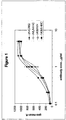


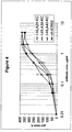

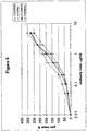
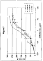
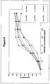

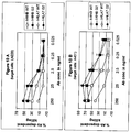

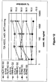
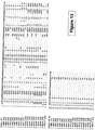
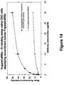
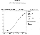
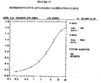


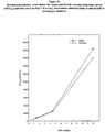

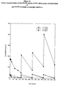
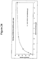

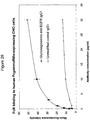

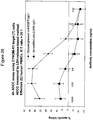
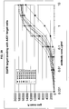
Classifications
-
- C—CHEMISTRY; METALLURGY
- C07—ORGANIC CHEMISTRY
- C07K—PEPTIDES
- C07K16/00—Immunoglobulins [IGs], e.g. monoclonal or polyclonal antibodies
- C07K16/18—Immunoglobulins [IGs], e.g. monoclonal or polyclonal antibodies against material from animals or humans
- C07K16/28—Immunoglobulins [IGs], e.g. monoclonal or polyclonal antibodies against material from animals or humans against receptors, cell surface antigens or cell surface determinants
- C07K16/2863—Immunoglobulins [IGs], e.g. monoclonal or polyclonal antibodies against material from animals or humans against receptors, cell surface antigens or cell surface determinants against receptors for growth factors, growth regulators
-
- C—CHEMISTRY; METALLURGY
- C07—ORGANIC CHEMISTRY
- C07K—PEPTIDES
- C07K16/00—Immunoglobulins [IGs], e.g. monoclonal or polyclonal antibodies
-
- A—HUMAN NECESSITIES
- A61—MEDICAL OR VETERINARY SCIENCE; HYGIENE
- A61K—PREPARATIONS FOR MEDICAL, DENTAL OR TOILETRY PURPOSES
- A61K38/00—Medicinal preparations containing peptides
- A61K38/16—Peptides having more than 20 amino acids; Gastrins; Somatostatins; Melanotropins; Derivatives thereof
- A61K38/17—Peptides having more than 20 amino acids; Gastrins; Somatostatins; Melanotropins; Derivatives thereof from animals; from humans
- A61K38/177—Receptors; Cell surface antigens; Cell surface determinants
- A61K38/179—Receptors; Cell surface antigens; Cell surface determinants for growth factors; for growth regulators
-
- A—HUMAN NECESSITIES
- A61—MEDICAL OR VETERINARY SCIENCE; HYGIENE
- A61K—PREPARATIONS FOR MEDICAL, DENTAL OR TOILETRY PURPOSES
- A61K39/00—Medicinal preparations containing antigens or antibodies
- A61K39/395—Antibodies; Immunoglobulins; Immune serum, e.g. antilymphocytic serum
- A61K39/39533—Antibodies; Immunoglobulins; Immune serum, e.g. antilymphocytic serum against materials from animals
-
- A—HUMAN NECESSITIES
- A61—MEDICAL OR VETERINARY SCIENCE; HYGIENE
- A61K—PREPARATIONS FOR MEDICAL, DENTAL OR TOILETRY PURPOSES
- A61K45/00—Medicinal preparations containing active ingredients not provided for in groups A61K31/00 - A61K41/00
- A61K45/06—Mixtures of active ingredients without chemical characterisation, e.g. antiphlogistics and cardiaca
-
- A—HUMAN NECESSITIES
- A61—MEDICAL OR VETERINARY SCIENCE; HYGIENE
- A61K—PREPARATIONS FOR MEDICAL, DENTAL OR TOILETRY PURPOSES
- A61K47/00—Medicinal preparations characterised by the non-active ingredients used, e.g. carriers or inert additives; Targeting or modifying agents chemically bound to the active ingredient
- A61K47/50—Medicinal preparations characterised by the non-active ingredients used, e.g. carriers or inert additives; Targeting or modifying agents chemically bound to the active ingredient the non-active ingredient being chemically bound to the active ingredient, e.g. polymer-drug conjugates
- A61K47/51—Medicinal preparations characterised by the non-active ingredients used, e.g. carriers or inert additives; Targeting or modifying agents chemically bound to the active ingredient the non-active ingredient being chemically bound to the active ingredient, e.g. polymer-drug conjugates the non-active ingredient being a modifying agent
- A61K47/68—Medicinal preparations characterised by the non-active ingredients used, e.g. carriers or inert additives; Targeting or modifying agents chemically bound to the active ingredient the non-active ingredient being chemically bound to the active ingredient, e.g. polymer-drug conjugates the non-active ingredient being a modifying agent the modifying agent being an antibody, an immunoglobulin or a fragment thereof, e.g. an Fc-fragment
- A61K47/6801—Drug-antibody or immunoglobulin conjugates defined by the pharmacologically or therapeutically active agent
- A61K47/6803—Drugs conjugated to an antibody or immunoglobulin, e.g. cisplatin-antibody conjugates
- A61K47/6811—Drugs conjugated to an antibody or immunoglobulin, e.g. cisplatin-antibody conjugates the drug being a protein or peptide, e.g. transferrin or bleomycin
- A61K47/6817—Toxins
-
- A—HUMAN NECESSITIES
- A61—MEDICAL OR VETERINARY SCIENCE; HYGIENE
- A61P—SPECIFIC THERAPEUTIC ACTIVITY OF CHEMICAL COMPOUNDS OR MEDICINAL PREPARATIONS
- A61P35/00—Antineoplastic agents
-
- C—CHEMISTRY; METALLURGY
- C07—ORGANIC CHEMISTRY
- C07K—PEPTIDES
- C07K16/00—Immunoglobulins [IGs], e.g. monoclonal or polyclonal antibodies
- C07K16/18—Immunoglobulins [IGs], e.g. monoclonal or polyclonal antibodies against material from animals or humans
- C07K16/28—Immunoglobulins [IGs], e.g. monoclonal or polyclonal antibodies against material from animals or humans against receptors, cell surface antigens or cell surface determinants
-
- A—HUMAN NECESSITIES
- A61—MEDICAL OR VETERINARY SCIENCE; HYGIENE
- A61K—PREPARATIONS FOR MEDICAL, DENTAL OR TOILETRY PURPOSES
- A61K39/00—Medicinal preparations containing antigens or antibodies
- A61K2039/505—Medicinal preparations containing antigens or antibodies comprising antibodies
-
- C—CHEMISTRY; METALLURGY
- C07—ORGANIC CHEMISTRY
- C07K—PEPTIDES
- C07K2317/00—Immunoglobulins specific features
- C07K2317/20—Immunoglobulins specific features characterized by taxonomic origin
- C07K2317/21—Immunoglobulins specific features characterized by taxonomic origin from primates, e.g. man
-
- C—CHEMISTRY; METALLURGY
- C07—ORGANIC CHEMISTRY
- C07K—PEPTIDES
- C07K2317/00—Immunoglobulins specific features
- C07K2317/40—Immunoglobulins specific features characterized by post-translational modification
- C07K2317/41—Glycosylation, sialylation, or fucosylation
-
- C—CHEMISTRY; METALLURGY
- C07—ORGANIC CHEMISTRY
- C07K—PEPTIDES
- C07K2317/00—Immunoglobulins specific features
- C07K2317/50—Immunoglobulins specific features characterized by immunoglobulin fragments
- C07K2317/52—Constant or Fc region; Isotype
-
- C—CHEMISTRY; METALLURGY
- C07—ORGANIC CHEMISTRY
- C07K—PEPTIDES
- C07K2317/00—Immunoglobulins specific features
- C07K2317/50—Immunoglobulins specific features characterized by immunoglobulin fragments
- C07K2317/56—Immunoglobulins specific features characterized by immunoglobulin fragments variable (Fv) region, i.e. VH and/or VL
- C07K2317/565—Complementarity determining region [CDR]
-
- C—CHEMISTRY; METALLURGY
- C07—ORGANIC CHEMISTRY
- C07K—PEPTIDES
- C07K2317/00—Immunoglobulins specific features
- C07K2317/70—Immunoglobulins specific features characterized by effect upon binding to a cell or to an antigen
- C07K2317/73—Inducing cell death, e.g. apoptosis, necrosis or inhibition of cell proliferation
- C07K2317/732—Antibody-dependent cellular cytotoxicity [ADCC]
-
- C—CHEMISTRY; METALLURGY
- C07—ORGANIC CHEMISTRY
- C07K—PEPTIDES
- C07K2319/00—Fusion polypeptide
Definitions
- the present invention relates to a humanized anti-EGFR antibody that specifically binds to human EGFR, wherein said antibody comprises: (a) a heavy chain variable region comprising the SEQ ID NO: 15; and (b) a light chain variable region comprising the sequence of SEQ ID NO: 45; and wherein said antibody comprises a glycoengineered, human Fc region having a reduced number of fucose residues as compared to the non-glycoengineered form of the antibody.
- the present disclosure generally relates to antigen binding molecules (ABMs).
- the present disclosure relates to recombinant monoclonal antibodies, including chimeric, primatized or humanized antibodies specific for human epidermal growth factor receptor (EGFR).
- the present disclosure relates to nucleic acid molecules encoding such ABMs, and vectors and host cells comprising such nucleic acid molecules.
- the disclosure further relates to methods for producing the ABMs described herein, and to methods of using these ABMs in treatment of disease.
- the present disclosure relates to ABMs with modified glycosylation having improved therapeutic properties, including antibodies with increased Fc receptor binding and increased effector function.
- Human epidermal growth factor receptor also known as HER-1 or Erb-B1, and referred to herein as "EGFR"
- EGFR Human epidermal growth factor receptor
- HER-1 or Erb-B1 is a 170 kDa transmembrane receptor encoded by the c-erb B protooncogene, and exhibits intrinsic tyrosine kinase activity ( Modjtahedi et al., Br. J. Cancer 73:228-235 (1996 ); Herbst and Shin, Cancer 94:1593-1611 (2002 )).
- SwissProt database entry P00533 provides the sequence of EGFR.
- EGFR epidermal growth factor
- TGf- ⁇ transforming growth factor- ⁇
- hb-EGF amphiregulin
- betacellulin betacellulin
- epiregulin Herbst and Shin, Cancer 94:1593-1611 (2002 ); Mendelsohn and Baselga, Oncogene 19:6550-6565 (2000 )).
- EGFR regulates numerous cellular processes via tyrosine-kinase mediated signal transduction pathways, including, but not limited to, activation of signal transduction pathways that control cell proliferation, differentiation, cell survival, apoptosis, angiogenesis, mitogenesis, and metastasis ( Atalay et al., Ann. Oncology 14:1346-1363 (2003 ); Tsao and Herbst, Signal 4:4-9 (2003 ); Herbst and Shin, Cancer 94:1593-1611 (2002 ); Modjtahedi et al., Br. J. Cancer 73:228-235 (1996 )).
- EGFR is also expressed in the cells of normal tissues, particularly the epithelial tissues of the skin, liver, and gastrointestinal tract, although at generally lower levels than in malignant cells ( Herbst and Shin, Cancer 94:1593-1611 (2002 )).
- Unconjugated monoclonal antibodies can be useful medicines for the treatment of cancer, as demonstrated by the U.S. Food and Drug Administration's approval of Trastuzumab (HerceptinTM; Genentech Inc,) for the treatment of advanced breast cancer ( Grillo-Lopez, A.-J., et al., Semin. Oncol. 26:66-73 (1999 ); Goldenberg, M. M., Clin. Ther.
- Rituximab (RituxanTM; IDEC Pharmaceuticals, San Diego, CA, and Genentech Inc., San Francisco, CA), for the treatment of CD20 positive B-cell, low-grade or follicular Non-Hodgkin's lymphoma, Gemtuzumab (MylotargTM, Celltech/Wyeth-Ayerst) for the treatment of relapsed acute myeloid leukemia, and Alemtuzumab (CAMPATHTM, Millenium Pharmaceuticals/Schering AG) for the treatment of B cell chronic lymphocytic leukemia.
- Rituximab RituxanTM; IDEC Pharmaceuticals, San Diego, CA, and Genentech Inc., San Francisco, CA
- CD20 positive B-cell low-grade or follicular Non-Hodgkin's lymphoma
- Gemtuzumab (MylotargTM, Celltech/Wyeth-Ayerst) for the treatment of relapsed acute myeloid leukemia
- EMD 55900 is a murine anti-EGFR monoclonal antibody that was raised against human epidermoid carcinoma cell line A431 and was tested in clinical studies of patients with advanced squamous cell carcinoma of the larynx or hypopharynx ( Bier et al., Eur. Arch. Otorhinolaryngol. 252:433-9 (1995 )).
- the rat monoclonal antibodies ICR16, ICR62, and ICR80 which bind the extracellular domain of EGFR, have been shown to be effective at inhibiting the binding of EGF and TGF- ⁇ the receptor. ( Modjtahedi et al., Int. J. Cancer 75:310-316 (1998 )).
- the murine monoclonal antibody 425 is another MAb that was raised against the human A431 carcinoma cell line and was found to bind to a polypeptide epitope on the external domain of the human epidermal growth factor receptor. ( Murthy et al., Arch. Biochem. Biophys. 252(2):549-560 (1987 ).
- a potential problem with the use of murine antibodies in therapeutic treatments is that non-human monoclonal antibodies can be recognized by the human host as a foreign protein; therefore, repeated injections of such foreign antibodies can lead to the induction of immune responses leading to harmful hypersensitivity reactions.
- HAMA Human Anti-Mouse Antibody response
- HSA Human Anti-Rat Antibody
- HARA Human Anti-Rat Antibody response
- these "foreign” antibodies can_be attacked by the immune system of the host such that they are, in effect, neutralized before they reach their target site.
- non-human monoclonal antibodies e.g., murine monoclonal antibodies
- non-human monoclonal antibodies typically lack human effector functionality, i.e., they are unable to, inter alia, mediate complement dependent lysis or lyse human target cells through antibody dependent cellular toxicity or Fc-receptor mediated phagocytosis.
- Chimeric antibodies comprising portions of antibodies from two or more different species (e.g., mouse and human) have been developed as an alternative to "conjugated" antibodies.
- U.S. Patent No. 5,891,996 (Mateo de Acosta del Rio et al .) discusses a mouse/human chimeric antibody, R3, directed against EGFR
- U.S. Pat. No. 5,558,864 discusses generation of chimeric and humanized forms of the murine anti-EGFR MAb 425.
- IMC-C225 (Erbitux®; ImClone) is a chimeric mouse/human anti-EGFR monoclonal antibody (based on mouse M225 monoclonal antibody, which resulted in HAMA responses in human clinical trials) that has been reported to demonstrate antitumor efficacy in various human xenograft models.
- Herbst and Shin, Cancer 94:1593-1611 (2002 ) The efficacy of IMC-C225 has been attributed to several mechanisms, including inhibition of cell events regulated by EGFR signaling pathways, and possibly by increased antibody-dependent cellular toxicity (ADCC) activity ( Herbst and Shin, Cancer 94:1593-1611 (2002 )).
- IMC-C225 was also used in clinical trials, including in combination with radiotherapy and chemotherapy ( Herbst and Shin, Cancer 94:1593-1611 (2002 )).
- Abgenix, Inc. (Fremont, CA) developed ABX-EGF for cancer therapy.
- ABX-EGF is a fully human anti-EGFR monoclonal antibody. ( Yang et al., Crit. Rev. Oncol./Hematol. 38: 17-23 (2001 )).
- WO2004/065540 discloses anti-EGFR antibodies engineered to increase the therapeutic potential.
- WO95/20045 discloses the rat anti-EGFR antibody ICR62 and its use in a phase I trial in human.
- the oligosaccharide component can significantly affect properties relevant to the efficacy of a therapeutic glycoprotein, including physical stability, resistance to protease attack, interactions with the immune system, pharmacokinetics, and specific biological activity. Such properties may depend not only on the presence or absence, but also on the specific structures, of oligosaccharides. Some generalizations between oligosaccharide structure and glycoprotein function can be made. For example, certain oligosaccharide structures mediate rapid clearance of the glycoprotein from the bloodstream through interactions with specific carbohydrate binding proteins, while others can be bound by antibodies and trigger undesired immune reactions. ( Jenkins et al., Nature Biotechnol. 14:975-81 (1996 )).
- Mammalian cells are the preferred hosts for production of therapeutic glycoproteins, due to their capability to glycosylate proteins in the most compatible form for human application. ( Cumming et al., Glycobiology 1:115-30 (1991 ); Jenkins et al., Nature Biotechnol. 14:975-81 (1996 )). Bacteria very rarely glycosylate proteins, and like other types of common hosts, such as yeasts, filamentous fungi, insect and plant cells, yield glycosylation patterns associated with rapid clearance from the blood stream, undesirable immune interactions, and in some specific cases, reduced biological activity. Among mammalian cells, Chinese hamster ovary (CHO) cells have been most commonly used during the last two decades.
- these cells allow consistent generation of genetically stable, highly productive clonal cell lines. They can be cultured to high densities in simple bioreactors using serum-free media, and permit the development of safe and reproducible bioprocesses.
- Other commonly used animal cells include baby hamster kidney (BHK) cells, NS0- and SP2/0-mouse myeloma cells. More recently, production from transgenic animals has also been tested. ( Jenkins et al., Nature Biotechnol. 14:975-81 (1996 )).
- All antibodies contain carbohydrate structures at conserved positions in the heavy chain constant regions, with each isotype possessing a distinct array of N-linked carbohydrate structures, which variably affect protein assembly, secretion or functional activity.
- the structure of the attached N-linked carbohydrate varies considerably, depending on the degree of processing, and can include high-mannose, multiply-branched as well as biantennary complex oligosaccharides. ( Wright, A., and Morrison, S. L., Trends Biotech. 15:26-32 (1997 )).
- IgG1 type antibodies the most commonly used antibodies in cancer immunotherapy, are glycoproteins that have a conserved N-linked glycosylation site at Asn297 in each CH2 domain.
- ADCC antibody dependent cellular cytotoxicity
- Uma ⁇ a et al. showed previously that overexpression in Chinese hamster ovary (CHO) cells of ⁇ (1,4)-N-acetylglucosaminyltransferase III (“GnTIII”), a glycosyltransferase catalyzing the formation of bisected oligosaccharides, significantly increases the in vitro ADCC activity of an anti-neuroblastoma chimeric monoclonal antibody (chCE7) produced by the engineered CHO cells. ( See Uma ⁇ a, P. et al., Nature Biotechnol. 17:176-180 (1999 ); and International Publication No. WO 99/54342 ).
- the antibody chCE7 belongs to a large class of unconjugated mAbs which have high tumor affinity and specificity, but have too little potency to be clinically useful when produced in standard industrial cell lines lacking the GnTIII enzyme ( Umana, P., et al., Nature Biotechnol. 17:176-180 (1999 )). That study was the first to show that large increases of ADCC activity could be obtained by engineering the antibody-producing cells to express GnTIII, which also led to an increase in the proportion of constant region (Fc)-associated, bisected oligosaccharides, including bisected, nonfucosylated oligosaccharides, above the levels found in naturally-occurring antibodies.
- Fc constant region
- EGFR expression particularly abnormal expression (e.g., overexpression) including, but not limited to, cancers of the bladder, brain, head and neck, pancreas, lung, breast, ovary, colon, prostate, and kidney.
- ABSMs antigen binding molecules
- rat ICR62 antibody e.g., bind the same epitope
- present inventors developed a method for producing such ABMs. Inter alia, this method involves producing recombinant, chimeric antibodies or chimeric fragments thereof. The efficacy of these ABMs is further enhanced by engineering the glycosylation profile of the antibody Fc region.
- the disclosure is directed to an isolated polynucleotide comprising: (a) a sequence selected from a group consisting of: SEQ ID NO:54, SEQ ID NO:56, SEQ ID NO:58, SEQ ID NO:60, SEQ ID NO:62, SEQ ID NO:64, SEQ ID NO:66, SEQ ID NO:68, SEQ ID NO:70, SEQ ID NO:72, SEQ ID NO:74, SEQ ID NO:122, and SEQ ID NO:124; (b) a sequence selected from a group consisting of: SEQ ID NO:76, SEQ ID NO:78, SEQ ID NO:80, SEQ ID NO:82, SEQ ID NO:84, SEQ ID NO:86, SEQ ID NO:88, SEQ ID NO:90, SEQ ID NO:92, SEQ ID NO:94, SEQ ID NO:96, SEQ ID NO:98, SEQ ID NO:100, SEQ ID NO:102, SEQ ID NO:104, SEQ ID NO
- the disclosure is directed to an isolated polynucleotide comprising (a) a sequence selected from the group consisting of SEQ ID NO:112 and SEQ ID NO:114; (b) a sequence selected from the group consisting of SEQ ID NO:116 and SEQ ID NO:118; and (c) SEQ ID NO:119.
- any of these polynucleotides encodes a fusion polypeptide.
- the disclosure is directed to an isolated polynucleotide comprising a sequence selected from the group consisting of SEQ ID No:2; SEQ ID No:4; SEQ ID No:6; SEQ ID No:8; SEQ ID No:10; SEQ ID No:12; SEQ ID No:14; SEQ ID No:16; SEQ ID No:18; SEQ ID No:20; SEQ ID No:22; SEQ ID No:24; SEQ ID No:26; SEQ ID No:28; SEQ ID No:30; SEQ ID No32; SEQ ID No:34; SEQ ID No:36; SEQ ID No:38; SEQ ID No:40 and SEQ ID No:120.
- the disclosure is directed to an isolated polynucleotide comprising a sequence selected from the group consisting of SEQ ID No:44; SEQ ID No:46; SEQ ID No:50; and SEQ ID No.:52.
- such polynucleotides encode fusion polypeptides.
- the disclosure is further directed to an isolated polynucleotide comprising a sequence having at least 80%, 85%, 90%, 95%, or 99% identity to a sequence selected from the group consisting of SEQ ID No:2; SEQ ID No:4; SEQ ID No:6; SEQ ID No:8; SEQ ID No:10; SEQ ID No:12; SEQ ID No:14; SEQ ID No:16; SEQ ID No:18; SEQ ID No:20; SEQ ID No:22; SEQ ID No:24; SEQ ID No:26; SEQ ID No:28; SEQ ID No:30; SEQ ID No32; SEQ ID No:34; SEQ ID No:36; SEQ ID No:38; SEQ ID No:40 and SEQ ID No:120, wherein said isolated polynucleotide encodes a fusion polypeptide.
- the disclosure is directed to an isolated polynucleotide comprising a sequence having at least 80% identity to a sequence selected from the group consisting of SEQ ID No:44; SEQ ID No:46; SEQ ID No:50; and SEQ ID No.:52, wherein said isolated polynucleotide encodes a fusion polypeptide.
- the disclosure is also directed to an isolated polynucleotide encoding a chimeric polypeptide having the sequence of SEQ ID No.:1.
- the polynucleotide comprises a sequence encoding a polypeptide having the sequence of SEQ ID No.:1; and a sequence encoding a polypeptide having the sequence of an antibody Fc region, or a fragment thereof, from a species other than rat.
- the disclosure is also directed to an isolated polynucleotide encoding a chimeric polypeptide having a sequence selected from the group consisting of SEQ ID No:3; SEQ ID No:5; SEQ ID No:7; SEQ ID No:9; SEQ ID No:11; SEQ ID No:13; SEQ ID No:15; SEQ ID No:17; SEQ ID No:19; SEQ ID No:21; SEQ ID No:23; SEQ ID No:25; SEQ ID No:27; SEQ ID No:29; SEQ ID No:31; SEQ ID No33; SEQ ID No:35; SEQ ID No:37; SEQ ID No:39; and SEQ ID No:121.
- the polynucleotide comprises a sequence encoding a polypeptide having a sequence selected from the group consisting of SEQ ID No:3; SEQ ID No:5; SEQ ID No:7; SEQ ID No:9; SEQ ID No:11; SEQ ID No:13; SEQ ID No:15; SEQ ID No:17; SEQ ID No:19; SEQ ID No:21; SEQ ID No:23; SEQ ID No:25; SEQ ID No:27; SEQ ID No:29; SEQ ID No:31; SEQ ID No33; SEQ ID No:35; SEQ ID No:37; SEQ ID No:39; and SEQ ID No:121; and a sequence encoding a polypeptide having the sequence of an antibody Fc region, or a fragment thereof, from a species other than rat.
- the disclosure is directed to an isolated polynucleotide encoding a chimeric polypeptide having the sequence of SEQ ID No.:43.
- the polynucleotide comprises a sequence encoding a polypeptide having the sequence of SEQ ID No.:43; and a sequence encoding a polypeptide having the sequence of an antibody Fc region, or a fragment thereof, from a species other than rat.
- the disclosure is directed to an isolated polynucleotide encoding a chimeric polypeptide having a sequence selected from the group consisting of SEQ ID No:45; SEQ ID No:49; and SEQ ID No.:51.
- the polynucleotide comprises a sequence encoding a polypeptide having a sequence selected from the group consisting of SEQ ID No:45; SEQ ID No:49; and SEQ ID No.:51, and a sequence encoding a polypeptide having the sequence of an antibody light chain constant region (CL), or a fragment thereof, from a species other than rat.
- CL antibody light chain constant region
- the disclosure is also directed to an isolated polynucleotide comprising a sequence encoding a polypeptide having the VH region of the ICR62 antibody, or functional variants thereof, and a sequence encoding a polypeptide having the sequence of an antibody Fc region, or a fragment thereof, from a species other than rat.
- the disclosure is directed to an isolated polynucleotide comprising a sequence encoding a polypeptide having the VL region of the ICR62 antibody, or functional variants thereof, and a sequence encoding a polypeptide having the sequence of an antibody CL region, or a fragment thereof, from a species other than rat.
- the disclosure is further directed to an expression vector comprising any of the isolated polynucleotides described above, and to a host cell that comprises such an expression vector. In a further aspect, the disclosure is directed to a host cell comprising any of the isolated polynucleotides described above.
- the disclosure is directed to an isolated polypeptide comprising: (a) a sequence selected from a group consisting of: SEQ ID NO:53 SEQ ID NO:55, SEQ ID NO:57, SEQ ID NO:59, SEQ ID NO:61, SEQ ID NO:63, SEQ ID NO:65, SEQ ID NO:67, SEQ ID NO:69, SEQ ID NO:71, SEQ ID NO:73, SEQ ID NO:123, and SEQ ID NO:125; (b) a sequence selected from a group consisting of: SEQ ID NO:75, SEQ ID NO:77, SEQ ID NO:79, SEQ ID NO:81, SEQ ID NO:83, SEQ ID NO:85, SEQ ID NO:87, SEQ ID NO:89, SEQ ID NO:91, SEQ ID NO:93, SEQ ID NO:95, SEQ ID NO:97, SEQ ID NO:99, SEQ ID NO:101, SEQ ID NO:103, SEQ ID NO:105, and SEQ ID NO:
- polypeptide is a fusion polypeptide.
- the disclosure is directed to an isolated polypeptide comprising (a) a sequence selected from the group consisting of SEQ ID NO:111 and SEQ ID NO:113; (b) SEQ ID NO:115; and (c) SEQ ID NO:117, wherein said polypeptide is a fusion polypeptide.
- the disclosure is also directed to a chimeric polypeptide comprising the sequence of SEQ ID NO.:1 or a variant thereof.
- the disclosure is further directed to a chimeric polypeptide comprising the sequence of SEQ ID NO.:43 or a variant thereof.
- any one of these polypeptides further comprises a human Fc region and/or a human CL region.
- the disclosure is also directed to a chimeric polypeptide comprising a sequence selected from the group consisting of SEQ ID No:3; SEQ ID No:5; SEQ ID No:7; SEQ ID No:9; SEQ ID No:11; SEQ ID No:13; SEQ ID No:15; SEQ ID No:17; SEQ ID No:19; SEQ ID No:21; SEQ ID No:23; SEQ ID No:25; SEQ ID No:27; SEQ ID No:29; SEQ ID No:31; SEQ ID No33; SEQ ID No:35; SEQ ID No:37; SEQ ID No:39; and SEQ ID No:121, or a variant thereof.
- the disclosure is further directed to a chimeric polypeptide comprising s sequence selected from the group consisting of SEQ ID No:45; SEQ ID No:49; and SEQ ID No.:51, or a variant thereof.
- any one of these polypeptides further comprises a human Fc region and/or a human CL region.
- the human Fc region comprises IgG1.
- the disclosure is directed to a polypeptide comprising a sequence derived from the ICR62 antibody and a sequence derived from a heterologous polypeptide and to an antigen-binding molecule comprising such a polypeptide.
- the antigen-binding molecule is an antibody.
- the antibody is chimeric.
- the antibody is humanized or primatized.
- the disclosure is directed to an ABM, which is capable of competing with the rat ICR62 antibody for binding to EGFR and which is chimeric.
- the ABM is an antibody or a fragment thereof.
- the ABM is a recombinant antibody comprising a VH region having an amino acid sequence selected from the group consisting of SEQ ID NO.: 1; SEQ ID No:3; SEQ ID No:5; SEQ ID No:7; SEQ ID No:9; SEQ ID No:11; SEQ ID No:13; SEQ ID No:15; SEQ ID No:17; SEQ ID No:19; SEQ ID No:21; SEQ ID No:23; SEQ ID No:25; SEQ ID No:27; SEQ ID No:29; SEQ ID No:31; SEQ ID No33; SEQ ID No:35; SEQ ID No:37; SEQ ID No:39; and SEQ ID No:121.
- the ABM is a recombinant antibody comprising a VL region having an amino acid sequence selected from the group consisting of SEQ ID NO:43, SEQ ID No:45; SEQ ID No:49; and SEQ ID No.:51.
- the ABM is a recombinant antibody that is primatized.
- the ABM is a recombinant antibody that is humanized.
- the ABM is a recombinant antibody comprising a human Fc region.
- any of the ABMs discussed above may be conjugated to a moiety such as a toxin or a radiolabel.
- the disclosure is further related to an ABM of the present disclosure, said ABM having modified oligosaccharides.
- the modified oligosaccharides have reduced fucosylation as compared to non-modified oligosaccharides.
- the modified oligosaccharides are hybrid or complex.
- the ABM has an increased proportion of nonfucosylated oligosaccharides or bisected, nonfucosylated oligosaccharides in the Fc region of said molecule.
- the bisected, nonfucosylated oligosaccharides are hybrid.
- the bisected, nonfucosylated oligosaccharides are complex.
- At least 20% of the oligosaccharides in the Fc region of said polypeptide are nonfucosylated or bisected, nonfucosylated. In more preferred embodiments, at least 30%, 35%, 40%, 45%, 50%, 55%, 60%, 65%, 70% or 75% or more of the oligosaccharides are nonfucosylated or bisected, nonfucosylated.
- the disclosure is further related to a polynucleotide encoding any of the ABMs discussed above, and to expression vectors and cells comprising such a polynucleotide.
- the disclosure is further related to a method of producing an ABM, which is capable of competing with the rat ICR62 antibody for binding to EGFR and wherein said ABM is chimeric; said method comprising: (a) culturing a host cell comprising a polynucleotide that encodes an ABM of the present disclosure in a medium under conditions allowing the expression of said polynucleotide encoding said ABM; and (b) recovering said ABM from the resultant culture.
- the disclosure is related to a pharmaceutical composition
- a pharmaceutical composition comprising the ABM of the disclosure. It is contemplated that the pharmaceutical composition may further comprise a pharmaceutically acceptable carrier, an adjuvant or a combination thereof.
- the disclosure is related to a method of treating a disease characterized by expression of EGFR (e.g., abnormal or overexpression of EGFR).
- the method comprises administering a therapeutically effective amount of the ABM of the present disclosure to a subject, preferably a mammalian subject, and more preferably a human in need thereof.
- the disease is treated by administering an ABM that is a chimeric (e.g. humanized) antibody, or a chimeric fragment of an antibody.
- the ABM is administered in an amount of about 1.0 mg/kg to about 15.0 mg/kg.
- the ABM is administered in an amount of about 1.5 mg/kg to about 12.0 mg/kg.
- the ABM is administered in an amount of about 1.5 mg/kg to about 4.5 mg/kg. In a further embodiment, the ABM is administered in an amount of about 4.5 mg/kg to about 12.0 mg/kg. In a further embodiment, the ABM is administered in an amount selected from the group consisting of about 1.5, about 4.5, and about 12.0 mg/kg.
- the disclosure is related to a host cell engineered to express at least one nucleic acid encoding a polypeptide having GnTIII activity in an amount sufficient to modify the oligosaccharides in the Fc region of the ABM produced by the host cell, wherein the ABM is capable of competing with the rat ICR62 antibody for binding to EGFR and wherein the ABM is chimeric.
- the polypeptide having GnTIII activity is a fusion polypeptide.
- the ABM produced by the host cell is an antibody or an antibody fragment.
- the antibody or antibody fragment is humanized.
- the ABM comprises a region equivalent to the Fc region of a human IgG.
- the disclosure is also directed to an isolated polynucleotide comprising at least one (e.g., one, two, three, four, five, or six) complementarity determining region of the rat ICR62 antibody, or a variant or truncated form thereof containing at least the specificity-determining residues for said complementarity determining region, wherein said isolated polynucleotide encodes a fusion polypeptide.
- such isolated polynucleotides encode a fusion polypeptide that is an antigen binding molecule.
- the polynucleotide comprises three complementarity determining regions of the rat ICR62 antibody, or variants or truncated forms thereof containing at least the specificity-determining residues for each of said three complementarity determining regions.
- the polynucleotide encodes the entire variable region of the light or heavy chain of a chimeric (e.g., humanized) antibody. The disclosure is further directed to the polypeptides encoded by such polynucleotides.
- the disclosure is directed to an antigen binding molecule comprising at least one (e.g., one, two, three, four, five, or six) complementarity determining region of the rat ICR62 antibody, or a variant or truncated form thereof containing at least the specificity-determining residues for said complementarity determining region, and comprising a sequence derived from a heterologous polypeptide.
- the antigen binding molecule comprises three complementarity determining regions of the rat ICR62 antibody, or variants or truncated forms thereof containing at least the specificity-determining residues for each of said three complementarity determining regions.
- the antigen binding molecule comprises the variable region of an antibody light or heavy chain.
- the antigen binding molecule is a chimeric, e.g., humanized, antibody.
- the disclosure is also directed to methods of making such antigen binding molecules, and the use of same in the treatment of disease, including malignancies such as cancers of the bladder, brain, head and neck, pancreas, lung, breast, ovary, colon, prostate, skin, and kidney.
- the host cell of the present disclosure may be selected from the group that includes, but is not limited to, an HEK293-EBNA cell, a CHO cell, a BHK cell, a NSO cell, a SP2/0 cell, a YO myeloma cell, a P3X63 mouse myeloma cell, a PER cell, a PER.C6 cell or a hybridoma cell.
- the host cell of the disclosure further comprises a transfected polynucleotide comprising a polynucleotide encoding the VL region of the rat ICR62 antibody or variants thereof and a sequence encoding a region equivalent to the Fc region of a human immunoglobulin.
- the host cell of the disclosure further comprises a transfected polynucleotide comprising a polynucleotide encoding the VH region of the rat ICR62 antibody or variants thereof and a sequence encoding a region equivalent to the Fc region of a human immunoglobulin.
- the disclosure is directed to a host cell that produces an ABM that exhibits increased Fc receptor binding affinity and/or increased effector function as a result of the modification of its oligosaccharides.
- the increased binding affinity is to an Fc receptor, particularly, the Fc ⁇ RIIIA receptor.
- the effector function contemplated herein may be selected from the group that includes, but is not limited to, increased Fc-mediated cellular cytotoxicity; increased binding to NK cells; increased binding to macrophages; increased binding to polymorphonuclear cells; increased binding to monocytes; increased direct signaling inducing apoptosis; increased dendritic cell maturation; and increased T cell priming.
- the host cell of the present disclosure comprises at least one nucleic acid encoding a polypeptide having GnTIII activity that is operably linked to a constitutive promoter element.
- the disclosure is directed to a method for producing an ABM in a host cell, comprising: (a) culturing a host cell engineered to express at least one polynucleotide encoding a fusion polypeptide having GnTIII activity under conditions which permit the production of said ABM and which permit the modification of the oligosaccharides present on the Fc region of said ABM; and (b) isolating said ABM; wherein said ABM is capable of competing with the rat ICR62 antibody for binding to EGFR and wherein said ABM is chimeric (e.g., humanized).
- the polypeptide having GnTIII activity is a fusion polypeptide, preferably comprising the catalytic domain of GnTIII and the Golgi localization domain of a heterologous Golgi resident polypeptide selected from the group consisting of the localization domain of mannosidase II, the localization domain of ⁇ (1,2)-N-acetylglucosaminyltransferase I ("GnTI"), the localization domain of mannosidase I, the localization domain of ⁇ (1,2)-N-acetylglucosaminyltransferase II (“GnTII”), and the localization domain of ⁇ 1-6 core fucosyltransferase.
- the Golgi localization domain is from mannosidase II or GnTI.
- the disclosure is directed to a method for modifying the glycosylation profile of an anti-EGFR ABM produced by a host cell comprising introducing into the host cell at least one nucleic acid or expression vector of the disclosure.
- the ABM is an antibody or a fragment thereof; preferably comprising the Fc region of an IgG.
- the polypeptide is a fusion protein that includes a region equivalent to the Fc region of a human IgG.
- the disclosure is related to a recombinant, chimeric antibody, or a fragment thereof, capable of competing with the rat ICR62 antibody for binding to EGFR and having reduced fucosylation.
- the present disclosure is directed to a method of modifying the glycosylation of the recombinant antibody or a fragment thereof of the disclosure by using a fusion polypeptide having GnTIII activity and comprising the Golgi localization domain of a heterologous Golgi resident polypeptide.
- the fusion polypeptides of the disclosure comprise the catalytic domain of GnTIII.
- the Golgi localization domain is selected from the group consisting of: the localization domain of mannosidase II, the localization domain of GnTI, the localization domain of mannosidase I, the localization domain of GnTII and the localization domain of ⁇ 1-6 core fucosyltransferase.
- the Golgi localization domain is from mannosidase II or GnTI.
- the method of the disclosure is directed towards producing a recombinant, chimeric antibody or a fragment thereof, with modified oligosaccharides wherein said modified oligosaccharides have reduced fucosylation as compared to non-modified oligosaccharides.
- these modified oligosaccharides may be hybrid or complex.
- the method of the disclosure is directed towards producing a recombinant, chimeric (e.g., humanized) antibody or a fragment thereof having an increased proportion of bisected, nonfucosylated oligosaccharides in the Fc region of said polypeptide.
- the bisected, nonfucosylated oligosaccharides are hybrid.
- the bisected, nonfucosylated oligosaccharides are complex.
- the method of the disclosure is directed towards producing a recombinant, chimeric antibody or a fragment thereof having at least 20% of the oligosaccharides in the Fc region of said polypeptide that are bisected, nonfucosylated.
- at least 30% of the oligosaccharides in the Fc region of said polypeptide are bisected, nonfucosylated.
- at least 35% of the oligosaccharides in the Fc region of said polypeptide are bisected, nonfucosylated.
- the disclosure is directed to a recombinant, chimeric antibody or a fragment thereof, that exhibits increased Fc receptor binding affinity and/or increased effector function as a result of the modification of its oligosaccharides.
- the increased binding affinity is to an Fc activating receptor.
- the Fc receptor is Fc ⁇ activating receptor, particularly, the Fc ⁇ RIIIA receptor.
- the effector function contemplated herein may be selected from the group that includes, but is not limited to, increased Fc-mediated cellular cytotoxicity; increased binding to NK cells; increased binding to macrophages; increased binding to polymorphonuclear cells; increased binding to monocytes; increased direct signaling inducing apoptosis; increased dendritic cell maturation; and increased T cell priming.
- the disclosure is directed to a recombinant, chimeric (e.g., humanized) antibody fragment, having the binding specificity of the rat ICR62 antibody and containing the Fc region, that is engineered to have increased effector function produced by any of the methods of the present disclosure.
- chimeric e.g., humanized
- the present disclosure is directed to a fusion protein that includes a polypeptide having a sequence selected from the group consisting of SEQ ID NO.: 1; SEQ ID No:3; SEQ ID No:5; SEQ ID No:7; SEQ ID No:9; SEQ ID No:11; SEQ ID No:13; SEQ ID No:15; SEQ ID No:17; SEQ ID No:19; SEQ ID No:21; SEQ ID No:23; SEQ ID No:25; SEQ ID No:27; SEQ ID No:29; SEQ ID No:31; SEQ ID No33; SEQ ID No:35; SEQ ID No:37; SEQ ID No:39; and SEQ ID No:121, and a region equivalent to the Fc region of an immunoglobulin and engineered to have increased effector function produced by any of the methods of the present disclosure.
- the present disclosure is directed to a fusion protein that includes a polypeptide having a sequence selected from the group consisting of SEQ ID NO:43, SEQ ID No:45; SEQ ID No:49; and SEQ ID No.:51and a region equivalent to the Fc region of an immunoglobulin and engineered to have increased effector function produced by any of the methods of the present invention.
- the present disclosure is directed to a pharmaceutical composition comprising a recombinant, chimeric (e.g.., humanized) antibody, produced by any of the methods of the present disclosure, and a pharmaceutically acceptable carrier.
- the present disclosure is directed to a pharmaceutical composition comprising a recombinant, chimeric (e.g., humanized) antibody fragment produced by any of the methods of the present disclosure, and a pharmaceutically acceptable carrier.
- the present disclosure is directed to a pharmaceutical composition comprising a fusion protein produced by any of the methods of the present disclosure and a pharmaceutically acceptable carrier.
- the disclosure is directed to a method for targeting in vivo or in vitro cells expressing EGFR.
- a method for targeting cells expressing EGFR in a subject comprising administering to the subject a composition comprising an ABM of the disclosure.
- the present disclosure is directed to a method for detecting in vivo or in vitro the presence of EGFR in a sample, e.g., for diagnosing a disorder related to EGFR expression.
- the detection is performed by contacting a sample to be tested, optionally with a control sample, with an ABM of the present disclosure under conditions that allow for formation of a complex between the ABM and EGFR.
- the complex formation is then detected (e.g., by ELISA or other methods known in the art).
- any statistically significant difference in the formation of ABM-EGFR complexes when comparing the test and control samples is indicative of the presence of EGFR in the test sample.
- the disclosure is further directed to a method of treating a disorder related to EGFR expression, in particular, a cell proliferation disorder wherein EGFR is expressed, and more particularly, wherein EGFR is abnormally expressed (e.g. overexpressed), including cancers of the bladder, brain, head and neck, pancreas, lung, breast, ovary, colon, prostate, skin, and kidney comprising administering a therapeutically effective amount of the recombinant, chimeric (e.g., humanized) antibody or fragment thereof, produced by any of the methods of the present disclosure, to a human subject in need thereof.
- a disorder related to EGFR expression in particular, a cell proliferation disorder wherein EGFR is expressed, and more particularly, wherein EGFR is abnormally expressed (e.g. overexpressed), including cancers of the bladder, brain, head and neck, pancreas, lung, breast, ovary, colon, prostate, skin, and kidney comprising administering a therapeutically effective amount of the recombinant, chimeric (e
- antibody is intended to include whole antibody molecules, including monoclonal, polyclonal and multispecific (e.g ., bispecific) antibodies, as well as antibody fragments having the Fc region and retaining binding specificity, and fusion proteins that include a region equivalent to the Fc region of an immunoglobulin and that retain binding specificity.
- antibody fragments that retain binding specificity including, but not limited to, VH fragments, VL fragments, Fab fragments, F(ab') 2 fragments, scFv fragments, Fv fragments, minibodies, diabodies, triabodies, and tetrabodies ( see, e.g. , Hudson and Souriau, Nature Med. 9: 129-134 (2003 )).
- humanized, primatized and chimeric antibodies are also encompassed.
- Fc region is intended to refer to a C-terminal region of an IgG heavy chain. Although the boundaries of the Fc region of an IgG heavy chain might vary slightly, the human IgG heavy chain Fc region is usually defined to stretch from the amino acid residue at position Cys226 to the carboxyl-terminus.
- region equivalent to the Fc region of an immunoglobulin is intended to include naturally occurring allelic variants of the Fc region of an immunoglobulin as well as variants having alterations which produce substitutions, additions, or deletions but which do not decrease substantially the ability of the immunoglobulin to mediate effector functions (such as antibody dependent cellular cytotoxicity).
- one or more amino acids can be deleted from the N-terminus or C-terminus of the Fc region of an immunoglobulin without substantial loss of biological function.
- variants can be selected according to general rules known in the art so as to have minimal effect on activity. ( See, e.g. , Bowie, J. U. et al., Science 247:1306-1310 (1990 ).
- EGFR refers to the human epidermal growth factor receptor (also known as HER-1 or Erb-B1) ( Ulrich, A. et al., Nature 309:418-425 (1984 ); SwissProt Accession #P00533; secondary accession numbers: O00688, O00732, P06268, Q14225, Q92795, Q9BZS2, Q9GZX1, Q9H2C9, Q9H3C9, Q9UMD7, Q9UMD8, Q9UMG5), as well as naturally-occurring isoforms and variants thereof.
- Such isoforms and variants include but are not limited to the EGFRvIII variant, alternative splicing products (e.g., as identified by SwissProt Accession numbers P00533-1, P00533-2, P00533-3, P00533-4), variants GLN-98, ARG-266, Lys-521, ILE-674, GLY-962, and PRO-988 ( Livingston, R.J.
- EGFR ligand refers to a polypeptide which binds to and/or activates EGFR.
- the term includes membrane-bound precursor forms of the EGFR ligand, as well as proteolytically processed soluble forms of the EGFR ligand.
- ligand activation of EGFR refers to signal transduction (e.g., that caused by an intracellular kinase domain of EGFR receptor phosphorylating tyrosine residues in the EGFR or a substrate polypeptide) mediated by EGFR ligand binding.
- the term disease or disorder characterized by abnormal activation or production of EGFR or an EGFR ligand or disorder related to EGFR expression refers to a condition, which may or may not involve malignancy or cancer, where abnormal activation and/or production of EGFR and/or an EGFR ligand is occurring in cells or tissues of a subject having, or predisposed to, the disease or disorder.
- overexpress As used herein, the terms overexpress, overexpressed, and overexpressing, as used in connection with cells expressing EGFR, refer to cells which have measurably higher levels of EGFR on the surface thereof compared to a normal cell of the same tissue type. Such overexpression may be caused by gene amplification or by increased transcription or translation.
- EGFR expression may be determined in a diagnostic or prognostic assay by evaluating levels of EGFR present on the surface of a cell or in a cell lysate by techniques that are known in the art: e.g., via an immunohistochemistry assay, immunofluorescence assay, immunoenzyme assay, ELISA, flow cytometry, radioimmunoassay, Western blot, ligand binding, kinase activity, etc. ( See generally, CELL BIOLOGY: A LABORATORY HANDBOOK, Celis, J., ed., Academic Press (2d ed., 1998 ); CURRENT PROTOCOLS IN PROTEIN SCIENCE, Coligan, J.E.
- EGFR-encoding nucleic acid molecules in the cell e.g., via fluorescent in situ hybridization, Southern blotting, or PCR techniques.
- the levels of EGFR in normal cells are compared to the levels of cells affected by a cell proliferation disorder (e.g., cancer) to determine if EGFR is overexpressed.
- an antigen binding molecule refers in its broadest sense to a molecule that specifically binds an antigenic determinant. More specifically, an antigen binding molecule that binds EGFR is a molecule which specifically binds to a transmembrane receptor of 170 kDa, typically designated as the epidermal growth factor receptor (EGFR), but also known as HER-1 or ErbB1. By “specifically binds” is meant that the binding is selective for the antigen and can be discriminated from unwanted or nonspecific interactions.
- EGFR epidermal growth factor receptor
- fusion and chimeric when used in reference to polypeptides such as ABMs refer to polypeptides comprising amino acid sequences derived from two or more heterologous polypeptides, such as portions of antibodies from different species.
- the non-antigen binding components may be derived from a wide variety of species, including primates such as chimpanzees and humans.
- the constant region of the chimeric ABM is most preferably substantially identical to the constant region of a natural human antibody; the variable region of the chimeric antibody is most preferably substantially identical to that of a recombinant anti-EGFR antibody having the amino acid sequence of the murine variable region.
- Humanized antibodies are a particularly preferred form of fusion or chimeric antibody.
- a polypeptide having GnTIII activity refers to polypeptides that are able to catalyze the addition of a N-acetylglucosamine (GlcNAc) residue in ⁇ -1-4 linkage to the ⁇ -linked mannoside of the trimannosyl core of N-linked oligosaccharides.
- GlcNAc N-acetylglucosamine
- ⁇ (1,4)-N-acetylglucosaminyltransferase III also known as ⁇ -1,4-mannosyl-glycoprotein 4-beta-N-acetylglucosaminyl-transferase (EC 2.4.1.144)
- NC-IUBMB Nomenclature Committee of the International Union of Biochemistry and Molecular Biology
- the candidate polypeptide will exhibit greater activity or not more than about 25-fold less and, preferably, not more than about tenfold less activity, and most preferably, not more than about three-fold less activity relative to the GnTIII.
- variant refers to a polypeptide differing from a specifically recited polypeptide of the disclosure by amino acid insertions, deletions, and substitutions, created using, e g. , recombinant DNA techniques.
- variants of the ABMs of the present disclosure include chimeric, primatized or humanized antigen binding molecules wherein one or several of the amino acid residues are modified by substitution, addition and/or deletion in such manner that does not substantially affect antigen (e.g., EGFR) binding affinity.
- Guidance in determining which amino acid residues may be replaced, added or deleted without abolishing activities of interest may be found by comparing the sequence of the particular polypeptide with that of homologous peptides and minimizing the number of amino acid sequence changes made in regions of high homology (conserved regions) or by replacing amino acids with consensus sequence.
- recombinant variants encoding these same or similar polypeptides may be synthesized or selected by making use of the "redundancy" in the genetic code.
- Various codon substitutions such as the silent changes which produce various restriction sites, may be introduced to optimize cloning into a plasmid or viral vector or expression in a particular prokaryotic or eukaryotic system.
- Mutations in the polynucleotide sequence may be reflected in the polypeptide or domains of other peptides added to the polypeptide to modify the properties of any part of the polypeptide, to change characteristics such as ligand-binding affinities, interchain affinities, or degradation/turnover rate.
- amino acid substitutions are the result of replacing one amino acid with another amino acid having similar structural and/or chemical properties, i.e., conservative amino acid replacements.
- conservative amino acid replacements may be made on the basis of similarity in polarity, charge, solubility, hydrophobicity, hydrophilicity, and/or the amphipathic nature of the residues involved.
- nonpolar (hydrophobic) amino acids include alanine, leucine, isoleucine, valine, proline, phenylalanine, tryptophan, and methionine; polar neutral amino acids include glycine, serine, threonine, cysteine, tyrosine, asparagine, and glutamine; positively charged (basic) amino acids include arginine, lysine, and histidine; and negatively charged (acidic) amino acids include aspartic acid and glutamic acid.
- “Insertions” or “deletions” are preferably in the range of about 1 to 20 amino acids, more preferably 1 to 10 amino acids. The variation allowed may be experimentally determined by systematically making insertions, deletions, or substitutions of amino acids in a polypeptide molecule using recombinant DNA techniques and assaying the resulting recombinant variants for activity.
- humanized is used to refer to an antigen-binding molecule derived from a non-human antigen-binding molecule, for example, a murine antibody, that retains or substantially retains the antigen-binding properties of the parent molecule but which is less immunogenic in humans.
- critical framework residues e.g., those that are important for retaining good antigen binding affinity or antibody functions
- CDRs complementarity determining regions, or CDRs, (CDR1, CDR2 and CDR3) in each of the heavy and light chain variable domains of an antibody, which are flanked by four framework subregions (i.e., FR1, FR2, FR3, and FR4) in each of the heavy and light chain variable domains of an antibody: FR1-CDR1-FR2-CDR2-FR3-CDR3-FR4.
- primatized is used to refer to an antigen-binding molecule derived from a non-primate antigen-binding molecule, for example, a murine antibody, that retains or substantially retains the antigen-binding properties of the parent molecule but which is less immunogenic in primates.
- CDR complementarity determining region
- Kabat et al. also defined a numbering system for variable domain sequences that is applicable to any antibody.
- One of ordinary skill in the art can unambiguously assign this system of "Kabat numbering" to any variable domain sequence, without reliance on any experimental data beyond the sequence itself.
- Kabat numbering refers to the numbering system set forth by Kabat et al., U.S. Dept. of Health and Human Services, "Sequence of Proteins of Immunological Interest” (1983 ). Unless otherwise specified, references to the numbering of specific amino acid residue positions in an ABM are according to the Kabat numbering system.
- sequences of the sequence listing are not numbered according to the Kabat numbering system. However, as stated above, it is well within the ordinary skill of one in the art to determine the Kabat numbering scheme of any variable region sequence in the Sequence Listing based on the numbering of the sequences as presented therein.
- nucleic acid or polynucleotide having a nucleotide sequence at least, for example, 95% "identical" to a reference nucleotide sequence of the present disclosure it is intended that the nucleotide sequence of the polynucleotide is identical to the reference sequence except that the polynucleotide sequence may include up to five point mutations per each 100 nucleotides of the reference nucleotide sequence.
- nucleotide sequence at least 95% identical to a reference nucleotide sequence up to 5% of the nucleotides in the reference sequence may be deleted or substituted with another nucleotide, or a number of nucleotides up to 5% of the total nucleotides in the reference sequence may be inserted into the reference sequence.
- nucleic acid molecule or polypeptide is at least 80%, 85%, 90%, 95%, 96%, 97%, 98% or 99% identical to a nucleotide sequence or polypeptide sequence of the present disclosure can be determined conventionally using known computer programs.
- a preferred method for determining the best overall match between a query sequence (a sequence of the present disclosure) and a subject sequence, also referred to as a global sequence alignment, can be determined using the FASTDB computer program based on the algorithm of Brutlag et al., Comp. App. Biosci. 6:237-245 (1990 ). In a sequence alignment the query and subject sequences are both DNA sequences.
- RNA sequence can be compared by converting U's to T's.
- the result of said global sequence alignment is in percent identity.
- the percent identity is corrected by calculating the number of bases of the query sequence that are 5' and 3' of the subject sequence, which are not matched/aligned, as a percent of the total bases of the query sequence. Whether a nucleotide is matched/aligned is determined by results of the FASTDB sequence alignment.
- This percentage is then subtracted from the percent identity, calculated by the above FASTDB program using the specified parameters, to arrive at a final percent identity score.
- This corrected score is what is used for the purposes of the present disclosure. Only bases outside the 5' and 3' bases of the subject sequence, as displayed by the FASTDB alignment, which are not matched/aligned with the query sequence, are calculated for the purposes of manually adjusting the percent identity score.
- a 90 base subject sequence is aligned to a 100 base query sequence to determine percent identity.
- the deletions occur at the 5' end of the subject sequence and therefore, the FASTDB alignment does not show a matched/alignment of the first 10 bases at 5' end.
- the 10 unpaired bases represent 10% of the sequence (number of bases at the 5' and 3' ends not matched/total number of bases in the query sequence) so 10% is subtracted from the percent identity score calculated by the FASTDB program. If the remaining 90 bases were perfectly matched the final percent identity would be 90%.
- a 90 base subject sequence is compared with a 100 base query sequence.
- deletions are internal deletions so that there are no bases on the 5' or 3' of the subject sequence which are not matched/aligned with the query.
- percent identity calculated by FASTDB is not manually corrected.
- bases 5' and 3' of the subject sequence which are not matched/aligned with the query sequence are manually corrected for. No other manual corrections are to made for the purposes of the present disclosure.
- polypeptide having an amino acid sequence at least, for example, 95% "identical" to a query amino acid sequence of the present disclosure it is intended that the amino acid sequence of the subject polypeptide is identical to the query sequence except that the subject polypeptide sequence may include up to five amino acid alterations per each 100 amino acids of the query amino acid sequence.
- the subject polypeptide sequence may include up to five amino acid alterations per each 100 amino acids of the query amino acid sequence.
- up to 5% of the amino acid residues in the subject sequence may be inserted, deleted, or substituted with another amino acid.
- These alterations of the reference sequence may occur at the amino or carboxy terminal positions of the reference amino acid sequence or anywhere between those terminal positions, interspersed either individually among residues in the reference sequence or in one or more contiguous groups within the reference sequence.
- any particular polypeptide is at least 80%, 85%, 90%, 95%, 96%, 97%, 98% or 99% identical to a reference polypeptide can be determined conventionally using known computer programs.
- a preferred method for determining the best overall match between a query sequence (a sequence of the present disclosure) and a subject sequence, also referred to as a global sequence alignment, can be determined using the FASTDB computer program based on the algorithm of Brutlag et al., Comp. App. Biosci. 6:237-245 (1990 ).
- the query and subject sequences are either both nucleotide sequences or both amino acid sequences.
- the result of said global sequence alignment is in percent identity.
- the percent identity is corrected by calculating the number of residues of the query sequence that are N- and C-terminal of the subject sequence, which are not matched/aligned with a corresponding subject residue, as a percent of the total bases of the query sequence. Whether a residue is matched/aligned is determined by results of the FASTDB sequence alignment.
- This percentage is then subtracted from the percent identity, calculated by the above FASTDB program using the specified parameters, to arrive at a final percent identity score.
- This final percent identity score is what is used for the purposes of the present disclosure. Only residues to the N- and C-termini of the subject sequence, which are not matched/aligned with the query sequence, are considered for the purposes of manually adjusting the percent identity score. That is, only query residue positions outside the farthest N- and C-terminal residues of the subject sequence.
- a 90 amino acid residue subject sequence is aligned with a 100 residue query sequence to determine percent identity.
- the deletion occurs at the N-terminus of the subject sequence and therefore, the FASTDB alignment does not show a matching/alignment of the first 10 residues at the N-terminus.
- the 10 unpaired residues represent 10% of the sequence (number of residues at the N- and C- termini not matched/total number of residues in the query sequence) so 10% is subtracted from the percent identity score calculated by the FASTDB program. If the remaining 90 residues were perfectly matched the final percent identity would be 90%.
- a 90 residue subject sequence is compared with a 100 residue query sequence.
- deletions are internal deletions so there are no residues at the N- or C-termini of the subject sequence which are not matched/aligned with the query.
- percent identity calculated by FASTDB is not manually corrected.
- residue positions outside the N- and C-terminal ends of the subject sequence, as displayed in the FASTDB alignment, which are not matched/aligned with the query sequence are manually corrected for. No other manual corrections are to be made for the purposes of the present disclosure.
- a nucleic acid that "hybridizes under stringent conditions" to a nucleic acid sequence of the disclosure refers to a polynucleotide that hybridizes in an overnight incubation at 42°C in a solution comprising 50% formamide, 5x SSC (750 mM NaCl, 75 mM sodium citrate), 50 mM sodium phosphate (pH 7.6), 5x Denhardt's solution, 10% dextran sulfate, and 20 ⁇ g/ml denatured, sheared salmon sperm DNA, followed by washing the filters in 0.1x SSC at about 65°C.
- 5x SSC 750 mM NaCl, 75 mM sodium citrate
- 50 mM sodium phosphate pH 7.6
- 5x Denhardt's solution 10% dextran sulfate
- 20 ⁇ g/ml denatured, sheared salmon sperm DNA followed by washing the filters in 0.1x SSC at about 65°C.
- Golgi localization domain refers to the amino acid sequence of a Golgi resident polypeptide which is responsible for anchoring the polypeptide in location within the Golgi complex.
- localization domains comprise amino terminal "tails" of an enzyme.
- effector function refers to those biological activities attributable to the Fc region (a native sequence Fc region or amino acid sequence variant Fc region) of an antibody.
- antibody effector functions include, but are not limited to, Fc receptor binding affinity, antibody-dependent cellular cytotoxicity (ADCC), antibody-dependent cellular phagocytosis (ADCP), cytokine secretion, immune-complex-mediated antigen uptake by antigen-presenting cells, down-regulation of cell surface receptors, etc.
- glycosylation engineering includes metabolic engineering of the glycosylation machinery of a cell, including genetic manipulations of the oligosaccharide synthesis pathways to achieve altered glycosylation of glycoproteins expressed in cells.
- glycosylation engineering includes the effects of mutations and cell environment on glycosylation.
- the glycosylation engineering is an alteration in glycosyltransferase activity.
- the engineering results in altered glucosaminyltransferase activity and/or fucosyltransferase activity.
- the term host cell covers any kind of cellular system which can be engineered to generate the polypeptides and antigen-binding molecules of the present disclosure.
- the host cell is engineered to allow the production of an antigen binding molecule with modified glycoforms.
- the antigen binding molecule is an antibody, antibody fragment, or fusion protein.
- the host cells have been further manipulated to express increased levels of one or more polypeptides having GnTIII activity.
- Host cells include cultured cells, e.g.
- mammalian cultured cells such as CHO cells, HEK293-EBNA cells, BHK cells, NS0 cells, SP2/0 cells, YO myeloma cells, P3X63 mouse myeloma cells, PER cells, PER.C6 cells or hybridoma cells, yeast cells, insect cells, and plant cells, to name only a few, but also cells comprised within a transgenic animal, transgenic plant or cultured plant or animal tissue.
- Fc-mediated cellular cytotoxicity includes antibody-dependent cellular cytotoxicity and cellular cytotoxicity mediated by a soluble Fc-fusion protein containing a human Fc-region. It is an immune mechanism leading to the lysis of "antibody-targeted cells” by "human immune effector cells”, wherein:
- the human immune effector cells are a population of leukocytes that display Fc receptors on their surface through which they bind to the Fc-region of antibodies or of Fc-fusion proteins and perform effector functions.
- Such a population may include, but is not limited to, peripheral blood mononuclear cells (PBMC) and/or natural killer (NK) cells.
- PBMC peripheral blood mononuclear cells
- NK natural killer
- the antibody-targeted cells are cells bound by the antibodies or Fc-fusion proteins.
- the antibodies or Fc fusion-proteins bind to target cells via the protein part N-terminal to the Fc region.
- the term increased Fc-mediated cellular cytotoxicity is defined as either an increase in the number of "antibody-targeted cells” that are lysed in a given time, at a given concentration of antibody, or of Fc-fusion protein, in the medium surrounding the target cells, by the mechanism of Fc-mediated cellular cytotoxicity defined above, and/or a reduction in the concentration of antibody, or of Fc-fusion protein, in the medium surrounding the target cells, required to achieve the lysis of a given number of "antibody-targeted cells", in a given time, by the mechanism of Fc - mediated cellular cytotoxicity.
- Fc-mediated cellular cytotoxicity is relative to the cellular cytotoxicity mediated by the same antibody, or Fc-fusion protein, produced by the same type of host cells, using the same standard production, purification, formulation and storage methods, which are known to those skilled in the art, but that has not been produced by host cells engineered to express the glycosyltransferase GnTIII by the methods described herein.
- ADCC antibody dependent cellular cytotoxicity
- the present disclosure is related to antigen binding molecules having the binding specificity of the rat ICR62 (i.e., binds to substantially the same epitope), and to the discovery that their effector functions can be enhanced by altered glycosylation.
- the antigen binding molecule is a chimeric antibody.
- the disclosure is directed to a chimeric antibody, or a fragment thereof, comprising one or more (e.g., one, two, three, four, five, or six) of the the CDRs of any of SEQ ID NOs:53-108 and/or SEQ ID NO:s 122-127.
- the disclosure is directed to an isolated polynucleotide comprising: (a) a sequence selected from a group consisting of: SEQ ID NO:54 SEQ ID NO:56, SEQ ID NO:58, SEQ ID NO:60, SEQ ID NO:62, SEQ ID NO:64, SEQ ID NO:66, SEQ ID NO:68, SEQ ID NO:70, SEQ ID NO:72, SEQ ID NO:74, SEQ ID NO:122, and SEQ ID NO:124; (b) a sequence selected from a group consisting of: SEQ ID NO:76, SEQ ID NO:78, SEQ ID NO:80, SEQ ID NO:82, SEQ ID NO:84, SEQ ID NO:86, SEQ ID NO:88, SEQ ID NO:90, SEQ ID NO:92, SEQ ID NO:94, SEQ ID NO:96, SEQ ID NO:98, SEQ ID NO:100, SEQ ID NO:102, SEQ ID NO:104, SEQ ID NO:56, SEQ ID NO
- the disclosure is directed to an isolated polynucleotide comprising (a) a sequence selected from the group consisting of SEQ ID NO:112 and SEQ ID NO:114; (b) a sequence selected from the group consisting of SEQ ID NO:116 and SEQ ID NO:118; and (c) SEQ ID NO:119.
- any of these polynucleotides encodes a fusion polypeptide.
- the antigen binding molecule comprises the V H domain of the rat ICR62 antibody encoded by SEQ ID NO:1 or SEQ ID NO:2, or a variant thereof; and a non-murine polypeptide.
- the disclosure is directed to an antigen binding molecule comprising the V L domain of the rat antibody encoded by SEQ ID NO:43 or SEQ ID NO:44, or a variant thereof; and a non-murine polypeptide.
- the disclosure is directed to antigen binding molecules comprising one or more (e.g., one, two, three, four, five, or six) truncated CDRs of ICR62.
- truncated CDRs will contain, at a minimum, the specificity-determining amino acid residues for the given CDR.
- specificity-determining residue is meant those residues that are directly involved in the interaction with the antigen. In general, only about one-fifth to one-third of the residues in a given CDR participate in binding to antigen.
- the specificity-determining residues in a particular CDR can be identified by, for example, computation of interatomic contacts from three-dimensional modeling and determination of the sequence variability at a given residue position in accordance with the methods described in Padlan et al., FASEB J. 9(1):133-139 (1995 ).
- the disclosure is also directed to an isolated polynucleotide comprising at least one (e.g., one, two, three, four, five, or six) complementarity determining region of the rat ICR62 antibody, or a variant or truncated form thereof containing at least the specificity-determining residues for said complementarity determining region, wherein said isolated polynucleotide encodes a fusion polypeptide.
- such isolated polynucleotides encode a fusion polypeptide that is an antigen binding molecule.
- the polynucleotide comprises three complementarity determining regions of the rat ICR62 antibody, or variants or truncated forms thereof containing at least the specificity-determining residues for each of said three complementarity determining regions.
- the polynucleotide comprises at least one of the CDRs set forth in Tables 2-5, below.
- the polynucleotide encodes the entire variable region of the light or heavy chain of a chimeric (e.g., humanized) antibody. The disclosure is further directed to the polypeptides encoded by such polynucleotides.
- the disclosure is directed to an antigen binding molecule comprising at least one (e.g., one, two, three, four, five, or six) complementarity determining region of the rat ICR62 antibody, or a variant or truncated form thereof containing at least the specificity-determining residues for said complementarity determining region, and comprising a sequence derived from a heterologous polypeptide.
- the antigen binding molecule comprises three complementarity determining regions of the rat ICR62 antibody, or variants or truncated forms thereof containing at least the specificity-determining residues for each of said three complementarity determining regions.
- the antigen binding molecule comprises at least one of the CDRs set forth in Tables 2-5, below.
- the antigen binding molecule comprises the variable region of an antibody light or heavy chain.
- the antigen binding molecule is a chimeric, e.g., humanized, antibody. The disclosure is also directed to methods of making such antigen binding molecules, and the use of same in the treatment of disease, particularly cell proliferation disorders wherein EGFR is expressed, particularly wherein EGFR is abnormally expressed (e.g., overexpressed) compared to normal tissue of the same cell type.
- Such disorders include, but are not limited to cancers of the bladder, brain, head and neck, pancreas, lung, breast, ovary, colon, prostate, skin, and kidney.
- EGFR expression levels may be determined by methods known in the art and those described herein (e.g., via immunohistochemistry assay, immunofluorescence assay, immunoenzyme assay, ELISA, flow cytometry, radioimmunoassay, Western blot, ligand binding, kinase activity, etc.).
- the disclosure is also directed to a method for targeting in vivo or in vitro cells expressing EGFR.
- Cells that express EGFR may be targeted for therapeutic purposes (e.g., to treat a disorder that is treatable by disruption of EGFR-mediated signaling, for example by blocking ligand binding, or by targeting EGFR-expressing cells for destruction by the immune system).
- the present disclosure is directed to a method for targeting cells expressing EGFR in a subject comprising administering to the subject a composition comprising an ABM of the disclosure.
- Cells that express EGFR may also be targeted for diagnostic purposes (e.g., to determine if they are expressing EGFR, either normally or abnormally).
- the disclosure is also directed to methods for detecting the presence of EGFR or a cell expressing EGFR, either in vivo or in vitro.
- One method of detecting EGFR expression according to the present disclosure comprises contacting a sample to be tested, optionally with a control sample, with an ABM of the present disclosure, under conditions that allow for formation of a complex between the ABM and EGFR. The complex formation is then detected (e.g., by ELISA or other methods known in the art).
- any statistically significant difference in the formation of ABM-EGFR complexes when comparing the test and control samples is indicative of the presence of EGFR in the test sample.
- ligand e.g., EGF, TGF- ⁇ , etc.
- ADCC antibody dependent cellular cytotoxicity
- the rat monoclonal antibody ICR62 (IgG2b) was discussed in PCT Publication No. WO 95/20045 . It was directed to the C epitope of EGFR, and was shown to inhibit ligand binding, inhibit growth in vitro of squamous cell carcinomas expressing EGFR, and induce regression of xenografts of tumors in athymic mice ( WO 95/20045 ; Modjtahedi et al., Br. J. Cancer 73:228-235 (1996 )). As a fully rodent antibody, administration of ICR62 rat monoclonal antibody to humans resulted a HARA response in some patients following even a single dose. ( WO 95/20045 ; Modjtahedi et al., Br. J. Cancer 73:228-235 (1996 )).
- IMC-C225 is a chimeric monoclonal antibody directed against EGFR and having a mouse variable region and a human constant region ( See Herbst and Shin, Cancer 94: 1593-1611 (2002 )).
- the murine portion of IMC-225 is derived from M225, which was found to bind EGFR and inhibit EGF-induced tyrosine kinase-dependent phosphorylation, as well as inducing apoptosis in tumor cell lines over-expressing EGFR ( Herbst and Shin, Cancer 94: 1593-1611 (2002 )). However, M225 elicited a HAMA reaction in patients treated with the antibody in Phase I clinical trials ( Herbst and Shin, Cancer 94: 1593-1611 (2002 )).
- IMC-225 has been tested in vivo and in vitro, and has been used in combination with radiation therapy and chemotherapy in a number of tumor types, including those associated with poor prognosis ( Herbst and Shin, Cancer 94: 1593-1611 (2002 )). However, IMC-225 has been associated with toxicities such as allergic and skin reactions in patients administered the IMC-225 antibody in clincial trials ( Herbst and Shin, Cancer 94: 1593-1611 (2002 )).
- the chimeric ABM of the present disclosure is a humanized antibody.
- Methods for humanizing non-human antibodies are known in the art.
- humanized ABMs of the present disclosure can be prepared according to the methods of U.S. Pat. No. 5,225,539 to Winter , U.S. Pat. No. 6,180,370 to Queen et al. , U.S. Pat. No. 6,632,927 to Adair et al ., or U.S. Pat. Appl. Pub. No. 2003/0039649 to Foote .
- a humanized antibody has one or more amino acid residues introduced into it from a source which is non-human.
- humanized antibodies are typically human antibodies in which some hypervariable region residues and possibly some FR residues are substituted by residues from analogous sites in rodent antibodies.
- the subject humanized anti-EGFR antibodies will comprise constant regions of human immunoglobulin.
- variable domains both light and heavy
- sequence of the variable domain of a rodent antibody is screened against the entire library of known human variable-domain sequences.
- the human sequence which is closest to that of the rodent is then accepted as the human framework region (FR) for the humanized antibody ( Sims et al., J. Immunol., 151:2296 (1993 ); Chothia et al., J. Mol. Biol., 196:901 (1987 )).
- Another method of selecting the human framework sequence is to compare the sequence of each individual subregion of the full rodent framework (i.e., FR1, FR2, FR3, and FR4) or some combination of the individual subregions (e.g., FR1 and FR2) against a library of known human variable region sequences that correspond to that framework subregion (e.g., as determined by Kabat numbering), and choose the human sequence for each subregion or combination that is the closest to that of the rodent ( Leung, U.S. Patent Application Publication No. 2003/0040606A1, published Feb. 27, 2003 ).
- Another method uses a particular framework region derived from the consensus sequence of all human antibodies of a particular subgroup of light or heavy chains.
- the same framework may be used for several different humanized antibodies ( Carter et al., Proc. Natl. Acad. Sci. USA, 89:4285 (1992 ); Presta et al., J. Immunol., 151:2623 (1993 )).
- humanized antibodies are prepared by a process of analysis of the parental sequences and various conceptual humanized products using three-dimensional models of the parental and humanized sequences.
- Three-dimensional immunoglobulin models can be generated using computer programs familiar to those skilled in the art (e.g., InsightII, Accelrys, Inc. (formerly MSI), or at http://swissmodel.expasy.org). These computer programs can illustrate and display probable three-dimensional conformational structures of selected candidate immunoglobulin sequences.
- the antibodies of the present disclosure comprise a human Fc region.
- the human constant region is IgG1, as set forth in SEQ ID NOs 109 and 110, and set forth below:
- variants and isoforms of the human Fc region are also encompassed by the present disclosure.
- variant Fc regions suitable for use in the present disclosure can be produced according to the methods taught in U.S. Pat. No. 6,737,056 to Presta (Fc region variants with altered effector function due to one or more amino acid modifications); or in U.S. Pat. Appl. Nos. 60/439,498 ; 60/456,041 ; 60/514,549 ; or WO 2004/063351 (variant Fc regions with increased binding affinity due to amino acid modification); or in U.S. Pat. Appl. No. 10/672,280 or WO 2004/099249 (Fc variants with altered binding to FcyR due to amino acid modification).
- the antigen binding molecules of the present disclosure are engineered to have enhanced binding affinity according to, for example, the methods disclosed in U.S. Pat. Appl. Pub. No. 2004/0132066 to Balint et al. .
- the antigen binding molecule of the present disclosure is conjugated to an additional moiety, such as a radiolabel or a toxin.
- an additional moiety such as a radiolabel or a toxin.
- conjugated ABMs can be produced by numerous methods that are well known in the art.
- 131 iodine is a well known radionuclide used for targeted immunotherapy.
- the clinical usefulness of 131 iodine can be limited by several factors including: eight-day physical half-life; dehalogenation of iodinated antibody both in the blood and at tumor sites; and emission characteristics (e.g., large gamma component) which can be suboptimal for localized dose deposition in tumor.
- 90 Yttrium provides several benefits for utilization in radioimmunotherapeutic applications: the 64 hour half-life of 90 yttrium is long enough to allow antibody accumulation by tumor and, unlike e.g., 131 iodine, 90 yttrium is a pure beta emitter of high energy with no accompanying gamma irradiation in its decay, with a range in tissue of 100 to 1000 cell diameters. Furthermore, the minimal amount of penetrating radiation allows for outpatient administration of 90 yttrium-labeled antibodies. Additionally, internalization of labeled antibody is not required for cell killing, and the local emission of ionizing radiation should be lethal for adjacent tumor cells lacking the target antigen.
- Effective single treatment dosages i.e., therapeutically effective amounts
- Effective single treatment non-marrow ablative dosages of 131 iodine labeled anti-EGFR antibodies range from between about 5 and about 70 mCi, more preferably between about 5 and about 40 mCi.
- Effective single treatment ablative dosages i.e., may require autologous bone marrow transplantation
- 131 iodine labeled anti-EGFR antibodies range from between about 30 and about 600 mCi, more preferably between about 50 and less than about 500 mCi.
- an effective single treatment non-marrow ablative dosages of 131 iodine labeled chimeric anti-EGFR antibodies range from between about 5 and about 40 mCi, more preferably less than about 30 mCi. Imaging criteria for, e.g., the 111 indium label, are typically less than about 5 mCi.
- radiolabeled anti-EGFR antibodies therapy therewith can also occur using a single therapy treatment or using multiple treatments. Because of the radionuclide component, it is preferred that prior to treatment, peripheral stem cells (“PSC”) or bone marrow (“BM”) be “harvested” for patients experiencing potentially fatal bone marrow toxicity resulting from radiation. BM and/or PSC are harvested using standard techniques, and then purged and frozen for possible reinfusion.
- PSC peripheral stem cells
- BM bone marrow
- a diagnostic dosimetry study using a diagnostic labeled antibody e.g., using 111 indium
- a diagnostic labeled antibody e.g., using 111 indium
- the therapeutically labeled antibody e.g., using 90 yttrium
- the present disclosure is directed to an isolated polynucleotide comprising a sequence that encodes a polypeptide having an amino acid sequence in Table 7 below.
- the disclosure is directed to an isolated polynucleotide comprising a sequence shown in Table 6 below.
- the disclosure is further directed to an isolated nucleic acid comprising a sequence at least 80%, 85%, 90%, 95%, 96%, 97%, 98% or 99% identical to a nucleotide sequence shown in Table 6 below.
- the disclosure is directed to an isolated nucleic acid comprising a sequence that encodes a polypeptide having an amino acid sequence at least 80%, 85%, 90%, 95%, 96%, 97%, 98% or 99% identical to an amino acid sequence in Table 7.
- the disclosure also encompasses an isolated nucleic acid comprising a sequence that encodes a polypeptide having the amino acid sequence of any of the constructs in Table 7 with conservative amino acid substitutions.
- the present disclosure is directed to an expression vector and/or a host cell which comprise one or more isolated polynucleotides of the present disclosure.
- any type of cultured cell line can be used to express the ABM of the present disclosure.
- HEK293-EBNA cells CHO cells, BHK cells, NS0 cells, SP2/0 cells, YO myeloma cells, P3X63 mouse myeloma cells, PER cells, PER.C6 cells or hybridoma cells, other mammalian cells, yeast cells, insect cells, or plant cells are used as the background cell line to generate the engineered host cells of the disclosure.
- the therapeutic efficacy of the ABMs of the present disclosure can be enhanced by producing them in a host cell that further expresses a polynucleotide encoding a polypeptide having GnTIII activity.
- the polypeptide having GnTIII activity is a fusion polypeptide comprising the Golgi localization domain of a Golgi resident polypeptide.
- the expression of the ABMs of the present disclosure in a host cell that expresses a polynucleotide encoding a polypeptide having GnTIII activity results in ABMs with increased Fc receptor binding affinity and increased effector function.
- the present disclosure is directed to a host cell comprising (a) an isolated nucleic acid comprising a sequence encoding a polypeptide having GnTIII activity; and (b) an isolated polynucleotide encoding an ABM of the present disclosure, such as a chimeric, primatized or humanized antibody that binds human EGFR.
- the polypeptide having GnTIII activity is a fusion polypeptide comprising the catalytic domain of GnTIII and the Golgi localization domain is the localization domain of mannosidase II.
- the chimeric ABM is a chimeric antibody or a fragment thereof, having the binding specificity of the rat ICR62 antibody.
- the chimeric antibody comprises a human Fc.
- the antibody is primatized or humanized.
- one or several polynucleotides encoding an ABM of the present disclosure may be expressed under the control of a constitutive promoter or, alternately, a regulated expression system.
- Suitable regulated expression systems include, but are not limited to, a tetracycline-regulated expression system, an ecdysone-inducible expression system, a lac-switch expression system, a glucocorticoid-inducible expression system, a temperature-inducible promoter system, and a metallothionein metal-inducible expression system.
- nucleic acids encoding an ABM of the present disclosure are comprised within the host cell system, some of them may be expressed under the control of a constitutive promoter, while others are expressed under the control of a regulated promoter.
- the maximal expression level is considered to be the highest possible level of stable polypeptide expression that does not have a significant adverse effect on cell growth rate, and will be determined using routine experimentation. Expression levels are determined by methods generally known in the art, including Western blot analysis using an antibody specific for the ABM or an antibody specific for a peptide tag fused to the ABM; and Northern blot analysis.
- the polynucleotide may be operatively linked to a reporter gene; the expression levels of a chimeric (e.g., humanized) ABM having substantially the same binding specificity of the rat ICR62 antibody are determined by measuring a signal correlated with the expression level of the reporter gene.
- the reporter gene may be transcribed together with the nucleic acid(s) encoding said fusion polypeptide as a single mRNA molecule; their respective coding sequences may be linked either by an internal ribosome entry site (IRES) or by a cap-independent translation enhancer (CITE).
- the reporter gene may be translated together with at least one nucleic acid encoding a chimeric (e.g., humanized) ABM having substantially the same binding specificity of the rat ICR62 antibody such that a single polypeptide chain is formed.
- the nucleic acids encoding the AMBs of the present disclosure may be operatively linked to the reporter gene under the control of a single promoter, such that the nucleic acid encoding the fusion polypeptide and the reporter gene are transcribed into an RNA molecule which is alternatively spliced into two separate messenger RNA (mRNA) molecules; one of the resulting mRNAs is translated into said reporter protein, and the other is translated into said fusion polypeptide.
- mRNA messenger RNA
- Methods which are well known to those skilled in the art can be used to construct expression vectors containing the coding sequence of an ABM having substantially the same binding specificity of the rat ICR62 antibody along with appropriate transcriptional/translational control signals.
- These methods include in vitro recombinant DNA techniques, synthetic techniques and in vivo recombination/genetic recombination. See, for example, the techniques described in Maniatis et al., Molecular Cloning A Laboratory Manual, Cold Spring Harbor Laboratory, N.Y. (1989 ) and Ausubel et al., Current Protocols in Molecular Biology, Greene Publishing Associates and Wiley Interscience, N.Y (1989 ).
- a variety of host-expression vector systems may be utilized to express the coding sequence of the ABMs of the present disclosure.
- mammalian cells are used as host cell systems transfected with recombinant plasmid DNA or cosmid DNA expression vectors containing the coding sequence of the protein of interest and the coding sequence of the fusion polypeptide.
- HEK293-EBNA cells CHO cells, BHK cells, NS0 cells, SP2/0 cells, YO myeloma cells, P3X63 mouse myeloma cells, PER cells, PER.C6 cells or hybridoma cells, other mammalian cells, yeast cells, insect cells, or plant cells are used as host cell system.
- yeast cells transformed with recombinant yeast expression vectors containing the coding sequence of an ABM of the present disclosure such as the expression systems taught in U.S. Pat. Appl. No.
- WO 03/056914 methods for producing human-like glycoprotein in a non-human eukaryotic host cell
- insect cell systems infected with recombinant virus expression vectors (e.g ., baculovirus) containing the coding sequence of a chimeric ABM having substantially the same binding specificity of the rat ICR62 antibody
- plant cell systems infected with recombinant virus expression vectors e.g ., cauliflower mosaic virus, CaMV; tobacco mosaic virus, TMV
- recombinant plasmid expression vectors e.g ., Ti plasmid
- Pat. No. 6,815,184 (methods for expression and secretion of biologically active polypeptides from genetically engineered duckweed); WO 2004/057002 (production of glycosylated proteins in bryophyte plant cells by introduction of a glycosyl transferase gene) and WO 2004/024927 (methods of generating extracellular heterologous non-plant protein in moss protoplast); and U.S. Pat. Appl. Nos.
- 60/365,769 , 60/368,047 , and WO 2003/078614 glycoprotein processing in transgenic plants comprising a functional mammalian GnTIII enzyme
- animal cell systems infected with recombinant virus expression vectors e.g ., adenovirus, vaccinia virus
- virus expression vectors e.g ., adenovirus, vaccinia virus
- the vector comprising the polynucleotide(s) encoding the ABM of the disclosure is polycistronic.
- the ABM discussed above is an antibody or a fragment thereof.
- the ABM is a humanized antibody.
- stable expression is generally preferred to transient expression because it typically achieves more reproducible results and also is more amenable to large-scale production.
- host cells can be transformed with the respective coding nucleic acids controlled by appropriate expression control elements (e.g ., promoter, enhancer, sequences, transcription terminators, polyadenylation sites, etc.), and a selectable marker.
- appropriate expression control elements e.g ., promoter, enhancer, sequences, transcription terminators, polyadenylation sites, etc.
- engineered cells may be allowed to grow for 1-2 days in an enriched media, and then are switched to a selective media.
- the selectable marker in the recombinant plasmid confers resistance to the selection and allows selection of cells which have stably integrated the plasmid into their chromosomes and grow to form foci which in turn can be cloned and expanded into cell lines.
- a number of selection systems may be used, including, but not limited to, the herpes simplex virus thymidine kinase ( Wigler et al., Cell 11:223 (1977 )), hypoxanthine-guanine phosphoribosyltransferase ( Szybalska & Szybalski, Proc. Natl. Acad. Sci. USA 48:2026 (1962 )), and adenine phosphoribosyltransferase ( Lowy et al., Cell 22:817 (1980 )) genes, which can be employed in tk-, hgprt- or aprt- cells, respectively.
- the herpes simplex virus thymidine kinase Wigler et al., Cell 11:223 (1977 )
- hypoxanthine-guanine phosphoribosyltransferase Szybalska & Szybalski, Proc. Natl. Acad. Sci
- antimetabolite resistance can be used as the basis of selection for dhfr, which confers resistance to methotrexate ( Wigler et al., Natl. Acad. Sci. USA 77:3567 (1989 ); O'Hare et al., Proc. Natl. Acad. Sci. USA 78:1527 (1981 )); gpt, which confers resistance to mycophenolic acid ( Mulligan & Berg, Proc. Natl. Acad. Sci. USA 78:2072 (1981 )); neo, which confers resistance to the aminoglycoside G-418 ( Colberre-Garapin et al., J. Mol. Biol.
- hygro which confers resistance to hygromycin
- trpB which allows cells to utilize indole in place of tryptophan
- hisD which allows cells to utilize histinol in place of histidine
- the present disclosure is further directed to a method for modifying the glycosylation profile of the ABMs of the present disclosure that are produced by a host cell, comprising expressing in said host cell a nucleic acid encoding an ABM of the disclosure and a nucleic acid encoding a polypeptide with GnTIII activity, or a vector comprising such nucleic acids.
- the modified polypeptide is IgG or a fragment thereof comprising the Fc region.
- the ABM is a humanized antibody or a fragment thereof.
- the modified ABMs produced by the host cells of the disclosure exhibit increased Fc receptor binding affinity and/or increased effector function as a result of the modification.
- the ABM is a humanized antibody or a fragment thereof containing the Fc region.
- the increased Fc receptor binding affinity is increased binding to a Fc ⁇ activating receptor, such as the Fc ⁇ RIIIa receptor.
- the increased effector function is preferably an increase in one or more of the following: increased antibody-dependent cellular cytotoxicity, increased antibody-dependent cellular phagocytosis (ADCP), increased cytokine secretion, increased immune-complex-mediated antigen uptake by antigen-presenting cells, increased Fc-mediated cellular cytotoxicity, increased binding to NK cells, increased binding to macrophages, increased binding to polymorphonuclear cells (PMNs), increased binding to monocytes, increased crosslinking of target-bound antibodies, increased direct signaling inducing apoptosis, increased dendritic cell maturation, and increased T cell priming.
- ADCP antibody-dependent cellular cytotoxicity
- ADCP antibody-dependent cellular phagocytosis
- cytokine secretion increased immune-complex-mediated antigen uptake by antigen-presenting cells
- Fc-mediated cellular cytotoxicity increased binding to NK cells
- macrophages increased binding to macrophages
- PMNs polymorphonuclear cells
- the present disclosure is also directed to a method for producing an ABM of the present disclosure, having modified oligosaccharides in a host cell comprising (a) culturing a host cell engineered to express at least one nucleic acid encoding a polypeptide having GnTIII activity under conditions which permit the production of an ABM according to the present disclosure, wherein said polypeptide having GnTIII activity is expressed in an amount sufficient to modify the oligosaccharides in the Fc region of said ABM produced by said host cell; and (b) isolating said ABM.
- the polypeptide having GnTIII activity is a fusion polypeptide comprising the catalytic domain of GnTIII.
- the fusion polypeptide further comprises the Golgi localization domain of a Golgi resident polypeptide.
- the Golgi localization domain is the localization domain of mannosidase II or GnTI.
- the Golgi localization domain is selected from the group consisting of: the localization domain of mannosidase I, the localization domain of GnTII, and the localization domain of ⁇ 1-6 core fucosyltransferase.
- the ABMs produced by the methods of the present disclosure have increased Fc receptor binding affinity and/or increased effector function.
- the increased effector function is one or more of the following: increased Fc-mediated cellular cytotoxicity (including increased antibody-dependent cellular cytotoxicity), increased antibody-dependent cellular phagocytosis (ADCP), increased cytokine secretion, increased immune-complex-mediated antigen uptake by antigen-presenting cells, increased binding to NK cells, increased binding to macrophages, increased binding to monocytes, increased binding to polymorphonuclear cells, increased direct signaling inducing apoptosis, increased crosslinking of target-bound antibodies, increased dendritic cell maturation, or increased T cell priming.
- the increased Fc receptor binding affinity is preferably increased binding to Fc activating receptors such as FcyRIIIa.
- the ABM is a humanized antibody or a fragment thereof.
- the present disclosure is directed to a chimeric ABM having substantially the same binding specificity of the rat ICR62 antibody produced by the methods of the disclosure which has an increased proportion of bisected oligosaccharides in the Fc region of said polypeptide. It is contemplated that such an ABM encompasses antibodies and fragments thereof comprising the Fc region. In a preferred embodiment, the ABM is a humanized antibody. In one embodiment, the percentage of bisected oligosaccharides in the Fc region of the ABM is at least 50%, more preferably, at least 60%, at least 70%, at least 80%, or at least 90%, and most preferably at least 90-95% of the total oligosaccharides.
- the ABM produced by the methods of the disclosure has an increased proportion of nonfucosylated oligosaccharides in the Fc region as a result of the modification of its oligosaccharides by the methods of the present disclosure.
- the percentage of nonfucosylated oligosaccharides is at least 50%, preferably, at least 60% to 70%, most preferably at least 75%.
- the nonfucosylated oligosaccharides may be of the hybrid or complex type.
- the ABM produced by the host cells and methods of the disclosure has an increased proportion of bisected, nonfucosylated oligosaccharides in the Fc region.
- the bisected, nonfucosylated oligosaccharides may be either hybrid or complex.
- the methods of the present disclosure may be used to produce ABMs in which at least 15%, more preferably at least 20%, more preferably at least 25%, more preferably at least 30%, more preferably at least 35% of the oligosaccharides in the Fc region of the ABM are bisected, nonfucosylated.
- the methods of the present disclosure may also be used to produce polypeptides in which at least 15%, more preferably at least 20%, more preferably at least 25%, more preferably at least 30%, more preferably at least 35% of the oligosaccharides in the Fc region of the polypeptide are bisected hybrid nonfucosylated.
- the present disclosure is directed to a chimeric ABM having substantially the same binding specificity of the rat ICR62 antibody engineered to have increased effector function and/or increased Fc receptor binding affinity, produced by the methods of the disclosure.
- the increased effector function is one or more of the following: increased Fc-mediated cellular cytotoxicity (including increased antibody-dependent cellular cytotoxicity), increased antibody-dependent cellular phagocytosis (ADCP), increased cytokine secretion, increased immune-complex-mediated antigen uptake by antigen-presenting cells, increased binding to NK cells, increased binding to macrophages, increased binding to monocytes, increased binding to polymorphonuclear cells, increased direct signaling inducing apoptosis, increased crosslinking of target-bound antibodies, increased dendritic cell maturation, or increased T cell priming.
- the increased Fc receptor binding affinity is increased binding to a Fc activating receptor, most preferably Fc ⁇ RIIIa.
- the ABM is an antibody, an antibody fragment containing the Fc region, or a fusion protein that includes a region equivalent to the Fc region of an immunoglobulin.
- the ABM is a humanized antibody.
- the present disclosure is further directed to pharmaceutical compositions comprising the ABMs of the present disclosure and a pharmaceutically acceptable carrier.
- the present disclosure is further directed to the use of such pharmaceutical compositions in the method of treatment of cancer.
- the present disclosure is directed to a method for the treatment of cancer comprising administering a therapeutically effective amount of the pharmaceutical composition of the disclosure.
- the present disclosure further describes methods for the generation and use of host cell systems for the production of glycoforms of the ABMs of the present disclosure, having increased Fc receptor binding affinity, preferably increased binding to Fc activating receptors, and/or having increased effector functions, including antibody-dependent cellular cytotoxicity.
- the glycoengineering methodology that can be used with the ABMs of the present disclosure has been described in greater detail in U.S. Pat. No. 6,602,684 , U.S. Pat. Appl. Publ. No. 2004/0241817 A1 , U.S. Pat. Appl. Publ. No. 2003/0175884 A1 , Provisional U.S. Patent Application No. 60/441,307 and WO 2004/065540 .
- the ABMs of the present disclosure can alternatively be glycoengineered to have reduced fucose residues in the Fc region according to the techniques disclosed in U.S. Pat. Appl. Pub. No. 2003/0157108 (Genentech), or in EP 1 176 195 A1 , WO 03/084570 , WO 03/085119 and U.S. Pat. Appl. Pub. Nos. 2003/0115614 , 2004/093621 , 2004/110282 , 2004/110704 , 2004/132140 (Kyowa).
- Glycoengineered ABMs of the disclosure may also be produced in expression systems that produce modified glycoproteins, such as those taught in U.S. Pat. Appl. Pub. No. 60/344,169 and WO 03/056914 (GlycoFi, Inc.) or in WO 2004/057002 and WO 2004/024927 (Greenovation).
- the present disclosure describes host cell expression systems for the generation of the ABMs of the present disclosure having modified glycosylation patterns.
- the present disclosure describes host cell systems for the generation of glycoforms of the ABMs of the present disclosure having an improved therapeutic value. Therefore, the disclosure describes host cell expression systems selected or engineered to express a polypeptide having GnTIII activity.
- the polypeptide having GnTIII activity is a fusion polypeptide comprising the Golgi localization domain of a heterologous Golgi resident polypeptide.
- such host cell expression systems may be engineered to comprise a recombinant nucleic acid molecule encoding a polypeptide having GnTIII, operatively linked to a constitutive or regulated promoter system.
- the present disclosure describes a host cell that has been engineered to express at least one nucleic acid encoding a fusion polypeptide having GnTIII activity and comprising the Golgi localization domain of a heterologous Golgi resident polypeptide.
- the host cell is engineered with a nucleic acid molecule comprising at least one gene encoding a fusion polypeptide having GnTIII activity and comprising the Golgi localization domain of a heterologous Golgi resident polypeptide.
- any type of cultured cell line including the cell lines discussed above, can be used as a background to engineer the host cell lines of the present disclosure.
- HEK293-EBNA cells CHO cells, BHK cells, NS0 cells, SP2/0 cells, YO myeloma cells, P3X63 mouse myeloma cells, PER cells, PER.C6 cells or hybridoma cells, other mammalian cells, yeast cells, insect cells, or plant cells are used as the background cell line to generate the engineered host cells of the disclosure.
- the disclosure is contemplated to encompass any engineered host cells expressing a polypeptide having GnTIII activity, including a fusion polypeptide that comprises the Golgi localization domain of a heterologous Golgi resident polypeptide as defined herein.
- nucleic acids encoding a polypeptide having GnTIII activity may be expressed under the control of a constitutive promoter or, alternately, a regulated expression system. Such systems are well known in the art, and include the systems discussed above. If several different nucleic acids encoding fusion polypeptides having GnTIII activity and comprising the Golgi localization domain of a heterologous Golgi resident polypeptide are comprised within the host cell system, some of them may be expressed under the control of a constitutive promoter, while others are expressed under the control of a regulated promoter.
- Expression levels of the fusion polypeptides having GnTIII activity are determined by methods generally known in the art, including Western blot analysis, Northern blot analysis, reporter gene expression analysis or measurement of GnTIII activity.
- a lectin may be employed which binds to biosynthetic products of the GnTIII, for example, E 4 -PHA lectin.
- a functional assay which measures the increased Fc receptor binding or increased effector function mediated by antibodies produced by the cells engineered with the nucleic acid encoding a polypeptide with GnTIII activity may be used.
- the host cells which contain the coding sequence of a chimeric (e.g., humanized) ABM having substantially the same binding specificity of the rat ICR62 antibody and which express the biologically active gene products may be identified by at least four general approaches; (a) DNA-DNA or DNA-RNA hybridization; (b) the presence or absence of "marker" gene functions; (c) assessing the level of transcription as measured by the expression of the respective mRNA transcripts in the host cell; and (d) detection of the gene product as measured by immunoassay or by its biological activity.
- the presence of the coding sequence of a chimeric (e.g., humanized) ABM having substantially the same binding specificity of the rat ICR62 antibody and the coding sequence of the polypeptide having GnTIII activity can be detected by DNA-DNA or DNA-RNA hybridization using probes comprising nucleotide sequences that are homologous to the respective coding sequences, respectively, or portions or derivatives thereof.
- the recombinant expression vector/host system can be identified and selected based upon the presence or absence of certain "marker" gene functions (e.g ., thymidine kinase activity, resistance to antibiotics, resistance to methotrexate, transformation phenotype, occlusion body formation in baculovirus, etc.).
- certain "marker" gene functions e.g ., thymidine kinase activity, resistance to antibiotics, resistance to methotrexate, transformation phenotype, occlusion body formation in baculovirus, etc.
- a marker gene can be placed in tandem with the coding sequences under the control of the same or different promoter used to control the expression of the coding sequences. Expression of the marker in response to induction or selection indicates expression of the coding sequence of the ABM of the disclosure and the coding sequence of the polypeptide having GnTIII activity.
- transcriptional activity for the coding region of the ABM of the disclosure, or a fragment thereof, and the coding sequence of the polypeptide having GnTIII activity can be assessed by hybridization assays.
- RNA can be isolated and analyzed by Northern blot using a probe homologous to the coding sequences of the ABM of the disclosure, or a fragment thereof, and the coding sequence of the polypeptide having GnTIII activity or particular portions thereof.
- total nucleic acids of the host cell may be extracted and assayed for hybridization to such probes.
- the expression of the protein products can be assessed immunologically, for example by Western blots, immunoassays such as radioimmunoprecipitation, enzyme-linked immunoassays and the like.
- immunoassays such as radioimmunoprecipitation
- enzyme-linked immunoassays and the like.
- the ultimate test of the success of the expression system involves the detection of the biologically active gene products.
- the present disclosure describes glycoforms of chimeric (e.g., humanized) ABMs having substantially the same binding specificity of the rat ICR62 antibody and having increased effector function including antibody-dependent cellular cytotoxicity.
- Glycosylation engineering of antibodies has been previously described. See e.g., U.S. Patent No. 6,602,684 .
- ADCC a lytic attack on antibody-targeted cells, is triggered upon binding of leukocyte receptors to the constant region (Fc) of antibodies. Deo et al., Immunology Today 18:127 (1997 ).
- An IgG molecule carries two N-linked oligosaccharides in its Fc region, one on each heavy chain.
- an antibody is produced as a population of glycoforms which share the same polypeptide backbone but have different oligosaccharides attached to the glycosylation sites.
- the oligosaccharides normally found in the Fc region of serum IgG are of complex bi-antennary type ( Wormald et al., Biochemistry 36:130-38 (1997 ), with a low level of terminal sialic acid and bisecting N-acetylglucosamine (GlcNAc), and a variable degree of terminal galactosylation and core fucosylation.
- the mouse- or hamster-derived cell lines used in industry and academia for production of unconjugated therapeutic mAbs normally attach the required oligosaccharide determinants to Fc sites.
- IgGs expressed in these cell lines lack, however, the bisecting GlcNAc found in low amounts in serum IgGs. Lifely et al., Glycobiology 318:813-22 (1995 ).
- a rat myeloma-produced, humanized IgG1(CAMPATH-1H) carried a bisecting GlcNAc in some of its glycoforms. Lifely et al., Glycobiology 318:813-22 (1995 ).
- the rat cell-derived antibody reached a similar maximal in vitro ADCC activity as CAMPATH-1H antibodies produced in standard cell lines, but at significantly lower antibody concentrations.
- the CAMPATH antigen is normally present at high levels on lymphoma cells, and this chimeric mAb has high ADCC activity in the absence of a bisecting GlcNAc. Lifely et al., Glycobiology 318:813-22 (1995 ). In the N-linked glycosylation pathway, a bisecting GlcNAc is added by GnTIII. Schachter, Biochem. Cell Biol. 64:163-81 (1986 ).
- the increased GnTIII activity results in an increase in the percentage of bisected oligosaccharides, as well as a decrease in the percentage of fucose residues, in the Fc region of the ABM.
- This antibody, or fragment thereof has increased Fc receptor binding affinity and increased effector function.
- the disclosure is directed to antibody fragment and fusion proteins comprising a region that is equivalent to the Fc region of immunoglobulins.
- the antibody is humanized.
- the ABMs of the present disclosure can be used target cells in vivo or in vitro that express EGFR.
- the cells expressing EGFR can be targetted for diagnostic or therapeutic purposes.
- the ABMs of the present disclosure can be used to detect the presence of EGFR in a sample.
- the ABMs of the present disclosure can be used to inhibit or reduce EGFR-mediated signal transduction in cells expressing EGFR on the surface.
- EGFR is abnormally expressed (e.g., overexpressed) in many human tumors compared to non-tumor tissue of the same cell type.
- the ABMs of the disclosure are particularly useful in the prevention of tumor formation, eradication of tumors and inhibition of tumor growth.
- the ABMs of the disclosure inhibit EGF-dependent tumor cell activation, including EGFR tyrosine phosphorylation, increased extracellular acidification rate, and cell proliferation.
- the ABMs of the disclosure also act to arrest the cell cycle, cause apoptosis of the target cells (e.g., tumor cells), and inhibit angiogenesis and/or differentiation of target cells.
- the ABMs of the disclosure can be used to treat any tumor expressing EGFR.
- Particular malignancies that can be treated with the ABMs of the disclosure include, but are not limited to, epidermal and squamous cell carcinomas, non-small cell lung carcinomas, gliomas, pancreatic cancer, ovarian cancer, prostate cancer, breast cancer, bladder cancer, head and neck cancer, and renal cell carcinomas.
- the ABMs of the present disclosure can be used alone to target and kill tumor cells in vivo.
- the ABMs can also be used in conjunction with an appropriate therapeutic agent to treat human carcinoma.
- the ABMs can be used in combination with standard or conventional treatment methods such as chemotherapy, radiation therapy or can be conjugated or linked to a therapeutic drug, or toxin, as well as to a lymphokine or a tumor-inhibitory growth factor, for delivery of the therapeutic agent to the site of the carcinoma.
- the conjugates of the ABMs of this disclosure that are of prime importance are (1) immunotoxins (conjugates of the ABM and a cytotoxic moiety) and (2) labeled (e.g.
- ABMs in which the label provides a means for identifying immune complexes that include the labeled ABM.
- the ABMs can also be used to induce lysis through the natural complement process, and to interact with antibody dependent cytotoxic cells normally present.
- the cytotoxic moiety of the immunotoxin may be a cytotoxic drug or an enzymatically active toxin of bacterial or plant origin, or an enzymatically active fragment ("A chain") of such a toxin.
- Enzymatically active toxins and fragments thereof used are diphtheria A chain, nonbinding active fragments of diphtheria toxin, exotoxin A chain (from Pseudomonas aeruginosa), ricin A chain, abrin A chain, modeccin A chain, alpha-sarcin, Aleurites fordii proteins, dianthin proteins, Phytolacca americana proteins (PAPI, PAPII, and PAP-S), momordica charantia inhibitor, curcin, crotin, saponaria officinalis inhibitor, gelonin, mitogellin, restrictocin, phenomycin, and enomycin.
- the ABMs are conjugated to small molecule anticancer drugs.
- Conjugates of the ABM and such cytotoxic moieties are made using a variety of bifunctional protein coupling agents.
- examples of such reagents are SPDP, IT, bifunctional derivatives of imidoesters such a dimethyl adipimidate HCl, active esters such as disuccinimidyl suberate, aldehydes such as glutaraldehyde, bis-azido compounds such as bis (p-azidobenzoyl) hexanediamine, bis-diazonium derivatives such as bis-(p-diazoniumbenzoyl)-ethylenediamine, diisocyanates such as tolylene 2,6-diisocyanate, and bis-active fluorine compounds such as 1,5-difluoro-2,4-dinitrobenzene.
- the lysing portion of a toxin may be joined to the Fab fragment of the ABMs
- a chimeric (e.g., humanized), glycoengineered ABM having substantially the same binding specificity of the rat ICR62 antibody is conjugated to ricin A chain.
- the ricin A chain is deglycosylated and produced through recombinant means.
- An advantageous method of making the ricin immunotoxin is described in Vitetta et al., Science 238, 1098 (1987 ).
- the conjugates When used to kill human cancer cells in vitro for diagnostic purposes, the conjugates will typically be added to the cell culture medium at a concentration of at least about 10 nM.
- the formulation and mode of administration for in vitro use are not critical. Aqueous formulations that are compatible with the culture or perfusion medium will normally be used. Cytotoxicity may be read by conventional techniques to determine the presence or degree of cancer.
- a cytotoxic radiopharmaceutical for treating cancer may be made by conjugating a radioactive isotope (e.g., I, Y, Pr) to a chimeric, glycoengineered ABM having substantially the same binding specificity of the rat ICR62 antibody.
- a radioactive isotope e.g., I, Y, Pr
- the term "cytotoxic moiety" as used herein is intended to include such isotopes.
- liposomes are filled with a cytotoxic drug and the liposomes are coated with the ABMs of the present disclosure. Because there are many EGFR molecules on the surface of the EGFR-expressing malignant cell, this method permits delivery of large amounts of drug to the correct cell type.
- Still other therapeutic applications for the ABMs of the disclosure include conjugation or linkage, e.g., by recombinant DNA techniques, to an enzyme capable of converting a prodrug into a cytotoxic drug and the use of that antibody-enzyme conjugate in combination with the prodrug to convert the prodrug to a cytotoxic agent at the tumor site (see, e.g., Senter et al., "Anti-Tumor Effects of Antibody-alkaline Phosphatase", Proc. Natl. Acad. Sci.
- Still another therapeutic use for the ABMs of the disclosure involves use, either unconjugated, in the presence of complement, or as part of an antibody-drug or antibody-toxin conjugate, to remove tumor cells from the bone marrow of cancer patients.
- autologous bone marrow may be purged ex vivo by treatment with the antibody and the marrow infused back into the patient [see, e.g., Ramsay et al., "Bone Marrow Purging Using Monoclonal Antibodies", J. Clin. Immunol., 8(2):81-88 (1988 )].
- the disclosure comprises a single-chain immunotoxin comprising antigen binding domains that allow substantially the same specificity of binding as the rat ICR62 antibody (e.g., polypeptides comprising the CDRs of the rat ICR62 antibody) and further comprising a toxin polypeptide.
- the single-chain immunotoxins of the disclosure may be used to treat human carcinoma in vivo.
- fusion protein comprising at least the antigen-binding region of an ABM of the disclosure joined to at least a functionally active portion of a second protein having anti-tumor activity, e.g ., a lymphokine or oncostatin, can be used to treat human carcinoma in vivo.
- the present disclosure describes a method for selectively killing tumor cells expressing EGFR.
- This method comprises reacting the immunoconjugate (e.g., the immunotoxin) of the disclosure with said tumor cells.
- These tumor cells may be from a human carcinoma.
- this disclosure describes a method of treating carcinomas (for example, human carcinomas) in vivo.
- This method comprises administering to a subject a pharmaceutically effective amount of a composition containing at least one of the immunoconjugates (e.g., the immunotoxin) of the disclosure.
- the disclosure is directed to an improved method for treating cell proliferation disorders wherein EGFR is expressed, particularly wherein EGFR is abnormally expressed (e.g. overexpressed), including cancers of the bladder, brain, head and neck, pancreas, lung, breast, ovary, colon, prostate, skin, and kidney, comprising administering a therapeutically effective amount of an ABM of the present disclosure to a human subject in need thereof.
- the ABM is a glycoengineered anti-EGFR antibody with a binding specificity substantially the same as that of the rat ICR62 antibody.
- the antibody is humanized.
- Examples of cell proliferation disorders that can be treated by an ABM of the present disclosure include, but are not limited to neoplasms located in the: abdomen, bone, breast, digestive system, liver, pancreas, peritoneum, endocrine glands (adrenal, parathyroid, pituitary, testicles, ovary, thymus, thyroid), eye, head and neck, nervous system (central and peripheral), lymphatic system, pelvic, skin, soft tissue, spleen, thoracic region, and urogenital system.
- neoplasms located in the: abdomen, bone, breast, digestive system, liver, pancreas, peritoneum, endocrine glands (adrenal, parathyroid, pituitary, testicles, ovary, thymus, thyroid), eye, head and neck, nervous system (central and peripheral), lymphatic system, pelvic, skin, soft tissue, spleen, thoracic region, and urogenital system.
- cell proliferation disorders can also be treated by the ABMs of the present disclosure.
- cell proliferation disorders include, but are not limited to: hypergammaglobulinemia, lymphoproliferative disorders, paraproteinemias, purpura, sarcoidosis, Sezary Syndrome, Waldenstron's Macroglobulinemia, Gaucher's Disease, histiocytosis, and any other cell proliferation disease, besides neoplasia, located in an organ system listed above.
- the subject may be a human, equine, porcine, bovine, murine, canine, feline, and avian subjects.
- Other warm blooded animals are also included in this disclosure.
- the subject disclosure further describes methods for inhibiting the growth of human tumor cells, treating a tumor in a subject, and treating a proliferative type disease in a subject. These methods comprise administering to the subject an effective amount of the composition of the disclosure.
- the disclosure is further directed to methods for treating non-malignant diseases or disorders in a mammal characterized by abnormal activation or production of EGFR or one or more EGFR ligands, comprising administering to the mammal a therapeutically effective amount of the ABMs of the disclosure.
- the subject will generally have EGFR-expressing cells, for instance in diseased tissue thereof, such that the ABMs of the disclosure are able to bind to cells within the subject.
- Abnormal activation or expression of EGFR or an EGFR ligand may be occurring in cells of the subject, e.g. in diseased tissue of the subject. Abnormal activation of EGFR may be attributable to amplification, overexpression or aberrant production of the EGFR and/or EGFR ligand.
- a diagnostic or prognostic assay will be performed to determine whether abnormal production or activation of EGFR (or EGFR ligand) is occurring the subject. For example, gene amplification and/or overexpression of EGFR and/or ligand may be determined.
- Assays for determining such amplification/overexpression are available in the art and include the IHC, FISH and shed antigen assays described above.
- levels of an EGFR ligand, such as TGF- ⁇ in or associated with the sample may be determined according to known procedures. Such assays may detect protein and/or nucleic acid encoding it in the sample to be tested.
- EGFR ligand levels in a sample may be determined using immunohistochemistry (IHC); see, for example, Scher et al. Clin. Cancer Research 1:545-550 (1995 ).
- IHC immunohistochemistry
- one may evaluate levels of EGFR -encoding nucleic acid in the sample to be tested; e.g. via FISH, southern blotting, or PCR techniques.
- EGFR or EGFR ligand overexpression or amplification may be evaluated using an in vivo diagnostic assay, e.g. by administering a molecule (such as an antibody) which binds the molecule to be detected and is tagged with a detectable label (e.g. a radioactive isotope) and externally scanning the patient for localization of the label.
- a detectable label e.g. a radioactive isotope
- EGFR heterodimers include, without limitation, immunoaffinity-based methods, such as immunoprecipitation; fluorescence resonance energy transfer (FRET) ( Selvin, Nat. Struct. Biol. 7:730-34 (2000 ); Gadella & Jovin, J. Cell Biol.
- the present disclosure encompasses pharmaceutical compositions, combinations and methods for treating human malignancies such as cancers of the bladder, brain, head and neck, pancreas, lung, breast, ovary, colon, prostate, skin, and kidney.
- human malignancies such as cancers of the bladder, brain, head and neck, pancreas, lung, breast, ovary, colon, prostate, skin, and kidney.
- the disclosure includes pharmaceutical compositions for use in the treatment of human malignancies comprising a pharmaceutically effective amount of an antibody of the present disclosure and a pharmaceutically acceptable carrier.
- compositions of the disclosure can be administered using conventional modes of administration including, but not limited to, intravenous, intraperitoneal, oral, intralymphatic or administration directly into the tumor. Intravenous administration is preferred.
- therapeutic formulations containing the ABMs of the disclosure are prepared for storage by mixing an antibody having the desired degree of purity with optional pharmaceutically acceptable carriers, excipients or stabilizers ( REMINGTON'S PHARMACEUTICAL SCIENCES, 16th edition, Osol, A. Ed. (1980 )), in the form of lyophilized formulations or aqueous solutions.
- Acceptable carriers, excipients, or stabilizers are nontoxic to recipients at the dosages and concentrations employed, and include buffers such as phosphate, citrate, and other organic acids; antioxidants including ascorbic acid and methionine; preservatives (such as octadecyldimethylbenzyl ammonium chloride; hexamethonium chloride; benzalkonium chloride, benzethonium chloride; phenol, butyl or benzyl alcohol; alkyl parabens such as methyl or propyl paraben; catechol; resorcinol; cyclohexanol; 3-pentanol; and m-cresol); low molecular weight (less than about 10 residues) polypeptides; proteins, such as serum albumin, gelatin, or immunoglobulins; hydrophilic polymers such as polyvinylpyrrolidone; amino acids such as glycine, glutamine, asparagine, histidine,
- the ABMs of the present disclosure may be administered to a subject to treat a disease or disorder characterized by abnormal EGFR or EGFR ligand activity, such as a tumor, either alone or in combination therapy with, for example, a chemotherapeutic agent and/or radiation therapy.
- a disease or disorder characterized by abnormal EGFR or EGFR ligand activity such as a tumor
- chemotherapeutic agent and/or radiation therapy include cisplatin, doxorubicin, topotecan, paclitaxel, vinblastine, carboplatin, and etoposide.
- Lyophilized formulations adapted for subcutaneous administration are described in WO97/04801 .
- Such lyophihized formulations may be reconstituted with a suitable diluent to a high protein concentration and the reconstituted formulation may be administered subcutaneously to the mammal to be treated herein.
- the formulation herein may also contain more than one active compound as necessary for the particular indication being treated, preferably those with complementary activities that do not adversely affect each other.
- a cytotoxic agent, chemotherapeutic agent, cytokine or immunosuppressive agent e.g. one which acts on T cells, such as cyclosporin or an antibody that binds T cells, e.g., one which binds LFA-1).
- the effective amount of such other agents depends on the amount of antagonist present in the formulation, the type of disease or disorder or treatment, and other factors discussed above. These are generally used in the same dosages and with administration routes as used hereinbefore or about from 1 to 99% of the heretofore employed dosages.
- the active ingredients may also be entrapped in microcapsules prepared, for example, by coacervation techniques or by interfacial polymerization, for example, hydroxymethylcellulose or gelatin-microcapsules and poly-(methylmethacrylate) microcapsules, respectively, in colloidal drug delivery systems (for example, liposomes, albumin microspheres, microemulsions, nano-particles and nanocapsules) or in macroemulsions.
- colloidal drug delivery systems for example, liposomes, albumin microspheres, microemulsions, nano-particles and nanocapsules
- Sustained-release preparations may be prepared. Suitable examples of sustained-release preparations include semipermeable matrices of solid hydrophobic polymers containing the antagonist, which matrices are in the form of shaped articles, e.g., films, or microcapsules. Examples of sustained-release matrices include polyesters, hydrogels (for example, poly(2-hydroxyethyl-methacrylate), or poly(vinylalcohol)), polylactides ( U.S. Pat. No.
- copolymers of L-glutamic acid and ⁇ ethyl-L-glutamate copolymers of L-glutamic acid and ⁇ ethyl-L-glutamate, nondegradable ethylene-vinyl acetate, degradable lactic acid-glycolic acid copolymers such as the LUPRON DEPOTTM (injectable microspheres composed of lactic acid-glycolic acid copolymer and leuprolide acetate), and poly-D-(-)-3-hydroxybutyric acid.
- the formulations to be used for in vivo administration must be sterile. This is readily accomplished by filtration through sterile filtration membranes.
- compositions of the disclosure may be in a variety of dosage forms which include, but are not limited to, liquid solutions or suspension, tablets, pills, powders, suppositories, polymeric microcapsules or microvesicles, liposomes, and injectable or infusible solutions.
- dosage forms include, but are not limited to, liquid solutions or suspension, tablets, pills, powders, suppositories, polymeric microcapsules or microvesicles, liposomes, and injectable or infusible solutions.
- the preferred form depends upon the mode of administration and the therapeutic application.
- compositions of the disclosure also preferably include conventional pharmaceutically acceptable carriers and adjuvants known in the art such as human serum albumin, ion exchangers, alumina, lecithin, buffer substances such as phosphates, glycine, sorbic acid, potassium sorbate, and salts or electrolytes such as protamine sulfate.
- conventional pharmaceutically acceptable carriers and adjuvants known in the art such as human serum albumin, ion exchangers, alumina, lecithin, buffer substances such as phosphates, glycine, sorbic acid, potassium sorbate, and salts or electrolytes such as protamine sulfate.
- an effective dose of the compositions of this disclosure will generally be in the range of from about 0.01 to about 2000 mg/kg.
- the effective dose is in the range of from about 1.0 mg/kg to about 15.0 mg/kg.
- the dose is in the range of from about 1.5 mg/kg to about 12 mg/kg.
- the dose is in the range of from about 1.5 mg/kg to about 4.5 mg/kg, or from about 4.5 mg/kg to about 12 mg/kg.
- the dose of the present disclosure may also be any dose within these ranges, including, but not limited to, 1.0 mg/kg, 1.5 mg/kg, 2.0 mg/kg, 2.5 mg/kg, 3.0 mg/kg, 3.5 mg/kg, 4.0 mg/kg, 4.5 mg/kg, 5.0 mg/kg, 5.5 mg/kg, 6.0 mg/kg, 6.5 mg/kg, 7.0 mg/kg, 7.5 mg/kg, 8.0 mg/kg, 8.5 mg/kg, 9.0 mg/kg, 9.5 mg/kg, 10.0 mg/kg, 10.5 mg/kg, 11.0 mg/kg, 11.5 mg/kg, 12.0 mg/kg, 12.5 mg/kg, 13.0 mg/kg, 13.5 mg/kg, 14.0 mg/kg, 14.5 mg/kg, or 15.0 mg/kg.
- the molecules described herein may be in a variety of dosage forms which include, but are not limited to, liquid solutions or suspensions, tablets, pills, powders, suppositories, polymeric microcapsules or microvesicles, liposomes, and injectable or infusible solutions.
- dosage forms include, but are not limited to, liquid solutions or suspensions, tablets, pills, powders, suppositories, polymeric microcapsules or microvesicles, liposomes, and injectable or infusible solutions.
- the preferred form depends upon the mode of administration and the therapeutic application.
- the dosages of the present disclosure may, in some cases, be determined by the use of predictive biomarkers.
- Predictive biomarkers are molecular markers that are used to determine (i.e., observe and/or quantitate) a pattern of expression and/or activation of tumor related genes or proteins, or cellular components of a tumor related signalling pathway. Elucidating the biological effects of targeted-therapies in tumor tissue and correlating these effects with clinical response helps identify the predominant growth and survival pathways operative in tumors, thereby establishing a profile of likely responders and conversely providing a rational for designing strategies to overcoming resistance.
- biomarkers for anti-EGFR therapy may comprise molecules that are in the EGFR downstream signalling pathway that leads to a cell proliferation disorder including, but not limited to, Akt, RAS, RAF, MAPK, ERK1, ERK2, PKC, STAT3, STAT5 ( Mitchell, Nature Biotech. 22: 363-364 (2004 ); Becker, Nature Biotech 22:15-18 (2004 ); Tsao and Herbst, Signal 4:4-9 (2003 )).
- Biomarkers for anti-EGFR therapy may also comprise growth factor receptors such as EGFR, ErbB-2 (HER2/neu), and ErbB-3 (HER3), and may be positive or negative predictors of patient response to anti-EGFR therapy.
- the growth factor receptor ErbB-3 (HER3) was determined to be a negative predictive biomarker for the anti-EGFR antibody ABX-EGF ( U.S. Pat. Appl. Pub. No. 2004/0132097 A1 ).
- Predictive biomarkers may be measured by cellular assays that are well known in the art including, but not limited to immunohistochemistry, flow cytometry, immunofluorescence, capture-and-detection assays, and reversed phase assays, and/or assays set forth in U.S. Pat. Appl. Pub. No. 2004/0132097 A1 .
- Predictive biomarkers of anti-EGFR therapy themselves, can be identified according to the techniques set forth in U.S. Pat. Appl. Pub. No. 2003/0190689A1 .
- the present disclosure describes a method for treating an EGFR-related disorder comprising predicting a response to anti-EGFR therapy in a human subject in need of treatment by assaying a sample from the human subject prior to therapy with one or a plurality of reagents that detect expression and/or activation of predictive biomarkers for an EGFR-related disorder such as cancer; determining a pattern of expression and/or activation of one or more of the predictive biomarkers, wherein the pattern predicts the human subject's response to the anti-EGFR therapy; and administering to a human subject who is predicted to respond positively to anti-EGFR treatment a therapeutically effective amount of a composition comprising an ABM of the present disclosure.
- a human subject who is predicted to respond positively to anti-EGFR treatment is one for whom anti-EGFR will have a measurable effect on the EGFR-related disorder (e.g., tumor regression/shrinkage) and for whom the benefits of anti-EGFR therapy are not outweighed by adverse effects (e.g., toxicity).
- anti-EGFR will have a measurable effect on the EGFR-related disorder (e.g., tumor regression/shrinkage) and for whom the benefits of anti-EGFR therapy are not outweighed by adverse effects (e.g., toxicity).
- a sample means any biological sample from an organism, particularly a human, comprising one or more cells, including single cells of any origin, tissue or biopsy samples which has been removed from organs such as breast, lung, gastrointestinal tract, skin, cervix, ovary, prostate, kidney, brain, head and neck ,or any other organ or tissue of the body, and other body samples including, but not limited to, smears, sputum, secretions, cerebrospinal fluid, bile, blood, lymph fluid, urine and feces.
- composition comprising an ABM of the present disclosure will be formulated, dosed, and administered in a fashion consistent with good medical practice.
- Factors for consideration in this context include the particular disease or disorder being treated, the particular mammal being treated, the clinic condition of the individual patient, the cause of the disease or disorder, the site of delivery of the agent, the method of administration, the scheduling of administration, and other factors known to medical practitioners.
- the therapeutically effective amount of the antagonist to be administered will be governed by such considerations.
- the therapeutically effective amount of the antibody administered parenterally per dose will be in the range of about 0.1 to 20 mg/kg of patient body weight per day, with the typical initial range of antagonist used being in the range of about 2 to 10 mg/kg.
- the therapeutically effective amount is in the range of from about 1.0 mg/kg to about 15.0 mg/kg.
- the dose is in the range of from about 1.5 mg/kg to about 12 mg/kg.
- the dose is in the range of from about 1.5 mg/kg to about 4.5 mg/kg, or from about 4.5 mg/kg to about 12 mg/kg.
- the dose of the present disclosure may also be any dose within these ranges, including, but not limited to, 1.0 mg/kg, 1.5 mg/kg, 2.0 mg/kg, 2.5 mg/kg, 3.0 mg/kg, 3.5 mg/kg, 4.0 mg/kg, 4.5 mg/kg, 5.0 mg/kg, 5.5 mg/kg, 6.0 mg/kg, 6.5 mg/kg, 7.0 mg/kg, 7.5 mg/kg, 8.0 mg/kg, 8.5 mg/kg, 9.0 mg/kg, 9.5 mg/kg, 10.0 mg/kg, 10.5 mg/kg, 11.0 mg/kg, 11.5 mg/kg, 12.0 mg/kg, 12.5 mg/kg, 13.0 mg/kg, 13.5 mg/kg, 14.0 mg/kg, 14.5 mg/kg, or 15.0 mg/kg.
- the ABM is an antibody, preferably a humanized antibody.
- Suitable dosages for such an unconjugated antibody are, for example, in the range from about 20 mg/m 2 to about 1000 mg/m 2 .
- one may administer one or more initial dose(s) of the antibody followed by one or more subsequent dose(s), wherein the mg/m 2 dose of the antibody in the subsequent dose(s) exceeds the mg/m 2 dose of the antibody in the initial dose(s).
- the initial dose may be in the range from about 20 mg/m 2 to about 250 mg/m 2 (e.g., from about 50 mg/m 2 to about 200mg/m 2 ) and the subsequent dose may be in the range from about 250 mg/m 2 to about 1000 mg/m 2 .
- the antagonist is administered as close to the first sign, diagnosis, appearance, or occurrence of the disease or disorder as possible or during remissions of the disease or disorder.
- optimum therapeutic results have generally been achieved with a dose that is sufficient to completely saturate the EGF receptors on the target cells.
- the dose necessary to achieve saturation will depend on the number of EGF receptors expressed per tumor cell (which can vary significantly between different tumor types). Serum concentrations as low as 30 nM have been effective in treating some tumors, while concentrations above 100 nM may be necessary to achieve optimum therapeutic effect with other tumors.
- the dose necessary to achieve saturation for a given tumor can be readily determined in vitro by radioimmunoassay or immunoprecipitation.
- one suitable therapeutic regimen involves eight weekly infusions of an anti-EGFR ABM of the disclosure at a loading dose of 100-500 mg/m 2 followed by maintenance doses at 100-250 mg/m 2 and radiation in the amount of 70.0 Gy at a dose of 2.0 Gy daily.
- one suitable therapeutic regimen involves administering an anti-EGFR ABM of the disclosure as loading/maintenance doses weekly of 100/100 mg/m 2 , 400/250 mg/m 2 , or 500/250 mg/m 2 in combination with cisplatin at a dose of 100 mg/m 2 every three weeks.
- gemcitabine or irinotecan can be used in place of cisplatin.
- the ABM of the present disclosure is administered by any suitable means, including parenteral, subcutaneous, intraperitoneal, intrapulmonary, and intranasal, and, if desired for local immunosuppressive treatment, intralesional administration.
- Parenteral infusions include intramuscular, intravenous, intraarterial, intraperitoneal, or subcutaneous administration.
- the antagonist may suitably be administered by pulse infusion, e.g., with declining doses of the antagonist.
- the dosing is given by injections, most preferably intravenous or subcutaneous injections, depending in part on whether the administration is brief or chronic.
- the combined administration includes coadministration, using separate formulations or a single pharmaceutical formulation, and consecutive administration in either order, wherein preferably there is a time period while both (or all) active agents simultaneously exert their biological activities.
- the pharmaceutical carrier may be a lipid carrier.
- the lipid carrier may be a phospholipid.
- the lipid carrier may be a fatty acid.
- the lipid carrier may be a detergent.
- a detergent is any substance that alters the surface tension of a liquid, generally lowering it.
- the detergent may be a nonionic detergent.
- nonionic detergents include, but are not limited to, polysorbate 80 (also known as Tween 80 or (polyoxyethylenesorbitan monooleate), Brij, and Triton (for example Triton WR-1339 and Triton A-20).
- the detergent may be an ionic detergent.
- An example of an ionic detergent includes, but is not limited to, alkyltrimethylammonium bromide.
- the lipid carrier may be a liposome.
- a liposome is any membrane bound vesicle which contains any molecules of the disclosure or combinations thereof.
- the disclosure relates to an ABM according to the present disclosure for use as a medicament, in particular for use in the treatment or prophylaxis of cancer or for use in a precancerous condition or lesion.
- the cancer may be, for example, lung cancer, non small cell lung (NSCL) cancer, bronchioalveolar cell lung cancer, bone cancer, pancreatic cancer, skin cancer, cancer of the head or neck, cutaneous or intraocular melanoma, uterine cancer, ovarian cancer, rectal cancer, cancer of the anal region, stomach cancer, gastric cancer, colon cancer, breast cancer, uterine cancer, carcinoma of the fallopian tubes, carcinoma of the endometrium, carcinoma of the cervix, carcinoma of the vagina, carcinoma of the vulva, Hodgkin's Disease, cancer of the esophagus, cancer of the small intestine, cancer of the endocrine system, cancer of the thyroid gland, cancer of the parathyroid gland, cancer of the adrenal gland, sarcoma of soft
- the precancerous condition or lesion includes, for example, the group consisting of oral leukoplakia, actinic keratosis (solar keratosis), precancerous polyps of the colon or rectum, gastric epithelial dysplasia, adenomatous dysplasia, hereditary nonpolyposis colon cancer syndrome (HNPCC), Barrett's esophagus, bladder dysplasia, and precancerous cervical conditions.
- oral leukoplakia actinic keratosis (solar keratosis)
- precancerous polyps of the colon or rectum gastric epithelial dysplasia
- adenomatous dysplasia adenomatous dysplasia
- HNPCC hereditary nonpolyposis colon cancer syndrome
- Barrett's esophagus bladder dysplasia
- precancerous cervical conditions for example, the group consisting of oral leukoplakia, actin
- said cancer is selected from the group consisting of breast cancer, bladder cancer, head & neck cancer, skin cancer, pancreatic cancer, lung cancer, ovarian cancer, colon cancer, prostate cancer, kidney cancer, and brain cancer.
- Yet another embodiment is the use of the ABM according to the present disclosure for the manufacture of a medicament for the treatment or prophylaxis of cancer. Cancer is as defined above.
- said cancer is selected from the group consisting of breast cancer, bladder cancer, head & neck cancer, skin cancer, pancreatic cancer, lung cancer, ovarian cancer, colon cancer, prostate cancer, kidney cancer, and brain cancer.
- said antigen binding molecule is used in a therapeutically effective amount from about 1.0 mg/kg to about 15 mg/kg.
- said antigen binding molecule is used in a therapeutically effective amount from about 1.5 mg/kg to about 12 mg/kg.
- said antigen binding molecule is used in a therapeutically effective amount from about 1.5 mg/kg to about 4.5 mg/kg.
- said antigen binding molecule is used in a therapeutically effective amount from about 4.5 mg/kg to about 12 mg/kg.
- said antigen binding molecule is used in a therapeutically effective amount of about 1.5 mg/kg.
- said antigen binding molecule is used in a therapeutically effective amount of about 4.5 mg/kg.
- said antigen binding molecule is used in a therapeutically effective amount of about 12 mg/kg.
- the present disclosure is directed to method of treating an EGFR-related disorder in a mammal in need of treatment thereof comprising administering to the mammal an ABM of the present disclosure, wherein the treatment results in serum concentrations of said ABM between about 1 and about 500 ⁇ g/ml, for a period of at least 4 weeks, and wherein the treatment does not cause a clinically significant level of toxicity in said mammal.
- the serum concentration is an amount selected from the group consisting of above 1 ⁇ g/ml, above 25 ( ⁇ g/ml, above 50 ⁇ g/ml, above 100 ⁇ g/ml, above 200 ⁇ g/ml, above 300 ⁇ g/ml, above 400 ⁇ g/ml, above 500 ⁇ g/ml, between about 1 and about 100 ⁇ g/ml, between about 1 and about 200 ⁇ g/ml, between about 1 and about 300 ⁇ g/ml, between about 1 and about 400 ⁇ g/ml, and between about 1 and about 500 ⁇ g/ml.
- the mammal is a human.
- the ABM is an antibody.
- the antibody is glycoengineered and has increased FcgammaRIII binding compared to a nonglycoengineered form of the antibody.
- an article of manufacture containing materials useful for the treatment of the disorders described above comprises a container and a label or package insert on or associated with the container.
- Suitable containers include, for example, bottles, vials, syringes, etc.
- the containers may be formed from a variety of materials such as glass or plastic.
- the container holds a composition which is effective for treating the condition and may have a sterile access port (for example the container may be an intravenous solution bag or a vial having a stopper pierceable by a hypodermic injection needle).
- At least one active agent in the composition is an anti-EGFR antibody.
- the label or package insert indicates that the composition is used for treating the condition of choice, such as a non-malignant disease or disorder, where the disease or disorder involves abnormal activation or production of an EGFR receptor and/or a EGFR-ligand, for example a benign hyperproliferative disease or disorder.
- the article of manufacture may comprise (a) a first container with a composition contained therein, wherein the composition comprises a first antibody which binds EGFR and inhibits growth of cells which overexpress EGFR; and (b) a second container with a composition contained therein, wherein the composition comprises a second antibody which binds EGFR and blocks ligand activation of an EGFR receptor.
- the article of manufacture in this embodiment of the disclosure may further comprises a package insert indicating that the first and second antibody compositions can be used to treat a non-malignant disease or disorder from the list of such diseases or disorders in the definition section above.
- the package insert may instruct the user of the composition (comprising an antibody which binds EGFR and blocks ligand activation of an EGFR receptor) to combine therapy with the antibody and any of the adjunct therapies described in the preceding section (e.g. a chemotherapeutic agent, an EGFR-targeted drug, an anti-angiogenic agent, an immunosuppressive agent, tyrosine kinase inhibitor, an anti-hormonal compound, a cardioprotectant and/or a cytokine).
- a chemotherapeutic agent e.g. a chemotherapeutic agent, an EGFR-targeted drug, an anti-angiogenic agent, an immunosuppressive agent, tyrosine kinase inhibitor, an anti-hormonal compound,
- the article of manufacture may further comprise a second (or third) container comprising a pharmaceutically-acceptable buffer, such as bacteriostatic water for injection (BWFI), phosphate-buffered saline, Ringer's solution and dextrose solution. It may further include other materials desirable from a commercial and user standpoint, including other buffers, diluents, filters, needles, and syringes.
- BWFI bacteriostatic water for injection
- phosphate-buffered saline such as bacteriostatic water for injection (BWFI), phosphate-buffered saline, Ringer's solution and dextrose solution.
- BWFI bacteriostatic water for injection
- phosphate-buffered saline such as bacteriostatic water for injection (BWFI), phosphate-buffered saline, Ringer's solution and dextrose solution.
- BWFI bacteriostatic water for injection
- Ringer's solution such as phosphate
- references to the numbering of specific amino acid residue positions in the following Examples are according to the Kabat numbering system. Unless otherwise noted, the materials and methods used to make the antibodies described in these working examples are in accordance with those set forth in the examples of U.S. Patent Appl. No. 10/981,738 .
- the high homology antibody acceptor framework search was performed by aligning the rat ICR62 protein sequence to a collection of human germ-line sequences and choosing that human sequence that showed the highest sequence identity, while conserving all canonical residues on a functional level.
- sequence 1-e from the VH1 family within the VBase database was chosen as the heavy chain framework acceptor sequence
- A30 sequence from the VK1 family of the VBase database was chosen to be the framework acceptor for the light chain.
- the three complementarity determining regions (CDRs) and/or specificity-determining residues of those CDRs of the rat ICR62 heavy and light variable domains were grafted.
- the framework 4 region is not part of the variable region of the germ line gene, the alignment for that position was performed individually.
- the JH6 region was chosen for the heavy chain, and the JK2 region was chosen for the light chain.
- Molecular modelling of the designed immunoglobulin domain revealed one position outside of the Kabat CDR1 potentially requiring the murine amino acid residues instead of the human ones outside of the CDR. Reintroduction of murine amino acid residues into the human framework would generate so-called "back mutations.”
- the human acceptor amino acid residue at Kabat position 27 (Glycine in 1-e) was back mutated to a tyrosine residue.
- humanized antibody variants were designed that either included or omitted the back mutations.
- the humanized antibody light chain did not require any back mutations.
- framework region 1 (FR1), framework regions 1 (FR1) and 2 (FR2) together, or framework region 3 (FR3) of a functionally humanized antibody could be replaced by human antibody sequences already having donor residues, or amino acid residues that are functionally equivalent to donor residues, at important residue positions in the natural human germline sequence.
- VH frameworks 1, 2 and 3 of the rat ICR62 VH sequence were aligned individually to human germ-line sequences.
- highest sequence identity was not used for choosing acceptor frameworks; instead matching of several critical residues was performed.
- critical residues comprise the so-called canonical residues, and also those residues at position 27, 28, and 30 (Kabat numbering), which lie outside of the CDR1 definition by Kabat, but often are involved in antigen binding.
- critical residues are those that show important interaction with the CDRs, as can be determined using molecular modelling.
- the IMGT sequence IGHV1-58 accesion No. M29809), IGHV5-51 (Accession No. M99686), as well as the VBase sequence 1-02 from the VH1 family were chosen as suitable ones for replacing either FR1, 2, or 3.
- IGHV1-58 showed a promising pattern in the Kabat positions 27 to 30, but does not fulfill the criteria for the canonical position 71.
- the IGHV5-51 has a matching residue 71, so its FR3 could be used. Also its FR1 is close to the desired FR1 sequence.
- the DNA sequence was generated.
- the DNA sequence data of the individual frame work regions was found in the databases (e.g. IMGT or VBase) for human germ line sequences.
- the DNA sequence information of the CDR regions was taken from the published sequence of the rat ICR62 antibody ( see, e.g. , PCT Publication WO 95/20045 ). With these sequences, the whole DNA sequence was virtually assembled. Having this DNA sequence data, diagnostic restriction sites were introduced in the virtual sequence by introducing silent mutations, creating recognition sites for restriction endonucleases. To obtain the physical DNA chain, gene synthesis was performed ( see, e.g. , Wheeler et al. 1995).
- oligonucleotides are designed from the genes of interest, such, that a series of oligonucleotides is derived from the coding strand, and one other series is from the non-coding strand.
- the 3' and 5' ends of each oligonucleotide always show complementary sequences to two primers derived from the opposite strand.
- the newly formed 3' end of one primer then anneals with the next primer of the opposite strand, and extending its sequence further under conditions suitable for template dependant DNA chain elongation.
- the final product was cloned into a conventional vector for propagation in E. coli.
- human heavy and light chain leader sequences for secretion were added upstream of the variable region DNA sequences. Downstream of the variable regions, the constant regions of IgG1 for the heavy chain were added, and the kappa constant region for the light chain using standard molecular biology techniques.
- the resulting full antibody heavy and light chain DNA sequences were subcloned into mammalian expression vectors (one for the light chain and one for the heavy chain) under the control of the MPSV promoter and upstream of a synthetic polyA site, each vector carrying an EBV OriP sequence.
- Antibodies were produced by co-transfecting HEK293-EBNA cells with the mammalian antibody heavy and light chain expression vectors using a calcium phosphate-transfection approach. Exponentially growing HEK293-EBNA cells were transfected by the calcium phosphate method. For the production of unmodified antibody, the cells were transfected only with antibody heavy and light chain expression vectors in a 1:1 ratio. For the production of the glycoengineered antibody, the cells were co-transfected with four plasmids, two for antibody expression, one for a fusion GnTIII polypeptide expression, and one for mannosidase II expression at a ratio of 4:4:1:1, respectively.
- a solution of DNA, CaCl2 and water was prepared by mixing 47 ⁇ g total plasmid vector DNA divided equally between the light and heavy chain expression vectors, 235 ⁇ l of a 1M CaCl2 solution, and adding water to a final volume of 469 ⁇ l.
- 469 ⁇ l of a 50mM HEPES, 280 mM NaCl, 1.5 mM Na2HPO4 solution at pH 7.05 were added, mixed immediately for 10 sec and left to stand at room temperature for 20 sec.
- the suspension was diluted with 12 ml of DMEM supplemented with 2% FCS, and added to the T75 in place of the existing medium.
- the cells were incubated at 37°C, 5% CO2 for about 17 to 20 hours, then medium was replaced with 12 ml DMEM, 10% FCS.
- the conditioned culture medium was harvested 5 to 7 days post-transfection centrifuged for 5 min at 1200 rpm, followed by a second centrifugation for 10 min at 4000 rpm and kept at 4°C.
- the secreted antibodies were purified by Protein A affinity chromatography, followed by cation exchange chromatography and a final size exclusion chromatographic step on a Superdex 200 column (Amersham Pharmacia) exchanging the buffer to phosphate buffer saline and collecting the pure monomeric IgG1 antibodies.
- Antibody concentration was estimated using a spectrophotometer from the absorbance at 280 nm.
- the antibodies were formulated in a 25 mM potassium phosphate, 125 mM sodium chloride, 100 mM glycine solution of pH 6.7.
- Glycoengineered variants of the humanized antibody were produced by co-transfection of the antibody expression vectors together with a GnT-III glycosyltransferase expression vector, or together with a GnT-III expression vector plus a Golgi mannosidase II expression vector.
- Glycoengineered antibodies were purified and formulated as described above for the non-glycoengineered antibodies.
- the oligosaccharides attached to the Fc region of the antibodies were analysed by MALDI/TOF-MS as described below.
- Oligosaccharides were enzymatically released from the antibodies by PNGaseF digestion, with the antibodies being either immobilized on a PVDF membrane or in solution.
- the resulting digest solution containing the released oligosaccharides either prepared directly for MALDI/TOF-MS analysis or was further digested with EndoH glycosidase prior to sample preparation for MALDI/TOF-MS analysis.
- the wells of a 96-well plate made with a PVDF (Immobilon P, Millipore, Bedford, Massachusetts) membrane were wetted with 100 ⁇ l methanol and the liquid was drawn through the PVDF membrane using vacuum applied to the Multiscreen vacuum manifold (Millipore, Bedford, Massachusetts).
- the PVDF membranes were washed three times with 300 ⁇ l of water.
- the wells were then washed with 50 ⁇ l RCM buffer (8M Urea, 360mM Tris, 3.2mM EDTA, pH 8.6). Between 30-40 ⁇ g antibody was loaded in a well containing 10 ⁇ l RCM buffer.
- the liquid in the well was drawn through the membrane by applying vacuum, and the membrane was subsequently washed twice with 50 ⁇ l RCM buffer.
- the reduction of disulfide bridges was performed by addition of 50 ⁇ l of 0.1M dithiothreitol in RCM and incubation at 37°C for 1 h.
- the wells were drawn with vacuum and subsequently washed three times with 300 ⁇ l water.
- the PVDF membrane was then blocked, to prevent adsorption of the endoglycosidase, by incubating 100 ⁇ l of a 1% aqueous solution of polyvinylpyrrolidone 360 at room temperature for 1 hour.
- the blocking reagent was then removed by gentle vacuum followed by three washes with 300 ⁇ l water.
- N-linked oligosaccharides were released by addition of 2.5 mU peptide-N-glycosydase F (recombinant N-Glycanase, GLYKO, Novato, CA) and 0.1 mU Sialidase (GLYKO, Novato, CA), to remove any potential charged monosaccharide residues, in a final volume of 25 ⁇ l in 20mM NaHCO 3 , pH7.0). Digestion was performed for 3 hours at 37°C.
- the PNGaseF released oligosaccharides were subsequently digested with Endoglycosidase H (EC 3.2.1.96).
- Endoglycosidase H EC 3.2.1.96
- 15 mU of EndoH (Roche, Switzerland) were added to the PNGaseF digest (antibody in solution method above) to give a final volume of 30 microliters, and the mix was incubated for 3 hours at 37°C.
- EndoH cleaves between the N-acetylglucosamine residues of the chitobiose core of N-linked oligosaccharides.
- the enzyme can only digest oligomannose and most hybrid type glycans, whereas complex type oligosaccharides are not hydrolyzed.
- the enzymatic digests containing the released oligosaccharides were incubated for a further 3 h at room after the addition of acetic acid to a final concentration of 150 mM, and were subsequently passed through 0.6 ml of cation exchange resin (AG50W-X8 resin, hydrogen form, 100-200 mesh, BioRad, Switzerland) packed into a micro-bio-spin chromatography column (BioRad, Switzerland) to remove cations and proteins.
- cation exchange resin AG50W-X8 resin, hydrogen form, 100-200 mesh, BioRad, Switzerland
- micro-bio-spin chromatography column BioRad, Switzerland
- sDHB matrix was prepared by dissolving 2 mg of 2,5-dihydroxybenzoic acid plus 0.1 mg of 5-methoxysalicylic acid in 1 ml of ethanol/10 mM aqueous sodium chloride 1:1 (v/v). The samples were air dried, 0.2 ⁇ l ethanol was applied, and the samples were finally allowed to re-crystallize under air.
- the MALDI-TOF mass spectrometer used to acquire the mass spectra was a Voyager Elite (Perspective Biosystems). The instrument was operated in the linear configuration, with an acceleration of 20kV and 80 ns delay. External calibration using oligosaccharide standards was used for mass assignment of the ions. The spectra from 200 laser shots were summed to obtain the final spectrum.
- the purified, monomeric humanized antibody variants were tested for binding to human epidermal growth factor receptor (EGFR, also referred to in the literature as HER-1 or ErbB1) on the A431 human epidermal cell line, using a flow cytometry-based assay.
- EGFR human epidermal growth factor receptor
- HER-1 or ErbB1 human epidermal growth factor receptor
- 200,000 cells e.g., from human A431 cell line
- FACS buffer PBS containing 2% FCS and 5mM EDTA
- 20 ⁇ l 10 fold concentrated anti-EGFR antibody (primary antibody) samples (1-5000 ng/ml final concentration) or PBS only were added. After gently mixing the samples, the tubes were incubated at 4°C for 30min in the dark.
- CHO cells were transfected by electroporation (280 V, 950 ⁇ F, 0.4 cm) with an expression vector coding for the FcgammaRIIIA-Va1158 ⁇ -chain and the ⁇ -chain.
- Transfectants were selected by addition of 6 ⁇ g/ml puromycin and stable clones were analyzed by FACS using 10 ⁇ l FITC-conjugated-anti-FcgammaRIII 3G8 monoclonal antibody (BD Biosciences, Allschwil/Switzerland) for 106 cells. Binding of IgG1 to FcgammaRIIIA-Val158-expressing CHO cells was performed.
- the anti-FcgammaRIIIA 3G8 F(ab')2 fragments (Ancell, Bayport, MN/USA) were added at a concentration of 10 ⁇ g/ml to compete binding of antibody glycovariants (3 ⁇ g/ml).
- the fluorescence intensity referring to the bound antibody variants was determined on a FACSCalibur (BD Biosciences, Allschwil /Switzerland).
- PBMC Human peripheral blood mononuclear cells
- the interphase containing the PBMC was collected and washed with PBS (50 ml per cells from two gradients) and harvested by centrifugation at 300 x g for 10 minutes at RT. After resuspension of the pellet with PBS, the PBMC were counted and washed a second time by centrifugation at 200 x g for 10 minutes at RT. The cells were then resuspended in the appropriate medium for the subsequent procedures.
- PBS 50 ml per cells from two gradients
- the effector to target ratio used for the ADCC assays was 25:1 and 10:1 for PBMC and NK cells, respectively.
- the effector cells were prepared in AIM-V medium at the appropriate concentration in order to add 50 ⁇ l per well of round bottom 96 well plates.
- Target cells were human EGFR expressing cells (e.g., A431, EBC-1, or LN229) grown in DMEM containing 10% FCS.
- Target cells were washed in PBS, counted and resuspended in AIM-V at 0.3 million per ml in order to add 30,000 cells in 100 ⁇ l per microwell.
- Antibodies were diluted in AIM-V, added in 50 ⁇ l to the pre-plated target cells and allowed to bind to the targets for 10 minutes at RT. Then the effector cells were added and the plate was incubated for 4 hours at 37°C in a humidified atmosphere containing 5% CO2. Killing of target cells was assessed by measurement of lactate dehydrogenase (LDH) release from damaged cells using the Cytotoxicity Detection kit (Roche Diagnostics, Rotnch, Switzerland). After the 4-hour incubation the plates were centrifuged at 800 x g. 100 ⁇ l supernatant from each well was transferred to a new transparent flat bottom 96 well plate. 100 ⁇ l color substrate buffer from the kit were added per well.
- LDH lactate dehydrogenase
- Vmax values of the color reaction were determined in an ELISA reader at 490 nm for at least 10 min using SOFTmax PRO software (Molecular Devices, Sunnyvale, CA94089, USA).
- Spontaneous LDH release was measured from wells containing only target and effector cells but no antibodies.
- Maximal release was determined from wells containing only target cells and 1% Triton X-100. Percentage of specific antibody-mediated killing was calculated as follows: ((x - SR)/(MR - SR) ⁇ 100, where x is the mean of Vmax at a specific antibody concentration, SR is the mean of Vmax of the spontaneous release and MR is the mean of Vmax of the maximal release.
- Figure 1 shows the functional activity of the individual chimeric ICR62 (ch-ICR62) polypeptide chains when combined with the humanized constructs I-HHC and I-KB, respectively.
- ch-ICR62 chimeric ICR62
- the light chain, the heavy chain or both chains simultaneously of the ch-ICR62 were replaced by the above mentioned humanized constructs. This shows that the VH/VL interface formation seems to work as well in the rodent antibody as well as in the humanized constructs.
- the humanized heavy chain I-HHA could not restore binding activity with either the I-KA, or the I-KB light chain. Since the I-HLA did show binding with both the I-KA, and the I-KB, the present inventors concluded that the heavy chain of I-HHA is not functional in antigen binding.
- Figures 1 and 2 combined with Figure 3 , show that the light chain constructs I-KA, I-KB, and I-KC show binding behavior indistinguishable from the rodent counterpart.
- Variant I-KC does not possess any back mutations, and additionally has its CDR1 partially humanized, such that residues 24-29 can be derived from the human acceptor sequence (A30 of VK1, as mentioned before).
- Figures 4 and 5 compare the constructs of the series I-HLA1, 1-HLA2, I-HLA3, I-HLA4, I-HLA5, and I-HLA6.
- the best binding behavior was observed in the constructs ch-ICR62, I-HLA1, and IHLA2, with an EC50 value of approx. 300 ng/ml.
- the other constructs had this value increased by a factor of two, and therefore have slightly reduced binding activity.
- the first conclusion from this data is that, within the Kabat CDR1, the Lys33Tyr, and the Ile34Met substitutions were tolerated. These two positions are located within the Kabat definition of CDR1, but outside of the Chothia CDR boundaries (that were based on structural rather than sequence analysis). In the latter part of CDR1, then, at least some promiscuity is permitted.
- I-HLA1, I-HLA2, I-HLA3, I-HLA4, I-HLA5, and I-HLA6 is the involvement of Phe27 in antigen binding, either directly or indirectly, via modifying the loop conformation of CDR1.
- Variants I-HLA5 and I-HLA6 have the FR1 of I-HLA1 and I-HLA2, respectively, replaced by another germ-line acceptor sequence with the Phe27 naturally present (i.e. , IGHV1-58). This could only be achieved by simultaneously introducing several other mutations which are: Val2Met, Ala9Pro, and Alal6Thr. By doing so, the beneficial effect of (re-)introducing the Phe27 was again abrogated.
- the I-HLA7 construct was assessed to determine whether the restoration of additional donor residues in the heavy chain CDR1 and CDR2 of the I-HLA6 construct would restore full binding activity as compared to ICR62. As shown in Figure 6 , this was not the case.
- I-HLA8 and I-HLA9 were tested to determine if the full binding activity compared to ch-ICR62 could be achieved.
- the FR1 regions were replaced by FR1 regions having maximal homology within the Chothia CDR1 region.
- the I-HLA8 construct has the FR1 of the IGHV1-58 sequence, and the I-HLA9 has the IGHV5-51 FR1 region. Both constructs bound the antigen at least as well as the ch-ICR62 antibody.
- the I-HLA8 construct may, in fact, be even better, with the EC50 lowered by a factor of 2.
- the I-HLA8 construct has the same FR1 sequence as the I-HLA5 and I-HLA6 constructs and therefore has the same non-donor residues (i.e., Val2Met, Ala9Pro, and Ala16Thr), suggesting that the presence of these non-donor residues does not have a negative effect on binding.
- Non-beneficial mutations occurring in the I-HLA5 and 6 arise from the combination of a VH1 FR1 paired with a VH5 FR3, which could potentially be compensated for by having a FR1 and a FR3 of the same VH family.
- FIG. 8 Shown in Figure 8 are constructs that contain non-donor residues within the CDRs. Thus, these constructs are even further humanized within the CDRs because the non-donor residues occur in the human framework regions that were chosen for these constructs.
- the I-HHE (His35Ser), I-HHF (Thr58Asn) and I-HHG (Ser57Ala) constructs all have one residue within the CDR1 or CDR2 that is humanized (compared to the I-HHB construct).
- Construct I-HHD (Gly24Ala) was also assayed. I-HHF showed reduced binding indicating the importance of Thr58. In contrast to the Kabat CDR residue 58, amino acid 57 is more tolerant to substitutions, since the Ser57Ala mutation apparently has no influence on binding ( Figure 8 ).
- the FR1, FR2, and FR3 of IGHV5-51 was designed to be used together as an acceptor for loop grafting.
- VL Kabat position 2: Ile probably required.
- Kabat position 71 Ile or Phe allowed.
- VH Pos. 24, Gly, Thr, Ala, Val, Ser allowed. Pos. 26, Gly allowed. Pos. 29, Phe, Ile, Leu, Val, Ser allowed.
- Pos. 34 Ile, Met, Leu, Val, Trp, Tyr, Thr allowed.
- Pos 94 Arg, Gly, Asn, Lys, Ser, His, Thr, Ala allowed.
- Figure 9 shows a comparison of the antibody mediated cellular cytotoxicity (ADCC) is shown for the various glycoforms of the chimeric ICR62 antibody, as well as for the humanized variant I-HLA4.
- the different glycoforms are marked by a label that either indicates not-glycoengineered (WT), Glycoform 1 (G1), or Glycoform 2 (G2).
- WT refers to antibodies that were not glycoengineered.
- the light chain for all the humanized constructs is the I-KC variant, and was not labeled explicitly.
- the chimeric, as well as the humanized antibody were improved in their potency and efficacy by the two different glycoengineering approaches.
- the ch-ICR62 construct performed slightly better than I-HLA4 for the wild-type or the glycoforms, respective.
- the ch-ICR62 had a twofold lower EC50 value. This difference in affinity is here reflected in differences in efficacy.
- Figure 10 shows a comparison of the antibody mediated cellular cytotoxicity (ADCC) for the non-glycoengineered ("wild-type") and the G2 glycoform of the humanized ICR62 antibody constructs I-HHB and I-HLA7.
- ADCC antibody mediated cellular cytotoxicity
- the percentage of cell killing at maximal antibody concentration for the I-HHB could be shifted from ⁇ 30% to ⁇ 40% by introducing the G2 glycoengineered variant, when using the LN229 target cell line. When using the A431 cell line, this value was apparently unchanged. This behavior was completely different for the I-HLA7 antibody.
- Target cell killing at maximal antibody concentration was shifted from about 10% to about 50%, for the LN229 cells, and from about 40% to about 70% for the A431 cells by introducing the G2 glycoengineering variants. In this case, despite having lower activity in the non glycoengineered antibody for the I-HLA7 relative to I-HHB, the ranking of activity is reversed for the glycoengineered antibodies.
- Figures 11 and 12 also show comparisons of non-glycoengineered forms (WT) and G2 glycoforms of chimeric ICR62 and the humanized ICR62 antibody constructs I-HHB and I-HLA7.
- This Bioanalytical Analysis describes the measurement of anti-EGFR in samples originating from cynomolgus monkeys following intravenous (bolus) administration of anti-EGFR (recombinant, glycoengineered anti-EGFR antibody produced from transfected mammalian cells in culture with antibody expression vectors harboring the heavy chain I-HHB and the light chain I-KC genes as described above, and purified as described above) as described in the protocol set forth herein below.
- a total of 78 monkey serum samples were stored frozen at about -20°C until use.
- Bioanalytical methods used for the determination of anti-EGFR used an ELISA method to measure serum concentrations of anti-EGFR. Acceptance criteria were set at ⁇ 20% ( ⁇ 25% low QC) for precision and inaccuracy.
- TABLE 17 Tissue Fixation Standard 10% Neutral Buffered Formalin. Others Testes and epididymides: Initially in Bouin's fluid. Eyes: Initially in Davidson's fluid. TABLE 18: Histology Processing All animals. Routine staining 4-5 ⁇ m sections stained with haematoxylin and eosin.
- a plate was coated with 100 ⁇ l per well of coating solution (5 ⁇ l sheep anti-human IgG (monkey adsorbed IgG, the Binding Site, UK) added to 11495 ⁇ l bicarbonate buffer (0.05M, pH9.6) and incubated for approximately 2 hours at room temperature.
- the plate was washed 3 times with 400 ⁇ l per well of wash solution (PBS (Sigma Chemical Co., UK) 0.01% (v/v) Triton-X100 (Sigma Chemical Co., UK)) and tapped dry.
- Assay buffer 1% w/v BSA, Sigma Chemical Co., UK
- Assay buffer 1% w/v BSA, Sigma Chemical Co., UK
- the calibration standards, Quality Controls (QC) and/or samples were added at 100 ⁇ l per well and incubated for approximately 2 hours at room temperature, after which the plate was washed 3 times with 400 ⁇ l per well of wash solution and tapped dry.
- the conjugate solution (6 ⁇ l goat anti-human IgG kappa-HRP conjugate (Bethyl Laboratories Inc., USA) added to 12 ml assay buffer) was added at 100 ⁇ l per well and incubated for approximately 1 hour at room temperature. The plate was washed 3 times with 400 ⁇ l per well of wash solution and tapped dry.
- Trimethylbenzidine (TMB; Europa Bioproducts, Ely, UK) was added at 100 ⁇ l per well. The plate was covered and incubated for approximately 15 minutes at room temperature. 100 ⁇ l of stop solution (0.5M HCl, Fisher, UK) was then added to each well. Absorbances were read at 450 nm (reference filter 630 nm) on a DYNATECH MRX microplate reader (Mettler Instruments, UK).
- the AUC 168 was ca 2.8-fold higher than that predicted by a linear relationship.
- AUC 168 The extent (AUC 168 ) of systemic exposure of female monkeys to anti-EGFR was generally similar to the exposure in male monkeys.
- the pre-dose serum concentrations of anti-EGFR were generally higher than those values after a single dose and indicated accumulation of anti-EGFR in serum throughout the period of the study.
- Three groups of one male and one female cynomolgus monkey were administered anti-EGFR by intravenous bolus injection, at dose levels of 1.5, 4.5 and 12 mg/kg/occasion on Days 1, 8, 15 and 22 of a preliminary toxicity study.
- Blood samples were taken from each animal at the following time-points following administration on Day 1: 1, 4, 12, 24, 72 and 120 hours post-dose.
- samples were taken pre-dose and at 1 hour post-dose on Days 8, 15 and 22 and at 672 hours after the first dose on Day 1.
- the separated serum was frozen at ca -20°C prior to analysis of serum concentrations of anti-EGFR by an immunoassay method.
- Anti-EGFR an anti-EGFR antibody Fc-engineered for increased Fc-FcgammaRIII receptor binding affinity and increased ADCC
- Anti-EGFR an anti-EGFR antibody Fc-engineered for increased Fc-FcgammaRIII receptor binding affinity and increased ADCC
- antibody was produced by co-transfection of HEK-293-EBNA cells with plasmid DNA vectors for expression of I-HHB antibody heavy chain, I-KC antibody light chain, GnT-III and ManII.
- a linearly scaled-up version of the transfection method described above was employed, transfecting cell monolayers cultured in roller bottles instead of T-flasks.
- An additional flow-through anion-exchange chromatographic step using Q-sepharose matrix was included in the purification process immediately before the size exclusion chromatographic step described above.
- the glycosylation pattern of the Fc-engineered antibody was analyzed as described above using MALDI/TOF-MS spectrometry of enzymatically released Fc-derived oligosaccharides.
- the oligosaccharide profile is shown in Figure 23 .
- Binding to human EGFR and monkey EGFR was demonstrated by whole-cell binding as described above using A431 and COS-7 cells, respectively, and FACS-based analysis. Binding curves are shown in Figures 24 and 25 respectively.
- Such Fc-engineered antibodies have up to 50-fold increased binding affinity for human FcgammaRIII relative to a standard non-Fc engineered antibody (Equilibrium dissociation constant of 15 and 150 nM for the 158V and 158F polymorphic variants of the human receptor vs. 750 and 5000 nM for the same receptor variants, respectively, when binding to non-Fc engineered human IgG1 antibodies).
- ADCC was measured as described above using two target cell lines: A549 human lung carcinoma cells and CYNOM-K1 cynomolgus monkey keratinocyte cells. Results are shown in Figure 27 and 28 , respectively.
- Pharmacokinetic parameters were calculated using the computer program WinNonlin Pro version 3.3 (Pharsight Corporation, USA).
- AUC AUC 168 + Clast / k
- Clast the predicted serum concentration at the last quantifiable sample point
- k the terminal rate constant
- Terminal rate constants (k) were estimated by fitting a linear regression of log concentration against time. For the estimate of k to be accepted as reliable, the following criteria were imposed:
- Terminal half-lives (t 1 ⁇ 2 ) were calculated as ln2/k.
- Total serum clearance (CL) was calculated as Dose / AUC.
- Volume of distribution at steady-state (Vss) was calculated as Dose.AUMC/AUC2.
- Accumulation (R) was assessed as the ratio of the trough concentration following the last dose (Day 22) to the trough concentration following the first dose (Day 1) i.e. serum concentration at 672 hours/ serum concentration at 168 hours (pre-dose on Day 8).
- Blood samples were taken up to 120 hours after dosing on Day 1; at pre-dose and 1 hour post-dose on Days 8, 15 and 22, and at 672 hours post dosing on Day 1 during a toxicity study to assess the systemic exposure of male and female monkeys to anti-EGFR following intravenous bolus administration of anti-EGFR at dose levels of 1.5, 4.5 and 12 mg/kg/occasion on Days 1, 8, 15 and 22 of the study.
- Serum concentrations of anti-EGFR in samples taken up to 168 hours post-dose are presented in Tables 27-29, and the mean serum concentration-time profiles are illustrated in Figures 18 and 19 .
- T max The times at which the maximum serum concentrations occurred (T max ) were generally 1 hour post-dose (the first sample point) but occurred at 4 hours post-dose (the second sample point) in females 2F462 (4.5 mg/kg) and 3F612 (12 mg/kg). However, for both these females, the concentrations at 4 hours post-dose were very similar to those concentrations at 1 hour post-dose and were probably within the variability of the assay. Therefore the apparent delay in Tmax is unlikely to be of any significance.
- Serum concentrations of anti-EGFR prior to the succeeding dose were quantifiable in all animals except male 2M461 on Day 22 (4.5 mg/kg/occasion dose level) therefore, in general, animals were continuously exposed to quantifiable concentrations of anti-EGFR during a dosing interval.
- AUC 168 The relationships between areas under the serum anti-EGFR concentration-time curves (AUC 168 ) and dose level on Day 1 are presented below: TABLE 25 Dose level Dose level AUC 168 ratio (mg/kg/occasion) ratio Males Females 1.5 1 1 1 4.5 3.0 3.1 3.2 12 8.0 22.0 21.4
- the AUC 168 was ca 2.8-fold higher than that predicted by a linear relationship ( Figure 20 ).
- AUC 168 The extent (AUC 168 ) of systemic exposure of female monkeys to anti-EGFR was generally similar to the exposure in male monkeys.
- the terminal rate constants (k), and corresponding terminal half-lives (t 1 ⁇ 2 ), of anti-EGFR on Day 1 are presented in Table 30.
- the terminal half-life could not be estimated adequately for all animals, but where it could be estimated was in the range 32.5 to 63.1 hours, and appeared to increase with dose in male animals.
- Total serum clearance of anti-EGFR appeared to be independent of dose over the range 1.5 - 4.5 mg/kg/occasion but was reduced at the highest dose level in male and female monkeys.
- Samples for haematological, peripheral blood analysis were taken from the femoral vein of cynomolgus monkey that had been administered an intravenous bolus injection of GlycoMAB anti-EGFR on days 1, 8, 15, and 22. Samples were taken from the limb not used for dose administration, following overnight deprivation of food (not decedents). Samples were examined at pretreatment, three days after the second dose, and on termination for the following parameters:
- Pericholangitis inflammation of connective tissue around the bile duct was reported in the female monkey dosed at 12 mg/kg/day, but not in any other female or male monkeys. This finding may be related to treatment with Glyco-mAb (Anti-EGFR), but with such small numbers of animals the significance is uncertain. All other findings were considered to be incidental and of no toxicological significance.
- Kidneys -Cortical Lymphocytic Infiltration,-Slight, Focal Left Saphenous: -Epidermal Hyperplasia,-Minimal Liver: Liver: -Median Cleft Pale Area(S); One, Subcapsular, 3mm.
- NECROPSY HISTOPATHOLOGY Kidneys -Cortical Lymphocytic Infiltration,-Minimal, Focal Liver: -Inflammatory Cell Foci,-Minimal Lungs & Bronchi: Lungs & Bronchi: -Incomplete Collapse; Left Lobes. No Significant Lesion Ovaries: Ovaries : -Raised Area(S); One On Each, Left, 3mm; Right, 2mm.
- NECROPSY HISTOPATHOLOGY Kidneys -Cortical Lymphocytic Infiltration,-Minimal, Focal Liver: Liver : -Median Cleft Pale Area(S); One, Subcapsular, 3mm. -Inflammatory Cell Foci,-Minimal -Cyst(S); Within Cleft, One, Dark Fluid-Filled, Green, 2mm. -Bile Duct Proliferation,-Minimal -Pericholangitis,-Minimal Lungs & Bronchi: Lungs & Bronchi : -Incomplete Collapse; Left Lobes.
- EGFR is not a tumor specific target, since it is present on the surface of various normal tissues including liver, kidney and skin.
- Anti-EGFR antibodies with human IgG1 Fc region have previously been administered to humans and have shown a tolerable side-effect profile ( Vanhoefer, U. et al., Clin. Oncol. 2004 Jan 1;22(1):175-84 ; Needle MN, Semin Oncol. 2002 Oct;29 (5 Suppl 14):55-60 ).
- the present inventors have found that administering such an anti-EGFR antibody, Fc engineered as described above and with up to 1000-fold increased ADCC activity, in vivo to mammals did not lead to significant toxicities.
- the concentrations of antibody were kept above 1 microgram per milliliter for at least 4 weeks (and above 100 micrograms per milliliter for some animals). Such exposure levels are typical for antibody therapy. Maximal ADCC for the antibody of this study is already achieved at concentrations of 1 microgram per milliliter.
- Single dose administrations of doses of 40 and 100 mg of anti-EGFR antibody (the parental rat ICR62 antibody) to human cancer patients have shown specific targeting of tumors in vivo ( Modjtahedi, H. et al., Br J Cancer.
- Cynomolgus monkey effector cells have highly-homologous FcgammaRIII receptor and have been shown to mediate enhanced ADCC with Fc engineered antibodies (and with antibodies glycoengineered for increased levels of non-fucosylated oligosaccharides in the Fc region).
- the level of ADCC increase is very similar to that observed with human effector cells (PBMCs).
- anti-EGFR antibodies Fc engineered for increased Fc-FcgammaRIII binding affinity and for increased ADCC can be administered to mammals to give concentrations above 1 microgram of antibody per milliliter of serum for a period of at least 4 weeks in order to give drug exposures normally associated with significant accumulation of antibody on target cells in vivo, without leading to significant toxicity.
- Toxicity of an antigen binding molecule of the present invention can be measured and/or determined using any of the methods and/or parameters (e.g. blood chemistry values, histopathological indicators, etc.) described herein above, or by any means known to those of skill in the art.
- a clinically significant level of toxicity is understood by one of skill in the art to be a level that exceeds levels generally accepted by the U.S. Food and Drug Administration for antibodies administered clinically
- anti-EGFR light chain variable region variants were generated from the I-KC light chain variable region construct (SEQ ID NO:43 and SEQ ID NO:45), wherein the sequence encoding the amino acid residue at varous positions in the rat ICR62 CDRs were replaced with the corresponding amino acid residue from a human germline variable gene sequence.
- Table 38 shows the substitutions that were made within the CDRs of the I-KC light chain variable region construct (SEQ ID NO:45): Table 38: Minimized Light Chain CDRs NAME OF CONSTRUCT AMINO ACID SUBSTITUTION MADE IN SEQ ID NO:45 LIGHT CHAIN CDR IN WHICH SUBSTITUTION WAS MADE I-KC1 N30R* CDR1 I-KC2 Y32W CDR1 I-KC3 N34G CDR1 I-KC4 N50T CDR2 I-KC5 T51A CDR2 I-KC6 N52S CDR2 I-KC7 N53S CDR2 I-KC8 T56S CDR2 I-KC9 F94Y CDR3 * Identified according to standard nomenclature (e.g., "N30R” means the Asparagine (N) residue at position 30 of SEQ ID NO:45 is replaced with an argininen (R) residue).
- N30R means the Asparagine (N) residue at position
- I-KC variant constructs I-KC1 to I-KC9 were paired with a heavy chain variable region comprising construct I-HHD (SEQ ID NO:16 and SEQ ID NO:15) and a binding assay performed according to the methods described in the previous examples.
- Constructs I-KC1 to I-KC9 were compared to the I-KC construct (SEQ ID NO:46 and SEQ ID NO:45) for binding affinity to EGFR in A431 target cells ( Figure 29 ).
- N34G the modification of residue 34 to its corresponding human sequence
- All other constructs retained binding activity comparable to the I-KC construct (SEQ ID NO:45).
- the light chain when paired with a chimeric (e.g., humanized) heavy chain construct specific for EGFR, the light chain can be entirely human (e.g., from a human light chain V gene sequence) and still retain specific binding for EGFR.
- CDR2 and CDR3 can be entirely in human germline form.
- Antigen Binding Molecules Comprising EGFR-specific CDRs
- an antigen binding molecule comprising a chimeric (e.g., humanized) heavy chain variable region comprising EGFR-specific CDRs paired with a light chain variable region, wherein the light chain variable region has fewer than ten non-human amino acid residues.
- the light chain variable region has fewer than nine, eight seven, six, five, four, three, two, or one non-human amino acid residue(s).
- the light chain variable region has fewer than two or fewer than one (i.e., no) non-human amino acid residues.
- the light chain variable region comprises one or more human germline variable region gene sequences.
- Human germline variable region gene sequences encoding light chain variable regions are known in the art, and can be found, for example, in the IMGT database, available at http://imgt.cines.fr/home.html.
- the human germline sequence is derived from the VK1_6 germline sequence.
- amino acid residues within the human germline light chain variable region amino acid sequence can be substituted with one or more residues from another human germline light chain variable region sequence.
- the present disclosure is directed to an antigen binding molecule comprising a sequence selected from the group consisting of SEQ ID NO.:1; SEQ ID No:3; SEQ ID No:5; SEQ ID No:7; SEQ ID No:9; SEQ ID No:11; SEQ ID No:13; SEQ ID No:15; SEQ ID No:17; SEQ ID No:19; SEQ ID No:21; SEQ ID No:23; SEQ ID No:25; SEQ ID No:27; SEQ ID No:29; SEQ ID No:31; SEQ ID No33; SEQ ID No:35; SEQ ID No:37; SEQ ID No:39; and SEQ ID No:121, and a light chain comprising a polypeptide encoded by one or more human germline variable gene sequence.
- the human germline sequence is derived from the VK1_6 germline sequence.
- the present disclosure is directed to an antigen binding molecule comprising a sequence selected from the group consisting of: SEQ ID NO:54, SEQ ID NO:56, SEQ ID NO:58, SEQ ID NO:60, SEQ ID NO:62, SEQ ID NO:64, SEQ ID NO:66, SEQ ID NO:68, SEQ ID NO:70, SEQ ID NO:72, SEQ ID NO:74, SEQ ID NO:122, and SEQ ID NO:124; (b) a sequence selected from a group consisting of: SEQ ID NO:76, SEQ ID NO:78, SEQ ID NO:80, SEQ ID NO:82, SEQ ID NO:84, SEQ ID NO:86, SEQ ID NO:88, SEQ ID NO:90, SEQ ID NO:92, SEQ ID NO:94, SEQ ID NO:96, SEQ ID NO:98, SEQ ID NO:100, SEQ ID NO:102, SEQ ID NO:104, SEQ ID NO:106, and SEQ ID NO:126
- the human germline sequence is derived from the VK1_6 germline sequence.
- the human germline variable region gene sequence comprises the VK1_6 germline gene sequence with a substitution of one or more amino acid codons with a sequence from a different human germline light chain variable region gene sequence.
- the antigen binding molecule of the present disclosure comprises an EGFR-specific heavy chain variable region of the present disclosure, and a variant of SEQ ID NO:45.
- the variant of SEQ ID NO:45 comprises an amino acid substitution at one or more positions in the complementarity determining regions (CDRs).
- the substitution is of an amino acid residue at a position selected from the group consisting of: amino acid position 30 of SEQ ID NO:45; amino acid position 32 of SEQ ID NO:45; amino acid position 34 of SEQ ID NO:45; amino acid position 50 of SEQ ID NO:45; amino acid position 51 of SEQ ID NO:45; amino acid position 52 of SEQ ID NO:45; amino acid position 53 of SEQ ID NO:45; amino acid position 56 of SEQ ID NO:45; amino acid position 94 of SEQ ID NO:45; and any combination of substitutions thereof.
- the substitution in SEQ ID NO:45 is selected from the group consisting of: substitution of an arginine (R) for the asparagine (N) at position 30 of SEQ ID NO:45; substitution of a tryptophan (W) for the tyrosine (Y) at position 32 of SEQ ID NO:45; substitution of a glycine (G) for the asparagine (N) at position 34 of SEQ ID NO:45; substitution of a threonine (T) for the asparagine (N) at position 50 of SEQ ID NO:45; substitution of an alanine (A) for the threonine (T) at position 51 of SEQ ID NO:45; substitution of a serine (S) for the asparagine (N) at position 52 of SEQ ID NO:45; substitution of a serine (S) for the asparagine (N) at position 53 of SEQ ID NO:45; substitution of a serine (S) for the threonine (
- antigen binding molecules comprising the light chain variants with amino acid substitutions for the ICR62 CDRs retain specific binding to EGFR (as compared to an antigen binding molecule comprising a light chain variable region comprising the sequence of SEQ ID NO:45) when the light chain variant is paired with a polypeptide comprising a heavy chain variable region of the present disclosure.
- the present disclosure is also directed to polynucleotides that encode any of the above polypeptides and/or antigen binding molecules.
- the present disclosure is further directed to the antigen binding molecules described above, with a pharmaceutically acceptable carrier.
Description
- The present invention relates to a humanized anti-EGFR antibody that specifically binds to human EGFR, wherein said antibody comprises: (a) a heavy chain variable region comprising the SEQ ID NO: 15; and (b) a light chain variable region comprising the sequence of SEQ ID NO: 45; and wherein said antibody comprises a glycoengineered, human Fc region having a reduced number of fucose residues as compared to the non-glycoengineered form of the antibody. The present disclosure generally relates to antigen binding molecules (ABMs). In particular embodiments, the present disclosure relates to recombinant monoclonal antibodies, including chimeric, primatized or humanized antibodies specific for human epidermal growth factor receptor (EGFR). In addition, the present disclosure relates to nucleic acid molecules encoding such ABMs, and vectors and host cells comprising such nucleic acid molecules. The disclosure further relates to methods for producing the ABMs described herein, and to methods of using these ABMs in treatment of disease. In addition, the present disclosure relates to ABMs with modified glycosylation having improved therapeutic properties, including antibodies with increased Fc receptor binding and increased effector function.
- Human epidermal growth factor receptor (also known as HER-1 or Erb-B1, and referred to herein as "EGFR") is a 170 kDa transmembrane receptor encoded by the c-erbB protooncogene, and exhibits intrinsic tyrosine kinase activity (Modjtahedi et al., Br. J. Cancer 73:228-235 (1996); Herbst and Shin, Cancer 94:1593-1611 (2002)). SwissProt database entry P00533 provides the sequence of EGFR. There are also isoforms and variants of EGFR (e.g., alternative RNA transcripts, truncated versions, polymorphisms, etc.) including but not limited to those identified by Swissprot database entry numbers P00533-1, P00533-2, P00533-3, and P00533-4. EGFR is known to bind ligands including epidermal growth factor (EGF), transforming growth factor-α (TGf-α), amphiregulin, heparin-binding EGF (hb-EGF), betacellulin, and epiregulin (Herbst and Shin, Cancer 94:1593-1611 (2002); Mendelsohn and Baselga, Oncogene 19:6550-6565 (2000)). EGFR regulates numerous cellular processes via tyrosine-kinase mediated signal transduction pathways, including, but not limited to, activation of signal transduction pathways that control cell proliferation, differentiation, cell survival, apoptosis, angiogenesis, mitogenesis, and metastasis (Atalay et al., Ann. Oncology 14:1346-1363 (2003); Tsao and Herbst, Signal 4:4-9 (2003); Herbst and Shin, Cancer 94:1593-1611 (2002); Modjtahedi et al., Br. J. Cancer 73:228-235 (1996)).
- Overexpression of EGFR has been reported in numerous human malignant conditions, including cancers of the bladder, brain, head and neck, pancreas, lung, breast, ovary, colon, prostate, and kidney. (Atalay et al., Ann. Oncology 14:1346-1363 (2003); Herbst and Shin, Cancer 94:1593-1611 (2002) Modjtahedi et al., Br. J. Cancer 73:228-235 (1996)). In many of these conditions, the overexpression of EGFR correlates or is associated with poor prognosis of the patients. (Herbst and Shin, Cancer 94:1593-1611 (2002) Modjtahedi et al., Br. J. Cancer 73:228-235 (1996)). EGFR is also expressed in the cells of normal tissues, particularly the epithelial tissues of the skin, liver, and gastrointestinal tract, although at generally lower levels than in malignant cells (Herbst and Shin, Cancer 94:1593-1611 (2002)).
- Unconjugated monoclonal antibodies (mAbs) can be useful medicines for the treatment of cancer, as demonstrated by the U.S. Food and Drug Administration's approval of Trastuzumab (Herceptin™; Genentech Inc,) for the treatment of advanced breast cancer (Grillo-Lopez, A.-J., et al., Semin. Oncol. 26:66-73 (1999); Goldenberg, M. M., Clin. Ther. 21:309-18 (1999)), Rituximab (Rituxan™; IDEC Pharmaceuticals, San Diego, CA, and Genentech Inc., San Francisco, CA), for the treatment of CD20 positive B-cell, low-grade or follicular Non-Hodgkin's lymphoma, Gemtuzumab (Mylotarg™, Celltech/Wyeth-Ayerst) for the treatment of relapsed acute myeloid leukemia, and Alemtuzumab (CAMPATH™, Millenium Pharmaceuticals/Schering AG) for the treatment of B cell chronic lymphocytic leukemia. The success of these products relies not only on their efficacy but also on their outstanding safety profiles (Grillo-Lopez, A.J., et al., Semin. Oncol. 26:66-73 (1999); Goldenberg, M. M., Clin. Ther. 21:309-18 (1999)). In spite of the achievements of these drugs, there is currently a large interest in obtaining higher specific antibody activity than what is typically afforded by unconjugated mAb therapy.
- The results of a number of studies suggest that Fc-receptor-dependent mechanisms contribute substantially to the action of cytotoxic antibodies against tumors and indicate that an optimal antibody against tumors would bind preferentially to activation Fc receptors and minimally to the inhibitory partner FcγRIIB. (Clynes, R. A., et al., Nature Medicine 6(4):443-446 (2000); Kalergis, A.M., and Ravetch, J. V., J. Exp. Med. 195(12):1653-1659 (June 2002). For example, the results of at least one study suggest that polymorphism in the FcγRIIIa receptor, in particular, is strongly associated with the efficacy of antibody therapy. (Cartron, G., et al., Blood 99(3):754-757 (February 2002)). That study showed that patients homozygous for FcyRIIIa have a better response to Rituximab than heterozygous patients. The authors concluded that the superior response was due to better in vivo binding of the antibody to FcyRIIIa, which resulted in better ADCC activity against lymphoma cells. (Cartron, G., et al., Blood 99(3):754-757 (February 2002)).
- Various strategies to target EGFR and block EGFR signaling pathways have been reported. Small-molecule tyrosine kinase inhibitors like gefitinib, erlotinib, and CI-1033 block autophosphorylation of EGFR in the intracellular tyrosine kinase region, thereby inhibiting downstream signaling events (Tsao and Herbst, Signal 4: 4-9 (2003)). Monoclonal antibodies, on the other hand, target the extracellular portion of EGFR, which results in blocking ligand binding and thereby inhibits downstream events such as cell proliferation (Tsao and Herbst, Signal 4: 4-9 (2003)).
- Several murine monoclonal antibodies have been generated which achieve such a block in vitro and which have been evaluated for their ability to affect tumor growth in mouse xenograft models (Masui, et al., Cancer Res. 46:5592-5598 (1986); Masui, et al., Cancer Res. 44:1002-1007 (1984); Goldstein, et al., Clin. Cancer Res. 1: 1311-1318 (1995)). For example, EMD 55900 (EMD Pharmaceuticals) is a murine anti-EGFR monoclonal antibody that was raised against human epidermoid carcinoma cell line A431 and was tested in clinical studies of patients with advanced squamous cell carcinoma of the larynx or hypopharynx (Bier et al., Eur. Arch. Otorhinolaryngol. 252:433-9 (1995)). In addition, the rat monoclonal antibodies ICR16, ICR62, and ICR80, which bind the extracellular domain of EGFR, have been shown to be effective at inhibiting the binding of EGF and TGF-α the receptor. (Modjtahedi et al., Int. J. Cancer 75:310-316 (1998)). The murine monoclonal antibody 425 is another MAb that was raised against the human A431 carcinoma cell line and was found to bind to a polypeptide epitope on the external domain of the human epidermal growth factor receptor. (Murthy et al., Arch. Biochem. Biophys. 252(2):549-560 (1987). A potential problem with the use of murine antibodies in therapeutic treatments is that non-human monoclonal antibodies can be recognized by the human host as a foreign protein; therefore, repeated injections of such foreign antibodies can lead to the induction of immune responses leading to harmful hypersensitivity reactions. For murine-based monoclonal antibodies, this is often referred to as a Human Anti-Mouse Antibody response, or "HAMA" response, or a Human Anti-Rat Antibody, or "HARA" response. Additionally, these "foreign" antibodies can_be attacked by the immune system of the host such that they are, in effect, neutralized before they reach their target site. Furthermore, non-human monoclonal antibodies (e.g., murine monoclonal antibodies) typically lack human effector functionality, i.e., they are unable to, inter alia, mediate complement dependent lysis or lyse human target cells through antibody dependent cellular toxicity or Fc-receptor mediated phagocytosis.
- Chimeric antibodies comprising portions of antibodies from two or more different species (e.g., mouse and human) have been developed as an alternative to "conjugated" antibodies. For example,
U.S. Patent No. 5,891,996 (Mateo de Acosta del Rio et al .) discusses a mouse/human chimeric antibody, R3, directed against EGFR, andU.S. Pat. No. 5,558,864 discusses generation of chimeric and humanized forms of the murine anti-EGFR MAb 425. Also, IMC-C225 (Erbitux®; ImClone) is a chimeric mouse/human anti-EGFR monoclonal antibody (based on mouse M225 monoclonal antibody, which resulted in HAMA responses in human clinical trials) that has been reported to demonstrate antitumor efficacy in various human xenograft models. (Herbst and Shin, Cancer 94:1593-1611 (2002)). The efficacy of IMC-C225 has been attributed to several mechanisms, including inhibition of cell events regulated by EGFR signaling pathways, and possibly by increased antibody-dependent cellular toxicity (ADCC) activity (Herbst and Shin, Cancer 94:1593-1611 (2002)). IMC-C225 was also used in clinical trials, including in combination with radiotherapy and chemotherapy (Herbst and Shin, Cancer 94:1593-1611 (2002)). Recently, Abgenix, Inc. (Fremont, CA) developed ABX-EGF for cancer therapy. ABX-EGF is a fully human anti-EGFR monoclonal antibody. (Yang et al., Crit. Rev. Oncol./Hematol. 38: 17-23 (2001)). -
WO2004/065540 discloses anti-EGFR antibodies engineered to increase the therapeutic potential.WO95/20045 - The oligosaccharide component can significantly affect properties relevant to the efficacy of a therapeutic glycoprotein, including physical stability, resistance to protease attack, interactions with the immune system, pharmacokinetics, and specific biological activity. Such properties may depend not only on the presence or absence, but also on the specific structures, of oligosaccharides. Some generalizations between oligosaccharide structure and glycoprotein function can be made. For example, certain oligosaccharide structures mediate rapid clearance of the glycoprotein from the bloodstream through interactions with specific carbohydrate binding proteins, while others can be bound by antibodies and trigger undesired immune reactions. (Jenkins et al., Nature Biotechnol. 14:975-81 (1996)).
- Mammalian cells are the preferred hosts for production of therapeutic glycoproteins, due to their capability to glycosylate proteins in the most compatible form for human application. (Cumming et al., Glycobiology 1:115-30 (1991); Jenkins et al., Nature Biotechnol. 14:975-81 (1996)). Bacteria very rarely glycosylate proteins, and like other types of common hosts, such as yeasts, filamentous fungi, insect and plant cells, yield glycosylation patterns associated with rapid clearance from the blood stream, undesirable immune interactions, and in some specific cases, reduced biological activity. Among mammalian cells, Chinese hamster ovary (CHO) cells have been most commonly used during the last two decades. In addition to giving suitable glycosylation patterns, these cells allow consistent generation of genetically stable, highly productive clonal cell lines. They can be cultured to high densities in simple bioreactors using serum-free media, and permit the development of safe and reproducible bioprocesses. Other commonly used animal cells include baby hamster kidney (BHK) cells, NS0- and SP2/0-mouse myeloma cells. More recently, production from transgenic animals has also been tested. (Jenkins et al., Nature Biotechnol. 14:975-81 (1996)).
- All antibodies contain carbohydrate structures at conserved positions in the heavy chain constant regions, with each isotype possessing a distinct array of N-linked carbohydrate structures, which variably affect protein assembly, secretion or functional activity. (Wright, A., and Morrison, S. L., Trends Biotech. 15:26-32 (1997)). The structure of the attached N-linked carbohydrate varies considerably, depending on the degree of processing, and can include high-mannose, multiply-branched as well as biantennary complex oligosaccharides. (Wright, A., and Morrison, S. L., Trends Biotech. 15:26-32 (1997)). Typically, there is heterogeneous processing of the core oligosaccharide structures attached at a particular glycosylation site such that even monoclonal antibodies exist as multiple glycoforms. Likewise, it has been shown that major differences in antibody glycosylation occur between cell lines, and even minor differences are seen for a given cell line grown under different culture conditions. (Lifely, M. R. et al., Glycobiology 5(8):813-22 (1995)).
- One way to obtain large increases in potency, while maintaining a simple production process and potentially avoiding significant, undesirable side effects, is to enhance the natural, cell-mediated effector functions of monoclonal antibodies by engineering their oligosaccharide component as described in Umaña, P. et al., Nature Biotechnol. 17:176-180 (1999) and
U.S. Pat. No. 6,602,684 . IgG1 type antibodies, the most commonly used antibodies in cancer immunotherapy, are glycoproteins that have a conserved N-linked glycosylation site at Asn297 in each CH2 domain. The two complex biantennary oligosaccharides attached to Asn297 are buried between the CH2 domains, forming extensive contacts with the polypeptide backbone, and their presence is essential for the antibody to mediate effector functions such as antibody dependent cellular cytotoxicity (ADCC) (Lifely, M. R., et al., Glycobiology 5:813-822 (1995); Jefferis, R., et al., Immunol Rev. 163:59-76 (1998); Wright, A. and Morrison, S. L., Trends Biotechnol. 15:26-32 (1997)). - Umaña et al. showed previously that overexpression in Chinese hamster ovary (CHO) cells of β(1,4)-N-acetylglucosaminyltransferase III ("GnTIII"), a glycosyltransferase catalyzing the formation of bisected oligosaccharides, significantly increases the in vitro ADCC activity of an anti-neuroblastoma chimeric monoclonal antibody (chCE7) produced by the engineered CHO cells. (See Umaña, P. et al., Nature Biotechnol. 17:176-180 (1999); and International Publication No.
WO 99/54342 - There remains a need for enhanced therapeutic approaches targeting EGFR for the treatment of cell proliferation disorders in primates, including, but not limited to, humans, wherein such disorders are characterized by EGFR expression, particularly abnormal expression (e.g., overexpression) including, but not limited to, cancers of the bladder, brain, head and neck, pancreas, lung, breast, ovary, colon, prostate, and kidney.
- Recognizing the tremendous therapeutic potential of antigen binding molecules (ABMs) that have the binding specificity of the rat ICR62 antibody (e.g., bind the same epitope) and that have been glycoengineered to enhance Fc receptor binding affinity and effector function, the present inventors developed a method for producing such ABMs. Inter alia, this method involves producing recombinant, chimeric antibodies or chimeric fragments thereof. The efficacy of these ABMs is further enhanced by engineering the glycosylation profile of the antibody Fc region.
- Accordingly, in one aspect, the disclosure is directed to an isolated polynucleotide comprising: (a) a sequence selected from a group consisting of: SEQ ID NO:54, SEQ ID NO:56, SEQ ID NO:58, SEQ ID NO:60, SEQ ID NO:62, SEQ ID NO:64, SEQ ID NO:66, SEQ ID NO:68, SEQ ID NO:70, SEQ ID NO:72, SEQ ID NO:74, SEQ ID NO:122, and SEQ ID NO:124; (b) a sequence selected from a group consisting of: SEQ ID NO:76, SEQ ID NO:78, SEQ ID NO:80, SEQ ID NO:82, SEQ ID NO:84, SEQ ID NO:86, SEQ ID NO:88, SEQ ID NO:90, SEQ ID NO:92, SEQ ID NO:94, SEQ ID NO:96, SEQ ID NO:98, SEQ ID NO:100, SEQ ID NO:102, SEQ ID NO:104, SEQ ID NO:106, and SEQ ID NO:126; and (c) SEQ ID NO:108. In another aspect, the disclosure is directed to an isolated polynucleotide comprising (a) a sequence selected from the group consisting of SEQ ID NO:112 and SEQ ID NO:114; (b) a sequence selected from the group consisting of SEQ ID NO:116 and SEQ ID NO:118; and (c) SEQ ID NO:119. In one embodiment, any of these polynucleotides encodes a fusion polypeptide.
- In a further aspect, the disclosure is directed to an isolated polynucleotide comprising a sequence selected from the group consisting of SEQ ID No:2; SEQ ID No:4; SEQ ID No:6; SEQ ID No:8; SEQ ID No:10; SEQ ID No:12; SEQ ID No:14; SEQ ID No:16; SEQ ID No:18; SEQ ID No:20; SEQ ID No:22; SEQ ID No:24; SEQ ID No:26; SEQ ID No:28; SEQ ID No:30; SEQ ID No32; SEQ ID No:34; SEQ ID No:36; SEQ ID No:38; SEQ ID No:40 and SEQ ID No:120. In another aspect, the disclosure is directed to an isolated polynucleotide comprising a sequence selected from the group consisting of SEQ ID No:44; SEQ ID No:46; SEQ ID No:50; and SEQ ID No.:52. In one embodiment, such polynucleotides encode fusion polypeptides.
- The disclosure is further directed to an isolated polynucleotide comprising a sequence having at least 80%, 85%, 90%, 95%, or 99% identity to a sequence selected from the group consisting of SEQ ID No:2; SEQ ID No:4; SEQ ID No:6; SEQ ID No:8; SEQ ID No:10; SEQ ID No:12; SEQ ID No:14; SEQ ID No:16; SEQ ID No:18; SEQ ID No:20; SEQ ID No:22; SEQ ID No:24; SEQ ID No:26; SEQ ID No:28; SEQ ID No:30; SEQ ID No32; SEQ ID No:34; SEQ ID No:36; SEQ ID No:38; SEQ ID No:40 and SEQ ID No:120, wherein said isolated polynucleotide encodes a fusion polypeptide. In an additional aspect, the disclosure is directed to an isolated polynucleotide comprising a sequence having at least 80% identity to a sequence selected from the group consisting of SEQ ID No:44; SEQ ID No:46; SEQ ID No:50; and SEQ ID No.:52, wherein said isolated polynucleotide encodes a fusion polypeptide.
- The disclosure is also directed to an isolated polynucleotide encoding a chimeric polypeptide having the sequence of SEQ ID No.:1. In one embodiment, the polynucleotide comprises a sequence encoding a polypeptide having the sequence of SEQ ID No.:1; and a sequence encoding a polypeptide having the sequence of an antibody Fc region, or a fragment thereof, from a species other than rat. The disclosure is also directed to an isolated polynucleotide encoding a chimeric polypeptide having a sequence selected from the group consisting of SEQ ID No:3; SEQ ID No:5; SEQ ID No:7; SEQ ID No:9; SEQ ID No:11; SEQ ID No:13; SEQ ID No:15; SEQ ID No:17; SEQ ID No:19; SEQ ID No:21; SEQ ID No:23; SEQ ID No:25; SEQ ID No:27; SEQ ID No:29; SEQ ID No:31; SEQ ID No33; SEQ ID No:35; SEQ ID No:37; SEQ ID No:39; and SEQ ID No:121. In one embodiment, the polynucleotide comprises a sequence encoding a polypeptide having a sequence selected from the group consisting of SEQ ID No:3; SEQ ID No:5; SEQ ID No:7; SEQ ID No:9; SEQ ID No:11; SEQ ID No:13; SEQ ID No:15; SEQ ID No:17; SEQ ID No:19; SEQ ID No:21; SEQ ID No:23; SEQ ID No:25; SEQ ID No:27; SEQ ID No:29; SEQ ID No:31; SEQ ID No33; SEQ ID No:35; SEQ ID No:37; SEQ ID No:39; and SEQ ID No:121; and a sequence encoding a polypeptide having the sequence of an antibody Fc region, or a fragment thereof, from a species other than rat.
- In yet another aspect, the disclosure is directed to an isolated polynucleotide encoding a chimeric polypeptide having the sequence of SEQ ID No.:43. In one embodiment, the polynucleotide comprises a sequence encoding a polypeptide having the sequence of SEQ ID No.:43; and a sequence encoding a polypeptide having the sequence of an antibody Fc region, or a fragment thereof, from a species other than rat. In yet another aspect, the disclosure is directed to an isolated polynucleotide encoding a chimeric polypeptide having a sequence selected from the group consisting of SEQ ID No:45; SEQ ID No:49; and SEQ ID No.:51. In one embodiment, the polynucleotide comprises a sequence encoding a polypeptide having a sequence selected from the group consisting of SEQ ID No:45; SEQ ID No:49; and SEQ ID No.:51, and a sequence encoding a polypeptide having the sequence of an antibody light chain constant region (CL), or a fragment thereof, from a species other than rat.
- The disclosure is also directed to an isolated polynucleotide comprising a sequence encoding a polypeptide having the VH region of the ICR62 antibody, or functional variants thereof, and a sequence encoding a polypeptide having the sequence of an antibody Fc region, or a fragment thereof, from a species other than rat. In another aspect, the disclosure is directed to an isolated polynucleotide comprising a sequence encoding a polypeptide having the VL region of the ICR62 antibody, or functional variants thereof, and a sequence encoding a polypeptide having the sequence of an antibody CL region, or a fragment thereof, from a species other than rat.
- The disclosure is further directed to an expression vector comprising any of the isolated polynucleotides described above, and to a host cell that comprises such an expression vector. In a further aspect, the disclosure is directed to a host cell comprising any of the isolated polynucleotides described above.
- In one aspect, the disclosure is directed to an isolated polypeptide comprising: (a) a sequence selected from a group consisting of: SEQ ID NO:53 SEQ ID NO:55, SEQ ID NO:57, SEQ ID NO:59, SEQ ID NO:61, SEQ ID NO:63, SEQ ID NO:65, SEQ ID NO:67, SEQ ID NO:69, SEQ ID NO:71, SEQ ID NO:73, SEQ ID NO:123, and SEQ ID NO:125; (b) a sequence selected from a group consisting of: SEQ ID NO:75, SEQ ID NO:77, SEQ ID NO:79, SEQ ID NO:81, SEQ ID NO:83, SEQ ID NO:85, SEQ ID NO:87, SEQ ID NO:89, SEQ ID NO:91, SEQ ID NO:93, SEQ ID NO:95, SEQ ID NO:97, SEQ ID NO:99, SEQ ID NO:101, SEQ ID NO:103, SEQ ID NO:105, and SEQ ID NO:127; and (c) SEQ ID NO:107. wherein said polypeptide is a fusion polypeptide. In another aspect, the disclosure is directed to an isolated polypeptide comprising (a) a sequence selected from the group consisting of SEQ ID NO:111 and SEQ ID NO:113; (b) SEQ ID NO:115; and (c) SEQ ID NO:117, wherein said polypeptide is a fusion polypeptide.
- The disclosure is also directed to a chimeric polypeptide comprising the sequence of SEQ ID NO.:1 or a variant thereof. The disclosure is further directed to a chimeric polypeptide comprising the sequence of SEQ ID NO.:43 or a variant thereof. In one embodiment, any one of these polypeptides further comprises a human Fc region and/or a human CL region. The disclosure is also directed to a chimeric polypeptide comprising a sequence selected from the group consisting of SEQ ID No:3; SEQ ID No:5; SEQ ID No:7; SEQ ID No:9; SEQ ID No:11; SEQ ID No:13; SEQ ID No:15; SEQ ID No:17; SEQ ID No:19; SEQ ID No:21; SEQ ID No:23; SEQ ID No:25; SEQ ID No:27; SEQ ID No:29; SEQ ID No:31; SEQ ID No33; SEQ ID No:35; SEQ ID No:37; SEQ ID No:39; and SEQ ID No:121, or a variant thereof. The disclosure is further directed to a chimeric polypeptide comprising s sequence selected from the group consisting of SEQ ID No:45; SEQ ID No:49; and SEQ ID No.:51, or a variant thereof. In one embodiment, any one of these polypeptides further comprises a human Fc region and/or a human CL region. In one embodiment, the human Fc region comprises IgG1.
- In another aspect the disclosure is directed to a polypeptide comprising a sequence derived from the ICR62 antibody and a sequence derived from a heterologous polypeptide and to an antigen-binding molecule comprising such a polypeptide. In one embodiment the antigen-binding molecule is an antibody. In a preferred embodiment, the antibody is chimeric. In another preferred embodiment, the antibody is humanized or primatized.
- In another aspect, the disclosure is directed to an ABM, which is capable of competing with the rat ICR62 antibody for binding to EGFR and which is chimeric. In one embodiment, the ABM is an antibody or a fragment thereof. In a further embodiment, the ABM is a recombinant antibody comprising a VH region having an amino acid sequence selected from the group consisting of SEQ ID NO.: 1; SEQ ID No:3; SEQ ID No:5; SEQ ID No:7; SEQ ID No:9; SEQ ID No:11; SEQ ID No:13; SEQ ID No:15; SEQ ID No:17; SEQ ID No:19; SEQ ID No:21; SEQ ID No:23; SEQ ID No:25; SEQ ID No:27; SEQ ID No:29; SEQ ID No:31; SEQ ID No33; SEQ ID No:35; SEQ ID No:37; SEQ ID No:39; and SEQ ID No:121. In another embodiment, the ABM is a recombinant antibody comprising a VL region having an amino acid sequence selected from the group consisting of SEQ ID NO:43, SEQ ID No:45; SEQ ID No:49; and SEQ ID No.:51. In a further embodiment the ABM is a recombinant antibody that is primatized. In yet a further embodiment the ABM is a recombinant antibody that is humanized. In another embodiment, the ABM is a recombinant antibody comprising a human Fc region. In a further embodiment, any of the ABMs discussed above may be conjugated to a moiety such as a toxin or a radiolabel.
- The disclosure is further related to an ABM of the present disclosure, said ABM having modified oligosaccharides. In one embodiment the modified oligosaccharides have reduced fucosylation as compared to non-modified oligosaccharides. In other embodiments, the modified oligosaccharides are hybrid or complex. In a further embodiment, the ABM has an increased proportion of nonfucosylated oligosaccharides or bisected, nonfucosylated oligosaccharides in the Fc region of said molecule. In one embodiment, the bisected, nonfucosylated oligosaccharides are hybrid. In a further embodiment, the bisected, nonfucosylated oligosaccharides are complex. In a one embodiment, at least 20% of the oligosaccharides in the Fc region of said polypeptide are nonfucosylated or bisected, nonfucosylated. In more preferred embodiments, at least 30%, 35%, 40%, 45%, 50%, 55%, 60%, 65%, 70% or 75% or more of the oligosaccharides are nonfucosylated or bisected, nonfucosylated.
- The disclosure is further related to a polynucleotide encoding any of the ABMs discussed above, and to expression vectors and cells comprising such a polynucleotide.
- The disclosure is further related to a method of producing an ABM, which is capable of competing with the rat ICR62 antibody for binding to EGFR and wherein said ABM is chimeric; said method comprising: (a) culturing a host cell comprising a polynucleotide that encodes an ABM of the present disclosure in a medium under conditions allowing the expression of said polynucleotide encoding said ABM; and (b) recovering said ABM from the resultant culture.
- In another aspect, the disclosure is related to a pharmaceutical composition comprising the ABM of the disclosure. It is contemplated that the pharmaceutical composition may further comprise a pharmaceutically acceptable carrier, an adjuvant or a combination thereof.
- In a further aspect, the disclosure is related to a method of treating a disease characterized by expression of EGFR (e.g., abnormal or overexpression of EGFR). The method comprises administering a therapeutically effective amount of the ABM of the present disclosure to a subject, preferably a mammalian subject, and more preferably a human in need thereof. In a preferred embodiment, the disease is treated by administering an ABM that is a chimeric (e.g. humanized) antibody, or a chimeric fragment of an antibody. In one embodiment, the ABM is administered in an amount of about 1.0 mg/kg to about 15.0 mg/kg. In another embodiment, the ABM is administered in an amount of about 1.5 mg/kg to about 12.0 mg/kg. In a further embodiment, the ABM is administered in an amount of about 1.5 mg/kg to about 4.5 mg/kg. In a further embodiment, the ABM is administered in an amount of about 4.5 mg/kg to about 12.0 mg/kg. In a further embodiment, the ABM is administered in an amount selected from the group consisting of about 1.5, about 4.5, and about 12.0 mg/kg.
- In yet another aspect, the disclosure is related to a host cell engineered to express at least one nucleic acid encoding a polypeptide having GnTIII activity in an amount sufficient to modify the oligosaccharides in the Fc region of the ABM produced by the host cell, wherein the ABM is capable of competing with the rat ICR62 antibody for binding to EGFR and wherein the ABM is chimeric. In one embodiment, the polypeptide having GnTIII activity is a fusion polypeptide. In another embodiment, the ABM produced by the host cell is an antibody or an antibody fragment. In one embodiment, the antibody or antibody fragment is humanized. In a further embodiment, the ABM comprises a region equivalent to the Fc region of a human IgG.
- The disclosure is also directed to an isolated polynucleotide comprising at least one (e.g., one, two, three, four, five, or six) complementarity determining region of the rat ICR62 antibody, or a variant or truncated form thereof containing at least the specificity-determining residues for said complementarity determining region, wherein said isolated polynucleotide encodes a fusion polypeptide. Preferably, such isolated polynucleotides encode a fusion polypeptide that is an antigen binding molecule. In one embodiment, the polynucleotide comprises three complementarity determining regions of the rat ICR62 antibody, or variants or truncated forms thereof containing at least the specificity-determining residues for each of said three complementarity determining regions. In another embodiment, the polynucleotide encodes the entire variable region of the light or heavy chain of a chimeric (e.g., humanized) antibody. The disclosure is further directed to the polypeptides encoded by such polynucleotides.
- In another embodiment, the disclosure is directed to an antigen binding molecule comprising at least one (e.g., one, two, three, four, five, or six) complementarity determining region of the rat ICR62 antibody, or a variant or truncated form thereof containing at least the specificity-determining residues for said complementarity determining region, and comprising a sequence derived from a heterologous polypeptide. In one embodiment, the antigen binding molecule comprises three complementarity determining regions of the rat ICR62 antibody, or variants or truncated forms thereof containing at least the specificity-determining residues for each of said three complementarity determining regions. In another aspect, the antigen binding molecule comprises the variable region of an antibody light or heavy chain. In one particularly useful embodiment, the antigen binding molecule is a chimeric, e.g., humanized, antibody. The disclosure is also directed to methods of making such antigen binding molecules, and the use of same in the treatment of disease, including malignancies such as cancers of the bladder, brain, head and neck, pancreas, lung, breast, ovary, colon, prostate, skin, and kidney.
- The host cell of the present disclosure may be selected from the group that includes, but is not limited to, an HEK293-EBNA cell, a CHO cell, a BHK cell, a NSO cell, a SP2/0 cell, a YO myeloma cell, a P3X63 mouse myeloma cell, a PER cell, a PER.C6 cell or a hybridoma cell. In one embodiment, the host cell of the disclosure further comprises a transfected polynucleotide comprising a polynucleotide encoding the VL region of the rat ICR62 antibody or variants thereof and a sequence encoding a region equivalent to the Fc region of a human immunoglobulin. In another embodiment, the host cell of the disclosure further comprises a transfected polynucleotide comprising a polynucleotide encoding the VH region of the rat ICR62 antibody or variants thereof and a sequence encoding a region equivalent to the Fc region of a human immunoglobulin.
- In a further aspect, the disclosure is directed to a host cell that produces an ABM that exhibits increased Fc receptor binding affinity and/or increased effector function as a result of the modification of its oligosaccharides. In one embodiment, the increased binding affinity is to an Fc receptor, particularly, the FcγRIIIA receptor. The effector function contemplated herein may be selected from the group that includes, but is not limited to, increased Fc-mediated cellular cytotoxicity; increased binding to NK cells; increased binding to macrophages; increased binding to polymorphonuclear cells; increased binding to monocytes; increased direct signaling inducing apoptosis; increased dendritic cell maturation; and increased T cell priming.
- In a further embodiment, the host cell of the present disclosure comprises at least one nucleic acid encoding a polypeptide having GnTIII activity that is operably linked to a constitutive promoter element.
- In another aspect, the disclosure is directed to a method for producing an ABM in a host cell, comprising: (a) culturing a host cell engineered to express at least one polynucleotide encoding a fusion polypeptide having GnTIII activity under conditions which permit the production of said ABM and which permit the modification of the oligosaccharides present on the Fc region of said ABM; and (b) isolating said ABM; wherein said ABM is capable of competing with the rat ICR62 antibody for binding to EGFR and wherein said ABM is chimeric (e.g., humanized). In one embodiment, the polypeptide having GnTIII activity is a fusion polypeptide, preferably comprising the catalytic domain of GnTIII and the Golgi localization domain of a heterologous Golgi resident polypeptide selected from the group consisting of the localization domain of mannosidase II, the localization domain of β(1,2)-N-acetylglucosaminyltransferase I ("GnTI"), the localization domain of mannosidase I, the localization domain of β(1,2)-N-acetylglucosaminyltransferase II ("GnTII"), and the localization domain of α1-6 core fucosyltransferase. Preferably, the Golgi localization domain is from mannosidase II or GnTI.
- In a further aspect, the disclosure is directed to a method for modifying the glycosylation profile of an anti-EGFR ABM produced by a host cell comprising introducing into the host cell at least one nucleic acid or expression vector of the disclosure. In one embodiment, the ABM is an antibody or a fragment thereof; preferably comprising the Fc region of an IgG. Alternatively, the polypeptide is a fusion protein that includes a region equivalent to the Fc region of a human IgG.
- In one aspect, the disclosure is related to a recombinant, chimeric antibody, or a fragment thereof, capable of competing with the rat ICR62 antibody for binding to EGFR and having reduced fucosylation.
- In another aspect, the present disclosure is directed to a method of modifying the glycosylation of the recombinant antibody or a fragment thereof of the disclosure by using a fusion polypeptide having GnTIII activity and comprising the Golgi localization domain of a heterologous Golgi resident polypeptide. In one embodiment, the fusion polypeptides of the disclosure comprise the catalytic domain of GnTIII. In another embodiment, the Golgi localization domain is selected from the group consisting of: the localization domain of mannosidase II, the localization domain of GnTI, the localization domain of mannosidase I, the localization domain of GnTII and the localization domain of α1-6 core fucosyltransferase. Preferably, the Golgi localization domain is from mannosidase II or GnTI.
- In one embodiment, the method of the disclosure is directed towards producing a recombinant, chimeric antibody or a fragment thereof, with modified oligosaccharides wherein said modified oligosaccharides have reduced fucosylation as compared to non-modified oligosaccharides. According to the present disclosure, these modified oligosaccharides may be hybrid or complex. In another embodiment, the method of the disclosure is directed towards producing a recombinant, chimeric (e.g., humanized) antibody or a fragment thereof having an increased proportion of bisected, nonfucosylated oligosaccharides in the Fc region of said polypeptide. In one embodiment, the bisected, nonfucosylated oligosaccharides are hybrid. In another embodiment, the bisected, nonfucosylated oligosaccharides are complex. In a further embodiment, the method of the disclosure is directed towards producing a recombinant, chimeric antibody or a fragment thereof having at least 20% of the oligosaccharides in the Fc region of said polypeptide that are bisected, nonfucosylated. In a preferred embodiment, at least 30% of the oligosaccharides in the Fc region of said polypeptide are bisected, nonfucosylated. In another preferred embodiment, wherein at least 35% of the oligosaccharides in the Fc region of said polypeptide are bisected, nonfucosylated.
- In a further aspect, the disclosure is directed to a recombinant, chimeric antibody or a fragment thereof, that exhibits increased Fc receptor binding affinity and/or increased effector function as a result of the modification of its oligosaccharides. In one embodiment, the increased binding affinity is to an Fc activating receptor. In a further embodiment, the Fc receptor is Fcγ activating receptor, particularly, the FcγRIIIA receptor. The effector function contemplated herein may be selected from the group that includes, but is not limited to, increased Fc-mediated cellular cytotoxicity; increased binding to NK cells; increased binding to macrophages; increased binding to polymorphonuclear cells; increased binding to monocytes; increased direct signaling inducing apoptosis; increased dendritic cell maturation; and increased T cell priming.
- In another aspect, the disclosure is directed to a recombinant, chimeric (e.g., humanized) antibody fragment, having the binding specificity of the rat ICR62 antibody and containing the Fc region, that is engineered to have increased effector function produced by any of the methods of the present disclosure.
- In another aspect, the present disclosure is directed to a fusion protein that includes a polypeptide having a sequence selected from the group consisting of SEQ ID NO.: 1; SEQ ID No:3; SEQ ID No:5; SEQ ID No:7; SEQ ID No:9; SEQ ID No:11; SEQ ID No:13; SEQ ID No:15; SEQ ID No:17; SEQ ID No:19; SEQ ID No:21; SEQ ID No:23; SEQ ID No:25; SEQ ID No:27; SEQ ID No:29; SEQ ID No:31; SEQ ID No33; SEQ ID No:35; SEQ ID No:37; SEQ ID No:39; and SEQ ID No:121, and a region equivalent to the Fc region of an immunoglobulin and engineered to have increased effector function produced by any of the methods of the present disclosure.
- In another aspect, the present disclosure is directed to a fusion protein that includes a polypeptide having a sequence selected from the group consisting of SEQ ID NO:43, SEQ ID No:45; SEQ ID No:49; and SEQ ID No.:51and a region equivalent to the Fc region of an immunoglobulin and engineered to have increased effector function produced by any of the methods of the present invention.
- In one aspect, the present disclosure is directed to a pharmaceutical composition comprising a recombinant, chimeric (e.g.., humanized) antibody, produced by any of the methods of the present disclosure, and a pharmaceutically acceptable carrier. In another aspect, the present disclosure is directed to a pharmaceutical composition comprising a recombinant, chimeric (e.g., humanized) antibody fragment produced by any of the methods of the present disclosure, and a pharmaceutically acceptable carrier. In another aspect, the present disclosure is directed to a pharmaceutical composition comprising a fusion protein produced by any of the methods of the present disclosure and a pharmaceutically acceptable carrier.
- In a further aspect, the disclosure is directed to a method for targeting in vivo or in vitro cells expressing EGFR. Disclosed is a method for targeting cells expressing EGFR in a subject comprising administering to the subject a composition comprising an ABM of the disclosure.
- In yet another aspect, the present disclosure is directed to a method for detecting in vivo or in vitro the presence of EGFR in a sample, e.g., for diagnosing a disorder related to EGFR expression. In one embodiment, the detection is performed by contacting a sample to be tested, optionally with a control sample, with an ABM of the present disclosure under conditions that allow for formation of a complex between the ABM and EGFR. The complex formation is then detected (e.g., by ELISA or other methods known in the art). When using a control sample with the test sample, any statistically significant difference in the formation of ABM-EGFR complexes when comparing the test and control samples is indicative of the presence of EGFR in the test sample.
- The disclosure is further directed to a method of treating a disorder related to EGFR expression, in particular, a cell proliferation disorder wherein EGFR is expressed, and more particularly, wherein EGFR is abnormally expressed (e.g. overexpressed), including cancers of the bladder, brain, head and neck, pancreas, lung, breast, ovary, colon, prostate, skin, and kidney comprising administering a therapeutically effective amount of the recombinant, chimeric (e.g., humanized) antibody or fragment thereof, produced by any of the methods of the present disclosure, to a human subject in need thereof.
-
-
FIGURE 1 shows the functional activity of individual heavy and light chimeric rat-human ICR62 polypeptide chains when combined with the humanized ICR62 constructs I-HHC (heavy chain) and I-KB (light chain). rVL represents the chimeric light chain, and rVH represents the chimeric heavy chain. The "r" designation indicates that the variable domains are from the original rat antibody. -
FIGURE 2 shows binding activity of humanized ICR62 antibodies comprising heavy chain variable region constructs I-HHC, I-HHA and I-HLA and humanized light chain variable region constructs I-KA and I-KB paired in various configurations. -
FIGURE 3 shows binding activity of humanized ICR62 antibodies comprising heavy chain variable region constructs I-HLB, I-HLC and I-HLA and humanized light chain variable region constructs I-KA and I-KC paired in various configurations. -
FIGURE 4 shows binding activity of humanized ICR62 antibodies comprising heavy chain variable region constructs I-HLA2, I-HLA3 and I-HLA4 and humanized light chain variable region construct I-KC as compared to chimeric rat-human ICR62 antibody. -
FIGURE 5 shows binding activity of humanized ICR62 antibodies comprising heavy chain variable region constructs 1-HLA1, I-HLA3, I-HLA5 and I-HLA6 and humanized light chain variable region construct I-KC as compared to chimeric rat-human ICR62 antibody. -
FIGURE 6 shows binding activity of humanized ICR62 antibodies comprising heavy chain variable region constructs I-HLA7, I-HLA6, and I-HHB and humanized light chain variable region construct I-KC as compared to chimeric rat-human ICR62 antibody. -
FIGURE 7 shows binding activity of humanized ICR62 antibodies comprising heavy chain variable region constructs I-HHF, I-HLA9, and I-HLA8 and humanized light chain variable region construct I-KC as compared to chimeric rat-human ICR62 antibody. -
FIGURE 8 shows binding activity of humanized antibodies comprising heavy chain variable region constructs I-HHB, I-HHD, I-HHG, I-HHF, I-HLA7, and I-HLA9 and humanized light chain variable region construct I-KC. -
FIGURE 9 shows a comparison of antibody mediated cellular cytotoxicity (ADCC) for various glycoforms of the chimeric ICR62 antibody, as well as for the humanized variant I-HLA4. "G1" refers to glcyoengineering of the antibody by co-expression with GnTIII. "G2" refers to glycoengineering of the antibody by co-expression with GnTIII and ManII. "WT" refers to antibodies that were not glycoengineered. The humanzed heavy chain constructs were paired with the I-KC light chain construct. -
FIGURE 10 shows a comparison of ADCC for the non-glycoengineered form (WT) and the G2 glycoform (i.e., glycoengineered by co-expression with GnTIII and ManII) of the humanized ICR62 antibody constructs I-HHB and I-HLA7. The same antibodies were applied to two different target cell lines: in Panel A, the target cell line LN229 was used; in Panel B, the cell line A431 was used. The humanzed heavy chain constructs were paired with the I-KC light chain construct. -
FIGURE 11A and 11B show a comparison of ADCC for non-glycoengineered forms (WT) and G2 glycoforms of chimeric ICR62 and the humanized ICR62 antibody constructs I-HHB and I-HLA7. The target cell line A431 was used. The humanzed heavy chain constructs were paired with the I-KC light chain construct. -
FIGURE 12 shows a comparison of 72h ADCC for G2 glycoforms of chimeric ICR62 and the humanized ICR62 antibody constructs I-HHB and I-HLA7. The humanzed heavy chain constructs were paired with the I-KC light chain construct. -
FIGURE 13 shows an amino acid sequence alignment of humanized ICR62 heavy chain variable region constructs compared to the rat ICR62 sequences. Dots represent identity of amino acid residues at a given position within a given construct. -
FIGURE 14 shows an FcgammaRIIIa-Fc binding assay using CHO cells displaying recombinant human FcgammaRIIIa. A glycoengineered I-HHB/KC humanized anti-EGFR IgG1 antibody was compared to a non-glycoengineered (Wt) antibody. -
FIGURE 15 shows a MALD/TOF-MS oligosaccharide profile for glycoengineered humanized anti-EGFR IgG1 antibody, I-HHB/KC. Glycoengineering achieved by overexpression in the antibody-producing cells of genes encoding enzymes with GnTIII and Golgi Mannosidase II activities, yielding over 70% of non-fucosylated Fc-Asn297-linked oligosaccharides. -
FIGURE 16 shows an anti-EGFR precision profile (n=6 replicates across the calibration range) for the determination of anti-EGFR in 1% monkey serum matrix (monkey serum pool CMS25/31/33, supplied by HLS). -
FIGURE 17 shows a representative anti-EGFR calibration curve for the determination of anti-EGFR in 1% monkey serum matrix. -
FIGURE 18 shows serum concentrations of anti-EGFR onDay 1 of weekly intravenous administration of anti-EGFR to male cynomolgus monkeys. -
FIGURE 19 shows serum concentrations of anti-EGFR onDay 1 of weekly intravenous administration of anti-EGFR to female cynomolgus monkeys. -
FIGURE 20 shows the relationship between areas under the serum anti-EGFR concentration-time curves (AUC168) and dose level onDay 1 of weekly intravenous administration of anti-EGFR to cynomolgus monkeys. -
FIGURE 21 shows serum concentrations of anti-EGFR during weekly intravenous administration of anti-EGFR to male cynomolgus monkeys. -
FIGURE 22 shows serum concentrations of anti-EGFR during weekly intravenous administration of anti-EGFR to female cynomolgus monkeys. -
FIGURE 23 shows the MALDI/TOF-MS profile of oligosaccharides from Fc-engineered (glycoengineered) anti-EGFR antibody used for the in vivo monkey studies described in the Examples herein below. -
FIGURE 24 shows binding to EGFR expressed on the surface of human A431 epidermoid carcinoma cells. The antibody used for the binding study was the Fc-engineered anti-EGFR antibody (I-HHB construct) used for the in vivo monkey studies described in the Examples herein below. -
FIGURE 25 shows binding to EGFR expressed on surface of monkey COS-7 kidney cells. The antibody used was anti-EGFR antibody (I-HHB heavy chain; I-KC light chain). For reference, binding to low human EGFR-expressing cells, MCF-7 breast cancer cells, is shown. -
FIGURE 26 shows Fc-FcgammaRIIIa binding using a whole cell (CHO cells engineered to express human FcgRIIIa on their surface). The antibody used was the Fc-engineered (glycoengineered) anti-EGFR antibody used for the in vivo monkey studies described in the Examples herein below. Binding for a non-Fc-engineered (unmodified) control IgG1 antibody is shown for comparison. -
FIGURE 27 shows ADCC mediated by Fc-engineered (glycoengineered) anti-EGFR antibody. Target cells are A549 human lung carcinoma cells. ADCC activity for the non-Fc engineered (unmodified) form of the antibody is shown for comparison. -
FIGURE 28 shows ADCC mediated by Fc-engineered (glycoengineered) anti-EGFR antibody. Target cells are CYNOM-K1 cynomolgus monkey keratinocyte cell line. ADCC activity for the non-Fc engineered (unmodified) form of the antibody is shown for comparison. -
FIGURE 29 shows EGFR target binding of various light chain construct variants based on the I-KC construct paired with the heavy chain I-HHD construct. - Terms are used herein as generally used in the art, unless otherwise defined as follows.
- As used herein, the term antibody is intended to include whole antibody molecules, including monoclonal, polyclonal and multispecific (e.g., bispecific) antibodies, as well as antibody fragments having the Fc region and retaining binding specificity, and fusion proteins that include a region equivalent to the Fc region of an immunoglobulin and that retain binding specificity. Also encompassed are antibody fragments that retain binding specificity including, but not limited to, VH fragments, VL fragments, Fab fragments, F(ab')2 fragments, scFv fragments, Fv fragments, minibodies, diabodies, triabodies, and tetrabodies (see, e.g., Hudson and Souriau, Nature Med. 9: 129-134 (2003)). Also encompassed are humanized, primatized and chimeric antibodies.
- As used herein, the term Fc region is intended to refer to a C-terminal region of an IgG heavy chain. Although the boundaries of the Fc region of an IgG heavy chain might vary slightly, the human IgG heavy chain Fc region is usually defined to stretch from the amino acid residue at position Cys226 to the carboxyl-terminus.
- As used herein, the term region equivalent to the Fc region of an immunoglobulin is intended to include naturally occurring allelic variants of the Fc region of an immunoglobulin as well as variants having alterations which produce substitutions, additions, or deletions but which do not decrease substantially the ability of the immunoglobulin to mediate effector functions (such as antibody dependent cellular cytotoxicity). For example, one or more amino acids can be deleted from the N-terminus or C-terminus of the Fc region of an immunoglobulin without substantial loss of biological function. Such variants can be selected according to general rules known in the art so as to have minimal effect on activity. (See, e.g., Bowie, J. U. et al., Science 247:1306-1310 (1990).
- As used herein, the term EGFR refers to the human epidermal growth factor receptor (also known as HER-1 or Erb-B1) (Ulrich, A. et al., Nature 309:418-425 (1984); SwissProt Accession #P00533; secondary accession numbers: O00688, O00732, P06268, Q14225, Q92795, Q9BZS2, Q9GZX1, Q9H2C9, Q9H3C9, Q9UMD7, Q9UMD8, Q9UMG5), as well as naturally-occurring isoforms and variants thereof. Such isoforms and variants include but are not limited to the EGFRvIII variant, alternative splicing products (e.g., as identified by SwissProt Accession numbers P00533-1, P00533-2, P00533-3, P00533-4), variants GLN-98, ARG-266, Lys-521, ILE-674, GLY-962, and PRO-988 (Livingston, R.J. et al., NIEHS-SNPs, environmental genome project, NIEHS ES15478, Department of Genome Sciences, Seattle, WA (2004)), and others identified by the following accession numbers: NM_005228.3, NM_201282.1, NM_201283.1, NM_201284.1 (REFSEQ mRNAs); AF125253.1, AF277897.1, AF288738.1, AI217671.1, AK127817.1, AL598260.1, AU137334.1, AW163038.1, AW295229.1, BC057802.1, CB160831.1, K03193.1, U48722.1, U95089.1, X00588.1, X00663.1; H54484S1, H54484S3, H54484S2 (MIPS assembly); DT.453606, DT.86855651, DT.95165593, DT.97822681, DT.95165600, DT.100752430, DT.91654361, DT.92034460, DT.92446349, DT.97784849, DT.101978019, DT.418647, DT.86842167, DT.91803457, DT.92446350, DT.95153003, DT.95254161, DT.97816654, DT.87014330, DT.87079224 (DOTS Assembly).
- As used herein, the term EGFR ligand refers to a polypeptide which binds to and/or activates EGFR. The term includes membrane-bound precursor forms of the EGFR ligand, as well as proteolytically processed soluble forms of the EGFR ligand.
- As used herein, the term ligand activation of EGFR refers to signal transduction (e.g., that caused by an intracellular kinase domain of EGFR receptor phosphorylating tyrosine residues in the EGFR or a substrate polypeptide) mediated by EGFR ligand binding.
- As used herein, the term disease or disorder characterized by abnormal activation or production of EGFR or an EGFR ligand or disorder related to EGFR expression, refers to a condition, which may or may not involve malignancy or cancer, where abnormal activation and/or production of EGFR and/or an EGFR ligand is occurring in cells or tissues of a subject having, or predisposed to, the disease or disorder.
- As used herein, the terms overexpress, overexpressed, and overexpressing, as used in connection with cells expressing EGFR, refer to cells which have measurably higher levels of EGFR on the surface thereof compared to a normal cell of the same tissue type. Such overexpression may be caused by gene amplification or by increased transcription or translation. EGFR expression (and, hence, overexpression) may be determined in a diagnostic or prognostic assay by evaluating levels of EGFR present on the surface of a cell or in a cell lysate by techniques that are known in the art: e.g., via an immunohistochemistry assay, immunofluorescence assay, immunoenzyme assay, ELISA, flow cytometry, radioimmunoassay, Western blot, ligand binding, kinase activity, etc. (See generally, CELL BIOLOGY: A LABORATORY HANDBOOK, Celis, J., ed., Academic Press (2d ed., 1998); CURRENT PROTOCOLS IN PROTEIN SCIENCE, Coligan, J.E. et al., eds., John Wiley & Sons (1995-2003); see also, Sumitomo et al., Clin. Cancer Res. 10: 794-801 (2004) (describing Western blot, flow cytometry, and immunohistochemistry))). Alternatively, or additionally, one may measure levels of EGFR-encoding nucleic acid molecules in the cell, e.g., via fluorescent in situ hybridization, Southern blotting, or PCR techniques. The levels of EGFR in normal cells are compared to the levels of cells affected by a cell proliferation disorder (e.g., cancer) to determine if EGFR is overexpressed.
- As used herein, the term antigen binding molecule refers in its broadest sense to a molecule that specifically binds an antigenic determinant. More specifically, an antigen binding molecule that binds EGFR is a molecule which specifically binds to a transmembrane receptor of 170 kDa, typically designated as the epidermal growth factor receptor (EGFR), but also known as HER-1 or ErbB1. By "specifically binds" is meant that the binding is selective for the antigen and can be discriminated from unwanted or nonspecific interactions.
- As used herein, the terms fusion and chimeric, when used in reference to polypeptides such as ABMs refer to polypeptides comprising amino acid sequences derived from two or more heterologous polypeptides, such as portions of antibodies from different species. For chimeric ABMs, for example, the non-antigen binding components may be derived from a wide variety of species, including primates such as chimpanzees and humans. The constant region of the chimeric ABM is most preferably substantially identical to the constant region of a natural human antibody; the variable region of the chimeric antibody is most preferably substantially identical to that of a recombinant anti-EGFR antibody having the amino acid sequence of the murine variable region. Humanized antibodies are a particularly preferred form of fusion or chimeric antibody.
- As used herein, a polypeptide having GnTIII activity refers to polypeptides that are able to catalyze the addition of a N-acetylglucosamine (GlcNAc) residue in β-1-4 linkage to the β-linked mannoside of the trimannosyl core of N-linked oligosaccharides. This includes fusion polypeptides exhibiting enzymatic activity similar to, but not necessarily identical to, an activity of β(1,4)-N-acetylglucosaminyltransferase III, also known as β-1,4-mannosyl-glycoprotein 4-beta-N-acetylglucosaminyl-transferase (EC 2.4.1.144), according to the Nomenclature Committee of the International Union of Biochemistry and Molecular Biology (NC-IUBMB), as measured in a particular biological assay, with or without dose dependency. In the case where dose dependency does exist, it need not be identical to that of GnTIII, but rather substantially similar to the dose-dependence in a given activity as compared to the GnTIII (i.e., the candidate polypeptide will exhibit greater activity or not more than about 25-fold less and, preferably, not more than about tenfold less activity, and most preferably, not more than about three-fold less activity relative to the GnTIII.)
- As used herein, the term variant (or analog) refers to a polypeptide differing from a specifically recited polypeptide of the disclosure by amino acid insertions, deletions, and substitutions, created using, e g., recombinant DNA techniques. Variants of the ABMs of the present disclosure include chimeric, primatized or humanized antigen binding molecules wherein one or several of the amino acid residues are modified by substitution, addition and/or deletion in such manner that does not substantially affect antigen (e.g., EGFR) binding affinity. Guidance in determining which amino acid residues may be replaced, added or deleted without abolishing activities of interest, may be found by comparing the sequence of the particular polypeptide with that of homologous peptides and minimizing the number of amino acid sequence changes made in regions of high homology (conserved regions) or by replacing amino acids with consensus sequence.
- Alternatively, recombinant variants encoding these same or similar polypeptides may be synthesized or selected by making use of the "redundancy" in the genetic code. Various codon substitutions, such as the silent changes which produce various restriction sites, may be introduced to optimize cloning into a plasmid or viral vector or expression in a particular prokaryotic or eukaryotic system. Mutations in the polynucleotide sequence may be reflected in the polypeptide or domains of other peptides added to the polypeptide to modify the properties of any part of the polypeptide, to change characteristics such as ligand-binding affinities, interchain affinities, or degradation/turnover rate.
- Preferably, amino acid "substitutions" are the result of replacing one amino acid with another amino acid having similar structural and/or chemical properties, i.e., conservative amino acid replacements. "Conservative" amino acid substitutions may be made on the basis of similarity in polarity, charge, solubility, hydrophobicity, hydrophilicity, and/or the amphipathic nature of the residues involved. For example, nonpolar (hydrophobic) amino acids include alanine, leucine, isoleucine, valine, proline, phenylalanine, tryptophan, and methionine; polar neutral amino acids include glycine, serine, threonine, cysteine, tyrosine, asparagine, and glutamine; positively charged (basic) amino acids include arginine, lysine, and histidine; and negatively charged (acidic) amino acids include aspartic acid and glutamic acid. "Insertions" or "deletions" are preferably in the range of about 1 to 20 amino acids, more preferably 1 to 10 amino acids. The variation allowed may be experimentally determined by systematically making insertions, deletions, or substitutions of amino acids in a polypeptide molecule using recombinant DNA techniques and assaying the resulting recombinant variants for activity.
- As used herein, the term humanized is used to refer to an antigen-binding molecule derived from a non-human antigen-binding molecule, for example, a murine antibody, that retains or substantially retains the antigen-binding properties of the parent molecule but which is less immunogenic in humans. This may be achieved by various methods including (a) grafting the entire non-human variable domains onto human constant regions to generate chimeric antibodies, (b) grafting only the non-human CDRs onto human framework and constant regions with or without retention of critical framework residues (e.g., those that are important for retaining good antigen binding affinity or antibody functions), or (c) transplanting the entire non-human variable domains, but "cloaking" them with a human-like section by replacement of surface residues. Such methods are disclosed in Jones et al., Morrison et al., Proc. Natl. Acad. Sci., 81:6851-6855 (1984); Morrison and Oi, Adv. Immunol., 44:65-92 (1988); Verhoeyen et al., Science, 239:1534-1536 (1988); Padlan, Molec. Immun., 28:489-498 (1991); Padlan, Molec. Immun., 31(3):169-217 (1994). There are generally 3 complementarity determining regions, or CDRs, (CDR1, CDR2 and CDR3) in each of the heavy and light chain variable domains of an antibody, which are flanked by four framework subregions (i.e., FR1, FR2, FR3, and FR4) in each of the heavy and light chain variable domains of an antibody: FR1-CDR1-FR2-CDR2-FR3-CDR3-FR4. A discussion of humanized antibodies can be found, inter alia, in
U.S. Patent No. 6,632,927 , and in publishedU.S. Application No. 2003/0175269 . - Similarly, as used herein, the term primatized is used to refer to an antigen-binding molecule derived from a non-primate antigen-binding molecule, for example, a murine antibody, that retains or substantially retains the antigen-binding properties of the parent molecule but which is less immunogenic in primates.
- In the case where there are two or more definitions of a term which is used and/or accepted within the art, the definition of the term as used herein is intended to include all such meanings unless explicitly stated to the contrary. A specific example is the use of the term "complementarity determining region" ("CDR") to describe the non-contiguous antigen combining sites found within the variable region of both heavy and light chain polypeptides. This particular region has been described by Kabat et al., U.S. Dept. of Health and Human Services, "Sequences of Proteins of Immunological Interest" (1983) and by Chothia et al., J. Mol. Biol. 196:901-917 (1987), where the definitions include overlapping or subsets of amino acid residues when compared against each other. Nevertheless, application of either definition to refer to a CDR of an antibody or variants thereof is intended to be within the scope of the term as defined and used herein. The appropriate amino acid residues which encompass the CDRs as defined by each of the above cited references are set forth below in Table I as a comparison. The exact residue numbers which encompass a particular CDR will vary depending on the sequence and size of the CDR. Those skilled in the art can routinely determine which residues comprise a particular CDR given the variable region amino acid sequence of the antibody.
TABLE 1. CDR Definitions1 Kabat Chothia AbM2 VH CDR1 31-35 26-32 26-35 VH CDR2 50-65 52-58 50-58 VH CDR3 95-102 95-102 95-102 VL CDR1 24-34 26-32 24-34 VL CDR2 50-56 50-52 50-56 VL CDR3 89-97 91-96 89-97 1 Numbering of all CDR definitions in Table 1 is according to the numbering conventions set forth by Kabat et al. (see below).
2 "AbM" refers to the CDRs as defined by Oxford Molecular's "AbM" antibody modeling software. - Kabat et al. also defined a numbering system for variable domain sequences that is applicable to any antibody. One of ordinary skill in the art can unambiguously assign this system of "Kabat numbering" to any variable domain sequence, without reliance on any experimental data beyond the sequence itself. As used herein, "Kabat numbering" refers to the numbering system set forth by Kabat et al., U.S. Dept. of Health and Human Services, "Sequence of Proteins of Immunological Interest" (1983). Unless otherwise specified, references to the numbering of specific amino acid residue positions in an ABM are according to the Kabat numbering system. The sequences of the sequence listing (i.e., SEQ ID NO:1 to SEQ ID NO:127) are not numbered according to the Kabat numbering system. However, as stated above, it is well within the ordinary skill of one in the art to determine the Kabat numbering scheme of any variable region sequence in the Sequence Listing based on the numbering of the sequences as presented therein.
- By a nucleic acid or polynucleotide having a nucleotide sequence at least, for example, 95% "identical" to a reference nucleotide sequence of the present disclosure, it is intended that the nucleotide sequence of the polynucleotide is identical to the reference sequence except that the polynucleotide sequence may include up to five point mutations per each 100 nucleotides of the reference nucleotide sequence. In other words, to obtain a polynucleotide having a nucleotide sequence at least 95% identical to a reference nucleotide sequence, up to 5% of the nucleotides in the reference sequence may be deleted or substituted with another nucleotide, or a number of nucleotides up to 5% of the total nucleotides in the reference sequence may be inserted into the reference sequence.
- As a practical matter, whether any particular nucleic acid molecule or polypeptide is at least 80%, 85%, 90%, 95%, 96%, 97%, 98% or 99% identical to a nucleotide sequence or polypeptide sequence of the present disclosure can be determined conventionally using known computer programs. A preferred method for determining the best overall match between a query sequence (a sequence of the present disclosure) and a subject sequence, also referred to as a global sequence alignment, can be determined using the FASTDB computer program based on the algorithm of Brutlag et al., Comp. App. Biosci. 6:237-245 (1990). In a sequence alignment the query and subject sequences are both DNA sequences. An RNA sequence can be compared by converting U's to T's. The result of said global sequence alignment is in percent identity. Preferred parameters used in a FASTDB alignment of DNA sequences to calculate percent identity are: Matrix=Unitary, k-tuple=4, Mismatch Penalty=1, Joining Penalty=30, Randomization Group Length=0, Cutoff Score=1, Gap Penalty=5, Gap Size Penalty 0.05, Window Size=500 or the length of the subject nucleotide sequence, whichever is shorter.
- If the subject sequence is shorter than the query sequence because of 5' or 3' deletions, not because of internal deletions, a manual correction must be made to the results. This is because the FASTDB program does not account for 5' and 3' truncations of the subject sequence when calculating percent identity. For subject sequences truncated at the 5' or 3' ends, relative to the query sequence, the percent identity is corrected by calculating the number of bases of the query sequence that are 5' and 3' of the subject sequence, which are not matched/aligned, as a percent of the total bases of the query sequence. Whether a nucleotide is matched/aligned is determined by results of the FASTDB sequence alignment. This percentage is then subtracted from the percent identity, calculated by the above FASTDB program using the specified parameters, to arrive at a final percent identity score. This corrected score is what is used for the purposes of the present disclosure. Only bases outside the 5' and 3' bases of the subject sequence, as displayed by the FASTDB alignment, which are not matched/aligned with the query sequence, are calculated for the purposes of manually adjusting the percent identity score.
- For example, a 90 base subject sequence is aligned to a 100 base query sequence to determine percent identity. The deletions occur at the 5' end of the subject sequence and therefore, the FASTDB alignment does not show a matched/alignment of the first 10 bases at 5' end. The 10 unpaired bases represent 10% of the sequence (number of bases at the 5' and 3' ends not matched/total number of bases in the query sequence) so 10% is subtracted from the percent identity score calculated by the FASTDB program. If the remaining 90 bases were perfectly matched the final percent identity would be 90%. In another example, a 90 base subject sequence is compared with a 100 base query sequence. This time the deletions are internal deletions so that there are no bases on the 5' or 3' of the subject sequence which are not matched/aligned with the query. In this case the percent identity calculated by FASTDB is not manually corrected. Once again, only bases 5' and 3' of the subject sequence which are not matched/aligned with the query sequence are manually corrected for. No other manual corrections are to made for the purposes of the present disclosure.
- By a polypeptide having an amino acid sequence at least, for example, 95% "identical" to a query amino acid sequence of the present disclosure, it is intended that the amino acid sequence of the subject polypeptide is identical to the query sequence except that the subject polypeptide sequence may include up to five amino acid alterations per each 100 amino acids of the query amino acid sequence. In other words, to obtain a polypeptide having an amino acid sequence at least 95% identical to a query amino acid sequence, up to 5% of the amino acid residues in the subject sequence may be inserted, deleted, or substituted with another amino acid. These alterations of the reference sequence may occur at the amino or carboxy terminal positions of the reference amino acid sequence or anywhere between those terminal positions, interspersed either individually among residues in the reference sequence or in one or more contiguous groups within the reference sequence.
- As a practical matter, whether any particular polypeptide is at least 80%, 85%, 90%, 95%, 96%, 97%, 98% or 99% identical to a reference polypeptide can be determined conventionally using known computer programs. A preferred method for determining the best overall match between a query sequence (a sequence of the present disclosure) and a subject sequence, also referred to as a global sequence alignment, can be determined using the FASTDB computer program based on the algorithm of Brutlag et al., Comp. App. Biosci. 6:237-245 (1990). In a sequence alignment the query and subject sequences are either both nucleotide sequences or both amino acid sequences. The result of said global sequence alignment is in percent identity. Preferred parameters used in a FASTDB amino acid alignment are: Matrix=
PAM 0, k-tuple=2, Mismatch Penalty=1, Joining Penalty=20, Randomization Group Length=0, Cutoff Score=1, Window Size=sequence length, Gap Penalty=5, Gap Size Penalty=0.05, Window Size=500 or the length of the subject amino acid sequence, whichever is shorter. - If the subject sequence is shorter than the query sequence due to N- or C-terminal deletions, not because of internal deletions, a manual correction must be made to the results. This is because the FASTDB program does not account for N- and C-terminal truncations of the subject sequence when calculating global percent identity. For subject sequences truncated at the N- and C-termini, relative to the query sequence, the percent identity is corrected by calculating the number of residues of the query sequence that are N- and C-terminal of the subject sequence, which are not matched/aligned with a corresponding subject residue, as a percent of the total bases of the query sequence. Whether a residue is matched/aligned is determined by results of the FASTDB sequence alignment. This percentage is then subtracted from the percent identity, calculated by the above FASTDB program using the specified parameters, to arrive at a final percent identity score. This final percent identity score is what is used for the purposes of the present disclosure. Only residues to the N- and C-termini of the subject sequence, which are not matched/aligned with the query sequence, are considered for the purposes of manually adjusting the percent identity score. That is, only query residue positions outside the farthest N- and C-terminal residues of the subject sequence.
- For example, a 90 amino acid residue subject sequence is aligned with a 100 residue query sequence to determine percent identity. The deletion occurs at the N-terminus of the subject sequence and therefore, the FASTDB alignment does not show a matching/alignment of the first 10 residues at the N-terminus. The 10 unpaired residues represent 10% of the sequence (number of residues at the N- and C- termini not matched/total number of residues in the query sequence) so 10% is subtracted from the percent identity score calculated by the FASTDB program. If the remaining 90 residues were perfectly matched the final percent identity would be 90%. In another example, a 90 residue subject sequence is compared with a 100 residue query sequence. This time the deletions are internal deletions so there are no residues at the N- or C-termini of the subject sequence which are not matched/aligned with the query. In this case the percent identity calculated by FASTDB is not manually corrected. Once again, only residue positions outside the N- and C-terminal ends of the subject sequence, as displayed in the FASTDB alignment, which are not matched/aligned with the query sequence are manually corrected for. No other manual corrections are to be made for the purposes of the present disclosure.
- As used herein, a nucleic acid that "hybridizes under stringent conditions" to a nucleic acid sequence of the disclosure, refers to a polynucleotide that hybridizes in an overnight incubation at 42°C in a solution comprising 50% formamide, 5x SSC (750 mM NaCl, 75 mM sodium citrate), 50 mM sodium phosphate (pH 7.6), 5x Denhardt's solution, 10% dextran sulfate, and 20 µg/ml denatured, sheared salmon sperm DNA, followed by washing the filters in 0.1x SSC at about 65°C.
- As used herein, the term Golgi localization domain refers to the amino acid sequence of a Golgi resident polypeptide which is responsible for anchoring the polypeptide in location within the Golgi complex. Generally, localization domains comprise amino terminal "tails" of an enzyme.
- As used herein, the term effector function refers to those biological activities attributable to the Fc region (a native sequence Fc region or amino acid sequence variant Fc region) of an antibody. Examples of antibody effector functions include, but are not limited to, Fc receptor binding affinity, antibody-dependent cellular cytotoxicity (ADCC), antibody-dependent cellular phagocytosis (ADCP), cytokine secretion, immune-complex-mediated antigen uptake by antigen-presenting cells, down-regulation of cell surface receptors, etc.
- As used herein, the terms engineer, engineered, engineering and glycosylation engineering are considered to include any manipulation of the glycosylation pattern of a naturally occurring or recombinant polypeptide or fragment thereof. Glycosylation engineering includes metabolic engineering of the glycosylation machinery of a cell, including genetic manipulations of the oligosaccharide synthesis pathways to achieve altered glycosylation of glycoproteins expressed in cells. Furthermore, glycosylation engineering includes the effects of mutations and cell environment on glycosylation. In one embodiment, the glycosylation engineering is an alteration in glycosyltransferase activity. In a particular embodiment, the engineering results in altered glucosaminyltransferase activity and/or fucosyltransferase activity.
- As used herein, the term host cell covers any kind of cellular system which can be engineered to generate the polypeptides and antigen-binding molecules of the present disclosure. In one embodiment, the host cell is engineered to allow the production of an antigen binding molecule with modified glycoforms. In a preferred embodiment, the antigen binding molecule is an antibody, antibody fragment, or fusion protein. In certain embodiments, the host cells have been further manipulated to express increased levels of one or more polypeptides having GnTIII activity. Host cells include cultured cells, e.g., mammalian cultured cells, such as CHO cells, HEK293-EBNA cells, BHK cells, NS0 cells, SP2/0 cells, YO myeloma cells, P3X63 mouse myeloma cells, PER cells, PER.C6 cells or hybridoma cells, yeast cells, insect cells, and plant cells, to name only a few, but also cells comprised within a transgenic animal, transgenic plant or cultured plant or animal tissue.
- As used herein, the term Fc-mediated cellular cytotoxicity includes antibody-dependent cellular cytotoxicity and cellular cytotoxicity mediated by a soluble Fc-fusion protein containing a human Fc-region. It is an immune mechanism leading to the lysis of "antibody-targeted cells" by "human immune effector cells", wherein:
- The human immune effector cells are a population of leukocytes that display Fc receptors on their surface through which they bind to the Fc-region of antibodies or of Fc-fusion proteins and perform effector functions. Such a population may include, but is not limited to, peripheral blood mononuclear cells (PBMC) and/or natural killer (NK) cells.
- The antibody-targeted cells are cells bound by the antibodies or Fc-fusion proteins. The antibodies or Fc fusion-proteins bind to target cells via the protein part N-terminal to the Fc region.
- As used herein, the term increased Fc-mediated cellular cytotoxicity is defined as either an increase in the number of "antibody-targeted cells" that are lysed in a given time, at a given concentration of antibody, or of Fc-fusion protein, in the medium surrounding the target cells, by the mechanism of Fc-mediated cellular cytotoxicity defined above, and/or a reduction in the concentration of antibody, or of Fc-fusion protein, in the medium surrounding the target cells, required to achieve the lysis of a given number of "antibody-targeted cells", in a given time, by the mechanism of Fc - mediated cellular cytotoxicity. The increase in Fc-mediated cellular cytotoxicity is relative to the cellular cytotoxicity mediated by the same antibody, or Fc-fusion protein, produced by the same type of host cells, using the same standard production, purification, formulation and storage methods, which are known to those skilled in the art, but that has not been produced by host cells engineered to express the glycosyltransferase GnTIII by the methods described herein.
- By antibody having increased antibody dependent cellular cytotoxicity (ADCC) is meant an antibody, as that term is defined herein, having increased ADCC as determined by any suitable method known to those of ordinary skill in the art. One accepted in vitro ADCC assay is as follows:
- 1) the assay uses target cells that are known to express the target antigen recognized by the antigen-binding region of the antibody;
- 2) the assay uses human peripheral blood mononuclear cells (PBMCs), isolated from blood of a randomly chosen healthy donor, as effector cells;
- 3) the assay is carried out according to following protocol:
- i) the PBMCs are isolated using standard density centrifugation procedures and are suspended at 5 x 10⁶ cells/ml in RPMI cell culture medium;
- ii) the target cells are grown by standard tissue culture methods, harvested from the exponential growth phase with a viability higher than 90%, washed in RPMI cell culture medium, labeled with 100 micro-Curies of 51Cr, washed twice with cell culture medium, and resuspended in cell culture medium at a density of 105 cells/ml;
- iii) 100 microliters of the final target cell suspension above are transferred to each well of a 96-well microtiter plate;
- iv) the antibody is serially-diluted from 4000 ng/ml to 0.04 ng/ml in cell culture medium and 50 microliters of the resulting antibody solutions are added to the target cells in the 96-well microtiter plate, testing in triplicate various antibody concentrations covering the whole concentration range above;
- v) for the maximum release (MR) controls, 3 additional wells in the plate containing the labeled target cells, receive 50 microliters of a 2% (V/V) aqueous solution of non-ionic detergent (Nonidet, Sigma, St. Louis), instead of the antibody solution (point iv above);
- vi) for the spontaneous release (SR) controls, 3 additional wells in the plate containing the labeled target cells, receive 50 microliters of RPMI cell culture medium instead of the antibody solution (point iv above);
- vii) the 96-well microtiter plate is then centrifuged at 50 x g for 1 minute and incubated for 1 hour at 4°C;
- viii) 50 microliters of the PBMC suspension (point i above) are added to each well to yield an effector:target cell ratio of 25:1 and the plates are placed in an incubator under 5% CO2 atmosphere at 37°C for 4 hours;
- ix) the cell-free supernatant from each well is harvested and the experimentally released radioactivity (ER) is quantified using a gamma counter;
- x) the percentage of specific lysis is calculated for each antibody concentration according to the formula (ER-MR)/(MR-SR) x 100, where ER is the average radioactivity quantified (see point ix above) for that antibody concentration, MR is the average radioactivity quantified (see point ix above) for the MR controls (see point v above), and SR is the average radioactivity quantified (see point ix above) for the SR controls (see point vi above);
- 4) "increased ADCC" is defined as either an increase in the maximum percentage of specific lysis observed within the antibody concentration range tested above, and/or a reduction in the concentration of antibody required to achieve one half of the maximum percentage of specific lysis observed within the antibody concentration range tested above. The increase in ADCC is relative to the ADCC, measured with the above assay, mediated by the same antibody, produced by the same type of host cells, using the same standard production, purification, formulation and storage methods, which are known to those skilled in the art, but that has not been produced by host cells engineered to overexpress GnTIII.
- In one aspect, the present disclosure is related to antigen binding molecules having the binding specificity of the rat ICR62 (i.e., binds to substantially the same epitope), and to the discovery that their effector functions can be enhanced by altered glycosylation. In one embodiment, the antigen binding molecule is a chimeric antibody. In a preferred embodiment, the disclosure is directed to a chimeric antibody, or a fragment thereof, comprising one or more (e.g., one, two, three, four, five, or six) of the the CDRs of any of SEQ ID NOs:53-108 and/or SEQ ID NO:s 122-127. Specifically, in a preferred embodiment, the disclosure is directed to an isolated polynucleotide comprising: (a) a sequence selected from a group consisting of: SEQ ID NO:54 SEQ ID NO:56, SEQ ID NO:58, SEQ ID NO:60, SEQ ID NO:62, SEQ ID NO:64, SEQ ID NO:66, SEQ ID NO:68, SEQ ID NO:70, SEQ ID NO:72, SEQ ID NO:74, SEQ ID NO:122, and SEQ ID NO:124; (b) a sequence selected from a group consisting of: SEQ ID NO:76, SEQ ID NO:78, SEQ ID NO:80, SEQ ID NO:82, SEQ ID NO:84, SEQ ID NO:86, SEQ ID NO:88, SEQ ID NO:90, SEQ ID NO:92, SEQ ID NO:94, SEQ ID NO:96, SEQ ID NO:98, SEQ ID NO:100, SEQ ID NO:102, SEQ ID NO:104, SEQ ID NO:106, and SEQ ID NO:126; and (c) SEQ ID NO:108. In another preferred embodiment, the disclosure is directed to an isolated polynucleotide comprising (a) a sequence selected from the group consisting of SEQ ID NO:112 and SEQ ID NO:114; (b) a sequence selected from the group consisting of SEQ ID NO:116 and SEQ ID NO:118; and (c) SEQ ID NO:119. In one embodiment, any of these polynucleotides encodes a fusion polypeptide.
- In another embodiment, the antigen binding molecule comprises the VH domain of the rat ICR62 antibody encoded by SEQ ID NO:1 or SEQ ID NO:2, or a variant thereof; and a non-murine polypeptide. In another preferred embodiment, the disclosure is directed to an antigen binding molecule comprising the VL domain of the rat antibody encoded by SEQ ID NO:43 or SEQ ID NO:44, or a variant thereof; and a non-murine polypeptide.
- In another aspect, the disclosure is directed to antigen binding molecules comprising one or more (e.g., one, two, three, four, five, or six) truncated CDRs of ICR62. Such truncated CDRs will contain, at a minimum, the specificity-determining amino acid residues for the given CDR. By "specificity-determining residue" is meant those residues that are directly involved in the interaction with the antigen. In general, only about one-fifth to one-third of the residues in a given CDR participate in binding to antigen. The specificity-determining residues in a particular CDR can be identified by, for example, computation of interatomic contacts from three-dimensional modeling and determination of the sequence variability at a given residue position in accordance with the methods described in Padlan et al., FASEB J. 9(1):133-139 (1995).
- Accordingly, the disclosure is also directed to an isolated polynucleotide comprising at least one (e.g., one, two, three, four, five, or six) complementarity determining region of the rat ICR62 antibody, or a variant or truncated form thereof containing at least the specificity-determining residues for said complementarity determining region, wherein said isolated polynucleotide encodes a fusion polypeptide. Preferably, such isolated polynucleotides encode a fusion polypeptide that is an antigen binding molecule. In one embodiment, the polynucleotide comprises three complementarity determining regions of the rat ICR62 antibody, or variants or truncated forms thereof containing at least the specificity-determining residues for each of said three complementarity determining regions. In one embodiment, the polynucleotide comprises at least one of the CDRs set forth in Tables 2-5, below. In another embodiment, the polynucleotide encodes the entire variable region of the light or heavy chain of a chimeric (e.g., humanized) antibody. The disclosure is further directed to the polypeptides encoded by such polynucleotides.
- In another embodiment, the disclosure is directed to an antigen binding molecule comprising at least one (e.g., one, two, three, four, five, or six) complementarity determining region of the rat ICR62 antibody, or a variant or truncated form thereof containing at least the specificity-determining residues for said complementarity determining region, and comprising a sequence derived from a heterologous polypeptide. In one embodiment, the antigen binding molecule comprises three complementarity determining regions of the rat ICR62 antibody, or variants or truncated forms thereof containing at least the specificity-determining residues for each of said three complementarity determining regions. In one embodiment, the antigen binding molecule comprises at least one of the CDRs set forth in Tables 2-5, below. In another aspect, the antigen binding molecule comprises the variable region of an antibody light or heavy chain. In one particularly useful embodiment, the antigen binding molecule is a chimeric, e.g., humanized, antibody. The disclosure is also directed to methods of making such antigen binding molecules, and the use of same in the treatment of disease, particularly cell proliferation disorders wherein EGFR is expressed, particularly wherein EGFR is abnormally expressed (e.g., overexpressed) compared to normal tissue of the same cell type. Such disorders include, but are not limited to cancers of the bladder, brain, head and neck, pancreas, lung, breast, ovary, colon, prostate, skin, and kidney. EGFR expression levels may be determined by methods known in the art and those described herein (e.g., via immunohistochemistry assay, immunofluorescence assay, immunoenzyme assay, ELISA, flow cytometry, radioimmunoassay, Western blot, ligand binding, kinase activity, etc.).
- The disclosure is also directed to a method for targeting in vivo or in vitro cells expressing EGFR. Cells that express EGFR may be targeted for therapeutic purposes (e.g., to treat a disorder that is treatable by disruption of EGFR-mediated signaling, for example by blocking ligand binding, or by targeting EGFR-expressing cells for destruction by the immune system). In one embodiment, the present disclosure is directed to a method for targeting cells expressing EGFR in a subject comprising administering to the subject a composition comprising an ABM of the disclosure. Cells that express EGFR may also be targeted for diagnostic purposes (e.g., to determine if they are expressing EGFR, either normally or abnormally). Thus, the disclosure is also directed to methods for detecting the presence of EGFR or a cell expressing EGFR, either in vivo or in vitro. One method of detecting EGFR expression according to the present disclosure comprises contacting a sample to be tested, optionally with a control sample, with an ABM of the present disclosure, under conditions that allow for formation of a complex between the ABM and EGFR. The complex formation is then detected (e.g., by ELISA or other methods known in the art). When using a control sample with the test sample,any statistically significant difference in the formation of ABM-EGFR complexes when comparing the test and control samples is indicative of the presence of EGFR in the test sample.
TABLE 2 CDR Nucleotide Sequence SEQ ID NO Heavy Chain CDR1 Kabat GACTACAAGATACAC 54 GACTACGCCATCAGC 56 GACTACTATATGCAC 58 GACTACAAGATATCC 122 Chothia GGTTTTACATTCACTGACTAC 60 GGTTACACATTCACTGACTAC 62 GGTTATTCATTCACTGACTAC 64 AbM GGTTTTACATTCACTGACTACAAGATACAC 66 GGTTTTACATTCACTGACTACGCCATCAGC 68 GGTTTTACATTCACTGACTACTATATGCAC 70 GGTTACACATTCACTGACTACTATATGCAC 72 GGTTATTCATTCACTGACTACAAGATACAC 74 GGTTTCACATTCACTGACTACAAGATATCC 124 Heavy Chain CDR2 Kabat 
76 
78 
80 
82 
84 
86 
88 
90 
126 
Chothia AATCCTAACAGTGGTTATAGTACC 92 AACCCTAACAGCGGTTATTCGAAC 94 AACCCTAACAGCGGTTATGCCACG 96 AbM TATTTTAATCCTAACAGTGGTTATAGTACC 98 GGGATCAATCCTAACAGTGGTTATAGTACC 100 TGGATCAATCCTAACAGTGGTTATAGTACC 102 TATTTCAACCCTAACAGCGGTTATTCGAAC 104 TATTTCAACCCTAACAGCGGTTATGCCACG 106 Heavy Chain CDR3 Kabat Chothia AbM 
108 TABLE 3 CDR Amino Acid Sequence SEQ ID NO Heavy Chain CDR1 Kabat DYKIH 53 DYAIS 55 DYYMH 57 DYKIS 123 Chothia GFTFTDY 59 GYTFTDY 61 GYSFTDY 63 AbM GFTFTDYKIH 65 GFTFTDYAIS 67 GFTFTDYYMH 69 GYTFTDYYMH 71 GYSFTDYKIH 73 GFTFTDYKIS 125 Heavy Chain CDR2 Kabat YFNPNSGYSTYNEKFKS 75 GINPNSGYSTYAQKFQG 77 YFNPNSGYSTYAQKFQG 79 WINPNSGYSTYAQKFQG 81 WINPNSGYSTYSPSFQG 83 WINPNSGYSTYNEKFQG 85 YFNPNSGYSNYAQKFQG 87 YFNPNSGYATYAQKFQG 89 YFNPNSGYSTYSPSFQG 127 Chothia NPNSGYST 91 NPNSGYSN 93 NPNSGYAT 95 AbM YFNPNSGYST 97 GINPNSGYST 99 WINPNSGYST 101 YFNPNSGYSN 103 YFNPNSGYAT 105 Heavy Chain CDR3 Kabat Chothia AbM LSPGGYYVMDA 107 TABLE 4 CDR Amino Acid Sequence SEQ ID NO Kabat Light Chain CDR1 KASQNINNYLN 111 RASQGINNYLN 113 Kabat Light Chain CDR2 NTNNLQT 115 Kabat Light Chain CDR3 LQHNSFPT 117 TABLE 5 CDR Nucleotide Sequence SEQ ID NO Kabat Light Chain CDR1 AAAGCAAGTCAGAATATTAACAATTACTTAAAC 112 CGGGCAAGTCAGGGCATTAACAATTACTTAAAT 114 Kabat Light Chain CDR2 AATACAAACAATTTGCAAACA 116 AATACCAACAACTTGCAGACA 118 Kabat Light Chain CDR3 TTGCAGCATAATAGTTTTCCCACG 119 - It is known that several mechanism are involved in the therapeutic efficacy of anti-EGFR antibodies, including blocking of ligand (e.g., EGF, TGF-α, etc.) binding to EGFR and subsequent activation of signaling pathways, antibody dependent cellular cytotoxicity (ADCC), and the induction of growth arrest or terminal differentiation.
- The rat monoclonal antibody ICR62 (IgG2b) was discussed in
PCT Publication No. WO 95/20045 WO 95/20045 WO 95/20045 - Chimeric mouse/human antibodies have been described. See, for example, Morrison, S. L. et al., PNAS I1:6851-6854 (November 1984); European Patent Publication No.
173494 125023 - In a particularly preferred embodiment, the chimeric ABM of the present disclosure is a humanized antibody. Methods for humanizing non-human antibodies are known in the art. For example, humanized ABMs of the present disclosure can be prepared according to the methods of
U.S. Pat. No. 5,225,539 to Winter ,U.S. Pat. No. 6,180,370 to Queen et al. ,U.S. Pat. No. 6,632,927 to Adair et al ., orU.S. Pat. Appl. Pub. No. 2003/0039649 to Foote . Preferably, a humanized antibody has one or more amino acid residues introduced into it from a source which is non-human. These non-human amino acid residues are often referred to as "import" residues, which are typically taken from an "import" variable domain. Humanization can be essentially performed following the method of Winter and co-workers (Jones et al., Nature, 321:522-525 (1986); Riechmann et al., Nature, 332:323-327 (1988); Verhoeyen et al., Science, 239:1534-1536 (1988)), by substituting hypervariable region sequences for the corresponding sequences of a human antibody. Accordingly, such "humanized" antibodies are chimeric antibodies (U.S. Pat. No. 4,816,567 ) wherein substantially less than an intact human variable domain has been substituted by the corresponding sequence from a non-human species. In practice, humanized antibodies are typically human antibodies in which some hypervariable region residues and possibly some FR residues are substituted by residues from analogous sites in rodent antibodies. The subject humanized anti-EGFR antibodies will comprise constant regions of human immunoglobulin. - The choice of human variable domains, both light and heavy, to be used in making the humanized antibodies is very important to reduce antigenicity. According to the so-called "best-fit" method, the sequence of the variable domain of a rodent antibody is screened against the entire library of known human variable-domain sequences. The human sequence which is closest to that of the rodent is then accepted as the human framework region (FR) for the humanized antibody (Sims et al., J. Immunol., 151:2296 (1993); Chothia et al., J. Mol. Biol., 196:901 (1987)). Another method of selecting the human framework sequence is to compare the sequence of each individual subregion of the full rodent framework (i.e., FR1, FR2, FR3, and FR4) or some combination of the individual subregions (e.g., FR1 and FR2) against a library of known human variable region sequences that correspond to that framework subregion (e.g., as determined by Kabat numbering), and choose the human sequence for each subregion or combination that is the closest to that of the rodent (
Leung, U.S. Patent Application Publication No. 2003/0040606A1, published Feb. 27, 2003 ). Another method uses a particular framework region derived from the consensus sequence of all human antibodies of a particular subgroup of light or heavy chains. The same framework may be used for several different humanized antibodies (Carter et al., Proc. Natl. Acad. Sci. USA, 89:4285 (1992); Presta et al., J. Immunol., 151:2623 (1993)). - It is further important that antibodies be humanized with retention of high affinity for the antigen and other favorable biological properties. To achieve this goal, according to a preferred method, humanized antibodies are prepared by a process of analysis of the parental sequences and various conceptual humanized products using three-dimensional models of the parental and humanized sequences. Three-dimensional immunoglobulin models can be generated using computer programs familiar to those skilled in the art (e.g., InsightII, Accelrys, Inc. (formerly MSI), or at http://swissmodel.expasy.org). These computer programs can illustrate and display probable three-dimensional conformational structures of selected candidate immunoglobulin sequences. Inspection of these displays permits analysis of the likely role of the residues in the functioning of the candidate immunoglobulin sequence, i.e., the analysis of residues that influence the ability of the candidate immunoglobulin to bind its antigen. In this way, FR residues can be selected and combined from the recipient and import sequences so that the desired antibody characteristic, such as increased affinity for the target antigen(s), is achieved. In general, the hypervariable region residues are directly and most substantially involved in influencing antigen binding.
- In one embodiment, the antibodies of the present disclosure comprise a human Fc region. In a specific embodiment, the human constant region is IgG1, as set forth in SEQ ID NOs 109 and 110, and set forth below:
- IgG1 Nucleotide Sequence (SEQ ID NO/110)


- IGg1 Amino Acid Sequence (SEQ ID NO:109)

- However, variants and isoforms of the human Fc region are also encompassed by the present disclosure. For example, variant Fc regions suitable for use in the present disclosure can be produced according to the methods taught in
U.S. Pat. No. 6,737,056 to Presta (Fc region variants with altered effector function due to one or more amino acid modifications); or inU.S. Pat. Appl. Nos. 60/439,498 ;60/456,041 60/514,549 WO 2004/063351 (variant Fc regions with increased binding affinity due to amino acid modification); or inU.S. Pat. Appl. No. 10/672,280 orWO 2004/099249 (Fc variants with altered binding to FcyR due to amino acid modification). - In another embodiment, the antigen binding molecules of the present disclosure are engineered to have enhanced binding affinity according to, for example, the methods disclosed in
U.S. Pat. Appl. Pub. No. 2004/0132066 to Balint et al. . - In one embodiment, the antigen binding molecule of the present disclosure is conjugated to an additional moiety, such as a radiolabel or a toxin. Such conjugated ABMs can be produced by numerous methods that are well known in the art.
- A variety of radionuclides are applicable to the present disclosure and those skilled in the art are credited with the ability to readily determine which radionuclide is most appropriate under a variety of circumstances. For example, 131iodine is a well known radionuclide used for targeted immunotherapy. However, the clinical usefulness of 131iodine can be limited by several factors including: eight-day physical half-life; dehalogenation of iodinated antibody both in the blood and at tumor sites; and emission characteristics (e.g., large gamma component) which can be suboptimal for localized dose deposition in tumor. With the advent of superior chelating agents, the opportunity for attaching metal chelating groups to proteins has increased the opportunities to utilize other radionuclides such as 111indium and 90yttrium. 90Yttrium provides several benefits for utilization in radioimmunotherapeutic applications: the 64 hour half-life of 90yttrium is long enough to allow antibody accumulation by tumor and, unlike e.g., 131iodine, 90yttrium is a pure beta emitter of high energy with no accompanying gamma irradiation in its decay, with a range in tissue of 100 to 1000 cell diameters. Furthermore, the minimal amount of penetrating radiation allows for outpatient administration of 90yttrium-labeled antibodies. Additionally, internalization of labeled antibody is not required for cell killing, and the local emission of ionizing radiation should be lethal for adjacent tumor cells lacking the target antigen.
- Effective single treatment dosages (i.e., therapeutically effective amounts) of 90yttrium labeled anti-EGFR antibodies range from between about 5 and about 75 mCi, more preferably between about 10 and about 40 mCi. Effective single treatment non-marrow ablative dosages of 131iodine labeled anti-EGFR antibodies range from between about 5 and about 70 mCi, more preferably between about 5 and about 40 mCi. Effective single treatment ablative dosages (i.e., may require autologous bone marrow transplantation) of 131iodine labeled anti-EGFR antibodies range from between about 30 and about 600 mCi, more preferably between about 50 and less than about 500 mCi. In conjunction with a chimeric anti-EGFR antibody, owing to the longer circulating half life vis-à-vis murine antibodies, an effective single treatment non-marrow ablative dosages of 131iodine labeled chimeric anti-EGFR antibodies range from between about 5 and about 40 mCi, more preferably less than about 30 mCi. Imaging criteria for, e.g., the 111indium label, are typically less than about 5 mCi.
- With respect to radiolabeled anti-EGFR antibodies, therapy therewith can also occur using a single therapy treatment or using multiple treatments. Because of the radionuclide component, it is preferred that prior to treatment, peripheral stem cells ("PSC") or bone marrow ("BM") be "harvested" for patients experiencing potentially fatal bone marrow toxicity resulting from radiation. BM and/or PSC are harvested using standard techniques, and then purged and frozen for possible reinfusion. Additionally, it is most preferred that prior to treatment a diagnostic dosimetry study using a diagnostic labeled antibody (e.g., using 111indium) be conducted on the patient, a purpose of which is to ensure that the therapeutically labeled antibody (e.g., using 90yttrium) will not become unnecessarily "concentrated" in any normal organ or tissue.
- In a preferred embodiment, the present disclosure is directed to an isolated polynucleotide comprising a sequence that encodes a polypeptide having an amino acid sequence in Table 7 below. In a preferred embodiment, the disclosure is directed to an isolated polynucleotide comprising a sequence shown in Table 6 below. The disclosure is further directed to an isolated nucleic acid comprising a sequence at least 80%, 85%, 90%, 95%, 96%, 97%, 98% or 99% identical to a nucleotide sequence shown in Table 6 below. In another embodiment, the disclosure is directed to an isolated nucleic acid comprising a sequence that encodes a polypeptide having an amino acid sequence at least 80%, 85%, 90%, 95%, 96%, 97%, 98% or 99% identical to an amino acid sequence in Table 7. The disclosure also encompasses an isolated nucleic acid comprising a sequence that encodes a polypeptide having the amino acid sequence of any of the constructs in Table 7 with conservative amino acid substitutions.
TABLE 6 CONSTRUCT NUCLEOTIDE SEQUENCE SEQ ID NO ICR62 VH 
2 I-HHA 
4 I-HHB 
6 I-HHC 
8 I-HLA 
10 I-HLB 
12 I-HLC 
14 I-HHD 
16 I-HHE 
18 I-HHF 
20 I-HHG 
22 I-HLA1 
24 I-HLA2 
26 I-HLA3 
28 I-HLA4 
30 I-HLA5 
32 I-HLA6 
34 I-HLA7 
36 I-HLA8 
38 I-HLA9 
40 I- HLA10 
120 VH Signal Sequence 
42 ICR62 VL 
44 I-KC 
46 VL Signal Sequence 
48 I-KA 
50 I-KB 
52 TABLE 7 CONSTRUCT AMINO ACID SEQUENCE SEQ ID NO ICR62 VH 
1 I-HHA 
3 I-HHB 
5 I-HHC 
7 I-HLA 
9 I-HLB 
11 I-HLC 
13 I-HHD 
15 I-HHE 
17 I-HHF 
19 I-HHG 
21 I-HLA1 
23 I-HLA2 
25 I-HLA3 
27 I-HLA4 
29 I-HLA5 
31 I-HLA6 
33 I-HLA7 
35 I-HLA8 
37 I-HLA9 
39 I-HLA10 
121 VH Signal Sequence MDWTWRILFLVAAATGAHS 41 ICR62 VL 
43 I- KC 
45 VL Signal Sequence MDMRVPAQLLGLLLLWFPGARC 47 I-KA 
49 I-KB 
51 - In another embodiment, the present disclosure is directed to an expression vector and/or a host cell which comprise one or more isolated polynucleotides of the present disclosure.
- Generally, any type of cultured cell line can be used to express the ABM of the present disclosure. In a preferred embodiment, HEK293-EBNA cells, CHO cells, BHK cells, NS0 cells, SP2/0 cells, YO myeloma cells, P3X63 mouse myeloma cells, PER cells, PER.C6 cells or hybridoma cells, other mammalian cells, yeast cells, insect cells, or plant cells are used as the background cell line to generate the engineered host cells of the disclosure.
- The therapeutic efficacy of the ABMs of the present disclosure can be enhanced by producing them in a host cell that further expresses a polynucleotide encoding a polypeptide having GnTIII activity. In a preferred embodiment, the polypeptide having GnTIII activity is a fusion polypeptide comprising the Golgi localization domain of a Golgi resident polypeptide. In another preferred embodiment, the expression of the ABMs of the present disclosure in a host cell that expresses a polynucleotide encoding a polypeptide having GnTIII activity results in ABMs with increased Fc receptor binding affinity and increased effector function. Accordingly, in one embodiment, the present disclosure is directed to a host cell comprising (a) an isolated nucleic acid comprising a sequence encoding a polypeptide having GnTIII activity; and (b) an isolated polynucleotide encoding an ABM of the present disclosure, such as a chimeric, primatized or humanized antibody that binds human EGFR. In a preferred embodiment, the polypeptide having GnTIII activity is a fusion polypeptide comprising the catalytic domain of GnTIII and the Golgi localization domain is the localization domain of mannosidase II. Methods for generating such fusion polypeptides and using them to produce antibodies with increased effector functions are disclosed in
U.S. Provisional Pat. Appl. No. 60/495,142 andU.S. Pat. Appl. Publ. No. 2004/0241817 A1 . In another preferred embodiment, the chimeric ABM is a chimeric antibody or a fragment thereof, having the binding specificity of the rat ICR62 antibody. In a particularly preferred embodiment, the chimeric antibody comprises a human Fc. In another preferred embodiment, the antibody is primatized or humanized. - In one embodiment, one or several polynucleotides encoding an ABM of the present disclosure may be expressed under the control of a constitutive promoter or, alternately, a regulated expression system. Suitable regulated expression systems include, but are not limited to, a tetracycline-regulated expression system, an ecdysone-inducible expression system, a lac-switch expression system, a glucocorticoid-inducible expression system, a temperature-inducible promoter system, and a metallothionein metal-inducible expression system. If several different nucleic acids encoding an ABM of the present disclosure are comprised within the host cell system, some of them may be expressed under the control of a constitutive promoter, while others are expressed under the control of a regulated promoter. The maximal expression level is considered to be the highest possible level of stable polypeptide expression that does not have a significant adverse effect on cell growth rate, and will be determined using routine experimentation. Expression levels are determined by methods generally known in the art, including Western blot analysis using an antibody specific for the ABM or an antibody specific for a peptide tag fused to the ABM; and Northern blot analysis. In a further alternative, the polynucleotide may be operatively linked to a reporter gene; the expression levels of a chimeric (e.g., humanized) ABM having substantially the same binding specificity of the rat ICR62 antibody are determined by measuring a signal correlated with the expression level of the reporter gene. The reporter gene may be transcribed together with the nucleic acid(s) encoding said fusion polypeptide as a single mRNA molecule; their respective coding sequences may be linked either by an internal ribosome entry site (IRES) or by a cap-independent translation enhancer (CITE). The reporter gene may be translated together with at least one nucleic acid encoding a chimeric (e.g., humanized) ABM having substantially the same binding specificity of the rat ICR62 antibody such that a single polypeptide chain is formed. The nucleic acids encoding the AMBs of the present disclosure may be operatively linked to the reporter gene under the control of a single promoter, such that the nucleic acid encoding the fusion polypeptide and the reporter gene are transcribed into an RNA molecule which is alternatively spliced into two separate messenger RNA (mRNA) molecules; one of the resulting mRNAs is translated into said reporter protein, and the other is translated into said fusion polypeptide.
- Methods which are well known to those skilled in the art can be used to construct expression vectors containing the coding sequence of an ABM having substantially the same binding specificity of the rat ICR62 antibody along with appropriate transcriptional/translational control signals. These methods include in vitro recombinant DNA techniques, synthetic techniques and in vivo recombination/genetic recombination. See, for example, the techniques described in Maniatis et al., Molecular Cloning A Laboratory Manual, Cold Spring Harbor Laboratory, N.Y. (1989) and Ausubel et al., Current Protocols in Molecular Biology, Greene Publishing Associates and Wiley Interscience, N.Y (1989).
- A variety of host-expression vector systems may be utilized to express the coding sequence of the ABMs of the present disclosure. Preferably, mammalian cells are used as host cell systems transfected with recombinant plasmid DNA or cosmid DNA expression vectors containing the coding sequence of the protein of interest and the coding sequence of the fusion polypeptide. Most preferably, HEK293-EBNA cells, CHO cells, BHK cells, NS0 cells, SP2/0 cells, YO myeloma cells, P3X63 mouse myeloma cells, PER cells, PER.C6 cells or hybridoma cells, other mammalian cells, yeast cells, insect cells, or plant cells are used as host cell system. Some examples of expression systems and selection methods are described in the following references, and references therein: Borth et al., Biotechnol. Bioen. 71(4):266-73 (2000-2001), in Werner et al., Arzneimittelforschung/Drug Res. 48(8):870-80 (1998), in Andersen and Krummen, Curr. Op. Biotechnol. 13:117-123 (2002), in Chadd and Chamow, Curr. Op. Biotechnol. 12:188-194 (2001), and in Giddings, Curr. Op. Biotechnol. 12: 450-454 (2001). In alternate embodiments, other eukaryotic host cell systems may be used, including yeast cells transformed with recombinant yeast expression vectors containing the coding sequence of an ABM of the present disclosure, such as the expression systems taught in
U.S. Pat. Appl. No. 60/344,169 andWO 03/056914 U.S. Pat. No. 6,815,184 (methods for expression and secretion of biologically active polypeptides from genetically engineered duckweed);WO 2004/057002 (production of glycosylated proteins in bryophyte plant cells by introduction of a glycosyl transferase gene) andWO 2004/024927 (methods of generating extracellular heterologous non-plant protein in moss protoplast); andU.S. Pat. Appl. Nos. 60/365,769 ,60/368,047 WO 2003/078614 (glycoprotein processing in transgenic plants comprising a functional mammalian GnTIII enzyme); or animal cell systems infected with recombinant virus expression vectors (e.g., adenovirus, vaccinia virus) including cell lines engineered to contain multiple copies of the DNA encoding a chimeric ABM having substantially the same binding specificity of the rat ICR62 antibody either stably amplified (CHO/dhfr) or unstably amplified in double-minute chromosomes (e.g., murine cell lines). In one embodiment, the vector comprising the polynucleotide(s) encoding the ABM of the disclosure is polycistronic. Also, in one embodiment the ABM discussed above is an antibody or a fragment thereof. In a preferred embodiment, the ABM is a humanized antibody. - For the methods of this disclosure, stable expression is generally preferred to transient expression because it typically achieves more reproducible results and also is more amenable to large-scale production. Rather than using expression vectors which contain viral origins of replication, host cells can be transformed with the respective coding nucleic acids controlled by appropriate expression control elements (e.g., promoter, enhancer, sequences, transcription terminators, polyadenylation sites, etc.), and a selectable marker. Following the introduction of foreign DNA, engineered cells may be allowed to grow for 1-2 days in an enriched media, and then are switched to a selective media. The selectable marker in the recombinant plasmid confers resistance to the selection and allows selection of cells which have stably integrated the plasmid into their chromosomes and grow to form foci which in turn can be cloned and expanded into cell lines.
- A number of selection systems may be used, including, but not limited to, the herpes simplex virus thymidine kinase (Wigler et al., Cell 11:223 (1977)), hypoxanthine-guanine phosphoribosyltransferase (Szybalska & Szybalski, Proc. Natl. Acad. Sci. USA 48:2026 (1962)), and adenine phosphoribosyltransferase (Lowy et al., Cell 22:817 (1980)) genes, which can be employed in tk-, hgprt- or aprt- cells, respectively. Also, antimetabolite resistance can be used as the basis of selection for dhfr, which confers resistance to methotrexate (Wigler et al., Natl. Acad. Sci. USA 77:3567 (1989); O'Hare et al., Proc. Natl. Acad. Sci. USA 78:1527 (1981)); gpt, which confers resistance to mycophenolic acid (Mulligan & Berg, Proc. Natl. Acad. Sci. USA 78:2072 (1981)); neo, which confers resistance to the aminoglycoside G-418 (Colberre-Garapin et al., J. Mol. Biol. 150:1 (1981)); and hygro, which confers resistance to hygromycin (Santerre et al., Gene 30:147 (1984) genes. Recently, additional selectable genes have been described, namely trpB, which allows cells to utilize indole in place of tryptophan; hisD, which allows cells to utilize histinol in place of histidine (Hartman & Mulligan, Proc. Natl. Acad. Sci. USA 85:8047 (1988)); the glutamine synthase system; and ODC (ornithine decarboxylase) which confers resistance to the ornithine decarboxylase inhibitor, 2-(difluoromethyl)-DL-ornithine, DFMO (McConlogue, in: Current Communications in Molecular Biology, Cold Spring Harbor Laboratory ed. (1987)).
- The present disclosure is further directed to a method for modifying the glycosylation profile of the ABMs of the present disclosure that are produced by a host cell, comprising expressing in said host cell a nucleic acid encoding an ABM of the disclosure and a nucleic acid encoding a polypeptide with GnTIII activity, or a vector comprising such nucleic acids. Preferably, the modified polypeptide is IgG or a fragment thereof comprising the Fc region. In a particularly preferred embodiment the ABM is a humanized antibody or a fragment thereof.
- The modified ABMs produced by the host cells of the disclosure exhibit increased Fc receptor binding affinity and/or increased effector function as a result of the modification. In a particularly preferred embodiment the ABM is a humanized antibody or a fragment thereof containing the Fc region. Preferably, the increased Fc receptor binding affinity is increased binding to a Fcγ activating receptor, such as the FcγRIIIa receptor. The increased effector function is preferably an increase in one or more of the following: increased antibody-dependent cellular cytotoxicity, increased antibody-dependent cellular phagocytosis (ADCP), increased cytokine secretion, increased immune-complex-mediated antigen uptake by antigen-presenting cells, increased Fc-mediated cellular cytotoxicity, increased binding to NK cells, increased binding to macrophages, increased binding to polymorphonuclear cells (PMNs), increased binding to monocytes, increased crosslinking of target-bound antibodies, increased direct signaling inducing apoptosis, increased dendritic cell maturation, and increased T cell priming.
- The present disclosure is also directed to a method for producing an ABM of the present disclosure, having modified oligosaccharides in a host cell comprising (a) culturing a host cell engineered to express at least one nucleic acid encoding a polypeptide having GnTIII activity under conditions which permit the production of an ABM according to the present disclosure, wherein said polypeptide having GnTIII activity is expressed in an amount sufficient to modify the oligosaccharides in the Fc region of said ABM produced by said host cell; and (b) isolating said ABM. In a preferred embodiment, the polypeptide having GnTIII activity is a fusion polypeptide comprising the catalytic domain of GnTIII. In a particularly preferred embodiment, the fusion polypeptide further comprises the Golgi localization domain of a Golgi resident polypeptide.
- Preferably, the Golgi localization domain is the localization domain of mannosidase II or GnTI. Alternatively, the Golgi localization domain is selected from the group consisting of: the localization domain of mannosidase I, the localization domain of GnTII, and the localization domain of α 1-6 core fucosyltransferase. The ABMs produced by the methods of the present disclosure have increased Fc receptor binding affinity and/or increased effector function. Preferably, the increased effector function is one or more of the following: increased Fc-mediated cellular cytotoxicity (including increased antibody-dependent cellular cytotoxicity), increased antibody-dependent cellular phagocytosis (ADCP), increased cytokine secretion, increased immune-complex-mediated antigen uptake by antigen-presenting cells, increased binding to NK cells, increased binding to macrophages, increased binding to monocytes, increased binding to polymorphonuclear cells, increased direct signaling inducing apoptosis, increased crosslinking of target-bound antibodies, increased dendritic cell maturation, or increased T cell priming. The increased Fc receptor binding affinity is preferably increased binding to Fc activating receptors such as FcyRIIIa. In a particularly preferred embodiment the ABM is a humanized antibody or a fragment thereof.
- In another embodiment, the present disclosure is directed to a chimeric ABM having substantially the same binding specificity of the rat ICR62 antibody produced by the methods of the disclosure which has an increased proportion of bisected oligosaccharides in the Fc region of said polypeptide. It is contemplated that such an ABM encompasses antibodies and fragments thereof comprising the Fc region. In a preferred embodiment, the ABM is a humanized antibody. In one embodiment, the percentage of bisected oligosaccharides in the Fc region of the ABM is at least 50%, more preferably, at least 60%, at least 70%, at least 80%, or at least 90%, and most preferably at least 90-95% of the total oligosaccharides. In yet another embodiment, the ABM produced by the methods of the disclosure has an increased proportion of nonfucosylated oligosaccharides in the Fc region as a result of the modification of its oligosaccharides by the methods of the present disclosure. In one embodiment, the percentage of nonfucosylated oligosaccharides is at least 50%, preferably, at least 60% to 70%, most preferably at least 75%. The nonfucosylated oligosaccharides may be of the hybrid or complex type. In a particularly preferred embodiment, the ABM produced by the host cells and methods of the disclosure has an increased proportion of bisected, nonfucosylated oligosaccharides in the Fc region. The bisected, nonfucosylated oligosaccharides may be either hybrid or complex. Specifically, the methods of the present disclosure may be used to produce ABMs in which at least 15%, more preferably at least 20%, more preferably at least 25%, more preferably at least 30%, more preferably at least 35% of the oligosaccharides in the Fc region of the ABM are bisected, nonfucosylated. The methods of the present disclosure may also be used to produce polypeptides in which at least 15%, more preferably at least 20%, more preferably at least 25%, more preferably at least 30%, more preferably at least 35% of the oligosaccharides in the Fc region of the polypeptide are bisected hybrid nonfucosylated.
- In another embodiment, the present disclosure is directed to a chimeric ABM having substantially the same binding specificity of the rat ICR62 antibody engineered to have increased effector function and/or increased Fc receptor binding affinity, produced by the methods of the disclosure. Preferably, the increased effector function is one or more of the following: increased Fc-mediated cellular cytotoxicity (including increased antibody-dependent cellular cytotoxicity), increased antibody-dependent cellular phagocytosis (ADCP), increased cytokine secretion, increased immune-complex-mediated antigen uptake by antigen-presenting cells, increased binding to NK cells, increased binding to macrophages, increased binding to monocytes, increased binding to polymorphonuclear cells, increased direct signaling inducing apoptosis, increased crosslinking of target-bound antibodies, increased dendritic cell maturation, or increased T cell priming. In a preferred embodiment, the increased Fc receptor binding affinity is increased binding to a Fc activating receptor, most preferably FcγRIIIa. In one embodiment, the ABM is an antibody, an antibody fragment containing the Fc region, or a fusion protein that includes a region equivalent to the Fc region of an immunoglobulin. In a particularly preferred embodiment, the ABM is a humanized antibody.
- The present disclosure is further directed to pharmaceutical compositions comprising the ABMs of the present disclosure and a pharmaceutically acceptable carrier.
- The present disclosure is further directed to the use of such pharmaceutical compositions in the method of treatment of cancer. Specifically, the present disclosure is directed to a method for the treatment of cancer comprising administering a therapeutically effective amount of the pharmaceutical composition of the disclosure.
- The present disclosure further describes methods for the generation and use of host cell systems for the production of glycoforms of the ABMs of the present disclosure, having increased Fc receptor binding affinity, preferably increased binding to Fc activating receptors, and/or having increased effector functions, including antibody-dependent cellular cytotoxicity. The glycoengineering methodology that can be used with the ABMs of the present disclosure has been described in greater detail in
U.S. Pat. No. 6,602,684 ,U.S. Pat. Appl. Publ. No. 2004/0241817 A1 ,U.S. Pat. Appl. Publ. No. 2003/0175884 A1 , ProvisionalU.S. Patent Application No. 60/441,307 andWO 2004/065540 . The ABMs of the present disclosure can alternatively be glycoengineered to have reduced fucose residues in the Fc region according to the techniques disclosed inU.S. Pat. Appl. Pub. No. 2003/0157108 (Genentech), or inEP 1 176 195 A1WO 03/084570 WO 03/085119 U.S. Pat. Appl. Pub. Nos. 2003/0115614 ,2004/093621 ,2004/110282 ,2004/110704 ,2004/132140 (Kyowa). Glycoengineered ABMs of the disclosure may also be produced in expression systems that produce modified glycoproteins, such as those taught inU.S. Pat. Appl. Pub. No. 60/344,169 andWO 03/056914 WO 2004/057002 andWO 2004/024927 (Greenovation). - The present disclosure describes host cell expression systems for the generation of the ABMs of the present disclosure having modified glycosylation patterns. In particular, the present disclosure describes host cell systems for the generation of glycoforms of the ABMs of the present disclosure having an improved therapeutic value. Therefore, the disclosure describes host cell expression systems selected or engineered to express a polypeptide having GnTIII activity. In one embodiment, the polypeptide having GnTIII activity is a fusion polypeptide comprising the Golgi localization domain of a heterologous Golgi resident polypeptide. Specifically, such host cell expression systems may be engineered to comprise a recombinant nucleic acid molecule encoding a polypeptide having GnTIII, operatively linked to a constitutive or regulated promoter system.
- In one specific embodiment, the present disclosure describes a host cell that has been engineered to express at least one nucleic acid encoding a fusion polypeptide having GnTIII activity and comprising the Golgi localization domain of a heterologous Golgi resident polypeptide. In one aspect, the host cell is engineered with a nucleic acid molecule comprising at least one gene encoding a fusion polypeptide having GnTIII activity and comprising the Golgi localization domain of a heterologous Golgi resident polypeptide.
- Generally, any type of cultured cell line, including the cell lines discussed above, can be used as a background to engineer the host cell lines of the present disclosure. In a preferred embodiment, HEK293-EBNA cells, CHO cells, BHK cells, NS0 cells, SP2/0 cells, YO myeloma cells, P3X63 mouse myeloma cells, PER cells, PER.C6 cells or hybridoma cells, other mammalian cells, yeast cells, insect cells, or plant cells are used as the background cell line to generate the engineered host cells of the disclosure.
- The disclosure is contemplated to encompass any engineered host cells expressing a polypeptide having GnTIII activity, including a fusion polypeptide that comprises the Golgi localization domain of a heterologous Golgi resident polypeptide as defined herein.
- One or several nucleic acids encoding a polypeptide having GnTIII activity may be expressed under the control of a constitutive promoter or, alternately, a regulated expression system. Such systems are well known in the art, and include the systems discussed above. If several different nucleic acids encoding fusion polypeptides having GnTIII activity and comprising the Golgi localization domain of a heterologous Golgi resident polypeptide are comprised within the host cell system, some of them may be expressed under the control of a constitutive promoter, while others are expressed under the control of a regulated promoter. Expression levels of the fusion polypeptides having GnTIII activity are determined by methods generally known in the art, including Western blot analysis, Northern blot analysis, reporter gene expression analysis or measurement of GnTIII activity. Alternatively, a lectin may be employed which binds to biosynthetic products of the GnTIII, for example, E4-PHA lectin. Alternatively, a functional assay which measures the increased Fc receptor binding or increased effector function mediated by antibodies produced by the cells engineered with the nucleic acid encoding a polypeptide with GnTIII activity may be used.
- The host cells which contain the coding sequence of a chimeric (e.g., humanized) ABM having substantially the same binding specificity of the rat ICR62 antibody and which express the biologically active gene products may be identified by at least four general approaches; (a) DNA-DNA or DNA-RNA hybridization; (b) the presence or absence of "marker" gene functions; (c) assessing the level of transcription as measured by the expression of the respective mRNA transcripts in the host cell; and (d) detection of the gene product as measured by immunoassay or by its biological activity.
- In the first approach, the presence of the coding sequence of a chimeric (e.g., humanized) ABM having substantially the same binding specificity of the rat ICR62 antibody and the coding sequence of the polypeptide having GnTIII activity can be detected by DNA-DNA or DNA-RNA hybridization using probes comprising nucleotide sequences that are homologous to the respective coding sequences, respectively, or portions or derivatives thereof.
- In the second approach, the recombinant expression vector/host system can be identified and selected based upon the presence or absence of certain "marker" gene functions (e.g., thymidine kinase activity, resistance to antibiotics, resistance to methotrexate, transformation phenotype, occlusion body formation in baculovirus, etc.). For example, if the coding sequence of the ABM of the disclosure, or a fragment thereof, and the coding sequence of the polypeptide having GnTIII activity are inserted within a marker gene sequence of the vector, recombinants containing the respective coding sequences can be identified by the absence of the marker gene function. Alternatively, a marker gene can be placed in tandem with the coding sequences under the control of the same or different promoter used to control the expression of the coding sequences. Expression of the marker in response to induction or selection indicates expression of the coding sequence of the ABM of the disclosure and the coding sequence of the polypeptide having GnTIII activity.
- In the third approach, transcriptional activity for the coding region of the ABM of the disclosure, or a fragment thereof, and the coding sequence of the polypeptide having GnTIII activity can be assessed by hybridization assays. For example, RNA can be isolated and analyzed by Northern blot using a probe homologous to the coding sequences of the ABM of the disclosure, or a fragment thereof, and the coding sequence of the polypeptide having GnTIII activity or particular portions thereof. Alternatively, total nucleic acids of the host cell may be extracted and assayed for hybridization to such probes.
- In the fourth approach, the expression of the protein products can be assessed immunologically, for example by Western blots, immunoassays such as radioimmunoprecipitation, enzyme-linked immunoassays and the like. The ultimate test of the success of the expression system, however, involves the detection of the biologically active gene products.
- In preferred embodiments, the present disclosure describes glycoforms of chimeric (e.g., humanized) ABMs having substantially the same binding specificity of the rat ICR62 antibody and having increased effector function including antibody-dependent cellular cytotoxicity. Glycosylation engineering of antibodies has been previously described. See e.g.,
U.S. Patent No. 6,602,684 . - Clinical trials of unconjugated monoclonal antibodies (mAbs) for the treatment of some types of cancer have recently yielded encouraging results. Dillman, Cancer Biother. & Radiopharm. 12:223-25 (1997); Deo et al., Immunology Today 18:127 (1997). A chimeric, unconjugated IgG1 has been approved for low-grade or follicular B-cell non-Hodgkin's lymphoma. Dillman, Cancer Biother. & Radiopharm. 12:223-25 (1997), while another unconjugated mAb, a humanized IgG1 targeting solid breast tumors, has also been showing promising results in phase III clinical trials. Deo et al., Immunology Today 18:127 (1997). The antigens of these two mAbs are highly expressed in their respective tumor cells and the antibodies mediate potent tumor destruction by effector cells in vitro and in vivo. In contrast, many other unconjugated mAbs with fine tumor specificities cannot trigger effector functions of sufficient potency to be clinically useful. Frost et al., Cancer 80:317-33 (1997); Surfus et al., J. Immunother. 19:184-91 (1996). For some of these weaker mAbs, adjunct cytokine therapy is currently being tested. Addition of cytokines can stimulate antibody-dependent cellular cytotoxicity (ADCC) by increasing the activity and number of circulating lymphocytes. Frost et al., Cancer 80:317-33 (1997); Surfus et al., J. Immunother. 19:184-91 (1996). ADCC, a lytic attack on antibody-targeted cells, is triggered upon binding of leukocyte receptors to the constant region (Fc) of antibodies. Deo et al., Immunology Today 18:127 (1997).
- A different, but complementary, approach to increase ADCC activity of unconjugated IgGls is to engineer the Fc region of the antibody. Protein engineering studies have shown that FcγRs interact with the lower hinge region of the IgG CH2 domain. Lund et al., J. Immunol. 157:4963-69 (1996). However, FcγR binding also requires the presence of oligosaccharides covalently attached at the conserved Asn 297 in the CH2 region. Lund et al., J. Immunol. 157:4963-69 (1996); Wright and Morrison, Trends Biotech. 15:26-31 (1997), suggesting that either oligosaccharide and polypeptide both directly contribute to the interaction site or that the oligosaccharide is required to maintain an active CH2 polypeptide conformation. Modification of the oligosaccharide structure can therefore be explored as a means to increase the affinity of the interaction.
- An IgG molecule carries two N-linked oligosaccharides in its Fc region, one on each heavy chain. As any glycoprotein, an antibody is produced as a population of glycoforms which share the same polypeptide backbone but have different oligosaccharides attached to the glycosylation sites. The oligosaccharides normally found in the Fc region of serum IgG are of complex bi-antennary type (Wormald et al., Biochemistry 36:130-38 (1997), with a low level of terminal sialic acid and bisecting N-acetylglucosamine (GlcNAc), and a variable degree of terminal galactosylation and core fucosylation. Some studies suggest that the minimal carbohydrate structure required for FcyR binding lies within the oligosaccharide core. Lund et al., J. Immunol. 157:4963-69 (1996).
- The mouse- or hamster-derived cell lines used in industry and academia for production of unconjugated therapeutic mAbs normally attach the required oligosaccharide determinants to Fc sites. IgGs expressed in these cell lines lack, however, the bisecting GlcNAc found in low amounts in serum IgGs. Lifely et al., Glycobiology 318:813-22 (1995). In contrast, it was recently observed that a rat myeloma-produced, humanized IgG1(CAMPATH-1H) carried a bisecting GlcNAc in some of its glycoforms. Lifely et al., Glycobiology 318:813-22 (1995). The rat cell-derived antibody reached a similar maximal in vitro ADCC activity as CAMPATH-1H antibodies produced in standard cell lines, but at significantly lower antibody concentrations.
- The CAMPATH antigen is normally present at high levels on lymphoma cells, and this chimeric mAb has high ADCC activity in the absence of a bisecting GlcNAc. Lifely et al., Glycobiology 318:813-22 (1995). In the N-linked glycosylation pathway, a bisecting GlcNAc is added by GnTIII. Schachter, Biochem. Cell Biol. 64:163-81 (1986).
- Previous studies used a single antibody-producing CHO cell line, that was previously engineered to express, in an externally-regulated fashion, different levels of a cloned GnT III gene enzyme (Umana, P., et al., Nature Biotechnol. 17:176-180 (1999)). This approach established for the first time a rigorous correlation between expression of GnTIII and the ADCC activity of the modified antibody. Thus, the disclosure contemplates a recombinant, chimeric antibody or a fragment thereof with the binding specificity of the rat ICR62 antibody, having altered glycosylation resulting from increased GnTIII activity. The increased GnTIII activity results in an increase in the percentage of bisected oligosaccharides, as well as a decrease in the percentage of fucose residues, in the Fc region of the ABM. This antibody, or fragment thereof, has increased Fc receptor binding affinity and increased effector function. In addition, the disclosure is directed to antibody fragment and fusion proteins comprising a region that is equivalent to the Fc region of immunoglobulins. In a preferred embodiment, the antibody is humanized.
- In the broadest sense, the ABMs of the present disclosure can be used target cells in vivo or in vitro that express EGFR. The cells expressing EGFR can be targetted for diagnostic or therapeutic purposes. In one aspect, the ABMs of the present disclosure can be used to detect the presence of EGFR in a sample. In another aspect, the ABMs of the present disclosure can be used to inhibit or reduce EGFR-mediated signal transduction in cells expressing EGFR on the surface. EGFR is abnormally expressed (e.g., overexpressed) in many human tumors compared to non-tumor tissue of the same cell type. Thus, the ABMs of the disclosure are particularly useful in the prevention of tumor formation, eradication of tumors and inhibition of tumor growth. By blocking the binding of EGFR ligands to EGFR, the ABMs of the disclosure inhibit EGF-dependent tumor cell activation, including EGFR tyrosine phosphorylation, increased extracellular acidification rate, and cell proliferation. The ABMs of the disclosure also act to arrest the cell cycle, cause apoptosis of the target cells (e.g., tumor cells), and inhibit angiogenesis and/or differentiation of target cells. The ABMs of the disclosure can be used to treat any tumor expressing EGFR. Particular malignancies that can be treated with the ABMs of the disclosure include, but are not limited to, epidermal and squamous cell carcinomas, non-small cell lung carcinomas, gliomas, pancreatic cancer, ovarian cancer, prostate cancer, breast cancer, bladder cancer, head and neck cancer, and renal cell carcinomas.
- The ABMs of the present disclosure can be used alone to target and kill tumor cells in vivo. The ABMs can also be used in conjunction with an appropriate therapeutic agent to treat human carcinoma. For example, the ABMs can be used in combination with standard or conventional treatment methods such as chemotherapy, radiation therapy or can be conjugated or linked to a therapeutic drug, or toxin, as well as to a lymphokine or a tumor-inhibitory growth factor, for delivery of the therapeutic agent to the site of the carcinoma. The conjugates of the ABMs of this disclosure that are of prime importance are (1) immunotoxins (conjugates of the ABM and a cytotoxic moiety) and (2) labeled (e.g. radiolabeled, enzyme-labeled, or fluorochrome-labeled) ABMs in which the label provides a means for identifying immune complexes that include the labeled ABM. The ABMs can also be used to induce lysis through the natural complement process, and to interact with antibody dependent cytotoxic cells normally present.
- The cytotoxic moiety of the immunotoxin may be a cytotoxic drug or an enzymatically active toxin of bacterial or plant origin, or an enzymatically active fragment ("A chain") of such a toxin. Enzymatically active toxins and fragments thereof used are diphtheria A chain, nonbinding active fragments of diphtheria toxin, exotoxin A chain (from Pseudomonas aeruginosa), ricin A chain, abrin A chain, modeccin A chain, alpha-sarcin, Aleurites fordii proteins, dianthin proteins, Phytolacca americana proteins (PAPI, PAPII, and PAP-S), momordica charantia inhibitor, curcin, crotin, saponaria officinalis inhibitor, gelonin, mitogellin, restrictocin, phenomycin, and enomycin. In another embodiment, the ABMs are conjugated to small molecule anticancer drugs. Conjugates of the ABM and such cytotoxic moieties are made using a variety of bifunctional protein coupling agents. Examples of such reagents are SPDP, IT, bifunctional derivatives of imidoesters such a dimethyl adipimidate HCl, active esters such as disuccinimidyl suberate, aldehydes such as glutaraldehyde, bis-azido compounds such as bis (p-azidobenzoyl) hexanediamine, bis-diazonium derivatives such as bis-(p-diazoniumbenzoyl)-ethylenediamine, diisocyanates such as
tolylene 2,6-diisocyanate, and bis-active fluorine compounds such as 1,5-difluoro-2,4-dinitrobenzene. The lysing portion of a toxin may be joined to the Fab fragment of the ABMs. Additional appropriate toxins are known in the art, as evidenced in e.g., publishedU.S. Patent Application No. 2002/0128448 . - In one embodiment, a chimeric (e.g., humanized), glycoengineered ABM having substantially the same binding specificity of the rat ICR62 antibody, is conjugated to ricin A chain. Most advantageously, the ricin A chain is deglycosylated and produced through recombinant means. An advantageous method of making the ricin immunotoxin is described in Vitetta et al., Science 238, 1098 (1987).
- When used to kill human cancer cells in vitro for diagnostic purposes, the conjugates will typically be added to the cell culture medium at a concentration of at least about 10 nM. The formulation and mode of administration for in vitro use are not critical. Aqueous formulations that are compatible with the culture or perfusion medium will normally be used. Cytotoxicity may be read by conventional techniques to determine the presence or degree of cancer.
- As discussed above, a cytotoxic radiopharmaceutical for treating cancer may be made by conjugating a radioactive isotope (e.g., I, Y, Pr) to a chimeric, glycoengineered ABM having substantially the same binding specificity of the rat ICR62 antibody. The term "cytotoxic moiety" as used herein is intended to include such isotopes.
- In another embodiment, liposomes are filled with a cytotoxic drug and the liposomes are coated with the ABMs of the present disclosure. Because there are many EGFR molecules on the surface of the EGFR-expressing malignant cell, this method permits delivery of large amounts of drug to the correct cell type.
- Techniques for conjugating such therapeutic agents to antibodies are well known (see, e.g., Arnon et al., "Monoclonal Antibodies for Immunotargeting of Drugs in Cancer Therapy", in Monoclonal Antibodies and Cancer Therapy, Reisfeld et al. (eds.), pp. 243-56 (Alan R. Liss, Inc. 1985); Hellstrom et al., "Antibodies For Drug Delivery", in Controlled Drug Delivery (2nd Ed.), Robinson et al. (eds.), pp. 623-53 (Marcel Dekker, Inc. 1987); Thorpe, "Antibody Carriers Of Cytotoxic Agents In Cancer Therapy: A Review", in Monoclonal Antibodies '84: Biological And Clinical Applications, Pinchera et al. (eds.), pp. 475-506 (1985); and Thorpe et al., "The Preparation And Cytotoxic Properties Of Antibody-Toxin Conjugates", Immunol. Rev., 62:119-58 (1982)).
- Still other therapeutic applications for the ABMs of the disclosure include conjugation or linkage, e.g., by recombinant DNA techniques, to an enzyme capable of converting a prodrug into a cytotoxic drug and the use of that antibody-enzyme conjugate in combination with the prodrug to convert the prodrug to a cytotoxic agent at the tumor site (see, e.g., Senter et al., "Anti-Tumor Effects of Antibody-alkaline Phosphatase", Proc. Natl. Acad. Sci. USA 85:4842-46 (1988); "Enhancement of the in vitro and in vivo Antitumor Activities of Phosphorylated Mitocycin C and Etoposide Derivatives by Monoclonal Antibody-Alkaline Phosphatase Conjugates", Cancer Research 49:5789-5792 (1989); and Senter, "Activation of Prodrugs by Antibody-Enzyme Conjugates: A New Approach to Cancer Therapy," FASEB J. 4:188-193 (1990)).
- Still another therapeutic use for the ABMs of the disclosure involves use, either unconjugated, in the presence of complement, or as part of an antibody-drug or antibody-toxin conjugate, to remove tumor cells from the bone marrow of cancer patients. According to this approach, autologous bone marrow may be purged ex vivo by treatment with the antibody and the marrow infused back into the patient [see, e.g., Ramsay et al., "Bone Marrow Purging Using Monoclonal Antibodies", J. Clin. Immunol., 8(2):81-88 (1988)].
- Furthermore, it is contemplated that the disclosure comprises a single-chain immunotoxin comprising antigen binding domains that allow substantially the same specificity of binding as the rat ICR62 antibody (e.g., polypeptides comprising the CDRs of the rat ICR62 antibody) and further comprising a toxin polypeptide. The single-chain immunotoxins of the disclosure may be used to treat human carcinoma in vivo.
- Similarly, a fusion protein comprising at least the antigen-binding region of an ABM of the disclosure joined to at least a functionally active portion of a second protein having anti-tumor activity, e.g., a lymphokine or oncostatin, can be used to treat human carcinoma in vivo.
- The present disclosure describes a method for selectively killing tumor cells expressing EGFR. This method comprises reacting the immunoconjugate (e.g., the immunotoxin) of the disclosure with said tumor cells. These tumor cells may be from a human carcinoma.
- Additionally, this disclosure describes a method of treating carcinomas (for example, human carcinomas) in vivo. This method comprises administering to a subject a pharmaceutically effective amount of a composition containing at least one of the immunoconjugates (e.g., the immunotoxin) of the disclosure.
- In a further aspect, the disclosure is directed to an improved method for treating cell proliferation disorders wherein EGFR is expressed, particularly wherein EGFR is abnormally expressed (e.g. overexpressed), including cancers of the bladder, brain, head and neck, pancreas, lung, breast, ovary, colon, prostate, skin, and kidney, comprising administering a therapeutically effective amount of an ABM of the present disclosure to a human subject in need thereof. In a preferred embodiment, the ABM is a glycoengineered anti-EGFR antibody with a binding specificity substantially the same as that of the rat ICR62 antibody. In another preferred embodiment the antibody is humanized. Examples of cell proliferation disorders that can be treated by an ABM of the present disclosure include, but are not limited to neoplasms located in the: abdomen, bone, breast, digestive system, liver, pancreas, peritoneum, endocrine glands (adrenal, parathyroid, pituitary, testicles, ovary, thymus, thyroid), eye, head and neck, nervous system (central and peripheral), lymphatic system, pelvic, skin, soft tissue, spleen, thoracic region, and urogenital system.
- Similarly, other cell proliferation disorders can also be treated by the ABMs of the present disclosure. Examples of such cell proliferation disorders include, but are not limited to: hypergammaglobulinemia, lymphoproliferative disorders, paraproteinemias, purpura, sarcoidosis, Sezary Syndrome, Waldenstron's Macroglobulinemia, Gaucher's Disease, histiocytosis, and any other cell proliferation disease, besides neoplasia, located in an organ system listed above.
- In accordance with the practice of this disclosure, the subject may be a human, equine, porcine, bovine, murine, canine, feline, and avian subjects. Other warm blooded animals are also included in this disclosure.
- The subject disclosure further describes methods for inhibiting the growth of human tumor cells, treating a tumor in a subject, and treating a proliferative type disease in a subject. These methods comprise administering to the subject an effective amount of the composition of the disclosure.
- The disclosure is further directed to methods for treating non-malignant diseases or disorders in a mammal characterized by abnormal activation or production of EGFR or one or more EGFR ligands, comprising administering to the mammal a therapeutically effective amount of the ABMs of the disclosure. The subject will generally have EGFR-expressing cells, for instance in diseased tissue thereof, such that the ABMs of the disclosure are able to bind to cells within the subject.
- Abnormal activation or expression of EGFR or an EGFR ligand may be occurring in cells of the subject, e.g. in diseased tissue of the subject. Abnormal activation of EGFR may be attributable to amplification, overexpression or aberrant production of the EGFR and/or EGFR ligand. In one embodiment of the disclosure, a diagnostic or prognostic assay will be performed to determine whether abnormal production or activation of EGFR (or EGFR ligand) is occurring the subject. For example, gene amplification and/or overexpression of EGFR and/or ligand may be determined. Various assays for determining such amplification/overexpression are available in the art and include the IHC, FISH and shed antigen assays described above. Alternatively, or additionally, levels of an EGFR ligand, such as TGF-α in or associated with the sample may be determined according to known procedures. Such assays may detect protein and/or nucleic acid encoding it in the sample to be tested. In one embodiment, EGFR ligand levels in a sample may be determined using immunohistochemistry (IHC); see, for example, Scher et al. Clin. Cancer Research 1:545-550 (1995). Alternatively, or additionally, one may evaluate levels of EGFR -encoding nucleic acid in the sample to be tested; e.g. via FISH, southern blotting, or PCR techniques.
- Moreover, EGFR or EGFR ligand overexpression or amplification may be evaluated using an in vivo diagnostic assay, e.g. by administering a molecule (such as an antibody) which binds the molecule to be detected and is tagged with a detectable label (e.g. a radioactive isotope) and externally scanning the patient for localization of the label.
- Alternatively, one may detect EGFR heterodimers, especially EGFR-ErbB2, EGFR -ErbB3 or EGFR -ErbB4 heterodimers, in the patient, e.g. in diseased tissue thereof, prior to therapy. Various methods to detect noncovalent protein-protein interactions or otherwise indicate proximity between proteins of interest are available. Exemplary methods for detecting EGFR heterodimers include, without limitation, immunoaffinity-based methods, such as immunoprecipitation; fluorescence resonance energy transfer (FRET) (Selvin, Nat. Struct. Biol. 7:730-34 (2000); Gadella & Jovin, J. Cell Biol. 129:1543-58 (1995); and Nagy et al., Cytometry 32:120-131 (1998)); co-localization of EGFR with either ErbB2 or ErbB3 using standard direct or indirect immunofluorescence techniques and confocal laser scanning microscopy; laser scanning imaging (LSI) to detect antibody binding and co-localization of EGFR with either ErbB2 or ErbB3 in a high-throughput format, such as a microwell plate (Zuck et al, Proc. Natl. Acad. Sci. USA 96:11122-11127 (1999)); or eTag/m assay system (Aclara Bio Sciences, Mountain View, Calif.; and
U.S. Patent Application 2001/0049105, published Dec. 6, 2001 ). - It is apparent, therefore, that the present disclosure encompasses pharmaceutical compositions, combinations and methods for treating human malignancies such as cancers of the bladder, brain, head and neck, pancreas, lung, breast, ovary, colon, prostate, skin, and kidney. For example, the disclosure includes pharmaceutical compositions for use in the treatment of human malignancies comprising a pharmaceutically effective amount of an antibody of the present disclosure and a pharmaceutically acceptable carrier.
- The ABM compositions of the disclosure can be administered using conventional modes of administration including, but not limited to, intravenous, intraperitoneal, oral, intralymphatic or administration directly into the tumor. Intravenous administration is preferred.
- In one aspect of the disclosure, therapeutic formulations containing the ABMs of the disclosure are prepared for storage by mixing an antibody having the desired degree of purity with optional pharmaceutically acceptable carriers, excipients or stabilizers (REMINGTON'S PHARMACEUTICAL SCIENCES, 16th edition, Osol, A. Ed. (1980)), in the form of lyophilized formulations or aqueous solutions. Acceptable carriers, excipients, or stabilizers are nontoxic to recipients at the dosages and concentrations employed, and include buffers such as phosphate, citrate, and other organic acids; antioxidants including ascorbic acid and methionine; preservatives (such as octadecyldimethylbenzyl ammonium chloride; hexamethonium chloride; benzalkonium chloride, benzethonium chloride; phenol, butyl or benzyl alcohol; alkyl parabens such as methyl or propyl paraben; catechol; resorcinol; cyclohexanol; 3-pentanol; and m-cresol); low molecular weight (less than about 10 residues) polypeptides; proteins, such as serum albumin, gelatin, or immunoglobulins; hydrophilic polymers such as polyvinylpyrrolidone; amino acids such as glycine, glutamine, asparagine, histidine, arginine, or lysine; monosaccharides, disaccharides, and other carbohydrates including glucose, mannose, or dextrins; chelating agents such as EDTA; sugars such as sucrose, mannitol, trehalose or sorbitol; salt-forming counter-ions such as sodium; metal complexes (e.g., Zn-protein complexes); and/or non-ionic surfactants such as TWEEN™, PLURONICS™ or polyethylene glycol (PEG).
- The ABMs of the present disclosure may be administered to a subject to treat a disease or disorder characterized by abnormal EGFR or EGFR ligand activity, such as a tumor, either alone or in combination therapy with, for example, a chemotherapeutic agent and/or radiation therapy. Exemplary anti-EGFR antibody formulations are described in Herbst and Shen, Cancer 94(5):1593-1611. Suitable chemotherapeutic agents include cisplatin, doxorubicin, topotecan, paclitaxel, vinblastine, carboplatin, and etoposide.
- Lyophilized formulations adapted for subcutaneous administration are described in
WO97/04801 - The formulation herein may also contain more than one active compound as necessary for the particular indication being treated, preferably those with complementary activities that do not adversely affect each other. For example, it may be desirable to further provide a cytotoxic agent, chemotherapeutic agent, cytokine or immunosuppressive agent (e.g. one which acts on T cells, such as cyclosporin or an antibody that binds T cells, e.g., one which binds LFA-1). The effective amount of such other agents depends on the amount of antagonist present in the formulation, the type of disease or disorder or treatment, and other factors discussed above. These are generally used in the same dosages and with administration routes as used hereinbefore or about from 1 to 99% of the heretofore employed dosages.
- The active ingredients may also be entrapped in microcapsules prepared, for example, by coacervation techniques or by interfacial polymerization, for example, hydroxymethylcellulose or gelatin-microcapsules and poly-(methylmethacrylate) microcapsules, respectively, in colloidal drug delivery systems (for example, liposomes, albumin microspheres, microemulsions, nano-particles and nanocapsules) or in macroemulsions. Such techniques are disclosed in Remington's Pharmaceutical Sciences 16th edition, Osol, A. Ed. (1980).
- Sustained-release preparations may be prepared. Suitable examples of sustained-release preparations include semipermeable matrices of solid hydrophobic polymers containing the antagonist, which matrices are in the form of shaped articles, e.g., films, or microcapsules. Examples of sustained-release matrices include polyesters, hydrogels (for example, poly(2-hydroxyethyl-methacrylate), or poly(vinylalcohol)), polylactides (
U.S. Pat. No. 3,773,919 ), copolymers of L-glutamic acid and γethyl-L-glutamate, nondegradable ethylene-vinyl acetate, degradable lactic acid-glycolic acid copolymers such as the LUPRON DEPOTTM (injectable microspheres composed of lactic acid-glycolic acid copolymer and leuprolide acetate), and poly-D-(-)-3-hydroxybutyric acid. - The formulations to be used for in vivo administration must be sterile. This is readily accomplished by filtration through sterile filtration membranes.
- The compositions of the disclosure may be in a variety of dosage forms which include, but are not limited to, liquid solutions or suspension, tablets, pills, powders, suppositories, polymeric microcapsules or microvesicles, liposomes, and injectable or infusible solutions. The preferred form depends upon the mode of administration and the therapeutic application.
- The compositions of the disclosure also preferably include conventional pharmaceutically acceptable carriers and adjuvants known in the art such as human serum albumin, ion exchangers, alumina, lecithin, buffer substances such as phosphates, glycine, sorbic acid, potassium sorbate, and salts or electrolytes such as protamine sulfate.
- The most effective mode of administration and dosage regimen for the pharmaceutical compositions of this disclosure depends upon the severity and course of the disease, the patient's health and response to treatment and the judgment of the treating physician. Accordingly, the dosages of the compositions should be titrated to the individual patient. Nevertheless, an effective dose of the compositions of this disclosure will generally be in the range of from about 0.01 to about 2000 mg/kg. In one embodiment, the effective dose is in the range of from about 1.0 mg/kg to about 15.0 mg/kg. In a more specific embodiment, the dose is in the range of from about 1.5 mg/kg to about 12 mg/kg. In other embodiments, the dose is in the range of from about 1.5 mg/kg to about 4.5 mg/kg, or from about 4.5 mg/kg to about 12 mg/kg. The dose of the present disclosure may also be any dose within these ranges, including, but not limited to, 1.0 mg/kg, 1.5 mg/kg, 2.0 mg/kg, 2.5 mg/kg, 3.0 mg/kg, 3.5 mg/kg, 4.0 mg/kg, 4.5 mg/kg, 5.0 mg/kg, 5.5 mg/kg, 6.0 mg/kg, 6.5 mg/kg, 7.0 mg/kg, 7.5 mg/kg, 8.0 mg/kg, 8.5 mg/kg, 9.0 mg/kg, 9.5 mg/kg, 10.0 mg/kg, 10.5 mg/kg, 11.0 mg/kg, 11.5 mg/kg, 12.0 mg/kg, 12.5 mg/kg, 13.0 mg/kg, 13.5 mg/kg, 14.0 mg/kg, 14.5 mg/kg, or 15.0 mg/kg.
- The molecules described herein may be in a variety of dosage forms which include, but are not limited to, liquid solutions or suspensions, tablets, pills, powders, suppositories, polymeric microcapsules or microvesicles, liposomes, and injectable or infusible solutions. The preferred form depends upon the mode of administration and the therapeutic application.
- The dosages of the present disclosure may, in some cases, be determined by the use of predictive biomarkers. Predictive biomarkers are molecular markers that are used to determine (i.e., observe and/or quantitate) a pattern of expression and/or activation of tumor related genes or proteins, or cellular components of a tumor related signalling pathway. Elucidating the biological effects of targeted-therapies in tumor tissue and correlating these effects with clinical response helps identify the predominant growth and survival pathways operative in tumors, thereby establishing a profile of likely responders and conversely providing a rational for designing strategies to overcoming resistance. For example, biomarkers for anti-EGFR therapy may comprise molecules that are in the EGFR downstream signalling pathway that leads to a cell proliferation disorder including, but not limited to, Akt, RAS, RAF, MAPK, ERK1, ERK2, PKC, STAT3, STAT5 (Mitchell, Nature Biotech. 22: 363-364 (2004); Becker, Nature Biotech 22:15-18 (2004); Tsao and Herbst, Signal 4:4-9 (2003)). Biomarkers for anti-EGFR therapy may also comprise growth factor receptors such as EGFR, ErbB-2 (HER2/neu), and ErbB-3 (HER3), and may be positive or negative predictors of patient response to anti-EGFR therapy. For example, the growth factor receptor ErbB-3 (HER3) was determined to be a negative predictive biomarker for the anti-EGFR antibody ABX-EGF (
U.S. Pat. Appl. Pub. No. 2004/0132097 A1 ). - Predictive biomarkers may be measured by cellular assays that are well known in the art including, but not limited to immunohistochemistry, flow cytometry, immunofluorescence, capture-and-detection assays, and reversed phase assays, and/or assays set forth in
U.S. Pat. Appl. Pub. No. 2004/0132097 A1 . Predictive biomarkers of anti-EGFR therapy, themselves, can be identified according to the techniques set forth inU.S. Pat. Appl. Pub. No. 2003/0190689A1 . - In one aspect, the present disclosure describes a method for treating an EGFR-related disorder comprising predicting a response to anti-EGFR therapy in a human subject in need of treatment by assaying a sample from the human subject prior to therapy with one or a plurality of reagents that detect expression and/or activation of predictive biomarkers for an EGFR-related disorder such as cancer; determining a pattern of expression and/or activation of one or more of the predictive biomarkers, wherein the pattern predicts the human subject's response to the anti-EGFR therapy; and administering to a human subject who is predicted to respond positively to anti-EGFR treatment a therapeutically effective amount of a composition comprising an ABM of the present disclosure. As used herein, a human subject who is predicted to respond positively to anti-EGFR treatment is one for whom anti-EGFR will have a measurable effect on the EGFR-related disorder (e.g., tumor regression/shrinkage) and for whom the benefits of anti-EGFR therapy are not outweighed by adverse effects (e.g., toxicity). As used herein, a sample means any biological sample from an organism, particularly a human, comprising one or more cells, including single cells of any origin, tissue or biopsy samples which has been removed from organs such as breast, lung, gastrointestinal tract, skin, cervix, ovary, prostate, kidney, brain, head and neck ,or any other organ or tissue of the body, and other body samples including, but not limited to, smears, sputum, secretions, cerebrospinal fluid, bile, blood, lymph fluid, urine and feces.
- The composition comprising an ABM of the present disclosure will be formulated, dosed, and administered in a fashion consistent with good medical practice. Factors for consideration in this context include the particular disease or disorder being treated, the particular mammal being treated, the clinic condition of the individual patient, the cause of the disease or disorder, the site of delivery of the agent, the method of administration, the scheduling of administration, and other factors known to medical practitioners. The therapeutically effective amount of the antagonist to be administered will be governed by such considerations.
- As a general proposition, the therapeutically effective amount of the antibody administered parenterally per dose will be in the range of about 0.1 to 20 mg/kg of patient body weight per day, with the typical initial range of antagonist used being in the range of about 2 to 10 mg/kg. In one embodiment, the therapeutically effective amount is in the range of from about 1.0 mg/kg to about 15.0 mg/kg. In a more specific embodiment, the dose is in the range of from about 1.5 mg/kg to about 12 mg/kg. In other embodiments, the dose is in the range of from about 1.5 mg/kg to about 4.5 mg/kg, or from about 4.5 mg/kg to about 12 mg/kg. The dose of the present disclosure may also be any dose within these ranges, including, but not limited to, 1.0 mg/kg, 1.5 mg/kg, 2.0 mg/kg, 2.5 mg/kg, 3.0 mg/kg, 3.5 mg/kg, 4.0 mg/kg, 4.5 mg/kg, 5.0 mg/kg, 5.5 mg/kg, 6.0 mg/kg, 6.5 mg/kg, 7.0 mg/kg, 7.5 mg/kg, 8.0 mg/kg, 8.5 mg/kg, 9.0 mg/kg, 9.5 mg/kg, 10.0 mg/kg, 10.5 mg/kg, 11.0 mg/kg, 11.5 mg/kg, 12.0 mg/kg, 12.5 mg/kg, 13.0 mg/kg, 13.5 mg/kg, 14.0 mg/kg, 14.5 mg/kg, or 15.0 mg/kg.
- In a preferred embodiment, the ABM is an antibody, preferably a humanized antibody. Suitable dosages for such an unconjugated antibody are, for example, in the range from about 20 mg/m2 to about 1000 mg/m2. For example, one may administer to the patient one or more doses of substantially less than 375 mg/m2 of the antibody, e.g., where the dose is in the range from about 20 mg/m2 to about 250 mg/m2, for example from about 50 mg/m2 to about 200 mg/m2.
- Moreover, one may administer one or more initial dose(s) of the antibody followed by one or more subsequent dose(s), wherein the mg/m2 dose of the antibody in the subsequent dose(s) exceeds the mg/m2 dose of the antibody in the initial dose(s). For example, the initial dose may be in the range from about 20 mg/m2 to about 250 mg/m2 (e.g., from about 50 mg/m2 to about 200mg/m2) and the subsequent dose may be in the range from about 250 mg/m2 to about 1000 mg/m2.
- As noted above, however, these suggested amounts of ABM are subject to a great deal of therapeutic discretion. The key factor in selecting an appropriate dose and scheduling is the result obtained, as indicated above. For example, relatively higher doses may be needed initially for the treatment of ongoing and acute diseases. To obtain the most efficacious results, depending on the disease or disorder, the antagonist is administered as close to the first sign, diagnosis, appearance, or occurrence of the disease or disorder as possible or during remissions of the disease or disorder.
- In the case of anti-EGFR antibodies used to treat tumors, optimum therapeutic results have generally been achieved with a dose that is sufficient to completely saturate the EGF receptors on the target cells. The dose necessary to achieve saturation will depend on the number of EGF receptors expressed per tumor cell (which can vary significantly between different tumor types). Serum concentrations as low as 30 nM have been effective in treating some tumors, while concentrations above 100 nM may be necessary to achieve optimum therapeutic effect with other tumors. The dose necessary to achieve saturation for a given tumor can be readily determined in vitro by radioimmunoassay or immunoprecipitation.
- In general, for combination therapy with radiation, one suitable therapeutic regimen involves eight weekly infusions of an anti-EGFR ABM of the disclosure at a loading dose of 100-500 mg/m2 followed by maintenance doses at 100-250 mg/m2 and radiation in the amount of 70.0 Gy at a dose of 2.0 Gy daily. For combination therapy with chemotherapy, one suitable therapeutic regimen involves administering an anti-EGFR ABM of the disclosure as loading/maintenance doses weekly of 100/100 mg/m2, 400/250 mg/m2, or 500/250 mg/m2 in combination with cisplatin at a dose of 100 mg/m2 every three weeks. Alternatively, gemcitabine or irinotecan can be used in place of cisplatin.
- The ABM of the present disclosure is administered by any suitable means, including parenteral, subcutaneous, intraperitoneal, intrapulmonary, and intranasal, and, if desired for local immunosuppressive treatment, intralesional administration. Parenteral infusions include intramuscular, intravenous, intraarterial, intraperitoneal, or subcutaneous administration. In addition, the antagonist may suitably be administered by pulse infusion, e.g., with declining doses of the antagonist. Preferably the dosing is given by injections, most preferably intravenous or subcutaneous injections, depending in part on whether the administration is brief or chronic.
- One may administer other compounds, such as cytotoxic agents, chemotherapeutic agents, immunosuppressive agents and/or cytokines with the antagonists herein. The combined administration includes coadministration, using separate formulations or a single pharmaceutical formulation, and consecutive administration in either order, wherein preferably there is a time period while both (or all) active agents simultaneously exert their biological activities.
- It would be clear that the dose of the composition of the disclosure required to achieve cures may be further reduced with schedule optimization.
- In accordance with the practice of the disclosure, the pharmaceutical carrier may be a lipid carrier. The lipid carrier may be a phospholipid. Further, the lipid carrier may be a fatty acid. Also, the lipid carrier may be a detergent. As used herein, a detergent is any substance that alters the surface tension of a liquid, generally lowering it.
- In one example of the disclosure, the detergent may be a nonionic detergent. Examples of nonionic detergents include, but are not limited to, polysorbate 80 (also known as
Tween 80 or (polyoxyethylenesorbitan monooleate), Brij, and Triton (for example Triton WR-1339 and Triton A-20). - Alternatively, the detergent may be an ionic detergent. An example of an ionic detergent includes, but is not limited to, alkyltrimethylammonium bromide.
- Additionally, in accordance with the disclosure, the lipid carrier may be a liposome. As used in this application, a "liposome" is any membrane bound vesicle which contains any molecules of the disclosure or combinations thereof.
- In yet another embodiment, the disclosure relates to an ABM according to the present disclosure for use as a medicament, in particular for use in the treatment or prophylaxis of cancer or for use in a precancerous condition or lesion. The cancer may be, for example, lung cancer, non small cell lung (NSCL) cancer, bronchioalveolar cell lung cancer, bone cancer, pancreatic cancer, skin cancer, cancer of the head or neck, cutaneous or intraocular melanoma, uterine cancer, ovarian cancer, rectal cancer, cancer of the anal region, stomach cancer, gastric cancer, colon cancer, breast cancer, uterine cancer, carcinoma of the fallopian tubes, carcinoma of the endometrium, carcinoma of the cervix, carcinoma of the vagina, carcinoma of the vulva, Hodgkin's Disease, cancer of the esophagus, cancer of the small intestine, cancer of the endocrine system, cancer of the thyroid gland, cancer of the parathyroid gland, cancer of the adrenal gland, sarcoma of soft tissue, cancer of the urethra, cancer of the penis, prostate cancer, cancer of the bladder, cancer of the kidney or ureter, renal cell carcinoma, carcinoma of the renal pelvis, mesothelioma, hepatocellular cancer, biliary cancer, chronic or acute leukemia, lymphocytic lymphomas, neoplasms of the central nervous system (CNS), spinal axis tumors, brain stem glioma, glioblastoma multiforme, astrocytomas, schwannomas, ependymomas, medulloblastomas, meningiomas, squamous cell carcinomas, pituitary adenomas, including refractory versions of any of the above cancers, or a combination of one or more of the above cancers. The precancerous condition or lesion includes, for example, the group consisting of oral leukoplakia, actinic keratosis (solar keratosis), precancerous polyps of the colon or rectum, gastric epithelial dysplasia, adenomatous dysplasia, hereditary nonpolyposis colon cancer syndrome (HNPCC), Barrett's esophagus, bladder dysplasia, and precancerous cervical conditions.
- Preferably, said cancer is selected from the group consisting of breast cancer, bladder cancer, head & neck cancer, skin cancer, pancreatic cancer, lung cancer, ovarian cancer, colon cancer, prostate cancer, kidney cancer, and brain cancer.
- Yet another embodiment is the use of the ABM according to the present disclosure for the manufacture of a medicament for the treatment or prophylaxis of cancer. Cancer is as defined above.
- Preferably, said cancer is selected from the group consisting of breast cancer, bladder cancer, head & neck cancer, skin cancer, pancreatic cancer, lung cancer, ovarian cancer, colon cancer, prostate cancer, kidney cancer, and brain cancer.
- Also preferably, said antigen binding molecule is used in a therapeutically effective amount from about 1.0 mg/kg to about 15 mg/kg.
- Also more preferably, said antigen binding molecule is used in a therapeutically effective amount from about 1.5 mg/kg to about 12 mg/kg.
- Also more preferably, said antigen binding molecule is used in a therapeutically effective amount from about 1.5 mg/kg to about 4.5 mg/kg.
- Also more preferably, said antigen binding molecule is used in a therapeutically effective amount from about 4.5 mg/kg to about 12 mg/kg.
- Most preferably, said antigen binding molecule is used in a therapeutically effective amount of about 1.5 mg/kg.
- Also most preferably, said antigen binding molecule is used in a therapeutically effective amount of about 4.5 mg/kg.
- Also most preferably, said antigen binding molecule is used in a therapeutically effective amount of about 12 mg/kg.
- In another embodiment, the present disclosure is directed to method of treating an EGFR-related disorder in a mammal in need of treatment thereof comprising administering to the mammal an ABM of the present disclosure, wherein the treatment results in serum concentrations of said ABM between about 1 and about 500 µg/ml, for a period of at least 4 weeks, and wherein the treatment does not cause a clinically significant level of toxicity in said mammal. In other embodiments, the serum concentration is an amount selected from the group consisting of above 1µg/ml, above 25 (µg/ml, above 50 µg/ml, above 100 µg/ml, above 200 µg/ml, above 300 µg/ml, above 400 µg/ml, above 500 µg/ml, between about 1 and about 100 µg/ml, between about 1 and about 200 µg/ml, between about 1 and about 300 µg/ml, between about 1 and about 400 µg/ml, and between about 1 and about 500 µg/ml. In a preferred embodiment, the mammal is a human. In one embodiment the ABM is an antibody. In a preferred embodiment, the antibody is glycoengineered and has increased FcgammaRIII binding compared to a nonglycoengineered form of the antibody.
- In another embodiment of the disclosure, an article of manufacture containing materials useful for the treatment of the disorders described above is provided. The article of manufacture comprises a container and a label or package insert on or associated with the container. Suitable containers include, for example, bottles, vials, syringes, etc. The containers may be formed from a variety of materials such as glass or plastic. The container holds a composition which is effective for treating the condition and may have a sterile access port (for example the container may be an intravenous solution bag or a vial having a stopper pierceable by a hypodermic injection needle). At least one active agent in the composition is an anti-EGFR antibody. The label or package insert indicates that the composition is used for treating the condition of choice, such as a non-malignant disease or disorder, where the disease or disorder involves abnormal activation or production of an EGFR receptor and/or a EGFR-ligand, for example a benign hyperproliferative disease or disorder. Moreover, the article of manufacture may comprise (a) a first container with a composition contained therein, wherein the composition comprises a first antibody which binds EGFR and inhibits growth of cells which overexpress EGFR; and (b) a second container with a composition contained therein, wherein the composition comprises a second antibody which binds EGFR and blocks ligand activation of an EGFR receptor. The article of manufacture in this embodiment of the disclosure may further comprises a package insert indicating that the first and second antibody compositions can be used to treat a non-malignant disease or disorder from the list of such diseases or disorders in the definition section above. Moreover, the package insert may instruct the user of the composition (comprising an antibody which binds EGFR and blocks ligand activation of an EGFR receptor) to combine therapy with the antibody and any of the adjunct therapies described in the preceding section (e.g. a chemotherapeutic agent, an EGFR-targeted drug, an anti-angiogenic agent, an immunosuppressive agent, tyrosine kinase inhibitor, an anti-hormonal compound, a cardioprotectant and/or a cytokine). Alternatively, or additionally, the article of manufacture may further comprise a second (or third) container comprising a pharmaceutically-acceptable buffer, such as bacteriostatic water for injection (BWFI), phosphate-buffered saline, Ringer's solution and dextrose solution. It may further include other materials desirable from a commercial and user standpoint, including other buffers, diluents, filters, needles, and syringes.
- The examples below explain the disclosure in more detail. The following preparations and examples are given to enable those skilled in the art to more clearly understand and to practice the present disclosure.
- Unless otherwise specified, references to the numbering of specific amino acid residue positions in the following Examples are according to the Kabat numbering system. Unless otherwise noted, the materials and methods used to make the antibodies described in these working examples are in accordance with those set forth in the examples of
U.S. Patent Appl. No. 10/981,738 . - The high homology antibody acceptor framework search was performed by aligning the rat ICR62 protein sequence to a collection of human germ-line sequences and choosing that human sequence that showed the highest sequence identity, while conserving all canonical residues on a functional level. Here, the sequence 1-e from the VH1 family within the VBase database was chosen as the heavy chain framework acceptor sequence, and the A30 sequence from the VK1 family of the VBase database was chosen to be the framework acceptor for the light chain. On these two acceptor frameworks the three complementarity determining regions (CDRs) and/or specificity-determining residues of those CDRs of the rat ICR62 heavy and light variable domains were grafted. Since the
framework 4 region is not part of the variable region of the germ line gene, the alignment for that position was performed individually. The JH6 region was chosen for the heavy chain, and the JK2 region was chosen for the light chain. Molecular modelling of the designed immunoglobulin domain revealed one position outside of the Kabat CDR1 potentially requiring the murine amino acid residues instead of the human ones outside of the CDR. Reintroduction of murine amino acid residues into the human framework would generate so-called "back mutations." For example, the human acceptor amino acid residue at Kabat position 27 (Glycine in 1-e) was back mutated to a tyrosine residue. To show the importance of the residues for antigen binding, humanized antibody variants were designed that either included or omitted the back mutations. The humanized antibody light chain did not require any back mutations. After designing the protein sequences, DNA sequences encoding these proteins were synthesized as detailed below. - To avoid the need for introducing back mutations at critical positions (critical to retain good antigen binding affinity or antibody functions) of the human acceptor framework, it was investigated whether framework region 1 (FR1), framework regions 1 (FR1) and 2 (FR2) together, or framework region 3 (FR3) of a functionally humanized antibody could be replaced by human antibody sequences already having donor residues, or amino acid residues that are functionally equivalent to donor residues, at important residue positions in the natural human germline sequence. For this purpose, the
VH frameworks position - The 1-e of VH1 fulfilled all criteria apart from
position 27. Sequence 1-02 was considered acceptable for the FR1 and FR2 regions, but would require a back mutation in FR3. - After designing the protein sequences, DNA sequences encoding these proteins were synthesized as detailed below. Using this approach back mutations were not necessary in most of the constructs of the heavy chain, in order to retain good levels of antigen binding. The chronology and the reasoning of the mixed framework constructs is explained in the results section.
- After having designed the amino acid sequence of the humanized antibody V region, the DNA sequence was generated. The DNA sequence data of the individual frame work regions was found in the databases (e.g. IMGT or VBase) for human germ line sequences. The DNA sequence information of the CDR regions was taken from the published sequence of the rat ICR62 antibody (see, e.g.,
PCT Publication WO 95/20045 - For construction of the chimeric (i.e., fully rat V region and human C region) and humanized anti-EGFR light and heavy chain expression vectors, human heavy and light chain leader sequences (for secretion) were added upstream of the variable region DNA sequences. Downstream of the variable regions, the constant regions of IgG1 for the heavy chain were added, and the kappa constant region for the light chain using standard molecular biology techniques. The resulting full antibody heavy and light chain DNA sequences were subcloned into mammalian expression vectors (one for the light chain and one for the heavy chain) under the control of the MPSV promoter and upstream of a synthetic polyA site, each vector carrying an EBV OriP sequence.
- Antibodies were produced by co-transfecting HEK293-EBNA cells with the mammalian antibody heavy and light chain expression vectors using a calcium phosphate-transfection approach. Exponentially growing HEK293-EBNA cells were transfected by the calcium phosphate method. For the production of unmodified antibody, the cells were transfected only with antibody heavy and light chain expression vectors in a 1:1 ratio. For the production of the glycoengineered antibody, the cells were co-transfected with four plasmids, two for antibody expression, one for a fusion GnTIII polypeptide expression, and one for mannosidase II expression at a ratio of 4:4:1:1, respectively. Cells were grown as adherent monolayer cultures in T flasks using DMEM culture medium supplemented with 10% FCS, and were transfected when they were between 50 and 80% confluent. For the transfection of a T75 flask, 8 million cells were seeded 24 hours before transfection in 14 ml DMEM culture medium supplemented with FCS (at 10% V/V final), 250 µg/ml neomycin, and cells were placed at 37°C in an incubator with a 5% CO2 atmosphere overnight. For each T75 flask to be transfected, a solution of DNA, CaCl2 and water was prepared by mixing 47 µg total plasmid vector DNA divided equally between the light and heavy chain expression vectors, 235 µl of a 1M CaCl2 solution, and adding water to a final volume of 469 µl. To this solution, 469 µl of a 50mM HEPES, 280 mM NaCl, 1.5 mM Na2HPO4 solution at pH 7.05 were added, mixed immediately for 10 sec and left to stand at room temperature for 20 sec. The suspension was diluted with 12 ml of DMEM supplemented with 2% FCS, and added to the T75 in place of the existing medium. The cells were incubated at 37°C, 5% CO2 for about 17 to 20 hours, then medium was replaced with 12 ml DMEM, 10% FCS. The conditioned culture medium was harvested 5 to 7 days post-transfection centrifuged for 5 min at 1200 rpm, followed by a second centrifugation for 10 min at 4000 rpm and kept at 4°C.
- The secreted antibodies were purified by Protein A affinity chromatography, followed by cation exchange chromatography and a final size exclusion chromatographic step on a
Superdex 200 column (Amersham Pharmacia) exchanging the buffer to phosphate buffer saline and collecting the pure monomeric IgG1 antibodies. Antibody concentration was estimated using a spectrophotometer from the absorbance at 280 nm. The antibodies were formulated in a 25 mM potassium phosphate, 125 mM sodium chloride, 100 mM glycine solution of pH 6.7. - Glycoengineered variants of the humanized antibody were produced by co-transfection of the antibody expression vectors together with a GnT-III glycosyltransferase expression vector, or together with a GnT-III expression vector plus a Golgi mannosidase II expression vector. Glycoengineered antibodies were purified and formulated as described above for the non-glycoengineered antibodies. The oligosaccharides attached to the Fc region of the antibodies were analysed by MALDI/TOF-MS as described below.
- Oligosaccharides were enzymatically released from the antibodies by PNGaseF digestion, with the antibodies being either immobilized on a PVDF membrane or in solution.
- The resulting digest solution containing the released oligosaccharides either prepared directly for MALDI/TOF-MS analysis or was further digested with EndoH glycosidase prior to sample preparation for MALDI/TOF-MS analysis.
- The wells of a 96-well plate made with a PVDF (Immobilon P, Millipore, Bedford, Massachusetts) membrane were wetted with 100 µl methanol and the liquid was drawn through the PVDF membrane using vacuum applied to the Multiscreen vacuum manifold (Millipore, Bedford, Massachusetts). The PVDF membranes were washed three times with 300 µl of water. The wells were then washed with 50 µl RCM buffer (8M Urea, 360mM Tris, 3.2mM EDTA, pH 8.6). Between 30-40 µg antibody was loaded in a well containing 10 µl RCM buffer. The liquid in the well was drawn through the membrane by applying vacuum, and the membrane was subsequently washed twice with 50 µl RCM buffer. The reduction of disulfide bridges was performed by addition of 50 µl of 0.1M dithiothreitol in RCM and incubation at 37°C for 1 h.
- Following reduction, a vacuum was applied to remove the dithiothreitol solution from the well. The wells were washed three times with 300 µl water before performing the carboxymethylation of the cysteine residues by addition of 50 µl 0.1M iodoacetic acid in RCM buffer and incubation at room temperature in the dark for 30 min.
- After carboxymethylation, the wells were drawn with vacuum and subsequently washed three times with 300 µl water. The PVDF membrane was then blocked, to prevent adsorption of the endoglycosidase, by incubating 100 µl of a 1% aqueous solution of polyvinylpyrrolidone 360 at room temperature for 1 hour. The blocking reagent was then removed by gentle vacuum followed by three washes with 300 µl water.
- N-linked oligosaccharides were released by addition of 2.5 mU peptide-N-glycosydase F (recombinant N-Glycanase, GLYKO, Novato, CA) and 0.1 mU Sialidase (GLYKO, Novato, CA), to remove any potential charged monosaccharide residues, in a final volume of 25 µl in 20mM NaHCO3, pH7.0). Digestion was performed for 3 hours at 37°C.
- Between 40 and 50 µg of antibody were mixed with 2.5 mU of PNGaseF (Glyko, U.S.A.) in 2 mM Tris, pH7.0 in a final volume of 25 microliters, and the mix was incubated for 3 hours at 37°C.
Use of Endoglycosidase H digestion of PNGaseF-released oligosaccharides for the assignment of hybrid bisected oligosaccharide structures to MALDI/TOF-MS neutral oligosaccharide peaks - The PNGaseF released oligosaccharides were subsequently digested with Endoglycosidase H (EC 3.2.1.96). For the EndoH digestion, 15 mU of EndoH (Roche, Switzerland) were added to the PNGaseF digest (antibody in solution method above) to give a final volume of 30 microliters, and the mix was incubated for 3 hours at 37°C. EndoH cleaves between the N-acetylglucosamine residues of the chitobiose core of N-linked oligosaccharides. The enzyme can only digest oligomannose and most hybrid type glycans, whereas complex type oligosaccharides are not hydrolyzed.
- The enzymatic digests containing the released oligosaccharides were incubated for a further 3 h at room after the addition of acetic acid to a final concentration of 150 mM, and were subsequently passed through 0.6 ml of cation exchange resin (AG50W-X8 resin, hydrogen form, 100-200 mesh, BioRad, Switzerland) packed into a micro-bio-spin chromatography column (BioRad, Switzerland) to remove cations and proteins. One microliter of the resulting sample was applied to a stainless steel target plate, and mixed on the plate with 1 µl of sDHB matrix. sDHB matrix was prepared by dissolving 2 mg of 2,5-dihydroxybenzoic acid plus 0.1 mg of 5-methoxysalicylic acid in 1 ml of ethanol/10 mM aqueous sodium chloride 1:1 (v/v). The samples were air dried, 0.2 µl ethanol was applied, and the samples were finally allowed to re-crystallize under air.
- The MALDI-TOF mass spectrometer used to acquire the mass spectra was a Voyager Elite (Perspective Biosystems). The instrument was operated in the linear configuration, with an acceleration of 20kV and 80 ns delay. External calibration using oligosaccharide standards was used for mass assignment of the ions. The spectra from 200 laser shots were summed to obtain the final spectrum.
- The purified, monomeric humanized antibody variants were tested for binding to human epidermal growth factor receptor (EGFR, also referred to in the literature as HER-1 or ErbB1) on the A431 human epidermal cell line, using a flow cytometry-based assay. 200,000 cells (e.g., from human A431 cell line) in 180 µl FACS buffer (PBS containing 2% FCS and 5mM EDTA) were transferred to 5 ml polystyrene tubes and 20
µl 10 fold concentrated anti-EGFR antibody (primary antibody) samples (1-5000 ng/ml final concentration) or PBS only were added. After gently mixing the samples, the tubes were incubated at 4°C for 30min in the dark. Subsequently, samples were washed twice with FACS buffer and pelleted at 300 x g for 3min. Supernatant was aspirated off and cells were taken up in 50 µl FACS buffer and 2 µl secondary antibody (anti-Fc-specific F(ab')2-FITC fragments (Jackson Immuno Research Laboratories, USA)) was added and the tubes were incubated at 4°C for 30min. Samples were washed twice with FACS buffer and taken up in 500 µl of FACS buffer for analysis by Flow Cytometry. Binding was determined by plotting the geometric mean fluorescence against the antibody concentrations. - CHO cells were transfected by electroporation (280 V, 950 µF, 0.4 cm) with an expression vector coding for the FcgammaRIIIA-Va1158 α-chain and the γ-chain. Transfectants were selected by addition of 6 µg/ml puromycin and stable clones were analyzed by FACS using 10 µl FITC-conjugated-anti-FcgammaRIII 3G8 monoclonal antibody (BD Biosciences, Allschwil/Switzerland) for 106 cells. Binding of IgG1 to FcgammaRIIIA-Val158-expressing CHO cells was performed. Briefly, the anti-FcgammaRIIIA 3G8 F(ab')2 fragments (Ancell, Bayport, MN/USA) were added at a concentration of 10 µg/ml to compete binding of antibody glycovariants (3 µg/ml). The fluorescence intensity referring to the bound antibody variants was determined on a FACSCalibur (BD Biosciences, Allschwil /Switzerland).
- Human peripheral blood mononuclear cells (PBMC) were used as effector cells and were prepared using Histopaque-1077 (Sigma Diagnostics Inc., St. Louis, MO63178 USA) following essentially the manufacturer's instructions. In brief, venous blood was taken with heparinized syringes from healthy volunteers. The blood was diluted 1:0.75-1.3 with PBS (not containing Ca++ or Mg++) and layered on Histopaque-1077. The gradient was centrifuged at 400 x g for 30 min at room temperature (RT) without breaks. The interphase containing the PBMC was collected and washed with PBS (50 ml per cells from two gradients) and harvested by centrifugation at 300 x g for 10 minutes at RT. After resuspension of the pellet with PBS, the PBMC were counted and washed a second time by centrifugation at 200 x g for 10 minutes at RT. The cells were then resuspended in the appropriate medium for the subsequent procedures.
- The effector to target ratio used for the ADCC assays was 25:1 and 10:1 for PBMC and NK cells, respectively. The effector cells were prepared in AIM-V medium at the appropriate concentration in order to add 50 µl per well of round bottom 96 well plates. Target cells were human EGFR expressing cells (e.g., A431, EBC-1, or LN229) grown in DMEM containing 10% FCS. Target cells were washed in PBS, counted and resuspended in AIM-V at 0.3 million per ml in order to add 30,000 cells in 100 µl per microwell. Antibodies were diluted in AIM-V, added in 50 µl to the pre-plated target cells and allowed to bind to the targets for 10 minutes at RT. Then the effector cells were added and the plate was incubated for 4 hours at 37°C in a humidified atmosphere containing 5% CO2. Killing of target cells was assessed by measurement of lactate dehydrogenase (LDH) release from damaged cells using the Cytotoxicity Detection kit (Roche Diagnostics, Rotkreuz, Switzerland). After the 4-hour incubation the plates were centrifuged at 800 x g. 100 µl supernatant from each well was transferred to a new transparent flat bottom 96 well plate. 100 µl color substrate buffer from the kit were added per well. The Vmax values of the color reaction were determined in an ELISA reader at 490 nm for at least 10 min using SOFTmax PRO software (Molecular Devices, Sunnyvale, CA94089, USA). Spontaneous LDH release was measured from wells containing only target and effector cells but no antibodies. Maximal release was determined from wells containing only target cells and 1% Triton X-100. Percentage of specific antibody-mediated killing was calculated as follows: ((x - SR)/(MR - SR)∗100, where x is the mean of Vmax at a specific antibody concentration, SR is the mean of Vmax of the spontaneous release and MR is the mean of Vmax of the maximal release.
- Comparison of the binding to human EGF-receptor of antibody variants I-HHA, I-HHB, I-HHC, I-HLA, I-HLB, I-HLC, I-HLA1, I-HLA2, I-HLA3, I-HLA4, I-HLA5, I-HLA6, I-HLA7, I-HLA8, I-HLA-9, I-HHD, I-HHE, I-HHF, and I-HHG, either complexed with the chimeric ICR62 light chain or with the humanized ICR62 light chains (I-KA, I-KB, or I-KC) and the parental, chimeric antibody ch-ICR62 shows that all antibodies have within one log unit similar EC50 values. Only the I-HHA has strongly diminished binding activity (see
Figure 2 ).Figure 1 shows the functional activity of the individual chimeric ICR62 (ch-ICR62) polypeptide chains when combined with the humanized constructs I-HHC and I-KB, respectively. In this experiment, either the light chain, the heavy chain or both chains simultaneously of the ch-ICR62 were replaced by the above mentioned humanized constructs. This shows that the VH/VL interface formation seems to work as well in the rodent antibody as well as in the humanized constructs. - As shown in
Figure 2 , the humanized heavy chain I-HHA could not restore binding activity with either the I-KA, or the I-KB light chain. Since the I-HLA did show binding with both the I-KA, and the I-KB, the present inventors concluded that the heavy chain of I-HHA is not functional in antigen binding.Figures 1 and2 , combined withFigure 3 , show that the light chain constructs I-KA, I-KB, and I-KC show binding behavior indistinguishable from the rodent counterpart. Variant I-KC does not possess any back mutations, and additionally has its CDR1 partially humanized, such that residues 24-29 can be derived from the human acceptor sequence (A30 of VK1, as mentioned before). - In the series I-HHA, I-HHB, and I-HHC, only the latter two variants showed satisfactory binding behavior (
Figures 2 and3 ). Sequence analysis of the I-HHA revealed three potential amino acid residues responsible for this behavior: Lys33, His35, and Tyr50. Constructs that have Lys33 replaced by tyrosine showed good binding, as well as constructs having the Tyr50 replaced by tryptophan. Only when these two residues were replaced by alanine and glycine, respectively, was the binding lost. Since I-HHC did not show better binding than I-HHB, the present inventors concluded that residues Asn60, Glu61, Lys64, and Ser65 need not be of rodent origin; or they can be replaced by Ala, Gln, Gln, and Gly, respectively. This procedure leads to a construct in which the CDR2 is more humanized, since amino acid positions 60 to 65 are part of the Kabat CDR definition, but there is no need to graft the rodent donor residues for this antibody. -
Figures 4 and5 compare the constructs of the series I-HLA1, 1-HLA2, I-HLA3, I-HLA4, I-HLA5, and I-HLA6. The best binding behavior was observed in the constructs ch-ICR62, I-HLA1, and IHLA2, with an EC50 value of approx. 300 ng/ml. The other constructs had this value increased by a factor of two, and therefore have slightly reduced binding activity. The first conclusion from this data is that, within the Kabat CDR1, the Lys33Tyr, and the Ile34Met substitutions were tolerated. These two positions are located within the Kabat definition of CDR1, but outside of the Chothia CDR boundaries (that were based on structural rather than sequence analysis). In the latter part of CDR1, then, at least some promiscuity is permitted. - The second conclusion is that, within CDR2, in addition to the above-mentioned replacement of residues Asn60 and Glu61 by non-donor residues, Asn60Ser, Glu61Pro, and Lys62Ser non-donor substitutions within the Kabat CDR were also allowed. These residues were derived from the human germ-line IGHV5-51 acceptor sequence, which was used as an FR3 acceptor sequence. Constructs I-HLA3 and I-HLA4 differ from I-HLA1 and I-HLA2 only by the removal of the Phe27Tyr back mutation, and both the I-HLA3 and I-HLA4 constructs lose affinity compared to their parental construct. Therefore, the third conclusion of the comparison of I-HLA1, I-HLA2, I-HLA3, I-HLA4, I-HLA5, and I-HLA6 is the involvement of Phe27 in antigen binding, either directly or indirectly, via modifying the loop conformation of CDR1.
- Variants I-HLA5 and I-HLA6 have the FR1 of I-HLA1 and I-HLA2, respectively, replaced by another germ-line acceptor sequence with the Phe27 naturally present (i.e., IGHV1-58). This could only be achieved by simultaneously introducing several other mutations which are: Val2Met, Ala9Pro, and Alal6Thr. By doing so, the beneficial effect of (re-)introducing the Phe27 was again abrogated.
- The I-HLA7 construct was assessed to determine whether the restoration of additional donor residues in the heavy chain CDR1 and CDR2 of the I-HLA6 construct would restore full binding activity as compared to ICR62. As shown in
Figure 6 , this was not the case. - As shown in
Figures 7 and8 , two additional constructs, I-HLA8 and I-HLA9, were tested to determine if the full binding activity compared to ch-ICR62 could be achieved. Starting from the I-HHB construct, the FR1 regions were replaced by FR1 regions having maximal homology within the Chothia CDR1 region. The I-HLA8 construct has the FR1 of the IGHV1-58 sequence, and the I-HLA9 has the IGHV5-51 FR1 region. Both constructs bound the antigen at least as well as the ch-ICR62 antibody. The I-HLA8 construct may, in fact, be even better, with the EC50 lowered by a factor of 2. The I-HLA8 construct has the same FR1 sequence as the I-HLA5 and I-HLA6 constructs and therefore has the same non-donor residues (i.e., Val2Met, Ala9Pro, and Ala16Thr), suggesting that the presence of these non-donor residues does not have a negative effect on binding. Non-beneficial mutations occurring in the I-HLA5 and 6 arise from the combination of a VH1 FR1 paired with a VH5 FR3, which could potentially be compensated for by having a FR1 and a FR3 of the same VH family. - Shown in
Figure 8 are constructs that contain non-donor residues within the CDRs. Thus, these constructs are even further humanized within the CDRs because the non-donor residues occur in the human framework regions that were chosen for these constructs. The I-HHE (His35Ser), I-HHF (Thr58Asn) and I-HHG (Ser57Ala) constructs all have one residue within the CDR1 or CDR2 that is humanized (compared to the I-HHB construct). Construct I-HHD (Gly24Ala) was also assayed. I-HHF showed reduced binding indicating the importance of Thr58. In contrast to the Kabat CDR residue 58, amino acid 57 is more tolerant to substitutions, since the Ser57Ala mutation apparently has no influence on binding (Figure 8 ). - Since the FR3 region of IGHV5-51 seemed to show promising properties in the I-HLA1 and 2 constructs, and the FR1 of the same germ-line sequence proved to be useful in the I-HLA9 construct, the FR1, FR2, and FR3 of IGHV5-51 was designed to be used together as an acceptor for loop grafting.
- Summary of the analysis of the canonical residues in humanized ICR62 constructs:
VL: Kabat position 2: Ile probably required. Kabat position 71: Ile or Phe allowed. VH: Pos. 24, Gly, Thr, Ala, Val, Ser allowed. Pos. 26, Gly allowed. Pos. 29, Phe, Ile, Leu, Val, Ser allowed. Pos. 34, Ile, Met, Leu, Val, Trp, Tyr, Thr allowed. Pos. 71, Ala, Leu, Val, Thr allowed. Pos 94, Arg, Gly, Asn, Lys, Ser, His, Thr, Ala allowed. -
Figure 9 shows a comparison of the antibody mediated cellular cytotoxicity (ADCC) is shown for the various glycoforms of the chimeric ICR62 antibody, as well as for the humanized variant I-HLA4. The different glycoforms are marked by a label that either indicates not-glycoengineered (WT), Glycoform 1 (G1), or Glycoform 2 (G2). "Gl" refers to glycoengineering of the antibody by co-expression with GnTIII. "G2" refers to glycoengineering of the antibody by co-expression with GnTIII and ManII. "WT" refers to antibodies that were not glycoengineered. The light chain for all the humanized constructs is the I-KC variant, and was not labeled explicitly. - The chimeric, as well as the humanized antibody were improved in their potency and efficacy by the two different glycoengineering approaches. The ch-ICR62 construct performed slightly better than I-HLA4 for the wild-type or the glycoforms, respective. As seen in
Figure 4 , when comparing the affinities of the two antibodies towards their antigen, the ch-ICR62 had a twofold lower EC50 value. This difference in affinity is here reflected in differences in efficacy. -
Figure 10 shows a comparison of the antibody mediated cellular cytotoxicity (ADCC) for the non-glycoengineered ("wild-type") and the G2 glycoform of the humanized ICR62 antibody constructs I-HHB and I-HLA7. The same antibodies were applied to two different target cell lines. In panel A ofFigure 10 , the target cell line LN229 is used; and in panel B ofFigure 10 , the cell line A431 was used. The A431 cells are apparently more susceptible towards antibody mediated cell killing than the LN229 cells. More importantly, the glycoengineering enhanced the potency of both antibodies. This effect seemed to be more pronounced for the I-HLA7 than for the I-HHB. The percentage of cell killing at maximal antibody concentration for the I-HHB could be shifted from ∼30% to ∼40% by introducing the G2 glycoengineered variant, when using the LN229 target cell line. When using the A431 cell line, this value was apparently unchanged. This behavior was completely different for the I-HLA7 antibody. Target cell killing at maximal antibody concentration was shifted from about 10% to about 50%, for the LN229 cells, and from about 40% to about 70% for the A431 cells by introducing the G2 glycoengineering variants. In this case, despite having lower activity in the non glycoengineered antibody for the I-HLA7 relative to I-HHB, the ranking of activity is reversed for the glycoengineered antibodies.Figures 11 and12 also show comparisons of non-glycoengineered forms (WT) and G2 glycoforms of chimeric ICR62 and the humanized ICR62 antibody constructs I-HHB and I-HLA7. - This Bioanalytical Analysis describes the measurement of anti-EGFR in samples originating from cynomolgus monkeys following intravenous (bolus) administration of anti-EGFR (recombinant, glycoengineered anti-EGFR antibody produced from transfected mammalian cells in culture with antibody expression vectors harboring the heavy chain I-HHB and the light chain I-KC genes as described above, and purified as described above) as described in the protocol set forth herein below. A total of 78 monkey serum samples were stored frozen at about -20°C until use.
- The Bioanalytical methods used for the determination of anti-EGFR used an ELISA method to measure serum concentrations of anti-EGFR. Acceptance criteria were set at ±20% (±25% low QC) for precision and inaccuracy.
- Objective: The objective of this study was the assessment of systemic toxic potential of Glyco-mAb (anti-EGFR) intravenous (bolus) administration to Cynomolgus Monkeys followed by an 8-week recovery period.
TABLE 8 Animal model Cynomolgus Monkeys, accepted by regulatory agencies, background data available. Justification for use of the primate The primate was the non-rodent species of choice because it alone conserves two critical parameters: EGFR antigen recognition by the test antibody, and test antibody Fc region recognition by immune system Fc receptors. Route Intravenous (Bolus), to simulate the conditions of clinical administration. TABLE 9: Treatment groups and dosages Group 1 2 3 Compound ___________Glyco-mAb(Anti-EGFR)__________ Dosage (mg/kg/day) 1.5 4.5 12 - 1.5-7.5 mg/kg is the expected range for human studies (7.5 mg/kg being the corresponding dose for a similar compound in humans)..
TABLE 10: Identity of treatment groups Group Treatment Dosage (mg/kg/day) # Number of animals Animal ID Numbers Male Female Male Female 1 Glyco-mAb (Anti-EGFR) 1.5 1 1 623 590 2 Glyco-mAb (Anti-EGFR) 4.5 1 1 461 462 3 Glyco-mAb (Anti-EGFR) 12 1 1 463 612 # Expressed in terms of the test substance as supplied. TABLE 11: Animals Species Cynomolgus monkey (purpose bred). Age received Approximately 15 months. Weight range ordered 1.5 to 2.5 kg. TABLE 12: Administration of Anti-EGFR Route Intravenous injection. Treated at Constant dosages in mg/kg/occasion. Volume dosage Calculated in advance, based on the most recently recorded bodyweight. Individual dose volume 1 ml/kg/ day Frequency Days 1, 8, 15 and 22, immediately before feeding. Sequence By group. Dose sites Using left saphenous veins. Injection Bolus, new sterile disposable needle per animal. Formulation A record of the usage of formulation was maintained based on weights. This balance is compared with the expected usage as a check of correct administration. TABLE 13: Clinical observations Animals and their cage trays Visually inspected at least twice daily for evidence of reaction to treatment or ill-health. Deviations from normal recorded at the time in respect of Nature and severity. Date and time of onset. Duration and progress of the observed condition. Physical examination Once each week for all animals. Daily records of cage trays For vomitus, blood, diarrhoea, etc. TABLE 14: Dosing Frequency: Frequency 1. Immediately pre-dose. 2. ½ to 2 hours after completion of dosing. 3. As late as possible in the working day. Injection sites Daily. TABLE 15: Toxicokinetics Day Animals Sample times hours after dosing. 1 All animals 1, 4, 12, 24, 72, 120. 8 All animals Predose, 1 hour post-dose (169 hours post Day 1 dose).15 All animals Pre-dose, 1 hour post-dose (337 hours post Day 1 dose).22 All animals Pre-dose, 1 hour post-dose (505 hours post Day 1 dose).29 All animals 672 hours post Day 1 dose.TABLE 16: Samples Sample site Suitable vein. Anticoagulant/Sample volume No anticoagulant/0.7 ml. Total number of samples taken 104. Separation of serum By centrifugation at ambient temperature unless otherwise indicated to provide a minimum of 0.3 ml, where possible. Storage of serum Appropriately labeled plastic tubes. Deep frozen (approximately -20°C), while awaiting bioanalysis. -
TABLE 17: Tissue Fixation Standard 10% Neutral Buffered Formalin. Others Testes and epididymides: Initially in Bouin's fluid. Eyes: Initially in Davidson's fluid. TABLE 18: Histology Processing All animals. Routine staining 4-5 µm sections stained with haematoxylin and eosin. - A plate was coated with 100 µl per well of coating solution (5 µl sheep anti-human IgG (monkey adsorbed IgG, the Binding Site, UK) added to 11495 µl bicarbonate buffer (0.05M, pH9.6) and incubated for approximately 2 hours at room temperature. The plate was washed 3 times with 400 µl per well of wash solution (PBS (Sigma Chemical Co., UK) 0.01% (v/v) Triton-X100 (Sigma Chemical Co., UK)) and tapped dry.
- Assay buffer (1% w/v BSA, Sigma Chemical Co., UK) was added at 200 µl per well and incubated for approximately 1 hour at room temperature. The plate was washed 3 times with 400 µl per well of wash solution and tapped dry.
- The calibration standards, Quality Controls (QC) and/or samples were added at 100 µl per well and incubated for approximately 2 hours at room temperature, after which the plate was washed 3 times with 400 µl per well of wash solution and tapped dry.
- The conjugate solution (6 µl goat anti-human IgG kappa-HRP conjugate (Bethyl Laboratories Inc., USA) added to 12 ml assay buffer) was added at 100 µl per well and incubated for approximately 1 hour at room temperature. The plate was washed 3 times with 400 µl per well of wash solution and tapped dry.
- Trimethylbenzidine (TMB; Europa Bioproducts, Ely, UK) was added at 100 µl per well. The plate was covered and incubated for approximately 15 minutes at room temperature. 100 µl of stop solution (0.5M HCl, Fisher, UK) was then added to each well. Absorbances were read at 450 nm (reference filter 630 nm) on a DYNATECH MRX microplate reader (Mettler Instruments, UK).
- Concentrations of anti-EGFR were measured by an immunoassay method (ELISA) in 78 monkey serum samples generated according to the protocol described herein above. These results are presented in Tables 19-21, below.
TABLE 29 Serum concentration of anti-EGFR in monkey serum (µg/ml) following Intravenous administration of 1.5 mg/kg anti-EGFR on days 1, 8, 15 and 22 (GROUP 1) Timepoint Animal number Mean sd 1M 623 1F 590 Day 1 1 hour 33.42 30.86 32.14 1.8 Day 1 4 hours 27.33 27.49 27.41 0.1 Day 1 12 hours 13.09 17.01 15.05 2.8 Day 1 24 hours 9.656 9.468 9.562 0.1 Day 1 72 hours 2.528 0.786 1.657 1.2 Day 1 120 hours 0.845 0.431 0.638 0.3 Day 8 predose 0.538 0.287 0.413 0.2 Day 8 1 hour 30.02 19.07 24.55 7.7 Day 15 predose 0.902 0.382 0.642 0.4 Day 15 1 hour 17.91 33.08 25.50 10.7 Day 22 predose 1.065 0.595 0.830 0.3 Day 22 1hour 19.41 33.00 26.21 9.6 Day 1 672 hours 1.202 0.362 0.782 0.6 sd standard deviation TABLE 20 Serum concentration of anti-EGFR in monkey serum (µg/ml) following Intravenous administration of 5.0 mg/kg anti-EGFR on days 1, 8, 15 and 22 (GROUP 2) Timepoint Animal number Mean sd 2M 461 2F 462 Day 1 1 hour 32.45 29.51 30.98 2.1 Day 1 4 hours 32.39 29.57 30.98 2.0 Day 1 12 hours 28.05 25.88 26.97 1.5 Day 1 24 hours 23.70 23.78 23.74 0.1 Day 1 72 hours 14.03 14.38 14.21 0.2 Day 1 120 hours 10.42 8.137 9.279 1.6 Day 8 predose 4.672 3.683 4.178 0.7 Day 8 1 hour 25.91 31.06 28.49 3.6 Day 15 predose 5.752 5.450 5.601 0.2 Day 15 1 hour 32.20 35.38 33.79 2.2 Day 22 predose BLQ 6.497 3.249 - Day 22 1 hour 26.98 30.23 28.61 2.3 Day 1 672 hours BLQ 4.845 2.423 - BLQ below limit of quantification (<0.195 µg/ml)
sd standard deviation
Note: BLQ entered as zero in calculationsTABLE 21 Serum concentration of anti-EGFR in monkey serum (µg/ml) following Intravenous administration of 15 mg/kg anti-EGFR on days (GROUP 3) Timepoint Animal number Mean sd 3M 463 3F 612 Day 1 1 hour262.2 168.0 215.1 66.6 Day 1 4 hours223.3 174.5 198.9 34.5 Day 1 12 hours164.9 165.7 165.3 0.6 Day 1 24 hours141.7 146.0 143.9 3.0 Day 1 72 hours99.54 86.64 93.09 9.1 Day 1 120 hours86.64 69.08 77.86 12.4 Day 8 predose65.86 45.21 55.54 14.6 Day 8 1 hour282.1 209.9 246.0 51.1 Day 15 predose98.43 71.21 84.82 19.2 Day 15 1 hour385.9 231.4 308.7 109.2 Day 22 predose 117.3 105.6 111.5 8.3 Day 22 1 hour 234.1 402.5 318.3 119.1 Day 1 672 hours127.5 122.9 125.2 3.3 sd standard deviation - Three groups of cynomolgus monkeys (1 male and 1 female per group) were administered intravenous bolus doses of anti-EGFR on
Days TABLE 22 Dose (mg/kg) Animal Cmax (µg/mL) Tmax (h) AUCt (µg.h/mL) AUC (µg.h/mL) CL (mL/h/kg) Vss (mL/kg) k (1/h) t1/2 (h) 1.5 1M623 33.42 1 830.4 849.4 1.778 60.79 0.0214 32.5 1.5 1F590 30.86 1 748.4 774.9a 1.962a 57.85a 0.0105a 66.0a 4.5 2M461 32.45 1 2537 3005 1.488 133.6 0.0110 63.1 4.5 2F462 29.57 4 2378 2719 1.663 133.2 0.0121 57.4 12 3M463 262.2 1 18310 29870a 0.4058a 71.33a 0.0056a 124.3a 12 3F612 174.5 4 15980 21400a 0.5552a 66.94a 0.0082a 84.4a a Value is an estimate as the data did not meet all the acceptance criteria defined in Data Processing and should be treated with caution - The relationships between areas under the serum anti-EGFR concentration-time curves (AUC168) and dose level on
Day 1 are presented below:TABLE 23 Dose level Dose level AUC168 ratio (mg/kg/occasion) ratio Males Females 1.5 1 1 1 4.5 3.0 3.1 3.2 12 8.0 22.0 21.4 - The rate and extent of systemic exposure of monkeys to anti-EGFR, characterised by AUC168, increased approximately proportionately with increasing dose over the dose range 1.5 to 4.5 mg/kg/occasion but by more than the proportionate dose increase over the dose range 4.5 to 12 mg/kg/occasion on
Day 1. At the highest dose (12 mg/kg/occasion) the AUC168 was ca 2.8-fold higher than that predicted by a linear relationship. - The extent (AUC168) of systemic exposure of female monkeys to anti-EGFR was generally similar to the exposure in male monkeys.
- After repeated intravenous doses, the pre-dose serum concentrations of anti-EGFR were generally higher than those values after a single dose and indicated accumulation of anti-EGFR in serum throughout the period of the study.
- The terminal half-life could not be estimated adequately for all animals, but where it could be estimated was in the range 32.5 to 63.1 hours, and appeared to increase with dose in male animals. Total serum clearance of anti-EGFR appeared to be independent of dose over the range 1.5 - 4.5 mg/kg/occasion but was reduced at the top dose level in male and female monkeys.
- In conclusion, the extent of systemic exposure of cynomolgus monkeys to anti-EGFR appeared to be characterised by non-linear (dose-dependent) kinetics over the dose range 1.5 to 12 mg/kg/occasion on
Day 1 of the intravenous toxicity study. Increasing the dose of anti-EGFR above 4.5 mg/kg/occasion is likely to result in a higher systemic exposure than would be predicted from a linear relationship, which is consistent with the possibility of a capacity limited process for the elimination of anti-EGFR. - In addition, the study also provided evidence that in general there were no differences in the systemic exposure of male and female monkeys to anti-EGFR and that there was accumulation after repeated intravenous administration.
- Three groups of one male and one female cynomolgus monkey were administered anti-EGFR by intravenous bolus injection, at dose levels of 1.5, 4.5 and 12 mg/kg/occasion on
Days Days Day 1. The separated serum was frozen at ca -20°C prior to analysis of serum concentrations of anti-EGFR by an immunoassay method. -
- AUC
- Area under the serum concentration-time curve to infinite time
- AUC168
- Area under the serum concentration-time curve during a 168-hour dosing interval
- BLQ
- Below the limit of quantification
- ca
- Approximately
- CL
- Total serum clearance
- Cmax
- Maximum serum concentration
- F
- Female
- k
- Terminal rate constant
- M
- Male
- t½
- Terminal half-life
- Tmax
- Time at which Cmax occurred
- Vss
- Volume of distribution at steady-state
- Glyco-mAb (Anti-EGFR), an anti-EGFR antibody Fc-engineered for increased Fc-FcgammaRIII receptor binding affinity and increased ADCC, was produced, purified and characterized as described above. Briefly, antibody was produced by co-transfection of HEK-293-EBNA cells with plasmid DNA vectors for expression of I-HHB antibody heavy chain, I-KC antibody light chain, GnT-III and ManII. A linearly scaled-up version of the transfection method described above was employed, transfecting cell monolayers cultured in roller bottles instead of T-flasks. An additional flow-through anion-exchange chromatographic step using Q-sepharose matrix was included in the purification process immediately before the size exclusion chromatographic step described above.
- The glycosylation pattern of the Fc-engineered antibody was analyzed as described above using MALDI/TOF-MS spectrometry of enzymatically released Fc-derived oligosaccharides. The oligosaccharide profile is shown in
Figure 23 . - Binding to human EGFR and monkey EGFR was demonstrated by whole-cell binding as described above using A431 and COS-7 cells, respectively, and FACS-based analysis. Binding curves are shown in
Figures 24 and25 respectively. - Increased FcgammaRIII receptor binding resulting from the applied Fc engineering was demonstrated as described above using whole cell binding to CHO cells engineered for surface expression of human FcgammaRIII and FACS-based analysis. Results are shown in
Figure 26 . Additionally, the engineered antibody has equivalent degree of Fc-engineering to the "Glyco-2" antibody (75% on Fc-oligosaccharides being of non-fucosylated type) described elsewhere (Ferrara, C. et al., J Biol Chem. 2005 ; [E-publication ahead of print]). Such Fc-engineered antibodies have up to 50-fold increased binding affinity for human FcgammaRIII relative to a standard non-Fc engineered antibody (Equilibrium dissociation constant of 15 and 150 nM for the 158V and 158F polymorphic variants of the human receptor vs. 750 and 5000 nM for the same receptor variants, respectively, when binding to non-Fc engineered human IgG1 antibodies). - ADCC was measured as described above using two target cell lines: A549 human lung carcinoma cells and CYNOM-K1 cynomolgus monkey keratinocyte cells. Results are shown in
Figure 27 and28 , respectively. - Pharmacokinetic parameters were calculated using the computer program WinNonlin Pro version 3.3 (Pharsight Corporation, USA).
- All serum concentrations supplied as part of this study were reported to 4 significant figures or 3 decimal places. Pharmacokinetic parameters were reported as follows: Cmax , AUC168, CL and Vss to 4 significant figures; k to 4 decimal places; t½ to 1 decimal place.
- Values that were BLQ (<0.195 µg/mL) were entered as zero in the pharmacokinetic processing.
- Maximum serum concentrations of anti-EGFR (Cmax) and their times of occurrence (Tmax) were the observed values. Areas under the serum anti-EGFR concentration-time curves within a 168-hour dosing interval (AUC168), were estimated by the linear trapezoidal rule. In the calculation of AUC168 values the serum anti-EGFR concentrations at zero hours were estimated by back extrapolation using log-linear regression analysis, based on the first two sampling times, however, if the serum concentration did not decline during this period then the serum concentration at zero hours was considered to be equivalent to the concentration at the first sampling time. Areas under the serum anti-EGFR concentration-time curves to infinite time (AUC), were estimated by the following expression:

- Terminal rate constants (k) were estimated by fitting a linear regression of log concentration against time. For the estimate of k to be accepted as reliable, the following criteria were imposed:
- 1. The terminal data points were apparently randomly distributed about a single straight line (on visual inspection)
- 2. A minimum of 3 data points was available for the regression
- 3. The regression coefficient was ≥0.95, and the fraction of the variance accounted for was ≥0.90
- 4. The interval including the data points chosen for the regression was at least two-fold greater than the half-life itself
- Terminal half-lives (t½) were calculated as ln2/k. Total serum clearance (CL) was calculated as Dose / AUC. Volume of distribution at steady-state (Vss) was calculated as Dose.AUMC/AUC2. Accumulation (R) was assessed as the ratio of the trough concentration following the last dose (Day 22) to the trough concentration following the first dose (Day 1) i.e. serum concentration at 672 hours/ serum concentration at 168 hours (pre-dose on Day 8).
- Blood samples were taken up to 120 hours after dosing on
Day 1; at pre-dose and 1 hour post-dose onDays Day 1 during a toxicity study to assess the systemic exposure of male and female monkeys to anti-EGFR following intravenous bolus administration of anti-EGFR at dose levels of 1.5, 4.5 and 12 mg/kg/occasion onDays Figures 18 and19 . - Pharmacokinetic parameters of anti-EGFR are presented in Table 50, and the AUC168 values are summarised below:
TABLE 24 Dose level AUC168 (µg.h/mL) (mg/kg/occasion) Males Females 1.5 830.4 748.4 4.5 2537 2378 12 18310 15980 - The times at which the maximum serum concentrations occurred (Tmax) were generally 1 hour post-dose (the first sample point) but occurred at 4 hours post-dose (the second sample point) in females 2F462 (4.5 mg/kg) and 3F612 (12 mg/kg). However, for both these females, the concentrations at 4 hours post-dose were very similar to those concentrations at 1 hour post-dose and were probably within the variability of the assay. Therefore the apparent delay in Tmax is unlikely to be of any significance.
- Serum concentrations of anti-EGFR prior to the succeeding dose were quantifiable in all animals except male 2M461 on Day 22 (4.5 mg/kg/occasion dose level) therefore, in general, animals were continuously exposed to quantifiable concentrations of anti-EGFR during a dosing interval.
- The relationships between areas under the serum anti-EGFR concentration-time curves (AUC168) and dose level on
Day 1 are presented below:TABLE 25 Dose level Dose level AUC168 ratio (mg/kg/occasion) ratio Males Females 1.5 1 1 1 4.5 3.0 3.1 3.2 12 8.0 22.0 21.4 - The rate and extent of systemic exposure of monkeys to anti-EGFR, characterised by AUC168, increased approximately proportionately with increasing dose over the dose range 1.5 to 4.5 mg/kg/occasion but by more than the proportionate dose increase over the dose range 4.5 to 12 mg/kg/occasion on
Day 1. At the highest dose (12 mg/kg/occasion) the AUC168 was ca 2.8-fold higher than that predicted by a linear relationship (Figure 20 ). - The extent (AUC168) of systemic exposure of female monkeys to anti-EGFR was generally similar to the exposure in male monkeys.
- After repeated intravenous doses, the pre-dose serum concentrations of anti-EGFR were generally higher than those values after a single dose (
Figures 21-22 ) and indicated accumulation of anti-EGFR in serum throughout the period of the study. This accumulation was generally lower in females than in males, except at the highest dose level. The ratios of the trough (pre-dose) concentrations following the last dose on Day 22 (672 hours postDay 1 dose) to the trough concentration following the first dose onDay 1 are presented in the Table 26, below:TABLE 26 Dose level R (mg/kg/occasion) Males Females 1.5 2.23 1.26 4.5 * 1.32 12 1.94 2.72 *Could not be calculated as trough concentration was BLQ - The terminal rate constants (k), and corresponding terminal half-lives (t½), of anti-EGFR on
Day 1 are presented in Table 30. The terminal half-life could not be estimated adequately for all animals, but where it could be estimated was in the range 32.5 to 63.1 hours, and appeared to increase with dose in male animals. Total serum clearance of anti-EGFR appeared to be independent of dose over the range 1.5 - 4.5 mg/kg/occasion but was reduced at the highest dose level in male and female monkeys. - In conclusion, the extent of systemic exposure of cynomolgus monkeys to anti-EGFR appeared to be characterised by non-linear (dose-dependent) kinetics over the dose range 1.5 to 12 mg/kg/occasion on
Day 1 of the intravenous toxicity study. Increasing the dose of anti-EGFR above 4.5 mg/kg/occasion is likely to result in a higher systemic exposure than would be predicted from a linear relationship, which is consistent with the possibility of a capacity limited process for the elimination of anti-EGFR. - In addition, the study also provided evidence that in general there were no differences in the systemic exposure of male and female monkeys to anti-EGFR and that there was accumulation after repeated intravenous administration.
TABLE 27 Serum concentrations (µg/ml) of anti-EGFR in monkey serum following intravenous administration of 1.5 mg/kg anti-EGFR on Days 1, 8, 15 and 22 Timepoint Animal number 1M 623 1F 590 Day 1 1 hour 33.42 30.86 Day 1 4 hours 27.33 27.49 Day 1 12 hours 13.09 17.01 Day 1 24 hours 9.656 9.468 Day 1 72 hours 2.528 0.786 Day 1 120 hours 0.845 0.431 Day 8 pre-dose 0.538 0.287 Day 8 1 hour 30.02 19.07 Day 15 pre-dose 0.902 0.382 Day 15 1 hour 17.91 33.08 Day 22 pre-dose 1.065 0.595 Day 22 1 hour 19.41 33.00 Day 1 672 hours 1.202 0.362 TABLE 28 Serum concentrations (µg/ml) of anti-EGFR in cynomolgus monkey serum following intravenous administration of 4.5 mg/kg anti-EGFR on Days 1, 8, 15 and 22 Timepoint Animal number 2M 461 2F 462 Day 1 1 hour 32.45 29.51 Day 1 4 hours 32.39 29.57 Day 1 12 hours 28.05 25.88 Day 1 24 hours 23.70 23.78 Day 1 72 hours 14.03 14.38 Day 1 120 hours 10.42 8.137 Day 8 pre-dose 4.672 3.683 Day 8 1 hour 25.91 31.06 Day 15 pre-dose 5.752 5.450 Day 15 1 hour 32.20 35.38 Day 22 pre-dose BLQ 6.497 Day 22 1 hour 26.98 30.23 Day 1 672 hours BLQ 4.845 TABLE 29 Serum concentrations (µg/ml) of anti-EGFR in cynomolgus monkey serum following intravenous administration of 12 mg/kg anti-EGFR on Days 1, 8, 15 and 22 Timepoint Animal number 1M 623 1F 590 Day 1 1 hour 262.2 168.0 Day 1 4 hours 223.3 174.5 Day 1 12 hours 164.9 165.7 Day 1 24 hours 141.7 146.0 Day 1 72 hours 99.54 86.64 Day 1 120 hours 86.64 69.08 Day 8 pre-dose 65.86 45.21 Day 8 1 hour 282.1 209.9 Day 15 pre-dose 98.43 71.21 Day 15 1 hour 385.9 231.4 Day 22 pre-dose 117.3 105.6 Day 22 1 hour 234.1 402.5 Day 1 672 hours 127.5 122.9 TABLE 30 Pharmacokinetic parameters of anti-EGFR on Day 1 of weekly intravenous administration of anti-EGFR to cynomolgus monkeys Dose (mg/kg) Animal Cmax (µg/mL) Tmax (h) AUCt (µg.h/mL) AUC (µg.h/mL) CL (mL/h/kg) Vss (mL/kg) k (1/h) t1/2 (h) 1.5 1M623 33.42 1 830.4 849.4 1.778 60.79 0.0214 32.5 1.5 1F590 30.86 1 748.4 774.9a 1.962a 57.853 0.0105a 66.0a 4.5 2M461 32.45 1 2537 3005 1.488 133.6 0.0110 63.1 4.5 2F462 29.57 4 2378 2719 1.663 133.2 0.0121 57.4 12 3M463 262.2 1 18310 29870a 0.4058a 71.33a 0.0056a 124.3a 12 3F612 174.5 4 15980 21400a 0.5552a 66.94a 0.0082a 84.4a a Value is an estimate as the data did not meet all the acceptance criteria defined in Data Processing and should be treated with caution - Blood samples were taken from the femoral vein of cynomolgus monkeys that had been administered an intravenous bolus injection of GlycoMAB anti-EGFR on
days - Alkaline phosphatase
- Alanine amino-transferase
- Aspartate amino-transferase
- Bilirubin - total
- Urea
- Creatinine
- Glucose
- Cholesterol - total
- Triglycerides
- Sodium
- Potassium
- Chloride
- Calcium
- Phosphorus
- Total protein
- Protein electrophoretogram
- Alburnin/globulin ratio
- Average normal cynomolgus monkey blood chemistry analysis data are presented in Table 31.
TABLE 31 Cynomolgus monkeys (origin Mauritius)--Blood Chemistry Parameter sex n 1% 5% 50% 95% 99% mean s.d. ALP M 949 837 1339 2147 3201 3899 2175.5 565.44 F 881 946 1342 2144 3163 3740 2164.1 552.82 ALP-N M 511 481 579 881 1453 1771 928.0 260.88 F 499 427 546 846 1362 1694 879.5 240.24 ALT M 1489 24 30 50 87 127 53.7 19.07 F 1407 23 28 46 84 111 49.3 18.65 AST M 1487 26 30 41 63 101 43.6 17.57 F 1404 25 29 39 61 89 41.7 13.59 gGT M 663 95 118 178 292 342 188.5 52.47 F 641 81 102 153 232 266 158.6 38.38 LAP M 207 18 26 40 79 217 45.1 28.25 F 205 15 20 35 62 89 37.1 12.73 GLDH M 159 8 10 17 35 126 20.1 16.46 F 159 6 8 15 27 35 16.3 5.97 Bilirubin M 1494 1 1 3 8 11 3.8 2.04 F 1413 1 2 4 8 11 4.1 2.07 LDH M 99 160 596 808 1166 2029 838.3 218.75 F 82 477 529 711 945 1021 715.0 117.07 CPK M 331 68 83 179 713 1867 287.4 464.03 F 335 57 77 184 925 2628 309.7 534.02 Indir Bili M 59 1 2 4 10 11 4.2 2.15 F 57 1 2 4 5 7 3.6 1.13 Direct Bilirubin M 59 0 0 0 2 3 0.2 0.65 F 57 0 0 0 1 3 0.2 0.52 Bile Acids M 386 0.9 2.5 6.4 15.3 23.4 7.38 4.574 F 380 1.4 3.0 7.0 12.9 17.8 7.41 3.474 Urea M 1457 3.01 3.66 5.50 8.81 10.53 5.775 1.5710 F 1379 2.77 3.42 5.33 8.56 9.90 5.559 1.5432 Creatinine M 1458 55 59 71 87 94 71.6 8.67 F 1383 56 60 72 87 85 72.7 8.36 Glucose M 1455 2.22 2.64 3.71 5.21 6.37 3.809 0.8135 F 1380 2.23 2.65 3.63 5.18 6.34 3.735 0.7990 Cholesterol M 1455 1.69 1.93 2.68 3.55 3.96 2.706 1.4909 F 1382 1.83 2.15 2.86 3.69 4.05 2.885 0.4813 Chol HDL M 45 1.26 1.34 1.79 2.23 2.59 1.784 0.3128 F 45 1.09 1.31 1.82 2.43 2.55 1.844 0.3179 Chol VLDL M 45 0.00 0.00 0.00 0.03 0.11 0.004 0.0175 F 45 0.00 0.00 0.00 0.12 0.19 0.012 0.0370 NEFA M 132 0.10 0.28 0.98 1.84 2.44 0.994 0.4700 F 132 0.14 0.22 1.06 1.90 2.18 1.070 0.4704 Triglycerides M 1453 0.26 0.32 0.53 0.86 1.30 0.561 0.2051 F 1374 0.26 0.34 0.57 0.90 1.14 0.587 0.1778 Ph Lipid M 64 1.65 1.68 2.18 2.91 3.15 2.254 0.3769 F 49 1.77 1.83 2.44 2.91 2.98 2.405 0.3267 Uric Acid M 17 0 0 0 8 8 1.1 2.34 F 17 0 0 0 1 1 0.4 0.51 Na M 1461 141 142 147 152 155 146.8 3.00 F 1382 140 142 147 153 157 147.3 3.34 K M 1460 3.2 3.4 4.0 5.0 5.6 4.08 0.511 F 1382 3.2 3.3 4.0 4.9 5.4 4.01 0.484 Cl M 1461 102 104 108 112 115 107.7 2.71 F 1382 102 104 108 113 116 108.4 2.83 Ca M 1462 2.31 2.39 2.56 2.76 2.87 2.568 0.1176 F 1382 2.32 2.39 2.57 2.77 2.89 2.572 0.1168 Phos M 1172 1.16 1.40 1.93 2.43 2.69 1.921 0.3126 F 1098 1.17 1.37 1.84 2.35 2.60 1.844 0.2978 Chol LDL M 45 0.54 0.62 1.20 1.87 1.92 1.253 0.3694 F 45 0.69 0.78 1.19 1.83 1.88 1.233 0.2906 Bicarbonate M 288 7 10 17 22 25 16.5 3.51 F 283 6 10 16 22 25 15.9 3.81 Total Protein M 1455 71 74 80 87 90 80.2 4.06 F 1381 71 74 81 89 92 81.2 4.32 Albumin (Chemical) M 346 34 38 43 46 49 42.6 2.67 F 342 36 38 43 47 49 42.7 2.60 Albumin M 1089 33 36 45 51 54 44.3 4.46 F 1019 34 37 45 52 55 44.7 4.58 Globulin M 289 31 32 36 41 45 36.3 2.83 F 285 30 32 37 43 48 37.5 3.59 A/G Ratio (Chemical) M 340 0.79 0.98 1.17 1.39 1.45 1.172 0.1215 F 336 0.84 0.95 1.14 1.34 1.42 1.143 0.1274 A/G Ratio M 1068 0.68 0.80 1.26 1.65 1.82 1.252 0.2522 F 998 0.67 0.79 1.26 1.66 1.81 1.247 0.2647 A1 Globulin M 1105 2 2 3 4 4 2.8 0.62 F 1035 2 2 3 4 5 2.8 0.64 A2 Globulin M 1105 3 3 4 6 7 4.2 0.92 F 1035 3 3 4 6 7 4.2 1.01 beta Globulin M 1105 12 13 16 22 24 16.6 2.63 F 1035 12 13 16 22 25 16.8 2.85 gamma Globulin M 1105 8 9 12 17 18 12.6 2.26 F 1035 8 9 13 17 20 13.1 2.52 Aldolase M 96 9 14 21 38 57 22.0 7.48 F 97 10 13 19 48 84 21.9 11.91 Plasm CHE M 17 4159 4159 5745 9160 9160 5919.8 1181.68 F 17 3371 3371 5869 8367 8367 5689.2 1512.98 CRP M 57 0.000 0.000 0.002 0.013 0.026 0.0032 0.00414 F 56 0.000 0.000 0.002 0.004 0.007 0.0017 0.00167 T3 M 40 1.90 1.90 2.50 3.38 3.58 2.537 0.4011 F 40 1.71 1.96 2.59 3.06 4.02 2.631 0.3799 T4 M 40 35 42 59 86 92 59.7 11.84 F 40 38 40 56 81 107 58.6 14.37 - Samples for haematological, peripheral blood analysis were taken from the femoral vein of cynomolgus monkey that had been administered an intravenous bolus injection of GlycoMAB anti-EGFR on
days - 1) Using EDTA as anticoagulant -
- HaematocritHaemoglobin concentration
- Erythrocyte count
- Reticulocytes
- Mean cell haemoglobin
- Mean cell haemoglobin concentration
- Mean cell volume
- Total leukocyte count
- Differential leukocyte count
- Platelet count
- Abnormalities of the blood morphology
- Anisocytosis
- Microcytosis
- Macrocytosis
- Hypochromasia
- Hyperchromasia
- 2) Using citrate as anticoagulant -
- Prothrombin time
- Activated partial thromboplastin time
- Average normal cynomolgus monkey hematology analysis data are presented in Table 32.
TABLE 32 Cynomolgus monkeys (origin Mauritius)--Hematology Parameter sex n 1% 5% 50% 95% 99% mean s.d. HCT M 1495 0.385 0.401 0.443 0.488 0.512 0.4435 0.02776 F 1426 0.376 0.399 0.442 0.489 0.508 0.4424 0.02947 Haemoglobin M 1495 11.4 12.1 13.3 14.5 15.1 13.31 0.769 F 1426 11.2 11.9 13.2 14.5 15.1 13.21 0.855 RBC M 1495 5.67 6.04 6.74 7.51 7.92 6.744 0.4792 F 1426 5.58 5.96 6.71 7.45 7.73 6.707 0.4894 Retic (1) % M 20 0.1 0.1 0.4 1.8 1.8 0.48 0.438 F 20 0.1 0.1 0.3 0.9 0.9 0.42 0.268 Retic (2) % M 1476 0.21 0.27 0.49 0.95 1.58 0.551 0.3804 F 1408 0.22 0.28 0.54 1.06 1.60 0.595 0.2934 MCH M 1495 17.0 17.8 19.8 21.6 22.5 19.80 1.432 F 1425 16.7 17.7 19.7 21.8 22.6 19.74 1.216 MCHC M 1495 27.2 28.2 30.1 31.9 32.7 30.06 1.801 F 1425 27.0 27.9 29.9 31.8 32.6 29.88 1.174 MCV M 1495 57.9 60.5 65.8 71.4 73.5 65.88 3.278 F 1425 58.3 60.4 66.0 71.7 74.5 66.07 3.353 RDW M 280 12.6 12.9 14.4 16.1 16.7 14.40 0.934 F 285 12.4 12.8 14.2 15.7 16.3 14.21 0.879 WBC M 1507 5.61 6.62 10.52 18.59 30.24 11.372 4.7766 F 1432 5.39 6.58 10.62 19.55 28.79 11.637 4.7307 Neutrophils M 1507 0.88 1.28 3.49 10.24 16.27 4.319 3.1741 F 1432 1.06 1.62 4.45 12.27 17.41 5.392 3.4777 Lymphocytes M 1507 2.19 2.96 5.53 9.81 15.91 6.021 3.4066 F 1432 2.16 2.67 4.86 8.55 13.95 5.265 2.9997 Eosinophils M 1507 0.00 0.01 0.17 0.81 1.49 0.254 0.3127 F 1432 0.00 0.01 0.14 0.73 1.55 0.232 0.3188 Basophils M 1507 0.01 0.02 0.04 0.10 0.25 0.053 0.0627 F 1432 0.01 0.02 0.04 0.10 0.21 0.051 0.0540 Monocytes M 1507 0.17 0.25 0.51 1.03 1.45 0.562 0.2575 F 1432 0.16 0.23 0.49 1.04 1.56 0.547 0.2705 Large Unstained Cells M 1507 0.04 0.06 0.14 0.32 0.60 0.163 0.1330 F 1432 0.04 0.06 0.13 0.29 0.50 0.148 0.1147 Platelets M 1495 158 238 359 497 575 362.1 81.69 F 1426 181 234 359 496 560 360.5 80.04 PT M 1481 9.6 9.9 10.8 12.0 14.8 10.88 0.877 F 1406 9.7 10.0 10.8 12.1 14.1 10.93 0.847 Act PTT M 1483 23.1 24.4 29.1 37.9 50.1 30.06 5.267 F 1408 22.8 24.4 29.4 37.4 47.2 30.19 5.185 Fibrinogen M 265 1.61 1.86 2.61 3.51 4.88 2.664 0.6178 F 252 1.58 1.84 2.43 3.27 3.89 2.487 0.4545 - The Biochemistry Cumulative Individual Values for the monkeys are presented in Tables 33a-h, below:
TABLE 33a Animal Number Group/Sex Occn. Code ALP U/L ALT U/L AST U/L Bili µmol/L Urea mmol/L Creat µmol/L Gluc mmol/L Chol mmol/L 615 1M PT 740 36 39 3 5.35 69 3.65 2.34 D11 743 35 33 3 5.43 73 3.66 2.00 TERM 597 29 31 2 5.16 76 3.74 2.30 465 2M PT 647 44 33 3 5.73 70 4.69 2.50 PD 775 47 36 2 3.60 73 4.50 2.72 D11 655 74 46 5 4.34 74 3.35 2.64 TERM 768 48 38 3 4.42 73 3.67 2.54 639 2M PD 741 29 26 2 4.41 87 3.31 3.22 D11 629 29 28 3 3.87 99 2.64 2.99 TERM 599 34 22 2 3.62 83 3.46 2.45 613 3M PT 1003 37 31 5 3.80 90 5.72 2.61 D11 793 36 29 4 4.45 80 3.28 2.70 TERM 931 34 32 5 5.16 83 2.78 2.46 631 4M PD 590 34 37 2 3.43 83 3.92 2.49 D11 508 38 36 3 3.33 82 3.38 2.30 TERM 578 25 25 2 4.00 89 3.70 2.26 TABLE 33b Animal Number Group/Sex Occn. Code Trigmmol/L Nammol/L K mmol/L Cl mmol/L Ca mmol/L Phos mmol/L Total Prot g/L Alb g/L 615 1M PT 0.24 146 4.7 108 2.54 1.77 91 35 D11 0.39 145 3.9 106 2.55 1.85 94 41 TERM 0.34 147 4.2 107 2.47 1.63 87 38 465 2M PT 0.74 147 3.7 108 2.71 1.32 81 37 PD 0.93 147 4.1 109 2.70 1.54 84 46 D11 0.66 151 3.9 111 2.70 1.98 83 39 TERM 0.51 146 3.6 105 2.67 1.85 80 40 639 2M PD 0.27 146 4.2 107 2.68 1.85 84 44 D11 0.32 150 4.8 108 2.81 2.07 85 42 TERM 0.38 146 4.5 107 2.70 2.03 74 39 613 3M PT 0.47 151 4.5 106 2.75 1.85 83 41 D11 0.44 147 4.2 106 2.69 1.76 80 45 TERM 0.69 147 4.3 106 2.63 1.62 76 40 631 4M PD 0.45 149 4.2 107 2.67 2.00 87 46 D11 0.77 151 5.0 112 2.68 2.00 84 39 TERM 0.64 150 4.7 107 2.77 1.89 86 46 TABLE 33c Animal Number Group/Sex Occn. Code a1 g/L a2 g/L Beta g/L Gamma g/L A/G Ratio Alb % a1 % a2 % 615 1M PT 3 5 25 23 0.63 39.0 3.4 5.1 D11 3 4 22 25 0.77 43.1 3.0 4.5 TERM 3 4 21 21 0.78 43.5 3.3 4.9 465 2M PT 4 5 22 13 0.84 45.7 4.5 5.9 PD 3 4 18 12 1.21 54.9 3.6 5.0 D11 4 5 20 15 0.89 46.7 4.9 6.4 TERM 3 5 20 11 1.00 50.5 4.0 5.8 639 2M PD 3 5 17 14 1.10 52.9 3.5 5.8 D11 3 6 18 16 0.98 49.1 4.1 6.6 TERM 3 4 17 11 1.11 52.2 3.7 6.0 613 3M PT 4 4 20 14 0.98 49.5 5.0 4.7 D11 3 3 17 13 1.29 55.8 3.7 3.7 TERM 3 3 15 14 1.11 53.0 4.6 3.9 631 4M PD 3 4 18 16 1.12 53.3 3.6 4.3 D11 4 4 19 18 0.87 46.4 4.4 4.4 TERM 3 4 19 15 1.15 53.1 3.4 4.3 TABLE 33d Animal Number Group/Sex Occn. Code Beta % Gamma % 615 1M PT 27.3 25.3 D11 23.2 26.2 TERM 24.4 23.9 465 2M PT 27.4 16.6 PD 21.8 14.7 D11 24.0 17.9 TERM 25.5 14.2 639 2M PD 20.7 17.0 D11 21.5 18.7 TERM 23.2 14.9 613 3M PT 24.5 16.3 D11 21.0 15.8 TERM 20.0 18.5 631 4M PD 20.7 18.0 D11 22.8 22.0 TERM 22.1 17.1 TABLE 33e Animal Number Group/Sex Occn. Code ALP U/L ALT U/L AST U/L Bili µmol/L Urea mmol/L Creat µmol/L Gluc mmol/L Chol mmol/L 614 1F PT 687 71 50 3 4.92 60 4.45 2.77 D11 576 63 44 2 5.22 63 4.32 2.73 TERM 517 59 41 3 4.78 70 3.98 2.83 652 1F PT 598 43 25 3 7.26 69 3.26 2.42 D11 540 40 28 3 6.94 78 3.28 2.46 TERM 511 40 27 4 6.78 80 3.46 2.38 624 2F PD 533 56 35 3 3.85 73 3.91 2.31 D11 410 40 34 13 4.18 78 2.72 2.56 TERM 432 41 25 3 3.70 78 3.30 2.16 632 3F PT 559 35 34 5 5.44 80 3.08 2.47 D11 510 37 31 5 4.36 85 3.89 2.61 TERM 428 37 34 5 5.32 88 3.10 2.63 640 4F PD 343 23 32 4 4.09 65 3.46 1.24 D11 292 25 28 4 4.12 63 2.69 1.13 TERM 266 22 27 2 4.55 69 3.69 1.00 TABLE 33f Animal Number Group/Sex Occn. Code Trig mmol/L Na mmol/L K mmol/L Cl mmol/L Ca mmol/L Phos mmol/L Total Prot g/L Alb g/L 614 1F PT 0.37 148 4.6 109 2.69 2.03 80 39 D11 0.33 147 4.1 109 2.72 2.08 82 43 TERM 0.54 148 3.6 108 2.59 1.84 80 41 652 1F PT 0.57 147 4.3 105 2.58 1.52 78 35 D11 0.43 149 4.7 108 2.74 1.80 88 43 TERM 0.59 149 4.4 108 2.59 1.51 80 40 624 2F PD 0.60 145 4.1 108 2.54 1.50 77 40 D11 0.51 148 4.0 111 2.58 1.42 77 39 TERM 0.43 146 3.9 108 2.56 1.46 76 44 632 3F PT 0.31 151 4.5 109 2.48 1.72 76 34 D11 0.34 149 4.6 109 2.58 1.85 80 39 TERM 0.49 150 4.5 111 2.55 1.47 78 38 640 4F PD 0.36 144 4.8 111 2.31 1.45 68 29 D11 0.31 145 4.3 112 2.24 1.53 63 25 TERM 0.27 144 4.7 108 2.20 1.33 60 25 TABLE 33g Animal Number Group/Sex Occn. Code a1 g/L a2 g/L Beta g/L Gamma g/L A/G Ratio Alb % a1 % a2 % 614 1F PT 4 5 20 13 0.95 49.2 4.4 5.9 D11 3 4 19 13 1.10 52.7 3.3 4.9 TERM 3 4 18 13 1.05 51.7 3.8 5.2 652 1F PT 4 5 19 15 0.81 45.1 4.6 5.9 D11 3 5 20 17 0.96 49.3 3.4 5.5 TERM 4 5 18 14 1.00 49.4 4.7 6.2 624 2F PD 3 4 16 14 1.08 52.2 4.1 5.4 D11 4 4 17 14 1.03 50.1 4.9 5.5 TERM 3 4 15 10 1.38 58.5 3.4 5.3 632 3F PT 4 4 19 15 0.81 44.7 5.1 5.0 D11 3 4 17 16 0.95 49.2 4.2 4.7 TERM 4 4 17 15 0.95 49.2 4.6 4.9 640 4F PD 4 4 17 15 0.74 42.2 5.7 5.6 D11 4 3 17 13 0.66 40.1 6.6 5.5 TERM 4 3 16 13 0.71 41.4 6.4 4.6 TABLE 33h Animal Number Group/Sex Occn. Code Beta % Gamma % 614 1F PT 24.8 15.7 D11 23.1 16.0 TERM 22.8 16.5 652 1F PT 24.7 19.7 D11 22.3 19.4 TERM 22.1 17.6 624 2F PD 20.7 17.6 D11 21.6 18.0 TERM 19.9 12.9 632 3F PT 24.9 20.2 D11 21.5 20.4 TERM 21.8 19.5 640 4F PD 24.9 21.6 D11 26.9 20.9 TERM 26.5 21.1 - The Haematology Cumulative Individual Values for the monkeys are presented in Table 34a-1, below:
TABLE 34a Animal Number Group/Sex Occn. Code Hct L/L Hb g/dL RBC x1012/L Retic % MCH pg MCHC g/dL MCV fL 615 1M PT 0.389 12.4 5.94 0.38 20.9 31.9 65.5 D11 0.366 11.5 5.59 0.76 20.6 31.4 65.5 TERM 0.381 11.8 5.91 0.25 20.0 31.0 64.5 465 2M PT 0.439 13.2 6.76 0.56 19.5 30.1 65.0 PTR PD 0.460 13.7 7.22 0.50 19.0 29.9 63.8 D11 0.391 11.7 6.11 1.62 19.1 29.9 64.0 TERM 0.441 13.6 6.93 0.65 19.7 30.9 63.7 639 2M PD 0.419 12.7 6.23 0.51 20.4 30.3 67.2 D11 0.400 11.7 5.99 0.52 19.6 29.3 66.7 TERM 0.388 12.2 5.70 1.14 21.4 31.4 68.1 613 3M PT 0.461 14.1 6.79 0.48 20.7 30.5 67.8 PTR D11 0.396 12.7 6.05 0.92 21.0 32.1 65.4 TERM 0.410 12.9 6.23 0.49 20.8 31.5 65.9 TABLE 34b Animal Number Group/Sex Occn. Code WBC x109/L N x109/L L x109/L E x109/L Basophil x10-9/L Monocyte x10-9/L LUC x109/L Plt x109/L 615 1M PT 7.57 3.06 3.68 0.06 0.03 0.53 0.20 236 D11 7.56 2.78 4.35 0.06 0.02 0.21 0.13 284 TERM 7.93 3.77 3.52 0.06 0.02 0.49 0.07 254 465 2M PT 14.19 1.78 10.37 1.24 0.05 0.55 0.20 302 PTR 13.69 3.69 8.25 0.93 0.04 0.47 0.29 PD 13.36 1.55 9.95 1.01 0.04 0.58 0.25 325 D11 12.26 4.70 5.63 1.27 0.04 0.51 0.11 403 TERM 15.45 1.54 11.57 1.65 0.07 0.42 0.20 356 639 2M PD 10.02 5.21 3.42 0.90 0.01 0.39 0.10 306 D11 8.26 4.06 2.47 1.17 0.01 0.45 0.10 371 TERM 8.70 2.55 4.20 1.04 0.02 0.80 0.09 253 613 3M PT 20.21 12.99 6.45 0.02 0.04 0.50 0.21 438 PTR 16.85 8.87 6.90 0.08 0.06 0.67 0.26 D11 10.85 6.11 4.15 0.03 0.02 0.41 0.12 441 TERM 23.26 17.70 4.27 0.05 0.04 1.11 0.08 434 TABLE 34c Animal Number Group/Sex Occn. Code PT sec APTT sec 615 1M PT 11.3 37.3 D11 10.3 32.7 TERM 11.3 33.3 465 2M PT 10.0 33.9 PTR PD 9.9 26.1 D11 10.0 31.6 TERM 10.2 29.4 639 2M PD 10.6 22.6 D11 10.5 26.3 TERM 10.3 28.9 613 3M PT 10.3 35.5 PTR D11 11.4 26.7 TERM 10.3 30.8 TABLE 34d Animal Number Group/Sex Occn. Code Anisocytosis Microcytosis Macrocytosis Hypochromasia Hyperchromasia 615 1M PT - - - - + D11 - - - - - TERM - - - - + 465 2M PT - - - - - PTR PD - - - - - D11 - - - - - TERM - - - - - 639 2M PD - - - - - D11 - - - - - TERM - - - - - 613 3M PT - - - - - PTR D11 - - - - + TERM - - - - - TABLE 34e Animal Number Group/Sex Occn. Code Hct L/L Hb g/dL RBC x1012/L Retic % MCH pg MCHC g/dL MCV fL 631 4M PD 0.460 13.6 7.11 0.43 19.1 29.5 64.6 D11 0.395 11.9 6.36 0.45 18.7 30.2 62.1 TERM 0.449 13.7 6.97 0.30 19.6 30.5 64.4 614 1F PT CTD CTD CTD CTD CTD CTD CTD PTR D11 0.404 13.1 6.33 0.57 20.6 32.3 63.8 TERM 0.424 13.0 6.59 0.68 19.7 30.7 64.3 652 1F PT 0.390 11.3 5.72 1.15 19.8 29.1 68.2 D11 0.374 11.9 5.55 1.06 21.4 31.7 67.4 TERM 0.384 11.5 5.71 0.58 20.2 30.0 67.2 624 2F PD 0.407 11.8 7.14 0.73 16.6 29.0 57.1 D11 0.377 10.8 6.69 0.48 16.1 28.6 56.3 TERM 0.401 11.7 6.99 1.07 16.7 29.1 57.4 TABLE 34f Animal Number Group/Sex Occn. Code WBC x109/L N x109/L L x109/L E x109/L Basop hil Monoc yte LUC x109/L Plt x109/L 631 4M PD 10.53 2.82 6.36 0.62 0.02 0.58 0.13 378 D11 7.97 2.86 3.81 0.61 0.01 0.53 0.16 424 TERM 8.08 1.73 5.04 0.63 0.02 0.60 0.05 384 614 1F PT CTD CTD CTD CTD CTD CTD CTD CTD PTR 11.32 3.47 6.36 0.17 0.05 0.96 0.31 D11 9.70 4.74 3.73 0.09 0.02 0.84 0.28 429 TERM 9.64 3.65 5.09 0.13 0.01 0.62 0.13 414 652 1F PT 10.66 3.21 6.05 0.42 0.03 0.80 0.15 373 D11 12.01 5.53 5.20 0.34 0.02 0.78 0.13 380 TERM 11.88 7.59 3.24 0.35 0.02 0.61 0.08 338 624 2F PD 9.06 3.10 5.02 0.41 0.02 0.33 0.18 362 D11 7.82 5.14 2.06 0.14 0.02 0.34 0.13 353 TERM 11.69 4.33 5.46 0.96 0.02 0.76 0.16 426 TABLE 34g Animal Number Group/Sex Occn. Code PT sec APTT sec 631 4M PD 10.9 29.3 D11 11.3 27.9 TERM 10.8 32.1 614 1F PT CTD CTD PTR D11 10.2 32.1 TERM 10.5 29.3 652 1F PT 10.2 33.0 D11 9.6 27.9 TERM 10.3 30.3 624 2F PD 10.6 27.1 D11 10.6 29.7 TERM 10.8 33.3 TABLE 34h Animal Number Group/Sex Occn. Code Anisocytosis Microcytosis Macrocytosis Hypochromasia Hyper-Chromasia 631 4M PD - - - - - D11 - - - - - TERM - - - - - 614 1F PT CTD CTD CTD CTD CTD PTR D11 - - - - - TERM - - - - - 652 1F PT - - - - - D11 - - - - - TERM - - - - - 624 2F PD - + - - - D11 - + - - - TERM + + - - - TABLE 34i Animal Number Group/Sex Occn. Code Hct L/L Hb g/dL RBC x1012/L Retic % MCH pg MCHC g/dL MCV fL 632 3F PT 0.416 12.4 6.36 0.83 19.6 29.9 65.4 PTR 0.410 12.2 6.29 0.64 19.5 29.8 65.3 D11 0.392 12.2 6.19 0.73 19.7 31.0 63.4 TERM 0.412 12.3 6.46 0.55 19.1 29.9 63.8 640 4F PD 0.398 11.8 5.81 0.95 20.3 29.7 68.5 D11 0.369 11.0 5.30 1.17 20.7 29.8 69.6 TERM 0.401 11.9 5.58 0.83 21.3 29.6 71.9 TABLE 34j Animal Number Group/Sex Occn. Code WBC x109/L N x109/L L x109/L E x109/L Basop hil Monocyte LUC x109/L Plt x109/L 632 3F PT 18.12 10.58 5.61 1.07 0.04 0.61 0.21 335 PTR 15.16 5.99 7.24 0.95 0.03 0.63 0.31 308 D11 10.23 4.69 4.15 0.66 0.01 0.48 0.23 348 TERM 13.04 6.89 4.62 0.44 0.03 0.93 0.13 321 640 4F PD 12.49 8.29 2.91 0.46 0.02 0.64 0.17 567 D11 13.71 10.31 2.18 0.34 0.01 0.74 0.12 578 TERM 11.49 5.41 4.18 0.63 0.03 1.12 0.12 555 TABLE 34k Animal Number Group/Sex Occn. Code PT sec APTT sec 632 3F PT 10.6 47.8 PTR 10.6 44.7 D11 10.2 33.1 TERM 10.6 37.2 640 4F PD 12.0 25.8 D11 12.4 28.0 TERM 12.8 30.1 TABLE 34l Animal Number Group/Sex Occn. Code Anisocytosis Microcytosis Macrocytosis Hypochromasia Hyperchromasia 632 3F PT - - - - - PTR - - - - - D11 - - - - - TERM - - - - - 640 4F PD - - - - - D11 - - - - - TERM - - - - - - Pericholangitis (inflammation of connective tissue around the bile duct) was reported in the female monkey dosed at 12 mg/kg/day, but not in any other female or male monkeys. This finding may be related to treatment with Glyco-mAb (Anti-EGFR), but with such small numbers of animals the significance is uncertain. All other findings were considered to be incidental and of no toxicological significance.
- The summary of histopathological for all animals tested is set forth in Table 35, below:
TABLE 35 Histopathology - group distribution and severity of findings for all animals Group 1 2 3 Compound -GLYCO-MAB (ANTI-EGFR)- Dosage 1.5 4.5 12 Number of Animals Affected Sex Male Female Group 1 2 3 1 2 3 Organ/Tissue Examined Number 1 1 1 1 1 1 Colon No. Examined 1 1 1 1 1 1 Heart No. Examined 1 1 1 1 1 1 Kidneys No. Examined 1 1 1 1 1 1 Cortical Lymphocytic Infiltration Minimal 1 1 0 0 1 1 Slight 0 0 0 1 0 [?] Total 1 1 0 1 1 1 Left Cephalic No. Examined 0 0 0 0 0 0 Left Saphenous No. Examined 1 1 1 1 1 1 --Epidermal Hyperplasia Minimal 0 0 0 1 0 0 Total 0 0 0 1 0 0 Liver --Inflammatory Cell Foci No. Examined 1 1 1 1 1 1 Minimal 1 1 1 1 1 1 Total 1 1 1 1 1 1 --Bile Duct Proliferation Minimal 0 0 0 0 0 1 Total 0 0 0 0 0 1 --Hepatocyte Vacuolation - Median Cleft Minimal 0 1 0 0 0 0 Total 0 1 0 0 0 0 --Pericholangitis Minimal 0 0 0 0 0 1 Total 0 0 0 0 0 1 Lungs & Bronchi No. Examined 1 1 1 1 1 1 --Bronchi/Bronchioles - Mucosal/Submucosal Inflammatory Cells Slight 1 0 0 0 0 0 Total 1 0 0 0 0 0 --Alveolar Macrophages Minimal 0 1 0 0 0 1 Total 0 1 0 0 0 1 --Perivascular Inflammatory/Lymphoid Cells Minimal 0 0 1 0 0 1 Total 0 0 1 0 0 1 --Lymphoid Aggregates Minimal 0 0 1 0 0 0 Total 0 0 1 0 0 0 Oesophagus No. Examined 1 1 1 1 1 1 --Lymphoid Aggregates Minimal 0 0 0 0 0 1 Total 0 0 0 0 0 1 Organ/Tissue Examined Number 1 1 1 1 1 1 Ovaries No. Examined 0 0 0 1 1 1 --Follicular Cyst(S) Present 0 0 0 1 0 0 Total 0 0 0 1 0 0 --Prominent Corpora Lutea Present 0 0 0 0 1 0 Total 0 0 0 0 1 0 Pancreas No. Examined 1 1 1 1 1 1 --Acinar Atrophy Minimal 0 0 1 1 0 1 Total 0 0 1 1 0 1 --Lymphoid Aggregates Minimal 0 0 0 1 0 0 Total 0 0 0 1 0 0 Right Cephalic No. Examined 0 0 0 0 0 0 Right Saphenous No. Examined 0 0 0 0 0 0 Skin (Protocol) No. Examined 1 1 1 1 1 1 --Epidermal Hyperplasia Minimal 0 0 0 0 0 1 Moderate 0 0 0 1 0 0 Total 0 0 0 1 0 1 Spinal Cord No. Examined 1 1 1 1 1 1 --Haemorrhage Minimal 0 0 1 1 1 1 Slight 0 1 0 0 0 0 Total 0 1 1 1 1 1 Spleen No. Examined 1 1 1 1 1 1 Sternum & Marrow No. Examined 0 0 0 0 0 0 Stomach No. Examined 1 1 1 1 1 1 Testes No. Examined 1 1 1 0 0 0 -- Immaturity Present 1 1 1 0 0 0 Total 1 1 1 0 0 0 Thymus No. Examined 1 1 1 1 1 1 --Cyst(S) Present 0 0 0 0 1 0 Total 0 0 0 0 1 0 --Involution/Atrophy Minimal 0 0 0 0 1 0 Total 0 0 0 0 1 0 Organ/Tissue Examined Number 1 1 1 1 1 1 Urinary Bladder No. Examined 1 1 1 1 1 1 Uterine Cervix No. Examined 0 0 0 1 1 1 -- Epithelial Mucification Present 0 0 0 1 1 1 Total 0 0 0 1 1 1 Uterus No. Examined 0 0 0 1 1 1 --Congestion Minimal 0 0 0 0 1 0 Total 0 0 0 0 1 0 Caecum No. Examined 1 0 0 1 0 0 --Prominent Submucosal Adipose Tissue Minimal 1 0 0 1 0 0 Total 1 0 0 1 0 0 Fallopian Tube No. Examined 0 0 0 1 1 1 Ln Mesenteric No. Examined 0 0 0 1 0 0 --Increased Pigmented Macrophages Slight 0 0 0 1 0 0 Total 0 0 0 1 0 0 - The pathology observations for individual animals are set forth in Table 36, below:
TABLE 36 Macropathology and histopathology - individual findings for all animals Group 1 2 3 Compound -GLYCO-MAB (ANTI-EGFR)- Dosage 1.5 4.5 12 Pathology Observations Sex Male Dose Group 1 Animal No. 0623 Study week of Sacrifice 11 Terminal body weight 2715.0 grams Study day of sacrifice 77 NECROPSY HISTOPATHOLOGY Caecum: Caecum: -Raised Area(S); Mucosal Aspect, Multiple, Up To 3mm. -Prominent Submucosal Adipose Tissue,-Minimal, Focal Colon: Colon: -Raised Area(S); Mucosal Aspect, Multiple, Up To 2mm. No Significant Lesion Kidneys : -Cortical Lymphocytic Infiltration,-Minimal Liver: Liver : -Median Cleft Pale Area(S); One, Subcapsular, 3mm. -Inflammatory Cell Foci,-Minimal Lungs & Bronchi : -Bronchi/Bronchioles Mucosal/Submucosal Inflammatory Cells,-Slight Stomach: Stomach: -Corpus Raised Area(S); Mucosa, One, Near To Antrum, 3mm. >No Significant Lesion Testes : -Immaturity,-Present Sex Male Dose Group 2 Animal No. 0461 Study week of Sacrifice 18 Terminal body weight 2573.0 grams Study day of sacrifice 125 NECROPSY HISTOPATHOLOGY Kidneys -Cortical Lymphocytic Infiltration, -Minimal Liver: Liver -Median Cleft Pale Area(S); One, Subcapsular, 4mm. -Inflammatory Cell Foci,-Minimal -Hepatocyte Vacuolation - Median Cleft,-Minimal Lungs & Bronchi: Lungs & Bronchi : -Incomplete Collapse; Right Lobes. -Alveolar Macrophages,-Minimal Spinal Cord : -Haemorrhage,-Slight, Multi-Focal Testes : -Immaturity, -Present Sex Male Dose Group 3 Animal No. 0463 Study week of Sacrifice 18 Terminal body weight 2919.0 grams Study day of sacrifice 125 NECROPSY HISTOPATHOLOGY Liver: -Inflammatory Cell Foci,-Minimal Lungs & Bronchi: -Perivascular Inflammatory/Lymphoid Cells,-Minimal -Lymphoid Aggregates,-Minimal, Focal Pancreas : -Acinar Atrophy,-Minimal, Focal Spinal Cord : - Haemorrhage,-Minimal Testes : -Immaturity, -Present ***Animal has no gross observations recorded*** Sex Female Dose Group 1 Animal No. 0590 Study week of Sacrifice 11 Terminal body weight 3176.0 grams Study day of sacrifice 77 NECROPSY HISTOPATHOLOGY Caecum: Caecum: -Raised Area(S); Mucosal Aspect, Multiple, Up To 2mm. -Prominent Submucosal Adipose Tissue,-Minimal, Multi-Focal Colon: Colon: -Raised Area(S); Mucosal Aspect, Multiple, Up To 2mm. >No Significant Lesion Kidneys : -Cortical Lymphocytic Infiltration,-Slight, Focal Left Saphenous: -Epidermal Hyperplasia,-Minimal Liver: Liver: -Median Cleft Pale Area(S); One, Subcapsular, 3mm. -Inflammatory Cell Foci,-Minimal Ln Mesenteric: Ln Mesenteric : -Congested, Minimal -Increased Pigmented Macrophages,-Slight Ovaries: Ovaries : -Cyst(S); Left, One, Clear Fluid-Filled, 4mm. -Follicular Cyst(S),-Present Pancreas : -Acinar Atrophy,-Minimal -Lymphoid Aggregates,-Minimal Skin (Protocol) : -Epidermal Hyperplasia,-Moderate Spinal Cord: - Haemorrhage, -Minimal Spleen: Spleen: -Capsule Thickened; Area, Diffuse. >No Significant Lesion Uterine Cervix : -Epithelial Mucification,-Present Sex Female Dose Group 2 Animal No. 0462 Study week of Sacrifice 18 Terminal body weight 2910.0 grams Study day of sacrifice 125 NECROPSY HISTOPATHOLOGY Kidneys: -Cortical Lymphocytic Infiltration,-Minimal, Focal Liver: -Inflammatory Cell Foci,-Minimal Lungs & Bronchi: Lungs & Bronchi: -Incomplete Collapse; Left Lobes. No Significant Lesion Ovaries: Ovaries : -Raised Area(S); One On Each, Left, 3mm; Right, 2mm. (Follicles) -Prominent Corpora Lutea,-Present Spinal Cord: -Haemorrhage,-Minimal Thymus: Thymus: -Small; 1.066g. -Cyst(S),-Present -Involution/ Atrophy,-Minimal Uterine Cervix : -Epithelial Mucification,-Present Uterus: Uterus : -Congested, Minimal -Congestion,-Minimal Sex Female Dose Group 3 Animal No. 0612 Study week of Sacrifice 18 Terminal body weight 2934.0 grams Study day of sacrifice 125 NECROPSY HISTOPATHOLOGY Kidneys : -Cortical Lymphocytic Infiltration,-Minimal, Focal Liver: Liver : -Median Cleft Pale Area(S); One, Subcapsular, 3mm. -Inflammatory Cell Foci,-Minimal -Cyst(S); Within Cleft, One, Dark Fluid-Filled, Green, 2mm. -Bile Duct Proliferation,-Minimal -Pericholangitis,-Minimal Lungs & Bronchi: Lungs & Bronchi : -Incomplete Collapse; Left Lobes. -Alveolar Macrophages,-Minimal -Perivascular Inflammatory/Lymphoid Cells,-Minimal Oesophagus: -Lymphoid Aggregates,-Minimal Pancreas : -Acinar Atrophy,-Minimal, Focal Skin (Protocol) : -Epidermal Hyperplasia,-Minimal Spinal Cord: -Haemorrhage,-Minimal Uterine Cervix : -Epithelial Mucification,-Present - Individual body weights of the cynomolgus monkeys are presented in Table 37, below:
TABLE 37: Bodyweights: Individual Values Animal No. Bodyweight (kg) on Day -17 -9 1* 8* 15* 22* 29 36 50 57 64 71 77 Weight change (kg) D 1 to 71D 29 to 71Group 1: GA201-ge, 1.5 mg/kg/occasion 623m 2.67 2.65 (-0.02) 2.69 (+0.04) 2.72 (+0.03) 2.76 (+0.04) 2.62 (-0.14) 2.72 (+0.10) 2.65 (-0.07) 2.71 (+0.06) 2.73 (+0.02) 2.61 (-0.12) 2.74 (+0.13) 2.71 (-0.03) 2.75 (+0.04) +0.06 +0.03 590f 2.98 2.92 (-0.06) 2.97 (+0.05) 3.14 (+0.17) 3.13 (-0.01) 3.01 (-0.12) 3.06 (+0.05) 3.00 (-0.06) 3.19 (+0.19) 3.16 (-0.03) 3.11 (-0.05) 3.18 (+0.07) 3.27 (+0.09) 3.23 (-0.04) +0.27 +0.17 Group 2: GA201-ge, 4.5 mg/kg/occasion 461m 2.50 2.56 (+0.06) 2.53 (-0.03) 2.47 (-0.06) 2.53 (+0.06) 2.53 NC 2.53 NC 2.61 (+0.08) 2.53 (-0.08) 2.45 (-0.08) 2.54 (+0.09) 2.60 (+0.06) 2.59 (-0.01) +0.03 +0.06 462f 2.87 2.96 +0.09 2.91 (-0.05) 2.82 (-0.09) 2.95 (+0.13) 2.89 (-0.06) 2.88 (-0.01) 2.79 (-0.09) 2.91 (+0.12) 2.95 (+0.04) 3.05 (+0.10) 3.02 (-0.03) 2.96 (-0.06) +0.05 +0.08 Group 3: GA201-ge, 12 mg/kg/occasion 463m 2.69 2.81 (+0.12) 2.86 (+0.05) 2.74 (-0.12) 2.81 (+0.07) 2.81 NC 2.81 NC 2.81 NC 2.75 (-0.06) 2.71 (-0.04) 2.92 (+0.21) 2.98 (+0.06) 3.09 (+0.11) +0.23 +0.28 612f 2.84 2.96 +0.12 2.92 (-0.04) 2.98 (+0.06) 3.06 (+0.08) 3.01 (-0.05) 3.01 NC 2.94 (-0.07) 2.91 (-0.03) 2.81 (-0.10) 3.04 (+0.23) 3.05 (+0.01) 3.10 (0.05) +0.18 +0.09 - There was no effect of treatment at the injection sites and no clinical findings considered to be related to treatment with Glyco-mAb (anti-EGFR). Bodyweight changes were within normal expected ranges. There were no findings considered to be related to treatment at macroscopic examination and organ weights of animals were within normal expected ranges. In conclusion, treatment at 1.5, 4.5 or 12 mg/kg/occasion was well tolerated with no clear findings of systemic toxicity.
- EGFR is not a tumor specific target, since it is present on the surface of various normal tissues including liver, kidney and skin. Anti-EGFR antibodies with human IgG1 Fc region have previously been administered to humans and have shown a tolerable side-effect profile (Vanhoefer, U. et al., Clin. Oncol. 2004 Jan 1;22(1):175-84; Needle MN, Semin Oncol. 2002 Oct;29 (5 Suppl 14):55-60). Clearly, there would be significant concerns for administering to a human or other mammal an anti-EGFR antibody with significantly increased ADCC, due to enhanced killing activity that could be displayed against critical normal tissues such as liver, kidney and skin. Surprisingly, the present inventors have found that administering such an anti-EGFR antibody, Fc engineered as described above and with up to 1000-fold increased ADCC activity, in vivo to mammals did not lead to significant toxicities. The concentrations of antibody were kept above 1 microgram per milliliter for at least 4 weeks (and above 100 micrograms per milliliter for some animals). Such exposure levels are typical for antibody therapy. Maximal ADCC for the antibody of this study is already achieved at concentrations of 1 microgram per milliliter. Single dose administrations of doses of 40 and 100 mg of anti-EGFR antibody (the parental rat ICR62 antibody) to human cancer patients have shown specific targeting of tumors in vivo (Modjtahedi, H. et al., Br J Cancer. 1996 Jan;73(2):228-35.). Cynomolgus monkey effector cells have highly-homologous FcgammaRIII receptor and have been shown to mediate enhanced ADCC with Fc engineered antibodies (and with antibodies glycoengineered for increased levels of non-fucosylated oligosaccharides in the Fc region). The level of ADCC increase is very similar to that observed with human effector cells (PBMCs).
- In summary, we have found that anti-EGFR antibodies Fc engineered for increased Fc-FcgammaRIII binding affinity and for increased ADCC can be administered to mammals to give concentrations above 1 microgram of antibody per milliliter of serum for a period of at least 4 weeks in order to give drug exposures normally associated with significant accumulation of antibody on target cells in vivo, without leading to significant toxicity.
- Toxicity of an antigen binding molecule of the present invention can be measured and/or determined using any of the methods and/or parameters (e.g. blood chemistry values, histopathological indicators, etc.) described herein above, or by any means known to those of skill in the art. A clinically significant level of toxicity is understood by one of skill in the art to be a level that exceeds levels generally accepted by the U.S. Food and Drug Administration for antibodies administered clinically
- Using methods described above, anti-EGFR light chain variable region variants were generated from the I-KC light chain variable region construct (SEQ ID NO:43 and SEQ ID NO:45), wherein the sequence encoding the amino acid residue at varous positions in the rat ICR62 CDRs were replaced with the corresponding amino acid residue from a human germline variable gene sequence. Table 38 shows the substitutions that were made within the CDRs of the I-KC light chain variable region construct (SEQ ID NO:45):
Table 38: Minimized Light Chain CDRs NAME OF CONSTRUCT AMINO ACID SUBSTITUTION MADE IN SEQ ID NO:45 LIGHT CHAIN CDR IN WHICH SUBSTITUTION WAS MADE I-KC1 N30R* CDR1 I-KC2 Y32W CDR1 I-KC3 N34G CDR1 I-KC4 N50T CDR2 I-KC5 T51A CDR2 I-KC6 N52S CDR2 I-KC7 N53S CDR2 I-KC8 T56S CDR2 I-KC9 F94Y CDR3 * Identified according to standard nomenclature (e.g., "N30R" means the Asparagine (N) residue at position 30 of SEQ ID NO:45 is replaced with an argininen (R) residue). - All substitution residues identified above were derived from the human VK1_6 acceptor sequence except for the Y32W exchange, wherein the W of a related human germline sequence was substituted for the Y at position 32 in SEQ ID NO:45.
- Each of the I-KC variant constructs (I-KC1 to I-KC9) was paired with a heavy chain variable region comprising construct I-HHD (SEQ ID NO:16 and SEQ ID NO:15) and a binding assay performed according to the methods described in the previous examples. Constructs I-KC1 to I-KC9 were compared to the I-KC construct (SEQ ID NO:46 and SEQ ID NO:45) for binding affinity to EGFR in A431 target cells (
Figure 29 ). As seen inFigure 29 , only the modification of residue 34 to its corresponding human sequence (N34G) resulted in a slight decrease in binding affinity (EC50 value increased by a factor of 10). All other constructs retained binding activity comparable to the I-KC construct (SEQ ID NO:45). Therefore, when paired with a chimeric (e.g., humanized) heavy chain construct specific for EGFR, the light chain can be entirely human (e.g., from a human light chain V gene sequence) and still retain specific binding for EGFR. In particular, CDR2 and CDR3 can be entirely in human germline form. - The present disclosure therefore contemplates an antigen binding molecule comprising a chimeric (e.g., humanized) heavy chain variable region comprising EGFR-specific CDRs paired with a light chain variable region, wherein the light chain variable region has fewer than ten non-human amino acid residues. In other embodiments, the light chain variable region has fewer than nine, eight seven, six, five, four, three, two, or one non-human amino acid residue(s). In preferred embodiments, the light chain variable region has fewer than two or fewer than one (i.e., no) non-human amino acid residues. In one embodiment, the light chain variable region comprises one or more human germline variable region gene sequences. Human germline variable region gene sequences encoding light chain variable regions are known in the art, and can be found, for example, in the IMGT database, available at http://imgt.cines.fr/home.html. In a preferred embodiment, the the human germline sequence is derived from the VK1_6 germline sequence. In other embodiments, amino acid residues within the human germline light chain variable region amino acid sequence can be substituted with one or more residues from another human germline light chain variable region sequence.
- In one embodiment, the present disclosure is directed to an antigen binding molecule comprising a sequence selected from the group consisting of SEQ ID NO.:1; SEQ ID No:3; SEQ ID No:5; SEQ ID No:7; SEQ ID No:9; SEQ ID No:11; SEQ ID No:13; SEQ ID No:15; SEQ ID No:17; SEQ ID No:19; SEQ ID No:21; SEQ ID No:23; SEQ ID No:25; SEQ ID No:27; SEQ ID No:29; SEQ ID No:31; SEQ ID No33; SEQ ID No:35; SEQ ID No:37; SEQ ID No:39; and SEQ ID No:121, and a light chain comprising a polypeptide encoded by one or more human germline variable gene sequence. In a preferred embodiment, the human germline sequence is derived from the VK1_6 germline sequence.
- In another embodiment, the present disclosure is directed to an antigen binding molecule comprising a sequence selected from the group consisting of: SEQ ID NO:54, SEQ ID NO:56, SEQ ID NO:58, SEQ ID NO:60, SEQ ID NO:62, SEQ ID NO:64, SEQ ID NO:66, SEQ ID NO:68, SEQ ID NO:70, SEQ ID NO:72, SEQ ID NO:74, SEQ ID NO:122, and SEQ ID NO:124; (b) a sequence selected from a group consisting of: SEQ ID NO:76, SEQ ID NO:78, SEQ ID NO:80, SEQ ID NO:82, SEQ ID NO:84, SEQ ID NO:86, SEQ ID NO:88, SEQ ID NO:90, SEQ ID NO:92, SEQ ID NO:94, SEQ ID NO:96, SEQ ID NO:98, SEQ ID NO:100, SEQ ID NO:102, SEQ ID NO:104, SEQ ID NO:106, and SEQ ID NO:126; (c) SEQ ID NO:108; and (d) a polypeptide comprising a human light chain variable region encoded by one or more human germline gene sequences. In a particular embodiment, the human germline sequence is derived from the VK1_6 germline sequence. In another embodiment, the human germline variable region gene sequence comprises the VK1_6 germline gene sequence with a substitution of one or more amino acid codons with a sequence from a different human germline light chain variable region gene sequence.
- In other embodiments, the antigen binding molecule of the present disclosure comprises an EGFR-specific heavy chain variable region of the present disclosure, and a variant of SEQ ID NO:45. In one embodiment, the variant of SEQ ID NO:45 comprises an amino acid substitution at one or more positions in the complementarity determining regions (CDRs). In specific embodiments, the substitution is of an amino acid residue at a position selected from the group consisting of:
amino acid position 30 of SEQ ID NO:45; amino acid position 32 of SEQ ID NO:45; amino acid position 34 of SEQ ID NO:45;amino acid position 50 of SEQ ID NO:45; amino acid position 51 of SEQ ID NO:45; amino acid position 52 of SEQ ID NO:45; amino acid position 53 of SEQ ID NO:45; amino acid position 56 of SEQ ID NO:45; amino acid position 94 of SEQ ID NO:45; and any combination of substitutions thereof. In more specific embodiments, the substitution in SEQ ID NO:45 is selected from the group consisting of: substitution of an arginine (R) for the asparagine (N) atposition 30 of SEQ ID NO:45; substitution of a tryptophan (W) for the tyrosine (Y) at position 32 of SEQ ID NO:45; substitution of a glycine (G) for the asparagine (N) at position 34 of SEQ ID NO:45; substitution of a threonine (T) for the asparagine (N) atposition 50 of SEQ ID NO:45; substitution of an alanine (A) for the threonine (T) at position 51 of SEQ ID NO:45; substitution of a serine (S) for the asparagine (N) at position 52 of SEQ ID NO:45; substitution of a serine (S) for the asparagine (N) at position 53 of SEQ ID NO:45; substitution of a serine (S) for the threonine (T) at position 56 of SEQ ID NO:45; substitution of a tyrosine (Y) for the phenylalanine (F) at position 94 of SEQ ID NO:45; and any combination thereof. In a particular embodiment, all of these substitutions of amino acid residues in SEQ ID NO:45 are incorporated in a single light chain variant. In preferred embodiments, antigen binding molecules comprising the light chain variants with amino acid substitutions for the ICR62 CDRs retain specific binding to EGFR (as compared to an antigen binding molecule comprising a light chain variable region comprising the sequence of SEQ ID NO:45) when the light chain variant is paired with a polypeptide comprising a heavy chain variable region of the present disclosure. - The present disclosure is also directed to polynucleotides that encode any of the above polypeptides and/or antigen binding molecules. The present disclosure is further directed to the antigen binding molecules described above, with a pharmaceutically acceptable carrier.
-
- <110> GlycArt Biotechnology AG
- <120> Antigen Binding Molecules That Bind EGFR, Vectors Encoding Same, And Uses Thereof
- <130> 1975.043PC02/TJS/T-M
- <150>
60/650,115
<151> 2005-02-07 - <160> 127
- <170> PatentIn version 3.3
- <210> 1
<211> 120
<212> PRT
<213> Rattus sp. - <220>
<223> ICR62 VH - <400> 1

- <210> 2
<211> 359
<212> DNA
<213> Rattus sp. - <220>
<223> ICR62 VH - <400> 2

- <210> 3
<211> 120
<212> PRT
<213> Artificial - <220>
<223> I-HHA - <400> 3

- <210> 4
<211> 360
<212> DNA
<213> Artificial - <220>
<223> I-HHA - <400> 4

- <210> 5
<211> 120
<212> PRT
<213> Artificial - <220>
<223> I-HHB - <400> 5


- <210> 6
<211> 360
<212> DNA
<213> Artificial - <220>
<223> I-HHB - <400> 6

- <210> 7
<211> 120
<212> PRT
<213> Artificial - <220>
<223> I-HHC - <400> 7


- <210> 8
<211> 360
<212> DNA
<213> Artificial - <220>
<223> I-HHC - <400> 8

- <210> 9
<211> 120
<212> PRT
<213> Artificial - <220>
<223> I-HLA - <400> 9


- <210> 10
<211> 360
<212> DNA
<213> Artificial - <220>
<223> I-HLA - <400> 10

- <210> 11
<211> 120
<212> PRT
<213> Artificial - <220>
<223> I-HLB - <400> 11


- <210> 12
<211> 360
<212> DNA
<213> Artificial - <220>
<223> I-HLB - <400> 12

- <210> 13
<211> 120
<212> PRT
<213> Artificial - <220>
<223> I-HLC - <400> 13


- <210> 14
<211> 360
<212> DNA
<213> Artificial - <220>
<223> I-HLC - <400> 14

- <210> 15
<211> 120
<212> PRT
<213> Artificial - <220>
<223> I-HHD - <400> 15


- <210> 16
<211> 360
<212> DNA
<213> Artificial - <220>
<223> I-HHD - <400> 16

- <210> 17
<211> 120
<212> PRT
<213> Artificial - <220>
<223> I-HHE - <400> 17


- <210> 18
<211> 360
<212> DNA
<213> Artificial - <220>
<223> I-HHE - <400> 18

- <210> 19
<211> 120
<212> PRT
<213> Artificial - <220>
<223> I-HHF - <400> 19


- <210> 20
<211> 360
<212> DNA
<213> Artificial - <220>
<223> I-HHF - <400> 20

- <210> 21
<211> 120
<212> PRT
<213> Artificial - <220>
<223> I-HHG - <400> 21


- <210> 22
<211> 360
<212> DNA
<213> Artificial - <220>
<223> I-HHG - <400> 22

- <210> 23
<211> 120
<212> PRT
<213> Artificial - <220>
<223> I-HLA1 - <400> 23

- <210> 24
<211> 360
<212> DNA
<213> Artificial - <220>
<223> I-HLA1 - <400> 24

- <210> 25
<211> 120
<212> PRT
<213> Artificial - <220>
<223> I-HLA2 - <400> 25

- <210> 26
<211> 360
<212> DNA
<213> Artificial - <220>
<223> I-HLA2 - <400> 26

- <210> 27
<211> 120
<212> PRT
<213> Artificial - <220>
<223> I-HLA3 - <400> 27

- <210> 28
<211> 360
<212> DNA
<213> Artificial - <220>
<223> I-HLA3 - <400> 28

- <210> 29
<211> 120
<212> PRT
<213> Artificial - <220>
<223> I-HLA4 - <400> 29

- <210> 30
<211> 360
<212> DNA
<213> Artificial - <220>
<223> I-HLA4 - <400> 30

- <210> 31
<211> 120
<212> PRT
<213> Artificial - <220>
<223> I-HLA5 - <400> 31

- <210> 32
<211> 360
<212> DNA
<213> Artificial - <220>
<223> I-HLA5 - <400> 32


- <210> 33
<211> 120
<212> PRT
<213> Artificial - <220>
<223> I-HLA6 - <400> 33

- <210> 34
<211> 360
<212> DNA
<213> Artificial - <220>
<223> I-HLA6 - <400> 34


- <210> 35
<211> 120
<212> PRT
<213> Artificial - <220>
<223> I-HLA7 - <400> 35

- <210> 36
<211> 360
<212> DNA
<213> Artificial - <220>
<223> I-HLA7 - <400> 36


- <210> 37
<211> 120
<212> PRT
<213> Artificial - <220>
<223> I-HLA8 - <400> 37

- <210> 38
<211> 360
<212> DNA
<213> Artificial - <220>
<223> I-HLA8 - <400> 38

- <210> 39
<211> 120
<212> PRT
<213> Artificial - <220>
<223> I-HLA9 - <400> 39

- <210> 40
<211> 360
<212> DNA
<213> Artificial - <220>
<223> I-HLA9 - <400> 40

- <210> 41
<211> 19
<212> PRT
<213> Artificial - <220>
<223> Signal Sequence - <400> 41

- <210> 42
<211> 57
<212> DNA
<213> Artificial - <220>
<223> Signal Sequence - <400> 42
atggactgga cctggaggat cctcttcttg gtggcagcag ccacaggagc ccactcc 57 - <210> 43
<211> 108
<212> PRT
<213> Rattus sp. - <220>
<223> ICR62 VL - <400> 43


- <210> 44
<211> 324
<212> DNA
<213> Rattus sp. - <220>
<223> ICR62 VL - <400> 44

- <210> 45
<211> 108
<212> PRT
<213> Artificial - <220>
<223> I-KC - <400> 45


- <210> 46
<211> 324
<212> DNA
<213> Artificial - <220>
<223> I-KC - <400> 46

- <210> 47
<211> 22
<212> PRT
<213> Artificial - <220>
<223> Signal Sequence - <400> 47

- <210> 48
<211> 66
<212> DNA
<213> Artificial - <220>
<223> Signal Sequence - <400> 48

- <210> 49
<211> 109
<212> PRT
<213> Artificial - <220>
<223> I-KA - <400> 49

- <210> 50
<211> 327
<212> DNA
<213> Artificial - <220>
<223> I-KA - <400> 50

- <210> 51
<211> 109
<212> PRT
<213> Artificial - <220>
<223> I-KB - <400> 51

- <210> 52
<211> 327
<212> DNA
<213> Artificial - <220>
<223> I-KB - <400> 52

- <210> 53
<211> 5
<212> PRT
<213> Artificial - <220>
<223> Heavy Chain CDR1 Kabat - <400> 53

- <210> 54
<211> 15
<212> DNA
<213> Artificial - <220>
<223> Heavy Chain CDR1 Kabat - <400> 54
gactacaaga tacac 15 - <210> 55
<211> 5
<212> PRT
<213> Artificial - <220>
<223> Heavy Chain CDR1 Kabat - <400> 55

- <210> 56
<211> 15
<212> DNA
<213> Artificial - <220>
<223> Heavy Chain CDR1 Kabat - <400> 56
gactacgcca tcagc 15 - <210> 57
<211> 5
<212> PRT
<213> Artificial - <220>
<223> Heavy Chain CDR1 Kabat - <400> 57

- <210> 58
<211> 15
<212> DNA
<213> Artificial - <220>
<223> Heavy Chain CDR1 Kabat - <400> 58
gactactata tgcac 15 - <210> 59
<211> 7
<212> PRT
<213> Artificial - <220>
<223> Heavy Chain CDR1 Chothia - <400> 59

- <210> 60
<211> 21
<212> DNA
<213> Artificial - <220>
<223> Heavy Chain CDR1 Chothia - <400> 60
ggttttacat tcactgactac 21 - <210> 61
<211> 7
<212> PRT
<213> Artificial - <220>
<223> Heavy Chain CDR1 Chothia - <400> 61

- <210> 62
<211> 21
<212> DNA
<213> Artificial - <220>
<223> Heavy Chain CDR1 Chothia - <400> 62
ggttacacat tcactgactac 21 - <210> 63
<211> 7
<212> PRT
<213> Artificial - <220>
<223> Heavy Chain CDR1 Chothia - <400> 63

- <210> 64
<211> 21
<212> DNA
<213> Artificial - <220>
<223> Heavy Chain CDR1 Chothia - <400> 64
ggttattcat tcactgactac 21 - <210> 65
<211> 10
<212> PRT
<213> Artificial - <220>
<223> Heavy Chain CDR1 AbM - <400> 65

- <210> 66
<211> 30
<212> DNA
<213> Artificial - <220>
<223> Heavy Chain CDR1 AbM - <400> 66
ggttttacat tcactgacta caagatacac 30 - <210> 67
<211> 10
<212> PRT
<213> Artificial - <220>
<223> Heavy Chain CDR1 AbM - <400> 67

- <210> 68
<211> 30
<212> DNA
<213> Artificial - <220>
<223> Heavy Chain CDR1 AbM - <400> 68
ggttttacat tcactgacta cgccatcagc 30 - <210> 69
<211> 10
<212> PRT
<213> Artificial - <220>
<223> Heavy Chain CDR1 AbM - <400> 69

- <210> 70
<211> 30
<212> DNA
<213> Artificial - <220>
<223> Heavy Chain CDR1 AbM - <400> 70
ggttttacat tcactgacta ctatatgcac 30 - <210> 71
<211> 10
<212> PRT
<213> Artificial - <220>
<223> Heavy Chain CDR1 AbM - <400> 71

- <210> 72
<211> 30
<212> DNA
<213> Artificial - <220>
<223> Heavy Chain CDR1 AbM - <400> 72
ggttacacat tcactgacta ctatatgcac 30 - <210> 73
<211> 10
<212> PRT
<213> Artificial - <220>
<223> Heavy Chain CDR1 AbM - <400> 73

- <210> 74
<211> 30
<212> DNA
<213> Artificial - <220>
<223> Heavy Chain CDR1 AbM - <400> 74
ggttattcat tcactgacta caagatacac 30 - <210> 75
<211> 17
<212> PRT
<213> Artificial - <220>
<223> Heavy Chain CDR2 Kabat - <400> 75

- <210> 76
<211> 51
<212> DNA
<213> Artificial - <220>
<223> Heavy Chain CDR2 Kabat - <400> 76
tattttaatc ctaacagtgg ttatagtacc tacaatgaaa agttcaagag c 51 - <210> 77
<211> 17
<212> PRT
<213> Artificial - <220>
<223> Heavy Chain CDR2 Kabat - <400> 77

- <210> 78
<211> 51
<212> DNA
<213> Artificial - <220>
<223> Heavy Chain CDR2 Kabat - <400> 78
gggatcaatc ctaacagtgg ttatagtacc tacgcacaga agttccaggg c 51 - <210> 79
<211> 17
<212> PRT
<213> Artificial - <220>
<223> Heavy Chain CDR2 Kabat - <400> 79

- <210> 80
<211> 51
<212> DNA
<213> Artificial - <220>
<223> Heavy Chain CDR2 Kabat - <400> 80
tatttcaacc ctaacagcgg ttatagtacc tacgcacaga agttccaggg c 51 - <210> 81
<211> 17
<212> PRT
<213> Artificial - <220>
<223> Heavy Chain CDR2 Kabat - <400> 81

- <210> 82
<211> 51
<212> DNA
<213> Artificial - <220>
<223> Heavy Chain CDR2 Kabat - <400> 82
tggatcaatc ctaacagtgg ttatagtacc tacgcacaga agtttcaggg c 51 - <210> 83
<211> 17
<212> PRT
<213> Artificial - <220>
<223> Heavy Chain CDR2 Kabat - <400> 83

- <210> 84
<211> 51
<212> DNA
<213> Artificial - <220>
<223> Heavy Chain CDR2 Kabat - <400> 84
tggatcaatc ctaacagtgg ttatagtacc tacagcccaa gcttccaagg c 51 - <210> 85
<211> 17
<212> PRT
<213> Artificial - <220>
<223> Heavy Chain CDR2 Kabat - <400> 85

- <210> 86
<211> 51
<212> DNA
<213> Artificial - <220>
<223> Heavy Chain CDR2 Kabat - <400> 86
tggatcaatc ctaacagtgg ttatagtacc tacaacgaga agttccaagg c 51 - <210> 87
<211> 17
<212> PRT
<213> Artificial - <220>
<223> Heavy Chain CDR2 Kabat - <400> 87

- <210> 88
<211> 51
<212> DNA
<213> Artificial - <220>
<223> Heavy Chain CDR2 Kabat - <400> 88
tatttcaacc ctaacagcgg ttattcgaac tacgcacaga agttccaggg c 51 - <210> 89
<211> 17
<212> PRT
<213> Artificial - <220>
<223> Heavy Chain CDR2 Kabat - <400> 89

- <210> 90
<211> 51
<212> DNA
<213> Artificial - <220>
<223> Heavy Chain CDR2 Kabat - <400> 90
tatttcaacc ctaacagcgg ttatgccacg tacgcacaga agttccaggg c 51 - <210> 91
<211> 8
<212> PRT
<213> Artificial - <220>
<223> Heavy Chain CDR2 Chothia - <400> 91

- <210> 92
<211> 24
<212> DNA
<213> Artificial - <220>
<223> Heavy Chain CDR2 Chothia - <400> 92
aatcctaaca gtggttatag tacc 24 - <210> 93
<211> 8
<212> PRT
<213> Artificial - <220>
<223> Heavy Chain CDR2 Chothia - <400> 93

- <210> 94
<211> 24
<212> DNA
<213> Artificial - <220>
<223> Heavy Chain CDR2 Chothia - <400> 94
aaccctaaca gcggttattc gaac 24 - <210> 95
<211> 8
<212> PRT
<213> Artificial - <220>
<223> Heavy Chain CDR2 Chothia - <400> 95

- <210> 96
<211> 24
<212> DNA
<213> Artificial - <220>
<223> Heavy Chain CDR2 Chothia - <400> 96
aaccctaaca gcggttatgc cacg 24 - <210> 97
<211> 10
<212> PRT
<213> Artificial - <220>
<223> Heavy Chain CDR2 AbM - <400> 97

- <210> 98
<211> 30
<212> DNA
<213> Artificial - <220>
<223> Heavy Chain CDR2 AbM - <400> 98
tattttaatc ctaacagtgg ttatagtacc 30 - <210> 99
<211> 10
<212> PRT
<213> Artificial - <220>
<223> Heavy Chain CDR2 AbM - <400> 99

- <210> 100
<211> 30
<212> DNA
<213> Artificial - <220>
<223> Heavy Chain CDR2 AbM - <400> 100
gggatcaatc ctaacagtgg ttatagtacc 30 - <210> 101
<211> 10
<212> PRT
<213> Artificial - <220>
<223> Heavy Chain CDR2 AbM - <400> 101

- <210> 102
<211> 30
<212> DNA
<213> Artificial - <220>
<223> Heavy Chain CDR2 AbM - <400> 102
tggatcaatc ctaacagtgg ttatagtacc 30 - <210> 103
<211> 10
<212> PRT
<213> Artificial - <220>
<223> Heavy Chain CDR2 AbM - <400> 103

- <210> 104
<211> 30
<212> DNA
<213> Artificial - <220>
<223> Heavy Chain CDR2 AbM - <400> 104
tatttcaacc ctaacagcgg ttattcgaac 30 - <210> 105
<211> 10
<212> PRT
<213> Artificial - <220>
<223> Heavy Chain CDR2 AbM - <400> 105

- <210> 106
<211> 30
<212> DNA
<213> Artificial - <220>
<223> Heavy Chain CDR2 AbM - <400> 106
tatttcaacc ctaacagcgg ttatgccacg 30 - <210> 107
<211> 11
<212> PRT
<213> Artificial - <220>
<223> Heavy Chain CDR3 Kabat Chothia AbM - <400> 107

- <210> 108
<211> 33
<212> DNA
<213> Artificial - <220>
<223> Heavy Chain CDR3 Kabat Chothia AbM - <400> 108
ctatccccag gcggttacta tgttatggatgcc 33 - <210> 109
<211> 328
<212> PRT
<213> Homo sapiens - <220>
<223> IgG1 constant region - <400> 109


- <210> 110
<211> 987
<212> DNA
<213> Homo sapiens - <220>
<223> IgG1 constant region - <400> 110

- <210> 111
<211> 11
<212> PRT
<213> Artificial - <220>
<223> Kabat Light Chain CDR1 - <400> 111

- <210> 112
<211> 33
<212> DNA
<213> Artificial - <220>
<223> Kabat Light Chain CDR1 - <400> 112
aaagcaagtc agaatattaa caattacttaaac 33 - <210> 113
<211> 11
<212> PRT
<213> Artificial - <220>
<223> Kabat Light Chain CDR1 - <400> 113

- <210> 114
<211> 33
<212> DNA
<213> Artificial - <220>
<223> Kabat Light Chain CDR1 - <400> 114
cgggcaagtc agggcattaa caattacttaaat 33 - <210> 115
<211> 7
<212> PRT
<213> Artificial - <220>
<223> Kabat Light Chain CDR2 - <400> 115

- <210> 116
<211> 21
<212> DNA
<213> Artificial - <220>
<223> Kabat Light Chain CDR2 - <400> 116
aatacaaaca atttgcaaac a 21 - <210> 117
<211> 8
<212> PRT
<213> Artificial - <220>
<223> Kabat Light Chain CDR3 - <400> 117

- <210> 118
<211> 21
<212> DNA
<213> Artificial - <220>
<223> Kabat Light Chain CDR2 - <400> 118
aataccaaca acttgcagac a 21 - <210> 119
<211> 24
<212> DNA
<213> Artificial - <220>
<223> Kabat Light Chain CDR3 - <400> 119
ttgcagcata atagttttcc cacg 24 - <210> 120
<211> 361
<212> DNA
<213> Artificial - <220>
<223> I-HLA10 - <400> 120

- <210> 121
<211> 120
<212> PRT
<213> Artificial - <220>
<223> I-HLA10 - <400> 121

- <210> 122
<211> 15
<212> DNA
<213> Artificial - <220>
<223> Kabat Heavy Chain CDR1 - <400> 122
gactacaaga tatcc 15 - <210> 123
<211> 5
<212> PRT
<213> Artificial - <220>
<223> Kabat Heavy Chain CDR1 - <400> 123

- <210> 124
<211> 30
<212> DNA
<213> Artificial - <220>
<223> AbM Heavy Chain CDR1 - <400> 124
ggtttcacat tcactgacta caagatatcc 30 - <210> 125
<211> 10
<212> PRT
<213> Artificial - <220>
<223> AbM Heavy Chain CDR1 - <400> 125

- <210> 126
<211> 51
<212> DNA
<213> Artificial - <220>
<223> Kabat Heavy Chain CDR2 - <400> 126
tacttcaatc ctaacagtgg ttatagtacc tacagcccaa gcttccaagg c 51 - <210> 127
<211> 17
<212> PRT
<213> Artificial - <220>
<223> Kabat Heavy Chain CDR2 - <400> 127

Claims (20)
- A humanized anti-EGFR antibody that specifically binds human EGFR, wherein said antibody comprises:(a) a heavy chain variable region comprising the sequence of SEQ ID NO:15; and(b) a light chain variable region comprising the sequence of SEQ ID NO:45;and wherein said antibody comprises a glycoengineered, human Fc region having a reduced number of fucose residues as compared to the non-glycoengineered form of the antibody.
- The antibody of claim 1, wherein said Fc region has been glycoengineered:(a) to have at least 70% non-fucosylated oligosaccharides;(b) to have an increased ratio of GlcNAc residues to fucose residues compared to the non-glycoengineered form of the antibody;(c) to have an increased Fc receptor binding affinity as compared to the non-glycoengineered form of the antibody; or(d) to have an increased effector function compared to the non-glycoengineered form of the antibody.
- The antibody of claim 2, wherein said effector function is selected from the group consisting of increased Fc-mediated cellular cytotoxicity, increased binding to NK cells, increased binding to macrophages, increased binding to polymorphonuclear cells, increased binding to monocytes, increased direct signaling inducing apoptosis, increased dendritic cell maturation and increased T cell priming.
- A polypeptide comprising the amino acid sequence of the heavy and light chain variable region of the antibody of any one of claims 1 to 3.
- A polynucleotide encoding a heavy chain variable region of the antibody of any one of claims 1 to 4 and a light chain variable region of the antibody of any one of claims 1 to 4.
- A vector comprising a polynucleotide encoding a heavy chain variable region of the antibody of any one of claims 1 to 4 and a polynucleotide encoding a light chain variable region of the antibody of any one of claims 1 to 4.
- The vector of claim 6, which is polycistronic.
- A host cell comprising the polynucleotide encoding a heavy chain variable region of the antibody of any one of claims 1 to 4 and a polynucleotide encoding a light chain variable region of the antibody of any one of claims 1 to 4, or the vector of claim 6 or 7.
- The host cell of claim 8, wherein said host cell is engineered to express at least one nucleic acid encoding a polypeptide having β(1,4)-N-acetylglucosaminyltransferase III activity.
- The host cell of claim 9, wherein said host cell is further engineered to express a nucleic acid encoding a polypeptide having Golgi mannosidase II activity.
- The host cell of claim 9, wherein said polypeptide having β(1,4)-N-acetylglucosaminyltransferase III activity is a fusion polypeptide further comprising the Golgi localization domain of a heterologous Golgi resident polypeptide.
- The host cell of claim 11, wherein said Golgi localization domain is selected from the localization domain of mannosidase II, the localization domain of β(1,2)-N-acetylglucosaminyltransferase I, β(1,2)-N-acetylglucosaminyltransferase II, mannosidase I or α1-6 core fucosyltransferase.
- A method for producing a humanized glycoengineered anti-EGFR antibody having a glycoengineered, human Fc region, which is capable of competing with the rat ICR62 antibody for binding human EGFR, wherein said rat ICR62 antibody comprises a heavy chain variable region of SEQ ID NO:1 and a light chain variable region of SEQ ID NO:43, said method comprising:(a) culturing the host cell of any one of claims 8 to 12 under conditions which permit the production of said antibody; and(b) isolating said antibody.
- An antibody that specifically binds human EGFR, wherein said antibody is obtainable by the method of claim 13 or obtainable by the host cell of any one of claims 8 to 12.
- A pharmaceutical composition comprising the antibody according to any one of claims 1 to 3 or 14 and a pharmaceutically acceptable carrier.
- An antibody of any one of claims 1 to 3 or 14 for use as a medicament.
- An antibody of any one of claims 1 to 3 or 14 for use in the treatment or prophylaxis of cancer.
- Use of an antibody of any one of claims 1 to 3 or 14 for the manufacture of a pharmaceutical composition for the treatment or prophylaxis of cancer.
- The antibody of claim 17 or the use of claim 18, wherein said antibody is used in a therapeutically effective amount from 1.0 mg/kg to 15 mg/kg.
- The antibody of any one of claims 17 to 19 or the use of claim 18 or 19, wherein said cancer is selected from the group consisting of bladder cancer, brain cancer, head and neck cancer, pancreas cancer, lung cancer, breast cancer, ovary cancer, colon cancer, prostate cancer, skin cancer and kidney cancer.
Priority Applications (7)
| Application Number | Priority Date | Filing Date | Title |
|---|---|---|---|
| PL06710336T PL1871805T3 (en) | 2005-02-07 | 2006-02-07 | Antigen binding molecules that bind egfr, vectors encoding same, and uses thereof |
| RS20191498A RS59761B1 (en) | 2005-02-07 | 2006-02-07 | Antigen binding molecules that bind egfr, vectors encoding same, and uses thereof |
| EP11176829A EP2404937A1 (en) | 2005-02-07 | 2006-02-07 | Antigen binding molecules that bind EGFR, vectors encoding same, and uses thereof |
| EP11176828A EP2402374A1 (en) | 2005-02-07 | 2006-02-07 | Antigen binding molecules that bind EGFR, vectors encoding same, and uses thereof |
| SI200632361T SI1871805T1 (en) | 2005-02-07 | 2006-02-07 | Antigen binding molecules that bind egfr, vectors encoding same, and uses thereof |
| EP19198853.4A EP3660049A1 (en) | 2005-02-07 | 2006-02-07 | Antigen binding molecules that bind egfr, vectors encoding same, and uses thereof |
| HRP20192134TT HRP20192134T1 (en) | 2005-02-07 | 2019-11-27 | Antigen binding molecules that bind egfr, vectors encoding same, and uses thereof |
Applications Claiming Priority (2)
| Application Number | Priority Date | Filing Date | Title |
|---|---|---|---|
| US65011505P | 2005-02-07 | 2005-02-07 | |
| PCT/IB2006/000238 WO2006082515A2 (en) | 2005-02-07 | 2006-02-07 | Antigen binding molecules that bind egfr, vectors encoding same, and uses thereof |
Related Child Applications (3)
| Application Number | Title | Priority Date | Filing Date |
|---|---|---|---|
| EP11176829A Division-Into EP2404937A1 (en) | 2005-02-07 | 2006-02-07 | Antigen binding molecules that bind EGFR, vectors encoding same, and uses thereof |
| EP19198853.4A Division EP3660049A1 (en) | 2005-02-07 | 2006-02-07 | Antigen binding molecules that bind egfr, vectors encoding same, and uses thereof |
| EP11176828A Division-Into EP2402374A1 (en) | 2005-02-07 | 2006-02-07 | Antigen binding molecules that bind EGFR, vectors encoding same, and uses thereof |
Publications (2)
| Publication Number | Publication Date |
|---|---|
| EP1871805A2 EP1871805A2 (en) | 2008-01-02 |
| EP1871805B1 true EP1871805B1 (en) | 2019-09-25 |
Family
ID=36777599
Family Applications (4)
| Application Number | Title | Priority Date | Filing Date |
|---|---|---|---|
| EP06710336.6A Active EP1871805B1 (en) | 2005-02-07 | 2006-02-07 | Antigen binding molecules that bind egfr, vectors encoding same, and uses thereof |
| EP11176829A Withdrawn EP2404937A1 (en) | 2005-02-07 | 2006-02-07 | Antigen binding molecules that bind EGFR, vectors encoding same, and uses thereof |
| EP19198853.4A Withdrawn EP3660049A1 (en) | 2005-02-07 | 2006-02-07 | Antigen binding molecules that bind egfr, vectors encoding same, and uses thereof |
| EP11176828A Withdrawn EP2402374A1 (en) | 2005-02-07 | 2006-02-07 | Antigen binding molecules that bind EGFR, vectors encoding same, and uses thereof |
Family Applications After (3)
| Application Number | Title | Priority Date | Filing Date |
|---|---|---|---|
| EP11176829A Withdrawn EP2404937A1 (en) | 2005-02-07 | 2006-02-07 | Antigen binding molecules that bind EGFR, vectors encoding same, and uses thereof |
| EP19198853.4A Withdrawn EP3660049A1 (en) | 2005-02-07 | 2006-02-07 | Antigen binding molecules that bind egfr, vectors encoding same, and uses thereof |
| EP11176828A Withdrawn EP2402374A1 (en) | 2005-02-07 | 2006-02-07 | Antigen binding molecules that bind EGFR, vectors encoding same, and uses thereof |
Country Status (28)
| Country | Link |
|---|---|
| US (8) | US7722867B2 (en) |
| EP (4) | EP1871805B1 (en) |
| JP (2) | JP5336089B2 (en) |
| KR (2) | KR101569300B1 (en) |
| CN (2) | CN101115773B (en) |
| AR (1) | AR052285A1 (en) |
| AU (1) | AU2006211037B2 (en) |
| BR (1) | BRPI0607315B1 (en) |
| CA (1) | CA2596835C (en) |
| CR (2) | CR9236A (en) |
| DK (1) | DK1871805T3 (en) |
| ES (1) | ES2755976T3 (en) |
| HK (2) | HK1110873A1 (en) |
| HR (1) | HRP20192134T1 (en) |
| IL (1) | IL184498A (en) |
| LT (1) | LT1871805T (en) |
| MA (1) | MA29279B1 (en) |
| MX (1) | MX2007008619A (en) |
| NO (1) | NO20073662L (en) |
| NZ (1) | NZ556286A (en) |
| PL (1) | PL1871805T3 (en) |
| PT (1) | PT1871805T (en) |
| RS (1) | RS59761B1 (en) |
| RU (2) | RU2488597C2 (en) |
| SI (1) | SI1871805T1 (en) |
| TW (1) | TWI387602B (en) |
| UA (1) | UA95068C2 (en) |
| WO (1) | WO2006082515A2 (en) |
Families Citing this family (152)
| Publication number | Priority date | Publication date | Assignee | Title |
|---|---|---|---|---|
| DK1071700T3 (en) * | 1998-04-20 | 2010-06-07 | Glycart Biotechnology Ag | Glycosylation modification of antibodies to enhance antibody-dependent cellular cytotoxicity |
| KR20040054669A (en) | 2001-08-03 | 2004-06-25 | 글리카트 바이오테크놀로지 아게 | Antibody glycosylation variants having increased antibody-dependent cellular cytotoxicity |
| DK1871805T3 (en) * | 2005-02-07 | 2019-12-02 | Roche Glycart Ag | ANTI-BINDING MOLECULES BINDING EGFR, VECTORS CODING THEREOF, AND APPLICATIONS THEREOF |
| AR062223A1 (en) * | 2006-08-09 | 2008-10-22 | Glycart Biotechnology Ag | MOLECULES OF ADHESION TO THE ANTIGEN THAT ADHER TO EGFR, VECTORS THAT CODE THEM, AND THEIR USES OF THESE |
| CA2687819A1 (en) | 2007-06-06 | 2008-12-11 | F.Hoffmann-La Roche Ag | Composition of a first non-labeled monoclonal antibody binding to a tumor antigen and a non-cross reactive second monoclonal antibody labeled with a nir fluorescence label |
| US9266967B2 (en) | 2007-12-21 | 2016-02-23 | Hoffmann-La Roche, Inc. | Bivalent, bispecific antibodies |
| US20090162359A1 (en) | 2007-12-21 | 2009-06-25 | Christian Klein | Bivalent, bispecific antibodies |
| PE20110926A1 (en) * | 2008-09-26 | 2011-12-29 | Roche Glycart Ag | ANTI-EGFR / ANTI-IGF-1R BIESPECIFIC ANTIBODIES |
| AR075982A1 (en) | 2009-03-31 | 2011-05-11 | Roche Glycart Ag | COMBINATION THERAPY OF A AFUCOSILATED ANTIBODY AND ONE OR MORE OF THE SELECTED CYTOKINS OF GM-CSF HUMAN, M-CSF HUMAN AND / OR IL-3 HUMAN AND COMPOSITION |
| CN102325549A (en) | 2009-03-31 | 2012-01-18 | 罗氏格黎卡特股份公司 | Treatment of cancer with umanized anti-egfr igg1 antibody and irinotecan |
| WO2010112194A1 (en) | 2009-04-02 | 2010-10-07 | F. Hoffmann-La Roche Ag | Antigen-binding polypeptides and multispecific antibodies comprising them |
| BRPI1014089A2 (en) | 2009-04-02 | 2016-04-19 | Roche Glycart Ag | multispecific antibodies comprising full length antibodies and single chain fab fragments |
| MX2011010169A (en) | 2009-04-07 | 2011-10-11 | Roche Glycart Ag | Bispecific anti-erbb-1/anti-c-met antibodies. |
| JP5616428B2 (en) | 2009-04-07 | 2014-10-29 | ロシュ グリクアート アクチェンゲゼルシャフト | Trivalent bispecific antibody |
| US9676845B2 (en) | 2009-06-16 | 2017-06-13 | Hoffmann-La Roche, Inc. | Bispecific antigen binding proteins |
| SG10201408401RA (en) | 2009-09-16 | 2015-01-29 | Genentech Inc | Coiled coil and/or tether containing protein complexes and uses thereof |
| CN102686241A (en) * | 2009-12-29 | 2012-09-19 | 霍夫曼-拉罗奇有限公司 | Antibody formulation |
| SG182408A1 (en) * | 2010-01-11 | 2012-08-30 | Alexion Pharma Inc | Biomarkers of immunomodulatory effects in humans treated with anti-cd200 antibodies |
| US20110200595A1 (en) * | 2010-02-18 | 2011-08-18 | Roche Glycart | TREATMENT WITH A HUMANIZED IgG CLASS ANTI EGFR ANTIBODY AND AN ANTIBODY AGAINST INSULIN LIKE GROWTH FACTOR 1 RECEPTOR |
| TW201138821A (en) * | 2010-03-26 | 2011-11-16 | Roche Glycart Ag | Bispecific antibodies |
| CN101875697B (en) * | 2010-04-15 | 2014-04-30 | 北京天广实生物技术股份有限公司 | Design of novel anti-EGFR humanized antibody TGM10 and application thereof |
| WO2012025530A1 (en) | 2010-08-24 | 2012-03-01 | F. Hoffmann-La Roche Ag | Bispecific antibodies comprising a disulfide stabilized - fv fragment |
| TW201302793A (en) | 2010-09-03 | 2013-01-16 | Glaxo Group Ltd | Novel antigen binding proteins |
| WO2012085111A1 (en) | 2010-12-23 | 2012-06-28 | F. Hoffmann-La Roche Ag | Polypeptide-polynucleotide-complex and its use in targeted effector moiety delivery |
| CN102153647B (en) * | 2011-01-27 | 2012-07-04 | 中国人民解放军军事医学科学院生物工程研究所 | Anti-EGFR (epidemic growth factor receptor) humanized antibody L3-H3 and coding gene and application thereof |
| AU2012215572A1 (en) | 2011-02-10 | 2013-05-02 | Roche Glycart Ag | Improved immunotherapy |
| AR085403A1 (en) | 2011-02-28 | 2013-09-25 | Hoffmann La Roche | MONOVALENT PROTEINS THAT JOIN ANTIGENS |
| RU2607038C2 (en) | 2011-02-28 | 2017-01-10 | Ф. Хоффманн-Ля Рош Аг | Antigen-binding proteins |
| US9152861B2 (en) * | 2011-03-04 | 2015-10-06 | Nec Corporation | Individual product identification system, individual product identification method, and device and program used by same |
| SI2748202T1 (en) | 2011-08-23 | 2018-10-30 | Roche Glycart Ag | Bispecific antigen binding molecules |
| US20130058936A1 (en) | 2011-08-23 | 2013-03-07 | Peter Bruenker | Bispecific antibodies specific for t-cell activating antigens and a tumor antigen and methods of use |
| US20130078250A1 (en) | 2011-08-23 | 2013-03-28 | Oliver Ast | Bispecific t cell activating antigen binding molecules |
| MY169358A (en) | 2011-08-23 | 2019-03-26 | Roche Glycart Ag | Bispecific t cell activating antigen binding molecules |
| CN103857700A (en) | 2011-08-26 | 2014-06-11 | 梅里麦克制药股份有限公司 | Tandem FC bispecific antibodies |
| EP2758435A1 (en) * | 2011-09-23 | 2014-07-30 | Roche Glycart AG | Bispecific anti-egfr/anti igf-1r antibodies |
| WO2013113641A1 (en) | 2012-01-31 | 2013-08-08 | Roche Glycart Ag | Use of nkp46 as a predictive biomarker for cancer treatment with adcc- enhanced antibodies |
| CA2861124A1 (en) | 2012-02-10 | 2013-08-15 | Genentech, Inc. | Single-chain antibodies and other heteromultimers |
| RU2014138586A (en) | 2012-03-02 | 2016-04-20 | Рош Гликарт Аг | PROGNOSTIC BIOMARKER FOR TREATING CANCER WITH ANTIBODIES WITH AN INCREASED ANTIBODY-DEPENDENT CELL CYTOTOXICITY |
| ES2694679T3 (en) * | 2012-03-27 | 2018-12-26 | Green Cross Corporation | Epitopes of epidermal growth factor receptor surface antigen and use thereof |
| CN104918956A (en) * | 2012-05-17 | 2015-09-16 | 索伦托治疗有限公司 | Antigen binding proteins that bind EGFR |
| CN104395339A (en) | 2012-06-27 | 2015-03-04 | 弗·哈夫曼-拉罗切有限公司 | Method for selection and production of tailor-made highly selective and multi-specific targeting entities containing at least two different binding entities and uses thereof |
| RU2015100656A (en) | 2012-06-27 | 2016-08-20 | Ф. Хоффманн-Ля Рош Аг | METHOD FOR PRODUCING ANTIBODY FC-FRAGMENT CONNECTING, INCLUDING AT LEAST ONE CONNECTING GROUP, WHICH SPECIALLY RELATED TO THE TARGET, AND THEIR APPLICATION |
| SG10201800535XA (en) | 2012-08-07 | 2018-02-27 | Roche Glycart Ag | Composition comprising two antibodies engineered to have reduced and increased effector function |
| JP6444874B2 (en) | 2012-10-08 | 2018-12-26 | ロシュ グリクアート アーゲー | Fc-free antibody comprising two Fab fragments and methods of use |
| AR094403A1 (en) * | 2013-01-11 | 2015-07-29 | Hoffmann La Roche | ANTI-HER3 ANTIBODY COMBINATION THERAPY |
| WO2014114595A1 (en) | 2013-01-23 | 2014-07-31 | Roche Glycart Ag | Predictive biomarker for cancer treatment with adcc-enhanced antibodies |
| EP2961773B1 (en) | 2013-02-26 | 2019-03-20 | Roche Glycart AG | Bispecific t cell activating antigen binding molecules |
| HUE047925T2 (en) | 2013-02-26 | 2020-05-28 | Roche Glycart Ag | Bispecific t cell activating antigen binding molecules specific to cd3 and cea |
| WO2014131711A1 (en) | 2013-02-26 | 2014-09-04 | Roche Glycart Ag | Bispecific t cell activating antigen binding molecules |
| KR20150143458A (en) | 2013-03-06 | 2015-12-23 | 메리맥 파마슈티컬즈, 인크. | ANTI-C-MET Tandem Fc Bispecific Antibodies |
| US20160068609A1 (en) * | 2013-04-22 | 2016-03-10 | Glycotope Gmbh | Anti-cancer treatments with anti-egfr antibodies having a low fucosylation |
| JP6422956B2 (en) | 2013-10-11 | 2018-11-14 | エフ.ホフマン−ラ ロシュ アーゲーF. Hoffmann−La Roche Aktiengesellschaft | Multispecific domain exchange common variable light chain antibody |
| US10273303B2 (en) | 2013-11-13 | 2019-04-30 | Zymeworks Inc. | Monovalent antigen binding constructs targeting EGFR and/or HER2 and uses thereof |
| CA2941029C (en) * | 2014-04-10 | 2021-02-16 | Obi Pharma Inc. | Antibodies, pharmaceutical compositions and uses thereof |
| MX2017001556A (en) | 2014-08-04 | 2017-05-15 | Hoffmann La Roche | Bispecific t cell activating antigen binding molecules. |
| US9616114B1 (en) | 2014-09-18 | 2017-04-11 | David Gordon Bermudes | Modified bacteria having improved pharmacokinetics and tumor colonization enhancing antitumor activity |
| CN107073116A (en) | 2014-10-23 | 2017-08-18 | 依奈特制药公司 | Use anti-NKG2A agent therapies cancer |
| KR20240024318A (en) | 2014-11-20 | 2024-02-23 | 에프. 호프만-라 로슈 아게 | Combination therapy of t cell activating bispecific antigen binding molecules cd3 abd folate receptor 1 (folr1) and pd-1 axis binding antagonists |
| EP3747905A1 (en) | 2014-11-20 | 2020-12-09 | F. Hoffmann-La Roche AG | Common light chains and methods of use |
| CN107001482B (en) | 2014-12-03 | 2021-06-15 | 豪夫迈·罗氏有限公司 | Multispecific antibodies |
| AR106188A1 (en) | 2015-10-01 | 2017-12-20 | Hoffmann La Roche | ANTI-CD19 HUMANIZED HUMAN ANTIBODIES AND METHODS OF USE |
| WO2017055393A1 (en) | 2015-10-02 | 2017-04-06 | F. Hoffmann-La Roche Ag | Anti-cd3xtim-3 bispecific t cell activating antigen binding molecules |
| WO2017055385A1 (en) | 2015-10-02 | 2017-04-06 | F. Hoffmann-La Roche Ag | Anti-cd3xgd2 bispecific t cell activating antigen binding molecules |
| JP6937746B2 (en) | 2015-10-02 | 2021-09-22 | エフ・ホフマン−ラ・ロシュ・アクチェンゲゼルシャフト | Bispecific anti-CD19 × CD3T cell-activating antigen-binding molecule |
| JP2018536389A (en) | 2015-10-02 | 2018-12-13 | エフ・ホフマン−ラ・ロシュ・アクチェンゲゼルシャフト | Bispecific cell-activating antigen binding molecule that binds mesothelin and CD3 |
| EP3356410B1 (en) | 2015-10-02 | 2021-10-20 | F. Hoffmann-La Roche AG | Bispecific anti-ceaxcd3 t cell activating antigen binding molecules |
| EP3356409A2 (en) | 2015-10-02 | 2018-08-08 | H. Hoffnabb-La Roche Ag | Bispecific t cell activating antigen binding molecules |
| WO2017055395A1 (en) | 2015-10-02 | 2017-04-06 | F. Hoffmann-La Roche Ag | Anti-cd3xrob04 bispecific t cell activating antigen binding molecules |
| WO2017055392A1 (en) | 2015-10-02 | 2017-04-06 | F. Hoffmann-La Roche Ag | Anti-cd3xcd44v6 bispecific t cell activating antigen binding molecules |
| MX2018005229A (en) | 2015-12-09 | 2019-04-29 | F HoffmannLa Roche Ag | Type ii anti-cd20 antibody for reducing formation of anti-drug antibodies. |
| KR20180097615A (en) | 2016-01-08 | 2018-08-31 | 에프. 호프만-라 로슈 아게 | Methods for the treatment of CEA-positive cancers using PD-1 axis-binding antagonists and anti-CEA / anti-CD3 bispecific antibodies |
| SI3433280T1 (en) | 2016-03-22 | 2023-07-31 | F. Hoffmann-La Roche Ag | Protease-activated t cell bispecific molecules |
| PT3433280T (en) | 2016-03-22 | 2023-06-15 | Hoffmann La Roche | Protease-activated t cell bispecific molecules |
| WO2018060301A1 (en) | 2016-09-30 | 2018-04-05 | F. Hoffmann-La Roche Ag | Bispecific antibodies against cd3 |
| US11129906B1 (en) | 2016-12-07 | 2021-09-28 | David Gordon Bermudes | Chimeric protein toxins for expression by therapeutic bacteria |
| US11180535B1 (en) | 2016-12-07 | 2021-11-23 | David Gordon Bermudes | Saccharide binding, tumor penetration, and cytotoxic antitumor chimeric peptides from therapeutic bacteria |
| CA3056837A1 (en) | 2017-03-27 | 2018-10-04 | F. Hoffmann-La Roche Ag | Improved antigen binding receptors |
| BR112019017629A2 (en) | 2017-03-27 | 2020-04-07 | Hoffmann La Roche | antigen-binding receptor, isolated polynucleotide, vector, transduced t cell, methods for treating a disease and for inducing lysis, use of the receptor and receptor |
| JP7148539B2 (en) | 2017-04-03 | 2022-10-05 | エフ・ホフマン-ラ・ロシュ・アクチェンゲゼルシャフト | immunoconjugate |
| EP3606963B1 (en) | 2017-04-03 | 2023-08-30 | F. Hoffmann-La Roche AG | Antibodies binding to steap-1 |
| BR112019017329A2 (en) | 2017-04-03 | 2020-04-14 | Hoffmann La Roche | immunoconjugates, one or more polynucleotides and vectors, methods for the production of an immunoconjugate, treatment of a disease and for the stimulation of the immune system, composition, use of the immunoconjugate, invention and uses of the composition |
| KR102294136B1 (en) | 2017-04-05 | 2021-08-26 | 에프. 호프만-라 로슈 아게 | anti-LAG3 antibody |
| EP3609537A1 (en) | 2017-04-13 | 2020-02-19 | H. Hoffnabb-La Roche Ag | An interleukin-2 immunoconjugate, a cd40 agonist, and optionally a pd-1 axis binding antagonist for use in methods of treating cancer |
| GB201710836D0 (en) * | 2017-07-05 | 2017-08-16 | Ucl Business Plc | ROR1 Car T-Cells |
| CN107881160A (en) * | 2017-08-11 | 2018-04-06 | 百奥泰生物科技(广州)有限公司 | There are recombinant antibodies of unique sugar spectrum and preparation method thereof caused by a kind of CHO host cells edited as genome |
| US11802154B2 (en) | 2017-12-20 | 2023-10-31 | Alexion Pharmaceuticals, Inc. | Humanized anti-CD200 antibodies and uses thereof |
| JP7394058B2 (en) | 2017-12-21 | 2023-12-07 | エフ・ホフマン-ラ・ロシュ・アクチェンゲゼルシャフト | Universal reporter cell assay for specificity testing of novel antigen-binding moieties |
| CN111527107A (en) | 2017-12-21 | 2020-08-11 | 豪夫迈·罗氏有限公司 | Antibodies that bind HLA-A2/WT1 |
| JP7436365B2 (en) | 2017-12-29 | 2024-02-21 | エフ. ホフマン-ラ ロシュ アーゲー | Anti-VEGF antibodies and methods of use |
| CR20200391A (en) | 2018-02-08 | 2020-10-19 | Genentech Inc | Bispecific antigen-binding molecules and methods of use |
| TWI829667B (en) | 2018-02-09 | 2024-01-21 | 瑞士商赫孚孟拉羅股份公司 | Antibodies binding to gprc5d |
| JP2021519073A (en) | 2018-03-29 | 2021-08-10 | ジェネンテック, インコーポレイテッド | Regulation of lactogenic activity in mammalian cells |
| US20210208156A1 (en) * | 2018-06-01 | 2021-07-08 | Musc Foundation For Research Development | Glycan analysis of proteins and cells |
| EP3898683A1 (en) | 2018-12-21 | 2021-10-27 | F. Hoffmann-La Roche AG | Tumor-targeted superagonistic cd28 antigen binding molecules |
| BR112021011939A2 (en) | 2018-12-21 | 2021-09-14 | F. Hoffmann-La Roche Ag | CD3 BINDING ANTIBODY, CD3 AND TYRP-1 BINDING ANTIBODIES, ISOLATED POLYNUCLEOTIDE, HOST CELL, METHOD FOR PRODUCING A CD3 BINDING ANTIBODY, PHARMACEUTICAL COMPOSITION AND USES OF THE ANTIBODY |
| MX2021007421A (en) | 2018-12-21 | 2021-08-05 | Hoffmann La Roche | Antibody that binds to vegf and il-1beta and methods of use. |
| UA128001C2 (en) | 2018-12-21 | 2024-03-06 | Ф. Хоффманн-Ля Рош Аг | Tumor-targeted agonistic cd28 antigen binding molecules |
| CN113330033A (en) | 2018-12-26 | 2021-08-31 | 先天制药公司 | Leukocyte immunoglobulin-like receptor 2 neutralizing antibodies |
| TW202115124A (en) | 2019-06-26 | 2021-04-16 | 瑞士商赫孚孟拉羅股份公司 | Novel antigen binding molecules binding to cea |
| WO2021001289A1 (en) | 2019-07-02 | 2021-01-07 | F. Hoffmann-La Roche Ag | Immunoconjugates comprising a mutant interleukin-2 and an anti-cd8 antibody |
| AR119382A1 (en) | 2019-07-12 | 2021-12-15 | Hoffmann La Roche | PRE-TARGETING ANTIBODIES AND METHODS OF USE |
| AR119393A1 (en) | 2019-07-15 | 2021-12-15 | Hoffmann La Roche | ANTIBODIES THAT BIND NKG2D |
| EP4003526A2 (en) | 2019-07-31 | 2022-06-01 | F. Hoffmann-La Roche AG | Antibodies binding to gprc5d |
| JP2022543551A (en) | 2019-07-31 | 2022-10-13 | エフ・ホフマン-ラ・ロシュ・アクチェンゲゼルシャフト | Antibody that binds to GPRC5D |
| CA3150999A1 (en) | 2019-09-18 | 2021-03-25 | James Thomas Koerber | Anti-klk7 antibodies, anti-klk5 antibodies, multispecific anti-klk5/klk7 antibodies, and methods of use |
| MX2022007635A (en) | 2019-12-18 | 2022-07-19 | Hoffmann La Roche | Antibodies binding to hla-a2/mage-a4. |
| CN111018990A (en) * | 2020-02-10 | 2020-04-17 | 张喜田 | Novel recombinant anti-epidermal growth factor receptor monoclonal antibody and application thereof |
| CA3169908A1 (en) | 2020-03-26 | 2021-09-30 | Genentech, Inc. | Modified mammalian cells having reduced host cell proteins |
| AU2021256936A1 (en) | 2020-04-15 | 2022-07-21 | F. Hoffmann-La Roche Ag | Immunoconjugates |
| JP2023527690A (en) | 2020-05-11 | 2023-06-30 | エフ・ホフマン-ラ・ロシュ・アクチェンゲゼルシャフト | Combination therapy with modified PBMC and immunoconjugate |
| BR112022024996A2 (en) | 2020-06-08 | 2022-12-27 | Hoffmann La Roche | ANTIBODIES, NUCLEIC ACID, HOST CELL, METHOD FOR PRODUCING AN ANTIBODY, PHARMACEUTICAL COMPOSITION, THERAPEUTIC AGENT, USE OF THE ANTIBODY, AND METHOD FOR TREATING AN INDIVIDUAL WITH HEPATITIS B |
| JP2023529981A (en) | 2020-06-19 | 2023-07-12 | エフ・ホフマン-ラ・ロシュ・アクチェンゲゼルシャフト | Immunostimulatory Fc domain binding molecules |
| CR20220637A (en) | 2020-06-19 | 2023-01-31 | Hoffmann La Roche | Antibodies binding to cd3 and cd19 |
| TW202216767A (en) | 2020-06-19 | 2022-05-01 | 瑞士商赫孚孟拉羅股份公司 | Antibodies binding to cd3 and folr1 |
| KR20230025667A (en) | 2020-06-19 | 2023-02-22 | 에프. 호프만-라 로슈 아게 | Protease Activated T Cell Bispecific Antibody |
| WO2021255146A1 (en) | 2020-06-19 | 2021-12-23 | F. Hoffmann-La Roche Ag | Antibodies binding to cd3 and cea |
| BR112022025574A2 (en) | 2020-06-19 | 2023-01-03 | Hoffmann La Roche | ANTIBODIES THAT BINDS CD3, POLYNUCLEOTIDE ISOLATED, HOST CELL, METHOD FOR PRODUCING AN ANTIBODY THAT BINDS CD3 AND FOR TREAT A DISEASE IN AN INDIVIDUAL, PHARMACEUTICAL COMPOSITION, ANTIBODY FOR USE AND INVENTION |
| TW202216769A (en) | 2020-06-23 | 2022-05-01 | 瑞士商赫孚孟拉羅股份公司 | Agonistic cd28 antigen binding molecules targeting her2 |
| IL299161A (en) | 2020-06-24 | 2023-02-01 | Genentech Inc | Apoptosis resistant cell lines |
| WO2021260064A1 (en) | 2020-06-25 | 2021-12-30 | F. Hoffmann-La Roche Ag | Anti-cd3/anti-cd28 bispecific antigen binding molecules |
| JP2023533533A (en) | 2020-07-10 | 2023-08-03 | エフ・ホフマン-ラ・ロシュ・アクチェンゲゼルシャフト | Antibodies that bind to cancer cells and target radionucleotides to those cells |
| BR112023000839A2 (en) | 2020-07-17 | 2023-02-07 | Genentech Inc | ISOLATED ANTIBODIES, ISOLATED NUCLEIC ACID, HOST CELL, METHODS FOR PRODUCING AN ANTIBODY THAT BINDS TO HUMAN NOTCH2, FOR TREATING AN INDIVIDUAL WITH A MUCO-OBSTRUCTIVE PULMONARY DISEASE, AND FOR REDUCING THE NUMBER OF SECRETORY CELLS IN AN INDIVIDUAL, PHARMACEUTICAL COMPOSITION, ANTIBODY, ANTIBODY FOR USE AND USE OF THE ANTIBODY |
| JP2023536169A (en) | 2020-08-03 | 2023-08-23 | エフ・ホフマン-ラ・ロシュ・アクチェンゲゼルシャフト | Improved antigen binding receptor |
| WO2022047222A2 (en) | 2020-08-28 | 2022-03-03 | Genentech, Inc. | Crispr/cas9 multiplex knockout of host cell proteins |
| AU2021335509A1 (en) | 2020-09-04 | 2023-03-09 | F. Hoffmann-La Roche Ag | Antibody that binds to VEGF-A and ANG2 and methods of use |
| AR123855A1 (en) | 2020-10-20 | 2023-01-18 | Genentech Inc | PEG-CONJUGATED ANTI-MERTK ANTIBODIES AND METHODS OF USE |
| WO2022090181A1 (en) | 2020-10-28 | 2022-05-05 | F. Hoffmann-La Roche Ag | Improved antigen binding receptors |
| WO2022148853A1 (en) | 2021-01-11 | 2022-07-14 | F. Hoffmann-La Roche Ag | Immunoconjugates |
| EP4277705A1 (en) | 2021-01-12 | 2023-11-22 | F. Hoffmann-La Roche AG | Split antibodies which bind to cancer cells and target radionuclides to said cells |
| WO2022152701A1 (en) | 2021-01-13 | 2022-07-21 | F. Hoffmann-La Roche Ag | Combination therapy |
| JP2024509695A (en) | 2021-02-03 | 2024-03-05 | ジェネンテック, インコーポレイテッド | Multispecific binding proteolysis platform and methods of use |
| JP2024512377A (en) | 2021-03-12 | 2024-03-19 | ジェネンテック, インコーポレイテッド | Anti-KLK7 antibodies, anti-KLK5 antibodies, multispecific anti-KLK5/KLK7 antibodies, and methods of use |
| CN117157312A (en) | 2021-03-30 | 2023-12-01 | 豪夫迈·罗氏有限公司 | Protease-activated polypeptides |
| IL307501A (en) | 2021-04-19 | 2023-12-01 | Hoffmann La Roche | Modified mammalian cells |
| KR20240010469A (en) | 2021-05-21 | 2024-01-23 | 제넨테크, 인크. | Modified cells for production of recombinant products of interest |
| AR126009A1 (en) | 2021-06-02 | 2023-08-30 | Hoffmann La Roche | CD28 ANTIGEN-BINDING AGONIST MOLECULES THAT TARGET EPCAM |
| US20230048743A1 (en) | 2021-07-12 | 2023-02-16 | Genentech Inc. | Structures for Reducing Antibody-Lipase Binding |
| WO2023288241A1 (en) | 2021-07-14 | 2023-01-19 | Genentech, Inc. | Anti-c-c motif chemokine receptor 8 (ccr8) antibodies and methods of use |
| CA3219606A1 (en) | 2021-07-22 | 2023-01-26 | F. Hoffmann-La Roche Ag | Heterodimeric fc domain antibodies |
| CN117794953A (en) | 2021-08-03 | 2024-03-29 | 豪夫迈·罗氏有限公司 | Bispecific antibodies and methods of use |
| AU2022362681A1 (en) | 2021-10-14 | 2024-04-04 | F. Hoffmann-La Roche Ag | New interleukin-7 immunoconjugates |
| WO2023062048A1 (en) | 2021-10-14 | 2023-04-20 | F. Hoffmann-La Roche Ag | Alternative pd1-il7v immunoconjugates for the treatment of cancer |
| WO2023094413A1 (en) | 2021-11-25 | 2023-06-01 | F. Hoffmann-La Roche Ag | Improved antigen binding receptors |
| WO2023094569A1 (en) | 2021-11-26 | 2023-06-01 | F. Hoffmann-La Roche Ag | Combination therapy of anti-tyrp1/anti-cd3 bispecific antibodies and tyrp1-specific antibodies |
| AR127887A1 (en) | 2021-12-10 | 2024-03-06 | F Hoffmann La Roche Ag | ANTIBODIES THAT BIND CD3 AND PLAP |
| CN116333117A (en) * | 2021-12-16 | 2023-06-27 | 徕特康(苏州)生物制药有限公司 | anti-EGF receptor antibody, preparation method and application thereof |
| CN116333116B (en) * | 2021-12-16 | 2024-03-05 | 徕特康(苏州)生物制药有限公司 | anti-EGF receptor antibody, preparation method and application thereof |
| WO2023141445A1 (en) | 2022-01-19 | 2023-07-27 | Genentech, Inc. | Anti-notch2 antibodies and conjugates and methods of use |
| WO2023180511A1 (en) | 2022-03-25 | 2023-09-28 | F. Hoffmann-La Roche Ag | Improved chimeric receptors |
| TW202402810A (en) | 2022-05-11 | 2024-01-16 | 瑞士商赫孚孟拉羅股份公司 | Antibody that binds to vegf-a and il6 and methods of use |
| WO2024030856A2 (en) | 2022-08-01 | 2024-02-08 | Flagship Pioneering Innovations Vii, Llc | Immunomodulatory proteins and related methods |
| WO2024068705A1 (en) | 2022-09-29 | 2024-04-04 | F. Hoffmann-La Roche Ag | Protease-activated polypeptides |
Family Cites Families (58)
| Publication number | Priority date | Publication date | Assignee | Title |
|---|---|---|---|---|
| US2820404A (en) | 1954-11-23 | 1958-01-21 | Kwei Chung Shu | Photoprinting apparatus |
| US3773919A (en) | 1969-10-23 | 1973-11-20 | Du Pont | Polylactide-drug mixtures |
| CU22545A1 (en) | 1994-11-18 | 1999-03-31 | Centro Inmunologia Molecular | OBTAINING A CHEMICAL AND HUMANIZED ANTIBODY AGAINST THE RECEPTOR OF THE EPIDERMAL GROWTH FACTOR FOR DIAGNOSTIC AND THERAPEUTIC USE |
| US4816567A (en) | 1983-04-08 | 1989-03-28 | Genentech, Inc. | Recombinant immunoglobin preparations |
| EP0173494A3 (en) | 1984-08-27 | 1987-11-25 | The Board Of Trustees Of The Leland Stanford Junior University | Chimeric receptors by dna splicing and expression |
| US5225539A (en) * | 1986-03-27 | 1993-07-06 | Medical Research Council | Recombinant altered antibodies and methods of making altered antibodies |
| US5530101A (en) * | 1988-12-28 | 1996-06-25 | Protein Design Labs, Inc. | Humanized immunoglobulins |
| US5859205A (en) | 1989-12-21 | 1999-01-12 | Celltech Limited | Humanised antibodies |
| SK281142B6 (en) | 1991-03-06 | 2000-12-11 | Merck Patent Gesellschaft Mit Beschr�Nkter Haftung | Humanised monoclonal antibodies, expression vectors and pharmaceutical compositions |
| DE69233254T2 (en) | 1991-06-14 | 2004-09-16 | Genentech, Inc., South San Francisco | Humanized Heregulin antibody |
| GB9401182D0 (en) * | 1994-01-21 | 1994-03-16 | Inst Of Cancer The Research | Antibodies to EGF receptor and their antitumour effect |
| DK1516628T3 (en) | 1995-07-27 | 2013-09-08 | Genentech Inc | Stable, isotonic lyophilized protein formulation |
| US6001358A (en) | 1995-11-07 | 1999-12-14 | Idec Pharmaceuticals Corporation | Humanized antibodies to human gp39, compositions containing thereof |
| DE19746874A1 (en) | 1997-10-23 | 1999-04-29 | Qiagen Gmbh | Isolation of nucleic acids |
| DK1071700T3 (en) | 1998-04-20 | 2010-06-07 | Glycart Biotechnology Ag | Glycosylation modification of antibodies to enhance antibody-dependent cellular cytotoxicity |
| US6737056B1 (en) | 1999-01-15 | 2004-05-18 | Genentech, Inc. | Polypeptide variants with altered effector function |
| US6322980B1 (en) | 1999-04-30 | 2001-11-27 | Aclara Biosciences, Inc. | Single nucleotide detection using degradation of a fluorescent sequence |
| EP2264166B1 (en) | 1999-04-09 | 2016-03-23 | Kyowa Hakko Kirin Co., Ltd. | Method for controlling the activity of immunologically functional molecule |
| ES2223705T3 (en) * | 1999-04-28 | 2005-03-01 | Board Of Regents, The University Of Texas System | COMPOSITIONS AND METHODS FOR THE TREATMENT OF CANCER THROUGH INHIBITION SELECTIVE OF VEGF. |
| CN101676400A (en) * | 2000-07-31 | 2010-03-24 | 比洛克西治疗公司 | Expression of biologically active polypeptides in duckweed |
| US6946292B2 (en) | 2000-10-06 | 2005-09-20 | Kyowa Hakko Kogyo Co., Ltd. | Cells producing antibody compositions with increased antibody dependent cytotoxic activity |
| AU2002213357A1 (en) * | 2000-10-20 | 2002-05-06 | Idec Pharmaceuticals Corporation | Variant igg3 rituxan r and therapeutic use thereof |
| PT1392359E (en) * | 2001-05-11 | 2010-01-27 | Ludwig Inst For Cancer Res Ltd | Specific binding proteins and uses thereof |
| CA2450285C (en) * | 2001-06-13 | 2016-08-02 | Genmab A/S | Human monoclonal antibodies to epidermal growth factor receptor (egfr) |
| US7321026B2 (en) | 2001-06-27 | 2008-01-22 | Skytech Technology Limited | Framework-patched immunoglobulins |
| CA2491864C (en) * | 2001-07-12 | 2012-09-11 | Jefferson Foote | Super humanized antibodies |
| KR20040054669A (en) | 2001-08-03 | 2004-06-25 | 글리카트 바이오테크놀로지 아게 | Antibody glycosylation variants having increased antibody-dependent cellular cytotoxicity |
| US7084257B2 (en) | 2001-10-05 | 2006-08-01 | Amgen Inc. | Fully human antibody Fab fragments with human interferon-gamma neutralizing activity |
| HUP0600342A3 (en) * | 2001-10-25 | 2011-03-28 | Genentech Inc | Glycoprotein compositions |
| US20040093621A1 (en) | 2001-12-25 | 2004-05-13 | Kyowa Hakko Kogyo Co., Ltd | Antibody composition which specifically binds to CD20 |
| KR101001243B1 (en) | 2001-12-27 | 2010-12-17 | 글리코파이, 인크. | Methods to engineer mammalian-type carbohydrate structures |
| US7432063B2 (en) | 2002-02-14 | 2008-10-07 | Kalobios Pharmaceuticals, Inc. | Methods for affinity maturation |
| US20040132101A1 (en) * | 2002-09-27 | 2004-07-08 | Xencor | Optimized Fc variants and methods for their generation |
| US7897842B2 (en) | 2002-03-19 | 2011-03-01 | Plant Research International B.V. | GnTIII expression in plants |
| KR100708398B1 (en) | 2002-03-22 | 2007-04-18 | (주) 에이프로젠 | Humanized antibody and process for preparing same |
| AU2003217841A1 (en) | 2002-03-28 | 2003-10-13 | Advanced Micro Devices, Inc. | Synchronization data detection unit and method |
| US20030190689A1 (en) | 2002-04-05 | 2003-10-09 | Cell Signaling Technology,Inc. | Molecular profiling of disease and therapeutic response using phospho-specific antibodies |
| US20040132140A1 (en) * | 2002-04-09 | 2004-07-08 | Kyowa Hakko Kogyo Co., Ltd. | Production process for antibody composition |
| EP1500698B1 (en) | 2002-04-09 | 2011-03-30 | Kyowa Hakko Kirin Co., Ltd. | Cell with depression or deletion of the activity of protein participating in gdp-fucose transport |
| JPWO2003085119A1 (en) | 2002-04-09 | 2005-08-11 | 協和醗酵工業株式会社 | Method for enhancing binding activity of antibody composition to Fcγ receptor IIIa |
| AU2003236022A1 (en) | 2002-04-09 | 2003-10-20 | Kyowa Hakko Kogyo Co., Ltd. | Cells with modified genome |
| AU2003236019A1 (en) | 2002-04-09 | 2003-10-20 | Kyowa Hakko Kirin Co., Ltd. | Drug containing antibody composition appropriate for patient suffering from Fc Gamma RIIIa polymorphism |
| AU2003251597A1 (en) | 2002-06-19 | 2004-01-06 | Abgenix, Inc. | Method for predicting response to epidermal growth factor receptor-directed therapy |
| ATE472604T1 (en) | 2002-09-12 | 2010-07-15 | Greenovation Biotech Gmbh | METHOD FOR PRODUCING PROTEINS |
| RU2325186C2 (en) * | 2002-09-27 | 2008-05-27 | Ксенкор, Инк. | ANTIBODY CONTAINING Fc-VARIANT PART (VERSIONS), PHARMACEUTICAL FORMULATION, CONTAINING ANTIBODY AND METHOD OF MAMMAL MEDICAL TREATMENT |
| US6938692B2 (en) | 2002-12-17 | 2005-09-06 | Halliburton Energy Services, Inc. | Permeable cement composition and method for preparing the same |
| JP4559358B2 (en) | 2002-12-20 | 2010-10-06 | グリーンオベーション バイオテク ゲーエムベーハー | Production of heterologous glycosylated proteins in moss cells |
| EP1587540B1 (en) | 2003-01-09 | 2021-09-15 | MacroGenics, Inc. | IDENTIFICATION AND ENGINEERING OF ANTIBODIES WITH VARIANT Fc REGIONS AND METHODS OF USING SAME |
| PL224786B1 (en) | 2003-01-22 | 2017-01-31 | Glycart Biotechnology Ag | Fusion constructs and use of same to produce antibodies with increased fc receptor binding affinity and effector function |
| AU2004287643C1 (en) | 2003-11-05 | 2012-05-31 | Roche Glycart Ag | CD20 antibodies with increased FC receptor binding affinity and effector function |
| EP1701979A2 (en) * | 2003-12-03 | 2006-09-20 | Xencor, Inc. | Optimized antibodies that target the epidermal growth factor receptor |
| EP1697741A4 (en) * | 2003-12-04 | 2008-02-13 | Xencor Inc | Methods of generating variant proteins with increased host string content and compositions thereof |
| DK1871805T3 (en) * | 2005-02-07 | 2019-12-02 | Roche Glycart Ag | ANTI-BINDING MOLECULES BINDING EGFR, VECTORS CODING THEREOF, AND APPLICATIONS THEREOF |
| US20100068163A1 (en) * | 2005-07-13 | 2010-03-18 | L'oreal | Keratin Fibre Coating Composition Comprising a Liquid Fatty Phase and a Tackifying Resin |
| CN101291954B (en) | 2005-08-26 | 2013-03-27 | 罗氏格黎卡特股份公司 | Modified antigen binding molecules with altered cell signaling activity |
| AR062223A1 (en) * | 2006-08-09 | 2008-10-22 | Glycart Biotechnology Ag | MOLECULES OF ADHESION TO THE ANTIGEN THAT ADHER TO EGFR, VECTORS THAT CODE THEM, AND THEIR USES OF THESE |
| US20090017050A1 (en) * | 2007-07-13 | 2009-01-15 | Ventana Medical Systems, Inc. | Egfr antigen-binding molecules and uses thereof |
| PE20110926A1 (en) * | 2008-09-26 | 2011-12-29 | Roche Glycart Ag | ANTI-EGFR / ANTI-IGF-1R BIESPECIFIC ANTIBODIES |
-
2006
- 2006-02-07 DK DK06710336T patent/DK1871805T3/en active
- 2006-02-07 LT LT06710336T patent/LT1871805T/en unknown
- 2006-02-07 NZ NZ556286A patent/NZ556286A/en not_active IP Right Cessation
- 2006-02-07 CA CA2596835A patent/CA2596835C/en active Active
- 2006-02-07 SI SI200632361T patent/SI1871805T1/en unknown
- 2006-02-07 TW TW095104103A patent/TWI387602B/en not_active IP Right Cessation
- 2006-02-07 BR BRPI0607315-8A patent/BRPI0607315B1/en active IP Right Grant
- 2006-02-07 WO PCT/IB2006/000238 patent/WO2006082515A2/en active Application Filing
- 2006-02-07 AU AU2006211037A patent/AU2006211037B2/en not_active Ceased
- 2006-02-07 PT PT67103366T patent/PT1871805T/en unknown
- 2006-02-07 ES ES06710336T patent/ES2755976T3/en active Active
- 2006-02-07 AR ARP060100439A patent/AR052285A1/en unknown
- 2006-02-07 CN CN200680004310.4A patent/CN101115773B/en active Active
- 2006-02-07 MX MX2007008619A patent/MX2007008619A/en active IP Right Grant
- 2006-02-07 EP EP06710336.6A patent/EP1871805B1/en active Active
- 2006-02-07 EP EP11176829A patent/EP2404937A1/en not_active Withdrawn
- 2006-02-07 JP JP2007553735A patent/JP5336089B2/en active Active
- 2006-02-07 UA UAA200710028A patent/UA95068C2/en unknown
- 2006-02-07 PL PL06710336T patent/PL1871805T3/en unknown
- 2006-02-07 CN CN201410056372.6A patent/CN103981190A/en active Pending
- 2006-02-07 KR KR1020147002356A patent/KR101569300B1/en active IP Right Grant
- 2006-02-07 EP EP19198853.4A patent/EP3660049A1/en not_active Withdrawn
- 2006-02-07 US US11/348,526 patent/US7722867B2/en active Active
- 2006-02-07 RS RS20191498A patent/RS59761B1/en unknown
- 2006-02-07 EP EP11176828A patent/EP2402374A1/en not_active Withdrawn
- 2006-02-07 RU RU2007133439/10A patent/RU2488597C2/en not_active Application Discontinuation
-
2007
- 2007-07-09 IL IL184498A patent/IL184498A/en active IP Right Grant
- 2007-07-09 CR CR9236A patent/CR9236A/en unknown
- 2007-07-17 NO NO20073662A patent/NO20073662L/en not_active Application Discontinuation
- 2007-08-17 US US11/889,981 patent/US7846432B2/en active Active
- 2007-08-31 MA MA30180A patent/MA29279B1/en unknown
- 2007-09-07 KR KR1020077020499A patent/KR101467566B1/en active IP Right Grant
-
2008
- 2008-05-13 HK HK08105244.5A patent/HK1110873A1/en unknown
- 2008-05-13 HK HK14112920.4A patent/HK1199470A1/en unknown
-
2010
- 2010-11-02 US US12/938,180 patent/US8097436B2/en active Active
-
2011
- 2011-12-09 US US13/315,989 patent/US8614065B2/en active Active
-
2012
- 2012-11-19 CR CR20120587A patent/CR20120587A/en unknown
-
2013
- 2013-02-28 JP JP2013038450A patent/JP5763695B2/en active Active
- 2013-04-26 RU RU2013119417A patent/RU2610688C2/en active
- 2013-11-19 US US14/084,303 patent/US9309317B2/en active Active
-
2016
- 2016-02-23 US US15/050,821 patent/US9957326B2/en active Active
-
2018
- 2018-04-27 US US15/965,143 patent/US20190106497A1/en not_active Abandoned
-
2019
- 2019-11-27 HR HRP20192134TT patent/HRP20192134T1/en unknown
- 2019-12-02 US US16/700,217 patent/US20200332012A1/en not_active Abandoned
Non-Patent Citations (2)
| Title |
|---|
| ELVIN A KABAT ET AL: "IDENTICAL V REGION AMINO ACID SEQUENCES AND SEGMENTS OF SEQUENCES IN ANTIBODIES OF DIFFERENT SPECIFICITIES - Relative Contributions of VH and VL Genes, Minigenes, and Complementarity- Determining Regions to Binding of Antibody-Combining Sites' Institute of General Medical Sciences, and the National", J. IMMUNOL., 1 September 1991 (1991-09-01), pages 1709 - 1719, XP055204516, Retrieved from the Internet <URL:http://www.jimmunol.org/content/147/5/1709.full.pdf> [retrieved on 20150724] * |
| HOLLYWOOD E: "CLINICAL ISSUES IN THE ADMINISTRATION OF AN ANTI-EPIDERMAL GROWTH FACTOR RECEPTOR MONOCLONAL ANTIBODY: IMC-C225", SEMINARS IN ONCOLOGY NURSING, W.B. SAUNDERS, AMSTERDAM, NL, vol. 18, no. 2, SUPPL. 02, 1 May 2002 (2002-05-01), pages 30 - 35, XP009043436, ISSN: 0749-2081, DOI: 10.1053/SONU.2002.33070 * |
Also Published As
Similar Documents
| Publication | Publication Date | Title |
|---|---|---|
| US9957326B2 (en) | Antigen binding molecules that bind EGFR, vectors encoding same, and uses thereof | |
| EP2444422B1 (en) | Antigen binding molecules that bind EGFR, vectors encoding same, and uses thereof |
Legal Events
| Date | Code | Title | Description |
|---|---|---|---|
| PUAI | Public reference made under article 153(3) epc to a published international application that has entered the european phase |
Free format text: ORIGINAL CODE: 0009012 |
|
| 17P | Request for examination filed |
Effective date: 20070907 |
|
| AK | Designated contracting states |
Kind code of ref document: A2 Designated state(s): AT BE BG CH CY CZ DE DK EE ES FI FR GB GR HU IE IS IT LI LT LU LV MC NL PL PT RO SE SI SK TR |
|
| AX | Request for extension of the european patent |
Extension state: AL BA HR MK YU |
|
| RAX | Requested extension states of the european patent have changed |
Extension state: YU Payment date: 20070907 Extension state: HR Payment date: 20070907 |
|
| 17Q | First examination report despatched |
Effective date: 20090730 |
|
| RAP1 | Party data changed (applicant data changed or rights of an application transferred) |
Owner name: ROCHE GLYCART AG |
|
| STAA | Information on the status of an ep patent application or granted ep patent |
Free format text: STATUS: EXAMINATION IS IN PROGRESS |
|
| GRAP | Despatch of communication of intention to grant a patent |
Free format text: ORIGINAL CODE: EPIDOSNIGR1 |
|
| STAA | Information on the status of an ep patent application or granted ep patent |
Free format text: STATUS: GRANT OF PATENT IS INTENDED |
|
| INTG | Intention to grant announced |
Effective date: 20180314 |
|
| RIN1 | Information on inventor provided before grant (corrected) |
Inventor name: MOSSNER, EKKEHARD Inventor name: UMANA, PABLO |
|
| GRAJ | Information related to disapproval of communication of intention to grant by the applicant or resumption of examination proceedings by the epo deleted |
Free format text: ORIGINAL CODE: EPIDOSDIGR1 |
|
| STAA | Information on the status of an ep patent application or granted ep patent |
Free format text: STATUS: EXAMINATION IS IN PROGRESS |
|
| INTC | Intention to grant announced (deleted) | ||
| RIN1 | Information on inventor provided before grant (corrected) |
Inventor name: UMANA, PABLO Inventor name: MOESSNER, EKKEHARD |
|
| RAP1 | Party data changed (applicant data changed or rights of an application transferred) |
Owner name: ROCHE GLYCART AG |
|
| GRAP | Despatch of communication of intention to grant a patent |
Free format text: ORIGINAL CODE: EPIDOSNIGR1 |
|
| STAA | Information on the status of an ep patent application or granted ep patent |
Free format text: STATUS: GRANT OF PATENT IS INTENDED |
|
| INTG | Intention to grant announced |
Effective date: 20181029 |
|
| RIN1 | Information on inventor provided before grant (corrected) |
Inventor name: UMANA, PABLO Inventor name: MOESSNER, EKKEHARD |
|
| GRAJ | Information related to disapproval of communication of intention to grant by the applicant or resumption of examination proceedings by the epo deleted |
Free format text: ORIGINAL CODE: EPIDOSDIGR1 |
|
| STAA | Information on the status of an ep patent application or granted ep patent |
Free format text: STATUS: EXAMINATION IS IN PROGRESS |
|
| GRAS | Grant fee paid |
Free format text: ORIGINAL CODE: EPIDOSNIGR3 |
|
| STAA | Information on the status of an ep patent application or granted ep patent |
Free format text: STATUS: GRANT OF PATENT IS INTENDED |
|
| GRAP | Despatch of communication of intention to grant a patent |
Free format text: ORIGINAL CODE: EPIDOSNIGR1 |
|
| INTC | Intention to grant announced (deleted) | ||
| INTG | Intention to grant announced |
Effective date: 20190404 |
|
| GRAA | (expected) grant |
Free format text: ORIGINAL CODE: 0009210 |
|
| STAA | Information on the status of an ep patent application or granted ep patent |
Free format text: STATUS: THE PATENT HAS BEEN GRANTED |
|
| AK | Designated contracting states |
Kind code of ref document: B1 Designated state(s): AT BE BG CH CY CZ DE DK EE ES FI FR GB GR HU IE IS IT LI LT LU LV MC NL PL PT RO SE SI SK TR |
|
| AX | Request for extension of the european patent |
Extension state: HR YU |
|
| REG | Reference to a national code |
Ref country code: GB Ref legal event code: FG4D |
|
| REG | Reference to a national code |
Ref country code: CH Ref legal event code: EP |
|
| REG | Reference to a national code |
Ref country code: DE Ref legal event code: R096 Ref document number: 602006058619 Country of ref document: DE |
|
| REG | Reference to a national code |
Ref country code: AT Ref legal event code: REF Ref document number: 1183740 Country of ref document: AT Kind code of ref document: T Effective date: 20191015 |
|
| REG | Reference to a national code |
Ref country code: IE Ref legal event code: FG4D |
|
| REG | Reference to a national code |
Ref country code: RO Ref legal event code: EPE |
|
| REG | Reference to a national code |
Ref country code: HR Ref legal event code: TUEP Ref document number: P20192134T Country of ref document: HR |
|
| REG | Reference to a national code |
Ref country code: DK Ref legal event code: T3 Effective date: 20191125 Ref country code: PT Ref legal event code: SC4A Ref document number: 1871805 Country of ref document: PT Date of ref document: 20191202 Kind code of ref document: T Free format text: AVAILABILITY OF NATIONAL TRANSLATION Effective date: 20191119 |
|
| REG | Reference to a national code |
Ref country code: NL Ref legal event code: FP |
|
| REG | Reference to a national code |
Ref country code: SE Ref legal event code: TRGR |
|
| REG | Reference to a national code |
Ref country code: HR Ref legal event code: ODRP Ref document number: P20192134 Country of ref document: HR Payment date: 20200204 Year of fee payment: 15 Ref country code: HR Ref legal event code: T1PR Ref document number: P20192134 Country of ref document: HR |
|
| PG25 | Lapsed in a contracting state [announced via postgrant information from national office to epo] |
Ref country code: LV Free format text: LAPSE BECAUSE OF FAILURE TO SUBMIT A TRANSLATION OF THE DESCRIPTION OR TO PAY THE FEE WITHIN THE PRESCRIBED TIME-LIMIT Effective date: 20190925 |
|
| REG | Reference to a national code |
Ref country code: CH Ref legal event code: NV Representative=s name: VOSSIUS AND PARTNER PATENTANWAELTE RECHTSANWAE, CH |
|
| REG | Reference to a national code |
Ref country code: SK Ref legal event code: T3 Ref document number: E 32917 Country of ref document: SK |
|
| REG | Reference to a national code |
Ref country code: GR Ref legal event code: EP Ref document number: 20190403842 Country of ref document: GR Effective date: 20200318 |
|
| REG | Reference to a national code |
Ref country code: ES Ref legal event code: FG2A Ref document number: 2755976 Country of ref document: ES Kind code of ref document: T3 Effective date: 20200424 |
|
| PG25 | Lapsed in a contracting state [announced via postgrant information from national office to epo] |
Ref country code: EE Free format text: LAPSE BECAUSE OF FAILURE TO SUBMIT A TRANSLATION OF THE DESCRIPTION OR TO PAY THE FEE WITHIN THE PRESCRIBED TIME-LIMIT Effective date: 20190925 |
|
| PG25 | Lapsed in a contracting state [announced via postgrant information from national office to epo] |
Ref country code: IS Free format text: LAPSE BECAUSE OF FAILURE TO SUBMIT A TRANSLATION OF THE DESCRIPTION OR TO PAY THE FEE WITHIN THE PRESCRIBED TIME-LIMIT Effective date: 20200224 |
|
| REG | Reference to a national code |
Ref country code: DE Ref legal event code: R097 Ref document number: 602006058619 Country of ref document: DE |
|
| PG2D | Information on lapse in contracting state deleted |
Ref country code: IS |
|
| PG25 | Lapsed in a contracting state [announced via postgrant information from national office to epo] |
Ref country code: IS Free format text: LAPSE BECAUSE OF FAILURE TO SUBMIT A TRANSLATION OF THE DESCRIPTION OR TO PAY THE FEE WITHIN THE PRESCRIBED TIME-LIMIT Effective date: 20200126 |
|
| PLBE | No opposition filed within time limit |
Free format text: ORIGINAL CODE: 0009261 |
|
| STAA | Information on the status of an ep patent application or granted ep patent |
Free format text: STATUS: NO OPPOSITION FILED WITHIN TIME LIMIT |
|
| 26N | No opposition filed |
Effective date: 20200626 |
|
| PG25 | Lapsed in a contracting state [announced via postgrant information from national office to epo] |
Ref country code: LU Free format text: LAPSE BECAUSE OF NON-PAYMENT OF DUE FEES Effective date: 20200207 Ref country code: MC Free format text: LAPSE BECAUSE OF FAILURE TO SUBMIT A TRANSLATION OF THE DESCRIPTION OR TO PAY THE FEE WITHIN THE PRESCRIBED TIME-LIMIT Effective date: 20190925 |
|
| REG | Reference to a national code |
Ref country code: HR Ref legal event code: ODRP Ref document number: P20192134 Country of ref document: HR Payment date: 20210202 Year of fee payment: 16 |
|
| REG | Reference to a national code |
Ref country code: HR Ref legal event code: ODRP Ref document number: P20192134 Country of ref document: HR Payment date: 20220204 Year of fee payment: 17 |
|
| REG | Reference to a national code |
Ref country code: AT Ref legal event code: UEP Ref document number: 1183740 Country of ref document: AT Kind code of ref document: T Effective date: 20190925 |
|
| PG25 | Lapsed in a contracting state [announced via postgrant information from national office to epo] |
Ref country code: CY Free format text: LAPSE BECAUSE OF FAILURE TO SUBMIT A TRANSLATION OF THE DESCRIPTION OR TO PAY THE FEE WITHIN THE PRESCRIBED TIME-LIMIT Effective date: 20190925 |
|
| PGFP | Annual fee paid to national office [announced via postgrant information from national office to epo] |
Ref country code: BG Payment date: 20221229 Year of fee payment: 18 |
|
| REG | Reference to a national code |
Ref country code: HR Ref legal event code: ODRP Ref document number: P20192134 Country of ref document: HR Payment date: 20221229 Year of fee payment: 18 |
|
| PGFP | Annual fee paid to national office [announced via postgrant information from national office to epo] |
Ref country code: PL Payment date: 20221220 Year of fee payment: 18 |
|
| PGFP | Annual fee paid to national office [announced via postgrant information from national office to epo] |
Ref country code: RO Payment date: 20230127 Year of fee payment: 18 Ref country code: LT Payment date: 20230206 Year of fee payment: 18 Ref country code: IE Payment date: 20230127 Year of fee payment: 18 Ref country code: FR Payment date: 20230109 Year of fee payment: 18 Ref country code: FI Payment date: 20230127 Year of fee payment: 18 Ref country code: ES Payment date: 20230303 Year of fee payment: 18 Ref country code: DK Payment date: 20230201 Year of fee payment: 18 Ref country code: CZ Payment date: 20230112 Year of fee payment: 18 Ref country code: CH Payment date: 20230307 Year of fee payment: 18 Ref country code: AT Payment date: 20230127 Year of fee payment: 18 |
|
| PGFP | Annual fee paid to national office [announced via postgrant information from national office to epo] |
Ref country code: TR Payment date: 20230206 Year of fee payment: 18 Ref country code: SK Payment date: 20230117 Year of fee payment: 18 Ref country code: SI Payment date: 20230118 Year of fee payment: 18 Ref country code: SE Payment date: 20230208 Year of fee payment: 18 Ref country code: PT Payment date: 20230127 Year of fee payment: 18 Ref country code: IT Payment date: 20230213 Year of fee payment: 18 Ref country code: HU Payment date: 20230125 Year of fee payment: 18 Ref country code: GR Payment date: 20230127 Year of fee payment: 18 Ref country code: GB Payment date: 20230111 Year of fee payment: 18 Ref country code: DE Payment date: 20230111 Year of fee payment: 18 Ref country code: BE Payment date: 20230113 Year of fee payment: 18 |
|
| P01 | Opt-out of the competence of the unified patent court (upc) registered |
Effective date: 20230507 |
|
| PGFP | Annual fee paid to national office [announced via postgrant information from national office to epo] |
Ref country code: NL Payment date: 20230117 Year of fee payment: 18 |
|
| REG | Reference to a national code |
Ref country code: HR Ref legal event code: ODRP Ref document number: P20192134 Country of ref document: HR Payment date: 20240125 Year of fee payment: 19 |
|
| PGFP | Annual fee paid to national office [announced via postgrant information from national office to epo] |
Ref country code: NL Payment date: 20240123 Year of fee payment: 19 |