EP1870711B1 - Marker protein for use in diagnosis of pancreatic cancer - Google Patents
Marker protein for use in diagnosis of pancreatic cancer Download PDFInfo
- Publication number
- EP1870711B1 EP1870711B1 EP06712425A EP06712425A EP1870711B1 EP 1870711 B1 EP1870711 B1 EP 1870711B1 EP 06712425 A EP06712425 A EP 06712425A EP 06712425 A EP06712425 A EP 06712425A EP 1870711 B1 EP1870711 B1 EP 1870711B1
- Authority
- EP
- European Patent Office
- Prior art keywords
- protein
- blood
- pancreatic carcinoma
- cation
- spectrometric analysis
- Prior art date
- Legal status (The legal status is an assumption and is not a legal conclusion. Google has not performed a legal analysis and makes no representation as to the accuracy of the status listed.)
- Expired - Lifetime
Links
Images




Classifications
-
- G—PHYSICS
- G01—MEASURING; TESTING
- G01N—INVESTIGATING OR ANALYSING MATERIALS BY DETERMINING THEIR CHEMICAL OR PHYSICAL PROPERTIES
- G01N33/00—Investigating or analysing materials by specific methods not covered by groups G01N1/00 - G01N31/00
- G01N33/48—Biological material, e.g. blood, urine; Haemocytometers
- G01N33/50—Chemical analysis of biological material, e.g. blood, urine; Testing involving biospecific ligand binding methods; Immunological testing
- G01N33/53—Immunoassay; Biospecific binding assay; Materials therefor
- G01N33/575—Immunoassay; Biospecific binding assay; Materials therefor for cancer
- G01N33/57525—Immunoassay; Biospecific binding assay; Materials therefor for cancer of the liver or pancreas
-
- C—CHEMISTRY; METALLURGY
- C07—ORGANIC CHEMISTRY
- C07K—PEPTIDES
- C07K14/00—Peptides having more than 20 amino acids; Gastrins; Somatostatins; Melanotropins; Derivatives thereof
- C07K14/435—Peptides having more than 20 amino acids; Gastrins; Somatostatins; Melanotropins; Derivatives thereof from animals; from humans
- C07K14/46—Peptides having more than 20 amino acids; Gastrins; Somatostatins; Melanotropins; Derivatives thereof from animals; from humans from vertebrates
- C07K14/47—Peptides having more than 20 amino acids; Gastrins; Somatostatins; Melanotropins; Derivatives thereof from animals; from humans from vertebrates from mammals
-
- G—PHYSICS
- G01—MEASURING; TESTING
- G01N—INVESTIGATING OR ANALYSING MATERIALS BY DETERMINING THEIR CHEMICAL OR PHYSICAL PROPERTIES
- G01N33/00—Investigating or analysing materials by specific methods not covered by groups G01N1/00 - G01N31/00
- G01N33/48—Biological material, e.g. blood, urine; Haemocytometers
- G01N33/50—Chemical analysis of biological material, e.g. blood, urine; Testing involving biospecific ligand binding methods; Immunological testing
- G01N33/68—Chemical analysis of biological material, e.g. blood, urine; Testing involving biospecific ligand binding methods; Immunological testing involving proteins, peptides or amino acids
- G01N33/6803—General methods of protein analysis not limited to specific proteins or families of proteins
- G01N33/6848—Methods of protein analysis involving mass spectrometry
- G01N33/6851—Methods of protein analysis involving laser desorption ionisation mass spectrometry
Definitions
- the present invention relates to a plurality of pancreatic carcinoma marker proteins having specific molecular weights and to a method for detecting pancreatic carcinoma using the marker proteins as indicators.
- Pancreatic carcinoma is one of intractable cancers. No effective therapy for it, except for surgical operation, has so far been established, so that early treatment is important.
- CA19-9 an existing serum tumor marker, is known as a means of diagnosing pancreatic carcinoma ( Shuyo Maka Rinsho Manyuaru (Clinical Manual of Tumor Markers), Hisanao Okura, Igaku-Shoin, pp. 88-89 and pp.124-127 ). Further, in the diagnosis of pancreatic carcinoma, helical scan CT, magnetic resonance imaging (MRI), endoscopic ultrasonography (EUS), etc. are used in these days.
- MRI magnetic resonance imaging
- EUS endoscopic ultrasonography
- pancreatic carcinoma since the symptoms of pancreatic carcinoma are not clear when the disease is in its early stages, there are still many such cases that at the time when the disease is detected, it has already progressed to an advanced stage and is hard to cure. There is therefore a need for development of a diagnostic technique with which pancreatic carcinoma can be detected accurately and easily when it is still in its early stages.
- an object of the present invention is to provide a method for detecting pancreatic carcinoma.
- a method for detecting pancreatic carcinoma according to the present invention comprises:
- pancreatic carcinoma by using the blood proteins having specific molecular weights as pancreatic carcinoma markers, pancreatic carcinoma can be detected accurately and easily, which makes it possible to detect and treat the disease in its early stages. It is therefore very significant that the present invention using the blood proteins as pancreatic carcinoma markers is provided.
- Pancreatic carcinoma marker proteins according to the present invention are blood proteins whose molecular weights are 17,272 ⁇ 9and 17,253 ⁇ 9 D.
- the molecular weight of each blood protein is the center value of the mass distribution of the blood protein in mass spectrometric analysis.
- the blood proteins having molecular weights of 17,272 ⁇ 9 and 17,253 ⁇ 9 D are characterized in that their expression levels are lower in pancreatic carcinoma patients than in healthy individuals.
- pancreatic carcinoma marker proteins of the present invention can be used as an indicator of pancreatic carcinoma either singly or in combination. Although these marker proteins make it possible to detect pancreatic carcinoma accurately even when they are used singly, the disease can be detected more accurately when two or more of them are used in combination as a multi-marker. Furthermore, the pancreatic carcinoma marker proteins according to the present invention may also be used in combination with a known pancreatic carcinoma marker, such as CA 19-9, for the detection of pancreatic carcinoma, and the present invention also encompasses such an embodiment.
- a known pancreatic carcinoma marker such as CA 19-9
- any technique can be used to determine the expression levels of the pancreatic carcinoma marker proteins as long as it can quantify the pancreatic carcinoma marker proteins
- a preferred technique is mass spectrometric analysis
- a more preferred technique is time-of-flight mass spectrometric analysis.
- mass spectrometric analysis the ionic intensities of the mass peaks of the pancreatic carcinoma marker proteins, for example, can be used to quantify the marker proteins.
- any instrument or system can be used without limitation for the above-described mass spectrometric analysis as long as it can measure the expression levels of the pancreatic carcinoma marker proteins.
- instruments and systems useful herein include such instruments as a quadrupole mass spectrometer, a time-of-flight mass spectrometer, a quadrupole ion trap mass spectrometer and a Fourier transform ion cyclotron resonance mass spectrometer, and a protein chip system comprising protein chips, a time-of-flight mass spectrometer, and a computer on which software for determination and analysis is installed.
- a quadrupole mass spectrometer, a time-of-flight mass spectrometer, and a protein chip system are preferred.
- Any energy-absorbing molecule may be used in the mass spectrometric analysis in the present invention as long as it can be used in the mass spectrometric analysis of the marker proteins according to the present invention.
- Examples of such molecules include sinapinic acid and ⁇ -CHCA ( ⁇ -cyano-4-hydroxy cinnamic acid).
- the protein chip method herein refers to a technique in which a sample to be analyzed is added to protein chips, and the molecular weights of proteins contained in the sample, captured by the protein chips, are measured by means of mass spectrometry.
- the protein chip method makes it possible to conduct purification, identification, or the like of a plurality of proteins rapidly in a short time with high efficiency by the use of a small amount of a sample, so that it can be advantageously used in the detection of pancreatic carcinoma.
- the pancreatic carcinoma marker proteins are captured by at least one protein chip selected from the group consisting of reverse-phase protein chips, metal-ion-fixed protein chips, and cation-exchange protein chips.
- the protein chip is a reverse-phase protein chip and/or a cation-exchange protein chip.
- the protein chip is a metal-ion-fixed protein chip and/or a cation-exchange protein chip.
- pancreatic carcinoma marker proteins according to the present invention are well captured by cation-exchange protein chips at a pH of 4 or 7. Therefore, according to a more preferred embodiment of the present invention, the cation-exchange protein chips are prepared to have a pH of 4 or 7.
- a cation-exchange group in cation-exchange protein chips is preferably carboxyl group or a carboxyalkyl group, more preferably carboxymethyl group.
- Preferred specific examples of the cation-exchange protein chips include WCX 2 and CM 10 (both manufactured by Ciphergen Biosystems Inc., 14F, East Tower, Yokohama Business Park, 134, Godo-cho, Hodogaya-ku, Yokohama-shi, Kanagawa-ken, Japan), and CM 10 is more preferred.
- a functional group in reverse-phase protein chips is preferably at least one group selected from alkyl groups, aralkyl groups and aryl groups, more preferably at least one group having 6 to 12 carbon atoms, selected from alkyl groups, aralkyl groups and aryl groups, and most preferably an alkyl group having 6 to 12 carbon atoms.
- the above functional group may be substituted, it is preferably unsubstituted.
- Preferred specific examples of the reverse-phase protein chips include H4 and H50 (both manufactured by Ciphergen Biosystems Inc., Japan), and H50 is more preferred.
- a metal ion to be fixed to the metal-ion-fixed protein chips is preferably copper or zinc ion, more preferably copper ion.
- Preferred specific examples of the metal-ion-fixed protein chips include IMAC 30 (manufactured by Ciphergen Biosystems Inc., Japan).
- the protein chips are, if necessary, treated with a solution, such as a buffer solution, in order to make the protein chips quickly capture proteins contained in the sample and also to remove impurities, and are then washed.
- a solution for use in this treatment is specifically a sodium acetate buffer solution, a tris(hydroxymethyl)aminomethane/hydrogen chloride buffer solution, a phosphoric acid buffer solution, or the like.
- a buffer solution at a pH of 4 or 7 is preferably used to treat the protein chips because the pancreatic carcinoma marker proteins according to the present invention have the characteristics of being captured by cation-exchange protein chips at a pH of 4 or 7.
- the cation-exchange protein chips are prepared to have a pH of 4 or 7.
- an acetonitrile or trifluoroacetate solution is preferably used to treat the protein chips.
- a sodium phosphate, chloride, or acetate solution is preferably used to treat the protein chips.
- Another aspect of the present invention is the use of a blood protein having a molecular weight of 17,272 ⁇ 9 or 17,253 ⁇ 9 D as a pancreatic carcinoma marker protein
- pancreatic carcinoma marker proteins according to the present invention are useful in accurately and easily detecting pancreatic carcinoma, as mentioned above. Therefore, a further aspect of the present invention is a method for detecting pancreatic carcinoma, comprising:
- pancreatic carcinoma is detected or not in a subject is judged by comparing the expression levels of the pancreatic carcinoma marker proteins in a blood sample taken from the subject with those of the marker proteins in a blood sample taken from a healthy individual, as mentioned above.
- the expression levels of the marker proteins in a healthy individual are set beforehand as thresholds depending on race, sex, age, etc.
- the expression levels of the marker proteins in a healthy individual can be set by a known statistical technique with reference to the amounts of the pancreatic carcinoma marker proteins in a blood sample taken from a healthy individual, quantified by the above-described method.
- pancreatic carcinoma marker protein levels in a blood sample taken from a healthy individual ⁇ standard deviation examples include (the mean value of pancreatic carcinoma marker protein levels in a blood sample taken from a healthy individual ⁇ standard deviation) and ROC (Receiver Operating Characteristics) analysis, and ROC analysis is preferred. Whether pancreatic carcinoma is detected or not in a subject may also be judged by comparing the expression levels of the pancreatic carcinoma marker proteins in a blood sample taken from the subject with the thresholds established beforehand by the above statistical technique from the quantified amounts of the marker proteins in a blood sample taken from a pancreatic carcinoma patient. The present invention also encompasses this embodiment.
- the blood sample in the present invention is usually human-derived one.
- the blood sample in the present invention is preferably blood plasma. Any method can be used to collect blood and prepare a blood sample as long as it does not affect the measurement of the marker proteins according to the present invention, and any method known to those skilled in the art can be used.
- pancreatic carcinoma marker proteins according to the present invention can also be used in a method for monitoring pancreatic carcinoma in a patient.
- the monitoring method can be effectively used to grasp accurately the progress of pancreatic carcinoma in a patient, thereby administering a more adequate medicinal agent to the patient at more adequate timing, or subjecting the patient to adequate treatment.
- a further aspect of the present invention is a method for monitoring pancreatic carcinoma in a patient, comprising:
- Blood was collected from 104 pancreatic carcinoma patients at National Cancer Center Hospital, Tokyo, Japan, not yet subjected to any treatment, and from 116 healthy individuals by the use of EDTA blood-collection tubes during the period between September 2002 and October 2003, and the blood plasma obtained were used for the following experiments.
- a test sample was prepared by treating each blood plasma with 9M urea and 2% CHAPS (3-[(3-cholamidopropyl)dimethylammonio]propane-sulfonic acid).
- CHAPS 3-[(3-cholamidopropyl)dimethylammonio]propane-sulfonic acid.
- the protein chips used are cation-exchange protein chips CM10 (manufactured by Ciphergen Biosystems, Inc., Japan), metal-ion-fixed protein chips IMAC 30 (manufactured by Ciphergen Biosystems, Inc., Japan), and reverse-phase protein chips H50 (manufactured by Ciphergen Biosystems, Inc., Japan).
- CM10 was washed under strict conditions using a pH 7 buffer solution for washing, or under moderate conditions using a pH 4 buffer solution for washing, in accordance with the description in the handling manual prepared by Ciphergen Biosystems, Inc. and was used in the following mass spectrometric analysis.
- Sinapinic acid a laser absorbent matrix
- the chips were then dried. Subsequently, the chips were placed in a quadrupole hybrid mass spectrometer Q-star XL (manufactured by Applied Biosystems Co.) equipped with a time-of-flight mass spectrometer, and the mass and ionization quantity of low-molecular-weight proteins and peptides of below about 2 - 40 KD were measured.
- the mass/ionization information from the mass spectrometer Q-star XL was converted to peak information by smoothing the mass/ionization information data with a Gaussian window function, using software for analysis Analyst QS (Applied Biosystems Co.), and then adjusting the base line and the noise level in accordance with the description in "Improving protein identification from peptide mass fingerprinting through a parameterized multi-level scoring algorithm and an optimized peak detection ( Electrophoresis, 1999 Dec; 20(18): 3535-50 ).
- the data were analyzed on the conditions that the limit of inaccuracy in mass of the mass spectrometer is approximately 0.05%.
- SVM support vector machine: rbf (radius basis function) and linear
- NN neural network
- FNN fuzzy neural network
- Table 1 Table 1 m/z T test U test SVM SVM FNN NN p value p value (rbf) (linear) C10 17272 0.000 0.000 84.5 83.1 84.5 84.5 (pH4) C10 17253 0.000 0.000 82.4 81.0 82.4 81.7 (pH4) H50 17253 0.000 0.000 82.4 81.0 79.6 81.7
- Peak charts showing the peak at 17,272 ⁇ 9 (m/z) when CM10 (pH4) was used obtained from the analysis of the blood samples taken from healthy individuals and from pancreatic carcinoma patients, were as shown in Fig. 1 .
- Peak charts showing the peak at 17,253 ⁇ 9 (m/z) when CM10 (pH4) was used were as shown in Fig. 2 .
- Peak charts showing the peak at 17,253 ⁇ 9 (m/z) when H50 was used were as shown in Fig. 3 .
- the peak charts shown in Figs. 1 to 3 plot relative ionic intensity as the ordinate. All the three peaks were lower in the pancreatic carcinoma patients than in the healthy individuals.
- Example 1 mass spectrometric analysis was made with the same technique as in Example 1, using, as a verification set, 33 pancreatic carcinoma patients and 45 healthy individuals who were different from those in Example 1.
- the rate of right answer for pancreatic carcinoma was analyzed in terms of the peaks at 17,272 ⁇ 9 and 17,253 ⁇ 9 (m/z) when CM10 (pH4) was used and the peak at 17,253 ⁇ 9 (m/z) when H50 was used.
- the rate of discrimination was 72.2%, the sensitivity, 58.8%, and the specificity, 82.2%.
- the rate of discrimination was 79.7%, the sensitivity, 85.3%, and the specificity, 75.6%.
- the rate of discrimination was 79.7%, the sensitivity, 82.4%, and the specificity, 77.8%.
- the ionic intensities of the mass peaks at molecular weights of 28,080 ⁇ 15, 17,272 ⁇ 9, 17,253 ⁇ 9, and 8,766 ⁇ 5 (m/z) were significantly lower in the pancreatic cancer patient group than in the healthy individual group, while the ionic intensity of the mass peak at 14,779 ⁇ 8 (m/z) was significantly higher in the pancreatic cancer patient group than in the healthy individual group.
- the peak intensities of the mass peaks at 17,272 ⁇ 9, 17,253 ⁇ 9, and 8,766 ⁇ 5 (m/z) were significantly lower in the pancreatic cancer patient group than in the healthy individual group, and the peak intensity of the mass peak at 14,779 ⁇ 8 (m/z) was significantly higher in the pancreatic cancer patient group than in the healthy individual group.
- the metal-ion-fixed protein chips IMAC30, protein chips of copper-ion-fixed type
- Samples were prepared from blood collected from 71 pancreatic carcinoma patients at National Cancer Center Hospital, Tokyo, Japan, not yet subjected to any treatment, and from 71 healthy individuals and the samples were mass analyzed with the same technique as in Example 1.
- Cation-exchange protein chips CM10 manufactured by Ciphergen Biosystems, Inc., Japan
- reverse-phase protein chips H50 manufactured by Ciphergen Biosystems, Inc., Japan
- CM10 was washed under moderate conditions using a pH 4 buffer solution for washing, in accordance with the description in the handling manual prepared by Ciphergen Biosystems, Inc.
- Peak charts obtained from the analyses in Example 4 were as shown in Figs. 7 to 10 .
- the peak charts plot relative ionic intensity as the ordinate.
- the intensities of the peaks at 17,272 ⁇ 9, 8,766 ⁇ 5, and 28,080 ⁇ 15 (m/z) when CM10 (pH 4) was used were lower in the pancreatic carcinoma patient group than in the healthy individual group.
- the intensity of the peak at 14,779 ⁇ 8 (m/z) when H50 was used was higher in the pancreatic carcinoma patient group than in the healthy subject group.
- Verification tests were carried out with the same technique as in Example 4, using as a verification set 33 pancreatic carcinoma patients, not yet subjected to any treatment, and 45 healthy subjects, who were different from those in Example 4.
- the rate of discrimination was 91.0%, the sensitivity, 90.9%, and the specificity, 91.1%.
- SELDI refers to the case where all the peaks at 17,272 ⁇ 9, 8,766 ⁇ 5, and 28,080 ⁇ 15 (m/z) when CM10 (pH 4) was used and at 14,779 ⁇ 8 (m/z) when H50 was used were detected.
- Combination in the table refers to the case where both SELDI and CA19-9 were detected.
- n is a number of the subjects in which CA 19-9, SELDI, or Combination was found.
- Clinical stage is the stages of the disease classified according to the description in the handling rules for pancreatic carcinoma, edited by the Japan Pancreatic Society.
- Table 4 Table 4 number of subjects CA19-9, n (%) SELDI, n (%) Combination, n (%) Healthy subjects 39 3(7.7) 4(10.3) 6(15.4) Pancreatic carcinoma patients 29 25(86.2) 26(89.7) 29(100) Clinical stage I 1 1(100) 1(100) 1(100) II 3 3(100) 2(66.6) 3(100) III 1 0(0) 1(100) 1(100) IV 24 21(87.5) 22(91.7) 24(100)
- Pancreatic carcinoma was detected in 100% of the pancreatic carcinoma patients when the peaks at 17,272 ⁇ 9, 8,766 ⁇ 5, and 28,080 ⁇ 15 (m/z) when CM10 (pH 4) was used, the peak at 14,779 ⁇ 8 (m/z) when H50 was used, and CA19-9 were employed together as indicators.
Landscapes
- Health & Medical Sciences (AREA)
- Life Sciences & Earth Sciences (AREA)
- Engineering & Computer Science (AREA)
- Molecular Biology (AREA)
- Chemical & Material Sciences (AREA)
- Immunology (AREA)
- Physics & Mathematics (AREA)
- Biomedical Technology (AREA)
- Hematology (AREA)
- Urology & Nephrology (AREA)
- Bioinformatics & Computational Biology (AREA)
- Bioinformatics & Cheminformatics (AREA)
- Biochemistry (AREA)
- General Health & Medical Sciences (AREA)
- Medicinal Chemistry (AREA)
- Microbiology (AREA)
- Biophysics (AREA)
- Food Science & Technology (AREA)
- General Physics & Mathematics (AREA)
- Pathology (AREA)
- Cell Biology (AREA)
- Biotechnology (AREA)
- Organic Chemistry (AREA)
- Spectroscopy & Molecular Physics (AREA)
- Analytical Chemistry (AREA)
- Proteomics, Peptides & Aminoacids (AREA)
- Toxicology (AREA)
- Zoology (AREA)
- Gastroenterology & Hepatology (AREA)
- Genetics & Genomics (AREA)
- Optics & Photonics (AREA)
- Investigating Or Analysing Biological Materials (AREA)
- Other Investigation Or Analysis Of Materials By Electrical Means (AREA)
- Measuring Or Testing Involving Enzymes Or Micro-Organisms (AREA)
- Peptides Or Proteins (AREA)
Description
- The present invention relates to a plurality of pancreatic carcinoma marker proteins having specific molecular weights and to a method for detecting pancreatic carcinoma using the marker proteins as indicators.
- Pancreatic carcinoma is one of intractable cancers. No effective therapy for it, except for surgical operation, has so far been established, so that early treatment is important. The use of CA19-9, an existing serum tumor marker, is known as a means of diagnosing pancreatic carcinoma (Shuyo Maka Rinsho Manyuaru (Clinical Manual of Tumor Markers), Hisanao Okura, Igaku-Shoin, pp. 88-89 and pp.124-127). Further, in the diagnosis of pancreatic carcinoma, helical scan CT, magnetic resonance imaging (MRI), endoscopic ultrasonography (EUS), etc. are used in these days. However, since the symptoms of pancreatic carcinoma are not clear when the disease is in its early stages, there are still many such cases that at the time when the disease is detected, it has already progressed to an advanced stage and is hard to cure. There is therefore a need for development of a diagnostic technique with which pancreatic carcinoma can be detected accurately and easily when it is still in its early stages.
- The present inventors analyzed blood samples taken from patients with pancreatic carcinoma by means of mass spectrometry using the protein chip method, and have now found that the expression levels of a plurality of blood proteins having specific molecular weights in the blood samples are significantly different from those of the blood proteins in blood samples taken from healthy individuals and that it is possible to detect pancreatic carcinoma accurately and easily by using these blood proteins as indicators. The present invention is based on this finding.
Accordingly, an object of the present invention is to provide a method for detecting pancreatic carcinoma. - A method for detecting pancreatic carcinoma according to the present invention comprises:
- determining the expression level of a blood protein having a molecular weight of 17,272±9 or 17,253±9 D in a blood sample taken from a subject, and
- judging that pancreatic cancer is detected in the subject when the expression level of the blood protein in the blood sample taken from the subject is lower than that of the protein in a blood sample taken from a healthy individual.
- According to the present invention, by using the blood proteins having specific molecular weights as pancreatic carcinoma markers, pancreatic carcinoma can be detected accurately and easily, which makes it possible to detect and treat the disease in its early stages. It is therefore very significant that the present invention using the blood proteins as pancreatic carcinoma markers is provided.
-
-
Fig. 1 shows mass spectrometric analysis charts showing the peak of the blood protein having a molecular weight of 17,272±9 (m/z) taken from a healthy subject and from a pancreatic carcinoma patient, captured by cation-exchange protein chips (C10, pH 4). -
Fig. 2 shows mass spectrometric analysis charts showing the peak of the blood protein having a molecular weight of 17,253±9 (m/z) taken from a healthy subject and from a pancreatic carcinoma patient, captured by cation-exchange protein chips (C10, pH 4). -
Fig. 3 shows mass spectrometric analysis charts showing the peak of the blood protein having a molecular weight of 17,253±9 (m/z) taken from a healthy subject and from a pancreatic carcinoma patient, captured by reverse-phase protein chips (H50). -
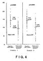
Fig. 4 shows the results of U tests comparing a healthy subject group and a pancreatic carcinoma patient group, made on the ionic intensity of the mass peak of the protein having a molecular weight of 17,272±9 (m/z) captured by cation-exchange protein chips (C10, pH 4) in Examples 1 and 2. -
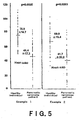
Fig. 5 shows the results of U tests comparing a healthy subject group and a pancreatic carcinoma patient group, made on the ionic intensity of the mass peak of the protein having a molecular weight of 17,253±9 (m/z) captured by cation-exchange protein chips (C10, pH 4) in Examples 1 and 2. -
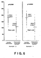
Fig. 6 shows the results of U tests comparing a healthy subject group and a pancreatic carcinoma patient group, made on the ionic intensity of the mass peak of the protein having a molecular weight of 17,253±9 (m/z) captured by reverse-phase protein chips (H 50) in Examples 1 and 2. -
Fig. 7 shows mass spectrometric analysis charts obtained in Example 4, showing the peak of the blood protein having a molecular weight of 17,272±9 (m/z) taken from a healthy subject and from a pancreatic carcinoma patient, captured by cation-exchange protein chips (C10, pH 4). -
Fig. 8 shows mass spectrometric analysis charts obtained in Example 4, showing the peak of the blood protein having a molecular weight of 8,766±5 (m/z) taken from a healthy subject and from a pancreatic carcinoma patient, captured by cation-exchange protein chips (C10, pH 4). -
Fig. 9 shows mass spectrometric analysis charts obtained in Example 4, showing the peak of the blood protein having a molecular weight of 28,080±15 (m/z) taken from a healthy subject and from a pancreatic carcinoma patient, captured by cation-exchange protein chips (C10, pH 4). -
Fig. 10 shows mass spectrometric analysis charts obtained in Example 4, showing the peak of the blood protein having a molecular weight of 14,799±8 (m/z) taken from a healthy subject and from a pancreatic carcinoma patient, captured by reverse-phase protein chips (H50). -
Fig. 11 is a diagram showing ROC curves and their AUC values obtained in Example 4. - Pancreatic carcinoma marker proteins according to the present invention are blood proteins whose molecular weights are 17,272±9and 17,253±9 D. Here, the molecular weight of each blood protein is the center value of the mass distribution of the blood protein in mass spectrometric analysis. The blood proteins having molecular weights of 17,272±9 and 17,253±9
D are characterized in that their expression levels are lower in pancreatic carcinoma patients than in healthy individuals. - The pancreatic carcinoma marker proteins of the present invention can be used as an indicator of pancreatic carcinoma either singly or in combination. Although these marker proteins make it possible to detect pancreatic carcinoma accurately even when they are used singly, the disease can be detected more accurately when two or more of them are used in combination as a multi-marker. Furthermore, the pancreatic carcinoma marker proteins according to the present invention may also be used in combination with a known pancreatic carcinoma marker, such as CA 19-9, for the detection of pancreatic carcinoma, and the present invention also encompasses such an embodiment.
- Although any technique can be used to determine the expression levels of the pancreatic carcinoma marker proteins as long as it can quantify the pancreatic carcinoma marker proteins, a preferred technique is mass spectrometric analysis, and a more preferred technique is time-of-flight mass spectrometric analysis. In such mass spectrometric analysis, the ionic intensities of the mass peaks of the pancreatic carcinoma marker proteins, for example, can be used to quantify the marker proteins.
- Any instrument or system can be used without limitation for the above-described mass spectrometric analysis as long as it can measure the expression levels of the pancreatic carcinoma marker proteins. Examples of instruments and systems useful herein include such instruments as a quadrupole mass spectrometer, a time-of-flight mass spectrometer, a quadrupole ion trap mass spectrometer and a Fourier transform ion cyclotron resonance mass spectrometer, and a protein chip system comprising protein chips, a time-of-flight mass spectrometer, and a computer on which software for determination and analysis is installed. Of these, a quadrupole mass spectrometer, a time-of-flight mass spectrometer, and a protein chip system are preferred.
- Any energy-absorbing molecule may be used in the mass spectrometric analysis in the present invention as long as it can be used in the mass spectrometric analysis of the marker proteins according to the present invention. Examples of such molecules include sinapinic acid and α-CHCA (α-cyano-4-hydroxy cinnamic acid).
- It is preferred that the above-described mass spectrometric analysis be performed by the protein chip method. The protein chip method herein refers to a technique in which a sample to be analyzed is added to protein chips, and the molecular weights of proteins contained in the sample, captured by the protein chips, are measured by means of mass spectrometry. The protein chip method makes it possible to conduct purification, identification, or the like of a plurality of proteins rapidly in a short time with high efficiency by the use of a small amount of a sample, so that it can be advantageously used in the detection of pancreatic carcinoma.
- Although any type of protein chips can be used in the present invention as long as it can capture the pancreatic carcinoma marker proteins, preferred types of protein chips are reverse-phase protein chips, metal-ion-fixed protein chips, and cation-exchange protein chips. Therefore, according to one embodiment of the present invention, the pancreatic carcinoma marker proteins are captured by at least one protein chip selected from the group consisting of reverse-phase protein chips, metal-ion-fixed protein chips, and cation-exchange protein chips.
According to a more preferred embodiment of the present invention, the protein chip is a reverse-phase protein chip and/or a cation-exchange protein chip.
According to another preferred embodiment of the present invention, the protein chip is a metal-ion-fixed protein chip and/or a cation-exchange protein chip.
Further, the pancreatic carcinoma marker proteins according to the present invention are well captured by cation-exchange protein chips at a pH of 4 or 7. Therefore, according to a more preferred embodiment of the present invention, the cation-exchange protein chips are prepared to have a pH of 4 or 7. - A cation-exchange group in cation-exchange protein chips is preferably carboxyl group or a carboxyalkyl group, more preferably carboxymethyl group. Preferred specific examples of the cation-exchange protein chips include WCX 2 and CM 10 (both manufactured by Ciphergen Biosystems Inc., 14F, East Tower, Yokohama Business Park, 134, Godo-cho, Hodogaya-ku, Yokohama-shi, Kanagawa-ken, Japan), and CM 10 is more preferred. A functional group in reverse-phase protein chips is preferably at least one group selected from alkyl groups, aralkyl groups and aryl groups, more preferably at least one group having 6 to 12 carbon atoms, selected from alkyl groups, aralkyl groups and aryl groups, and most preferably an alkyl group having 6 to 12 carbon atoms. Although the above functional group may be substituted, it is preferably unsubstituted. Preferred specific examples of the reverse-phase protein chips include H4 and H50 (both manufactured by Ciphergen Biosystems Inc., Japan), and H50 is more preferred. A metal ion to be fixed to the metal-ion-fixed protein chips is preferably copper or zinc ion, more preferably copper ion. Preferred specific examples of the metal-ion-fixed protein chips include IMAC 30 (manufactured by Ciphergen Biosystems Inc., Japan).
- In the protein chip method, after adding a sample to protein chips, the protein chips are, if necessary, treated with a solution, such as a buffer solution, in order to make the protein chips quickly capture proteins contained in the sample and also to remove impurities, and are then washed. The solution for use in this treatment is specifically a sodium acetate buffer solution, a tris(hydroxymethyl)aminomethane/hydrogen chloride buffer solution, a phosphoric acid buffer solution, or the like. In the case where cation-exchange protein chips are used, a buffer solution at a pH of 4 or 7 is preferably used to treat the protein chips because the pancreatic carcinoma marker proteins according to the present invention have the characteristics of being captured by cation-exchange protein chips at a pH of 4 or 7. Therefore, according to a preferred embodiment of the present invention, the cation-exchange protein chips are prepared to have a pH of 4 or 7.
When reverse-phase protein chips are used, an acetonitrile or trifluoroacetate solution is preferably used to treat the protein chips.
When metal-ion-fixed protein chips are used, a sodium phosphate, chloride, or acetate solution is preferably used to treat the protein chips. - Another aspect of the present invention is the use of a blood protein having a molecular weight of 17,272±9 or 17,253±9 D as a pancreatic carcinoma marker protein
- The pancreatic carcinoma marker proteins according to the present invention are useful in accurately and easily detecting pancreatic carcinoma, as mentioned above. Therefore, a further aspect of the present invention is a method for detecting pancreatic carcinoma, comprising:
- determining the expression level of a blood protein having a molecular weight of 17,272±9 or 17,253±9 D in a blood sample taken from a subject, and
- judging that pancreatic cancer is detected in the subject when the expression level of the blood protein in the blood specimen taken from the subject is lower than that of the protein in a blood sample taken from a healthy individual.
- In the above-described method according to the present invention, whether pancreatic carcinoma is detected or not in a subject is judged by comparing the expression levels of the pancreatic carcinoma marker proteins in a blood sample taken from the subject with those of the marker proteins in a blood sample taken from a healthy individual, as mentioned above. The expression levels of the marker proteins in a healthy individual are set beforehand as thresholds depending on race, sex, age, etc. The expression levels of the marker proteins in a healthy individual can be set by a known statistical technique with reference to the amounts of the pancreatic carcinoma marker proteins in a blood sample taken from a healthy individual, quantified by the above-described method. Specific examples of techniques of setting the expression level of each marker protein include (the mean value of pancreatic carcinoma marker protein levels in a blood sample taken from a healthy individual ± standard deviation) and ROC (Receiver Operating Characteristics) analysis, and ROC analysis is preferred. Whether pancreatic carcinoma is detected or not in a subject may also be judged by comparing the expression levels of the pancreatic carcinoma marker proteins in a blood sample taken from the subject with the thresholds established beforehand by the above statistical technique from the quantified amounts of the marker proteins in a blood sample taken from a pancreatic carcinoma patient. The present invention also encompasses this embodiment.
- Further, the blood sample in the present invention is usually human-derived one. Furthermore, the blood sample in the present invention is preferably blood plasma. Any method can be used to collect blood and prepare a blood sample as long as it does not affect the measurement of the marker proteins according to the present invention, and any method known to those skilled in the art can be used.
- Furthermore, the pancreatic carcinoma marker proteins according to the present invention can also be used in a method for monitoring pancreatic carcinoma in a patient. The monitoring method can be effectively used to grasp accurately the progress of pancreatic carcinoma in a patient, thereby administering a more adequate medicinal agent to the patient at more adequate timing, or subjecting the patient to adequate treatment.
- Accordingly, a further aspect of the present invention is a method for monitoring pancreatic carcinoma in a patient, comprising:
- determining the expression level of a blood protein having a molecular weight of 17,272±9 or 17,253±9 D in a blood sample taken from a patient, and
- judging that pancreatic carcinoma has progressed when the expression level of the protein has decreased with time.
- The following example will specifically explain the present invention. However, they are not to be construed to limit the scope of the invention.
- Blood was collected from 104 pancreatic carcinoma patients at National Cancer Center Hospital, Tokyo, Japan, not yet subjected to any treatment, and from 116 healthy individuals by the use of EDTA blood-collection tubes during the period between September 2002 and October 2003, and the blood plasma obtained were used for the following experiments.
- A test sample was prepared by treating each blood plasma with 9M urea and 2% CHAPS (3-[(3-cholamidopropyl)dimethylammonio]propane-sulfonic acid). Using a workstation robot Biomech 2000 (manufactured by Beckman Coulter Co.), the sample was added to three different types of protein chips. The protein chips used are cation-exchange protein chips CM10 (manufactured by Ciphergen Biosystems, Inc., Japan), metal-ion-fixed protein chips IMAC 30 (manufactured by Ciphergen Biosystems, Inc., Japan), and reverse-phase protein chips H50 (manufactured by Ciphergen Biosystems, Inc., Japan). CM10 was washed under strict conditions using a pH 7 buffer solution for washing, or under moderate conditions using a
pH 4 buffer solution for washing, in accordance with the description in the handling manual prepared by Ciphergen Biosystems, Inc. and was used in the following mass spectrometric analysis. - Sinapinic acid, a laser absorbent matrix, was added to the respective chips, and the chips were then dried. Subsequently, the chips were placed in a quadrupole hybrid mass spectrometer Q-star XL (manufactured by Applied Biosystems Co.) equipped with a time-of-flight mass spectrometer, and the mass and ionization quantity of low-molecular-weight proteins and peptides of below about 2 - 40 KD were measured. The mass/ionization information from the mass spectrometer Q-star XL was converted to peak information by smoothing the mass/ionization information data with a Gaussian window function, using software for analysis Analyst QS (Applied Biosystems Co.), and then adjusting the base line and the noise level in accordance with the description in "Improving protein identification from peptide mass fingerprinting through a parameterized multi-level scoring algorithm and an optimized peak detection (Electrophoresis, 1999 Dec; 20(18): 3535-50). The data were analyzed on the conditions that the limit of inaccuracy in mass of the mass spectrometer is approximately 0.05%.
- SVM (support vector machine: rbf (radius basis function) and linear), an NN (neural network), and an FNN (fuzzy neural network) were subjected to algorithmic machine learning of the peak information in the range of 2,000 to 35,000 Da obtained in the above-described manner, using as a learning set 71 pancreatic carcinoma subjects and 71 healthy individuals. The results were as shown in
Fig. 1 . The rates of discrimination of the three peaks at 17,272±9 (m/z) and 17,253±9 (m/z) when CM10 (pH 4) was used and at 17,253±9 (m/z) when H50 was used were above ca. 80% in every algorithmic analysis. - [Table 1]
Table 1 m/z T test U test SVM SVM FNN NN p value p value (rbf) (linear) C10 17272 0.000 0.000 84.5 83.1 84.5 84.5 (pH4) C10 17253 0.000 0.000 82.4 81.0 82.4 81.7 (pH4) H50 17253 0.000 0.000 82.4 81.0 79.6 81.7 - Peak charts showing the peak at 17,272±9 (m/z) when CM10 (pH4) was used, obtained from the analysis of the blood samples taken from healthy individuals and from pancreatic carcinoma patients, were as shown in
Fig. 1 .
Peak charts showing the peak at 17,253±9 (m/z) when CM10 (pH4) was used were as shown inFig. 2 .
Peak charts showing the peak at 17,253±9 (m/z) when H50 was used were as shown inFig. 3 .
The peak charts shown inFigs. 1 to 3 plot relative ionic intensity as the ordinate.
All the three peaks were lower in the pancreatic carcinoma patients than in the healthy individuals. - In terms of the three peaks found in Example 1, mass spectrometric analysis was made with the same technique as in Example 1, using, as a verification set, 33 pancreatic carcinoma patients and 45 healthy individuals who were different from those in Example 1. The rate of right answer for pancreatic carcinoma was analyzed in terms of the peaks at 17,272±9 and 17,253±9 (m/z) when CM10 (pH4) was used and the peak at 17,253±9 (m/z) when H50 was used.
As for the peak at 17,272±9 (m/z) when CM10 (pH4) was used, the rate of discrimination was 72.2%, the sensitivity, 58.8%, and the specificity, 82.2%.
As for the peak at 17,253±9 (m/z) when CM10 (pH4) was used, the rate of discrimination was 79.7%, the sensitivity, 85.3%, and the specificity, 75.6%.
As for the peak at 17,253±9 (m/z) when H50 was used, the rate of discrimination was 79.7%, the sensitivity, 82.4%, and the specificity, 77.8%. - U tests comparing the healthy individual group and the pancreatic carcinoma patient group in Example 1 or 2 were made on the ionic intensities of the three mass peaks found in Example 1.
The results concerning the peak at 17,272±9 (m/z) when CM10 (pH4) was used were as shown inFig. 4 .
The results concerning the peak at 17,253±9 (m/z) when CM10 (pH4) was used were as shown inFig. 5 .
The results concerning the peak at 17,253±9 (m/z) when H50 was used were as shown inFig. 6 .
The charts shown inFigs. 4 to 6 plot relative ionic intensity as the ordinate.
The ionic intensities of all the three peaks were significantly lower in the pancreatic carcinoma patient group than in the healthy subject group.
Moreover, the results obtained from the two groups in Example 1 and those obtained from the two groups in Example 2 showed the same tendency. - In terms of the ionic intensity of the mass peak at 28,080±15, 17,272±9, 17,253±9, 8,766±5, or 14,779±8 (m/z) detected by the mass spectrometric analysis in Example 1, statistical analysis was made by means of T test. The results were as shown in Table 2. [Table 2]
Table 2 Example1 (n=220) Peak ionic intensity (mean value) p value Peak(m/z) Protein chips Pancreatic carcinoma patients (n=104) Healthy individuals (n=116) 28,080 CM10(PH4) 107 123 0.000 IMAC 3023.5 30.3 0.000 17,272 CM10(PH4) 9.6 14.6 0.000 H50 42 64 0.000 17,252 CM10(PH4) 45 73 0.000 H50 113 175 0.000 14,779 CM10(PH7) 13.9 10.4 0.000 H50 11.3 7.1 0.000 8,766 CM10(PH4) 7.3 12.6 0.000 H50 13.1 18.8 0.000 IMAC 308.0 15.3 0.000 - In the case where the cation-exchange protein chips (CM10, pH 4) were used, the ionic intensities of the mass peaks at molecular weights of 28,080±15, 17,272±9, 17,253±9, and 8,766±5 (m/z) were significantly lower in the pancreatic cancer patient group than in the healthy individual group, while the ionic intensity of the mass peak at 14,779±8 (m/z) was significantly higher in the pancreatic cancer patient group than in the healthy individual group. Further, when the reverse-phase protein chips (H50) were used, the peak intensities of the mass peaks at 17,272±9, 17,253±9, and 8,766±5 (m/z) were significantly lower in the pancreatic cancer patient group than in the healthy individual group, and the peak intensity of the mass peak at 14,779±8 (m/z) was significantly higher in the pancreatic cancer patient group than in the healthy individual group. Furthermore, when the metal-ion-fixed protein chips (IMAC30, protein chips of copper-ion-fixed type) were used, the peak intensities of the mass peaks at 28,080±15 and 8,766±5 (m/z) were significantly lower in the pancreatic cancer patient group than in the healthy individual group.
- Samples were prepared from blood collected from 71 pancreatic carcinoma patients at National Cancer Center Hospital, Tokyo, Japan, not yet subjected to any treatment, and from 71 healthy individuals and the samples were mass analyzed with the same technique as in Example 1. Cation-exchange protein chips CM10 (manufactured by Ciphergen Biosystems, Inc., Japan) and reverse-phase protein chips H50 (manufactured by Ciphergen Biosystems, Inc., Japan), were used in the analysis. CM10 was washed under moderate conditions using a
pH 4 buffer solution for washing, in accordance with the description in the handling manual prepared by Ciphergen Biosystems, Inc. - An rbf SVM was subjected to algorithmic machine learning, using the peak information obtained from the above-described mass spectrometric analysis as the learning set. As a result, peaks were detected at 28,080±15, 17,272±9 and 8,766±5 (m/z) when CM10 (pH 4) was used and at 14,779±8 (m/z) when H50 was used.
- Peak charts obtained from the analyses in Example 4 were as shown in
Figs. 7 to 10 . The peak charts plot relative ionic intensity as the ordinate.
As shown inFigs. 7 to 9 , the intensities of the peaks at 17,272±9, 8,766±5, and 28,080±15 (m/z) when CM10 (pH 4) was used were lower in the pancreatic carcinoma patient group than in the healthy individual group. On the other hand, as shown inFig. 10 , the intensity of the peak at 14,779±8 (m/z) when H50 was used was higher in the pancreatic carcinoma patient group than in the healthy subject group. - The ROC curves of the above four peaks, and their AUC values were as shown in
Fig. 11 . When all the above four peaks were detected (4-peak combination), the sensitivity was 97.2%, the specificity, 94.4%, and the AUC value, 0.978. With respect to the four peaks, the discrimination power was confirmed also by means of LOO (leave-one-out) cross validation. - Verification tests were carried out with the same technique as in Example 4, using as a verification set 33 pancreatic carcinoma patients, not yet subjected to any treatment, and 45 healthy subjects, who were different from those in Example 4. As a result, when all the four peaks found in Example 4 were detected, the rate of discrimination was 91.0%, the sensitivity, 90.9%, and the specificity, 91.1%.
- U tests comparing the healthy individual group with the pancreatic carcinoma patient group in Example 4 or 5 were made on the peak intensities of the above-described four mass peaks.
The results of the U tests were as shown in Table 3. Each statistically obtained value shown in the table is (the mean ionic intensity of the peak) ± S.D. - [Table 3]
Table 3 Example 4 (n=142) Example 5 (n=78) Protein chips Peak (m/z) Pancreatic carcinoma patients Healthy individuals p value Pancreatic carcinoma patients Healthy individuals p value (n=71) (n=71) (n=33) (n=45) CM10 (PH4) 17,272 9.49±2.88 14.6±2.29 0.0000 9.74±4.22 14.5±2.29 0.0000 8,766 7.65±3.53 12.1±5.55 0.0000 7.04±4.39 13.4±5.81 0.0000 28,080 113±36.7 132±33.5 0.0022 92.4±24.3 110±21.6 0.0078 H50 14,779 11.8±4.43 7.85±3.68 10.4±3.85 0.0000 6.46±1.63 0.00000 - The results of the U tests of the peak intensities in Example 4 and those of the U tests of the peak intensities in Example 5 showed the same tendency.
The peak intensities of all the peaks at 17,272±9, 8,766±5, and 28,080±15 (m/z) detected when CM10 (pH 4) was used were significantly lower in the pancreatic carcinoma patient group than in the healthy individual group. On the other hand, the peak intensity of the peak at 14,779±8 (m/z) detected when H50 was used was significantly higher in the pancreatic carcinoma patient group than in the healthy individual group. - With the same technique as in Example 1, samples were prepared at Tokyo Medical University Hospital, Japan from blood collected from 29 pancreatic carcinoma patients, not yet subjected to any treatment, and from 39 healthy individuals and the samples were mass analyzed. Peaks were detected at 17,272±9, 8,766±5, and 28,080±15 (m/z) when CM10 (pH 4) was used, and at 14,779±8 (m/z) when H50 was used. Further, the detection of CA19-9 in the blood plasma taken from the pancreatic carcinoma patients and the healthy individuals was made using a CA19-9 radioimmunoassay kit (manufactured by FUJIREBIO INC., Japan). Setting the cut-off value of blood plasma CA19-9 to 37 U/ml, the detection of pancreatic carcinoma was made.
- The results were as shown in Table 4. In Table 4, SELDI refers to the case where all the peaks at 17,272±9, 8,766±5, and 28,080±15 (m/z) when CM10 (pH 4) was used and at 14,779±8 (m/z) when H50 was used were detected. "Combination" in the table refers to the case where both SELDI and CA19-9 were detected. "n" is a number of the subjects in which CA 19-9, SELDI, or Combination was found. "Clinical stage" is the stages of the disease classified according to the description in the handling rules for pancreatic carcinoma, edited by the Japan Pancreatic Society.
[Table 4]Table 4 number of subjects CA19-9, n (%) SELDI, n (%) Combination, n (%) Healthy subjects 39 3(7.7) 4(10.3) 6(15.4) Pancreatic carcinoma patients 29 25(86.2) 26(89.7) 29(100) Clinical stage I 1 1(100) 1(100) 1(100) II 3 3(100) 2(66.6) 3(100) III 1 0(0) 1(100) 1(100) IV 24 21(87.5) 22(91.7) 24(100) - Pancreatic carcinoma was detected in 100% of the pancreatic carcinoma patients when the peaks at 17,272±9, 8,766±5, and 28,080±15 (m/z) when CM10 (pH 4) was used, the peak at 14,779±8 (m/z) when H50 was used, and CA19-9 were employed together as indicators.
Claims (17)
- A method for detecting pancreatic carcinoma, comprising:determining the expression level of a blood protein having a molecular weight of 17,272±9 or 17,253±9(D) in a blood sample taken from a subject, andjudging that pancreatic cancer is detected in the subject when the expression level of the blood protein in the blood sample taken from the subject is lower than that of the protein in a blood sample taken from a healthy individual.
- A method for monitoring pancreatic carcinoma in a patient, comprising:determining the expression level of a blood protein having a molecular weight of 17,272±9 or 17,253±9 (D) in a blood sample taken from the patient, andjudging that pancreatic cancer has progressed when the expression level of the protein has decreased with time.
- The method according to claim 1 or 2, wherein the expression level of the blood protein is lower in pancreatic carcinoma patients than in healthy individuals.
- The method according to claim 3, wherein the determination of the expression level of the blood protein is made by mass spectrometric analysis.
- The method according to claim 4, wherein the mass spectrometric analysis is time-of-flight mass spectrometric analysis.
- The method according to claim 4 or 5, wherein the mass spectrometric analysis is performed by a protein chip method.
- The method according to claim 6, wherein the blood protein is captured by at least one protein chip selected from the group consisting of reverse-phase protein chips, metal-ion-fixed protein chips, and cation-exchange protein chips.
- The method according to claim 7, wherein the protein chip is a reverse-phase protein chip, and/or a cation-exchange protein chip.
- The method according to claim 8, wherein the cation-exchange protein chip is prepared to have a pH of 4 or 7.
- Use of a blood protein having a molecular weight of 17,272±9 or 17,253±9 (D) as pancreatic carcinoma marker.
- The use according to claim 10, wherein the expression level of the blood protein is lower in pancreatic carcinoma patients than in healthy individuals.
- The use according to claim 11, wherein the determination of the expression level of the blood protein is made by mass spectrometric analysis.
- The use according to claim 12, wherein the mass spectrometric analysis is time-of-flight mass spectrometric analysis.
- The use according to claim 12 or 13, wherein the mass spectrometric analysis is performed by a protein chip method.
- The use according to claim 14, wherein the blood protein is captured by at least one protein chip selected from the group consisting of reverse-phase protein chips, metal-ion-fixed protein chips, and cation-exchange protein chips.
- The use according to claim 15, wherein the protein chip is a reverse-phase protein chip, and/or a cation-exchange protein chip.
- The use according to claim 16, wherein the cation-exchange protein chip is prepared to have a pH of 4 or 7.
Applications Claiming Priority (2)
| Application Number | Priority Date | Filing Date | Title |
|---|---|---|---|
| JP2005070512 | 2005-03-14 | ||
| PCT/JP2006/301243 WO2006098087A1 (en) | 2005-03-14 | 2006-01-26 | Marker protein for use in diagnosis of pancreatic cancer |
Publications (3)
| Publication Number | Publication Date |
|---|---|
| EP1870711A1 EP1870711A1 (en) | 2007-12-26 |
| EP1870711A4 EP1870711A4 (en) | 2008-07-16 |
| EP1870711B1 true EP1870711B1 (en) | 2010-03-24 |
Family
ID=36991443
Family Applications (1)
| Application Number | Title | Priority Date | Filing Date |
|---|---|---|---|
| EP06712425A Expired - Lifetime EP1870711B1 (en) | 2005-03-14 | 2006-01-26 | Marker protein for use in diagnosis of pancreatic cancer |
Country Status (5)
| Country | Link |
|---|---|
| US (1) | US20090004671A1 (en) |
| EP (1) | EP1870711B1 (en) |
| JP (1) | JP5200246B2 (en) |
| DE (1) | DE602006013109D1 (en) |
| WO (1) | WO2006098087A1 (en) |
Families Citing this family (11)
| Publication number | Priority date | Publication date | Assignee | Title |
|---|---|---|---|---|
| JP5140809B2 (en) * | 2007-07-25 | 2013-02-13 | 財団法人ヒューマンサイエンス振興財団 | Modified α-fibrinogen protein or immunogenic fragment thereof |
| JP5422785B2 (en) * | 2008-09-12 | 2014-02-19 | 国立大学法人名古屋大学 | Methods and biomarkers for blood detection of multiple carcinomas using mass spectrometry |
| JP2010175452A (en) * | 2009-01-30 | 2010-08-12 | Japan Health Science Foundation | Method for detecting pancreatic cancer |
| EP3124980A1 (en) * | 2009-10-01 | 2017-02-01 | Phenomenome Discoveries Inc. | Mass spectrophotometric method to diagnose pancreatic cancer |
| AU2011260390B2 (en) * | 2010-06-01 | 2016-11-24 | Ernst-Moritz-Arndt Universitat Greifswald | Means and methods for diagnosing pancreatic cancer in a subject |
| JP6519056B2 (en) | 2013-10-01 | 2019-05-29 | 東レ株式会社 | Method for detecting pancreatic tumor, antibody and kit for detecting pancreatic tumor |
| DK3267197T3 (en) | 2015-03-02 | 2020-10-12 | Toray Industries | METHOD AND KIT FOR THE DETECTION OF PANCREATIC DYSFUNCTION |
| DK179161B1 (en) * | 2016-05-26 | 2017-12-18 | Man Diesel & Turbo Filial Af Man Diesel & Turbo Se Tyskland | A large two-stroke compression-ignited internal combustion engine with fuel injection system for low flashpoint fuel and a fuel valve therefore |
| EP3572805B1 (en) * | 2017-01-19 | 2026-05-13 | Shimadzu Corporation | Analysis data analytics method and analysis data analytics device |
| KR20220035088A (en) | 2019-07-18 | 2022-03-21 | 루머스 리미티드 | Encapsulated light guide optics |
| JPWO2022202826A1 (en) | 2021-03-24 | 2022-09-29 |
Family Cites Families (3)
| Publication number | Priority date | Publication date | Assignee | Title |
|---|---|---|---|---|
| US6936424B1 (en) * | 1999-11-16 | 2005-08-30 | Matritech, Inc. | Materials and methods for detection and treatment of breast cancer |
| AU2001249275B2 (en) * | 2000-03-20 | 2005-12-08 | Eastern Virginia Medical School | Prostate cancer markers |
| WO2003027138A1 (en) * | 2001-09-25 | 2003-04-03 | Japan As Represented By President Of National Cancer Center | Search for cancer marker by novel screening method |
-
2006
- 2006-01-26 EP EP06712425A patent/EP1870711B1/en not_active Expired - Lifetime
- 2006-01-26 US US11/908,694 patent/US20090004671A1/en not_active Abandoned
- 2006-01-26 DE DE602006013109T patent/DE602006013109D1/en not_active Expired - Lifetime
- 2006-01-26 WO PCT/JP2006/301243 patent/WO2006098087A1/en not_active Ceased
- 2006-01-26 JP JP2007508026A patent/JP5200246B2/en not_active Expired - Lifetime
Also Published As
| Publication number | Publication date |
|---|---|
| JPWO2006098087A1 (en) | 2008-08-21 |
| DE602006013109D1 (en) | 2010-05-06 |
| EP1870711A4 (en) | 2008-07-16 |
| US20090004671A1 (en) | 2009-01-01 |
| JP5200246B2 (en) | 2013-06-05 |
| WO2006098087A1 (en) | 2006-09-21 |
| EP1870711A1 (en) | 2007-12-26 |
Similar Documents
| Publication | Publication Date | Title |
|---|---|---|
| JP6412100B2 (en) | SRM assay to indicate cancer therapy | |
| CN103648585B (en) | Disposal Planning Based on Serum Markers of Peptide Radiation Toxicity | |
| EP1870711A1 (en) | Marker protein for use in diagnosis of pancreatic cancer | |
| US10828507B2 (en) | Clinical decision support (CDS) for radiotherapy in prostate cancer | |
| US9403889B2 (en) | Diagnostic lung cancer panel and methods for its use | |
| EP2012127A2 (en) | Diagnostic markers for detecting prostate cancer | |
| CN118362666A (en) | Metabolic marker composition for distinguishing health, non-colorectal cancer disease and colorectal cancer and its application | |
| Bunkowski et al. | MCC/IMS signals in human breath related to sarcoidosis—results of a feasibility study using an automated peak finding procedure | |
| KR101223270B1 (en) | Method for determining low―mass ions to screen colorectal cancer, method for providing information to screen colorectal cancer by using low―mass ions, and operational unit therefor | |
| Gaines et al. | Identification of serum biomarkers for canine B-cell lymphoma by use of surface-enhanced laser desorption-ionization time-of-flight mass spectrometry | |
| JP2010071953A (en) | Breast cancer marker, and diagnosis using it | |
| Zhang et al. | Discovery of serum biomarkers in astrocytoma by SELDI–TOF MS and proteinchip technology | |
| Yang et al. | Proteomic profiling of invasive ductal carcinoma (IDC) using magnetic beads‐based serum fractionation and MALDI‐TOF MS | |
| CN107273717A (en) | A kind of detection model of Sera of Lung Cancer gene and its construction method and application | |
| US10073075B2 (en) | SRM/MRM assay for the cyclin-dependent kinase inhibitor 2A (p16) protein | |
| EP3641788A1 (en) | Quantifying slfn11 protein for optimal cancer therapy | |
| US9362094B2 (en) | Biomarkers for determining breast cancer bone metastasis | |
| JP2008202978A (en) | Diagnosis of prostate cancer | |
| US20200096519A1 (en) | Mass spectrometry profiling of esophageal cancer markers | |
| JP4109032B2 (en) | Bladder cancer test determination method | |
| US10078084B2 (en) | SRM/MRM assay for the mesothelin (MSLN) protein | |
| JP2025131404A (en) | Methods for detecting pancreatic cancer | |
| CN120989241A (en) | Negative ion salivary biomarkers associated with gliomas and meningiomas and their applications | |
| CN121324660A (en) | A tissue marker for proton-induced radiation-induced cardiac injury and its application | |
| CN120924659A (en) | Positive ion saliva marker related to brain glioma and meningioma and application thereof |
Legal Events
| Date | Code | Title | Description |
|---|---|---|---|
| PUAI | Public reference made under article 153(3) epc to a published international application that has entered the european phase |
Free format text: ORIGINAL CODE: 0009012 |
|
| 17P | Request for examination filed |
Effective date: 20071012 |
|
| AK | Designated contracting states |
Kind code of ref document: A1 Designated state(s): AT BE BG CH CY CZ DE DK EE ES FI FR GB GR HU IE IS IT LI LT LU LV MC NL PL PT RO SE SI SK TR |
|
| DAX | Request for extension of the european patent (deleted) | ||
| A4 | Supplementary search report drawn up and despatched |
Effective date: 20080617 |
|
| 17Q | First examination report despatched |
Effective date: 20080905 |
|
| GRAP | Despatch of communication of intention to grant a patent |
Free format text: ORIGINAL CODE: EPIDOSNIGR1 |
|
| GRAS | Grant fee paid |
Free format text: ORIGINAL CODE: EPIDOSNIGR3 |
|
| GRAA | (expected) grant |
Free format text: ORIGINAL CODE: 0009210 |
|
| AK | Designated contracting states |
Kind code of ref document: B1 Designated state(s): AT BE BG CH CY CZ DE DK EE ES FI FR GB GR HU IE IS IT LI LT LU LV MC NL PL PT RO SE SI SK TR |
|
| REG | Reference to a national code |
Ref country code: GB Ref legal event code: FG4D |
|
| REG | Reference to a national code |
Ref country code: CH Ref legal event code: EP |
|
| REG | Reference to a national code |
Ref country code: IE Ref legal event code: FG4D |
|
| REF | Corresponds to: |
Ref document number: 602006013109 Country of ref document: DE Date of ref document: 20100506 Kind code of ref document: P |
|
| REG | Reference to a national code |
Ref country code: NL Ref legal event code: VDEP Effective date: 20100324 |
|
| PG25 | Lapsed in a contracting state [announced via postgrant information from national office to epo] |
Ref country code: LT Free format text: LAPSE BECAUSE OF FAILURE TO SUBMIT A TRANSLATION OF THE DESCRIPTION OR TO PAY THE FEE WITHIN THE PRESCRIBED TIME-LIMIT Effective date: 20100324 |
|
| LTIE | Lt: invalidation of european patent or patent extension |
Effective date: 20100324 |
|
| PG25 | Lapsed in a contracting state [announced via postgrant information from national office to epo] |
Ref country code: LV Free format text: LAPSE BECAUSE OF FAILURE TO SUBMIT A TRANSLATION OF THE DESCRIPTION OR TO PAY THE FEE WITHIN THE PRESCRIBED TIME-LIMIT Effective date: 20100324 Ref country code: SI Free format text: LAPSE BECAUSE OF FAILURE TO SUBMIT A TRANSLATION OF THE DESCRIPTION OR TO PAY THE FEE WITHIN THE PRESCRIBED TIME-LIMIT Effective date: 20100324 Ref country code: FI Free format text: LAPSE BECAUSE OF FAILURE TO SUBMIT A TRANSLATION OF THE DESCRIPTION OR TO PAY THE FEE WITHIN THE PRESCRIBED TIME-LIMIT Effective date: 20100324 Ref country code: AT Free format text: LAPSE BECAUSE OF FAILURE TO SUBMIT A TRANSLATION OF THE DESCRIPTION OR TO PAY THE FEE WITHIN THE PRESCRIBED TIME-LIMIT Effective date: 20100324 Ref country code: PL Free format text: LAPSE BECAUSE OF FAILURE TO SUBMIT A TRANSLATION OF THE DESCRIPTION OR TO PAY THE FEE WITHIN THE PRESCRIBED TIME-LIMIT Effective date: 20100324 |
|
| PG25 | Lapsed in a contracting state [announced via postgrant information from national office to epo] |
Ref country code: ES Free format text: LAPSE BECAUSE OF FAILURE TO SUBMIT A TRANSLATION OF THE DESCRIPTION OR TO PAY THE FEE WITHIN THE PRESCRIBED TIME-LIMIT Effective date: 20100705 Ref country code: BE Free format text: LAPSE BECAUSE OF FAILURE TO SUBMIT A TRANSLATION OF THE DESCRIPTION OR TO PAY THE FEE WITHIN THE PRESCRIBED TIME-LIMIT Effective date: 20100324 Ref country code: NL Free format text: LAPSE BECAUSE OF FAILURE TO SUBMIT A TRANSLATION OF THE DESCRIPTION OR TO PAY THE FEE WITHIN THE PRESCRIBED TIME-LIMIT Effective date: 20100324 Ref country code: GR Free format text: LAPSE BECAUSE OF FAILURE TO SUBMIT A TRANSLATION OF THE DESCRIPTION OR TO PAY THE FEE WITHIN THE PRESCRIBED TIME-LIMIT Effective date: 20100625 Ref country code: EE Free format text: LAPSE BECAUSE OF FAILURE TO SUBMIT A TRANSLATION OF THE DESCRIPTION OR TO PAY THE FEE WITHIN THE PRESCRIBED TIME-LIMIT Effective date: 20100324 Ref country code: RO Free format text: LAPSE BECAUSE OF FAILURE TO SUBMIT A TRANSLATION OF THE DESCRIPTION OR TO PAY THE FEE WITHIN THE PRESCRIBED TIME-LIMIT Effective date: 20100324 Ref country code: SE Free format text: LAPSE BECAUSE OF FAILURE TO SUBMIT A TRANSLATION OF THE DESCRIPTION OR TO PAY THE FEE WITHIN THE PRESCRIBED TIME-LIMIT Effective date: 20100324 |
|
| PG25 | Lapsed in a contracting state [announced via postgrant information from national office to epo] |
Ref country code: SK Free format text: LAPSE BECAUSE OF FAILURE TO SUBMIT A TRANSLATION OF THE DESCRIPTION OR TO PAY THE FEE WITHIN THE PRESCRIBED TIME-LIMIT Effective date: 20100324 Ref country code: IS Free format text: LAPSE BECAUSE OF FAILURE TO SUBMIT A TRANSLATION OF THE DESCRIPTION OR TO PAY THE FEE WITHIN THE PRESCRIBED TIME-LIMIT Effective date: 20100724 Ref country code: CZ Free format text: LAPSE BECAUSE OF FAILURE TO SUBMIT A TRANSLATION OF THE DESCRIPTION OR TO PAY THE FEE WITHIN THE PRESCRIBED TIME-LIMIT Effective date: 20100324 Ref country code: BG Free format text: LAPSE BECAUSE OF FAILURE TO SUBMIT A TRANSLATION OF THE DESCRIPTION OR TO PAY THE FEE WITHIN THE PRESCRIBED TIME-LIMIT Effective date: 20100624 |
|
| PLBE | No opposition filed within time limit |
Free format text: ORIGINAL CODE: 0009261 |
|
| STAA | Information on the status of an ep patent application or granted ep patent |
Free format text: STATUS: NO OPPOSITION FILED WITHIN TIME LIMIT |
|
| PG25 | Lapsed in a contracting state [announced via postgrant information from national office to epo] |
Ref country code: PT Free format text: LAPSE BECAUSE OF FAILURE TO SUBMIT A TRANSLATION OF THE DESCRIPTION OR TO PAY THE FEE WITHIN THE PRESCRIBED TIME-LIMIT Effective date: 20100726 Ref country code: DK Free format text: LAPSE BECAUSE OF FAILURE TO SUBMIT A TRANSLATION OF THE DESCRIPTION OR TO PAY THE FEE WITHIN THE PRESCRIBED TIME-LIMIT Effective date: 20100324 |
|
| 26N | No opposition filed |
Effective date: 20101228 |
|
| PG25 | Lapsed in a contracting state [announced via postgrant information from national office to epo] |
Ref country code: IT Free format text: LAPSE BECAUSE OF FAILURE TO SUBMIT A TRANSLATION OF THE DESCRIPTION OR TO PAY THE FEE WITHIN THE PRESCRIBED TIME-LIMIT Effective date: 20100324 |
|
| PG25 | Lapsed in a contracting state [announced via postgrant information from national office to epo] |
Ref country code: MC Free format text: LAPSE BECAUSE OF NON-PAYMENT OF DUE FEES Effective date: 20110131 |
|
| REG | Reference to a national code |
Ref country code: CH Ref legal event code: PL |
|
| REG | Reference to a national code |
Ref country code: IE Ref legal event code: MM4A |
|
| PG25 | Lapsed in a contracting state [announced via postgrant information from national office to epo] |
Ref country code: LI Free format text: LAPSE BECAUSE OF NON-PAYMENT OF DUE FEES Effective date: 20110131 Ref country code: CH Free format text: LAPSE BECAUSE OF NON-PAYMENT OF DUE FEES Effective date: 20110131 |
|
| PG25 | Lapsed in a contracting state [announced via postgrant information from national office to epo] |
Ref country code: IE Free format text: LAPSE BECAUSE OF NON-PAYMENT OF DUE FEES Effective date: 20110126 |
|
| REG | Reference to a national code |
Ref country code: DE Ref legal event code: R082 Ref document number: 602006013109 Country of ref document: DE Representative=s name: GRUENECKER, KINKELDEY, STOCKMAIR & SCHWANHAEUS, DE Ref country code: GB Ref legal event code: 732E Free format text: REGISTERED BETWEEN 20120607 AND 20120613 |
|
| REG | Reference to a national code |
Ref country code: DE Ref legal event code: R081 Ref document number: 602006013109 Country of ref document: DE Owner name: NATIONAL CANCER CENTER, JP Free format text: FORMER OWNER: JAPAN HEALTH SCIENCES FOUNDATION, TOKYO, JP Effective date: 20120704 Ref country code: DE Ref legal event code: R082 Ref document number: 602006013109 Country of ref document: DE Representative=s name: GRUENECKER, KINKELDEY, STOCKMAIR & SCHWANHAEUS, DE Effective date: 20120704 Ref country code: DE Ref legal event code: R082 Ref document number: 602006013109 Country of ref document: DE Representative=s name: GRUENECKER PATENT- UND RECHTSANWAELTE PARTG MB, DE Effective date: 20120704 |
|
| PG25 | Lapsed in a contracting state [announced via postgrant information from national office to epo] |
Ref country code: CY Free format text: LAPSE BECAUSE OF FAILURE TO SUBMIT A TRANSLATION OF THE DESCRIPTION OR TO PAY THE FEE WITHIN THE PRESCRIBED TIME-LIMIT Effective date: 20100324 Ref country code: LU Free format text: LAPSE BECAUSE OF NON-PAYMENT OF DUE FEES Effective date: 20110126 |
|
| PG25 | Lapsed in a contracting state [announced via postgrant information from national office to epo] |
Ref country code: TR Free format text: LAPSE BECAUSE OF FAILURE TO SUBMIT A TRANSLATION OF THE DESCRIPTION OR TO PAY THE FEE WITHIN THE PRESCRIBED TIME-LIMIT Effective date: 20100324 |
|
| PG25 | Lapsed in a contracting state [announced via postgrant information from national office to epo] |
Ref country code: HU Free format text: LAPSE BECAUSE OF FAILURE TO SUBMIT A TRANSLATION OF THE DESCRIPTION OR TO PAY THE FEE WITHIN THE PRESCRIBED TIME-LIMIT Effective date: 20100324 |
|
| REG | Reference to a national code |
Ref country code: FR Ref legal event code: PLFP Year of fee payment: 11 |
|
| REG | Reference to a national code |
Ref country code: FR Ref legal event code: PLFP Year of fee payment: 12 |
|
| REG | Reference to a national code |
Ref country code: FR Ref legal event code: PLFP Year of fee payment: 13 |
|
| P01 | Opt-out of the competence of the unified patent court (upc) registered |
Effective date: 20230530 |
|
| PGFP | Annual fee paid to national office [announced via postgrant information from national office to epo] |
Ref country code: DE Payment date: 20250121 Year of fee payment: 20 |
|
| PGFP | Annual fee paid to national office [announced via postgrant information from national office to epo] |
Ref country code: FR Payment date: 20250127 Year of fee payment: 20 |
|
| PGFP | Annual fee paid to national office [announced via postgrant information from national office to epo] |
Ref country code: GB Payment date: 20250128 Year of fee payment: 20 |
|
| REG | Reference to a national code |
Ref country code: DE Ref legal event code: R071 Ref document number: 602006013109 Country of ref document: DE |
|
| REG | Reference to a national code |
Ref country code: GB Ref legal event code: PE20 Expiry date: 20260125 |