EP1854491A1 - Catheter having denatured part for contact with body - Google Patents
Catheter having denatured part for contact with body Download PDFInfo
- Publication number
- EP1854491A1 EP1854491A1 EP06714403A EP06714403A EP1854491A1 EP 1854491 A1 EP1854491 A1 EP 1854491A1 EP 06714403 A EP06714403 A EP 06714403A EP 06714403 A EP06714403 A EP 06714403A EP 1854491 A1 EP1854491 A1 EP 1854491A1
- Authority
- EP
- European Patent Office
- Prior art keywords
- plasma
- catheter
- sample
- silicone
- adhesive
- Prior art date
- Legal status (The legal status is an assumption and is not a legal conclusion. Google has not performed a legal analysis and makes no representation as to the accuracy of the status listed.)
- Withdrawn
Links
Images




Classifications
-
- A—HUMAN NECESSITIES
- A61—MEDICAL OR VETERINARY SCIENCE; HYGIENE
- A61M—DEVICES FOR INTRODUCING MEDIA INTO, OR ONTO, THE BODY; DEVICES FOR TRANSDUCING BODY MEDIA OR FOR TAKING MEDIA FROM THE BODY; DEVICES FOR PRODUCING OR ENDING SLEEP OR STUPOR
- A61M25/00—Catheters; Hollow probes
- A61M25/0043—Catheters; Hollow probes characterised by structural features
- A61M25/0045—Catheters; Hollow probes characterised by structural features multi-layered, e.g. coated
-
- A—HUMAN NECESSITIES
- A61—MEDICAL OR VETERINARY SCIENCE; HYGIENE
- A61L—METHODS OR APPARATUS FOR STERILISING MATERIALS OR OBJECTS IN GENERAL; DISINFECTION, STERILISATION OR DEODORISATION OF AIR; CHEMICAL ASPECTS OF BANDAGES, DRESSINGS, ABSORBENT PADS OR SURGICAL ARTICLES; MATERIALS FOR BANDAGES, DRESSINGS, ABSORBENT PADS OR SURGICAL ARTICLES
- A61L29/00—Materials for catheters, medical tubing, cannulae, or endoscopes or for coating catheters
- A61L29/04—Macromolecular materials
- A61L29/06—Macromolecular materials obtained otherwise than by reactions only involving carbon-to-carbon unsaturated bonds
-
- A—HUMAN NECESSITIES
- A61—MEDICAL OR VETERINARY SCIENCE; HYGIENE
- A61L—METHODS OR APPARATUS FOR STERILISING MATERIALS OR OBJECTS IN GENERAL; DISINFECTION, STERILISATION OR DEODORISATION OF AIR; CHEMICAL ASPECTS OF BANDAGES, DRESSINGS, ABSORBENT PADS OR SURGICAL ARTICLES; MATERIALS FOR BANDAGES, DRESSINGS, ABSORBENT PADS OR SURGICAL ARTICLES
- A61L29/00—Materials for catheters, medical tubing, cannulae, or endoscopes or for coating catheters
- A61L29/14—Materials characterised by their function or physical properties, e.g. lubricating compositions
-
- A—HUMAN NECESSITIES
- A61—MEDICAL OR VETERINARY SCIENCE; HYGIENE
- A61L—METHODS OR APPARATUS FOR STERILISING MATERIALS OR OBJECTS IN GENERAL; DISINFECTION, STERILISATION OR DEODORISATION OF AIR; CHEMICAL ASPECTS OF BANDAGES, DRESSINGS, ABSORBENT PADS OR SURGICAL ARTICLES; MATERIALS FOR BANDAGES, DRESSINGS, ABSORBENT PADS OR SURGICAL ARTICLES
- A61L2400/00—Materials characterised by their function or physical properties
- A61L2400/18—Modification of implant surfaces in order to improve biocompatibility, cell growth, fixation of biomolecules, e.g. plasma treatment
Definitions
- the present invention relates to a catheter having denatured part for contact with body and the method of producing the same.
- the occurrence of infectious diseases caused by catheter placement can be prevented by improving compatibility between catheters and biological tissue.
- a variety of attempts have been made to cover a biocompatible product with a membrane that is excellent in biocompatibility.
- many methods of covering using a hydroxyapatite membrane have been suggested. Examples of a method of forming a hydroxyapatite membrane include a plasma spraying method (e.g., JP Patent Publication (Kokai) No. 63-46165 A (1988 )), a sputtering deposition method (e.g., JP Patent Publication (Kokai) No.
- JP Patent Publication (Kokai) No. 5-49689 A (1993 ) discloses a cell adhesive material, which is composed of polymeric material comprising carbon as a constitutional element, and at least a portion of the surface of which is modified by ion bombardment.
- JP Patent Publication (Kokai) No. 2002-315821 A discloses a material that adheres to bone and/or fascia, which is composed of polymeric material comprising carbon as a constitutional element and at least a portion of the surface of which is modified by ion bombardment.
- 2004-89361 A discloses a biological repair material compatible with a biological tissue adhesive (a polymeric material used in combination with a biological tissue adhesive, which comprises carbon or silicon as a constitutional element and at least a portion of the surface of which is modified by ion bombardment).
- a medical catheter such as a continuous ambulatory (mobile) peritoneal dialysis (CAPD) catheter, a central venous catheter, a hemodialysis shunt catheter or a ventricular drainage catheter and a biologically-derived adhesive (fibrin adhesive), cells, or biological tissue.
- a medical catheter such as a continuous ambulatory (mobile) peritoneal dialysis (CAPD) catheter, a central venous catheter, a hemodialysis shunt catheter or a ventricular drainage catheter and a biologically-derived adhesive (fibrin adhesive
- the present inventors have found that infection can be prevented with closure of a tunnel infection route by modifying an interface part for contact with body on the outer side of a medical catheter by a plasma ion irradiation method so as to improve adhesiveness between the medical catheter and a biologically-derived adhesive (fibrin adhesive), cells, or biological tissue. This has led to the completion of the present invention.
- the present invention provides a medical catheter which is composed of material selected from the group consisting of silicone, polyurethane, polypropylene and polytetrafluoroethylene (PTFE), at least a portion of the surface of which is modified by plasma ion implantation.
- a medical catheter which is composed of material selected from the group consisting of silicone, polyurethane, polypropylene and polytetrafluoroethylene (PTFE), at least a portion of the surface of which is modified by plasma ion implantation.
- Another aspect of the present invention provides a method of producing a medical catheter, which comprises modifying at least a portion of the surface of a material selected from the group consisting of silicone, polyurethane, polypropylene and polytetrafluoroethylene (PTFE) by plasma ion implantation.
- a material selected from the group consisting of silicone, polyurethane, polypropylene and polytetrafluoroethylene (PTFE) by plasma ion implantation.
- the medical catheter of the present invention is composed of silicone.
- a high-voltage negative pulse is applied to plasma-exposed material selected from the group consisting of silicone, polyurethane, polypropylene and polytetrafluoroethylene (PTFE) so as to attract ions such that the surface of the material is omnidirectionally irradiated with the ions.
- plasma-exposed material selected from the group consisting of silicone, polyurethane, polypropylene and polytetrafluoroethylene (PTFE) so as to attract ions such that the surface of the material is omnidirectionally irradiated with the ions.
- plasma ions are selected from the group consisting of He + , Ne + , Ar + , and Kr + .
- the plasma ion pressure during treatment is 1.0 x 10 -1 to 1.0 x 10 -4 torr, and the treatment time of plasma ion implantation is 1 minute to 3 hours.
- the present invention relates to a medical catheter which is composed of material selected from the group consisting of silicone, polyurethane, polypropylene and polytetrafluoroethylene (PTFE), at least a portion of the surface of which is modified by plasma ion implantation.
- a medical catheter which is composed of material selected from the group consisting of silicone, polyurethane, polypropylene and polytetrafluoroethylene (PTFE), at least a portion of the surface of which is modified by plasma ion implantation.
- the material of the catheter of the present invention can be selected from the group consisting of silicone, polyurethane, polypropylene, and polytetrafluoroethylene (PTFE).
- PTFE polytetrafluoroethylene
- silicone is polymeric material having a structure in which basic units of Si-O are polymerized.
- the type of silicone used in the present invention is not particularly limited as long as it can be used for catheters.
- At least a portion of the surface of the above material is modified by plasma ion implantation.
- the plasma ion implantation method used in the present invention is a method wherein a high-voltage negative pulse is applied to a plasma-exposed sample to be subjected to irradiation so as to attract ions such that the surface of a sample is omnidirectionally irradiated with the ions.
- a plasma generation method include a plasma generation method using a parallel plate electrode, a loop antenna, or the like and a method comprising directly applying a high frequency wave to a sample.
- the plasma ion implantation method is advantageous in that, unlike ion beam irradiation techniques using conventional linear accelerators, even a sample to be subjected to irradiation having a curved surface can be uniformly irradiated with ions.
- such sample may be a conductor or an insulator.
- a polymer catheter can be uniformly modified in a short period of time by applying the plasma ion implantation method to treat the surface of the polymer catheter which is the curved surface of an insulator.
- a piece of wire having a diameter almost equivalent to the inner diameter of the sample may be introduced inside the sample, and in such cases the sample is then allowed to stand on a sample holder, followed by plasma ion implantation treatment.
- cell adhesiveness of the catheter material and adhesiveness of a biological adhesive can be significantly improved through modification by plasma ion implantation.
- ion species to be implanted include, but are not particularly limited to, H + , He + , C + , N + , Ne + , Na + , N 2 + , O + , Ar + , and Kr + . Of these, He + , Ne + , Ar + , and Kr + are particularly preferable.
- the rate for introducing a gas comprising any of the above examples may be 5 to 50 ml/min.
- the pressure during treatment may be 1.0 x 10 -1 to 1.0 x 10 -4 torr.
- the voltage applied to a sample may be 0 to -50.0 kV, for example, and preferably 0 to -20.0 kV.
- the treatment time of plasma ion implantation is not particularly limited as long as effects of the present invention can be achieved. However, such treatment time may be generally 1 minute to 3 hours and preferably 5 minutes to 1 hour. In the case of a treatment time of less than 1 minute, effects of the present invention cannot be achieved, which is not preferable. Meanwhile, in the case of a treatment time of more than 3 hours, the structure of catheter material tends to be destroyed, which is not preferable.
- cell adhesiveness and the adhesive property of a biologically derived adhesive can be significantly improved by allowing a catheter material such as silicone to be subjected to plasma ion irradiation.
- a catheter material such as silicone
- Such improvement in cell adhesiveness is considered to be caused by surface carbonization and an increase in the amount of adhering proteins as a result of introduction of functional groups.
- improvement in the adhesive property of a fibrin adhesive is considered to be caused by surface carbonization and an increase in surface area.
- the catheter of the present invention is effective for prevention of tunnel infection. This is because a fibrin adhesive tightly adheres to a catheter at the beginning of catheter introduction, while on the other hand, biological tissue tightly adheres to the catheter after decomposition and absorption of the fibrin adhesive.
- Samples of silicone used in medical practice were subjected to plasma ion irradiation such that a part for contact with living body was modified. Modification effects were evaluated by tensile strength tests using a biologically-derived adhesive and ex vivo cell adhesion tests. Surfaces of the modified samples were subjected to measurements of morphology, compositions, binding states, and the like by AFM, RAMAN, or RBS. The measurement results were examined in connection with the aforementioned property-improving effects.
- the silicone material used was THE740 (obtained by mixing polyorganosiloxane and silicon dioxide followed by pressing) produced by GE Toshiba Silicones.
- the structure thereof contains two Si-O chains bound to each other as shown below.
- Samples were produced by the plasma ion irradiation method.
- a high-voltage negative pulse is applied to a plasma-exposed sample so as to attract ions such that the sample surface is irradiated with the ions.
- a parallel plate electrode, a loop antenna, or the like may be used.
- a method comprising directly applying a high frequency wave to a sample was used.
- Fig. 1 shows a schematic view of an irradiation system. The method used herein is advantageous in that plasma generation takes place in a manner such that the whole sample is covered by plasma.
- Fig. 2 illustrates the principle of ion irradiation.
- a high frequency (RF) pulse is applied to a sample such that gas around the sample is ionized, resulting in plasma generation (Fig. 2(a)).
- a high-voltage negative pulse is applied to the sample so as to attract positively charged ions, followed by ion irradiation (Fig. 2(b)).
- ion irradiation can be carried out regardless of whether or not a sample to be subjected to irradiation is conductive.
- the sample is attached to a sample holder by using a conductive double-sided adhesive tape, followed by treatment.
- a piece of wire having a diameter almost equivalent to the inner diameter of the sample is introduced inside the sample and the sample is then allowed to stand on a sample holder, followed by treatment.
- the ion species (introduced gas) used was He, Ar, or Kr.
- the rate of gas introduction was designated as 20 cc/min.
- the pressure during treatment was designated as 1.0 x 10 -2 torr.
- the applied voltage was designated as 0, -2.5, -5.0, -7.5, or -10.0 kV.
- the treatment time was designated as 30 minutes in all cases except for the case shown in Fig. 5 (60 minutes).
- the high frequency pulse width and the high-voltage negative pulse width were designated as 30 ⁇ sec and 10 ⁇ sec, respectively.
- the interval between the ending point of the RF pulse and the starting point of the high-voltage negative pulse was designated as 25 ⁇ sec.
- the RF output was 500 W, and the pulse repetition rate was 2 kHz.
- Fig. 3 shows examples of a current and a voltage measured using an oscilloscope during irradiation.
- a silicone sheet that had been irradiated with ions was cut into rectangles 5 mm x 15 mm in size.
- the rectangles were adhered to cardboard pieces having the same size with the use of a fibrin adhesive.
- each adhesion surface was determined to have a size of 5 mm x 5 mm (Fig. 4).
- the load upon detachment was measured when tension was applied to each rectangle in the horizontal direction at a rate of 5 mm per minute. Because of strong adhesion between the cardboard pieces and the fibrin adhesive, detachment always occurred at an interface between silicone and the adhesive. Measurement was carried out using a horizontal digital load gauge (Model 2255, Aikoh Engineering Co., Ltd.). Fig.
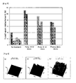
- Fig. 6 shows AFM images of untreated samples (a), treated samples (Ar-5 kV-30 min) (b), and other treated samples (Ar-10 kV-30 min) (c).
- the surface roughness increased along with the increase in treatment voltage. It is considered that the increase in surface area (adhesion surface area) caused improvement in the adhesive property of the fibrin adhesive. In addition, it is considered that strength against tension, particularly in the horizontal direction, increased due to the invasion of fibrin adhesive into concave portions.
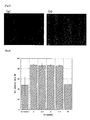
- Fig. 7 shows optical microscopic images of an unirradiated sample (a) and a treated sample (Ar-7.5 kV-30 min) (b) on day 3 of culture after seeding of mouse-derived fibroblasts.
- the numbers of cells are obviously different. However, the focus herein is the ratio of cells adhering to the sample surface (elongated shape) to cells not adhering thereto (round shape).
- the horizontal axis represents negative pulse voltage
- the vertical axis represents cell adhesion rate.
- the gas introduced was Ar and the treatment time was 30 minutes.
- a significant improvement in cell adhesiveness was observed in the sample treated with a negative pulse voltage of 0 to 7.5 kV. Meanwhile, cell adhesiveness of the sample treated with a negative pulse voltage of 10 kV was almost equivalent to that of the sample that had not been irradiated.
- Fig. 9 shows RBS spectra of silicone samples subjected to Ar plasma ion irradiation.
- the treatment time was 30 minutes.
- Argon ions used for irradiation and iron ions that were thought to be released from the inner wall of a vacuum chamber were confirmed to exist on each sample surface except in the cases of an untreated sample and a sample treated with a pulse voltage of 0 kV. These elements were found in minute amounts. Further, only small portions thereof were exposed on the sample surface. Consequently, it is considered that the existence of such elements does not substantially influence the adhesive property of a fibrin adhesive or cell adhesiveness.
- the percentage of C relative to Si and O increased, resulting in changes in not only the surface shape but also the adhesive property of a fibrin adhesive and cell adhesiveness.
- Fig. 10 shows Raman spectra, based on which it is understood the decrease in methyl groups (approximately 2900 cm -1 ) was caused by the increase in pulse voltage, indicating that the silicone structure was destroyed. Meanwhile, in the case of 10 kV irradiation, a graphite-derived peak (approximately 1560 cm -1 ) and an amorphous carbon-derived peak (approximately 1350 cm -1 ) were obviously observed. Accordingly, it is understood that surface carbonization is promoted by the increase in pulse voltage.
- cell adhesiveness and the adhesive property of a biologically-derived adhesive can be significantly improved by plasma ion irradiation of silicone.
- the improvement in cell adhesiveness is thought to be caused by surface carbonization and the increase in the amount of adhering proteins due to introduction of functional groups.
- the improvement in the adhesive property of a fibrin adhesive is thought to be caused by surface carbonization and the increase in surface area. It is considered that, when the above method is applied to treat a catheter surface, tunnel infection can be effectively prevented because the fibrin adhesive tightly adheres to the catheter at the beginning of catheter introduction, while on the other hand, biological tissue tightly adheres to the catheter after decomposition and absorption of the fibrin adhesive.
- Fig. 11 shows results of fibrin adhesive shearing strength measurement tests using three different ions. Significant increases in adhesive property were confirmed except for the case of the sample treated with helium at 0 kV (plasma exposure alone). In addition, the applied voltage was proportional to the shearing strength.
- Fig. 12 shows irradiation voltage dependences of cell adhesion rates and numbers of cells in connection with three different ions.
- the cell adhesion rate and the number of cells each temporarily increased and then decreased after reaching maximum value.
- the maximum point of the cell adhesion rate tended to move to the low voltage side along with the increase in the ion mass number. Therefore, it is understood that the modification rate becomes greater in the cases of heavier ions at an identical voltage.
- Fig. 13 shows FT-IR (Fourier-transform infrared) spectra of argon-irradiated samples.
- the positions "A" and “B” correspond to hydroxyl groups (-OH) and adsorption water, respectively.
- carbonyl groups may contribute to some extent to adsorption of proteins serving as scaffolds for surface cell adhesion.
Landscapes
- Health & Medical Sciences (AREA)
- Life Sciences & Earth Sciences (AREA)
- Animal Behavior & Ethology (AREA)
- Veterinary Medicine (AREA)
- Public Health (AREA)
- General Health & Medical Sciences (AREA)
- Epidemiology (AREA)
- Chemical & Material Sciences (AREA)
- Chemical Kinetics & Catalysis (AREA)
- Anesthesiology (AREA)
- Heart & Thoracic Surgery (AREA)
- Engineering & Computer Science (AREA)
- Pulmonology (AREA)
- Biomedical Technology (AREA)
- Hematology (AREA)
- Biophysics (AREA)
- Materials For Medical Uses (AREA)
- Media Introduction/Drainage Providing Device (AREA)
- Medicinal Chemistry (AREA)
- Polymers & Plastics (AREA)
- Organic Chemistry (AREA)
Abstract
It is an object of the present invention to provide a catheter, whereby a tunnel infection route is closed such that infection can be prevented, by improving adhesiveness between a medical catheter and a biologically-derived adhesive (fibrin adhesive), cells, or biological tissue. The present invention provides a medical catheter which is composed of material selected from the group consisting of silicone, polyurethane, polypropylene and polytetrafluoroethylene (PTFE), at least a portion of the surface of which is modified by plasma ion implantation.
Description
- The present invention relates to a catheter having denatured part for contact with body and the method of producing the same.
- The occurrence of infectious diseases caused by catheter placement can be prevented by improving compatibility between catheters and biological tissue. A variety of attempts have been made to cover a biocompatible product with a membrane that is excellent in biocompatibility. In particular, many methods of covering using a hydroxyapatite membrane have been suggested. Examples of a method of forming a hydroxyapatite membrane include a plasma spraying method (e.g.,
JP Patent Publication (Kokai) No. 63-46165 A (1988 JP Patent Publication (Kokai) No. 63-46165 A (1988 JP Patent Publication (Kokai) No. 5-57011 A (1993 JP Patent Publication (Kokai) No. 6-285151 A (1994 JP Patent Publication (Kokai) No. 10-28728 A (1998 - Meanwhile,
JP Patent Publication (Kokai) No. 5-49689 A (1993 JP Patent Publication (Kokai) No. 2002-315821 A JP Patent Publication (Kokai) No. 2004-89361 A - It is an object of the present invention to provide a catheter, whereby a tunnel infection route is closed such that infection can be prevented, by improving adhesiveness between a medical catheter, such as a continuous ambulatory (mobile) peritoneal dialysis (CAPD) catheter, a central venous catheter, a hemodialysis shunt catheter or a ventricular drainage catheter and a biologically-derived adhesive (fibrin adhesive), cells, or biological tissue.
- As a result of intensive studies to achieve the above object, the present inventors have found that infection can be prevented with closure of a tunnel infection route by modifying an interface part for contact with body on the outer side of a medical catheter by a plasma ion irradiation method so as to improve adhesiveness between the medical catheter and a biologically-derived adhesive (fibrin adhesive), cells, or biological tissue. This has led to the completion of the present invention.
- Thus, the present invention provides a medical catheter which is composed of material selected from the group consisting of silicone, polyurethane, polypropylene and polytetrafluoroethylene (PTFE), at least a portion of the surface of which is modified by plasma ion implantation.
- Another aspect of the present invention provides a method of producing a medical catheter, which comprises modifying at least a portion of the surface of a material selected from the group consisting of silicone, polyurethane, polypropylene and polytetrafluoroethylene (PTFE) by plasma ion implantation.
- Preferably, the medical catheter of the present invention is composed of silicone.
- Preferably, a high-voltage negative pulse is applied to plasma-exposed material selected from the group consisting of silicone, polyurethane, polypropylene and polytetrafluoroethylene (PTFE) so as to attract ions such that the surface of the material is omnidirectionally irradiated with the ions.
- Preferably, plasma ions are selected from the group consisting of He+, Ne+, Ar+, and Kr+.
- Preferably, the plasma ion pressure during treatment is 1.0 x 10⁻¹ to 1.0 x 10⁻⁴ torr, and the treatment time of plasma ion implantation is 1 minute to 3 hours.
- Hereafter, embodiments of the present invention are described in detail.
- The present invention relates to a medical catheter which is composed of material selected from the group consisting of silicone, polyurethane, polypropylene and polytetrafluoroethylene (PTFE), at least a portion of the surface of which is modified by plasma ion implantation.
- The material of the catheter of the present invention can be selected from the group consisting of silicone, polyurethane, polypropylene, and polytetrafluoroethylene (PTFE). Preferably, such material is silicone. Silicone is polymeric material having a structure in which basic units of Si-O are polymerized. The type of silicone used in the present invention is not particularly limited as long as it can be used for catheters.
- In the case of the catheter of the present invention, at least a portion of the surface of the above material is modified by plasma ion implantation.
- The plasma ion implantation method used in the present invention is a method wherein a high-voltage negative pulse is applied to a plasma-exposed sample to be subjected to irradiation so as to attract ions such that the surface of a sample is omnidirectionally irradiated with the ions. Examples of a plasma generation method include a plasma generation method using a parallel plate electrode, a loop antenna, or the like and a method comprising directly applying a high frequency wave to a sample. The plasma ion implantation method is advantageous in that, unlike ion beam irradiation techniques using conventional linear accelerators, even a sample to be subjected to irradiation having a curved surface can be uniformly irradiated with ions. In addition, such sample may be a conductor or an insulator. According to the present invention, it has been revealed that a polymer catheter can be uniformly modified in a short period of time by applying the plasma ion implantation method to treat the surface of the polymer catheter which is the curved surface of an insulator. In particular, in cases in which a sample is a catheter (in a tubular shape), a piece of wire having a diameter almost equivalent to the inner diameter of the sample may be introduced inside the sample, and in such cases the sample is then allowed to stand on a sample holder, followed by plasma ion implantation treatment. According to the present invention, cell adhesiveness of the catheter material and adhesiveness of a biological adhesive can be significantly improved through modification by plasma ion implantation.
- Examples of ion species to be implanted include, but are not particularly limited to, H+, He+, C+, N+, Ne+, Na+, N2 +, O+, Ar+, and Kr+. Of these, He+, Ne+, Ar+, and Kr+ are particularly preferable.
- For instance, the rate for introducing a gas comprising any of the above examples may be 5 to 50 ml/min. The pressure during treatment may be 1.0 x 10⁻¹ to 1.0 x 10⁻⁴ torr. The voltage applied to a sample may be 0 to -50.0 kV, for example, and preferably 0 to -20.0 kV. In addition, the treatment time of plasma ion implantation is not particularly limited as long as effects of the present invention can be achieved. However, such treatment time may be generally 1 minute to 3 hours and preferably 5 minutes to 1 hour. In the case of a treatment time of less than 1 minute, effects of the present invention cannot be achieved, which is not preferable. Meanwhile, in the case of a treatment time of more than 3 hours, the structure of catheter material tends to be destroyed, which is not preferable.
- As described above, cell adhesiveness and the adhesive property of a biologically derived adhesive (fibrin adhesive) can be significantly improved by allowing a catheter material such as silicone to be subjected to plasma ion irradiation. Such improvement in cell adhesiveness is considered to be caused by surface carbonization and an increase in the amount of adhering proteins as a result of introduction of functional groups. Further, such improvement in the adhesive property of a fibrin adhesive is considered to be caused by surface carbonization and an increase in surface area. The catheter of the present invention is effective for prevention of tunnel infection. This is because a fibrin adhesive tightly adheres to a catheter at the beginning of catheter introduction, while on the other hand, biological tissue tightly adheres to the catheter after decomposition and absorption of the fibrin adhesive.
- The present invention is hereafter described in greater detail with reference to the following examples, although the technical scope of the present invention is not limited thereto.
- Samples of silicone used in medical practice were subjected to plasma ion irradiation such that a part for contact with living body was modified. Modification effects were evaluated by tensile strength tests using a biologically-derived adhesive and ex vivo cell adhesion tests. Surfaces of the modified samples were subjected to measurements of morphology, compositions, binding states, and the like by AFM, RAMAN, or RBS. The measurement results were examined in connection with the aforementioned property-improving effects.
- The silicone material used was THE740 (obtained by mixing polyorganosiloxane and silicon dioxide followed by pressing) produced by GE Toshiba Silicones. The structure thereof contains two Si-O chains bound to each other as shown below.

- Samples were produced by the plasma ion irradiation method. According to the plasma ion irradiation method, a high-voltage negative pulse is applied to a plasma-exposed sample so as to attract ions such that the sample surface is irradiated with the ions. In the plasma generation method, a parallel plate electrode, a loop antenna, or the like may be used. Herein, a method comprising directly applying a high frequency wave to a sample was used. Fig. 1 shows a schematic view of an irradiation system. The method used herein is advantageous in that plasma generation takes place in a manner such that the whole sample is covered by plasma. Fig. 2 illustrates the principle of ion irradiation. First, a high frequency (RF) pulse is applied to a sample such that gas around the sample is ionized, resulting in plasma generation (Fig. 2(a)). Before plasma disappearance, a high-voltage negative pulse is applied to the sample so as to attract positively charged ions, followed by ion irradiation (Fig. 2(b)). According to the above method, ion irradiation can be carried out regardless of whether or not a sample to be subjected to irradiation is conductive. In cases in which a sample is in a sheet form, the sample is attached to a sample holder by using a conductive double-sided adhesive tape, followed by treatment. In cases in which a sample is in a catheter form (tubular shape), a piece of wire having a diameter almost equivalent to the inner diameter of the sample is introduced inside the sample and the sample is then allowed to stand on a sample holder, followed by treatment.
- The ion species (introduced gas) used was He, Ar, or Kr. The rate of gas introduction was designated as 20 cc/min. The pressure during treatment was designated as 1.0 x 10⁻² torr. The applied voltage was designated as 0, -2.5, -5.0, -7.5, or -10.0 kV. The treatment time was designated as 30 minutes in all cases except for the case shown in Fig. 5 (60 minutes).
- In addition, the high frequency pulse width and the high-voltage negative pulse width were designated as 30 µsec and 10 µsec, respectively. In addition, the interval between the ending point of the RF pulse and the starting point of the high-voltage negative pulse was designated as 25 µsec. The RF output was 500 W, and the pulse repetition rate was 2 kHz. Fig. 3 shows examples of a current and a voltage measured using an oscilloscope during irradiation.
- A silicone sheet that had been irradiated with ions was cut into
rectangles 5 mm x 15 mm in size. The rectangles were adhered to cardboard pieces having the same size with the use of a fibrin adhesive. At such time, each adhesion surface was determined to have a size of 5 mm x 5 mm (Fig. 4). After 2 hours of drying at ordinary temperature, the load upon detachment was measured when tension was applied to each rectangle in the horizontal direction at a rate of 5 mm per minute. Because of strong adhesion between the cardboard pieces and the fibrin adhesive, detachment always occurred at an interface between silicone and the adhesive. Measurement was carried out using a horizontal digital load gauge (Model 2255, Aikoh Engineering Co., Ltd.). Fig. 5 shows results of load measurement upon detachment of untreated samples, treated samples (10 kV-Ar-1 hour), other treated samples (10 kV-Ar-30 min), and samples subjected to Ar plasma exposure alone. Due to plasma exposure, the adhesive property of the fibrin adhesive was found to increase by approximately 1.5 times compared with the cases of untreated samples. As a result of application of ion irradiation, such adhesive property was found to increase by approximately 2.5 times at most compared with the cases of untreated samples. Also, upon energy irradiation at less than 10 kV, similar improvement in adhesive property was observed. - Fig. 6 shows AFM images of untreated samples (a), treated samples (Ar-5 kV-30 min) (b), and other treated samples (Ar-10 kV-30 min) (c). The surface roughness increased along with the increase in treatment voltage. It is considered that the increase in surface area (adhesion surface area) caused improvement in the adhesive property of the fibrin adhesive. In addition, it is considered that strength against tension, particularly in the horizontal direction, increased due to the invasion of fibrin adhesive into concave portions.
- Fig. 7 shows optical microscopic images of an unirradiated sample (a) and a treated sample (Ar-7.5 kV-30 min) (b) on day 3 of culture after seeding of mouse-derived fibroblasts. The numbers of cells are obviously different. However, the focus herein is the ratio of cells adhering to the sample surface (elongated shape) to cells not adhering thereto (round shape). In Fig. 8, the horizontal axis represents negative pulse voltage, and the vertical axis represents cell adhesion rate. (In each case, the gas introduced was Ar and the treatment time was 30 minutes). Compared with the untreated sample, a significant improvement in cell adhesiveness was observed in the sample treated with a negative pulse voltage of 0 to 7.5 kV. Meanwhile, cell adhesiveness of the sample treated with a negative pulse voltage of 10 kV was almost equivalent to that of the sample that had not been irradiated.
- Fig. 9 shows RBS spectra of silicone samples subjected to Ar plasma ion irradiation. In each case, the treatment time was 30 minutes. Argon ions used for irradiation and iron ions that were thought to be released from the inner wall of a vacuum chamber were confirmed to exist on each sample surface except in the cases of an untreated sample and a sample treated with a pulse voltage of 0 kV. These elements were found in minute amounts. Further, only small portions thereof were exposed on the sample surface. Consequently, it is considered that the existence of such elements does not substantially influence the adhesive property of a fibrin adhesive or cell adhesiveness. Along with the increase in pulse voltage, decreases in Si and O on the sample surface were observed. Thus, the percentage of C relative to Si and O increased, resulting in changes in not only the surface shape but also the adhesive property of a fibrin adhesive and cell adhesiveness.
- Fig. 10 shows Raman spectra, based on which it is understood the decrease in methyl groups (approximately 2900 cm-1) was caused by the increase in pulse voltage, indicating that the silicone structure was destroyed. Meanwhile, in the case of 10 kV irradiation, a graphite-derived peak (approximately 1560 cm-1) and an amorphous carbon-derived peak (approximately 1350 cm-1) were obviously observed. Accordingly, it is understood that surface carbonization is promoted by the increase in pulse voltage.
- As a result, it has been shown that cell adhesiveness and the adhesive property of a biologically-derived adhesive (fibrin adhesive) can be significantly improved by plasma ion irradiation of silicone. The improvement in cell adhesiveness is thought to be caused by surface carbonization and the increase in the amount of adhering proteins due to introduction of functional groups. In addition, the improvement in the adhesive property of a fibrin adhesive is thought to be caused by surface carbonization and the increase in surface area. It is considered that, when the above method is applied to treat a catheter surface, tunnel infection can be effectively prevented because the fibrin adhesive tightly adheres to the catheter at the beginning of catheter introduction, while on the other hand, biological tissue tightly adheres to the catheter after decomposition and absorption of the fibrin adhesive.
- Fig. 11 shows results of fibrin adhesive shearing strength measurement tests using three different ions. Significant increases in adhesive property were confirmed except for the case of the sample treated with helium at 0 kV (plasma exposure alone). In addition, the applied voltage was proportional to the shearing strength.
- Fig. 12 shows irradiation voltage dependences of cell adhesion rates and numbers of cells in connection with three different ions. The cell adhesion rate and the number of cells each temporarily increased and then decreased after reaching maximum value. In particular, the maximum point of the cell adhesion rate tended to move to the low voltage side along with the increase in the ion mass number. Therefore, it is understood that the modification rate becomes greater in the cases of heavier ions at an identical voltage.
- Fig. 13 shows FT-IR (Fourier-transform infrared) spectra of argon-irradiated samples. In Fig. 13, the positions "A" and "B" correspond to hydroxyl groups (-OH) and adsorption water, respectively. The higher the treatment voltage, the greater the amounts of such hydroxyl groups and adsorption water. Consequently, it is expected that the surface state was modified to become hydrophilic. In Fig. 13, the position "C" corresponds to carbonyl groups (C=O). At 5 kV and 7.5 kV, peaks appeared. However, no clear peak was observed at 10 kV. Such peak appearance corresponds to cell adhesiveness behavior. Thus, carbonyl groups may contribute to some extent to adsorption of proteins serving as scaffolds for surface cell adhesion.
- According to the present invention, it has become possible to provide a catheter having cell adhesiveness and the adhesive property of a biologically derived adhesive (fibrin adhesive) and a method of producing the same.
-
- Fig. 1 shows an overview of a plasma ion irradiation apparatus.
- Fig. 2 illustrates the principle of ion irradiation.
- Fig. 3 shows an RF pulse and a high-voltage negative pulse.
- Fig. 4 illustrates a piece tested regarding the adhesive property of a fibrin adhesive (side view).
- Fig. 5 shows results of tests regarding the adhesive property of a fibrin adhesive.
- Fig. 6 shows results of observation of surface morphology by AFM.
- Fig. 7 shows results of observation of cell adhesion states.
- Fig. 8 shows changes in cell adhesion rates.
- Fig. 9 shows RBS spectra of Ar-plasma-ion-irradiated silicone samples.
- Fig. 10 shows Raman spectra of Ar-plasma-ion-irradiated silicone samples.
- Fig. 11 shows results of adhesion tests for a fibrin adhesive.
- Fig. 12 shows cell adhesion rates and numbers of cells.
- Fig. 13 shows FT-IR spectra of argon-irradiated samples.
Claims (9)
- A medical catheter which is composed of material selected from the group consisting of silicone, polyurethane, polypropylene and polytetrafluoroethylene (PTFE), at least a portion of the surface of which is modified by plasma ion implantation.
- The medical catheter of claim 1 which is composed of silicone.
- The medical catheter of claim 1 or 2 wherein a high-voltage negative pulse is applied to plasma-exposed material selected from the group consisting of silicone, polyurethane, polypropylene and polytetrafluoroethylene (PTFE) so as to attract ions such that the surface of the material is omnidirectionally irradiated with the ions.
- The medical catheter of any of claims 1 to 3 wherein plasma ions are selected from the group consisting of He+, Ne+, Ar+, and Kr+.
- The medical catheter of any of claims 1 to 4 wherein the plasma ion pressure during treatment is 1.0 x 10⁻¹ to 1.0 x 10⁻⁴ torr, and the treatment time of plasma ion implantation is 1 minute to 3 hours.
- A method of producing the medical catheter of any of claims 1 to 5, which comprises modifying at least a portion of the surface of a material selected from the group consisting of silicone, polyurethane, polypropylene and polytetrafluoroethylene (PTFE) by plasma ion implantation.
- The method of producing the medical catheter according to claim 6 wherein a high-voltage negative pulse is applied to plasma-exposed material selected from the group consisting of silicone, polyurethane, polypropylene and polytetrafluoroethylene (PTFE) so as to attract ions such that the surface of the material is omnidirectionally irradiated with the ions.
- The method of producing the medical catheter according to claim 6 or 7 wherein plasma ions are selected from the group consisting of He+, Ne+, Ar+, and Kr+.
- The method of producing the medical catheter according to any of claims 6 to 8 wherein the plasma ion pressure during treatment is 1.0 x 10⁻¹ to 1.0 x 10⁻⁴ torr, and the treatment time of plasma ion implantation is 1 minute to 3 hours.
Applications Claiming Priority (2)
| Application Number | Priority Date | Filing Date | Title |
|---|---|---|---|
| JP2005048199A JP2006230639A (en) | 2005-02-24 | 2005-02-24 | Catheters with modified living body contact parts |
| PCT/JP2006/303263 WO2006090776A1 (en) | 2005-02-24 | 2006-02-23 | Catheter having denatured part for contact with body |
Publications (2)
| Publication Number | Publication Date |
|---|---|
| EP1854491A1 true EP1854491A1 (en) | 2007-11-14 |
| EP1854491A4 EP1854491A4 (en) | 2011-01-05 |
Family
ID=36927414
Family Applications (1)
| Application Number | Title | Priority Date | Filing Date |
|---|---|---|---|
| EP06714403A Withdrawn EP1854491A4 (en) | 2005-02-24 | 2006-02-23 | CATHETER HAVING A DENATURED PART FOR CONTACT WITH THE BODY |
Country Status (4)
| Country | Link |
|---|---|
| US (1) | US8858527B2 (en) |
| EP (1) | EP1854491A4 (en) |
| JP (1) | JP2006230639A (en) |
| WO (1) | WO2006090776A1 (en) |
Families Citing this family (7)
| Publication number | Priority date | Publication date | Assignee | Title |
|---|---|---|---|---|
| JP2009145196A (en) * | 2007-12-14 | 2009-07-02 | Lintec Corp | Modified substrate having low peptide adsorption region and manufacturing method therefor |
| WO2010105102A1 (en) * | 2009-03-11 | 2010-09-16 | Exogenesis Corporation | Methods for improving the bioactivity characteristics of a surface and objects with surfaces improved thereby |
| EP2567012B1 (en) * | 2010-05-05 | 2015-12-16 | Exogenesis Corporation | Methods for improving the bioactivity characteristics of a surface and objects with surfaces improved thereby |
| US20140379609A1 (en) * | 2013-06-24 | 2014-12-25 | Infosys Limited | Method and system for instant processing of an sms related to a business |
| JP6533908B2 (en) * | 2014-01-06 | 2019-06-26 | 公立大学法人首都大学東京 | Method of manufacturing molded body |
| JP6263305B1 (en) * | 2017-08-01 | 2018-01-17 | 株式会社多磨バイオ | Medical sheet |
| JP2023023431A (en) * | 2021-08-05 | 2023-02-16 | 信越ポリマー株式会社 | Catheter manufacturing method |
Family Cites Families (20)
| Publication number | Priority date | Publication date | Assignee | Title |
|---|---|---|---|---|
| JPS51125455A (en) * | 1975-04-14 | 1976-11-01 | Jiyunkichi Nakai | Method of surface treatment of molded article |
| JPS6346165A (en) | 1986-08-13 | 1988-02-27 | 株式会社明電舎 | Production of implant material |
| US4927676A (en) * | 1988-07-01 | 1990-05-22 | Becton, Dickinson And Company | Method for rapid adherence of endothelial cells onto a surface and surfaces prepared thereby |
| JPH0549689A (en) * | 1991-08-20 | 1993-03-02 | Sony Corp | Cell adhesive material and method for producing the same |
| JP3076637B2 (en) | 1991-09-04 | 2000-08-14 | 株式会社アドバンス | Composite implant |
| JP2684942B2 (en) | 1992-11-30 | 1997-12-03 | 日本電気株式会社 | Chemical vapor deposition method, chemical vapor deposition apparatus, and method for manufacturing multilayer wiring |
| JPH06285151A (en) | 1993-04-01 | 1994-10-11 | Nippon Sherwood Kk | Medical instrument coated with amorphous calcium phosphate |
| US6033582A (en) | 1996-01-22 | 2000-03-07 | Etex Corporation | Surface modification of medical implants |
| JPH1028728A (en) | 1996-05-13 | 1998-02-03 | Nissin Electric Co Ltd | Biocompatible catheter and manufacture thereof |
| JP3536051B2 (en) * | 1996-05-17 | 2004-06-07 | 独立行政法人理化学研究所 | Antithrombotic material and method for producing the same |
| JPH10108909A (en) * | 1996-10-03 | 1998-04-28 | Buaayu:Kk | Catheter |
| US6270831B2 (en) * | 1998-04-30 | 2001-08-07 | Medquest Products, Inc. | Method and apparatus for providing a conductive, amorphous non-stick coating |
| JP2000054125A (en) * | 1998-08-10 | 2000-02-22 | Nissin Electric Co Ltd | Surface treating method and device therefor |
| US6224536B1 (en) * | 1999-02-08 | 2001-05-01 | Advanced Cardiovascular Systems | Method for delivering radiation therapy to an intravascular site in a body |
| KR100351516B1 (en) | 2000-09-06 | 2002-09-05 | 한국과학기술연구원 | Method for Surface Modification of 3-Dimensional Bulk Polymers |
| US6504307B1 (en) * | 2000-11-30 | 2003-01-07 | Advanced Cardiovascular Systems, Inc. | Application of variable bias voltage on a cylindrical grid enclosing a target |
| JP5505752B2 (en) | 2001-04-23 | 2014-05-28 | 独立行政法人理化学研究所 | Artificial dura mater having cell adhesion and method for producing the same |
| JP3988982B2 (en) | 2001-12-07 | 2007-10-10 | 株式会社アルバック | Method for forming SiCN thin film |
| JP4445697B2 (en) | 2002-08-30 | 2010-04-07 | 独立行政法人理化学研究所 | Biological repair material having affinity with biological tissue adhesive |
| US7201935B1 (en) * | 2002-09-17 | 2007-04-10 | Advanced Cardiovascular Systems, Inc. | Plasma-generated coatings for medical devices and methods for fabricating thereof |
-
2005
- 2005-02-24 JP JP2005048199A patent/JP2006230639A/en active Pending
-
2006
- 2006-02-23 US US11/885,032 patent/US8858527B2/en not_active Expired - Fee Related
- 2006-02-23 EP EP06714403A patent/EP1854491A4/en not_active Withdrawn
- 2006-02-23 WO PCT/JP2006/303263 patent/WO2006090776A1/en not_active Ceased
Also Published As
| Publication number | Publication date |
|---|---|
| WO2006090776A1 (en) | 2006-08-31 |
| US20090024109A1 (en) | 2009-01-22 |
| JP2006230639A (en) | 2006-09-07 |
| US8858527B2 (en) | 2014-10-14 |
| EP1854491A4 (en) | 2011-01-05 |
Similar Documents
| Publication | Publication Date | Title |
|---|---|---|
| EP1754684B1 (en) | Method for treating surface of base, surface-treated base, material for medical use and instrument for medical use | |
| Gomathi et al. | RF plasma-treated polymers for biomedical applications | |
| Shen et al. | Inhibition of monocyte adhesion and fibrinogen adsorption on glow discharge plasma deposited tetraethylene glycol dimethyl ether | |
| DE60214513T2 (en) | PROCESS FOR SURFACE MODIFICATION | |
| Ye et al. | Surface modification of a biodegradable magnesium alloy with phosphorylcholine (PC) and sulfobetaine (SB) functional macromolecules for reduced thrombogenicity and acute corrosion resistance | |
| Zhou et al. | Platelet adhesion and protein adsorption on silicone rubber surface by ozone-induced grafted polymerization with carboxybetaine monomer | |
| US9549807B2 (en) | Tube with modified inner wall surface using plasma and a preparation method thereof | |
| Jamesh et al. | Evaluation of corrosion resistance and cytocompatibility of graded metal carbon film on Ti and NiTi prepared by hybrid cathodic arc/glow discharge plasma-assisted chemical vapor deposition | |
| US8858527B2 (en) | Catheter having denatured part for contact with body | |
| Ozcan et al. | Plasma modification of PMMA films: surface free energy and cell-attachment studies | |
| EP1656932A1 (en) | Material for aneurysm curing | |
| EP2827915B1 (en) | A coating comprising strontium for body implants | |
| EP3135314A1 (en) | Surface-modified metal and method for modifying metal surface | |
| CN109851831A (en) | Medical tubing and preparation method thereof | |
| JPH064089B2 (en) | Method for preparing transplant main body | |
| Yao et al. | Platelet adhesion-resistance of titanium substrate with mussel-inspired adhesive polymer bearing phosphorylcholine group | |
| JP2010070615A (en) | Crosslinked polymer composition for surface treatment of support | |
| KR102131101B1 (en) | Method for preparation of ePTFE-based artificial vessels with enhanced hemocompatibility via selective plasma etching | |
| Ratner et al. | Chapter I. 2.12-physicochemical surface modification of materials used in medicine | |
| WO2008117949A1 (en) | Barrier membranes for guided bone regeneration | |
| KR20200096197A (en) | Biomedical polymers having a surface metallized by selective plasma etching and preparation method thereof | |
| KR100841779B1 (en) | Surface treatment method of substrate and surface treated substrate, medical material, medical device | |
| JP4570445B2 (en) | Method for producing an indwelling medical device having a hybrid complex on its surface | |
| CN110772662A (en) | Antibacterial expanded polytetrafluoroethylene facial implant material and preparation process thereof | |
| JPWO2018193779A1 (en) | Articles, medical instruments, and artificial joints used in contact with a living body or a biological sample |
Legal Events
| Date | Code | Title | Description |
|---|---|---|---|
| PUAI | Public reference made under article 153(3) epc to a published international application that has entered the european phase |
Free format text: ORIGINAL CODE: 0009012 |
|
| 17P | Request for examination filed |
Effective date: 20070914 |
|
| AK | Designated contracting states |
Kind code of ref document: A1 Designated state(s): CH DE FR GB IT LI NL |
|
| DAX | Request for extension of the european patent (deleted) | ||
| RBV | Designated contracting states (corrected) |
Designated state(s): CH DE FR GB IT LI NL |
|
| A4 | Supplementary search report drawn up and despatched |
Effective date: 20101206 |
|
| STAA | Information on the status of an ep patent application or granted ep patent |
Free format text: STATUS: THE APPLICATION IS DEEMED TO BE WITHDRAWN |
|
| 18D | Application deemed to be withdrawn |
Effective date: 20160901 |