EP1842489A1 - Observation method of biological tissue - Google Patents
Observation method of biological tissue Download PDFInfo
- Publication number
- EP1842489A1 EP1842489A1 EP07006287A EP07006287A EP1842489A1 EP 1842489 A1 EP1842489 A1 EP 1842489A1 EP 07006287 A EP07006287 A EP 07006287A EP 07006287 A EP07006287 A EP 07006287A EP 1842489 A1 EP1842489 A1 EP 1842489A1
- Authority
- EP
- European Patent Office
- Prior art keywords
- observation
- adhesive
- distal end
- membranous tissue
- objective unit
- Prior art date
- Legal status (The legal status is an assumption and is not a legal conclusion. Google has not performed a legal analysis and makes no representation as to the accuracy of the status listed.)
- Withdrawn
Links
- 238000000034 method Methods 0.000 title claims abstract description 27
- 239000000853 adhesive Substances 0.000 claims abstract description 44
- 230000001070 adhesive effect Effects 0.000 claims abstract description 44
- 239000011248 coating agent Substances 0.000 claims abstract description 7
- 238000000576 coating method Methods 0.000 claims abstract description 7
- 210000001124 body fluid Anatomy 0.000 claims abstract description 5
- 239000010839 body fluid Substances 0.000 claims abstract description 5
- 239000003550 marker Substances 0.000 claims description 6
- 210000001691 amnion Anatomy 0.000 abstract description 33
- 210000001519 tissue Anatomy 0.000 abstract description 32
- 210000004381 amniotic fluid Anatomy 0.000 abstract description 22
- 239000007788 liquid Substances 0.000 abstract description 13
- 210000003754 fetus Anatomy 0.000 description 17
- 230000003287 optical effect Effects 0.000 description 7
- 210000002615 epidermis Anatomy 0.000 description 5
- 230000000694 effects Effects 0.000 description 2
- 230000003387 muscular Effects 0.000 description 2
- 102000004169 proteins and genes Human genes 0.000 description 2
- 108090000623 proteins and genes Proteins 0.000 description 2
- 238000007789 sealing Methods 0.000 description 2
- 229920002101 Chitin Polymers 0.000 description 1
- 102000008186 Collagen Human genes 0.000 description 1
- 108010035532 Collagen Proteins 0.000 description 1
- 229920001651 Cyanoacrylate Polymers 0.000 description 1
- 102000016942 Elastin Human genes 0.000 description 1
- 108010014258 Elastin Proteins 0.000 description 1
- JOYRKODLDBILNP-UHFFFAOYSA-N Ethyl urethane Chemical compound CCOC(N)=O JOYRKODLDBILNP-UHFFFAOYSA-N 0.000 description 1
- 102000009123 Fibrin Human genes 0.000 description 1
- 108010073385 Fibrin Proteins 0.000 description 1
- BWGVNKXGVNDBDI-UHFFFAOYSA-N Fibrin monomer Chemical compound CNC(=O)CNC(=O)CN BWGVNKXGVNDBDI-UHFFFAOYSA-N 0.000 description 1
- 108010010803 Gelatin Proteins 0.000 description 1
- BWMFRQKICHXLSH-ZZZNUFMVSA-N Grayanotoxin-III Natural products O[C@H]1[C@@H]2[C@@](O)(C)C[C@]31[C@H]([C@](O)(C)[C@H]1[C@](O)([C@H](O)C3)C(C)(C)[C@@H](O)C1)CC2 BWMFRQKICHXLSH-ZZZNUFMVSA-N 0.000 description 1
- MWCLLHOVUTZFKS-UHFFFAOYSA-N Methyl cyanoacrylate Chemical compound COC(=O)C(=C)C#N MWCLLHOVUTZFKS-UHFFFAOYSA-N 0.000 description 1
- 229920001436 collagen Polymers 0.000 description 1
- 229920002549 elastin Polymers 0.000 description 1
- 210000003195 fascia Anatomy 0.000 description 1
- 239000000835 fiber Substances 0.000 description 1
- 229950003499 fibrin Drugs 0.000 description 1
- 239000000499 gel Substances 0.000 description 1
- 239000008273 gelatin Substances 0.000 description 1
- 229920000159 gelatin Polymers 0.000 description 1
- 235000019322 gelatine Nutrition 0.000 description 1
- 235000011852 gelatine desserts Nutrition 0.000 description 1
- 238000005286 illumination Methods 0.000 description 1
- 238000001727 in vivo Methods 0.000 description 1
- 239000000700 radioactive tracer Substances 0.000 description 1
- 230000000638 stimulation Effects 0.000 description 1
- 239000012780 transparent material Substances 0.000 description 1
Images




Classifications
-
- A—HUMAN NECESSITIES
- A61—MEDICAL OR VETERINARY SCIENCE; HYGIENE
- A61B—DIAGNOSIS; SURGERY; IDENTIFICATION
- A61B17/00—Surgical instruments, devices or methods
- A61B17/34—Trocars; Puncturing needles
-
- A—HUMAN NECESSITIES
- A61—MEDICAL OR VETERINARY SCIENCE; HYGIENE
- A61B—DIAGNOSIS; SURGERY; IDENTIFICATION
- A61B10/00—Instruments for taking body samples for diagnostic purposes; Other methods or instruments for diagnosis, e.g. for vaccination diagnosis, sex determination or ovulation-period determination; Throat striking implements
- A61B10/0045—Devices for taking samples of body liquids
-
- A—HUMAN NECESSITIES
- A61—MEDICAL OR VETERINARY SCIENCE; HYGIENE
- A61B—DIAGNOSIS; SURGERY; IDENTIFICATION
- A61B1/00—Instruments for performing medical examinations of the interior of cavities or tubes of the body by visual or photographical inspection, e.g. endoscopes; Illuminating arrangements therefor
- A61B1/04—Instruments for performing medical examinations of the interior of cavities or tubes of the body by visual or photographical inspection, e.g. endoscopes; Illuminating arrangements therefor combined with photographic or television appliances
-
- A—HUMAN NECESSITIES
- A61—MEDICAL OR VETERINARY SCIENCE; HYGIENE
- A61B—DIAGNOSIS; SURGERY; IDENTIFICATION
- A61B10/00—Instruments for taking body samples for diagnostic purposes; Other methods or instruments for diagnosis, e.g. for vaccination diagnosis, sex determination or ovulation-period determination; Throat striking implements
- A61B10/0045—Devices for taking samples of body liquids
- A61B10/0048—Devices for taking samples of body liquids for taking amniotic fluid samples
-
- A—HUMAN NECESSITIES
- A61—MEDICAL OR VETERINARY SCIENCE; HYGIENE
- A61B—DIAGNOSIS; SURGERY; IDENTIFICATION
- A61B17/00—Surgical instruments, devices or methods
- A61B17/00491—Surgical glue applicators
-
- A—HUMAN NECESSITIES
- A61—MEDICAL OR VETERINARY SCIENCE; HYGIENE
- A61B—DIAGNOSIS; SURGERY; IDENTIFICATION
- A61B17/00—Surgical instruments, devices or methods
- A61B17/34—Trocars; Puncturing needles
- A61B17/3417—Details of tips or shafts, e.g. grooves, expandable, bendable; Multiple coaxial sliding cannulas, e.g. for dilating
- A61B2017/3419—Sealing means between cannula and body
Definitions
- the present invention relates to an observation method for observing a biological tissue in a living state (in vivo).
- an objective optical unit which stores an objective optical system in an outer sheath is supported in a hole formed in a biological tissue in an inserted state by a supporting member, to fix the supporting member to the biological tissue, and thereby the same observation target site in the marrow can be observed with a high reproducibility by inserting and withdrawing the objective optical unit through the supporting member.
- the inside of the amniotic membrane is filled with the amniotic fluid, and if a fetus exists inside thereof, the objective optical unit has to be inserted through the amniotic membrane. Moreover, after a through hole is formed in the amniotic membrane and the observation is completed, and after the objective optical unit is withdrawn, the amniotic fluid has to be continuously held in the amniotic membrane.
- the present invention takes the above problems into consideration, with the object of providing an observation method in which, when an observation target in a membranous tissue such as an amniotic membrane filled with a liquid such as an amniotic fluid is observed, the observation can be performed without leaking the liquid. Moreover, another object is to provide an observation method in which, after the observation, even if the objective unit is taken out, the liquid can be held in the membranous tissue without leaking, and the health of the internal observation target can be maintained.
- the present invention provides the following solutions.
- the present invention provides an observation method of an observation target arranged in a membranous tissue filled with a body fluid, comprising: a step for coating a gel-like adhesive on the outer surface of the membranous tissue; a step for cutting the membranous tissue through the coated adhesive; a step for inserting the distal end of the objective unit through the adhesive and the open cut in the membranous tissue; and a step for observing the observation target through the inserted distal end of the objective unit.
- the adhesive since the membranous tissue is cut through the gel-like adhesive coated in the step for coating the adhesive, the adhesive seals around a cutting member, and the liquid in the membranous tissue is prevented from leaking out. Moreover, since the distal end of the objective unit is also inserted into the membranous tissue through the open cut coated with the adhesive, around the objective unit is sealed by the adhesive, and the liquid in the membranous tissue is prevented from leaking out.
- the gel-like adhesive seals the open cut, and thereby the liquid in the membranous tissue can be prevented from leaking out.
- the adhesive is preferably transparent.
- the distal end of the objective unit When the distal end of the objective unit is inserted into the membranous tissue through the adhesive, even if the adhesive is adhered onto the distal end of the objective unit, as long as the adhesive is transparent, the field of vision is not blocked and the observation target can be observed.
- a marking member may be provided on the distal end of the objective unit, and a step for applying a marker to the observed site on the observation target may be further provided.
- the marker is applied to the observed site on the observation target, even in a continuous observation where the distal end of the objective unit is inserted and withdrawn, the already observed site can be ensured using the marker as a guide, and the observation can be readily performed at the same site or a site different from the already observed site.
- an effect is demonstrated in which, when an observation target in a membranous tissue such as an amniotic membrane filled with a liquid such as an amniotic fluid is observed, the observation can be performed without leaking the liquid. Moreover, an effect is demonstrated in which, after the observation, even if the objective unit is taken out, the liquid can be held in the membranous tissue without leaking, and the health of the internal observation target can be maintained.
- the observation method is a method for observing, for example, a fetus (observation target). 1 of a mouse A in a living state, comprising: as shown in FIG. 1 to FIG. 7, a step S 1 for incising the epidermis 2 and the like; a step S 2 for coating an adhesive 3; a step S 3 for cutting the amniotic membrane (membranous tissue) 4; a step S 4 for inserting an objective unit 5; and a step S 5 for observing the fetus 1.
- the fetus 1 of the mouse A is arranged in the uterine fascia and the amniotic membrane 4 filled with the amniotic fluid (body fluid) 6.
- step S 1 for incising the epidermis 2 and the like the epidermis 2 and the muscular tissue arranged between the ribs 7 of the mouse A are cut and opened by a cutting instrument 8 such as a scalpel. As a result, the amniotic membrane 4 arranged inside of the epidermis 2 and the muscular tissue is exposed.
- a gel-like adhesive 3 is heaped on the outer surface of the amniotic membrane 4 that has been exposed in step S 1 .
- the gel-like adhesive 3 is a biocompatible adhesive 3.
- adhesives 3 of fibrin type, gelatin type, GTXIII type, cyanoacrylate type, urethane type, collagen type, chitin type, or stimulation responsive gel protein type, or those mixed with elastin to adjust the elasticity are used.
- the adhesive 3 is not limited to these, and any arbitrary biocompatible adhesive 3 may be used.
- an adhesive 3 of a transparent material is preferably used.
- step S 3 for cutting the amniotic membrane 4 a boring instrument 9 having a scalpel or other sharp edge passes through the adhesive 3 that has been coated in step S 2 , to form an open cut 10 in the amniotic membrane 4 in the area coated with the adhesive 3. Since the space in the amniotic membrane 4 is filled with the amniotic fluid 6 and has a predetermined internal pressure, at the instant when the boring instrument 9 passes through the amniotic membrane 4, the amniotic fluid 6 is to be ejected from the amniotic membrane 4 . However, in the present embodiment, since around the boring instrument 9 is enclosed and sealed by the gel-like adhesive 3, the amniotic fluid 6 does not leak out to the outside.
- the adhesive 3 sealing around the boring instrument 9 sequentially blocks the track through which the boring instrument 9 passed, and thereby the amniotic fluid 6 is held so as not to leak out to the outside.
- step S 4 for inserting the objective unit 11 the small diameter distal end 11a of the objective unit 11 is inserted into the amniotic membrane 4 through the open cut 10 formed in the step S 3 .
- the objective unit 11 is attached to a microscopic observation device (not shown) and is designed such that illumination light such as exciting light like laser beams is irradiated from the distal end face 11b, and light such as fluorescence returning from the fetus 1 is collected.
- step S 4 the small diameter distal end 11a of the objective unit 11 passes through the adhesive 3 coated on the outer surface of the amniotic membrane 4, reaches the open cut 10 formed in step S 3 , and is inserted into the amniotic membrane 4 through the open cut 10.
- the amniotic fluid 6 in the amniotic membrane 4 is held so as not to leak out to the outside even when the objective unit 11 is being inserted.
- step S 5 for observing the fetus 1 the distal end face 11b of the small diameter distal end 11a of the objective unit 11 that has been inserted into the amniotic fluid 6 is abutted against the outer surface of the fetus 1 serving as the observation target, so as to be stuck thereto, then exciting light is irradiated from the distal end face 11b, and fluorescence generated at the fetus 1 is collected. As a result, a fluorescent image of the fetus 1 can be obtained.
- the transparent adhesive 3 is employed as the adhesive 3, even if the adhesive 3 is adhered onto the distal end face 11b of the objective unit 11 that has passed through the heaped adhesive 3, irradiation of exciting light and collection of fluorescence can be performed without much interference.
- the small diameter distal end 11a of the objective unit 11 is withdrawn from the open cut 10 in the amniotic membrane 4.
- the adhesive 3 sealing around the small diameter distal end 11a of the objective unit 11 sequentially blocks the track through which the small diameter distal end 11a passed, and thereby the amniotic fluid 6 is held so as not to leak out to the outside. Consequently, even after the observation, the amniotic fluid 6 can be held so as not to leak out to the outside, and the health of the observation target such as the fetus 1 in the amniotic fluid 6 can be maintained.
- the observation method according to the present embodiment when an observation target such as the fetus 1 in the amniotic membrane 4 is observed, the observation can be performed without leaking the amniotic fluid 6 to the outside. Therefore, there is an advantage in that the fetus 1 can be observed in a living and healthy state.
- the amniotic fluid 6 can be held in the amniotic membrane 4 without leaking. Therefore, even after the observation, the health of the fetus 1 can be maintained. As a result, even if the objective unit 11 is repeatedly inserted and withdrawn for a plurality of times with time intervals, the health of the fetus 1 can be maintained and the observation can be performed in a living state over time.
- the description is about the example where the method is applied to the case where the fetus 1 of the mouse A is observed, however it is not to be considered as limiting.
- the method may be applied to the case where an observation target existing in a membranous tissue filled with another arbitrary liquid is observed.
- amniotic membrane 4 is cut by the boring instrument 9, and then the objective unit 11 is inserted thereinto.
- the amniotic membrane 4 can be bored at the same time when the objective unit 11 is inserted.
- the description is about the case where the objective unit 11 is repeatedly inserted and withdrawn for a plurality of times with time intervals to perform the observation over time.
- a marking member that applies an arbitrary marker such as a tracer protein to the observation target, may be used. By so doing, the marker can be applied to the already observed site, and the next observation can be readily performed at the same site or a different site.
- the marking member is preferably provided to the small diameter distal end 11a of the objective unit 11.
- a hard mirror type having the small diameter distal end 11a was exemplified, however it is not to be considered as limiting.
- a fiber bundle or the like may be used.
Landscapes
- Health & Medical Sciences (AREA)
- Life Sciences & Earth Sciences (AREA)
- Surgery (AREA)
- Molecular Biology (AREA)
- Animal Behavior & Ethology (AREA)
- Biomedical Technology (AREA)
- Heart & Thoracic Surgery (AREA)
- Medical Informatics (AREA)
- Pathology (AREA)
- Veterinary Medicine (AREA)
- Engineering & Computer Science (AREA)
- General Health & Medical Sciences (AREA)
- Public Health (AREA)
- Hematology (AREA)
- Nuclear Medicine, Radiotherapy & Molecular Imaging (AREA)
- Sampling And Sample Adjustment (AREA)
- Endoscopes (AREA)
Abstract
Description
- The present invention relates to an observation method for observing a biological tissue in a living state (in vivo).
- This application is based on
Japanese Patent Application No. 2006-105591 - Conventionally, as this kind of observation method, for example, an observation method disclosed in
Japanese Unexamined Patent Application, Publication No. 2005-253836 - In this observation method, an objective optical unit which stores an objective optical system in an outer sheath is supported in a hole formed in a biological tissue in an inserted state by a supporting member, to fix the supporting member to the biological tissue, and thereby the same observation target site in the marrow can be observed with a high reproducibility by inserting and withdrawing the objective optical unit through the supporting member.
- However, in the observation method disclosed in
Japanese Unexamined Patent Application, Publication No. 2005-253836 - That is, for example, the inside of the amniotic membrane is filled with the amniotic fluid, and if a fetus exists inside thereof, the objective optical unit has to be inserted through the amniotic membrane. Moreover, after a through hole is formed in the amniotic membrane and the observation is completed, and after the objective optical unit is withdrawn, the amniotic fluid has to be continuously held in the amniotic membrane.
- However, if the amniotic membrane simply passes through by the distal end of the objective optical unit, there is a disadvantage in that the internal amniotic fluid leaks out due to the internal pressure inside the amniotic membrane, and the observation target such as the fetus floating in the amniotic fluid in a healthy state can not be observed. Moreover, there is a disadvantage in that, after a through hole is formed in the amniotic membrane and the observation is completed, when the objective optical unit is withdrawn, the internal amniotic fluid leaks out due to the internal pressure inside the amniotic membrane, and thereby the amniotic fluid can not be continuously held in the amniotic membrane.
- The present invention takes the above problems into consideration, with the object of providing an observation method in which, when an observation target in a membranous tissue such as an amniotic membrane filled with a liquid such as an amniotic fluid is observed, the observation can be performed without leaking the liquid. Moreover, another object is to provide an observation method in which, after the observation, even if the objective unit is taken out, the liquid can be held in the membranous tissue without leaking, and the health of the internal observation target can be maintained.
- In order to achieve the above object, the present invention provides the following solutions.
- The present invention provides an observation method of an observation target arranged in a membranous tissue filled with a body fluid, comprising: a step for coating a gel-like adhesive on the outer surface of the membranous tissue; a step for cutting the membranous tissue through the coated adhesive; a step for inserting the distal end of the objective unit through the adhesive and the open cut in the membranous tissue; and a step for observing the observation target through the inserted distal end of the objective unit.
- According to the present invention, since the membranous tissue is cut through the gel-like adhesive coated in the step for coating the adhesive, the adhesive seals around a cutting member, and the liquid in the membranous tissue is prevented from leaking out. Moreover, since the distal end of the objective unit is also inserted into the membranous tissue through the open cut coated with the adhesive, around the objective unit is sealed by the adhesive, and the liquid in the membranous tissue is prevented from leaking out.
- In this state, by operating the objective unit, the observation target in the membranous tissue can be observed through the distal end. As a result, even in the observation state, around the objective unit is sealed by the adhesive and the liquid in the membranous tissue can be prevented from leaking out.
- Furthermore, after the observation, even if the objective unit is withdrawn from the open cut in the membranous tissue, the gel-like adhesive seals the open cut, and thereby the liquid in the membranous tissue can be prevented from leaking out.
- In the above invention, the adhesive is preferably transparent.
- When the distal end of the objective unit is inserted into the membranous tissue through the adhesive, even if the adhesive is adhered onto the distal end of the objective unit, as long as the adhesive is transparent, the field of vision is not blocked and the observation target can be observed.
- In the above invention, a marking member may be provided on the distal end of the objective unit, and a step for applying a marker to the observed site on the observation target may be further provided.
- By so doing, since the marker is applied to the observed site on the observation target, even in a continuous observation where the distal end of the objective unit is inserted and withdrawn, the already observed site can be ensured using the marker as a guide, and the observation can be readily performed at the same site or a site different from the already observed site.
- According to the present invention, an effect is demonstrated in which, when an observation target in a membranous tissue such as an amniotic membrane filled with a liquid such as an amniotic fluid is observed, the observation can be performed without leaking the liquid. Moreover, an effect is demonstrated in which, after the observation, even if the objective unit is taken out, the liquid can be held in the membranous tissue without leaking, and the health of the internal observation target can be maintained.
-
- FIG. 1 is a flowchart describing the observation method according to an embodiment of the present invention.
- FIG. 2 illustrates a step for incising the epidermis, in the observation method of FIG. 1.
- FIG. 3 illustrates a step for coating an adhesive, in the observation method of FIG. 1.
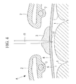
- FIG. 4 illustrates a step for cutting the membranous tissue, in the observation method of FIG. 1.
- FIG. 5 illustrates a state where an open cut is formed in the membranous tissue, in the observation method of FIG. 1.
- FIG. 6 illustrates a step for inserting an objective unit and observing, in the observation method of FIG. 1.
- FIG. 7 illustrates a state where the objective unit is withdrawn after the observation, in the observation method of FIG. 1.
- Hereunder is a description of the observation method according to an embodiment of the present invention, with reference to FIG. 1 to FIG. 7.
- The observation method according to the present embodiment is a method for observing, for example, a fetus (observation target). 1 of a mouse A in a living state, comprising: as shown in FIG. 1 to FIG. 7, a step S1 for incising the
epidermis 2 and the like; a step S2 for coating an adhesive 3; a step S3 for cutting the amniotic membrane (membranous tissue) 4; a step S4 for inserting an objective unit 5; and a step S5 for observing the fetus 1. As shown in FIG. 2, the fetus 1 of the mouse A is arranged in the uterine fascia and theamniotic membrane 4 filled with the amniotic fluid (body fluid) 6. - As shown in FIG. 2, in step S1 for incising the
epidermis 2 and the like, theepidermis 2 and the muscular tissue arranged between theribs 7 of the mouse A are cut and opened by a cutting instrument 8 such as a scalpel. As a result, theamniotic membrane 4 arranged inside of theepidermis 2 and the muscular tissue is exposed. - As shown in FIG. 3, in step S2 for coating an adhesive, a gel-
like adhesive 3 is heaped on the outer surface of theamniotic membrane 4 that has been exposed in step S1. The gel-like adhesive 3 is abiocompatible adhesive 3. For example,adhesives 3 of fibrin type, gelatin type, GTXIII type, cyanoacrylate type, urethane type, collagen type, chitin type, or stimulation responsive gel protein type, or those mixed with elastin to adjust the elasticity, are used. - The
adhesive 3 is not limited to these, and any arbitrarybiocompatible adhesive 3 may be used. - Moreover, for the adhesive 3, an adhesive 3 of a transparent material is preferably used.
- As shown in FIG. 4, in step S3 for cutting the
amniotic membrane 4, aboring instrument 9 having a scalpel or other sharp edge passes through theadhesive 3 that has been coated in step S2, to form anopen cut 10 in theamniotic membrane 4 in the area coated with theadhesive 3. Since the space in theamniotic membrane 4 is filled with theamniotic fluid 6 and has a predetermined internal pressure, at the instant when theboring instrument 9 passes through theamniotic membrane 4, theamniotic fluid 6 is to be ejected from theamniotic membrane 4 . However, in the present embodiment, since around theboring instrument 9 is enclosed and sealed by the gel-like adhesive 3, theamniotic fluid 6 does not leak out to the outside. - Moreover, as shown in FIG. 5, after cutting of the
amniotic membrane 4 is completed and theopen cut 10 is formed in theamniotic membrane 4, even if theboring instrument 9 is withdrawn, the adhesive 3 sealing around theboring instrument 9 sequentially blocks the track through which theboring instrument 9 passed, and thereby theamniotic fluid 6 is held so as not to leak out to the outside. - As shown in FIG. 6, in step S4 for inserting the
objective unit 11, the small diameterdistal end 11a of theobjective unit 11 is inserted into theamniotic membrane 4 through theopen cut 10 formed in the step S3. Theobjective unit 11 is attached to a microscopic observation device (not shown) and is designed such that illumination light such as exciting light like laser beams is irradiated from thedistal end face 11b, and light such as fluorescence returning from the fetus 1 is collected. - In this step S4, the small diameter
distal end 11a of theobjective unit 11 passes through the adhesive 3 coated on the outer surface of theamniotic membrane 4, reaches theopen cut 10 formed in step S3, and is inserted into theamniotic membrane 4 through theopen cut 10. As a result, since around the small diameterdistal end 11a of theobjective unit 11 is sealed by theadhesive 3, theamniotic fluid 6 in theamniotic membrane 4 is held so as not to leak out to the outside even when theobjective unit 11 is being inserted. - In step S5 for observing the fetus 1, the
distal end face 11b of the small diameterdistal end 11a of theobjective unit 11 that has been inserted into theamniotic fluid 6 is abutted against the outer surface of the fetus 1 serving as the observation target, so as to be stuck thereto, then exciting light is irradiated from thedistal end face 11b, and fluorescence generated at the fetus 1 is collected. As a result, a fluorescent image of the fetus 1 can be obtained. - In this case, even during the observation of the fetus 1, around the small diameter
distal end 11a of theobjective unit 11 is sealed by theadhesive 3, and therefore theamniotic fluid 6 in theamniotic membrane 4 is held so as not to leak out to the outside. - Moreover, according to the present embodiment, since the
transparent adhesive 3 is employed as the adhesive 3, even if theadhesive 3 is adhered onto thedistal end face 11b of theobjective unit 11 that has passed through theheaped adhesive 3, irradiation of exciting light and collection of fluorescence can be performed without much interference. - Furthermore, after the observation is completed, as shown in FIG. 7, the small diameter
distal end 11a of theobjective unit 11 is withdrawn from theopen cut 10 in theamniotic membrane 4. In this case, the adhesive 3 sealing around the small diameterdistal end 11a of theobjective unit 11 sequentially blocks the track through which the small diameterdistal end 11a passed, and thereby theamniotic fluid 6 is held so as not to leak out to the outside. Consequently, even after the observation, theamniotic fluid 6 can be held so as not to leak out to the outside, and the health of the observation target such as the fetus 1 in theamniotic fluid 6 can be maintained. - In this manner, according to the observation method according to the present embodiment, when an observation target such as the fetus 1 in the
amniotic membrane 4 is observed, the observation can be performed without leaking theamniotic fluid 6 to the outside. Therefore, there is an advantage in that the fetus 1 can be observed in a living and healthy state. - Moreover, after the observation, even if the
objective unit 11 is taken out, theamniotic fluid 6 can be held in theamniotic membrane 4 without leaking. Therefore, even after the observation, the health of the fetus 1 can be maintained. As a result, even if theobjective unit 11 is repeatedly inserted and withdrawn for a plurality of times with time intervals, the health of the fetus 1 can be maintained and the observation can be performed in a living state over time. - In the observation method according to the present embodiment, the description is about the example where the method is applied to the case where the fetus 1 of the mouse A is observed, however it is not to be considered as limiting. The method may be applied to the case where an observation target existing in a membranous tissue filled with another arbitrary liquid is observed.
- Moreover, the
amniotic membrane 4 is cut by theboring instrument 9, and then theobjective unit 11 is inserted thereinto. However, instead, as long as a sharp cutting blade can be arranged at the distal end of theobjective unit 11, theamniotic membrane 4 can be bored at the same time when theobjective unit 11 is inserted. - Furthermore, the description is about the case where the
objective unit 11 is repeatedly inserted and withdrawn for a plurality of times with time intervals to perform the observation over time. In this case, a marking member that applies an arbitrary marker such as a tracer protein to the observation target, may be used. By so doing, the marker can be applied to the already observed site, and the next observation can be readily performed at the same site or a different site. The marking member is preferably provided to the small diameterdistal end 11a of theobjective unit 11. - Moreover, as the
objective unit 11, a hard mirror type having the small diameterdistal end 11a was exemplified, however it is not to be considered as limiting. For example, a fiber bundle or the like may be used.
Claims (3)
- An observation method of an observation target arranged in a membranous tissue filled with a body fluid, comprising:a step for coating a gel-like adhesive on an outer surface of the membranous tissue;a step for cutting the membranous tissue through the coated adhesive;a step for inserting a distal end of the objective unit through the adhesive and an open cut in the membranous tissue; anda step for observing the observation target through the inserted distal end of the objective unit.
- An observation method according to claim 1, wherein the adhesive is transparent.
- An observation method according to claim 1, wherein there is further provided a step for applying a marker to the observed site on the observation target by a marking member provided on the distal end of the objective unit.
Applications Claiming Priority (2)
| Application Number | Priority Date | Filing Date | Title |
|---|---|---|---|
| JP2006105591 | 2006-04-06 | ||
| JP2007074971A JP2007296324A (en) | 2006-04-06 | 2007-03-22 | Observation method of biological tissue |
Publications (1)
| Publication Number | Publication Date |
|---|---|
| EP1842489A1 true EP1842489A1 (en) | 2007-10-10 |
Family
ID=38159236
Family Applications (1)
| Application Number | Title | Priority Date | Filing Date |
|---|---|---|---|
| EP07006287A Withdrawn EP1842489A1 (en) | 2006-04-06 | 2007-03-27 | Observation method of biological tissue |
Country Status (3)
| Country | Link |
|---|---|
| US (1) | US7874979B2 (en) |
| EP (1) | EP1842489A1 (en) |
| JP (1) | JP2007296324A (en) |
Family Cites Families (6)
| Publication number | Priority date | Publication date | Assignee | Title |
|---|---|---|---|---|
| US4929246A (en) * | 1988-10-27 | 1990-05-29 | C. R. Bard, Inc. | Method for closing and sealing an artery after removing a catheter |
| US5176649A (en) * | 1991-01-28 | 1993-01-05 | Akio Wakabayashi | Insertion device for use with curved, rigid endoscopic instruments and the like |
| US6296608B1 (en) * | 1996-07-08 | 2001-10-02 | Boston Scientific Corporation | Diagnosing and performing interventional procedures on tissue in vivo |
| US6726696B1 (en) * | 2001-04-24 | 2004-04-27 | Advanced Catheter Engineering, Inc. | Patches and collars for medical applications and methods of use |
| DE602004014697D1 (en) * | 2003-10-17 | 2008-08-14 | Olympus Co | Objective insertion device, fastening device for a lens system |
| JP4579563B2 (en) | 2004-03-15 | 2010-11-10 | オリンパス株式会社 | Objective optical system fixing device |
-
2007
- 2007-03-22 JP JP2007074971A patent/JP2007296324A/en not_active Withdrawn
- 2007-03-27 EP EP07006287A patent/EP1842489A1/en not_active Withdrawn
- 2007-03-29 US US11/730,073 patent/US7874979B2/en not_active Expired - Fee Related
Non-Patent Citations (1)
| Title |
|---|
| No Search * |
Also Published As
| Publication number | Publication date |
|---|---|
| US20070238965A1 (en) | 2007-10-11 |
| JP2007296324A (en) | 2007-11-15 |
| US7874979B2 (en) | 2011-01-25 |
Similar Documents
| Publication | Publication Date | Title |
|---|---|---|
| US10842566B1 (en) | Biophotonic surgical probe | |
| US20140228661A1 (en) | Biopsy method and gun set devices | |
| US20080045859A1 (en) | Devices and Methods for In-Vivo Pathology Diagnosis | |
| EP1685792B1 (en) | Optical penetrating adapter for surgical portal | |
| US5609562A (en) | Visually directed trocar and method | |
| US6296608B1 (en) | Diagnosing and performing interventional procedures on tissue in vivo | |
| JP4750672B2 (en) | Pre-fixed tissue cassette | |
| KR101250726B1 (en) | Device and method for mucosal detachment | |
| CN104755124B (en) | For determining the tool of sheath pipe transfer | |
| US20110130659A1 (en) | Imaging System for Following a Surgical Tool in an Operation Field | |
| US20080108991A1 (en) | Method and apparatus for performing pedicle screw fusion surgery | |
| GB1463784A (en) | Surgical tool for taking biological samples | |
| WO2004066828A3 (en) | Apparatus for endoscopic surgical procedures | |
| Prunier et al. | Procedures and applications of long-term intravital microscopy | |
| CN111065329A (en) | Endoscope system and method of use | |
| US11413191B2 (en) | Apparatus for perforation and aspiration of inner ear | |
| JP4481098B2 (en) | Living body observation device | |
| US7874979B2 (en) | Observation method of biological tissue | |
| US20100191043A1 (en) | Methods and devices for isolating a vessel | |
| CN109259834B (en) | Disposable oocyte collector | |
| CN205126314U (en) | Disposable endoscope live body sampling probe | |
| US20260000352A1 (en) | Light-based medical device | |
| JP2022126612A (en) | Real-time sampling device | |
| Lubatschowski | Laser microtomy: opening a new feasibility for tissue preparation | |
| CN118766613B (en) | A safe and accurate segmented positioning guidance system for deep tissue based on near-infrared second-zone light |
Legal Events
| Date | Code | Title | Description |
|---|---|---|---|
| PUAI | Public reference made under article 153(3) epc to a published international application that has entered the european phase |
Free format text: ORIGINAL CODE: 0009012 |
|
| AK | Designated contracting states |
Kind code of ref document: A1 Designated state(s): AT BE BG CH CY CZ DE DK EE ES FI FR GB GR HU IE IS IT LI LT LU LV MC MT NL PL PT RO SE SI SK TR |
|
| AX | Request for extension of the european patent |
Extension state: AL BA HR MK YU |
|
| 17P | Request for examination filed |
Effective date: 20071008 |
|
| AKX | Designation fees paid |
Designated state(s): DE FR GB |
|
| 17Q | First examination report despatched |
Effective date: 20090819 |
|
| STAA | Information on the status of an ep patent application or granted ep patent |
Free format text: STATUS: THE APPLICATION IS DEEMED TO BE WITHDRAWN |
|
| 18D | Application deemed to be withdrawn |
Effective date: 20091230 |