EP1839688A1 - Drug-eluting clinical device - Google Patents
Drug-eluting clinical device Download PDFInfo
- Publication number
- EP1839688A1 EP1839688A1 EP06006367A EP06006367A EP1839688A1 EP 1839688 A1 EP1839688 A1 EP 1839688A1 EP 06006367 A EP06006367 A EP 06006367A EP 06006367 A EP06006367 A EP 06006367A EP 1839688 A1 EP1839688 A1 EP 1839688A1
- Authority
- EP
- European Patent Office
- Prior art keywords
- dmso
- clinical device
- active ingredient
- stent
- eluting
- Prior art date
- Legal status (The legal status is an assumption and is not a legal conclusion. Google has not performed a legal analysis and makes no representation as to the accuracy of the status listed.)
- Withdrawn
Links
- IAZDPXIOMUYVGZ-UHFFFAOYSA-N Dimethylsulphoxide Chemical compound CS(C)=O IAZDPXIOMUYVGZ-UHFFFAOYSA-N 0.000 claims abstract description 305
- 239000004480 active ingredient Substances 0.000 claims abstract description 39
- 239000003814 drug Substances 0.000 claims abstract description 28
- 208000004476 Acute Coronary Syndrome Diseases 0.000 claims abstract description 18
- 230000002792 vascular Effects 0.000 claims abstract description 15
- 208000024172 Cardiovascular disease Diseases 0.000 claims abstract description 13
- 208000026106 cerebrovascular disease Diseases 0.000 claims abstract description 13
- 208000019553 vascular disease Diseases 0.000 claims abstract description 13
- 208000023516 stroke disease Diseases 0.000 claims abstract description 11
- 238000004519 manufacturing process Methods 0.000 claims abstract description 6
- 208000030613 peripheral artery disease Diseases 0.000 claims abstract 5
- 108010000499 Thromboplastin Proteins 0.000 claims description 73
- 102000002262 Thromboplastin Human genes 0.000 claims description 73
- 230000000694 effects Effects 0.000 claims description 38
- 208000007536 Thrombosis Diseases 0.000 claims description 24
- 229920000642 polymer Polymers 0.000 claims description 24
- 210000001519 tissue Anatomy 0.000 claims description 18
- 239000011159 matrix material Substances 0.000 claims description 16
- 238000000034 method Methods 0.000 claims description 12
- 230000005012 migration Effects 0.000 claims description 12
- 238000013508 migration Methods 0.000 claims description 12
- 230000035755 proliferation Effects 0.000 claims description 11
- 229940079593 drug Drugs 0.000 claims description 8
- 208000037803 restenosis Diseases 0.000 claims description 8
- 238000002399 angioplasty Methods 0.000 claims description 6
- 210000004509 vascular smooth muscle cell Anatomy 0.000 claims description 5
- 206010000891 acute myocardial infarction Diseases 0.000 claims description 3
- 208000006011 Stroke Diseases 0.000 claims 1
- 108060008682 Tumor Necrosis Factor Proteins 0.000 description 45
- 102000000852 Tumor Necrosis Factor-alpha Human genes 0.000 description 45
- 239000010410 layer Substances 0.000 description 28
- 210000004027 cell Anatomy 0.000 description 23
- 230000000638 stimulation Effects 0.000 description 21
- 230000006698 induction Effects 0.000 description 19
- 102000002574 p38 Mitogen-Activated Protein Kinases Human genes 0.000 description 14
- 108010068338 p38 Mitogen-Activated Protein Kinases Proteins 0.000 description 14
- 238000001727 in vivo Methods 0.000 description 13
- 108010007457 Extracellular Signal-Regulated MAP Kinases Proteins 0.000 description 12
- 108010055717 JNK Mitogen-Activated Protein Kinases Proteins 0.000 description 12
- 102100024193 Mitogen-activated protein kinase 1 Human genes 0.000 description 12
- 210000005119 human aortic smooth muscle cell Anatomy 0.000 description 12
- 239000003112 inhibitor Substances 0.000 description 12
- 239000000463 material Substances 0.000 description 12
- 208000027418 Wounds and injury Diseases 0.000 description 11
- 230000006378 damage Effects 0.000 description 11
- 230000001965 increasing effect Effects 0.000 description 11
- 208000014674 injury Diseases 0.000 description 11
- 108020004999 messenger RNA Proteins 0.000 description 11
- 108090000190 Thrombin Proteins 0.000 description 10
- 238000002474 experimental method Methods 0.000 description 10
- 229960004072 thrombin Drugs 0.000 description 10
- 102000019145 JUN kinase activity proteins Human genes 0.000 description 9
- 102000043136 MAP kinase family Human genes 0.000 description 9
- 108091054455 MAP kinase family Proteins 0.000 description 9
- 108010022233 Plasminogen Activator Inhibitor 1 Proteins 0.000 description 9
- 102100039418 Plasminogen activator inhibitor 1 Human genes 0.000 description 9
- 102100030951 Tissue factor pathway inhibitor Human genes 0.000 description 9
- 230000004913 activation Effects 0.000 description 9
- 230000005764 inhibitory process Effects 0.000 description 9
- 108010013555 lipoprotein-associated coagulation inhibitor Proteins 0.000 description 9
- 239000002953 phosphate buffered saline Substances 0.000 description 9
- 230000015572 biosynthetic process Effects 0.000 description 7
- 230000017531 blood circulation Effects 0.000 description 7
- 241000699670 Mus sp. Species 0.000 description 6
- 210000001715 carotid artery Anatomy 0.000 description 6
- 230000001419 dependent effect Effects 0.000 description 6
- 230000026731 phosphorylation Effects 0.000 description 6
- 238000006366 phosphorylation reaction Methods 0.000 description 6
- ZAHRKKWIAAJSAO-UHFFFAOYSA-N rapamycin Natural products COCC(O)C(=C/C(C)C(=O)CC(OC(=O)C1CCCCN1C(=O)C(=O)C2(O)OC(CC(OC)C(=CC=CC=CC(C)CC(C)C(=O)C)C)CCC2C)C(C)CC3CCC(O)C(C3)OC)C ZAHRKKWIAAJSAO-UHFFFAOYSA-N 0.000 description 6
- 229960002930 sirolimus Drugs 0.000 description 6
- QFJCIRLUMZQUOT-HPLJOQBZSA-N sirolimus Chemical compound C1C[C@@H](O)[C@H](OC)C[C@@H]1C[C@@H](C)[C@H]1OC(=O)[C@@H]2CCCCN2C(=O)C(=O)[C@](O)(O2)[C@H](C)CC[C@H]2C[C@H](OC)/C(C)=C/C=C/C=C/[C@@H](C)C[C@@H](C)C(=O)[C@H](OC)[C@H](O)/C(C)=C/[C@@H](C)C(=O)C1 QFJCIRLUMZQUOT-HPLJOQBZSA-N 0.000 description 6
- 108091003079 Bovine Serum Albumin Proteins 0.000 description 5
- 208000034827 Neointima Diseases 0.000 description 5
- 210000004204 blood vessel Anatomy 0.000 description 5
- 239000012894 fetal calf serum Substances 0.000 description 5
- 230000008692 neointimal formation Effects 0.000 description 5
- 230000001732 thrombotic effect Effects 0.000 description 5
- 206010003178 Arterial thrombosis Diseases 0.000 description 4
- 239000005552 B01AC04 - Clopidogrel Substances 0.000 description 4
- 239000006144 Dulbecco’s modified Eagle's medium Substances 0.000 description 4
- 102100031181 Glyceraldehyde-3-phosphate dehydrogenase Human genes 0.000 description 4
- 229930012538 Paclitaxel Natural products 0.000 description 4
- 238000004458 analytical method Methods 0.000 description 4
- 230000001028 anti-proliverative effect Effects 0.000 description 4
- 238000004113 cell culture Methods 0.000 description 4
- GKTWGGQPFAXNFI-HNNXBMFYSA-N clopidogrel Chemical compound C1([C@H](N2CC=3C=CSC=3CC2)C(=O)OC)=CC=CC=C1Cl GKTWGGQPFAXNFI-HNNXBMFYSA-N 0.000 description 4
- 229960003009 clopidogrel Drugs 0.000 description 4
- 230000003247 decreasing effect Effects 0.000 description 4
- 238000010790 dilution Methods 0.000 description 4
- 239000012895 dilution Substances 0.000 description 4
- 238000012377 drug delivery Methods 0.000 description 4
- 108020004445 glyceraldehyde-3-phosphate dehydrogenase Proteins 0.000 description 4
- 238000000338 in vitro Methods 0.000 description 4
- 231100000252 nontoxic Toxicity 0.000 description 4
- 229960001592 paclitaxel Drugs 0.000 description 4
- 239000000243 solution Substances 0.000 description 4
- RCINICONZNJXQF-MZXODVADSA-N taxol Chemical compound O([C@@H]1[C@@]2(C[C@@H](C(C)=C(C2(C)C)[C@H](C([C@]2(C)[C@@H](O)C[C@H]3OC[C@]3([C@H]21)OC(C)=O)=O)OC(=O)C)OC(=O)[C@H](O)[C@@H](NC(=O)C=1C=CC=CC=1)C=1C=CC=CC=1)O)C(=O)C1=CC=CC=C1 RCINICONZNJXQF-MZXODVADSA-N 0.000 description 4
- 230000001225 therapeutic effect Effects 0.000 description 4
- 238000002560 therapeutic procedure Methods 0.000 description 4
- 238000001262 western blot Methods 0.000 description 4
- KKJUPNGICOCCDW-UHFFFAOYSA-N 7-N,N-Dimethylamino-1,2,3,4,5-pentathiocyclooctane Chemical compound CN(C)C1CSSSSSC1 KKJUPNGICOCCDW-UHFFFAOYSA-N 0.000 description 3
- 101000635804 Homo sapiens Tissue factor Proteins 0.000 description 3
- -1 MP35N Substances 0.000 description 3
- QJJXYPPXXYFBGM-LFZNUXCKSA-N Tacrolimus Chemical compound C1C[C@@H](O)[C@H](OC)C[C@@H]1\C=C(/C)[C@@H]1[C@H](C)[C@@H](O)CC(=O)[C@H](CC=C)/C=C(C)/C[C@H](C)C[C@H](OC)[C@H]([C@H](C[C@H]2C)OC)O[C@@]2(O)C(=O)C(=O)N2CCCC[C@H]2C(=O)O1 QJJXYPPXXYFBGM-LFZNUXCKSA-N 0.000 description 3
- 230000003321 amplification Effects 0.000 description 3
- 229940121363 anti-inflammatory agent Drugs 0.000 description 3
- 239000002260 anti-inflammatory agent Substances 0.000 description 3
- 230000003110 anti-inflammatory effect Effects 0.000 description 3
- 230000005754 cellular signaling Effects 0.000 description 3
- 238000006243 chemical reaction Methods 0.000 description 3
- 238000007398 colorimetric assay Methods 0.000 description 3
- 239000002299 complementary DNA Substances 0.000 description 3
- 150000001875 compounds Chemical class 0.000 description 3
- 230000001276 controlling effect Effects 0.000 description 3
- 238000001514 detection method Methods 0.000 description 3
- 238000009792 diffusion process Methods 0.000 description 3
- 230000002757 inflammatory effect Effects 0.000 description 3
- 238000002347 injection Methods 0.000 description 3
- 239000007924 injection Substances 0.000 description 3
- 238000007912 intraperitoneal administration Methods 0.000 description 3
- 230000000670 limiting effect Effects 0.000 description 3
- 239000000203 mixture Substances 0.000 description 3
- 208000010125 myocardial infarction Diseases 0.000 description 3
- 229920000728 polyester Polymers 0.000 description 3
- 108090000623 proteins and genes Proteins 0.000 description 3
- 238000003753 real-time PCR Methods 0.000 description 3
- 230000009467 reduction Effects 0.000 description 3
- 230000002829 reductive effect Effects 0.000 description 3
- 239000002904 solvent Substances 0.000 description 3
- 238000010186 staining Methods 0.000 description 3
- 239000000126 substance Substances 0.000 description 3
- 238000001356 surgical procedure Methods 0.000 description 3
- 229960001967 tacrolimus Drugs 0.000 description 3
- QJJXYPPXXYFBGM-SHYZHZOCSA-N tacrolimus Natural products CO[C@H]1C[C@H](CC[C@@H]1O)C=C(C)[C@H]2OC(=O)[C@H]3CCCCN3C(=O)C(=O)[C@@]4(O)O[C@@H]([C@H](C[C@H]4C)OC)[C@@H](C[C@H](C)CC(=C[C@@H](CC=C)C(=O)C[C@H](O)[C@H]2C)C)OC QJJXYPPXXYFBGM-SHYZHZOCSA-N 0.000 description 3
- 231100000419 toxicity Toxicity 0.000 description 3
- 230000001988 toxicity Effects 0.000 description 3
- IICCLYANAQEHCI-UHFFFAOYSA-N 4,5,6,7-tetrachloro-3',6'-dihydroxy-2',4',5',7'-tetraiodospiro[2-benzofuran-3,9'-xanthene]-1-one Chemical compound O1C(=O)C(C(=C(Cl)C(Cl)=C2Cl)Cl)=C2C21C1=CC(I)=C(O)C(I)=C1OC1=C(I)C(O)=C(I)C=C21 IICCLYANAQEHCI-UHFFFAOYSA-N 0.000 description 2
- 108091093088 Amplicon Proteins 0.000 description 2
- 206010002388 Angina unstable Diseases 0.000 description 2
- BSYNRYMUTXBXSQ-UHFFFAOYSA-N Aspirin Chemical compound CC(=O)OC1=CC=CC=C1C(O)=O BSYNRYMUTXBXSQ-UHFFFAOYSA-N 0.000 description 2
- 108010014173 Factor X Proteins 0.000 description 2
- 108010074860 Factor Xa Proteins 0.000 description 2
- 102000009123 Fibrin Human genes 0.000 description 2
- 108010073385 Fibrin Proteins 0.000 description 2
- BWGVNKXGVNDBDI-UHFFFAOYSA-N Fibrin monomer Chemical compound CNC(=O)CNC(=O)CN BWGVNKXGVNDBDI-UHFFFAOYSA-N 0.000 description 2
- 241000699666 Mus <mouse, genus> Species 0.000 description 2
- 206010067482 No adverse event Diseases 0.000 description 2
- 229920000954 Polyglycolide Polymers 0.000 description 2
- FAPWRFPIFSIZLT-UHFFFAOYSA-M Sodium chloride Chemical compound [Na+].[Cl-] FAPWRFPIFSIZLT-UHFFFAOYSA-M 0.000 description 2
- GLNADSQYFUSGOU-GPTZEZBUSA-J Trypan blue Chemical compound [Na+].[Na+].[Na+].[Na+].C1=C(S([O-])(=O)=O)C=C2C=C(S([O-])(=O)=O)C(/N=N/C3=CC=C(C=C3C)C=3C=C(C(=CC=3)\N=N\C=3C(=CC4=CC(=CC(N)=C4C=3O)S([O-])(=O)=O)S([O-])(=O)=O)C)=C(O)C2=C1N GLNADSQYFUSGOU-GPTZEZBUSA-J 0.000 description 2
- 208000007814 Unstable Angina Diseases 0.000 description 2
- 229960001138 acetylsalicylic acid Drugs 0.000 description 2
- OIRDTQYFTABQOQ-KQYNXXCUSA-N adenosine Chemical compound C1=NC=2C(N)=NC=NC=2N1[C@@H]1O[C@H](CO)[C@@H](O)[C@H]1O OIRDTQYFTABQOQ-KQYNXXCUSA-N 0.000 description 2
- 230000002411 adverse Effects 0.000 description 2
- 238000000540 analysis of variance Methods 0.000 description 2
- 230000000692 anti-sense effect Effects 0.000 description 2
- 210000002403 aortic endothelial cell Anatomy 0.000 description 2
- 210000003433 aortic smooth muscle cell Anatomy 0.000 description 2
- 210000001367 artery Anatomy 0.000 description 2
- 230000003143 atherosclerotic effect Effects 0.000 description 2
- 230000009286 beneficial effect Effects 0.000 description 2
- 229920002988 biodegradable polymer Polymers 0.000 description 2
- 239000004621 biodegradable polymer Substances 0.000 description 2
- 210000004369 blood Anatomy 0.000 description 2
- 239000008280 blood Substances 0.000 description 2
- DQXBYHZEEUGOBF-UHFFFAOYSA-N but-3-enoic acid;ethene Chemical compound C=C.OC(=O)CC=C DQXBYHZEEUGOBF-UHFFFAOYSA-N 0.000 description 2
- 239000003795 chemical substances by application Substances 0.000 description 2
- 229920001577 copolymer Polymers 0.000 description 2
- 210000004351 coronary vessel Anatomy 0.000 description 2
- 239000003085 diluting agent Substances 0.000 description 2
- 229960001760 dimethyl sulfoxide Drugs 0.000 description 2
- 210000002889 endothelial cell Anatomy 0.000 description 2
- 239000005038 ethylene vinyl acetate Substances 0.000 description 2
- 229920000295 expanded polytetrafluoroethylene Polymers 0.000 description 2
- 229950003499 fibrin Drugs 0.000 description 2
- 210000003958 hematopoietic stem cell Anatomy 0.000 description 2
- 238000002513 implantation Methods 0.000 description 2
- 230000028709 inflammatory response Effects 0.000 description 2
- 239000003978 infusion fluid Substances 0.000 description 2
- 230000002401 inhibitory effect Effects 0.000 description 2
- 201000004332 intermediate coronary syndrome Diseases 0.000 description 2
- 230000003902 lesion Effects 0.000 description 2
- 238000011068 loading method Methods 0.000 description 2
- 230000004048 modification Effects 0.000 description 2
- 238000012986 modification Methods 0.000 description 2
- 238000011206 morphological examination Methods 0.000 description 2
- 238000010172 mouse model Methods 0.000 description 2
- 230000001400 myeloablative effect Effects 0.000 description 2
- 230000003000 nontoxic effect Effects 0.000 description 2
- 238000003199 nucleic acid amplification method Methods 0.000 description 2
- 239000000546 pharmaceutical excipient Substances 0.000 description 2
- 230000036470 plasma concentration Effects 0.000 description 2
- BASFCYQUMIYNBI-UHFFFAOYSA-N platinum Chemical compound [Pt] BASFCYQUMIYNBI-UHFFFAOYSA-N 0.000 description 2
- 229920001200 poly(ethylene-vinyl acetate) Polymers 0.000 description 2
- 229920000747 poly(lactic acid) Polymers 0.000 description 2
- 239000004633 polyglycolic acid Substances 0.000 description 2
- 239000004626 polylactic acid Substances 0.000 description 2
- 229920001343 polytetrafluoroethylene Polymers 0.000 description 2
- 239000004810 polytetrafluoroethylene Substances 0.000 description 2
- 239000013641 positive control Substances 0.000 description 2
- 230000002265 prevention Effects 0.000 description 2
- 230000008569 process Effects 0.000 description 2
- 102000004169 proteins and genes Human genes 0.000 description 2
- 230000004044 response Effects 0.000 description 2
- 229930187593 rose bengal Natural products 0.000 description 2
- 229940081623 rose bengal Drugs 0.000 description 2
- STRXNPAVPKGJQR-UHFFFAOYSA-N rose bengal A Natural products O1C(=O)C(C(=CC=C2Cl)Cl)=C2C21C1=CC(I)=C(O)C(I)=C1OC1=C(I)C(O)=C(I)C=C21 STRXNPAVPKGJQR-UHFFFAOYSA-N 0.000 description 2
- 239000000523 sample Substances 0.000 description 2
- 229920006395 saturated elastomer Polymers 0.000 description 2
- 208000010110 spontaneous platelet aggregation Diseases 0.000 description 2
- 239000003381 stabilizer Substances 0.000 description 2
- 238000007619 statistical method Methods 0.000 description 2
- 230000001629 suppression Effects 0.000 description 2
- 231100000331 toxic Toxicity 0.000 description 2
- 230000002588 toxic effect Effects 0.000 description 2
- 210000003462 vein Anatomy 0.000 description 2
- FDSYTWVNUJTPMA-UHFFFAOYSA-N 2-[3,9-bis(carboxymethyl)-3,6,9,15-tetrazabicyclo[9.3.1]pentadeca-1(15),11,13-trien-6-yl]acetic acid Chemical compound C1N(CC(O)=O)CCN(CC(=O)O)CCN(CC(O)=O)CC2=CC=CC1=N2 FDSYTWVNUJTPMA-UHFFFAOYSA-N 0.000 description 1
- QKNYBSVHEMOAJP-UHFFFAOYSA-N 2-amino-2-(hydroxymethyl)propane-1,3-diol;hydron;chloride Chemical compound Cl.OCC(N)(CO)CO QKNYBSVHEMOAJP-UHFFFAOYSA-N 0.000 description 1
- 101100207331 Arabidopsis thaliana TPPI gene Proteins 0.000 description 1
- 208000037260 Atherosclerotic Plaque Diseases 0.000 description 1
- 239000002126 C01EB10 - Adenosine Substances 0.000 description 1
- OKTJSMMVPCPJKN-UHFFFAOYSA-N Carbon Chemical compound [C] OKTJSMMVPCPJKN-UHFFFAOYSA-N 0.000 description 1
- 229920000049 Carbon (fiber) Polymers 0.000 description 1
- 206010007688 Carotid artery thrombosis Diseases 0.000 description 1
- 206010008089 Cerebral artery occlusion Diseases 0.000 description 1
- 206010065384 Cerebral hypoperfusion Diseases 0.000 description 1
- 102000008186 Collagen Human genes 0.000 description 1
- 108010035532 Collagen Proteins 0.000 description 1
- 206010011086 Coronary artery occlusion Diseases 0.000 description 1
- 102000004127 Cytokines Human genes 0.000 description 1
- 108090000695 Cytokines Proteins 0.000 description 1
- 102000016942 Elastin Human genes 0.000 description 1
- 108010014258 Elastin Proteins 0.000 description 1
- 102000004190 Enzymes Human genes 0.000 description 1
- 108090000790 Enzymes Proteins 0.000 description 1
- 102000010834 Extracellular Matrix Proteins Human genes 0.000 description 1
- 108010037362 Extracellular Matrix Proteins Proteins 0.000 description 1
- 108010054265 Factor VIIa Proteins 0.000 description 1
- 229940121710 HMGCoA reductase inhibitor Drugs 0.000 description 1
- 208000032843 Hemorrhage Diseases 0.000 description 1
- 206010061218 Inflammation Diseases 0.000 description 1
- 102000000589 Interleukin-1 Human genes 0.000 description 1
- 108010002352 Interleukin-1 Proteins 0.000 description 1
- 102000015696 Interleukins Human genes 0.000 description 1
- 108010063738 Interleukins Proteins 0.000 description 1
- 208000032382 Ischaemic stroke Diseases 0.000 description 1
- 102000003855 L-lactate dehydrogenase Human genes 0.000 description 1
- 108700023483 L-lactate dehydrogenases Proteins 0.000 description 1
- 206010067125 Liver injury Diseases 0.000 description 1
- VVQNEPGJFQJSBK-UHFFFAOYSA-N Methyl methacrylate Chemical compound COC(=O)C(C)=C VVQNEPGJFQJSBK-UHFFFAOYSA-N 0.000 description 1
- 208000034578 Multiple myelomas Diseases 0.000 description 1
- 241001529936 Murinae Species 0.000 description 1
- 239000000020 Nitrocellulose Substances 0.000 description 1
- 229910019142 PO4 Inorganic materials 0.000 description 1
- 239000002033 PVDF binder Substances 0.000 description 1
- 208000005764 Peripheral Arterial Disease Diseases 0.000 description 1
- 208000030831 Peripheral arterial occlusive disease Diseases 0.000 description 1
- 208000018262 Peripheral vascular disease Diseases 0.000 description 1
- 108091000080 Phosphotransferase Proteins 0.000 description 1
- 206010035226 Plasma cell myeloma Diseases 0.000 description 1
- 108010001014 Plasminogen Activators Proteins 0.000 description 1
- 102000001938 Plasminogen Activators Human genes 0.000 description 1
- 239000004952 Polyamide Substances 0.000 description 1
- 229920002732 Polyanhydride Polymers 0.000 description 1
- 229920000331 Polyhydroxybutyrate Polymers 0.000 description 1
- 206010063945 Pulmonary amyloidosis Diseases 0.000 description 1
- 206010061481 Renal injury Diseases 0.000 description 1
- 208000025747 Rheumatic disease Diseases 0.000 description 1
- 102000008847 Serpin Human genes 0.000 description 1
- 108050000761 Serpin Proteins 0.000 description 1
- 208000007718 Stable Angina Diseases 0.000 description 1
- 108010006785 Taq Polymerase Proteins 0.000 description 1
- RTAQQCXQSZGOHL-UHFFFAOYSA-N Titanium Chemical compound [Ti] RTAQQCXQSZGOHL-UHFFFAOYSA-N 0.000 description 1
- 239000007983 Tris buffer Substances 0.000 description 1
- 229920004890 Triton X-100 Polymers 0.000 description 1
- 239000013504 Triton X-100 Substances 0.000 description 1
- 208000032594 Vascular Remodeling Diseases 0.000 description 1
- FJWGYAHXMCUOOM-QHOUIDNNSA-N [(2s,3r,4s,5r,6r)-2-[(2r,3r,4s,5r,6s)-4,5-dinitrooxy-2-(nitrooxymethyl)-6-[(2r,3r,4s,5r,6s)-4,5,6-trinitrooxy-2-(nitrooxymethyl)oxan-3-yl]oxyoxan-3-yl]oxy-3,5-dinitrooxy-6-(nitrooxymethyl)oxan-4-yl] nitrate Chemical compound O([C@@H]1O[C@@H]([C@H]([C@H](O[N+]([O-])=O)[C@H]1O[N+]([O-])=O)O[C@H]1[C@@H]([C@@H](O[N+]([O-])=O)[C@H](O[N+]([O-])=O)[C@@H](CO[N+]([O-])=O)O1)O[N+]([O-])=O)CO[N+](=O)[O-])[C@@H]1[C@@H](CO[N+]([O-])=O)O[C@@H](O[N+]([O-])=O)[C@H](O[N+]([O-])=O)[C@H]1O[N+]([O-])=O FJWGYAHXMCUOOM-QHOUIDNNSA-N 0.000 description 1
- 238000002835 absorbance Methods 0.000 description 1
- 229960005305 adenosine Drugs 0.000 description 1
- 239000011543 agarose gel Substances 0.000 description 1
- 230000002776 aggregation Effects 0.000 description 1
- 238000004220 aggregation Methods 0.000 description 1
- 229910045601 alloy Inorganic materials 0.000 description 1
- 239000000956 alloy Substances 0.000 description 1
- 238000010171 animal model Methods 0.000 description 1
- 238000000137 annealing Methods 0.000 description 1
- 230000001567 anti-fibrinolytic effect Effects 0.000 description 1
- 229940082620 antifibrinolytics Drugs 0.000 description 1
- 239000000427 antigen Substances 0.000 description 1
- 102000036639 antigens Human genes 0.000 description 1
- 108091007433 antigens Proteins 0.000 description 1
- 239000002246 antineoplastic agent Substances 0.000 description 1
- 229940127218 antiplatelet drug Drugs 0.000 description 1
- 238000013459 approach Methods 0.000 description 1
- ZDQSOHOQTUFQEM-PKUCKEGBSA-N ascomycin Chemical class C/C([C@H]1OC(=O)[C@@H]2CCCCN2C(=O)C(=O)[C@]2(O)O[C@@H]([C@H](C[C@H]2C)OC)[C@@H](OC)C[C@@H](C)C\C(C)=C/[C@H](C(C[C@H](O)[C@H]1C)=O)CC)=C\[C@@H]1CC[C@@H](O)[C@H](OC)C1 ZDQSOHOQTUFQEM-PKUCKEGBSA-N 0.000 description 1
- 238000003556 assay Methods 0.000 description 1
- 210000000013 bile duct Anatomy 0.000 description 1
- 239000000560 biocompatible material Substances 0.000 description 1
- 230000036770 blood supply Effects 0.000 description 1
- 239000000872 buffer Substances 0.000 description 1
- 239000007853 buffer solution Substances 0.000 description 1
- 238000011088 calibration curve Methods 0.000 description 1
- 239000002775 capsule Substances 0.000 description 1
- 229910052799 carbon Inorganic materials 0.000 description 1
- 239000004917 carbon fiber Substances 0.000 description 1
- 210000001168 carotid artery common Anatomy 0.000 description 1
- 230000015556 catabolic process Effects 0.000 description 1
- 230000030833 cell death Effects 0.000 description 1
- 230000024245 cell differentiation Effects 0.000 description 1
- 210000000170 cell membrane Anatomy 0.000 description 1
- 230000004663 cell proliferation Effects 0.000 description 1
- 230000003833 cell viability Effects 0.000 description 1
- 229920002301 cellulose acetate Polymers 0.000 description 1
- 230000008859 change Effects 0.000 description 1
- 229940044683 chemotherapy drug Drugs 0.000 description 1
- 239000003593 chromogenic compound Substances 0.000 description 1
- 239000011248 coating agent Substances 0.000 description 1
- 238000000576 coating method Methods 0.000 description 1
- 229920001436 collagen Polymers 0.000 description 1
- 238000013270 controlled release Methods 0.000 description 1
- 208000029078 coronary artery disease Diseases 0.000 description 1
- 238000004132 cross linking Methods 0.000 description 1
- 230000009089 cytolysis Effects 0.000 description 1
- 231100000135 cytotoxicity Toxicity 0.000 description 1
- 230000003013 cytotoxicity Effects 0.000 description 1
- 230000034994 death Effects 0.000 description 1
- 238000006731 degradation reaction Methods 0.000 description 1
- LOKCTEFSRHRXRJ-UHFFFAOYSA-I dipotassium trisodium dihydrogen phosphate hydrogen phosphate dichloride Chemical compound P(=O)(O)(O)[O-].[K+].P(=O)(O)([O-])[O-].[Na+].[Na+].[Cl-].[K+].[Cl-].[Na+] LOKCTEFSRHRXRJ-UHFFFAOYSA-I 0.000 description 1
- 201000010099 disease Diseases 0.000 description 1
- 208000037265 diseases, disorders, signs and symptoms Diseases 0.000 description 1
- 239000002552 dosage form Substances 0.000 description 1
- 229920002549 elastin Polymers 0.000 description 1
- 238000010828 elution Methods 0.000 description 1
- 230000003511 endothelial effect Effects 0.000 description 1
- 229940088598 enzyme Drugs 0.000 description 1
- 210000003238 esophagus Anatomy 0.000 description 1
- ZMMJGEGLRURXTF-UHFFFAOYSA-N ethidium bromide Chemical compound [Br-].C12=CC(N)=CC=C2C2=CC=C(N)C=C2[N+](CC)=C1C1=CC=CC=C1 ZMMJGEGLRURXTF-UHFFFAOYSA-N 0.000 description 1
- 229960005542 ethidium bromide Drugs 0.000 description 1
- 230000007717 exclusion Effects 0.000 description 1
- 210000002744 extracellular matrix Anatomy 0.000 description 1
- 229940012414 factor viia Drugs 0.000 description 1
- 230000020764 fibrinolysis Effects 0.000 description 1
- 238000011049 filling Methods 0.000 description 1
- 230000002496 gastric effect Effects 0.000 description 1
- PCHJSUWPFVWCPO-UHFFFAOYSA-N gold Chemical compound [Au] PCHJSUWPFVWCPO-UHFFFAOYSA-N 0.000 description 1
- 229910052737 gold Inorganic materials 0.000 description 1
- 239000010931 gold Substances 0.000 description 1
- 239000008187 granular material Substances 0.000 description 1
- 231100000753 hepatic injury Toxicity 0.000 description 1
- 239000000017 hydrogel Substances 0.000 description 1
- 239000002471 hydroxymethylglutaryl coenzyme A reductase inhibitor Substances 0.000 description 1
- 206010020718 hyperplasia Diseases 0.000 description 1
- 229960003444 immunosuppressant agent Drugs 0.000 description 1
- 230000001861 immunosuppressant effect Effects 0.000 description 1
- 239000003018 immunosuppressive agent Substances 0.000 description 1
- 230000001976 improved effect Effects 0.000 description 1
- 238000011065 in-situ storage Methods 0.000 description 1
- 238000011534 incubation Methods 0.000 description 1
- 230000001939 inductive effect Effects 0.000 description 1
- 230000004054 inflammatory process Effects 0.000 description 1
- 238000001802 infusion Methods 0.000 description 1
- 239000004615 ingredient Substances 0.000 description 1
- 229940047122 interleukins Drugs 0.000 description 1
- 238000001361 intraarterial administration Methods 0.000 description 1
- 238000001990 intravenous administration Methods 0.000 description 1
- 230000009545 invasion Effects 0.000 description 1
- 238000011835 investigation Methods 0.000 description 1
- 230000002427 irreversible effect Effects 0.000 description 1
- 208000037906 ischaemic injury Diseases 0.000 description 1
- 208000037806 kidney injury Diseases 0.000 description 1
- 239000004816 latex Substances 0.000 description 1
- 229920000126 latex Polymers 0.000 description 1
- 239000006193 liquid solution Substances 0.000 description 1
- 239000012139 lysis buffer Substances 0.000 description 1
- 210000002540 macrophage Anatomy 0.000 description 1
- 230000010534 mechanism of action Effects 0.000 description 1
- 230000001404 mediated effect Effects 0.000 description 1
- 239000002609 medium Substances 0.000 description 1
- 238000011880 melting curve analysis Methods 0.000 description 1
- 239000012528 membrane Substances 0.000 description 1
- 229960002523 mercuric chloride Drugs 0.000 description 1
- LWJROJCJINYWOX-UHFFFAOYSA-L mercury dichloride Chemical compound Cl[Hg]Cl LWJROJCJINYWOX-UHFFFAOYSA-L 0.000 description 1
- 229910052751 metal Inorganic materials 0.000 description 1
- 239000002184 metal Substances 0.000 description 1
- VNWKTOKETHGBQD-UHFFFAOYSA-N methane Chemical compound C VNWKTOKETHGBQD-UHFFFAOYSA-N 0.000 description 1
- 210000004925 microvascular endothelial cell Anatomy 0.000 description 1
- 201000007309 middle cerebral artery infarction Diseases 0.000 description 1
- 239000003226 mitogen Substances 0.000 description 1
- 239000002829 mitogen activated protein kinase inhibitor Substances 0.000 description 1
- 210000001616 monocyte Anatomy 0.000 description 1
- 239000000178 monomer Substances 0.000 description 1
- 208000031225 myocardial ischemia Diseases 0.000 description 1
- 239000013642 negative control Substances 0.000 description 1
- 230000000926 neurological effect Effects 0.000 description 1
- 230000016273 neuron death Effects 0.000 description 1
- 208000004235 neutropenia Diseases 0.000 description 1
- HLXZNVUGXRDIFK-UHFFFAOYSA-N nickel titanium Chemical compound [Ti].[Ti].[Ti].[Ti].[Ti].[Ti].[Ti].[Ti].[Ti].[Ti].[Ti].[Ni].[Ni].[Ni].[Ni].[Ni].[Ni].[Ni].[Ni].[Ni].[Ni].[Ni].[Ni].[Ni].[Ni] HLXZNVUGXRDIFK-UHFFFAOYSA-N 0.000 description 1
- 229910001000 nickel titanium Inorganic materials 0.000 description 1
- 229920001220 nitrocellulos Polymers 0.000 description 1
- 210000000056 organ Anatomy 0.000 description 1
- 238000007911 parenteral administration Methods 0.000 description 1
- 239000002245 particle Substances 0.000 description 1
- 230000008506 pathogenesis Effects 0.000 description 1
- 230000007170 pathology Effects 0.000 description 1
- 229960001412 pentobarbital Drugs 0.000 description 1
- WEXRUCMBJFQVBZ-UHFFFAOYSA-N pentobarbital Chemical compound CCCC(C)C1(CC)C(=O)NC(=O)NC1=O WEXRUCMBJFQVBZ-UHFFFAOYSA-N 0.000 description 1
- 230000002093 peripheral effect Effects 0.000 description 1
- 230000009038 pharmacological inhibition Effects 0.000 description 1
- NBIIXXVUZAFLBC-UHFFFAOYSA-K phosphate Chemical compound [O-]P([O-])([O-])=O NBIIXXVUZAFLBC-UHFFFAOYSA-K 0.000 description 1
- 239000010452 phosphate Substances 0.000 description 1
- 102000020233 phosphotransferase Human genes 0.000 description 1
- 229940127126 plasminogen activator Drugs 0.000 description 1
- 229910052697 platinum Inorganic materials 0.000 description 1
- HWLDNSXPUQTBOD-UHFFFAOYSA-N platinum-iridium alloy Chemical compound [Ir].[Pt] HWLDNSXPUQTBOD-UHFFFAOYSA-N 0.000 description 1
- 239000005015 poly(hydroxybutyrate) Substances 0.000 description 1
- 229920001606 poly(lactic acid-co-glycolic acid) Polymers 0.000 description 1
- 229920002647 polyamide Polymers 0.000 description 1
- 229920001610 polycaprolactone Polymers 0.000 description 1
- 239000004632 polycaprolactone Substances 0.000 description 1
- 229920001296 polysiloxane Polymers 0.000 description 1
- 229920002635 polyurethane Polymers 0.000 description 1
- 239000004814 polyurethane Substances 0.000 description 1
- 239000011118 polyvinyl acetate Substances 0.000 description 1
- 229920002981 polyvinylidene fluoride Polymers 0.000 description 1
- 230000031915 positive regulation of coagulation Effects 0.000 description 1
- 230000036515 potency Effects 0.000 description 1
- 230000003389 potentiating effect Effects 0.000 description 1
- 238000004321 preservation Methods 0.000 description 1
- 230000000750 progressive effect Effects 0.000 description 1
- 230000001681 protective effect Effects 0.000 description 1
- 230000002685 pulmonary effect Effects 0.000 description 1
- 108020003175 receptors Proteins 0.000 description 1
- 230000000552 rheumatic effect Effects 0.000 description 1
- 238000013207 serial dilution Methods 0.000 description 1
- 239000003001 serine protease inhibitor Substances 0.000 description 1
- 231100000161 signs of toxicity Toxicity 0.000 description 1
- 239000011780 sodium chloride Substances 0.000 description 1
- 238000002415 sodium dodecyl sulfate polyacrylamide gel electrophoresis Methods 0.000 description 1
- 241000894007 species Species 0.000 description 1
- 239000010935 stainless steel Substances 0.000 description 1
- 229910001220 stainless steel Inorganic materials 0.000 description 1
- 230000002966 stenotic effect Effects 0.000 description 1
- 239000002344 surface layer Substances 0.000 description 1
- 230000004083 survival effect Effects 0.000 description 1
- 238000003786 synthesis reaction Methods 0.000 description 1
- 229910052715 tantalum Inorganic materials 0.000 description 1
- GUVRBAGPIYLISA-UHFFFAOYSA-N tantalum atom Chemical compound [Ta] GUVRBAGPIYLISA-UHFFFAOYSA-N 0.000 description 1
- 238000012360 testing method Methods 0.000 description 1
- 238000004324 time-proportional phase incrementation Methods 0.000 description 1
- 229910052719 titanium Inorganic materials 0.000 description 1
- 239000010936 titanium Substances 0.000 description 1
- 210000003437 trachea Anatomy 0.000 description 1
- 238000012546 transfer Methods 0.000 description 1
- 230000001052 transient effect Effects 0.000 description 1
- 230000001960 triggered effect Effects 0.000 description 1
- LENZDBCJOHFCAS-UHFFFAOYSA-N tris Chemical compound OCC(N)(CO)CO LENZDBCJOHFCAS-UHFFFAOYSA-N 0.000 description 1
- 238000012762 unpaired Student’s t-test Methods 0.000 description 1
- 210000003708 urethra Anatomy 0.000 description 1
- 230000002485 urinary effect Effects 0.000 description 1
- 229940070710 valerate Drugs 0.000 description 1
- NQPDZGIKBAWPEJ-UHFFFAOYSA-N valeric acid Chemical compound CCCCC(O)=O NQPDZGIKBAWPEJ-UHFFFAOYSA-N 0.000 description 1
- 210000005167 vascular cell Anatomy 0.000 description 1
- 238000012795 verification Methods 0.000 description 1
- 238000005303 weighing Methods 0.000 description 1
Images



Classifications
-
- A—HUMAN NECESSITIES
- A61—MEDICAL OR VETERINARY SCIENCE; HYGIENE
- A61L—METHODS OR APPARATUS FOR STERILISING MATERIALS OR OBJECTS IN GENERAL; DISINFECTION, STERILISATION OR DEODORISATION OF AIR; CHEMICAL ASPECTS OF BANDAGES, DRESSINGS, ABSORBENT PADS OR SURGICAL ARTICLES; MATERIALS FOR BANDAGES, DRESSINGS, ABSORBENT PADS OR SURGICAL ARTICLES
- A61L31/00—Materials for other surgical articles, e.g. stents, stent-grafts, shunts, surgical drapes, guide wires, materials for adhesion prevention, occluding devices, surgical gloves, tissue fixation devices
- A61L31/14—Materials characterised by their function or physical properties, e.g. injectable or lubricating compositions, shape-memory materials, surface modified materials
- A61L31/16—Biologically active materials, e.g. therapeutic substances
-
- A—HUMAN NECESSITIES
- A61—MEDICAL OR VETERINARY SCIENCE; HYGIENE
- A61L—METHODS OR APPARATUS FOR STERILISING MATERIALS OR OBJECTS IN GENERAL; DISINFECTION, STERILISATION OR DEODORISATION OF AIR; CHEMICAL ASPECTS OF BANDAGES, DRESSINGS, ABSORBENT PADS OR SURGICAL ARTICLES; MATERIALS FOR BANDAGES, DRESSINGS, ABSORBENT PADS OR SURGICAL ARTICLES
- A61L31/00—Materials for other surgical articles, e.g. stents, stent-grafts, shunts, surgical drapes, guide wires, materials for adhesion prevention, occluding devices, surgical gloves, tissue fixation devices
- A61L31/08—Materials for coatings
- A61L31/10—Macromolecular materials
-
- A—HUMAN NECESSITIES
- A61—MEDICAL OR VETERINARY SCIENCE; HYGIENE
- A61L—METHODS OR APPARATUS FOR STERILISING MATERIALS OR OBJECTS IN GENERAL; DISINFECTION, STERILISATION OR DEODORISATION OF AIR; CHEMICAL ASPECTS OF BANDAGES, DRESSINGS, ABSORBENT PADS OR SURGICAL ARTICLES; MATERIALS FOR BANDAGES, DRESSINGS, ABSORBENT PADS OR SURGICAL ARTICLES
- A61L2300/00—Biologically active materials used in bandages, wound dressings, absorbent pads or medical devices
- A61L2300/20—Biologically active materials used in bandages, wound dressings, absorbent pads or medical devices containing or releasing organic materials
-
- A—HUMAN NECESSITIES
- A61—MEDICAL OR VETERINARY SCIENCE; HYGIENE
- A61L—METHODS OR APPARATUS FOR STERILISING MATERIALS OR OBJECTS IN GENERAL; DISINFECTION, STERILISATION OR DEODORISATION OF AIR; CHEMICAL ASPECTS OF BANDAGES, DRESSINGS, ABSORBENT PADS OR SURGICAL ARTICLES; MATERIALS FOR BANDAGES, DRESSINGS, ABSORBENT PADS OR SURGICAL ARTICLES
- A61L2300/00—Biologically active materials used in bandages, wound dressings, absorbent pads or medical devices
- A61L2300/40—Biologically active materials used in bandages, wound dressings, absorbent pads or medical devices characterised by a specific therapeutic activity or mode of action
- A61L2300/416—Anti-neoplastic or anti-proliferative or anti-restenosis or anti-angiogenic agents, e.g. paclitaxel, sirolimus
-
- A—HUMAN NECESSITIES
- A61—MEDICAL OR VETERINARY SCIENCE; HYGIENE
- A61L—METHODS OR APPARATUS FOR STERILISING MATERIALS OR OBJECTS IN GENERAL; DISINFECTION, STERILISATION OR DEODORISATION OF AIR; CHEMICAL ASPECTS OF BANDAGES, DRESSINGS, ABSORBENT PADS OR SURGICAL ARTICLES; MATERIALS FOR BANDAGES, DRESSINGS, ABSORBENT PADS OR SURGICAL ARTICLES
- A61L2300/00—Biologically active materials used in bandages, wound dressings, absorbent pads or medical devices
- A61L2300/40—Biologically active materials used in bandages, wound dressings, absorbent pads or medical devices characterised by a specific therapeutic activity or mode of action
- A61L2300/42—Anti-thrombotic agents, anticoagulants, anti-platelet agents
Definitions
- the present invention relates to a clinical device for the treatment of vascular, cerebrovascular and cardiovascular disorders or acute coronary syndromes and the use of DMSO for the manufacture of a medicament for the treatment of vascular, cerebrovascular or cardiovascular disorders or acute coronary syndromes.
- DMSO Dimethyl sulfoxide
- Egorin myeloablative therapy
- DMSO Dimethyl sulfoxide
- Egorin myeloablative therapy
- DMSO has been successfully employed in treating human pathologies of dermatological (Burgess, 1998; Wong, 1988), neurological (Karaca, 1991, 2002), urinary (McCammon, 1998), pulmonary (lwasaki, 1994), rheumatic (Morassi, 1989) and gastrointestinal (Salim, 1992; 1994) origin.
- DMSO mercuric chloride induced kidney injury (Jo, 2004), chemical liver injury (Lind, 1997), middle cerebral artery occlusion (Bardutzky, 2005), and cerebral hypoperfusion related neuronal death (Farkas, 2004). Despite of all this information, the potential impact of DMSO on vascular or cardiovascular disorders has not been assessed.
- Tissue Factor is a key enzyme in the activation of coagulation (Steffel, 2005). Plaque rupture leading to arterial thrombosis is the main cause of acute coronary syndromes (ACS) such as unstable angina and myocardial infarction. TF plays a major role in determining plaque thrombogenicity (Moons, 2002), as it is expressed by different cell types within the atherosclerotic vessel wall and is highly exposed to the blood stream following plaque rupture. Many inflammatory mediators are capable of inducing TF expression in cells such as monocytes, macrophages, endothelial cells, and vascular smooth muscle cells (Moons, 2002).
- Circulatory diseases are widespread and are related to progressive narrowing of the blood vessel due to atherosclerotic plaques; plaque rupture often leads to ischemic injury, stroke or myocardial infarction. Acute myocardial infarction occurs, because the blood supply to a part of the organ is interrupted; indeed, atherosclerotic lesions, which limit coronary blood flow and eventually rupture, are the major cause of ischemic heart disease.
- PTA Percutaneous transluminal angioplasty
- PCTA coronary arteries
- a balloon catheter device which is inserted into the blood vessel, is inflated to dilate the stenotic (abnormally narrowed) blood vessel, which normally results from plaque or plaque rupture leading to thrombosis.
- the pressurized balloon exerts a force on the lesion, thereby increasing the inner diameter of the affected vessel.
- the increased interior vessel diameter facilitates improved blood flow.
- stent This re-narrowing, called restenosis, is inibited by introduction of a short flexible cylinder called stent.
- a stent is either an expandable wire mesh or hollow perforated tube that is inserted into a hollow structure of the body to keep it open.
- stents are mostly used in treatment of coronary arteries, they can also be used in several other structures, such as peripheral arteries and veins, bile ducts, esophagus, trachea or large bronchie, urethers and urethra.
- neointima formation which is the formation of new tissue, and constrictive vascular remodelling. Although the latter is prevented by a stent, neointima formation can still occur within the stent or at its edges.
- drug-eluting stents are known in the art.
- EP 1 600 180 A2 discloses a drug delivery device for treating in-stent intimal hyperplasia in human vessel walls comprising a therapeutic dosage of an agent having anti-proliferative and anti-inflammatory properties, which preferably comprises rapamycin.
- WO 2004/045578 A2 discloses a drug delivery device comprising a medical device, e.g. a coated stent or stent-graft, in conjunction therewith a therapeutic dosage form of an anti-inflammatory ascomycin derivative, e.g. afixed to the medical device.
- a medical device e.g. a coated stent or stent-graft
- a therapeutic dosage form of an anti-inflammatory ascomycin derivative e.g. afixed to the medical device.
- stent thrombosis is a rare, but much more dangerous complication after coronary stent placement. It usually occurs before endothelialisation has been completed. For bare-metal stents, this process takes a few weeks; the newer, drug-eluting stents inhibit restenosis by inhibiting vascular smooth muscle cell proliferation, but they also tend to delay the endothelialisation process. For this reason, patients who have had drug-eluting stents are prescribed aspirin plus clopidogrel therapy for at least six months.
- Clopidogrel is a known anti-platelet agent often used in the treatment of coronary artery diseases, peripheral vascular diseases and cerebrovascular diseases.
- the mechanism of action of clopidogrel is irreversible blockade of the adenosine II phosphate (ADP) receptor P2Y12 on the platelet cell membranes, which is important in platelet aggregation, in particular the crosslinking of platelets by fibrin.
- ADP adenosine II phosphate
- the blockade of this receptor inhibits platelet aggregation.
- adverse effects like severe neutropenia or the occurrence of hemorrhage, in particular occurring when co-administered with aspirin, clopidogrel has the disadvantage of being rather expensive. Therefore, there is still the need for an alternative inhibitor of thrombosis after coronary stent placement; in particular a local inhibitor of thrombosis.
- tissue factor in acute coronary syndromes (ACS) is known, as stated above. Recent studies even show increased levels of TF antigen and activity in atherectomy specimens from patients with unstable angina or myocardial infarction as compared to those with stable angina (Annex, 1995). Moreover, in ACS, plasma concentrations of inflammatory cytokines such as TNF- ⁇ (tumor necrosis factor ⁇ ) and interleukins are increased at the site of coronary artery occlusion to such an extent that TF is induced in vascular cells (Maier, 2005).
- TNF- ⁇ tumor necrosis factor ⁇
- DMSO is, besides being active in suppression of inflammatory response and inhibition of platelet activity, active in prevention of thrombosis.
- DMSO inhibits proliferation and migration of human aortic vascular smooth muscle cells (HASMC), and reduces TF activity in vitro and in vivo in addition to preventing TF-dependent thrombosis in vivo.
- HASMC human aortic vascular smooth muscle cells
- the present invention relates to a clinical device for being implanted that contains means for eluting an active ingredient, said active ingredient essentially comprising DMSO.
- the clinical device according to the present invention comprises an active ingredient, which consists of at least 80% of DMSO, more preferable of at least 95% of DMSO. Percent is given as percent by weight.
- the "clinical device” of the present invention refers to a device which is inserted into, or grafted onto, body tissues to remain for a period of time, such as an extended-released drug delivery device, stent, drug-eluting stent, or vascular graft.
- a clinical device comprises a surface or surface layer suitable for being implanted.
- the active ingredient of the clinical device of the present invention comprises DMSO, more preferable the active ingredient comprises from about 20 to about 100% DMSO, even more preferred from 60 to 100% DMSO, in particular the active ingredient contains 80 to 90% DMSO.
- the active ingredient may also comprise other pharmaceutically active compounds like rapamycin, paclitaxel, tacrolimus, statins, and similar compounds, which have beneficial effects for the tissues. DMSO and said other compound(s) add up to 100%.
- the active ingredient contains at least 20%, preferred at least 50%, more preferred at least 80%, most preferred at least 90%, in particular at least 95% DMSO as active ingredient.
- the active ingredient of the clinical device comprises DMSO in combination with further anti-proliferative and/or anti-inflammatory agents.
- Means for eluting the active ingredient(s) from the clinical device of the present invention are known in the art.
- materials that can be applied are layers of drug-eluting materials, such as polymer-matrices.
- Each polymer suitable for application on a device which can be implanted in an organism can be used, provided that the material is non-toxic and able to release an active ingredient in a pre-defined period of time.
- Preferred periods of release are from about 2 to 28 days, in particular 14 to 28 days- More preferably the period of release is in a range of 2 to 4 weeks with a maximum of drug-elution after 1 to 2 weeks.
- the material to be used as drug-eluting layer has to be able to release an active ingredient, which contains a high amount of DMSO, e.g. from about 50% to about 100% DMSO content, within a predefined period of time.
- the clinical device is covered with a degradable layer, which is the only or the first layer of the clinical device, said layer comprising one or a variety of polymer matrices.
- the matrix should be biocompatible, biodegradable, bioerodable, non-toxic, bioabsorbable, and with a defined rate of degradation.
- Biocompatible matrices that can be used in the invention include, but are not limited to, poly(lactide-co-glycolide), polyesters such as polylactic acid, polyglycolic acid and copolymers thereof, polyanhydride, polycaprolactone, polyhydroxybutyrate, valerate, and other biodegradable polymers and mixtures thereof.
- the matrix can also comprise naturally occurring polymeric materials such as collagen, fibrin, elastin, and extracellular matrix components.
- the polymer matrix of the clinical device further comprises pharmaceutically acceptable polymers, as the only or the second layer, for example, non-absorbable polymers, such as ethylene vinyl acetate (EVAC) and methylmethacrylate (MMA).
- non-absorbable polymers such as ethylene vinyl acetate (EVAC) and methylmethacrylate (MMA).
- EVAC ethylene vinyl acetate
- MMA methylmethacrylate
- the non-absorbable polymer for example, can aid in further controlling release of the substance by limiting diffusion, thereby delaying or slowing the rate of release of the active ingredient, in particular when used as second layer.
- the active ingredient is mainly present in the first, inner polymer matrix layer of the clinical device.
- This inner polymer matrix layer, containing the active ingredient, which comprises a significant amount of DMSO can for example be prepared by using DMSO as polymerisation solvent, or the polymer of the inner polymer matrix layer is saturated with DMSO and optionally other ingredients of the active ingredient after polymerisation, before or after application as first polymer matrix layer to the clinical device.
- the polymer of this inner layer is a degradable layer as specified above.
- a further, slowly degradable or preferably a non-absorbable polymer layer as defined above can be applied, preferably as diffusion limiting and controlling layer, thereby enabling controlled release of active ingredient, which comprises a significant amount of DMSO, into the surrounding tissue.
- the active ingredient comprising a significant amount of DMSO
- the active ingredient is contained in pre-formed particles or granules of matrix polymer, which are applied to the clinical device of the present invention, eventually within a further polymer matrix.
- the base material of the clinical device of the present invention which preferably is covered with means for eluting an active ingredient, can be of various types, for example, stainless steel, nitinol, MP35N, gold, tantalum, platinum, platinum-iridium or titanium, which is preferred, or other biocompatible materials and/or alloys such as carbon or carbon fiber, cellulose acetate, cellulose nitrate, silicone, cross-linked polyvinylacetate (PVA), hydrogel, polyurethane, polyamide, polyester, or a mixture of polymers thereof; polyesters such as polylactic acid, polyglycolic acid or copolymers thereof and other biodegradable polymers or suitable degradable materials can also be used.
- stainless steel nitinol, MP35N, gold, tantalum, platinum, platinum-iridium or titanium, which is preferred, or other biocompatible materials and/or alloys such as carbon or carbon fiber, cellulose acetate, cellulose nitrate, silicone, cross-linked polyvinylacetate (PVA),
- the drug-eluting layer of the clinical device or the clinical device itself is degraded by the organism, which the device is implanted in, thereby releasing the drug contained.
- the clinical device is a stent, preferably a coated or covered stent, such as those covered with polytetrafluoroethylene (PTFE), expanded polytetrafluoroethylene (ePTFE), and such as those covered stents which are alternatively or additionally covered with a polymer matrix as described above.
- PTFE polytetrafluoroethylene
- ePTFE expanded polytetrafluoroethylene
- a stent is either an expandable wire mesh or a hollow perforated tube that is inserted in a hollow structure in the body to keep it open.
- base material of the stent all materials as described for the clinical device can be used.
- the clinical device is a depot stent, which contains the active ingredient in suitable reservoirs or wells, e.g. holes within the stent which are filled with erodable polymer containing the active ingredient.
- a bioresorbable drug-delivery stent including a substantially cylindrical expandable stent formed of a bioresorbable material and a plurality of reservoirs or openings formed in the stent containing a beneficial agent matrix comprising a bioresorbable material and drug is for example disclosed in WO 2005/102222 , the content of which is incorporated by reference herein.
- DMSO lipophilic character
- DMSO is particularly useful as eluted drug in a stent, as the lipophily renders DMSO suitable for being easily incorporated into suitable non-erodable and/or erodable polymers to be used in a drug-eluting stent of the present invention.
- the stent may be dipped, sprayed with, or loaded with, for example, a liquid solution of the matrix polymer of moderate viscosity.
- the clinical device such as a stent may be coated with matrix polymer during or after polymerisation, for example by applying the devices into monomer solutions or prepolymerized matrix polymer solutions, preferably in presence of active ingredient, comprising a significant amount of DMSO, to form a layer, covering the device, which is saturated with active ingredient, preferably during polymerisation.
- a further diffusion limiting and controlling layer as specified above may be applied to the device as second layer.
- the stent is dried before application of the next layer.
- a thin, paint-like polymer matrix coating does not exceed an overall thickness of about 100 to about 500 ⁇ m.
- the clinical device is only covered by the drug-eluting layer at those positions, where said device is in direct contact to the organism, i.e. the surrounding tissue.
- the “surrounding tissue” according to the present invention is the tissue which is in direct contact to the material of the clinical device of the present invention or the tissue which is in direct contact and the tissue which is directly adjacent to the clinical device of the present invention.
- the active ingredient which comprises a significant amount of DMSO
- tissue factor protein is suppressed by at least 20%, more preferred by at least 30%, in particular by at least 50%, relative to the expression level of tissue factor protein of tissue.
- which has been exposed to an increased level of inflammatory mediators comparable to or such as TNF- ⁇ or thrombin, in particular compared to or such as concentrations of 5 ng/ml TNF- ⁇ or 1 U/ml thrombin.
- proliferation and/or migration of HVSMC is suppressed by at least 20%, more preferred at least 30%, in particular by at least 50%, relative to the level when no active ingredient is present.
- the active ingredient which comprises at least more than 50% by weight of DMSO, is eluted from the clinical device within a predetermined period of time, preferably during 1-28 days, e.g. for 14 days.
- the clinical device contains an amount of active ingredient in a range from about 10 ng to about 10 mg, preferably from about 1 ⁇ g to about 1000 ⁇ g.
- the active ingredient is incorporated into the drug-eluting layer, in case of two layers in both or one layer, in case of more than two layers in one, two or more layers in a percent loading of between 0.1% to 70% by weight, preferably between 5% and 50% by weight.
- the amount eluted from the clinical device into the surrounding tissue is from about 1 ng/mm 2 h to about 1 ⁇ g/mm 2 h, relative to the contacted surrounding tissue area and time after implantation.
- the present invention further relates to the use of DMSO for the manufacture of a medicament for the treatment of vascular, cerebrovascular or cardiovascular disorders, stroke or acute coronary syndromes.
- the DMSO used for the manufacture of a medicament for the treatment of vascular, cerebrovascular or cardiovascular disorders, stroke or acute coronary syndromes is used in combination with further anti-proliferative and/or anti-inflammatory agents, e.g. rapamycin, paclitaxel or tacrolimus or in combination with other medicaments applied for treating these conditions.
- further anti-proliferative and/or anti-inflammatory agents e.g. rapamycin, paclitaxel or tacrolimus or in combination with other medicaments applied for treating these conditions.
- the medicament for the treatment of vascular, cerebrovascular or cardiovascular disorders, stroke or acute coronary syndromes preferably contains 20-100% DMSO, more preferable 60-100% DMSO, in particular 80-90% DMSO.
- Said medicament may further comprise appropriate diluents, such as buffer solutions, excipients and stabilizers, being suitable for administration, for example by syringe or infusion.
- appropriate diluents such as buffer solutions, excipients and stabilizers, being suitable for administration, for example by syringe or infusion.
- Another possible form of administration is a bolus injection, wherein the DMSO can be premixed with an infusion solution or can be prepared in situ by addition of DMSO into the infusion solution prior to injection.
- the medicament can be administered parenterally or subcutaneously as desired.
- the therapeutic composition should be sterile, pyrogen-free and in a parenterally acceptable solution having due regard for pH, isotonicity, and stability. These conditions are known to
- the medicament for the treatment of vascular, cerebrovascular or cardiovascular disorders, stroke or acute coronary syndromes can be administered orally, for example in form of a tablet, capsule or any other form suitable for oral administration.
- Said medicament further comprises appropriate diluents, excipients and coadjuvants suitable for oral administration. Methods for producing these administration forms are known to those skilled in the art.
- the present invention further relates to the method of treating vascular, cerebrovascular or cardiovascular disorders, stroke or acute coronary syndromes by administration of drug-eluting stents able to release a drug comprising a significant amount of DMSO and/or administering a medicament comprising a significant amount of DMSO to a patient in need thereof.
- the method of treating vascular, cerebrovascular or cardiovascular disorders, stroke or acute coronary syndromes comprises administration of a medicament comprising a significant amount of DMSO in a combinational product, preferably by intravenous or oral administration.
- the medicament comprising a significant amount of DMSO is used in combination with further anti-proliferative and/or anti-inflammatory agents, e.g. rapamycin, paclitaxel or tacrolimus or in combination with other medicaments applied for treating these conditions.
- the present invention further relates to a method for reducing restenosis, in-stent restenosis, stent thrombosis or in-stent thrombosis comprising administration of a drug-eluting stent able to release a drug comprising a significant amount of DMSO and/or administering a medicament comprising a significant amount of DMSO to a patient in need thereof.
- the present invention further relates to a method of treating acute myocardial infarction, stroke or peripheral arterial disease comprising administering a medicament comprising a significant amount of DMSO to a subject receiving a stent in connection with percutaneous transluminal angioplasty.
- the present invention offers a completely new approach to specific inhibition of expression and activity of TF (cp. Example 1).
- DMSO is able to inhibit TF induction in human aortic endothelial cells (HAEC), human aortic vascular smooth muscle cells (HAVSMC) and THP-1 cells. This effect was mediated by a reduced activation of p38 and JNK MAP kinase activation (cp. Example 4) and expression of TF mRNA (cp. Example 5). Consistent with this observation, it was shown that DMSO protects from thrombotic occlusion in an in vivo model of TF-dependent thrombus formation (cp. Example 6). Further it could be demonstrated that DMSO inhibits proliferation and migration of human aortic smooth muscle cells (HASMC) (cp. Example 7).
- HASMC human aortic smooth muscle cells
- DMSO as an excellent active ingredient in a variety of applications, preferably in combination with a stent therapy.
- DMSO is first active in suppression of inflammatory response, corresponding complications caused by inflammation, in particular shortly after stent implantation can be reduced.
- DMSO is also active in prevention of thrombosis, in particular by inhibition of tissue factor and platelet activity.
- thrombosis in particular stent thrombosis, is a rare but mostly very dangerous complication often leading to death of the patient.
- DMSO is a well known solvent which is easy to handle and which is much less expensive than substances with a comparable field of application.
- DMSO was used in concentrations as high as 1% by weight (12.6 mmol/L) and no toxic effects by DMSO treatment could be observed, even at the highest concentration (cp. Example 2).
- a focused study found no toxic effect by DMSO treatment on endothelial cells at concentrations well above those (Bourne, 1994), and contemporary literature describes routine usage of DMSO to trigger cellular differentiation in vitro in concentrations as high as 1.25% (15.7 mM) (Heidari, 2004).
- DMSO is universally infused intravenously together with hematopoietic progenitor cells in patients after myeloablative therapy and in those patients, DMSO reaches plasma concentrations of 20 mmol/L without causing any adverse effect (Egorin, 1998).
- TF activity is counterbalanced by its endogenous inhibitor TFPI (tissue factor pathway inhibitor); equilibrium between these two factors is known to be essential in determining embryonic survival (Pedersen. 2005).
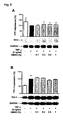
- Treatment with DMSO did not affect TPPI expression indicating that the effect of DMSO on TF is not antagonized by a concomitant reduction in TFPI expression.
- PAI-1 plasminogen activator inhibitor-1
- PAI-1 plasminogen activator inhibitor-1
- DMSO is able to prevent interleukin-1 induced PAI-1 expression in rat microvascular endothelial cells (Okada, 1998).
- MAP kinases mediates TF expression in response to several stimuli (Steffel, Akhmedov, 2005; Steffel, Hermann, 2005; Eto, 2002; Mechtcheriakova, 2001). Indeed, p38, ERK, and JNK were transiently activated in HAEC after stimulation with TNF- ⁇ , Treatment with DMSO prevented TF expression by specifically decreasing phosphorylation of JNK and p38, but not ERK thus indicating that inhibition of these two MAP kinases reduces TF expression in a potent manner. In line with this observation, it is shown that pharmacological inhibition of just one MAP kinase induces a less pronounced inhibition of TF expression. Consistent with inhibition of MAP kinases, treatment with DMSO led to a strong reduction in TNF- ⁇ -induced TF mRNA levels.
- thrombus formation is triggered by TF (Mackman, 2004; Mann, 1998, Nemerson, 1998).
- Photochemical injury was selected as a model of thrombosis because it is an established protocol for studying TF in vivo (Day, 2005); furthermore, this elegant procedure does not require intra-arterial invasion (Matsuno, 1991; Day, 2004). It was observed by present inventors that DMSO treated mice do not develop thrombotic occlusion following injury, indicating that treatment with DMSO prevents thrombus formation in vivo.
- DMSO reduces TF activity in vitro and in vivo in addition to preventing TF-dependent thrombosis in vivo.
- the use of DMSO as a novel strategy for treating ACS is therefore provided.
- DMSO can now be used as an attractive alternative for use in conjunction with widely employed immunosuppressant drugs such as rapamycin on stents, especially since rapamycin and paclitaxel has been reported to induce TF expression (Steffel, Latini, 2005, Stahli, submitted).
- HAEC Human aortic endothelial cells
- HASMC human aortic smooth muscle cells
- THP-1 cells LGC Prochem
- Cells were grown to confluence in 6-cm culture dishes and rendered quiescent for 24 hours before stimulation with 5 ng/mL TNF- ⁇ or thrombin 1 U/ml (both from Sigma) for 5 hours.
- SB203580 Sigma
- PD98059 Cell Signaling
- SP600125 Calbiochem
- LDH lactate dehydrogenase
- Protein expression was determined by Western blot analysis as described (Tanner, 2000). Cells were lysed in 50 mmol/L Tris buffer as described (Steffel, Hermann, 2005); 30 ⁇ g of sample were loaded, separated by 10% SDS-PAGE, and transferred to a PVDF membrane (Millipore) by semidry transfer. Equal loading was confirmed by Ponceau S staining. Antibodies against human TF, TFPI (both from American Diagnostica) and PAI-1 (SantaCruz Biotec.) were used at 1:2000 dilution.
- Antibodies against phosphorylated p38 mitogen-activated protein (MAP) kinase (p38), phosphorylated p44/42 MAP kinase (ERK), and phosphorylated JNK (all from Cell Signaling) were used at 1:1000, 1:5000, and 1:1000 dilution, respectively.
- Antibodies against total p38, total ERK, and total JNK (all from Cell Signaling) were used at 1:2000, 1:10'000, and 1:1000 dilution, respectively.
- Blots were normalized to GAPDH expression (1:10'000 dilution, Sigma). Representative blots are shown; bars represent at least 3 different experiments.
- TF surface activity was analyzed in HAEC with a colorimetric assay (American Diagnostica) according to the manufacturer's recommendations with some modifications as described (Steffel, Akhmedov, 2005).
- TF/factor Vlla complex converted human factor X to factor Xa, which was measured by its ability to metabolize a chromogenic substrate. Lipidated human TF was used as a positive control to confirm that the results obtained were in the linear range of detection.
- TF surface activity was analyzed in carotid artery homogenates employing a colorimetric assay (American Diagnostica) and following the manufacturer's protocol with some modifications that the cells were washed with PBS and were incubated with factor VIIa and factor X at 37°C. Then generation of factor Xa was measured.
- Right carotid arteries were homogenised in 50 ⁇ l of lysis buffer (50 mM Tris-HCl, 100mM NaCl, 0.1 % triton X-100 pH 7.4) and left to stand on ice for 30 minutes. Lipidated human TF was used as a positive control to confirm that the results obtained were in the linear range of detection.
- HAEC Lack of toxicity of DMSO was investigated.
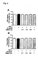
- HAEC were incubated with 1 % DMSO for 6 h, and a morphological examination was performed before and after treatment. No changes in cell morphology were observed under these conditions.
- cell death was assessed by LDH release; again, no signs of toxicity could be detected (Fig. 2A).
- Trypan blue staining revealed no increase in the number of stained cells (Fig. 2B). Therefore, treatment of HAEC cells as specified has to be regarded as not toxic.
- tissue factor For tissue factor ( F3 ): sense primer: 5'-TCCCCAGAGTTCACACCTTACC-3' (bases 508-529 of F3 cDNA; NCBI no. NM 001993), antisense primer: 5'-TGACCACAAATACCACAGCTCC-3' (bases 892-913 of F3 cDNA; NCBI no. NM 001993).
- sense primer 5'-GCATCTGCAATGGATGGT-3'
- antisense primer 5'-TGTTCTTGCGGATCATGTGT-3'.
- the amplification program consisted of 1 cycle at 95°C for 10 min, followed by 40 cycles with a denaturing phase at 95°C for 30 s, an annealing phase of 1 min at 60°C and an elongation phase at 72°C for 1 min.
- a melting curve analysis was performed after amplification to verify the accuracy of the amplicon.
- PCR products were analyzed on an ethidium bromide stained 1% agarose gel.
- Example 4 DMSO inhibits TF expression by decreasing MAPK activation
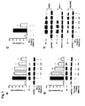
- HAEC were examined at different time points after stimulation with TNF- ⁇ (5 ng/ml). JNK, ERK, and p38 were transiently activated by TNF- ⁇ (Fig. 4A).
- Example 5 DMSO does not affect TFPI and PAI-1 expression
- Example 6 DMSO prevents arterial thrombosis in vivo
- Thrombotic occlusion was inhibited by treatment with DMSO (3.8 ml/Kg of 40% DMSO in PBS, corresponding to 80 mg/Kg of DMSO). Therefore, it is shown that DSMO prevents arterial thrombosis in vivo.
- mice were secured in a supine position, placed under a dissecting microscope (Olympus C-4040 Zoom; spatial resolution 4.1 megapixels), and the right common carotid artery was exposed following a midline cervical incision.
- a Doppler flow probe (Model 0.5 VB, Transonic Systems, Ithaca, NY) was then applied and connected to a flowmeter (Transonic, Model T106). Exactly 6 minutes after rose bengal injection, a 1.5-mW green light laser (540 nm) (Melles Griot, Carlsbad, CA) was applied to the desired site of injury at a distance of 6 cm for 60 minutes or until thrombosis occurred.
- DMSO i.p. (1 hour before surgery, 3.8ml/Kg of 40% DMSO in PBS), PBS i.p., (1 hour before surgery, 3.8ml/Kg of PBS), and negative control (no laser injury).
- Example 7 Proliferation and migration of aortic smooth muscle cells
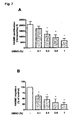
- HASMC For proliferation, HASMC were seeded at a density of 10,000 cells per 35-mm dish and cultured for 24 hours, resulting in random proliferation at the beginning of treatment. Dishes were then incubated with different DMSO concentrations and cells were maintained in DMEM (Dulbecco/Vogt Modified Eagle's Minimal Essential Medium) with 20% FCS (Fetal Calf Serum). Medium and appropriate DMSO concentrations were replaced daily and, cell number was determined after 3 days by using a hematocytometer.
- DMEM Dynamic Eagle's Minimal Essential Medium
- FCS Fetal Calf Serum
- HASMC HASMC were seeded at a density of 250,000 cells per 150-mm dish and cultured for 24 hours in DMEM with 20% FCS. Successively, cells were grown in DMEM with 1% FCS for 48 hours. The cells were harvested and resuspended (500'000 cells/ml) for analysis of migration in response to 20% FCS. Migration was assessed in a 2-chamber system (Neuroprobe Inc., Cabin John, Md) as previously described (Tanner, 2004). The number of migrated cells was determined by staining (Diff-Quik; Dade Diagnostics Inc., Aguada, Puerto Rico) and counting the cells at 400x magnification on 4 random microscopic fields per group.
Landscapes
- Health & Medical Sciences (AREA)
- Life Sciences & Earth Sciences (AREA)
- General Health & Medical Sciences (AREA)
- Epidemiology (AREA)
- Veterinary Medicine (AREA)
- Public Health (AREA)
- Animal Behavior & Ethology (AREA)
- Heart & Thoracic Surgery (AREA)
- Surgery (AREA)
- Vascular Medicine (AREA)
- Molecular Biology (AREA)
- Engineering & Computer Science (AREA)
- Chemical & Material Sciences (AREA)
- Medicinal Chemistry (AREA)
- Biomedical Technology (AREA)
- Pharmaceuticals Containing Other Organic And Inorganic Compounds (AREA)
- Medicines That Contain Protein Lipid Enzymes And Other Medicines (AREA)
Abstract
The invention relates to implantable clinical devices that contain means for eluting an active ingredient, said active ingredient essentially comprising DMSO, and the use of DMSO for the manufacture of a medicament for the treatment of vascular, cerebrovascular or cardiovascular disorders, acute coronary syndromes, stroke or peripheral artery disease.
Description
- The present invention relates to a clinical device for the treatment of vascular, cerebrovascular and cardiovascular disorders or acute coronary syndromes and the use of DMSO for the manufacture of a medicament for the treatment of vascular, cerebrovascular or cardiovascular disorders or acute coronary syndromes.
- Dimethyl sulfoxide (DMSO) is universally employed for preservation of hematopoietic progenitor cells infused into patients after myeloablative therapy (Egorin, 1998) and as a solvent for chemotherapeutic drugs. Moreover, due to its anti-inflammatory properties, DMSO has been successfully employed in treating human pathologies of dermatological (Burgess, 1998; Wong, 1988), neurological (Karaca, 1991, 2002), urinary (McCammon, 1998), pulmonary (lwasaki, 1994), rheumatic (Morassi, 1989) and gastrointestinal (Salim, 1992; 1994) origin. DMSO exhibits protective effects in animal models of mercuric chloride induced kidney injury (Jo, 2004), chemical liver injury (Lind, 1997), middle cerebral artery occlusion (Bardutzky, 2005), and cerebral hypoperfusion related neuronal death (Farkas, 2004). Despite of all this information, the potential impact of DMSO on vascular or cardiovascular disorders has not been assessed.
- Tissue Factor (TF) is a key enzyme in the activation of coagulation (Steffel, 2005). Plaque rupture leading to arterial thrombosis is the main cause of acute coronary syndromes (ACS) such as unstable angina and myocardial infarction. TF plays a major role in determining plaque thrombogenicity (Moons, 2002), as it is expressed by different cell types within the atherosclerotic vessel wall and is highly exposed to the blood stream following plaque rupture. Many inflammatory mediators are capable of inducing TF expression in cells such as monocytes, macrophages, endothelial cells, and vascular smooth muscle cells (Moons, 2002). Not surprisingly, experimental evidence has revealed an important contribution of TF to the pathogenesis of restenosis after balloon angioplasty (Kopp, 2004) and thrombosis after drug-eluting stent deployment (Steffel, 2006). As a consequence, recent efforts have focused on the therapeutic implications of tissue factor inhibition (Moons, 2002).
- Circulatory diseases are widespread and are related to progressive narrowing of the blood vessel due to atherosclerotic plaques; plaque rupture often leads to ischemic injury, stroke or myocardial infarction. Acute myocardial infarction occurs, because the blood supply to a part of the organ is interrupted; indeed, atherosclerotic lesions, which limit coronary blood flow and eventually rupture, are the major cause of ischemic heart disease.
- Percutaneous transluminal angioplasty (PTA) is a medical procedure the purpose of which is to increase blood flow through a blood vessel. PTA of the coronary arteries (PCTA) is featured by a high success rate besides minimal invasiveness compared to coronary bypass surgery. Typically during PTCA a balloon catheter device, which is inserted into the blood vessel, is inflated to dilate the stenotic (abnormally narrowed) blood vessel, which normally results from plaque or plaque rupture leading to thrombosis. During inflation the pressurized balloon exerts a force on the lesion, thereby increasing the inner diameter of the affected vessel. The increased interior vessel diameter facilitates improved blood flow. Soon after the PTCA treatment, however, a significant portion of treated vessels re-narrow.
- This re-narrowing, called restenosis, is inibited by introduction of a short flexible cylinder called stent. A stent is either an expandable wire mesh or hollow perforated tube that is inserted into a hollow structure of the body to keep it open.
- Although stents are mostly used in treatment of coronary arteries, they can also be used in several other structures, such as peripheral arteries and veins, bile ducts, esophagus, trachea or large bronchie, urethers and urethra.
- Restenosis after PTCA treatment remains a major problem in interventional cardiology and is caused by a combination of neointima formation, which is the formation of new tissue, and constrictive vascular remodelling. Although the latter is prevented by a stent, neointima formation can still occur within the stent or at its edges. Hence, to reduce neointima formation in addition to vascular remodeling, drug-eluting stents are known in the art.
-
EP 1 600 180 A2 -
WO 2004/045578 A2 discloses a drug delivery device comprising a medical device, e.g. a coated stent or stent-graft, in conjunction therewith a therapeutic dosage form of an anti-inflammatory ascomycin derivative, e.g. afixed to the medical device. - However, although at lower rates, neointima formation remains a problem in the art. Furthermore, stent thrombosis is a rare, but much more dangerous complication after coronary stent placement. It usually occurs before endothelialisation has been completed. For bare-metal stents, this process takes a few weeks; the newer, drug-eluting stents inhibit restenosis by inhibiting vascular smooth muscle cell proliferation, but they also tend to delay the endothelialisation process. For this reason, patients who have had drug-eluting stents are prescribed aspirin plus clopidogrel therapy for at least six months. Clopidogrel is a known anti-platelet agent often used in the treatment of coronary artery diseases, peripheral vascular diseases and cerebrovascular diseases. The mechanism of action of clopidogrel is irreversible blockade of the adenosine II phosphate (ADP) receptor P2Y12 on the platelet cell membranes, which is important in platelet aggregation, in particular the crosslinking of platelets by fibrin. The blockade of this receptor inhibits platelet aggregation. Besides adverse effects like severe neutropenia or the occurrence of hemorrhage, in particular occurring when co-administered with aspirin, clopidogrel has the disadvantage of being rather expensive. Therefore, there is still the need for an alternative inhibitor of thrombosis after coronary stent placement; in particular a local inhibitor of thrombosis.
- Involvement of tissue factor (TF) in acute coronary syndromes (ACS) is known, as stated above. Recent studies even show increased levels of TF antigen and activity in atherectomy specimens from patients with unstable angina or myocardial infarction as compared to those with stable angina (Annex, 1995). Moreover, in ACS, plasma concentrations of inflammatory cytokines such as TNF-α (tumor necrosis factor α) and interleukins are increased at the site of coronary artery occlusion to such an extent that TF is induced in vascular cells (Maier, 2005).
- Therefore, there is not only a need for an inhibitor of neointima formation, but also there is a need for an inhibitor of TF, which can be applied by or in combination with a medical device, e.g. a stent, or by parenteral administration.
- The inventors surprisingly found that DMSO is, besides being active in suppression of inflammatory response and inhibition of platelet activity, active in prevention of thrombosis. In particular it was found that DMSO inhibits proliferation and migration of human aortic vascular smooth muscle cells (HASMC), and reduces TF activity in vitro and in vivo in addition to preventing TF-dependent thrombosis in vivo.
- Therefore, the present invention relates to a clinical device for being implanted that contains means for eluting an active ingredient, said active ingredient essentially comprising DMSO. Preferably the clinical device according to the present invention comprises an active ingredient, which consists of at least 80% of DMSO, more preferable of at least 95% of DMSO. Percent is given as percent by weight.
- All concentrations given in the present application are expressed in terms of weight percent unless designated otherwise
- Within this application the terms "eluted" or "eluting" and "released" or "releasing" are used synonymously.
- The "clinical device" of the present invention refers to a device which is inserted into, or grafted onto, body tissues to remain for a period of time, such as an extended-released drug delivery device, stent, drug-eluting stent, or vascular graft.
- In a preferred embodiment of the present invention a clinical device comprises a surface or surface layer suitable for being implanted.
- Preferably the active ingredient of the clinical device of the present invention comprises DMSO, more preferable the active ingredient comprises from about 20 to about 100% DMSO, even more preferred from 60 to 100% DMSO, in particular the active ingredient contains 80 to 90% DMSO. Furthermore, the active ingredient may also comprise other pharmaceutically active compounds like rapamycin, paclitaxel, tacrolimus, statins, and similar compounds, which have beneficial effects for the tissues. DMSO and said other compound(s) add up to 100%.
- ln another aspect of the present invention the active ingredient contains at least 20%, preferred at least 50%, more preferred at least 80%, most preferred at least 90%, in particular at least 95% DMSO as active ingredient.
- In one embodiment of the present invention the active ingredient of the clinical device comprises DMSO in combination with further anti-proliferative and/or anti-inflammatory agents.
- Means for eluting the active ingredient(s) from the clinical device of the present invention are known in the art. Examples of materials that can be applied are layers of drug-eluting materials, such as polymer-matrices. Each polymer suitable for application on a device which can be implanted in an organism can be used, provided that the material is non-toxic and able to release an active ingredient in a pre-defined period of time. Preferred periods of release are from about 2 to 28 days, in particular 14 to 28 days- More preferably the period of release is in a range of 2 to 4 weeks with a maximum of drug-elution after 1 to 2 weeks. In particular, the material to be used as drug-eluting layer has to be able to release an active ingredient, which contains a high amount of DMSO, e.g. from about 50% to about 100% DMSO content, within a predefined period of time.
- In one preferred embodiment of the present invention the clinical device is covered with a degradable layer, which is the only or the first layer of the clinical device, said layer comprising one or a variety of polymer matrices. The matrix should be biocompatible, biodegradable, bioerodable, non-toxic, bioabsorbable, and with a defined rate of degradation. Biocompatible matrices that can be used in the invention include, but are not limited to, poly(lactide-co-glycolide), polyesters such as polylactic acid, polyglycolic acid and copolymers thereof, polyanhydride, polycaprolactone, polyhydroxybutyrate, valerate, and other biodegradable polymers and mixtures thereof. In another embodiment the matrix can also comprise naturally occurring polymeric materials such as collagen, fibrin, elastin, and extracellular matrix components.
- In another embodiment of the present invention the polymer matrix of the clinical device further comprises pharmaceutically acceptable polymers, as the only or the second layer, for example, non-absorbable polymers, such as ethylene vinyl acetate (EVAC) and methylmethacrylate (MMA). The non-absorbable polymer, for example, can aid in further controlling release of the substance by limiting diffusion, thereby delaying or slowing the rate of release of the active ingredient, in particular when used as second layer.
- In one preferred embodiment of the present invention the active ingredient is mainly present in the first, inner polymer matrix layer of the clinical device. This inner polymer matrix layer, containing the active ingredient, which comprises a significant amount of DMSO, can for example be prepared by using DMSO as polymerisation solvent, or the polymer of the inner polymer matrix layer is saturated with DMSO and optionally other ingredients of the active ingredient after polymerisation, before or after application as first polymer matrix layer to the clinical device. Preferably the polymer of this inner layer is a degradable layer as specified above. As second layer, a further, slowly degradable or preferably a non-absorbable polymer layer as defined above can be applied, preferably as diffusion limiting and controlling layer, thereby enabling controlled release of active ingredient, which comprises a significant amount of DMSO, into the surrounding tissue.
- In another preferred embodiment of the present invention the active ingredient, comprising a significant amount of DMSO, is contained in pre-formed particles or granules of matrix polymer, which are applied to the clinical device of the present invention, eventually within a further polymer matrix.
- The base material of the clinical device of the present invention, which preferably is covered with means for eluting an active ingredient, can be of various types, for example, stainless steel, nitinol, MP35N, gold, tantalum, platinum, platinum-iridium or titanium, which is preferred, or other biocompatible materials and/or alloys such as carbon or carbon fiber, cellulose acetate, cellulose nitrate, silicone, cross-linked polyvinylacetate (PVA), hydrogel, polyurethane, polyamide, polyester, or a mixture of polymers thereof; polyesters such as polylactic acid, polyglycolic acid or copolymers thereof and other biodegradable polymers or suitable degradable materials can also be used.
- In one embodiment of the present invention the drug-eluting layer of the clinical device or the clinical device itself is degraded by the organism, which the device is implanted in, thereby releasing the drug contained.
- In one preferred embodiment of the present invention the clinical device is a stent, preferably a coated or covered stent, such as those covered with polytetrafluoroethylene (PTFE), expanded polytetrafluoroethylene (ePTFE), and such as those covered stents which are alternatively or additionally covered with a polymer matrix as described above. A stent is either an expandable wire mesh or a hollow perforated tube that is inserted in a hollow structure in the body to keep it open. As base material of the stent all materials as described for the clinical device can be used.
- In one embodiment of the present invention the clinical device is a depot stent, which contains the active ingredient in suitable reservoirs or wells, e.g. holes within the stent which are filled with erodable polymer containing the active ingredient.
- A bioresorbable drug-delivery stent including a substantially cylindrical expandable stent formed of a bioresorbable material and a plurality of reservoirs or openings formed in the stent containing a beneficial agent matrix comprising a bioresorbable material and drug is for example disclosed in
WO 2005/102222 , the content of which is incorporated by reference herein. - Because of its lipophilic character DMSO is particularly useful as eluted drug in a stent, as the lipophily renders DMSO suitable for being easily incorporated into suitable non-erodable and/or erodable polymers to be used in a drug-eluting stent of the present invention.
- In order to coat a clinical device such as a stent, the stent may be dipped, sprayed with, or loaded with, for example, a liquid solution of the matrix polymer of moderate viscosity. Alternatively, the clinical device such as a stent may be coated with matrix polymer during or after polymerisation, for example by applying the devices into monomer solutions or prepolymerized matrix polymer solutions, preferably in presence of active ingredient, comprising a significant amount of DMSO, to form a layer, covering the device, which is saturated with active ingredient, preferably during polymerisation. A further diffusion limiting and controlling layer as specified above may be applied to the device as second layer. Optionally after application of one layer, the stent is dried before application of the next layer. In one embodiment, a thin, paint-like polymer matrix coating does not exceed an overall thickness of about 100 to about 500 µm.
- In one preferred embodiment of the present invention the clinical device is only covered by the drug-eluting layer at those positions, where said device is in direct contact to the organism, i.e. the surrounding tissue.
- The "surrounding tissue" according to the present invention is the tissue which is in direct contact to the material of the clinical device of the present invention or the tissue which is in direct contact and the tissue which is directly adjacent to the clinical device of the present invention.
- In one preferred embodiment of the present invention the active ingredient, which comprises a significant amount of DMSO, is eluted from the clinical device in an amount effective to significantly suppress expression of tissue factor protein within the tissue surrounding the clinical device. Preferably the expression of tissue factor protein is suppressed by at least 20%, more preferred by at least 30%, in particular by at least 50%, relative to the expression level of tissue factor protein of tissue. which has been exposed to an increased level of inflammatory mediators, comparable to or such as TNF-α or thrombin, in particular compared to or such as concentrations of 5 ng/ml TNF-α or 1 U/ml thrombin. Preferably proliferation and/or migration of HVSMC is suppressed by at least 20%, more preferred at least 30%, in particular by at least 50%, relative to the level when no active ingredient is present.
- In one embodiment of the present invention the active ingredient, which comprises at least more than 50% by weight of DMSO, is eluted from the clinical device within a predetermined period of time, preferably during 1-28 days, e.g. for 14 days.
- In one embodiment of the present invention the clinical device contains an amount of active ingredient in a range from about 10 ng to about 10 mg, preferably from about 1 µg to about 1000 µg.
- In one embodiment of the present invention the active ingredient is incorporated into the drug-eluting layer, in case of two layers in both or one layer, in case of more than two layers in one, two or more layers in a percent loading of between 0.1% to 70% by weight, preferably between 5% and 50% by weight.
- In a further preferred embodiment of the present invention the amount eluted from the clinical device into the surrounding tissue is from about 1 ng/mm2h to about 1 µg/mm2h, relative to the contacted surrounding tissue area and time after implantation.
- The present invention further relates to the use of DMSO for the manufacture of a medicament for the treatment of vascular, cerebrovascular or cardiovascular disorders, stroke or acute coronary syndromes.
- In one embodiment the DMSO used for the manufacture of a medicament for the treatment of vascular, cerebrovascular or cardiovascular disorders, stroke or acute coronary syndromes is used in combination with further anti-proliferative and/or anti-inflammatory agents, e.g. rapamycin, paclitaxel or tacrolimus or in combination with other medicaments applied for treating these conditions.
- The medicament for the treatment of vascular, cerebrovascular or cardiovascular disorders, stroke or acute coronary syndromes preferably contains 20-100% DMSO, more preferable 60-100% DMSO, in particular 80-90% DMSO. Said medicament may further comprise appropriate diluents, such as buffer solutions, excipients and stabilizers, being suitable for administration, for example by syringe or infusion. Another possible form of administration is a bolus injection, wherein the DMSO can be premixed with an infusion solution or can be prepared in situ by addition of DMSO into the infusion solution prior to injection. The medicament can be administered parenterally or subcutaneously as desired. When administered systemically, the therapeutic composition should be sterile, pyrogen-free and in a parenterally acceptable solution having due regard for pH, isotonicity, and stability. These conditions are known to those skilled in the art.
- In one further embodiment of the present invention the medicament for the treatment of vascular, cerebrovascular or cardiovascular disorders, stroke or acute coronary syndromes can be administered orally, for example in form of a tablet, capsule or any other form suitable for oral administration. Said medicament further comprises appropriate diluents, excipients and coadjuvants suitable for oral administration. Methods for producing these administration forms are known to those skilled in the art.
- The present invention further relates to the method of treating vascular, cerebrovascular or cardiovascular disorders, stroke or acute coronary syndromes by administration of drug-eluting stents able to release a drug comprising a significant amount of DMSO and/or administering a medicament comprising a significant amount of DMSO to a patient in need thereof.
- In one embodiment of the present invention the method of treating vascular, cerebrovascular or cardiovascular disorders, stroke or acute coronary syndromes comprises administration of a medicament comprising a significant amount of DMSO in a combinational product, preferably by intravenous or oral administration. In particular the medicament comprising a significant amount of DMSO is used in combination with further anti-proliferative and/or anti-inflammatory agents, e.g. rapamycin, paclitaxel or tacrolimus or in combination with other medicaments applied for treating these conditions.
- The present invention further relates to a method for reducing restenosis, in-stent restenosis, stent thrombosis or in-stent thrombosis comprising administration of a drug-eluting stent able to release a drug comprising a significant amount of DMSO and/or administering a medicament comprising a significant amount of DMSO to a patient in need thereof.
- The present invention further relates to a method of treating acute myocardial infarction, stroke or peripheral arterial disease comprising administering a medicament comprising a significant amount of DMSO to a subject receiving a stent in connection with percutaneous transluminal angioplasty.
- Some data obtained by the inventors are summarized in the Figures.
- Data presented in the Figures are given as mean±SEM. Statistical analysis was performed by 2-tailed unpaired Student test or ANOVA as appropriate. A probability value of <0.05 was considered significant.
- Figure 1. DMSO inhibits TF expression and activity.
- Fig. 1A shows a histogram of TNF-α induced TF expression relative to the concentration of DMSO. Values are given as percent of stimulation with TNF-α alone, concentration-dependent manner. Values are given as percent of stimulation with thrombin alone, normalized to GAPDH expression. *P<0.0001, **P<0.001 vs TNF-α alone.
- Fig. 1 B shows a histogram of thrombin-induced TF expression relative to the concentration of DMSO, normalized to GAPDH expression. Values are given as percent of stimulation with thrombin alone. *P<0.001, **P<0.0001 vs thrombin alone.
- Fig. 1C shows a histogram of TF-α induced TF surface activity. Values are given as percent of stimulation with TNF-α alone. **P<0.0001 vs TNF-α alone.
- Fig. 1 D shows Western blots of TNF-α induced TF expression relative to the concentration of DMSO in HAEC, THP-1 and HAVSMC-cells. All blots are normalized to GAPDH expression.
- Figure 2. Toxicity of DMSO.
- Fig. 2A shows a histogram of LDH-release of HAEC-cells relative to the concentration of DMSO. Values are given as percent of stimulation with TNF-α alone.
- Fig. 2B shows a histogram of trypan blue cell viability relative to the concentration of DMSO. Values are given as percent of the stimulation with TNF-α alone.
- Figure 3. Effect of DMSO on TF mRNA induction.
- Fig. 3A shows the histogram of TF/L28 mRNA expression (%) before and after stimulation with TNF-α and after stimulation with TNF-α with DMSO. Values are given as percent of stimulation with TNF-α alone. *P<0.001, vs TNF-α alone. Values are representative of four different experiments and are normalized to L28 mRNA.
- Fig. 3B shows ΔΔCT values without TNF-α induction and in absence of DMSO (left) and with TNF-α induction and in presence of DMSO (right). **P<0.0001 vs TNF-α alone. Values are representative of four different experiments and are normalized to L28 mRNA.
- Figure 4. Effect of DMSO on MAP kinase activation.
- Fig. 4A shows Western blots indicating phosphorylation (Pho) of three MAP kinases JNK, p38, and ERK, relative to their total amount (Tot) contained in the cells, with and without DMSO, investigated at several time points after TNF-α induction. Blots are representative of at least three different experiments.
- Fig. 4B shows a histogram showing the activities of the three MAP kinases JNK, p38 and ERK, in presence of DMSO and after induction with TNF-α.
- Fig. 4C shows a histogram indicating TF expression levels without and with TNF-α induction and further addition of SP600125 (SP), a specific inhibitor of JNK, addition of SB203580 (SB), a specific inhibitor of p38, and addition of PD98059 (PD), a specific inhibitor of ERK activation. *P<0.001, vs TNF-α alone. Blots are representative of at least three different experiments.
- Figure 5. Effect of DMSO on TFPI and PAI-1 expression.
- Fig. 5A shows the TFPI expression level (%) without and with TNF-α induction and with increasing concentrations of DMSO. Values are given as percent of stimulation with TNF-α alone. Blots are representative of at least three different experiments.
- Fig. 5B shows PAI-1 expression levels (%) without and with TNF-α induction and for increasing concentrations of DMSO. Values are given as percent of stimulation with TNF-α alone. **P<0.0001, vs TNF-α alone. Blots are representative of at least three different experiments.
- Figure 6. Effect of DMSO on carotid artery TF activity and thrombotic occlusion of the carotid artery in a mouse model of photochemical injury.
- Fig. 6A shows a histogram indicating TF activity (absorbance, 405 nm) in the carotid artery in a mouse,model of photochemical injury. ** P<0.0001 vs PBS treated.
- Fig. 6B shows blood flow recordings of vehicle treated (PBS), DMSO treated and uninjured mice in a mouse model of photochemical injury. Laser injury is initiated at time = 0 min. This figure shows that DMSO is active not only in vitro but also in vivo.
- Figure 7. Effect of DMSO on proliferation and migration of aortic smooth muscle cells.
- Fig.7A shows a histogram indicating HASMC proliferation for increasing DMSO concentrations.
- Fig. 7B shows a histogram indicating HASMC migration for increasing DMSO concentrations.
- The present invention offers a completely new approach to specific inhibition of expression and activity of TF (cp. Example 1). The present inventors found that DMSO is able to inhibit TF induction in human aortic endothelial cells (HAEC), human aortic vascular smooth muscle cells (HAVSMC) and THP-1 cells. This effect was mediated by a reduced activation of p38 and JNK MAP kinase activation (cp. Example 4) and expression of TF mRNA (cp. Example 5). Consistent with this observation, it was shown that DMSO protects from thrombotic occlusion in an in vivo model of TF-dependent thrombus formation (cp. Example 6). Further it could be demonstrated that DMSO inhibits proliferation and migration of human aortic smooth muscle cells (HASMC) (cp. Example 7).
- These findings denote DMSO as an excellent active ingredient in a variety of applications, preferably in combination with a stent therapy. As DMSO is first active in suppression of inflammatory response, corresponding complications caused by inflammation, in particular shortly after stent implantation can be reduced. Secondly DMSO is also active in prevention of thrombosis, in particular by inhibition of tissue factor and platelet activity. Thrombosis, in particular stent thrombosis, is a rare but mostly very dangerous complication often leading to death of the patient. Thirdly DMSO is a well known solvent which is easy to handle and which is much less expensive than substances with a comparable field of application.
- DMSO was used in concentrations as high as 1% by weight (12.6 mmol/L) and no toxic effects by DMSO treatment could be observed, even at the highest concentration (cp. Example 2). In support of these findings, a focused study found no toxic effect by DMSO treatment on endothelial cells at concentrations well above those (Bourne, 1994), and contemporary literature describes routine usage of DMSO to trigger cellular differentiation in vitro in concentrations as high as 1.25% (15.7 mM) (Heidari, 2004). Furthermore, DMSO is universally infused intravenously together with hematopoietic progenitor cells in patients after myeloablative therapy and in those patients, DMSO reaches plasma concentrations of 20 mmol/L without causing any adverse effect (Egorin, 1998).
- Present inventors showed that thrombin and TNF-α induced TF expression in HAEC with similar potencies, and incubation with DMSO suppressed this induction irrespective of the stimulus. Similarly, DMSO prevented TNF-α induced TF expression in HAVSMC and THP-1 cells.
- TF activity is counterbalanced by its endogenous inhibitor TFPI (tissue factor pathway inhibitor); equilibrium between these two factors is known to be essential in determining embryonic survival (Pedersen. 2005). Treatment with DMSO did not affect TPPI expression indicating that the effect of DMSO on TF is not antagonized by a concomitant reduction in TFPI expression. PAI-1 (plasminogen activator inhibitor-1) is a serpin that suppresses fibrinolysis by inhibiting the activity of plasminogen activator. It is known that DMSO is able to prevent interleukin-1 induced PAI-1 expression in rat microvascular endothelial cells (Okada, 1998). By contrast, we did not observe any effect of DMSO on TF expression in TNF-α stimulated HAEC. This difference, maybe related to the different species, the different origin of the cells, or the different stimuli used for induction.
- Activation of MAP kinases mediates TF expression in response to several stimuli (Steffel, Akhmedov, 2005; Steffel, Hermann, 2005; Eto, 2002; Mechtcheriakova, 2001). Indeed, p38, ERK, and JNK were transiently activated in HAEC after stimulation with TNF-α, Treatment with DMSO prevented TF expression by specifically decreasing phosphorylation of JNK and p38, but not ERK thus indicating that inhibition of these two MAP kinases reduces TF expression in a potent manner. In line with this observation, it is shown that pharmacological inhibition of just one MAP kinase induces a less pronounced inhibition of TF expression. Consistent with inhibition of MAP kinases, treatment with DMSO led to a strong reduction in TNF-α-induced TF mRNA levels.
- It is generally accepted that thrombus formation is triggered by TF (Mackman, 2004; Mann, 1998, Nemerson, 1998). Photochemical injury was selected as a model of thrombosis because it is an established protocol for studying TF in vivo (Day, 2005); furthermore, this elegant procedure does not require intra-arterial invasion (Matsuno, 1991; Day, 2004). It was observed by present inventors that DMSO treated mice do not develop thrombotic occlusion following injury, indicating that treatment with DMSO prevents thrombus formation in vivo. Inhibition of relatively high basal TF activity in mouse carotid artery in vivo occurred within 2 hours of DMSO administration and thus is the most likely explanation for the effect of DMSO treatment on thrombus formation. A recent report is very consistent with this observation by showing that basal arterial TF levels in vivo are high and do not undergo a large induction (Day, 2005). In addition, part of the effect displayed by DMSO in vivo may also be attributable to its known ability to impair platelet adherence and aggregation (Dujovny, 1983).
- Therefore, present inventors showed that DMSO reduces TF activity in vitro and in vivo in addition to preventing TF-dependent thrombosis in vivo. The use of DMSO as a novel strategy for treating ACS is therefore provided. In particular, DMSO can now be used as an attractive alternative for use in conjunction with widely employed immunosuppressant drugs such as rapamycin on stents, especially since rapamycin and paclitaxel has been reported to induce TF expression (Steffel, Latini, 2005, Stahli, submitted).
- The following examples further illustrate the invention.
- Data presented in the examples are given as mean±SEM. Statistical analysis was performed by 2-tailed unpaired Student t test or ANOVA as appropriate. A probability value of <0.05 was considered significant.
- The effect of DMSO on TF protein expression, which was induced by TNF-α, was investigated. Stimulation of HAEC with TNF-α (5 ng/ml) resulted in a 19-fold increase in TF expression, (Fig. 1A); similarly, thrombin (1 U/mL) induced a 24-fold increase (Fig. 1 B). When HAEC were pretreated with increasing concentrations of DMSO (0.1-1%) 1 hour before stimulation, DMSO inhibited TF induction by both TNF-α and thrombin in a concentration-dependent manner (Fig. 1A, B). This effect of DMSO was paralleled by a similar inhibition of TF surface activity (Fig. 1C). Stimulation with TNF-α (5 ng/ml) induced a 10- and 3.5-fold induction of TF expression in THP-1 and HAVSMC, respectively (Fig. 1 D). Treatment with DMSO prevented TF induction in THP-1 cells, but not in HASMC (Fig. 1D).
- Human aortic endothelial cells (HAEC) and human aortic smooth muscle cells (HASMC) (Clonetics, Allschwil, Switzerland) were cultured as described (Steffel, Akhmedov, 2005). THP-1 cells (LGC Prochem) were cultured according to the manufacturer's recommendation. Cells were grown to confluence in 6-cm culture dishes and rendered quiescent for 24 hours before stimulation with 5 ng/mL TNF-α or thrombin 1 U/ml (both from Sigma) for 5 hours. SB203580 (Sigma), PD98059 (Cell Signaling), or SP600125 (Calbiochem) were added 60 minutes before stimulation. To assess cytotoxicity, a colorimetric assay for detection of lactate dehydrogenase (LDH)was used according to the manufacturer's recommendations (Roche); in addition, trypan blue exclusion assay (0.4% solution, Sigma) and morphological examination (Leica, DMIL) was performed.
- Protein expression was determined by Western blot analysis as described (Tanner, 2000). Cells were lysed in 50 mmol/L Tris buffer as described (Steffel, Hermann, 2005); 30 µg of sample were loaded, separated by 10% SDS-PAGE, and transferred to a PVDF membrane (Millipore) by semidry transfer. Equal loading was confirmed by Ponceau S staining. Antibodies against human TF, TFPI (both from American Diagnostica) and PAI-1 (SantaCruz Biotec.) were used at 1:2000 dilution. Antibodies against phosphorylated p38 mitogen-activated protein (MAP) kinase (p38), phosphorylated p44/42 MAP kinase (ERK), and phosphorylated JNK (all from Cell Signaling) were used at 1:1000, 1:5000, and 1:1000 dilution, respectively. Antibodies against total p38, total ERK, and total JNK (all from Cell Signaling) were used at 1:2000, 1:10'000, and 1:1000 dilution, respectively. Blots were normalized to GAPDH expression (1:10'000 dilution, Sigma). Representative blots are shown; bars represent at least 3 different experiments.
- TF surface activity was analyzed in HAEC with a colorimetric assay (American Diagnostica) according to the manufacturer's recommendations with some modifications as described (Steffel, Akhmedov, 2005). TF/factor Vlla complex converted human factor X to factor Xa, which was measured by its ability to metabolize a chromogenic substrate. Lipidated human TF was used as a positive control to confirm that the results obtained were in the linear range of detection.
- TF surface activity was analyzed in carotid artery homogenates employing a colorimetric assay (American Diagnostica) and following the manufacturer's protocol with some modifications that the cells were washed with PBS and were incubated with factor VIIa and factor X at 37°C. Then generation of factor Xa was measured. Right carotid arteries were homogenised in 50 µl of lysis buffer (50 mM Tris-HCl, 100mM NaCl, 0.1 % triton X-100 pH 7.4) and left to stand on ice for 30 minutes. Lipidated human TF was used as a positive control to confirm that the results obtained were in the linear range of detection.
- Lack of toxicity of DMSO was investigated. To assess potential toxic effects of DMSO, HAEC were incubated with 1 % DMSO for 6 h, and a morphological examination was performed before and after treatment. No changes in cell morphology were observed under these conditions. In addition, cell death was assessed by LDH release; again, no signs of toxicity could be detected (Fig. 2A). Similarly, Trypan blue staining revealed no increase in the number of stained cells (Fig. 2B). Therefore, treatment of HAEC cells as specified has to be regarded as not toxic.
- Cell culture was performed as described in example 1.
- Investigations were performed to which extend DMSO inhibits TF mRNA expression. It is known from literature that peak induction of TF mRNA occurs 2 hours after stimulation with TNF-α (Steffel, Latini, 2005). Real-time PCR analysis was performed and it confirmed a 10 fold induction of TF mRNA after 2 hours of TNF-α stimulation (Fig. 3A). As a result it could be shown that treatment with DMSO (1%) inhibited mRNA synthesis by 65 ± 7% (Fig. 3A, B).
- Real-Time PCR analysis was performed as described (Steffel, Latini, 2005). An MX3000P PCR cycler (Stratagene, Amsterdam, The Netherlands) was used according to the manufacturer's instructions. All PCR experiments were performed in triplicates using the SYBR Green JumpStart kit provided by Sigma. Each reaction (25 µl) contained 2 µl cDNA, 1 pmol of each primer, 0.25 µl of internal reference dye and 12.5 µl of JumpStart Taq ReadyMix (containing buffer, dNTPs, stabilizers, SYBR Green, Taq polymerase and JumpStart Taq antibody). The following primers were used. For tissue factor (F3): sense primer: 5'-TCCCCAGAGTTCACACCTTACC-3' (bases 508-529 of F3 cDNA; NCBI no. NM 001993), antisense primer: 5'-TGACCACAAATACCACAGCTCC-3' (bases 892-913 of F3 cDNA; NCBI no. NM 001993). For human L28: sense primer: 5'-GCATCTGCAATGGATGGT-3', antisense primer: 5'-TGTTCTTGCGGATCATGTGT-3'. The amplification program consisted of 1 cycle at 95°C for 10 min, followed by 40 cycles with a denaturing phase at 95°C for 30 s, an annealing phase of 1 min at 60°C and an elongation phase at 72°C for 1 min. A melting curve analysis was performed after amplification to verify the accuracy of the amplicon. For verification of the correct amplification, PCR products were analyzed on an ethidium bromide stained 1% agarose gel. In each real-time PCR run for F3 and L28 a calibration curve was included, that was generated from serial dilutions (2×10⁷, 2×10⁶, 2×10⁵, 2×10⁴, 2×10³, 2×10³, 2×10², copies/reaction for F3, and 2×10⁷, 2×10⁶, 2×10⁵, 2×10⁴, 2×10³, 2×10³ copies/reaction for L28) of purified amplicons for F3 and L28.
- To assess whether DMSO affects MAP kinase activation, HAEC were examined at different time points after stimulation with TNF-α (5 ng/ml). JNK, ERK, and p38 were transiently activated by TNF-α (Fig. 4A).
- It could be shown that maximal phosphorylation occurred after 15 minutes and returned to basal levels within 60 minutes (Fig. 4A). DMSO inhibited phosphorylation of JNK and p38 by over 50%, while leaving phosphorylation of ERK unchanged (Fig. 4A). Maximal JNK activation was decreased by 51±6% and that of p38 by 50±3% (Fig. 4B). Maximal activation of ERK was not significantly reduced (Fig. 4B). No change in total expression of p38, ERK, or JNK was observed at any time point with or without DMSO. To verify the involvement of MAP kinases in TF induction by TNF-α under our experimental conditions, the effect of MAP kinase inhibitors on TF expression was examined. SP600125 (10-6 mol/L), a specific inhibitor of JNK, SB203580 (10-6 mol/L), a specific inhibitor of p38, and PD98059 (3x10⁻⁷ mol/L), a specific inhibitor of ERK phosphorylation, inhibited TF induction after TNF-α stimulation (Fig. 4C).
- Cell culture was performed as described in example 1.
- The effect of DMSO on the physiological inhibitor of TF, TFPI, as well as on the anti-fibrinolytic factor, PAI-1, was assessed for a control. It could be shown that treatment of TNF-α stimulated HAEC with DMSO affected neither TFPI nor PAI-1 expression (Fig. 5A, B).
- Cell culture was performed as described in example 1 .
- Photochemically induced endothelial injury is an established model of arterial thrombosis and is dependent on TF (Matsuno, 1991). Following onset of injury, vehicle (PBS) treated mice developed thrombotic occlusion within 1 hour (mean occlusion time 63±9.0 minutes, n=5) (Fig. 6A). It can be seen from transient reductions in blood flow that a dynamic state of thrombus formation and lysis occurred during the short time interval prior to complete occlusion. Cessation of blood flow was accompanied by the appearance of a faint white filling inside the lumen of the artery. Thrombotic occlusion was inhibited by treatment with DMSO (3.8 ml/Kg of 40% DMSO in PBS, corresponding to 80 mg/Kg of DMSO). Therefore, it is shown that DSMO prevents arterial thrombosis in vivo.
- C57BU6 mice (6-8 weeks old) weighing an average of 23±2 g were anesthetized with 2 mg intraperitoneal sodium pentobarbital (Butler, Columbus, OH) as previously described (Eitzman, 2000). Rose bengal (Fisher Scientific, Fair Lawn, NJ) was diluted to 10 mg/mL in phosphate-buffered saline and then injected into the tail vein in a volume of 0.12 mL at a concentration of 50 mg/kg using a 27-gauge Precision Glide needle with a 1-mL latex free syringe (Becton Dickinson, Franklin Lakes, NJ). Mice were secured in a supine position, placed under a dissecting microscope (Olympus C-4040 Zoom; spatial resolution 4.1 megapixels), and the right common carotid artery was exposed following a midline cervical incision. A Doppler flow probe (Model 0.5 VB, Transonic Systems, Ithaca, NY) was then applied and connected to a flowmeter (Transonic, Model T106). Exactly 6 minutes after rose bengal injection, a 1.5-mW green light laser (540 nm) (Melles Griot, Carlsbad, CA) was applied to the desired site of injury at a distance of 6 cm for 60 minutes or until thrombosis occurred. From the onset of injury, blood flow in the vessel was monitored for 120 minutes, at which time the experiment was terminated. Occlusion was defined as a flow of ≤0.1 ml/min for at least 1 minute (Eitzman, 2003). Mice were divided into 3 groups: DMSO i.p. (1 hour before surgery, 3.8ml/Kg of 40% DMSO in PBS), PBS i.p., (1 hour before surgery, 3.8ml/Kg of PBS), and negative control (no laser injury).
- Proliferation and migration of HASMC were investigated at different concentrations of DMSO.
- It could be shown that proliferation and migration of HASMC is decreased with increasing DMSO concentrations.
- For proliferation, HASMC were seeded at a density of 10,000 cells per 35-mm dish and cultured for 24 hours, resulting in random proliferation at the beginning of treatment. Dishes were then incubated with different DMSO concentrations and cells were maintained in DMEM (Dulbecco/Vogt Modified Eagle's Minimal Essential Medium) with 20% FCS (Fetal Calf Serum). Medium and appropriate DMSO concentrations were replaced daily and, cell number was determined after 3 days by using a hematocytometer.
- For migration, HASMC were seeded at a density of 250,000 cells per 150-mm dish and cultured for 24 hours in DMEM with 20% FCS. Successively, cells were grown in DMEM with 1% FCS for 48 hours. The cells were harvested and resuspended (500'000 cells/ml) for analysis of migration in response to 20% FCS. Migration was assessed in a 2-chamber system (Neuroprobe Inc., Cabin John, Md) as previously described (Tanner, 2004). The number of migrated cells was determined by staining (Diff-Quik; Dade Diagnostics Inc., Aguada, Puerto Rico) and counting the cells at 400x magnification on 4 random microscopic fields per group.
-
- Annex BH, Denning SM, Channon KM, Sketch MH, Jr., Stack RS, Morrissey JH, Peters KG. Differential expression of tissue factor protein in directional atherectomy specimens from patients with stable and unstable coronary syndromes. Circulation. 1995;91:619-22.
- Bardutzky J, Meng X, Bouley J, Duong TQ, Ratan R, Fisher M. Effects of intravenous dimethyl sulfoxide on ischemia evolution in a rat permanent occlusion model. J Cereb Blood Flow Metab. 2005;25:968-77.
- Bourne WM, Shearer DR, Nelson LR. Human corneal endothelial tolerance to glycerol, dimethylsulfoxide, 1,2-propanediol, and 2,3-butanediol. Cryobiology. 1994; 31:1-9.
- Burgess JL, Hamner AP, Robertson WO. Sulfhemoglobinemia after dermal application of DMSO. Vet Hum Toxicol. 1998;40:87-9.
- Day SM, Reeve JL, Myers DD, Fay WP. Murine thrombosis models. Thromb Haemost. 2004; 92:486-94.
- Day SM, Reeve JL, Pedersen B, Farris DM, Myers DD, lm M, Wakefield TW, Mackman N, Fay WP. Macrovascular thrombosis is driven by tissue factor derived primarily from the blood vessel wall. Blood. 2005;105:192-8.
- Dujovny M, Rozario R, Kossovsky N, Diaz FG, Segal R. Antiplatelet effect of dimethyl sulfoxide, barbiturates, and methyl prednisolone. Ann N YAcad Sci. 1983; 411:234-44.
- Egorin MJ, Rosen DM, Sridhara R, Sensenbrenner L, Cottler-Fox M. Plasma concentrations and pharmacokinetics of dimethylsulfoxide and its metabolites in patients undergoing peripheral-blood stem-cell transplants. J Clin Oncol. 1998;16:610-5.
- Eitzman DT, Bodary PF, Shen Y, Khairallah CG, Wild SR, Abe A, Shaffer-Hartman J, Shayman JA. Fabry disease in mice is associated with age-dependent susceptibility to vascular thrombosis. J Am Soc Nephrol. 2003;14:298-302.
- Eitzman DT, Westrick RJ, Xu Z, Tyson J, Ginsburg D. Plasminogen activator inhibitor-1 deficiency protects against atherosclerosis progression in the mouse carotid artery. Blood. 2000;96:4212-5.
- Eto M, Kozai T, Cosentino F, Joch H, Luscher TF. Statin prevents tissue factor expression in human endothelial cells: role of Rho/Rho-kinase and Akt pathways. Circulation. 2002;105:1756-9.
- Farkas E, Institoris A, Domoki F, Mihaly A, Luiten PG, Bari F. Diazoxide and dimethyl sulphoxide prevent cerebral hypoperfusion-related learning dysfunction and brain damage after carotid artery occlusion. Brain Res. 2004;1008:252-60.
- Heidari Y, Shah AM, Gove C. NOX-2S is a new member of the NOX family of NADPH oxidases. Gene. 2004;335:133-40.
- lwasaki T, Hamano T, Aizawa K, Kobayashi K, Kakishita E. A case of pulmonary amyloidosis associated with multiple myeloma successfully treated with dimethyl sulfoxide. Acta Haematol. 1994;91:91-4.
- Jo SK, Hu X, Yuen PS, Aslamkhan AG, Pritchard JB, Dear JW, Star RA. Delayed DMSO administration protects the kidney from mercuric chloride-induced injury. J Am Soc Nephrol. 2004;15:2648-54.
- Karaca M, Bilgin UY, Akar M, de la Torre JC. Dimethly sulphoxide lowers ICP after closed head trauma. Eur J Clin Pharmacol. 1991;40:113-4.
- Karaca M, Kilic E, Yazici B, Demir S, de la Torre JC. Ischemic stroke in elderly patients treated with a free radical scavenger-glycolytic intermediate solution: a preliminary pilot trial. Neurol Res. 2002;24:73-80.
- Kopp CW, Holzenbein T, Steiner S, Marculescu R, Bergmeister H, Seidinger D, Mosberger I, Kaun C, Cejna M, Horvat R, Wojta J, Maurer G, Binder BR, Breuss JM, Ecker RC, de Martin R, Minar E. Inhibition of restenosis by tissue factor pathway inhibitor: in vivo and in vitro evidence for suppressed monocyte chemoattraction and reduced gelatinolytic activity. Blood. 2004;103:1653-61.
- Lind RC, Gandolfi AJ. Late dimethyl sulfoxide administration provides a protective action against chemically induced injury in both the liver and the kidney. Toxicol Appl Pharmacol. 1997;142:201-7.
- Mackman N. Role of tissue factor in hemostasis, thrombosis, and vascular development. Arterioscler Thromb Vasc Biol. 2004;24:1015-22.
- Maier W, Altwegg LA, Corti R, Gay S, Hersberger M, Maly FE, Sutsch G, Roffi M, Neidhart M, Eberli FR, Tanner FC, Gobbi S, von Eckardstein A, Luscher TF. Inflammatory markers at the site of ruptured plaque in acute myocardial infarction: locally increased interleukin-6 and serum amyloid A but decreased C-reactive protein. Circulation. 2005;111:1355-61.
- Mann KG, van't Veer C, Cawthern K, Butenas S. The role of the tissue factor pathway in initiation of coagulation. Blood Coagul Fibrinolysis. 1998;9 Suppl 1:S3-7.
- Matsuno H, Uematsu T, Nagashima S, Nakashima M. Photochemically induced thrombosis model in rat femoral artery and evaluation of effects of heparin and tissue-type plasminogen activator with use of this model. J Pharmacol Methods. 1991;25:303-17.
- McCammon KA; Lentzner AN, Moriarty RP, Schellhammer PF. lntravesical dimethyl sulfoxide for primary amyloidosis of the bladder. Urology. 1998;52:1136-8.
- Mechtcheriakova D, Schabbauer G, Lucerna M, Clauss M, De Martin R, Binder BR, Hofer E. Specificity, diversity, and convergence in VEGF and TNF-alpha signaling events leading to tissue factor up-regulation via EGR-1 in endothelial cells. Faseb J. 2001;15:230-242.
- Moons AH, Levi M, Peters RJ. Tissue factor and coronary artery disease. Cardiovasc Res. 2002;53:313-25.
- Morassi P, Massa F, Mesesnel E, Magris D, D'Agnolo B. [Treatment of amyloidosis with dimethyl sulfoxide (DMSO)]. Minerva Med. 1989;80:65-70.
- Nemerson Y. Tissue factor and hemostasis. Blood. 1988;71:1-8.
- Okada H, Woodcock-Mitchell J, Mitchell J, Sakamoto T, Marutsuka K, Sobel BE, Fujii S. Induction of plasminogen .
- Pedersen B, Holscher T, Sato Y, Pawlinski R, Mackman N. A balance between tissue factor and tissue factor pathway inhibitor is required for embryonic development and hemostasis in adult mice. Blood. 2005;105:2777-82.
- Salim AS. Role of oxygen-derived free radical scavengers in the management of recurrent attacks of ulcerative colitis: a new approach. J Lab Clin Med. 1992;119:710-7.
- Salim AS. Role of free radical scavengers in the management of refractory duodenal ulceration. A new approach. J Surg Res. 1994;56:45-52.
- Steffel J, Akhmedov A, Greutert H, Luscher TF, Tanner FC. Histamine induces tissue factor expression: implications for acute coronary syndromes. Circulation. 2005; 112:341-9.
- Steffel J, Hermann M, Greutert H, Gay S, Luscher TF, Ruschitzka F, Tanner FC. Celecoxib decreases endothelial tissue factor expression through inhibition of c-Jun terminal NH2 kinase phosphorylation. Circulation. 2005;111:1685-9.
- Steffel J, Latini RA, Akhmedov A, Zimmermann D, Zimmerling P, Luscher TF, Tanner FC. Rapamycin, but not FK-506, increases endothelial tissue factor expression: implications for drug-eluting stent design. Circulation. 2005;112:2002-11.
- Steffel J, Luscher TF, Tanner FC. Tissue factor in cardiovascular diseases: molecular mechanisms and clinical implications. Circulation. 2006;113:722-31.
- Tanner FC, Largiader T, Greutert H, Yang Z, Luscher TF. Nitric oxide synthase gene transfer inhibits biological features of bypass graft disease in the human saphenous vein. J Thorac Cardiovasc Surg. 2004;127:20-6.
- Tanner FC, Meier P, Greutert H, Champion C, Nabel EG, Luscher TF. Nitric oxide modulates expression of cell cycle regulatory proteins: a cytostatic strategy for inhibition of human vascular smooth muscle cell proliferation. Circulation. 2000;101:1982-9.
- Wong CK, Lin CS. Remarkable response of lipoid proteinosis to oral dimethyl sulphoxide. Br J Dermatol. 1988;119:541-4.
Claims (16)
- Clinical device for being implanted, characterized in that if comprises means for eluting an active ingredient, said active ingredient essentially comprising DMSO.
- Clinical device according to claim 1, wherein at least 80% of the active ingredient is DMSO.
- Clinical device according to claims 1 or 2, wherein at least 95% of the active ingredient is DMSO.
- Clinical device according to claims 1 to 3, characterized in that it contains means for eluting the active ingredient into the surrounding tissue.
- Clinical device according to claims 1 to 4, characterized in that it contains from about 10 ng to about 10 mg of active ingredient.
- Clinical device according to claims 1 to 5, wherein the active ingredient is eluted within a predetermined period of time of from about 1 to about 28 days.
- Clinical device according to claims 1 to 6, characterized in that the eluted amount of DMSO is effective to significantly suppress expression and/or activity of tissue factor protein in the surrounding tissue and/or effective to significantly suppress proliferation and/or migration of human vascular smooth muscle cells.
- Clinical device according to claims 1 to 7, characterized in that the amount of DMSO eluted into the surrounding tissue is from 1 nmol/mm2h to about 1 µmol/mm2h.
- Clinical device according to claims 1 to 8, characterized in that the amount of DMSO eluted is such that the concentration of DMSO in the surrounding tissue is from about 1 nmol/L to about 1 mmol/L.
- Clinical device according to claims 1 to 9, characterized in that the means for eluting an amount of DMSO into the surrounding tissue is a polymer matrix.
- Clinical device according to any one of the preceding claims which is a stent.
- Use of DMSO for the manufacture of a medicament for the treatment of vascular, cerebrovascular or cardiovascular disorders, acute coronary syndromes, stroke or peripheral artery disease.
- Use of DMSO for the manufacture of a medical device for the treatment of vascular, cerebrovascular or cardiovascular disorders, acute coronary syndromes, stroke or peripheral artery disease.
- Method of treating vascular, cerebrovascular or cardiovascular disorders, stroke, acute coronary syndromes or peripheral artery disease by administration of a drug-eluting stent able to release a drug comprising a significant amount of DMSO and/or by administering a medicament comprising a significant amount of DMSO to a patient in need thereof.
- Method of reducing restenosis, in-stent restenosis, stent thrombosis or in-stent thrombosis comprising providing a drug-eluting stent able to release a drug comprising a significant amount of DMSO and/or administering a medicament comprising a significant amount of DMSO to a patient in need thereof.
- Method of treating acute myocardial infarction, stroke or peripheral artery disease comprising administering a medicament comprising a significant amount of DMSO to a subject receiving a stent in connection with percutaneous transluminal angioplasty.
Priority Applications (2)
| Application Number | Priority Date | Filing Date | Title |
|---|---|---|---|
| EP06006367A EP1839688A1 (en) | 2006-03-28 | 2006-03-28 | Drug-eluting clinical device |
| PCT/EP2006/009318 WO2007110104A1 (en) | 2006-03-28 | 2006-09-26 | Drug-eluting clinical device |
Applications Claiming Priority (1)
| Application Number | Priority Date | Filing Date | Title |
|---|---|---|---|
| EP06006367A EP1839688A1 (en) | 2006-03-28 | 2006-03-28 | Drug-eluting clinical device |
Publications (1)
| Publication Number | Publication Date |
|---|---|
| EP1839688A1 true EP1839688A1 (en) | 2007-10-03 |
Family
ID=36645720
Family Applications (1)
| Application Number | Title | Priority Date | Filing Date |
|---|---|---|---|
| EP06006367A Withdrawn EP1839688A1 (en) | 2006-03-28 | 2006-03-28 | Drug-eluting clinical device |
Country Status (2)
| Country | Link |
|---|---|
| EP (1) | EP1839688A1 (en) |
| WO (1) | WO2007110104A1 (en) |
Cited By (1)
| Publication number | Priority date | Publication date | Assignee | Title |
|---|---|---|---|---|
| WO2023200951A1 (en) * | 2022-04-14 | 2023-10-19 | James Biggins | Methods, devices and systems for treating neointimal growth |
Families Citing this family (1)
| Publication number | Priority date | Publication date | Assignee | Title |
|---|---|---|---|---|
| CN103163295B (en) * | 2011-12-08 | 2015-11-18 | 吴宗贵 | A kind of liquid phase chip reagent box for acute coronary syndrome is with its preparation method |
Citations (2)
| Publication number | Priority date | Publication date | Assignee | Title |
|---|---|---|---|---|
| EP0747069A2 (en) * | 1995-06-07 | 1996-12-11 | Cook Incorporated | Implantable medical device |
| WO2003015760A1 (en) * | 2001-08-16 | 2003-02-27 | Hadasit Medical Research Services & Development Ltd. | Use of dmso in the treatment of neurodegenerative diseases caused by prions |
Family Cites Families (4)
| Publication number | Priority date | Publication date | Assignee | Title |
|---|---|---|---|---|
| US7208011B2 (en) * | 2001-08-20 | 2007-04-24 | Conor Medsystems, Inc. | Implantable medical device with drug filled holes |
| US6908624B2 (en) * | 1999-12-23 | 2005-06-21 | Advanced Cardiovascular Systems, Inc. | Coating for implantable devices and a method of forming the same |
| AU2005222719B2 (en) * | 2004-03-19 | 2011-03-24 | Abbott Laboratories | Multiple drug delivery from a balloon and a prosthesis |
| US20050281858A1 (en) * | 2004-06-18 | 2005-12-22 | Kloke Tim M | Devices, articles, coatings, and methods for controlled active agent release |
-
2006
- 2006-03-28 EP EP06006367A patent/EP1839688A1/en not_active Withdrawn
- 2006-09-26 WO PCT/EP2006/009318 patent/WO2007110104A1/en not_active Ceased
Patent Citations (3)
| Publication number | Priority date | Publication date | Assignee | Title |
|---|---|---|---|---|
| EP0747069A2 (en) * | 1995-06-07 | 1996-12-11 | Cook Incorporated | Implantable medical device |
| US6096070A (en) * | 1995-06-07 | 2000-08-01 | Med Institute Inc. | Coated implantable medical device |
| WO2003015760A1 (en) * | 2001-08-16 | 2003-02-27 | Hadasit Medical Research Services & Development Ltd. | Use of dmso in the treatment of neurodegenerative diseases caused by prions |
Non-Patent Citations (1)
| Title |
|---|
| PARRY ET AL: "Drug-eluting stents: Sirolimus and paclitaxel differentially affect cultured cells and injured arteries", EUROPEAN JOURNAL OF PHARMACOLOGY, AMSTERDAM, NL, vol. 524, no. 1-3, 7 November 2005 (2005-11-07), pages 19 - 29, XP005172496, ISSN: 0014-2999 * |
Cited By (1)
| Publication number | Priority date | Publication date | Assignee | Title |
|---|---|---|---|---|
| WO2023200951A1 (en) * | 2022-04-14 | 2023-10-19 | James Biggins | Methods, devices and systems for treating neointimal growth |
Also Published As
| Publication number | Publication date |
|---|---|
| WO2007110104A1 (en) | 2007-10-04 |
Similar Documents
| Publication | Publication Date | Title |
|---|---|---|
| EP1638624B1 (en) | Vascular closure device | |
| Edelman et al. | Effect of controlled adventitial heparin delivery on smooth muscle cell proliferation following endothelial injury. | |
| EP1351681B1 (en) | Implantable device containing resorbable matrix material and anti-proliferative drugs for preventing or treating failure of hemodialysis vascular access and other vascular grafts | |
| EP1808167B1 (en) | Drug-eluting articles with improved drug release profiles | |
| AU2003283399B2 (en) | Drug delivery system | |
| EP2258414A2 (en) | Sustained drug-releasing stent | |
| AU2007208023B2 (en) | Drug delivery system for retarding release of water soluble drugs | |
| KR20080088524A (en) | Short-Term Sustained Drug Delivery System and Implantation Method for Implantable Medical Devices | |
| CN105833358B (en) | A kind of intracranial drug-eluting stent system and preparation method thereof | |
| PT1699503E (en) | Devices coated with pec polymers | |
| JP2010501229A (en) | Medical stent with a combination of melatonin and paclitaxel | |
| TW200814999A (en) | Drug eluting stent and therapeutic methods using c-Jun N-terminal kinase inhibitor | |
| JP2016503316A (en) | Methods for treating vascular disease in diabetic patients | |
| US20090010987A1 (en) | Methods and Devices for Reducing Tissue Damage After Ischemic Injury | |
| WO2007110104A1 (en) | Drug-eluting clinical device | |
| WO2007134271A2 (en) | Method and apparatus for reducing injury from acute myocardial infarction | |
| US20060222627A1 (en) | Optimizing pharmacodynamics of therapeutic agents for treating vascular tissue | |
| WO2006115279A1 (en) | Composition for preservation of vascular endothelium | |
| CA2586421A1 (en) | Pyridoxal-5-phosphate and stent for the treatment and prevention of atherosclerosis and restenosis | |
| Matter | Dimethyl Sulfoxide Inhibits Tissue Factor Expression, Thrombus Formation, and Vascular Smooth Muscle Cell Activation: A Potential Treatment Strategy for Drug-Eluting Stents | |
| JP2008119199A (en) | Drug-coated stent that suppresses increased calcineurin production | |
| Camici | Regulation of tissue factor expression | |
| Panning | Antithrombotic Therapy during and after Intracoronary Stenting |
Legal Events
| Date | Code | Title | Description |
|---|---|---|---|
| PUAI | Public reference made under article 153(3) epc to a published international application that has entered the european phase |
Free format text: ORIGINAL CODE: 0009012 |
|
| AK | Designated contracting states |
Kind code of ref document: A1 Designated state(s): AT BE BG CH CY CZ DE DK EE ES FI FR GB GR HU IE IS IT LI LT LU LV MC NL PL PT RO SE SI SK TR |
|
| AX | Request for extension of the european patent |
Extension state: AL BA HR MK YU |
|
| AKX | Designation fees paid | ||
| STAA | Information on the status of an ep patent application or granted ep patent |
Free format text: STATUS: THE APPLICATION IS DEEMED TO BE WITHDRAWN |
|
| 18D | Application deemed to be withdrawn |
Effective date: 20080404 |
|
| REG | Reference to a national code |
Ref country code: DE Ref legal event code: 8566 |