EP1826268A1 - Ccr6- deficient mice or cells and modulation of inflammatory diseases - Google Patents
Ccr6- deficient mice or cells and modulation of inflammatory diseases Download PDFInfo
- Publication number
- EP1826268A1 EP1826268A1 EP04766993A EP04766993A EP1826268A1 EP 1826268 A1 EP1826268 A1 EP 1826268A1 EP 04766993 A EP04766993 A EP 04766993A EP 04766993 A EP04766993 A EP 04766993A EP 1826268 A1 EP1826268 A1 EP 1826268A1
- Authority
- EP
- European Patent Office
- Prior art keywords
- ccr6
- test compound
- mouse
- cells
- mice
- Prior art date
- Legal status (The legal status is an assumption and is not a legal conclusion. Google has not performed a legal analysis and makes no representation as to the accuracy of the status listed.)
- Withdrawn
Links
- 230000002950 deficient Effects 0.000 title claims abstract description 85
- 241000699670 Mus sp. Species 0.000 title claims description 140
- 208000027866 inflammatory disease Diseases 0.000 title description 2
- 101000716068 Homo sapiens C-C chemokine receptor type 6 Proteins 0.000 claims abstract description 237
- 230000004054 inflammatory process Effects 0.000 claims abstract description 50
- 206010061218 Inflammation Diseases 0.000 claims abstract description 49
- 230000000968 intestinal effect Effects 0.000 claims abstract description 25
- 241000282414 Homo sapiens Species 0.000 claims abstract description 15
- 238000011813 knockout mouse model Methods 0.000 claims abstract description 14
- 102100025074 C-C chemokine receptor-like 2 Human genes 0.000 claims abstract 34
- 210000001744 T-lymphocyte Anatomy 0.000 claims description 110
- 150000001875 compounds Chemical class 0.000 claims description 75
- 241000699666 Mus <mouse, genus> Species 0.000 claims description 71
- 238000012360 testing method Methods 0.000 claims description 69
- 210000004027 cell Anatomy 0.000 claims description 64
- 230000004044 response Effects 0.000 claims description 47
- 238000000034 method Methods 0.000 claims description 42
- 241001465754 Metazoa Species 0.000 claims description 41
- 208000022559 Inflammatory bowel disease Diseases 0.000 claims description 33
- 238000003556 assay Methods 0.000 claims description 31
- 208000010247 contact dermatitis Diseases 0.000 claims description 28
- LOTKRQAVGJMPNV-UHFFFAOYSA-N 1-fluoro-2,4-dinitrobenzene Chemical compound [O-][N+](=O)C1=CC=C(F)C([N+]([O-])=O)=C1 LOTKRQAVGJMPNV-UHFFFAOYSA-N 0.000 claims description 27
- NHBKXEKEPDILRR-UHFFFAOYSA-N 2,3-bis(butanoylsulfanyl)propyl butanoate Chemical compound CCCC(=O)OCC(SC(=O)CCC)CSC(=O)CCC NHBKXEKEPDILRR-UHFFFAOYSA-N 0.000 claims description 27
- 102000004127 Cytokines Human genes 0.000 claims description 25
- 108090000695 Cytokines Proteins 0.000 claims description 25
- 230000001419 dependent effect Effects 0.000 claims description 25
- 208000027930 type IV hypersensitivity disease Diseases 0.000 claims description 24
- 238000012546 transfer Methods 0.000 claims description 22
- 210000004988 splenocyte Anatomy 0.000 claims description 21
- 210000001519 tissue Anatomy 0.000 claims description 21
- 108020004999 messenger RNA Proteins 0.000 claims description 20
- 206010053613 Type IV hypersensitivity reaction Diseases 0.000 claims description 18
- 230000005951 type IV hypersensitivity Effects 0.000 claims description 18
- 108700028369 Alleles Proteins 0.000 claims description 17
- 230000000735 allogeneic effect Effects 0.000 claims description 15
- 230000004083 survival effect Effects 0.000 claims description 14
- 208000005141 Otitis Diseases 0.000 claims description 13
- 210000000952 spleen Anatomy 0.000 claims description 13
- 101150102105 CCR6 gene Proteins 0.000 claims description 12
- 101100005653 Homo sapiens CCR6 gene Proteins 0.000 claims description 12
- 238000000338 in vitro Methods 0.000 claims description 11
- 206010040882 skin lesion Diseases 0.000 claims description 11
- 231100000444 skin lesion Toxicity 0.000 claims description 11
- 238000012217 deletion Methods 0.000 claims description 10
- 230000037430 deletion Effects 0.000 claims description 10
- 210000004877 mucosa Anatomy 0.000 claims description 10
- 210000003289 regulatory T cell Anatomy 0.000 claims description 9
- 206010012735 Diarrhoea Diseases 0.000 claims description 8
- 230000008961 swelling Effects 0.000 claims description 8
- 230000009261 transgenic effect Effects 0.000 claims description 8
- 210000001671 embryonic stem cell Anatomy 0.000 claims description 7
- 238000004519 manufacturing process Methods 0.000 claims description 7
- 230000035772 mutation Effects 0.000 claims description 7
- 210000002459 blastocyst Anatomy 0.000 claims description 6
- 230000000694 effects Effects 0.000 claims description 6
- 238000012216 screening Methods 0.000 claims description 6
- 230000000112 colonic effect Effects 0.000 claims description 5
- 241000283984 Rodentia Species 0.000 claims description 4
- 230000008595 infiltration Effects 0.000 claims description 4
- 238000001764 infiltration Methods 0.000 claims description 4
- 230000028709 inflammatory response Effects 0.000 claims description 4
- 210000004962 mammalian cell Anatomy 0.000 claims description 4
- NHJVRSWLHSJWIN-UHFFFAOYSA-N 2,4,6-trinitrobenzenesulfonic acid Chemical compound OS(=O)(=O)C1=C([N+]([O-])=O)C=C([N+]([O-])=O)C=C1[N+]([O-])=O NHJVRSWLHSJWIN-UHFFFAOYSA-N 0.000 claims description 3
- 238000002744 homologous recombination Methods 0.000 claims description 3
- 230000006801 homologous recombination Effects 0.000 claims description 3
- 230000001235 sensitizing effect Effects 0.000 claims description 3
- 108091081024 Start codon Proteins 0.000 claims description 2
- 230000004931 aggregating effect Effects 0.000 claims description 2
- 238000009395 breeding Methods 0.000 claims description 2
- 230000001488 breeding effect Effects 0.000 claims description 2
- 230000009711 regulatory function Effects 0.000 claims description 2
- 238000009877 rendering Methods 0.000 claims description 2
- 208000024908 graft versus host disease Diseases 0.000 abstract description 57
- 208000009329 Graft vs Host Disease Diseases 0.000 abstract description 56
- 230000028993 immune response Effects 0.000 abstract description 18
- 108010017079 CCR6 Receptors Proteins 0.000 abstract description 8
- 230000002757 inflammatory effect Effects 0.000 abstract description 7
- 206010009900 Colitis ulcerative Diseases 0.000 abstract description 5
- 206010012442 Dermatitis contact Diseases 0.000 abstract description 5
- 201000006704 Ulcerative Colitis Diseases 0.000 abstract description 5
- 208000002029 allergic contact dermatitis Diseases 0.000 abstract description 5
- 229940079593 drug Drugs 0.000 abstract description 5
- 239000003814 drug Substances 0.000 abstract description 5
- 102000004288 CCR6 Receptors Human genes 0.000 abstract description 4
- 208000011231 Crohn disease Diseases 0.000 abstract description 4
- 239000003112 inhibitor Substances 0.000 abstract description 2
- 102100036302 C-C chemokine receptor type 6 Human genes 0.000 description 202
- 229920003045 dextran sodium sulfate Polymers 0.000 description 30
- 102000019034 Chemokines Human genes 0.000 description 29
- 108010012236 Chemokines Proteins 0.000 description 29
- 206010009887 colitis Diseases 0.000 description 29
- 102100036848 C-C motif chemokine 20 Human genes 0.000 description 28
- 210000003491 skin Anatomy 0.000 description 28
- 210000001072 colon Anatomy 0.000 description 23
- 230000001404 mediated effect Effects 0.000 description 22
- 208000037265 diseases, disorders, signs and symptoms Diseases 0.000 description 18
- 230000014509 gene expression Effects 0.000 description 17
- 210000004698 lymphocyte Anatomy 0.000 description 17
- 210000001165 lymph node Anatomy 0.000 description 16
- 210000003071 memory t lymphocyte Anatomy 0.000 description 16
- 108700018351 Major Histocompatibility Complex Proteins 0.000 description 15
- 210000004443 dendritic cell Anatomy 0.000 description 15
- 238000011161 development Methods 0.000 description 15
- 230000018109 developmental process Effects 0.000 description 15
- 201000010099 disease Diseases 0.000 description 14
- 210000000265 leukocyte Anatomy 0.000 description 14
- 101000713099 Homo sapiens C-C motif chemokine 20 Proteins 0.000 description 13
- 230000001154 acute effect Effects 0.000 description 13
- 230000020382 suppression by virus of host antigen processing and presentation of peptide antigen via MHC class I Effects 0.000 description 13
- 238000011746 C57BL/6J (JAX™ mouse strain) Methods 0.000 description 12
- LFQSCWFLJHTTHZ-UHFFFAOYSA-N Ethanol Chemical compound CCO LFQSCWFLJHTTHZ-UHFFFAOYSA-N 0.000 description 12
- LOKCTEFSRHRXRJ-UHFFFAOYSA-I dipotassium trisodium dihydrogen phosphate hydrogen phosphate dichloride Chemical compound P(=O)(O)(O)[O-].[K+].P(=O)(O)([O-])[O-].[Na+].[Na+].[Cl-].[K+].[Cl-].[Na+] LOKCTEFSRHRXRJ-UHFFFAOYSA-I 0.000 description 12
- 239000002953 phosphate buffered saline Substances 0.000 description 12
- 239000000243 solution Substances 0.000 description 12
- 108010083700 Chemokine CCL20 Proteins 0.000 description 11
- 238000010240 RT-PCR analysis Methods 0.000 description 11
- 238000002474 experimental method Methods 0.000 description 11
- 239000003981 vehicle Substances 0.000 description 11
- 238000011725 BALB/c mouse Methods 0.000 description 10
- 108090000174 Interleukin-10 Proteins 0.000 description 10
- 230000003247 decreasing effect Effects 0.000 description 10
- 230000003111 delayed effect Effects 0.000 description 10
- 239000012636 effector Substances 0.000 description 10
- 210000004347 intestinal mucosa Anatomy 0.000 description 10
- 230000035755 proliferation Effects 0.000 description 10
- 241000792859 Enema Species 0.000 description 9
- 108010074328 Interferon-gamma Proteins 0.000 description 9
- 241001529936 Murinae Species 0.000 description 9
- 239000007920 enema Substances 0.000 description 9
- 210000001035 gastrointestinal tract Anatomy 0.000 description 9
- 230000001771 impaired effect Effects 0.000 description 9
- 230000007115 recruitment Effects 0.000 description 9
- 238000011282 treatment Methods 0.000 description 9
- 101710112541 C-C motif chemokine 20 Proteins 0.000 description 8
- 102100037850 Interferon gamma Human genes 0.000 description 8
- 239000000427 antigen Substances 0.000 description 8
- 108091007433 antigens Proteins 0.000 description 8
- 102000036639 antigens Human genes 0.000 description 8
- 230000006870 function Effects 0.000 description 8
- 230000013632 homeostatic process Effects 0.000 description 8
- 230000003902 lesion Effects 0.000 description 8
- 210000001986 peyer's patch Anatomy 0.000 description 8
- 210000005212 secondary lymphoid organ Anatomy 0.000 description 8
- 208000024891 symptom Diseases 0.000 description 8
- 238000011740 C57BL/6 mouse Methods 0.000 description 7
- 102000009571 Macrophage Inflammatory Proteins Human genes 0.000 description 7
- 108010009474 Macrophage Inflammatory Proteins Proteins 0.000 description 7
- 238000012512 characterization method Methods 0.000 description 7
- 210000005069 ears Anatomy 0.000 description 7
- 239000003446 ligand Substances 0.000 description 7
- 238000007799 mixed lymphocyte reaction assay Methods 0.000 description 7
- 208000016261 weight loss Diseases 0.000 description 7
- CSCPPACGZOOCGX-UHFFFAOYSA-N Acetone Chemical compound CC(C)=O CSCPPACGZOOCGX-UHFFFAOYSA-N 0.000 description 6
- 108020004414 DNA Proteins 0.000 description 6
- 108090000978 Interleukin-4 Proteins 0.000 description 6
- 208000024340 acute graft versus host disease Diseases 0.000 description 6
- 210000000612 antigen-presenting cell Anatomy 0.000 description 6
- 230000006378 damage Effects 0.000 description 6
- 230000007812 deficiency Effects 0.000 description 6
- 210000000936 intestine Anatomy 0.000 description 6
- 102000005962 receptors Human genes 0.000 description 6
- 108020003175 receptors Proteins 0.000 description 6
- 210000000813 small intestine Anatomy 0.000 description 6
- 230000004580 weight loss Effects 0.000 description 6
- 102000001902 CC Chemokines Human genes 0.000 description 5
- 108010040471 CC Chemokines Proteins 0.000 description 5
- 206010070834 Sensitisation Diseases 0.000 description 5
- 208000025865 Ulcer Diseases 0.000 description 5
- 210000001015 abdomen Anatomy 0.000 description 5
- 230000004913 activation Effects 0.000 description 5
- 210000003719 b-lymphocyte Anatomy 0.000 description 5
- 210000004369 blood Anatomy 0.000 description 5
- 239000008280 blood Substances 0.000 description 5
- 230000001684 chronic effect Effects 0.000 description 5
- 210000003162 effector t lymphocyte Anatomy 0.000 description 5
- 229940095399 enema Drugs 0.000 description 5
- 230000036039 immunity Effects 0.000 description 5
- 238000002347 injection Methods 0.000 description 5
- 239000007924 injection Substances 0.000 description 5
- 108090000623 proteins and genes Proteins 0.000 description 5
- 230000001105 regulatory effect Effects 0.000 description 5
- 230000008313 sensitization Effects 0.000 description 5
- OVOJUAKDTOOXRF-UHFFFAOYSA-N 2,4-dinitrobenzenesulfonic acid Chemical compound OS(=O)(=O)C1=CC=C([N+]([O-])=O)C=C1[N+]([O-])=O OVOJUAKDTOOXRF-UHFFFAOYSA-N 0.000 description 4
- 102000011006 CC chemokine receptor 6 Human genes 0.000 description 4
- 108091008927 CC chemokine receptors Proteins 0.000 description 4
- 108010082161 Chemokine CCL19 Proteins 0.000 description 4
- 102000003805 Chemokine CCL19 Human genes 0.000 description 4
- 102000009410 Chemokine receptor Human genes 0.000 description 4
- 108050000299 Chemokine receptor Proteins 0.000 description 4
- WZUVPPKBWHMQCE-UHFFFAOYSA-N Haematoxylin Chemical compound C12=CC(O)=C(O)C=C2CC2(O)C1C1=CC=C(O)C(O)=C1OC2 WZUVPPKBWHMQCE-UHFFFAOYSA-N 0.000 description 4
- 201000004681 Psoriasis Diseases 0.000 description 4
- 238000002105 Southern blotting Methods 0.000 description 4
- 238000000692 Student's t-test Methods 0.000 description 4
- 210000000662 T-lymphocyte subset Anatomy 0.000 description 4
- IQFYYKKMVGJFEH-XLPZGREQSA-N Thymidine Chemical compound O=C1NC(=O)C(C)=CN1[C@@H]1O[C@H](CO)[C@@H](O)C1 IQFYYKKMVGJFEH-XLPZGREQSA-N 0.000 description 4
- 238000004458 analytical method Methods 0.000 description 4
- 230000004888 barrier function Effects 0.000 description 4
- 230000037396 body weight Effects 0.000 description 4
- 229940079360 enema for constipation Drugs 0.000 description 4
- 238000010195 expression analysis Methods 0.000 description 4
- 230000006698 induction Effects 0.000 description 4
- 230000000670 limiting effect Effects 0.000 description 4
- 210000005210 lymphoid organ Anatomy 0.000 description 4
- 210000002540 macrophage Anatomy 0.000 description 4
- 230000005012 migration Effects 0.000 description 4
- 238000013508 migration Methods 0.000 description 4
- 238000010369 molecular cloning Methods 0.000 description 4
- 210000004400 mucous membrane Anatomy 0.000 description 4
- 210000000056 organ Anatomy 0.000 description 4
- 230000007170 pathology Effects 0.000 description 4
- 238000002360 preparation method Methods 0.000 description 4
- 230000008569 process Effects 0.000 description 4
- 239000000523 sample Substances 0.000 description 4
- 230000032258 transport Effects 0.000 description 4
- 230000036269 ulceration Effects 0.000 description 4
- 230000003827 upregulation Effects 0.000 description 4
- MZOFCQQQCNRIBI-VMXHOPILSA-N (3s)-4-[[(2s)-1-[[(2s)-1-[[(1s)-1-carboxy-2-hydroxyethyl]amino]-4-methyl-1-oxopentan-2-yl]amino]-5-(diaminomethylideneamino)-1-oxopentan-2-yl]amino]-3-[[2-[[(2s)-2,6-diaminohexanoyl]amino]acetyl]amino]-4-oxobutanoic acid Chemical compound OC[C@@H](C(O)=O)NC(=O)[C@H](CC(C)C)NC(=O)[C@H](CCCN=C(N)N)NC(=O)[C@H](CC(O)=O)NC(=O)CNC(=O)[C@@H](N)CCCCN MZOFCQQQCNRIBI-VMXHOPILSA-N 0.000 description 3
- 102000005674 CCR Receptors Human genes 0.000 description 3
- 108091026890 Coding region Proteins 0.000 description 3
- 206010014025 Ear swelling Diseases 0.000 description 3
- 241000282412 Homo Species 0.000 description 3
- 101000738771 Homo sapiens Receptor-type tyrosine-protein phosphatase C Proteins 0.000 description 3
- 206010020880 Hypertrophy Diseases 0.000 description 3
- 108010002352 Interleukin-1 Proteins 0.000 description 3
- 108010065805 Interleukin-12 Proteins 0.000 description 3
- 102000013462 Interleukin-12 Human genes 0.000 description 3
- 102000043129 MHC class I family Human genes 0.000 description 3
- 108091054437 MHC class I family Proteins 0.000 description 3
- 238000000636 Northern blotting Methods 0.000 description 3
- 102100037422 Receptor-type tyrosine-protein phosphatase C Human genes 0.000 description 3
- 230000006043 T cell recruitment Effects 0.000 description 3
- 108060008682 Tumor Necrosis Factor Proteins 0.000 description 3
- 102000000852 Tumor Necrosis Factor-alpha Human genes 0.000 description 3
- 208000010399 Wasting Syndrome Diseases 0.000 description 3
- 230000004075 alteration Effects 0.000 description 3
- 230000010261 cell growth Effects 0.000 description 3
- 238000010367 cloning Methods 0.000 description 3
- 208000035475 disorder Diseases 0.000 description 3
- 239000003651 drinking water Substances 0.000 description 3
- 235000020188 drinking water Nutrition 0.000 description 3
- 210000000981 epithelium Anatomy 0.000 description 3
- 238000000684 flow cytometry Methods 0.000 description 3
- 239000012634 fragment Substances 0.000 description 3
- 238000010363 gene targeting Methods 0.000 description 3
- 210000004837 gut-associated lymphoid tissue Anatomy 0.000 description 3
- 238000001727 in vivo Methods 0.000 description 3
- 238000010253 intravenous injection Methods 0.000 description 3
- 210000001821 langerhans cell Anatomy 0.000 description 3
- 210000004185 liver Anatomy 0.000 description 3
- 230000004807 localization Effects 0.000 description 3
- 230000007246 mechanism Effects 0.000 description 3
- 239000004006 olive oil Substances 0.000 description 3
- 235000008390 olive oil Nutrition 0.000 description 3
- 230000002093 peripheral effect Effects 0.000 description 3
- 230000003389 potentiating effect Effects 0.000 description 3
- 239000002243 precursor Substances 0.000 description 3
- 230000002829 reductive effect Effects 0.000 description 3
- 238000012353 t test Methods 0.000 description 3
- 230000008685 targeting Effects 0.000 description 3
- 210000004876 tela submucosa Anatomy 0.000 description 3
- 210000001541 thymus gland Anatomy 0.000 description 3
- 210000003462 vein Anatomy 0.000 description 3
- XLYOFNOQVPJJNP-UHFFFAOYSA-N water Substances O XLYOFNOQVPJJNP-UHFFFAOYSA-N 0.000 description 3
- NLXLAEXVIDQMFP-UHFFFAOYSA-N Ammonia chloride Chemical compound [NH4+].[Cl-] NLXLAEXVIDQMFP-UHFFFAOYSA-N 0.000 description 2
- 108091003079 Bovine Serum Albumin Proteins 0.000 description 2
- 102100021943 C-C motif chemokine 2 Human genes 0.000 description 2
- 101710155857 C-C motif chemokine 2 Proteins 0.000 description 2
- 102000018348 CC chemokine receptor 5 Human genes 0.000 description 2
- 108010017088 CCR5 Receptors Proteins 0.000 description 2
- 210000001266 CD8-positive T-lymphocyte Anatomy 0.000 description 2
- 208000017667 Chronic Disease Diseases 0.000 description 2
- 206010056979 Colitis microscopic Diseases 0.000 description 2
- 108010062580 Concanavalin A Proteins 0.000 description 2
- 206010016654 Fibrosis Diseases 0.000 description 2
- 102100022297 Integrin alpha-X Human genes 0.000 description 2
- 102000006992 Interferon-alpha Human genes 0.000 description 2
- 108010047761 Interferon-alpha Proteins 0.000 description 2
- 206010028116 Mucosal inflammation Diseases 0.000 description 2
- NWIBSHFKIJFRCO-WUDYKRTCSA-N Mytomycin Chemical compound C1N2C(C(C(C)=C(N)C3=O)=O)=C3[C@@H](COC(N)=O)[C@@]2(OC)[C@@H]2[C@H]1N2 NWIBSHFKIJFRCO-WUDYKRTCSA-N 0.000 description 2
- 229930193140 Neomycin Natural products 0.000 description 2
- 239000012980 RPMI-1640 medium Substances 0.000 description 2
- 238000011579 SCID mouse model Methods 0.000 description 2
- 108020004440 Thymidine kinase Proteins 0.000 description 2
- 230000000961 alloantigen Effects 0.000 description 2
- 230000008901 benefit Effects 0.000 description 2
- 230000033228 biological regulation Effects 0.000 description 2
- 238000010322 bone marrow transplantation Methods 0.000 description 2
- 230000020411 cell activation Effects 0.000 description 2
- 230000024245 cell differentiation Effects 0.000 description 2
- 230000022534 cell killing Effects 0.000 description 2
- 230000012292 cell migration Effects 0.000 description 2
- 230000004663 cell proliferation Effects 0.000 description 2
- 230000007547 defect Effects 0.000 description 2
- 230000001934 delay Effects 0.000 description 2
- 210000002889 endothelial cell Anatomy 0.000 description 2
- YQGOJNYOYNNSMM-UHFFFAOYSA-N eosin Chemical compound [Na+].OC(=O)C1=CC=CC=C1C1=C2C=C(Br)C(=O)C(Br)=C2OC2=C(Br)C(O)=C(Br)C=C21 YQGOJNYOYNNSMM-UHFFFAOYSA-N 0.000 description 2
- 210000003426 epidermal langerhans cell Anatomy 0.000 description 2
- 210000002919 epithelial cell Anatomy 0.000 description 2
- 210000003743 erythrocyte Anatomy 0.000 description 2
- 229940014425 exodus Drugs 0.000 description 2
- 239000012894 fetal calf serum Substances 0.000 description 2
- 230000004761 fibrosis Effects 0.000 description 2
- 238000010353 genetic engineering Methods 0.000 description 2
- 239000003102 growth factor Substances 0.000 description 2
- 238000011134 hematopoietic stem cell transplantation Methods 0.000 description 2
- 210000002865 immune cell Anatomy 0.000 description 2
- 238000011065 in-situ storage Methods 0.000 description 2
- 230000002779 inactivation Effects 0.000 description 2
- 210000005024 intraepithelial lymphocyte Anatomy 0.000 description 2
- 150000002500 ions Chemical class 0.000 description 2
- 210000002510 keratinocyte Anatomy 0.000 description 2
- 230000007774 longterm Effects 0.000 description 2
- 210000005174 lung dendritic cell Anatomy 0.000 description 2
- 208000004341 lymphocytic colitis Diseases 0.000 description 2
- 229920002521 macromolecule Polymers 0.000 description 2
- 239000000203 mixture Substances 0.000 description 2
- -1 molecule Chemical class 0.000 description 2
- 210000001616 monocyte Anatomy 0.000 description 2
- OHDXDNUPVVYWOV-UHFFFAOYSA-N n-methyl-1-(2-naphthalen-1-ylsulfanylphenyl)methanamine Chemical compound CNCC1=CC=CC=C1SC1=CC=CC2=CC=CC=C12 OHDXDNUPVVYWOV-UHFFFAOYSA-N 0.000 description 2
- 229960004927 neomycin Drugs 0.000 description 2
- 238000006386 neutralization reaction Methods 0.000 description 2
- 210000002741 palatine tonsil Anatomy 0.000 description 2
- 230000036961 partial effect Effects 0.000 description 2
- 230000001575 pathological effect Effects 0.000 description 2
- 230000002265 prevention Effects 0.000 description 2
- 102000004196 processed proteins & peptides Human genes 0.000 description 2
- 108090000765 processed proteins & peptides Proteins 0.000 description 2
- 230000000770 proinflammatory effect Effects 0.000 description 2
- 238000011321 prophylaxis Methods 0.000 description 2
- 102000004169 proteins and genes Human genes 0.000 description 2
- 230000001185 psoriatic effect Effects 0.000 description 2
- 230000004043 responsiveness Effects 0.000 description 2
- 230000004936 stimulating effect Effects 0.000 description 2
- 238000007910 systemic administration Methods 0.000 description 2
- 230000010415 tropism Effects 0.000 description 2
- 230000003442 weekly effect Effects 0.000 description 2
- IPJDHSYCSQAODE-UHFFFAOYSA-N 5-chloromethylfluorescein diacetate Chemical compound O1C(=O)C2=CC(CCl)=CC=C2C21C1=CC=C(OC(C)=O)C=C1OC1=CC(OC(=O)C)=CC=C21 IPJDHSYCSQAODE-UHFFFAOYSA-N 0.000 description 1
- 206010002091 Anaesthesia Diseases 0.000 description 1
- 208000031648 Body Weight Changes Diseases 0.000 description 1
- 102100036301 C-C chemokine receptor type 7 Human genes 0.000 description 1
- 102100032367 C-C motif chemokine 5 Human genes 0.000 description 1
- 102100032366 C-C motif chemokine 7 Human genes 0.000 description 1
- 102100031658 C-X-C chemokine receptor type 5 Human genes 0.000 description 1
- 102100036150 C-X-C motif chemokine 5 Human genes 0.000 description 1
- 102000006432 Chemokine CCL20 Human genes 0.000 description 1
- 108010055166 Chemokine CCL5 Proteins 0.000 description 1
- 108010055124 Chemokine CCL7 Proteins 0.000 description 1
- 201000004624 Dermatitis Diseases 0.000 description 1
- 206010015150 Erythema Diseases 0.000 description 1
- 102000011652 Formyl peptide receptors Human genes 0.000 description 1
- 108010076288 Formyl peptide receptors Proteins 0.000 description 1
- 102000003688 G-Protein-Coupled Receptors Human genes 0.000 description 1
- 108090000045 G-Protein-Coupled Receptors Proteins 0.000 description 1
- 208000005577 Gastroenteritis Diseases 0.000 description 1
- 102100031573 Hematopoietic progenitor cell antigen CD34 Human genes 0.000 description 1
- 208000009889 Herpes Simplex Diseases 0.000 description 1
- 101000716065 Homo sapiens C-C chemokine receptor type 7 Proteins 0.000 description 1
- 101000922405 Homo sapiens C-X-C chemokine receptor type 5 Proteins 0.000 description 1
- 101000947186 Homo sapiens C-X-C motif chemokine 5 Proteins 0.000 description 1
- 101000777663 Homo sapiens Hematopoietic progenitor cell antigen CD34 Proteins 0.000 description 1
- 101001046686 Homo sapiens Integrin alpha-M Proteins 0.000 description 1
- 102100022338 Integrin alpha-M Human genes 0.000 description 1
- 102000015271 Intercellular Adhesion Molecule-1 Human genes 0.000 description 1
- 108010064593 Intercellular Adhesion Molecule-1 Proteins 0.000 description 1
- 102000008070 Interferon-gamma Human genes 0.000 description 1
- 108090000176 Interleukin-13 Proteins 0.000 description 1
- 108090001007 Interleukin-8 Proteins 0.000 description 1
- 102000043131 MHC class II family Human genes 0.000 description 1
- 108091054438 MHC class II family Proteins 0.000 description 1
- 239000004677 Nylon Substances 0.000 description 1
- 206010030113 Oedema Diseases 0.000 description 1
- 108091034117 Oligonucleotide Proteins 0.000 description 1
- 102000011755 Phosphoglycerate Kinase Human genes 0.000 description 1
- 206010037888 Rash pustular Diseases 0.000 description 1
- 206010038063 Rectal haemorrhage Diseases 0.000 description 1
- 238000011803 SJL/J (JAX™ mice strain) Methods 0.000 description 1
- 206010041660 Splenomegaly Diseases 0.000 description 1
- 208000010513 Stupor Diseases 0.000 description 1
- 230000037453 T cell priming Effects 0.000 description 1
- 230000005867 T cell response Effects 0.000 description 1
- 241000030538 Thecla Species 0.000 description 1
- 101001099217 Thermotoga maritima (strain ATCC 43589 / DSM 3109 / JCM 10099 / NBRC 100826 / MSB8) Triosephosphate isomerase Proteins 0.000 description 1
- 102000014456 Trefoil Factor-3 Human genes 0.000 description 1
- 108010078184 Trefoil Factor-3 Proteins 0.000 description 1
- JLCPHMBAVCMARE-UHFFFAOYSA-N [3-[[3-[[3-[[3-[[3-[[3-[[3-[[3-[[3-[[3-[[3-[[5-(2-amino-6-oxo-1H-purin-9-yl)-3-[[3-[[3-[[3-[[3-[[3-[[5-(2-amino-6-oxo-1H-purin-9-yl)-3-[[5-(2-amino-6-oxo-1H-purin-9-yl)-3-hydroxyoxolan-2-yl]methoxy-hydroxyphosphoryl]oxyoxolan-2-yl]methoxy-hydroxyphosphoryl]oxy-5-(5-methyl-2,4-dioxopyrimidin-1-yl)oxolan-2-yl]methoxy-hydroxyphosphoryl]oxy-5-(6-aminopurin-9-yl)oxolan-2-yl]methoxy-hydroxyphosphoryl]oxy-5-(6-aminopurin-9-yl)oxolan-2-yl]methoxy-hydroxyphosphoryl]oxy-5-(6-aminopurin-9-yl)oxolan-2-yl]methoxy-hydroxyphosphoryl]oxy-5-(6-aminopurin-9-yl)oxolan-2-yl]methoxy-hydroxyphosphoryl]oxyoxolan-2-yl]methoxy-hydroxyphosphoryl]oxy-5-(5-methyl-2,4-dioxopyrimidin-1-yl)oxolan-2-yl]methoxy-hydroxyphosphoryl]oxy-5-(4-amino-2-oxopyrimidin-1-yl)oxolan-2-yl]methoxy-hydroxyphosphoryl]oxy-5-(5-methyl-2,4-dioxopyrimidin-1-yl)oxolan-2-yl]methoxy-hydroxyphosphoryl]oxy-5-(5-methyl-2,4-dioxopyrimidin-1-yl)oxolan-2-yl]methoxy-hydroxyphosphoryl]oxy-5-(6-aminopurin-9-yl)oxolan-2-yl]methoxy-hydroxyphosphoryl]oxy-5-(6-aminopurin-9-yl)oxolan-2-yl]methoxy-hydroxyphosphoryl]oxy-5-(4-amino-2-oxopyrimidin-1-yl)oxolan-2-yl]methoxy-hydroxyphosphoryl]oxy-5-(4-amino-2-oxopyrimidin-1-yl)oxolan-2-yl]methoxy-hydroxyphosphoryl]oxy-5-(4-amino-2-oxopyrimidin-1-yl)oxolan-2-yl]methoxy-hydroxyphosphoryl]oxy-5-(6-aminopurin-9-yl)oxolan-2-yl]methoxy-hydroxyphosphoryl]oxy-5-(4-amino-2-oxopyrimidin-1-yl)oxolan-2-yl]methyl [5-(6-aminopurin-9-yl)-2-(hydroxymethyl)oxolan-3-yl] hydrogen phosphate Polymers Cc1cn(C2CC(OP(O)(=O)OCC3OC(CC3OP(O)(=O)OCC3OC(CC3O)n3cnc4c3nc(N)[nH]c4=O)n3cnc4c3nc(N)[nH]c4=O)C(COP(O)(=O)OC3CC(OC3COP(O)(=O)OC3CC(OC3COP(O)(=O)OC3CC(OC3COP(O)(=O)OC3CC(OC3COP(O)(=O)OC3CC(OC3COP(O)(=O)OC3CC(OC3COP(O)(=O)OC3CC(OC3COP(O)(=O)OC3CC(OC3COP(O)(=O)OC3CC(OC3COP(O)(=O)OC3CC(OC3COP(O)(=O)OC3CC(OC3COP(O)(=O)OC3CC(OC3COP(O)(=O)OC3CC(OC3COP(O)(=O)OC3CC(OC3COP(O)(=O)OC3CC(OC3COP(O)(=O)OC3CC(OC3COP(O)(=O)OC3CC(OC3CO)n3cnc4c(N)ncnc34)n3ccc(N)nc3=O)n3cnc4c(N)ncnc34)n3ccc(N)nc3=O)n3ccc(N)nc3=O)n3ccc(N)nc3=O)n3cnc4c(N)ncnc34)n3cnc4c(N)ncnc34)n3cc(C)c(=O)[nH]c3=O)n3cc(C)c(=O)[nH]c3=O)n3ccc(N)nc3=O)n3cc(C)c(=O)[nH]c3=O)n3cnc4c3nc(N)[nH]c4=O)n3cnc4c(N)ncnc34)n3cnc4c(N)ncnc34)n3cnc4c(N)ncnc34)n3cnc4c(N)ncnc34)O2)c(=O)[nH]c1=O JLCPHMBAVCMARE-UHFFFAOYSA-N 0.000 description 1
- 102000019997 adhesion receptor Human genes 0.000 description 1
- 108010013985 adhesion receptor Proteins 0.000 description 1
- 230000002776 aggregation Effects 0.000 description 1
- 238000004220 aggregation Methods 0.000 description 1
- 230000008484 agonism Effects 0.000 description 1
- 230000000172 allergic effect Effects 0.000 description 1
- 235000019270 ammonium chloride Nutrition 0.000 description 1
- 230000037005 anaesthesia Effects 0.000 description 1
- 238000010171 animal model Methods 0.000 description 1
- 230000008485 antagonism Effects 0.000 description 1
- 230000030741 antigen processing and presentation Effects 0.000 description 1
- 208000010668 atopic eczema Diseases 0.000 description 1
- 239000005441 aurora Substances 0.000 description 1
- 230000003115 biocidal effect Effects 0.000 description 1
- 239000012620 biological material Substances 0.000 description 1
- 230000015572 biosynthetic process Effects 0.000 description 1
- 230000004579 body weight change Effects 0.000 description 1
- 210000001185 bone marrow Anatomy 0.000 description 1
- 239000000872 buffer Substances 0.000 description 1
- 210000004534 cecum Anatomy 0.000 description 1
- 230000009087 cell motility Effects 0.000 description 1
- 238000006243 chemical reaction Methods 0.000 description 1
- 239000003795 chemical substances by application Substances 0.000 description 1
- 230000001276 controlling effect Effects 0.000 description 1
- 230000016396 cytokine production Effects 0.000 description 1
- 230000009089 cytolysis Effects 0.000 description 1
- 230000007423 decrease Effects 0.000 description 1
- 230000007123 defense Effects 0.000 description 1
- 230000029087 digestion Effects 0.000 description 1
- 230000003292 diminished effect Effects 0.000 description 1
- 231100000673 dose–response relationship Toxicity 0.000 description 1
- 230000003828 downregulation Effects 0.000 description 1
- 230000002500 effect on skin Effects 0.000 description 1
- 238000001962 electrophoresis Methods 0.000 description 1
- 210000001842 enterocyte Anatomy 0.000 description 1
- 231100000321 erythema Toxicity 0.000 description 1
- 230000002550 fecal effect Effects 0.000 description 1
- IRSCQMHQWWYFCW-UHFFFAOYSA-N ganciclovir Chemical compound O=C1NC(N)=NC2=C1N=CN2COC(CO)CO IRSCQMHQWWYFCW-UHFFFAOYSA-N 0.000 description 1
- 229960002963 ganciclovir Drugs 0.000 description 1
- 230000002496 gastric effect Effects 0.000 description 1
- 238000012239 gene modification Methods 0.000 description 1
- 230000002068 genetic effect Effects 0.000 description 1
- 230000005017 genetic modification Effects 0.000 description 1
- 235000013617 genetically modified food Nutrition 0.000 description 1
- 238000003205 genotyping method Methods 0.000 description 1
- 230000002489 hematologic effect Effects 0.000 description 1
- 102000045734 human CCL20 Human genes 0.000 description 1
- 102000044238 human CCR6 Human genes 0.000 description 1
- 238000009396 hybridization Methods 0.000 description 1
- 230000003053 immunization Effects 0.000 description 1
- 238000002649 immunization Methods 0.000 description 1
- 230000004957 immunoregulator effect Effects 0.000 description 1
- 238000010348 incorporation Methods 0.000 description 1
- 210000004969 inflammatory cell Anatomy 0.000 description 1
- 230000004968 inflammatory condition Effects 0.000 description 1
- 230000005764 inhibitory process Effects 0.000 description 1
- 238000011221 initial treatment Methods 0.000 description 1
- 230000003993 interaction Effects 0.000 description 1
- 229960003130 interferon gamma Drugs 0.000 description 1
- 229940117681 interleukin-12 Drugs 0.000 description 1
- 238000011835 investigation Methods 0.000 description 1
- 230000007794 irritation Effects 0.000 description 1
- 210000002429 large intestine Anatomy 0.000 description 1
- 230000035800 maturation Effects 0.000 description 1
- 210000004379 membrane Anatomy 0.000 description 1
- 239000012528 membrane Substances 0.000 description 1
- 229960004857 mitomycin Drugs 0.000 description 1
- 230000004048 modification Effects 0.000 description 1
- 238000012986 modification Methods 0.000 description 1
- 230000016379 mucosal immune response Effects 0.000 description 1
- 230000003387 muscular Effects 0.000 description 1
- 230000037311 normal skin Effects 0.000 description 1
- 229920001778 nylon Polymers 0.000 description 1
- 230000005305 organ development Effects 0.000 description 1
- 230000002018 overexpression Effects 0.000 description 1
- 244000052769 pathogen Species 0.000 description 1
- 230000000750 progressive effect Effects 0.000 description 1
- 208000029561 pustule Diseases 0.000 description 1
- 210000000664 rectum Anatomy 0.000 description 1
- 238000005096 rolling process Methods 0.000 description 1
- 230000019491 signal transduction Effects 0.000 description 1
- 208000017520 skin disease Diseases 0.000 description 1
- 238000010186 staining Methods 0.000 description 1
- 238000007619 statistical method Methods 0.000 description 1
- 210000000130 stem cell Anatomy 0.000 description 1
- 238000003786 synthesis reaction Methods 0.000 description 1
- 230000001225 therapeutic effect Effects 0.000 description 1
- 238000002627 tracheal intubation Methods 0.000 description 1
- 238000013518 transcription Methods 0.000 description 1
- 230000035897 transcription Effects 0.000 description 1
- 229960001005 tuberculin Drugs 0.000 description 1
- 231100000397 ulcer Toxicity 0.000 description 1
- 238000011144 upstream manufacturing Methods 0.000 description 1
Images








Classifications
-
- C—CHEMISTRY; METALLURGY
- C07—ORGANIC CHEMISTRY
- C07K—PEPTIDES
- C07K14/00—Peptides having more than 20 amino acids; Gastrins; Somatostatins; Melanotropins; Derivatives thereof
- C07K14/435—Peptides having more than 20 amino acids; Gastrins; Somatostatins; Melanotropins; Derivatives thereof from animals; from humans
- C07K14/705—Receptors; Cell surface antigens; Cell surface determinants
- C07K14/715—Receptors; Cell surface antigens; Cell surface determinants for cytokines; for lymphokines; for interferons
- C07K14/7158—Receptors; Cell surface antigens; Cell surface determinants for cytokines; for lymphokines; for interferons for chemokines
-
- A—HUMAN NECESSITIES
- A01—AGRICULTURE; FORESTRY; ANIMAL HUSBANDRY; HUNTING; TRAPPING; FISHING
- A01K—ANIMAL HUSBANDRY; AVICULTURE; APICULTURE; PISCICULTURE; FISHING; REARING OR BREEDING ANIMALS, NOT OTHERWISE PROVIDED FOR; NEW BREEDS OF ANIMALS
- A01K67/00—Rearing or breeding animals, not otherwise provided for; New or modified breeds of animals
- A01K67/027—New or modified breeds of vertebrates
- A01K67/0275—Genetically modified vertebrates, e.g. transgenic
- A01K67/0276—Knock-out vertebrates
-
- A—HUMAN NECESSITIES
- A01—AGRICULTURE; FORESTRY; ANIMAL HUSBANDRY; HUNTING; TRAPPING; FISHING
- A01K—ANIMAL HUSBANDRY; AVICULTURE; APICULTURE; PISCICULTURE; FISHING; REARING OR BREEDING ANIMALS, NOT OTHERWISE PROVIDED FOR; NEW BREEDS OF ANIMALS
- A01K2217/00—Genetically modified animals
- A01K2217/07—Animals genetically altered by homologous recombination
- A01K2217/075—Animals genetically altered by homologous recombination inducing loss of function, i.e. knock out
-
- A—HUMAN NECESSITIES
- A01—AGRICULTURE; FORESTRY; ANIMAL HUSBANDRY; HUNTING; TRAPPING; FISHING
- A01K—ANIMAL HUSBANDRY; AVICULTURE; APICULTURE; PISCICULTURE; FISHING; REARING OR BREEDING ANIMALS, NOT OTHERWISE PROVIDED FOR; NEW BREEDS OF ANIMALS
- A01K2227/00—Animals characterised by species
- A01K2227/10—Mammal
- A01K2227/105—Murine
-
- A—HUMAN NECESSITIES
- A01—AGRICULTURE; FORESTRY; ANIMAL HUSBANDRY; HUNTING; TRAPPING; FISHING
- A01K—ANIMAL HUSBANDRY; AVICULTURE; APICULTURE; PISCICULTURE; FISHING; REARING OR BREEDING ANIMALS, NOT OTHERWISE PROVIDED FOR; NEW BREEDS OF ANIMALS
- A01K2267/00—Animals characterised by purpose
- A01K2267/03—Animal model, e.g. for test or diseases
- A01K2267/035—Animal model for multifactorial diseases
- A01K2267/0368—Animal model for inflammation
Definitions
- the present invention relates generally to the generation of a CCR6 knockout mouse (genetically deprived of CCR6), and particularly, but not by way of limitation, to the identification of CCR6 as a target for drugs that reduce inflammatory, immunological responses.
- the invention relates more particularly, but not by way of limitation, to cells and non-human animals deficient for the CCR6 receptor, the use of CCR6 as a target for drugs, and the therapeutic use of inhibitors of CCR6 to treat graft-versus-host disease, cutaneous inflammations such as allergic contact dermatitis, and intestinal inflammations such as ulcerative colitis and Crohn's disease.
- DCs dendritic cells
- Chemokines are a family of proteins that control leukocyte trafficking by binding to specific seven-transmembrane receptors [ P.M Murphy, M. Baggiolini, I.F. Charo, C.A. Hebert, R. Horuk, K. Matsushima, L.H. Miller, J. J. Oppenheim. And C.A. Power, International Union of Pharmacology. XXII. Nomenclature for chemokine receptors. Pharmacol. Rev. 2000. 52: 145-176 ; A. Zlotnik and O. Yoshie, Chemokines: a new classification system and their role in immunity. Immunity 2000, 12: 121-127 ].
- chemokines As important factors in the regulation of leukocyte trafficking for lymphoid organ architecture, and in physiological and pathological situations ( R. Forster et al., 1996. A putative chemokine receptor, BLR1, directs B cell migration to defined lymphoid organs and specific anatomic compartments of the spleen. Cell. 87:1037-1047 ; F. Sallusto, A. Lanzavecchia and C. R. Mackay, 1998, Chemokines and chemokine receptors in T-cell priming and Th1/Th2-mediated responses; Immunol. Today. 19:568-574 ; F. Sallusto, D. Lenig, R. Forster, M. Lipp and A.
- CCR6 is a ⁇ -chemokine-specific receptor for CCL20 (MIP-3a ⁇ /LARC/Exodus) ( M. Baba et al., 1997, Identification of CCR6, the specific receptor for a novel lymphocyte-directed CC chemokine LARC, J. Biol. Chem. 272:14893-14898 ; F. Liao et al., 1997, STRL22 is a receptor for the CC chemokine MIP-3 ⁇ , Biochem. Biophys. Res. Commun. 236:212-217 ; C. A. Power et al., 1997, Cloning and characterization of a specific receptor for the novel CC chemokine MIP-3 ⁇ from lung dendritic cells, J.
- CCR6 Human CCR6 is expressed in immature DCs derived in vitro from CD34+ precursors and is downregulated as DCs mature ( M. C. Val et al. 1998, Selective recruitment of immature and mature dendritic cells by distinct chemokines expressed in different anatomic sites, J Exp. Med. 188:373-386 ; L.
- CCL20 is known to interact only with CCR6 in both the human and murine systems.
- Human CCL20 triggers adhesion of memory CD4+ T cells to ICAM-1 ( J. J. Campbell et al., 1998. Chemokines and the arrest of lymphocytes rolling under flow conditions, Science, 279: 381-384 ) and is expressed in epithelial crypts of inflamed tonsils ( M. C. Val et al., 1998. Selective recruitment of immature and mature dendritic cells by distinct chemokines expressed in different anatomic sites. J Exp. Med. 188:373-386 ), epithelial cells from appendix ( Y. Tanaka et al., 1999.
- liver and activation-regulated chemokine (LARC) in intestinal epithelium in mice and humans, Eur. J. Immunol. 29:633-642 .), as well as keratinocytes and venular endothelial cells from non pathological skin ( A. S. Charbonnier et al., 1999. Macrophage inflammatory protein 3a is involved in the constitutive trafficking of epidermal Langerhans cells, J. Exp. Med. 190:1755-1768 ) and psoriatic skin ( B. Homey et al., 2000, Up-regulation of macrophage inflammatory protein-3 ⁇ /CCL20 and CC chemokine receptor 6 in psoriasis, J. Immunol.
- Macrophage inflammatory protein 3 ⁇ is expressed at inflamed epithelial surfaces and is the most potent chemokine known in attracting Langerhans cell precursors, J. Exp. Med. 192:705-717 ).
- Murine CCL20 (mCCL20) was also reported to be expressed in epithelial cells from intestinal tissue ( Y. Tanaka et al., 1999, Selective expression of liver and activation-regulated chemokine (LARC) in intestinal epithelium in mice and humans., Eur.J. Immunol. 29:633-642 ).
- mCCL20 is expressed only by the follicle-associated epithelium (FAE) overlying the subepithelial dome (SED), where CCR6-expressing cells accumulate ( A. Iwasaki and B. L.
- CCR6-/- mice CCR6 knockouts
- CCR6 knockout mice were generated to investigate the role of CCR6 in vivo.
- Analysis of CCR6 knockout mice revealed defects in leukocyte homing to the intestinal mucosa as evidenced by their underdeveloped Peyer's patches, in which clear alterations in the positioning of myeloid CD11b+ CD11c+ DCs were observed.
- IEL intraepithelial lymphocyte subpopulations.
- Allergic contact dermatitis also known as contact hypersensitivity, is a T cell-dependent skin disease with the kinetics of a delayed-type hypersensitivity response ( S. Grabbe and T.
- CRS contact hypersensitivity
- DTH delayed-type hypersensitivity
- human inflammatory bowel disease is a chronic, relapsing and remitting inflammatory condition, clinically characterized by two phenotypes, ulcerative colitis and Crohn's disease, that affect the colon and/or the distal small intestine ( R. S. Blumberg, L. J. Saubermann and W. Strober, Animal models of mucosal inflammation and their relation to human inflammatory bowel disease, Curr. Opin. Immunol., 1999, 11: 648-656 ).
- Chemokine overexpression has been observed in patients with ulcerative colitis or Crohn's disease ( L. Mazzucchelli, C. Hauser, K. Zgraggen, H. Wagner, M. Hess, J. A. Laissue and C. Mueller, Expression of interleukin-8 gene in inflammatory bowel disease is related to the histological grade of active inflammation, Am. J. Pathol. 1994, 144:997-1007 ; M. C. Grimm, S. K. Elsbury, P. Pavli and W. F. Doe, Enhanced expression and production of monocyte chemoattractant protein-1 in inflammatory bowel disease mucosa. J. Leukoc. Biol. 1996. 59: 804-812 ; S. Keates, A.
- Macrophage inflammatory protein 3 ⁇ is involved in the constitutive trafficking of epidermal Langerhans cells, J. Exp. Med. 190:1755-1768 ) and psoriatic skin ( B. Homey, et al., 2000, Up-regulation of macrophage inflammatory protein-3 ⁇ /CCL20 and CC chemokine receptor 6 in psoriasis, J. Immunol. 164:6621-6632 ; M. C. Gro-Nosjean et al., 2000, Macrophage inflammatory protein 3 ⁇ is expressed at inflamed epithelial surfaces and is the most potent chemokine known in attracting Langerhans cell precursors, J. Exp. Med. 192:705-717 ) supports the hypothesis that this chemokine has an important function in the interface between the organism and the environment.
- CCR6-deficient mice demonstrated CCR6/CCL20 involvement in the control of leukocyte homeostasis in the gut ( R. Varona, R. Villares, L. Carramolino, I. I. Goya, A. A. Zaballos, J. Gutierrez, M. Torres, A. C. Martinez and G. Marquez, CCR6-deficient mice have impaired leukocyte homeostasis and altered contact hypersensitivity and delayed-type hypersensitivity responses. J. Clin. Invest. 2001. 107: R37-R45 ).
- CCR6-deficient mice CD11 b+CD11c+ myeloid DC are clearly decreased in the subepithelial dome of PP, since these cells are displaced mainly to the interfollicular region ( R. Varona, R. Villares, L. Carramolino, I. I. Goya, A. A. Zaballos, J. Gutierrez, M. Torres, A. C. Martinez, and G. Marquez, CCR6-deficient mice have impaired leukocyte homeostasis and altered contact hypersensitivity and delayed-type hypersensitivity responses. J. Clin. Invest. 2001. 107: R37-R45 ). CCR6-deficient mice also have increased numbers of T cell subpopulations within the intestinal mucosa ( R.
- the immune response has been now studied in two IBD models in CCR6-deficient mice.
- the IBD models are mediated by the administration of dextran sodium sulfate (DSS) or 2,4,6-trinitrobenzene sulfonic acid (TNBS).
- DSS dextran sodium sulfate
- TNBS 2,4,6-trinitrobenzene sulfonic acid
- the obtained data show that the absence of CCR6 results in less severe intestinal pathology in DSS-treated mice, with no ulcerations observed in colon.
- CCR6 deficiency alters leukocyte homeostasis and the cytokine environment in the intestinal mucosa. These changes are sufficient to confer susceptibility to TNBS-mediated intestinal inflammation in the otherwise resistant C57B1/6J mouse strain.
- our data suggest that the CCR6/CCL20 axis has a critical, non-redundant role in the control of immune responses in the intestine.
- Graft-versus-host disease is a common, debilitating complication of allogeneic bone marrow transplantation ( V. T. Ho & R. J. Soiffer, The history and future of T-cell depletion as graft-versus-host disease prophylaxis for allogeneic hematopoietic stem cell transplantation. Blood 98, 3192-3204 (2001) ).
- the disease is a multistep process involving lymphocyte activation and proliferation and target cell killing.
- Donor T cells recognize the major histocompatibility complex (MHC) and associated peptides on host-derived antigen-presenting cells.
- MHC major histocompatibility complex
- Interferon- ⁇ IFN- ⁇
- W. Krenger and J. L. Ferrara Graft-versus-host disease and the Th1/Th2 paradigm, Immunol. Res. 15, 50-73 (1996) ;
- W. D. Shlomchik et al. Prevention of graft versus host disease by inactivation of host antigen-presenting cells, Science 285, 412-415 (1999) ).
- Recent data showing that neutralization of TNF- ⁇ and IL-1 prevents acute GvHD confirm the central role of inflammatory cytokines in this disease [ T. Teshima et al., Acute graft-versus-host disease does not require alloantigen expression on host epithelium, Nat. Med. 8: 575-581 (2002) ].
- the adhesion and chemoattractant receptors expressed by effector/memory T cells interact with the adhesion molecules and chemokines expressed by target tissues, controlling T cell homing specificity [ E. J. Kunkel and E. C. Butcher, Chemokines and the tissue-specific migration of lymphocytes, Immunity 16, 1-4 (2002) ].
- the chemokine CCL20 and its receptor CCR6 have a central role in chemoattracting specific T, B, and dendritic cell subsets to the peripheral tissues and lymphoid organs associated to epithelial surfaces [ E. Schutyser, S. Struyf, and J. Van Damme, The CC chemokine CCL20 and its receptor CCR6, Cytokine Growth Factor Rev.
- CCR6 expression is restricted to the CLA+, ⁇ 4 ⁇ 7 +, or CLA- ⁇ 4 ⁇ 7 - cell subsets of the effector/memory T cell pool, which home to skin, gut or other non-lymphoid peripheral tissues, respectively [ F. Liao et al., CC-chemokine receptor 6 is expressed on diverse memory subsets of T cells and determines responsiveness to macrophage inflammatory protein 3 ⁇ , J. Immunol. 162: 186-194 (1999) ]. This suggests that CCR6 is induced on T cells that are activated in diverse environments.
- CCL20 is expressed constitutively by keratinocytes and venular endothelial cells from normal skin, as well as by mucosa-associated tissues; inflammatory stimuli upregulate these basal levels [ E. Schutyser, S. Struyf and J. Van Damme, The CC chemokine CCL20 and its receptor CCR6, Cytokine Growth Factor Rev. 14, 409-426 (2003) ].
- the data available indicate that CCL20 acts through CCR6 to recruit differentiated memory cells to inflammation sites in skin and mucosa.
- CCR6 chronic GvHD development in MHC-mismatched recipients was than analyzed. Disease onset was delayed and the mortality ratio decreased in MHC class 11-mismatched recipients of CD4+ T cells from CCR6-deficient compared to wild type (WT) donors. Conversely, CD8+ T cells from both CCR6-deficient and WT donors provoked similar GvHD symptoms in MHC class 1-mismatched recipients. CCR6 was also critical for GvHD development across minor histocompatibility complex (mhc)-mismatched barriers. Onset of skin lesions and intestinal inflammation was delayed and symptoms were milder in MHC-matched/mhc-mismatched recipients of naive CD4+CD45RB high T cells from CCR6-deficient donors.
- mhc minor histocompatibility complex
- CD4 + CD45RB low memory/regulatory T cells from CCR6-deficient mice failed to suppress GvHD produced by WT na ⁇ ve T cell transfer, indicating a critical role for CCR6 in the regulatory function of this T cell subset.
- the present invention provides a transgenic, non-human, knockout animal whose genome comprises an introduced null mutation in at least one CCR6 allele.
- An aspect of the present invention provides a mouse cell line designated CCR6KOES192, deposited with the Deutsche Sammlung von Mikroorganismen und Zellkulturen GmbH (DSM) under accession number DSM ACC2636.
- a further aspect provides a mouse, or part thereof, comprising cells grown from cells of DSM ACC2636.
- An aspect of the present invention provides a mammalian cell having a deletion in at least one CCR6 allele and having the characteristics of the mouse cell line designated CCR6KOES192, deposited with the DSM under accession number DSM ACC2636.
- a further aspect provides a mouse, or part thereof, comprising cells having the characteristics of the mouse cell line designated CCR6KOES192, deposited with the DSM under accession number DSM ACC2636.
- An aspect of the present invention provides a transgenic, non-human, knockout animal whose genome comprises an introduced null mutation in at least one CCR6 allele, wherein said mutation has the characteristics of the cell line designated CCR6KOES192, deposited with the DSM under accession number DSM ACC2636.
- An aspect of the present invention provides that the knockout animal is a rodent.
- a further aspect provides that the knockout animal is a mouse.
- the present invention provides a knockout mouse whose genome comprises an introduced null mutation in both CCR6 alleles.
- the present invention provides a method of making a transgenic knockout mouse having a disrupted CCR6 gene comprising: deleting the first exon of the CCR6 gene including the initiation codon by homologous recombination in a mouse embryonic stem cell; aggregating a clump of said stem cells with morulae and developing the corresponding blastocysts in vitro; transplanting said blastocyst into a pseudopregnant mouse; allowing said blastocyst to develop into a chimeric mouse; breeding said chimeric mouse to produce offspring; and screening said offspring to identify a homozygous transgenic knockout mouse whose genome comprises deletion of the first exon of both alleles of the CCR6 gene.
- the present invention provides a method to assay for a test compound that moderates CCR6-dependent GvHD comprising: an assay for binding of the test compound to CCR6, determining whether the test compound inhibits CCR6, providing CD4 + T cells from a WT mouse; providing a CCR6 WT, MHC(II)-mismatched mouse; providing a test compound; transferring said T cells to said WT mouse in the presence and absence of said test compound; and comparing a GvHD response in the presence and absence of said test compound.
- An aspect of the present invention provides a method to assay for a compound that moderates CCR6-dependent GvHD, wherein the GvHD response is selected from the group consisting of % survival at inflection day post transfer, recipient weight at inflection day, ear inflammation, skin lesions, diarrhea, spleen index, and CD4 + T cell expansion.
- the present invention provides a method to assay for a compound that moderates CCR6-dependent, inflammatory bowel disease comprising: providing a C57BL/6J WT mouse; rendering said mouse as DSS-type IBD models; administering a test compound to said mouse; and comparing an IBD response of said mouse in the presence and absence of said test compound.
- An aspect of the present invention provides a method to assay for a compound that moderates CCR6-dependent, inflammatory bowel disease, wherein the IBD response is selected from the group showing intestinal inflammation and appropriate colonic mucosa Th1 and Th2 cytokine mRNA levels.
- the present invention provides a method to assay for a compound that moderates CCR6-dependent, inflammatory bowel disease comprising: providing a C57BL/6J WT mouse; administering trinitrobenzenesulfonic acid to said mouse; administering a test compound to said mouse; and comparing an IBD response of said mouse in the presence and absence of said test compound.
- An aspect of the present invention provides a method to assay for a compound that moderates CCR6-dependent, TNBS-type inflammatory bowel disease, wherein the IBD response is selected from the group showing intestinal inflammation and appropriate colonic mucosa Th1 and Th2 cytokine mRNA levels.
- the present invention provides a method to assay for a compound that moderates CCR6-dependent, contact hypersensitivity inflammation comprising: providing a C57BL/6J WT mouse; sensitizing said mouse with DNFB; challenging said mouse with DNFB; administering a test compound to said mouse; and comparing a contact hypersensitivity inflammation response of said mouse in the presence and absence of said test compound.
- An aspect of the present invention provides a method to assay for a compound that moderates CCR6-dependent, DNFB-type contact hypersensitivity inflammation wherein said contact hypersensitivity inflammation response is ear inflammation.
- mice were 8- to 16-week-old 129 SvJ X C57BL/6 F3 or F4 CCR6-/- knockout mice.
- DSS- and TNBS-mediated intestinal inflammation studies as well as for the GvHD models, we used mice that had been backcrossed onto a C57BL/6 background for 8 generations.
- Wild type mice for the CHS and DHT studies, were 8- to 16-week-old 129 SvJ X C57BL/6 F3 or F4 CCR6+/+ mice.
- mice on a C57BL/6 background were used.
- test compound is any ion, molecule, compound, or complex that binds to a specified macromolecule.
- a test compound is that agent that binds to a CCR6 receptor.
- a "test compound” is any ion, molecule, compound, or complex that is being tested for its ability to bind to a specified macromolecule, preferably, a CCR6 receptor.
- a moderating test compound is any test compound, the binding of which affects the activity of the CCR6 in any of its roles in signal transduction. Moderation includes activation, inhibition, potentiation, agonism, and antagonism.
- GvHD response may be determined as the % survival at a specified time post induction of GvHD.
- a preferred day is day 15 post-induction.
- the curve of survival is decreasing sigmoid as a function of time post induction.
- a preferred day to determine % survival is that day correlating to the inflection point in the survival curve.
- Alternative measures of GvHD response include, but are not limited to, recipient weight at inflection day, Fig 7d; ear inflammation, Fig 8a, skin lesions, diarrhea, spleen index, and CD4 + T cell expansion.
- Gene targeting was performed according to established methods known to persons of skill. A 7-kb BamHI DNA fragment containing the CCR6 gene was subcloned from a phage P 1 mouse genomic library (Genome Systems Inc., St. Louis, Missouri, USA), as described [ R. Varona, et al., Molecular cloning, functional characterization and mRNA expression analysis of the murine chemokine receptor CCR6 and its specific ligand MIP-3 ⁇ . FEBS Lett. 188 (1998 )].
- the herpes simplex thymidine kinase gene was fused at the 5' end of the cloned CCR6 sequences in the replacement targeting construct, which was then linearized by NotI digestion and electroporated into the 129 SvJ R1 embryonic stem (ES) cell line.
- Ganciclovir and G418-resistant clones were selected, and 10 micrograms of genomic DNA from each clone was BamHI digested, subjected to electrophoresis, and blotted onto Hybond-N+ membranes (Amersham Pharmacia Biotech, Piscataway, New Jersey, USA). High-stringency hybridizations with CCR6-specific 32 P-labeled probes were performed in Rapid-Hyb buffer (Amersham Pharmacia Biotech). Probe A was derived from a region upstream of the 5' homology region ( Figure 1).
- FIG. 1 shows the CCR6 WT locus with partial restriction map.
- the coding sequence (CDS), neomycin resistance gene (Neo), and thymidine kinase gene (TK) are shown as open, filled, and shaded boxes, respectively.
- a thick filled bar shows probe A, used in Southern blot analysis for screening genomic DNA. Arrows mark the position and direction of synthesis of oligonucleotides used in PCR genotyping.
- Figure 1 (b) presents a representative Southern blot analysis of BamHI-digested tail DNA from WT (+/+), heterozygous (+/-), and homozygous (-/-) CCR6 mice using probe A. Band sizes in kilobases for the WT and knockout alleles are indicated on the left.
- Figure 1(c) presents a representative Northern blot analysis of the CCR6 mRNA expression in thymus and spleen from CCR6 WT (+/+) and knockout (-/-) animals. The size of the CCR6 transcript in kilobases is indicated on the left.
- CD4 T cell-enriched samples were depleted of CD8 + T cells and B220 + cells with Dynabeads (Dynal Biotech, Oslo, Norway), following manufacturer's instructions.
- CD8 + T cell preparations (>98% pure) were obtained by positive selection using specific Dynabeads.
- CD4+ T cell-enriched preparations were injected into the tail vein of recipient mice (2 x 10 7 cells/mouse). After 16 hours, mice were challenged by injecting 1.3 x 10 7 BALB/c splenocytes (without red blood cells) into the right footpads, and swelling was monitored over the following days.
- CD4 + CD45RB high and CD4 + CD45RB low T cells were isolated from the CD4 + T cell-enriched population by positive selection using anti-mouse CD4 and CD45RB antibodies (BD Pharmingen) in a Beckman Coulter Epics Altra sorter (Coulter Electronics, Hialeah, Florida, USA).
- CD4 + CD45RB high (3.5 x 10 5 ) or CD8 + (2.5 x 10 6 ) T cells from WT or CCR6-deficient C57BL/6J mice were injected via the lateral tail vein to sublethally-irradiated (6 Gy) age- and sex-matched C57BL/6J bm1 or bm12 recipients, 2-4 h after irradiation. Recipients were maintained on acidified antibiotic water for 3 weeks after transplant.
- CD4+CD45RB high (3 x 10 5 ) T cells, alone or with CD4 + CD45RB low (3 x 10 5 ) T cells from WT or CCR6-deficient mice (H-2 d haplotype-selected CD1 strain), were injected into the tail vein of CB17-Prkdc SCID /J (CB17/SCID) recipients.
- Ear skin was split into dorsal and ventral portions, and epidermal and dermal sheets were prepared from dorsal portions, before (fresh skin) or after 72-hour culture in complete RPMI-1640 media (skin explants), as described ( C. P. Larsen et al., 1990, Migration and maturation of Langerhans cells in skin transplants and explants. J Exp. Med.172:1483-1493 ).
- mice were sensitized topically by applying 25 microliters of 0.5% 2,4-dinitrofluorobenzene (DNFB; Sigma Chemical Co.) solution in acetone/ olive oil (4: 1) to the shaved abdomen. Five days later, 20 microliters of 0.2% DNFB in the same vehicle was applied to the right ears, and vehicle alone to the left ears. Ear thickness was measured with a dial thickness gauge (Mitutoyo Corp., Kawasaki, Japan), and ear swelling was estimated by subtracting the pre-challenge from the post-challenge value, and by further subtracting any swelling detected in the vehicle-challenged contralateral ear.
- DNFB 2,4-dinitrofluorobenzene
- mice were sensitized by intravenous injection of 10 6 BALB/c splenocytes, and challenged on day 5 with 13 x 10 6 BALB/c splenocytes (50 ⁇ l PBS) in the right footpads. Control left footpads received 50 ⁇ l PBS. Right footpad swelling was calculated on different days by subtracting the pre-challenge value and any swelling measured in left footpads from the post-challenge value.
- lymphocytes Polyclonal activation of lymphocytes was performed by incubating LN cells with 5 ⁇ g/ml of Con A (Sigma Chemical Co.) for 48 hours. All cultures were then pulsed with 37000 Bq/well of 3 H-thymidine for 18 hours.
- Con A Sigma Chemical Co.
- Allogeneic mixed lymphocyte reactions were performed as follows: Stimulating cells were BALB/c splenocytes, inactivated with 50 ⁇ g/ml of mitomycin C (37°C, 20 minutes). Responding cells were WT or CCR6-/- lymphocyte pools from IAB LNs. Stimulating and responding cells were co-cultured (37°C, 72 hours) at different ratios, then pulsed with 37000 Bq/well of 3 H-thymidine for 24 hours.
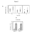
- Figure 2(b) shows that DNFB-induced contact hypersensitivity inflammation is not a function of differences in cell number. Similar number of lymph node cells were present in both CCR6+/+ and CCR6-/- mice. Animals were sensitized with DNFB (filled bars) or left untreated (open bars); 5 days later, lymphocytes from inguinal, axial and brachial (I+A+B) LNs were prepared and counted.
- Figure 2(c) shows CCR6-/-lymphocyte proliferation in response to specific and nonspecific stimuli.
- CCR6-/- and CCR6+/+ animals were sensitized with DNFB (filled symbols) or untreated (open symbols).
- DNFB filled symbols
- I+A+B axial and brachial
- control mesenteric LNs were recovered and cultured for 36 hours alone or in the presence of DNBS, as indicated. Cultures were pulsed with 3 H-thymidine for 18 hours, and cell proliferation was estimated.
- Similar experiments were performed to measure in vitro responses to a polyclonal stimulus, Con A. Each value represents the average of three separate groups, consisting of pooled LNs from two mice. All assays were performed in triplicate.
- the present invention provides a method to assay for a test compound that moderates CCR6-dependent, DNFB-type contact hypersensitivity inflammation.
- the method uses as a control a CCR6 knockout animal as provided in the present invention.
- a preferred knockout animal is a mouse.
- a preferred knockout mouse is a C57BL/6 mouse having deletions in both CCR6 alleles.
- the method uses an animal possessing an intact, functional CCR6 allele.
- a preferred control animal possesses two intact, functional CCR6 alleles.
- a preferred control animal is a C57BL/6J WT mouse.
- Mice are sensitized by applying a solution of DNFB to an area of skin.
- a preferred area of skin is the abdomen.
- the abdomen is shaved prior to application of DNFB.
- the solution may comprise a vehicle that dissolves DNFB.
- a preferred vehicle comprises a mixture of acetone and olive oil.
- a more preferred vehicle is a 4:1 (by volume) ratio of acetone/olive oil.
- the solution may comprise up to 1% (v/v) DNFB.
- a preferred solution comprises 0.05% DNFB.
- mice are challenged by applying a test volume of a solution of DNFB to an ear.
- the solution comprises a vehicle of substantially the same composition as the vehicle of the sensitization solution.
- the solution may comprise up to 0.5% DNFB.
- the solution comprises 0.2% DNFB.
- a volume of vehicle is applied to the contralateral ear.
- a volume of vehicle, substantially identical to the test volume is applied to the contralateral ear.
- the method further comprises administering a test compound to the WT mice.
- the ligand may be administered before, after, or simultaneously with the challenge.
- a test compound may be administered systemically.
- a preferred route of systemic administration is orally.
- a more preferred route of systemic administration is by injection.
- a test compound may be administered topically.
- a test compound may be dissolved in a vehicle and dropped or swabbed on an exposed area of skin.
- the method further comprises comparing a contact hypersensitivity inflammation response of a WT mouse in the presence or absence of said test compound.
- a preferred response is ear swelling.
- FIG. 3(a) shows a delayed-type hypersensitivity (DTH) response of CCR6 knockout and WT mice.
- Figure 3(b) shows CCR6 knockout and WT, in vitro MLR to allogeneic BALB/c splenocytes.
- Lymphocytes were prepared from IAB LNs from CCR6+/+ (filled squares) and CCR6-/- mice (filled circles) and cultured in 96-well plates (2 x 10 5 cells/well) with increasing amounts of stimulator allogeneic BALB/c splenocytes. After 72 hours, cultures were pulsed with 3 H-thymidine for 24 hours and cell proliferation was estimated. Background proliferation is also shown (open symbols). Each point represents the average value of two separate groups, each consisting of pooled LNs from two mice. Assays were performed in triplicate.
- Figure 3(c) shows that adoptive transfer of sensitized CCR6+/+ CD4+ T cells to CCR6-/- mice restores their ability to produce a DTH response.
- Unsensitized CCR6+/+ and CCR6-/- mice were adoptively transferred with 2 x 10 7 CD4+ T cell-enriched preparations purified from sensitized CCR6+/+ and CCR6-/- donors, as indicated.
- T cells from CCR6-/- IAB LNs were also allowed to proliferate in an in vitro MLR assay with BALB/ c splenocytes before being injected to CCR6-/- hosts (++). After 16 hours, animals were challenged with 13 x 10 6 BALB/c splenocytes in footpads, and swelling was measured 24 hours later.
- the present invention provides a method to assay for a test compound that moderates CCR6-dependent, DTH inflammation.
- the method uses as a control a CCR6 knockout animal as provided in the present invention.
- a preferred knockout animal is a mouse.
- a preferred knockout mouse is a C57BL/6 mouse having deletions in both CCR6 alleles.
- the method uses an animal possessing an intact, functional CCR6 allele.
- a preferred control animal possesses two intact, functional CCR6 alleles.
- a preferred control animal is a C57BL/6J WT mouse.
- the present invention provides a method to assay for a test compound that moderates a CCR6-dependent delayed-type hypersensitivity inflammation response.
- An aspect of the method provides CCR6 knockout and WT mice.
- mice are sensitized by intravenous injection of allogeneic splenocytes.
- Preferred, but non-limiting, splenocytes are obtained from BALB/c mice.
- mice are challenged by injection of allogeneic splenocytes into a footpad. It is preferable that the challenge injection be administered about five days post sensitization.
- a footpad swelling in WT mice is compared in the presence and absence of a test compound.
- the test compound may be administered systemically as, for example, by injection.
- the test compound may be administered before, after, or simultaneously with sensitization.
- CCR6 in inflammatory bowel disease IBD. Mice with a targeted deletion of the CCR6 gene were generated as described and backcrossed onto a C57BL/6J background for eight generations. C57BL/6J WT littermates were used as controls. BALB/c mice were purchased from the Jackson Laboratory (Bar Harbor, Maine, USA).
- DSS Dextran sodium sulfate
- Colitis was induced by addition of 2.5% (w/v) DSS (M.W. 40,000; ICN Biomedicals, Aurora, Ohio, USA) to drinking water for 7 days [ H. Mashimo, D. C. Wu, D. K. Podolsky, and M. C. Fishman, Impaired defense of intestinal mucosa in mice lacking intestinal trefoil factor, Science 1996. 274: 262-265 ).
- DSS administration was then stopped and mice received water alone for 3 weeks, after which 2.5% DSS was re-introduced to drinking water for another 3 days. Dosage was established empirically to induce moderate to severe colitis, minimizing mortality. Mean DSS/water consumption, body weight and stool consistency were recorded every 2 days. Fecal blood was assessed using the Hematest System (Bayer Corp., Pittsburgh, PA).
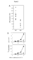
- FIG. 4(a) shows that CCR6 deficiency protects against DSS-mediated colitis weight loss.
- Mouse weight was monitored every 2 days.
- DSS chronic phase
- Figure 4(b) shows that CCR6 deficiency protects against DSS-mediated colitis intestinal inflammation.
- Distance was measured from the muscular limbal to the intestinal lumen as an estimation of intestinal inflammation at day 7 in control (CTR) and DSS-treated (DSS) WT and CCR6 -/- mice.
- CTR control
- DSS DSS-treated
- Figure 4(c) shows altered cytokine response to DSS treatment in CCR6-deficient mice.
- IL-4 and IL-10 transcript levels were measured in colon samples to estimate the cytokine response following DSS treatment.
- Basal IL-10 mRNA levels were higher in CCR6-deficient mice (about twofold), decreasing at days 1 and 3 of DSS treatment in both animal groups, particularly in the WT animals (Fig. 4c); this may be due to destruction of immune cells resident in the mucosal tissue subsequent to DSS administration.
- absolute IL-10 transcript levels remained higher in CCR6-deficient than in WT mice on days 1 and 3, recovering to a similar level in both mouse groups by day 7 (Fig. 4c).
- WT mice responded with an immediate increase in IL-10 mRNA levels (about fourfold, day 1; Fig. 4c).
- mice received enemas of TNBS (Sigma, St. Louis, Missouri, USA) dissolved in 50% ethanol in phosphate buffered saline (PBS), given under isofluorane anesthesia using a tuberculin syringe fitted with a 2.5-cm gastric intubation needle. Control mice received enemas of 50% ethanol in PBS. Mice were sensitized three times at weekly intervals (2 mg for initial treatment, 1 mg for subsequent doses) [ M. F. Neurath, I. Fuss, B. L. Kelsall, E. Stuber and W. Strober, Antibodies to interleukin 12 abrogate established experimental colitis in mice, J. Exp. Med. 1995, 182: 1281-1290 ]. At 5-7 days after the third TNBS administration, mice were killed and analyzed as described below.
- PBS phosphate buffered saline
- Figure 5 shows that TNBS treatment provokes distinct T cell and cytokine responses in WT and CCR6-deficient C57B/6J mice.
- Samples were processed for real-time quantitative RT-PCR analysis of CD4 and CD8 (a) and cytokine transcripts (b, c) as indicated. Representative results are shown from two experiments.
- Figure 6 shows that CCR6-deficiency confers susceptibility to TNBS-induced colitis in the normally resistant C57BL/6J mouse strain.
- Colon tissue sections from control (PBS) or TNBS-treated (TNBS) animals were obtained at day 7 after the last enema and stained with hematoxylin/eosin.
- Photomicrographs (left column, 40x; middle and right column, 200x magnification) show different aspects of the TNBS-mediated colitis and are representative of two experiments performed. Locally extensive inflammatory reaction forming microgranulomas in TNBS-treated BALB/c mice are marked by arrows.
- Colitis extent and severity were assessed macroscopically by clinical scoring of the percentage of the colon and cecum showing macroscopic evidence of colitis, as follows: 0, normal; 1, erythema; 2, presence of ulcers; and 3, diffuse ulceration. Tissue sections from the entire colon were examined and scored in a blind fashion for severity and extent of ulceration. Tissue thickness was determined from the muscularis propria to the luminal border. Lesion severity was graded according to a previously defined scoring system: 0, normal; 1, mild; 2, moderate; and 3, severe. ( P. G, Andres, P. L. Beck, E. Mizoguchi, A.
- Moderate lesions were multifocal or locally extensive and showed inflammation or fibrosis extending into the submucosa. Severe lesions had inflammatory cells extending into the muscularis basement. Colitis onset and progress were monitored by registering rectal bleeding, diarrhea and body weight loss every 2 days.
- CCR6 deficiency confers susceptibility to TNBS-induced colitis in normally resistant C57BL / 6J mice.
- Administration of TNBS per rectum in the presence of ethanol induces a mucosal immune response leading to large intestine inflammation in susceptible (BALB/c) but not in resistant (C57BL/6J) mouse strains ( C. O. Elson, K. W. Beagley, A. T. Shannanov, K. Fujihashi, H. Kiyono, G. S. Tennyson, Y. Cong, C. A. Black, B. W. Ridwan and J. R. McGhee, Hapten-induced model of murine inflammatory bowel disease: mucosa immune responses and protection by tolerance. J.
- TNBS-mediated colitis thus has a Th1 character in the SJL/J mouse strain, whereas it shows a Th2 (or mixed Th1/Th2) character in the BALB/ c background ( W. Strober, I. J. Fuss, R. S. and Blumberg, The immunology of mucosal models of inflammation, Annu. Rev. Immunol. 2002, 20: 495-549 ).
- TNBS colitis is a T cell-mediated process
- W. Strober, I. J. Fuss, and R. Blumberg, The immunology of mucosal models of inflammation. Annu. Rev. Immunol. 2002, 20: 495-549 we used a real-time quantitative RT-PCR procedure to compare CD4 and CD8 mRNA expression in mouse colons 7 days after administration of the last enema. No major changes were observed in TNBS-treated WT C57BL/6J mice, except for a mild increase in CD8 transcript levels (Figure 5a).
- CCR6-deficient C57BL/6J mice had higher basal levels of both CD4 (about fourfold) and CD8 (about twofold) transcripts after control PBS/ethanol enema treatment; these levels decreased by about tenfold after hapten treatment.
- TNBS treatment induced an about threefold increase in CD4 mRNA ( Figure 5b), which may correspond to CD4 T cell infiltration or in situ proliferation.
- TNBS administration-induced cytokine response in the colon To analyze the T cell response in TNBS-mediated colitis, we measured cytokine mRNA levels in the distal half of the colon 7 days after administration of the last enema. Real-time quantitative RT-PCR assays showed that the cytokine profile of CCR6-deficient C57BL/6J mice was equivalent to that of BALB/c mice and differed from that of WT C57BL/6J mice ( Figures 5b, c). Consistent with the inflammation developed and the mixed Th1/Th2 character described in TNBS colitis, IL-12, IL-10, and IL-4 transcript levels were higher in colons of TNBS-treated than in control-treated BALB/c mice.
- GvHD graft-versus-host disease
- An aspect of the present invention provides a method to assay for a test compound that moderates CCR6-dependent GvHD comprising providing CD4 + T cells from a CCR6 knockout mouse as a control; providing CD4 + T cells from a CCR6 WT mouse; providing a CCR6 WT, MHC(II)-mismatched mouse; providing a test compound; transferring said T cells to said WT mouse in the presence and absence of said test compound; and comparing a GvHD response in the presence and absence of said test compound.
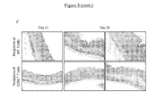
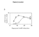
- a further aspect of the present invention provides convenient, non-limiting GvHD responses include percent survival at a pre-determined time post transfer (Figure 7a), recipient weight at a pre-determined time post transfer (Figure 7d), ear inflammation (Figure 8a), skin lesions (Figure 8b), diarrhea (Figure 8d), spleen index, and CD4 + T cell expansion.
- Figure 7a shows that percent survival may be described by a sigmoid curve as a function of time post transfer.
- the time post transfer at which percent survival is determined may be any day post transfer.
- a preferred day is day fifteen post transfer.
- a more preferred time to determine percent survival is at an inflection point on a survival curve.
- the inflection point may relate to the curve describing survival of WT or CCR6-deficient animals.
- GvHD onset is delayed and lesion severity decreased in mhc-mismatched recipients of naive T cells from CCR6-deficient donors.
- CD4 + T cells from H-2 d haplotype-selected WT or CCR6-deficient mice transferred to MHC-matched/mhc-mismatched CB 17/SCID recipient mice.
- CD4 + CD45RB high T cell recipients developed a wasting syndrome associated with splenomegaly, erythrosquamous skin lesions and intestinal inflammation. Ear thickness in recipient mice was evaluated at several time points as an overall measure of skin inflammation, and skin lesions were scored clinically. Weight loss and diarrhea were monitored as an indication of intestinal damage.
- CD4 + CD45RB low memory/regulatory T cells do not provoke GvHD in recipient mice and are able to suppress GvHD caused by CD4 + CD45RB high na ⁇ ve T cells when both cell types are cotransferred ( M.P. Schon, M. Detmar and C. M. Parker, 1997, Murine psoriasis-like disorder induced by naive CD4+ T cells, Nat. Med. 3: 183-188 ; P. J. Morrissey, K. Charrier, S. Braddy, D. Liggitt and J. D. Watson, 1993, CD4+ T cells that express high levels of CD45RB induce wasting disease when transferred into congenic severe combined immunodeficient mice.
- Figure 8 shows that transfer of naive CD4 + T cells from CCR6-deficient mice to MHC-matched/mhc-mismatched recipients delays GvHD onset and reduces severity of symptoms, and that the regulatory activity of CCR6-deficient CD4 + CD45RB low regulatory T cells is impaired.
- the time course of symptom development is shown for (a), ear inflammation, statistical analysis using Student's t test comparing WT vs.
- Results are also shown for ear inflammation (f) and body weight change (g) from control experiments in which naive ( ⁇ ) or memory/regulatory ( ⁇ ) BALB/c CD4 + T cells were transferred to recipient mice.
- Figure 9 shows that in vivo homing of CD4 + T cells to secondary lymphoid organs and on-site proliferation is CCR6-independent.
- CMFDA-labeled WT and CMTMR-labeled CCR6-deficient CD4 + T cells (3 x 10 6 each) were co-transferred into BALB/c-SCID mice and the percentage of donor cells in recipient spleens determined at different times ACT (a).
- WT or CCR6-deficient CD4 + T cells transferred to BALB/c-SCID mice showed a similar proliferation ratio, as deduced from the sequential halving of the fluoresce intensity with each generation of CD4 + T cells in spleen, peripheral (PLN) and mesenteric (MLN) lymph nodes.
- Figure 11 shows delayed CD4 + T cell homing to skin and small intestine of recipients of cells from CCR6-deficient donors.
- (a) Flow cytometric analysis of CD45 + and CD4 + cells infiltrated in ear skin.
- Figure 12 shows that recipients of CD4 + naive T cells from CCR6-deficient donors have lower and delayed expression of pro-inftammatory cytokines and chemokines in GvHD target organs.
- Cytokine and chemokine transcript levels were measured over time by real-time quantitative RT-PCR in ear skin (a) and small intestine (b) of naive T cell recipients from WT (o) and CCR6-deficient ( ⁇ ) donors.
- CCR6KOES192 Homologous, recombinant embryonic stem cells, CCR6KOES192, containing a CCR6 deletion were deposited on February 11, 2004 under Accession Number DSM ACC2636 at the Deutsche Sammlung von Mikroorganismen und Zellkulturen GmbH, Mascheroder Weg 1b, D-38124 Braunschweig, Germany. Members of the public may access the deposit under conditions set forth in 37 CFR 1.808.
- This invention identifies CCR6 as a target for drugs that reduce inflammatory, immunological responses and has industrial applicability in providing means to assay for test compounds that moderate CCR6-dependent physiological functioning and disease processes based thereon.
- BLR1 A putative chemokine receptor, BLR1 directs B cell migration to defined lymphoid organs and specific anatomic compartments of the spleen, Cell. 87: 1.037-1.047 .
- CCR7 coordinates the primary immune response by establishing functional microenvironments in secondary lymphoid organs, Cell, 99: 23-33 .
- CC-chemokine receptor 6 is expressed on diverse memory subsets of T cells and determines responsiveness to macrophage inflammatory protein 3 ⁇ , J. Immunol. 162: 186-194 .
- Macrophage inflammatory protein 3 ⁇ is expressed at inflamed epithelial surfaces and is the most potent chemokine known in attracting Langerhans cell precursors, J. Exp. Med. 192: 705-717 .
- CC-chemokine receptor 6 is expressed on diverse memory subsets of T cells and determines responsiveness to macrophage inflammatory protein 3 ⁇ , J. Immunol. 162: 186-194 (1999 ).
- CC-chemokine receptor 6 is expressed on diverse memory subsets of cells and determines responsiveness to macrophage inflammatory protein 3 ⁇ , J. Immunol. 162, 186-194 (1999 ).
Landscapes
- Life Sciences & Earth Sciences (AREA)
- Health & Medical Sciences (AREA)
- Chemical & Material Sciences (AREA)
- Environmental Sciences (AREA)
- Organic Chemistry (AREA)
- Zoology (AREA)
- General Health & Medical Sciences (AREA)
- Medicinal Chemistry (AREA)
- Engineering & Computer Science (AREA)
- Biophysics (AREA)
- Gastroenterology & Hepatology (AREA)
- Genetics & Genomics (AREA)
- Toxicology (AREA)
- Molecular Biology (AREA)
- Proteomics, Peptides & Aminoacids (AREA)
- Immunology (AREA)
- Biochemistry (AREA)
- Biotechnology (AREA)
- Veterinary Medicine (AREA)
- Animal Behavior & Ethology (AREA)
- Animal Husbandry (AREA)
- Biodiversity & Conservation Biology (AREA)
- Cell Biology (AREA)
- Pharmaceuticals Containing Other Organic And Inorganic Compounds (AREA)
- Medicines That Contain Protein Lipid Enzymes And Other Medicines (AREA)
Abstract
The present invention generally relates to the generation of a knockout mouse for CCR6 and particularly, but not exclusively, to the identification of CCR6 as a target for drugs that reduce inflammatory immunological responses. More specifically, the invention relates to, but in not limited to, non-human animals and cells deficient in the CCR6 receptor, to the use of CCR6 as a target for drugs and to the use of CCR6 inhibitors for graft versus host disease, cutaneous inflammations such as allergic contact dermatitis and intestinal inflammations such as ulcerative colitis and Crohn's disease.
Description
- The present invention relates generally to the generation of a CCR6 knockout mouse (genetically deprived of CCR6), and particularly, but not by way of limitation, to the identification of CCR6 as a target for drugs that reduce inflammatory, immunological responses. The invention relates more particularly, but not by way of limitation, to cells and non-human animals deficient for the CCR6 receptor, the use of CCR6 as a target for drugs, and the therapeutic use of inhibitors of CCR6 to treat graft-versus-host disease, cutaneous inflammations such as allergic contact dermatitis, and intestinal inflammations such as ulcerative colitis and Crohn's disease.
- The information provided below is not admitted to be prior art to the present invention, but is provided solely to assist the understanding of the reader.
- Leukocyte traffic is the hallmark of the immune response. Controlled cell movements allow the precise interactions necessary to bring different leukocytes into physical contact. The possibility of rare T and B lymphocytes encountering their specific antigen is maximized by their recirculation through secondary lymphoid organs, in which antigens are displayed to them by antigen-presenting cells. Of these, dendritic cells (DCs) are an important heterogeneous population of antigen-presenting cells that can induce, sustain, and regulate immune responses (J. Banchereau et al. 2000. Immunobiology of Dendritic Cells. Annu. Rev. Immunol. 18:767-811). Once activated, effector T cells are able to migrate to tissues in which pathogens have been detected. Eventually, memory T cells constitute a surveillance system that ensures tissue protection in case of a new antigen challenge. Chemokines are a family of proteins that control leukocyte trafficking by binding to specific seven-transmembrane receptors [P.M Murphy, M. Baggiolini, I.F. Charo, C.A. Hebert, R. Horuk, K. Matsushima, L.H. Miller, J. J. Oppenheim. And C.A. Power, International Union of Pharmacology. XXII. Nomenclature for chemokine receptors. Pharmacol. Rev. 2000. 52: 145-176; A. Zlotnik and O. Yoshie, Chemokines: a new classification system and their role in immunity. Immunity 2000, 12: 121-127].
- In recent years, several reports have pointed to chemokines as important factors in the regulation of leukocyte trafficking for lymphoid organ architecture, and in physiological and pathological situations (R. Forster et al., 1996. A putative chemokine receptor, BLR1, directs B cell migration to defined lymphoid organs and specific anatomic compartments of the spleen. Cell. 87:1037-1047; F. Sallusto, A. Lanzavecchia and C. R. Mackay, 1998, Chemokines and chemokine receptors in T-cell priming and Th1/Th2-mediated responses; Immunol. Today. 19:568-574; F. Sallusto, D. Lenig, R. Forster, M. Lipp and A. Lanzavecchia, 1999, Two subsets of memory T lymphocytes with distinct homing potentials and effector functions. Nature. 401:708-712; R. Forster et al., 1999, CCR7 coordinates the primary immune response by establishing functional microenvironments in secondary lymphoid organs, Cell, 99:23-33; J. G. Cyster, 1999, Chemokines and cell migration in secondary lymphoid organs, Science, 286:2098-2102; J. J. Campbell and E. C. Butcher, 2000, Chemokines in tissue-specific and microenvironment-specific lymphocyte homing, Curr. Opin. Immunol., 12:336-341; B. Moser and P. Loetscher, Lymphocyte traffic control by chemokines, Nat. Immunol., 2001, 2: 123-128; K. M. Ansel and J. G. Cyster, Chemokines in lymphopoiesis and lymphoid organ development, Curr. Opin. Immunol., 2001, 13: 172-179; F. Sallusto, C. R. Mackay, and A. Lanzavecchia, The role of chemokine receptors in primary, effector, and memory immune responses, Annu. Rev. Immunol., 2000, 18: 593-620; C. Gerard and B. J. Rollins, Chemokines and disease, Nat. Immunol., 2001, 2: 108-115).
- CCR6 is a β-chemokine-specific receptor for CCL20 (MIP-3aα/LARC/Exodus) (M. Baba et al., 1997, Identification of CCR6, the specific receptor for a novel lymphocyte-directed CC chemokine LARC, J. Biol. Chem. 272:14893-14898; F. Liao et al., 1997, STRL22 is a receptor for the CC chemokine MIP-3α, Biochem. Biophys. Res. Commun. 236:212-217; C. A. Power et al., 1997, Cloning and characterization of a specific receptor for the novel CC chemokine MIP-3α from lung dendritic cells, J. Exp. Med. 186:825-835; D. R. Greaves et al., 1997, CCR6, a CC chemokine receptor that interacts with macrophage inflammatory protein 3α and is highly expressed in human dendritic cells, J. Exp. Med. 186:837-844; D.L. Rossi, A. P. Vicari, B. K. Franz, T. K. McClanahan and A. Zlotnik, 1997, Identification through bioinformatics of two new macrophage proinflammatory human chemokines: MIP-3α and MIP-3β, J. Immunol. 158: 1033-1036; R. Hromas et al., 1997, Cloning and characterization of exodus, a novel β-chemokine. Blood. 89:3315-3322). In addition to having only one highly specific ligand, other features make CCR6 an interesting receptor. Human CCR6 is expressed in immature DCs derived in vitro from CD34+ precursors and is downregulated as DCs mature (M. C. Dieu et al. 1998, Selective recruitment of immature and mature dendritic cells by distinct chemokines expressed in different anatomic sites, J Exp. Med. 188:373-386; L. Carramolino et al., 1999, Down-regulation of the β-chemokine receptor CCR6 in dendritic cells mediated by TNF-α and IL-4, J. Leukoc. Biol. 66:837-844.); it is also expressed in memory T cells, CLA+ cells and B cells (F. Liao et al., 1999, CC-). Similar CCR6 expression patterns have been reported in the mouse, in which CCR6 is expressed in the myeloid but not in the lymphoid DC subpopulation (R. Varona et al., 1998, Molecular cloning, functional characterization and mRNA expression analysis of the murine chemokine receptor CCR6 and its specific ligand MIP-3α. FEBS Lett. 440: 188-194; a. Iwasaki and B. L. Kelsall, 2000, Localization of distinct Peyer's patch dendritic cell subsets and their recruitment by chemokines macrophage inflammatory protein (MIP)-3α, MIP-3β, and secondary lymphoid organ chemokine, J. Exp. Med. 191:1381-1394), B cells, and CD4+ T cells (R. Varona et al., 1998. Molecular cloning, functional characterization and mRNA expression analysis of the murine chemokine receptor CCR6 and its specific ligand MIP-3α, FEBS Lett. 440: 188-194).
- CCL20 is known to interact only with CCR6 in both the human and murine systems. Human CCL20 triggers adhesion of memory CD4+ T cells to ICAM-1 (J. J. Campbell et al., 1998. Chemokines and the arrest of lymphocytes rolling under flow conditions, Science, 279: 381-384) and is expressed in epithelial crypts of inflamed tonsils (M. C. Dieu et al., 1998. Selective recruitment of immature and mature dendritic cells by distinct chemokines expressed in different anatomic sites. J Exp. Med. 188:373-386), epithelial cells from appendix (Y. Tanaka et al., 1999. Selective expression of liver and activation-regulated chemokine (LARC) in intestinal epithelium in mice and humans, Eur. J. Immunol. 29:633-642.), as well as keratinocytes and venular endothelial cells from non pathological skin (A. S. Charbonnier et al., 1999. Macrophage inflammatory protein 3a is involved in the constitutive trafficking of epidermal Langerhans cells, J. Exp. Med. 190:1755-1768) and psoriatic skin (B. Homey et al., 2000, Up-regulation of macrophage inflammatory protein-3α/CCL20 and ; M. C. Dieu-Nosjean et al., 2000, Macrophage inflammatory protein 3α is expressed at inflamed epithelial surfaces and is the most potent chemokine known in attracting Langerhans cell precursors, J. Exp. Med. 192:705-717).
- Murine CCL20 (mCCL20) was also reported to be expressed in epithelial cells from intestinal tissue (Y. Tanaka et al., 1999, Selective expression of liver and activation-regulated chemokine (LARC) in intestinal epithelium in mice and humans., Eur.J. Immunol. 29:633-642). Interestingly, in Peyer's patches (PPs), mCCL20 is expressed only by the follicle-associated epithelium (FAE) overlying the subepithelial dome (SED), where CCR6-expressing cells accumulate (A. Iwasaki and B. L. Kelsall, 2000, Localization of distinct Peyer's patch dendritic cell subsets and their recruitment by chemokines macrophage inflammatory protein (MIP)-3α, MIP-3β, and secondary lymphoid organ chemokine, J. Exp. Med. 191:1381-1394).
- Taken together, these data suggest a role for CCR6 and its ligand as regulators of the migration and recruitment of antigen-presenting and immunocompetent cells during inflammatory and immunological responses.
- Role of CCR6 in contact hypersensitivity (CHS) and delayed-type hypersensitivity (DTH) models of inflammation. CCR6-/- mice (CCR6 knockouts) were generated to investigate the role of CCR6 in vivo. Analysis of CCR6 knockout mice revealed defects in leukocyte homing to the intestinal mucosa as evidenced by their underdeveloped Peyer's patches, in which clear alterations in the positioning of myeloid CD11b+ CD11c+ DCs were observed. These animals also showed increased numbers of intraepithelial lymphocyte (IEL) subpopulations. Allergic contact dermatitis, also known as contact hypersensitivity, is a T cell-dependent skin disease with the kinetics of a delayed-type hypersensitivity response (S. Grabbe and T. Schwarz, 1998, Immunoregulatory mechanisms involved in the elicitation of allergic contact hypersensitivity, Immunol. Today 19:37-44). This disorder is rarely life-threatening, but the costs to society of occupation-related allergic contact dermatitis are high (R. L. Riestchel, 1995, Human and economic impact of allergic contact dermatitis and the role of patch testing, J. Acad. Dermatol. 33:812-815). In this disease, the antigen is introduced epicutaneously through intact skin. The sensitizing antigens are typically unstable reactive molecules that can form complexes with host proteins. Potent contact-sensitizing antigens induce a dose-dependent cutaneous irritation that is independent of their antigenicity. The altered responses of CCR6-/- mice in contact hypersensitivity (CHS) and delayed-type hypersensitivity (DTH) models of inflammation suggest a critical role for CCR6 in the activation and/or migration of CD4+ T-cell subsets that home into skin.
- Role of CCR6 in the development of inflammatory bowel disease. The intestine is bathed by a large variety of foreign antigens, which normally elicit an appropriate immune response. Defects in the mechanisms that control the reaction to these antigens are thought to provoke the excessive immunological response that may lead to tissue destruction and intestinal inflammatory disease (C. Nagler-Anderson, Man the barrier! Strategic defences in the intestinal mucosa, Nat. Rev. Immunol. 2001. 1: 59-67).
- Among these pathologies, human inflammatory bowel disease (IBD) is a chronic, relapsing and remitting inflammatory condition, clinically characterized by two phenotypes, ulcerative colitis and Crohn's disease, that affect the colon and/or the distal small intestine (R. S. Blumberg, L. J. Saubermann and W. Strober, Animal models of mucosal inflammation and their relation to human inflammatory bowel disease, Curr. Opin. Immunol., 1999, 11: 648-656).
- Chemokine overexpression has been observed in patients with ulcerative colitis or Crohn's disease (L. Mazzucchelli, C. Hauser, K. Zgraggen, H. Wagner, M. Hess, J. A. Laissue and C. Mueller, Expression of interleukin-8 gene in inflammatory bowel disease is related to the histological grade of active inflammation, Am. J. Pathol. 1994, 144:997-1007; M. C. Grimm, S. K. Elsbury, P. Pavli and W. F. Doe, Enhanced expression and production of monocyte chemoattractant protein-1 in inflammatory bowel disease mucosa. J. Leukoc. Biol. 1996. 59: 804-812; S. Keates, A. C. Keates, E. Mizoguchi, A. Shan and C. P. Kelly, Enterocytes are the primary source of the chemokine ENA-78 in normal colon and ulcerative colitis, Am. J. Physiol. 1997, 273:G75-G82; J. Wedemeyer, A. Lorentz, M. Goke, P. N. Meier, P. Flemming, C. A. Dahinden, M. P. Manns and S. C. Bischoff, Enhanced production of monocyte ; L. Mazzucchelli, C. Hauser, K. Zgraggen, H. E. Wagner, M. W. Hess, J. A. Laissue and C. Mueller, Differential in situ expression of the genes encoding the chemokines MCP-1 and RANTES in human inflammatory bowel disease, J. Pathol. 1996. 178: 201-206).
- CCL20 expression in epithelia from tonsils [Dieu-Nosjean et al., Selective recruitment of immature and mature dendritic cells by distinct chemokines expressed in different anatomic sites, J. Exp. Med. 1998. 188: 373-386], appendix, colon and other intestinal tissue [Y. Tanaka et al., 1999. Selective expression of liver and activation-regulated chemokine (LARC) in intestinal epithelium in mice and humans, Eur. J. Immunol. 29: 633-642; A. Izadpanah, M. B. Dwinell, L. Eckmann, N. M. Varki and M. F. Kagnoff, Regulated MIP-3α/CCL20 production by human intestinal epithelium: mechanism for modulating mucosal immunity, Am. J. Physiol. Gastrointest. Liver Physiol. 2001, 280: G710-G719], Peyer's patches [A. Iwasaki and B. L. Kelsall, Localization of distinct Peyer's patch dendritic cell subsets and their recruitment by chemokines macrophage inflammatory protein (MIP)-3α, MIP-3β, and secondary lymphoid organ chemokine, J. Exp. Med. 2000, 191:1381-1394], as well as normal [ A. S. Charbonnier et al. 1999, Macrophage inflammatory protein 3α is involved in the constitutive trafficking of epidermal Langerhans cells, J. Exp. Med. 190:1755-1768) and psoriatic skin (B. Homey, et al., 2000, Up-regulation of macrophage inflammatory protein-3α/CCL20 and ; M. C. Dieu-Nosjean et al., 2000, Macrophage inflammatory protein 3α is expressed at inflamed epithelial surfaces and is the most potent chemokine known in attracting Langerhans cell precursors, J. Exp. Med. 192:705-717) supports the hypothesis that this chemokine has an important function in the interface between the organism and the environment.
- A recent study of CCR6-deficient mice demonstrated CCR6/CCL20 involvement in the control of leukocyte homeostasis in the gut (R. Varona, R. Villares, L. Carramolino, I. I. Goya, A. A. Zaballos, J. Gutierrez, M. Torres, A. C. Martinez and G. Marquez, CCR6-deficient mice have impaired leukocyte homeostasis and altered contact hypersensitivity and delayed-type hypersensitivity responses. J. Clin. Invest. 2001. 107: R37-R45). In CCR6-deficient mice, CD11 b+CD11c+ myeloid DC are clearly decreased in the subepithelial dome of PP, since these cells are displaced mainly to the interfollicular region (R. Varona, R. Villares, L. Carramolino, I. I. Goya, A. A. Zaballos, J. Gutierrez, M. Torres, A. C. Martinez, and G. Marquez, CCR6-deficient mice have impaired leukocyte homeostasis and altered contact hypersensitivity and delayed-type hypersensitivity responses. J. Clin. Invest. 2001. 107: R37-R45). CCR6-deficient mice also have increased numbers of T cell subpopulations within the intestinal mucosa (R. Varona, R. Villares, L. Carramolino, I. I. Goya, A. A. Zaballos, J. Gutierrez, M. Torres, A. C. Martinez, and G. Marquez, CCR6-deficient mice have impaired leukocyte homeostasis and altered contact hypersensitivity and delayed-type hypersensitivity responses. J. Clin. Invest. 2001. 107: R37-R45).
- The immune response has been now studied in two IBD models in CCR6-deficient mice. The IBD models are mediated by the administration of dextran sodium sulfate (DSS) or 2,4,6-trinitrobenzene sulfonic acid (TNBS). The obtained data show that the absence of CCR6 results in less severe intestinal pathology in DSS-treated mice, with no ulcerations observed in colon. CCR6 deficiency alters leukocyte homeostasis and the cytokine environment in the intestinal mucosa. These changes are sufficient to confer susceptibility to TNBS-mediated intestinal inflammation in the otherwise resistant C57B1/6J mouse strain. Taken together, our data suggest that the CCR6/CCL20 axis has a critical, non-redundant role in the control of immune responses in the intestine.
- CCR6 regulation of CD4+ T cell-mediated acute graft-versus-host disease responses. Graft-versus-host disease (GvHD) is a common, debilitating complication of allogeneic bone marrow transplantation (V. T. Ho & R. J. Soiffer, The history and future of T-cell depletion as graft-versus-host disease prophylaxis for allogeneic hematopoietic stem cell transplantation. Blood 98, 3192-3204 (2001)). The disease is a multistep process involving lymphocyte activation and proliferation and target cell killing. Donor T cells recognize the major histocompatibility complex (MHC) and associated peptides on host-derived antigen-presenting cells. This leads to cell activation and differentiation into various effector and memory T cells with selective tropism [F. Sallusto, D. Lenig, R. Forster, M. Lipp and A. Lanzavecchia, Two subsets of memory T lymphocytes with distinct homing potentials and effector functions, Nature 401,708-712 (1999); J. H. Antin, Acute graft-versus-host disease: inflammation run amok? J. Clin. Invest. 107, 1497-1498 (2001)]. In acute GvHD, activated T cells infiltrate mainly the skin, liver, and gastrointestinal tract, where they initiate a Th1-mediated immune response. Interferon-γ (IFN-γ) collaborates in recruitment of other T cell subsets, monocytes, and macrophages that further damage affected tissues (W. Krenger and J. L. Ferrara, Graft-versus-host disease and the Th1/Th2 paradigm, Immunol. Res. 15, 50-73 (1996); W. D. Shlomchik et al., Prevention of graft versus host disease by inactivation of host antigen-presenting cells, Science 285, 412-415 (1999)). Recent data showing that neutralization of TNF-α and IL-1 prevents acute GvHD confirm the central role of inflammatory cytokines in this disease [T. Teshima et al., Acute graft-versus-host disease does not require alloantigen expression on host epithelium, Nat. Med. 8: 575-581 (2002)].
- The adhesion and chemoattractant receptors expressed by effector/memory T cells interact with the adhesion molecules and chemokines expressed by target tissues, controlling T cell homing specificity [E. J. Kunkel and E. C. Butcher, Chemokines and the tissue-specific migration of lymphocytes, ]. The chemokine CCL20 and its receptor CCR6 have a central role in chemoattracting specific T, B, and dendritic cell subsets to the peripheral tissues and lymphoid organs associated to epithelial surfaces [E. Schutyser, S. Struyf, and J. Van Damme, The CC chemokine CCL20 and its receptor CCR6, Cytokine Growth Factor Rev. 14, 409-426 (2003)]. CCR6 expression is restricted to the CLA+, α4β7+, or CLA-α4β7- cell subsets of the effector/memory T cell pool, which home to skin, gut or other non-lymphoid peripheral tissues, respectively [F. Liao et al., CC-]. This suggests that CCR6 is induced on T cells that are activated in diverse environments.
- It has been shown now that mucosal immunity is impaired in CCR6-deficient mice and that these animals have altered immune responses to cutaneous and intestinal inflammations [R. Varona et al., CCR6-deficient mice have impaired leukocyte homeostasis and altered contact hypersensitivity and delayed-type hypersensitivity responses, J. Clin. Invest. 107: R37-R45 (2001); R. Varona, V. Cadenas, J. Flores, A. C. Martinez and G. Marquez, CCR6 has a non-redundant role in the development of inflammatory bowel disease, Eur. J. Immunol. 33: 2937-2946 (2003)]. CCL20 is expressed constitutively by keratinocytes and venular endothelial cells from normal skin, as well as by mucosa-associated tissues; inflammatory stimuli upregulate these basal levels [E. Schutyser, S. Struyf and J. Van Damme, The CC chemokine CCL20 and its receptor CCR6, Cytokine Growth Factor Rev. 14, 409-426 (2003)]. The data available indicate that CCL20 acts through CCR6 to recruit differentiated memory cells to inflammation sites in skin and mucosa.
- The role of CCR6 in acute GvHD development in MHC-mismatched recipients was than analyzed. Disease onset was delayed and the mortality ratio decreased in MHC class 11-mismatched recipients of CD4+ T cells from CCR6-deficient compared to wild type (WT) donors. Conversely, CD8+ T cells from both CCR6-deficient and WT donors provoked similar GvHD symptoms in MHC class 1-mismatched recipients. CCR6 was also critical for GvHD development across minor histocompatibility complex (mhc)-mismatched barriers. Onset of skin lesions and intestinal inflammation was delayed and symptoms were milder in MHC-matched/mhc-mismatched recipients of naive CD4+CD45RBhigh T cells from CCR6-deficient donors. Significant delay was observed in CD45+ and CD4+ cell infiltration to skin and intestine in recipients of naive T cells from CCR6-deficient donors. We found lower IFN-γ and IL-10 levels in skin and gut of these mice. Moreover, the levels of the chemokines that control activated T cell homing to skin and gut were also decreased. These data suggest that by facilitating alloreactive effector/memory CD4+ T cell recruitment to target tissues, CCR6 plays a major role in GvHD development. Transferred CD4+CD45RBlow memory/regulatory T cells do not provoke GvHD in recipient mice and are able to suppress GvHD caused by CD4+CD45RBhigh naïve T cells in cotransfer experiments (M.P. Schon, M. Detmar and C M. Parker, 1997, Murine psoriasis-like disorder induced by naive CD4+ T cells, Nat. Med. 3: 183-188; P. J. Morrissey, K. Charrier, S. Braddy, D. Liggitt and J.D. Watson, 1993, CD4+ T cells that express high levels of CD45RB induce wasting disease when transferred into congenic severe combined immunodeficient mice. Disease development is prevented by cotransfer of purified CD4+ T cells. J Exp Med 178: 237-244). These studies showed that CD4+CD45RBlow memory/regulatory T cells from CCR6-deficient mice failed to suppress GvHD produced by WT naïve T cell transfer, indicating a critical role for CCR6 in the regulatory function of this T cell subset.
- Other objects and advantages will become apparent from the following disclosure.
- To study the role of CCR6 in several inflammation diseases, the inventors have used gene targeting by homologous recombination to create mutant mice that lack CCR6. The present invention provides a transgenic, non-human, knockout animal whose genome comprises an introduced null mutation in at least one CCR6 allele.
- An aspect of the present invention provides a mouse cell line designated CCR6KOES192, deposited with the Deutsche Sammlung von Mikroorganismen und Zellkulturen GmbH (DSM) under accession number DSM ACC2636. A further aspect provides a mouse, or part thereof, comprising cells grown from cells of DSM ACC2636.
- An aspect of the present invention provides a mammalian cell having a deletion in at least one CCR6 allele and having the characteristics of the mouse cell line designated CCR6KOES192, deposited with the DSM under accession number DSM ACC2636. A further aspect provides a mouse, or part thereof, comprising cells having the characteristics of the mouse cell line designated CCR6KOES192, deposited with the DSM under accession number DSM ACC2636.
- An aspect of the present invention provides a transgenic, non-human, knockout animal whose genome comprises an introduced null mutation in at least one CCR6 allele, wherein said mutation has the characteristics of the cell line designated CCR6KOES192, deposited with the DSM under accession number DSM ACC2636.
- An aspect of the present invention provides that the knockout animal is a rodent.
- A further aspect provides that the knockout animal is a mouse.
- The present invention provides a knockout mouse whose genome comprises an introduced null mutation in both CCR6 alleles.
- The present invention provides a method of making a transgenic knockout mouse having a disrupted CCR6 gene comprising: deleting the first exon of the CCR6 gene including the initiation codon by homologous recombination in a mouse embryonic stem cell; aggregating a clump of said stem cells with morulae and developing the corresponding blastocysts in vitro; transplanting said blastocyst into a pseudopregnant mouse; allowing said blastocyst to develop into a chimeric mouse; breeding said chimeric mouse to produce offspring; and screening said offspring to identify a homozygous transgenic knockout mouse whose genome comprises deletion of the first exon of both alleles of the CCR6 gene.
- The present invention provides a method to assay for a test compound that moderates CCR6-dependent GvHD comprising: an assay for binding of the test compound to CCR6, determining whether the test compound inhibits CCR6, providing CD4+ T cells from a WT mouse; providing a CCR6 WT, MHC(II)-mismatched mouse; providing a test compound; transferring said T cells to said WT mouse in the presence and absence of said test compound; and comparing a GvHD response in the presence and absence of said test compound.
- An aspect of the present invention provides a method to assay for a compound that moderates CCR6-dependent GvHD, wherein the GvHD response is selected from the group consisting of % survival at inflection day post transfer, recipient weight at inflection day, ear inflammation, skin lesions, diarrhea, spleen index, and CD4+ T cell expansion.
- The present invention provides a method to assay for a compound that moderates CCR6-dependent, inflammatory bowel disease comprising: providing a C57BL/6J WT mouse; rendering said mouse as DSS-type IBD models; administering a test compound to said mouse; and comparing an IBD response of said mouse in the presence and absence of said test compound.
- An aspect of the present invention provides a method to assay for a compound that moderates CCR6-dependent, inflammatory bowel disease, wherein the IBD response is selected from the group showing intestinal inflammation and appropriate colonic mucosa Th1 and Th2 cytokine mRNA levels.
- The present invention provides a method to assay for a compound that moderates CCR6-dependent, inflammatory bowel disease comprising: providing a C57BL/6J WT mouse; administering trinitrobenzenesulfonic acid to said mouse; administering a test compound to said mouse; and comparing an IBD response of said mouse in the presence and absence of said test compound.
- An aspect of the present invention provides a method to assay for a compound that moderates CCR6-dependent, TNBS-type inflammatory bowel disease, wherein the IBD response is selected from the group showing intestinal inflammation and appropriate colonic mucosa Th1 and Th2 cytokine mRNA levels.
- The present invention provides a method to assay for a compound that moderates CCR6-dependent, contact hypersensitivity inflammation comprising: providing a C57BL/6J WT mouse; sensitizing said mouse with DNFB; challenging said mouse with DNFB; administering a test compound to said mouse; and comparing a contact hypersensitivity inflammation response of said mouse in the presence and absence of said test compound.
- An aspect of the present invention provides a method to assay for a compound that moderates CCR6-dependent, DNFB-type contact hypersensitivity inflammation wherein said contact hypersensitivity inflammation response is ear inflammation.
- Still other objects and advantages of the present invention will become readily apparent by those skilled in the art from the following detailed description, wherein it is shown and described preferred embodiments of the invention, simply by way of illustration of the best mode contemplated of carrying out the invention. As will be realized the invention is capable of other and different embodiments, and its several details are capable of modifications in various obvious respects, without departing from the invention. Accordingly, the description is to be regarded as illustrative in nature and not as restrictive.
- The invention is best understood from the following detailed description when read in connection with the accompanying drawing. It is emphasized that, according to common practice, the various features of the drawing are not to scale. On the contrary, the dimensions of the various features are arbitrarily expanded or reduced for clarity. Included in the drawing are the following figures:
- Figure 1(a) depicts a partial restriction map of the CCR6 WT (wild type) locus, the targeting vector used, and the resulting genetic modification of the CCR6 gene.
- Figure 1(b) depicts a representative Southern blot analysis of BamH1-digested tail DNA from WT (+/+), heterozygous (+/-), and homozygous (-/-) CCR6 mice;
- Figure 1(c) depicts a representative Northern blot analysis of CCR6 mRNA expressed in thymus and spleen from CCR6 WT (+/+) and knockout (-/-) mice;
- Figure 2(a) shows altered ear inflammation response of CCR6-/- mice to DNFB-induced contact hypersensitivity inflammation;
- Figure 2(b) shows that DNFB-induced contact hypersensitivity inflammation is not a function of differences in cell number in inguinal, axial and brachial lymph nodes of untreated (white bars) or treated (black bars) animals;
- Figure 2(c) shows similar WT and CCR6-/- lymphocyte proliferation in response to specific and nonspecific stimuli;
- Figure 3(a) shows a delayed-type hypersensitivity (DTH) response of CCR6 knockout and WT mice;
- Figure 3(b) shows CCR6 knockout and WT, in vitro mixed lymphocyte reaction (MLR) to allogeneic BALB/c splenocytes;
- Figure 3(c) shows that adoptive transfer of sensitized CCR6+/+ CD4+ T cells to CCR6-/- mice restores their ability to produce a DTH response;
- Figure 4(a) shows that CCR6 deficiency protects against DSS-mediated colitis weight loss;
- Figure 4(b) shows that CCR6 deficiency protects against DSS-mediated colitis intestinal inflammation;
- Figure 4(c) shows altered cytokine response to DSS treatment in CCR6-deficient mice (white bars) compared to WT mice (black bars);
- Figure 5(a) shows real-time quantitative analyses by reverse transcriptase - polymerase chain reaction (RT-PCR) of CD4 and CD8 transcripts in the colon of PBS- or TNBS-treated C57BL/6J WT or CCR6-deficient mice;
- Figure 5(b) shows real-time quantitative RT-PCR analysis of cytokine and CD4 transcripts in the colon of PBS- or TNBS-treated BALB/c mice;
- Figure 5(c) shows real-time quantitative RT-PCR analysis of cytokine transcripts in the colon of PBS- or TNBS-treated C57BL/6J WT or CCR6-deficient mice;
- Figure 6(a) shows TNBS-induced colitis in BALB/c mice;
- Figure 6(b) shows C57BL/6J WT mice are resistant to TNBS-induced colitis;
- Figure 6(c) shows TNBS-induced colitis in C57BL/6J CCR6-deficient mice;
- Figure 7 shows that the transfer of CD4+ T cells from CCR6-deficient mice to MHC class II-mismatched recipients (squares) results in milder GvHD compared to recipients of WT CD4+ T cells (circles);
- Figure 8 shows that transfer of naive CD4+ T cells from CCR6-deficient mice to MHC-matched/mhc-mismatched recipients delays GvHD onset and reduces severity of symptoms, and that cotransfer of WT naïve CD4+ and CCR6-deficient regulatory T cells does not prevent or moderate the symptoms;
- Figure 9 shows that in vivo homing of CD4+ T cells to secondary lymphoid organs and on-site proliferation is CCR6-independent;
- Figure 10 shows that long-term expansion of CD4+ T cells is reduced in recipients of CCR6-deficient donor cells;
- Figure 11 shows delayed CD4+ T cell homing to skin and small intestine of recipients of cells from CCR6-deficient donors; and
- Figure 12 shows that recipients of CD4+ naive T cells from CCR6-deficient donors have lower and delayed expression of pro-inflammatory cytokines and chemokines in GvHD target organs.
- It is to be noted, however, that the appended drawings illustrate only typical embodiments of this invention and are therefore not to be considered limiting of its scope, for the invention may admit to other equally effective embodiments.
- CHS and DHT studies used mice were 8- to 16-week-old 129 SvJ X C57BL/6 F3 or F4 CCR6-/- knockout mice. For DSS- and TNBS-mediated intestinal inflammation studies, as well as for the GvHD models, we used mice that had been backcrossed onto a C57BL/6 background for 8 generations.
- Wild type (WT) mice, for the CHS and DHT studies, were 8- to 16-week-old 129 SvJ X C57BL/6 F3 or F4 CCR6+/+ mice. For the DSS- and TNBS-mediated intestinal inflammation studies, as well as for the GvHD models, mice on a C57BL/6 background were used.
- A test compound is any ion, molecule, compound, or complex that binds to a specified macromolecule. Specifically for the purposes of the present invention, a test compound is that agent that binds to a CCR6 receptor. A "test compound" is any ion, molecule, compound, or complex that is being tested for its ability to bind to a specified macromolecule, preferably, a CCR6 receptor. A moderating test compound is any test compound, the binding of which affects the activity of the CCR6 in any of its roles in signal transduction. Moderation includes activation, inhibition, potentiation, agonism, and antagonism.
- GvHD response may be determined as the % survival at a specified time post induction of GvHD. A preferred day is
day 15 post-induction. As shown in Figure 7a, the curve of survival is decreasing sigmoid as a function of time post induction. A preferred day to determine % survival is that day correlating to the inflection point in the survival curve. Alternative measures of GvHD response include, but are not limited to, recipient weight at inflection day, Fig 7d; ear inflammation, Fig 8a, skin lesions, diarrhea, spleen index, and CD4+ T cell expansion. - Reference is made to the figures to illustrate selected embodiments and preferred modes of carrying out the invention. It is to be understood that the invention is not hereby limited to those aspects depicted in the figures.
- Gene Targeting. Gene targeting was performed according to established methods known to persons of skill. A 7-kb BamHI DNA fragment containing the CCR6 gene was subcloned from a phage P1 mouse genomic library (Genome Systems Inc., St. Louis, Missouri, USA), as described [R. Varona, et al., Molecular cloning, functional characterization and mRNA expression analysis of the murine chemokine receptor CCR6 and its specific ligand MIP-3α. FEBS Lett. 188 (1998)]. A 2.7kb PstI-EcoRI and a 2.7-kb SmaI-XbaI fragment from the CCR6 gene 5' and 3' regions, respectively, were then subcloned at either end of a neomycin resistance gene, under the control of the phosphoglycerate kinase promoter. The herpes simplex thymidine kinase gene was fused at the 5' end of the cloned CCR6 sequences in the replacement targeting construct, which was then linearized by NotI digestion and electroporated into the 129 SvJ R1 embryonic stem (ES) cell line. Ganciclovir and G418-resistant clones were selected, and 10 micrograms of genomic DNA from each clone was BamHI digested, subjected to electrophoresis, and blotted onto Hybond-N+ membranes (Amersham Pharmacia Biotech, Piscataway, New Jersey, USA). High-stringency hybridizations with CCR6-specific 32P-labeled probes were performed in Rapid-Hyb buffer (Amersham Pharmacia Biotech). Probe A was derived from a region upstream of the 5' homology region (Figure 1).
- The targeting strategy is presented schematically in Figure 1 (a), which shows the CCR6 WT locus with partial restriction map. The coding sequence (CDS), neomycin resistance gene (Neo), and thymidine kinase gene (TK) are shown as open, filled, and shaded boxes, respectively. A thick filled bar shows probe A, used in Southern blot analysis for screening genomic DNA. Arrows mark the position and direction of synthesis of oligonucleotides used in PCR genotyping. B, BamHI; P, PstI; H, HindIII; E, EcoRI; S, SmaI; X, XbaI; N, NotI; K, KpnI; O, XhoI.
- Figure 1 (b) presents a representative Southern blot analysis of BamHI-digested tail DNA from WT (+/+), heterozygous (+/-), and homozygous (-/-) CCR6 mice using probe A. Band sizes in kilobases for the WT and knockout alleles are indicated on the left.
- Figure 1(c) presents a representative Northern blot analysis of the CCR6 mRNA expression in thymus and spleen from CCR6 WT (+/+) and knockout (-/-) animals. The size of the CCR6 transcript in kilobases is indicated on the left.
- Three independent correctly targeted ES were used to produce chimeric mice by aggregation with
CD 1 morulae, which were then transferred topseudopregnant CD 1 females, as described elsewhere [M. Torres, 1998, The use of embryonic stem cells for the genetic manipulation of the mouse, Curr. Top. Dev. Biol. 99 (1998)]. Chimeric males were bred to C57BL/6 females, and offspring genotype was analyzed by Southern blotting and PCR with specific primers. Transcription of the CCR6 gene was analyzed by Northern blotting, using a DNA fragment internal to the CCR6 coding sequence as probe. No phenotypic differences were detected between the two CCR6-/- mouse lines generated. Several organs, including spleen, thymus, and lymph nodes (LNs), and their resident leukocyte populations were examined, and no differences were found between CCR6-/- mice and the corresponding WT animals. The hematological and bone marrow profiles of WT and CCR6-/- mice were also similar. - Cell preparations. Mice were sacrificed by cervical dislocation. Inguinal, axillary, and brachial (IAB) lymph nodes (LNs), mesenteric LNs, Peyer's patches, spleen, and small intestine were collected. The organs were gently disrupted in RPMI 1640 medium, (Bio Whittaker, Verviers, Belgium) with 10% fetal calf serum (FCS) and filtered through nylon mesh to remove aggregates. Splenocytes were depleted of erythrocytes by lysis with 0.83% ammonium chloride. To prepare CD4 T cell-enriched samples, splenocytes were depleted of CD8+ T cells and B220+ cells with Dynabeads (Dynal Biotech, Oslo, Norway), following manufacturer's instructions. CD8+ T cell preparations (>98% pure) were obtained by positive selection using specific Dynabeads. For adoptive transfer experiments in the delayed-type hypersensitivity assays, CD4+ T cell-enriched preparations were injected into the tail vein of recipient mice (2 x 10⁷ cells/mouse). After 16 hours, mice were challenged by injecting 1.3 x 10⁷ BALB/c splenocytes (without red blood cells) into the right footpads, and swelling was monitored over the following days. CD4+CD45RBhigh and CD4+CD45RBlow T cells (>98% pure) were isolated from the CD4+ T cell-enriched population by positive selection using anti-mouse CD4 and CD45RB antibodies (BD Pharmingen) in a Beckman Coulter Epics Altra sorter (Coulter Electronics, Hialeah, Florida, USA). For cell transfer across an MHC class I or II mismatch, CD4+CD45RBhigh (3.5 x 10⁵) or CD8+ (2.5 x 10⁶) T cells from WT or CCR6-deficient C57BL/6J mice were injected via the lateral tail vein to sublethally-irradiated (6 Gy) age- and sex-matched C57BL/6J bm1 or bm12 recipients, 2-4 h after irradiation. Recipients were maintained on acidified antibiotic water for 3 weeks after transplant. For cell transfer across mhc mismatches, CD4+CD45RBhigh (3 x 10⁵) T cells, alone or with CD4+CD45RBlow (3 x 10⁵) T cells from WT or CCR6-deficient mice (H-2d haplotype-selected CD1 strain), were injected into the tail vein of CB17-PrkdcSCID/J (CB17/SCID) recipients.
- Flow-cytometry studies. The following FITC-, phycoerythrin- (PE-), Tri-color-(TC-), or SpectraIRed- (SPRD-) conjugated monoclonal antibodies (mAb's) from PharMingen (San Diego, California, USA) were used in this work: anti-CD4 (H129.19), anti-CD8 (53-6.7), anti-B220 (RA3-6B2), and anti-CD45 (30F11). Cell staining and flow cytometry were performed in an EPICS XL flow cytometer (Coulter Electronics Ltd., Hialeah, Florida, USA), according to standard protocols.
- Skin explants. Ear skin was split into dorsal and ventral portions, and epidermal and dermal sheets were prepared from dorsal portions, before (fresh skin) or after 72-hour culture in complete RPMI-1640 media (skin explants), as described (C. P. Larsen et al., 1990, Migration and maturation of Langerhans cells in skin transplants and explants. J Exp. Med.172:1483-1493).
- Contact hypersensitivity. Mice were sensitized topically by applying 25 microliters of 0.5% 2,4-dinitrofluorobenzene (DNFB; Sigma Chemical Co.) solution in acetone/ olive oil (4: 1) to the shaved abdomen. Five days later, 20 microliters of 0.2% DNFB in the same vehicle was applied to the right ears, and vehicle alone to the left ears. Ear thickness was measured with a dial thickness gauge (Mitutoyo Corp., Kawasaki, Japan), and ear swelling was estimated by subtracting the pre-challenge from the post-challenge value, and by further subtracting any swelling detected in the vehicle-challenged contralateral ear.
- Delayed-type hypersensitivity. Mice were sensitized by intravenous injection of 106 BALB/c splenocytes, and challenged on
day 5 with 13 x 106 BALB/c splenocytes (50 µl PBS) in the right footpads. Control left footpads received 50 µl PBS. Right footpad swelling was calculated on different days by subtracting the pre-challenge value and any swelling measured in left footpads from the post-challenge value. - In vitro antigen-presentation assays. In vitro lymphocyte proliferation in response to 2,4-dinitrobenzenesulphonic acid (DNBS), the water-soluble analogue of dinitrofluorobenzene (DNFB), was determined. Five days prior to cell collection, CCR6-/- knockout and WT littermate control mice were sensitized topically by applying 25 microliters of a 0.5% DNFB solution to the shaved abdomen. Vehicle alone was applied to untreated mice. IAB and mesenteric LNs from sensitized or untreated mice were collected and pooled. LN cells were then cultured in the presence or absence of DNBS (50 µg/ml). Polyclonal activation of lymphocytes was performed by incubating LN cells with 5 µg/ml of Con A (Sigma Chemical Co.) for 48 hours. All cultures were then pulsed with 37000 Bq/well of 3H-thymidine for 18 hours.
- Allogeneic mixed lymphocyte reactions (MLR) were performed as follows: Stimulating cells were BALB/c splenocytes, inactivated with 50 µg/ml of mitomycin C (37°C, 20 minutes). Responding cells were WT or CCR6-/- lymphocyte pools from IAB LNs. Stimulating and responding cells were co-cultured (37°C, 72 hours) at different ratios, then pulsed with 37000 Bq/well of 3H-thymidine for 24 hours.
- DNFB-induced contact hypersensitivity. Figure 2(a). CCR6-/- (n = 12; circles) and CCR6+/+ (n = 12; squares) mice were sensitized by epicutaneous application of 25 µl of 0.5% DNFB solution on the shaved abdomen. Increases in ear swelling were measured 24, 48, and 72 hours after challenge with 20 µl of 0.2% DNFB on the ears. Individual data and the mean value for each group are presented. Two-tailed t-test values for DNFB data: P = 0.006 (24 hours), P = 0.005 (48 hours), P = 0.002 (72 hours).
- Figure 2(b) shows that DNFB-induced contact hypersensitivity inflammation is not a function of differences in cell number. Similar number of lymph node cells were present in both CCR6+/+ and CCR6-/- mice. Animals were sensitized with DNFB (filled bars) or left untreated (open bars); 5 days later, lymphocytes from inguinal, axial and brachial (I+A+B) LNs were prepared and counted.
- Figure 2(c) shows CCR6-/-lymphocyte proliferation in response to specific and nonspecific stimuli. CCR6-/- and CCR6+/+ animals were sensitized with DNFB (filled symbols) or untreated (open symbols). Five days post sensitization, cells from draining inguinal, axial and brachial (I+A+B) or control mesenteric LNs were recovered and cultured for 36 hours alone or in the presence of DNBS, as indicated. Cultures were pulsed with 3H-thymidine for 18 hours, and cell proliferation was estimated. Similar experiments were performed to measure in vitro responses to a polyclonal stimulus, Con A. Each value represents the average of three separate groups, consisting of pooled LNs from two mice. All assays were performed in triplicate.
- The present invention provides a method to assay for a test compound that moderates CCR6-dependent, DNFB-type contact hypersensitivity inflammation. The method uses as a control a CCR6 knockout animal as provided in the present invention. A preferred knockout animal is a mouse. A preferred knockout mouse is a C57BL/6 mouse having deletions in both CCR6 alleles. The method uses an animal possessing an intact, functional CCR6 allele. A preferred control animal possesses two intact, functional CCR6 alleles. A preferred control animal is a C57BL/6J WT mouse.
- Mice are sensitized by applying a solution of DNFB to an area of skin. A preferred area of skin is the abdomen. Preferably, the abdomen is shaved prior to application of DNFB. The solution may comprise a vehicle that dissolves DNFB. A preferred vehicle comprises a mixture of acetone and olive oil. A more preferred vehicle is a 4:1 (by volume) ratio of acetone/olive oil. The solution may comprise up to 1% (v/v) DNFB. A preferred solution comprises 0.05% DNFB.
- Five days post sensitization, mice are challenged by applying a test volume of a solution of DNFB to an ear. Preferably, the solution comprises a vehicle of substantially the same composition as the vehicle of the sensitization solution. The solution may comprise up to 0.5% DNFB. Preferably, the solution comprises 0.2% DNFB. Preferably, a volume of vehicle is applied to the contralateral ear. Preferably, a volume of vehicle, substantially identical to the test volume is applied to the contralateral ear.
- The method further comprises administering a test compound to the WT mice. The ligand may be administered before, after, or simultaneously with the challenge.
- A test compound may be administered systemically. A preferred route of systemic administration is orally. A more preferred route of systemic administration is by injection. A test compound may be administered topically. By way of non-limiting example, a test compound may be dissolved in a vehicle and dropped or swabbed on an exposed area of skin.
- The method further comprises comparing a contact hypersensitivity inflammation response of a WT mouse in the presence or absence of said test compound. A preferred response is ear swelling.
- CCR6-l mice have a markedly diminished DTH response to allogeneic BALB/c splenocytes. Figure 3(a) shows a delayed-type hypersensitivity (DTH) response of CCR6 knockout and WT mice. CCR6-/- (n = 10; circles) and CCR6+/+ (n = 11; squares) animals were sensitized by intravenous injection of 106 BALB/c splenocytes. Five days later, mice were challenged by injecting 13 x 106 BALB/c splenocytes into their right footpads, and swelling was measured 24 hours later. Individual data and the mean value for each group are presented. The two-tailed t-test value for these data was P = 0.000016. Figure 3(b) shows CCR6 knockout and WT, in vitro MLR to allogeneic BALB/c splenocytes.
- In vitro MLR to allogeneic BALB/c splenocytes. Lymphocytes were prepared from IAB LNs from CCR6+/+ (filled squares) and CCR6-/- mice (filled circles) and cultured in 96-well plates (2 x 10⁵ cells/well) with increasing amounts of stimulator allogeneic BALB/c splenocytes. After 72 hours, cultures were pulsed with 3H-thymidine for 24 hours and cell proliferation was estimated. Background proliferation is also shown (open symbols). Each point represents the average value of two separate groups, each consisting of pooled LNs from two mice. Assays were performed in triplicate.
- Figure 3(c) shows that adoptive transfer of sensitized CCR6+/+ CD4+ T cells to CCR6-/- mice restores their ability to produce a DTH response. Unsensitized CCR6+/+ and CCR6-/- mice were adoptively transferred with 2 x 10⁷ CD4+ T cell-enriched preparations purified from sensitized CCR6+/+ and CCR6-/- donors, as indicated. T cells from CCR6-/- IAB LNs were also allowed to proliferate in an in vitro MLR assay with BALB/ c splenocytes before being injected to CCR6-/- hosts (++). After 16 hours, animals were challenged with 13 x 106 BALB/c splenocytes in footpads, and swelling was measured 24 hours later.
- The present invention provides a method to assay for a test compound that moderates CCR6-dependent, DTH inflammation. The method uses as a control a CCR6 knockout animal as provided in the present invention. A preferred knockout animal is a mouse. A preferred knockout mouse is a C57BL/6 mouse having deletions in both CCR6 alleles. The method uses an animal possessing an intact, functional CCR6 allele. A preferred control animal possesses two intact, functional CCR6 alleles. A preferred control animal is a C57BL/6J WT mouse.
- The present invention provides a method to assay for a test compound that moderates a CCR6-dependent delayed-type hypersensitivity inflammation response. An aspect of the method provides CCR6 knockout and WT mice.
- A further aspect of the present invention provides that the mice are sensitized by intravenous injection of allogeneic splenocytes. Preferred, but non-limiting, splenocytes are obtained from BALB/c mice.
- In yet a further aspect, the mice are challenged by injection of allogeneic splenocytes into a footpad. It is preferable that the challenge injection be administered about five days post sensitization.
- In an aspect, a footpad swelling in WT mice is compared in the presence and absence of a test compound.
- The test compound may be administered systemically as, for example, by injection. The test compound may be administered before, after, or simultaneously with sensitization.
- CCR6 in inflammatory bowel disease (IBD). Mice with a targeted deletion of the CCR6 gene were generated as described and backcrossed onto a C57BL/6J background for eight generations. C57BL/6J WT littermates were used as controls. BALB/c mice were purchased from the Jackson Laboratory (Bar Harbor, Maine, USA).
- Dextran sodium sulfate (DSS)-mediated colitis. Colitis was induced by addition of 2.5% (w/v) DSS (M.W. 40,000; ICN Biomedicals, Aurora, Ohio, USA) to drinking water for 7 days [H. Mashimo, D. C. Wu, D. K. Podolsky, and M. C. Fishman, Impaired defense of intestinal mucosa in mice lacking intestinal trefoil factor, Science 1996. 274: 262-265). DSS administration was then stopped and mice received water alone for 3 weeks, after which 2.5% DSS was re-introduced to drinking water for another 3 days. Dosage was established empirically to induce moderate to severe colitis, minimizing mortality. Mean DSS/water consumption, body weight and stool consistency were recorded every 2 days. Fecal blood was assessed using the Hematest System (Bayer Corp., Pittsburgh, PA).
- Figure 4(a) shows that CCR6 deficiency protects against DSS-mediated colitis weight loss. Mouse weight was monitored every 2 days. WT (n=18; circles) and CCR6-deficient mice (CCR6-/-; n=18; squares) were treated with 2.5% (w/v) DSS for 7 days (acute phase), followed by a 3-week rest period and re-administration of DSS (chronic phase). Arrows indicate
days - Figure 4(b) shows that CCR6 deficiency protects against DSS-mediated colitis intestinal inflammation. Distance was measured from the muscular propria to the intestinal lumen as an estimation of intestinal inflammation at
day 7 in control (CTR) and DSS-treated (DSS) WT and CCR6-/- mice. Individual data and the mean (bar) for each group are shown (p=0.00377; two-tailed t-test). - Figure 4(c) shows altered cytokine response to DSS treatment in CCR6-deficient mice. Colon samples from WT (n=6; gray) and CCR6-/- (n=5; black) mice were obtained on the days indicated during acute and chronic phases of DSS-induced colitis, and processed for real-time quantitative RT-PCR analysis of CD4, CD8 and cytokine transcripts. Representative results are shown from two experiments.
- To measure cytokine production in response to DSS administration, whole colon tissue samples were analyzed by real-time quantitative RT-PCR. At
day 0, basal levels of IFN-γ and IL-10 were about twofold higher in CCR6-deficient than in WT mice (Fig. 4c). During the acute colitis phase (days 1-7), there was no marked variation in IFN-γ mRNA production in WT or CCR6-deficient animals, although WT mice showed an increase in IFN-γ transcripts (Fig. 4c). In the chronic disease phase there was nonetheless a clear Th1 response in WT mice, as deduced from the notable increase in IFN-γ mRNA levels (three- to fourfold) just 24 h after DSS re-administration (Fig. 4c). In contrast, DSS re-administration to CCR6-deficient mice provoked no significant changes in IFN-γ transcript levels, which did not exceed the basal levels observed in untreated CCR6-deficient animals (Fig. 4c). These data suggest that there is no specific Th1 cytokine response to DSS administration in CCR6-deficient animals. - We also measured IL-4 and IL-10 transcript levels in colon samples to estimate the cytokine response following DSS treatment. Basal IL-10 mRNA levels were higher in CCR6-deficient mice (about twofold), decreasing at
days days day 1; Fig. 4c). The response differed in CCR6-deficient mice, as the IL-10 transcript level decreased slightly as in the acute phase (Fig. 4c), suggesting that acute-phase immunization of these animals was less efficient than in WT mice. IL-4 mRNA up-regulation in the chronic phase was stronger and more rapid in the WT mouse group than that observed in CCR6-deficient mice (day 1; Fig. 4c). - TNBS-induced colitis. Mice received enemas of TNBS (Sigma, St. Louis, Missouri, USA) dissolved in 50% ethanol in phosphate buffered saline (PBS), given under isofluorane anesthesia using a tuberculin syringe fitted with a 2.5-cm gastric intubation needle. Control mice received enemas of 50% ethanol in PBS. Mice were sensitized three times at weekly intervals (2 mg for initial treatment, 1 mg for subsequent doses) [M. F. Neurath, I. Fuss, B. L. Kelsall, E. Stuber and W. Strober, Antibodies to ]. At 5-7 days after the third TNBS administration, mice were killed and analyzed as described below.
- Figure 5 shows that TNBS treatment provokes distinct T cell and cytokine responses in WT and CCR6-deficient C57B/6J mice. Seven days after administration of the last enema, samples of distal half colons were obtained from control (PBS; n = 6) or TNBS-treated mice of the indicated genetic backgrounds and genotypes: BALB/c, n = 6 (PBS, white; TNBS, black); C57BL/6J WT, n=8 (PBS, white; TNBS, striped); C57BL/6J CCR6-/-, n = 7 (PBS, gray; TNBS, black). Samples were processed for real-time quantitative RT-PCR analysis of CD4 and CD8 (a) and cytokine transcripts (b, c) as indicated. Representative results are shown from two experiments.
- Figure 6 shows that CCR6-deficiency confers susceptibility to TNBS-induced colitis in the normally resistant C57BL/6J mouse strain. Colon tissue sections from control (PBS) or TNBS-treated (TNBS) animals were obtained at
day 7 after the last enema and stained with hematoxylin/eosin. Photomicrographs (left column, 40x; middle and right column, 200x magnification) show different aspects of the TNBS-mediated colitis and are representative of two experiments performed. Locally extensive inflammatory reaction forming microgranulomas in TNBS-treated BALB/c mice are marked by arrows. - Colitis assessment. The general procedure was as described elsewhere (P. G, Andres, P. L. Beck, E. Mizoguchi, A. Mizoguchi, A. K. Bhan, T. Dawson, W. A. Kuziel, N. Maeda, R. R. MacDennott, D. K. Podolsky and H. C. Reinecker, Mice with a selective deletion of the ). Animals were sacrificed by CO2 narcosis and the entire colon was removed. Colon length and number of adhesions between colon and other peritoneal structures were recorded. Colitis extent and severity were assessed macroscopically by clinical scoring of the percentage of the colon and cecum showing macroscopic evidence of colitis, as follows: 0, normal; 1, erythema; 2, presence of ulcers; and 3, diffuse ulceration. Tissue sections from the entire colon were examined and scored in a blind fashion for severity and extent of ulceration. Tissue thickness was determined from the muscularis propria to the luminal border. Lesion severity was graded according to a previously defined scoring system: 0, normal; 1, mild; 2, moderate; and 3, severe. (P. G, Andres, P. L. Beck, E. Mizoguchi, A. Mizoguchi, A. K. Bhan, T. Dawson, W. A. Kuziel, N. Maeda, R. R. MacDennott, D. K. Podolsky and H. C. Reinecker, Mice with a selective deletion of the ). Mild lesions contained small, focal, or widely dispersed areas of inflammation and/or fibrosis above the muscularis mucosa. Moderate lesions were multifocal or locally extensive and showed inflammation or fibrosis extending into the submucosa. Severe lesions had inflammatory cells extending into the muscularis propria. Colitis onset and progress were monitored by registering rectal bleeding, diarrhea and body weight loss every 2 days.
- CCR6 deficiency confers susceptibility to TNBS-induced colitis in normally resistant C57BL/6J mice. Administration of TNBS per rectum in the presence of ethanol induces a mucosal immune response leading to large intestine inflammation in susceptible (BALB/c) but not in resistant (C57BL/6J) mouse strains (C. O. Elson, K. W. Beagley, A. T. Shannanov, K. Fujihashi, H. Kiyono, G. S. Tennyson, Y. Cong, C. A. Black, B. W. Ridwan and J. R. McGhee, Hapten-induced model of murine inflammatory bowel disease: mucosa immune responses and protection by tolerance. J. Immunol. 1996, 157: 2174-2185]. The strain used also influences disease manifestations; TNBS-mediated colitis thus has a Th1 character in the SJL/J mouse strain, whereas it shows a Th2 (or mixed Th1/Th2) character in the BALB/ c background (W. Strober, I. J. Fuss, R. S. and Blumberg, The immunology of mucosal models of inflammation, Annu. Rev. Immunol. 2002, 20: 495-549).
- We studied whether the CCR6 deficiency-provoked alterations in lymphocyte homeostasis and basal Th1/Th2 cytokine levels in intestinal mucosa were sufficient to modify C57BL/6J resistance to TNBS-induced colitis. To emulate a chronic disease state, animals received three enemas of TNBS in 50% ethanol at weekly intervals. Compared to control animals, which received PBS/ethanol enemas and showed histologically normal colons, susceptible BALB/c mice developed diffuse lymphocytic colitis of moderate intensity (
grade 2; Figure 6a) and severe hypertrophy of gut-associated lymphoid tissue (Figure 6a). This was accompanied by mucosal inflammation that affected mainly the distal half of the colon, characterized by a multifocal or locally extensive inflammatory reaction composed of leukocytes and macrophages that formed microgranulomas (Figure 6a, arrows) that distended the lamina propria and separated the crypts. Some increase in lymphocyte number was also observed in submucosa. No edema or ulceration was present (Figure 6a). - On the C57BL/6J background, clear differences were found between TNBS-treated WT and CCR6-deficient mice. WT C57BL/6J mice behaved as a resistant strain, with mild hypertrophy of gut-associated lymphoid tissue and almost no inflammatory infiltrate in epithelia and lamina propria (Figure 6b). Conversely, CCR6-deficient C57BL/6J mice had mild diffuse lymphocytic colitis, with increased numbers of immune cells infiltrating the lamina propria and submucosa (Figure 6c). In addition, hypertrophy of gut-associated lymphoid tissue was seen in colonic sections (Figure 6c). Although the inflammatory process was milder in CCR6-deficient C57BL/6J than in BALB/c mice, the disease in CCR6-deficient mice was more wasting than in WT C57BL/6J mice, which were practically resistant.
- As TNBS colitis is a T cell-mediated process (W. Strober, I. J. Fuss, and R. Blumberg, The immunology of mucosal models of inflammation. Annu. Rev. Immunol. 2002, 20: 495-549), we used a real-time quantitative RT-PCR procedure to compare CD4 and CD8 mRNA expression in
mouse colons 7 days after administration of the last enema. No major changes were observed in TNBS-treated WT C57BL/6J mice, except for a mild increase in CD8 transcript levels (Figure 5a). In contrast, CCR6-deficient C57BL/6J mice had higher basal levels of both CD4 (about fourfold) and CD8 (about twofold) transcripts after control PBS/ethanol enema treatment; these levels decreased by about tenfold after hapten treatment. In BALB/c mice, TNBS treatment induced an about threefold increase in CD4 mRNA (Figure 5b), which may correspond to CD4 T cell infiltration or in situ proliferation. - TNBS administration-induced cytokine response in the colon. To analyze the T cell response in TNBS-mediated colitis, we measured cytokine mRNA levels in the distal half of the
colon 7 days after administration of the last enema. Real-time quantitative RT-PCR assays showed that the cytokine profile of CCR6-deficient C57BL/6J mice was equivalent to that of BALB/c mice and differed from that of WT C57BL/6J mice (Figures 5b, c). Consistent with the inflammation developed and the mixed Th1/Th2 character described in TNBS colitis, IL-12, IL-10, and IL-4 transcript levels were higher in colons of TNBS-treated than in control-treated BALB/c mice. In CCR6-deficient C57BL/6J mice, baseline mRNA levels of IFN-α, IL-10, IL-4, and IL-13, but not IL-12, were about fourfold higher than those of WT C57BL/6J animals, which are resistant to TNBS-induced colitis. In addition, increased levels of IL-10 and IL-4 cytokine transcripts were observed in CCR6-deficient C57BL/6J mice after TNBS treatment (Figure 5c). - Graft-versus-host disease (GvHD) is a common, debilitating complication of allogeneic bone marrow transplantation [V. T. Ho and R. J. Soiffer, The history and future of T-cell depletion as graft-versus-host disease prophylaxis for allogeneic hematopoietic stem cell transplantation, Blood 98: 3192-3204 (2001)]. The disease is a multistep process involving lymphocyte activation, proliferation and target cell killing. Donor T cells recognize the major histocompatibility complex (MHC) and associated peptides on host-derived antigen-presenting cells. This leads to cell activation and differentiation into various effector and memory T cells with selective tropism [F. Sallusto, D. Lenig, R. Forster, M. Lipp and A. Lanzavecchia, Two subsets of memory T lymphocytes with distinct homing potentials and effector functions. Nature 401,708-712 (1999); J. H. Antin, Acute graft-versus-host disease: inflammation run amok? J. Clin. Invest. 107, 1497-1498 (2001)]. In acute GvHD, activated T cells infiltrate mainly the skin, liver, and gastrointestinal tract, where they initiate a Th1-mediated immune response [D. Snider and H. Liang, Early intestinal Th1 inflammation and mucosal T cell recruitment during acute graft-versus-host reaction, J. Immunol. 166, 5991-5999 (2001)]. IFN-γ collaborates in recruitment of other T cell subsets, monocytes and macrophages that further damage affected tissues (W. Krenger and J. L. Ferrara, Graft-versus-host disease and the Th1/Th2 paradigm, Immunol. Res. 15, 50-73 (1996); W. D. Shlomchik et al., Prevention of graft versus host disease by inactivation of host antigen-presenting cells, Science 285, 412-415 (1999)]. Recent data showing that neutralization of TNF-α and IL-1 prevents acute GvHD confirm the central role of inflammatory cytokines in this disease [T. Teshima et al., Acute graft-versus-host disease does not require alloantigen expression on host epithelium, Nat. Med. 8: 575-581 (2002)].
- We analyzed the role of CCR6 in acute GvHD development in MHC-mismatched recipients. Results show that disease onset was delayed and the mortality ratio decreased in MHC class II-mismatched recipients of CD4+ T cells from CCR6-deficient compared to WT donors. Conversely, CD8+ T cells from both CCR6-deficient and WT donors provoked similar GvHD symptoms in MHC class 1-mismatched recipients. CCR6 was also critical for GvHD development across minor histocompatibility complex (mhc)-mismatched barriers. Onset of skin lesions and intestinal inflammation was delayed and symptoms were milder in MHC-matched/mhc-mismatched recipients of naive CD4+CD45RBhigh T cells from CCR6-deficient donors. Significant delay was observed in CD45+ and CD4+ cell infiltration to skin and intestine in recipients of naive T cells from CCR6-deficient donors. Lower IFN-α and IL-1 0 levels were found in skin and gut of these mice, and levels of the chemokines that control activated T cell homing to skin and gut were also decreased. Our data suggest that by facilitating alloreactive effector/memory CD4+ T cell recruitment to target tissues, CCR6 plays a major role in GvHD development.
- Lack of CCR6 activity in donor T cells protects MHC class II-mismatched recipient mice from GvHD. To study the role of CCR6 in GvHD development, we transferred T cells from C57BL/6J WT or CCR6-deficient donors to sub-lethally irradiated (6 Gy) bm1 and bm12 mice, which differed from donor mice at a single MHC class I (BALB/C B6.C-H2bm1/By; bm1) or class II (BALB/c B6.C-H2-2bm12/KhEg; bm12) allele. Injection of WT CD4+CD45RBhigh (naive) lymphocytes into bm12 mice produced acute GvHD. Animals began dying shortly after cell transplant (ACT) and all mice had died by day 18 (Figure 7a).
- When naive T cells from CCR6-deficient donors were transferred, however, we observed a significant decrease in mortality of recipient bm12 mice; 40% of transplanted animals were alive 70 days ACT (Figure 7a). Mortality onset was also delayed in this group, as mice did not begin to die until day 17 ACT (Figure 7a). Weight loss at
day 15 ACT was significantly lower in bm12 mice that received T cells from CCR6-deficient donors compared to recipients of WT cells (Figure 7d). - Figure 7 shows that the transfer of CD4+ T cells from CCR6-deficient mice to MHC class II-mismatched recipients results in milder GvHD. Survival (a, b, c) and animal weight at indicated days (d, e, f) of bm1 and bm12 (n = 8) mice recipients of WT (o) and CCR6-deficient (□) CD4+ and CD8+ T cells, as indicated. Recipient weight is also shown prior to cell transfer (Δ).
- An aspect of the present invention provides a method to assay for a test compound that moderates CCR6-dependent GvHD comprising providing CD4+ T cells from a CCR6 knockout mouse as a control; providing CD4+ T cells from a CCR6 WT mouse; providing a CCR6 WT, MHC(II)-mismatched mouse; providing a test compound; transferring said T cells to said WT mouse in the presence and absence of said test compound; and comparing a GvHD response in the presence and absence of said test compound.
- A further aspect of the present invention provides convenient, non-limiting GvHD responses include percent survival at a pre-determined time post transfer (Figure 7a), recipient weight at a pre-determined time post transfer (Figure 7d), ear inflammation (Figure 8a), skin lesions (Figure 8b), diarrhea (Figure 8d), spleen index, and CD4+ T cell expansion.
- Figure 7a shows that percent survival may be described by a sigmoid curve as a function of time post transfer. The time post transfer at which percent survival is determined may be any day post transfer. A preferred day is day fifteen post transfer. A more preferred time to determine percent survival is at an inflection point on a survival curve. The inflection point may relate to the curve describing survival of WT or CCR6-deficient animals.
- In contrast, 100% of bm1 mice transplanted with CD8+ T cells from WT or CCR6-deficient donors died from acute GvHD 15-16 days ACT (Figure 7b). Similar weight loss was observed in both bm1 mouse groups by
day 13 ACT (Figure 7e). Following control cell transfer of CD4+ naive T cells from WT or CCR6-deficient donors to bm1 mice, no alterations were detected in body weight or survival for 70 days ACT (Figures 7c, 7f). The results show that WT and CCR6-deficient CD8+ T cells have similar ability to cause GvHD across MHC class I barriers. - GvHD onset is delayed and lesion severity decreased in mhc-mismatched recipients of naive T cells from CCR6-deficient donors. We analyzed the role of CCR6 in GvHD mediated by CD4+ T cells from H-2d haplotype-selected WT or CCR6-deficient mice transferred to MHC-matched/mhc-mismatched CB 17/SCID recipient mice. CD4+CD45RBhigh T cell recipients developed a wasting syndrome associated with splenomegaly, erythrosquamous skin lesions and intestinal inflammation. Ear thickness in recipient mice was evaluated at several time points as an overall measure of skin inflammation, and skin lesions were scored clinically. Weight loss and diarrhea were monitored as an indication of intestinal damage.
- As early as 19 days ACT, ear inflammation increased significantly in WT naive T cell recipients (0.370 ± 0.022 mm), a 46% increment over untransplanted control mice (0.261 ± 0.006 mm) (Figure 8a). Subsequent damage to ear tissue in WT naive T cell recipients prevented registering further increases in ear inflammation. By 19 days ACT, however, only a slight increase in ear thickness was observed in T cell recipients from CCR6-deficient donors (0.305 ± 0.024 mm), 17% higher than that of controls (Figure 8a). This inflammation increased with time, peaking at days 30-35 ACT (0.406 ± 0.051 mm, 55% over control).
- Skin lesion development differed between groups of cell-transplanted mice. Lesions were first detected on ears and head, later extending to trunk and feet; progression was rapid and homogeneous in WT T cell recipients (Figure 8b). Lesion onset on ears and head was apparent 3 weeks ACT (day 23-24); at 5 weeks, all mice showed the maximum clinical score, with ears, head, trunk and feet affected. Pustules were also observed on the trunk at later stages. By 3 weeks ACT, T cell recipients from CCR6-deficient donors had only developed mild ear inflammation. Lesions in ears and head were not observed until 6 weeks ACT. Disease was progressive thereafter, and 80% of the population reached the maximum score by 9 weeks ACT (day 62).
- Adoptively transferred CD4+CD45RBlow memory/regulatory T cells do not provoke GvHD in recipient mice and are able to suppress GvHD caused by CD4+CD45RBhigh naïve T cells when both cell types are cotransferred (M.P. Schon, M. Detmar and C. M. Parker, 1997, Murine psoriasis-like disorder induced by naive CD4+ T cells, Nat. Med. 3: 183-188; P. J. Morrissey, K. Charrier, S. Braddy, D. Liggitt and J. D. Watson, 1993, CD4+ T cells that express high levels of CD45RB induce wasting disease when transferred into congenic severe combined immunodeficient mice. Disease development is prevented by cotransfer of purified CD4+ T cells. J. Exp. Med. 178: 237-244). Control cell transfers in which CB17/SCID recipients received memory/regulatory T cells from BALB/cJ donors did not provoke any intestinal or cutaneous pathology nine weeks ACT (Figures 8f, g). Cotransfer of naïve and memory T cells showed that WT memory T cells suppressed ear inflammation, skin lesions, diarrhea and weight loss induced by naïve T cells from CCR6-deficient donors in recipient mice (Figures 8a, b, d, e). This regulatory activity was impaired in memory cells from CCR6-deficient mice, which did not prevent development or reduce severity of the cutaneous and intestinal pathologies induced by WT naïve T cells in recipients cotransferred with both cell types (Fig. 8a, b, d, e).
- Figure 8 shows that transfer of naive CD4+ T cells from CCR6-deficient mice to MHC-matched/mhc-mismatched recipients delays GvHD onset and reduces severity of symptoms, and that the regulatory activity of CCR6-deficient CD4+CD45RBlow regulatory T cells is impaired. The time course of symptom development is shown for (a), ear inflammation, statistical analysis using Student's t test comparing WT vs. CCR6-deficient naive T cell recipients yielded P = 1.5 x 10⁻⁶ (
day 19 ACT) and P = 0.0013 (day 22 ACT); (b), erythrosquamous skin lesions, P < 0.003 x 10⁻⁶ (days 19-34 ACT); (c), representative hematoxylin/eosin-stained dorsal skin sections of T cell recipients from WT or CCR6-deficient donors taken atdays - Results are also shown for ear inflammation (f) and body weight change (g) from control experiments in which naive (△) or memory/regulatory (▲) BALB/c CD4+ T cells were transferred to recipient mice.
- Figure 9 shows that in vivo homing of CD4+ T cells to secondary lymphoid organs and on-site proliferation is CCR6-independent. CMFDA-labeled WT and CMTMR-labeled CCR6-deficient CD4+ T cells (3 x 10⁶ each) were co-transferred into BALB/c-SCID mice and the percentage of donor cells in recipient spleens determined at different times ACT (a). WT or CCR6-deficient CD4+ T cells transferred to BALB/c-SCID mice showed a similar proliferation ratio, as deduced from the sequential halving of the fluoresce intensity with each generation of CD4+ T cells in spleen, peripheral (PLN) and mesenteric (MLN) lymph nodes. Data are also shown from control transplants in which CFSE-labeled CD4+ T cells from BALB/c donors were transferred to BALB/c or BALB/c-SCID recipients. Plots represent cells recovered from 4 mice, 4 days after receiving a transplant of 10 x 106 CFSE-labeled CD4+ T cells of the indicated genotypes. Similar results were obtained in three different experiments (b).
- Figure 10 shows that long-term expansion of CD4+ T cells is reduced in recipients of CCR6-deficient donor cells: (a) spleen index comparison for the first 28 days after GvHD induction in CB17/SCID recipients of WT (■), CCR6-deficient (■) or BALB/c (□) CD4+ naïve T cells. CD4+ T cell number is also shown for recipient spleen (b) and blood (c) at different time-points after GvHD induction. Values represent the mean ± SD (n = 2-4 mice/group/time-point). Similar results were observed in two independent experiments.
- Figure 11 shows delayed CD4+ T cell homing to skin and small intestine of recipients of cells from CCR6-deficient donors. (a), Flow cytometric analysis of CD45+ and CD4+ cells infiltrated in ear skin. (b), Real-time quantitative RT-PCR analysis of CD4 mRNA levels in the small intestine of naive T cell recipients. Data represent the mean ± SD (n = 2-4 mice/group/time-point) and illustrate the x-fold increase in transplanted (o, WT donors; □, CCR6-deficient donors) compared to un-transplanted mice.
- Figure 12 shows that recipients of CD4+ naive T cells from CCR6-deficient donors have lower and delayed expression of pro-inftammatory cytokines and chemokines in GvHD target organs. Cytokine and chemokine transcript levels (indicated) were measured over time by real-time quantitative RT-PCR in ear skin (a) and small intestine (b) of naive T cell recipients from WT (o) and CCR6-deficient (□) donors. Data represent the mean ± SD (n = 2-4 mice/group/time-point) and illustrate the x-fold up-regulation observed in transplanted vs. un-transplanted mice.
- All publications and patent applications cited in this specification are herein incorporated by reference, and for any and all purposes, as if each individual publication or patent application were specifically and individually indicated to be incorporated by reference. In the case of inconsistencies the present disclosure will prevail. Particularly, but not by way of limitation, Varona et al., CCR6-Deficient Mice Have Impaired Leukocyte Homeostasis And Altered Contact Hypersensitivity And Delayed-Type Hypersensitivity Responses, 107 Journal of Clinical Investigation R37 (2001); R. Varona, et al., Molecular cloning, functional characterization and mRNA expression analysis of the murine chemokine receptor CCR6 and its specific ligand MIP-3a. 440 FEBS Lett. 188 (1998); M. Torres, 1998, The use of embryonic stem cells for the genetic manipulation of the mouse, 36 Curr. Top. Dev. Biol. 99 (1998); Varona, et al., CCR6 Has A Non-Redundant Role In The Development Of Inflammatory Bowel Disease, 33 Eur. J. Immunol. 2937 (2003); and Rosa Varona, Vanesa Cadenas, Lucio Gomez, Carlos Martinez-A. and Gabriel Marquez, CCR6 regulates CD4+ T cell-mediated acute graft-versus-host disease responses, submitted for publication; are specifically incorporated by reference in their entirety and for all purposes.
- Homologous, recombinant embryonic stem cells, CCR6KOES192, containing a CCR6 deletion were deposited on February 11, 2004 under Accession Number DSM ACC2636 at the Deutsche Sammlung von Mikroorganismen und Zellkulturen GmbH, Mascheroder Weg 1b, D-38124 Braunschweig, Germany. Members of the public may access the deposit under conditions set forth in 37 CFR 1.808.
- This invention identifies CCR6 as a target for drugs that reduce inflammatory, immunological responses and has industrial applicability in providing means to assay for test compounds that moderate CCR6-dependent physiological functioning and disease processes based thereon.
- J. Banchereau et al., 2000, Immunobiology of Dendritic Cells, Annu. Rev. Immunol. 18: 767-811.
- R. Forster et al., 1996, A putative chemokine receptor, BLR1, directs B cell migration to defined lymphoid organs and specific anatomic compartments of the spleen, Cell. 87: 1.037-1.047.
- F. Sallusto, A. Lanzavecchia y C. R. Mackay, 1998. Chemokines and chemokine receptors in T-cell priming and Th1/Th2-mediated responses, Immunol. Today 19: 568-574.
- F. Sallusto, D. Lenig, R. Forster, M. Lipp y A. Lanzavecchia, 1999, Two subsets of memory T lymphocytes with distinct homing potentials and effector functions, Nature, 401: 708-712.
- R. Forster et al., 1999. CCR7 coordinates the primary immune response by establishing functional microenvironments in secondary lymphoid organs, Cell, 99: 23-33.
- J. G. Cyster, 1999, Chemokines and cell migration in secondary lymphoid organs, Science, 286: 2.098-2.102.
- J. J.Campbell y E. C. Butcher, 2000, Chemokines in tissue-specific and microenvironment-specific lymphocyte homing, Curr. Opin. Immunol. 12: 336-341.
- M. Baba et al., 1997, Identification of CCR6, the specific receptor for a novel lymphocyte-directed CC chemokine LARC, J. Biol. Chem. 272: 14.893-14.898.
- F. Liao et al. 1997, STRL22 is a receptor for the CC chemokine MIP-3α, Biochem. Biophys. Res. Commun. 236: 212-217.
- C. A. Power et al., 1997. Cloning and characterization of a specific receptor for the novel CC chemokine MIP-3V from lung dendritic cells, J. Exp. Med. 186: 825-835.
- D. R. Greaves et al., 1997, CCR6, a CC chemokine receptor that interacts with macrophage inflammatory protein 3α and is highly expressed in human dendritic cells, J. Exp. Med. 186: 837-844.
- D. L. Rossi, A. P. Vicari, B. K. Franz, T. K. McClanahan y A. Zlotnik, 1997, Identification through bioinformatics of two new macrophage proinflammatory human chemokines: MIP-3α and MIP-3β, J. Immunol. 158: 1.033-1.036.
- R. Hromas et al., 1997, Cloning and characterization of exodus, a novel β-chemokine, Blood 89: 3.315-3.322.
- M. C. Dieu et al., 1998, Selective recruitment of immature and mature dendritic cells by distinct chemokines expressed in different anatomic sites, J. Exp. Med. 188: 373-386.
- L. Carramolino et al., 1999, Down-regulation of the β-chemokine receptor CCR6 in dendritic cells mediated by TNF-α and IL-4, J. Leukoc. Biol. 66: 837-844.
- F. Liao et al., 1999, CC-.
- R. Varona et al., 1998, Molecular cloning, functional characterization and mRNA expression analysis of the murine chemokine receptor CCR6 and its specific ligand MIP-3α, FEBS Lett. 440: 188-194.
- A. Iwasaki y B. L. Kelsall, 2000, Localization of distinct Peyer's patch dendritic cell subsets and their recruitment by chemokines macrophage inflammatory protein (MIP)-3α, MIP-3β, and secondary lymphoid organ chemokine, J. Exp. Med. 191: 1.381-1.394.
- J. J. Campbell et al., 1998, Chemokines and the arrest of lymphocytes rolling under flow conditions, Science 279: 381-384.
- Y. Tanaka et al., 1999, Selective expression of liver and activation-regulated chemokine (LARC) in intestinal epithelium in mice and humans, Eur. J. Immunol. 29: 633-642.
- A. S. Charbonnier et al., 1999, Macrophage inflammatory protein 3α is involved in the constitutive trafficking of epidermal Langerhans cells, J. Exp. Med. 190: 1.755-1.768.
- B. Homey et al., 2000, Up-regulation of macrophage inflammatory protein-3∀/CCL20 and .
- M. C. Dieu-Nosjean et al., 2000, Macrophage inflammatory protein 3α is expressed at inflamed epithelial surfaces and is the most potent chemokine known in attracting Langerhans cell precursors, J. Exp. Med. 192: 705-717.
- C. Nagler-Anderson, Man the barrier! Strategic defences in the intestinal mucosa, Nat. Rev. Immunol., 2001, 1: 59-67.
- R. S. Blumberg, L. J. Saubermann y W. Strober, Animal models of mucosal inflammation and their relation to human inflammatory bowel disease, Curr. Opin. Immunol. 1999, 11: 648-656.
- P. M. Murphy, M. Baggiolini, I. F. Charo, C. A. Hebert, R. Horuk, K. Matsushima, L. H. Miller, J. J. Oppenheim y C. A. Power, International Union of Pharmacology. XXII. Nomenclature for chemokine receptors. Pharmacol. Rev. 2000, 52: 145-176.
- A. Zlotnik y O. Yoshie, Chemokines: a new classification system and their role in immunity, Immunity 2000,12: 121-127.
- B. Moser y P. Loetscher, Lymphocyte traffic control by chemokines, Nat. Immunol. 2001, 2: 123-128.
- K. M. Ansel y J. G. Cyster, Chemokines in lymphopoiesis and lymphoid organ development, Curr. Opin. Immunol. 2001, 13: 172-179.
- F. Sallusto, C. R. Mackay y A. Lanzavecchia, The role of chemokine receptors in primary, effector, and memory immune responses, Annu. Rev. Immunol. 2000, 18: 593-620.
- C. Gerard y B. J. Rollins, Chemokines and disease, Nat. Immunol. 2001, 2: 108-115.
- L. Mazzucchelli, C. Hauser, K. Zgraggen, H. Wagner, M. Hess, J. A. Laissue y C. Mueller, Expression of interleukin-8 gene in inflammatory bowel disease is related to the histological grade of active inflammation, Am. J. Pathol. 1994, 144: 997-1.007.
- M. C. Grimm, S. K. Elsbury, P. Pavli y W. F. Doe, Enhanced expression and production of monocyte chemoattractant protein-1 in inflammatory bowel disease mucosa, J. Leukoc. Biol. 1996. 59: 804-812.
- S. Keates, A. C. Keates, E. Mizoguchi, A. Shan y C. P. Kelly, Enterocytes are the primary source of the chemokine ENA-78 in normal colon and ulcerative colitis, Am. J. Physiol. 1997. 273: G75-G82.
- J. Wedemeyer, A. Lorentz, M. Goke, P. N. Meier, P. Flemming, C. A. Dahinden, M. P. Manns y S. C. Bischoff, Enhanced production of monocyte .
- L. Mazzucchelli, C. Hauser, K. Zgraggen, H. E. Wagner, M. W. Hess, J. A. Laissue y C. Mueller, Differential in situ expression of the genes encoding the chemokines MCP-1 and RANTES in human inflammatory bowel disease, J. Pathol. 1996, 178: 201-206.
- M. C. Dieu-Nosjean, B. Vanbervliet, A. Vicari, J. M. Bridon, E. Oldham, S. Aït-Yahia, F. Briere, A. Zlotnik, S. Lebecque y C. Caux, Selective recruitment of immature and mature dendritic cells by distinct chemokines expressed in different anatomic sites, J. Exp. Med. 1998, 188: 373-386.
- A. Izadpanah, M. B. Dwinell, L. Eckmann, N. M. Varki y M. F. Kagnoff, Regulated MIP-3α/CCL20 production by human intestinal epithelium: mechanism for modulating mucosal immunity, Am. J. Physiol. Gastrointest. Liver Physiol. 2001, 280: G710-G719.
- D. N. Cook, D. M. Prosser, R. Forster, J. Zhang, N. A. Kuklin, S. J. Abbondanzo, X. D. Niu, S. C. Chen, D. J. Manfra, M. T. Wiekowski, L. M. Sullivan, S. R. Smith, H. B. Greenberg, S. K. Narula, M. Lipp y S. A. Lira, CCR6 mediates dendritic cell localization, lymphocyte homeostasis, and immune responses in mucosal tissue, Immunity 2000, 12: 495-503.
- R. Varona, R. Villares, L. Carramolino, I. I. Goya, A. A. Zaballos, J. Gutiérrez, M. Torres, A. C. Martinez y G. Márquez, CCR6-deficient mice have impaired leukocyte homeostasis and altered contact hypersensitivity and delayed-type hypersensitivity responses, J. Clin. Invest. 2001, 107: R37-R45.
- V. T. Ho y R. J. Soiffer, The history and future of T-cell depletion as graft-versus-host disease prophylaxis for allogeneic hematopoietic stem cell transplantation, Blood 98: 3.192-3.204 (2001).
- F. Sallusto, D. Lenig, R. Forster, M. Lipp y A. Lanzavecchia, Two subsets of memory T lymphocytes with distinct homing potentials and effector functions, Nature 401, 708-712 (1999).
- J. H. Antin, Acute graft-versus-host disease: inflammation run amok?, J. Clin. Invest. 107: 1.497-1.498 (2001).
- D. Snider y H. Liang, Early intestinal Th1 inflammation and mucosal T cell recruitment during acute graft-versus-host reaction, J. Immunol. 166, 5.991-5.999 (2001).
- W. Krenger y J. L. Ferrara, Graft-versus-host disease and the Th1/Th2 paradigm, Immunol. Res. 15: 50-73 (1996).
- W. D. Shlomchik et al., Prevention of graft versus host disease by inactivation of host antigen-presenting cells, Science 285, 412-415 (1999).
- T. Teshima et al., Acute graft-versus-host disease does not require alloantigen expression on host epithelium, Nat. Med. 8, 575-581 (2002).
- E. J. Kunkel y E. C. Butcher, Chemokines and the tissue-specific migration of lymphocytes, ).
- E. Schutyser, S. Struyf y J. Van Damme, The CC chemokine CCL20 and its receptor CCR6, Cytokine Growth Factor Rev. 14, 409-426 (2003).
- F. Liao et al., CC-).
- D. N. Cook et al., CCR6 mediates dendritic cell localization, lymphocyte homeostasis, and immune responses in mucosal tissue, ).
- R. Varona et al., CCR6-deficient mice have impaired leukocyte homeostasis and altered contact hypersensitivity and delayed-type hypersensitivity responses, J. Clin. Invest. 107: R37-R45 (2001).
- R. Varona, V. Cadenas, J. Flores, A. C. Martínez y G. Márquez, CCR6 has a non-redundant role in the development of inflammatory bowel disease, Eur. J. Immunol. 33: 2.937-2.946 (2003).
- C. P. Larsen et al., 1990, Migration and maturation of Langerhans cells in skin transplants and explants, J. Exp. Med. 172: 1.483-1.493.
- P. G. Andres, P. L. Beck, E. Mizoguchi, A. Mizoguchi, A. K. Bhan, T. Dawson, W. A. Kuziel, N. Maeda, R. R. MacDennott, D. K. Podolsky y H. C. Reinecker, Mice with a selective deletion of the .
- H. Mashimo, D. C. Wu, D. K. Podolsky y M. C. Fishman, Impaired defense of intestinal mucosa in mice lacking intestinal trefoil factor, Science 1996, 274: 262-265.
- M. F. Neurath, I. Fuss, B. L. Kelsall, E. Stuber y W. Strober, Antibodies to .
- V. T. Ho y R. J. Soiffer, The history and future of T-cell depletion as graft-versus-host disease prophylaxis for allogeneic hematopoietic stem cell transplantation, Blood 98: 3.192-3.204 (2001).
- F. Sallusto, D. Lenig, R. Forster, M. Lipp y A. Lanzavecchia, Two subsets of memory T lymphocytes with distinct homing potentials and effector functions, Nature 401, 708-712 (1999).
- J. H. Antin, Acute graft-versus-host disease: inflammation run amok?, J. Clin. Invest. 107, 1.497-1.498 (2001).
- D. Snider y H. Liang, Early intestinal Th1 inflammation and mucosal T cell recruitment during acute graft-versus-host reaction, J. Immunol. 166, 5.991-5.999 (2001).
- W. Krenger y J. L. Ferrara, Graft-versus-host disease and the Th1/Th2 paradigm, Immunol. Res. 15, 50-73 (1996).
- W. D. Shlomchik et al., Prevention of graft versus host disease by inactivation of host antigen-presenting cells, Science 285, 412-415 (1999).
- T. Teshima et al., Acute graft-versus-host disease does not require alloantigen expression on host epithelium, Nat. Med. 8, 575-581 (2002).
- E. J. Kunkel y E. C. Butcher, Chemokines and the tissue-specific migration of lymphocytes, ).
- E. Schutyser, S. Struyf y J. Van Damme, The CC chemokine CCL20 and its receptor CCR6, Cytokine Growth Factor Rev. 14, 409-426 (2003).
- F. Liao, et al., CC-).
- D. N. Cook et al., CCR6 mediates dendritic cell localization, lymphocyte homeostasis, and immune responses in mucosal tissue, ).
- R. Varona et al., CCR6-deficient mice have impaired leukocyte homeostasis and altered contact hypersensitivity and delayed-type hypersensitivity responses, J. Clin. Invest. 107, R37-R45 (2001).
- R. Varona, V. Cadenas, J. Flores, A. C. Martinez y G. Márquez, CCR6 has a non-redundant role in the development of inflammatory bowel disease, Eur. J. Immunol. 33, 2.937-2.946 (2003).
- W. Strober, I. J. Fuss y R. S. Blumberg, The immunology of mucosal models of inflammation, Annu. Rev. Immunol. 2002, 20: 495-549.
- C. O. Elson, K. W. Beagley, A. T. Shannanov, K. Fujihashi, H. Kiyono, G. S. Tennyson, Y. Cong, C. A. Black, B. W. Ridwan y J. R. McGhee, Hapten-induced model of murine inflammatory bowel disease: mucosa immune responses and protection by tolerance, J. Immunol. 1996, 157: 2.174-2.185.
- R. Varona et al., Molecular cloning, functional characterization and mRNA expression analysis of the murine chemokine receptor CCR6 and its specific ligand MIP-3α, 440 FEBS Lett. 188 (1998).
- M. Torres, 1.998, The use of embryonic stem cells for the genetic manipulation of the mouse, 36 Curr. Top. Dev. Biol. 99 (1998).
Claims (25)
- A mouse cell line designated CCR6KOES192, deposited with the DSM under accession number DSM ACC2636.
- A mammalian cell having a deletion in at least one CCR6 allele characteristic of the mouse cell line designated CCR6KOES192, deposited with the DSM under accession number DSM ACC2636.
- A mouse, or part thereof, comprising cells grown from the mammalian cell of Claim 1.
- A mouse, or part thereof, comprising cells grown from the mammalian cell of Claim 2.
- A transgenic, non-human, knockout animal whose genome comprises an introduced null mutation in at least one CCR6 allele, wherein said mutation has the characteristics of the cell line designated CCR6KOES192, deposited with the DSM under accession number DSM ACC2636.
- The knockout animal of claim 5, wherein said animal is a rodent.
- The knockout rodent of claim 6, wherein said rodent is a mouse.
- The knockout mouse of claim 7 whose genome comprises an introduced null mutation in both CCR6 alleles.
- A method of making a transgenic knockout mouse having a disrupted CCR6 gene comprising:deleting the first exon of the CCR6 gene including the initiation codon by homologous recombination in mouse embryonic stem cells;aggregating said embryonic stem cells with morulae and allowing the cells to reach the blastocyst stage in vitro;transplanting said blastocyst into a pseudopregnant mouse;allowing said blastocyst to develop into a chimeric mouse;breeding said chimeric mouse to produce offspring; andscreening said offspring to identify a homozygous transgenic knockout mouse whose genome comprises deletion of the first exon of both alleles of the CCR6 gene.
- The method of making a transgenic knockout mouse having a disrupted CCR6 gene, according to claim 9, wherein said embryonic stem cells comprise cells grown from a CCR6KOES192 cell line deposited as Accession Number DSM ACC2636.
- A method for screening for a test compound that modulates CCR6 activity, said method comprising:exposing CCR6 to a test compound in vitro;assaying for binding of the test compound to CCR6; andassaying whether the test compound modulates CCR6 activity.
- The method for screening for a test compound that modulates CCR6 activity, according to Claim 11, wherein said CCR6 is expressed on a cell surface.
- The method for screening for a test compound that modulates CCR6 activity, according to Claim 12, wherein said cell is selected from the group consisting of mouse and human transfectant cells, CCR6-expressing primary cells, and CCR6-expressing cell lines.
- A method to assay for a test compound that moderates CCR6-dependent GvHD comprising:providing CD4+ T cells from a CCR6 WT mouse;providing a CCR6 WT, MHC(II)-mismatched or MHC-matched, mhc-mismatched mouse;providing a test compound;transferring said T cells to said WT mouse in the presence and absence of said test compound; andcomparing a GvHD response in the presence and absence of said test compound.
- The method to assay for a test compound that moderates CCR6-dependent GvHD, according to claim 14, wherein said GvHD response is selected from the group consisting of % survival at inflection day post transfer, recipient weight at inflection day, ear inflammation, skin lesions, diarrhea, spleen index, and CD4+ T cell infiltration into the GvHD-affected tissues.
- A method to assay for a test compound that can revert the lack of regulatory function of CCR6-deficient CD4+CD45RBlow regulatory T cells comprising:providing CD4+ T cells from a CCR6 WT mouse;providing CD4+CD45RBlow regulatory T cells from a CCR6-deficient mouse;providing a CCR6 WT, MHC(II)-mismatched or MHC-matched, mhc-mismatched mouse;providing a test compound;cotransferring said T cells to said WT mouse in the presence and absence of said test compound; andcomparing a GvHD response in the presence and absence of said test compound.
- A method to assay for a test compound that moderates CCR6-dependent, inflammatory bowel disease comprising:providing a C57BL/6J WT mouse;rendering said mice as DSS-type IBD models;administering a test compound to said mice; andcomparing an IBD response of said WT mouse in the presence andabsence of said test compound.
- The method to assay for a test compound that moderates CCR6-dependent, inflammatory bowel disease, according to claim 17, wherein said IBD response is selected from the group consisting of intestinal inflammation and colonic mucosa Th1 and Th2 cytokine mRNA levels.
- A method to assay for a test compound that moderates CCR6-dependent, inflammatory bowel disease comprising:providing a C57BL/6J WT mouse;administering trinitrobenzenesulfonic acid to said mice;administering a test compound to said mice; andcomparing an IBD response of said WT mouse in the presence andabsence of said test compound.
- The method to assay for a test compound that moderates CCR6-dependent, inflammatory bowel disease, according to claim 19, wherein said IBD response is selected from the group consisting of intestinal inflammation and colonic mucosa Th1 and Th2 cytokine mRNA levels.
- A method to assay for a test compound that moderates CCR6-dependent, contact hypersensitivity cutaneous inflammation comprising:providing a C57BL/6J WT mouse;sensitizing said mice with DNFB;challenging said mice with DNFB;administering a test compound to said mice; andcomparing a contact hypersensitivity cutaneous inflammation response of said WT mouse in the presence and absence of said test compound.
- The method to assay for a test compound that moderates CCR6-dependent, contact hypersensitivity inflammation according to claim 21, wherein said contact hypersensitivity inflammation response is ear inflammation.
- A method to assay for a test compound that moderates a CCR6-dependent delayed-type hypersensitivity inflammatory response comprising:providing a CCR6 WT mouse;providing a test compound;injecting said mice with allogeneic splenocytes;challenging said mice with allogeneic splenocytes; andcomparing a footpad swelling in the presence and absence of said test compound.
- The method to assay for a test compound that moderates a CCR6-dependent delayed-type hypersensitivity inflammatory response, according to claim 23, wherein said allogeneic splenocytes are BALB/c.
- The method to assay for a test compound that moderates a CCR6-dependent delayed-type hypersensitivity inflammatory response, according to claim 23, wherein said test compound is provided systemically.
Applications Claiming Priority (1)
| Application Number | Priority Date | Filing Date | Title |
|---|---|---|---|
| PCT/ES2004/070070 WO2006056623A1 (en) | 2004-09-16 | 2004-09-16 | Ccr6-deficient mice or cells and modulation of inflammatory diseases |
Publications (1)
| Publication Number | Publication Date |
|---|---|
| EP1826268A1 true EP1826268A1 (en) | 2007-08-29 |
Family
ID=34958902
Family Applications (1)
| Application Number | Title | Priority Date | Filing Date |
|---|---|---|---|
| EP04766993A Withdrawn EP1826268A1 (en) | 2004-09-16 | 2004-09-16 | Ccr6- deficient mice or cells and modulation of inflammatory diseases |
Country Status (2)
| Country | Link |
|---|---|
| EP (1) | EP1826268A1 (en) |
| WO (1) | WO2006056623A1 (en) |
Cited By (1)
| Publication number | Priority date | Publication date | Assignee | Title |
|---|---|---|---|---|
| WO2015144714A1 (en) * | 2014-03-24 | 2015-10-01 | INSERM (Institut National de la Santé et de la Recherche Médicale) | Methods and pharmaceutical compositions for the treatment of allergic contact dermatitis |
Family Cites Families (1)
| Publication number | Priority date | Publication date | Assignee | Title |
|---|---|---|---|---|
| US20030140361A1 (en) * | 2001-09-24 | 2003-07-24 | Brennan Thomas J. | CCR6 chemokine receptor disruptions, compositions and methods relating thereto |
-
2004
- 2004-09-16 EP EP04766993A patent/EP1826268A1/en not_active Withdrawn
- 2004-09-16 WO PCT/ES2004/070070 patent/WO2006056623A1/en not_active Ceased
Non-Patent Citations (1)
| Title |
|---|
| See references of WO2006056623A1 * |
Cited By (1)
| Publication number | Priority date | Publication date | Assignee | Title |
|---|---|---|---|---|
| WO2015144714A1 (en) * | 2014-03-24 | 2015-10-01 | INSERM (Institut National de la Santé et de la Recherche Médicale) | Methods and pharmaceutical compositions for the treatment of allergic contact dermatitis |
Also Published As
| Publication number | Publication date |
|---|---|
| WO2006056623A1 (en) | 2006-06-01 |
Similar Documents
| Publication | Publication Date | Title |
|---|---|---|
| Shull et al. | Targeted disruption of the mouse transforming growth factor-β1 gene results in multifocal inflammatory disease | |
| Lee et al. | Transgenic expression of the activating natural killer receptor Ly49H confers resistance to cytomegalovirus in genetically susceptible mice | |
| Rudolph et al. | Ulcerative colitis and adenocarcinoma of the colon in Gαi2-deficient mice | |
| US6664107B1 (en) | CD45 disrupted nucleic acid | |
| Tepper et al. | IL-4 induces allergic-like inflammatory disease and alters T cell development in transgenic mice | |
| Makino et al. | Impaired T cell function in RANTES-deficient mice | |
| US7402728B2 (en) | Transgenic mice expressing human CD20 and/or CD16 | |
| CN105744829B (en) | Humanized IL-15 Animals | |
| JP4955642B2 (en) | BAFF, related blocking agents and their use in stimulating and inhibiting B cells and immunoglobulins in the immune response | |
| Dighe et al. | Tissue-specific targeting of gytokine unresponsiveness in transgenic mice | |
| US6552246B1 (en) | Transgenic mice comprising CD45 knockout | |
| EP0679191B1 (en) | Detection and treatment of mutations in a cd40 ligand gene | |
| CA2382383A1 (en) | Transgenic mammal capable of facilitating production of donor-specific functional immunity | |
| US20030028911A1 (en) | Transgenic mammal capable of facilitating production of donor-specific functional immunity | |
| JPH10201396A (en) | H2-m altered transgenic animal | |
| WO2012135549A2 (en) | Tlr8 transgenic animals | |
| US5684222A (en) | Mutant mouse having a disrupted TNFRp55 | |
| WO2006056623A1 (en) | Ccr6-deficient mice or cells and modulation of inflammatory diseases | |
| JP2000209980A (en) | Interleukin-1-related disease model knockout animal | |
| Shul et al. | Targeted disruption of the mouse transforming growth factor-B1 gene | |
| WO2018201282A1 (en) | Pd-1h as target in modulation of pool size of inducible regulatory t cells | |
| Grabowski | Identification and characterization of IL-4-producing cells in the sensitization and effector phases of asthma development |
Legal Events
| Date | Code | Title | Description |
|---|---|---|---|
| PUAI | Public reference made under article 153(3) epc to a published international application that has entered the european phase |
Free format text: ORIGINAL CODE: 0009012 |
|
| 17P | Request for examination filed |
Effective date: 20070416 |
|
| AK | Designated contracting states |
Kind code of ref document: A1 Designated state(s): AT BE BG CH CY CZ DE DK EE ES FI FR GB GR HU IE IT LI LU MC NL PL PT RO SE SI SK TR |
|
| DAX | Request for extension of the european patent (deleted) | ||
| 17Q | First examination report despatched |
Effective date: 20080825 |
|
| STAA | Information on the status of an ep patent application or granted ep patent |
Free format text: STATUS: THE APPLICATION IS DEEMED TO BE WITHDRAWN |
|
| 18D | Application deemed to be withdrawn |
Effective date: 20090106 |