EP1820500A1 - Use of proteinase inhibitor in order to inhibit the cleavage of growth factor precursor - Google Patents
Use of proteinase inhibitor in order to inhibit the cleavage of growth factor precursor Download PDFInfo
- Publication number
- EP1820500A1 EP1820500A1 EP06020038A EP06020038A EP1820500A1 EP 1820500 A1 EP1820500 A1 EP 1820500A1 EP 06020038 A EP06020038 A EP 06020038A EP 06020038 A EP06020038 A EP 06020038A EP 1820500 A1 EP1820500 A1 EP 1820500A1
- Authority
- EP
- European Patent Office
- Prior art keywords
- growth
- proteinase
- egf
- factor receptor
- egfr
- Prior art date
- Legal status (The legal status is an assumption and is not a legal conclusion. Google has not performed a legal analysis and makes no representation as to the accuracy of the status listed.)
- Withdrawn
Links
Images






Classifications
-
- A—HUMAN NECESSITIES
- A61—MEDICAL OR VETERINARY SCIENCE; HYGIENE
- A61K—PREPARATIONS FOR MEDICAL, DENTAL OR TOILETRY PURPOSES
- A61K38/00—Medicinal preparations containing peptides
- A61K38/16—Peptides having more than 20 amino acids; Gastrins; Somatostatins; Melanotropins; Derivatives thereof
- A61K38/55—Protease inhibitors
-
- A—HUMAN NECESSITIES
- A61—MEDICAL OR VETERINARY SCIENCE; HYGIENE
- A61K—PREPARATIONS FOR MEDICAL, DENTAL OR TOILETRY PURPOSES
- A61K31/00—Medicinal preparations containing organic active ingredients
-
- A—HUMAN NECESSITIES
- A61—MEDICAL OR VETERINARY SCIENCE; HYGIENE
- A61K—PREPARATIONS FOR MEDICAL, DENTAL OR TOILETRY PURPOSES
- A61K31/00—Medicinal preparations containing organic active ingredients
- A61K31/33—Heterocyclic compounds
- A61K31/38—Heterocyclic compounds having sulfur as a ring hetero atom
- A61K31/381—Heterocyclic compounds having sulfur as a ring hetero atom having five-membered rings
-
- A—HUMAN NECESSITIES
- A61—MEDICAL OR VETERINARY SCIENCE; HYGIENE
- A61K—PREPARATIONS FOR MEDICAL, DENTAL OR TOILETRY PURPOSES
- A61K38/00—Medicinal preparations containing peptides
- A61K38/04—Peptides having up to 20 amino acids in a fully defined sequence; Derivatives thereof
- A61K38/05—Dipeptides
-
- A—HUMAN NECESSITIES
- A61—MEDICAL OR VETERINARY SCIENCE; HYGIENE
- A61P—SPECIFIC THERAPEUTIC ACTIVITY OF CHEMICAL COMPOUNDS OR MEDICINAL PREPARATIONS
- A61P11/00—Drugs for disorders of the respiratory system
- A61P11/06—Antiasthmatics
-
- A—HUMAN NECESSITIES
- A61—MEDICAL OR VETERINARY SCIENCE; HYGIENE
- A61P—SPECIFIC THERAPEUTIC ACTIVITY OF CHEMICAL COMPOUNDS OR MEDICINAL PREPARATIONS
- A61P31/00—Antiinfectives, i.e. antibiotics, antiseptics, chemotherapeutics
-
- A—HUMAN NECESSITIES
- A61—MEDICAL OR VETERINARY SCIENCE; HYGIENE
- A61P—SPECIFIC THERAPEUTIC ACTIVITY OF CHEMICAL COMPOUNDS OR MEDICINAL PREPARATIONS
- A61P35/00—Antineoplastic agents
-
- A—HUMAN NECESSITIES
- A61—MEDICAL OR VETERINARY SCIENCE; HYGIENE
- A61P—SPECIFIC THERAPEUTIC ACTIVITY OF CHEMICAL COMPOUNDS OR MEDICINAL PREPARATIONS
- A61P43/00—Drugs for specific purposes, not provided for in groups A61P1/00-A61P41/00
Definitions
- the present invention relates to agents and methods for modulating growth-factor receptor activation by modulating G-protein mediated signal transduction.
- GPCR G-protein-coupled receptors
- the invention relates to the use of modulators of G-protein mediated signal transduction for the manufacture of an agent which modulates growth-factor receptor activation.
- the activation of the growth-factor receptor is mediated by its extracellular domain and via an extracellular signal pathway.
- the modulator may act on cells which are heterologous to the growth-factor receptor carrying target cells.
- the growth-factor receptor activation preferably occurs by tyrosine phosphorylation, by which an intracellular signal cascade is mediated.
- suitable growth-factor receptors are EGFR, and other members of the EGFR family such as HER-2, HER-3 or HER-4, but also other growth-factor receptors such as TNF receptor 1, TNF receptor 2, CD 30 and IL-6 receptor.
- the modulator of the G-protein mediated signal transduction may act on one or several compounds of the signal transduction pathway.
- the modulator may act on a G-protein, a G-protein coupled receptor, a proteinase and/or a growth-factor precursor which are key elements of the signal transduction pathway.
- the modulator acts on a proteinase.
- the substrate which is subject to cleavage by the protease is preferably a growth-factor receptor ligand precursor.
- This precursor is preferably a membrane-associated molecule.
- the growth-factor ligand precursor is proHB-EGF which is cleaved to HB-EGF and the growth-factor receptor is EGFR.
- Other preferred examples of growth-factor ligands which are cleaved from precursors are other members of the EGF family such as TGF ⁇ , amphiregulin, epiregulin, EGF, betacellulin, members of the heregulin/NDF family including isoforms thereof and TNF ⁇ .
- the proteinase which is modulated is usually a membrane-associated proteinase, preferably a metalloproteinase such a zinc-dependent proteinase.
- these proteinases are members of the ADAM family.
- the modulation of proteinase activity may comprise a stimulation or inhibition.
- the proteinase activity is inhibited which in turn results in an inhibition of growth-factor receptor activation.
- the modulation of proteinase activity is preferably effected by adding an acitvator or inhibitor of proteinase activity to the system which in a particulary preferred embodiment directly modulates the proteinase activity.
- a preferred example for such a modulator for proteinase activity is the proteinase inhibitor batimastat.
- Further examples are marimastat (British Biotech), TAPI (Immunex) and TIMP-1, -2, -3 or -4, particularly TIMP-3 31 .
- Still a further example is CRM 197, a catalytically inactive form of the diphtheria toxin, which specifically binds to proHB-EGF and which is capable of blocking the processing of proHB-EGF by metalloproteinases.
- the modulation of G-protein modulated signal transduction has great significance for diagnostic and clinical applications.
- the modulation of G-protein mediated signal transduction is a target for the prevention or treatment of disorders associated with or accompanied by a disturbed e.g. pathologically enhanced growth-factor receptor acitvation.
- the present invention provides methods for preventing or treating, among other diseases, hyperproliferative diseases such as colon, kidney, liver, bladder, pancreatic, prostate, gastric, breast, lung, thyroid, pituitary, adrenal and ovarian tumors or glioblastomas, as well as thyroid hyperplasia, retinitis pigmentosa, precocious puberty, acromegaly and asthma.
- proteinase inhibitors such as batimastat (BB-94).
- BB-94 proteinase inhibitors
- proteinase inhibitors are effective against human gastric, ovarian, kidney, liver, colon, breast, pancreas, lung and bladder cancer cells and human glioblastoma cells.
- the present invention provides a method for modulating growth-factor activation comprising contacting a cell or an organism which contains a growth-factor receptor capable of being activated with a modulator of G-protein mediated signal transduction.
- the contacting step may occur in vitro, e.g. in a cell culture or in vivo, e.g. in a subject in the need of medical treatment, preferably a human.
- the active agent is added in an amount sufficient to modulate growth-factor receptor activation, particularly in an amount sufficient to inhibit growth-factor receptor activation at least partially.
- the active agent is administered as a pharmaceutically acceptable composition, which may contain suitable diluents, carriers and auxiliary agents.
- the composition may also contain further pharmaceutically active agents e.g. cytotoxic agents for the treatment of cancer.
- compositions suitable for use in the present invention include compositions wherein the active ingredients are contained in an effective amount to achieve its intended purpose.
- a therapeutically effective dose refers to that amount of the compound that results in amelioration of symptoms or a prolongation of survival in a patient. Toxicity and therapeutic efficacy of such compounds can be determined by standard pharmaceutical procedures in cell cultures or experimental animals, e.g. for determining the LD50 (the dose lethal to 50% of the population) and the ED50 (the dose therapeutically effective in 50% of the population).
- the therapeutically effective dose can be estimated initially from cell culture assays.
- a dose can be formulated in animal models to achieve a circulating concentration range that includes the IC50 as determined in cell culture (i.e.
- the concentration of the test compound which achieves a half-maximal inhibition of the growth-factor receptor activity can be used to more accurately determine useful doses in humans.
- the dose ratio between toxic and therapeutic effects is the therapeutic index and it can be expressed as the ratio between LD50 and ED50. Compounds which exhibit high therapeutic indices are preferred.
- the exact formulation, route of administration and dosage can be chosen by the individual physician in view of the patient's condition (see e.g. Fingl et al., 1975, in "The Pharmacological Basis of Therapeutics", Ch. 1, p. 1). Dosage amount and interval may be adjusted individually to provide plasma levels of the active moiety which are sufficient to maintain the receptor modulating effects, or minimal effective concentration (MEC).
- MEC minimal effective concentration
- the MEC will vary for each compound but can be estimated from in vitro data, e.g. the concentration necessary to achieve a 50-90% inhibition of the receptor using the assays described herein.
- Compounds should be administered using a regimen which maintains plasma levels above the MEC for 10-90% of the time, preferably between 30-90% and most preferably between 50-90%. Dosages necessary to achieve the MEC will depend on individual characteristics and route of administration. In cases of local administration or selective uptake, the effective local concentration of the drug may not be related to plasma concentration.
- compositions administered will, of course, be dependent on the subject being treated, on the subject's weight, the severity of the affliction, the manner of administration and the judgement of the prescribing physician.
- a daily dosage of 1 to 200 mg/kg, particularly 10 to 100 mg/kg per day is suitable.
- Suitable routes of administration may, for example, include oral, rectal, transmucosal, or intestinal administration; parenteral delivery, including intramuscular, subcutaneous, intramedullary injections, as well as intrathecal, direct intraventricular, intravenous, intraperitoneal, intranasal, or intraocular injections.
- the liposomes will be targeted to and taken up selectively by the tumor.
- Still a further aspect of the present invention is a method for identifying and providing modulators of G-protein mediated signal transduction comprising contacting a cell which contains a growth-factor receptor capable of being activated with a test compound suspected to be a modulator of G-protein mediated signal transduction and determining the degree of growth-factor receptor activation.
- This method is suitable as a high-throughput screening procedure for identifying novel compounds or classes of compounds which are capable of modulating G-protein signal transduction.
- the present invention encompasses any novel modulator identified by the disclosed method.
- cell lines expressing G-protein coupled receptors and/or metalloproteinases may be used to screen for and identify compounds that inhibit the activity of growth-factor receptors.
- test compounds to inhibit the activity of growth-factor receptors extracellulary activated by G-protein coupled receptor mediated signalling pathways can be determined as described in the examples.
- pcDNA1-BombR pcDNA3-M 1 R 1 .
- M 1 R pLXSN
- pro-HB-EGF and the Endothelin receptor were amplified by PCR from a MCF-7 or Rat-1 cDNA library and subcloned into pcDNA3-VSV or pcDNA3, respectively.
- Rat-1 cells and COS-7 cells were grown and infected or transfected, respectively, as described 1,2 .
- Rat-1HERc cells have been described elsewhere 1 .
- HEK 293 cells were grown in DMEM containing 10% fetal calf serum (FCS) and transfections were carried out using the Ca-phosphate method.
- CRM197 (10 ⁇ g/ml,Sigma) or batimastat (BB-94), (5 ⁇ M, British Biotech) were added 20 minutes before the respective growth- factor.
- Tyrphostin AG1478 250 nM, Calbiochem
- AG1295 (1 ⁇ M, Calbiochem
- COS-7 cells were seeded in 6 cm-dishes; 20h later cells were washed and cultured for a further 24h in serum-free medium until treatment with growth factors as indicated. After collection cells were incubated with goat ⁇ HB-EGF antibody (R&D Systems) for 30 minutes on ice. After washing with PBS, cells were incubated with FITC-conjugated rabbit anti-goat antibody (Sigma) for 20 minutes on ice. Cells were analysed with FACSCalibur (Becton Dickinson).
- EGFR transactivation was identified as a critical element in mitogenic signalling 1,5,6 induced by G-protein-coupled receptors (GPCR), regulation of chloride channels 7 , as well as modulation of potassium channel activity 8 . Since the process was found to be very rapid 1,7,9 , and GPCR-induced release of EGFR ligands into the cell culture medium could not be detected 5,8 , EGFR transactivation has been generally assumed to be exclusively mediated via intracellular signals 3,4 .
- GPCR G-protein-coupled receptors
- Rat-1 cells either stably overexpressing the M 1 muscarinic acetylcholine receptor (M1R) or the human EGFR (HERc) at a ratio of one to one.
- M1R M 1 muscarinic acetylcholine receptor
- HERc human EGFR
- HERc was immunoprecipitated from subconfluent versus confluent co-cultures of Rat-1/M1R and Rat-1/HERc cells following stimulation with carbachol.
- EGFR tyrosine phosphorylation in response to M1R agonist only occurred in confluent co-cultures and was completely inhibited by preincubation with ICR-3R antibody, heparitinase or chlorate. This further demonstrated the requirement of the EGFR ligand binding function for intercellular signal transmission and the necessity of close cell-cell contact.
- EGF-like ligands synthesized as transmembrane precursors and converted to the mature form by proteolytic cleavage 14 , may be involved in GPCR-mediated transactivation.
- Ectodomain shedding has been shown to be induced by stimuli such as activators of heterotrimeric G-proteins, AIF 4 - and GTP ⁇ S 16 , as well as the PKC activator tetradecanoyl-phorbol-13-acetate (TPA) and the Ca 2+ -ionophore ionomycin 17,18 .
- the latter which induces HB-EGF release in prostate epithelial cells 18 , has recently been shown to be a potent activator of EGFR transactivation in PC12 cells 19 , and TPA has been reported to induce EGFR tyrosine phosphorylation in HEK 293 cells 8 .
- HB-EGF a member of the EGF family, has the ability to bind to cell surface heparan sulfate proteoglycans 20 , which prevents the immediate releae of the growth-factor and increases the local growth factor concentration in the cellular microenvironment. Based on these properties the proHB-EGF precursor matched our proposed requirement for GPCR-induced EGFR transactivation. Besides its function as a growth-factor precursor, proHB-EGF serves as a high-affinity receptor for diphteria toxin (DT) 21 . CRM197, a non toxic mutant of DT, was shown to inhibit strongly and specifically the mitogenic activity of HB-EGF 22 .
- DT diphteria toxin
- Tyrosine phosphorylation of the adaptor protein SHC is considered to be a critical step in the coupling of GPCR activation to Ras-dependent signalling pathways 23 .
- HB-EGF diphteria toxin mutant CRM197
- Fig. 3c in COS-7 cells, LPA-, carbachol-and TPA-induced SHC tyrosine phosphorylation was dramatically reduced by CRM197 pretreatment, while the EGF-mediated response was not affected.
- the same inhibitory effect of CRM197 was observed in HEK 293 cells (data not shown).
- VSV-proHB-EGF In contrast to GPCR-induced tyrosine phosphorylation of endogenous EGFR which is fast and transient 1,7,9 , overexpression of the protease substrate VSV-proHB-EGF led to a rapid but more sustained ectodomain cleavage of proHB-EGF.
- BB-94 a protease inhibitor which has recently been shown to block proteolytic maturation of human amphiregulin 26 .
- BB-94 treatment significantly reduced HB-EGF processing in response to carbachol supporting our conclusion that metalloproteinases are critical elements in GPCR-induced HB-EGF generation and EGFR activation.
- PGL-hydroxamate an MMP-specific inhibitor has no effect on LPA- or carbachol-induced transactivation (not shown).
- GPCR-induced EGFR signal transactivation represents a new paradigm because it entails three different transmembrane signal transmission events: First, a ligand activates heterotrimeric G-proteins by interaction with a GPCR which results in an intracellular signal that induces the extracellular activity of a transmembrane metalloproteinase.
- EGFR-transactivation and the effect of the proteinase inhibitor BB-94 were tested on a plurality of human cancer cells.
- the summary of the results is as follows: Cancer cell line from: Tested Transactivation BB94 effect gastric 1 1 - prostate 3 1 1 ovary 5 3 5 kidney 7 5 3 liver 2 1 1 colon 4 2 2 breast 8 3 2 pancreas 8 5 8 bladder 6 4 2 glioblastoma 5 3 3 3 lung (NSCLC) 7 4 4 4 4
Landscapes
- Health & Medical Sciences (AREA)
- Life Sciences & Earth Sciences (AREA)
- Pharmacology & Pharmacy (AREA)
- Veterinary Medicine (AREA)
- Public Health (AREA)
- Chemical & Material Sciences (AREA)
- General Health & Medical Sciences (AREA)
- Animal Behavior & Ethology (AREA)
- Medicinal Chemistry (AREA)
- Nuclear Medicine, Radiotherapy & Molecular Imaging (AREA)
- Epidemiology (AREA)
- General Chemical & Material Sciences (AREA)
- Chemical Kinetics & Catalysis (AREA)
- Organic Chemistry (AREA)
- Engineering & Computer Science (AREA)
- Bioinformatics & Cheminformatics (AREA)
- Pulmonology (AREA)
- Gastroenterology & Hepatology (AREA)
- Immunology (AREA)
- Proteomics, Peptides & Aminoacids (AREA)
- Communicable Diseases (AREA)
- Oncology (AREA)
- Medicines That Contain Protein Lipid Enzymes And Other Medicines (AREA)
- Peptides Or Proteins (AREA)
- Investigating Or Analysing Biological Materials (AREA)
- Measuring Or Testing Involving Enzymes Or Micro-Organisms (AREA)
- Micro-Organisms Or Cultivation Processes Thereof (AREA)
- Pharmaceuticals Containing Other Organic And Inorganic Compounds (AREA)
Abstract
Description
- The present invention relates to agents and methods for modulating growth-factor receptor activation by modulating G-protein mediated signal transduction.
- Crosstalk between different signalling systems allows the integration of a great diversity of stimuli that a cell receives under varying physiological situations. Transactivation of EGF receptor-dependent signalling pathways upon stimulation of G-protein-coupled receptors (GPCR) which are critical for the mitogenic activity of ligands such as LPA, endothelin, thrombin, bombesin and carbachol represents evidence for such an interconnected communication network. The mechanism of this cross-communication is not understood, but based on reported data it was proposed to be transmitted by intracellular elements1-4.
- We report here that activation of growth-factor receptors such as epidermal growth-factor receptor (EGFR) upon GPCR stimulation requires the receptor's extracellular domain. As key element of this mechanism we identify a membrane-spanning growth-factor ligand precursor, such as proHB-EGF, and a proteinase activity that is rapidly induced upon GPCR-ligand interaction. We show that inhibition of growth-factor precursor processing blocks GPCR-induced growth-factor receptor transactivation and downstream signals. As evidence for the pathophysiological significance of this mechanism we demonstrate inhibition of constitutive EGFR activity upon treatment of human PC-3 prostate carcinoma cells with the metalloproteinase inhibitor batimastat. Together, these results establish a new mechanistic concept for crosstalk among different signalling systems. Further, the results demonstrate the importance of proteinases as targets for the treatment or prevention of diseases which are associated with pathological growth-factor receptor overexpression.
- In a first aspect the invention relates to the use of modulators of G-protein mediated signal transduction for the manufacture of an agent which modulates growth-factor receptor activation. Preferably the activation of the growth-factor receptor is mediated by its extracellular domain and via an extracellular signal pathway. Thus the modulator may act on cells which are heterologous to the growth-factor receptor carrying target cells. The growth-factor receptor activation preferably occurs by tyrosine phosphorylation, by which an intracellular signal cascade is mediated. Examples of suitable growth-factor receptors are EGFR, and other members of the EGFR family such as HER-2, HER-3 or HER-4, but also other growth-factor receptors such as
TNF receptor 1, TNF receptor 2,CD 30 and IL-6 receptor. - The modulator of the G-protein mediated signal transduction may act on one or several compounds of the signal transduction pathway. Particularly, the modulator may act on a G-protein, a G-protein coupled receptor, a proteinase and/or a growth-factor precursor which are key elements of the signal transduction pathway. Preferably the modulator acts on a proteinase.
- The substrate which is subject to cleavage by the protease is preferably a growth-factor receptor ligand precursor. This precursor is preferably a membrane-associated molecule. In a particularly preferred example the growth-factor ligand precursor is proHB-EGF which is cleaved to HB-EGF and the growth-factor receptor is EGFR. Other preferred examples of growth-factor ligands which are cleaved from precursors are other members of the EGF family such as TGFα, amphiregulin, epiregulin, EGF, betacellulin, members of the heregulin/NDF family including isoforms thereof and TNFα.
- The proteinase which is modulated is usually a membrane-associated proteinase, preferably a metalloproteinase such a zinc-dependent proteinase. Examples of these proteinases are members of the ADAM family. The modulation of proteinase activity may comprise a stimulation or inhibition. Preferably the proteinase activity is inhibited which in turn results in an inhibition of growth-factor receptor activation.
- The modulation of proteinase activity is preferably effected by adding an acitvator or inhibitor of proteinase activity to the system which in a particulary preferred embodiment directly modulates the proteinase activity. A preferred example for such a modulator for proteinase activity is the proteinase inhibitor batimastat. Further examples are marimastat (British Biotech), TAPI (Immunex) and TIMP-1, -2, -3 or -4, particularly TIMP-331. Still a further example is CRM 197, a catalytically inactive form of the diphtheria toxin, which specifically binds to proHB-EGF and which is capable of blocking the processing of proHB-EGF by metalloproteinases.
- The modulation of G-protein modulated signal transduction has great significance for diagnostic and clinical applications. For example, the modulation of G-protein mediated signal transduction is a target for the prevention or treatment of disorders associated with or accompanied by a disturbed e.g. pathologically enhanced growth-factor receptor acitvation. More particularly, the present invention provides methods for preventing or treating, among other diseases, hyperproliferative diseases such as colon, kidney, liver, bladder, pancreatic, prostate, gastric, breast, lung, thyroid, pituitary, adrenal and ovarian tumors or glioblastomas, as well as thyroid hyperplasia, retinitis pigmentosa, precocious puberty, acromegaly and asthma. More particulary, the growth of human prostate cancer cells may be inhibited by treatment with proteinase inhibitors such as batimastat (BB-94). In further investigations it was found that proteinase inhibitors are effective against human gastric, ovarian, kidney, liver, colon, breast, pancreas, lung and bladder cancer cells and human glioblastoma cells.
- Thus, the present invention provides a method for modulating growth-factor activation comprising contacting a cell or an organism which contains a growth-factor receptor capable of being activated with a modulator of G-protein mediated signal transduction. The contacting step may occur in vitro, e.g. in a cell culture or in vivo, e.g. in a subject in the need of medical treatment, preferably a human. The active agent is added in an amount sufficient to modulate growth-factor receptor activation, particularly in an amount sufficient to inhibit growth-factor receptor activation at least partially. Preferably the active agent is administered as a pharmaceutically acceptable composition, which may contain suitable diluents, carriers and auxiliary agents. The composition may also contain further pharmaceutically active agents e.g. cytotoxic agents for the treatment of cancer.
- Pharmaceutical compositions suitable for use in the present invention include compositions wherein the active ingredients are contained in an effective amount to achieve its intended purpose. A therapeutically effective dose refers to that amount of the compound that results in amelioration of symptoms or a prolongation of survival in a patient. Toxicity and therapeutic efficacy of such compounds can be determined by standard pharmaceutical procedures in cell cultures or experimental animals, e.g. for determining the LD50 (the dose lethal to 50% of the population) and the ED50 (the dose therapeutically effective in 50% of the population). For any compound used in the method of the invention, the therapeutically effective dose can be estimated initially from cell culture assays. For example, a dose can be formulated in animal models to achieve a circulating concentration range that includes the IC50 as determined in cell culture (i.e. the concentration of the test compound which achieves a half-maximal inhibition of the growth-factor receptor activity). Such information can be used to more accurately determine useful doses in humans. The dose ratio between toxic and therapeutic effects is the therapeutic index and it can be expressed as the ratio between LD50 and ED50. Compounds which exhibit high therapeutic indices are preferred. The exact formulation, route of administration and dosage can be chosen by the individual physician in view of the patient's condition (see e.g. Fingl et al., 1975, in "The Pharmacological Basis of Therapeutics", Ch. 1, p. 1). Dosage amount and interval may be adjusted individually to provide plasma levels of the active moiety which are sufficient to maintain the receptor modulating effects, or minimal effective concentration (MEC). The MEC will vary for each compound but can be estimated from in vitro data, e.g. the concentration necessary to achieve a 50-90% inhibition of the receptor using the assays described herein. Compounds should be administered using a regimen which maintains plasma levels above the MEC for 10-90% of the time, preferably between 30-90% and most preferably between 50-90%. Dosages necessary to achieve the MEC will depend on individual characteristics and route of administration. In cases of local administration or selective uptake, the effective local concentration of the drug may not be related to plasma concentration.
- The actual amount of composition administered will, of course, be dependent on the subject being treated, on the subject's weight, the severity of the affliction, the manner of administration and the judgement of the prescribing physician. For batimastat, and other compounds e.g. a daily dosage of 1 to 200 mg/kg, particularly 10 to 100 mg/kg per day is suitable.
- Suitable routes of administration may, for example, include oral, rectal, transmucosal, or intestinal administration; parenteral delivery, including intramuscular, subcutaneous, intramedullary injections, as well as intrathecal, direct intraventricular, intravenous, intraperitoneal, intranasal, or intraocular injections.
- Alternatively, one may administer the compound in a local rather than a systematic manner, for example, via injection of the compound directly into a solid tumor, often in a depot or sustained release formulation.
- Furthermore, one may administer the drug in a targeted drug delivery system, for example in a liposome coated with a tumor-specific antibody. The liposomes will be targeted to and taken up selectively by the tumor.
- Still a further aspect of the present invention is a method for identifying and providing modulators of G-protein mediated signal transduction comprising contacting a cell which contains a growth-factor receptor capable of being activated with a test compound suspected to be a modulator of G-protein mediated signal transduction and determining the degree of growth-factor receptor activation. This method is suitable as a high-throughput screening procedure for identifying novel compounds or classes of compounds which are capable of modulating G-protein signal transduction. Further, the present invention encompasses any novel modulator identified by the disclosed method.
- In a preferred embodiment of the invention cell lines expressing G-protein coupled receptors and/or metalloproteinases may be used to screen for and identify compounds that inhibit the activity of growth-factor receptors.
- The ability of test compounds to inhibit the activity of growth-factor receptors extracellulary activated by G-protein coupled receptor mediated signalling pathways can be determined as described in the examples.
- Further, the present invention is described in detail by the following figures and examples:
-
- Figure 1
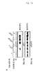
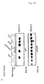
- GPCR-induced EP-R transactivation redefines endogenous EGFR-mediated signalling to PDGFR-specific signals. Proteins were immunoblotted with αPY antibody (4G10).
a) Rat-1/EP-R cells were 3 minutes treated with ET-1 (200 nM), thrombin (2U/ml) and EGF (2ng/ml) or
b) preincubated with tyrphostins as indicated prior to thrombin stimulation and EP-R was selectively precipitated with mAb 108.1.
c) Different stable Rat-1 cell lines were untreated or
d) 1 h preincubated with EGFR-E Ab ICR-3R (20µg/ml), stimulated for 3 minutes with GPCR agonists, EGF or PDGF-BB (25 ng/ml) as indicated and SHP-2 was precipitated.
e) Rat-1/EP-R were treated as in b) and SHC was immunoprecipitated. - Figure 2
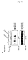
- Carbachol-induced intercellular transactivation of the EGF receptor. Stable Rat-1 cell lines either expressing M1R or HERc and control cells were mixed in 1:3 ratio. In
a) after stimulation with carbachol (1mM), HERc was precipitated and immunoblotted with αPY antibody.
b) Co-cultures of Rat-1/M1R and Rat-1/HERc cells were planted in different densities, preincubated with EGFR-E blocking Ab ICR-3R (20µg/ml) and HERc was precipitated following carbachol-stimulation.
c) High density co-cultures of Rat-1/M1 R and Rat-1/HERc cells were incubated with heparitinase or chlorate and HERc was precipitated following carbacol- or EGF-stimulation. - Figure 3
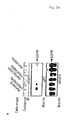
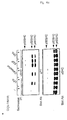
- GPCR-induced EGFR transactivation and adapter protein tyrosine phosphorylation is dependent on HB-EGF function. a), c), d) COS-7 and b)
HEK 293 cells, transfected with the M 1 R or ET-R, respectively, untreated-or CRM197 preincubated, were stimulated for 3 minutes with the GPCR agonists LPA (10 µM) or Carbachol (1 mM), EGF (2ng/ml) or 1 µM TPA (5 min) as indicated. Subsequently EGFR (a,b), SHC (c) or Gab 1(d) was immunoprecipitated and proteins were immunoblotted with αPY antibody (4G10). - Figure 4
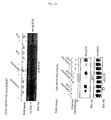
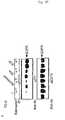
- GPCR-induced proteolytic processing of proHB-EGF and EGFR transactivation are critically dependent on metalloproteinase function.
a) COS-7 cells were co-transfected with either M1R or BombR (0.5 µg each) and VSV-proHB-EGF (0.7 µg) and stimulated with carbachol (1 mM), bombesin (200 nM), TPA (1 µM) or EGF (2 ng/ml). ProHB-EGF was analysed with αHB-EGF Ab (upper part), cleaved VSV-HB-EGF was monitored by anti VSV immunoblotting (lower part).
b) COS-7 cells transfected as in a) were preincubated with batimastat (5 µM, 30 min), stimulated as indicated and anti-VSV immunoprecipitates were subjected to αHB-EGF immunoblotting.
c) Flow cytometric analyses of proHB-EGF in COS-7 cells treated for 10 minutes with LPA, TPA, EGF or batimastat preincubation following LPA stimulation.
d,e) COS-7 cells, transfected with the M1R, untreated or BB-94 preincubated, were stimulated as in Fig. 3a) and EGFR (d) or SHC (e) were immunoprecipitated. Proteins were immunoblotted with αPY antibody (4G10).
f) PC-3 cells were serum-starved for 36 hours, preincubated with batimastat and stimulated for 3 minutes with bombesin, TPA or EGF (7ng/ml) as indicated. EGFR was immunoprecipitated and immunoblotted with αPY antibody.
g) Unstarved PC-3 cells were treated for indicated times with DMSO or batimastat and EGFR tyrosine phosphorylation was monitored with αPY immunoblot. - The following plasmids have been described: pcDNA1-BombR and pcDNA3-
M 1 R1. For stable expression of the M 1 R in Rat-1 cells the receptor was subcloned into pLXSN. pro-HB-EGF and the Endothelin receptor were amplified by PCR from a MCF-7 or Rat-1 cDNA library and subcloned into pcDNA3-VSV or pcDNA3, respectively. - Rat-1 cells and COS-7 cells were grown and infected or transfected, respectively, as described1,2. Rat-1HERc cells have been described elsewhere1.
HEK 293 cells were grown in DMEM containing 10% fetal calf serum (FCS) and transfections were carried out using the Ca-phosphate method. CRM197 (10µg/ml,Sigma) or batimastat (BB-94), (5 µM, British Biotech) were added 20 minutes before the respective growth- factor. Tyrphostin AG1478 (250 nM, Calbiochem) and AG1295 (1 µM, Calbiochem) were added 15 minutes before stimulation. - The antibodies against human EGFR (108.1), SHP-2, Shc and Gab1 have been characterized 1,12,19,2. Western blotting against the EP-R chimera was performed using rabbit polyclonal α-hPDGFRβ antibody (Upstate Biotechnology). Cells were lysed and proteins were subsequently immunoprecipitated as described 1. To precipitate the VSV-tagged HB-EGF a monoclonal VSV antibody (P5D4, Boehringer) in combination with Protein G-Sepharose was used, HB-EGF was detected with antibody C-1 8 (Santa-Cruz). Due to the small size of pro-HB-EGF and the processed form of HB-EGF we used the Tricine SDS-PAGE system established by Schlägger as described30.
- COS-7 cells were seeded in 6 cm-dishes; 20h later cells were washed and cultured for a further 24h in serum-free medium until treatment with growth factors as indicated. After collection cells were incubated with goat αHB-EGF antibody (R&D Systems) for 30 minutes on ice. After washing with PBS, cells were incubated with FITC-conjugated rabbit anti-goat antibody (Sigma) for 20 minutes on ice. Cells were analysed with FACSCalibur (Becton Dickinson).
- Epidermal growth-factor receptor (EGFR) transactivation was identified as a critical element in mitogenic signalling 1,5,6 induced by G-protein-coupled receptors (GPCR), regulation of chloride channels7, as well as modulation of potassium channel activity 8. Since the process was found to be very rapid1,7,9, and GPCR-induced release of EGFR ligands into the cell culture medium could not be detected5,8, EGFR transactivation has been generally assumed to be exclusively mediated via intracellular signals3,4.
- Surprisingly, however, even though PDGF receptors are not transactivated upon treatment of Rat-1 cells with GPCR ligands2, this was the case for a chimera EP-R consisting of an EGFR extracellular and the platelet-derived growth-factor receptor (PDGFR) transmembrane and cytoplasmic signalling domain10 (Fig. 1 a). This receptor chimera immunoprecipitates with monoclonal antibody 108.1 which recognizes the extracellular portion of human but not rat EGFR. Treatment of Rat-1/EP-R cells with the PDGFR inhibitor AG129511, but not with the EGFR kinase antagonist AG14781, blocked thrombin-induced tyrosine phosphorylation of the chimeric receptor (Fig. 1b), which clearly demonstrated a critical function of the EGFR extracellular domain for GPCR-mediated transactivation. As shown in Fig. 1c, this EP-R transactivation results in a PDGF-characteristic downstream signal, since the SH2 domain-containing phosphatase 2 (SHP-2), a preferred mediator of PDGFR signalling12, was tyrosine phosphorylated upon endothelin (ET-1) and thrombin stimulation of Rat-1/EP-R cells, while exposure to the same ligands did not induce SHP-2 tyrosine phosphoryation in Rat-1 cells overexpressing the PDGFR or control cells. Pretreatment of Rat-1/EP-R cells with monoclonal antibody ICR-3R13 that blocks ligand binding to the human EGFR resulted in complete inhibition of ET-1 and EGF-induced SHP-2 tyrosine phosphorylation, whereas the PDGF-mediated response was not affected (Fig. 1d), confirming that GPCR-induced transactivation of the EP-R chimera depends on the extracellular EGFR domain. In contrast to the results obtained for SHP-2 (Fig. 1 c), tyrosine phosphorylation of the adaptor protein SHC following thrombin stimulation was completely blocked by pretreatment of Rat-1/EP-R cells with AG1478, but remained unaffected by preincubation with the PDGFR antagonist AG1295 (Fig. 1 e). This confirms that thrombin transactivates endogenuos rat EGFR in Rat-1/EP-R cells resulting in SHC tyrosine phosphorylation, whereas activation of the EP-R chimera redefines thrombin stimulation to generate a PDGFR-characteristic SHP-2 signal.
- To address the question whether the extracellular signal which activates the EP-R chimera acts via an autocrine or paracrine mode, we performed a co-culture experiment with Rat-1 cells either stably overexpressing the
M 1 muscarinic acetylcholine receptor (M1R) or the human EGFR (HERc) at a ratio of one to one. Stimulation of the Rat-1/M1R+Rat-1/HERc co-culture with the M 1R agonist carbachol prior to immunoprecipitation with human EGFR-specific antibody 108.1, rapidly induced tyrosine phosphorylation of HERc (Fig. 2a). Since neither of the control cells responded to carbachol, this result clearly demonstrated the possibility of transactivation between two cells. To investigate the influence of cell density on this paracrine process, HERc was immunoprecipitated from subconfluent versus confluent co-cultures of Rat-1/M1R and Rat-1/HERc cells following stimulation with carbachol. As shown in Fig. 2b and 2c, EGFR tyrosine phosphorylation in response to M1R agonist only occurred in confluent co-cultures and was completely inhibited by preincubation with ICR-3R antibody, heparitinase or chlorate. This further demonstrated the requirement of the EGFR ligand binding function for intercellular signal transmission and the necessity of close cell-cell contact. Together, these results lead us to conclude that EGF-like ligands, synthesized as transmembrane precursors and converted to the mature form by proteolytic cleavage14, may be involved in GPCR-mediated transactivation. The discrepancy between previous results obtained from medium-transfer experiments5,8 in which EGF-like ligands could not be detected upon GPCR activation and our finding of density-dependent intercellular crosstalk might be due to a scenario in which upon proteolytic processing EGF-like ligands remain with the heparin sulfate proteoglycan matrix prior to interaction with their high-affinity receptors as shown for fibroblast growth-factors15. - Ectodomain shedding has been shown to be induced by stimuli such as activators of heterotrimeric G-proteins, AIF4 - and GTPγS16, as well as the PKC activator tetradecanoyl-phorbol-13-acetate (TPA) and the Ca2+-ionophore ionomycin17,18. The latter, which induces HB-EGF release in prostate epithelial cells18, has recently been shown to be a potent activator of EGFR transactivation in PC12 cells19, and TPA has been reported to induce EGFR tyrosine phosphorylation in
HEK 293 cells8. HB-EGF, a member of the EGF family, has the ability to bind to cell surface heparan sulfate proteoglycans20, which prevents the immediate releae of the growth-factor and increases the local growth factor concentration in the cellular microenvironment. Based on these properties the proHB-EGF precursor matched our proposed requirement for GPCR-induced EGFR transactivation. Besides its function as a growth-factor precursor, proHB-EGF serves as a high-affinity receptor for diphteria toxin (DT)21. CRM197, a non toxic mutant of DT, was shown to inhibit strongly and specifically the mitogenic activity of HB-EGF22. Therefore, we tested the influence of CRM197 on GPCR-mediated EGFR transactivation. We found that CRM197 pretreatment completely inhibits tyrosine phosphorylation of the EGFR induced by the GPCR agonists lysophosphatidic acid (LPA) or carbachol as well as TPA in COS-7 cells (Fig. 3a). Inhibition was also observed for ET-1 or TPA-stimulatedHEK 293 cells transiently transfected with the endothelin receptor (Fig. 3b). In contrast, EGF-induced receptor tyrosine phosphorylation was unaltered demonstrating CRM 197 specificity. Furthermore, complete abrogation of LPA- and carbachol-induced receptor tyrosine phosphorylation suggested that HB-EGF is the only growth-factor mediating EGFR transactivation in the cell lines presented here. - Tyrosine phosphorylation of the adaptor protein SHC is considered to be a critical step in the coupling of GPCR activation to Ras-dependent signalling pathways23. In order to investigate the role of HB-EGF in this process, we examined the effect of the diphteria toxin mutant CRM197 on GPCR ligand and TPA-mediated SHC tyrosine phosphorylation. As shown in Fig. 3c, in COS-7 cells, LPA-, carbachol-and TPA-induced SHC tyrosine phosphorylation was dramatically reduced by CRM197 pretreatment, while the EGF-mediated response was not affected. The same inhibitory effect of CRM197 was observed in
HEK 293 cells (data not shown). Similarly, in COS-7 cells, tyrosine phosphorylation of the multidocking protein Gab1 in response- to LPA or thrombin was not detected in the presence of CRM197 (Fig. 3d) confirming its signalling position downstream of the EGFR2. - Next, in order to examine whether proHB-EGF is proteilytically processed upon stimulation of GPCRs, we transfected plasmids containing VSV-tagged proHB-EGF in COS-7 cells together with the M1R or the bombesin receptor (BombR) and stimulated with respective ligands for different times. TPA, a potent inducer of proHB-EGF processing, or EGF were added as positive and negative controls, respectively. Figure 4a shows that as previously described proHB-EGF is expressed in form of heterogenous translation products of 20 to 30 KDa17, which can be detected with antibodies against the C-terminus of the precursor (upper panel) or the VSV-tag (lower panel). Stimulation with carbachol or bombesin led to a rapid breakdown of the membrane-anchored growth-factor precursor and proteolytic cleavage was concommitant with the appearance of the 9 KDa VSV-tagged HB-EGF fragment containing the transmembrane anchor. Interestingly, under these conditions the GPCR signal induced proteolytic proHB-EGF proccessing as fast and potently as TPA. As for TPA17, GPCR-induced conversion of proHB-EGF is an extremely rapid process that generates mature HB-EGF. In contrast to GPCR-induced tyrosine phosphorylation of endogenous EGFR which is fast and transient1,7,9, overexpression of the protease substrate VSV-proHB-EGF led to a rapid but more sustained ectodomain cleavage of proHB-EGF.
- Since zinc-dependent metalloproteinases have been implicated in pro-HB-EGF shedding by TPA24, we analysed carbachol-induced processing in the presence of batimastat (BB-94)25, a protease inhibitor which has recently been shown to block proteolytic maturation of human amphiregulin26. As shown in Fig. 4b, BB-94 treatment significantly reduced HB-EGF processing in response to carbachol supporting our conclusion that metalloproteinases are critical elements in GPCR-induced HB-EGF generation and EGFR activation. In contrast thereto, PGL-hydroxamate, an MMP-specific inhibitor has no effect on LPA- or carbachol-induced transactivation (not shown).
- To confirm GPCR-induced proHB-EGF processing, we used an ectodomain-specific antibody and flow cytometry upon treatment of non-transfected COS-7 cells with LPA, TPA or EGF. Within 10 minutes after addition of LPA and TPA, the content of cell surface proHB-EGF was reduced while EGF stimulation showed no effect (Fig. 4c). In contrast to the experiments with transfected cells shown in Fig. 4a and b, activation of endogenous LPA receptors was not as potent as TPA to induce proteolytic cleavage of proHB-EGF. Nonetheless, consistent with Fig. 4b, the modest LPA-induced effect was completely inhibited by batimastat.
- Our results demonstrate that metalloproteinase-dependent cleavage of proHB-EGF is rapidly induced upon activation of GPCRs and consequently suggest a critical and general role of this process in EGFR transactivation. We therefore investigated the effect of the metalloproteinase inhibitor batimastat in GPCR- as well as TPA-induced EGFR transactivation. In COS-7 cells, BB-94 pretreatment completely abrogated LPA- and carbachol-induced tyrosine phosphorylation of the EGFR, as well as TPA-mediated receptor activation (Fig. 4d). Since TPA- but not GPCR-mediated EGFR tyrosine phosphorylation is sensitive to PKC inhibition in COS-7 cells (data not shown), it appears that at least two distinct metalloproteinase-dependent transactivation pathways exist. Analogous results were obtained for ET-1-induced transactivation in
HEK 293 cells and bradykinin-stimulated EGFR tyrosine-phosphorylation in PC12 cells (data not shown). Finally, the general implication of proteolytic processing in EGFR transactivation and downstream signal transmission is demonstrated by the complete abrogation of GPCR- and TPA-induced SHC tyrosine phosphorylation by batimastat (Fig. 4e). - Because of the well established role of EGFR family members in the pathogenesis of a variety of cancers and the physiological abundance of GPCR ligands such as LPA, we addressed the pathophysiological significance of transactivation with the human prostate cancer cell line PC-3 which has been reported to utilize EGFR-dependent pathways for growth promotion and is also responsive to the GPCR ligand bombesin27,28. Figure 4f shows that in PC-3 cells that were starved for 36 hours, bombesin, TPA and EGF induce tyrosine phosphorylation of the EGFR which is completely blocked by batimastat-treatment. Moreover, even high constitutive phosphotyrosine content of the EGFR in unstarved PC-3 cells is reduced by long-term treatment with BB-94 (Fig. 4g). All in all, our results allow the conclusion that metalloproteinase-mediated precursor cleavage represents a direct link between BombR activation, constitutive tyrosine phosphorylation of the EGFR and proliferation of human prostate cancer cells. Recently, ADAM9, a member of the metalloproteinase-disintagrin family has been reported to process proHB-EGF upon TPA treatment of Vero-H cells24. We were unable, however, to block EGFR transactivation with dominant-negative ADAM9 mutants in COS-7 and
HEK 293 cells (data not shown) leaving the identity of the precursor processing protease unresolved. - Our findings identify the ubiquitously expressed HB-EGF precursor and a metalloproteinase activity as critical pathway elements between GPCR signals and activation of the EGFR and extend our understanding of the mechanisms that underly the multiple biological processes known to be regulated by heterotrimeric G-proteins. Based on our current state of understanding, GPCR-induced EGFR signal transactivation represents a new paradigm because it entails three different transmembrane signal transmission events: First, a ligand activates heterotrimeric G-proteins by interaction with a GPCR which results in an intracellular signal that induces the extracellular activity of a transmembrane metalloproteinase. This then results in extracellular processing of a transmembrane growth-factor precursor and release of the mature factor which, directly or via the proteoglycan matrix, interacts with the ectodomain of the EGFR leading to intracellular autophosphorylation and signal generation. Our previous findings indicate that this pathway may be utilized by a variety of GPCRs in diverse cell types and that the preferred transactivation target is the EGFR and its relatives1-4. The demonstration of the pathophysiological relevance of this novel mechanism in prostrate cancer cells leads us to propose that EGFR transactivation via G-protein-mediated proteolytic growth precursor processing represents a general mechanism with broad significance. Moreover, since a great variety of bioactive polypeptides as diverse as TNF-α, FAS-ligand or L-selectin are processing products of transmembrane precursors29 that have been connected to pathophysiological disorders, our findings shed new light on the importance of membrane-associated proteinases as targets for disease intervention strategies.
- EGFR-transactivation and the effect of the proteinase inhibitor BB-94 were tested on a plurality of human cancer cells. The summary of the results is as follows:
Cancer cell line from: Tested Transactivation BB94 effect gastric 1 1 - prostate 3 1 1 ovary 5 3 5 kidney 7 5 3 liver 2 1 1 colon 4 2 2 breast 8 3 2 pancreas 8 5 8 bladder 6 4 2 glioblastoma 5 3 3 lung (NSCLC) 7 4 4 -
- 1.
- Daub et al., EMBO J. 16, 7032-7044 (1997)
- 2.
- Daub et al., Nature 379, 557-560 (1996)
- 3.
- Luttrell et al., Curr.Opin. Cell Biol. 11, 177-183 (1999)
- 4.
- Hackel et al., Curr. Opin. Cell Biol. 11, 184-189 (1999)
- 5.
- Eguchi et al., J. Biol. Chem. 273, 8890-8896 (1998)
- 6.
- Luttrell et al., J. Biol. Chem. 272, 4637-4644 (1997)
- 7.
- Keely et al., J. Biol. Chem. 273, 27111-27117 (1998)
- 8.
- Tsai et al., EMBO J. 16, 4597-4605 (1997)
- 9.
- Li et al., EMBO J. 9, 2574-2583 (1998)
- 10.
- Seedorf et al., J. Biol. Chem. 266, 12424-12431 (1991)
- 11.
- Kovalenko et al., Cancer Res. 54, 6106-6114 (1994)
- 12.
- Vogel et al, Science 259, 1611-1614 (1993)
- 13.
- Matco et al.,
- 14.
- Massagué & Pandiella, Annu. Rev. Biochem. 62, 515-541 (1993)
- 15.
- Green et al, BioEssays 18, 639-646 (1996)
- 16.
- Bosenberg et al., J. Cell Biol. 122, 95-101 (1993)
- 17.
- Goishi et al., Mol. Biol. Cell 6, 967-980 (1995)
- 18.
- Dethlefsen et al., C.J. Cell. Biochem. 69, 143-153 (1998)
- 19.
- Zwick et al., J. Biol. Chem. 272, 24767-24770 (1997)
- 20.
- Raab & Klagsbrun, Biochim. Biophys. Acta 1333, F179-F199 (1997)
- 21.
- Naglich et al., Cell 69, 1051-1061 (1992)
- 22.
- Mitamura et al., J. Biol. Chem. 270, 1015-1019 (1995)
- 23.
- Chen et al., EMBO J. 15, 1037-1044 (1996)
- 24.
- Izumi et al., EMBO J. 17, 7260-7272 (1998)
- 25.
- Wojtowicz-Praga et al.,
- 26.
- Brown et al., J. Biol. Chem. 273, 17258-17268 (1998)
- 27.
- Ching et al., Mol. Cell Biochem. 126, 161-158 (1993)
- 28.
- Hoosein et al., J. Urol. 149, 1209-1213 (1993)
- 29.
- Werb, Cell 91, 439-442 (1997)
- 30.
- Schägger & von Jagow, Anal. Biochem. 166, 368-379 (1987)
- 31.
- Amour et al., FEBS Lett. 435, 39-44 (1998)
Claims (20)
- A method for modulating growth-factor activation comprising contacting a cell or an organism which contains a growth-factor receptor capable of being activated with a modulator of G-protein mediated signal transduction.
- The method of use of claim 1, wherein the activation of the growth-factor receptor is mediated by its extracellular domain.
- The method of claim 1 or 2, wherein the activation of the growth-factor receptor is mediated via an extracellular signal pathway.
- The method of any one of claims 1-3, wherein the growth-factor receptor is activated by tyrosine phosphorylation.
- The method of any one of the previous claims, wherein said growth-factor receptor is EGFR.
- The method of any one of the previous claims wherein the modulator acts on a G-protein, a G-protein coupled receptor and/or a proteinase.
- The method of claim 6, wherein the modulator acts on a proteinase.
- The method of claim 7, wherein said modulator acts on said proteinase by directly stimulating or inhibiting the proteinase activity.
- The method of any one of claims 7 or 8, wherein said proteinase cleaves a growth-factor ligand precursor.
- The method of claim 9, wherein said precursor is a membrane-associated molecule.
- The method of claim 9 or 10, wherein said growth factor ligand precursor is proHB-EGF and-said growth-factor receptor is EGFR.
- The method of any one of claims 7-11, wherein said proteinase is a membrane-associated proteinase.
- The method of any one of the claims 7-12, wherein said proteinase is a metalloproteinase.
- The method of claim 13, wherein said metalloproteinase is a zinc-dependent proteinase.
- The method of any one of claims 7 to 14, wherein said proteinase activity is inhibited by batimastat.
- The method of any one of the previous claims, wherein said modulator acts on cell which is different from the cell which contains the growth-factor.
- The method of any one of the previous claims for the prevention or treatment of disorders associated with or accompanied by a disturbed, e.g. pathologically enhanced growth-factor receptor activation.
- The method of claim 17 for the treatment of cancer or asthma.
- The method of any one the previous claims, wherein said modulator is administered as a pharmaceutically acceptable composition.
- A method for identifying and providing modulators of G-protein mediated signal transduction comprising contacting a cell which contains a growth-factor receptor capable of being activated with a test compound suspected to be a modulator of G-protein mediated signal transduction and determining the degree of growth-factor receptor activation.
Priority Applications (1)
| Application Number | Priority Date | Filing Date | Title |
|---|---|---|---|
| EP06020038A EP1820500A1 (en) | 1999-08-16 | 2000-08-16 | Use of proteinase inhibitor in order to inhibit the cleavage of growth factor precursor |
Applications Claiming Priority (4)
| Application Number | Priority Date | Filing Date | Title |
|---|---|---|---|
| EP99116056 | 1999-08-16 | ||
| US09/461,090 US7632645B1 (en) | 1999-08-16 | 1999-12-14 | EGF receptor transactivation by G-protein-coupled receptors requires metalloproteinase cleavage of proHB-EGF |
| EP00951521A EP1207876B9 (en) | 1999-08-16 | 2000-08-16 | Use of a modulator which acts on a growth factor precursor in order to inhibit growth factor precursor processing |
| EP06020038A EP1820500A1 (en) | 1999-08-16 | 2000-08-16 | Use of proteinase inhibitor in order to inhibit the cleavage of growth factor precursor |
Related Parent Applications (1)
| Application Number | Title | Priority Date | Filing Date |
|---|---|---|---|
| EP00951521A Division EP1207876B9 (en) | 1999-08-16 | 2000-08-16 | Use of a modulator which acts on a growth factor precursor in order to inhibit growth factor precursor processing |
Publications (1)
| Publication Number | Publication Date |
|---|---|
| EP1820500A1 true EP1820500A1 (en) | 2007-08-22 |
Family
ID=26153086
Family Applications (2)
| Application Number | Title | Priority Date | Filing Date |
|---|---|---|---|
| EP00951521A Expired - Lifetime EP1207876B9 (en) | 1999-08-16 | 2000-08-16 | Use of a modulator which acts on a growth factor precursor in order to inhibit growth factor precursor processing |
| EP06020038A Withdrawn EP1820500A1 (en) | 1999-08-16 | 2000-08-16 | Use of proteinase inhibitor in order to inhibit the cleavage of growth factor precursor |
Family Applications Before (1)
| Application Number | Title | Priority Date | Filing Date |
|---|---|---|---|
| EP00951521A Expired - Lifetime EP1207876B9 (en) | 1999-08-16 | 2000-08-16 | Use of a modulator which acts on a growth factor precursor in order to inhibit growth factor precursor processing |
Country Status (6)
| Country | Link |
|---|---|
| EP (2) | EP1207876B9 (en) |
| JP (1) | JP2003506489A (en) |
| AU (1) | AU779298B2 (en) |
| CA (1) | CA2380753C (en) |
| DE (1) | DE1207876T1 (en) |
| WO (1) | WO2001012182A1 (en) |
Families Citing this family (3)
| Publication number | Priority date | Publication date | Assignee | Title |
|---|---|---|---|---|
| AU2003219029B2 (en) * | 2002-03-08 | 2008-09-11 | Max-Planck-Gesellschaft Zur Forderung Der Wissenschaften E.V | Use of EGFR transactivation inhibitors in human cancer |
| EP1493445A1 (en) * | 2003-07-04 | 2005-01-05 | Max-Planck-Gesellschaft zur Förderung der Wissenschaften e.V. | Inhibition of stress-induced ligand-dependent EGFR activation |
| EP2561759A1 (en) | 2011-08-26 | 2013-02-27 | Bayer Cropscience AG | Fluoroalkyl-substituted 2-amidobenzimidazoles and their effect on plant growth |
Citations (1)
| Publication number | Priority date | Publication date | Assignee | Title |
|---|---|---|---|---|
| EP0780386A1 (en) * | 1995-12-20 | 1997-06-25 | F. Hoffmann-La Roche Ag | Matrix metalloprotease inhibitors |
-
2000
- 2000-08-16 EP EP00951521A patent/EP1207876B9/en not_active Expired - Lifetime
- 2000-08-16 CA CA2380753A patent/CA2380753C/en not_active Expired - Fee Related
- 2000-08-16 JP JP2001516528A patent/JP2003506489A/en active Pending
- 2000-08-16 WO PCT/EP2000/008007 patent/WO2001012182A1/en not_active Ceased
- 2000-08-16 DE DE1207876T patent/DE1207876T1/en active Pending
- 2000-08-16 EP EP06020038A patent/EP1820500A1/en not_active Withdrawn
- 2000-08-16 AU AU64426/00A patent/AU779298B2/en not_active Ceased
Patent Citations (1)
| Publication number | Priority date | Publication date | Assignee | Title |
|---|---|---|---|---|
| EP0780386A1 (en) * | 1995-12-20 | 1997-06-25 | F. Hoffmann-La Roche Ag | Matrix metalloprotease inhibitors |
Non-Patent Citations (42)
| Title |
|---|
| AMOUR, FEBS LETT., vol. 435, 1998, pages 39 - 44 |
| BOSENBERG, J. CELL BIOL., vol. 122, 1993, pages 95 - 101 |
| BROWN ET AL: "cell surface ectodomain cleavage of human amphiregulin precursor is sensitive to a metalloprotease inhibitor", JOURNAL OF BIOLOGICAL CHEMISTRY, vol. 273, no. 27, 1998, pages 17258 - 17268, XP002136688 * |
| BROWN, J. BIOL. CHEM., vol. 273, 1998, pages 17258 - 17268 |
| CHEN, EMBO J., vol. 15, 1996, pages 1037 - 1044 |
| CHING, MOL. CELL BIOCHEM., vol. 126, 1993, pages 161 - 158 |
| DAUB ET AL: "role of transactivation of the EGF receptor in signalling by G-protein-coupled receptors", NATURE, vol. 379, 1996, pages 557 - 560, XP002136689 * |
| DAUB ET AL: "signal characteristics of g protein transactivated EGF receptor", EMBO JOURNAL, vol. 16, no. 23, 1997, pages 7032 - 7044, XP002136690 * |
| DAUB, EMBO J., vol. 16, 1997, pages 7032 - 7044 |
| DAUB, NATURE, vol. 379, 1996, pages 557 - 560 |
| DETHLEFSEN, C.J. CELL . BIOCHEM., vol. 69, 1998, pages 143 - 153 |
| DONG JIANYING ET AL: "Metalloprotease-mediated ligand release regulates autocrine signaling through the epidermal growth factor receptor.", PROCEEDINGS OF THE NATIONAL ACADEMY OF SCIENCES OF THE UNITED STATES OF AMERICA MAY 25, 1999, vol. 96, no. 11, 25 May 1999 (1999-05-25), pages 6235 - 6240, XP002136687, ISSN: 0027-8424 * |
| EGUCHI, J. BIOL. CHEM., vol. 273, 1998, pages 8890 - 8896 |
| GOISHI, MOL. BIOL. CELL, vol. 6, 1995, pages 967 - 980 |
| GREEN, BIOESSAYS, vol. 18, 1996, pages 639 - 646 |
| HACKEL, CURR. OPIN. CELL BIOL., vol. 11, 1999, pages 184 - 189 |
| HOOSEIN, J. UROL., vol. 149, 1993, pages 1209 - 1213 |
| IZUMI ET AL: "a metalloprotease disintegrin, mdc9/meltrin-gamma/adam9 and pkcdelta are involved in tpa induced ectodomain shedding of membrane anchred heparin binding egf like growth factor", EMBO JOURNAL, vol. 17, no. 24, 1998, pages 7260 - 7272, XP002136691 * |
| IZUMI, EMBO J., vol. 17, 1998, pages 7260 - 7272 |
| KEELY, J. BIOL. CHEM., vol. 273, 1998, pages 27111 - 27117 |
| KOVALENKO, CANCER RES., vol. 54, 1994, pages 6106 - 6114 |
| LI, EMBO J., vol. 9, 1998, pages 2574 - 2583 |
| LUTTRELL, CURR.OPIN. CELL BIOL., vol. 11, 1999, pages 177 - 183 |
| LUTTRELL, J. BIOL. CHEM., vol. 272, 1997, pages 4637 - 4644 |
| MASSAGUÉ; PANDIELLA, ANNU. REV. BIOCHEM., vol. 62, 1993, pages 515 - 541 |
| MATCO, IMMUNOTECHNOLOGY, vol. 3, 1997, pages 71 - 81 |
| MITAMURA, J. BIOL. CHEM., vol. 270, 1995, pages 1015 - 1019 |
| NAGLICH, CELL, vol. 69, 1992, pages 1051 - 1061 |
| PARSONS S L ET AL: "Phase I/II trial of batimastat, a matrix metalloproteinase inhibitor, in patients with malignant ascites.", EUROPEAN JOURNAL OF SURGICAL ONCOLOGY : THE JOURNAL OF THE EUROPEAN SOCIETY OF SURGICAL ONCOLOGY AND THE BRITISH ASSOCIATION OF SURGICAL ONCOLOGY DEC 1997 LNKD- PUBMED:9484924, vol. 23, no. 6, December 1997 (1997-12-01), pages 526 - 531, ISSN: 0748-7983 * |
| PRENZEL ET AL: "egf receptor transactivation by G-protein-coupled receptors requires metallproteinase cleavage of proHB-egf", NATURE, vol. 402, 23 December 1999 (1999-12-23) - 30 December 1999 (1999-12-30), pages 884 - 888, XP002136695 * |
| RAAB; KLAGSBRUN, BIOCHIM. BIOPHYS. ACTA, vol. 1333, 1997, pages F179 - F199 |
| ROMANO ET AL: "helicobacter pylori upregulates expression of epidermal growth factor related peptides, but inhibits their proliferative effect in mkn 28 gastric mucosal cells", JOURNAL OF CLINICAL INVESTIGATION, vol. 101, no. 8, 1998, pages 1604 - 1613, XP002136694 * |
| SCHÄGGER; VON JAGOW, ANAL. BIOCHEM., vol. 166, 1987, pages 368 - 379 |
| SEEDORF, J. BIOL. CHEM., vol. 266, 1991, pages 12424 - 12431 |
| TAKEYAMA KIYOSHI ET AL: "Epidermal growth factor system regulates mucin production in airways.", PROCEEDINGS OF THE NATIONAL ACADEMY OF SCIENCES OF THE UNITED STATES OF AMERICA MARCH 16, 1999, vol. 96, no. 6, 16 March 1999 (1999-03-16), pages 3081 - 3086, XP002136693, ISSN: 0027-8424 * |
| TSAI, EMBO J., vol. 16, 1997, pages 4597 - 4605 |
| VOGEL, SCIENCE, vol. 259, 1993, pages 1611 - 1614 |
| WANG X ET AL: "Matrix metalloproteinase inhibitor BB-94 (Batimastat) inhibits human colon tumor growth and spread in a patient-like orthotopic model in nude mice", CANCER RESEARCH, AMERICAN ASSOCIATION FOR CANCER RESEARCH, US, vol. 54, no. 17, 1 January 1994 (1994-01-01), pages 4726 - 4728, XP002142892, ISSN: 0008-5472, DOI: 10.1016/S0065-230X(08)60834-9 * |
| WERB Z: "ecm and cell surface proteolysis : regulating cellular ecology", CELL, vol. 91, 1997, pages 439 - 442, XP002136692 * |
| WERB, CELL, vol. 91, 1997, pages 439 - 442 |
| WOJTOWICZ-PRAGA, INVESTIGATIONAL NEW DRUGS, vol. 15, 1997, pages 61 - 75 |
| ZWICK, J. BIOL. CHEM., vol. 272, 1997, pages 24767 - 24770 |
Also Published As
| Publication number | Publication date |
|---|---|
| JP2003506489A (en) | 2003-02-18 |
| CA2380753A1 (en) | 2001-02-22 |
| CA2380753C (en) | 2013-10-08 |
| AU779298B2 (en) | 2005-01-13 |
| AU6442600A (en) | 2001-03-13 |
| EP1207876B1 (en) | 2011-06-22 |
| EP1207876A1 (en) | 2002-05-29 |
| WO2001012182A1 (en) | 2001-02-22 |
| DE1207876T1 (en) | 2003-01-09 |
| EP1207876B9 (en) | 2012-04-18 |
Similar Documents
| Publication | Publication Date | Title |
|---|---|---|
| Webb et al. | Plasminogen activator inhibitor 1 functions as a urokinase response modifier at the level of cell signaling and thereby promotes MCF-7 cell growth | |
| Fitzgerald et al. | Shedding of syndecan-1 and-4 ectodomains is regulated by multiple signaling pathways and mediated by a TIMP-3–sensitive metalloproteinase | |
| Ling et al. | Endothelin‐2, the forgotten isoform: emerging role in the cardiovascular system, ovarian development, immunology and cancer | |
| Sims et al. | Interleukin‐11 receptor signaling is required for normal bone remodeling | |
| Duong | Therapeutic inhibition of cathepsin K—reducing bone resorption while maintaining bone formation | |
| Zaidi et al. | Osteoclastogenesis, bone resorption, and osteoclast‐based therapeutics | |
| Casimiro et al. | The critical role of the bone microenvironment in cancer metastases | |
| US6268163B1 (en) | Identification of drug candidates modulating factor VII-mediated intracellular signaling | |
| NO329133B1 (en) | Use of an FPT Inhibitor in the Preparation of a Pharmaceutical Preparation for the Treatment of Cancer in a Patient and for Regression of Tumor Volume in a Cancer Patient | |
| US5744442A (en) | Regulation of cellular invasiveness | |
| Hamma-Kourbali et al. | A novel non-containing-nitrogen bisphosphonate inhibits both in vitro and in vivo angiogenesis | |
| Yang et al. | Over‐expression of parathyroid hormone Type 1 receptor confers an aggressive phenotype in osteosarcoma | |
| Smith et al. | Recombinant PAPP-A resistant insulin-like growth factor binding protein 4 (dBP4) inhibits angiogenesis and metastasis in a murine model of breast cancer | |
| Gratio et al. | Aberrant expression of proteinase‐activated receptor 4 promotes colon cancer cell proliferation through a persistent signaling that involves Src and ErbB‐2 kinase | |
| US8501702B2 (en) | Overexpression of Wnt ligands and treatment of lytic bone diseases | |
| EP1207876B9 (en) | Use of a modulator which acts on a growth factor precursor in order to inhibit growth factor precursor processing | |
| US20100048684A1 (en) | EGF RECEPTOR TRANSACTIVATION BY G-PROTEIN-COUPLED RECEPTORS REQUIRES METALLOPROTEINASE CLEAVAGE OF proHB-EGF | |
| Mather | Follistatins and α2-macroglobulin are soluble binding proteins for inhibin and activin | |
| JP4167061B2 (en) | Use of long pentraxin PTX3 for the treatment of diseases caused by altered activation of growth factor FGF-2 | |
| Parish et al. | Mechanisms of tumour cell metastasis | |
| Al-Salihi et al. | Cyclooxygenase-2 transactivates the epidermal growth factor receptor through specific E-prostanoid receptors and Tumor Necrosis Factor-α converting enzyme | |
| Annabi et al. | Contribution of the 37-kDa laminin receptor precursor in the anti-metastatic PSP94-derived peptide PCK3145 cell surface binding | |
| Xu et al. | The prevention of latanoprost on osteoclastgenesis in vitro and lipopolysaccharide‐induced murine calvaria osteolysis in vivo | |
| JP4522047B2 (en) | Regulation of angiogenesis | |
| US20070232542A1 (en) | Methods for Modifying Cell Motility Using Factor VIIa Antagonist |
Legal Events
| Date | Code | Title | Description |
|---|---|---|---|
| PUAI | Public reference made under article 153(3) epc to a published international application that has entered the european phase |
Free format text: ORIGINAL CODE: 0009012 |
|
| 17P | Request for examination filed |
Effective date: 20060925 |
|
| AC | Divisional application: reference to earlier application |
Ref document number: 1207876 Country of ref document: EP Kind code of ref document: P |
|
| AK | Designated contracting states |
Kind code of ref document: A1 Designated state(s): AT BE CH CY DE DK ES FI FR GB GR IE IT LI LU MC NL PT SE |
|
| 17Q | First examination report despatched |
Effective date: 20080222 |
|
| AKX | Designation fees paid |
Designated state(s): AT BE CH CY DE DK ES FI FR GB GR IE IT LI LU MC NL PT SE |
|
| RAP3 | Party data changed (applicant data changed or rights of an application transferred) |
Owner name: MAX-PLANCK-GESELLSCHAFT ZUR FOERDERUNG DER WISSENS |
|
| STAA | Information on the status of an ep patent application or granted ep patent |
Free format text: STATUS: THE APPLICATION IS DEEMED TO BE WITHDRAWN |
|
| 18D | Application deemed to be withdrawn |
Effective date: 20120317 |