EP1820023B1 - Stem cells, method for their purification, identification, and use - Google Patents
Stem cells, method for their purification, identification, and use Download PDFInfo
- Publication number
- EP1820023B1 EP1820023B1 EP05815640A EP05815640A EP1820023B1 EP 1820023 B1 EP1820023 B1 EP 1820023B1 EP 05815640 A EP05815640 A EP 05815640A EP 05815640 A EP05815640 A EP 05815640A EP 1820023 B1 EP1820023 B1 EP 1820023B1
- Authority
- EP
- European Patent Office
- Prior art keywords
- cells
- nanog
- cell
- stem
- levels
- Prior art date
- Legal status (The legal status is an assumption and is not a legal conclusion. Google has not performed a legal analysis and makes no representation as to the accuracy of the status listed.)
- Expired - Lifetime
Links
Images




Classifications
-
- A—HUMAN NECESSITIES
- A61—MEDICAL OR VETERINARY SCIENCE; HYGIENE
- A61K—PREPARATIONS FOR MEDICAL, DENTAL OR TOILETRY PURPOSES
- A61K35/00—Medicinal preparations containing materials or reaction products thereof with undetermined constitution
- A61K35/12—Materials from mammals; Compositions comprising non-specified tissues or cells; Compositions comprising non-embryonic stem cells; Genetically modified cells
- A61K35/28—Bone marrow; Haematopoietic stem cells; Mesenchymal stem cells of any origin, e.g. adipose-derived stem cells
-
- A—HUMAN NECESSITIES
- A61—MEDICAL OR VETERINARY SCIENCE; HYGIENE
- A61P—SPECIFIC THERAPEUTIC ACTIVITY OF CHEMICAL COMPOUNDS OR MEDICINAL PREPARATIONS
- A61P1/00—Drugs for disorders of the alimentary tract or the digestive system
- A61P1/16—Drugs for disorders of the alimentary tract or the digestive system for liver or gallbladder disorders, e.g. hepatoprotective agents, cholagogues, litholytics
-
- A—HUMAN NECESSITIES
- A61—MEDICAL OR VETERINARY SCIENCE; HYGIENE
- A61P—SPECIFIC THERAPEUTIC ACTIVITY OF CHEMICAL COMPOUNDS OR MEDICINAL PREPARATIONS
- A61P1/00—Drugs for disorders of the alimentary tract or the digestive system
- A61P1/18—Drugs for disorders of the alimentary tract or the digestive system for pancreatic disorders, e.g. pancreatic enzymes
-
- A—HUMAN NECESSITIES
- A61—MEDICAL OR VETERINARY SCIENCE; HYGIENE
- A61P—SPECIFIC THERAPEUTIC ACTIVITY OF CHEMICAL COMPOUNDS OR MEDICINAL PREPARATIONS
- A61P11/00—Drugs for disorders of the respiratory system
-
- A—HUMAN NECESSITIES
- A61—MEDICAL OR VETERINARY SCIENCE; HYGIENE
- A61P—SPECIFIC THERAPEUTIC ACTIVITY OF CHEMICAL COMPOUNDS OR MEDICINAL PREPARATIONS
- A61P11/00—Drugs for disorders of the respiratory system
- A61P11/02—Nasal agents, e.g. decongestants
-
- A—HUMAN NECESSITIES
- A61—MEDICAL OR VETERINARY SCIENCE; HYGIENE
- A61P—SPECIFIC THERAPEUTIC ACTIVITY OF CHEMICAL COMPOUNDS OR MEDICINAL PREPARATIONS
- A61P13/00—Drugs for disorders of the urinary system
-
- A—HUMAN NECESSITIES
- A61—MEDICAL OR VETERINARY SCIENCE; HYGIENE
- A61P—SPECIFIC THERAPEUTIC ACTIVITY OF CHEMICAL COMPOUNDS OR MEDICINAL PREPARATIONS
- A61P13/00—Drugs for disorders of the urinary system
- A61P13/12—Drugs for disorders of the urinary system of the kidneys
-
- A—HUMAN NECESSITIES
- A61—MEDICAL OR VETERINARY SCIENCE; HYGIENE
- A61P—SPECIFIC THERAPEUTIC ACTIVITY OF CHEMICAL COMPOUNDS OR MEDICINAL PREPARATIONS
- A61P25/00—Drugs for disorders of the nervous system
-
- A—HUMAN NECESSITIES
- A61—MEDICAL OR VETERINARY SCIENCE; HYGIENE
- A61P—SPECIFIC THERAPEUTIC ACTIVITY OF CHEMICAL COMPOUNDS OR MEDICINAL PREPARATIONS
- A61P27/00—Drugs for disorders of the senses
- A61P27/02—Ophthalmic agents
-
- A—HUMAN NECESSITIES
- A61—MEDICAL OR VETERINARY SCIENCE; HYGIENE
- A61P—SPECIFIC THERAPEUTIC ACTIVITY OF CHEMICAL COMPOUNDS OR MEDICINAL PREPARATIONS
- A61P27/00—Drugs for disorders of the senses
- A61P27/16—Otologicals
-
- A—HUMAN NECESSITIES
- A61—MEDICAL OR VETERINARY SCIENCE; HYGIENE
- A61P—SPECIFIC THERAPEUTIC ACTIVITY OF CHEMICAL COMPOUNDS OR MEDICINAL PREPARATIONS
- A61P37/00—Drugs for immunological or allergic disorders
-
- A—HUMAN NECESSITIES
- A61—MEDICAL OR VETERINARY SCIENCE; HYGIENE
- A61P—SPECIFIC THERAPEUTIC ACTIVITY OF CHEMICAL COMPOUNDS OR MEDICINAL PREPARATIONS
- A61P43/00—Drugs for specific purposes, not provided for in groups A61P1/00-A61P41/00
-
- A—HUMAN NECESSITIES
- A61—MEDICAL OR VETERINARY SCIENCE; HYGIENE
- A61P—SPECIFIC THERAPEUTIC ACTIVITY OF CHEMICAL COMPOUNDS OR MEDICINAL PREPARATIONS
- A61P9/00—Drugs for disorders of the cardiovascular system
-
- A—HUMAN NECESSITIES
- A61—MEDICAL OR VETERINARY SCIENCE; HYGIENE
- A61P—SPECIFIC THERAPEUTIC ACTIVITY OF CHEMICAL COMPOUNDS OR MEDICINAL PREPARATIONS
- A61P9/00—Drugs for disorders of the cardiovascular system
- A61P9/10—Drugs for disorders of the cardiovascular system for treating ischaemic or atherosclerotic diseases, e.g. antianginal drugs, coronary vasodilators, drugs for myocardial infarction, retinopathy, cerebrovascula insufficiency, renal arteriosclerosis
-
- G—PHYSICS
- G01—MEASURING; TESTING
- G01N—INVESTIGATING OR ANALYSING MATERIALS BY DETERMINING THEIR CHEMICAL OR PHYSICAL PROPERTIES
- G01N33/00—Investigating or analysing materials by specific methods not covered by groups G01N1/00 - G01N31/00
- G01N33/48—Biological material, e.g. blood, urine; Haemocytometers
- G01N33/50—Chemical analysis of biological material, e.g. blood, urine; Testing involving biospecific ligand binding methods; Immunological testing
- G01N33/53—Immunoassay; Biospecific binding assay; Materials therefor
- G01N33/569—Immunoassay; Biospecific binding assay; Materials therefor for microorganisms, e.g. protozoa, bacteria, viruses
- G01N33/56966—Animal cells
-
- G—PHYSICS
- G01—MEASURING; TESTING
- G01N—INVESTIGATING OR ANALYSING MATERIALS BY DETERMINING THEIR CHEMICAL OR PHYSICAL PROPERTIES
- G01N2333/00—Assays involving biological materials from specific organisms or of a specific nature
- G01N2333/435—Assays involving biological materials from specific organisms or of a specific nature from animals; from humans
- G01N2333/46—Assays involving biological materials from specific organisms or of a specific nature from animals; from humans from vertebrates
- G01N2333/47—Assays involving proteins of known structure or function as defined in the subgroups
- G01N2333/4701—Details
- G01N2333/4703—Regulators; Modulating activity
- G01N2333/4706—Regulators; Modulating activity stimulating, promoting or activating activity
-
- G—PHYSICS
- G01—MEASURING; TESTING
- G01N—INVESTIGATING OR ANALYSING MATERIALS BY DETERMINING THEIR CHEMICAL OR PHYSICAL PROPERTIES
- G01N2333/00—Assays involving biological materials from specific organisms or of a specific nature
- G01N2333/435—Assays involving biological materials from specific organisms or of a specific nature from animals; from humans
- G01N2333/705—Assays involving receptors, cell surface antigens or cell surface determinants
- G01N2333/70596—Molecules with a "CD"-designation not provided for elsewhere in G01N2333/705
Definitions
- the invention relates to new stem cell populations, to their purification and use for treatment of vascular pathologies and other types of human pathologies.
- EPCs endothelial progenitor cells
- EPCs can be obtained from CD133+ cells, from CD34+CD14- cells and from CD14+CD34- cells, even though it is still uncertain whether the latter are truly endowed with stem capacity. Furthermore, the real number of endothelial progenitors in peripheral blood and bone marrow is still a matter of discussion. CD14/CD34 double positive cells are known in prior art (Ferrero et al. 1998).
- NANOG a gene has been recently described, termed NANOG, which plays a key role in renewal and maintenance of embryonic stem cell pluripotency. NANOG expression has not yet been studied in adult stems cells.
- the present invention allows the use of a new population of stem cells that can be applied to tissue reconstruction therapy, and enables identification and recovery of said cells. Moreover, the disclosure allows realisation of a diagnostic kit to evaluate the percentage (and the absolute number) of circulating cells endowed with stem capacity and a kit for purification of said cells for therapeutic purposes.
- NANOG is a useful marker for identification of adult stem cells, since it is expressed by CD133+ cells obtained from the umbilical cord or peripheral blood or from CD34+ cells. These are notoriously adult cells endowed with stem capacity.
- NANOG is not expressed in primary cultures of endothelial, epithelial, smooth muscle cells, in lymphocytes or other terminally differentiated cell types ( Fig. 1 ).
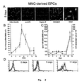
- NANOG is expressed at high levels in mononucleated circulating cells (MNC) cultured in order to obtain EPCs, while the expression of this gene disappears when cells acquire phenotypic characteristics of terminally differentiated cells and mature endothelial cell markers, such as Von Willebrand factor, KDR and Tie-2 ( Fig. 2 ).
- EPCs express high levels of NANOG, CD14+, CD34+, CD105+, as shown by Real-time quantitative RT-PCR analysis ( Fig. 3 ).
- said cells are also CD14+, CD105+, CD11c+, CD31+, CD86+, HLA-DR+, whereas the presence of CD34+ on the surface of the cells of the present invention was not detectable by normal cytofluorimetric techniques (Table 1).
- the cell population of the present invention consists of cells showing the simultaneous presence of CD14 and CD34 markers on their surface, which made possible the isolation of this population from the remaining mononucleated cells in peripheral blood.
- the isolation of cells can be similarly performed from umbilical cord blood, bone marrow, placenta or adult tissues.
- CD14+CD34+ EPCs originated from selection of a pre-existing population in peripheral blood
- CD14+ cells were purified from the population of mononucleated cells of peripheral blood by means of a positive separation method involving a magnetic column. Once the number of cells was determined, these were incubated in buffer containing anti-CD14 antibodies coupled to superparamagnetic microspheres and applied to a column in a magnetic field. The CD14+ cell fraction was recovered by elution upon removal of the column from the magnetic field.
- the purified CD14+ cell fraction (>95% homogeneous) was then sequentially incubated with: fluorescein coupled anti-CD34 antibodies, digoxigenin coupled anti-fluorescein antibodies, biotin coupled anti-digoxigenin antibodies and, finally, with fluorescent liposomes (Ab-CMFL) to which anti-biotin antibodies were previously coupled ( Fig. 5 (A) ).
- CD14+CD34+ cell population was isolated (purity > 98%).
- the percentage of CD14+ cells that also express CD34+ was measured in 10 healthy subjects with age ranging between 24 and 40 years.
- the percentages of CD14+CD34+ cells were comprised from 1.2 to 7.5% of all leukocytes.
- CD14+CD34+ cells were purified from the mononucleated population and subdivided into CD34+ and CD34- cells, using the Ab-CMFL technique. The residual population of CD14-CD34- cells was also evaluated as further control. The three populations isolated by these means were then tested for NANOG expression by quantitative RT-PCR. NANOG expression turned out to be significantly higher in CD14+CD34+ cells and significantly lower in CD14+CD34- cells, as compared to non-separated CD14+ cells ( Fig. 5 (B) ).
- CD14+CD34+, CD14+CD34- and CD14-CD34- sub-populations were cultured in presence of VEGF under the above conditions and examined on day 12 for expression of typical endothelial cell markers, such as KDR and vWf.
- KDR and vWf mRNAs were absent or extremely low on day 0.
- expression of these markers progressively increased in the CD14+CD34+ subgroup, reaching a maximum on day 12, while remained virtually unchanged for all the culture period in the CD14+CD34- population and in the CD14-CD34- population.
- EPC cells expressing NANOG were CD14+CD34+ positive. These cells originate from selection in culture of circulating CD14+CD34+ cells expressing NANOG, which pre-exist in peripheral blood.
- CD14+CD34+ population which is characterised by the expression of NANOG, is the main source of EPCs.
- NANOG vascular damage
- testing for NANOG expression may represent a useful way to identify adult human cells with stem capacity.
- NANOG expression a feature of pluripotent stem cells, suggests that this stem cell population may be therapeutically useful also in other types of pathologies characterised by organ or tissue damage.
- the invention also relates to pharmaceutical compositions that can be useful in cell therapy for treatment of ischemia, of neurodegenerative or cerebrovascular diseases, and for therapy of organ damage.
- organ damage hereinstein, kidney, pulmonary, liver or pancreatic failure and reconstruction of urogenital, visual, hearing, olfactory, cutaneous and/or mucous, bony and/or cartilaginous organs and tissues
- haematological diseases and immunodeficiencies can be useful in cell therapy for treatment of ischemia, of neurodegenerative or cerebrovascular diseases, and for therapy of organ damage.
- the ability to determine the number of cells present in the blood represents an interesting diagnostic method to evaluate appropriateness of adopting cell therapy for treatment of the above-mentioned pathologies, based on presence or absence of a sufficient number of cells in the patient.
- CD14+ or HMVEC cells (8x10 6 cells/ml, cell density 2.5x10 6 cells/cm 2 ) were plated on human fibronectin coated culture dishes and maintained in basal endothelial medium enriched with EGM Single Quotes, VEGF (100nm/ml) and 20% FCS.
- RNA from said cells was extracted and treated with Dnase I in order to remove any possible genomic DNA contaminant.
- Taq-Man RT-PCR was performed as described by Lasagni L et al. «An alternative spliced variant of CXCR3 mediates IP-10, Mig e I-TAC induced-inhibition of endothelial cell growth and acts as functional receptor for PF-4» J. Exp. Med, 2003; 197: 1537-1549 .
- Tie-2, KDR determination and CD1 a quantification were then performed by a Taq-Man assay using a commercial kit (Applied Biosystems, Warrington, UK).
- Standard curves were generated using a series of dilutions of the mRNA prepared from HMVEC.
- oligonucleotide pairs capable of detecting the human ortholog of the murine Nanog gene, mapped at 12p 13.31.
- the oligonucleotide pair selected on the basis of specificity was used for a quantitative Taq-man RT-PCR assay, together with a VIC-labelled fluorescent probe. Specificity and optimal amplification efficiency of said oligonucleotides were tested on a plasmid carrying the NANOG cDNA sequence.
- mRNA levels were quantified by comparison of the experimental data with standard curves generated from a dilution series using identical amounts of plasmid DNA.
- This method makes possible to increase the intensity of the fluorescence signal 100 - 1000 fold compared to conventional methods.
- Adherent cells were incubated on poly-L-lysine coated substrates for 30' at 37°C in presence of acLDL double labeled probes for one hour; after fixation, samples were incubated in presence of FITC-labeled Ulex europeus agglutinin I. Samples were mounted in anti-fading media and examined by conventional confocal microscopy.
Landscapes
- Health & Medical Sciences (AREA)
- Life Sciences & Earth Sciences (AREA)
- Engineering & Computer Science (AREA)
- Chemical & Material Sciences (AREA)
- General Health & Medical Sciences (AREA)
- Medicinal Chemistry (AREA)
- Pharmacology & Pharmacy (AREA)
- Veterinary Medicine (AREA)
- Public Health (AREA)
- Animal Behavior & Ethology (AREA)
- Bioinformatics & Cheminformatics (AREA)
- Chemical Kinetics & Catalysis (AREA)
- Nuclear Medicine, Radiotherapy & Molecular Imaging (AREA)
- General Chemical & Material Sciences (AREA)
- Organic Chemistry (AREA)
- Immunology (AREA)
- Cell Biology (AREA)
- Biomedical Technology (AREA)
- Urology & Nephrology (AREA)
- Hematology (AREA)
- Molecular Biology (AREA)
- Developmental Biology & Embryology (AREA)
- Biotechnology (AREA)
- Virology (AREA)
- Zoology (AREA)
- Physics & Mathematics (AREA)
- Pulmonology (AREA)
- Tropical Medicine & Parasitology (AREA)
- Epidemiology (AREA)
- Cardiology (AREA)
- Microbiology (AREA)
- Pathology (AREA)
- Food Science & Technology (AREA)
- Heart & Thoracic Surgery (AREA)
- Analytical Chemistry (AREA)
- Biochemistry (AREA)
- General Physics & Mathematics (AREA)
- Vascular Medicine (AREA)
- Neurology (AREA)
- Neurosurgery (AREA)
Abstract
Description
- The invention relates to new stem cell populations, to their purification and use for treatment of vascular pathologies and other types of human pathologies.
- It is by now well known that adult peripheral blood contains bone marrow-derived circulating cells capable of differentiating in mature endothelial cells (ECs), termed endothelial progenitor cells (EPCs); these cells are able to localise at the sites of ischemia contributing to formation of new blood vessels. Due to this property, these cells have been successfully used for tissue repair both in in vivo animal models and in patients affected by acute myocardial infarction and chronic ischemic cardiopathy.
- On the other hand, it is also well known that post-ischemic infusion of terminally differentiated mature endothelial cells does not result in increased vascularization, hence it is clear that these cells lose their potency upon differentiation.
- It is well known that EPCs can be obtained from CD133+ cells, from CD34+CD14- cells and from CD14+CD34- cells, even though it is still uncertain whether the latter are truly endowed with stem capacity. Furthermore, the real number of endothelial progenitors in peripheral blood and bone marrow is still a matter of discussion. CD14/CD34 double positive cells are known in prior art (Ferrero et al. 1998).
- There is a clear interest for in depth studies in this field, in view of the potential importance of these cells for reconstruction of tissues damaged by ischemia and pathological or traumatic events.
- It is also well known that a gene has been recently described, termed NANOG, which plays a key role in renewal and maintenance of embryonic stem cell pluripotency. NANOG expression has not yet been studied in adult stems cells.
-
-
Fig. 1 shows the differential expression of NANOG in adult cells endowed with stem capacity and in cells from other tissues. -
Fig. 2 shows the expression of NANOG in cultured EPCs. -
Fig. 3 shows the expression of various markers by cultured EPCs, at mRNA level. -
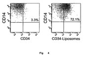
Fig. 4 demonstrates that cultured EPCs are CD14+CD34+ -
Fig. 5 (A) shows the separation of CD14+CD34- cells and of CD14+CD34+ cells from circulating CD14+ cells. -
Fig. 5 (B) shows the expression of NANOG in the three distinct CD14+CD34+, CD14+CD34-, CD14+ populations. -
Fig. 6 shows the different ability of CD14+CD34+, CD14+CD34- and CD14-CD34- cells to differentiate in endothelial cells, and to express endothelial-specific markers. - The present invention allows the use of a new population of stem cells that can be applied to tissue reconstruction therapy, and enables identification and recovery of said cells. Moreover, the disclosure allows realisation of a diagnostic kit to evaluate the percentage (and the absolute number) of circulating cells endowed with stem capacity and a kit for purification of said cells for therapeutic purposes.
- In fact, it has been surprisingly found that the NANOG gene is a useful marker for identification of adult stem cells, since it is expressed by CD133+ cells obtained from the umbilical cord or peripheral blood or from CD34+ cells. These are notoriously adult cells endowed with stem capacity. In contrast, NANOG is not expressed in primary cultures of endothelial, epithelial, smooth muscle cells, in lymphocytes or other terminally differentiated cell types (
Fig. 1 ). - Moreover, it has been found that NANOG is expressed at high levels in mononucleated circulating cells (MNC) cultured in order to obtain EPCs, while the expression of this gene disappears when cells acquire phenotypic characteristics of terminally differentiated cells and mature endothelial cell markers, such as Von Willebrand factor, KDR and Tie-2 (
Fig. 2 ). EPCs express high levels of NANOG, CD14+, CD34+, CD105+, as shown by Real-time quantitative RT-PCR analysis (Fig. 3 ). - By cytofluorimetric surface analysis, said cells are also CD14+, CD105+, CD11c+, CD31+, CD86+, HLA-DR+, whereas the presence of CD34+ on the surface of the cells of the present invention was not detectable by normal cytofluorimetric techniques (Table 1).
TABLE 1 Surface markers MNCs EPCs CD1a 0.05 ± 0.05 2.3 ± 0.7 CD1c 1.75 ± 0.2 0.5 ± 0.1 CD3 58.1 ± 0.1 7.9 ± 0.9 CD11c 32.9 ± 0.4 86.0 ± 1.9 CD14 18.7 ± 1.5 84.9 ± 2.0 CD16 17.4 ± 1.3 81.8 ± 2.8 CD19 6.8 ± 1.0 2.7 ± 0.6 CD31 60.5 ± 0.8 84.0 ± 4.8 CD34 0.15 ± 0.05 0.7 ± 0.2 CD45 95.4 ± 0.2 98.2 ± 0.4 CD80 0.7 ± 0.2 3.1 ± 0.1 CD86 19.2 ± 1.0 74.3 ± 6.6 CD105 8.2 ± 1.2 83.1 ± 1.8 CD133 0.2 ± 0.05 0.6 ± 0.3 HLA-DR 38.3 ± 0.2 86.5 ± 3.2 - A much more sensitive method has been applied to find out whether CD34 is also expressed on the surface of said cells, as suggested by the presence of high levels of the corresponding mRNA. In particular, the cytofluorimetric technique employing antibody (Ab)-coupled magnetofluorescent liposomes (Ab-CMFL) has been used for this purpose. This technique, that can increase the fluorescent signal about 100-1000 fold, demonstrates that almost all cultured cells are CD14+CD34+ (
Fig. 4 ). - As mentioned above, the cell population of the present invention consists of cells showing the simultaneous presence of CD14 and CD34 markers on their surface, which made possible the isolation of this population from the remaining mononucleated cells in peripheral blood. In addition to circulating blood, as mentioned above, the isolation of cells can be similarly performed from umbilical cord blood, bone marrow, placenta or adult tissues.
- To verify whether the population of cultured CD14+CD34+ EPCs originated from selection of a pre-existing population in peripheral blood, mononucleated cells were isolated from the other cellular elements present in the blood, by separation in a density gradient (density = 1.077 gram/liter). CD14+ cells were purified from the population of mononucleated cells of peripheral blood by means of a positive separation method involving a magnetic column. Once the number of cells was determined, these were incubated in buffer containing anti-CD14 antibodies coupled to superparamagnetic microspheres and applied to a column in a magnetic field. The CD14+ cell fraction was recovered by elution upon removal of the column from the magnetic field.
- The purified CD14+ cell fraction (>95% homogeneous) was then sequentially incubated with: fluorescein coupled anti-CD34 antibodies, digoxigenin coupled anti-fluorescein antibodies, biotin coupled anti-digoxigenin antibodies and, finally, with fluorescent liposomes (Ab-CMFL) to which anti-biotin antibodies were previously coupled (
Fig. 5 (A) ). - After this treatment, cells were analysed by means of a sorting cytofluorimeter and the CD14+CD34+ cell population was isolated (purity > 98%). Using the Ab-CMFL technique, the percentage of CD14+ cells that also express CD34+ was measured in 10 healthy subjects with age ranging between 24 and 40 years. The percentages of CD14+CD34+ cells were comprised from 1.2 to 7.5% of all leukocytes.
- To confirm that circulating CD14+CD34+ cells are the primary source of EPCs, CD14+ cells were purified from the mononucleated population and subdivided into CD34+ and CD34- cells, using the Ab-CMFL technique. The residual population of CD14-CD34- cells was also evaluated as further control. The three populations isolated by these means were then tested for NANOG expression by quantitative RT-PCR. NANOG expression turned out to be significantly higher in CD14+CD34+ cells and significantly lower in CD14+CD34- cells, as compared to non-separated CD14+ cells (
Fig. 5 (B) ). - CD14+CD34+, CD14+CD34- and CD14-CD34- sub-populations were cultured in presence of VEGF under the above conditions and examined on
day 12 for expression of typical endothelial cell markers, such as KDR and vWf. The results have shown that, in both populations, the expression of KDR and vWf mRNAs was absent or extremely low onday 0. However, expression of these markers progressively increased in the CD14+CD34+ subgroup, reaching a maximum onday 12, while remained virtually unchanged for all the culture period in the CD14+CD34- population and in the CD14-CD34- population. - Flow cytometric tests for surface expression of KDR and Tie-2 confirmed that basically all cells of the CD14+CD34+ population expressed both KDR and Tie-2, while these same markers were virtually absent in cells derived from CD14+CD34- and CD14-CD34- cell populations (
Fig. 6 ). - EPC cells expressing NANOG were CD14+CD34+ positive. These cells originate from selection in culture of circulating CD14+CD34+ cells expressing NANOG, which pre-exist in peripheral blood.
- Hence it is evident that the CD14+CD34+ population, which is characterised by the expression of NANOG, is the main source of EPCs.
- Therefore the recovery from blood and the use of CD14+CD34+ cells expressing NANOG can be important for treatment of vascular damage. Furthermore, testing for NANOG expression may represent a useful way to identify adult human cells with stem capacity. NANOG expression, a feature of pluripotent stem cells, suggests that this stem cell population may be therapeutically useful also in other types of pathologies characterised by organ or tissue damage.
- Thus the invention also relates to pharmaceutical compositions that can be useful in cell therapy for treatment of ischemia, of neurodegenerative or cerebrovascular diseases, and for therapy of organ damage. (heart, kidney, pulmonary, liver or pancreatic failure and reconstruction of urogenital, visual, hearing, olfactory, cutaneous and/or mucous, bony and/or cartilaginous organs and tissues), treatment of haematological diseases and immunodeficiencies.
- Moreover, the ability to determine the number of cells present in the blood represents an interesting diagnostic method to evaluate appropriateness of adopting cell therapy for treatment of the above-mentioned pathologies, based on presence or absence of a sufficient number of cells in the patient.
- CD14+ or HMVEC cells (8x10⁶ cells/ml, cell density 2.5x10⁶ cells/cm2) were plated on human fibronectin coated culture dishes and maintained in basal endothelial medium enriched with EGM Single Quotes, VEGF (100nm/ml) and 20% FCS.
- Total RNA from said cells was extracted and treated with Dnase I in order to remove any possible genomic DNA contaminant. Taq-Man RT-PCR was performed as described by Lasagni L et al. «An alternative spliced variant of CXCR3 mediates IP-10, Mig e I-TAC induced-inhibition of endothelial cell growth and acts as functional receptor for PF-4» J. Exp. Med, 2003; 197: 1537-1549.
- Tie-2, KDR determination and CD1 a quantification were then performed by a Taq-Man assay using a commercial kit (Applied Biosystems, Warrington, UK).
- Standard curves were generated using a series of dilutions of the mRNA prepared from HMVEC. For specific and quantitative determination of the expression of NANOG mRNA in different human tissues and cultures, we prepared different oligonucleotide pairs capable of detecting the human ortholog of the murine Nanog gene, mapped at 12p 13.31.
- To ensure specificity, the entire sequence amplified by said oligonucleotides was used for tBLASTn searches of the whole human genome, at the NCBI.
- The oligonucleotide pair selected on the basis of specificity was used for a quantitative Taq-man RT-PCR assay, together with a VIC-labelled fluorescent probe. Specificity and optimal amplification efficiency of said oligonucleotides were tested on a plasmid carrying the NANOG cDNA sequence.
- Primers and probes are:
- Nanog: VIC probe, 5'-TCCATCCTTGCAAATGTCTTCTGCTGAGAT-3': forward 5' GAT TTGTGGGCCTGAAGAAAACT-3'; REVERSE 5'-AGGAGAGACAGTCTCCGTGTGAG-3'.
- mRNA levels were quantified by comparison of the experimental data with standard curves generated from a dilution series using identical amounts of plasmid DNA.
- Flow cytometric analysis of cell surface molecules was performed as described in the above mentioned paper, followed by the Ab-CMFL technique (see also Scheffold A. et al. «High sensitivity immunofluorescence for detection of the pro- and anti-inflammatory cytokines gamma interferon and interleukin-10 on the surface of cytokine-secreting cells» Nat. Med. 2000; 6:107-110.).
- This method makes possible to increase the intensity of the fluorescence signal 100 - 1000 fold compared to conventional methods.
- Adherent cells were incubated on poly-L-lysine coated substrates for 30' at 37°C in presence of acLDL double labeled probes for one hour; after fixation, samples were incubated in presence of FITC-labeled Ulex europeus agglutinin I. Samples were mounted in anti-fading media and examined by conventional confocal microscopy.
-
- <110> Azienda Ospedaliera Careggi
- <120> Stem cells, method for their purification and identification and their use
- <130>
FI 2004A000238 - <150>
FI 2004A000238
<151> 2004-11-15 - <160> 3
- <170> PatentIn version 3.3
- <210> 1
<211> 30
<212> DNA
<213> Artificial - <220>
<223> probe for human ortholog of mouse NANOG gene mapped at 12p 13.31 - <400> 1
tccatccttg caaatgtctt ctgctgagat 30 - <210> 2
<211> 23
<212> DNA
<213> Artificial - <220>
<223> Reverse primer for VIC-marked probe for human ortholog of mouse NANOG gene mapped at 12p 13.31 - <400> 2
aggagagaca gtctccgtgt gag 23 - <210> 3
<211> 23
<212> DNA
<213> Artificial - <220>
<223> Forward primer for VIC-marked probe for human ortholog of mouse NANOG gene mapped at 12p 13.31 - <400> 3
gatttgtggg cctgaagaaa act 23
Claims (3)
- Adult human cells endowed with stem capacity and endothelial cell regeneration potential characterised by the simultaneous presence of CD14 and CD34 markers on their surface as well as the stem cell marker NANOG and by the fact that they express levels of CD34 detectable only by Ab-conjugated magnetofluorescent liposomes or other secondary reactants capable of detecting even low levels of CD14+ and CD34+ expressed on the cell surface.
- Method for the identification of cells according to Claim 1 wherein the simultanoeous presence CD14, CD34 and Nanog is detected wherein the levels of CD34 are detected by Ab-conjugated magnetofluorescent liposomes or other secondary reactants capable of detecting even low levels of CD14+ and CD34+ expressed on the cell surface.
- Use of cells according to Claim 1 for the preparation of a pharmaceutical composition for cell therapy useful for treatment of ischemia, neurodegenerative or cerebrovacsular diseases and for therapy of damage of heart, kidney, pulmonary, liver or pancreatic failure and reconstruction of urogenital, visual, hearing, olfactory, cutaneous and mucous, bony and cartilagineous organs and tissues, treatment of haematological diseases and immunodeficiencies.
Applications Claiming Priority (2)
| Application Number | Priority Date | Filing Date | Title |
|---|---|---|---|
| IT000238A ITFI20040238A1 (en) | 2004-11-15 | 2004-11-15 | STEM CELLS, METHOD FOR THEIR PURIFICATION AND IDENTIFICATION AND THEIR USE |
| PCT/EP2005/055950 WO2006051112A1 (en) | 2004-11-15 | 2005-11-14 | Stem cells, method for their purification, identification, and use |
Publications (2)
| Publication Number | Publication Date |
|---|---|
| EP1820023A1 EP1820023A1 (en) | 2007-08-22 |
| EP1820023B1 true EP1820023B1 (en) | 2009-05-13 |
Family
ID=35789105
Family Applications (1)
| Application Number | Title | Priority Date | Filing Date |
|---|---|---|---|
| EP05815640A Expired - Lifetime EP1820023B1 (en) | 2004-11-15 | 2005-11-14 | Stem cells, method for their purification, identification, and use |
Country Status (13)
| Country | Link |
|---|---|
| US (1) | US20080248000A1 (en) |
| EP (1) | EP1820023B1 (en) |
| JP (1) | JP2008524985A (en) |
| CN (1) | CN101133327A (en) |
| AT (1) | ATE431554T1 (en) |
| AU (1) | AU2005303779B2 (en) |
| BR (1) | BRPI0517700A (en) |
| CA (1) | CA2587515A1 (en) |
| DE (1) | DE602005014505D1 (en) |
| ES (1) | ES2326675T3 (en) |
| IL (1) | IL183118A0 (en) |
| IT (1) | ITFI20040238A1 (en) |
| WO (1) | WO2006051112A1 (en) |
Families Citing this family (3)
| Publication number | Priority date | Publication date | Assignee | Title |
|---|---|---|---|---|
| WO2010015633A1 (en) * | 2008-08-04 | 2010-02-11 | Synmed Research Gmbh | Method for characterizing, in particular for quantifying, molecular markers that are intracellularly absorbed from tissues by blood macrophages that are recirculated from the tissues into the circulatory system |
| US20100196905A1 (en) * | 2009-01-30 | 2010-08-05 | Kci Licensing, Inc. | Adult Stem Cell Assays |
| CN104830773B (en) * | 2015-05-28 | 2018-07-17 | 中国医学科学院血液病医院(血液学研究所) | A kind of cultural method of large amplification stem cell external from peripheral blood |
-
2004
- 2004-11-15 IT IT000238A patent/ITFI20040238A1/en unknown
-
2005
- 2005-11-14 AU AU2005303779A patent/AU2005303779B2/en not_active Expired - Fee Related
- 2005-11-14 BR BRPI0517700-6A patent/BRPI0517700A/en not_active IP Right Cessation
- 2005-11-14 ES ES05815640T patent/ES2326675T3/en not_active Expired - Lifetime
- 2005-11-14 JP JP2007540656A patent/JP2008524985A/en active Pending
- 2005-11-14 WO PCT/EP2005/055950 patent/WO2006051112A1/en not_active Ceased
- 2005-11-14 US US11/667,578 patent/US20080248000A1/en not_active Abandoned
- 2005-11-14 EP EP05815640A patent/EP1820023B1/en not_active Expired - Lifetime
- 2005-11-14 AT AT05815640T patent/ATE431554T1/en active
- 2005-11-14 DE DE602005014505T patent/DE602005014505D1/en not_active Expired - Lifetime
- 2005-11-14 CN CNA2005800390478A patent/CN101133327A/en active Pending
- 2005-11-14 CA CA002587515A patent/CA2587515A1/en not_active Abandoned
-
2007
- 2007-05-10 IL IL183118A patent/IL183118A0/en not_active IP Right Cessation
Non-Patent Citations (2)
| Title |
|---|
| FERRERO: "CD14+CD34+ Peripheral Blood Mononuclear Cells Migrte Across Endothelium and Give Rise to Immunostimultory Dendritic Cells", JOURNAL OF IMMUNOLOGY, vol. 160, 1998, pages 2675 - 2683, XP002403428 * |
| SANTEGOETS: "Interstitial DC and Langerhans cells, derived from the human MUTZ-3 cell line, differentiate from a common CD14+ precursor and display lymph node migration and CTL priming capabilities", IMMUNOBIOLOGY, vol. 209, 2004, pages 357, XP000611720 * |
Also Published As
| Publication number | Publication date |
|---|---|
| US20080248000A1 (en) | 2008-10-09 |
| JP2008524985A (en) | 2008-07-17 |
| EP1820023A1 (en) | 2007-08-22 |
| ITFI20040238A1 (en) | 2005-02-15 |
| WO2006051112A1 (en) | 2006-05-18 |
| CN101133327A (en) | 2008-02-27 |
| ES2326675T3 (en) | 2009-10-16 |
| CA2587515A1 (en) | 2006-05-18 |
| BRPI0517700A (en) | 2008-10-21 |
| ATE431554T1 (en) | 2009-05-15 |
| AU2005303779A1 (en) | 2006-05-18 |
| AU2005303779B2 (en) | 2011-08-04 |
| IL183118A0 (en) | 2007-09-20 |
| DE602005014505D1 (en) | 2009-06-25 |
Similar Documents
| Publication | Publication Date | Title |
|---|---|---|
| Gnani et al. | An early‐senescence state in aged mesenchymal stromal cells contributes to hematopoietic stem and progenitor cell clonogenic impairment through the activation of a pro‐inflammatory program | |
| Asakura et al. | Side population cells from diverse adult tissues are capable of in vitro hematopoietic differentiation | |
| Zuba-Surma et al. | Bone marrow-derived pluripotent very small embryonic-like stem cells (VSELs) are mobilized after acute myocardial infarction | |
| US11952589B2 (en) | Stromal stem cells | |
| He et al. | Concise review: multipotent mesenchymal stromal cells in blood | |
| EP3029061B1 (en) | Mesenchymal stem cells, compositions, and methods for treatment of cardiac tissue damage | |
| US12478646B2 (en) | Umbilical cord-derived adherent stem cells, preparation method therefor, and use thereof | |
| Diar-Bakirly et al. | Human gingival fibroblasts: Isolation, characterization, and evaluation of CD146 expression | |
| JP2020173265A (en) | A method for assessing the purity of mesenchymal stem cell preparations. | |
| Li et al. | Isolation and identification of epithelial and stromal stem cells from eutopic endometrium of women with endometriosis | |
| Antoniou et al. | Investigation of bone marrow mesenchymal stem cells (BM MSCs) involvement in idiopathic pulmonary fibrosis (IPF) | |
| Hoogduijn et al. | Donor-derived mesenchymal stem cells remain present and functional in the transplanted human heart | |
| Li et al. | Staphylococcus aureus infection of intestinal epithelial cells induces human umbilical cord-derived mesenchymal stem cell migration | |
| JP2025538783A (en) | Pre-approved compositions and cell culture methods | |
| AU2005303779B2 (en) | Stem cells, method for their purification, identification, and use | |
| US20200345782A1 (en) | Stem cell subpopulations with differential gstt1 expression or genotype | |
| Ohishi et al. | A novel population of cells expressing both hematopoietic and mesenchymal markers is present in the normal adult bone marrow and is augmented in a murine model of marrow fibrosis | |
| CA2574186A1 (en) | Isolation of endothelial progenitor cell subsets and methods for their use | |
| KR20250034958A (en) | Cryopreserved intermediates and their efficacy assays | |
| JP4540948B2 (en) | Method for identifying and separating mesenchymal stem cells using differentiation markers | |
| Lee‐Pullen et al. | Muscle‐derived stem cells: Implications for effective myoblast transfer therapy | |
| del Prado Soriano-Campos et al. | Comparative phenotypic and transcriptomic analysis of equine ovarian follicular aspirates-derived mesenchymal stromal cells and dermal fibroblasts | |
| AU2024218133A1 (en) | Potency assay and manufacturing method | |
| Willemze et al. | Preferential chondrogenic differentiation potential of human bone marrow-derived mesenchymal stem cells | |
| PL242803B1 (en) | The expression panel and the use of the expression panel for testing the suitability of ADSC human mesenchymal stem cells to support skeletal muscle regeneration and the method of assessing the suitability of ADSC cells to support skeletal muscle regeneration |
Legal Events
| Date | Code | Title | Description |
|---|---|---|---|
| PUAI | Public reference made under article 153(3) epc to a published international application that has entered the european phase |
Free format text: ORIGINAL CODE: 0009012 |
|
| 17P | Request for examination filed |
Effective date: 20070614 |
|
| AK | Designated contracting states |
Kind code of ref document: A1 Designated state(s): AT BE BG CH CY CZ DE DK EE ES FI FR GB GR HU IE IS IT LI LT LU LV MC NL PL PT RO SE SI SK TR |
|
| 17Q | First examination report despatched |
Effective date: 20071016 |
|
| DAX | Request for extension of the european patent (deleted) | ||
| GRAP | Despatch of communication of intention to grant a patent |
Free format text: ORIGINAL CODE: EPIDOSNIGR1 |
|
| GRAS | Grant fee paid |
Free format text: ORIGINAL CODE: EPIDOSNIGR3 |
|
| GRAA | (expected) grant |
Free format text: ORIGINAL CODE: 0009210 |
|
| AK | Designated contracting states |
Kind code of ref document: B1 Designated state(s): AT BE BG CH CY CZ DE DK EE ES FI FR GB GR HU IE IS IT LI LT LU LV MC NL PL PT RO SE SI SK TR |
|
| REG | Reference to a national code |
Ref country code: GB Ref legal event code: FG4D |
|
| REG | Reference to a national code |
Ref country code: CH Ref legal event code: EP |
|
| REG | Reference to a national code |
Ref country code: IE Ref legal event code: FG4D |
|
| REF | Corresponds to: |
Ref document number: 602005014505 Country of ref document: DE Date of ref document: 20090625 Kind code of ref document: P |
|
| REG | Reference to a national code |
Ref country code: CH Ref legal event code: NV Representative=s name: N&G PATENT SERVICES SA |
|
| REG | Reference to a national code |
Ref country code: SE Ref legal event code: TRGR |
|
| REG | Reference to a national code |
Ref country code: ES Ref legal event code: FG2A Ref document number: 2326675 Country of ref document: ES Kind code of ref document: T3 |
|
| PG25 | Lapsed in a contracting state [announced via postgrant information from national office to epo] |
Ref country code: PT Free format text: LAPSE BECAUSE OF FAILURE TO SUBMIT A TRANSLATION OF THE DESCRIPTION OR TO PAY THE FEE WITHIN THE PRESCRIBED TIME-LIMIT Effective date: 20090913 Ref country code: LT Free format text: LAPSE BECAUSE OF FAILURE TO SUBMIT A TRANSLATION OF THE DESCRIPTION OR TO PAY THE FEE WITHIN THE PRESCRIBED TIME-LIMIT Effective date: 20090513 Ref country code: FI Free format text: LAPSE BECAUSE OF FAILURE TO SUBMIT A TRANSLATION OF THE DESCRIPTION OR TO PAY THE FEE WITHIN THE PRESCRIBED TIME-LIMIT Effective date: 20090513 |
|
| PG25 | Lapsed in a contracting state [announced via postgrant information from national office to epo] |
Ref country code: SI Free format text: LAPSE BECAUSE OF FAILURE TO SUBMIT A TRANSLATION OF THE DESCRIPTION OR TO PAY THE FEE WITHIN THE PRESCRIBED TIME-LIMIT Effective date: 20090513 Ref country code: PL Free format text: LAPSE BECAUSE OF FAILURE TO SUBMIT A TRANSLATION OF THE DESCRIPTION OR TO PAY THE FEE WITHIN THE PRESCRIBED TIME-LIMIT Effective date: 20090513 Ref country code: LV Free format text: LAPSE BECAUSE OF FAILURE TO SUBMIT A TRANSLATION OF THE DESCRIPTION OR TO PAY THE FEE WITHIN THE PRESCRIBED TIME-LIMIT Effective date: 20090513 Ref country code: IS Free format text: LAPSE BECAUSE OF FAILURE TO SUBMIT A TRANSLATION OF THE DESCRIPTION OR TO PAY THE FEE WITHIN THE PRESCRIBED TIME-LIMIT Effective date: 20090913 |
|
| PG25 | Lapsed in a contracting state [announced via postgrant information from national office to epo] |
Ref country code: CZ Free format text: LAPSE BECAUSE OF FAILURE TO SUBMIT A TRANSLATION OF THE DESCRIPTION OR TO PAY THE FEE WITHIN THE PRESCRIBED TIME-LIMIT Effective date: 20090513 Ref country code: DK Free format text: LAPSE BECAUSE OF FAILURE TO SUBMIT A TRANSLATION OF THE DESCRIPTION OR TO PAY THE FEE WITHIN THE PRESCRIBED TIME-LIMIT Effective date: 20090513 Ref country code: EE Free format text: LAPSE BECAUSE OF FAILURE TO SUBMIT A TRANSLATION OF THE DESCRIPTION OR TO PAY THE FEE WITHIN THE PRESCRIBED TIME-LIMIT Effective date: 20090513 |
|
| REG | Reference to a national code |
Ref country code: HK Ref legal event code: DE Ref document number: 1131820 Country of ref document: HK |
|
| PG25 | Lapsed in a contracting state [announced via postgrant information from national office to epo] |
Ref country code: SK Free format text: LAPSE BECAUSE OF FAILURE TO SUBMIT A TRANSLATION OF THE DESCRIPTION OR TO PAY THE FEE WITHIN THE PRESCRIBED TIME-LIMIT Effective date: 20090513 |
|
| PLBE | No opposition filed within time limit |
Free format text: ORIGINAL CODE: 0009261 |
|
| STAA | Information on the status of an ep patent application or granted ep patent |
Free format text: STATUS: NO OPPOSITION FILED WITHIN TIME LIMIT |
|
| PG25 | Lapsed in a contracting state [announced via postgrant information from national office to epo] |
Ref country code: BG Free format text: LAPSE BECAUSE OF FAILURE TO SUBMIT A TRANSLATION OF THE DESCRIPTION OR TO PAY THE FEE WITHIN THE PRESCRIBED TIME-LIMIT Effective date: 20090813 |
|
| 26N | No opposition filed |
Effective date: 20100216 |
|
| PG25 | Lapsed in a contracting state [announced via postgrant information from national office to epo] |
Ref country code: MC Free format text: LAPSE BECAUSE OF NON-PAYMENT OF DUE FEES Effective date: 20091130 |
|
| PG25 | Lapsed in a contracting state [announced via postgrant information from national office to epo] |
Ref country code: GR Free format text: LAPSE BECAUSE OF FAILURE TO SUBMIT A TRANSLATION OF THE DESCRIPTION OR TO PAY THE FEE WITHIN THE PRESCRIBED TIME-LIMIT Effective date: 20090814 Ref country code: IE Free format text: LAPSE BECAUSE OF NON-PAYMENT OF DUE FEES Effective date: 20091114 |
|
| PG25 | Lapsed in a contracting state [announced via postgrant information from national office to epo] |
Ref country code: RO Free format text: LAPSE BECAUSE OF FAILURE TO SUBMIT A TRANSLATION OF THE DESCRIPTION OR TO PAY THE FEE WITHIN THE PRESCRIBED TIME-LIMIT Effective date: 20090513 |
|
| PG25 | Lapsed in a contracting state [announced via postgrant information from national office to epo] |
Ref country code: IT Free format text: LAPSE BECAUSE OF NON-PAYMENT OF DUE FEES Effective date: 20091114 |
|
| PG25 | Lapsed in a contracting state [announced via postgrant information from national office to epo] |
Ref country code: LU Free format text: LAPSE BECAUSE OF NON-PAYMENT OF DUE FEES Effective date: 20091114 |
|
| PG25 | Lapsed in a contracting state [announced via postgrant information from national office to epo] |
Ref country code: HU Free format text: LAPSE BECAUSE OF FAILURE TO SUBMIT A TRANSLATION OF THE DESCRIPTION OR TO PAY THE FEE WITHIN THE PRESCRIBED TIME-LIMIT Effective date: 20091114 |
|
| PGRI | Patent reinstated in contracting state [announced from national office to epo] |
Ref country code: IT Effective date: 20110616 |
|
| PG25 | Lapsed in a contracting state [announced via postgrant information from national office to epo] |
Ref country code: TR Free format text: LAPSE BECAUSE OF FAILURE TO SUBMIT A TRANSLATION OF THE DESCRIPTION OR TO PAY THE FEE WITHIN THE PRESCRIBED TIME-LIMIT Effective date: 20090513 |
|
| PG25 | Lapsed in a contracting state [announced via postgrant information from national office to epo] |
Ref country code: CY Free format text: LAPSE BECAUSE OF FAILURE TO SUBMIT A TRANSLATION OF THE DESCRIPTION OR TO PAY THE FEE WITHIN THE PRESCRIBED TIME-LIMIT Effective date: 20090513 |
|
| PGFP | Annual fee paid to national office [announced via postgrant information from national office to epo] |
Ref country code: ES Payment date: 20120525 Year of fee payment: 7 |
|
| PGFP | Annual fee paid to national office [announced via postgrant information from national office to epo] |
Ref country code: AT Payment date: 20120524 Year of fee payment: 7 |
|
| PGFP | Annual fee paid to national office [announced via postgrant information from national office to epo] |
Ref country code: BE Payment date: 20130425 Year of fee payment: 8 Ref country code: SE Payment date: 20130423 Year of fee payment: 8 Ref country code: GB Payment date: 20130423 Year of fee payment: 8 Ref country code: DE Payment date: 20130424 Year of fee payment: 8 Ref country code: CH Payment date: 20130423 Year of fee payment: 8 |
|
| PGFP | Annual fee paid to national office [announced via postgrant information from national office to epo] |
Ref country code: NL Payment date: 20130423 Year of fee payment: 8 Ref country code: IT Payment date: 20130419 Year of fee payment: 8 Ref country code: FR Payment date: 20130522 Year of fee payment: 8 |
|
| BERE | Be: lapsed |
Owner name: AZIENDA OSPEDALIERO-UNIVERSITARIA CAREGGI Effective date: 20131130 |
|
| REG | Reference to a national code |
Ref country code: NL Ref legal event code: V1 Effective date: 20140601 |
|
| REG | Reference to a national code |
Ref country code: CH Ref legal event code: PL |
|
| REG | Reference to a national code |
Ref country code: SE Ref legal event code: EUG |
|
| REG | Reference to a national code |
Ref country code: AT Ref legal event code: MM01 Ref document number: 431554 Country of ref document: AT Kind code of ref document: T Effective date: 20131114 |
|
| GBPC | Gb: european patent ceased through non-payment of renewal fee |
Effective date: 20131114 |
|
| PG25 | Lapsed in a contracting state [announced via postgrant information from national office to epo] |
Ref country code: CH Free format text: LAPSE BECAUSE OF NON-PAYMENT OF DUE FEES Effective date: 20131130 Ref country code: LI Free format text: LAPSE BECAUSE OF NON-PAYMENT OF DUE FEES Effective date: 20131130 |
|
| REG | Reference to a national code |
Ref country code: FR Ref legal event code: ST Effective date: 20140731 |
|
| PG25 | Lapsed in a contracting state [announced via postgrant information from national office to epo] |
Ref country code: SE Free format text: LAPSE BECAUSE OF NON-PAYMENT OF DUE FEES Effective date: 20131115 Ref country code: NL Free format text: LAPSE BECAUSE OF NON-PAYMENT OF DUE FEES Effective date: 20140601 Ref country code: AT Free format text: LAPSE BECAUSE OF NON-PAYMENT OF DUE FEES Effective date: 20131114 Ref country code: IT Free format text: LAPSE BECAUSE OF NON-PAYMENT OF DUE FEES Effective date: 20131114 Ref country code: DE Free format text: LAPSE BECAUSE OF NON-PAYMENT OF DUE FEES Effective date: 20140603 |
|
| REG | Reference to a national code |
Ref country code: DE Ref legal event code: R119 Ref document number: 602005014505 Country of ref document: DE Effective date: 20140603 |
|
| PG25 | Lapsed in a contracting state [announced via postgrant information from national office to epo] |
Ref country code: BE Free format text: LAPSE BECAUSE OF NON-PAYMENT OF DUE FEES Effective date: 20131130 |
|
| PG25 | Lapsed in a contracting state [announced via postgrant information from national office to epo] |
Ref country code: GB Free format text: LAPSE BECAUSE OF NON-PAYMENT OF DUE FEES Effective date: 20131114 Ref country code: FR Free format text: LAPSE BECAUSE OF NON-PAYMENT OF DUE FEES Effective date: 20131202 |
|
| REG | Reference to a national code |
Ref country code: ES Ref legal event code: FD2A Effective date: 20150709 |
|
| PG25 | Lapsed in a contracting state [announced via postgrant information from national office to epo] |
Ref country code: ES Free format text: LAPSE BECAUSE OF NON-PAYMENT OF DUE FEES Effective date: 20131115 |
|
| REG | Reference to a national code |
Ref country code: HK Ref legal event code: WD Ref document number: 1131820 Country of ref document: HK |