EP1813945B1 - A method for determining the phenotype of cells - Google Patents
A method for determining the phenotype of cells Download PDFInfo
- Publication number
- EP1813945B1 EP1813945B1 EP06256613A EP06256613A EP1813945B1 EP 1813945 B1 EP1813945 B1 EP 1813945B1 EP 06256613 A EP06256613 A EP 06256613A EP 06256613 A EP06256613 A EP 06256613A EP 1813945 B1 EP1813945 B1 EP 1813945B1
- Authority
- EP
- European Patent Office
- Prior art keywords
- pharmingen
- sample material
- phenotypic
- cells
- cell
- Prior art date
- Legal status (The legal status is an assumption and is not a legal conclusion. Google has not performed a legal analysis and makes no representation as to the accuracy of the status listed.)
- Not-in-force
Links
- 238000000034 method Methods 0.000 title claims abstract description 33
- 239000000523 sample Substances 0.000 claims abstract description 61
- 239000003153 chemical reaction reagent Substances 0.000 claims description 23
- 239000003550 marker Substances 0.000 claims description 15
- 238000003908 quality control method Methods 0.000 claims description 3
- 238000003860 storage Methods 0.000 claims description 3
- 210000004027 cell Anatomy 0.000 description 79
- 238000004458 analytical method Methods 0.000 description 22
- 238000000684 flow cytometry Methods 0.000 description 20
- 238000003556 assay Methods 0.000 description 12
- 108090000623 proteins and genes Proteins 0.000 description 10
- 102000004169 proteins and genes Human genes 0.000 description 9
- 102100032912 CD44 antigen Human genes 0.000 description 7
- 102100022749 Aminopeptidase N Human genes 0.000 description 6
- 102100025222 CD63 antigen Human genes 0.000 description 6
- 102100030886 Complement receptor type 1 Human genes 0.000 description 6
- 101000934368 Homo sapiens CD63 antigen Proteins 0.000 description 6
- 101000917858 Homo sapiens Low affinity immunoglobulin gamma Fc region receptor III-A Proteins 0.000 description 6
- 102100032816 Integrin alpha-6 Human genes 0.000 description 6
- 102100029193 Low affinity immunoglobulin gamma Fc region receptor III-A Human genes 0.000 description 6
- 102000018697 Membrane Proteins Human genes 0.000 description 6
- 108010052285 Membrane Proteins Proteins 0.000 description 6
- 102100023472 P-selectin Human genes 0.000 description 6
- 239000000427 antigen Substances 0.000 description 6
- 102000036639 antigens Human genes 0.000 description 6
- 108091007433 antigens Proteins 0.000 description 6
- 102100035248 Alpha-(1,3)-fucosyltransferase 4 Human genes 0.000 description 5
- 102100038080 B-cell receptor CD22 Human genes 0.000 description 5
- 102100022002 CD59 glycoprotein Human genes 0.000 description 5
- 102100027221 CD81 antigen Human genes 0.000 description 5
- 102100023126 Cell surface glycoprotein MUC18 Human genes 0.000 description 5
- 101001022185 Homo sapiens Alpha-(1,3)-fucosyltransferase 4 Proteins 0.000 description 5
- 101000897400 Homo sapiens CD59 glycoprotein Proteins 0.000 description 5
- 101000914479 Homo sapiens CD81 antigen Proteins 0.000 description 5
- 101000623903 Homo sapiens Cell surface glycoprotein MUC18 Proteins 0.000 description 5
- 101000994365 Homo sapiens Integrin alpha-6 Proteins 0.000 description 5
- 101000599852 Homo sapiens Intercellular adhesion molecule 1 Proteins 0.000 description 5
- 101001018097 Homo sapiens L-selectin Proteins 0.000 description 5
- 102100022337 Integrin alpha-V Human genes 0.000 description 5
- 102100037877 Intercellular adhesion molecule 1 Human genes 0.000 description 5
- 102100033467 L-selectin Human genes 0.000 description 5
- 102100026966 Thrombomodulin Human genes 0.000 description 5
- 102100030859 Tissue factor Human genes 0.000 description 5
- 102100022153 Tumor necrosis factor receptor superfamily member 4 Human genes 0.000 description 5
- 102100023543 Vascular cell adhesion protein 1 Human genes 0.000 description 5
- 210000004369 blood Anatomy 0.000 description 5
- 239000008280 blood Substances 0.000 description 5
- 210000000265 leukocyte Anatomy 0.000 description 5
- 239000000203 mixture Substances 0.000 description 5
- 102100031585 ADP-ribosyl cyclase/cyclic ADP-ribose hydrolase 1 Human genes 0.000 description 4
- 102100024222 B-lymphocyte antigen CD19 Human genes 0.000 description 4
- 102100032367 C-C motif chemokine 5 Human genes 0.000 description 4
- 102100036166 C-X-C chemokine receptor type 1 Human genes 0.000 description 4
- 102100028990 C-X-C chemokine receptor type 3 Human genes 0.000 description 4
- 102100031650 C-X-C chemokine receptor type 4 Human genes 0.000 description 4
- 102000049320 CD36 Human genes 0.000 description 4
- 108010045374 CD36 Antigens Proteins 0.000 description 4
- 102100032768 Complement receptor type 2 Human genes 0.000 description 4
- 102100023471 E-selectin Human genes 0.000 description 4
- 102100031573 Hematopoietic progenitor cell antigen CD34 Human genes 0.000 description 4
- 102100026122 High affinity immunoglobulin gamma Fc receptor I Human genes 0.000 description 4
- 101000777636 Homo sapiens ADP-ribosyl cyclase/cyclic ADP-ribose hydrolase 1 Proteins 0.000 description 4
- 101000884305 Homo sapiens B-cell receptor CD22 Proteins 0.000 description 4
- 101000980825 Homo sapiens B-lymphocyte antigen CD19 Proteins 0.000 description 4
- 101000947174 Homo sapiens C-X-C chemokine receptor type 1 Proteins 0.000 description 4
- 101000916050 Homo sapiens C-X-C chemokine receptor type 3 Proteins 0.000 description 4
- 101000922348 Homo sapiens C-X-C chemokine receptor type 4 Proteins 0.000 description 4
- 101000868273 Homo sapiens CD44 antigen Proteins 0.000 description 4
- 101000622123 Homo sapiens E-selectin Proteins 0.000 description 4
- 101000777663 Homo sapiens Hematopoietic progenitor cell antigen CD34 Proteins 0.000 description 4
- 101000913074 Homo sapiens High affinity immunoglobulin gamma Fc receptor I Proteins 0.000 description 4
- 101001078158 Homo sapiens Integrin alpha-1 Proteins 0.000 description 4
- 101001078133 Homo sapiens Integrin alpha-2 Proteins 0.000 description 4
- 101000994378 Homo sapiens Integrin alpha-3 Proteins 0.000 description 4
- 101000994375 Homo sapiens Integrin alpha-4 Proteins 0.000 description 4
- 101000994369 Homo sapiens Integrin alpha-5 Proteins 0.000 description 4
- 101001046677 Homo sapiens Integrin alpha-V Proteins 0.000 description 4
- 101000935043 Homo sapiens Integrin beta-1 Proteins 0.000 description 4
- 101000935040 Homo sapiens Integrin beta-2 Proteins 0.000 description 4
- 101001015004 Homo sapiens Integrin beta-3 Proteins 0.000 description 4
- 101001015006 Homo sapiens Integrin beta-4 Proteins 0.000 description 4
- 101001001420 Homo sapiens Interferon gamma receptor 1 Proteins 0.000 description 4
- 101001055144 Homo sapiens Interleukin-2 receptor subunit alpha Proteins 0.000 description 4
- 101000599048 Homo sapiens Interleukin-6 receptor subunit alpha Proteins 0.000 description 4
- 101000917826 Homo sapiens Low affinity immunoglobulin gamma Fc region receptor II-a Proteins 0.000 description 4
- 101000622137 Homo sapiens P-selectin Proteins 0.000 description 4
- 101001117317 Homo sapiens Programmed cell death 1 ligand 1 Proteins 0.000 description 4
- 101000738771 Homo sapiens Receptor-type tyrosine-protein phosphatase C Proteins 0.000 description 4
- 101000914484 Homo sapiens T-lymphocyte activation antigen CD80 Proteins 0.000 description 4
- 101000763314 Homo sapiens Thrombomodulin Proteins 0.000 description 4
- 101000638161 Homo sapiens Tumor necrosis factor ligand superfamily member 6 Proteins 0.000 description 4
- 101000679851 Homo sapiens Tumor necrosis factor receptor superfamily member 4 Proteins 0.000 description 4
- 101000611023 Homo sapiens Tumor necrosis factor receptor superfamily member 6 Proteins 0.000 description 4
- 102100025323 Integrin alpha-1 Human genes 0.000 description 4
- 102100025305 Integrin alpha-2 Human genes 0.000 description 4
- 102100032819 Integrin alpha-3 Human genes 0.000 description 4
- 102100032818 Integrin alpha-4 Human genes 0.000 description 4
- 102100025390 Integrin beta-2 Human genes 0.000 description 4
- 102100035678 Interferon gamma receptor 1 Human genes 0.000 description 4
- 102100029204 Low affinity immunoglobulin gamma Fc region receptor II-a Human genes 0.000 description 4
- 102100030984 Lymphocyte function-associated antigen 3 Human genes 0.000 description 4
- 102100027347 Neural cell adhesion molecule 1 Human genes 0.000 description 4
- 102100037589 OX-2 membrane glycoprotein Human genes 0.000 description 4
- 102100024616 Platelet endothelial cell adhesion molecule Human genes 0.000 description 4
- 102100024216 Programmed cell death 1 ligand 1 Human genes 0.000 description 4
- 102100024213 Programmed cell death 1 ligand 2 Human genes 0.000 description 4
- 102100027208 T-cell antigen CD7 Human genes 0.000 description 4
- 102100027222 T-lymphocyte activation antigen CD80 Human genes 0.000 description 4
- 102100031988 Tumor necrosis factor ligand superfamily member 6 Human genes 0.000 description 4
- 102100040245 Tumor necrosis factor receptor superfamily member 5 Human genes 0.000 description 4
- 102100040403 Tumor necrosis factor receptor superfamily member 6 Human genes 0.000 description 4
- 102100024689 Urokinase plasminogen activator surface receptor Human genes 0.000 description 4
- 230000003834 intracellular effect Effects 0.000 description 4
- 238000004519 manufacturing process Methods 0.000 description 4
- 239000002245 particle Substances 0.000 description 4
- 102100022464 5'-nucleotidase Human genes 0.000 description 3
- 102100022005 B-lymphocyte antigen CD20 Human genes 0.000 description 3
- 102100032412 Basigin Human genes 0.000 description 3
- 102100028989 C-X-C chemokine receptor type 2 Human genes 0.000 description 3
- 108010009992 CD163 antigen Proteins 0.000 description 3
- 102000017420 CD3 protein, epsilon/gamma/delta subunit Human genes 0.000 description 3
- 108050005493 CD3 protein, epsilon/gamma/delta subunit Proteins 0.000 description 3
- 108091016585 CD44 antigen Proteins 0.000 description 3
- 102100025680 Complement decay-accelerating factor Human genes 0.000 description 3
- 102100039498 Cytotoxic T-lymphocyte protein 4 Human genes 0.000 description 3
- 108020004414 DNA Proteins 0.000 description 3
- 102100025137 Early activation antigen CD69 Human genes 0.000 description 3
- 102100037241 Endoglin Human genes 0.000 description 3
- 102000004190 Enzymes Human genes 0.000 description 3
- 108090000790 Enzymes Proteins 0.000 description 3
- 102100039622 Granulocyte colony-stimulating factor receptor Human genes 0.000 description 3
- 108010010378 HLA-DP Antigens Proteins 0.000 description 3
- 102000015789 HLA-DP Antigens Human genes 0.000 description 3
- 101000678236 Homo sapiens 5'-nucleotidase Proteins 0.000 description 3
- 101000757160 Homo sapiens Aminopeptidase N Proteins 0.000 description 3
- 101000897405 Homo sapiens B-lymphocyte antigen CD20 Proteins 0.000 description 3
- 101000916059 Homo sapiens C-X-C chemokine receptor type 2 Proteins 0.000 description 3
- 101000856022 Homo sapiens Complement decay-accelerating factor Proteins 0.000 description 3
- 101000727061 Homo sapiens Complement receptor type 1 Proteins 0.000 description 3
- 101000941929 Homo sapiens Complement receptor type 2 Proteins 0.000 description 3
- 101000889276 Homo sapiens Cytotoxic T-lymphocyte protein 4 Proteins 0.000 description 3
- 101000934374 Homo sapiens Early activation antigen CD69 Proteins 0.000 description 3
- 101000746364 Homo sapiens Granulocyte colony-stimulating factor receptor Proteins 0.000 description 3
- 101001046687 Homo sapiens Integrin alpha-E Proteins 0.000 description 3
- 101000917824 Homo sapiens Low affinity immunoglobulin gamma Fc region receptor II-b Proteins 0.000 description 3
- 101000917839 Homo sapiens Low affinity immunoglobulin gamma Fc region receptor III-B Proteins 0.000 description 3
- 101001063392 Homo sapiens Lymphocyte function-associated antigen 3 Proteins 0.000 description 3
- 101000692455 Homo sapiens Platelet-derived growth factor receptor beta Proteins 0.000 description 3
- 101001117312 Homo sapiens Programmed cell death 1 ligand 2 Proteins 0.000 description 3
- 101000800116 Homo sapiens Thy-1 membrane glycoprotein Proteins 0.000 description 3
- 101000835093 Homo sapiens Transferrin receptor protein 1 Proteins 0.000 description 3
- 101000622304 Homo sapiens Vascular cell adhesion protein 1 Proteins 0.000 description 3
- 102100032817 Integrin alpha-5 Human genes 0.000 description 3
- 102100022341 Integrin alpha-E Human genes 0.000 description 3
- 102100025304 Integrin beta-1 Human genes 0.000 description 3
- 102100032999 Integrin beta-3 Human genes 0.000 description 3
- 102100033000 Integrin beta-4 Human genes 0.000 description 3
- 102100037871 Intercellular adhesion molecule 3 Human genes 0.000 description 3
- 102100026878 Interleukin-2 receptor subunit alpha Human genes 0.000 description 3
- 102100037792 Interleukin-6 receptor subunit alpha Human genes 0.000 description 3
- -1 Leu-13 Proteins 0.000 description 3
- 102100027754 Mast/stem cell growth factor receptor Kit Human genes 0.000 description 3
- 108090000028 Neprilysin Proteins 0.000 description 3
- 102000003729 Neprilysin Human genes 0.000 description 3
- 108010032605 Nerve Growth Factor Receptors Proteins 0.000 description 3
- 102100026547 Platelet-derived growth factor receptor beta Human genes 0.000 description 3
- 102100037422 Receptor-type tyrosine-protein phosphatase C Human genes 0.000 description 3
- 102100027744 Semaphorin-4D Human genes 0.000 description 3
- 102100029946 Sialic acid-binding Ig-like lectin 7 Human genes 0.000 description 3
- 102100034258 Sialomucin core protein 24 Human genes 0.000 description 3
- 210000001744 T-lymphocyte Anatomy 0.000 description 3
- 102100026144 Transferrin receptor protein 1 Human genes 0.000 description 3
- 102100033725 Tumor necrosis factor receptor superfamily member 16 Human genes 0.000 description 3
- 102100036857 Tumor necrosis factor receptor superfamily member 8 Human genes 0.000 description 3
- 238000004113 cell culture Methods 0.000 description 3
- 108010047295 complement receptors Proteins 0.000 description 3
- 201000010099 disease Diseases 0.000 description 3
- 208000037265 diseases, disorders, signs and symptoms Diseases 0.000 description 3
- 229940079593 drug Drugs 0.000 description 3
- 239000003814 drug Substances 0.000 description 3
- 230000000694 effects Effects 0.000 description 3
- 210000001671 embryonic stem cell Anatomy 0.000 description 3
- 229940088598 enzyme Drugs 0.000 description 3
- 102000052116 epidermal growth factor receptor activity proteins Human genes 0.000 description 3
- 108700015053 epidermal growth factor receptor activity proteins Proteins 0.000 description 3
- 230000006870 function Effects 0.000 description 3
- 210000001616 monocyte Anatomy 0.000 description 3
- YOHYSYJDKVYCJI-UHFFFAOYSA-N n-[3-[[6-[3-(trifluoromethyl)anilino]pyrimidin-4-yl]amino]phenyl]cyclopropanecarboxamide Chemical compound FC(F)(F)C1=CC=CC(NC=2N=CN=C(NC=3C=C(NC(=O)C4CC4)C=CC=3)C=2)=C1 YOHYSYJDKVYCJI-UHFFFAOYSA-N 0.000 description 3
- 210000005259 peripheral blood Anatomy 0.000 description 3
- 239000011886 peripheral blood Substances 0.000 description 3
- 102000005962 receptors Human genes 0.000 description 3
- 108020003175 receptors Proteins 0.000 description 3
- 230000011664 signaling Effects 0.000 description 3
- 238000010186 staining Methods 0.000 description 3
- 210000004881 tumor cell Anatomy 0.000 description 3
- PJOHVEQSYPOERL-SHEAVXILSA-N (e)-n-[(4r,4as,7ar,12br)-3-(cyclopropylmethyl)-9-hydroxy-7-oxo-2,4,5,6,7a,13-hexahydro-1h-4,12-methanobenzofuro[3,2-e]isoquinoline-4a-yl]-3-(4-methylphenyl)prop-2-enamide Chemical compound C1=CC(C)=CC=C1\C=C\C(=O)N[C@]1(CCC(=O)[C@@H]2O3)[C@H]4CC5=CC=C(O)C3=C5[C@]12CCN4CC1CC1 PJOHVEQSYPOERL-SHEAVXILSA-N 0.000 description 2
- 102100021569 Apoptosis regulator Bcl-2 Human genes 0.000 description 2
- 102100027205 B-cell antigen receptor complex-associated protein alpha chain Human genes 0.000 description 2
- 102100027203 B-cell antigen receptor complex-associated protein beta chain Human genes 0.000 description 2
- 102100027314 Beta-2-microglobulin Human genes 0.000 description 2
- 102100022595 Broad substrate specificity ATP-binding cassette transporter ABCG2 Human genes 0.000 description 2
- 102100032532 C-type lectin domain family 10 member A Human genes 0.000 description 2
- 102100040843 C-type lectin domain family 4 member M Human genes 0.000 description 2
- 102100032957 C5a anaphylatoxin chemotactic receptor 1 Human genes 0.000 description 2
- 101150013553 CD40 gene Proteins 0.000 description 2
- 102100035793 CD83 antigen Human genes 0.000 description 2
- 108010055166 Chemokine CCL5 Proteins 0.000 description 2
- 102000006354 HLA-DR Antigens Human genes 0.000 description 2
- 108010058597 HLA-DR Antigens Proteins 0.000 description 2
- 101000914489 Homo sapiens B-cell antigen receptor complex-associated protein alpha chain Proteins 0.000 description 2
- 101000914491 Homo sapiens B-cell antigen receptor complex-associated protein beta chain Proteins 0.000 description 2
- 101000823298 Homo sapiens Broad substrate specificity ATP-binding cassette transporter ABCG2 Proteins 0.000 description 2
- 101000797762 Homo sapiens C-C motif chemokine 5 Proteins 0.000 description 2
- 101000942296 Homo sapiens C-type lectin domain family 10 member A Proteins 0.000 description 2
- 101000749311 Homo sapiens C-type lectin domain family 4 member M Proteins 0.000 description 2
- 101000867983 Homo sapiens C5a anaphylatoxin chemotactic receptor 1 Proteins 0.000 description 2
- 101000946856 Homo sapiens CD83 antigen Proteins 0.000 description 2
- 101001019455 Homo sapiens ICOS ligand Proteins 0.000 description 2
- 101000599862 Homo sapiens Intercellular adhesion molecule 3 Proteins 0.000 description 2
- 101001057504 Homo sapiens Interferon-stimulated gene 20 kDa protein Proteins 0.000 description 2
- 101000946889 Homo sapiens Monocyte differentiation antigen CD14 Proteins 0.000 description 2
- 101000934338 Homo sapiens Myeloid cell surface antigen CD33 Proteins 0.000 description 2
- 101000581981 Homo sapiens Neural cell adhesion molecule 1 Proteins 0.000 description 2
- 101001126417 Homo sapiens Platelet-derived growth factor receptor alpha Proteins 0.000 description 2
- 101000611936 Homo sapiens Programmed cell death protein 1 Proteins 0.000 description 2
- 101000932478 Homo sapiens Receptor-type tyrosine-protein kinase FLT3 Proteins 0.000 description 2
- 101000863882 Homo sapiens Sialic acid-binding Ig-like lectin 7 Proteins 0.000 description 2
- 101000863883 Homo sapiens Sialic acid-binding Ig-like lectin 9 Proteins 0.000 description 2
- 101000974834 Homo sapiens Sodium/potassium-transporting ATPase subunit beta-3 Proteins 0.000 description 2
- 101000801228 Homo sapiens Tumor necrosis factor receptor superfamily member 1A Proteins 0.000 description 2
- 101000801232 Homo sapiens Tumor necrosis factor receptor superfamily member 1B Proteins 0.000 description 2
- 101000851376 Homo sapiens Tumor necrosis factor receptor superfamily member 8 Proteins 0.000 description 2
- 101000760337 Homo sapiens Urokinase plasminogen activator surface receptor Proteins 0.000 description 2
- 102100034980 ICOS ligand Human genes 0.000 description 2
- 102100022339 Integrin alpha-L Human genes 0.000 description 2
- 102100031775 Leptin receptor Human genes 0.000 description 2
- 206010027476 Metastases Diseases 0.000 description 2
- 102100035877 Monocyte differentiation antigen CD14 Human genes 0.000 description 2
- 102100025243 Myeloid cell surface antigen CD33 Human genes 0.000 description 2
- 108050003738 Neural cell adhesion molecule 1 Proteins 0.000 description 2
- 102100028762 Neuropilin-1 Human genes 0.000 description 2
- 108010035766 P-Selectin Proteins 0.000 description 2
- 102100030485 Platelet-derived growth factor receptor alpha Human genes 0.000 description 2
- 102100040678 Programmed cell death protein 1 Human genes 0.000 description 2
- 206010060862 Prostate cancer Diseases 0.000 description 2
- 208000000236 Prostatic Neoplasms Diseases 0.000 description 2
- 108010014608 Proto-Oncogene Proteins c-kit Proteins 0.000 description 2
- 102000016971 Proto-Oncogene Proteins c-kit Human genes 0.000 description 2
- 102100020718 Receptor-type tyrosine-protein kinase FLT3 Human genes 0.000 description 2
- 102100029965 Sialic acid-binding Ig-like lectin 9 Human genes 0.000 description 2
- 102100022792 Sodium/potassium-transporting ATPase subunit beta-3 Human genes 0.000 description 2
- 102000016266 T-Cell Antigen Receptors Human genes 0.000 description 2
- 108010092262 T-Cell Antigen Receptors Proteins 0.000 description 2
- 108010000499 Thromboplastin Proteins 0.000 description 2
- 102100033523 Thy-1 membrane glycoprotein Human genes 0.000 description 2
- 101800000385 Transmembrane protein Proteins 0.000 description 2
- 102100033732 Tumor necrosis factor receptor superfamily member 1A Human genes 0.000 description 2
- 102100033733 Tumor necrosis factor receptor superfamily member 1B Human genes 0.000 description 2
- 108010000134 Vascular Cell Adhesion Molecule-1 Proteins 0.000 description 2
- 102100033177 Vascular endothelial growth factor receptor 2 Human genes 0.000 description 2
- 238000007405 data analysis Methods 0.000 description 2
- 238000011161 development Methods 0.000 description 2
- 230000004069 differentiation Effects 0.000 description 2
- 238000010494 dissociation reaction Methods 0.000 description 2
- 230000005593 dissociations Effects 0.000 description 2
- 210000002889 endothelial cell Anatomy 0.000 description 2
- 210000003714 granulocyte Anatomy 0.000 description 2
- 239000007788 liquid Substances 0.000 description 2
- 210000001161 mammalian embryo Anatomy 0.000 description 2
- 239000000463 material Substances 0.000 description 2
- 230000009401 metastasis Effects 0.000 description 2
- 230000037361 pathway Effects 0.000 description 2
- 230000008569 process Effects 0.000 description 2
- 210000000130 stem cell Anatomy 0.000 description 2
- 230000032258 transport Effects 0.000 description 2
- PGOHTUIFYSHAQG-LJSDBVFPSA-N (2S)-6-amino-2-[[(2S)-5-amino-2-[[(2S)-2-[[(2S)-2-[[(2S)-2-[[(2S)-4-amino-2-[[(2S)-2-[[(2S)-2-[[(2S)-2-[[(2S)-2-[[(2S)-5-amino-2-[[(2S)-5-amino-2-[[(2S)-2-[[(2S)-2-[[(2S)-2-[[(2S,3R)-2-[[(2S)-5-amino-2-[[(2S)-2-[[(2S)-2-[[(2S,3R)-2-[[(2S)-2-[[(2S)-2-[[(2S)-2-[[(2S)-2-[[(2S)-5-amino-2-[[(2S)-1-[(2S,3R)-2-[[(2S)-2-[[(2S)-2-[[(2R)-2-[[(2S)-2-[[(2S)-2-[[2-[[(2S)-2-[[(2S)-2-[[(2S)-2-[[(2S)-1-[(2S)-2-[[(2S)-2-[[(2S)-2-[[(2S)-2-amino-4-methylsulfanylbutanoyl]amino]-3-(1H-indol-3-yl)propanoyl]amino]-5-carbamimidamidopentanoyl]amino]propanoyl]pyrrolidine-2-carbonyl]amino]-3-methylbutanoyl]amino]-4-methylpentanoyl]amino]-4-methylpentanoyl]amino]acetyl]amino]-3-hydroxypropanoyl]amino]-4-methylpentanoyl]amino]-3-sulfanylpropanoyl]amino]-4-methylsulfanylbutanoyl]amino]-5-carbamimidamidopentanoyl]amino]-3-hydroxybutanoyl]pyrrolidine-2-carbonyl]amino]-5-oxopentanoyl]amino]-3-hydroxypropanoyl]amino]-3-hydroxypropanoyl]amino]-3-(1H-imidazol-5-yl)propanoyl]amino]-4-methylpentanoyl]amino]-3-hydroxybutanoyl]amino]-3-(1H-indol-3-yl)propanoyl]amino]-5-carbamimidamidopentanoyl]amino]-5-oxopentanoyl]amino]-3-hydroxybutanoyl]amino]-3-hydroxypropanoyl]amino]-3-carboxypropanoyl]amino]-3-hydroxypropanoyl]amino]-5-oxopentanoyl]amino]-5-oxopentanoyl]amino]-3-phenylpropanoyl]amino]-5-carbamimidamidopentanoyl]amino]-3-methylbutanoyl]amino]-4-methylpentanoyl]amino]-4-oxobutanoyl]amino]-5-carbamimidamidopentanoyl]amino]-3-(1H-indol-3-yl)propanoyl]amino]-4-carboxybutanoyl]amino]-5-oxopentanoyl]amino]hexanoic acid Chemical compound CSCC[C@H](N)C(=O)N[C@@H](Cc1c[nH]c2ccccc12)C(=O)N[C@@H](CCCNC(N)=N)C(=O)N[C@@H](C)C(=O)N1CCC[C@H]1C(=O)N[C@@H](C(C)C)C(=O)N[C@@H](CC(C)C)C(=O)N[C@@H](CC(C)C)C(=O)NCC(=O)N[C@@H](CO)C(=O)N[C@@H](CC(C)C)C(=O)N[C@@H](CS)C(=O)N[C@@H](CCSC)C(=O)N[C@@H](CCCNC(N)=N)C(=O)N[C@@H]([C@@H](C)O)C(=O)N1CCC[C@H]1C(=O)N[C@@H](CCC(N)=O)C(=O)N[C@@H](CO)C(=O)N[C@@H](CO)C(=O)N[C@@H](Cc1cnc[nH]1)C(=O)N[C@@H](CC(C)C)C(=O)N[C@@H]([C@@H](C)O)C(=O)N[C@@H](Cc1c[nH]c2ccccc12)C(=O)N[C@@H](CCCNC(N)=N)C(=O)N[C@@H](CCC(N)=O)C(=O)N[C@@H]([C@@H](C)O)C(=O)N[C@@H](CO)C(=O)N[C@@H](CC(O)=O)C(=O)N[C@@H](CO)C(=O)N[C@@H](CCC(N)=O)C(=O)N[C@@H](CCC(N)=O)C(=O)N[C@@H](Cc1ccccc1)C(=O)N[C@@H](CCCNC(N)=N)C(=O)N[C@@H](C(C)C)C(=O)N[C@@H](CC(C)C)C(=O)N[C@@H](CC(N)=O)C(=O)N[C@@H](CCCNC(N)=N)C(=O)N[C@@H](Cc1c[nH]c2ccccc12)C(=O)N[C@@H](CCC(O)=O)C(=O)N[C@@H](CCC(N)=O)C(=O)N[C@@H](CCCCN)C(O)=O PGOHTUIFYSHAQG-LJSDBVFPSA-N 0.000 description 1
- LAQPKDLYOBZWBT-NYLDSJSYSA-N (2s,4s,5r,6r)-5-acetamido-2-{[(2s,3r,4s,5s,6r)-2-{[(2r,3r,4r,5r)-5-acetamido-1,2-dihydroxy-6-oxo-4-{[(2s,3s,4r,5s,6s)-3,4,5-trihydroxy-6-methyloxan-2-yl]oxy}hexan-3-yl]oxy}-3,5-dihydroxy-6-(hydroxymethyl)oxan-4-yl]oxy}-4-hydroxy-6-[(1r,2r)-1,2,3-trihydrox Chemical compound O[C@H]1[C@H](O)[C@H](O)[C@H](C)O[C@H]1O[C@H]([C@@H](NC(C)=O)C=O)[C@@H]([C@H](O)CO)O[C@H]1[C@H](O)[C@@H](O[C@]2(O[C@H]([C@H](NC(C)=O)[C@@H](O)C2)[C@H](O)[C@H](O)CO)C(O)=O)[C@@H](O)[C@@H](CO)O1 LAQPKDLYOBZWBT-NYLDSJSYSA-N 0.000 description 1
- SXFHOXJWTISVLQ-UHFFFAOYSA-N 2-[4-[2-[4-[2-(2-methylprop-2-enoyloxy)ethoxycarbonyloxy]phenyl]propan-2-yl]phenoxy]carbonyloxyethyl 2-methylprop-2-enoate Chemical compound C1=CC(OC(=O)OCCOC(=O)C(=C)C)=CC=C1C(C)(C)C1=CC=C(OC(=O)OCCOC(=O)C(C)=C)C=C1 SXFHOXJWTISVLQ-UHFFFAOYSA-N 0.000 description 1
- 101150058497 ANPEP gene Proteins 0.000 description 1
- 102000007469 Actins Human genes 0.000 description 1
- 108010085238 Actins Proteins 0.000 description 1
- 208000024893 Acute lymphoblastic leukemia Diseases 0.000 description 1
- 208000014697 Acute lymphocytic leukaemia Diseases 0.000 description 1
- 102100026402 Adhesion G protein-coupled receptor E2 Human genes 0.000 description 1
- 102000002260 Alkaline Phosphatase Human genes 0.000 description 1
- 108020004774 Alkaline Phosphatase Proteins 0.000 description 1
- 101710145634 Antigen 1 Proteins 0.000 description 1
- 101710187595 B-cell receptor CD22 Proteins 0.000 description 1
- 108091012583 BCL2 Proteins 0.000 description 1
- 108010064528 Basigin Proteins 0.000 description 1
- 102000015279 Basigin Human genes 0.000 description 1
- 102100026596 Bcl-2-like protein 1 Human genes 0.000 description 1
- 101150008012 Bcl2l1 gene Proteins 0.000 description 1
- 102100037086 Bone marrow stromal antigen 2 Human genes 0.000 description 1
- 102100025423 Bone morphogenetic protein receptor type-1A Human genes 0.000 description 1
- 102100027052 Bone morphogenetic protein receptor type-1B Human genes 0.000 description 1
- 102100027138 Butyrophilin subfamily 3 member A1 Human genes 0.000 description 1
- 102100028668 C-type lectin domain family 4 member C Human genes 0.000 description 1
- 102100025351 C-type mannose receptor 2 Human genes 0.000 description 1
- 108010059108 CD18 Antigens Proteins 0.000 description 1
- 102100038078 CD276 antigen Human genes 0.000 description 1
- 102100025238 CD302 antigen Human genes 0.000 description 1
- 108010009575 CD55 Antigens Proteins 0.000 description 1
- 108010084313 CD58 Antigens Proteins 0.000 description 1
- 210000001266 CD8-positive T-lymphocyte Anatomy 0.000 description 1
- 108010078136 CDw49d Proteins 0.000 description 1
- 102100029382 CMRF35-like molecule 6 Human genes 0.000 description 1
- 102100022436 CMRF35-like molecule 8 Human genes 0.000 description 1
- 102100035350 CUB domain-containing protein 1 Human genes 0.000 description 1
- 102100025805 Cadherin-1 Human genes 0.000 description 1
- 101100289995 Caenorhabditis elegans mac-1 gene Proteins 0.000 description 1
- 101100351961 Caenorhabditis elegans pgp-1 gene Proteins 0.000 description 1
- OYPRJOBELJOOCE-UHFFFAOYSA-N Calcium Chemical compound [Ca] OYPRJOBELJOOCE-UHFFFAOYSA-N 0.000 description 1
- 108010048623 Collagen Receptors Proteins 0.000 description 1
- 101100044298 Drosophila melanogaster fand gene Proteins 0.000 description 1
- 102000015689 E-Selectin Human genes 0.000 description 1
- 108010024212 E-Selectin Proteins 0.000 description 1
- 108060006698 EGF receptor Proteins 0.000 description 1
- 101150084967 EPCAM gene Proteins 0.000 description 1
- 101710181478 Envelope glycoprotein GP350 Proteins 0.000 description 1
- 101710126496 Envelope glycoprotein I Proteins 0.000 description 1
- 108010066687 Epithelial Cell Adhesion Molecule Proteins 0.000 description 1
- 102000018651 Epithelial Cell Adhesion Molecule Human genes 0.000 description 1
- 102100031940 Epithelial cell adhesion molecule Human genes 0.000 description 1
- 108010037362 Extracellular Matrix Proteins Proteins 0.000 description 1
- 102000010834 Extracellular Matrix Proteins Human genes 0.000 description 1
- 101150064015 FAS gene Proteins 0.000 description 1
- 102100031507 Fc receptor-like protein 5 Human genes 0.000 description 1
- 102100023593 Fibroblast growth factor receptor 1 Human genes 0.000 description 1
- 102100023600 Fibroblast growth factor receptor 2 Human genes 0.000 description 1
- 102100027842 Fibroblast growth factor receptor 3 Human genes 0.000 description 1
- 102100027844 Fibroblast growth factor receptor 4 Human genes 0.000 description 1
- 102100024405 GPI-linked NAD(P)(+)-arginine ADP-ribosyltransferase 1 Human genes 0.000 description 1
- 101710170453 Glycoprotein 55 Proteins 0.000 description 1
- 208000031886 HIV Infections Diseases 0.000 description 1
- 102100028967 HLA class I histocompatibility antigen, alpha chain G Human genes 0.000 description 1
- 101710197836 HLA class I histocompatibility antigen, alpha chain G Proteins 0.000 description 1
- 102100031618 HLA class II histocompatibility antigen, DP beta 1 chain Human genes 0.000 description 1
- 108010045483 HLA-DPB1 antigen Proteins 0.000 description 1
- 102100022623 Hepatocyte growth factor receptor Human genes 0.000 description 1
- 108010088652 Histocompatibility Antigens Class I Proteins 0.000 description 1
- 102000008949 Histocompatibility Antigens Class I Human genes 0.000 description 1
- 101000718211 Homo sapiens Adhesion G protein-coupled receptor E2 Proteins 0.000 description 1
- 101000740785 Homo sapiens Bone marrow stromal antigen 2 Proteins 0.000 description 1
- 101000934638 Homo sapiens Bone morphogenetic protein receptor type-1A Proteins 0.000 description 1
- 101000984546 Homo sapiens Bone morphogenetic protein receptor type-1B Proteins 0.000 description 1
- 101000984934 Homo sapiens Butyrophilin subfamily 3 member A1 Proteins 0.000 description 1
- 101000766907 Homo sapiens C-type lectin domain family 4 member C Proteins 0.000 description 1
- 101000576898 Homo sapiens C-type mannose receptor 2 Proteins 0.000 description 1
- 101000884279 Homo sapiens CD276 antigen Proteins 0.000 description 1
- 101000934335 Homo sapiens CD320 antigen Proteins 0.000 description 1
- 101000990034 Homo sapiens CMRF35-like molecule 6 Proteins 0.000 description 1
- 101000901669 Homo sapiens CMRF35-like molecule 8 Proteins 0.000 description 1
- 101000737742 Homo sapiens CUB domain-containing protein 1 Proteins 0.000 description 1
- 101001024566 Homo sapiens Ecto-ADP-ribosyltransferase 4 Proteins 0.000 description 1
- 101000846908 Homo sapiens Fc receptor-like protein 5 Proteins 0.000 description 1
- 101000827746 Homo sapiens Fibroblast growth factor receptor 1 Proteins 0.000 description 1
- 101000827688 Homo sapiens Fibroblast growth factor receptor 2 Proteins 0.000 description 1
- 101000917134 Homo sapiens Fibroblast growth factor receptor 4 Proteins 0.000 description 1
- 101000981252 Homo sapiens GPI-linked NAD(P)(+)-arginine ADP-ribosyltransferase 1 Proteins 0.000 description 1
- 101000972946 Homo sapiens Hepatocyte growth factor receptor Proteins 0.000 description 1
- 101001046683 Homo sapiens Integrin alpha-L Proteins 0.000 description 1
- 101001034844 Homo sapiens Interferon-induced transmembrane protein 1 Proteins 0.000 description 1
- 101001046633 Homo sapiens Junctional adhesion molecule A Proteins 0.000 description 1
- 101001050320 Homo sapiens Junctional adhesion molecule B Proteins 0.000 description 1
- 101001138062 Homo sapiens Leukocyte-associated immunoglobulin-like receptor 1 Proteins 0.000 description 1
- 101001138059 Homo sapiens Leukocyte-associated immunoglobulin-like receptor 2 Proteins 0.000 description 1
- 101001109501 Homo sapiens NKG2-D type II integral membrane protein Proteins 0.000 description 1
- 101000589301 Homo sapiens Natural cytotoxicity triggering receptor 1 Proteins 0.000 description 1
- 101000589305 Homo sapiens Natural cytotoxicity triggering receptor 2 Proteins 0.000 description 1
- 101000589307 Homo sapiens Natural cytotoxicity triggering receptor 3 Proteins 0.000 description 1
- 101001123834 Homo sapiens Neprilysin Proteins 0.000 description 1
- 101001116302 Homo sapiens Platelet endothelial cell adhesion molecule Proteins 0.000 description 1
- 101001117314 Homo sapiens Prostaglandin D2 receptor 2 Proteins 0.000 description 1
- 101000931590 Homo sapiens Prostaglandin F2 receptor negative regulator Proteins 0.000 description 1
- 101000633784 Homo sapiens SLAM family member 7 Proteins 0.000 description 1
- 101000863880 Homo sapiens Sialic acid-binding Ig-like lectin 6 Proteins 0.000 description 1
- 101000738413 Homo sapiens T-cell surface glycoprotein CD3 gamma chain Proteins 0.000 description 1
- 101000763579 Homo sapiens Toll-like receptor 1 Proteins 0.000 description 1
- 101000831567 Homo sapiens Toll-like receptor 2 Proteins 0.000 description 1
- 101000831496 Homo sapiens Toll-like receptor 3 Proteins 0.000 description 1
- 101000669447 Homo sapiens Toll-like receptor 4 Proteins 0.000 description 1
- 101000611185 Homo sapiens Tumor necrosis factor receptor superfamily member 5 Proteins 0.000 description 1
- 101000851007 Homo sapiens Vascular endothelial growth factor receptor 2 Proteins 0.000 description 1
- 108010073807 IgG Receptors Proteins 0.000 description 1
- 102000009490 IgG Receptors Human genes 0.000 description 1
- 102100036489 Immunoglobulin superfamily member 8 Human genes 0.000 description 1
- 102100021317 Inducible T-cell costimulator Human genes 0.000 description 1
- 102000000507 Integrin alpha2 Human genes 0.000 description 1
- 108010072255 Integrin alpha3beta1 Proteins 0.000 description 1
- 108010041100 Integrin alpha6 Proteins 0.000 description 1
- 108010030465 Integrin alpha6beta1 Proteins 0.000 description 1
- 108010064600 Intercellular Adhesion Molecule-3 Proteins 0.000 description 1
- 102100040021 Interferon-induced transmembrane protein 1 Human genes 0.000 description 1
- 102100022304 Junctional adhesion molecule A Human genes 0.000 description 1
- 102100023430 Junctional adhesion molecule B Human genes 0.000 description 1
- 102100023970 Keratin, type I cytoskeletal 10 Human genes 0.000 description 1
- 101710183404 Keratin, type I cytoskeletal 10 Proteins 0.000 description 1
- 102100040487 Keratin, type I cytoskeletal 13 Human genes 0.000 description 1
- 102100040445 Keratin, type I cytoskeletal 14 Human genes 0.000 description 1
- 102100040443 Keratin, type I cytoskeletal 15 Human genes 0.000 description 1
- 102100040441 Keratin, type I cytoskeletal 16 Human genes 0.000 description 1
- 102100033420 Keratin, type I cytoskeletal 19 Human genes 0.000 description 1
- 102100025756 Keratin, type II cytoskeletal 5 Human genes 0.000 description 1
- 102100023974 Keratin, type II cytoskeletal 7 Human genes 0.000 description 1
- 102100023972 Keratin, type II cytoskeletal 8 Human genes 0.000 description 1
- 108010065070 Keratin-13 Proteins 0.000 description 1
- 108010066321 Keratin-14 Proteins 0.000 description 1
- 108010066330 Keratin-15 Proteins 0.000 description 1
- 108010066364 Keratin-16 Proteins 0.000 description 1
- 102000018329 Keratin-18 Human genes 0.000 description 1
- 108010066327 Keratin-18 Proteins 0.000 description 1
- 108010066302 Keratin-19 Proteins 0.000 description 1
- 108010070553 Keratin-5 Proteins 0.000 description 1
- 102000005706 Keratin-6 Human genes 0.000 description 1
- 108010070557 Keratin-6 Proteins 0.000 description 1
- 108010070507 Keratin-7 Proteins 0.000 description 1
- 108010070511 Keratin-8 Proteins 0.000 description 1
- 108010092694 L-Selectin Proteins 0.000 description 1
- 102000016551 L-selectin Human genes 0.000 description 1
- 102100020943 Leukocyte-associated immunoglobulin-like receptor 1 Human genes 0.000 description 1
- 102100020858 Leukocyte-associated immunoglobulin-like receptor 2 Human genes 0.000 description 1
- 108010064548 Lymphocyte Function-Associated Antigen-1 Proteins 0.000 description 1
- 108010009489 Lysosomal-Associated Membrane Protein 3 Proteins 0.000 description 1
- 102000043129 MHC class I family Human genes 0.000 description 1
- 108091054437 MHC class I family Proteins 0.000 description 1
- 108010037255 Member 7 Tumor Necrosis Factor Receptor Superfamily Proteins 0.000 description 1
- 101001065566 Mus musculus Lymphocyte antigen 6A-2/6E-1 Proteins 0.000 description 1
- 101000934342 Mus musculus T-cell surface glycoprotein CD5 Proteins 0.000 description 1
- 102100022680 NKG2-D type II integral membrane protein Human genes 0.000 description 1
- 102100032870 Natural cytotoxicity triggering receptor 1 Human genes 0.000 description 1
- 102100032851 Natural cytotoxicity triggering receptor 2 Human genes 0.000 description 1
- 102100032852 Natural cytotoxicity triggering receptor 3 Human genes 0.000 description 1
- 102100035488 Nectin-2 Human genes 0.000 description 1
- 206010028980 Neoplasm Diseases 0.000 description 1
- 102100028782 Neprilysin Human genes 0.000 description 1
- 108090000772 Neuropilin-1 Proteins 0.000 description 1
- 101100342977 Neurospora crassa (strain ATCC 24698 / 74-OR23-1A / CBS 708.71 / DSM 1257 / FGSC 987) leu-1 gene Proteins 0.000 description 1
- 101150026055 Ngfr gene Proteins 0.000 description 1
- 108091028043 Nucleic acid sequence Proteins 0.000 description 1
- 102000007982 Phosphoproteins Human genes 0.000 description 1
- 108010089430 Phosphoproteins Proteins 0.000 description 1
- 108010004729 Phycoerythrin Proteins 0.000 description 1
- 102100031574 Platelet glycoprotein 4 Human genes 0.000 description 1
- 101710202087 Platelet glycoprotein 4 Proteins 0.000 description 1
- 108010051742 Platelet-Derived Growth Factor beta Receptor Proteins 0.000 description 1
- 101100335198 Pneumocystis carinii fol1 gene Proteins 0.000 description 1
- 102100024218 Prostaglandin D2 receptor 2 Human genes 0.000 description 1
- 102100020864 Prostaglandin F2 receptor negative regulator Human genes 0.000 description 1
- 235000011449 Rosa Nutrition 0.000 description 1
- 102100029198 SLAM family member 7 Human genes 0.000 description 1
- 108090000899 Serralysin Proteins 0.000 description 1
- 102100029947 Sialic acid-binding Ig-like lectin 6 Human genes 0.000 description 1
- 101000873420 Simian virus 40 SV40 early leader protein Proteins 0.000 description 1
- 101710142606 Sliding clamp Proteins 0.000 description 1
- 102100037911 T-cell surface glycoprotein CD3 gamma chain Human genes 0.000 description 1
- 101150057140 TACSTD1 gene Proteins 0.000 description 1
- 108010079274 Thrombomodulin Proteins 0.000 description 1
- 102100027010 Toll-like receptor 1 Human genes 0.000 description 1
- 102100024333 Toll-like receptor 2 Human genes 0.000 description 1
- 102100024324 Toll-like receptor 3 Human genes 0.000 description 1
- 102100039360 Toll-like receptor 4 Human genes 0.000 description 1
- 102100033117 Toll-like receptor 9 Human genes 0.000 description 1
- 102000004142 Trypsin Human genes 0.000 description 1
- 108090000631 Trypsin Proteins 0.000 description 1
- 108060008683 Tumor Necrosis Factor Receptor Proteins 0.000 description 1
- 101710187830 Tumor necrosis factor receptor superfamily member 1B Proteins 0.000 description 1
- 101710165473 Tumor necrosis factor receptor superfamily member 4 Proteins 0.000 description 1
- 108091005966 Type III transmembrane proteins Proteins 0.000 description 1
- 108010042352 Urokinase Plasminogen Activator Receptors Proteins 0.000 description 1
- 108010053099 Vascular Endothelial Growth Factor Receptor-2 Proteins 0.000 description 1
- 108010048673 Vitronectin Receptors Proteins 0.000 description 1
- 108010076089 accutase Proteins 0.000 description 1
- 230000004913 activation Effects 0.000 description 1
- 229940060587 alpha e Drugs 0.000 description 1
- 230000033115 angiogenesis Effects 0.000 description 1
- 230000001028 anti-proliverative effect Effects 0.000 description 1
- 230000006907 apoptotic process Effects 0.000 description 1
- 108700000711 bcl-X Proteins 0.000 description 1
- 230000008901 benefit Effects 0.000 description 1
- 108010081355 beta 2-Microglobulin Proteins 0.000 description 1
- 239000012472 biological sample Substances 0.000 description 1
- 210000000601 blood cell Anatomy 0.000 description 1
- 229910052791 calcium Inorganic materials 0.000 description 1
- 239000011575 calcium Substances 0.000 description 1
- 201000011510 cancer Diseases 0.000 description 1
- 230000021164 cell adhesion Effects 0.000 description 1
- 230000022131 cell cycle Effects 0.000 description 1
- 230000005779 cell damage Effects 0.000 description 1
- 208000037887 cell injury Diseases 0.000 description 1
- 239000002771 cell marker Substances 0.000 description 1
- 230000004663 cell proliferation Effects 0.000 description 1
- 239000002458 cell surface marker Substances 0.000 description 1
- 230000005754 cellular signaling Effects 0.000 description 1
- 238000012512 characterization method Methods 0.000 description 1
- 150000001875 compounds Chemical class 0.000 description 1
- 230000008878 coupling Effects 0.000 description 1
- 238000010168 coupling process Methods 0.000 description 1
- 238000005859 coupling reaction Methods 0.000 description 1
- 230000001461 cytolytic effect Effects 0.000 description 1
- BFMYDTVEBKDAKJ-UHFFFAOYSA-L disodium;(2',7'-dibromo-3',6'-dioxido-3-oxospiro[2-benzofuran-1,9'-xanthene]-4'-yl)mercury;hydrate Chemical compound O.[Na+].[Na+].O1C(=O)C2=CC=CC=C2C21C1=CC(Br)=C([O-])C([Hg])=C1OC1=C2C=C(Br)C([O-])=C1 BFMYDTVEBKDAKJ-UHFFFAOYSA-L 0.000 description 1
- 230000002500 effect on skin Effects 0.000 description 1
- 239000012636 effector Substances 0.000 description 1
- 238000005516 engineering process Methods 0.000 description 1
- 230000002255 enzymatic effect Effects 0.000 description 1
- 210000002919 epithelial cell Anatomy 0.000 description 1
- 230000005284 excitation Effects 0.000 description 1
- 238000002474 experimental method Methods 0.000 description 1
- 210000002744 extracellular matrix Anatomy 0.000 description 1
- 238000000249 far-infrared magnetic resonance spectroscopy Methods 0.000 description 1
- 239000012530 fluid Substances 0.000 description 1
- GNBHRKFJIUUOQI-UHFFFAOYSA-N fluorescein Chemical compound O1C(=O)C2=CC=CC=C2C21C1=CC=C(O)C=C1OC1=CC(O)=CC=C21 GNBHRKFJIUUOQI-UHFFFAOYSA-N 0.000 description 1
- MHMNJMPURVTYEJ-UHFFFAOYSA-N fluorescein-5-isothiocyanate Chemical compound O1C(=O)C2=CC(N=C=S)=CC=C2C21C1=CC=C(O)C=C1OC1=CC(O)=CC=C21 MHMNJMPURVTYEJ-UHFFFAOYSA-N 0.000 description 1
- 238000001943 fluorescence-activated cell sorting Methods 0.000 description 1
- 239000007850 fluorescent dye Substances 0.000 description 1
- 150000004676 glycans Chemical class 0.000 description 1
- 230000012010 growth Effects 0.000 description 1
- 239000003102 growth factor Substances 0.000 description 1
- 238000003306 harvesting Methods 0.000 description 1
- 238000003018 immunoassay Methods 0.000 description 1
- 238000000338 in vitro Methods 0.000 description 1
- 238000001727 in vivo Methods 0.000 description 1
- 239000000411 inducer Substances 0.000 description 1
- 230000028709 inflammatory response Effects 0.000 description 1
- 108040006849 interleukin-2 receptor activity proteins Proteins 0.000 description 1
- 108010038415 interleukin-8 receptors Proteins 0.000 description 1
- 102000010681 interleukin-8 receptors Human genes 0.000 description 1
- 230000004068 intracellular signaling Effects 0.000 description 1
- 238000011835 investigation Methods 0.000 description 1
- 238000002955 isolation Methods 0.000 description 1
- 108010019813 leptin receptors Proteins 0.000 description 1
- 208000032839 leukemia Diseases 0.000 description 1
- 150000002632 lipids Chemical class 0.000 description 1
- 210000005229 liver cell Anatomy 0.000 description 1
- 210000002540 macrophage Anatomy 0.000 description 1
- 238000004949 mass spectrometry Methods 0.000 description 1
- 238000005259 measurement Methods 0.000 description 1
- 239000012528 membrane Substances 0.000 description 1
- 108020004999 messenger RNA Proteins 0.000 description 1
- 238000005065 mining Methods 0.000 description 1
- 238000012544 monitoring process Methods 0.000 description 1
- 210000000822 natural killer cell Anatomy 0.000 description 1
- 210000005170 neoplastic cell Anatomy 0.000 description 1
- 108020004707 nucleic acids Proteins 0.000 description 1
- 102000039446 nucleic acids Human genes 0.000 description 1
- 150000007523 nucleic acids Chemical class 0.000 description 1
- 230000003287 optical effect Effects 0.000 description 1
- 101710135378 pH 6 antigen Proteins 0.000 description 1
- 230000001575 pathological effect Effects 0.000 description 1
- 239000008177 pharmaceutical agent Substances 0.000 description 1
- 229920001184 polypeptide Polymers 0.000 description 1
- 229920001282 polysaccharide Polymers 0.000 description 1
- 239000005017 polysaccharide Substances 0.000 description 1
- 239000002243 precursor Substances 0.000 description 1
- 238000002360 preparation method Methods 0.000 description 1
- 102000004196 processed proteins & peptides Human genes 0.000 description 1
- 108090000765 processed proteins & peptides Proteins 0.000 description 1
- 238000012545 processing Methods 0.000 description 1
- 239000000376 reactant Substances 0.000 description 1
- 108010003189 recombinant human tumor necrosis factor-binding protein-1 Proteins 0.000 description 1
- 238000011160 research Methods 0.000 description 1
- 230000028327 secretion Effects 0.000 description 1
- 208000010110 spontaneous platelet aggregation Diseases 0.000 description 1
- 239000000126 substance Substances 0.000 description 1
- 239000000758 substrate Substances 0.000 description 1
- 238000012360 testing method Methods 0.000 description 1
- 239000012588 trypsin Substances 0.000 description 1
- 230000004614 tumor growth Effects 0.000 description 1
- 102000003298 tumor necrosis factor receptor Human genes 0.000 description 1
- 238000005406 washing Methods 0.000 description 1
Images



Classifications
-
- G—PHYSICS
- G01—MEASURING; TESTING
- G01N—INVESTIGATING OR ANALYSING MATERIALS BY DETERMINING THEIR CHEMICAL OR PHYSICAL PROPERTIES
- G01N33/00—Investigating or analysing materials by specific methods not covered by groups G01N1/00 - G01N31/00
- G01N33/48—Biological material, e.g. blood, urine; Haemocytometers
- G01N33/50—Chemical analysis of biological material, e.g. blood, urine; Testing involving biospecific ligand binding methods; Immunological testing
- G01N33/53—Immunoassay; Biospecific binding assay; Materials therefor
- G01N33/543—Immunoassay; Biospecific binding assay; Materials therefor with an insoluble carrier for immobilising immunochemicals
- G01N33/54306—Solid-phase reaction mechanisms
-
- G—PHYSICS
- G01—MEASURING; TESTING
- G01N—INVESTIGATING OR ANALYSING MATERIALS BY DETERMINING THEIR CHEMICAL OR PHYSICAL PROPERTIES
- G01N33/00—Investigating or analysing materials by specific methods not covered by groups G01N1/00 - G01N31/00
- G01N33/48—Biological material, e.g. blood, urine; Haemocytometers
- G01N33/50—Chemical analysis of biological material, e.g. blood, urine; Testing involving biospecific ligand binding methods; Immunological testing
- G01N33/53—Immunoassay; Biospecific binding assay; Materials therefor
- G01N33/543—Immunoassay; Biospecific binding assay; Materials therefor with an insoluble carrier for immobilising immunochemicals
- G01N33/54366—Apparatus specially adapted for solid-phase testing
Definitions
- This invention is directed to methods for the rapid, efficient and accurate determination of the phenotype of cells.
- this invention is directed to methods employing antibodies.
- the surface of every cell in the body is coated with proteins.
- These cell surface proteins have several functions.
- the cell surface proteins may be receptors, which have the capability of selectively binding or adhering to other "signaling" molecules.
- these proteins may be functional or structural.
- the protein may facilitate the adherence of the cell to a substrate, or the protein may facilitate the secretion of a molecule.
- Each cell type for example a liver cell, has a certain combination of proteins on their surface that makes them distinguishable from other kinds of cells or cell systems.
- cell surface proteins on a given cell type may vary, depending on a variety of factors. The may include, for example, changes as a result of cellular proliferation, differentiation, disease, isolation of the cell from the body and culture in vitro.
- cell surface proteins have been reported to play vital roles in multiple steps of prostate cancer metastasis, such as detachment of tumor cells from the extracellular matrix, resistance of tumor cells to the detachment-induced apoptosis, adhesion of tumor cells to endothelial cells and angiogenesis to support tumor growth.
- cell surface proteins either uniquely or differentially expressed in both epithelial and endothelial cells that are important for individual processes of prostate cancer metastasis.
- Antibodies are used extensively as diagnostic tools in a wide array of different analyses. Monoclonal and recombinant antibodies may be used as probes to tag or mark cells. Antibody-based immunoassays are the most commonly used type of diagnostic assay and still one of the fastest growing technologies for the analysis of biomolecules. Another example with particular importance in diagnostics that has become a routine during the past decade is flow cytometric analysis.
- Flow cytometry is a method for quantifying components or structural features of cells primarily by optical means. Although it makes measurements on one cell at a time, it can process thousands of cells in a few seconds. Since different cell types can be distinguished by quantifying structural features, flow cytometry can be used to count cells of different types in a mixture. Flow cytometers may also be employed to select or "sort" cells based on their cell surface marker expression.
- Intracellular components may also be reported by fluorescent probes, including total DNA/cell (allowing cell cycle analysis), newly synthesized DNA, specific nucleotide sequences in DNA or mRNA, filamentous actin, and any structure for which an antibody is available.
- Flow cytometry can also monitor rapid changes in intracellular free calcium, membrane potential, pH, or cell signaling pathways.
- Krutzik et al Journal of Immunology, 2005, 175: 2357-2365
- phosphospecific flow cytometry has emerged as a powerful tool to analyze intracellular signaling events in complex populations of cells because of its ability to simultaneously discriminate cell types based on surface marker expression and measure levels of intracellular phosphoproteins. This has provided novel insights into the cell- and pathway-specific nature of immune signaling.”
- the antibodies chosen for flow cytometry reagents may not be optimal: They may not be specific, resulting in a high background staining.
- the efficiency of coupling the fluorochrome to the antibody may vary from batch to batch. Therefore, comparisons between populations of cells may be further complicated.
- the present invention provides a kit comprising a multi-well plate containing antibodies that are lyophilised or otherwise configured for convenient transport and storage, wherein the antibodies are for detecting the panel of phenotypic markers described in Table 2.
- the present invention also provides a kit for comparing the phenotypic state of a first unknown sample material with a sample material of a known phenotypic state wherein the sample material does not device from a human embryo comprising;
- the present invention also provides a method for comparing the phenotypic state of a first unknown sample material with a sample material of a known phenotypic state comprising the steps of:
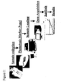
- FIGURE 1 Outline of the method of the present invention.
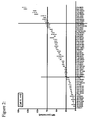
- FIGURE 2 Analysis of the phenotypic state of a population of cells using the marker reagent array shown in Table 2.
- FIGURE 3 Analysis on the effects of enzymatic treatment on the phenotypic state of different samples of populations of cell X, using the marker reagent array shown in Table 2.
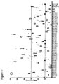
- FIGURE 4 Comparison of the phenotypic state of a sample of cell X with a sample of human dermal fibroblsts, using the marker reagent array shown in Table 2.
- FIGURE 5 Phenotypic analysis of whole blood.
- Figure 5 A shows the level of expression of a selected epitope in heterogenous cell samples. Leukocyte, monocyte and granulocyte populations were selected based upon size and granularity. Percentage of positive events and Mean Fluorescence Intensity was assessed for all selected cell populations for each epitope tested. As an example the expression levels of CD63 are shown for all selected cell populations.
- Figure 5 B shows the percentage of positive events and expression levels of leukocytes, monocytes and granulocytes for the array of surface epitopes shown. Cell populations were selected and analyzed from three healthy donors in 11 separate tests (Mean ⁇ SEM).
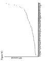
- Figure 5 C shows expression levels of the epitopes in the reagent array shown in table 1 for a leukocyte population taken from a healthy individual.
- assay components and methods for the characterization of the phenotype of cells are provided.
- the invention has an enormous number of applications in diagnostic assay techniques.
- Reagents may be prepared, for example, so as to detect or screen for any of a number of sample characteristics, pathological conditions, or reactants.
- the present invention provides a panel of reagents that is selected to cover various applications.
- Reagents suitable for use in the present invention are antibodies that recognize phenotypic markers.
- the phenotypic markers may be proteins expressed on the surface of cells, or they may be proteins that are expressed intracellularly. Alternatively, they may be proteins that are secreted from the cell. Alternatively, the phenotypic markers may be nucleic acids, lipids, polysaccharides or any biomolecule.
- Reagents suitable for use in the present invention are antibodies that specifically the phenotypic markers.
- the reagents are labeled with a fluorescent marker.
- the reagent may be intrinsically fluorescent.
- Antibodies suitable for use in the present invention may be obtained from commercial sources, or they may be generated for a specific application.
- the antibodies are labeled with a fluorescent marker. Examples of antibodies and phenotypic markers suitable for use in the present invention are shown in Table 2 .
- the reagent is in the form of a kit, comprising a multi-well plate containing antibodies that are lyophilized or otherwise configured allowing for convenient transport and storage.
- the multi-well plates may have different formats such as 6, or 8, or 12, or 24, or 96, or 384 wells and the like.
- the antibodies are selected according to the analysis that is to be performed.
- the antibodies may be selected to cover immune related epitopes. Alternatively, they may be selected to cover cancer epitopes, or stem cell epitopes.
- the antibodies may recognize extracellular molecules or they may recognize intracellular molecules.
- the selected antibodies may be dispensed into multi-well plates according to the layouts shown in Tables 2 and 3.
- the layouts are unique to the specific phenotypic marker panel.
- Each well of the multiwell plate may contain a single antibody or multiple antibodies.
- the selected antibodies are provided in bulk quantities and dispensed into a large number of plates to reduce variation between samples.
- the antibodies may be stored in the plates in solution prior to analysis. Alternatively, the antibodies may be stored lyophilized and re-constituted prior to analysis.
- the method of the present invention comprises a system that carries out the steps of collecting samples material of an unknown phenotypic state and collecting sample material of a known phenotypic state.
- the sample material may comprise, for example, cells, blood, or any biological sample.
- the system analyses the sample materials to identify patterns epitope markers in the sample material of known phenotypic state that is used as a comparator for the sample material of unknown phenotypic state.
- the identity of the epitopes in the identified pattern may be known. Alternatively, more than one epitope in the identified pattern may be unknown.
- An outline of the method of the present invention is shown in Figure 1 .
- the methods of the present invention may be used for quality control purposes.
- the sample material of known phenotypic state may be obtained from a control population of cells. This sample material may then be used as a comparator for the sample material of unknown phenotypic state that consists of populations of cell obtained from production runs. Changes from the patterns of epitope markers in the sample material of known phenotypic may be used to accept or reject production runs. An example of this is shown in Example 1.
- the present invention may be used to report changes in the pattern of epitope markers in a population of cells following treatment with a factor or pharmaceutical agent. An example of this is shown in Example 2.
- the present invention may be used to report changes in the pattern of epitope markers between distinct populations of cells. An example of this is shown in Example 3 .
- the present invention may also report changes in the pattern of epitope markers in distinct populations of cells in whole blood.
- the cell populations may be from a sample of blood from one patient. Alternatively, samples from more patient may be used.
- the methods of the present invention may be employed to analyze one sample or, alternatively, more than one sample.
- the high-throughput analysis of a large number of samples may be carried out.
- the steps of the method of the present invention may be carried manually. Alternatively, at least one step may be automated. Multiple copies of a phenotypic marker reagent plate may be made and used to analyze samples.
- Analysis of the samples may be performed by any suitable platform known to those of ordinary skill in the art.
- platforms may include, for example, a flow cytometry apparatus, a mass spectrometry apparatus, or a fluorescent microscope, and the like.
- Flow cytometry apparatuses rely upon flow of cells or other particles in a liquid flow stream in order to determine one or more characteristics of the cells under investigation. Further, the flow cytometry apparatus is useful for identifying the presence of certain cells or particles of interest, enumerating those cells or particles and, in some instances, providing a sorting capability so as to be able to collect those cells or particles of interest.
- a fluid sample containing cells is directed through the flow cytometry apparatus in a rapidly moving liquid stream so that each cell passes serially, and substantially one at a time, through a sensing region. Cell volume may be determined by changes in electrical impedance as each cell passes through the sensing region.
- the passing cells scatter such light as they pass there through.
- This scattered light serves as a function of cell shape and size, index of refraction, opacity, roughness and the like.
- fluorescence emitted by labeled cells, or autofluorescent cells, which have been excited as a result of passing through the excitation energy of the incident light beam is detectable for identification of cells having fluorescent properties. After cell analysis is performed by the flow cytometry apparatus, those cells that have been identified as having the desired properties may be sorted if the apparatus has been designed with such capability.
- Master plate preparation An antibody template was designed to monitor changes of expression across a wide array of epitopes in order to establish consistency of the cell product between production and expansion runs. Antibodies were selected and dispensed into a multiwell plate according to the layout shown in Table 2. Antibodies were purchased from the sources shown in Table 1. Antibodies were diluted in PBS into a 96-well master plate and stored in a refrigerator prior to analysis. For each assay 10 ⁇ l of master antibody solution was transferred from the master plate to a new 96 well plate as such that a copy was created. The array contained phycoerythrin fluorescein labeled antibodies against one specific marker per well. This eliminated the need for compensation of the machine, simplified the assay and reduced operator bias. In addition, the staining protocol was designed to minimize the number of cells needed for the assay and eliminate steps, such as washing, that can cause errors.
- Assay A vial of cells were thawed, washed and finally diluted to 2x10 5 cells per ml in PBS and a 100 ⁇ l sample was transferred to each well (20,000 cells/well) containing diluted antibodies. The plate was left to incubate for 30 minutes at 4°C. Next 100 ⁇ l fixation buffer (Cytofix, BD catalog no. 554655) was added and left for 15 minutes. A FACSCalibur machine with a high through put system was setup for analysis and 10,000 events per well were acquired.
- MFI Mean Fluorescent Intensity
- Results Cells obtained from different production/expansion runs were routinely stained using the 96-well plate array and the MFI were plotted and compared against the pattern of epitope markers obtained from the sample material of known phenotypic state. The results are shown in Figure 2 .
- Example 1 The methods and reagent panel used for this assay is the same as described in Example 1. Cells were grown in 3 separate culture flasks under identical conditions. These cultures were harvested each with a different dissociation enzyme mix. The cells were then stained using out 96-well method and the mean fluorescence intensity was measured for each parameter and compared to the pattern of epitope markers obtained from the sample material of known phenotypic state.
- This method allows for a rapid assessment of a wide array of proteins expressed on the cell surface. Changes to the phenotypic state that may occur following treatment with enzymes can readily be assessed by this method.
- the availability of a master plate allows for a quick assessment of phenotypic changes across a wide array of phenotypic markers due to treatment of the cells.
- Example 1 The methods and reagent panel used for this assay were the same as described in Example 1 .
- expression levels epitopes tested by the master plate on a different homogenous cell culture.
- the cell culture of interest was grown to confluency and harvested according our standard protocol. Both cell cultures were then stained in the 96-well master plate and analyzed according the methods described earlier. The phenotypic state of the cell populations were compared and key differences were identified.
- results The two cell types have similar expression levels for most of the epitopes except for example CD11a and CD54. These epitopes were differentially expressed. We selected an arbitrary cut-off for significance at a delta of 25 MFI compared to the sample material of known phenotypic state. The results are shown in Figure 4 .
- the 96 well plate can also be used for heterogeneous cell population.
- the data processing and mining becomes significantly more complex.
- Novel analysis methods are in development to simplify the analysis, which is especially important when this method is expanded to multiple color flow cytometry.
- This method may be used to determine the pattern of epitope markers in easy accessible peripheral blood samples for healthy (non-disease phenotype) individuals and use this a comparator for cytomes obtained from individuals with a particular disease. Differences can be exploited for, but not limited to, development of diagnostics, measure drug efficacy and selection of treatment regiment.
Landscapes
- Health & Medical Sciences (AREA)
- Immunology (AREA)
- Life Sciences & Earth Sciences (AREA)
- Engineering & Computer Science (AREA)
- Chemical & Material Sciences (AREA)
- Molecular Biology (AREA)
- Biomedical Technology (AREA)
- Hematology (AREA)
- Urology & Nephrology (AREA)
- Biotechnology (AREA)
- Biochemistry (AREA)
- Cell Biology (AREA)
- Food Science & Technology (AREA)
- Medicinal Chemistry (AREA)
- Physics & Mathematics (AREA)
- Analytical Chemistry (AREA)
- Microbiology (AREA)
- General Health & Medical Sciences (AREA)
- General Physics & Mathematics (AREA)
- Pathology (AREA)
- Chemical Kinetics & Catalysis (AREA)
- Measuring Or Testing Involving Enzymes Or Micro-Organisms (AREA)
- Apparatus Associated With Microorganisms And Enzymes (AREA)
Abstract
Description
- This invention is directed to methods for the rapid, efficient and accurate determination of the phenotype of cells. In particular, this invention is directed to methods employing antibodies.
- The surface of every cell in the body is coated with proteins. These cell surface proteins have several functions. For example, the cell surface proteins may be receptors, which have the capability of selectively binding or adhering to other "signaling" molecules. Alternatively, these proteins may be functional or structural. For example, the protein may facilitate the adherence of the cell to a substrate, or the protein may facilitate the secretion of a molecule. Each cell type, for example a liver cell, has a certain combination of proteins on their surface that makes them distinguishable from other kinds of cells or cell systems.
- The expression of cell surface proteins on a given cell type may vary, depending on a variety of factors. The may include, for example, changes as a result of cellular proliferation, differentiation, disease, isolation of the cell from the body and culture in vitro. For example, cell surface proteins have been reported to play vital roles in multiple steps of prostate cancer metastasis, such as detachment of tumor cells from the extracellular matrix, resistance of tumor cells to the detachment-induced apoptosis, adhesion of tumor cells to endothelial cells and angiogenesis to support tumor growth. There may be cell surface proteins either uniquely or differentially expressed in both epithelial and endothelial cells that are important for individual processes of prostate cancer metastasis.
- Researchers have taken advantage of the biological uniqueness of cell surface proteins and chemical properties of certain compounds to tag or "mark" cells. Cells marked in this manner can readily be isolated and characterized. In many cases, a combination of multiple markers is used to identify a particular cell type...
- Antibodies are used extensively as diagnostic tools in a wide array of different analyses. Monoclonal and recombinant antibodies may be used as probes to tag or mark cells. Antibody-based immunoassays are the most commonly used type of diagnostic assay and still one of the fastest growing technologies for the analysis of biomolecules. Another example with particular importance in diagnostics that has become a routine during the past decade is flow cytometric analysis. Flow cytometry is a method for quantifying components or structural features of cells primarily by optical means. Although it makes measurements on one cell at a time, it can process thousands of cells in a few seconds. Since different cell types can be distinguished by quantifying structural features, flow cytometry can be used to count cells of different types in a mixture. Flow cytometers may also be employed to select or "sort" cells based on their cell surface marker expression.
- Intracellular components may also be reported by fluorescent probes, including total DNA/cell (allowing cell cycle analysis), newly synthesized DNA, specific nucleotide sequences in DNA or mRNA, filamentous actin, and any structure for which an antibody is available. Flow cytometry can also monitor rapid changes in intracellular free calcium, membrane potential, pH, or cell signaling pathways. For example, Krutzik et al (Journal of Immunology, 2005, 175: 2357-2365) state "phosphospecific flow cytometry has emerged as a powerful tool to analyze intracellular signaling events in complex populations of cells because of its ability to simultaneously discriminate cell types based on surface marker expression and measure levels of intracellular phosphoproteins. This has provided novel insights into the cell- and pathway-specific nature of immune signaling."
- Flow cytometry analysis and sorting studies using monoclonal antibodies to define the surface markers on normal and neoplastic cell populations created the basis for routine clinical diagnostic assays that now range from leukemia classification to monitoring CD4 T-cell loss as HIV disease progresses.
- Despite the versatility of flow cytometry, there are some drawbacks. For example, the antibodies chosen for flow cytometry reagents may not be optimal: They may not be specific, resulting in a high background staining. Furthermore, the efficiency of coupling the fluorochrome to the antibody may vary from batch to batch. Therefore, comparisons between populations of cells may be further complicated.
- In addition, in order to assess multiple parameters on cell populations, a significant investment is required to purchase the required antibodies. For example, the analysis of 90 single parameters would require the purchase of 90 separate antibodies, which may amount to several tens of thousands of dollars and a large number of cells per analysis. This is problematic if the number of cells is finite and limited. Therefore, there is a significant need for cost effective, validated, reproducible reagents and antibodies for the phenotyping or analysis of cells.
- Rosa et at. (Nature Medicine, 7, 245 - 248, 2001) identified human naive T cells using flow cytometry.
- Borowitz et al. (Blood, 89:11, 3960-3966, 1997) used flow cytometry to measure surface membrane antigen expression in patients diagnosed with B-precursor acute lymphoblastic leukaemia.
- Hamann et al. (J. Exp. Med., 186:3, 1407-1418, 1997) characterised memory and effector human CD8+ T cells using flow cytometry.
-
WO 02/074789 - The present invention provides a kit comprising a multi-well plate containing antibodies that are lyophilised or otherwise configured for convenient transport and storage, wherein the antibodies are for detecting the panel of phenotypic markers described in Table 2.
- The present invention also provides a kit for comparing the phenotypic state of a first unknown sample material with a sample material of a known phenotypic state wherein the sample material does not device from a human embryo comprising;
- a. the panel of phenotypic markers described in Table 2, and
- b. antibodies to detect the presence of the phenotypic markers of Table 2 in a sample material.
- The present invention also provides a method for comparing the phenotypic state of a first unknown sample material with a sample material of a known phenotypic state comprising the steps of:
- a. Obtaining a first unknown sample material, and;
- b. Contacting the first unknown sample material with antibodies to detect the presence of the panel of phenotypic markers described in Table 2 in the first unknown sample material, and;
- c. Recording the presence of phenotypic markers in the first unknown sample material, and;
- d. Obtaining a sample material of a known phenotypic state, and;
- e. Contacting the sample material of a known phenotypic state with said reagents to detect the presence of said panel of phenotypic markers wherein the sample material does not device from a human embryo in the sample material of a known phenotypic state, and;
- f. Recording the presence of phenotypic markers in the sample material of a known phenotypic state, and;
- g. Reporting the differences between the unknown sample material and the sample material of a known phenotypic state.
-
FIGURE 1 : Outline of the method of the present invention. -
FIGURE 2 : Analysis of the phenotypic state of a population of cells using the marker reagent array shown in Table 2. -
FIGURE 3 : Analysis on the effects of enzymatic treatment on the phenotypic state of different samples of populations of cell X, using the marker reagent array shown in Table 2. -
FIGURE 4 : Comparison of the phenotypic state of a sample of cell X with a sample of human dermal fibroblsts, using the marker reagent array shown in Table 2. -
FIGURE 5 : Phenotypic analysis of whole blood.Figure 5 A shows the level of expression of a selected epitope in heterogenous cell samples. Leukocyte, monocyte and granulocyte populations were selected based upon size and granularity. Percentage of positive events and Mean Fluorescence Intensity was assessed for all selected cell populations for each epitope tested. As an example the expression levels of CD63 are shown for all selected cell populations.Figure 5 B shows the percentage of positive events and expression levels of leukocytes, monocytes and granulocytes for the array of surface epitopes shown. Cell populations were selected and analyzed from three healthy donors in 11 separate tests (Mean ± SEM).Figure 5 C shows expression levels of the epitopes in the reagent array shown in table 1 for a leukocyte population taken from a healthy individual. - According to the present invention, assay components and methods for the characterization of the phenotype of cells are provided. As those of ordinary skill in the art will recognize, the invention has an enormous number of applications in diagnostic assay techniques. Reagents may be prepared, for example, so as to detect or screen for any of a number of sample characteristics, pathological conditions, or reactants. The present invention provides a panel of reagents that is selected to cover various applications.
- Reagents suitable for use in the present invention are antibodies that recognize phenotypic markers. The phenotypic markers may be proteins expressed on the surface of cells, or they may be proteins that are expressed intracellularly. Alternatively, they may be proteins that are secreted from the cell. Alternatively, the phenotypic markers may be nucleic acids, lipids, polysaccharides or any biomolecule.
- Reagents suitable for use in the present invention are antibodies that specifically the phenotypic markers. In one embodiment, the reagents are labeled with a fluorescent marker. Alternatively the reagent may be intrinsically fluorescent.
- Antibodies suitable for use in the present invention may be obtained from commercial sources, or they may be generated for a specific application. In one embodiment, the antibodies are labeled with a fluorescent marker. Examples of antibodies and phenotypic markers suitable for use in the present invention are shown in Table 2.
- The present invention provides cost effective, optimized reagents for the reproducible analysis of cells. In one embodiment, the reagent is in the form of a kit, comprising a multi-well plate containing antibodies that are lyophilized or otherwise configured allowing for convenient transport and storage. The multi-well plates may have different formats such as 6, or 8, or 12, or 24, or 96, or 384 wells and the like. The antibodies are selected according to the analysis that is to be performed. The antibodies may be selected to cover immune related epitopes. Alternatively, they may be selected to cover cancer epitopes, or stem cell epitopes. The antibodies may recognize extracellular molecules or they may recognize intracellular molecules.
- The selected antibodies may be dispensed into multi-well plates according to the layouts shown in Tables 2 and 3. The layouts are unique to the specific phenotypic marker panel. Each well of the multiwell plate may contain a single antibody or multiple antibodies. The selected antibodies are provided in bulk quantities and dispensed into a large number of plates to reduce variation between samples. The antibodies may be stored in the plates in solution prior to analysis. Alternatively, the antibodies may be stored lyophilized and re-constituted prior to analysis.
- In one embodiment, the method of the present invention comprises a system that carries out the steps of collecting samples material of an unknown phenotypic state and collecting sample material of a known phenotypic state. The sample material may comprise, for example, cells, blood, or any biological sample. The system analyses the sample materials to identify patterns epitope markers in the sample material of known phenotypic state that is used as a comparator for the sample material of unknown phenotypic state. The identity of the epitopes in the identified pattern may be known. Alternatively, more than one epitope in the identified pattern may be unknown. An outline of the method of the present invention is shown in
Figure 1 . - For example, the methods of the present invention may be used for quality control purposes. In this case, the sample material of known phenotypic state may be obtained from a control population of cells. This sample material may then be used as a comparator for the sample material of unknown phenotypic state that consists of populations of cell obtained from production runs. Changes from the patterns of epitope markers in the sample material of known phenotypic may be used to accept or reject production runs. An example of this is shown in Example 1.
- Alternatively, the present invention may be used to report changes in the pattern of epitope markers in a population of cells following treatment with a factor or pharmaceutical agent. An example of this is shown in Example 2. Alternatively, the present invention may be used to report changes in the pattern of epitope markers between distinct populations of cells. An example of this is shown in Example 3.
- In one embodiment, the present invention may also report changes in the pattern of epitope markers in distinct populations of cells in whole blood. The cell populations may be from a sample of blood from one patient. Alternatively, samples from more patient may be used.
- The methods of the present invention may be employed to analyze one sample or, alternatively, more than one sample. In one embodiment, the high-throughput analysis of a large number of samples may be carried out. The steps of the method of the present invention may be carried manually. Alternatively, at least one step may be automated. Multiple copies of a phenotypic marker reagent plate may be made and used to analyze samples.
- Analysis of the samples may be performed by any suitable platform known to those of ordinary skill in the art. Such platforms may include, for example, a flow cytometry apparatus, a mass spectrometry apparatus, or a fluorescent microscope, and the like.
- Flow cytometry apparatuses rely upon flow of cells or other particles in a liquid flow stream in order to determine one or more characteristics of the cells under investigation. Further, the flow cytometry apparatus is useful for identifying the presence of certain cells or particles of interest, enumerating those cells or particles and, in some instances, providing a sorting capability so as to be able to collect those cells or particles of interest. In a typical flow cytometry apparatus, a fluid sample containing cells is directed through the flow cytometry apparatus in a rapidly moving liquid stream so that each cell passes serially, and substantially one at a time, through a sensing region. Cell volume may be determined by changes in electrical impedance as each cell passes through the sensing region. Similarly, if an incident beam of light is directed at the sensing region, the passing cells scatter such light as they pass there through. This scattered light serves as a function of cell shape and size, index of refraction, opacity, roughness and the like. Further, fluorescence emitted by labeled cells, or autofluorescent cells, which have been excited as a result of passing through the excitation energy of the incident light beam is detectable for identification of cells having fluorescent properties. After cell analysis is performed by the flow cytometry apparatus, those cells that have been identified as having the desired properties may be sorted if the apparatus has been designed with such capability.
- Representative flow cytometry apparatuses are described in
U.S. Pat. Nos. 3,826,364 and4,284,412 , and in the publication by Herzenberg et al., "Fluorescence-activated Cell Sorting," Sci. Am. 234 (3): 108, 1976. - The present invention is further illustrated, but not limited by, the following examples.
- Master plate preparation: An antibody template was designed to monitor changes of expression across a wide array of epitopes in order to establish consistency of the cell product between production and expansion runs. Antibodies were selected and dispensed into a multiwell plate according to the layout shown in Table 2. Antibodies were purchased from the sources shown in Table 1. Antibodies were diluted in PBS into a 96-well master plate and stored in a refrigerator prior to analysis. For each assay 10µl of master antibody solution was transferred from the master plate to a new 96 well plate as such that a copy was created. The array contained phycoerythrin fluorescein labeled antibodies against one specific marker per well. This eliminated the need for compensation of the machine, simplified the assay and reduced operator bias. In addition, the staining protocol was designed to minimize the number of cells needed for the assay and eliminate steps, such as washing, that can cause errors.
- Assay: A vial of cells were thawed, washed and finally diluted to 2x10⁵ cells per ml in PBS and a 100µl sample was transferred to each well (20,000 cells/well) containing diluted antibodies. The plate was left to incubate for 30 minutes at 4°C. Next 100µl fixation buffer (Cytofix, BD catalog no. 554655) was added and left for 15 minutes. A FACSCalibur machine with a high through put system was setup for analysis and 10,000 events per well were acquired.
- Data analysis: For this study the acquired data was exported to an analysis software program and the Mean Fluorescent Intensity (MFI) of each sample well was assessed. The MFI of the unstained sample was subtracted from each sample MFI and this delta MFI was plotted for each epitope. This was repeated eight times in independent experiments creating an average expression pattern for our cell product.
- Results: Cells obtained from different production/expansion runs were routinely stained using the 96-well plate array and the MFI were plotted and compared against the pattern of epitope markers obtained from the sample material of known phenotypic state. The results are shown in
Figure 2 . - Method: The methods and reagent panel used for this assay is the same as described in Example 1. Cells were grown in 3 separate culture flasks under identical conditions. These cultures were harvested each with a different dissociation enzyme mix. The cells were then stained using out 96-well method and the mean fluorescence intensity was measured for each parameter and compared to the pattern of epitope markers obtained from the sample material of known phenotypic state.
- Results: Each enzyme mix showed different results when compared to the pattern of epitope markers obtained from the sample material of known phenotypic state. Mixture C (Accutase) seems to affect the expression of certain phenotypic marker more than the standard mix A (Trypsin) and B (TriplE). For example the adhesion marker CD49f and the signaling molecule CD63 show lower expression levels after the harvesting procedure, which may affect the cell product's function. Results are shown in
Figure 3 . - Conclusion: This method allows for a rapid assessment of a wide array of proteins expressed on the cell surface. Changes to the phenotypic state that may occur following treatment with enzymes can readily be assessed by this method. The availability of a master plate allows for a quick assessment of phenotypic changes across a wide array of phenotypic markers due to treatment of the cells.
- Method: The methods and reagent panel used for this assay were the same as described in Example 1. In this assay expression levels epitopes tested by the master plate on a different homogenous cell culture. The cell culture of interest was grown to confluency and harvested according our standard protocol. Both cell cultures were then stained in the 96-well master plate and analyzed according the methods described earlier. The phenotypic state of the cell populations were compared and key differences were identified.
- Results: The two cell types have similar expression levels for most of the epitopes except for example CD11a and CD54. These epitopes were differentially expressed. We selected an arbitrary cut-off for significance at a delta of 25 MFI compared to the sample material of known phenotypic state. The results are shown in
Figure 4 . - Conclusion: We showed expression differences between two cell types in our panel. These differences can be further explored and can become important starting points of further research especially if the phenotypic marker changes can be linked to in-vivo efficacy differences.
- Method: The methods and reagent panel used for this assay were the same as described in Example 1. Heparinized peripheral blood was obtained from healthy volunteers and white blood cells were isolated. The White blood cells were washed and suspended in PBS and transferred to the 96-well staining plate (50,000 cells per well) and data was acquired according to the standard protocol. Data analysis, however, is different since the sample contains multiple cell populations each with their specific expression pattern for each epitope. Analyzing the cells as a bulk may obscure these effects. Therefore three main populations were selected based upon size and granularity and each was analyzed for percentage positive events within the gate and expression levels of each epitope tested (
Figure 5 A,B and C). - Results: Using the 96-well platform we able to quickly assess expression levels of our array of epitopes for three distinct populations in peripheral blood (
Figure 5 ). - Conclusion: The 96 well plate can also be used for heterogeneous cell population. However the data processing and mining becomes significantly more complex. Novel analysis methods are in development to simplify the analysis, which is especially important when this method is expanded to multiple color flow cytometry. This method may be used to determine the pattern of epitope markers in easy accessible peripheral blood samples for healthy (non-disease phenotype) individuals and use this a comparator for cytomes obtained from individuals with a particular disease. Differences can be exploited for, but not limited to, development of diagnostics, measure drug efficacy and selection of treatment regiment.
TABLE 1: PHENOTYPIC MARKERS DISCLOSURE CD identification MOLECULE Gene Name Fluorescence company CD3 CD3/T-cell antigen receptor (TCR) CD3G/Z PE Caltag CD4 OKT4, Leu 3a, T4 CD4 PE BD Pharmingen CD5 T1, Leu1 CD5 PE BD PharMingen CD7 Leu 9, 3A1, gp40, T cell leukemia antigen CD7 PE BD Pharmingen CD8 MHC class I receptor CD8 PE BD PharMingen CD9 type III transmembrane protein (platelet aggregation/activation) CD9 PE BD PharMingen CD10 CALLA, membrane metallo-endopeptidase MME PE BD Pharmingen CD11a alphaL; LFA-1, gp180/95 ITGAL PE BD Pharmingen CD13 Aminopeptidase N, APN, gp150, EC 3.4.11.2 ANPEP PE BD Pharmingen CD15 fucosyltransferase 4 (alpha (1,3) fucosyltransferase, myeloid-specific) FUT4 PE BD PharMingen CD16 Fc gamma R III FCGR3A PE Caltag CD16a Fc gamma R IIIa FCGR3A PE BD Pharmingen CD18 β2-Integrin chain, macrophage antigen 1 (mac-1) ITGB2 PE BD Pharmingen CD19 B4, associated with CD21, CD81, CD225, Leu-13, Lyn, Fyn, Vav, P13-kinase CD19 PE BD PharMingen CD20 membrane-spanning 4-domains, subfamily A, member 1 MS4A1 PE BD PharMingen CD21 C3d receptor,CR2, gp140; EBV receptor CR2 PE BD Pharmingen CD22 Bgp135; BL-CAM, Siglec2 CD22 FITC BD Pharmingen CD25 Interleukin-2 receptor (IL-2R, inflammatory response) IL2RA PE BD PharMingen CD29 Integrin β1 chain; platelet GPlla; VLA (CD49) beta-chain ITGB1 PE BD Pharmingen CD30 tumor necrosis factor receptor superfamily, member 8. KI-1 TNFRSF8 PE BD PharMingen CD31 PECAM-1; platelet GPIIa'; endocam PECAM1 PE BD Pharmingen CD32 Fcgamma receptor type II (FcgRII), gp40 FCGR2A PE BD Pharmingen CD33 gp67 CD33 PE BD PharMingen CD34 My10, gp105-120 CD34 PE BD PharMingen CD35 C3b/C4b receptor; complement receptor type 1 (CR1) CR1 PE BD Pharmingen CD36 platelet GPIV, GPIIIb, OKM-5 antigen CD36 PE BD Pharmingen CD38 T10; gp45, ADP-ribosyl cyclase CD38 PE BD Pharmingen CD40 Bp50, TNF Receptor 5 TNFRSF5 PE BD PharMingen CD44 Pgp-1; gp80-95, Hermes antigen, ECMR-III and HUTCH-I. CD44 PE BD PharMingen CD45 LCA, B220, protein tyrosine phosphatase, receptor type, C PTPRC PE BD Pharmingen CD49a Integrin a1 chain, very late antigen, VLA 1a ITGA1 PE BD Pharmingen CD49b Integrin a2 chain, VLA-2-alpha chain, platelet gpla ITGA2 PE BD Pharmingen CD49c Integrin a3 chain, VLA-3 alpha chain ITGA3 PE BD Pharmingen CD49d Integrin a4 chain" VLA-4-alpha chain ITGA4 PE BD Pharmingen CD49e Integrin a5 chain" VLA-5 alpha chain ITGA5 PE BD Pharmingen CD49f Integrin a6 chain" VLA-6 alpha chain, platelet gplc ITGA6 PE BD Pharmingen CD50 ICAM-3, intercellular adhesion molecule 3 ICAM3 fitc BD Pharmingen CD51 Integrin alpha chain, vitronectin receptor a chain ITGAV PE BD Pharmingen CD54 ICAM-1, intercellular adhesion molecule 1 ICAM1 PE BD Pharmingen CD55 decay-accelerating factor (DAF, prevent cell damage) DAF PE BD PharMingen CD56 Neural cell adhesion molecule 1 (NCAM1), NKH-1 (NK cell marker) NCAM1 PE Caltag CD58 Lymphocyte function-associated antigen-3 (LFA-3, interacts with CD2 during cell adhesion) CD58 PE BD Pharmingen CD59 MACIF, MIRL, P-18, protectin CD59 PE BD Pharmingen CD61 Glycoprotein IIIa, beta3 integrin ITGB3 PE BD Pharmingen CD62E E-selectin, LECAM-2, ELAM-1 SELE PE BD Pharmingen CD62L L-selectin, LAM-1, Mel-14 SELL PE Caltag CD62P P-selectin, granule membrane protein-140 (GMP-140) SELP PE BD Pharmingen CD63 LIMP, gp55, LAMP-3 neuroglandular antigen, granulophysin CD63 PE BD Pharmingen CD64 FcgR1, FcgammaR1 (IgG-receptor) FCGR1A PE Caltag CD69 Early T-cell activation antigen CD69 PE BD PharMingen CD71 Transferrin receptor TFRC PE BD Pharmingen CD73 Ecto-5'-nucleotidase NT5E PE BD Pharmingen CD79a CD79A antigen (immunoglobulin-associated alpha), MB1 CD79A PE BD PharMingen CD79b CD79B antigen (immunoglobulin-associated beta), B29 CD79B PE BD PharMingen CD80 B7-1; BB1 CD80 PE BD Pharmingen CD81 Target of an antiproliferative antibody (TAPA-1); M38 CD81 PE BD Pharmingen CD83 HB15 CD83 PE BD Pharmingen CD86 B7-2; B70 CD86 PE Caltag Laboratories CD87 plasminogen activator, urokinase receptor (PLAUR), uPAR PLAUR PE BD PharMingen CD88 C5a-receptor C5R1 PE BD Pharmingen CD90 Thy-1 THY1 PE BD PharMingen CD95 APO-1, Fas, TNFRSF6 TNFRSF6 PE BD Pharmingen CD100 SEMA4D SEMA4D PE Serotec CD103 integrin, alpha E (human mucosal lymphocyte antigen 1; alpha polypeptide) ITGAE PE BD PharMingen CD104 Integrin beta 4 subunit, TSP-1180 ITGB4 PE BD Pharmingen CD105 Endoglin ENG PE Caltag CD106 VCAM-1 (vascular cell adhesion molecule-1), INCAM-110 VCAM1 PE BD Pharmingen CD114 G-CSFR, HG-CSFR, CSFR3 CSF3R PE BD Pharmingen CD117 SCFR, c-kit, stem cell factor receptor KIT PE BD Biosciences CD119 IFN gamma receptor alpha chain (GIR-208) IFNGR1 PE BD Pharmingen CD120b TNFRI; TNFRp55 TNFRSF1B PE BD Pharmingen CD126 IL-6 receptor alpha chain IL6R PE BD Pharmingen CD134 Tumor Necrosis Factor receptor 4, OX40 (promotes expression of BCL2/BCL-xL) TNFRSF4 PE Caltag CD135 fms-related tyrosine kinase 3 FLT3 PE BD PharMingen CD140b b-platelet derived growth factor (PDGF) receptor PDGFRB PE BD Pharmingen CD141 Thrombomodulin (TM), fetomodulin THBD PE BD Pharmingen CD142 Tissue factor, thromboplastin, coagulation factor III F3 PE BD Pharmingen CD146 Muc 18, MCAM, Mel-CAM, s-endo MCAM PE BD Pharmingen CD147 Basigin, M6, extracellular metalloproteinase inducer (EMMPRIN) BSG PE Serotec CD151 Platelet-endothelial tetra-span antigen (PETA)-3 CD151 PE BD Pharmingen CD152 Cytolytic T lymphocyte-associated antigen (CTLA-4) CTLA4 PE BD PharMingen CD163 M130, GHI/61, RM3/1 CD163 PE BD PharMingen CD164 MUC-24, MGC 24, multi-glycosylated core protein 24 CD164 PE BD Pharmingen CD165 AD2, gp 37 PE BD Pharmingen CD178 FAS ligand, CD95 ligand TNFSF6 PE Caltag Laboratories CD181 CXCR1 CDw128a: IL-8 receptor alpha, IL8RA PE BD Pharmingen CD182 CXCR2 IL8RB PE BD Pharmingen CD183 CXCR3 chemokine receptor, G protein-coupled receptor 9 CXCR3 PE BD Pharmingen CD184 CXCR4 chemokine receptor, Fusin CXCR4 PE BD Pharmingen CD200 OX2 (regulates Mf activity) CD200 PE BD PharMingen CD273 B7DC, PDL2 PDCD1 LG2 PE BD Pharmingen CD274 B7H1, PDL1 PDCD1LG1 PE BD Pharmingen CD275 B7H2, ICOSL ICOSL CD276 B7H3 N/A CD277 BT3.1 BTN3A1 CD278 ICOS ICOS CD279 PD1 PDCD1 CD280 ENDO180 MRC2 CD281 TLR1 TLR1 CD282 TLR2 TLR2 CD283 TLR3 TLR3 CD284 TLR4 TLR4 CD289 TLR9 TLR9 CD292 BMPR1A BMPR1A CDw293 BMPR1B BMPR1B CD294 CRTH2 GPR44 CD295 LeptinR LEPR CD296 ART1 ART1 CD297 ART4 DO CD298 Na+/K+ -ATPase b3 ATP1B3 CD299 DCSIGN-related CD209L CD300a CMRF35H CD300c CMRF35A CD300e CMRF35L1 CD301 MGL, CLECSF14 CLECSF14 CD302 DCL1 N/A CD303 BDCA2 CLECSF7 CD304 BDCA4, Neuropilin 1 NRP1 CD305 LAIR1 LAIR1 CD306 LAIR2 LAIR2 CD307 IRTA2 N/A CD309 VEGFR2, KDR KDR CD312 EMR2 EMR2 CD314 NKG2D KLRK1 CD315 CD9P1 PTGFRN CD316 EWI2 IGSF8 CD317 BST2 BST2 CD318 CDCP1 N/A CD319 CRACC SLAMF7 CD320 8D6A N/A CD321 JAM1 F11R CD322 JAM2 JAM2 CD324 E-Cadherin CDH1 CDw325 N-Cadherin CDH2 CD326 Ep-CAM TACSTD1 CDw327 siglec6 SIGLEC6 CDw328 sigiec7 SIGLEC7 CDw329 sigiec9 SIGLEC9 CD331 FGFR1 FGFR1 CD332 FGFR2 FGFR2 CD333 FGFR3 FGFR3 CD334 FGFR4 FGFR4 CD335 NKp46 NCR1 CD336 NKp44 NCR2 CD337 NKp30 NCR3 CDw338 ABCG2, BCRP ABCG2 CD339 Jagged-1 JAG1 ABCG2 alkaline phosphatase BCL-xL BCL2like-1 BCL2L1 PE Chemicon bCL-2 B-cell CLL/lymphoma-2 BCL2 PE BD Pharmingen CK 5 Cytokeratin 5 CK 6 Cytokeratin 6 CK 7 Cytokeratin 7 CK 8 Cytokeratin 8 CK 10 Cytokeratin 10 CK 13 Cytokeratin 13 CK 14 Cytokeratin 14 CK 15 Cytokeratin 15 CK 16 Cytokeratin 16 CK 18 Cytokeratin 18 CK 19 Cytokeratin 19 EGFr Epidermal growth factor receptor (growth and differentiation) EGFR PE BD PharMingen HGF-R Hepatocyte growth factor receptor HGFR NGFr NGFR (p75) NGFR PE BD Pharmingen RANTES CCL5 (chemokine (C-C motif) ligand 5 CCL5 PE Caltag b2-microglobulin Associates with HLA class I antigen complex B2M PE BD Pharmingen HLA-ABC MHC Class I ABC PE BD Pharmingen HLA-DP MHC II HLA-DP beta-1 HLA-DPB1 PE Serotec HLA DR PE BD Pharmingen SSEA-1 Embryonic Stem Cell Antigen 1 SSEA1 SSEA-3 Embryonic Stem Cell Antigen 3 SSEA3 SSEA-4 Embryonic Stem Cell Antigen 4 SSEA4 TABLE 2: A STEM CELL PHENOTYPIC MARKER ARRAY PLATE OF THE INVENTION 1 2 3 4 5 6 7 8 9 10 11 12 A Unstain Unstain Unstain Unstain Unstain Unstain CD9 CD10 CD11a CD13 CD14 CD16 B CD18 CD25 CD29 CD31 CD32 CD34 CD35 CD36 CD40 CD44 CD45 CD49a C CD49b CD49c CD49d CD49e CD49f CD51 CD54 CD55 CD58 CD59 CD61 CD62E D CD62L CD62P CD63 CD64 CD71 CD73 CD80 CD81 CD86 CD90 CD95 CD100 E CD104 CD105 CD106 CD117 CD119 CD120a CD126 CD134 CD140a CD140b CD141 CD142 F CD146 CD147 CD178 CD181 CD182 CD183 CD184 CD200 CD273 CD274 β2-microglobulin HLA-ABC G HLA-DP EGFR NGFR RANTES CD3 CD4 CD7 CD15 CD19 CD20 CD21 CD22 H CD38 CD50 CD62L CD83 CD88 CD112 CD114 CD151 CD164 CD165 CD172 HLA-DR TABLE 3: AN EXAMPLE OF AN ONCOLOGY PHENOTYPIC MARKER ARRAY PLATE 1 2 3 4 5 6 7 8 9 10 11 12 A Unstain Unstain Unstain Unstain Unstain Unstain CD3 CD4 CD5 CD7 CD8 CD9 B CD10 CD11a CD13 CD14 CD15 CD16 CD18 CD19 CD20 CD22 CD25 CD29 C CD30 CD31 CD32 CD33 CD34 CD35 CD36 CD38 CD40 CD44 CD45 CD49a D CD49b CD49c CD49d CD49e CD49f CD51 CD54 CD55 CD56 CD58 CD59 CD61 E CD62E CD62L CD62P CD63 CD64 CD69 CD71 CD73 CD79a CD79b CD80 CD81 F CD86 CD87 CD90 CD95 CD100 CD103 CD104 CD105 CD106 CD117 CD119 CD120a G CD126 CD134 CD135 CD140a CD140b CD141 CD142 CD146 CD147 CD152 CD163 CD178 H CD181 CD182 CD183 CD184 CD200 CD273 CD274 b2-micorglobulin EGFR HLA-ABC HLA-1 (DP) RANTES
Claims (7)
- A kit comprising a multi-well plate containing antibodies that are lyophilised or otherwise configured for convenient transport and storage, wherein the antibodies are for detecting the panel of phenotypic markers described in Table 2.
- A kit for comparing the phenotypic state of a first unknown sample material with a sample material of a known phenotypic state comprising;a. the panel of phenotypic markers described in Table 2, andb. antibodies to detect the presence of the phenotypic markers of Table 2 in a sample material.
- The kit of claim 1 or 2 wherein the antibodies are lyophilised antibodies.
- The kit of any of claims 1 to 3 wherein the antibodies are labelled with a fluorescent marker.
- A method for comparing the phenotypic state of a first unknown sample material with a sample material of a known phenotypic state comprising the steps of:a. Obtaining a first unknown sample material, and;b. Contacting the first unknown sample material with antibodies to detect the presence of the panel of phenotypic markers described in Table 2 in the first unknown sample material, and;c. Recording the presence of phenotypic markers in the first unknown sample material, and;d. Obtaining a sample material of a known phenotypic state, and;e. Contacting the sample material of a known phenotypic state wit sai reagents to detect the presence of said panel of phenotypic markers of Table 2 in the sample material of a known phenotypic state, and;f. Recording the presence of phenotypic markers in the sample material of a known phenotypic state, and;g. Reporting the differences between the unknown sample material and the sample material of a known phenotypic state.
- The method of claim 5 for use in quality control purposes.
- The kit of any of claims 1-4 or the method of claim 5 or claim 6 wherein the sample material is cells.
Applications Claiming Priority (1)
| Application Number | Priority Date | Filing Date | Title |
|---|---|---|---|
| US75561105P | 2005-12-30 | 2005-12-30 |
Publications (2)
| Publication Number | Publication Date |
|---|---|
| EP1813945A1 EP1813945A1 (en) | 2007-08-01 |
| EP1813945B1 true EP1813945B1 (en) | 2011-04-06 |
Family
ID=37951474
Family Applications (1)
| Application Number | Title | Priority Date | Filing Date |
|---|---|---|---|
| EP06256613A Not-in-force EP1813945B1 (en) | 2005-12-30 | 2006-12-29 | A method for determining the phenotype of cells |
Country Status (8)
| Country | Link |
|---|---|
| US (2) | US20070243599A1 (en) |
| EP (1) | EP1813945B1 (en) |
| JP (1) | JP2007181462A (en) |
| AT (1) | ATE504833T1 (en) |
| AU (1) | AU2006252302A1 (en) |
| CA (1) | CA2572530A1 (en) |
| DE (1) | DE602006021131D1 (en) |
| ES (1) | ES2362671T3 (en) |
Families Citing this family (9)
| Publication number | Priority date | Publication date | Assignee | Title |
|---|---|---|---|---|
| WO2009064789A2 (en) * | 2007-11-15 | 2009-05-22 | The Johns Hopkins University | Methods for detecting and monitoring circulating cancer stem cells |
| WO2009140452A2 (en) * | 2008-05-15 | 2009-11-19 | University Of Miami | Isolation of stem cell precursors and expansion in non-adherent conditions |
| US20100323393A1 (en) * | 2009-06-23 | 2010-12-23 | Xiuli An | Ordered Assembly of Membrane Proteins During Differentiation of Erythroblasts |
| US20110097748A1 (en) * | 2009-10-28 | 2011-04-28 | Warsaw Orthopedic, Inc. | Use of cell lines to determine levels of efficacy of pharmaceutical formulations |
| EP2495567A1 (en) | 2011-03-04 | 2012-09-05 | Erasmus University Medical Center Rotterdam | Methods and means for monitoring disruption of tissue homeostasis in the total body |
| JP6301263B2 (en) * | 2011-12-23 | 2018-03-28 | デピュイ・シンセス・プロダクツ・インコーポレイテッド | Detection of human umbilical cord tissue-derived cells |
| KR102583603B1 (en) * | 2017-03-16 | 2023-10-05 | 유니베르시테 리브레 드 브룩크젤즈 | Detection, quantification and/or isolation of circulating tumor cells based on expression of the CD321 marker |
| CN110314172A (en) * | 2019-07-17 | 2019-10-11 | 中国医科大学附属第一医院 | A kind of cell preparation and preparation method thereof for treating spinal cord injury |
| CN112666062B (en) * | 2020-11-20 | 2024-05-24 | 浙江省荣军医院 | Method for combined detection of flow cytometry in humoral cell immunoassay |
Family Cites Families (1)
| Publication number | Priority date | Publication date | Assignee | Title |
|---|---|---|---|---|
| US20030077576A1 (en) * | 2001-03-20 | 2003-04-24 | Joann Trial | Use of monoclonal antibodies and functional assays for prediction of risk of opportunistic infection |
-
2006
- 2006-12-28 CA CA002572530A patent/CA2572530A1/en not_active Abandoned
- 2006-12-28 JP JP2006355851A patent/JP2007181462A/en not_active Withdrawn
- 2006-12-29 US US11/618,168 patent/US20070243599A1/en not_active Abandoned
- 2006-12-29 DE DE602006021131T patent/DE602006021131D1/en active Active
- 2006-12-29 AT AT06256613T patent/ATE504833T1/en not_active IP Right Cessation
- 2006-12-29 AU AU2006252302A patent/AU2006252302A1/en not_active Abandoned
- 2006-12-29 ES ES06256613T patent/ES2362671T3/en active Active
- 2006-12-29 EP EP06256613A patent/EP1813945B1/en not_active Not-in-force
-
2010
- 2010-04-22 US US12/765,470 patent/US20100203622A1/en not_active Abandoned
Also Published As
| Publication number | Publication date |
|---|---|
| US20100203622A1 (en) | 2010-08-12 |
| AU2006252302A1 (en) | 2007-07-19 |
| CA2572530A1 (en) | 2007-06-30 |
| ES2362671T3 (en) | 2011-07-11 |
| ATE504833T1 (en) | 2011-04-15 |
| EP1813945A1 (en) | 2007-08-01 |
| JP2007181462A (en) | 2007-07-19 |
| DE602006021131D1 (en) | 2011-05-19 |
| US20070243599A1 (en) | 2007-10-18 |
Similar Documents
| Publication | Publication Date | Title |
|---|---|---|
| US20100203622A1 (en) | Method for Determining the Phenotype of Cells | |
| US8206907B2 (en) | SCD fingerprints | |
| US20250035615A1 (en) | Methods for isolating surface marker displaying agents | |
| Naeim | Atlas of hematopathology: morphology, immunophenotype, cytogenetics, and molecular approaches | |
| US20100248257A1 (en) | Compiled Methods for Analysing and Sorting Samples | |
| EP0559738B1 (en) | Methods for detection and quantitation of cell subsets within subpopulations of a mixed cell population | |
| US5326696A (en) | Selecting low frequency antigen-specific single B lymphocytes | |
| EP3870348A1 (en) | Methods for isolating surface marker displaying agents | |
| EP3384287A1 (en) | Improved cytometric assays | |
| US11079373B2 (en) | Biomarker detection methods and systems and kits for practicing same | |
| WO2004094458A2 (en) | Major histocompatibility complex (mhc)-peptide arrays | |
| US20060240490A1 (en) | Methods and compositions for identifying target cell cytolytic lymphocytes in a sample | |
| CN107923913A (en) | The method for assessing the cell surface receptor of haemocyte | |
| JP6697314B2 (en) | Immunological assay for soluble interleukin-2 receptor | |
| US20250084371A1 (en) | Cell monolayer blocking agents and uses thereof | |
| CN117222880A (en) | Threshold gating for flow cytometry methods | |
| Fadziruddin et al. | ADVANTAGES AND LIMITATIONS OF IMMUNOPHENOTYPING AND LYMPHOCYTE PROLIFERATION ASSAY IN CLINICAL FLOW CYTOMETRY IN THE FIELD OF PRIMARY IMMUNODEFICIENCY DISEASES: Received 2022-02-15; Accepted 2022-07-11; Published 2022-12-31 | |
| US10379118B2 (en) | Quantification of antigen molecules using dynamic flow cytometry | |
| Brower et al. | Cellular Immunophenotyping: Industrial Technologies and Emerging Tools | |
| DiGiuseppe | Flow cytometry | |
| EP4457026A1 (en) | Means and methods for modulating cellular avidity with receptors | |
| Sewell | Principles of immunological assays and molecular technologies Herb Sewell, Paddy Tighe, and Adrian Robins | |
| Maecker | 17 Measuring Human Cytokines | |
| DIGIUSEPPE | 13 Flow Cytometry | |
| Lee | Tetramer Analysis |
Legal Events
| Date | Code | Title | Description |
|---|---|---|---|
| PUAI | Public reference made under article 153(3) epc to a published international application that has entered the european phase |
Free format text: ORIGINAL CODE: 0009012 |
|
| AK | Designated contracting states |
Kind code of ref document: A1 Designated state(s): AT BE BG CH CY CZ DE DK EE ES FI FR GB GR HU IE IS IT LI LT LU LV MC NL PL PT RO SE SI SK TR |
|
| AX | Request for extension of the european patent |
Extension state: AL BA HR MK YU |
|
| 17P | Request for examination filed |
Effective date: 20080117 |
|
| 17Q | First examination report despatched |
Effective date: 20080218 |
|
| AKX | Designation fees paid |
Designated state(s): AT BE BG CH CY CZ DE DK EE ES FI FR GB GR HU IE IS IT LI LT LU LV MC NL PL PT RO SE SI SK TR |
|
| RAP1 | Party data changed (applicant data changed or rights of an application transferred) |
Owner name: CENTOCOR ORTHO BIOTECH INC. |
|
| GRAP | Despatch of communication of intention to grant a patent |
Free format text: ORIGINAL CODE: EPIDOSNIGR1 |
|
| GRAS | Grant fee paid |
Free format text: ORIGINAL CODE: EPIDOSNIGR3 |
|
| GRAA | (expected) grant |
Free format text: ORIGINAL CODE: 0009210 |
|
| AK | Designated contracting states |
Kind code of ref document: B1 Designated state(s): AT BE BG CH CY CZ DE DK EE ES FI FR GB GR HU IE IS IT LI LT LU LV MC NL PL PT RO SE SI SK TR |
|
| REG | Reference to a national code |
Ref country code: GB Ref legal event code: FG4D |
|
| REG | Reference to a national code |
Ref country code: CH Ref legal event code: EP Ref country code: CH Ref legal event code: NV Representative=s name: E. BLUM & CO. AG PATENT- UND MARKENANWAELTE VSP |
|
| REG | Reference to a national code |
Ref country code: IE Ref legal event code: FG4D |
|
| REG | Reference to a national code |
Ref country code: NL Ref legal event code: T3 |
|
| REF | Corresponds to: |
Ref document number: 602006021131 Country of ref document: DE Date of ref document: 20110519 Kind code of ref document: P |
|
| REG | Reference to a national code |
Ref country code: DE Ref legal event code: R096 Ref document number: 602006021131 Country of ref document: DE Effective date: 20110519 |
|
| REG | Reference to a national code |
Ref country code: ES Ref legal event code: FG2A Ref document number: 2362671 Country of ref document: ES Kind code of ref document: T3 Effective date: 20110711 |
|
| PG25 | Lapsed in a contracting state [announced via postgrant information from national office to epo] |
Ref country code: SI Free format text: LAPSE BECAUSE OF FAILURE TO SUBMIT A TRANSLATION OF THE DESCRIPTION OR TO PAY THE FEE WITHIN THE PRESCRIBED TIME-LIMIT Effective date: 20110406 |
|
| LTIE | Lt: invalidation of european patent or patent extension |
Effective date: 20110406 |
|
| PG25 | Lapsed in a contracting state [announced via postgrant information from national office to epo] |
Ref country code: SE Free format text: LAPSE BECAUSE OF FAILURE TO SUBMIT A TRANSLATION OF THE DESCRIPTION OR TO PAY THE FEE WITHIN THE PRESCRIBED TIME-LIMIT Effective date: 20110406 Ref country code: LT Free format text: LAPSE BECAUSE OF FAILURE TO SUBMIT A TRANSLATION OF THE DESCRIPTION OR TO PAY THE FEE WITHIN THE PRESCRIBED TIME-LIMIT Effective date: 20110406 Ref country code: PT Free format text: LAPSE BECAUSE OF FAILURE TO SUBMIT A TRANSLATION OF THE DESCRIPTION OR TO PAY THE FEE WITHIN THE PRESCRIBED TIME-LIMIT Effective date: 20110808 |
|
| PG25 | Lapsed in a contracting state [announced via postgrant information from national office to epo] |
Ref country code: LV Free format text: LAPSE BECAUSE OF FAILURE TO SUBMIT A TRANSLATION OF THE DESCRIPTION OR TO PAY THE FEE WITHIN THE PRESCRIBED TIME-LIMIT Effective date: 20110406 Ref country code: AT Free format text: LAPSE BECAUSE OF FAILURE TO SUBMIT A TRANSLATION OF THE DESCRIPTION OR TO PAY THE FEE WITHIN THE PRESCRIBED TIME-LIMIT Effective date: 20110406 Ref country code: IS Free format text: LAPSE BECAUSE OF FAILURE TO SUBMIT A TRANSLATION OF THE DESCRIPTION OR TO PAY THE FEE WITHIN THE PRESCRIBED TIME-LIMIT Effective date: 20110806 Ref country code: FI Free format text: LAPSE BECAUSE OF FAILURE TO SUBMIT A TRANSLATION OF THE DESCRIPTION OR TO PAY THE FEE WITHIN THE PRESCRIBED TIME-LIMIT Effective date: 20110406 Ref country code: CY Free format text: LAPSE BECAUSE OF FAILURE TO SUBMIT A TRANSLATION OF THE DESCRIPTION OR TO PAY THE FEE WITHIN THE PRESCRIBED TIME-LIMIT Effective date: 20110406 Ref country code: GR Free format text: LAPSE BECAUSE OF FAILURE TO SUBMIT A TRANSLATION OF THE DESCRIPTION OR TO PAY THE FEE WITHIN THE PRESCRIBED TIME-LIMIT Effective date: 20110707 |
|
| PG25 | Lapsed in a contracting state [announced via postgrant information from national office to epo] |
Ref country code: CZ Free format text: LAPSE BECAUSE OF FAILURE TO SUBMIT A TRANSLATION OF THE DESCRIPTION OR TO PAY THE FEE WITHIN THE PRESCRIBED TIME-LIMIT Effective date: 20110406 Ref country code: EE Free format text: LAPSE BECAUSE OF FAILURE TO SUBMIT A TRANSLATION OF THE DESCRIPTION OR TO PAY THE FEE WITHIN THE PRESCRIBED TIME-LIMIT Effective date: 20110406 |
|
| PLBE | No opposition filed within time limit |
Free format text: ORIGINAL CODE: 0009261 |
|
| STAA | Information on the status of an ep patent application or granted ep patent |
Free format text: STATUS: NO OPPOSITION FILED WITHIN TIME LIMIT |
|
| PG25 | Lapsed in a contracting state [announced via postgrant information from national office to epo] |
Ref country code: DK Free format text: LAPSE BECAUSE OF FAILURE TO SUBMIT A TRANSLATION OF THE DESCRIPTION OR TO PAY THE FEE WITHIN THE PRESCRIBED TIME-LIMIT Effective date: 20110406 Ref country code: RO Free format text: LAPSE BECAUSE OF FAILURE TO SUBMIT A TRANSLATION OF THE DESCRIPTION OR TO PAY THE FEE WITHIN THE PRESCRIBED TIME-LIMIT Effective date: 20110406 Ref country code: PL Free format text: LAPSE BECAUSE OF FAILURE TO SUBMIT A TRANSLATION OF THE DESCRIPTION OR TO PAY THE FEE WITHIN THE PRESCRIBED TIME-LIMIT Effective date: 20110406 Ref country code: SK Free format text: LAPSE BECAUSE OF FAILURE TO SUBMIT A TRANSLATION OF THE DESCRIPTION OR TO PAY THE FEE WITHIN THE PRESCRIBED TIME-LIMIT Effective date: 20110406 |
|
| 26N | No opposition filed |
Effective date: 20120110 |
|
| REG | Reference to a national code |
Ref country code: DE Ref legal event code: R097 Ref document number: 602006021131 Country of ref document: DE Effective date: 20120110 |
|
| BERE | Be: lapsed |
Owner name: CENTOCOR ORTHO BIOTECH INC. Effective date: 20111231 |
|
| REG | Reference to a national code |
Ref country code: NL Ref legal event code: V1 Effective date: 20120701 |
|
| PG25 | Lapsed in a contracting state [announced via postgrant information from national office to epo] |
Ref country code: MC Free format text: LAPSE BECAUSE OF NON-PAYMENT OF DUE FEES Effective date: 20111231 |
|
| REG | Reference to a national code |
Ref country code: CH Ref legal event code: PL |
|
| GBPC | Gb: european patent ceased through non-payment of renewal fee |
Effective date: 20111229 |
|
| REG | Reference to a national code |
Ref country code: FR Ref legal event code: ST Effective date: 20120831 |
|
| REG | Reference to a national code |
Ref country code: IE Ref legal event code: MM4A |
|
| REG | Reference to a national code |
Ref country code: DE Ref legal event code: R119 Ref document number: 602006021131 Country of ref document: DE Effective date: 20120703 |
|
| PG25 | Lapsed in a contracting state [announced via postgrant information from national office to epo] |
Ref country code: LI Free format text: LAPSE BECAUSE OF NON-PAYMENT OF DUE FEES Effective date: 20111231 Ref country code: GB Free format text: LAPSE BECAUSE OF NON-PAYMENT OF DUE FEES Effective date: 20111229 Ref country code: CH Free format text: LAPSE BECAUSE OF NON-PAYMENT OF DUE FEES Effective date: 20111231 Ref country code: DE Free format text: LAPSE BECAUSE OF NON-PAYMENT OF DUE FEES Effective date: 20120703 Ref country code: BE Free format text: LAPSE BECAUSE OF NON-PAYMENT OF DUE FEES Effective date: 20111231 Ref country code: IE Free format text: LAPSE BECAUSE OF NON-PAYMENT OF DUE FEES Effective date: 20111229 |
|
| PG25 | Lapsed in a contracting state [announced via postgrant information from national office to epo] |
Ref country code: NL Free format text: LAPSE BECAUSE OF NON-PAYMENT OF DUE FEES Effective date: 20120701 |
|
| PG25 | Lapsed in a contracting state [announced via postgrant information from national office to epo] |
Ref country code: FR Free format text: LAPSE BECAUSE OF NON-PAYMENT OF DUE FEES Effective date: 20120102 |
|
| PG25 | Lapsed in a contracting state [announced via postgrant information from national office to epo] |
Ref country code: LU Free format text: LAPSE BECAUSE OF NON-PAYMENT OF DUE FEES Effective date: 20111229 |
|
| PG25 | Lapsed in a contracting state [announced via postgrant information from national office to epo] |
Ref country code: BG Free format text: LAPSE BECAUSE OF FAILURE TO SUBMIT A TRANSLATION OF THE DESCRIPTION OR TO PAY THE FEE WITHIN THE PRESCRIBED TIME-LIMIT Effective date: 20110706 |
|
| PG25 | Lapsed in a contracting state [announced via postgrant information from national office to epo] |
Ref country code: TR Free format text: LAPSE BECAUSE OF FAILURE TO SUBMIT A TRANSLATION OF THE DESCRIPTION OR TO PAY THE FEE WITHIN THE PRESCRIBED TIME-LIMIT Effective date: 20110406 |
|
| REG | Reference to a national code |
Ref country code: ES Ref legal event code: FD2A Effective date: 20131022 |
|
| PG25 | Lapsed in a contracting state [announced via postgrant information from national office to epo] |
Ref country code: HU Free format text: LAPSE BECAUSE OF FAILURE TO SUBMIT A TRANSLATION OF THE DESCRIPTION OR TO PAY THE FEE WITHIN THE PRESCRIBED TIME-LIMIT Effective date: 20110406 |
|
| PG25 | Lapsed in a contracting state [announced via postgrant information from national office to epo] |
Ref country code: ES Free format text: LAPSE BECAUSE OF NON-PAYMENT OF DUE FEES Effective date: 20111230 |
|
| PG25 | Lapsed in a contracting state [announced via postgrant information from national office to epo] |
Ref country code: IT Free format text: LAPSE BECAUSE OF FAILURE TO SUBMIT A TRANSLATION OF THE DESCRIPTION OR TO PAY THE FEE WITHIN THE PRESCRIBED TIME-LIMIT Effective date: 20110406 |