EP1812058B1 - Chlamydia trachomatis antigens for vaccine and diagnostic use - Google Patents
Chlamydia trachomatis antigens for vaccine and diagnostic use Download PDFInfo
- Publication number
- EP1812058B1 EP1812058B1 EP05790937A EP05790937A EP1812058B1 EP 1812058 B1 EP1812058 B1 EP 1812058B1 EP 05790937 A EP05790937 A EP 05790937A EP 05790937 A EP05790937 A EP 05790937A EP 1812058 B1 EP1812058 B1 EP 1812058B1
- Authority
- EP
- European Patent Office
- Prior art keywords
- seq
- chlamydia trachomatis
- chlamydia
- dna
- polypeptide
- Prior art date
- Legal status (The legal status is an assumption and is not a legal conclusion. Google has not performed a legal analysis and makes no representation as to the accuracy of the status listed.)
- Expired - Lifetime
Links
Images


Classifications
-
- C—CHEMISTRY; METALLURGY
- C07—ORGANIC CHEMISTRY
- C07K—PEPTIDES
- C07K14/00—Peptides having more than 20 amino acids; Gastrins; Somatostatins; Melanotropins; Derivatives thereof
- C07K14/195—Peptides having more than 20 amino acids; Gastrins; Somatostatins; Melanotropins; Derivatives thereof from bacteria
- C07K14/295—Peptides having more than 20 amino acids; Gastrins; Somatostatins; Melanotropins; Derivatives thereof from bacteria from Chlamydiales (O)
-
- A—HUMAN NECESSITIES
- A61—MEDICAL OR VETERINARY SCIENCE; HYGIENE
- A61K—PREPARATIONS FOR MEDICAL, DENTAL OR TOILETRY PURPOSES
- A61K39/00—Medicinal preparations containing antigens or antibodies
- A61K39/118—Chlamydiaceae, e.g. Chlamydia trachomatis or Chlamydia psittaci
-
- A—HUMAN NECESSITIES
- A61—MEDICAL OR VETERINARY SCIENCE; HYGIENE
- A61P—SPECIFIC THERAPEUTIC ACTIVITY OF CHEMICAL COMPOUNDS OR MEDICINAL PREPARATIONS
- A61P31/00—Antiinfectives, i.e. antibiotics, antiseptics, chemotherapeutics
- A61P31/04—Antibacterial agents
-
- A—HUMAN NECESSITIES
- A61—MEDICAL OR VETERINARY SCIENCE; HYGIENE
- A61P—SPECIFIC THERAPEUTIC ACTIVITY OF CHEMICAL COMPOUNDS OR MEDICINAL PREPARATIONS
- A61P37/00—Drugs for immunological or allergic disorders
- A61P37/02—Immunomodulators
- A61P37/04—Immunostimulants
Definitions
- the present invention discloses the use of immunogenic polypeptides and immunogenic compositions based on polypeptides and nucleic acid derived from C . trachomatis as vaccine and diagnostic agents.
- Chlamydial species cause a wide range of diseases in both animals and humans. Of particular concern is C . trachomatis, an obligatory intracellular bacterium, which infects and multiplies in epithelial cells. It is the most frequent cause of sexually transmitted disease (STD) in developed countries and it is the most common cause of ocular disease in developing countries (Schachter, Moncada et al. 1988). There is an estimated 92 million individuals who carry the infection globally (WHO, 1999).
- C. trachomatis holds, as well as secretes, several proteins of potential relevance for the generation of a chlamydia vaccine.
- the search for candidate molecules has primarily focused on proteins associated with the surface of the infectious form the Elementary Body (EB). Despite the characterization of a large number of such proteins only a few of these have been demonstrated to elicit partial protection as subunit vaccines in animal models.
- the first immunogenic molecule described was the major outer membrane protein (MOMP), and this molecule has therefore been studied in great detail as a candidate vaccine.
- MOMP major outer membrane protein
- Immunity to C . trachomatis is characterized by some basic features; specifically sensitized T lymphocytes mediates protection (Su and Caldwell 1995; Morrison, Su et al. 2000; Morrison and Caldwell 2002), and the most important mediator molecule seems to be interferon gamma (IFN ⁇ ) (Morrison and Caldwell 2002).
- antibodies of the IgG, IgM, and IgA isotypes may also play a role (Cotter, Meng et al. 1995).
- Tripples et al. (Tipples and McClarty 1995) isolated the gene for the CTP synthetase and Gu et al. (Gu, Wenman et al. 1995) cloned the region surrounding the gene for the alpha subunit of RNA polymerase. This region also contains genes for the proteins SecY, S 13, S 11, and L17, which are equivalent to Escherichia coli and Bacillus subtilis proteins.
- the gene for elongation factor Ts was isolated (Zhang, Tao et al. 1997).
- WO9928475 describes the complete genome sequence of C . trachomatis but has no evidence in support of any immunogenic effect whatsoever.
- WO9927105 describes the complete genome sequence of C. pneumoniae.
- Database TREMBL entry 084047-CHLTR from 1 november 1998 disclose a hypothetical protein CT043 from Chlamydia trachomatis with a given sequence corresponding to SEQ ID NO. 1 of the present invention.
- the only way to determine if a protein is recognized by the immune system during or after an infection with C. trachomatis is to produce the given protein and test it in an appropriate assay as described herein and possibly determine the fragment or epitope that has an immunogenic effect.
- the invention is related to prevention, treatment and detection of infections caused by Chlamydia species (C. trachomatis ssp and C. pneumonia) by the use of a polypeptide comprising a C. trachomatis antigen or an immunogenic portion or other variant thereof, or by the use of a DNA sequence encoding a C. trachomatis antigen or an immunogenic portion or other variant thereof.
- C. trachomatis ssp and C. pneumonia Chlamydia species
- the present invention discloses the use of a substantially pure polypeptide of the Chlamydia antigen (polypeptides or nucleic acids) ctO43, or fragments (immunogenic portion, e.g. a T-cell or B-cell epitope) or homologs or hereof for preparation of a pharmaceutical composition for preventing or treating or diagnosing infections caused by a bacteria from the Chlamydia species, wherein the polypeptide is fused to a fusion partner.
- a substantially pure polypeptide of the Chlamydia antigen polypeptides or nucleic acids
- fragments immunologs or hereof
- the present invention also discloses a pharmaceutical composition in the form of a vaccine or a diagnostic agent
- the polypeptide used for preparation of the pharmaceutical composition can be lipidated to allow a self adjuvating effect and is fused to a fusion partner where the fusion partner can be another polypeptide derived from C . trachomatis, including, but not limited to, one or more polypeptide fragments derived from CT812, CT579, CT587, Cap, CT713, CT442 or MOMP or at least one T-cell or B-cell epitope of any of the above mentioned.
- the invention also pertains to a fusion polypeptide comprising mutual fusions of two or more of the polypeptides (or immunogenic portions thereof) of the invention.
- the vaccine disclosed by the invention can be used for preventing or treating an infection of the Chlamydia species, e.g. C . trachomatis.
- the diagnostic agent disclosed by the invention can be used for diagnosis of an infection of the Chlamydia species, e.g. C . trachomatis.
- Diagnosis by cell mediated immunity of previous or ongoing infection with a bacterium from the Chlamydia species comprises contacting a sample, e.g. a blood sample comprising mononuclear cells (e.g. T-lymphocytes), with the diagnostic reagent in order to detect a positive reaction, e.g. proliferation of the cells or release of cytokines such as IFN ⁇ .
- a sample e.g. a blood sample comprising mononuclear cells (e.g. T-lymphocytes)
- the diagnostic reagent in order to detect a positive reaction, e.g. proliferation of the cells or release of cytokines such as IFN ⁇ .
- Diagnosis by serology of previous or ongoing infection with a bacterium from the Chlamydia species said method comprising contacting a sample, e.g.
- a skin test comprises intradermally injecting or applying to the skin, e.g. by a patch, the diagnostic reagent, a positive skin response at the location of injection or applying being indicative of an infection with a bacterium from the Chlamydia species.
- the present invention also discloses a method for immunizing against an infection of a bacterium from the Chlamydia species, comprising administering the above mentioned vaccine of the invention to a mammal.
- polypeptide in the present invention should have its usual meaning. That is an amino acid chain of any length, including a full-length protein, oligopeptides, short peptides and fragments thereof, wherein the amino acid residues are linked by covalent peptide bonds.
- the polypeptide may be chemically modified by being glycosylated, by being lipidated (e.g. by chemical lipidation with palmitoyloxy succinimide as described by Mowat et al. 1991 or with dodecanoyl chloride as described by Lustig et al. 1976), by comprising prosthetic groups, or by containing additional amino acids such as e.g. a his-tag or a signal peptide.
- Each polypeptide may thus be characterised by specific amino acids and be encoded by specific nucleic acid sequences. It will be understood that such sequences include analogues and variants produced by recombinant or synthetic methods wherein such polypeptide sequences have been modified by substitution, insertion, addition or deletion of one or more amino acid residues in the recombinant polypeptide and still be immunogenic in any of the biological assays described herein. Substitutions are preferably "conservative". These are defined according to the following table. Amino acids in the same block in the second column and preferably in the same line in the third column may be substituted for each other. The amino acids in the third column are indicated in one-letter code. ALIPHATIC Non-polar GAP ILV Polar-uncharged CSTM NQ Polar-charged DE KR AROMATIC HFWY
- a preferred polypeptide within the present invention is an immunogenic antigen from C . trachomatis.
- Such antigen can for example be derived from the C . trachomatis cell and/or C. trachomatis culture filtrate.
- a polypeptide comprising an immunogenic portion of one of the above antigens may consist entirely of the immunogenic portion, or may contain additional sequences.
- the additional sequences may be derived from the native C . trachomatis antigen or be heterologous and such sequences may, but need not, be immunogenic.
- Each polypeptide is encoded by a specific nucleic acid sequence. It will be understood that such sequences include analogues and variants hereof wherein such nucleic acid sequences have been modified by substitution, insertion, addition or deletion of one or more nucleic acid. Substitutions are preferably silent substitutions in the codon usage which will not lead to any change in the amino acid sequence, but may be introduced to enhance the expression of the protein.
- substantially pure polypeptide fragment means a polypeptide preparation which contains at most 5% by weight of other polypeptide material with which it is natively associated (lower percentages of other polypeptide material are preferred, e.g. at most 4%, at most 3%, at most 2%, at most 1 %, and at most 1 ⁇ 2%). It is preferred that the substantially pure polypeptide is at least 96% pure, i.e. that the polypeptide constitutes at least 96% by weight of total polypeptide material present in the preparation, and higher percentages are preferred, such as at least 97%, at least 98%, at least 99%, at least 99,25%, at least 99,5%, and at least 99,75%.
- the polypeptide fragment is in "essentially pure form", i.e . that the polypeptide fragment is essentially free of any other antigen with which it is natively associated, i.e. free of any other antigen from bacteria belonging to the Chlamydia species.
- This can be accomplished by preparing the polypeptide fragment by means of recombinant methods in a non-chlamydia host cell as will be described in detail below, or by synthesizing the polypeptide fragment by the well-known methods of solid or liquid phase peptide synthesis, e.g. by the method described by Merrifield or variations thereof.
- Chlamydia species is understood a bacterium capable of causing the Chlamydia infection in an animal or in a human being. Examples are C . trachomatis, C. pneumoniae and C . muridarum.
- MOMP Major Outer Membrane Protein
- C. trachomatis The Major Outer Membrane Protein (MOMP) of C. trachomatis, is expressed during all phases of the developmental life cycle of C . trachomatis and constitute approximately 60% of the total protein content of the chlamydia outer membrane.
- MOMP can be divided into conserved domains interrupted by four highly variable domains (VD1-4) (Stephens, Wagar et al. 1988). In general T celle epitopes are located in the conserved regions (Ortiz, Demick et al. 1996) whereas the human antibody response is primarily directed against the variable domains. Based on the reactivity of specific mono clonal antibodies and detailed sequence analysis of the variable regions C .
- trachomatis can be divided into 15 different serovariants and of these serovariants A, B, Ba and C causes Trachoma, D - K causes sexually transmitted disease (STD), L1 - L3 causes Lymphogranuloma venerum, and MoPn ( C . muridarum ) infects mice.
- Chlamydia patient an individual with culture or PCR proven infection with Chlamydia spp. Culture, microscopy and PCR diagnosis of Chlamydia are well known by any person skilled in the art.
- DTH delayed type hypersensitivity reaction
- IFN ⁇ interferon-gamma.
- the measurement of IFN ⁇ is used as an indication of an immunological response.
- nucleic acid fragment and “nucleic acid sequence” are understood any nucleic acid molecule including DNA, RNA, LNA (locked nucleic acids), PNA, RNA, dsRNA and RNA-DNA-hybrids. Also included are nucleic acid molecules comprising non-naturally occurring nucleosides. The term includes nucleic acid molecules of any length e.g. from 10 to 10000 nucleotides, depending on the use. When the nucleic acid molecule is for use as a pharmaceutical, e.g.
- a molecule encoding at least one epitope is preferably used, having a length from about 18 to about 1000 nucleotides, the molecule being optionally inserted into a vector.
- a molecule having a length of 10-100 is preferably used.
- molecule lengths can be used, for instance a molecule having at least 12, 15, 21, 24, 27, 30, 33, 36, 39, 42, 50, 60, 70, 80, 90, 100, 200, 300, 400, 500 or 1000 nucleotides (or nucleotide derivatives), or a molecule having at most 10000, 5000, 4000, 3000, 2000, 1000, 700, 500, 400, 300, 200, 100, 50, 40, 30 or 20 nucleotides (or nucleotide derivatives).
- stringent when used in conjunction with hybridization conditions is as defined in the art, i.e. the hybridization is performed at a temperature not more than 15-20°C under the melting point Tm, cf. Sambrook et al, 1989, pages 11.45-11.49.
- the conditions are "highly stringent", i.e. 5-10°C under the melting point Tm.
- sequence identity indicates a quantitative measure of the degree of homology between two amino acid sequences of equal length or between two nucleotide sequences of equal length.
- the two sequences to be compared must be aligned to best possible fit possible with the insertion of gaps or alternatively, truncation at the ends of the protein sequences.
- the sequence identity can be calculated as wherein N d i f is the total number of non-identical residues in the two sequences when aligned and wherein N ref is the number of residues in one of the sequences.
- Sequence identity can alternatively be calculated by the BLAST program e.g. the BLASTP program (Pearson and Lipman 1988) (www.ncbi.nlm.nih.gov/cgi-bin/BLAST).
- alignment is performed with the sequence alignment method ClustalW with default parameters as described by Thompson J., et al 1994, available at http://www2.ebi.ac.uk/clustalw/.
- a preferred minimum percentage of sequence identity is at least 90%.
- the polypeptide comprises an immunogenic portion of the polypeptide, such as an epitope for a B-cell or T-cell.
- the immunogenic portion of a polypeptide is a part of the polypeptide, which elicits an immune response in an animal or a human being, and/or in a biological sample determined by any of the biological assays described herein.
- the immunogenic portion of a polypeptide may be a T-cell epitope or a B-cell epitope.
- Immunogenic portions can be related to one or a few relatively small parts of the polypeptide, they can be scattered throughout the polypeptide sequence or be situated in specific parts of the polypeptide. For a few polypeptides epitopes have even been demonstrated to be scattered throughout the polypeptide covering the full sequence (Ravn, Demissie et al. 1999).
- T-cell epitopes are linear, deletion mutants of the polypeptide will, if constructed systematically, reveal what regions of the polypeptide are essential in immune recognition, e.g. by subjecting these deletion mutants e.g. to the IFN ⁇ assay described herein.
- Another method utilises overlapping oligopeptides for the detection of MHC class II epitopes, preferably synthetic, having a length of e.g. 20 amino acid residues derived from the polypeptide. These peptides can be tested in biological assays (e.g.
- the IFN ⁇ assay as described herein will give a positive response (and thereby be immunogenic) as evidence for the presence of a T cell epitope in the peptide.
- MHC class I epitopes it is possible to predict peptides that will bind (Stryhn, Pedersen et al. 1996) and hereafter produce these peptides synthetic and test them in relevant biological assays e.g. the IFN ⁇ assay as described herein.
- the peptides preferably having a length of e.g. 8 to 11 amino acid residues derived from the polypeptide.
- B-cell epitopes can be determined by analysing the B cell recognition to overlapping peptides covering the polypeptide of interest as e.g. described in Harboe et al (Harboe, Oettinger et al. 1996).
- the polypeptide fragment of the invention has a length of at least 7 amino acid residues, such as at least 8, at least 9, at least 10, at least 12, at least 14, at least 16, at least 18, at least 20, at least 22, at least 24, and at least 30 amino acid residues.
- the polypeptide fragment has a length of at most 50 amino acid residues, such as at most 40, 35, 30, 25, and 20 amino acid residues.
- the peptides having a length of between 10 and 20 amino acid residues will prove to be most efficient as MCH class II epitopes and therefore especially preferred lengths of the polypeptide fragment used in the inventive method are 18, such as 15, 14, 13, 12 and even 11 amino acid residues. It is expected that the peptides having a length of between 7 and 12 amino acid residues will prove to be most efficient as MCH class I epitopes and therefore especially preferred lengths of the polypeptide fragment used in the inventive method are 11, such as 10, 9, 8 and even 7 amino acid residues.
- Immunogenic portions of polypeptides may be recognised by a broad part (high frequency) or by a minor part (low frequency) of the genetically heterogenic human population.
- some immunogenic portions induce high immunological responses (dominant), whereas others induce lower, but still significant, responses (subdominant).
- High frequency> ⁇ low frequency can be related to the immunogenic portion binding to widely distributed MHC molecules (HLA type) or even by multiple MHC molecules (Kilgus, Jardetzky et al. 1991) (Sinigaglia, Guttinger et al. 1988).
- subdominat epitopes are however as relevant as are the dominat epitopes since it has been show that such epitopes can induce protection regardless of being subdominant.
- polypeptides of the invention are their capability to induce an immunological response as illustrated in the examples. It is understood that a variant of a polypeptide of the invention produced by substitution, insertion, addition or deletion is also immunogenic determined by any of the assays described herein.
- An immune individual is defined as a person or an animal, which has cleared or controlled an infection with chlamydia.
- An immunogenic polypeptide is defined as a polypeptide that induces an immune response in a biological sample or an individual currently or previously infected with a chlamydia.
- the immune response may be monitored by one of the following methods:
- C . trachomatis antigens and DNA sequences encoding such antigens, may be prepared using any one of a variety of procedures.
- Immunogenic antigens may also be produced recombinantly using a DNA sequence encoding the antigen, which has been inserted into an expression vector and expressed in an appropriate host. Examples of host cells are E . coli.
- the polypeptides or immunogenic portion hereof can also be produced synthetically having fewer than about 100 amino acids, and generally fewer than 50 amino acids and may be generated using techniques well known to those ordinarily skilled in the art, such as commercially available solid-phase techniques where amino acids are sequentially added to a growing amino acid chain.
- Plasmid DNA can then be prepared from overnight cultures of the host strain carrying the plasmid of interest, and purified using e.g. the Qiagen Giga -Plasmid column kit (Qiagen, Santa Clarita, CA, USA) including an endotoxin removal step. It is essential that plasmid DNA used for DNA vaccination is endotoxin free.
- the immunogenic polypeptides may also be produced as fusion proteins, by which methods superior characteristics of the polypeptide of the invention can be achieved. For instance, fusion partners that facilitate export of the polypeptide when produced recombinantly, fusion partners that facilitate purification of the polypeptide, and fusion partners which enhance the immunogenicity of the polypeptide fragment of the invention are all interesting possibilities. Therefore, the invention also pertains to a fusion polypeptide comprising at least one polypeptide or immunogenic portion defined above and at least one fusion partner.
- the fusion partner can, in order to enhance immunogenicity, be another polypeptide derived from C .
- trachomatis such as a polypeptide fragment derived from Chlamydia species, such as CT812, CT579, CT587 (Goodall, Yeo et al. 2001), Cap (Fling, Sutherland et al. 2001), CT713 (Kubo and Stephens 2000), CT442 (Starnbach, Loomis et al. 2003), or MOMP (Stephens, Wagar et al. 1988) or at least one T-cell epitope or B cell epitope of any of the above mentioned.
- the invention also pertains to a fusion polypeptide comprising mutual fusions of two or more of the polypeptides (or immunogenic portions thereof) of the invention.
- fusion partners which could enhance the immunogenicity of the product, are lymphokines such as IFN ⁇ , IL-2 and IL-12.
- the fusion partner can e.g. be a bacterial fimbrial protein, e.g. the pilus components pilin and papA; protein A; the ZZ-peptide (ZZ-fusions are marketed by Pharmacia in Sweden); the maltose binding protein; gluthatione S-transferase; ⁇ -galactosidase; or poly-histidine. Fusion proteins can be produced recombinantly in a host cell, which could be E . coli, and it is a possibility to induce a linker region between the different fusion partners.
- polypeptides which are lipidated so that the immunogenic polypeptide is presented in a suitable manner to the immune system.
- This effect is e.g. known from vaccines based on the Borrelia burgdorferi OspA polypeptide as described in e.g. WO 96/40718 A or vaccines based on the Pseudomonas aeruginosa OprI lipoprotein (Cote-Sierra, Jongert et al. 1998).
- Another possibility is N-terminal fusion of a known signal sequence and an N-terminal cystein to the immunogenic polypeptide. Such a fusion results in lipidation of the immunogenic polypeptide at the N-terminal cystein, when produced in a suitable production host.
- a pharmaceutical composition is defined as any vaccine (both therapeutic and prophylactic) or any diagnostic reagent as described in the following.
- Another part of the invention pertains to a vaccine composition
- a vaccine composition comprising a polypeptide fusion polypeptide according to the invention.
- it is preferred that it comprises an immunologically and pharmaceutically acceptable carrier, vehicle or adjuvant.
- An effective vaccine wherein a polypeptide of the invention is recognized by the animal, will in an animal model be able to decrease bacterial load in target organs, prolong survival times and/or diminish weight loss after challenge with virulent Chlamydia, compared to non-vaccinated animals
- Suitable carriers are selected from the group consisting of a polymer to which the polypeptide(s) is/are bound by hydrophobic non-covalent interaction, such as a plastic, e.g. polystyrene, or a polymer to which the polypeptide(s) is/are covalently bound, such as a polysaccharide, or a polypeptide, e.g. bovine serum albumin, ovalbumin or keyhole limpet haemocyanin.
- Suitable vehicles are selected from the group consisting of a diluent and a suspending agent.
- the adjuvant is preferably selected from the group consisting of dimethyldioctadecylammonium bromide (DDA), Quil A, poly LC, aluminium hydroxide, Freund's incomplete adjuvant, IFN ⁇ , IL-2, IL-12, monophosphoryl lipid A (MPL), Treholose Dimycolate (TDM), Trehalose Dibehenate (TDB) and muramyl dipeptide (MDP).
- DDA dimethyldioctadecylammonium bromide
- Quil A Quil A
- poly LC aluminium hydroxide
- Freund's incomplete adjuvant IFN ⁇
- IL-2 interleukin-12
- MPL monophosphoryl lipid A
- TDM Treholose Dimycolate
- TDB Trehalose Dibehenate
- MDP muramyl dipeptide
- agents such as aluminum hydroxide or phosphate (alum), synthetic polymers of sugars (Carbopol), aggregation of the protein in the vaccine by heat treatment, aggregation by reactivating with pepsin treated (Fab) antibodies to albumin, mixture with bacterial cells such as C. parvum or endotoxins or lipopolysaccharide components of gram-negative bacteria, emulsion in physiologically acceptable oil vehicles such as mannide mono-oleate (Aracel A) or emulsion with 20 percent solution of a perfluorocarbon (Fluosol-DA) used as a block substitute may also be employed.
- Other possibilities involve the use of immune modulating substances such as cytokines or synthetic IFN ⁇ inducers such as poly I:C in combination with the above-mentioned adjuvants.
- a relevant antigen such as an antigen of the present invention can be conjugated to an antibody (or antigen binding antibody fragment) against the Fc ⁇ receptors on monocytes/macrophages.
- the vaccines are administered in a manner compatible with the dosage formulation, and in such amount as will be therapeutically effective and immunogenic.
- the quantity to be administered depends on the subject to be treated, including, e.g., the capacity of the individual's immune system to mount an immune response, and the degree of protection desired.
- Suitable dosage ranges are of the order of several hundred micrograms active ingredient per vaccination with a preferred range from about 0.1 1 ⁇ g to 1000 ⁇ g, such as in the range from about 1 ⁇ g to 300 ⁇ g, and especially in the range from about 10 ⁇ g to 50 ⁇ g.
- Suitable regimens for initial administration and booster shots are also variable but are typified by an initial administration followed by subsequent inoculations or other administrations.
- the manner of application may be varied widely. Any of the conventional methods for administration of a vaccine are applicable. These are believed to include oral application on a solid physiologically acceptable base or in a physiologically acceptable dispersion, parenterally, by injection or the like.
- the dosage of the vaccine will depend on the route of administration and will vary according to the age of the person to be vaccinated and, to a lesser degree, the size of the person to be vaccinated.
- the vaccines are conventionally administered parenterally, by injection, for example, either subcutaneously or intramuscularly.
- Additional formulations which are suitable for other modes of administration include suppositories and, in some cases, oral formulations.
- suppositories traditional binders and carriers may include, for example, polyalkalene glycols or triglycerides; such suppositories may be formed from mixtures containing the active ingredient in the range of 0.5% to 10%, preferably 1-2%.
- Oral formulations include such normally employed excipients as, for example, pharmaceutical grades of mannitol, lactose, starch, magnesium stearate, sodium saccharine, cellulose, magnesium carbonate, and the like. These compositions take the form of solutions, suspensions, tablets, pills, capsules, sustained release formulations or powders and advantageously contain 10-95% of active ingredient, preferably 25-70%.
- vaccines can be administered to prevent an infection with chlamydia and/or to treat established chlamydia infection.
- the vaccine is given prophylactically, before definitive clinical signs or symptoms of an infection are present.
- the vaccine according to the invention may comprise several different polypeptides in order to increase the immune response.
- the vaccine may comprise two or more polypeptides or immunogenic portions, where all of the polypeptides are as defined above, or some but not all of the peptides may be derived from one or more of the other chlamydia serovariants.
- the polypeptides not necessarily fulfilling the criteria set forth above for polypeptides may either act due to their own immunogenicity or merely act as adjuvants.
- the vaccine may comprise 1-20, such as 2-20 or even 3-20 different fusion polypeptides, such as 3-10 different fusion polypeptides.
- nucleic acid fragments of the invention may be used for effecting in vivo expression of antigens, i.e. the nucleic acid fragments may be used in so-called DNA vaccines as reviewed in (Ulmer, Donnelly et al. 1993), which is included by reference.
- the invention also relates to a vaccine comprising a nucleic acid fragment according to the invention, the vaccine effecting in vivo expression of antigen by an animal, including a human being, to whom the vaccine has been administered, the amount of expressed antigen being effective to confer substantially increased resistance to infections caused by virulent chlamydia in an animal, including a human being.
- the efficacy of such a DNA vaccine can possibly be enhanced by administering the gene encoding the expression product together with a DNA fragment encoding a polypeptide which has the capability of modulating an immune response.
- One possibility for effectively activating a cellular immune response for a vaccine can be achieved by expressing the relevant antigen in a vaccine in a non-pathogenic microorganism or virus.
- a non-pathogenic microorganism or virus are Mycobacterium bovis BCG, Salmonella and Pseudomona and examples of viruses are Vaccinia Virus and Adenovirus.
- Another possibility is to integrate the DNA encoding the polypeptide according to the invention in an attenuated virus such as the vaccinia virus or Adenovirus (Rolph and Ramshaw 1997).
- the recombinant vaccinia virus is able to replicate within the cytoplasma of the infected host cell and the polypeptide of interest can therefore induce an immune response, which is envisioned to induce protection against Chlamydia.
- the invention also relates to the use of a polypeptide or nucleic acid of the invention for use as therapeutic vaccines as have been described in the literature exemplified by D. Lowry (Lowry et al 1999).
- Antigens with therapeutic properties may be identified based on their ability to diminish the severity of C. trachomatis infection in experimental animals or prevent reactivation of previous infection, when administered as a vaccine.
- the composition used for therapeutic vaccines can be prepared as described above for vaccines.
- a blood sample comprising mononuclear cells ⁇ i.e . T-lymphocytes) from a patient could be contacted with a sample of one or more polypeptides of the invention.
- This contacting can be performed in vitro and a positive reaction could e.g. be proliferation of the T-cells or release of cytokines such as IFN ⁇ into the extracellular phase.
- a positive reaction could e.g. be proliferation of the T-cells or release of cytokines such as IFN ⁇ into the extracellular phase.
- cytokines such as IFN ⁇ into the extracellular phase.
- the invention therefore also relates to an in vitro method for diagnosing ongoing or previous sensitisation in an animal or a human being with a Chlamydia species, the method comprising providing a blood sample from the animal or human being, and contacting the sample from the animal with the polypeptide of the invention, a significant release into the extracellular phase of at least one cytokine by mononuclear cells in the blood sample being indicative of the animal being sensitised.
- a positive response being a response more than release from a blood sample derived from a patient without the chlamydia diagnosis plus two standard deviations.
- the invention also relates to the in vitro method for diagnosing ongoing or previous sensitisation in an animal or a human being with chlamydia, the method comprising providing a blood sample from the animal or human being, and by contacting the sample from the animal with the polypeptide of the invention demonstrating the presence of antibodies recognizing the polypeptide of the invention in the serum sample.
- the immunogenic composition used for diagnosing may comprise 1-20, such as 2-20 or even 3-20 different polypeptides or fusion polypeptides, such as 3-10 different polypeptides or fusion polypeptides.
- the nucleic acid probes encoding the polypeptide of the invention can be used in a variety of diagnostic assays for detecting the presence of pathogenic organisms in a given sample.
- Such a method of diagnosing Chlamydia infection might involve the use of a composition comprising at least a part of a nucleotide sequence as defined above and detecting the presence of nucleotide sequences in a sample from the animal or human being to be tested which hybridise with the nucleic acid fragment (or a complementary fragment) by the use of PCR technique.
- a monoclonal antibody which is specifically reacting with a polypeptide of the invention in an immuno assay, or a specific binding fragment of said antibody, is also a part of the invention.
- the antibodies can be produced by methods known to the person skilled in the art. .
- the monoclonal antibodies according to the present invention may, for example, be produced by the hybridoma method first described by Kohler and Milstein (1975), or may be produced by recombinant DNA methods such as described in U.S. Pat. No. 4,816,567 .
- the monoclonal antibodies may also be isolated from phage libraries generated using the techniques described by (McCafferty, Griffiths et al. 1990), for example. Methods for producing antibodies are described in the literature, e.g. in US6136958 .
- a sample of a potentially infected organ may be contacted with such an antibody recognizing a polypeptide of the invention.
- the demonstration of the reaction by means of methods well known in the art between the sample and the antibody will be indicative of an ongoing infection. It is of course also a possibility to demonstrate the presence of anti-chlamydial antibodies in serum by contacting a serum sample from a subject with at least one of the polypeptide fragments of the invention and using well-known methods for visualising the reaction between the antibody and antigen.
- an antibody, a nucleic acid fragment and/or a polypeptide of the invention can be used either alone, or as a constituent in a composition.
- Such compositions are known in the art, and comprise compositions in which the antibody, the nucleic acid fragment or the polypeptide of the invention is coupled, preferably covalently, to at least one other molecule, e.g. a label (e.g. radioactive or fluorescent) or a carrier molecule.
- the present invention discloses antigenic components of C . trachomatis which have:
- a protein lysate of C . trachomatis serovar D (strain UW-3/Cx, ATCC No: VR-885) was fractionated by the multi-elution technique (Andersen and Heron 1993). This technique separates proteins in a complex protein mixture according to their molecular weight into narrow fractions which are then used to stimulate Peripheral Blood Mononuclear Cells (PBMCs) in vitro. After several days, of incubation the release of INF ⁇ is monitored by ELISA ( Fig 1 ).
- the responses of Chlamydia patients were compared to the responses of normal blood donors with no previous diagnosis of Chlamydia infection. This comparison allows identification of C. trachomatis proteins which have the capacity to trigger effector T cells to release INF ⁇ during the first phases of the human infection. Using this approach it was demonstrated that the targets for these protective T cells are proteins or fragments of proteins with apparent molecular weights of 5-12, 16-20, 25-35 and 58-74 kDa (Fig's 2 and 3). The precise identity of bacterial proteins within each stimulatory region was determined by mass spectrometry.
- each specific C . trachomatis antigens may be a) purified antigens from C . trachomatis extracts as exemplified in Example 1, b) antigens produced and purified from E . coli as exemplified in Example 1, c) overlapping synthetic peptides as exemplified in Example 1, or d) transduction of target patient PBMC directly with recombinant Adenovirus constructs as exemplified by Example 5.
- This method enabled the identification of single antigens and peptides derived thereof within each stimulatory region with exceedingly stimulating capacity measured by the release of INF ⁇ as exemplified in Figure 4 .
- a directed expression-library was constructed by amplifying full-length C . trachomatis genes by polymerase chain reaction (PCR) using gene-specific oligonucleotides containing a Kozak sequence in the 5'-primer and a stop codon in the 3'-primer.
- Genomic DNA from C . trachomatis serovar D was used as template for the PCR reactions and a newly developed UNIX program was used for automated primer design including primer position within the gene of interest and Tm.
- Amplicons were first inserted by recombination into the Gateway "entry vector” (Invitrogen) and then transferred by recombination into the pDEST17 expression vector (Invitrogen), which contains a His 6 -tag and the same recombination sequences as the entry vector.
- Individual clones were screened for the expression of C . trachomatis antigens by the colony blot method (French maul and maul 1986) using a pool of human serum samples with high levels of C . trachomatis specific IgG, IgM, or IgA antibodies.
- nitrocellulose filters used for the colony lift had been pre-soaked in 1 % arabinose solution in order to induce transcription originating for the plasmid encoded promoter prior to cell lysis.
- Positive clones which bind to serum IgG, IgM, or IgA antibodies from chlamydia patients were selected for further analysis by western blotting using the same pool of serum samples as used for the initial screening. This method led to the identification of clones encoding immunoreactive C . trachomatis proteins of vaccine and diagnostic relevance..
- a genomic expression library was constructed in E . coli phage lambda gt1 ( ⁇ gt11).
- High-molecular-weight chromosomal DNA of C . trachomatis serovar D was extracted from elementary bodies in a lysis buffer containing SDS (1 %) and Proteinase K (100ug/ml) followed by phenol extraction and ethanol precipitation.
- DNA was partially degraded by sonication and DNA fragments of 0,2 - 0,8kb in size were ligated into ⁇ gt11. The ligation mixture was packaged in vitro and the recombinant phages were plated on E .
- coli Y1090r- yielding a genomic expression library containing approximately 3.4 x 10 5 primary lambda phages.
- This primary library was amplified to resulting in a genome random expression library with 6.7 x 10 9 PFU/ml.
- this library was screened by a plaque-lift method using the same pool of human serum samples as used above for the screening of the full-length expression library. Eighty-eight immuno reactive plaques binding to C . trachomatis-specific IgG, IgM, or IgA antibodies were identified.
- plaques were pooled into eight pools (two pools of IgA reactive plaques, five pools of IgG reactive plaques and one pool of IgM reactive plaques) and rescreened with the same serum-pool (primary antibody) as used in the initial screening.
- Individual sero reactive phage plaques were isolated and the sequences of the DNA inserts of individually sero reactive phages were determined. This method identified a several clones encoding specific C . trachomatis immunoreactive peptides of vaccine and diagnostic relevance.
- mice can be experimentally infected with the closely related Chlamydia muridarum MoPn strain, and several researchers have previously demonstrated induction of partial immunity against experimental MoPn infection.
- a genital infection model has therefore been established and validated in C57 mice.
- Table 1 Chlamydia antigens Chlamydia antigen Protein sequence DNA sequence CT043 SEQ ID NO. 1 SEQ ID NO. 2 CT511 SEQ ID NO. 3 SEQ ID NO. 4 CT521 SEQ ID NO. 5 SEQ ID NO. 6 CT616 SEQ ID NO. 7 SEQ ID NO. 8 CT803 SEQ ID NO. 9 SEQ ID NO. 10 CT067 SEQ ID NO. 11 SEQ ID NO. 12 CT679 SEQ ID NO. 13 SEQ ID NO. 14 CT583 SEQ ID NO.
- SEQ ID NO. 16 CT603 SEQ ID NO. 17 SEQ ID NO. 18 CT026 SEQ ID NO. 19 SEQ ID NO. 20 CT093 SEQ ID NO. 21 SEQ ID NO. 22 CT357 SEQ ID NO. 23 SEQ ID NO. 24 CT659 SEQ ID NO. 25 SEQ ID NO. 26 CT111 SEQ ID NO. 27 SEQ ID NO. 28 CT509 SEQ ID NO. 29 SEQ ID NO. 30 CT587 SEQ ID NO. 31 SEQ ID NO. 32 CT023 SEQ ID NO. 33 SEQ ID NO. 34 CT025 SEQ ID NO. 35 SEQ ID NO. 36 CT078 SEQ ID NO. 37 SEQ ID NO. 38 CT082 SEQ ID NO. 39 SEQ ID NO.
- SEQ ID NO. 66 CT874 SEQ ID NO. 67 SEQ ID NO. 68 CT456 SEQ ID NO. 69 SEQ ID NO. 70 CT681 SEQ ID NO. 71 SEQ ID NO. 72 CT123 SEQ ID NO. 73 SEQ ID NO. 74 CT125 SEQ ID NO. 75 SEQ ID NO. 76 CT126 SEQ ID NO. 77 SEQ ID NO. 78 CT133 SEQ ID NO. 79 SEQ ID NO. 80 CT150 SEQ ID NO. 81 SEQ ID NO. 82 CT175 SEQ ID NO. 83 SEQ ID NO. 84 CT376 SEQ ID NO. 85 SEQ ID NO. 86 CT083 SEQ ID NO. 87 SEQ ID NO.
- CT842-PF1 (aa pos.433-515) SEQ ID NO. 135 SEQ ID NO. 136 CT283-PF1 (aa pos. 477-577) SEQ ID NO. 137 SEQ ID NO. 138 CT874-PF1 (aa pos. 330-426) SEQ ID NO. 139 SEQ ID NO. 140 CT051-PF1 (aa pos. 38-177) SEQ ID NO. 141 SEQ ID NO. 142 CT141-PF1 (aa pos. 17-126) SEQ ID NO. 143 SEQ ID NO. 144 CT643-PF (aa pos. 769-841) SEQ ID NO. 145 SEQ ID NO. 146 CT681-PF1 (aa pos.
- SEQ ID NO. 147 SEQ ID NO. 148 CT681-PF2 (aa pos. 199-329) SEQ ID NO. 149 SEQ ID NO. 150 CT681-PF3 (aa pos. 294-349) SEQ ID NO. 151 SEQ ID NO. 152 CT414-PF1 (aa pos. 605-722) SEQ ID NO. 153 SEQ ID NO. 154 CT414-PF2 (aa pos. 463-530) SEQ ID NO. 155 SEQ ID NO. 156 CT456-PF1 (aa pos. 695-840) SEQ ID NO. 157 SEQ ID NO. 158 CT456-PF2 (aa pos. 137-229) SEQ ID NO.
- Trachomatis serovar D was propagated in Hela 229 cells (ATCC, Rockville, MD, USA). The cells were cultivated in passage medium RPMI 1640 (Gibco BRL, Grand Island, NY, USA) containing 5% fetal calf serum (Gibco BRL; heat inactivated), 1% v/v Hepes, 1% v/v L-glutamine, 1% v/v pyrovate and 10 ⁇ g/ml gentamycine.
- Chlamydiae were harvested 72 h post infection. The cells were dislodged from the flasks with a cell scraper and centrifuged 30 minutes at 35,000g and 4°C. The pellets were resuspended in 5 ml HBSS per flask, sonicated on ice and centrifuged at 500g and 4°C for 15 minutes. The supernatant was collected and saved on ice and the pellet was resuspended to same volume as before and sonication and centrifugation were repeated.
- the two supernatants were pooled and centrifuged 30 minutes at 30000g and 4°C and the pellet resuspended with a needle and syringe in a SPG buffer (3 ml/T175). After a brief sonication the suspension was gently layered over a 30% Diatrizoate solution (50g Meglumine diatrizoate, 7.7g Sodium diatrizoate in 76ml H 2 O) and centrifuged at 40,000g for 30 min. After centrifugation the pellet were resuspended in SPG buffer and stored at-70 °C.
- a quantity of 6-8 mg of C. trachomatis was centrifuged 30000g for 30 minutes and the pellet was resuspended 1:1 in WFI and samplebuffer/DTT and boiled for 5 minutes. After 2 x 12 sec. of sonication the suspension was centrifuged 30000g for 30 minutes. The supernatant was stored at -70 °C until use.
- C. trachomatis lysate was fractionated as described by Andersen and Heron (1993). Briefly, C . trachomatis lysate in a quantity of around 6-8 mg of protein was separated by SDS-page (10 to 20% gel) overnight (11-cm-wide center well, 0.75-mm gel). Gels preequilibrated in elution buffer (ammonia Caps buffer pH 10.2) were transferred to a Multi-Eluter and electroeluted for 20 min. The protein fractions were aspirated and analysed by separation on SDS 10-20% polyacrylamide gels followed by silver staining (Blum and Gross 1987). The protein concentration in the fractions was estimated by the Micro BCA method (Pierce, Oud-beijerland, The Netherlands). 0.5 ml of all fractions were stabilized by 0.5 % human AB serum and kept frozen at -70 °C until use. The rest was stored at -70°C without serum in order to be used for mass spectrometry analysis
- Samples for peptide mass mapping were cut out of a silver stained SDS-PAGE gel. The band was washed, dried, reduced and alkylated with iodoacetamide before being digested overnight by modified trypsin essentially as described by Shevchenko et al, 1998.
- Samples from individual patients were annotated with a unique identifiable annotation by assigning a running number, either M or K for male or female, and optionally A, B, C... for 1 st , 2 nd , 3 rd ,... sample collected from the particular patient. In all cases, the A sample were collected before any treatment was initiated. For example, 12MB denotes the second sample taken from the male patient number 12. Control samples were annotated KK-xx.
- PBMC's Peripheral blood mononuclear cells
- the cells were cultured in triplicates in round-bottom microtiter plates (Nunc, Roskilde, Denmark) at 1.25 x 10 5 cells/well in a total volume of 100 ⁇ l.
- antigens were added in the following concentrations: SvD lysate: 2 ⁇ *g/ml, SvD fractions 2 ⁇ g/ml, rCT521 5 ⁇ g/ml, CT521 overlapping peptides 10 ⁇ g/ml.
- Phytohemagglutinin PHA, 2 ⁇ g/ml
- an cell cultures without antigen were included as a negative control.
- the amount of IFN ⁇ in the supernatants were determined by ELISA with commercially available antibodies (Endogen) and used according to the manufacturer's instructions. Recombinant IFN ⁇ was used as a standard (Endogen).
- CT genes encoding antigens identified by mass-spectrometry were cloned in frame with the NH 2 -terminal (His) 6 sequence of the pDEST17 vector according to the Gateway Cloning Technology Manual (Invitrogen).
- the plasmid vectors were cloned in the BL21-AI E . coli strain (Invitrogen) facilitating high-level recombinant protein production in the presence of arabinose.
- Bacterial cell pellets were suspended in 10mM Imidazole, 20mM NaH 2 PO 4 , 500mM NaCl, 8M Urea, subjected to cell disruption by BeadBeater according to manufacturer's instructions (BioSpec Products, Inc.), following incubation with gentle shaking at room temperature for 1h. The cleared supernatant was applied on a HisTrap column (Pharmacia Biotech), washed and eluted with 0.5M Imidazol, 20mM NaH 2 PO 4 , 500mM NaCl, 8M Urea. The eluted sample was separated by electrophoresis on a preparative SDS-PAGE.
- the recombinant polypeptide of interest was identified by Coomassie-Blue stain, cut out and electro eluted from the gel piece using the Model 422 Electro-Eluter according to Instruction manual (BioRad).
- the electro eluted recombinant antigen was precipitated in 80%-95% Acetone (Aldrich HPLC grade), washed in 95% Ethanol, and resuspended in a minimal volumen of 10mM Imidazole, 20mM NaH 2 PO 4 , 500mM NaCl, 8M Urea.
- the sample was finally dialysed to 50mM Tris pH 7.5; 150mM NaCl, 40% glycerol and stored at -20°C.
- Chlamydia patients were screened for their T cell recognition of a C .
- Trachomatis serovar D lysate harvested 72 h post infection of Hela cells.
- the lysate represents a mixture of all the components of the bacteria and cover the whole antigen repertoire of the bacteria.
- This preparation was used to stimulate PBMCs from 15 Chlamydia patients and 6 control donors ( Fig. 1 ).
- the response to the lysate was associated with a pronounced level of IFN ⁇ (> 1000 pg/ml) in 8 out of 15 patients. Only one control donor responded to the lysate with more than 1000 pg/ml of IFN ⁇ .
- the specificity of the T cell response was investigated by stimulating PBMCs with protein fractions obtained by the multielution technique.
- the technique was used on the lysate and resulted in narrow fractions with a minimal overlap between neighbouring fractions ( Fig. 2 ).
- the numbers of polypeptides in each fraction are estimated to be 10 to 30.
- Such a panel of fractions was used to screen the antigen recognition patterns of the 8 patients responding to the whole lysate and the 6 control donors ( Fig. 3 ).
- the cellular response to the fractions showed that the response was directed to multiple antigens. Peak production of IFNy was however observed in the molecular mass regions 5-12, 16-20, 25-35 and 58-74 KDa.
- rCT043, rCT511, rCT521, rCT616, rCT803, were purified from E. coli and the immunological activities of the 5 C . trachomatis proteins were investigated in 4 patients 1KA, 15KA, 7KA and 12KA ( Fig. 4 ).
- rCT521 was the most promising antigen out of the 4 tested.
- Three out of 4 patients (1KA, 7KA and 15KA) responded strongly (> 1000 pg/m) to rCT521 compared to the control donors.
- rCT803, rCT511 and rCT616 induced high levels of IFN ⁇ in two (1KA, 7KA) out of four patients whereas rCT043 induced low levels of IFN ⁇ in all patients.
- CT521 The recognition of CT521 by Chlamydia infected patients were tested in a larger panel of donors. A total of 41 chlamydia patients all responding to a Chlamydia lysate with more than 1500 pg/ml of IFN ⁇ were tested for recognition of CT521. In addition 11 control donors responding with less than 1500 pg/ml of IFN ⁇ to the lysate were included ( Fig. 5 ). Patients could be divided into CT521 positive and CT521 negative on the basis of IFN ⁇ responses exceeding 500 pg/ml. 34 out of the 41 patients were CT521 positive (82.9 %) whereas only two out of 11 controls responded to CT521 (18.2 %). These results demonstrate that CT521 is frequently recognized by Chlamydia patients responding to the whole Chlamydia lysate.
- the fine specificity of the T cell responses to CT521 was mapped by screening a panel of overlapping peptides covering the complete CT521 sequence.
- the peptides were synthetized as 22-23 mers with 9-12 amino acid overlap and were used to stimulate PBMC from 41 Chlamydia patients and 11 controls ( Fig. 6 ). Even though the response was highly heterogeneous certain hierarchy existed with certain regions being strong targets for the response. Epitopes present in the N-terminal part of the protein (aa14 to aa36), the central part (aa40 to aa88), and in the C-terminal part (aa116 to aa138) of the protein were more strongly or more frequently recognized than the others.
- a High Throughput approach was taken to test for serum reactive antigens in the C . trachomatis serovar D genome.
- a full length library was constructed of the first 200 Open Reading Frames (ORFs). This library was designed to express the antigens recombinantly in Escherichia coli.
- ORFs Open Reading Frames
- For screening of this library we used a pool of serum from 5 high responding patients which were selected based on their reactivity towards a whole C . trachomatis Elementary Body (EB) extract by Western blot analysis.
- C . trachomatis serovar D The genome of C . trachomatis serovar D is publicly available and the primary annotation was used as defined by Stephens et. al. (Stephens, Kalman et al. 1998). Genes Ct001 to Ct200 was selected for cloning. 5'and 3'primers for amplification of the specific genes was designed by a "in house" software. The full length sequences of the 200 specific C . trachomatis genes were cloned into the Entry Vector, pDONR 201 (Invitrogen), which enable to clone the genes of interest into different destination vectors of the Gateway cloning system (Invitrogen). The pDEST17 destination vector was used for expression of the recombinant C . trachomatis protein in E . coli with a 6XHistidine affinity tag. The bacterial host was BL21-AI TM for production of the recombinant C . trachomatis proteins by induction with
- the patient serum pool (diluted 10 times) was pre-treated with total E . coli protein extract at 2mg/ml for 3h at room temperature.
- the working patient pool serum was 1:200 in 10mM Tris-HCl, pH 8, 150mM NaCl, 0.05% Tween20 (TBST).
- E . coli expression library Screening of the full length E . coli expression library was basically performed according to French et al. (1986). Bacterial cultures (1ml) encoding the Ct001-Ct200 and selected genes throughout the genome were grown over night at 37°C in two 96 Deep Well plates. Using a (6 x 8) gripper tool, the bacterial cultures were transferred to Petri dishes containing LB-agar (containing 100ug/ml ampicillin). The colonies were left over night at 30°C. The colonies were replicated onto a nitrocellulose membrane presoaked in 1% L-arabinose and transferred to new LB-agar plates (with 100ug/ml ampicillin and 0.2% L-arabinose) with the colony side down.
- the plates were incubated at 37°C for 4 hours and finally the membranes were transferred to an empty Petri dish with colony side up for 15min over a filter paper presoaked in chloroform, thereby exposing the bacteria to chloroform vapor.
- the membranes were incubated over night in lysis buffer containing lysozyme and DNase. After repeated washing steps the membranes were incubated with primary antibody (working patient pool serum) for 2 hours at room temperature.
- the membranes were washed repeatedly (4 times with excess 1 x TBST) before incubating in secondary antibody for 1h.
- the second antibody was either:
- the membranes were developed by BCIP/NBT substrate (Sigma Fast).
- the identity of the CT antigens recognized by patient serum in the bacterial colony screening approach is: Antigen IgG IgA IgM Western Blot Ct080 + ++ Ct084 +++ + Ct089 ++ I ++ Ct110 +++ ++ + Ct115 ++ ++ + Ct118 ++ + Ct119 ++ +++ Ct125 + ++ ++ Ct147 + ++ Ct155 +++ +++ - Ct168 + + Ct174 ++ ++ Ct184 + ++ Ct228 + ++ Ct232 + + Ct614 + + Ct795 +++ +++ +++ +++ where +,++,+++, and -, indicate relative "visual intensity of reactivity" when analysed on colony blot or by Western blot.
- a random expression library was constructed in the expression vector ⁇ gt11. This library was designed to express randomly C . trachomatis peptide fragments of 100-400 amino acid residues fused in frame with the ⁇ -galactosidase. The library was screened with serum from patients which were selected based on their reactivity towards a whole C . trachomatis Elementary Body (EB) extract by Western blot analysis.
- EB trachomatis Elementary Body
- a crude C. trachomatis serovar D elementary body preparation containing ⁇ 8.6 x 10 9 IFU (infectious units) was further purified by 44-54% Diatrizoate solution step density gradient ultracentrifugation at 40.000xg for 60min.
- the elementary bodies banded at the 54% interface were collected, diluted in 10 volumes SPG buffer (250mM Sucrose; 10mM Na 2 HPO 4 ; 5mM L-Glutamic acid), and precipitated by centrifugation at 30.000xg for 30min.
- the elementary body pellet was resuspended in 5ml TENS buffer (50mM Tris pH 9; 100mM EDTA; 200mM NaCl; 1% SDS) and incubated with 100ug/ml Proteinase K at 37°C for 60min.
- the sample was diluted once in TENS buffer and the nucleic acids were purified by phenol/chloroform extraction and ethanol precipitation (Maniatis et al., 1987).
- RNA was removed by treatment with 25U/ml RNaseT1 and RNaseA cocktail (Stratagene) at 37°C for 60min followed by another phenol/chloroform extraction and ethanol precipitation.
- the C . trachomatis serovar D genomic DNA preparation was resuspended in TE at 0.4ug/ul and an aliquot was tested by agarose gel electrophoresis and shown to contain high molecular weight DNA >> 50kb.
- the generation of random C . trachomatis genomic DNA fragments by sonication was performed by placing a microcentrifuge tube containing 50ug of DNA in 175ul TM buffer (10mM Tris pH8; 10mM MgCl 2 ) into an ice-water bath placed in Soniprep150 sonicator (MSE). The microtip (1/8" ⁇ ) was placed ⁇ 2 mm below the sample surface and sonication was performed continue sly for 80min at 15 microns amplitude. Under these conditions the genomic DNA was randomly fractionated to fragment size ranges of 0.05 tolkb when analysed by agarose gel electrophoresis.
- the DNA was treated with EcoRI and the final DNA preparation was size fractionated on a 6% acrylamide gel and fragments of 0,2 - 0,8kb in size were eluted from the gel piece by incubation in 500ul GES buffer (0.5M NH 4 Acetate; 10mM MgAcetate; 0.1mM EDTA; 0.1% SDS) at 42°C over night. The cleared supernatant was ethanol precipitated twice, and the final pellet was resuspended in 10ul TE.
- the DNA was ligated to Eco RI digested and dephosphorylated ⁇ gt11 phage vector arms (Stratagene). The ligation mix was packaged in vitro with Gigapack III Gold extracts according to the manufacturer's instructions (Stratagene).
- Recombinant phages were plated on E . coli Y1090r- and a total of ⁇ 340.000 primary lambda phages were generated of which ⁇ 60% were true recombinant phages as jugded by the blue/white color selection assay upon plating phages in the presence of IPTG and X-gal.
- the primary phage expression library was amplified at densities of approximately 3 x10 4 PFU/ 135mm ⁇ plate, collected and stored in aliquots in 7% v/v DMSO at -80°C.
- the titer of the amplified whole- C . trachomatis-genome random expression library was 6.7 x 10 9 PFU/ml.
- the patient pool serum used in the screening of the random expression library was identical to the previously described in Example 2.
- the amplified ⁇ gt11 expression library was absorbed to E . coli Y1090r- cells and plated at 5 x 10 4 - 1x10 5 PFU per 135mm agar plate and incubated at 42°C for 31 ⁇ 2h. Plates are overlayed with dried nitrocellulose membrane filters (BioTrace NT, Pall Corporation) pre-saturated with 10mM IPTG in H 2 O and further incubated at 37°C for additional 31 ⁇ 2h. The filters were transferred to TBST containing 1.5% BSA and incubated at RT for 30min, following incubation with 1:200 diluted patient pool serum at RT for 30min.
- Positive immunoreactive plaque areas are collected in pools of 10 areas, titered, and rescreened at plating densities of 2.5-5 x10 3 PFU per 135mm agar plate for identification of individual positive plaques.
- the individual positive selected phage plaques were picked by pouncing the plaque area, suspended in 20ul H 2 O, vortexed for 10sec and incubated at 37°C for 15min. The suspension was centrifuged in microfuge at maximum speed for 30sec, and 4.5ul of the cleared supernatant was used for PCR amplification using 2.5 pmol each of Forward primer, 5'-ccagccatcgccatctgctgcacg-3', and ⁇ gt11 EcoRI Reverse Primer (BioLabs) and one volume of Hot StarTaq Master Mix (Qiagen). The remaining phage suspension was diluted in 100ul SM buffer and stored as phage stock at 4°C with 25ul CHCl 3 .
- the PCR amplification was performed in a Gene Amp PCR System 9700 thermocycler (Applied Biosystem) at 95°C for 15min, and then 30 cycles at 95°C for 1min, 60°C for 1min, and 72°C for 1min. Four ul were tested by agarose gel electrophoresis. For sequencing the amplified DNA, the remaining 6ul of PCR reaction is diluted five-fold and purified by MicroSpin S-300 HR columns according to the manufacturer's instructions (Amersham Biosciences).
- the sequencing was performed by the dideoxy chain termination method (contracted by MWG-BIOTECH, Germany) using either the sequencing primer, 5'-CACCAGACCAACTGGTAATG -3', priming 28bases downstream the Eco RI cloning site in the Lac Z gene, or the 5'-GCCATCGCCATCTGCTGCACG-3', priming 85 bases upstream the Eco RI cloning site in the Lac Z gene. Sequences were analysed with Vector NTI Suite software package (InforMax).
- the first screening of the C . trachomatis ⁇ gt11 expression library using the pooled patient serum as primary antibody identified several immunoreactive plaques areas when using either anti-human IgA, -IgG, or IgM as secondary detection antibodies, respectively.
- 88 positive plaques areas were picked and pooled: No. of plaque areas picked for rescreening: Seroreactive Class # plaque hits # pools for screeening IgA 24 2 pools á 12 plaque areas IgG 50 5 pools á 10 plaque areas IgM 14 1 pool á 14 plaque areas Total 88 8 pools
- the generated phage pools were rescreened using the same screening conditions as at the initial screening except that the plating density was much lower in order to enable identification of individual positive phage plaques.
- CT antigens were identified by screening the random expression library.
- Example 5 Generation of recombinant adenovirus encoding C. trachomatis antigens.
- Recombinant adenovirus encoding selected C. trachomatis antigens were generated essential by using the ViraPower Adenoviral Gateway Expression System (Invitrogen) introducing the CT genes in frame with an ATG initiation codon in the context of the Kozak sequence, ACC ATG G, into the pAd/CMV/V5-DEST vector (Invitrogen). Stop codons were introduced just ownstream the CT gene ORF's.
- Viable recombinant adenovirus are produced in transfected 293A cells according to the manufacturer's instructions (Invitrogen).
- Primary recombinant adenoviral stocks are prepared by the freeze-thaw method and stored in aliquots at -80°C. The titers measured as TCID 50 in 293A cells of the recombinant adenovirus stocks were determined by the Endpoint Method.
- CT antigens available as Adenovirus stocks:
- AdVpCT043, AdVpCT511, AdVpCT521 and AdVpCT616) were investigated in 9 patients and 4 controls ( Figure 7 ).
- AdVpCT521 induced a strong IFN ⁇ response (> 500 pg/ml) in 6 out of 9 patients.
- AdVpCT511 were recognized with levels of IFN- ⁇ exceeding 500 pg/ml in 4 out of 9 patients whereas AdVpCT616 and AdVpCT043 only stimulated a response in 2 and 3 patients respectively.
- the rodent protection strategy is used to evaluate the efficacy of Chlamydia antigens. Briefly, animals immunized with antigens will be infected with a vaginal challenge of C. muridarum. The protective capability of the immunizing antigen will be evaluated by quantitation of vaginal Chlamydial load and by scoring the chronic pathological changes. The pre-challenge immuneresponse to the vaccine antigen will be accessed by quantitation of INFg after restimulation of spleen cells and by assessing the serum antibody reactivity against a C. trachomatis EB lysate and the ELISA-reactivity against the immunogen.
- the antigens checked in this model are: Ct015, Ct025, Ct026, Ct030, Ct048, Ct063, Ct078, Ct080, Ct184, Ct521, Ct051, Ct089, Ct175, Ct443, Ct456, Ct511, Ct541, Ct583 &.Ct603
- mice Female C57BL/6J, mice, 8-12 weeks of age, were obtained from Harlan Laboratories. Animals were housed under standard environmental conditions and provided standard food and water ad libitum The use of mice is guided by the regulations set forward by the Danish ministry of justice (Lov om dyrefors ⁇ g, jvf lovbekendelser nr. 726 af 9. September 1993), and Animal protection committees. A detailed description of the proposed experiments has been submitted to and approved by the regional ethical review board (2003/561-786) held by the applicant.
- C. muridarum was propagated in HeLa 229 cells (ATCC, Rockville, MD, USA).
- the HeLa cells were grown in complete media (RPMI-1640 (Gibco BRL); 5% heat inactivated Fetal Bovine Serum (Cambrex bioscience); 1% v/v Hepes, 1% v/v L-glutamine, 1% v/v pyrovate and 10 ⁇ g/ml gentamycine.
- Subconfluent monolayers of HeLa 229 cells plated in 175 cm 2 flasks were pre-treated for 15 minutes at RT with 45 ⁇ g/ml DEAE-dextran in Hanks buffered salt solution (HBSS) and infected at an 1 MOI (i.e.
- IFU inclusion forming unit
- the pellets were resuspended in 5 ml HBSS per flask, sonicated on ice at 2 x 1000 joule and centrifuged at 500g for 15min 4°C. The supernatants were collected and stored on ice. The pellets were resuspended in 5 ml HBSS and sonicated and centrifuged as in the last step. The supernatants were pooled and centrifuged for 30min at 30.000g, 4°C and the pellets resuspended SPG buffer (250mM Sucrose; 10mM Na 2 HPO 4 ; 5mM L-Glutamic acid).
- Infectivity of the C. muridarum preparation was quantitated by titration on McCoy cells followed by enumeration of inclusions in immunofluorescence assay. Briefly, 90-95% subconfluent HeLa 229 monolayers were centrifuged for 1 hour at 750g at RT with titrated inoculum followed by incubation at for 2h at 35°C. The inoculum was replaced by complete medium supplemented with 5% glucose and 1 ⁇ g/ml cycloheximide and further incubated for 42-44h at 37°C. For staining the cells were fixed in 99% icecold ethanol for 15min.
- the fixed cells were incubatied with a rabbit polyclonal anti-Chlamydia MOMP antibody for 1h followed by secondary staining with a FITC labelled swine-anti rabbit Ig antibody.
- the cells were counterstained with Propidium iodine.
- the inclusion positive cells in 20 high-power (40x) fields were enumerated with a fluorescence microscope to quantitate the infectivity of the C. muridarum stock (expressed in IFU/ul).
- mice were infected by the intra vaginal route by 10 5 to 10 7 IFU's (100 - 10.000 ID 50 ). The infection was monitored at day 7 and day 14 after inoculation by obtaining cervicovaginal swabs followed by fluorescent staining and enumeration of infectious units in the specimen.
- mice were immunized subcutaneously (sc) three times with 2 weeks interval at the base of the tail.
- the vaccines consisted of 1-5ug of peptide (see above) emulsified in 250ug DDA and 100ug TDB.
- DDA/TDB alone, without peptide were injected.
- mice were infected intra nasally for 55-75 days with 10 5 IFU C. muridarum. The nasal infection leaves the animals almost completely protected, comparable to the protection induced by the vaginal infection.
- spleens were taken at 21 days after last immunization and spleen lymphocytes were obtained by rubbing the tissue through a metal mesh to a single cell suspension, washed once in RPMI-1640 at 800g at RT and resuspended in re-stimulation media.
- RPMI-1640 Gibo, 10% heat-inactivated Fetal Bovine Serum, Biochrom AG, Berlin, Penicillin G 100 U/ml, streptomycin 100ug/ml, 10 mM Hepes, 2 mM L-glutamine, 1 mM pyrovate).
- the isolated cells were cultured in triplicates in round-bottom 96-well plates at 2x10 5 cells per well in 200ul re-stimulation media. Peptides were added in concentrations ranging from 0.08 to 5ug /ml. and incubated for 72h. Negative and positive controls (either media or 5ug/ml ConA) were included in all experiments as necessary. After restimulation the supernatants were harvested and IFN- ⁇ quantitated by enzyme-linked immunosorbent assay(Brandt, Elhay et al. 2000).
- Vaccine candidates giving high levels of critical IFNg above 2000 pg/ul was: Ct015, Ct025, Ct026, Ct030, Ct048, Ct063, Ct078, Ct080, Ct184, Ct521, native C. Muridarum MOMP, Ct051, Ct175, Cy443, Ct456 & Ct603, ( Fig 16 ).
- blood samples were drawn from the eye sinus and serum prepared. Serum was tested for reactivity against Chlamydia trachomatis SvD and Chlamydia muridarum elementary bodies by western blot analysis (Theisen, Soe et al. 2004).
- Serum was tested by ELISA (Rosenkrands, Agger et al. 2005) for reactivity against the recombinant protein used for immunization and against heat-inactivated Chlamydia muridarum elementary bodies, Briefly, plates were coated with antigen (0.5ug/ml) in carbonate buffer o/n, blocked with BSA and washed. The plates were incubated with prediluted samples for 2 hrs at room temperature, washed and incubated with a peroxidase conjugated secondary antibody for 1 hr. Reactions were visualized by incubation with TMB substrate and the reaction stopped with sulphuric acid and read at 450 nm.
- mice were challenged 8-12 weeks after the first immunization by intra vaginal infection by 10 5 to 10 7 IFU's (100 - 10.000 ID 50 ).
- the protective efficacy of the vaccine candidates was monitored by pathological evaluation and by enumeration of infectious units obtained by cervicovaginal swabs.
- the bacterial load was determined by cervicocvaginal swabs obtained at 7, 14 and/or 21 days after challenge.
- the swabs were submerged in 1ml SPG buffer at 4°C until prepared.
- C. muridarum EB's were mechanically shaken off of the swab by vortexing the specimen for 30s at full speed in the presence of mm glass beads.
- the buffer was transferred to eppendorf tubes and stored at -80°C until analyzed. Infectious EB's were quantitated by enumeration of inclusions in subconfluent McCoy cells in immunofluorescence assay as described above.
- Ct184 and Ct521 are the antigens performing best in the challenge model. Formulated in Lipovacc, they are inducing the least pathology and the best protection against viginal Chlamydia.
- Example 7 Screening for C. trachomatis specific T-cell epitope targets using the whole-genome random expression library
- the whole-genome random expression library was used for directly screening for potential C. trachomatis specific antigen targets that stimulate T-cell proliferation in patient PBMC's.
- Pools of bacteria expressing random selected ⁇ gt11 phages expressing recombinant polypeptides in fusion with ⁇ -galactosidase in the lysogen host bacteria Y1089r- (facilitating lysogen phage growth) are directly administered to patient PBMC cells.
- the mixture is cleared for further bacterial growth by adding antibiotics, and further incubated 2 to 4 days essentially as described in Example 1.
- the read out may be INF ⁇ and/or specific T-cell proliferation.
- a whole-genome random expression library containing individual random expressed C. trachomatis gene sequences of 0.4-0.8kb in size covers any gene sequence (in correct orientation and in reading frame with the fusion partner, ⁇ -galactosidase), in about 1:10.000 individual lambda clones.
- screening of 10 to 20 pools each containing 500 to 1000 of randomly selected bacterial clones covers the whole C. trachomatis genome.
- Data by Alderson et al (2000) have shown that as much as adding 10 6 control bacteria/well containing as little as 10 4 T cells result in low level of unspecific INF ⁇ and proliferation.
- a ⁇ gt11- ⁇ gal/CT521 was constructed for use as positive T-cell epitope target control.
- the full length sequence encoding the CT521 was amplified by PCR using C. trachomatis serovar D genomic DNA as template and the specific forward primer, 5'-TATAGAATTCATGTTAATGCCTAAACGAACAAAA- 3', and reverse primer, 5'-TATAGAATTCTTATACCCTTTCCACACGCTTAACAAATCG -3', containing EcoRI sites for cloning into the EcoRI cloning site of the ⁇ gt11 exprssion vector in frame with ⁇ -galactosidase open rading frame.
- the cloned recombinant phage construct was verified for correct orientation and sequence by direct sequencing individual phage plaques (see example 3).
- the lysogen bacterial stock of the whole- C . trachomatis -genome random expression library in Y1089r- is essentially constructed by the method described by Singh et al (1989).
- PBMC from 10 Chlamydia patients and 5 controls were isolated and cultivated as described previously (Example 1).
- Cell cultures were established in triplicate cultures of 1.25 x 10 5 PBMCs and stimulated with 5 ⁇ g of protein.
- Cell cultures without antigen were included as negative controls (C), and PHA (2 ⁇ g/ml) was used as a mitogenic positive control (result not shown).
- CT375 CT376, CT004, CT048, CT078, CT110, CT583, CT603, CT681, CT184, CT175, CT025, CT002, CT015, CT063, CT456, CT168, CT396, CT443, CT124, CT028, CT030, CT43, CT048, CT080, CT111, CT316, CT322, CT342, CT375, CT492, CT512, CT520, CT521, CT523, CT541, CT611, CT613, CT630, CT649, CT734, CT801, CT803 ( Figure 11 ) whereas others are not recognized at all (ex. CT071, CT133, CT005).
- CT521 CT521
- TC0052 madarum major outer membrane protein
- mice Female C3H/HeN mice, 8-12 weeks of age, were obtained from Harlan Laboratory. Animals were housed under standard environmental conditions and provided standard food and water ad libitum
- C. muridarum was propagated in HeLa 229 cells and harvested as decribed in Example 6.
- mice were infected by the intravaginal route by 10 5 IFU's. The infection was monitored at day 7 day 14 and day 21 after inoculation by obtaining cervicovaginal swabs followed by fluorescent staining and enumeration of infectious units in the specimen as described in example 6.
- mice were immunized subcutaneously (sc) three times with 2 weeks interval at the base of the tail.
- the vaccines consisted of either 5 ⁇ g rCT521, 5 ⁇ g rTC0052 or the combination (5 ⁇ g rCT521 + 5 ⁇ g rTC0052) emulsified in 250ug DDA and 100ug TDB.
- DDA/TDB alone, without protein was injected.
- blood samples were drawn from the eye sinus 7 days after the last immunization, pooled in groups (10 mice) and the blood lymphocytes purified on density gradient and resuspended in re-stimulation media (RPMI-1640, Gibo, 10% heat-inactivated Fetal Bovine Serum, Biochrom AG, Berlin, Penicillin G 100 U/ml, streptomycin 100ug/ml, 10 mM Hepes, 2 mM L-glutamine, 1 mM pyrovate).
- RPMI-1640 Gibo, 10% heat-inactivated Fetal Bovine Serum
- Biochrom AG Berlin
- Penicillin G 100 U/ml
- streptomycin 100ug/ml
- 10 mM Hepes 10 mM Hepes
- 2 mM L-glutamine 1 mM pyrovate
- the isolated cells were cultured in triplicates in round-bottom 96-well plates at 2x10 5 cells per well in 200ul re-stimulation media. Proteins were added in concentrations ranging from 0.31ug/ml to 10 ⁇ g/ml and incubated for 72h. Negative and positive controls (either media or 5ug/ml ConA) were included. After restimulation the supernatants were harvested and IFN- ⁇ quantitated by enzyme-linked immunosorbent assay (Brandt et al., 2000) ( Figure 12 ).
- mice were challenged 10 weeks after the first immunization by intravaginal infection with 10 5 IFU's.
- the protective efficacy of the vaccine candidates was monitored by enumeration of infectious units obtained by cervicovaginal swabs as described in example 6.
- Both rCT521 and rTC0052 induced high levels of protection and the combination of the two proteins had a positive additive effect on protection ( Figure 13 ).
- Protection experiments with rCT521 have been repeated in the C3H/HeN mice with similar results and high levels of protection after immunisation with rCT521has also been found in BALB/c x C57BL/6j F1 mice (results not shown).
Landscapes
- Health & Medical Sciences (AREA)
- Chemical & Material Sciences (AREA)
- Life Sciences & Earth Sciences (AREA)
- Medicinal Chemistry (AREA)
- General Health & Medical Sciences (AREA)
- Organic Chemistry (AREA)
- Immunology (AREA)
- Veterinary Medicine (AREA)
- Pharmacology & Pharmacy (AREA)
- Animal Behavior & Ethology (AREA)
- Public Health (AREA)
- Biophysics (AREA)
- Genetics & Genomics (AREA)
- Molecular Biology (AREA)
- Proteomics, Peptides & Aminoacids (AREA)
- Epidemiology (AREA)
- Biochemistry (AREA)
- Gastroenterology & Hepatology (AREA)
- Microbiology (AREA)
- Mycology (AREA)
- Nuclear Medicine, Radiotherapy & Molecular Imaging (AREA)
- General Chemical & Material Sciences (AREA)
- Chemical Kinetics & Catalysis (AREA)
- Oncology (AREA)
- Communicable Diseases (AREA)
- Bioinformatics & Cheminformatics (AREA)
- Engineering & Computer Science (AREA)
- Medicines Containing Antibodies Or Antigens For Use As Internal Diagnostic Agents (AREA)
- Peptides Or Proteins (AREA)
- Medicines That Contain Protein Lipid Enzymes And Other Medicines (AREA)
- Preparation Of Compounds By Using Micro-Organisms (AREA)
- Micro-Organisms Or Cultivation Processes Thereof (AREA)
- Medicinal Preparation (AREA)
Abstract
Description
- The present invention discloses the use of immunogenic polypeptides and immunogenic compositions based on polypeptides and nucleic acid derived from C. trachomatis as vaccine and diagnostic agents.
- Chlamydial species cause a wide range of diseases in both animals and humans. Of particular concern is C. trachomatis, an obligatory intracellular bacterium, which infects and multiplies in epithelial cells. It is the most frequent cause of sexually transmitted disease (STD) in developed countries and it is the most common cause of ocular disease in developing countries (Schachter, Moncada et al. 1988). There is an estimated 92 million individuals who carry the infection globally (WHO, 1999).
- The duration of untreated Chlamydia STD is prolonged, and complete clearance is often not reached within the first 12 months. The protective immunity induced during the infection is thought to be serovariant-specific and short-lived, thus allowing frequent re-infections (Katz, Batteiger et al. 1987). These circumstances, the prolonged course of infection and the possible re-infections may lead to the development of serious sequelae, including pelvic inflammatory disease, infertility and ectopic pregnancies (Brunham 1999).
- The infection is effectively controlled by antibiotic therapy; however the high prevalence of asymptomatic cases suggests that sustainable Chlamydia control can only be envisaged if an effective Chlamydia vaccine is developed. While much effort has been devoted to a vaccine against Chlamydia infections over the last few decades, so far no vaccine has been developed.
- This makes the development of a vaccine against Chlamydia an urgent matter. Many attempts to define protective chlamydial substances have been made, however, the demonstration of a specific long-term protective immune response has not yet been achieved. Over the last several decades much effort has been devoted into developing a vaccine against Chlamydia infections however, so far no vaccine has been developed. Some of the first efforts were focused on controlling trachoma, and whole viable or inactivated organisms were used as the antigen to immunize humans and monkeys (Wang, Grayston et al. 1967; Grayston and Wang 1978). Children vaccinated with an inactivated whole-cell vaccine initially resulted in protection but the protection was serovar specific and short-lived (Grayston and Wang 1978). Furthermore, reinfection of partially protected individuals resulted in clinical disease that was more sever than the disease occurring in non-vaccinated controls (Grayston and Wang 1978). The fact that the initial trials with inactivated whole organisms resulted in some cases of what appeared to be a hypersensitivity reaction prompted attempts to develop subunit vaccines.
- C. trachomatis holds, as well as secretes, several proteins of potential relevance for the generation of a chlamydia vaccine. For a number of years, the search for candidate molecules has primarily focused on proteins associated with the surface of the infectious form the Elementary Body (EB). Despite the characterization of a large number of such proteins only a few of these have been demonstrated to elicit partial protection as subunit vaccines in animal models. The first immunogenic molecule described was the major outer membrane protein (MOMP), and this molecule has therefore been studied in great detail as a candidate vaccine. However, many attempts to immunize different animals with MOMP extracted from C. trachomatis or recombinant preparations gave variable results (Su, Parnell et al. 1995; Pal, Barnhart et al. 1999; Zhang, Yang et al. 1999; Pal, Theodor et al. 2001; Shaw, Grund et al. 2002). The reason for the relative ineffectiveness of MOMP as a vaccine is not known, but may result from inadequate adjuvants or delivery systems or from use of MOMP immunogens that do not mimic the native structure of the protein (Pal, Theodor et al. 2001)
- More recently, several other immunogenic molecules have been identified (Hassell, Reynolds et al. 1993; Kubo and Stephens 2000; LaVerda, Albanese et al. 2000; Fling, Sutherland et al. 2001; Goodall, Yeo et al. 2001; Starnbach, Loomis et al. 2003). Immunity to C. trachomatis is characterized by some basic features; specifically sensitized T lymphocytes mediates protection (Su and Caldwell 1995; Morrison, Su et al. 2000; Morrison and Caldwell 2002), and the most important mediator molecule seems to be interferon gamma (IFNγ) (Morrison and Caldwell 2002). Additionally antibodies of the IgG, IgM, and IgA isotypes may also play a role (Cotter, Meng et al. 1995).
In 1995 Tripples et al. (Tipples and McClarty 1995) isolated the gene for the CTP synthetase and Gu et al. (Gu, Wenman et al. 1995) cloned the region surrounding the gene for the alpha subunit of RNA polymerase. This region also contains genes for the proteins SecY,S 13,S 11, and L17, which are equivalent to Escherichia coli and Bacillus subtilis proteins. In 1997, the gene for elongation factor Ts was isolated (Zhang, Tao et al. 1997). - In 1998 Stevens et al published the complete genome sequence of C. trachomatis and predicted the presence of approximately 875 open reading frames. Among others, nucleotide sequences comprising CT442, CT460, CT509 CT579, CT587, CT713, CT812, or CT681 (MOMP) are described, and putative protein sequences for the above sequences are suggested. However importantly, this sequence information cannot be used to predict if the DNA is transcribed and translated into proteins in vivo.
- More importantly, it is not possible on the basis of the sequences, to predict whether a given sequence will encode an immunogenic or an inactive protein.
WO9928475 WO9927105 - Database TREMBL entry 084047-CHLTR from 1 november 1998 disclose a hypothetical protein CT043 from Chlamydia trachomatis with a given sequence corresponding to SEQ ID NO. 1 of the present invention.
- The only way to determine if a protein is recognized by the immune system during or after an infection with C. trachomatis is to produce the given protein and test it in an appropriate assay as described herein and possibly determine the fragment or epitope that has an immunogenic effect.
- The invention is related to prevention, treatment and detection of infections caused by Chlamydia species (C. trachomatis ssp and C. pneumonia) by the use of a polypeptide comprising a C. trachomatis antigen or an immunogenic portion or other variant thereof, or by the use of a DNA sequence encoding a C. trachomatis antigen or an immunogenic portion or other variant thereof.
- The present invention discloses the use of a substantially pure polypeptide of the Chlamydia antigen (polypeptides or nucleic acids) ctO43, or fragments (immunogenic portion, e.g. a T-cell or B-cell epitope) or homologs or hereof for preparation of a pharmaceutical composition for preventing or treating or diagnosing infections caused by a bacteria from the Chlamydia species, wherein the polypeptide is fused to a fusion partner.
- The present invention also discloses a pharmaceutical composition in the form of a vaccine or a diagnostic agent
- The polypeptide used for preparation of the pharmaceutical composition can be lipidated to allow a self adjuvating effect and is fused to a fusion partner where the fusion partner can be another polypeptide derived from C. trachomatis, including, but not limited to, one or more polypeptide fragments derived from CT812, CT579, CT587, Cap, CT713, CT442 or MOMP or at least one T-cell or B-cell epitope of any of the above mentioned. The invention also pertains to a fusion polypeptide comprising mutual fusions of two or more of the polypeptides (or immunogenic portions thereof) of the invention.
- The vaccine disclosed by the invention can be used for preventing or treating an infection of the Chlamydia species, e.g. C. trachomatis.
- The diagnostic agent disclosed by the invention (above mentioned polypeptide or an antibody against it) can be used for diagnosis of an infection of the Chlamydia species, e.g. C. trachomatis.
- The diagnostic methods disclosed are based on cell mediated immunity, serology or a simple skin test. Diagnosis by cell mediated immunity of previous or ongoing infection with a bacterium from the Chlamydia species, comprises contacting a sample, e.g. a blood sample comprising mononuclear cells (e.g. T-lymphocytes), with the diagnostic reagent in order to detect a positive reaction, e.g. proliferation of the cells or release of cytokines such as IFNγ. Diagnosis by serology of previous or ongoing infection with a bacterium from the Chlamydia species said method comprising contacting a sample, e.g. a blood sample, with an antibody against the antigen in order to detect a positive reaction in case of infection or by contacting the antigen with a bodily fluid of the subject and when detecting binding of an antibody to said polypeptide, said binding being an indication that said subject is infected by a bacterium from the Chlamydia species. A skin test comprises intradermally injecting or applying to the skin, e.g. by a patch, the diagnostic reagent, a positive skin response at the location of injection or applying being indicative of an infection with a bacterium from the Chlamydia species.
- The present invention also discloses a method for immunizing against an infection of a bacterium from the Chlamydia species, comprising administering the above mentioned vaccine of the invention to a mammal.
- The word "polypeptide" in the present invention should have its usual meaning. That is an amino acid chain of any length, including a full-length protein, oligopeptides, short peptides and fragments thereof, wherein the amino acid residues are linked by covalent peptide bonds.
- The polypeptide may be chemically modified by being glycosylated, by being lipidated (e.g. by chemical lipidation with palmitoyloxy succinimide as described by Mowat et al. 1991 or with dodecanoyl chloride as described by Lustig et al. 1976), by comprising prosthetic groups, or by containing additional amino acids such as e.g. a his-tag or a signal peptide.
- Each polypeptide may thus be characterised by specific amino acids and be encoded by specific nucleic acid sequences. It will be understood that such sequences include analogues and variants produced by recombinant or synthetic methods wherein such polypeptide sequences have been modified by substitution, insertion, addition or deletion of one or more amino acid residues in the recombinant polypeptide and still be immunogenic in any of the biological assays described herein. Substitutions are preferably "conservative". These are defined according to the following table. Amino acids in the same block in the second column and preferably in the same line in the third column may be substituted for each other. The amino acids in the third column are indicated in one-letter code.
ALIPHATIC Non-polar GAP ILV Polar-uncharged CSTM NQ Polar-charged DE KR AROMATIC HFWY - A preferred polypeptide within the present invention is an immunogenic antigen from C. trachomatis. Such antigen can for example be derived from the C. trachomatis cell and/or C. trachomatis culture filtrate. Thus, a polypeptide comprising an immunogenic portion of one of the above antigens may consist entirely of the immunogenic portion, or may contain additional sequences. The additional sequences may be derived from the native C. trachomatis antigen or be heterologous and such sequences may, but need not, be immunogenic.
- Each polypeptide is encoded by a specific nucleic acid sequence. It will be understood that such sequences include analogues and variants hereof wherein such nucleic acid sequences have been modified by substitution, insertion, addition or deletion of one or more nucleic acid. Substitutions are preferably silent substitutions in the codon usage which will not lead to any change in the amino acid sequence, but may be introduced to enhance the expression of the protein.
- In the present context the term "substantially pure polypeptide fragment" means a polypeptide preparation which contains at most 5% by weight of other polypeptide material with which it is natively associated (lower percentages of other polypeptide material are preferred, e.g. at most 4%, at most 3%, at most 2%, at most 1 %, and at most ½%). It is preferred that the substantially pure polypeptide is at least 96% pure, i.e. that the polypeptide constitutes at least 96% by weight of total polypeptide material present in the preparation, and higher percentages are preferred, such as at least 97%, at least 98%, at least 99%, at least 99,25%, at least 99,5%, and at least 99,75%. It is especially preferred that the polypeptide fragment is in "essentially pure form", i.e. that the polypeptide fragment is essentially free of any other antigen with which it is natively associated, i.e. free of any other antigen from bacteria belonging to the Chlamydia species. This can be accomplished by preparing the polypeptide fragment by means of recombinant methods in a non-chlamydia host cell as will be described in detail below, or by synthesizing the polypeptide fragment by the well-known methods of solid or liquid phase peptide synthesis, e.g. by the method described by Merrifield or variations thereof.
- By the term "Chlamydia species " is understood a bacterium capable of causing the Chlamydia infection in an animal or in a human being. Examples are C. trachomatis, C. pneumoniae and C. muridarum.
- The Major Outer Membrane Protein (MOMP) of C. trachomatis, is expressed during all phases of the developmental life cycle of C. trachomatis and constitute approximately 60% of the total protein content of the chlamydia outer membrane. MOMP can be divided into conserved domains interrupted by four highly variable domains (VD1-4) (Stephens, Wagar et al. 1988). In general T celle epitopes are located in the conserved regions (Ortiz, Demick et al. 1996) whereas the human antibody response is primarily directed against the variable domains. Based on the reactivity of specific mono clonal antibodies and detailed sequence analysis of the variable regions C. trachomatis can be divided into 15 different serovariants and of these serovariants A, B, Ba and C causes Trachoma, D - K causes sexually transmitted disease (STD), L1 - L3 causes Lymphogranuloma venerum, and MoPn (C. muridarum) infects mice.
- By "a Chlamydia patient" is understood an individual with culture or PCR proven infection with Chlamydia spp. Culture, microscopy and PCR diagnosis of Chlamydia are well known by any person skilled in the art.
- By the term "delayed type hypersensitivity reaction" (DTH) is understood a T-cell mediated inflammatory response elicited after the injection of a polypeptide into, or application to, the skin, said inflammatory response appearing 72-96 hours after the polypeptide injection or application.
- By the term "IFNγ" is understood interferon-gamma. The measurement of IFNγ is used as an indication of an immunological response.
- By the terms "nucleic acid fragment" and "nucleic acid sequence" are understood any nucleic acid molecule including DNA, RNA, LNA (locked nucleic acids), PNA, RNA, dsRNA and RNA-DNA-hybrids. Also included are nucleic acid molecules comprising non-naturally occurring nucleosides. The term includes nucleic acid molecules of any length e.g. from 10 to 10000 nucleotides, depending on the use. When the nucleic acid molecule is for use as a pharmaceutical, e.g. in DNA therapy, or for use in a method for producing a polypeptide according to the invention, a molecule encoding at least one epitope is preferably used, having a length from about 18 to about 1000 nucleotides, the molecule being optionally inserted into a vector. When the nucleic acid molecule is used as a probe, as a primer or in antisense therapy, a molecule having a length of 10-100 is preferably used. According to the invention, other molecule lengths can be used, for instance a molecule having at least 12, 15, 21, 24, 27, 30, 33, 36, 39, 42, 50, 60, 70, 80, 90, 100, 200, 300, 400, 500 or 1000 nucleotides (or nucleotide derivatives), or a molecule having at most 10000, 5000, 4000, 3000, 2000, 1000, 700, 500, 400, 300, 200, 100, 50, 40, 30 or 20 nucleotides (or nucleotide derivatives).
- The term "stringent" when used in conjunction with hybridization conditions is as defined in the art, i.e. the hybridization is performed at a temperature not more than 15-20°C under the melting point Tm, cf. Sambrook et al, 1989, pages 11.45-11.49. Preferably, the conditions are "highly stringent", i.e. 5-10°C under the melting point Tm.
- Throughout this specification, unless the context requires otherwise, the word "comprise", or variations thereof such as "comprises" or "comprising", will be understood to imply the inclusion of a stated element or integer or group of elements or integers but not the exclusion of any other element or integer or group of elements or integers.
- The term "sequence identity" indicates a quantitative measure of the degree of homology between two amino acid sequences of equal length or between two nucleotide sequences of equal length. The two sequences to be compared must be aligned to best possible fit possible with the insertion of gaps or alternatively, truncation at the ends of the protein sequences. The sequence identity can be calculated as

- A preferred minimum percentage of sequence identity is at least 90%.
- In a preferred embodiment of the invention, the polypeptide comprises an immunogenic portion of the polypeptide, such as an epitope for a B-cell or T-cell.
- The immunogenic portion of a polypeptide is a part of the polypeptide, which elicits an immune response in an animal or a human being, and/or in a biological sample determined by any of the biological assays described herein. The immunogenic portion of a polypeptide may be a T-cell epitope or a B-cell epitope. Immunogenic portions can be related to one or a few relatively small parts of the polypeptide, they can be scattered throughout the polypeptide sequence or be situated in specific parts of the polypeptide. For a few polypeptides epitopes have even been demonstrated to be scattered throughout the polypeptide covering the full sequence (Ravn, Demissie et al. 1999).
- In order to identify relevant T-cell epitopes which are recognised during an immune. response, it is possible to use a "brute force" method: Since T-cell epitopes are linear, deletion mutants of the polypeptide will, if constructed systematically, reveal what regions of the polypeptide are essential in immune recognition, e.g. by subjecting these deletion mutants e.g. to the IFNγ assay described herein. Another method utilises overlapping oligopeptides for the detection of MHC class II epitopes, preferably synthetic, having a length of e.g. 20 amino acid residues derived from the polypeptide. These peptides can be tested in biological assays (e.g. the IFNγ assay as described herein) and some of these will give a positive response (and thereby be immunogenic) as evidence for the presence of a T cell epitope in the peptide. For the detection of MHC class I epitopes it is possible to predict peptides that will bind (Stryhn, Pedersen et al. 1996) and hereafter produce these peptides synthetic and test them in relevant biological assays e.g. the IFNγ assay as described herein. The peptides preferably having a length of e.g. 8 to 11 amino acid residues derived from the polypeptide. B-cell epitopes can be determined by analysing the B cell recognition to overlapping peptides covering the polypeptide of interest as e.g. described in Harboe et al (Harboe, Oettinger et al. 1996).
- Although the minimum length of a T-cell epitope has been shown to be at least 6 amino acids, it is normal that such epitopes are constituted of longer stretches of amino acids. Hence, it is preferred that the polypeptide fragment of the invention has a length of at least 7 amino acid residues, such as at least 8, at least 9, at least 10, at least 12, at least 14, at least 16, at least 18, at least 20, at least 22, at least 24, and at least 30 amino acid residues. Hence, in important embodiments of the inventive method, it is preferred that the polypeptide fragment has a length of at most 50 amino acid residues, such as at most 40, 35, 30, 25, and 20 amino acid residues. It is expected that the peptides having a length of between 10 and 20 amino acid residues will prove to be most efficient as MCH class II epitopes and therefore especially preferred lengths of the polypeptide fragment used in the inventive method are 18, such as 15, 14, 13, 12 and even 11 amino acid residues. It is expected that the peptides having a length of between 7 and 12 amino acid residues will prove to be most efficient as MCH class I epitopes and therefore especially preferred lengths of the polypeptide fragment used in the inventive method are 11, such as 10, 9, 8 and even 7 amino acid residues.
- Immunogenic portions of polypeptides may be recognised by a broad part (high frequency) or by a minor part (low frequency) of the genetically heterogenic human population. In addition some immunogenic portions induce high immunological responses (dominant), whereas others induce lower, but still significant, responses (subdominant). High frequency><low frequency can be related to the immunogenic portion binding to widely distributed MHC molecules (HLA type) or even by multiple MHC molecules (Kilgus, Jardetzky et al. 1991) (Sinigaglia, Guttinger et al. 1988).
- In the context of providing candidate molecules for a new vaccine against Chlamydia infection, the subdominat epitopes are however as relevant as are the dominat epitopes since it has been show that such epitopes can induce protection regardless of being subdominant.
- A common feature of the polypeptides of the invention is their capability to induce an immunological response as illustrated in the examples. It is understood that a variant of a polypeptide of the invention produced by substitution, insertion, addition or deletion is also immunogenic determined by any of the assays described herein.
- An immune individual is defined as a person or an animal, which has cleared or controlled an infection with chlamydia.
- An immunogenic polypeptide is defined as a polypeptide that induces an immune response in a biological sample or an individual currently or previously infected with a chlamydia. The immune response may be monitored by one of the following methods:
- An in vitro cellular response is determined by release of a relevant cytokine such as IFNγ, from lymphocytes withdrawn from an animal or human being currently or previously infected with chlamydia, or by detection of proliferation of these T cells. The induction being performed by the addition of the polypeptide or the immunogenic portion to a suspension comprising from 1x10⁵ cells to 3x10⁵ cells per well. The cells being isolated from either the blood, the spleen, the liver or the lung and the addition of the polypeptide or the immunogenic portion resulting in a concentration of not more than 20 µg per ml suspension and the stimulation being performed from two to five days. For monitoring cell proliferation the cells are pulsed with radioactive labeled Thymidine and after 16-22 hours of incubation detecting the proliferation by liquid scintillation counting. A positive response being a response more than background plus two standard deviations. The release of IFNγ can be determined by the ELISA method, which is well known to a person skilled in the art. A positive response being a response more than background plus two standard deviations. Other cytokines than IFNγ could be relevant when monitoring the immunological response to the polypeptide, such as IL-12, TNF-α, IL-4, IL-5, IL-10, IL-6, TGF-β. Another and more sensitive method for determining the presence of a cytokine (e.g. IFNγ) is the ELISPOT method where the cells isolated from either the blood, the spleen, the liver or the lung are diluted to a concentration of preferable of 1 to 4 x 10⁶ cells /ml and incubated for 18-22 hrs in the presence of of the polypeptide or the immunogenic portion resulting in a concentration of not more than 20 µg per ml. The cell suspensions are hereafter diluted to 1 to 2 x 10⁶/ ml and transferred to Maxisorp plates coated with anti-IFNy and incubated for preferably 4 to 16 hours. The IFNγ producing cells are determined by the use of labelled secondary anti-IFNγ antibody and a relevant substrate giving rise to spots, which can be enumerated using a dissection microscope. It is also a possibility to determine the presence of mRNA coding for the relevant cytokine by the use of the PCR technique. Usually one or more cytokines will be measured utilizing for example the PCR, ELISPOT or ELISA. It will be appreciated by a person skilled in the art that a significant increase or decrease in the amount of any of these cytokines induced by a specific polypeptide can be used in evaluation of the immunological activity of the polypeptide.
- An in vitro cellular response may also be determined by the use of T cell lines derived from an immune individual or a C. trachomatis infected person where the T cell lines have been driven with either live chlamydia or extracts from the bacterial cell for 10 to 20 days with the addition of IL-2. The induction being performed by addition of not more than 20 µg polypeptide per ml suspension to the T cell lines containing from 1x10⁵ cells to 3x10⁵ cells per well and incubation being performed from two to six days. The induction of IFNγ or release of another relevant cytokine is detected by ELISA. The stimulation of T cells can also be monitored by detecting cell proliferation using radioactively labeled Thymidine as described above. For both assays a positive response being a response more than background plus two standard deviations.
- An in vivo cellular response which may be determined as a positive DTH response after intradermal injection or local application patch of at most 100 µg of the polypeptide or the immunogenic portion to an individual who is clinically or subclinically infected with chlamydia, a positive response having a diameter of at least 5 mm 72-96 hours after the injection or application.
- An in vitro humoral response is determined by a specific antibody response in an immune or infected individual. The presence of antibodies may be determined by an ELISA technique or a Western blot where the polypeptide or the immunogenic portion is absorbed to either a nitrocellulose membrane or a polystyrene surface. The serum is preferably diluted in PBS from 1:10 to 1:100 and added to the absorbed polypeptide and the incubation being performed from 1 to 12 hours. By the use of labeled secondary antibodies the presence of specific antibodies can be determined by measuring the OD e.g. by ELISA where a positive response is a response of more than background plus two standard deviations or alternatively a visual response in a Western blot.
- Another relevant parameter is measurement of the protection in animal models induced after vaccination with the polypeptide in an adjuvant or after DNA vaccination. Suitable animal models include primates, guinea pigs or mice, which are challenged with an infection of chlamydia. Readout for induced protection could be decrease of the bacterial load in target organs compared to non-vaccinated animals, prolonged survival times compared to non-vaccinated animals and diminished weight loss compared to non-vaccinated animals.
- In general, C. trachomatis antigens, and DNA sequences encoding such antigens, may be prepared using any one of a variety of procedures.
- They may be purified as native proteins from the C. trachomatis cell by procedures such as those described above. Immunogenic antigens may also be produced recombinantly using a DNA sequence encoding the antigen, which has been inserted into an expression vector and expressed in an appropriate host. Examples of host cells are E. coli. The polypeptides or immunogenic portion hereof can also be produced synthetically having fewer than about 100 amino acids, and generally fewer than 50 amino acids and may be generated using techniques well known to those ordinarily skilled in the art, such as commercially available solid-phase techniques where amino acids are sequentially added to a growing amino acid chain.
- In the construction and preparation of plasmid DNA encoding the polypeptide as defined for DNA vaccination a host strain such as E. coli can be used. Plasmid DNA can then be prepared from overnight cultures of the host strain carrying the plasmid of interest, and purified using e.g. the Qiagen Giga -Plasmid column kit (Qiagen, Santa Clarita, CA, USA) including an endotoxin removal step. It is essential that plasmid DNA used for DNA vaccination is endotoxin free.
- The immunogenic polypeptides may also be produced as fusion proteins, by which methods superior characteristics of the polypeptide of the invention can be achieved. For instance, fusion partners that facilitate export of the polypeptide when produced recombinantly, fusion partners that facilitate purification of the polypeptide, and fusion partners which enhance the immunogenicity of the polypeptide fragment of the invention are all interesting possibilities. Therefore, the invention also pertains to a fusion polypeptide comprising at least one polypeptide or immunogenic portion defined above and at least one fusion partner. The fusion partner can, in order to enhance immunogenicity, be another polypeptide derived from C. trachomatis, such as a polypeptide fragment derived from Chlamydia species, such as CT812, CT579, CT587 (Goodall, Yeo et al. 2001), Cap (Fling, Sutherland et al. 2001), CT713 (Kubo and Stephens 2000), CT442 (Starnbach, Loomis et al. 2003), or MOMP (Stephens, Wagar et al. 1988) or at least one T-cell epitope or B cell epitope of any of the above mentioned. The invention also pertains to a fusion polypeptide comprising mutual fusions of two or more of the polypeptides (or immunogenic portions thereof) of the invention.
- Other fusion partners, which could enhance the immunogenicity of the product, are lymphokines such as IFNγ, IL-2 and IL-12. In order to facilitate expression and/or purification, the fusion partner can e.g. be a bacterial fimbrial protein, e.g. the pilus components pilin and papA; protein A; the ZZ-peptide (ZZ-fusions are marketed by Pharmacia in Sweden); the maltose binding protein; gluthatione S-transferase; β-galactosidase; or poly-histidine. Fusion proteins can be produced recombinantly in a host cell, which could be E. coli, and it is a possibility to induce a linker region between the different fusion partners.
- Other interesting fusion partners are polypeptides, which are lipidated so that the immunogenic polypeptide is presented in a suitable manner to the immune system. This effect is e.g. known from vaccines based on the Borrelia burgdorferi OspA polypeptide as described in e.g.
WO 96/40718 A - A pharmaceutical composition is defined as any vaccine (both therapeutic and prophylactic) or any diagnostic reagent as described in the following.
- Another part of the invention pertains to a vaccine composition comprising a polypeptide fusion polypeptide according to the invention. In order to ensure optimum performance of such a vaccine composition it is preferred that it comprises an immunologically and pharmaceutically acceptable carrier, vehicle or adjuvant.
- An effective vaccine, wherein a polypeptide of the invention is recognized by the animal, will in an animal model be able to decrease bacterial load in target organs, prolong survival times and/or diminish weight loss after challenge with virulent Chlamydia, compared to non-vaccinated animals
- Suitable carriers are selected from the group consisting of a polymer to which the polypeptide(s) is/are bound by hydrophobic non-covalent interaction, such as a plastic, e.g. polystyrene, or a polymer to which the polypeptide(s) is/are covalently bound, such as a polysaccharide, or a polypeptide, e.g. bovine serum albumin, ovalbumin or keyhole limpet haemocyanin. Suitable vehicles are selected from the group consisting of a diluent and a suspending agent. The adjuvant is preferably selected from the group consisting of dimethyldioctadecylammonium bromide (DDA), Quil A, poly LC, aluminium hydroxide, Freund's incomplete adjuvant, IFNγ, IL-2, IL-12, monophosphoryl lipid A (MPL), Treholose Dimycolate (TDM), Trehalose Dibehenate (TDB) and muramyl dipeptide (MDP).
- Preparation of vaccines which contain peptide sequences as active ingredients is generally well understood in the art, as exemplified by
U.S. Patents 4,608,251 ;4,601,903 ;4,599,231 and4,599,230 , all incorporated herein by reference. - Other methods of achieving adjuvant effect for the vaccine include use of agents such as aluminum hydroxide or phosphate (alum), synthetic polymers of sugars (Carbopol), aggregation of the protein in the vaccine by heat treatment, aggregation by reactivating with pepsin treated (Fab) antibodies to albumin, mixture with bacterial cells such as C. parvum or endotoxins or lipopolysaccharide components of gram-negative bacteria, emulsion in physiologically acceptable oil vehicles such as mannide mono-oleate (Aracel A) or emulsion with 20 percent solution of a perfluorocarbon (Fluosol-DA) used as a block substitute may also be employed. Other possibilities involve the use of immune modulating substances such as cytokines or synthetic IFNγ inducers such as poly I:C in combination with the above-mentioned adjuvants.
- Another interesting possibility for achieving adjuvant effect is to employ the technique described in (Gosselin, Wardwell et al. 1992)(which is hereby incorporated by reference herein). In brief, a relevant antigen such as an antigen of the present invention can be conjugated to an antibody (or antigen binding antibody fragment) against the Fcγ receptors on monocytes/macrophages.
- The vaccines are administered in a manner compatible with the dosage formulation, and in such amount as will be therapeutically effective and immunogenic. The quantity to be administered depends on the subject to be treated, including, e.g., the capacity of the individual's immune system to mount an immune response, and the degree of protection desired. Suitable dosage ranges are of the order of several hundred micrograms active ingredient per vaccination with a preferred range from about 0.1 1 µg to 1000 µg, such as in the range from about 1 µg to 300 µg, and especially in the range from about 10 µg to 50 µg. Suitable regimens for initial administration and booster shots are also variable but are typified by an initial administration followed by subsequent inoculations or other administrations.
- The manner of application may be varied widely. Any of the conventional methods for administration of a vaccine are applicable. These are believed to include oral application on a solid physiologically acceptable base or in a physiologically acceptable dispersion, parenterally, by injection or the like. The dosage of the vaccine will depend on the route of administration and will vary according to the age of the person to be vaccinated and, to a lesser degree, the size of the person to be vaccinated.
- The vaccines are conventionally administered parenterally, by injection, for example, either subcutaneously or intramuscularly. Additional formulations which are suitable for other modes of administration include suppositories and, in some cases, oral formulations. For suppositories, traditional binders and carriers may include, for example, polyalkalene glycols or triglycerides; such suppositories may be formed from mixtures containing the active ingredient in the range of 0.5% to 10%, preferably 1-2%. Oral formulations include such normally employed excipients as, for example, pharmaceutical grades of mannitol, lactose, starch, magnesium stearate, sodium saccharine, cellulose, magnesium carbonate, and the like. These compositions take the form of solutions, suspensions, tablets, pills, capsules, sustained release formulations or powders and advantageously contain 10-95% of active ingredient, preferably 25-70%.
- In many instances, it will be necessary to have multiple administrations of the vaccine. Especially, vaccines can be administered to prevent an infection with chlamydia and/or to treat established chlamydia infection. When administered to prevent an infection, the vaccine is given prophylactically, before definitive clinical signs or symptoms of an infection are present.
- Due to genetic variation, different individuals may react with immune responses of varying strength to the same polypeptide. Therefore, the vaccine according to the invention may comprise several different polypeptides in order to increase the immune response. The vaccine may comprise two or more polypeptides or immunogenic portions, where all of the polypeptides are as defined above, or some but not all of the peptides may be derived from one or more of the other chlamydia serovariants. In the latter example, the polypeptides not necessarily fulfilling the criteria set forth above for polypeptides may either act due to their own immunogenicity or merely act as adjuvants.
- The vaccine may comprise 1-20, such as 2-20 or even 3-20 different fusion polypeptides, such as 3-10 different fusion polypeptides.
- The nucleic acid fragments of the invention may be used for effecting in vivo expression of antigens, i.e. the nucleic acid fragments may be used in so-called DNA vaccines as reviewed in (Ulmer, Donnelly et al. 1993), which is included by reference.
- Hence, the invention also relates to a vaccine comprising a nucleic acid fragment according to the invention, the vaccine effecting in vivo expression of antigen by an animal, including a human being, to whom the vaccine has been administered, the amount of expressed antigen being effective to confer substantially increased resistance to infections caused by virulent chlamydia in an animal, including a human being.
- The efficacy of such a DNA vaccine can possibly be enhanced by administering the gene encoding the expression product together with a DNA fragment encoding a polypeptide which has the capability of modulating an immune response.
- One possibility for effectively activating a cellular immune response for a vaccine can be achieved by expressing the relevant antigen in a vaccine in a non-pathogenic microorganism or virus. Well-known examples of such microorganisms are Mycobacterium bovis BCG, Salmonella and Pseudomona and examples of viruses are Vaccinia Virus and Adenovirus.
- Another possibility is to integrate the DNA encoding the polypeptide according to the invention in an attenuated virus such as the vaccinia virus or Adenovirus (Rolph and Ramshaw 1997). The recombinant vaccinia virus is able to replicate within the cytoplasma of the infected host cell and the polypeptide of interest can therefore induce an immune response, which is envisioned to induce protection against Chlamydia.
- The invention also relates to the use of a polypeptide or nucleic acid of the invention for use as therapeutic vaccines as have been described in the literature exemplified by D. Lowry (Lowry et al 1999). Antigens with therapeutic properties may be identified based on their ability to diminish the severity of C. trachomatis infection in experimental animals or prevent reactivation of previous infection, when administered as a vaccine. The composition used for therapeutic vaccines can be prepared as described above for vaccines.
- When diagnosis of previous or ongoing infection with virulent chlamydia is the aim, a blood sample comprising mononuclear cells {i.e. T-lymphocytes) from a patient could be contacted with a sample of one or more polypeptides of the invention. This contacting can be performed in vitro and a positive reaction could e.g. be proliferation of the T-cells or release of cytokines such as IFNγ into the extracellular phase. It is also conceivable to contact a serum sample from a subject with a polypeptide of the invention, the demonstration of a binding between antibodies in the serum sample and the polypeptide being indicative of previous or ongoing infection.
- The invention therefore also relates to an in vitro method for diagnosing ongoing or previous sensitisation in an animal or a human being with a Chlamydia species, the method comprising providing a blood sample from the animal or human being, and contacting the sample from the animal with the polypeptide of the invention, a significant release into the extracellular phase of at least one cytokine by mononuclear cells in the blood sample being indicative of the animal being sensitised. A positive response being a response more than release from a blood sample derived from a patient without the chlamydia diagnosis plus two standard deviations. The invention also relates to the in vitro method for diagnosing ongoing or previous sensitisation in an animal or a human being with chlamydia, the method comprising providing a blood sample from the animal or human being, and by contacting the sample from the animal with the polypeptide of the invention demonstrating the presence of antibodies recognizing the polypeptide of the invention in the serum sample. The immunogenic composition used for diagnosing may comprise 1-20, such as 2-20 or even 3-20 different polypeptides or fusion polypeptides, such as 3-10 different polypeptides or fusion polypeptides.
- The nucleic acid probes encoding the polypeptide of the invention can be used in a variety of diagnostic assays for detecting the presence of pathogenic organisms in a given sample. A method of determining the presence of chlamydial nucleic acids in an animal, including a human being, or in a sample, comprising administering a nucleic acid fragment of the invention to the animal or incubating the sample with the nucleic acid fragment of the invention or a nucleic acid fragment complementary thereto, and detecting the presence of hybridised nucleic acids resulting from the incubation (by using the hybridisation assays which are well-known in the art), is also included in the invention. Such a method of diagnosing Chlamydia infection might involve the use of a composition comprising at least a part of a nucleotide sequence as defined above and detecting the presence of nucleotide sequences in a sample from the animal or human being to be tested which hybridise with the nucleic acid fragment (or a complementary fragment) by the use of PCR technique.
- A monoclonal antibody, which is specifically reacting with a polypeptide of the invention in an immuno assay, or a specific binding fragment of said antibody, is also a part of the invention. The antibodies can be produced by methods known to the person skilled in the art. . The monoclonal antibodies according to the present invention may, for example, be produced by the hybridoma method first described by Kohler and Milstein (1975), or may be produced by recombinant DNA methods such as described in
U.S. Pat. No. 4,816,567 . The monoclonal antibodies may also be isolated from phage libraries generated using the techniques described by (McCafferty, Griffiths et al. 1990), for example. Methods for producing antibodies are described in the literature, e.g. inUS6136958 . - A sample of a potentially infected organ may be contacted with such an antibody recognizing a polypeptide of the invention. The demonstration of the reaction by means of methods well known in the art between the sample and the antibody will be indicative of an ongoing infection. It is of course also a possibility to demonstrate the presence of anti-chlamydial antibodies in serum by contacting a serum sample from a subject with at least one of the polypeptide fragments of the invention and using well-known methods for visualising the reaction between the antibody and antigen.
- In diagnostics, an antibody, a nucleic acid fragment and/or a polypeptide of the invention can be used either alone, or as a constituent in a composition. Such compositions are known in the art, and comprise compositions in which the antibody, the nucleic acid fragment or the polypeptide of the invention is coupled, preferably covalently, to at least one other molecule, e.g. a label (e.g. radioactive or fluorescent) or a carrier molecule.
- The present invention discloses antigenic components of C. trachomatis which have:
- 1) the capacity to stimulate T cells from patients with a urogenital Chlamydia infection to secrete INFγ, or
- 2) the capacity to stimulate T cells from patients with a urogenital Chlamydia infection to secrete cytokines which inhibit Chlamydia growth in vitro, or
- 3) is recognized by serum IgG, and / or IgM, and / or IgA, antibodies from patients with a urogenital chlamydia infection, or
- 4) is recognized by T cells and / or antibodies from mice experimentally infected with Chlamydia muridarum and / or C. trachomatis, or
- 5) is able by administration to induce an immune response in mice which recognize the C. trachomatis bacterial antigen, or
- 6) is able by vaccination to provide at least partial immunity against an experimental challenge infection with Chlamydia muridarum and / or C. trachomatis.
- Firstly, in order to identify the molecular targets of protective T cells among proteins from C. trachomatis, a protein lysate of C. trachomatis serovar D (strain UW-3/Cx, ATCC No: VR-885) was fractionated by the multi-elution technique (Andersen and Heron 1993). This technique separates proteins in a complex protein mixture according to their molecular weight into narrow fractions which are then used to stimulate Peripheral Blood Mononuclear Cells (PBMCs) in vitro. After several days, of incubation the release of INFγ is monitored by ELISA (
Fig 1 ). The responses of Chlamydia patients were compared to the responses of normal blood donors with no previous diagnosis of Chlamydia infection. This comparison allows identification of C. trachomatis proteins which have the capacity to trigger effector T cells to release INFγ during the first phases of the human infection. Using this approach it was demonstrated that the targets for these protective T cells are proteins or fragments of proteins with apparent molecular weights of 5-12, 16-20, 25-35 and 58-74 kDa (Fig's 2 and 3). The precise identity of bacterial proteins within each stimulatory region was determined by mass spectrometry. - To further identify and characterise the stimulating antigens, each specific C. trachomatis antigens may be a) purified antigens from C. trachomatis extracts as exemplified in Example 1, b) antigens produced and purified from E. coli as exemplified in Example 1, c) overlapping synthetic peptides as exemplified in Example 1, or d) transduction of target patient PBMC directly with recombinant Adenovirus constructs as exemplified by Example 5. This method enabled the identification of single antigens and peptides derived thereof within each stimulatory region with exceedingly stimulating capacity measured by the release of INFγ as exemplified in
Figure 4 . - Secondly, a directed expression-library was constructed by amplifying full-length C. trachomatis genes by polymerase chain reaction (PCR) using gene-specific oligonucleotides containing a Kozak sequence in the 5'-primer and a stop codon in the 3'-primer. Genomic DNA from C. trachomatis serovar D was used as template for the PCR reactions and a newly developed UNIX program was used for automated primer design including primer position within the gene of interest and Tm. Amplicons were first inserted by recombination into the Gateway "entry vector" (Invitrogen) and then transferred by recombination into the pDEST17 expression vector (Invitrogen), which contains a His6-tag and the same recombination sequences as the entry vector. Individual clones were screened for the expression of C. trachomatis antigens by the colony blot method (French maul and maul 1986) using a pool of human serum samples with high levels of C. trachomatis specific IgG, IgM, or IgA antibodies. The nitrocellulose filters used for the colony lift had been pre-soaked in 1 % arabinose solution in order to induce transcription originating for the plasmid encoded promoter prior to cell lysis. Positive clones which bind to serum IgG, IgM, or IgA antibodies from chlamydia patients were selected for further analysis by western blotting using the same pool of serum samples as used for the initial screening. This method led to the identification of clones encoding immunoreactive C. trachomatis proteins of vaccine and diagnostic relevance..
- Thirdly, a genomic expression library was constructed in E. coli phage lambda gt1 (λgt11). High-molecular-weight chromosomal DNA of C. trachomatis serovar D was extracted from elementary bodies in a lysis buffer containing SDS (1 %) and Proteinase K (100ug/ml) followed by phenol extraction and ethanol precipitation. DNA was partially degraded by sonication and DNA fragments of 0,2 - 0,8kb in size were ligated into λgt11. The ligation mixture was packaged in vitro and the recombinant phages were plated on E. coli Y1090r- yielding a genomic expression library containing approximately 3.4 x 10⁵ primary lambda phages. This primary library was amplified to resulting in a genome random expression library with 6.7 x 10⁹ PFU/ml. In a first experiment, this library was screened by a plaque-lift method using the same pool of human serum samples as used above for the screening of the full-length expression library. Eighty-eight immuno reactive plaques binding to C. trachomatis-specific IgG, IgM, or IgA antibodies were identified. These plaques were pooled into eight pools (two pools of IgA reactive plaques, five pools of IgG reactive plaques and one pool of IgM reactive plaques) and rescreened with the same serum-pool (primary antibody) as used in the initial screening. Individual sero reactive phage plaques were isolated and the sequences of the DNA inserts of individually sero reactive phages were determined. This method identified a several clones encoding specific C. trachomatis immunoreactive peptides of vaccine and diagnostic relevance.
- Lastly, animal models of the disease have been established in small rodent in order to identify antigens which are recognized by the murine immunesystem during an experimental Chlamydia infection or provides at least partial immunity against a challenge infection. Different chlamydia species exhibit a high degree of specificity towards their natural host. Thus, C. trachomatis serovar D used in the different screening strategies described above is a human pathogen, which does not cause pathological changes in mice as normally associated with the human infection. On the other hand, mice can be experimentally infected with the closely related Chlamydia muridarum MoPn strain, and several researchers have previously demonstrated induction of partial immunity against experimental MoPn infection. A genital infection model has therefore been established and validated in C57 mice. The protective efficacy of different antigens was studied in this model by evaluating 1) bacterial counts by cervical swaps, 2) pathological changes in the genital tract, and 3) cellular in vitro assays for immune reactive cells.
Table 1: Chlamydia antigens Chlamydia antigen Protein sequence DNA sequence CT043 SEQ ID NO. 1 SEQ ID NO. 2 CT511 SEQ ID NO. 3 SEQ ID NO. 4 CT521 SEQ ID NO. 5 SEQ ID NO. 6 CT616 SEQ ID NO. 7 SEQ ID NO. 8 CT803 SEQ ID NO. 9 SEQ ID NO. 10 CT067 SEQ ID NO. 11 SEQ ID NO. 12 CT679 SEQ ID NO. 13 SEQ ID NO. 14 CT583 SEQ ID NO. 15 SEQ ID NO. 16 CT603 SEQ ID NO. 17 SEQ ID NO. 18 CT026 SEQ ID NO. 19 SEQ ID NO. 20 CT093 SEQ ID NO. 21 SEQ ID NO. 22 CT357 SEQ ID NO. 23 SEQ ID NO. 24 CT659 SEQ ID NO. 25 SEQ ID NO. 26 CT111 SEQ ID NO. 27 SEQ ID NO. 28 CT509 SEQ ID NO. 29 SEQ ID NO. 30 CT587 SEQ ID NO. 31 SEQ ID NO. 32 CT023 SEQ ID NO. 33 SEQ ID NO. 34 CT025 SEQ ID NO. 35 SEQ ID NO. 36 CT078 SEQ ID NO. 37 SEQ ID NO. 38 CT082 SEQ ID NO. 39 SEQ ID NO. 40 CT118 SEQ ID NO. 41 SEQ ID NO. 42 CT174 SEQ ID NO. 43 SEQ ID NO. 44 CT003 SEQ ID NO. 45 SEQ ID NO. 46 CT005 SEQ ID NO. 47 SEQ ID NO. 48 CT027 SEQ ID NO. 49 SEQ ID NO. 50 CT032 SEQ ID NO. 51 SEQ ID NO. 52 CT008 SEQ ID NO. 53 SEQ ID NO. 54 CT016 SEQ ID NO. 55 SEQ ID NO. 56 CT028 SEQ ID NO. 57 SEQ ID NO. 58 CT035 SEQ ID NO. 59 SEQ ID NO. 60 CT141 SEQ ID NO. 61 SEQ ID NO. 62 CT643 SEQ ID NO. 63 SEQ ID NO. 64 CT414 SEQ ID NO. 65 SEQ ID NO. 66 CT874 SEQ ID NO. 67 SEQ ID NO. 68 CT456 SEQ ID NO. 69 SEQ ID NO. 70 CT681 SEQ ID NO. 71 SEQ ID NO. 72 CT123 SEQ ID NO. 73 SEQ ID NO. 74 CT125 SEQ ID NO. 75 SEQ ID NO. 76 CT126 SEQ ID NO. 77 SEQ ID NO. 78 CT133 SEQ ID NO. 79 SEQ ID NO. 80 CT150 SEQ ID NO. 81 SEQ ID NO. 82 CT175 SEQ ID NO. 83 SEQ ID NO. 84 CT376 SEQ ID NO. 85 SEQ ID NO. 86 CT083 SEQ ID NO. 87 SEQ ID NO. 88 CT089 SEQ ID NO. 89 SEQ ID NO. 90 CT155 SEQ ID NO. 91 SEQ ID NO. 92 CT168 SEQ ID NO. 93 SEQ ID NO. 94 CT184 SEQ ID NO. 95 SEQ ID NO. 96 CT124 SEQ ID NO. 97 SEQ ID NO. 98 CT336 SEQ ID NO. 99 SEQ ID NO. 100 CT342 SEQ ID NO. 101 SEQ ID NO. 102 CT842 SEQ ID NO. 103 SEQ ID NO. 104 CT323 SEQ ID NO. 105 SEQ ID NO. 106 CT080 SEQ ID NO. 107 SEQ ID NO. 108 CT084 SEQ ID NO. 109 SEQ ID NO. 110 CT110 SEQ ID NO. 111 SEQ ID NO. 112 CT119 SEQ ID NO. 113 SEQ ID NO. 114 CT541 SEQ ID NO. 115 SEQ ID NO. 116 CT443 SEQ ID NO. 117 SEQ ID NO. 118 CT795 SEQ ID NO. 119 SEQ ID NO. 120 CT396 SEQ ID NO. 121 SEQ ID NO. 122 CT283 SEQ ID NO. 123 SEQ ID NO. 124 CT051 SEQ ID NO. 125 SEQ ID NO. 126 CT002 SEQ ID NO. 185 SEQ ID NO. 186 CT009 SEQ ID NO. 187 SEQ ID NO. 188 CTO15 SEQ ID NO. 189 SEQ ID NO. 190 CT030 SEQ ID NO. 191 SEQ ID NO. 192 CT048 SEQ ID NO. 193 SEQ ID NO. 194 CT061 SEQ ID NO. 195 SEQ ID NO. 196 CT063 SEQ ID NO. 197 SEQ ID NO. 198 CT068 SEQ ID NO. 199 SEQ ID NO. 200 CT071 SEQ ID NO. 201 SEQ ID NO. 202 CT115 SEQ ID NO. 203 SEQ ID NO. 204 CT678 SEQ ID NO. 205 SEQ ID NO. 206 CT561 SEQ ID NO. 207 SEQ ID NO. 208 CT538 SEQ ID NO. 209 SEQ ID NO. 210 CT582 SEQ ID NO. 211 SEQ ID NO. 212 CT875 SEQ ID NO. 213 SEQ ID NO. 214 CT322 SEQ ID NO. 215 SEQ ID NO. 216 CT112 SEQ ID NO. 217 SEQ ID NO. 218 CT315 SEQ ID NO. 219 SEQ ID NO. 220 CT610 SEQ ID NO. 221 SEQ ID NO. 222 CT147 SEQ ID NO. 223 SEQ ID NO. 224 CT228 SEQ ID NO. 225 SEQ ID NO. 226 CT232 SEQ ID NO. 227 SEQ ID NO. 228 CT614 SEQ ID NO. 229 SEQ ID NO. 230 CT098 SEQ ID NO. 231 SEQ ID NO. 232 CT265 SEQ ID NO. 233 SEQ ID NO. 234 CT375 SEQ ID NO. 235 SEQ ID NO. 236 CT004 SEQ ID NO. 237 SEQ ID NO. 238 CT038 SEQ ID NO. 239 SEQ ID NO. 240 CT040 SEQ ID NO. 241 SEQ ID NO. 242 CT052 SEQ ID NO. 243 SEQ ID NO. 244 CT053 SEQ ID NO. 245 SEQ ID NO. 246 CT201 SEQ ID NO. 247 SEQ ID NO. 248 CT245 SEQ ID NO. 249 SEQ ID NO. 250 CT246 SEQ ID NO. 251 SEQ ID NO. 252 CT405 SEQ ID NO. 253 SEQ ID NO. 254 CT420 SEQ ID NO. 255 SEQ ID NO. 256 CT426 SEQ ID NO. 257 SEQ ID NO. 258 CT507 SEQ ID NO. 259 SEQ ID NO. 260 CT512 SEQ ID NO. 261 SEQ ID NO. 262 CT513 SEQ ID NO. 263 SEQ ID NO. 264 CT514 SEQ ID NO. 265 SEQ ID NO. 266 CT516 SEQ ID NO. 267 SEQ ID NO. 268 CT316 SEQ ID NO. 269 SEQ ID NO. 270 CT439 SEQ ID NO. 271 SEQ ID NO. 272 CT492 SEQ ID NO. 273 SEQ ID NO. 274 CT520 SEQ ID NO. 275 SEQ ID NO. 276 CT523 SEQ ID NO. 277 SEQ ID NO. 278 CT526 SEQ ID NO. 279 SEQ ID NO. 280 CT611 SEQ ID NO. 281 SEQ ID NO. 282 CT613 SEQ ID NO. 283 SEQ ID NO. 284 CT626 SEQ ID NO. 285 SEQ ID NO. 286 CT630 SEQ ID NO. 287 SEQ ID NO. 288 CT647 SEQ ID NO. 289 SEQ ID NO. 290 CT649 SEQ ID NO. 291 SEQ ID NO. 292 CT725 SEQ ID NO. 293 SEQ ID NO. 294 CT734 SEQ ID NO. 295 SEQ ID NO. 296 CT779 SEQ ID NO. 297 SEQ ID NO. 298 CT801 SEQ ID NO. 299 SEQ ID NO. 300 CT833 SEQ ID NO. 301 SEQ ID NO. 302 CT835 SEQ ID NO. 303 SEQ ID NO. 304 CT836 SEQ ID NO. 305 SEQ ID NO. 306 CT845 SEQ ID NO. 307 SEQ ID NO. 308 Table 2: Chlamydia antigenic fragments Peptide fragment Amino acid sequence DNA sequence CT541-PF1 (aa pos. 111-243) SEQ ID NO. 127 SEQ ID NO. 128 CT443-PF1 (aa pos. 214-291) SEQ ID NO. 129 SEQ ID NO. 130 CT795-PF1 (aa pos. 1-163) SEQ ID NO. 131 SEQ ID NO. 132 CT396-PF1 (aapos. 170-318) SEQ ID NO. 133 SEQ ID NO. 134 CT842-PF1 (aa pos.433-515) SEQ ID NO. 135 SEQ ID NO. 136 CT283-PF1 (aa pos. 477-577) SEQ ID NO. 137 SEQ ID NO. 138 CT874-PF1 (aa pos. 330-426) SEQ ID NO. 139 SEQ ID NO. 140 CT051-PF1 (aa pos. 38-177) SEQ ID NO. 141 SEQ ID NO. 142 CT141-PF1 (aa pos. 17-126) SEQ ID NO. 143 SEQ ID NO. 144 CT643-PF (aa pos. 769-841) SEQ ID NO. 145 SEQ ID NO. 146 CT681-PF1 (aa pos. 156-391) SEQ ID NO. 147 SEQ ID NO. 148 CT681-PF2 (aa pos. 199-329) SEQ ID NO. 149 SEQ ID NO. 150 CT681-PF3 (aa pos. 294-349) SEQ ID NO. 151 SEQ ID NO. 152 CT414-PF1 (aa pos. 605-722) SEQ ID NO. 153 SEQ ID NO. 154 CT414-PF2 (aa pos. 463-530) SEQ ID NO. 155 SEQ ID NO. 156 CT456-PF1 (aa pos. 695-840) SEQ ID NO. 157 SEQ ID NO. 158 CT456-PF2 (aa pos. 137-229) SEQ ID NO. 159 SEQ ID NO. 160 CT456-PF3 (aa pos. 243-321) SEQ ID NO. 161 SEQ ID NO. 162 CT456-PF4 (aa pos. 209-291) SEQ ID NO. 163 SEQ ID NO. 164 CT456-PF5 (aa pos. 175-279) SEQ ID NO. 165 SEQ ID NO. 166 CT456-PF6 (aa pos. 567-730) SEQ ID NO. 167 SEQ ID NO. 168 CT456-PF7 (aa pos. 210-540) SEQ ID NO. 169 SEQ ID NO. 170 CT456-PF8 (aa pos. 190-279) SEQ ID NO. 171 SEQ ID NO. 172 CT521-PF1 (aa pos. 14-36) SEQ ID NO. 173 SEQ ID NO. 174 CT521-PF2 (aa pos. 40-62) SEQ ID NO. 175 SEQ ID NO. 176 CT521-PF3 (aa pos. 52-75) SEQ ID NO. 177 SEQ ID NO. 178 CT521-PF4 (aa pos. 66-88) SEQ ID NO. 179 SEQ ID NO. 180 CT521-PF5 (aa pos. 116-138) SEQ ID NO. 181 SEQ ID NO. 182 CT504-PF1 (reverse) SEQ ID NO. 183 SEQ ID NO. 184 -
-
Figure 1
Cellular reactivity to a C. trachomatis serovar D lysate. IFNγ responses of PBMC's isolated from 6 control donors and 15 patients. PBMC 's were stimulated with 5 µg/ml of a C. Trachomatis lysate and IFNγ release was determined 5 days later in the supernatants. -
Figure 2
Protein fractions of C. trachomatis serovar D. A lysate from the bacteria was separated into narrow-molecular fractions by the multielution technique. The fractions were analysed by SDS- PAGE and silver staining. The migration of molecular weight markers is shown at the right (lane 1) in kilodaltons. The lysate is shown in the third and last lanes. -
Figure 3
Human T cell recognition of C. trachomatis serovar D protein fractions. PBMC's isolated from 8 Chlamydia patients (responding to the whole lysate > 1000 pg/ml) and 6 control donors were stimulated with 2 µg/ml of the individual fractions. The release of IFNγ were measured in thesupernatants 5 days later. Short line indicates the mean IFNγ release. -
Figure 4
T cell responses to recombinant proteins in 4 patients and 3 controls. PBMC's were stimulated with 5 µg/ml of rCT521, rCT511, rCT616, r CT043 and rCT803.
Values shown means of IFNγ for triplicate cultures. -
Figure 5
The recognition of rCT521 in 41 chlamydia patients (all responding to a C. trachomatis serovar D lysate with more than 1500 pg/ml of IFNγ and 11 control donors responding with less than 1500 pg/ml of IFNγ to the lysate. PBMC's were stimulated with rCT521 (5 µg/ml) and a pool of overlapping CT521 peptides (10ug/ml each) and the level of INFγ were measured in the supernatants. C: Control wells without antigen. Short Lines indicate the mean INFγ (pg/ml). -
Figure 6
INFγ release stimulated with CT521 peptides (10ug/ml). Short lines indicate the mean INFγ release for each peptide. Cut off is set to 200pg/ml INFγ (line). -
Figure 7
INFγ release by PBMC transduced with different recombinant Adenovirus encoding C. trachomatis antigens. Patient PBMC were tranduced with indicated Adenovirus at a multiplicity of infection of 1, and INF□ release was determined at day two. AdVaMock indicates activity of a transduced Adenovirus without insert. -
Figure 8
Inclusion Forming Units at PID7 andPID 14 -
Figure 9
Hydrosalpinx scores at PID49 -
Figure 10
T cell responses to C. Trachomatis proteins (Fig 10a-10i ). The proteins were testet in 10 patients (●) and 5 controls (□). C, cell cultures without antigen. Values shown are median and 75 and 25% percentiles. -
Figure 11
T cell responses to C. Trachomatis proteins where 5 or more patients responds with a level of IFN-γ above all controls. The proteins were testet in 10 patients (●) and 5 controls (●). C, cell cultures without antigen. Values shown are median and 75 and 25% percentiles. -
Figure 12
Antigen specific responses byblood lymphocytes 1 week after the last immunization. The IFN-γ response were measured in cell cultures pooled from 10 animals. Each bar represents the means of triplicate calues +/- standard deviation. -
Figure 13
Inclusion forming units -
Fig 14 .
Serum reactivity against immunogen measured by ELISA measured as dilution at OD=1.0. Each point represents a mean of 4 animals +/- standard error of the mean. -
Fig 15 :
Specific serum reactivity against whole elementary bodies lysates from either Chlamydia muridarum (MoPn EB's) or Chlamydia trachomatis (Serovar D EB's). Positives are marked by a red dot. Positives are bands with size in agreement with theoretical size. -
Fig16
Antigen specific responses bysplenocytes 3 weeks after the last immunization. The IFN-g response were measured in cell cultures from 4 individual animals. Each bar represents the means of triplicate calues +/- standard deviation. - We have analysed the human T cell responses to C. trachomatis proteins using narrow molecular weight fractions derived from complex protein mixtures separated by SDS-PAGE followed by electroelution. This technique enable direct analysis of the immune response and making comparison of stimulatory protein fractions possible. This has led to the identification of a number of stimulatory protein fractions and identification of T cell targets. Further evaluation of these T cell targets have been done using recombinant technologies and overlapping peptides spanning the entire sequence of the protein.
- C. Trachomatis serovar D (strain UW-3/Cx) was propagated in Hela 229 cells (ATCC, Rockville, MD, USA). The cells were cultivated in passage medium RPMI 1640 (Gibco BRL, Grand Island, NY, USA) containing 5% fetal calf serum (Gibco BRL; heat inactivated), 1% v/v Hepes, 1% v/v L-glutamine, 1% v/v pyrovate and 10 µg/ml gentamycine.
- Semiconfluent monolayers of Hela 229 cells in 175 cm2 flasks were pre-treated for 15 minutes at RT with DEAE-dextran (45 µg/ml in HBSS) and infected with one inclusion forming unit per cell of C. trachomatis serovar D in 3 ml HBSS. The flasks were incubated on a plate rocker for 2 h at 37 °C. After 2 h 50 ml passage medium RPMI 1640 supplemented with 5% glucose and 1 µg/ml cycloheximid were added pr. flask and the cells were further incubated for 72 h in an athmosphere of 5% CO2 in humidified air.
- Chlamydiae were harvested 72 h post infection. The cells were dislodged from the flasks with a cell scraper and centrifuged 30 minutes at 35,000g and 4°C. The pellets were resuspended in 5 ml HBSS per flask, sonicated on ice and centrifuged at 500g and 4°C for 15 minutes. The supernatant was collected and saved on ice and the pellet was resuspended to same volume as before and sonication and centrifugation were repeated. The two supernatants were pooled and centrifuged 30 minutes at 30000g and 4°C and the pellet resuspended with a needle and syringe in a SPG buffer (3 ml/T175). After a brief sonication the suspension was gently layered over a 30% Diatrizoate solution (50g Meglumine diatrizoate, 7.7g Sodium diatrizoate in 76ml H2O) and centrifuged at 40,000g for 30 min. After centrifugation the pellet were resuspended in SPG buffer and stored at-70 °C.
- A quantity of 6-8 mg of C. trachomatis was centrifuged 30000g for 30 minutes and the pellet was resuspended 1:1 in WFI and samplebuffer/DTT and boiled for 5 minutes. After 2 x 12 sec. of sonication the suspension was centrifuged 30000g for 30 minutes. The supernatant was stored at -70 °C until use.
- C. trachomatis lysate was fractionated as described by Andersen and Heron (1993). Briefly, C. trachomatis lysate in a quantity of around 6-8 mg of protein was separated by SDS-page (10 to 20% gel) overnight (11-cm-wide center well, 0.75-mm gel). Gels preequilibrated in elution buffer (ammonia Caps buffer pH 10.2) were transferred to a Multi-Eluter and electroeluted for 20 min. The protein fractions were aspirated and analysed by separation on SDS 10-20% polyacrylamide gels followed by silver staining (Blum and Gross 1987). The protein concentration in the fractions was estimated by the Micro BCA method (Pierce, Oud-beijerland, The Netherlands). 0.5 ml of all fractions were stabilized by 0.5 % human AB serum and kept frozen at -70 °C until use. The rest was stored at -70°C without serum in order to be used for mass spectrometry analysis
- Samples for peptide mass mapping were cut out of a silver stained SDS-PAGE gel. The band was washed, dried, reduced and alkylated with iodoacetamide before being digested overnight by modified trypsin essentially as described by Shevchenko et al, 1998.
- Patients diagnosed with Chlamydia at Bispebjerg hospital, Denmark were asked to participate in the study and to give a blood sample before initiation of antibiotic therapy. Control subjects with no records of Chlamydia infections were also asked to participated in the study. Samples from individual patients were annotated with a unique identifiable annotation by assigning a running number, either M or K for male or female, and optionally A, B, C... for 1st, 2nd, 3rd,... sample collected from the particular patient. In all cases, the A sample were collected before any treatment was initiated. For example, 12MB denotes the second sample taken from the
male patient number 12. Control samples were annotated KK-xx. - Peripheral blood mononuclear cells (PBMC's) were separated from whole blood by lymphoprep (Nycomed A/S, Oslo, Norway) density gradient centrifugation and frozen in liquid nitrogen until use. PBMC were thrawed and resuspended in RPMI 1640, supplemented with 1% penicillin/streptomycin, 1% nonessential amino acids, 1% glutamine (Gibco), 1% pyrovat, 1% heepes and 10% human AB Serum (local blood bank, Rigshospitalet, Copenhagen). The viability and number of cells were determined by Nigrosin staining. The cells were cultured in triplicates in round-bottom microtiter plates (Nunc, Roskilde, Denmark) at 1.25 x 10⁵ cells/well in a total volume of 100 µl. On the basis of initial dose-response studies, antigens were added in the following concentrations: SvD lysate: 2 µ*g/ml,
SvD fractions 2 µg/ml,rCT521 5 µg/ml,CT521 overlapping peptides 10 µg/ml. Phytohemagglutinin (PHA, 2 µg/ml) was used as a positive control an cell cultures without antigen were included as a negative control. After 5 days of incubation at 37 °C in humidified air (5% CO2 and 95% air), the supernatants were harvested. - The amount of IFNγ in the supernatants were determined by ELISA with commercially available antibodies (Endogen) and used according to the manufacturer's instructions. Recombinant IFNγ was used as a standard (Endogen).
- 10 synthetic 22-23 mer peptides (9-12 aa overlap) covering the complete primary sequence of CT521 were synthesized by solid phase methods (Schafer-N).
- The CT genes encoding antigens identified by mass-spectrometry were cloned in frame with the NH2-terminal (His)6 sequence of the pDEST17 vector according to the Gateway Cloning Technology Manual (Invitrogen). For production of the recombinant C. trachomatis antigens, the plasmid vectors were cloned in the BL21-AI E. coli strain (Invitrogen) facilitating high-level recombinant protein production in the presence of arabinose.
- Bacterial cell pellets were suspended in 10mM Imidazole, 20mM NaH2PO4, 500mM NaCl, 8M Urea, subjected to cell disruption by BeadBeater according to manufacturer's instructions (BioSpec Products, Inc.), following incubation with gentle shaking at room temperature for 1h. The cleared supernatant was applied on a HisTrap column (Pharmacia Biotech), washed and eluted with 0.5M Imidazol, 20mM NaH2PO4, 500mM NaCl, 8M Urea. The eluted sample was separated by electrophoresis on a preparative SDS-PAGE. The recombinant polypeptide of interest was identified by Coomassie-Blue stain, cut out and electro eluted from the gel piece using the Model 422 Electro-Eluter according to Instruction manual (BioRad). The electro eluted recombinant antigen was precipitated in 80%-95% Acetone (Aldrich HPLC grade), washed in 95% Ethanol, and resuspended in a minimal volumen of 10mM Imidazole, 20mM NaH2PO4, 500mM NaCl, 8M Urea. The sample was finally dialysed to 50mM Tris pH 7.5; 150mM NaCl, 40% glycerol and stored at -20°C.
- Chlamydia patients were screened for their T cell recognition of a C. Trachomatis serovar D lysate harvested 72 h post infection of Hela cells. The lysate represents a mixture of all the components of the bacteria and cover the whole antigen repertoire of the bacteria. This preparation was used to stimulate PBMCs from 15 Chlamydia patients and 6 control donors (
Fig. 1 ). The response to the lysate was associated with a pronounced level of IFNγ (> 1000 pg/ml) in 8 out of 15 patients. Only one control donor responded to the lysate with more than 1000 pg/ml of IFNγ. - The specificity of the T cell response was investigated by stimulating PBMCs with protein fractions obtained by the multielution technique. The technique was used on the lysate and resulted in narrow fractions with a minimal overlap between neighbouring fractions (
Fig. 2 ).The numbers of polypeptides in each fraction are estimated to be 10 to 30. Such a panel of fractions was used to screen the antigen recognition patterns of the 8 patients responding to the whole lysate and the 6 control donors (Fig. 3 ). The cellular response to the fractions showed that the response was directed to multiple antigens. Peak production of IFNy was however observed in the molecular mass regions 5-12, 16-20, 25-35 and 58-74 KDa. - An SDS page was run with
fraction 7 and the neighbouringfractions Fig. 2 ), the gel was silverstained and the areas containing the fractions were cut out of the gel, placed in Milli Q water and sent to mass-spectrometry for protein identification. Six hits were identified: CT521, CT043, CT511, CT616,CT315 and CT803. Furthermore fraction Fraction 22 covering the molecular mass region 58-74 was sent to mass-spectrometry.Three hits were identified CT875, CT110, CT112. Finally afraction 18 was sent to mass spectrometry and 2 hits were identified: CT587 and CT322. - The recombinant proteins, rCT043, rCT511, rCT521, rCT616, rCT803, were purified from E. coli and the immunological activities of the 5 C. trachomatis proteins were investigated in 4 patients 1KA, 15KA, 7KA and 12KA (
Fig. 4 ). rCT521 was the most promising antigen out of the 4 tested. Three out of 4 patients (1KA, 7KA and 15KA) responded strongly (> 1000 pg/m) to rCT521 compared to the control donors. rCT803, rCT511 and rCT616 induced high levels of IFNγ in two (1KA, 7KA) out of four patients whereas rCT043 induced low levels of IFNγ in all patients. The recombinant proteins CT043, CT511, CT603, CT561, CT610, CT583, CT679, CT067, CT681 CT875, CT110, CT112 CT587 and CT322 were produced in E-coli and testet for T cell recognition in 10 patient and 5 controls (Example 8) - The recognition of CT521 by Chlamydia infected patients were tested in a larger panel of donors. A total of 41 chlamydia patients all responding to a Chlamydia lysate with more than 1500 pg/ml of IFNγ were tested for recognition of CT521. In
addition 11 control donors responding with less than 1500 pg/ml of IFNγ to the lysate were included (Fig. 5 ). Patients could be divided into CT521 positive and CT521 negative on the basis of IFNγ responses exceeding 500 pg/ml. 34 out of the 41 patients were CT521 positive (82.9 %) whereas only two out of 11 controls responded to CT521 (18.2 %). These results demonstrate that CT521 is frequently recognized by Chlamydia patients responding to the whole Chlamydia lysate. - The fine specificity of the T cell responses to CT521 was mapped by screening a panel of overlapping peptides covering the complete CT521 sequence. The peptides were synthetized as 22-23 mers with 9-12 amino acid overlap and were used to stimulate PBMC from 41 Chlamydia patients and 11 controls (
Fig. 6 ). Even though the response was highly heterogeneous certain hierarchy existed with certain regions being strong targets for the response. Epitopes present in the N-terminal part of the protein (aa14 to aa36), the central part (aa40 to aa88), and in the C-terminal part (aa116 to aa138) of the protein were more strongly or more frequently recognized than the others. - A High Throughput approach was taken to test for serum reactive antigens in the C. trachomatis serovar D genome. A full length library was constructed of the first 200 Open Reading Frames (ORFs). This library was designed to express the antigens recombinantly in Escherichia coli. For screening of this library, we used a pool of serum from 5 high responding patients which were selected based on their reactivity towards a whole C. trachomatis Elementary Body (EB) extract by Western blot analysis.
- The genome of C. trachomatis serovar D is publicly available and the primary annotation was used as defined by Stephens et. al. (Stephens, Kalman et al. 1998). Genes Ct001 to Ct200 was selected for cloning. 5'and 3'primers for amplification of the specific genes was designed by a "in house" software. The full length sequences of the 200 specific C. trachomatis genes were cloned into the Entry Vector, pDONR 201 (Invitrogen), which enable to clone the genes of interest into different destination vectors of the Gateway cloning system (Invitrogen).The pDEST17 destination vector was used for expression of the recombinant C. trachomatis protein in E. coli with a 6XHistidine affinity tag. The bacterial host was BL21-AI™ for production of the recombinant C. trachomatis proteins by induction with arabinose.
- 2*96 Deep Well plates containing 1 ml cultures of were grown over night at 37°C. The culture was diluted to OD600 = 0.1 and incubated at 37°C with shaking (180 rpm) until OD600 = 0.5 was reached then the culture was induced by adding L-arabinose to a final concentration of 0.2%. After 4 hours of induction the cultures were put on ice and the bacterial pellet was collected by centrifugation (3.000g/20min.). Pellets were kept in the fridge until results from the colony blot was obtained.
- Serum from five positive C. trachomatis patients, 3KA, 11KA, 12KA, 13KA, and 17KA, was selected for preparing a patient serum pool to be used in the library screening. These patient sera were selected by their specific and high reactivity against C. trachomatis serovar D elementary body extract in Western blot analyses using alkaline phosphatase conjugated rabbit anti-human -IgA, -IgG, and -IgM, respectively, as secondary detection marker (DakoCytomation, Denmark).
- The patient serum pool (diluted 10 times) was pre-treated with total E. coli protein extract at 2mg/ml for 3h at room temperature. The working patient pool serum was 1:200 in 10mM Tris-HCl,
pH 8, 150mM NaCl, 0.05% Tween20 (TBST). - Screening of the full length E. coli expression library was basically performed according to French et al. (1986). Bacterial cultures (1ml) encoding the Ct001-Ct200 and selected genes throughout the genome were grown over night at 37°C in two 96 Deep Well plates. Using a (6 x 8) gripper tool, the bacterial cultures were transferred to Petri dishes containing LB-agar (containing 100ug/ml ampicillin). The colonies were left over night at 30°C. The colonies were replicated onto a nitrocellulose membrane presoaked in 1% L-arabinose and transferred to new LB-agar plates (with 100ug/ml ampicillin and 0.2% L-arabinose) with the colony side down. The plates were incubated at 37°C for 4 hours and finally the membranes were transferred to an empty Petri dish with colony side up for 15min over a filter paper presoaked in chloroform, thereby exposing the bacteria to chloroform vapor. The membranes were incubated over night in lysis buffer containing lysozyme and DNase. After repeated washing steps the membranes were incubated with primary antibody (working patient pool serum) for 2 hours at room temperature.
- The membranes were washed repeatedly (4 times with excess 1 x TBST) before incubating in secondary antibody for 1h. The second antibody was either:
- A. Rabbit anti human IgG (DO336) DakoCytomation
- B. Rabbit anti human IgA (DO338) DakoCytomation
- C. Rabbit anti human IgM (DO337) DakoCytomation
or - D. A pool of Rabbit anti human IgG (DO336) and Rabbit anti human IgA (DO338)
- All conjugated to alkaline phosphatase.
- After a second washing in 1 x TBST, the membranes were developed by BCIP/NBT substrate (Sigma Fast).
- Positive clones were selected in all categories (IgG, IgA and IgM).
- Bacterial pellets from the 1 ml cultures were resuspended in 200µl SDS-PAGE sample buffer and heated to 95°C for 5 min, electrophoresed by SDS-PAGE and transferred to nitrocellulose by standard Western blotting method. The membranes were incubated with the same patient serum pool and a pool of the secondary antibodies (A-C) as described above. As a control for protein induction a replicate membrane was incubated with anti Penta-His antibody and processed according to the manufacturer's instructions (Qiagen). Two colonies that did not react in the colony blot were included as controls.
- The identity of the CT antigens recognized by patient serum in the bacterial colony screening approach is:
Antigen IgG IgA IgM Western Blot Ct080 + ++ Ct084 +++ + Ct089 ++ I ++ Ct110 +++ ++ + Ct115 ++ ++ + Ct118 ++ + Ct119 ++ +++ Ct125 + ++ ++ Ct147 + ++ Ct155 +++ +++ - Ct168 + + Ct174 ++ ++ Ct184 + ++ Ct228 + ++ Ct232 + + Ct614 + + Ct795 +++ +++ +++ - In order to screen for serum reactive antigen in the C. trachomatis serovar D genome, a random expression library was constructed in the expression vector λgt11. This library was designed to express randomly C. trachomatis peptide fragments of 100-400 amino acid residues fused in frame with the β-galactosidase. The library was screened with serum from patients which were selected based on their reactivity towards a whole C. trachomatis Elementary Body (EB) extract by Western blot analysis.
- A crude C. trachomatis serovar D elementary body preparation containing ~8.6 x 10⁹ IFU (infectious units) was further purified by 44-54% Diatrizoate solution step density gradient ultracentrifugation at 40.000xg for 60min. The elementary bodies banded at the 54% interface were collected, diluted in 10 volumes SPG buffer (250mM Sucrose; 10mM Na2HPO4; 5mM L-Glutamic acid), and precipitated by centrifugation at 30.000xg for 30min. The elementary body pellet was resuspended in 5ml TENS buffer (
50mM Tris pH 9; 100mM EDTA; 200mM NaCl; 1% SDS) and incubated with 100ug/ml Proteinase K at 37°C for 60min. The sample was diluted once in TENS buffer and the nucleic acids were purified by phenol/chloroform extraction and ethanol precipitation (Maniatis et al., 1987). RNA was removed by treatment with 25U/ml RNaseT1 and RNaseA cocktail (Stratagene) at 37°C for 60min followed by another phenol/chloroform extraction and ethanol precipitation. The C. trachomatis serovar D genomic DNA preparation was resuspended in TE at 0.4ug/ul and an aliquot was tested by agarose gel electrophoresis and shown to contain high molecular weight DNA >> 50kb. - The generation of random C. trachomatis genomic DNA fragments by sonication was performed by placing a microcentrifuge tube containing 50ug of DNA in 175ul TM buffer (10mM Tris pH8; 10mM MgCl2) into an ice-water bath placed in Soniprep150 sonicator (MSE). The microtip (1/8" Ø) was placed ~2 mm below the sample surface and sonication was performed continue sly for 80min at 15 microns amplitude. Under these conditions the genomic DNA was randomly fractionated to fragment size ranges of 0.05 tolkb when analysed by agarose gel electrophoresis. During the subsequent preparation of the sonicated CT genomic DNA phenol/chloroform extraction and ethanol precipitation steps are included when appropriate. End-repair and phosphorylation of ~10ug sonicated DNA fragments were performed in a combined incubation with T4 DNA polymerase, Klenow DNA polymerase and T4 polynucleotid kinase. Furthermore, the random fragmented CT genome DNA was subjected to EcoRI Methylase treatment prior to linker ligation with 50-fold molar excess phosphorylated EcoRI linker (12-mer, BioLabs). The DNA was treated with EcoRI and the final DNA preparation was size fractionated on a 6% acrylamide gel and fragments of 0,2 - 0,8kb in size were eluted from the gel piece by incubation in 500ul GES buffer (0.5M NH4Acetate; 10mM MgAcetate; 0.1mM EDTA; 0.1% SDS) at 42°C over night. The cleared supernatant was ethanol precipitated twice, and the final pellet was resuspended in 10ul TE. The DNA was ligated to EcoRI digested and dephosphorylated λgt11 phage vector arms (Stratagene).The ligation mix was packaged in vitro with Gigapack III Gold extracts according to the manufacturer's instructions (Stratagene). Recombinant phages were plated on E. coli Y1090r- and a total of ~340.000 primary lambda phages were generated of which ~60% were true recombinant phages as jugded by the blue/white color selection assay upon plating phages in the presence of IPTG and X-gal. The primary phage expression library was amplified at densities of approximately 3 x10⁴ PFU/ 135mm Ø plate, collected and stored in aliquots in 7% v/v DMSO at -80°C. The titer of the amplified whole-C. trachomatis-genome random expression library was 6.7 x 10⁹ PFU/ml.
- The patient pool serum used in the screening of the random expression library was identical to the previously described in Example 2.
- The amplified λgt11 expression library was absorbed to E. coli Y1090r- cells and plated at 5 x 10⁴ - 1x10⁵ PFU per 135mm agar plate and incubated at 42°C for 3½h. Plates are overlayed with dried nitrocellulose membrane filters (BioTrace NT, Pall Corporation) pre-saturated with 10mM IPTG in H2O and further incubated at 37°C for additional 3½h. The filters were transferred to TBST containing 1.5% BSA and incubated at RT for 30min, following incubation with 1:200 diluted patient pool serum at RT for 30min. Excess patient serum is removed by 3 washings in TBST for 10min each, following incubation with either alkaline phosohatase conjugated rabbit anti-human -IgA, -IgG, or -IgM at RT for 30min. After final 3 washings in TBST for 10min each, the filters were developed by BCIP/NBT substrate (Sigma Fast).
- Positive immunoreactive plaque areas are collected in pools of 10 areas, titered, and rescreened at plating densities of 2.5-5 x10³ PFU per 135mm agar plate for identification of individual positive plaques.
- The individual positive selected phage plaques were picked by pouncing the plaque area, suspended in 20ul H2O, vortexed for 10sec and incubated at 37°C for 15min. The suspension was centrifuged in microfuge at maximum speed for 30sec, and 4.5ul of the cleared supernatant was used for PCR amplification using 2.5 pmol each of Forward primer, 5'-ccagccatcgccatctgctgcacg-3', and λgt11 EcoRI Reverse Primer (BioLabs) and one volume of Hot StarTaq Master Mix (Qiagen). The remaining phage suspension was diluted in 100ul SM buffer and stored as phage stock at 4°C with 25ul CHCl3.
- The PCR amplification was performed in a Gene Amp PCR System 9700 thermocycler (Applied Biosystem) at 95°C for 15min, and then 30 cycles at 95°C for 1min, 60°C for 1min, and 72°C for 1min. Four ul were tested by agarose gel electrophoresis. For sequencing the amplified DNA, the remaining 6ul of PCR reaction is diluted five-fold and purified by MicroSpin S-300 HR columns according to the manufacturer's instructions (Amersham Biosciences). The sequencing was performed by the dideoxy chain termination method (contracted by MWG-BIOTECH, Germany) using either the sequencing primer, 5'-CACCAGACCAACTGGTAATG -3', priming 28bases downstream the EcoRI cloning site in the LacZ gene, or the 5'-GCCATCGCCATCTGCTGCACG-3', priming 85 bases upstream the EcoRI cloning site in the LacZ gene. Sequences were analysed with Vector NTI Suite software package (InforMax).
- The first screening of the C. trachomatis λgt11 expression library using the pooled patient serum as primary antibody identified several immunoreactive plaques areas when using either anti-human IgA, -IgG, or IgM as secondary detection antibodies, respectively. In summary, 88 positive plaques areas were picked and pooled:
No. of plaque areas picked for rescreening:Seroreactive Class # plaque hits # pools for screeening IgA 24 2 pools á 12 plaque areas IgG 50 5 pools á 10 plaque areas IgM 14 1 pool á 14 plaque areasTotal 88 8 pools
No. of individual picked positive plaques:Sero reactive Class # Individual plaques IgA 41 IgG 79 IgM 9 Total 129 - The identity of CT antigens were identified by screening the random expression library.
- The sequences of a total of 103 individual plaques were determined and grouped in 22 unique sequence identities (PF=peptide fragment):
- CT541-PF1 (aa pos. 111-243)
- CT443-PF1 (aa pos. 214-291)
- CT795-PF1 (aa pos. 1-163)
- CT396-PF1 (aapos. 170-318)
- CT842-PF1 (aa pos. 433-515)
- CT283-PF1 (aa pos. 477-577)
- CT874-PF1 (aa pos. 330-426)
- CT051-PF1 (aa pos. 38-177)
- CT141-PF1 (aa pos. 17-126)
- CT643-PF1 (aa pos. 769-841)
- CT681-PF1 (aa pos. 156-391)
- CT681-PF2 (aa pos. 199-329)
- CT681-PF3 (aa pos. 294-349)
- CT414-PF1 (aa pos. 605-722)
- CT414-PF2 (aa pos. 463-530)
- CT456-PF1 (aa pos. 695-840)
- CT456-PF2 (aa pos. 137-229)
- CT456-PF3 (aa pos. 243-321)
- CT456-PF4 (aa pos. 209-291)
- CT456-PF5 (aa pos. 175-279)
- CT456-PF6 (aa pos.567-730)
- CT456-PF7 (aa pos. 71-180)
- CT456-PF8 (aa pos. 190-279)
- CT504-PF1
- In order to explore an alternative delivery route of C. trachomatis antigens to target cells for screening for T-cell reactivity, we constructed and tested recombinant Adenovirus encoding the antigens by direct transduction of patient PBMC.
- Recombinant adenovirus encoding selected C. trachomatis antigens were generated essential by using the ViraPower Adenoviral Gateway Expression System (Invitrogen) introducing the CT genes in frame with an ATG initiation codon in the context of the Kozak sequence, ACCATGG, into the pAd/CMV/V5-DEST vector (Invitrogen). Stop codons were introduced just ownstream the CT gene ORF's. Viable recombinant adenovirus are produced in transfected 293A cells according to the manufacturer's instructions (Invitrogen). Primary recombinant adenoviral stocks are prepared by the freeze-thaw method and stored in aliquots at -80°C. The titers measured as TCID50 in 293A cells of the recombinant adenovirus stocks were determined by the Endpoint Method.
- Full length C. trachomatis antigens were cloned in Adenovirus for direct transduction and expression of the CT antigens in the PBMC target cell assays.
- The following CT antigens available as Adenovirus stocks:
- CT460, CT529, CT579, CT587, CT681, CT509, CT713, CT043, CT511, CT521, CT616.
- T cell response to Adenoviral transduced C. trachomatis antigens.
- The immunological activities of four Adenovirus constructs (AdVpCT043, AdVpCT511, AdVpCT521 and AdVpCT616) were investigated in 9 patients and 4 controls (
Figure 7 ). AdVpCT521 induced a strong IFN□ response (> 500 pg/ml) in 6 out of 9 patients. AdVpCT511 were recognized with levels of IFN-γ exceeding 500 pg/ml in 4 out of 9 patients whereas AdVpCT616 and AdVpCT043 only stimulated a response in 2 and 3 patients respectively. In the control group one donor responded to AdVpCT511 and AdVpCT521. - The rodent protection strategy is used to evaluate the efficacy of Chlamydia antigens. Briefly, animals immunized with antigens will be infected with a vaginal challenge of C. muridarum. The protective capability of the immunizing antigen will be evaluated by quantitation of vaginal Chlamydial load and by scoring the chronic pathological changes. The pre-challenge immuneresponse to the vaccine antigen will be accessed by quantitation of INFg after restimulation of spleen cells and by assessing the serum antibody reactivity against a C. trachomatis EB lysate and the ELISA-reactivity against the immunogen. The antigens checked in this model are: Ct015, Ct025, Ct026, Ct030, Ct048, Ct063, Ct078, Ct080, Ct184, Ct521, Ct051, Ct089, Ct175, Ct443, Ct456, Ct511, Ct541, Ct583 &.Ct603
- Female C57BL/6J, mice, 8-12 weeks of age, were obtained from Harlan Laboratories. Animals were housed under standard environmental conditions and provided standard food and water ad libitum The use of mice is guided by the regulations set forward by the Danish ministry of justice (Lov om dyreforsøg, jvf lovbekendelser nr. 726
af 9. September 1993), and Animal protection committees. A detailed description of the proposed experiments has been submitted to and approved by the regional ethical review board (2003/561-786) held by the applicant. - C. muridarum was propagated in HeLa 229 cells (ATCC, Rockville, MD, USA). The HeLa cells were grown in complete media (RPMI-1640 (Gibco BRL); 5% heat inactivated Fetal Bovine Serum (Cambrex bioscience); 1% v/v Hepes, 1% v/v L-glutamine, 1% v/v pyrovate and 10 µg/ml gentamycine. Subconfluent monolayers of HeLa 229 cells plated in 175 cm2 flasks were pre-treated for 15 minutes at RT with 45 µg/ml DEAE-dextran in Hanks buffered salt solution (HBSS) and infected at an 1 MOI (i.e. one inclusion forming unit (IFU) of C. muridarum per HeLa cell) in 3 ml HBSS. After 2h of incubation at 37°C, 50 ml complete media supplemented with 5% glucose and 1 µg/ml cycloheximid were added and the infected cells were further incubated for 42-44 hours in a humidified incubator containing 5% CO2. After microscopically confirming the presence of inclusions within a proper amount of target cells the monolayer were dislodged from the flasks with a cell scraper and centrifuged 30min at 35.000g and 4°C. The pellets were resuspended in 5 ml HBSS per flask, sonicated on ice at 2 x 1000 joule and centrifuged at 500g for
15min 4°C. The supernatants were collected and stored on ice. The pellets were resuspended in 5 ml HBSS and sonicated and centrifuged as in the last step. The supernatants were pooled and centrifuged for 30min at 30.000g, 4°C and the pellets resuspended SPG buffer (250mM Sucrose; 10mM Na2HPO4; 5mM L-Glutamic acid). After a brief sonication the suspension was gently layered over a 30% Diatrizoate solution and centrifuged at 40,000g for 30min. After centrifugation the pellet were resuspended in SPG buffer and stored at - 70°C. - Infectivity of the C. muridarum preparation was quantitated by titration on McCoy cells followed by enumeration of inclusions in immunofluorescence assay. Briefly, 90-95% subconfluent HeLa 229 monolayers were centrifuged for 1 hour at 750g at RT with titrated inoculum followed by incubation at for 2h at 35°C. The inoculum was replaced by complete medium supplemented with 5% glucose and 1 µg/ml cycloheximide and further incubated for 42-44h at 37°C. For staining the cells were fixed in 99% icecold ethanol for 15min. The fixed cells were incubatied with a rabbit polyclonal anti-Chlamydia MOMP antibody for 1h followed by secondary staining with a FITC labelled swine-anti rabbit Ig antibody. The cells were counterstained with Propidium iodine. The inclusion positive cells in 20 high-power (40x) fields were enumerated with a fluorescence microscope to quantitate the infectivity of the C. muridarum stock (expressed in IFU/ul).
- Mice were infected by the intra vaginal route by 105 to 107 IFU's (100 - 10.000 ID50). The infection was monitored at
day 7 andday 14 after inoculation by obtaining cervicovaginal swabs followed by fluorescent staining and enumeration of infectious units in the specimen. - Mice were immunized subcutaneously (sc) three times with 2 weeks interval at the base of the tail. The vaccines consisted of 1-5ug of peptide (see above) emulsified in 250ug DDA and 100ug TDB. As a negative control, DDA/TDB alone, without peptide were injected. As a positive control, mice were infected intra nasally for 55-75 days with 105 IFU C. muridarum. The nasal infection leaves the animals almost completely protected, comparable to the protection induced by the vaginal infection.
- For evaluation of ability to induce a strong immuneresponse, spleens were taken at 21 days after last immunization and spleen lymphocytes were obtained by rubbing the tissue through a metal mesh to a single cell suspension, washed once in RPMI-1640 at 800g at RT and resuspended in re-stimulation media. (RPMI-1640, Gibo, 10% heat-inactivated Fetal Bovine Serum, Biochrom AG, Berlin, Penicillin G 100 U/ml, streptomycin 100ug/ml, 10 mM Hepes, 2 mM L-glutamine, 1 mM pyrovate).
- The isolated cells were cultured in triplicates in round-bottom 96-well plates at 2x10⁵ cells per well in 200ul re-stimulation media. Peptides were added in concentrations ranging from 0.08 to 5ug /ml. and incubated for 72h. Negative and positive controls (either media or 5ug/ml ConA) were included in all experiments as necessary. After restimulation the supernatants were harvested and IFN-γ quantitated by enzyme-linked immunosorbent assay(Brandt, Elhay et al. 2000). Vaccine candidates giving high levels of critical IFNg above 2000 pg/ul was: Ct015, Ct025, Ct026, Ct030, Ct048, Ct063, Ct078, Ct080, Ct184, Ct521, native C. Muridarum MOMP, Ct051, Ct175, Cy443, Ct456 & Ct603, (
Fig 16 ).
At the same timepoint, blood samples were drawn from the eye sinus and serum prepared. Serum was tested for reactivity against Chlamydia trachomatis SvD and Chlamydia muridarum elementary bodies by western blot analysis (Theisen, Soe et al. 2004). Briefly, density gradient purified elementarybodies were electrophorezed on a 4-12% polyacrylamid gel, electro blotted onto nitrocellulose and blocked in skimmed milk.in a Mesh buffer. Pools of sera (4 animals from each vaccine group) were diluted 1:100 and incubated with the blot for 1 hr, washed and further incubated with a secondary alkaline phosphatase coupled antibody for 1 hr. Reactions were visualized by incubation with BCIP/NBT (Sigma) substrate. Bands were evaluated as positives when obsevered size were in agreement with theoretical size. Positives were: Ct015, Ct030, Ct048, Ct078, Ct184 & Ct521 (Fig 15 ) - Serum was tested by ELISA (Rosenkrands, Agger et al. 2005) for reactivity against the recombinant protein used for immunization and against heat-inactivated Chlamydia muridarum elementary bodies, Briefly, plates were coated with antigen (0.5ug/ml) in carbonate buffer o/n, blocked with BSA and washed. The plates were incubated with prediluted samples for 2 hrs at room temperature, washed and incubated with a peroxidase conjugated secondary antibody for 1 hr. Reactions were visualized by incubation with TMB substrate and the reaction stopped with sulphuric acid and read at 450 nm. Titers at OD=1.0 were calculated after applying four-parameter fit.on the data (
Fig 14 ). Antigen high in IgG1 were: Ct015 &Ct030. Antigens high in IgG2b were: Ct063, Ct521High. - For evaluation of vaccine efficacy, mice were challenged 8-12 weeks after the first immunization by intra vaginal infection by 105 to 107 IFU's (100 - 10.000 ID50). The protective efficacy of the vaccine candidates was monitored by pathological evaluation and by enumeration of infectious units obtained by cervicovaginal swabs.
- The bacterial load was determined by cervicocvaginal swabs obtained at 7, 14 and/or 21 days after challenge. The swabs were submerged in 1ml SPG buffer at 4°C until prepared. At the same day, C. muridarum EB's were mechanically shaken off of the swab by vortexing the specimen for 30s at full speed in the presence of mm glass beads. The buffer was transferred to eppendorf tubes and stored at -80°C until analyzed. Infectious EB's were quantitated by enumeration of inclusions in subconfluent McCoy cells in immunofluorescence assay as described above. (
Figure 8 ) Antigens inducing protection after enumeration of swap-IFU's at PID7 are: Ct015, Ct025, Ct048, Ct184, Ct521, Ct443, Ct603 and native C. muridarum MOMP. ' - For pathology whole genital tracts were evaluated macroscopically for signs of acute and chronic pathology at PID49.. From the gross pathological evaluation a hydrosalpinx-score were calculated. The score is calculated as the ratio of hydrosalpinges over total number of fallopian tubes in the individual vaccine group (
Figure 9 ). Antigens inducing a fair protection at PID42 are Ct025, Ct063, Ct184, Ct521. - Based on the available material, Ct184 and Ct521 are the antigens performing best in the challenge model. Formulated in Lipovacc, they are inducing the least pathology and the best protection against viginal Chlamydia.
- The whole-genome random expression library was used for directly screening for potential C. trachomatis specific antigen targets that stimulate T-cell proliferation in patient PBMC's. Pools of bacteria expressing random selected λgt11 phages expressing recombinant polypeptides in fusion with β-galactosidase in the lysogen host bacteria Y1089r- (facilitating lysogen phage growth) are directly administered to patient PBMC cells. Following incubation, where the patient PBMC's are activated, possibly through specific effector T-cells due to the bacterial expressed C. trachomatis antigen exposure, the mixture is cleared for further bacterial growth by adding antibiotics, and further incubated 2 to 4 days essentially as described in Example 1. The read out may be INFγ and/or specific T-cell proliferation.
- In theory, a whole-genome random expression library containing individual random expressed C. trachomatis gene sequences of 0.4-0.8kb in size covers any gene sequence (in correct orientation and in reading frame with the fusion partner, β-galactosidase), in about 1:10.000 individual lambda clones. Thus, screening of 10 to 20 pools each containing 500 to 1000 of randomly selected bacterial clones covers the whole C. trachomatis genome. Data by Alderson et al (2000) have shown that as much as adding 106 control bacteria/well containing as little as 104 T cells result in low level of unspecific INFγ and proliferation. Significant and specific INFγ release as well as specific T-cell proliferation was found by adding as little as 103 antigen specific bacteria/well containing as little as 104 T-cells. Thus, a pool with 106 bacteria containing 500 different individual clones added to 105 PBMC cells/well may expose the T-cell population in each well with 2000 bacteria specifically expressing a particular recombinant fusion.
- A λgt11-βgal/CT521 was constructed for use as positive T-cell epitope target control. The full length sequence encoding the CT521 was amplified by PCR using C. trachomatis serovar D genomic DNA as template and the specific forward primer, 5'-TATAGAATTCATGTTAATGCCTAAACGAACAAAA- 3', and reverse primer, 5'-TATAGAATTCTTATACCCTTTCCACACGCTTAACAAATCG -3', containing EcoRI sites for cloning into the EcoRI cloning site of the λgt11 exprssion vector in frame with β-galactosidase open rading frame. The cloned recombinant phage construct was verified for correct orientation and sequence by direct sequencing individual phage plaques (see example 3).
- The lysogen bacterial stock of the whole- C. trachomatis -genome random expression library in Y1089r- is essentially constructed by the method described by Singh et al (1989).
- PBMC from 10 Chlamydia patients and 5 controls were isolated and cultivated as described previously (Example 1). Cell cultures were established in triplicate cultures of 1.25 x 10⁵ PBMCs and stimulated with 5 µg of protein. Cell cultures without antigen were included as negative controls (C), and PHA (2 µg/ml) was used as a mitogenic positive control (result not shown). The following antigens were tested : CT043, CT008 CT016 CT025 CT026, CT048, CT098, CT110, CT125, CT155, CT003, CT005, CT023, CT027, CT028, CT032, CT035, CT078, CT082, CT093, CT111, CT123, CT126, CT133, CT175, CT184, CT002, CT009, CT015, CT061, CT063, CT068, CT071, CT080; CT089, CT141, CT509, CT803, CT004, CT030, CT038, CT040, CT052, CT053, CT067, CT511, CT583, CT603, CT681, CT265, CT323, CT322, CT342, CT357r, CT375, CT376, CT456, CT213, CT168, CT396, CT443, CT587, CT610, CT679, CT842, CT875, CT561, CT659, CT112, CT124, CT150, CT201, CT245, CT246, CT405, CT420, CT426, CT507, CT512, CT513, CT514, CT516, CT316, CT439, CT492, CT520, CT523, CT526, CT611, CT613, CT626, CT630, CT647, CT649, CT725, CT734, CT779, CT 801, CT833, CT835, CT836, CT845 and CT541 (
Fig. 10 ). - As seen in
figure 10 the degree of human recognition varies. Some are strongly and frequently recognized - more than 5 patients responding with a level of IFN-γ above all controls. These includes CT375, CT376, CT004, CT048, CT078, CT110, CT583, CT603, CT681, CT184, CT175, CT025, CT002, CT015, CT063, CT456, CT168, CT396, CT443, CT124, CT028, CT030, CT43, CT048, CT080, CT111, CT316, CT322, CT342, CT375, CT492, CT512, CT520, CT521, CT523, CT541, CT611, CT613, CT630, CT649, CT734, CT801, CT803 (Figure 11 ) whereas others are not recognized at all (ex. CT071, CT133, CT005). - Mapia testing of antibody targets - essentially as described in Lyashchenko et. al. (2000). Briefly, antigens from example 2 and 3 were purified as described in example 1. Antigens were printed on nitrocelluose membrane and tested for reaction against a panel of patient sera and control sera (20 of each). Controls were used for defining the visual cut-off. Patient sera with a clear reaction over the visual cut-off are regarded as positive and are ranked from 1 to 20 positives.
Antigen Number of Positve Ct051 4 Ct080 0 Ct089 10 Ct110 18 Ct115 1 Ct118 6 Ct119 9 Ct125 8 Ct141 0 Ct155 0 Ct168 7 Ct174 0 Ct184 1 Ct283 1 Ct396 5 Ct443 19 Ct456 8 Ct541 9 Ct643 0 Ct681 19 Ct842 2 Ct874 4 - The antigens examined in this model are: CT521, TC0052 (muridarum major outer membrane protein) and the combination of the two proteins.
- Female C3H/HeN mice, 8-12 weeks of age, were obtained from Harlan Laboratory. Animals were housed under standard environmental conditions and provided standard food and water ad libitum
- C. muridarum was propagated in HeLa 229 cells and harvested as decribed in Example 6.
- Mice were infected by the intravaginal route by 105 IFU's. The infection was monitored at
day 7day 14 andday 21 after inoculation by obtaining cervicovaginal swabs followed by fluorescent staining and enumeration of infectious units in the specimen as described in example 6. - Mice were immunized subcutaneously (sc) three times with 2 weeks interval at the base of the tail. The vaccines consisted of either 5 µg rCT521, 5µg rTC0052 or the combination (5 µg rCT521 + 5µg rTC0052) emulsified in 250ug DDA and 100ug TDB. As a negative control, DDA/TDB alone, without protein was injected.
- For evaluation of ability to induce a strong immune response , blood samples were drawn from the
eye sinus 7 days after the last immunization, pooled in groups (10 mice) and the blood lymphocytes purified on density gradient and resuspended in re-stimulation media (RPMI-1640, Gibo, 10% heat-inactivated Fetal Bovine Serum, Biochrom AG, Berlin, Penicillin G 100 U/ml, streptomycin 100ug/ml, 10 mM Hepes, 2 mM L-glutamine, 1 mM pyrovate). - The isolated cells were cultured in triplicates in round-bottom 96-well plates at 2x10⁵ cells per well in 200ul re-stimulation media. Proteins were added in concentrations ranging from 0.31ug/ml to 10 µg/ml and incubated for 72h. Negative and positive controls (either media or 5ug/ml ConA) were included. After restimulation the supernatants were harvested and IFN-γ quantitated by enzyme-linked immunosorbent assay (Brandt et al., 2000) (
Figure 12 ). Immunization with rCT521 induced a strong IFN-γ release in response to restimulation with rCT521 and an epitope mapping (peptides decribed in example 1) of CT521 revealed P4 (aa 40-62) as the dominant epitope (Figure 12a ). Likewise immunization with rTC0052 also induced a strong release of IFN-γ in response to the homologous protein (Fgure 12b). Interestingly the mixture of rCT521 and rTC0052 very efficiently enhanced the response to rCT521 compared to immunization with rCT521 alone (Figure 12c ). - For evaluation of vaccine efficacy, mice were challenged 10 weeks after the first immunization by intravaginal infection with 105 IFU's. The protective efficacy of the vaccine candidates was monitored by enumeration of infectious units obtained by cervicovaginal swabs as described in example 6. Both rCT521 and rTC0052 induced high levels of protection and the combination of the two proteins had a positive additive effect on protection (
Figure 13 ). Protection experiments with rCT521 have been repeated in the C3H/HeN mice with similar results and high levels of protection after immunisation with rCT521has also been found in BALB/c x C57BL/6j F1 mice (results not shown). -
- Alderson, M.R., Bement, T., Day, C.H., Zhu, L., Molesh, D., Sleiky, Y. A. W., Coler, R. Lewinsohn, D.M., Reed, S.G., and Dillon, D.C. (2000). "Wxpression cloning of an immunodominant family of Mycobacterium tuberculosos antigen using human CD4+ T cells." J. Exp. Med. 191(3):551-9.
- Andersen, P. and I. Heron (1993). "Simultaneous electroelution of whole SDS-polyacrylamide gels for the direct cellular analysis of complex protein mixtures." J Immunol Methods 161(1): 29-39.
- Brandt, L., M. Elhay, et al. (2000). "ESAT-6 subunit vaccination against Mycobacterium tuberculosis." Infect Immun 68(2): 791-5.
- Brunham, R. C. (1999). Human immunity to Chlamydia. Chlamydia Intracellular biology, Pathogenesis, and Immunity. R. S. Stephens. Washington,D.C., ASM Press: 211-238.
- Cote-Sierra, J., E. Jongert, et al. (1998). "A new membrane-bound Oprl lipoprotein expression vector. High production of heterologous fusion proteins in gram (-) bacteria and the implications for oral vaccination." Gene 221(1): 25-34.
- Cotter, T. W., Q. Meng, et al. (1995). "Protective efficacy of major outer membrane protein-specific immunoglobulin A (IgA) and IgG monoclonal antibodies in a murine model of Chlamydia trachomatis genital tract infection." Infect.Immun. 63(12): 4704-4714.
- Fling, S. P., R. A. Sutherland, et al. (2001). "CD8+ T cells recognize an inclusion membrane-associated protein from the vacuolar pathogen Chlamydia trachomatis." Proc.Natl.Acad.Sci.U.S.A 98(3): 1160-1165.
- Goodall, J. C., G. Yeo, et al. (2001). "Identification of Chlamydia trachomatis antigens recognized by human CD4+ T lymphocytes by screening an expression library." Eur.J.Immunol. 31(5): 1513-1522.
- Gosselin, E. J., K. Wardwell, et al. (1992). "Enhanced antigen presentation using human Fc gamma receptor (monocyte/macrophage)-specific immunogens." J Immunol 149(11): 3477-81.
- Grayston, J. T. and S. P. Wang (1978). "The potential for vaccine against infection of the genital tract with Chlamydia trachomatis." Sex Transm.Dis. 5: 73-77.
- Gu, L., W. M. Wenman, et al. (1995). "Chlamydia trachomatis RNA polymerase alpha subunit: sequence and structural analysis." J.Bacteriol. 177(9): 2594-2601.
- Harboe, M., T. Oettinger, et al. (1996). "Evidence for occurrence of the ESAT-6 protein in Mycobacterium tuberculosis and virulent Mycobacterium bovis and for its absence in Mycobacterium bovis BCG." Infect Immun 64(1): 16-22.
- Hassell, A. B., D. J. Reynolds, et al. (1993). "Identification of T-cell stimulatory antigens of Chlamydia trachomatis using synovial fluid-derived T-cell clones." Immunology 79(4): 513-519.
- Katz, B. P., B. E. Batteiger, et al. (1987). "Effect of prior sexually transmitted disease on the isolation of Chlamydia trachomatis." Sex Transm.Dis. 14(3): 160-164.
- Kilgus, J., T. Jardetzky, et al. (1991). "Analysis of the permissive association of a malaria T cell epitope with DR molecules." J Immunol 146(1): 307-15.
- Kubo, A. and R. S. Stephens (2000). "Characterization and functional analysis of PorB, a Chlamydia porin and neutralizing target." Mol.Microbiol. 38(4): 772-780.
- LaVerda, D., L. N. Albanese, et al. (2000). "Seroreactivity to Chlamydia trachomatis Hsp10 correlates with severity of human genital tract disease." Infect.Immun. 68(1): 303-309.
- McCafferty, J., A. D. Griffiths, et al. (1990). "Phage antibodies: filamentous phage displaying antibody variable domains." Nature 348(6301): 552-4.
- Morrison, R. P. and H. D. Caldwell (2002). "Immunity to murine chlamydial genital infection." Infect Immun 70(6): 2741-51.
- Morrison, S. G., H. Su, et al. (2000). "Immunity to murine Chlamydia trachomatis genital tract reinfection involves B cells and CD4(+) T cells but not CD8(+) T cells." Infect.Immun. 68(12): 6979-6987.
- Ortiz, L., K. P. Demick, et al. (1996). "Chlamydia trachomatis major outer membrane protein (MOMP) epitopes that activate HLA class II-restricted T cells from infected humans." J Immunol 157(10): 4554-67.
- Pal, S., K. M. Barnhart, et al. (1999). "Vaccination of mice with DNA plasmids coding for the Chlamydia trachomatis major outer membrane protein elicits an immune response but fails to protect against a genital challenge." Vaccine 17(5): 459-465.
- Pal, S., I. Theodor, et al. (2001). "Immunization with the Chlamydia trachomatis mouse pneumonitis major outer membrane protein can elicit a protective immune response against a genital challenge." Infect.Immun. 69(10): 6240-6247.
- Pearson, W. R. and D. J. Lipman (1988). "Improved tools for biological sequence comparison." Proc Natl Acad Sci U S A 85(8): 2444-8.
- Ravn, P., A. Demissie, et al. (1999). "Human T cell responses to the ESAT-6 antigen from Mycobacterium tuberculosis." J Infect Dis 179(3): 637-45.
- Rolph, M. S. and I. A. Ramshaw (1997). "Recombinant viruses as vaccines and immunological tools." Curr Opin Immunol 9(4): 517-24.
- Rosenkrands, I., E. M. Agger, et al. (2005). "Cationic liposomes containing mycobacterial lipids: a new powerful Th1 adjuvant system." Infect Immun 73(9): 5817-26.
- Schachter, J., J. Moncada, et al. (1988). "Nonculture methods for diagnosing chlamydial infection in patients with trachoma: a clue to the pathogenesis of the disease?" J.Infect.Dis. 158(6): 1347-1352.
- Shaw, J., V. Grund, et al. (2002). "Dendritic cells pulsed with a recombinant chlamydial major outer membrane protein antigen elicit a CD4(+) .
- Sinigaglia, F., M. Guttinger, et al. (1988). "A malaria T-cell epitope recognized in association with most mouse and human MHC class II molecules." Nature 336(6201): 778-80.
- Starnbach, M. N., W. P. Loomis, et al. (2003). "An inclusion membrane protein from Chlamydia trachomatis enters the MHC class I pathway and stimulates a CD8+ T cell response." J Immunol 171(9): 4742-9.
- Stephens, R. S., S. Kalman, et al. (1998). "Genome sequence of an obligate intracellular pathogen of humans: Chlamydia trachomatis." Science 282(5389): 754-759.
- Stephens, R. S., E. A. Wagar, et al. (1988). "High-resolution mapping of serovar-specific and common antigenic determinants of the major outer membrane protein of Chlamydia trachomatis." J.Exp.Med. 167(3): 817-831.
- Stryhn, A., L. O. Pedersen, et al. (1996). "Peptide binding specificity of major histocompatibility complex class I resolved into an array of apparently independent subspecificities: quantitation by peptide libraries and improved prediction of binding." Eur J Immunol 26(8): 1911-8.
- Su, H. and H. D. Caldwell (1995). "CD4+ T cells play a significant role in adoptive immunity to Chlamydia trachomatis infection of the mouse genital tract." Infect.Immun. 63(9): 3302-3308.
- Su, H., M. Parnell, et al. (1995). "Protective efficacy of a parenterally administered MOMP-derived synthetic oligopeptide vaccine in a murine model of Chlamydia trachomatis genital tract infection: serum neutralizing IgG antibodies do not protect against chlamydial genital tract infection." Vaccine 13(11): 1023-1032.
- Theisen, M., S. Soe, et al. (2004). "A Plasmodium falciparum GLURP-MSP3 chimeric protein; expression in Lactococcus lactis, immunogenicity and induction of biologically active antibodies." Vaccine 22(9-10): 1188-98.
- Tipples, G. and G. McClarty (1995). "Cloning and expression of the Chlamydia trachomatis gene for CTP synthetase." J. Biol.Chem. 270(14): 7908-7914.
- Ulmer, J. B., J. J. Donnelly, et al. (1993). "Heterologous protection against influenza by injection of DNA encoding a viral protein." Science 259(5102): 1745-9.
- Wang, S. P., J. T. Grayston, et al. (1967). "Trachoma vaccine studies in monkeys" Am.J.Ophthalmol. 63(5): Suppl-30.
- Zhang, D. J., X. Yang, et al. (1999). "Characterization of immune responses following intramuscular DNA immunization with the MOMP gene of Chlamydia trachomatis mouse pneumonitis strain." Immunology 96(2): 314-321.
- Zhang, Y., J. Tao, et al. (1997). "Elongation factor Ts of Chlamydia trachomatis: structure of the gene and properties of the protein." Arch.Biochem.Biophys. 344(1): 43-52.
-
- <110> Statens Serum Institut
- <120> Chlamydia antigens
- <130> 15016pc1
- <160> 308
- <170> PatentIn version 3.1
- <210> 1
- <211> 167
- <212> PRT
- <213> Chlamydia trachomatis
- <400> 1

- <210> 2
- <211> 501
- <212> DNA
- <213> Chlamydia trachomatis
- <400> 2

- <210> 3
- <211> 144
- <212> PRT
- <213> Chlamydia trachomatis
- <400> 3


- <210> 4
- <211> 432
- <212> DNA
- <213> Chlamydia trachomatis
- <400> 4

- <210> 5
- <211> 138
- <212> PRT
- <213> Chlamydia trachomatis
- <400> 5

- <210> 6
- <211> 414
- <212> DNA
- <213> Chlamydia trachomatis
- <400> 6

- <210> 7
- <211> 429
- <212> PRT
- <213> Chlamydia trachomatis
- <400> 7

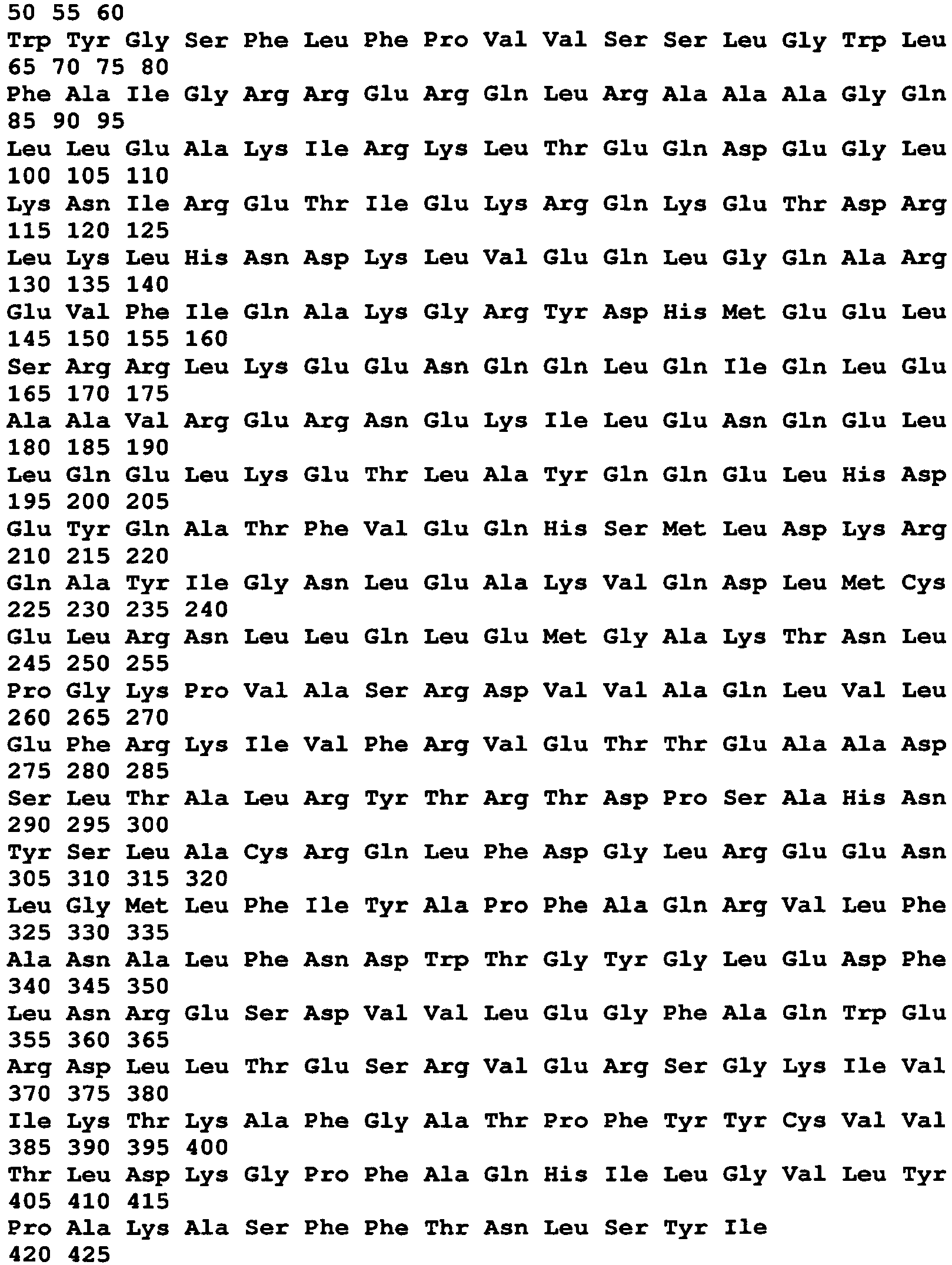
- <210> 8
- <211> 1287
- <212> DNA
- <213> Chlamydia trachomatis
- <400> 8


- <210> 9
- <211> 167
- <212> PRT
- <213> Chlamydia trachomatis
- <400> 9

- <210> 10
- <211> 501
- <212> DNA
- <213> Chlamydia trachomatis
- <400> 10

- <210> 11
- <211> 326
- <212> PRT
- <213> Chlamydia trachomatis
- <400> 11


- <210> 12
- <211> 978
- <212> DNA
- <213> Chlamydia trachomatis
- <400> 12

- <210> 13
- <211> 282
- <212> PRT
- <213> Chlamydia trachomatis
- <400> 13


- <210> 14
- <211> 846
- <212> DNA
- <213> Chlamydia trachomatis
- <400> 14

- <210> 15
- <211> 263
- <212> PRT
- <213> Chlamydia trachomatis
- <400> 15


- <210> 16
- <211> 789
- <212> DNA
- <213> Chlamydia trachomatis
- <400> 16

- <210> 17
- <211> 195
- <212> PRT
- <213> Chlamydia trachomatis
- <400> 17


- <210> 18
- <211> 585
- <212> DNA
- <213> Chlamydia trachomatis
- <400> 18

- <210> 19
- <211> 116
- <212> PRT
- <213> Chlamydia trachomatis
- <400> 19

- <210> 20
- <211> 348
- <212> DNA
- <213> Chlamydia trachomatis
- <400> 20

- <210> 21
- <211> 301
- <212> PRT
- <213> Chlamydia trachomatis
- <400> 21


- <210> 22
- <211> 903
- <212> DNA
- <213> Chlamydia trachomatis
- <400> 22

- <210> 23
- <211> 112
- <212> PRT
- <213> Chlamydia trachomatis
- <400> 23


- <210> 24
- <211> 339
- <212> DNA
- <213> Chlamydia trachomatis
- <400> 24

- <210> 25
- <211> 82
- <212> PRT
- <213> Chlamydia trachomatis
- <400> 25

- <210> 26
- <211> 249
- <212> DNA
- <213> Chlamydia trachomatis
- <400> 26

- <210> 27
- <211> 102
- <212> PRT
- <213> Chlamydia trachomatis
- <400> 27

- <210> 28
- <211> 306
- <212> DNA
- <213> Chlamydia trachomatis
- <400> 28

- <210> 29
- <211> 122
- <212> PRT
- <213> Chlamydia trachomatis
- <400> 29

- <210> 30
- <211> 366
- <212> DNA
- <213> Chlamydia trachomatis
- <400> 30

- <210> 31
- <211> 424
- <212> PRT
- <213> Chlamydia trachomatis
- <400> 31


- <210> 32
- <211> 1272
- <212> DNA
- <213> Chlamydia trachomatis
- <400> 32

- <210> 33
- <211> 359
- <212> PRT
- <213> Chlamydia trachomatis
- <400> 33

- <210> 34
- <211> 1077
- <212> DNA
- <213> Chlamydia trachomatis
- <400> 34


- <210> 35
- <211> 448
- <212> PRT
- <213> Chlamydia trachomatis
- <400> 35


- <210> 36
- <211> 1344
- <212> DNA
- <213> Chlamydia trachomatis
- <400> 36

- <210> 37
- <211> 287
- <212> PRT
- <213> Chlamydia trachomatis
- <400> 37


- <210> 38
- <211> 861
- <212> DNA
- <213> Chlamydia trachomatis
- <400> 38

- <210> 39
- <211> 560
- <212> PRT
- <213> Chlamydia trachomatis
- <400> 39


- <210> 40
- <211> 1680
- <212> DNA
- <213> Chlamydia trachomatis
- <400> 40
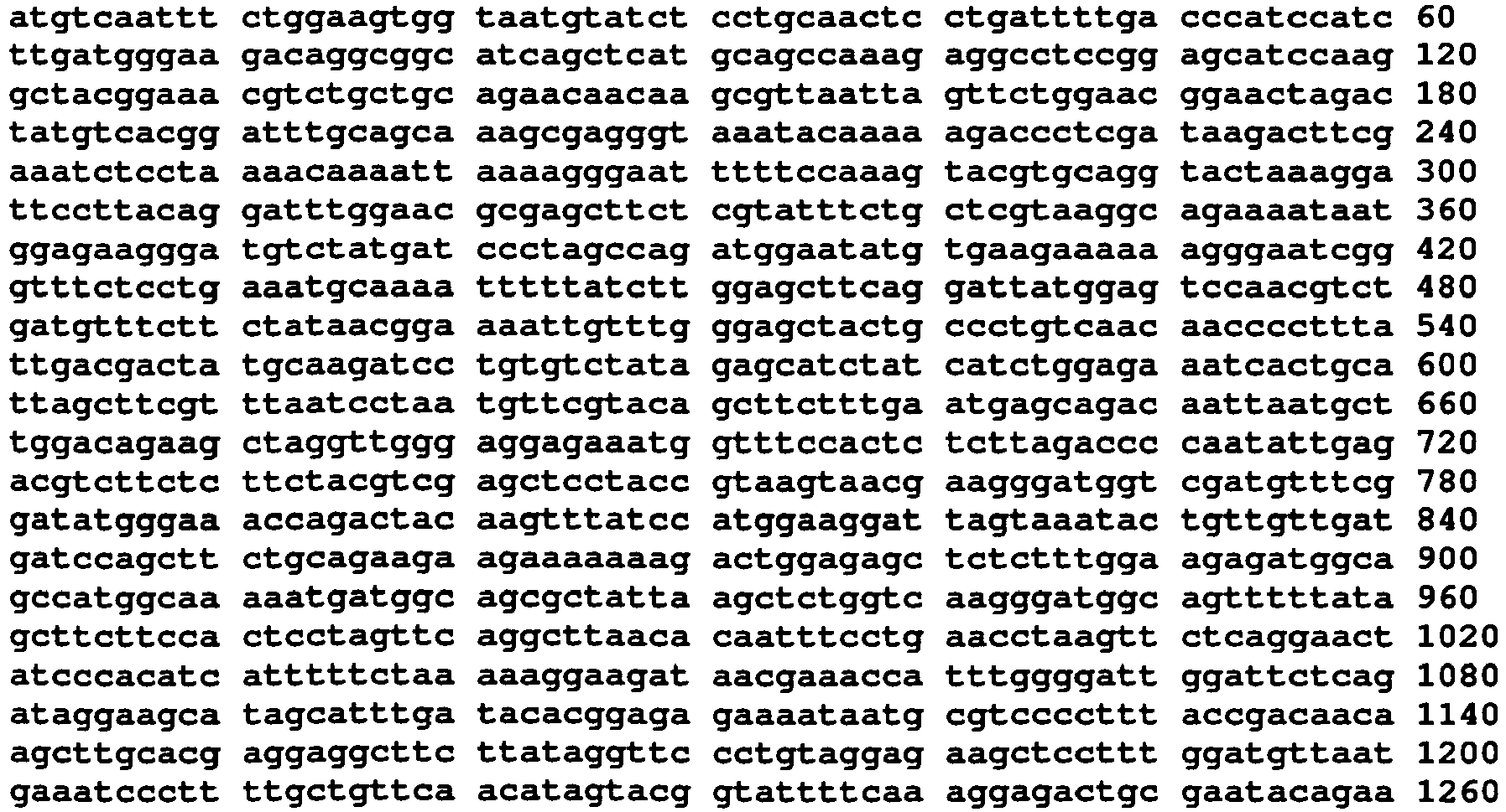

- <210> 41
- <211> 167
- <212> PRT
- <213> Chlamydia trachomatis
- <400> 41

- <210> 42
- <211> 501
- <212> DNA
- <213> Chlamydia trachomatis
- <400> 42

- <210> 43
- <211> 151
- <212> PRT
- <213> Chlamydia trachomatis
- <400> 43


- <210> 44
- <211> 453
- <212> DNA
- <213> Chlamydia trachomatis
- <400> 44

- <210> 45
- <211> 491
- <212> PRT
- <213> Chlamydia trachomatis
- <400> 45


- <210> 46
- <211> 1473
- <212> DNA
- <213> Chlamydia trachomatis
- <400> 46

- <210> 47
- <211> 363
- <212> PRT
- <213> Chlamydia trachomatis
- <400> 47


- <210> 48
- <211> 1089
- <212> DNA
- <213> Chlamydia trachomatis
- <400> 48

- <210> 49
- <211> 352
- <212> PRT
- <213> Chlamydia trachomatis
- <400> 49

- <210> 50
- <211> 1056
- <212> DNA
- <213> Chlamydia trachomatis
- <400> 50


- <210> 51
- <211> 550
- <212> PRT
- <213> Chlamydia trachomatis
- <400> 51
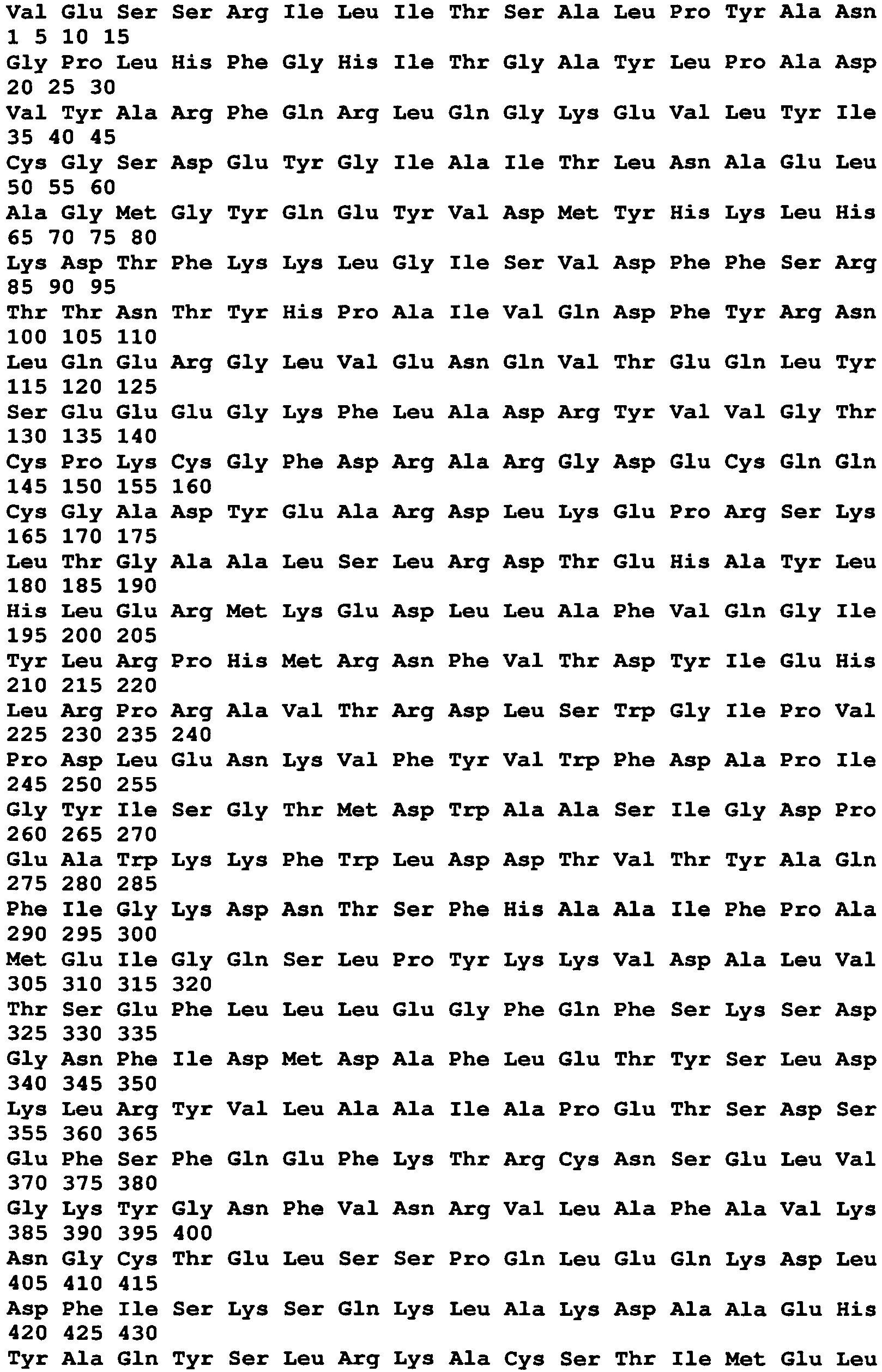

- <210> 52
- <211> 1650
- <212> DNA
- <213> Chlamydia trachomatis
- <400> 52

- <210> 53
- <211> 300
- <212> PRT
- <213> Chlamydia trachomatis
- <400> 53


- <210> 54
- <211> 900
- <212> DNA
- <213> Chlamydia trachomatis
- <400> 54

- <210> 55
- <211> 242
- <212> PRT
- <213> Chlamydia trachomatis
- <400> 55
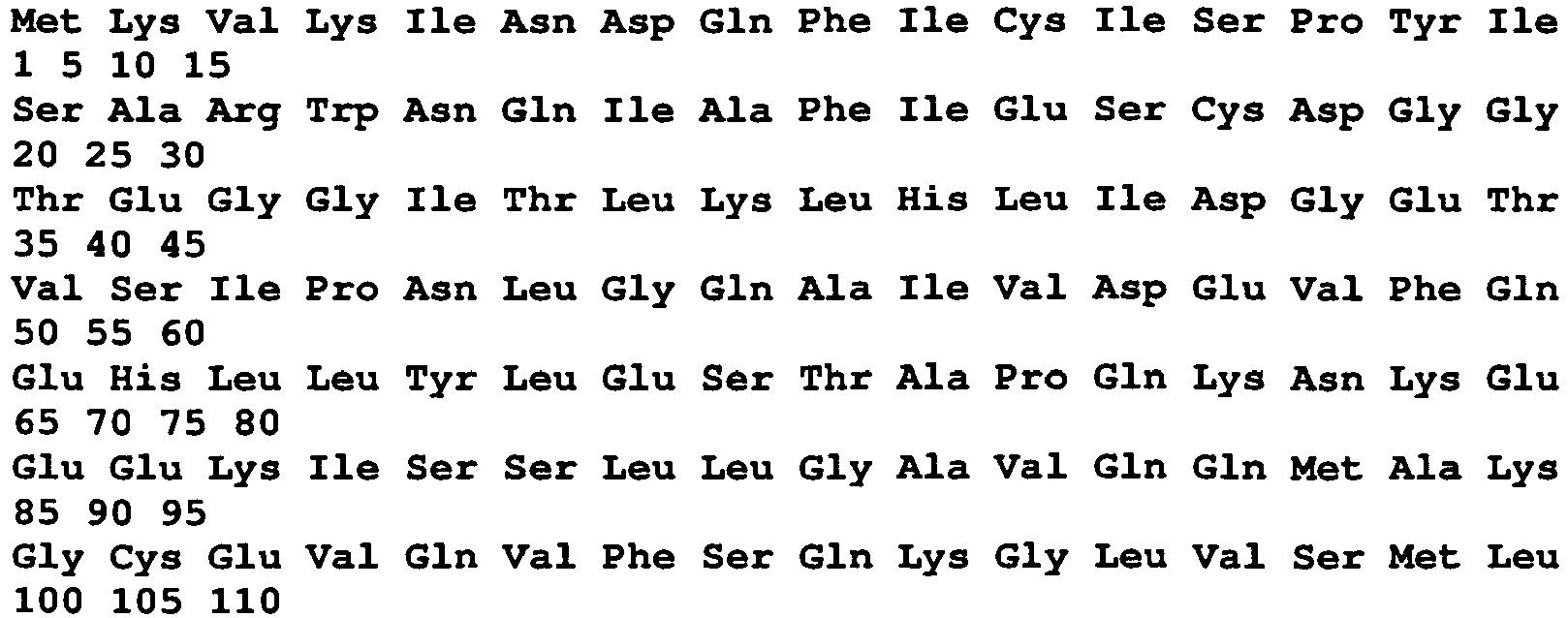

- <210> 56
- <211> 726
- <212> DNA
- <213> Chlamydia trachomatis
- <400> 56

- <210> 57
- <211> 121
- <212> PRT
- <213> Chlamydia trachomatis
- <400> 57

- <210> 58
- <211> 363
- <212> DNA
- <213> Chlamydia trachomatis
- <400> 58


- <210> 59
- <211> 257
- <212> PRT
- <213> Chlamydia trachomatis
- <400> 59

- <210> 60
- <211> 771
- <212> DNA
- <213> Chlamydia trachomatis
- <400> 60

- <210> 61
- <211> 148
- <212> PRT
- <213> Chlamydia trachomatis
- <400> 61

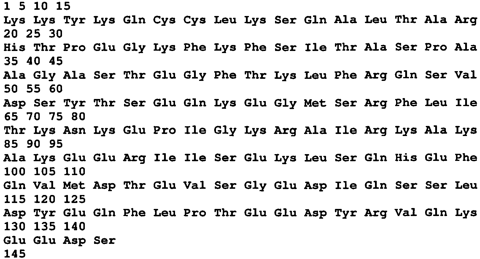
- <210> 62
- <211> 444
- <212> DNA
- <213> Chlamydia trachomatis
- <400> 62

- <210> 63
- <211> 857
- <212> PRT
- <213> Chlamydia trachomatis
- <400> 63


- <210> 64
- <211> 2571
- <212> DNA
- <213> Chlamydia trachomatis
- <400> 64

- <210> 65
- <211> 1770
- <212> PRT
- <213> Chlamydia trachomatis
- <400> 65


- <210> 66
- <211> 5310
- <212> DNA
- <213> Chlamydia trachomatis
- <400> 66


- <210> 67
- <211> 878
- <212> PRT
- <213> Chlamydia trachomatis
- <400> 67
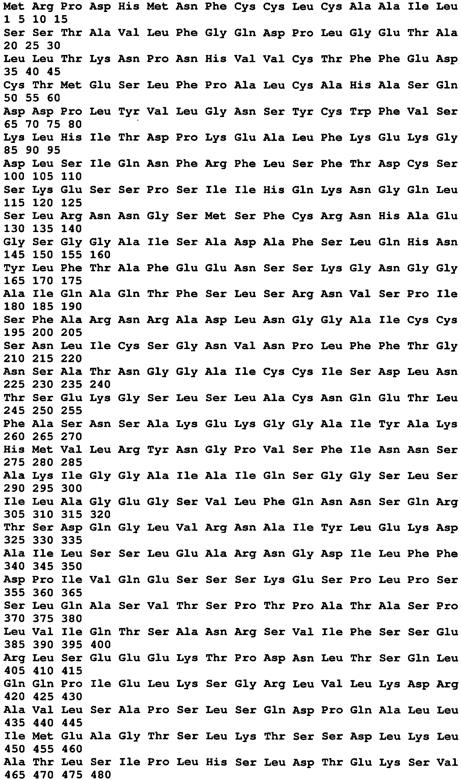

- <210> 68
- <211> 2634
- <212> DNA
- <213> Chlamydia trachomatis
- <400> 68


- <210> 69
- <211> 1005
- <212> PRT
- <213> Chlamydia trachomatis
- <400> 69


- <210> 70
- <211> 3015
- <212> DNA
- <213> Chlamydia trachomatis
- <400> 70


- <210> 71
- <211> 393
- <212> PRT
- <213> Chlamydia trachomatis
- <400> 71


- <210> 72
- <211> 1179
- <212> DNA
- <213> Chlamydia trachomatis
- <400> 72

- <210> 73
- <211> 164
- <212> PRT
- <213> Chlamydia trachomatis
- <400> 73


- <210> 74
- <211> 492
- <212> DNA
- <213> Chlamydia trachomatis
- <400> 74

- <210> 75
- <211> 150
- <212> PRT
- <213> Chlamydia trachomatis
- <400> 75

- <210> 76
- <211> 450
- <212> DNA
- <213> Chlamydia trachomatis
- <400> 76

- <210> 77
- <211> 129
- <212> PRT
- <213> Chlamydia trachomatis
- <400> 77


- <210> 78
- <211> 387
- <212> DNA
- <213> Chlamydia trachomatis
- <400> 78

- <210> 79
- <211> 267
- <212> PRT
- <213> Chlamydia trachomatis
- <400> 79

- <210> 80
- <211> 801
- <212> DNA
- <213> Chlamydia trachomatis
- <400> 80

- <210> 81
- <211> 52
- <212> PRT
- <213> Chlamydia trachomatis
- <400> 81

- <210> 82
- <211> 156
- <212> DNA
- <213> Chlamydia trachomatis
- <400> 82

- <210> 83
- <211> 529
- <212> PRT
- <213> Chlamydia trachomatis
- <400> 83


- <210> 84
- <211> 1587
- <212> DNA
- <213> Chlamydia trachomatis
- <400> 84


- <210> 85
- <211> 339
- <212> PRT
- <213> Chlamydia trachomatis
- <400> 85

- <210> 86
- <211> 1020
- <212> DNA
- <213> Chlamydia trachomatis
- <400> 86


- <210> 87
- <211> 160
- <212> PRT
- <213> Chlamydia trachomatis
- <400> 87

- <210> 88
- <211> 480
- <212> DNA
- <213> Chlamydia trachomatis
- <400> 88

- <210> 89
- <211> 421
- <212> PRT
- <213> Chlamydia trachomatis
- <400> 89


- <210> 90
- <211> 1263
- <212> DNA
- <213> Chlamydia trachomatis
- <400> 90

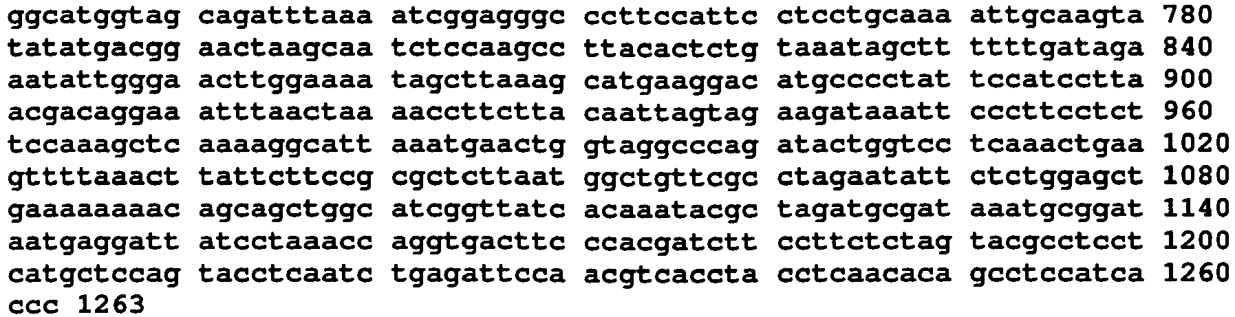
- <210> 91
- <211> 313
- <212> PRT
- <213> Chlamydia trachomatis
- <400> 91

- <210> 92
- <211> 939
- <212> DNA
- <213> Chlamydia trachomatis
- <400> 92


- <210> 93
- <211> 100
- <212> PRT
- <213> Chlamydia trachomatis
- <400> 93

- <210> 94
- <211> 300
- <212> DNA
- <213> Chlamydia trachomatis
- <400> 94

- <210> 95
- <211> 148
- <212> PRT
- <213> Chlamydia trachomatis
- <400> 95

- <210> 96
- <211> 444
- <212> DNA
- <213> Chlamydia trachomatis
- <400> 96
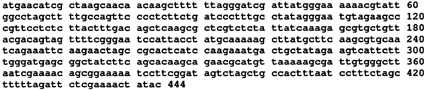
- <210> 97
- <211> 571
- <212> PRT
- <213> Chlamydia trachomatis
- <400> 97


- <210> 98
- <211> 1713
- <212> DNA
- <213> Chlamydia trachomatis
- <400> 98

- <210> 99
- <211> 90
- <212> PRT
- <213> Chlamydia trachomatis
- <400> 99


- <210> 100
- <211> 273
- <212> DNA
- <213> Chlamydia trachomatis
- <400> 100

- <210> 101
- <211> 695
- <212> PRT
- <213> Chlamydia trachomatis
- <400> 101
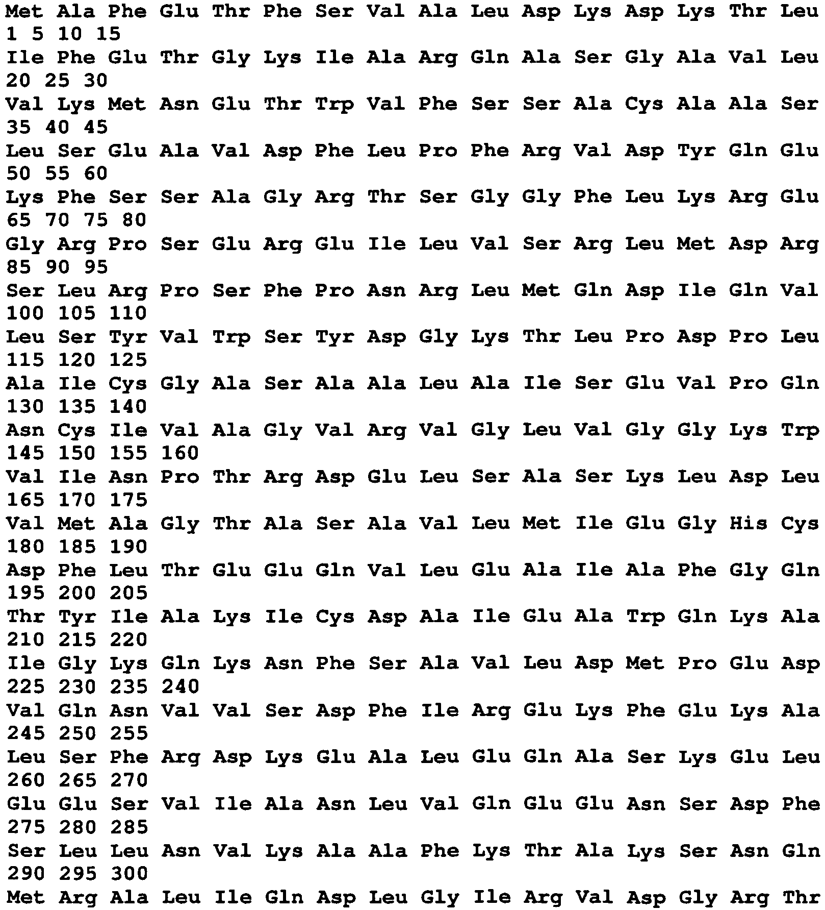

- <210> 102
- <211> 2085
- <212> DNA
- <213> Chlamydia trachomatis
- <400> 102
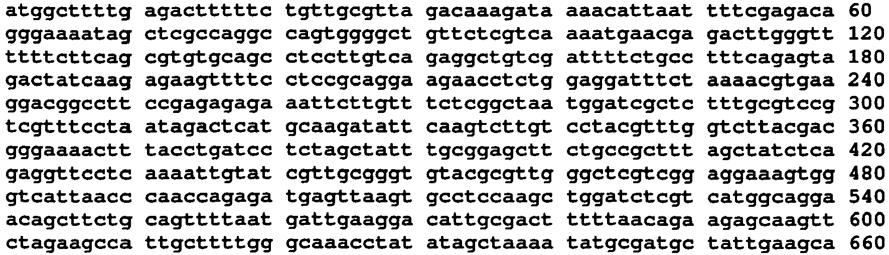

- <210> 103
- <211> 77
- <212> PRT
- <213> Chlamydia trachomatis
- <400> 103

- <210> 104
- <211> 234
- <212> DNA
- <213> Chlamydia trachomatis
- <400> 104

- <210> 105
- <211> 97
- <212> PRT
- <213> Chlamydia trachomatis
- <400> 105

- Leu
- <210> 106
- <211> 291
- <212> DNA
- <213> Chlamydia trachomatis
- <400> 106

- <210> 107
- <211> 361
- <212> PRT
- <213> Chlamydia trachomatis
- <400> 107

- <210> 108
- <211> 1083
- <212> DNA
- <213> Chlamydia trachomatis
- <400> 108
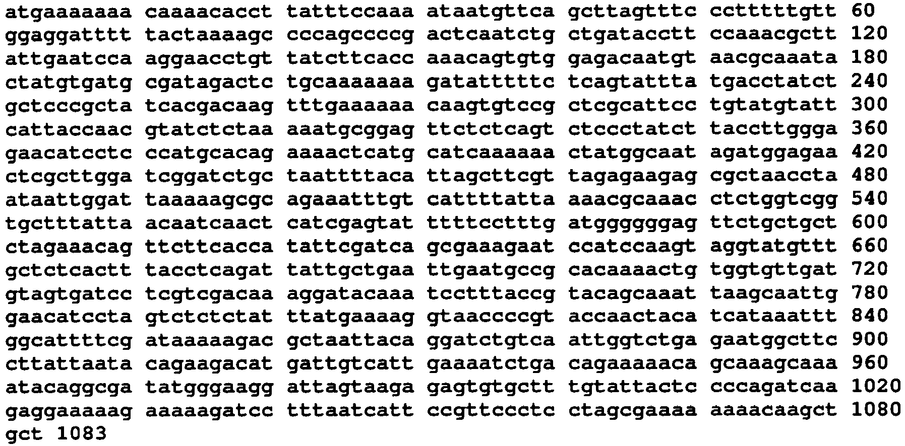
- <210> 109
- <211> 458
- <212> PRT
- <213> Chlamydia trachomatis
- <400> 109


- <210> 110
- <211> 1377
- <212> DNA
- <213> Chlamydia trachomatis
- <400> 110

- <210> 111
- <211> 544
- <212> PRT
- <213> Chlamydia trachomatis
- <400> 111


- <210> 112
- <211> 1632
- <212> DNA
- <213> Chlamydia trachomatis
- <400> 112


- <210> 113
- <211> 273
- <212> PRT
- <213> Chlamydia trachomatis
- <400> 113

- <210> 114
- <211> 819
- <212> DNA
- <213> Chlamydia trachomatis
- <400> 114

- <210> 115
- <211> 243
- <212> PRT
- <213> Chlamydia trachomatis
- <400> 115

- <210> 116
- <211> 729
- <212> DNA
- <213> Chlamydia trachomatis
- <400> 116


- <210> 117
- <211> 553
- <212> PRT
- <213> Chlamydia trachomatis
- <400> 117


- <210> 118
- <211> 1659
- <212> DNA
- <213> Chlamydia trachomatis
- <400> 118

- <210> 119
- <211> 163
- <212> PRT
- <213> Chlamydia trachomatis
- <400> 119


- <210> 120
- <211> 489
- <212> DNA
- <213> Chlamydia trachomatis
- <400> 120
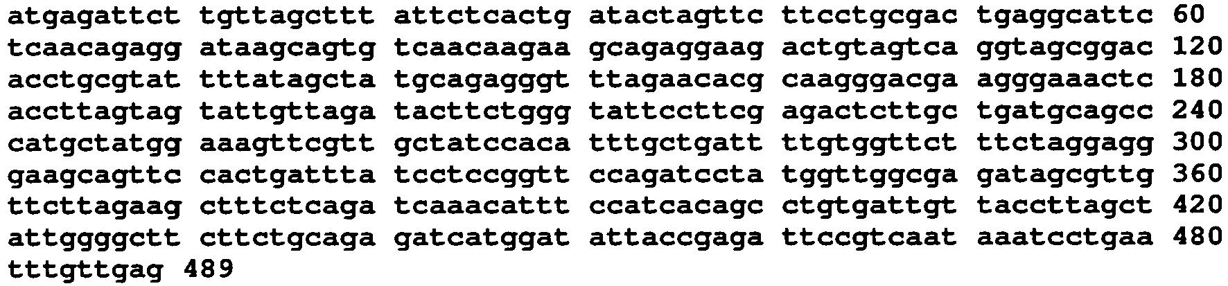
- <210> 121
- <211> 660
- <212> PRT
- <213> Chlamydia trachomatis
- <400> 121


- <210> 122
- <211> 1980
- <212> DNA
- <213> Chlamydia trachomatis
- <400> 122


- <210> 123
- <211> 698
- <212> PRT
- <213> Chlamydia trachomatis
- <400> 123


- <210> 124
- <211> 2094
- <212> DNA
- <213> Chlamydia trachomatis
- <400> 124


- <210> 125
- <211> 520
- <212> PRT
- <213> Chlamydia trachomatis
- <400> 125


- <210> 126
- <211> 1560
- <212> DNA
- <213> Chlamydia trachomatis
- <400> 126

- <210> 127
- <211> 133
- <212> PRT
- <213> Chlamydia trachomatis

- <210> 128
- <211> 399
- <212> DNA
- <213> Chlamydia trachomatis
- <400> 128

- <210> 129
- <211> 77
- <212> PRT
- <213> Chlamydia trachomatis
- <400> 129
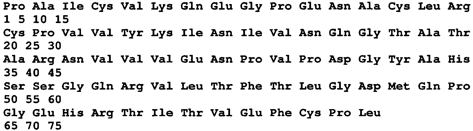
- <210> 130
- <211> 231
- <212> DNA
- <213> Chlamydia trachomatis
- <400> 130

- <210> 131
- <211> 163
- <212> PRT
- <213> Chlamydia trachomatis
- <400> 131


- <210> 132
- <211> 514
- <212> DNA
- <213> Chlamydia trachomatis
- <400> 132

- <210> 133
- <211> 149
- <212> PRT
- <213> Chlamydia trachomatis
- <400> 133

- <210> 134
- <211> 447
- <212> DNA
- <213> Chlamydia trachomatis
- <400> 134


- <210> 135
- <211> 82
- <212> PRT
- <213> Chlamydia trachomatis
- <400> 135

- <210> 136
- <211> 246
- <212> DNA
- <213> Chlamydia trachomatis
- <400> 136

- <210> 137
- <211> 101
- <212> PRT
- <213> Chlamydia trachomatis
- <400> 137

- <210> 138
- <211> 303
- <212> DNA
- <213> Chlamydia trachomatis
- <400> 138

- <210> 139
- <211> 97
- <212> PRT
- <213> Chlamydia trachomatis
- <400> 139


- <210> 140
- <211> 291
- <212> DNA
- <213> Chlamydia trachomatis
- <400> 140

- <210> 141
- <211> 141
- <212> PRT
- <213> Chlamydia trachomatis
- <400> 141

- <210> 142
- <211> 423
- <212> DNA
- <213> Chlamydia trachomatis
- <400> 142

- <210> 143
- <211> 110
- <212> PRT
- <213> Chlamydia trachomatis
- <400> 143


- <210> 144
- <211> 330
- <212> DNA
- <213> Chlamydia trachomatis
- <400> 144

- <210> 145
- <211> 73
- <212> PRT
- <213> Chlamydia trachomatis
- <400> 145

- <210> 146
- <211> 219
- <212> DNA
- <213> Chlamydia trachomatis
- <400> 146

- <210> 147
- <211> 236
- <212> PRT
- <213> Chlamydia trachomatis
- <400> 147

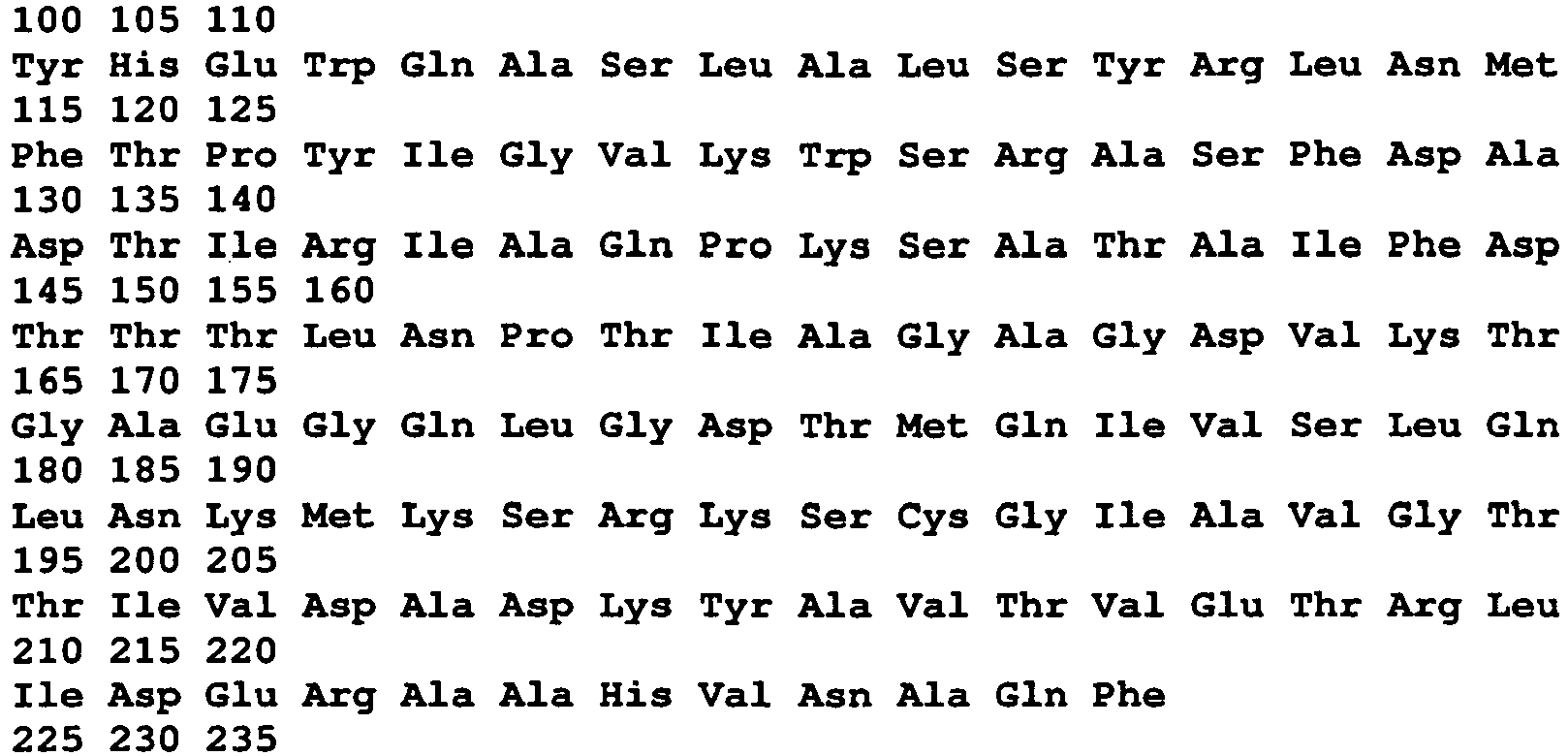
- <210> 148
- <211> 708
- <212> DNA
- <213> Chlamydia trachomatis
- <400> 148

- <210> 149
- <211> 131
- <212> PRT
- <213> Chlamydia trachomatis
- <400> 149

- <210> 150
- <211> 393
- <212> DNA
- <213> Chlamydia trachomatis
- <400> 150


- <210> 151
- <211> 56
- <212> PRT
- <213> Chlamydia trachomatis
- <400> 151

- <210> 152
- <211> 168
- <212> DNA
- <213> Chlamydia trachomatis
- <400> 152

- <210> 153
- <211> 118
- <212> PRT
- <213> Chlamydia trachomatis
- <400> 153

- <210> 154
- <211> 354
- <212> DNA
- <213> Chlamydia trachomatis
- <400> 154

- <210> 155
- <211> 68
- <212> PRT
- <213> Chlamydia trachomatis
- <400> 155


- <210> 156
- <211> 204
- <212> DNA
- <213> Chlamydia trachomatis
- <400> 156

- <210> 157
- <211> 243
- <212> PRT
- <213> Chlamydia trachomatis
- <400> 157

- <210> 158
- <211> 729
- <212> DNA
- <213> Chlamydia trachomatis
- <400> 158


- <210> 159
- <211> 93
- <212> PRT
- <213> Chlamydia trachomatis
- <400> 159

- <210> 160
- <211> 279
- <212> DNA
- <213> Chlamydia trachomatis
- <400> 160

- <210> 161
- <211> 79
- <212> PRT
- <213> Chlamydia trachomatis
- <400> 161

- <210> 162
- <211> 237
- <212> DNA
- <213> Chlamydia trachomatis
- <400> 162

- <210> 163
- <211> 83
- <212> PRT
- <213> Chlamydia trachomatis
- <400> 163


- <210> 164
- <211> 249
- <212> DNA
- <213> Chlamydia trachomatis
- <400> 164

- <210> 165
- <211> 105
- <212> PRT
- <213> Chlamydia trachomatis
- <400> 165

- <210> 166
- <211> 315
- <212> DNA
- <213> Chlamydia trachomatis
- <400> 166

- <210> 167
- <211> 164
- <212> PRT
- <213> Chlamydia trachomatis
- <400> 167


- <210> 168
- <211> 492
- <212> DNA
- <213> Chlamydia trachomatis
- <400> 168

- <210> 169
- <211> 110
- <212> PRT
- <213> Chlamydia trachomatis
- <400> 169

- <210> 170
- <211> 330
- <212> DNA
- <213> Chlamydia trachomatis
- <400> 170

- <210> 171
- <211> 90
- <212> PRT
- <213> Chlamydia trachomatis
- <400> 171


- <210> 172
- <211> 270
- <212> DNA
- <213> Chlamydia trachomatis
- <400> 172

- <210> 173
- <211> 23
- <212> PRT
- <213> Chlamydia trachomatis
- <400> 173

- <210> 174
- <211> 69
- <212> DNA
- <213> Chlamydia trachomatis
- <400> 174

- <210> 175
- <211> 23
- <212> PRT
- <213> Chlamydia trachomatis
- <400> 175

- <210> 176
- <211> 69
- <212> DNA
- <213> Chlamydia trachomatis
- <400> 176

- <210> 177
- <211> 23
- <212> PRT
- <213> Chlamydia trachomatis
- <400> 177

- <210> 178
- <211> 69
- <212> DNA
- <213> Chlamydia trachomatis <400> 178

- <210> 179
- <211> 23
- <212> PRT
- <213> Chlamydia trachomatis
- <400> 179

- <210> 180
- <211> 69
- <212> DNA
- <213> Chlamydia trachomatis
- <400> 180

- <210> 181
- <211> 23
- <212> PRT
- <213> Chlamydia trachomatis
- <400> 181

- <210> 182
- <211> 69
- <212> DNA
- <213> Chlamydia trachomatis
- <400> 182

- <210> 183
- <211> 28
- <212> PRT
- <213> Chlamydia trachomatis
- <400> 183

- <210> 184
- <211> 84
- <212> DNA
- <213> Chlamydia trachomatis
- <400> 184

- <210> 185
- <211> 106
- <212> PRT
- <213> Chlamydia trachomatis
- <400> 185

- <210> 186
- <211> 321
- <212> DNA
- <213> Chlamydia trachomatis
- <400> 186

- <210> 187
- <211> 159
- <212> PRT
- <213> Chlamydia trachomatis
- <400> 187

- <210> 188
- <211> 480
- <212> DNA
- <213> Chlamydia trachomatis
- <400> 188

- <210> 189
- <211> 437
- <212> PRT
- <213> Chlamydia trachomatis
- <400> 189


- <210> 190
- <211> 1314
- <212> DNA
- <213> Chlamydia trachomatis
- <400> 190


- <210> 191
- <211> 219
- <212> PRT
- <213> Chlamydia trachomatis
- <400> 191

- <210> 192
- <211> 660
- <212> DNA
- <213> Chlamydia trachomatis
- <400> 192

- <210> 193
- <211> 242
- <212> PRT
- <213> Chlamydia trachomatis
- <400> 193


- <210> 194
- <211> 729
- <212> DNA
- <213> Chlamydia trachomatis
- <400> 194
- <210> 195
- <211> 260
- <212> PRT
- <213> Chlamydia trachomatis
- <400> 195


- <210> 196
- <211> 783
- <212> DNA
- <213> Chlamydia trachomatis
- <400> 196

- <210> 197
- <211> 483
- <212> PRT
- <213> Chlamydia trachomatis
- <400> 197


- <210> 198
- <211> 1452
- <212> DNA
- <213> Chlamydia trachomatis
- <400> 198


- <210> 199
- <211> 307
- <212> PRT
- <213> Chlamydia trachomatis
- <400> 199

- <210> 200
- <211> 924
- <212> DNA
- <213> Chlamydia trachomatis
- <400> 200

- <210> 201
- <211> 389
- <212> PRT
- <213> Chlamydia trachomatis
- <400> 201

- <210> 202
- <211> 1170
- <212> DNA
- <213> Chlamydia trachomatis
- <400> 202


- <210> 203
- <211> 148
- <212> PRT
- <213> Chlamydia trachomatis
- <400> 203

- <210> 204
- <211> 447
- <212> DNA
- <213> Chlamydia trachomatis
- <400> 204

- <210> 205
- <211> 249
- <212> PRT
- <213> Chlamydia trachomatis
- <400> 205


- <210> 206
- <211> 750
- <212> DNA
- <213> Chlamydia trachomatis
- <400> 206
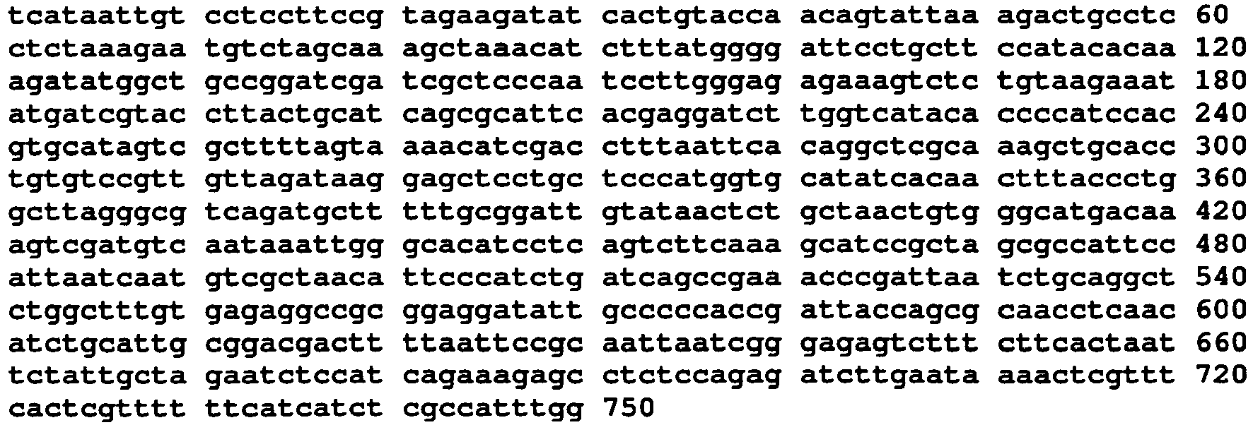
- <210> 207
- <211> 246
- <212> PRT
- <213> Chlamydia trachomatis
- <400> 207


- <210> 208
- <211> 741
- <212> DNA
- <213> Chlamydia trachomatis
- <400> 208

- <210> 209
- <211> 254
- <212> PRT
- <213> Chlamydia trachomatis
- <400> 209


- <210> 210
- <211> 765
- <212> DNA
- <213> Chlamydia trachomatis
- <400> 210

- <210> 211
- <211> 271
- <212> PRT
- <213> Chlamydia trachomatis
- <400> 211


- <210> 212
- <211> 816
- <212> DNA
- <213> Chlamydia trachomatis
- <400> 212

- <210> 213
- <211> 601
- <212> PRT
- <213> Chlamydia trachomatis
- <400> 213


- <210> 214
- <211> 1806
- <212> DNA
- <213> Chlamydia trachomatis
- <400> 214
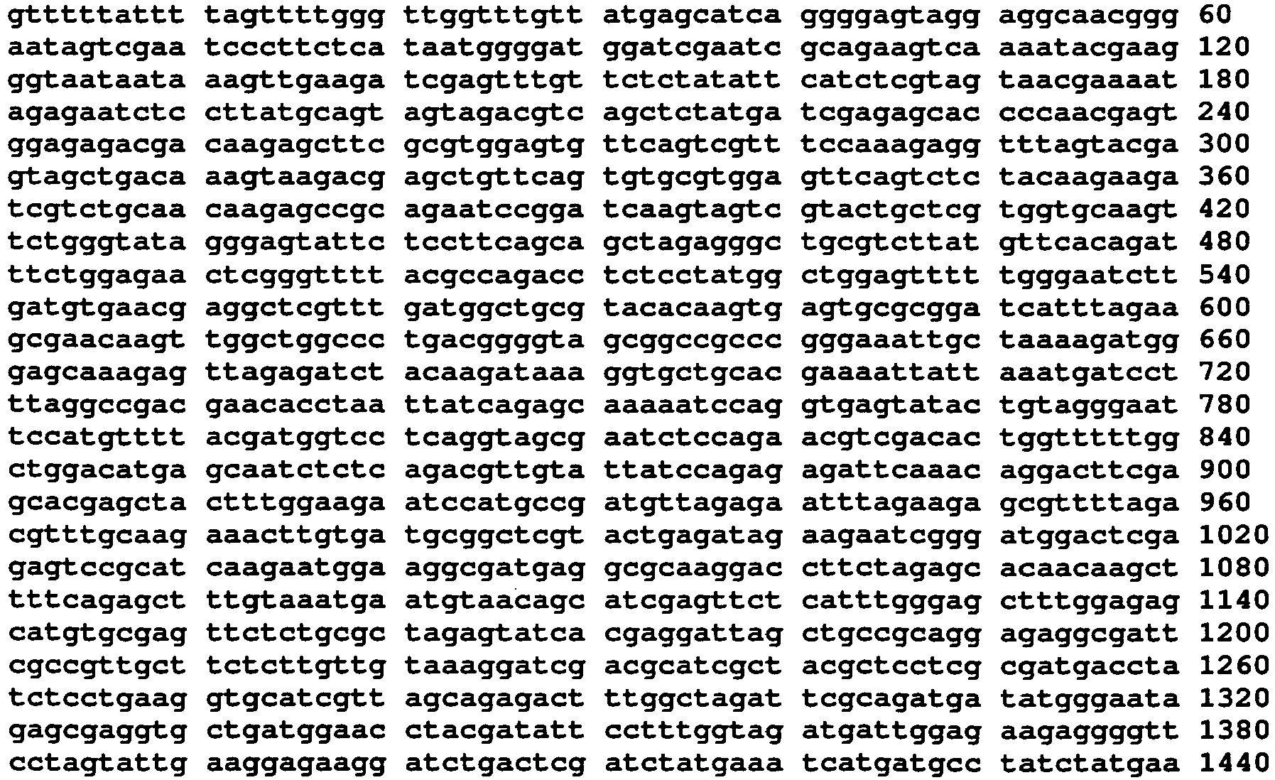

- <210> 215
- <211> 416
- <212> PRT
- <213> Chlamydia trachomatis
- <400> 215

- <210> 216
- <211> 1251
- <212> DNA
- <213> Chlamydia trachomatis
- <400> 216

- <210> 217
- <211> 616
- <212> PRT
- <213> Chlamydia trachomatis
- <400> 217

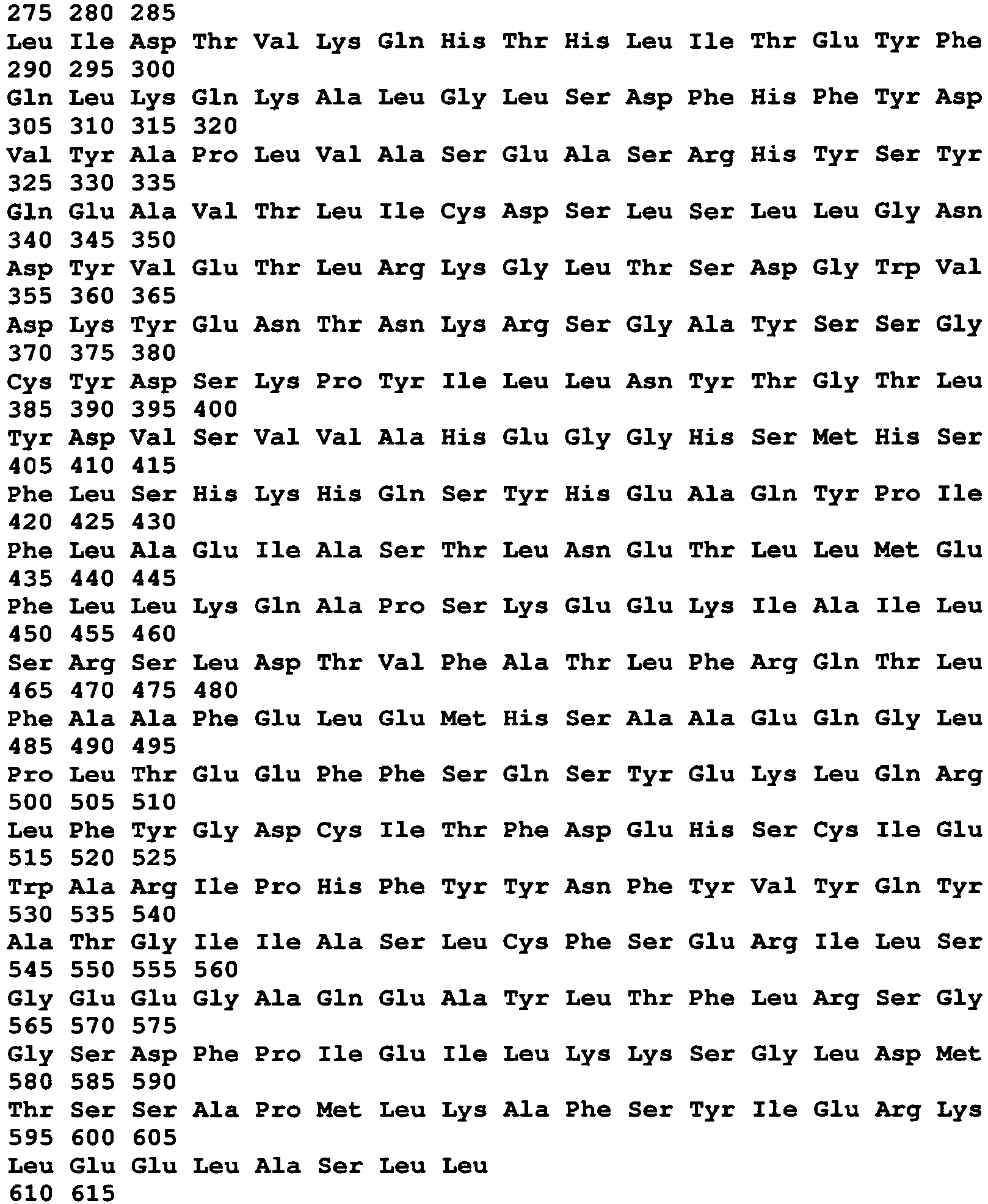
- <210> 218
- <211> 1851
- <212> DNA
- <213> Chlamydia trachomatis
- <400> 218
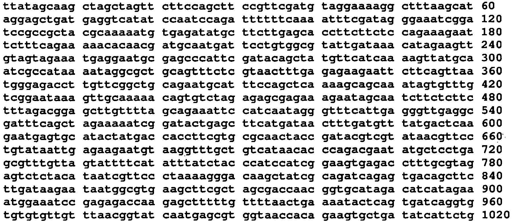

- <210> 219
- <211> 1258
- <212> PRT
- <213> Chlamydia trachomatis
- <400> 219


- <210> 220
- <211> 3777
- <212> DNA
- <213> Chlamydia trachomatis
- <400> 220


- <210> 221
- <211> 250
- <212> PRT
- <213> Chlamydia trachomatis
- <400> 221


- <210> 222
- <211> 753
- <212> DNA
- <213> Chlamydia trachomatis
- <400> 222

- <210> 223
- <211> 1462
- <212> PRT
- <213> Chlamydia trachomatis
- <400> 223
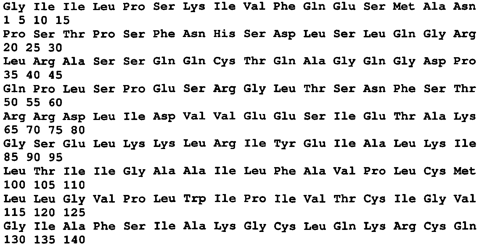

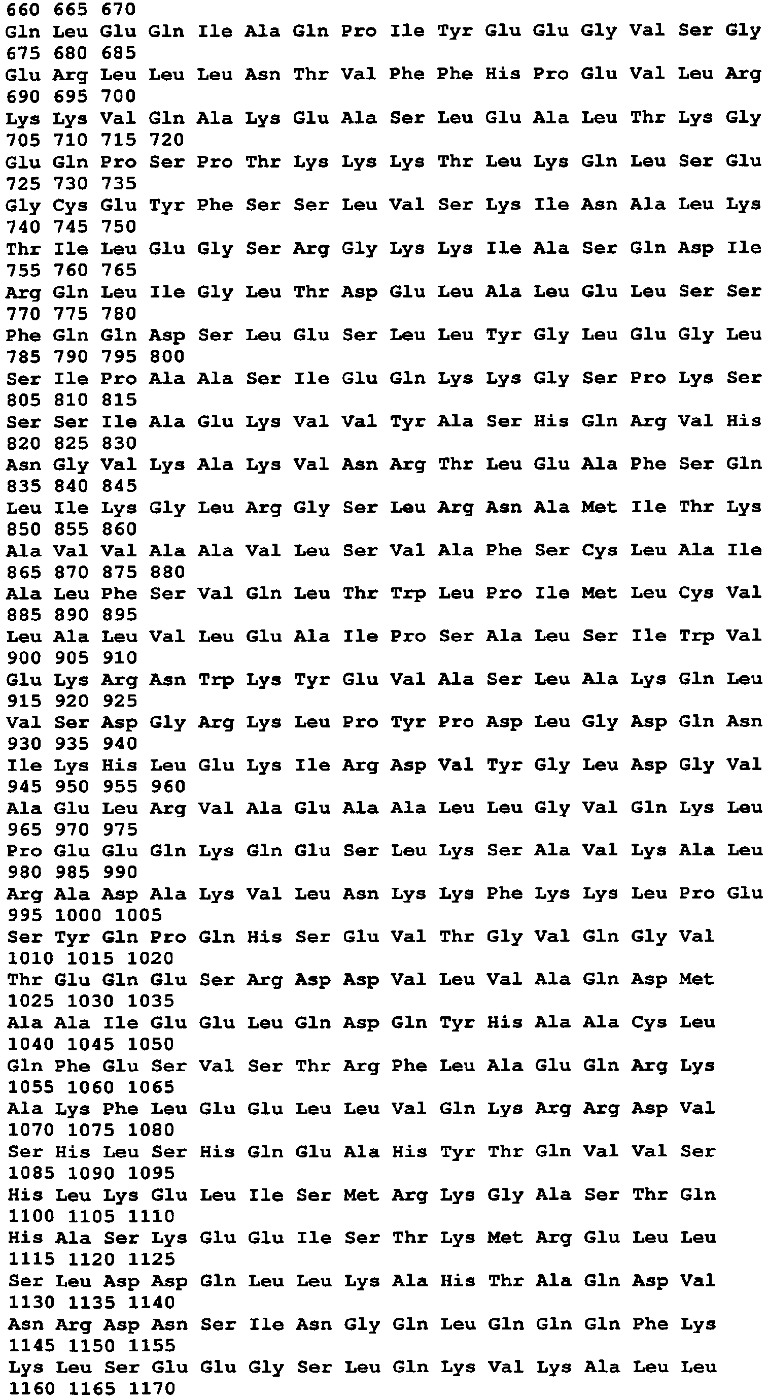

- <210> 224
- <211> 4389
- <212> DNA
- <213> Chlamydia trachomatis
- <400> 224
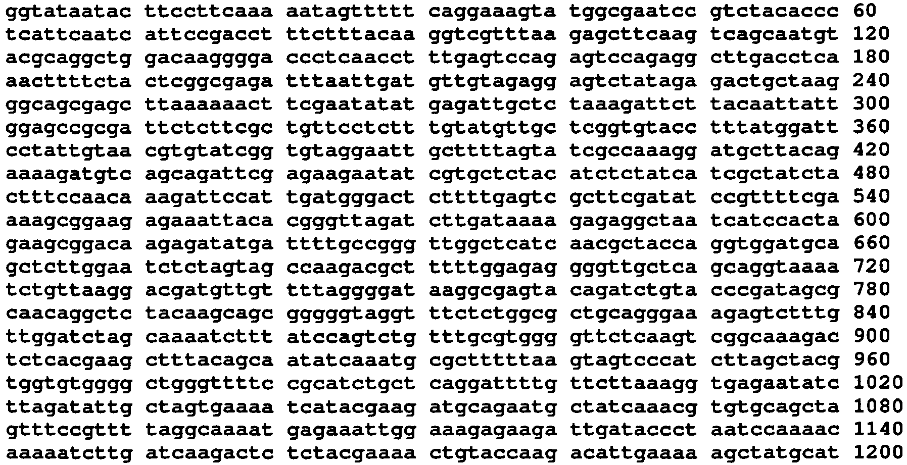

- <210> 225
- <211> 200
- <212> PRT
- <213> Chlamydia trachomatis
- <400> 225


- <210> 226
- <211> 603
- <212> DNA
- <213> Chlamydia trachomatis
- <400> 226

- <210> 227
- <211> 120
- <212> PRT
- <213> Chlamydia trachomatis
- <400> 227

- <210> 228
- <211> 363
- <212> DNA
- <213> Chlamydia trachomatis
- <400> 228


- <210> 229
- <211> 130
- <212> PRT
- <213> Chlamydia trachomatis
- <400> 229

- <210> 230
- <211> 393
- <212> DNA
- <213> Chlamydia trachomatis
- <400> 230

- <210> 231
- <211> 569
- <212> PRT
- <213> Chlamydia trachomatis
- <400> 231


- <210> 232
- <211> 1710
- <212> DNA
- <213> Chlamydia trachomatis
- <400> 232
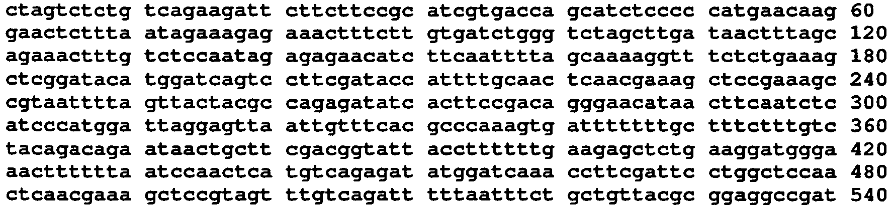
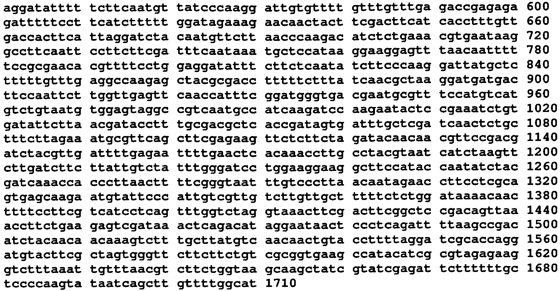
- <210> 233
- <211> 334
- <212> PRT
- <213> Chlamydia trachomatis
- <400> 233


- <210> 234
- <211> 1005
- <212> DNA
- <213> Chlamydia trachomatis
- <400> 234

- <210> 235
- <211> 355
- <212> PRT
- <213> Chlamydia trachomatis
- <400> 235


- <210> 236
- <211> 1068
- <212> DNA
- <213> Chlamydia trachomatis
- <400> 236

- <210> 237
- <211> 490
- <212> PRT
- <213> Chlamydia trachomatis
- <400> 237


- <210> 238
- <211> 1473
- <212> DNA
- <213> Chlamydia trachomatis
- <400> 238

- <210> 239
- <211> 121
- <212> PRT
- <213> Chlamydia trachomatis
- <400> 239

- <210> 240
- <211> 366
- <212> DNA
- <213> Chlamydia trachomatis
- <400> 240

- <210> 241
- <211> 342
- <212> PRT
- <213> Chlamydia trachomatis
- <400> 241


- <210> 242
- <211> 1029
- <212> DNA
- <213> Chlamydia trachomatis
- <400> 242

- <210> 243
- <211> 382
- <212> PRT
- <213> Chlamydia trachomatis
- <400> 243


- <210> 244
- <211> 1149
- <212> DNA
- <213> Chlamydia trachomatis
- <400> 244

- <210> 245
- <211> 169
- <212> PRT
- <213> Chlamydia trachomatis
- <400> 245


- <210> 246
- <211> 510
- <212> DNA
- <213> Chlamydia trachomatis
- <400> 246

- <210> 247
- <211> 277
- <212> PRT
- <213> Chlamydia trachomatis
- <400> 247


- <210> 248
- <211> 831
- <212> DNA
- <213> Chlamydia trachomatis
- <400> 248

- <210> 249
- <211> 340
- <212> PRT
- <213> Chlamydia trachomatis
- <400> 249


- <210> 250
- <211> 1020
- <212> DNA
- <213> Chlamydia trachomatis
- <400> 250

- <210> 251
- <211> 328
- <212> PRT
- <213> Chlamydia trachomatis
- <400> 251


- <210> 252
- <211> 984
- <212> DNA
- <213> Chlamydia trachomatis
- <400> 252

- <210> 253
- <211> 199
- <212> PRT
- <213> Chlamydia trachomatis
- <400> 253

- <210> 254
- <211> 597
- <212> DNA
- <213> Chlamydia trachomatis
- <400> 254
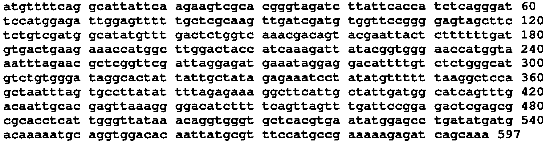
- <210> 255
- <211> 107
- <212> PRT
- <213> Chlamydia trachomatis
- <400> 255

- <210> 256
- <211> 321
- <212> DNA
- <213> Chlamydia trachomatis
- <400> 256

- <210> 257
- <211> 369
- <212> PRT
- <213> Chlamydia trachomatis
- <400> 257


- <210> 258
- <211> 1107
- <212> DNA
- <213> Chlamydia trachomatis
- <400> 258

- <210> 259
- <211> 377
- <212> PRT
- <213> Chlamydia trachomatis
- <400> 259


- <210> 260
- <211> 1131
- <212> DNA
- <213> Chlamydia trachomatis
- <400> 260


- <210> 261
- <211> 165
- <212> PRT
- <213> Chlamydia trachomatis
- <400> 261

- <210> 262
- <211> 495
- <212> DNA
- <213> Chlamydia trachomatis
- <400> 262

- <210> 263
- <211> 123
- <212> PRT
- <213> Chlamydia trachomatis
- <400> 263

- <210> 264
- <211> 369
- <212> DNA
- <213> Chlamydia trachomatis
- <400> 264

- <210> 265
- <211> 183
- <212> PRT
- <213> Chlamydia trachomatis
- <400> 265

- <210> 266
- <211> 549
- <212> DNA
- <213> Chlamydia trachomatis
- <400> 266

- <210> 267
- <211> 180
- <212> PRT
- <213> Chlamydia trachomatis
- <400> 267


- <210> 268
- <211> 540
- <212> DNA
- <213> Chlamydia trachomatis
- <400> 268

- <210> 269
- <211> 130
- <212> PRT
- <213> Chlamydia trachomatis
- <400> 269

- <210> 270
- <211> 390
- <212> DNA
- <213> Chlamydia trachomatis
- <400> 270


- <210> 271
- <211> 123
- <212> PRT
- <213> Chlamydia trachomatis
- <400> 271

- <210> 272
- <211> 369
- <212> DNA
- <213> Chlamydia trachomatis
- <400> 272

- <210> 273
- <211> 202
- <212> PRT
- <213> Chlamydia trachomatis
- <400> 273


- <210> 274
- <211> 606
- <212> DNA
- <213> Chlamydia trachomatis
- <400> 274

- <210> 275
- <211> 72
- <212> PRT
- <213> Chlamydia trachomatis
- <400> 275

- <210> 276
- <211> 216
- <212> DNA
- <213> Chlamydia trachomatis
- <400> 276

- <210> 277
- <211> 111
- <212> PRT
- <213> Chlamydia trachomatis
- <400> 277

- <210> 278
- <211> 333
- <212> DNA
- <213> Chlamydia trachomatis
- <400> 278

- <210> 279
- <211> 111
- <212> PRT
- <213> Chlamydia trachomatis
- <400> 279

- <210> 280
- <211> 333
- <212> DNA
- <213> Chlamydia trachomatis
- <400> 280

- <210> 281
- <211> 243
- <212> PRT
- <213> Chlamydia trachomatis
- <400> 281


- <210> 282
- <211> 729
- <212> DNA
- <213> Chlamydia trachomatis
- <400> 282
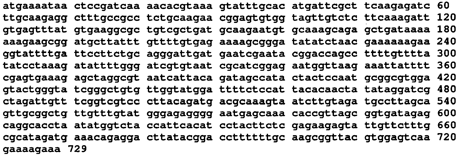
- <210> 283
- <211> 450
- <212> PRT
- <213> Chlamydia trachomatis
- <400> 283


- <210> 284
- <211> 1350
- <212> DNA
- <213> Chlamydia trachomatis
- <400> 284

- <210> 285
- <211> 209
- <212> PRT
- <213> Chlamydia trachomatis
- <400> 285


- <210> 286
- <211> 627
- <212> DNA
- <213> Chlamydia trachomatis
- <400> 286

- <210> 287
- <211> 227
- <212> PRT
- <213> Chlamydia trachomatis
- <400> 287


- <210> 288
- <211> 681
- <212> DNA
- <213> Chlamydia trachomatis
- <400> 288

- <210> 289
- <211> 192
- <212> PRT
- <213> Chlamydia trachomatis
- <400> 289

- <210> 290
- <211> 576
- <212> DNA
- <213> Chlamydia trachomatis
- <400> 290


- <210> 291
- <211> 178
- <212> PRT
- <213> Chlamydia trachomatis
- <400> 291

- <210> 292
- <211> 534
- <212> DNA
- <213> Chlamydia trachomatis
- <400> 292
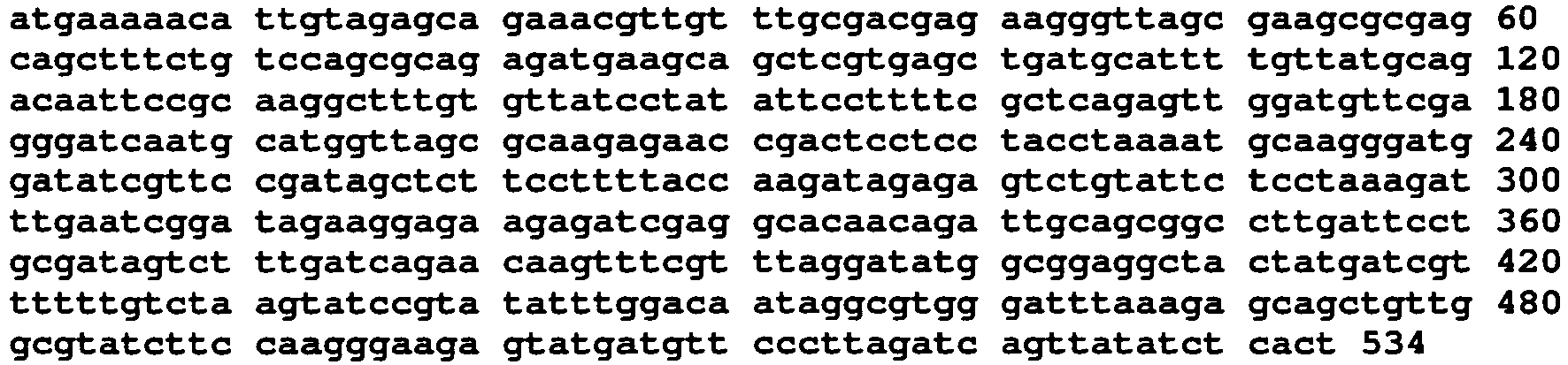
- <210> 293
- <211> 184
- <212> PRT
- <213> Chlamydia trachomatis
- <400> 293


- <210> 294
- <211> 552
- <212> DNA
- <213> Chlamydia trachomatis
- <400> 294

- <210> 295
- <211> 221
- <212> PRT
- <213> Chlamydia trachomatis
- <400> 295

- <210> 296
- <211> 663
- <212> DNA
- <213> Chlamydia trachomatis
- <400> 296


- <210> 297
- <211> 229
- <212> PRT
- <213> Chlamydia trachomatis
- <400> 297

- <210> 298
- <211> 687
- <212> DNA
- <213> Chlamydia trachomatis
- <400> 298

- <210> 299
- <211> 112
- <212> PRT
- <213> Chlamydia trachomatis
- <400> 299

- <210> 300
- <211> 336
- <212> DNA
- <213> Chlamydia trachomatis
- <400> 300

- <210> 301
- <211> 193
- <212> PRT
- <213> Chlamydia trachomatis
- <400> 301

- <210> 302
- <211> 579
- <212> DNA
- <213> Chlamydia trachomatis
- <400> 302


- <210> 303
- <211> 123
- <212> PRT
- <213> Chlamydia trachomatis
- <400> 303

- <210> 304
- <211> 369
- <212> DNA
- <213> Chlamydia trachomatis
- <400> 304

- <210> 305
- <211> 342
- <212> PRT
- <213> Chlamydia trachomatis
- <400> 305


- <210> 306
- <211> 1026
- <212> DNA
- <213> Chlamydia trachomatis
- <400> 306

- <210> 307
- <211> 92
- <212> PRT
- <213> Chlamydia trachomatis
- <400> 307

- <210> 308
- <211> 276
- <212> DNA
- <213> Chlamydia trachomatis
- <400> 308

Claims (15)
- Use of a substantially pure polypeptide, which comprises an amino acid sequence selected from(a) SEQ ID NO. 1, or(b) an immunogenic portion of the sequence in (a), or(c) an amino acid sequence analogue having at least 90% sequence identity to any one of the sequences in (a) or (b) and at the same time being immunogenic, or the nucleic acid encoding for polypeptides in (a), (b) or (c), for preparing a pharmaceutical composition for preventing or treating infections caused by a bacteria from a Chlamydia species, wherein the polypeptide is fused to a fusion partner.
- Use according to claim 1, where the polypeptide is lipidated so as to allow a self-adjuvating effect of the polypeptide.
- Use according to any of claims 1-2, where the fusion partner comprises a(a) a polypeptide derived from a Chlamydia species, or(b) at least one immunogenic portion of any of such polypeptides in (a).
- Use according to any of claims 1-2, where the nucleic acid sequence encoding for the polypeptide in (a) has the following corresponding sequence : SEQ ID NO 2.
- A pharmaceutical composition for use preventing or treating infections caused by a bacteria from a Chlamydia species, comprising a substantially pure polypeptide, which comprises an amino acid sequence selected from(a) SEQ ID NO. 1, or(b) an immunogenic portion of the sequence in (a); and /or(c) an amino acid sequence analogue having at least 90% sequence identity to any one of the sequences in (a) or (b) and at the same time being immunogenic or the nucleic acid encoding for (a), (b) or (c), wherein the polypeptide is fused to a fusion partner.
- A pharmaceutical composition for use according to claim 5, where the polypeptide is lipidated so as to allow a self-adjuvating effect of the polypeptide.
- A pharmaceutical composition for use according to claim 5, where the fusion partner comprises a(a) a polypeptide derived from a chlamydia species, or(b) at least one immunogenic portion of any of such polypeptides in (a), or the nucleic acid encoding herefor.
- A pharmaceutical composition for use according to claim 5 where the nucleic acid sequences encoding for polypeptides in (a) has the following corresponding sequence: SEQ ID NO. 2.
- A pharmaceutical composition for use according to claim 5-8, which is in the form of a vaccine.
- A vaccine according to claim 9 fo use in immunizing against or treating infections caused by a bacteria from a Chlamydia species.
- A vaccine for use according to claim 10, where the Chlamydia species is C.trachomatis.
- A monoclonal antibody which is specifically reacting with an amino acid sequence selected from:(a) SEQ ID NO. 1, or(b) an immunogenic portion of the sequence in (a); and /or(c) an amino acid sequence analogue having at least 90% sequence identity to any one of the sequences in (a) or (b) and at the same time being immunogenic in an immunoassay or a specific binding fragment of said antibody.
- A method for diagnosing previous or ongoing infection with a bacterium from a Chlamydia species, said method comprising contacting a sample, with a diagnostic reagent in order to detect a positive reaction, wherein said diagnostic reagent comprises a substantially pure polypeptide, which comprises an amino acid sequence selected from(a) SEQ ID NO.1, or(b) an immunogenic portion of the sequence in (a), or(c) an amino acid sequence analogue having at least 90% sequence identity to any one of the sequence in (a) or (b) and at the same time being immunogenic, or the nucleic acid encoding for polypeptides in (a), (b) or (c).
- A method for diagnosing previous or ongoing infection with a bacterium from a Chlamydia species said method comprising(a) contacting a sample with an antibody according to claim 12 in order to detect a positive reaction in case of infection
- An in vitro method of diagnosing previous or ongoing infection with a bacterium from a Chlamydia species in a subject comprising:(a) contacting a diagnostic reagent with a bodily fluid of the subject, wherein said diagnostic reagent comprises a substantially pure polypeptide, which comprises an amino acid sequence selected from(1) SEQ ID NO.1, or(2) an immunogenic portion of the sequence in (1), or(3) an amino acid sequence analogue having at least 90% sequence identity to any one of the sequence in (1) or (2) and at the same time being immunogenic, or the nucleic acid encoding for polypeptides in (1), (2) or (3);(b) detecting binding of an antibody to said polypeptide, said binding being an indication that said subject is infected by a bacterium from a Chlamydia species.
Priority Applications (5)
| Application Number | Priority Date | Filing Date | Title |
|---|---|---|---|
| PL05790937T PL1812058T3 (en) | 2004-10-25 | 2005-10-11 | Chlamydia trachomatis antigens for vaccine and diagnostic use |
| SI200531580T SI1812058T1 (en) | 2004-10-25 | 2005-10-11 | Chlamydia trachomatis antigens for vaccine and diagnostic use |
| EP10178623A EP2269636A3 (en) | 2004-10-25 | 2005-10-11 | Chlamydia trachomatis antigens for vaccine and diagnostic use |
| EP10185754A EP2314314A3 (en) | 2004-10-25 | 2005-10-11 | Chlamydia trachomatis antigens for vaccine and diagnostic use |
| CY20121100817T CY1113601T1 (en) | 2004-10-25 | 2012-09-10 | CHLAMYDIA C.TRACHOMATIS ANTIGONS FOR USE IN VACCINES AND DIAGNOSIS |
Applications Claiming Priority (3)
| Application Number | Priority Date | Filing Date | Title |
|---|---|---|---|
| DKPA200401633 | 2004-10-25 | ||
| DKPA200501069 | 2005-07-19 | ||
| PCT/DK2005/000651 WO2006045308A2 (en) | 2004-10-25 | 2005-10-11 | Chlamydia trachomatis antigens for vaccine and diagnostic use |
Related Child Applications (2)
| Application Number | Title | Priority Date | Filing Date |
|---|---|---|---|
| EP10178623.4 Division-Into | 2010-09-23 | ||
| EP10185754.8 Division-Into | 2010-10-01 |
Publications (2)
| Publication Number | Publication Date |
|---|---|
| EP1812058A2 EP1812058A2 (en) | 2007-08-01 |
| EP1812058B1 true EP1812058B1 (en) | 2012-06-13 |
Family
ID=35686446
Family Applications (3)
| Application Number | Title | Priority Date | Filing Date |
|---|---|---|---|
| EP05790937A Expired - Lifetime EP1812058B1 (en) | 2004-10-25 | 2005-10-11 | Chlamydia trachomatis antigens for vaccine and diagnostic use |
| EP10178623A Ceased EP2269636A3 (en) | 2004-10-25 | 2005-10-11 | Chlamydia trachomatis antigens for vaccine and diagnostic use |
| EP10185754A Ceased EP2314314A3 (en) | 2004-10-25 | 2005-10-11 | Chlamydia trachomatis antigens for vaccine and diagnostic use |
Family Applications After (2)
| Application Number | Title | Priority Date | Filing Date |
|---|---|---|---|
| EP10178623A Ceased EP2269636A3 (en) | 2004-10-25 | 2005-10-11 | Chlamydia trachomatis antigens for vaccine and diagnostic use |
| EP10185754A Ceased EP2314314A3 (en) | 2004-10-25 | 2005-10-11 | Chlamydia trachomatis antigens for vaccine and diagnostic use |
Country Status (11)
| Country | Link |
|---|---|
| US (1) | US8889142B2 (en) |
| EP (3) | EP1812058B1 (en) |
| JP (3) | JP5024830B2 (en) |
| CA (2) | CA2585011C (en) |
| CY (1) | CY1113601T1 (en) |
| DK (1) | DK1812058T3 (en) |
| ES (1) | ES2389562T3 (en) |
| PL (1) | PL1812058T3 (en) |
| PT (1) | PT1812058E (en) |
| SI (1) | SI1812058T1 (en) |
| WO (1) | WO2006045308A2 (en) |
Families Citing this family (23)
| Publication number | Priority date | Publication date | Assignee | Title |
|---|---|---|---|---|
| US20020061848A1 (en) | 2000-07-20 | 2002-05-23 | Ajay Bhatia | Compounds and methods for treatment and diagnosis of chlamydial infection |
| US8541007B2 (en) | 2005-03-31 | 2013-09-24 | Glaxosmithkline Biologicals S.A. | Vaccines against chlamydial infection |
| US8133973B2 (en) | 2005-05-12 | 2012-03-13 | Novartis Vaccines And Diagnostics, S.R.L. | Immunogenic compositions for Chlamydia trachomatis |
| US9259463B2 (en) * | 2006-01-16 | 2016-02-16 | The United States Of America, As Represented By The Secretary, Department Of Health & Human Services | Chlamydia vaccine |
| EP1970708A1 (en) * | 2007-03-07 | 2008-09-17 | Mikrogen Molekularbiologische Entwicklungs-GmbH | Bio chip and method for selective proof of Chlamydia trachomatis infections |
| EP2152303A4 (en) * | 2007-05-01 | 2011-10-05 | Univ Texas | CHLAMYDIA ANTIGENS AS REAGENTS FOR THE DIAGNOSIS AND TREATMENT OF CHLAMYDIA INFECTION AND DISEASE |
| WO2010027442A1 (en) * | 2008-08-26 | 2010-03-11 | Becton, Dickinson And Company | Assay for chlamydia trachomatis by amplification and detection of chlamydia trachomatis cytotoxin gene |
| JP2012505212A (en) * | 2008-10-09 | 2012-03-01 | ボード・オヴ・リージェンツ,ユニヴァーシティ・オヴ・テキサス・システム | Methods and compositions relating to chlamydia antigens for the diagnosis and treatment of chlamydia infections and diseases |
| WO2010085896A1 (en) * | 2009-01-29 | 2010-08-05 | British Columbia Cancer Agency Branch | Compositions comprising chlamydia antigens |
| WO2010108908A1 (en) * | 2009-03-24 | 2010-09-30 | Transgene Sa | Biomarker for monitoring patients |
| WO2011063133A1 (en) * | 2009-11-18 | 2011-05-26 | Auburn University | LOW ANTIGEN-DOSE IMMUNIZATION FOR MAXIMIZING T-HELPER CELL 1 (T h1) IMMUNITY AGAINST DISEASE |
| EP2544715A2 (en) * | 2010-03-09 | 2013-01-16 | Board Of Regents, University Of Texas System | Methods and compositions for chlamydial antigens for diagnosis and treatment of chlamydial infection and disease |
| CN103068837B (en) | 2010-05-28 | 2015-06-24 | 斯匹遐生物技术公司 | Chimeric momp antigen, method and use |
| CA2849391A1 (en) * | 2010-10-20 | 2012-04-26 | Genocea Biosciences, Inc. | Chlamydia antigens and uses thereof |
| WO2013044398A1 (en) * | 2011-09-30 | 2013-04-04 | The University Of British Columbia | Chlamydia antigen compositions and uses thereof |
| WO2013181224A1 (en) * | 2012-05-29 | 2013-12-05 | The Research Foundation For The State University Of New York | Combined therapy and prophylaxis for genital tract infections |
| ITNA20120046A1 (en) * | 2012-08-02 | 2014-02-03 | No Self S R L | USE OF NUCLEIC ACIDS OF PARASITIC BIOLOGICAL SYSTEMS, PATOGENES AND WEATHER FOR THE INHIBITION AND / OR CONTROL OF THE SAME SYSTEMS |
| RS60135B1 (en) | 2013-03-18 | 2020-05-29 | Statens Seruminstitut | Vaccines against chlamydia sp. |
| US10835594B2 (en) | 2014-01-16 | 2020-11-17 | Mcmaster University | Type III secretion injectisome proteins for treatment and prevention of chlamydial infections |
| NL2022581B1 (en) * | 2019-02-14 | 2020-08-27 | Koppert Bv | Composition comprising a mixture of dna molecules, uses thereof as biological inhibitor and method for production |
| WO2022140038A1 (en) * | 2020-12-23 | 2022-06-30 | Daykin Molecular Systems, LLC | Nucleic acid stabilizing solution for vaccines, therapy, diagnostics, storage, and transport |
| JP2026504797A (en) | 2023-03-02 | 2026-02-10 | サノフィ・パスツール | Compositions for use in the treatment of chlamydia |
| CN117106078B (en) * | 2023-07-18 | 2024-09-17 | 华南农业大学 | A polyclonal antibody for rice orange leaf phytoplasma antigen membrane protein and its application |
Family Cites Families (17)
| Publication number | Priority date | Publication date | Assignee | Title |
|---|---|---|---|---|
| US4816567A (en) | 1983-04-08 | 1989-03-28 | Genentech, Inc. | Recombinant immunoglobin preparations |
| US4599230A (en) | 1984-03-09 | 1986-07-08 | Scripps Clinic And Research Foundation | Synthetic hepatitis B virus vaccine including both T cell and B cell determinants |
| US4599231A (en) | 1984-03-09 | 1986-07-08 | Scripps Clinic And Research Foundation | Synthetic hepatitis B virus vaccine including both T cell and B cell determinants |
| US4608251A (en) | 1984-11-09 | 1986-08-26 | Pitman-Moore, Inc. | LHRH analogues useful in stimulating anti-LHRH antibodies and vaccines containing such analogues |
| US4601903A (en) | 1985-05-01 | 1986-07-22 | The United States Of America As Represented By The Department Of Health And Human Services | Vaccine against Neisseria meningitidis Group B serotype 2 invasive disease |
| GB9626864D0 (en) * | 1996-12-24 | 1997-02-12 | Smithkline Beecham Biolog | Vaccine |
| CA2704354A1 (en) | 1995-06-07 | 1996-12-19 | Connaught Laboratories, Inc. | Expression of lipoproteins |
| US6136958A (en) | 1996-09-30 | 2000-10-24 | Genentech, Inc. | Antibodies to vertebrate smoothened proteins |
| EP0892061A3 (en) * | 1997-07-18 | 1999-02-24 | Smithkline Beecham Corporation | The Chlamydia trachomatis Glutamyl-tRNAGln amidotransferase subunit ratB |
| JP2002536958A (en) * | 1997-11-21 | 2002-11-05 | ジェンセット | Chlamydiapneumoniae genomic sequences and polypeptides, fragments thereof, and their use especially for the diagnosis, prevention and treatment of infectious diseases |
| EP2218732A1 (en) * | 1997-11-28 | 2010-08-18 | Merck Serono Biodevelopment | Chlamydia trachomatis genomic sequence and polypeptides, fragments thereof and uses thereof, in particular for the diagnosis, prevention and treatment of infection |
| US6919187B2 (en) | 2000-04-21 | 2005-07-19 | Corixa Corporation | Compounds and methods for treatment and diagnosis of chlamydial infection |
| US6736222B2 (en) | 2001-11-05 | 2004-05-18 | Vector Magnetics, Llc | Relative drill bit direction measurement |
| EP2168596B1 (en) * | 2001-12-12 | 2012-05-23 | Novartis Vaccines and Diagnostics S.r.l. | Immunisation against chlamydia trachomatis |
| CA2516971A1 (en) * | 2003-02-24 | 2004-09-02 | Institut Pasteur | Secreted chlamydia polypeptides, polynucleotides coding therefor, therapeutic and diagnostic uses thereof |
| CA2526106A1 (en) * | 2003-06-26 | 2005-01-13 | Chiron Corporation | Immunogenic compositions for chlamydia trachomatis |
| WO2006050571A1 (en) | 2004-11-11 | 2006-05-18 | Queensland University Of Technology | Chlamydia antigens and uses thereof |
-
2005
- 2005-10-11 ES ES05790937T patent/ES2389562T3/en not_active Expired - Lifetime
- 2005-10-11 PT PT05790937T patent/PT1812058E/en unknown
- 2005-10-11 WO PCT/DK2005/000651 patent/WO2006045308A2/en not_active Ceased
- 2005-10-11 JP JP2007537113A patent/JP5024830B2/en not_active Expired - Fee Related
- 2005-10-11 CA CA2585011A patent/CA2585011C/en not_active Expired - Fee Related
- 2005-10-11 EP EP05790937A patent/EP1812058B1/en not_active Expired - Lifetime
- 2005-10-11 DK DK05790937.6T patent/DK1812058T3/en active
- 2005-10-11 EP EP10178623A patent/EP2269636A3/en not_active Ceased
- 2005-10-11 SI SI200531580T patent/SI1812058T1/en unknown
- 2005-10-11 PL PL05790937T patent/PL1812058T3/en unknown
- 2005-10-11 US US11/577,868 patent/US8889142B2/en not_active Expired - Fee Related
- 2005-10-11 EP EP10185754A patent/EP2314314A3/en not_active Ceased
- 2005-10-11 CA CA2820140A patent/CA2820140A1/en not_active Abandoned
-
2011
- 2011-12-14 JP JP2011273006A patent/JP2012102108A/en active Pending
-
2012
- 2012-09-10 CY CY20121100817T patent/CY1113601T1/en unknown
-
2013
- 2013-06-27 JP JP2013135131A patent/JP2013231050A/en active Pending
Non-Patent Citations (1)
| Title |
|---|
| FRANK FOLLMANN ET AL.: "Antigenic profiling of a Chlamydia trachomatis gene-expression library", JID, vol. 197, 15 March 2008 (2008-03-15), pages 897 - 905 * |
Also Published As
| Publication number | Publication date |
|---|---|
| JP5024830B2 (en) | 2012-09-12 |
| WO2006045308A8 (en) | 2011-08-11 |
| WO2006045308A3 (en) | 2006-12-07 |
| WO2006045308A2 (en) | 2006-05-04 |
| DK1812058T3 (en) | 2012-09-17 |
| PT1812058E (en) | 2012-09-17 |
| ES2389562T3 (en) | 2012-10-29 |
| JP2013231050A (en) | 2013-11-14 |
| SI1812058T1 (en) | 2013-02-28 |
| JP2012102108A (en) | 2012-05-31 |
| CY1113601T1 (en) | 2016-06-22 |
| CA2585011C (en) | 2014-02-18 |
| CA2585011A1 (en) | 2006-05-04 |
| JP2008520550A (en) | 2008-06-19 |
| PL1812058T3 (en) | 2012-11-30 |
| EP2269636A3 (en) | 2011-04-06 |
| US8889142B2 (en) | 2014-11-18 |
| EP1812058A2 (en) | 2007-08-01 |
| EP2269636A2 (en) | 2011-01-05 |
| EP2314314A2 (en) | 2011-04-27 |
| CA2820140A1 (en) | 2006-05-04 |
| US20090304722A1 (en) | 2009-12-10 |
| EP2314314A3 (en) | 2011-05-11 |
Similar Documents
| Publication | Publication Date | Title |
|---|---|---|
| US8889142B2 (en) | Chlamydia trachomatis antigens for vaccine and diagnostic use | |
| US7927609B2 (en) | Compounds and methods for immunotherapy and diagnosis of tuberculosis | |
| JP2008520550A5 (en) | ||
| JP7510142B2 (en) | Vaccine against Chlamydia sp. | |
| EP1203817A2 (en) | Compounds and methods for immunotherapy and diagnosis of tuberculosis | |
| US20110305721A1 (en) | Compounds and methods for immunotherapy and diagnosis of tuberculosis | |
| EP2248900A1 (en) | Compounds for immunotherapy and diagnosis of tuberculosis and methods of their use | |
| WO1997009428A9 (en) | Compounds and methods for immunotherapy and diagnosis of tuberculosis | |
| AU4036597A (en) | Compounds and methods for treatment and diagnosis of mycobacterial infections | |
| CA2505724C (en) | Malaria vaccine | |
| EP1073458A1 (en) | Chlamydia proteins and their uses | |
| US7026300B2 (en) | One step immunization procedure for inducing a Chlamydia specific immune response | |
| US6436898B1 (en) | Compounds for treatment of infectious and immune system disorders and methods for their use | |
| HK1148775A (en) | Compounds for immunotherapy and diagnosis of tuberculosis and methods of their use | |
| HK1099338A (en) | Compounds and methods for immunotherapy and diagnosis of tuberculosis | |
| HK1150852A (en) | Compounds for immunotherapy and diagnosis of tuberculosis and methods of their use |
Legal Events
| Date | Code | Title | Description |
|---|---|---|---|
| PUAI | Public reference made under article 153(3) epc to a published international application that has entered the european phase |
Free format text: ORIGINAL CODE: 0009012 |
|
| 17P | Request for examination filed |
Effective date: 20070524 |
|
| AK | Designated contracting states |
Kind code of ref document: A2 Designated state(s): AT BE BG CH CY CZ DE DK EE ES FI FR GB GR HU IE IS IT LI LT LU LV MC NL PL PT RO SE SI SK TR |
|
| DAX | Request for extension of the european patent (deleted) | ||
| 17Q | First examination report despatched |
Effective date: 20080215 |
|
| RAP1 | Party data changed (applicant data changed or rights of an application transferred) |
Owner name: STATENS SERUM INSTITUT |
|
| RAP1 | Party data changed (applicant data changed or rights of an application transferred) |
Owner name: STATENS SERUM INSTITUT |
|
| GRAP | Despatch of communication of intention to grant a patent |
Free format text: ORIGINAL CODE: EPIDOSNIGR1 |
|
| GRAS | Grant fee paid |
Free format text: ORIGINAL CODE: EPIDOSNIGR3 |
|
| GRAA | (expected) grant |
Free format text: ORIGINAL CODE: 0009210 |
|
| RAP1 | Party data changed (applicant data changed or rights of an application transferred) |
Owner name: STATENS SERUM INSTITUT |
|
| AK | Designated contracting states |
Kind code of ref document: B1 Designated state(s): AT BE BG CH CY CZ DE DK EE ES FI FR GB GR HU IE IS IT LI LT LU LV MC NL PL PT RO SE SI SK TR |
|
| REG | Reference to a national code |
Ref country code: GB Ref legal event code: FG4D |
|
| REG | Reference to a national code |
Ref country code: CH Ref legal event code: EP Ref country code: AT Ref legal event code: REF Ref document number: 561689 Country of ref document: AT Kind code of ref document: T Effective date: 20120615 |
|
| REG | Reference to a national code |
Ref country code: IE Ref legal event code: FG4D |
|
| REG | Reference to a national code |
Ref country code: DE Ref legal event code: R096 Ref document number: 602005034740 Country of ref document: DE Effective date: 20120809 |
|
| REG | Reference to a national code |
Ref country code: CH Ref legal event code: NV Representative=s name: FIAMMENGHI-FIAMMENGHI |
|
| REG | Reference to a national code |
Ref country code: PT Ref legal event code: SC4A Free format text: AVAILABILITY OF NATIONAL TRANSLATION Effective date: 20120911 Ref country code: DK Ref legal event code: T3 |
|
| REG | Reference to a national code |
Ref country code: SE Ref legal event code: TRGR |
|
| REG | Reference to a national code |
Ref country code: NL Ref legal event code: T3 |
|
| REG | Reference to a national code |
Ref country code: EE Ref legal event code: FG4A Ref document number: E006879 Country of ref document: EE Effective date: 20120906 |
|
| REG | Reference to a national code |
Ref country code: ES Ref legal event code: FG2A Ref document number: 2389562 Country of ref document: ES Kind code of ref document: T3 Effective date: 20121029 Ref country code: GR Ref legal event code: EP Ref document number: 20120402024 Country of ref document: GR Effective date: 20120915 |
|
| REG | Reference to a national code |
Ref country code: PL Ref legal event code: T3 |
|
| PG25 | Lapsed in a contracting state [announced via postgrant information from national office to epo] |
Ref country code: RO Free format text: LAPSE BECAUSE OF FAILURE TO SUBMIT A TRANSLATION OF THE DESCRIPTION OR TO PAY THE FEE WITHIN THE PRESCRIBED TIME-LIMIT Effective date: 20120613 Ref country code: IS Free format text: LAPSE BECAUSE OF FAILURE TO SUBMIT A TRANSLATION OF THE DESCRIPTION OR TO PAY THE FEE WITHIN THE PRESCRIBED TIME-LIMIT Effective date: 20121013 Ref country code: SK Free format text: LAPSE BECAUSE OF FAILURE TO SUBMIT A TRANSLATION OF THE DESCRIPTION OR TO PAY THE FEE WITHIN THE PRESCRIBED TIME-LIMIT Effective date: 20120613 |
|
| REG | Reference to a national code |
Ref country code: HU Ref legal event code: AG4A Ref document number: E014948 Country of ref document: HU |
|
| PLBE | No opposition filed within time limit |
Free format text: ORIGINAL CODE: 0009261 |
|
| STAA | Information on the status of an ep patent application or granted ep patent |
Free format text: STATUS: NO OPPOSITION FILED WITHIN TIME LIMIT |
|
| 26N | No opposition filed |
Effective date: 20130314 |
|
| REG | Reference to a national code |
Ref country code: DE Ref legal event code: R097 Ref document number: 602005034740 Country of ref document: DE Effective date: 20130314 |
|
| PG25 | Lapsed in a contracting state [announced via postgrant information from national office to epo] |
Ref country code: BG Free format text: LAPSE BECAUSE OF FAILURE TO SUBMIT A TRANSLATION OF THE DESCRIPTION OR TO PAY THE FEE WITHIN THE PRESCRIBED TIME-LIMIT Effective date: 20120913 |
|
| REG | Reference to a national code |
Ref country code: FR Ref legal event code: PLFP Year of fee payment: 11 |
|
| REG | Reference to a national code |
Ref country code: FR Ref legal event code: PLFP Year of fee payment: 12 |
|
| REG | Reference to a national code |
Ref country code: FR Ref legal event code: PLFP Year of fee payment: 13 |
|
| REG | Reference to a national code |
Ref country code: FR Ref legal event code: PLFP Year of fee payment: 14 |
|
| PG25 | Lapsed in a contracting state [announced via postgrant information from national office to epo] |
Ref country code: CY Free format text: LAPSE BECAUSE OF NON-PAYMENT OF DUE FEES Effective date: 20191011 |
|
| PGFP | Annual fee paid to national office [announced via postgrant information from national office to epo] |
Ref country code: CZ Payment date: 20200918 Year of fee payment: 16 Ref country code: LU Payment date: 20200911 Year of fee payment: 16 Ref country code: NL Payment date: 20200911 Year of fee payment: 16 Ref country code: GR Payment date: 20200911 Year of fee payment: 16 Ref country code: MC Payment date: 20200918 Year of fee payment: 16 Ref country code: DK Payment date: 20200917 Year of fee payment: 16 Ref country code: EE Payment date: 20200911 Year of fee payment: 16 Ref country code: IE Payment date: 20200911 Year of fee payment: 16 Ref country code: LT Payment date: 20200914 Year of fee payment: 16 Ref country code: TR Payment date: 20200916 Year of fee payment: 16 Ref country code: FR Payment date: 20200922 Year of fee payment: 16 Ref country code: GB Payment date: 20200922 Year of fee payment: 16 Ref country code: PT Payment date: 20200911 Year of fee payment: 16 Ref country code: FI Payment date: 20200911 Year of fee payment: 16 |
|
| PGFP | Annual fee paid to national office [announced via postgrant information from national office to epo] |
Ref country code: BE Payment date: 20200911 Year of fee payment: 16 Ref country code: PL Payment date: 20200914 Year of fee payment: 16 Ref country code: SE Payment date: 20200916 Year of fee payment: 16 Ref country code: CH Payment date: 20200916 Year of fee payment: 16 Ref country code: LV Payment date: 20200911 Year of fee payment: 16 |
|
| PGFP | Annual fee paid to national office [announced via postgrant information from national office to epo] |
Ref country code: ES Payment date: 20201105 Year of fee payment: 16 Ref country code: DE Payment date: 20200911 Year of fee payment: 16 Ref country code: CY Payment date: 20200929 Year of fee payment: 16 Ref country code: IT Payment date: 20200911 Year of fee payment: 16 Ref country code: HU Payment date: 20200911 Year of fee payment: 16 Ref country code: AT Payment date: 20200911 Year of fee payment: 16 |
|
| PGFP | Annual fee paid to national office [announced via postgrant information from national office to epo] |
Ref country code: SI Payment date: 20200911 Year of fee payment: 16 |
|
| REG | Reference to a national code |
Ref country code: DE Ref legal event code: R082 Ref document number: 602005034740 Country of ref document: DE Representative=s name: KILBURN & STRODE LLP, NL |
|
| REG | Reference to a national code |
Ref country code: DE Ref legal event code: R119 Ref document number: 602005034740 Country of ref document: DE |
|
| REG | Reference to a national code |
Ref country code: LT Ref legal event code: MM4D Effective date: 20211011 |
|
| REG | Reference to a national code |
Ref country code: EE Ref legal event code: MM4A Ref document number: E006879 Country of ref document: EE Effective date: 20211031 |
|
| REG | Reference to a national code |
Ref country code: DK Ref legal event code: EBP Effective date: 20211031 |
|
| REG | Reference to a national code |
Ref country code: FI Ref legal event code: MAE |
|
| REG | Reference to a national code |
Ref country code: SE Ref legal event code: EUG |
|
| PG25 | Lapsed in a contracting state [announced via postgrant information from national office to epo] |
Ref country code: CZ Free format text: LAPSE BECAUSE OF NON-PAYMENT OF DUE FEES Effective date: 20211011 |
|
| REG | Reference to a national code |
Ref country code: NL Ref legal event code: MM Effective date: 20211101 |
|
| REG | Reference to a national code |
Ref country code: CH Ref legal event code: PL Ref country code: AT Ref legal event code: MM01 Ref document number: 561689 Country of ref document: AT Kind code of ref document: T Effective date: 20211011 |
|
| REG | Reference to a national code |
Ref country code: BE Ref legal event code: MM Effective date: 20211031 |
|
| GBPC | Gb: european patent ceased through non-payment of renewal fee |
Effective date: 20211011 |
|
| PG25 | Lapsed in a contracting state [announced via postgrant information from national office to epo] |
Ref country code: TR Free format text: LAPSE BECAUSE OF NON-PAYMENT OF DUE FEES Effective date: 20211011 Ref country code: MC Free format text: LAPSE BECAUSE OF NON-PAYMENT OF DUE FEES Effective date: 20211102 |
|
| PG25 | Lapsed in a contracting state [announced via postgrant information from national office to epo] |
Ref country code: SE Free format text: LAPSE BECAUSE OF NON-PAYMENT OF DUE FEES Effective date: 20211012 Ref country code: PT Free format text: LAPSE BECAUSE OF NON-PAYMENT OF DUE FEES Effective date: 20220411 Ref country code: NL Free format text: LAPSE BECAUSE OF NON-PAYMENT OF DUE FEES Effective date: 20211101 Ref country code: LU Free format text: LAPSE BECAUSE OF NON-PAYMENT OF DUE FEES Effective date: 20211011 Ref country code: LT Free format text: LAPSE BECAUSE OF NON-PAYMENT OF DUE FEES Effective date: 20211011 Ref country code: HU Free format text: LAPSE BECAUSE OF NON-PAYMENT OF DUE FEES Effective date: 20211012 Ref country code: GB Free format text: LAPSE BECAUSE OF NON-PAYMENT OF DUE FEES Effective date: 20211011 Ref country code: EE Free format text: LAPSE BECAUSE OF NON-PAYMENT OF DUE FEES Effective date: 20211031 Ref country code: DE Free format text: LAPSE BECAUSE OF NON-PAYMENT OF DUE FEES Effective date: 20220503 Ref country code: CY Free format text: LAPSE BECAUSE OF NON-PAYMENT OF DUE FEES Effective date: 20211011 Ref country code: BE Free format text: LAPSE BECAUSE OF NON-PAYMENT OF DUE FEES Effective date: 20211031 |
|
| PG25 | Lapsed in a contracting state [announced via postgrant information from national office to epo] |
Ref country code: SI Free format text: LAPSE BECAUSE OF NON-PAYMENT OF DUE FEES Effective date: 20211012 Ref country code: LV Free format text: LAPSE BECAUSE OF NON-PAYMENT OF DUE FEES Effective date: 20211011 Ref country code: LI Free format text: LAPSE BECAUSE OF NON-PAYMENT OF DUE FEES Effective date: 20211031 Ref country code: GR Free format text: LAPSE BECAUSE OF NON-PAYMENT OF DUE FEES Effective date: 20220506 Ref country code: FI Free format text: LAPSE BECAUSE OF NON-PAYMENT OF DUE FEES Effective date: 20211011 Ref country code: CH Free format text: LAPSE BECAUSE OF NON-PAYMENT OF DUE FEES Effective date: 20211031 Ref country code: AT Free format text: LAPSE BECAUSE OF NON-PAYMENT OF DUE FEES Effective date: 20211011 |
|
| PG25 | Lapsed in a contracting state [announced via postgrant information from national office to epo] |
Ref country code: FR Free format text: LAPSE BECAUSE OF NON-PAYMENT OF DUE FEES Effective date: 20211031 |
|
| PG25 | Lapsed in a contracting state [announced via postgrant information from national office to epo] |
Ref country code: IT Free format text: LAPSE BECAUSE OF NON-PAYMENT OF DUE FEES Effective date: 20211011 Ref country code: IE Free format text: LAPSE BECAUSE OF NON-PAYMENT OF DUE FEES Effective date: 20211011 Ref country code: DK Free format text: LAPSE BECAUSE OF NON-PAYMENT OF DUE FEES Effective date: 20211031 |
|
| REG | Reference to a national code |
Ref country code: ES Ref legal event code: FD2A Effective date: 20230202 |
|
| PG25 | Lapsed in a contracting state [announced via postgrant information from national office to epo] |
Ref country code: ES Free format text: LAPSE BECAUSE OF NON-PAYMENT OF DUE FEES Effective date: 20211012 |
|
| PG25 | Lapsed in a contracting state [announced via postgrant information from national office to epo] |
Ref country code: PL Free format text: LAPSE BECAUSE OF NON-PAYMENT OF DUE FEES Effective date: 20211011 |