EP1806397A1 - Method for producing a model of vascular wall lesion using platelets - Google Patents
Method for producing a model of vascular wall lesion using platelets Download PDFInfo
- Publication number
- EP1806397A1 EP1806397A1 EP07000075A EP07000075A EP1806397A1 EP 1806397 A1 EP1806397 A1 EP 1806397A1 EP 07000075 A EP07000075 A EP 07000075A EP 07000075 A EP07000075 A EP 07000075A EP 1806397 A1 EP1806397 A1 EP 1806397A1
- Authority
- EP
- European Patent Office
- Prior art keywords
- cells
- platelets
- vascular wall
- wall lesion
- types
- Prior art date
- Legal status (The legal status is an assumption and is not a legal conclusion. Google has not performed a legal analysis and makes no representation as to the accuracy of the status listed.)
- Withdrawn
Links
- 230000003902 lesion Effects 0.000 title claims abstract description 63
- 230000002792 vascular Effects 0.000 title claims abstract description 47
- 238000004519 manufacturing process Methods 0.000 title claims abstract description 9
- 210000004027 cell Anatomy 0.000 claims abstract description 94
- 238000004113 cell culture Methods 0.000 claims abstract description 28
- 208000010110 spontaneous platelet aggregation Diseases 0.000 claims abstract description 14
- 210000003850 cellular structure Anatomy 0.000 claims abstract description 8
- 238000012258 culturing Methods 0.000 claims abstract description 5
- 238000000034 method Methods 0.000 claims description 44
- 239000000126 substance Substances 0.000 claims description 22
- 210000003556 vascular endothelial cell Anatomy 0.000 claims description 20
- 238000012360 testing method Methods 0.000 claims description 17
- HVAUUPRFYPCOCA-AREMUKBSSA-N 2-O-acetyl-1-O-hexadecyl-sn-glycero-3-phosphocholine Chemical compound CCCCCCCCCCCCCCCCOC[C@@H](OC(C)=O)COP([O-])(=O)OCC[N+](C)(C)C HVAUUPRFYPCOCA-AREMUKBSSA-N 0.000 claims description 12
- 230000000694 effects Effects 0.000 claims description 10
- 210000001789 adipocyte Anatomy 0.000 claims description 3
- 210000002540 macrophage Anatomy 0.000 claims description 3
- 210000004509 vascular smooth muscle cell Anatomy 0.000 claims description 3
- 230000003213 activating effect Effects 0.000 claims description 2
- 208000011775 arteriosclerosis disease Diseases 0.000 abstract description 13
- 206010003210 Arteriosclerosis Diseases 0.000 abstract description 12
- 208000037803 restenosis Diseases 0.000 abstract description 6
- 210000001772 blood platelet Anatomy 0.000 description 49
- 239000002609 medium Substances 0.000 description 18
- 241001465754 Metazoa Species 0.000 description 12
- 108010003541 Platelet Activating Factor Proteins 0.000 description 10
- 210000002966 serum Anatomy 0.000 description 8
- 150000001875 compounds Chemical class 0.000 description 7
- 239000003814 drug Substances 0.000 description 7
- 208000019553 vascular disease Diseases 0.000 description 7
- -1 cholesteryl iopanoate Chemical compound 0.000 description 6
- 101000783577 Dendroaspis angusticeps Thrombostatin Proteins 0.000 description 5
- 101000783578 Dendroaspis jamesoni kaimosae Dendroaspin Proteins 0.000 description 5
- 229940127218 antiplatelet drug Drugs 0.000 description 5
- 239000000106 platelet aggregation inhibitor Substances 0.000 description 5
- 210000001519 tissue Anatomy 0.000 description 5
- 108010047303 von Willebrand Factor Proteins 0.000 description 5
- 102100036537 von Willebrand factor Human genes 0.000 description 5
- 102000016359 Fibronectins Human genes 0.000 description 4
- 108010067306 Fibronectins Proteins 0.000 description 4
- 241000283973 Oryctolagus cuniculus Species 0.000 description 4
- 201000010099 disease Diseases 0.000 description 4
- 208000037265 diseases, disorders, signs and symptoms Diseases 0.000 description 4
- 206010033675 panniculitis Diseases 0.000 description 4
- 210000004304 subcutaneous tissue Anatomy 0.000 description 4
- 102000008186 Collagen Human genes 0.000 description 3
- 108010035532 Collagen Proteins 0.000 description 3
- 241001529936 Murinae Species 0.000 description 3
- 229920001436 collagen Polymers 0.000 description 3
- 239000002872 contrast media Substances 0.000 description 3
- 238000011161 development Methods 0.000 description 3
- 229960002768 dipyridamole Drugs 0.000 description 3
- IZEKFCXSFNUWAM-UHFFFAOYSA-N dipyridamole Chemical compound C=12N=C(N(CCO)CCO)N=C(N3CCCCC3)C2=NC(N(CCO)CCO)=NC=1N1CCCCC1 IZEKFCXSFNUWAM-UHFFFAOYSA-N 0.000 description 3
- 238000011156 evaluation Methods 0.000 description 3
- 239000000284 extract Substances 0.000 description 3
- 230000002401 inhibitory effect Effects 0.000 description 3
- 208000023589 ischemic disease Diseases 0.000 description 3
- 229950009365 limaprost Drugs 0.000 description 3
- OJZYRQPMEIEQFC-UAWLTFRCSA-N limaprost Chemical compound CCCC[C@H](C)C[C@H](O)\C=C\[C@H]1[C@H](O)CC(=O)[C@@H]1CCCC\C=C\C(O)=O OJZYRQPMEIEQFC-UAWLTFRCSA-N 0.000 description 3
- 238000005259 measurement Methods 0.000 description 3
- 239000012528 membrane Substances 0.000 description 3
- 102000004169 proteins and genes Human genes 0.000 description 3
- 108090000623 proteins and genes Proteins 0.000 description 3
- 238000012546 transfer Methods 0.000 description 3
- 206010002383 Angina Pectoris Diseases 0.000 description 2
- 208000031481 Pathologic Constriction Diseases 0.000 description 2
- 239000004743 Polypropylene Substances 0.000 description 2
- 239000004793 Polystyrene Substances 0.000 description 2
- 241000700159 Rattus Species 0.000 description 2
- 239000000556 agonist Substances 0.000 description 2
- 238000002583 angiography Methods 0.000 description 2
- 210000001367 artery Anatomy 0.000 description 2
- 210000004369 blood Anatomy 0.000 description 2
- 239000008280 blood Substances 0.000 description 2
- 244000309466 calf Species 0.000 description 2
- 230000010261 cell growth Effects 0.000 description 2
- 208000026106 cerebrovascular disease Diseases 0.000 description 2
- 229940039231 contrast media Drugs 0.000 description 2
- 238000012136 culture method Methods 0.000 description 2
- 239000006185 dispersion Substances 0.000 description 2
- 210000002889 endothelial cell Anatomy 0.000 description 2
- 210000003038 endothelium Anatomy 0.000 description 2
- 239000008187 granular material Substances 0.000 description 2
- 230000002209 hydrophobic effect Effects 0.000 description 2
- 238000003384 imaging method Methods 0.000 description 2
- 238000000338 in vitro Methods 0.000 description 2
- 239000000193 iodinated contrast media Substances 0.000 description 2
- 229940074884 iopanoate Drugs 0.000 description 2
- 210000004185 liver Anatomy 0.000 description 2
- 208000010125 myocardial infarction Diseases 0.000 description 2
- 229920000139 polyethylene terephthalate Polymers 0.000 description 2
- 239000005020 polyethylene terephthalate Substances 0.000 description 2
- 229920001155 polypropylene Polymers 0.000 description 2
- 229920002223 polystyrene Polymers 0.000 description 2
- 239000011148 porous material Substances 0.000 description 2
- 230000001737 promoting effect Effects 0.000 description 2
- 210000000952 spleen Anatomy 0.000 description 2
- 229960001134 von willebrand factor Drugs 0.000 description 2
- 108010088751 Albumins Proteins 0.000 description 1
- 102000009027 Albumins Human genes 0.000 description 1
- 200000000007 Arterial disease Diseases 0.000 description 1
- 241000282465 Canis Species 0.000 description 1
- 206010008111 Cerebral haemorrhage Diseases 0.000 description 1
- 108010048623 Collagen Receptors Proteins 0.000 description 1
- 241000699800 Cricetinae Species 0.000 description 1
- 241000282324 Felis Species 0.000 description 1
- 241000282412 Homo Species 0.000 description 1
- 206010061216 Infarction Diseases 0.000 description 1
- 102100025305 Integrin alpha-2 Human genes 0.000 description 1
- 241000124008 Mammalia Species 0.000 description 1
- 241000699670 Mus sp. Species 0.000 description 1
- 206010028980 Neoplasm Diseases 0.000 description 1
- 108010067902 Peptide Library Proteins 0.000 description 1
- 239000004698 Polyethylene Substances 0.000 description 1
- 239000012980 RPMI-1640 medium Substances 0.000 description 1
- 241000282898 Sus scrofa Species 0.000 description 1
- YVPYQUNUQOZFHG-UHFFFAOYSA-N amidotrizoic acid Chemical compound CC(=O)NC1=C(I)C(NC(C)=O)=C(I)C(C(O)=O)=C1I YVPYQUNUQOZFHG-UHFFFAOYSA-N 0.000 description 1
- 210000004102 animal cell Anatomy 0.000 description 1
- 238000010171 animal model Methods 0.000 description 1
- 238000013459 approach Methods 0.000 description 1
- 208000028922 artery disease Diseases 0.000 description 1
- 230000000747 cardiac effect Effects 0.000 description 1
- 230000001413 cellular effect Effects 0.000 description 1
- 206010008118 cerebral infarction Diseases 0.000 description 1
- 238000006243 chemical reaction Methods 0.000 description 1
- 210000004351 coronary vessel Anatomy 0.000 description 1
- 238000001514 detection method Methods 0.000 description 1
- 238000003745 diagnosis Methods 0.000 description 1
- 229960005223 diatrizoic acid Drugs 0.000 description 1
- 150000002148 esters Chemical class 0.000 description 1
- 210000002744 extracellular matrix Anatomy 0.000 description 1
- 238000000855 fermentation Methods 0.000 description 1
- 230000004151 fermentation Effects 0.000 description 1
- 238000009472 formulation Methods 0.000 description 1
- 230000002068 genetic effect Effects 0.000 description 1
- 239000011521 glass Substances 0.000 description 1
- 208000019622 heart disease Diseases 0.000 description 1
- 210000003630 histaminocyte Anatomy 0.000 description 1
- 238000011534 incubation Methods 0.000 description 1
- 230000006698 induction Effects 0.000 description 1
- 230000007574 infarction Effects 0.000 description 1
- 238000002608 intravascular ultrasound Methods 0.000 description 1
- 150000002497 iodine compounds Chemical class 0.000 description 1
- 239000002502 liposome Substances 0.000 description 1
- 210000002751 lymph Anatomy 0.000 description 1
- 239000000463 material Substances 0.000 description 1
- 235000012054 meals Nutrition 0.000 description 1
- 230000007246 mechanism Effects 0.000 description 1
- 210000003593 megakaryocyte Anatomy 0.000 description 1
- 230000003340 mental effect Effects 0.000 description 1
- 239000000203 mixture Substances 0.000 description 1
- 239000000419 plant extract Substances 0.000 description 1
- 239000004033 plastic Substances 0.000 description 1
- 229920003023 plastic Polymers 0.000 description 1
- 229920000573 polyethylene Polymers 0.000 description 1
- 229920000915 polyvinyl chloride Polymers 0.000 description 1
- 239000004800 polyvinyl chloride Substances 0.000 description 1
- 108090000765 processed proteins & peptides Proteins 0.000 description 1
- 102000004196 processed proteins & peptides Human genes 0.000 description 1
- 230000009863 secondary prevention Effects 0.000 description 1
- 150000003384 small molecules Chemical class 0.000 description 1
- 208000037804 stenosis Diseases 0.000 description 1
- 230000036262 stenosis Effects 0.000 description 1
- 208000024891 symptom Diseases 0.000 description 1
- 238000002604 ultrasonography Methods 0.000 description 1
Images
Classifications
-
- C—CHEMISTRY; METALLURGY
- C12—BIOCHEMISTRY; BEER; SPIRITS; WINE; VINEGAR; MICROBIOLOGY; ENZYMOLOGY; MUTATION OR GENETIC ENGINEERING
- C12N—MICROORGANISMS OR ENZYMES; COMPOSITIONS THEREOF; PROPAGATING, PRESERVING, OR MAINTAINING MICROORGANISMS; MUTATION OR GENETIC ENGINEERING; CULTURE MEDIA
- C12N5/00—Undifferentiated human, animal or plant cells, e.g. cell lines; Tissues; Cultivation or maintenance thereof; Culture media therefor
- C12N5/06—Animal cells or tissues; Human cells or tissues
- C12N5/0602—Vertebrate cells
- C12N5/069—Vascular Endothelial cells
- C12N5/0691—Vascular smooth muscle cells; 3D culture thereof, e.g. models of blood vessels
-
- G—PHYSICS
- G01—MEASURING; TESTING
- G01N—INVESTIGATING OR ANALYSING MATERIALS BY DETERMINING THEIR CHEMICAL OR PHYSICAL PROPERTIES
- G01N33/00—Investigating or analysing materials by specific methods not covered by groups G01N1/00 - G01N31/00
- G01N33/48—Biological material, e.g. blood, urine; Haemocytometers
- G01N33/50—Chemical analysis of biological material, e.g. blood, urine; Testing involving biospecific ligand binding methods; Immunological testing
- G01N33/5005—Chemical analysis of biological material, e.g. blood, urine; Testing involving biospecific ligand binding methods; Immunological testing involving human or animal cells
- G01N33/5008—Chemical analysis of biological material, e.g. blood, urine; Testing involving biospecific ligand binding methods; Immunological testing involving human or animal cells for testing or evaluating the effect of chemical or biological compounds, e.g. drugs, cosmetics
- G01N33/5044—Chemical analysis of biological material, e.g. blood, urine; Testing involving biospecific ligand binding methods; Immunological testing involving human or animal cells for testing or evaluating the effect of chemical or biological compounds, e.g. drugs, cosmetics involving specific cell types
-
- G—PHYSICS
- G01—MEASURING; TESTING
- G01N—INVESTIGATING OR ANALYSING MATERIALS BY DETERMINING THEIR CHEMICAL OR PHYSICAL PROPERTIES
- G01N33/00—Investigating or analysing materials by specific methods not covered by groups G01N1/00 - G01N31/00
- G01N33/48—Biological material, e.g. blood, urine; Haemocytometers
- G01N33/50—Chemical analysis of biological material, e.g. blood, urine; Testing involving biospecific ligand binding methods; Immunological testing
- G01N33/5005—Chemical analysis of biological material, e.g. blood, urine; Testing involving biospecific ligand binding methods; Immunological testing involving human or animal cells
- G01N33/5008—Chemical analysis of biological material, e.g. blood, urine; Testing involving biospecific ligand binding methods; Immunological testing involving human or animal cells for testing or evaluating the effect of chemical or biological compounds, e.g. drugs, cosmetics
- G01N33/5044—Chemical analysis of biological material, e.g. blood, urine; Testing involving biospecific ligand binding methods; Immunological testing involving human or animal cells for testing or evaluating the effect of chemical or biological compounds, e.g. drugs, cosmetics involving specific cell types
- G01N33/5064—Endothelial cells
-
- C—CHEMISTRY; METALLURGY
- C12—BIOCHEMISTRY; BEER; SPIRITS; WINE; VINEGAR; MICROBIOLOGY; ENZYMOLOGY; MUTATION OR GENETIC ENGINEERING
- C12N—MICROORGANISMS OR ENZYMES; COMPOSITIONS THEREOF; PROPAGATING, PRESERVING, OR MAINTAINING MICROORGANISMS; MUTATION OR GENETIC ENGINEERING; CULTURE MEDIA
- C12N2502/00—Coculture with; Conditioned medium produced by
- C12N2502/11—Coculture with; Conditioned medium produced by blood or immune system cells
-
- C—CHEMISTRY; METALLURGY
- C12—BIOCHEMISTRY; BEER; SPIRITS; WINE; VINEGAR; MICROBIOLOGY; ENZYMOLOGY; MUTATION OR GENETIC ENGINEERING
- C12N—MICROORGANISMS OR ENZYMES; COMPOSITIONS THEREOF; PROPAGATING, PRESERVING, OR MAINTAINING MICROORGANISMS; MUTATION OR GENETIC ENGINEERING; CULTURE MEDIA
- C12N2502/00—Coculture with; Conditioned medium produced by
- C12N2502/28—Vascular endothelial cells
-
- C—CHEMISTRY; METALLURGY
- C12—BIOCHEMISTRY; BEER; SPIRITS; WINE; VINEGAR; MICROBIOLOGY; ENZYMOLOGY; MUTATION OR GENETIC ENGINEERING
- C12N—MICROORGANISMS OR ENZYMES; COMPOSITIONS THEREOF; PROPAGATING, PRESERVING, OR MAINTAINING MICROORGANISMS; MUTATION OR GENETIC ENGINEERING; CULTURE MEDIA
- C12N2503/00—Use of cells in diagnostics
- C12N2503/04—Screening or testing on artificial tissues
Definitions
- the present invention relates to a cell culture system. More specifically, the present invention relates to a method for producing a cell culture system containing platelets that have aggregated by simultaneous culture of platelets and cells that form vascular wall lesion sites (e.g., vascular endothelial cells) in a culture vessel with the use of insert cells and the like. Further, the present invention relates to a method for evaluating the efficacy of a medicament on the vascular wall lesion using the aforementioned cell culture system.
- vascular wall lesion sites e.g., vascular endothelial cells
- vWFs bind to extracellular matrices, and are present in intravascular subcutaneous tissues. Upon contact with subcutaneous tissues, vWFs change their conformation, so as to be able to bind to blood platelets. Arteriosclerotic plaque accompanies the above vascular wall lesion. Thus, in order to treat and diagnose arteriosclerotic plaque, it is effective to allow a medicament to accumulate at platelet aggregation sites.
- ischemic diseases e.g., heart diseases such as cardiac infarction and angina pectoris, and cerebrovascular diseases such as cerebral infarction and cerebral hemorrhage
- Noninvasive methods methods for diagnosing arteriosclerosis are roughly divided into two categories: noninvasive methods, and invasive methods wherein a catheter or the like is inserted into an artery.
- Typical examples of noninvasive methods are x-ray angiography and ultrasound examination.
- CT, MRI, and the like are occasionally used as noninvasive methods.
- they have been developed mainly for tumor detection.
- they are problematic in terms of resolution of arteriosclerotic lesions.
- the above methods require expensive and large-scale apparatuses. Therefore, they are available in a limited number of hospitals and they are not generally used.
- a method using radioisotope has been discussed; however, it is still in the experimental stage.
- an arteriosclerosis lesion can be measured down to 0.1 mm by these methods.
- an ultrasonic oscillator or an endoscope that has been attached to the tip of a catheter into an artery.
- Such technique imposes not only heavy physical and mental burdens and but also risks on a patient.
- the methods are carried out for treatment or secondary prevention for a patient who has experienced attacks of myocardial infarction or the like.
- the methods cannot be used for diagnosis of the presence, absence, or progression of arteriosclerosis.
- x-ray angiography is the most widely used method for identification of arterial stenosed sites.
- contrast images of the bloodstream are obtained by administering a water-soluble iodinated contrast medium such that sites in which the bloodstream is disrupted are identified.
- a water-soluble iodinated contrast medium such that sites in which the bloodstream is disrupted are identified.
- lesions in which 50% or more stenoses are observed are identified by this method.
- 4,567,034 discloses a method for selective contrast of the liver, spleen, or the like, wherein the ester body of diatrizoic acid is encapsulated in liposomes.
- International Publication WO96/28414 and International Publication WO96/00089 disclose contrast media used for imaging vascular pools, the lymph system, or the like.
- these formulation methods are not sufficient in terms of efficiency or selectivity for the purpose of selectively obtaining contrast images of vascular diseases.
- the inventors of the present invention have succeeded in producing a cell culture system containing platelets that have aggregated by culturing the platelets in the presence of cells which form vascular wall lesion sites such as vascular endothelial cells. Thus, they have found that it is possible to reproduce an intravascular wall lesion model. This finding has led the completion of the present invention.
- the present invention provides a method for producing a cell culture system containing aggregated platelets, which comprises culturing platelets in the presence of one or more types of cells that form vascular wall lesion sites or the cell components thereof.
- one or more types of cells that form vascular wall lesion sites are primary culture cells that have been separated from organisms.
- one or more types of cells that form vascular wall lesion sites are selected from the group consisting of vascular endothelial cells, vascular smooth muscle cells, fat cells, and macrophages.
- one or more types of cells that form vascular wall lesion sites are vascular endothelial cells.
- platelets are cultured in the presence of one or more types of cells that form vascular wall lesion sites or the cell components thereof.
- the method of the present invention comprises activating platelets with the addition of PAF (platelet-activating factor) to a medium.
- PAF platelet-activating factor
- Another aspect of the present invention provides a method for producing a vascular wall lesion model, which comprises reproducing vascular wall lesion in a culture vessel with the use of the cell culture system produced by the aforementioned method of the present invention.
- Another aspect of the present invention provides a cell culture system containing aggregated platelets that is obtained by the aforementioned method of the present invention.
- Another aspect of the present invention provides a method for evaluating effects of a test substance upon vascular wall lesion, which comprises administering the test substance to a cell culture system containing aggregated platelets that is obtained by the aforementioned method of the present invention, and evaluating the effects of the test substance upon platelet aggregation.
- the present invention relates to a method for producing a cell culture system containing aggregated platelets, which comprises culturing platelets in the presence of one or more types of cells that form vascular wall lesion sites or the cell components thereof.
- a method for producing a cell culture system containing aggregated platelets which comprises culturing platelets in the presence of one or more types of cells that form vascular wall lesion sites or the cell components thereof.
- platelets and one or more types of cells that form vascular wall lesion sites are cultured in a single culture vessel.
- Cells used in the present invention may be primary culture cells or established subculture cell lines. However, primary culture cells are preferable. Any types of mammals can be used as animals from which culture cells are derived. However, particular examples thereof include humans, canines, felines, swines, miniswines, rabbits, hamsters, rats, mice, and the like. However, the origin of cells used in the present invention is not limited to such animal species.
- Two ore more types of different cells that are cultured in a single cell culture vessel in the present invention may be derived from the same animal species or different animal species. However, such cells are preferably derived from the same animal species.
- cells that form vascular wall lesion sites are cells that form arteriosclerotic lesions.
- Preferred examples thereof include vascular endothelial cells, vascular smooth muscle cells, fat cells (e.g., mast cells), and macrophages.
- platelets are cultured in the presence of vascular endothelial cells.
- Cells that form vascular wall lesion sites may be primary culture cells or established subculture cell lines. However, primary culture cells are preferable.
- platelets may be cultured in the presence of the cell components of one or more types of cells that form vascular wall lesion sites.
- the cell components used herein are not particularly limited as long as they contain components derived from cells that form vascular wall lesion sites. Examples thereof include, but are not limited to, pulverized cells, cell extracts, and cell fractions.
- culture vessels used for cell culture in the present invention are not particularly limited. However, preferred examples thereof are culture flasks, culture tubes, petri dishes, and microplates. Of these, petri dishes and microplates are more preferable.
- Material used for a culture vessel is not particularly limited. However, preferred examples thereof are hard glass, polystyrene, polypropylene, polyethylene, and polyvinyl chloride plastic. Of these, polystyrene and polypropylene are particularly preferable.
- insert cells can be used as culture vessels.
- the insert cells that are referred to as culture vessels are used by inserting them into microplate wells. They are made of PET (polyethylene terephthalate) and the like.
- the bottom of an insert cell is made up of a porous membrane.
- the porous membrane does not allow platelets or cells used, which form vascular wall lesion sites (e.g., vascular endothelial cells), to pass therethrough but allows proteins and albumin to readily pass therethrough. Pore sizes of such membrane are preferably 0.2 to 2 ⁇ m. The pore sizes may be selected depending on the types of cells used. Specific examples of versatile insert cells that can be used for 12-well microplates include FALCON (No. 3180), SUMIRON (No. 5123), Iwaki Glass (No. 12-001-01), and TERUMO (No. 001-12).
- Types of media used for cell culture are not particularly limited. However, preferred examples thereof include Eagle's MEM medium, Dulbecco's modified Eagle's MEM medium, RPMI1640 medium, F-10 medium, F-12 medium, McCoy's medium, NCTC-109 medium, and William's medium. Of these, Eagle's MEM medium, and Dulbecco's modified Eagle's MEM medium are preferable.
- Preferred examples of serum include calf serum, FBS, murine serum, rat serum, rabbit serum, human serum, and horse serum. Of these, calf serum and FBS are particularly preferable.
- polystyrene-made microplates are used as culture vessels, and Eagle's MEM medium containing FBS is used as a medium.
- Eagle's MEM medium containing FBS is used as a medium.
- such combination is not particularly limited to this example.
- the cell number of platelets to be introduced and the number of cells to be introduced are not particularly limited. However, preferably, the cell number of platelets is 0.5 to 7 x 10 6 cells/well. Also, preferably, the cell number of cells that form vascular wall lesion sites is 0.5 to 8 x 10 4 cells/well. More preferably, the cell number of platelets is 1 to 4 x 10 6 cells/well. Also, more preferably, the cell number of cells that form vascular wall lesion sites is 0.8 to 4 x 10 4 cells/well.
- Platelets and cells that form vascular wall lesion sites may be introduced into insert cells in a manner such that they form the upper or lower layer in each insert cell.
- cells that form vascular wall lesion sites are first introduced into insert cells, and then platelets are introduced into the upper layers that have reached a certain level of confluence.
- cells that form vascular wall lesion sites are introduced preferably 1 to 4 days prior to introduction of platelets and more preferably 2 to 3 days prior to introduction of platelets.
- cells that form vascular wall lesion sites (1 to 2 x 10 4 cells/well) are first introduced into a cell filter, followed by culture for 3 days. Next, platelets (1.8 to 2.2 x 10 6 cells/well) are introduced thereinto.
- a culture system can be produced.
- Culture of platelets and cells that form vascular wall lesion sites can be carried out under the conditions which are used for general culture of animal cells. For instance, culture can be carried out at 37°C in the presence of 5% CO 2 for several hours to several days and preferably for 1 day to 10 days or thereabout.
- a platelet-activating factor can be added to the medium.
- PAF platelet-activating factor
- Platelets and cells that form vascular wall lesion sites are introduced into insert cells, followed by incubation in a single culture vessel and addition of PAF.
- the platelets adhere to collagen components of the cells that form vascular wall lesion sites, such as vascular endothelial cells, followed by platelet aggregation.
- growth-activating substances derived from the platelets act on the cells that form vascular wall lesion sites, resulting in induction of cell growth. Therefore, states of vascular wall lesion sites of vascular diseases such as arteriosclerosis and post-PTCA restenosis can be reproduced in vitro.
- Test substances to be used herein are not particularly limited and any substances can be used. Examples of such test substances include peptides, proteins, non-peptidic compounds, synthetic low molecular weight compounds, fermentation products, cell extracts, plant extracts, and animal tissue extracts. These compounds may be novel compounds or known compounds. In addition, libraries containing many molecules, such as peptide libraries or compound libraries, can also be used as test substances.
- platelets and cells that form vascular wall lesion sites are cultured in insert cells, such that the effects of a test substance upon platelets and vascular endothelial cells that have aggregated with the addition of PAF can be determined.
- vascular endothelial cells e.g., vascular endothelial cells
- medicaments that are highly effective for vascular diseases such as arteriosclerosis and post-PTCA restenosis by examining uptake of a test substance or PAF into platelets and proliferated cells that form vascular wall lesion sites.
- test substance (medicament) screened for by the method of the present invention examples include, but are not limited to, substances promoting platelet aggregation, substances inhibiting platelet aggregation, substances promoting transfer of PAF to platelets, and substances inhibiting transfer of PAF to platelets.
- Example 1 Production of intravascular wall lesion sites with the use of a combination of vascular endothelial cells and platelets
- Vascular endothelial cells were separated from murine aortic endothelium ( Tissue Culture Method (10th edition) (Soshiki-baiyo-ho), Kodansha, 1998, the Japanese Tissue Culture Association, eds .). The thus separated vascular endothelial cells were suspended in Eagle's MEM medium (No. 11095-080, GIBCO) containing 10% FBS. The resultant was introduced into insert cells (No. 3180, FALCON) for 12-well microplates. The number of cells in each well was adjusted to 10,000 cells. The cells were cultured at 37°C in the presence of 5% CO 2 for 3 days.
- platelets were prepared from rabbit whole blood.
- the platelets (2,000,000 cells) were separated and introduced into upper part of wells of the insert cells, in each bottom of which endothelial cells had been cultured.
- the insert cells were placed on 12-well microplates (No. 3503, FALCON) and a medium was added thereto, followed by culture at 37°C in the presence of 5% CO 2 for 5 days.
- 10 ⁇ g/well of PAF No. 301-001-M001, Funakoshi
- was added thereto was added thereto, followed by culture at 37°C in the presence of 5% CO 2 for 24 hours.
- ADP which is an index of platelet aggregation
- fibronectin which is an index of platelet aggregation
- vascular endothelial cells and platelets were cultured in insert cells.
- vascular endothelial cells were separated from murine aortic endothelium ( Tissue Culture Method (10th edition) (Soshiki-baiyo-ho), Kodansha, 1998, the Japanese Tissue Culture Association, eds .).
- the thus separated vascular endothelial cells were suspended in Eagle's MEM medium (No. 11095-080, GIBCO) containing 10% FBS.
- the resultant was introduced into insert cells for 12-well microplates (No. 3180, FALCON). The number of cells in each well was adjusted to 10,000 cells.
- the cells were cultured at 37°C in the presence of 5% CO 2 for 3 days.
- platelets were prepared from rabbit whole blood.
- the platelets (2,000,000 cells) were separated and introduced into upper parts of wells of the insert cells, in each bottom of which endothelial cells had been cultured. Thereafter, the insert cells were placed on 12-well microplates (No. 3503, FALCON) and a medium was added thereto, followed by culture at 37°C in the presence of 5% CO 2 for 5 days.
- limaprost alfadex is said to have stronger platelet aggregation inhibitory effects than dipyridamole.
- the results shown in figs. 1 and 2 support such hypothesis. It has been demonstrated that the cell culture system of the present invention can be used as a test system for searching for platelet aggregation agonists and for predicting of the effects of such agonists in animals.
- a cell culture system in which platelet aggregation that is found at lesions of arteriosclerosis, restenosis, or the like is reproduced.
- a cell culture system of the present invention it is possible to provide a method for evaluating medicaments for vascular diseases.
Landscapes
- Health & Medical Sciences (AREA)
- Life Sciences & Earth Sciences (AREA)
- Engineering & Computer Science (AREA)
- Biomedical Technology (AREA)
- Immunology (AREA)
- Chemical & Material Sciences (AREA)
- Cell Biology (AREA)
- Biotechnology (AREA)
- Hematology (AREA)
- Molecular Biology (AREA)
- Urology & Nephrology (AREA)
- Bioinformatics & Cheminformatics (AREA)
- Biochemistry (AREA)
- General Health & Medical Sciences (AREA)
- Microbiology (AREA)
- Medicinal Chemistry (AREA)
- Genetics & Genomics (AREA)
- Physics & Mathematics (AREA)
- Analytical Chemistry (AREA)
- Tropical Medicine & Parasitology (AREA)
- Toxicology (AREA)
- General Physics & Mathematics (AREA)
- Pathology (AREA)
- Organic Chemistry (AREA)
- Food Science & Technology (AREA)
- Wood Science & Technology (AREA)
- Zoology (AREA)
- General Engineering & Computer Science (AREA)
- Vascular Medicine (AREA)
- Measuring Or Testing Involving Enzymes Or Micro-Organisms (AREA)
- Investigating Or Analysing Biological Materials (AREA)
- Micro-Organisms Or Cultivation Processes Thereof (AREA)
Abstract
Description
- The present invention relates to a cell culture system. More specifically, the present invention relates to a method for producing a cell culture system containing platelets that have aggregated by simultaneous culture of platelets and cells that form vascular wall lesion sites (e.g., vascular endothelial cells) in a culture vessel with the use of insert cells and the like. Further, the present invention relates to a method for evaluating the efficacy of a medicament on the vascular wall lesion using the aforementioned cell culture system.
- When a vascular wall lesion is developed, blood platelets come into contact with subcutaneous tissues and adhere to subcutaneous tissue components such as collagen. Thus, reactions by which many blood platelets aggregate each other are caused, resulting in the release of various mediators. The effects of collagen and blood platelets are affected by shear stress. At a low shear rate, binding with a blood platelet membrane protein GPIa/IIa mainly takes place. At a high shear rate, binding with GPIb/IX via a von Willebrand factor (vWF) mainly takes place. vWFs produced in megakaryocytes are stored within blood platelet granules and are released when the blood platelets are activated. Secreted vWFs bind to extracellular matrices, and are present in intravascular subcutaneous tissues. Upon contact with subcutaneous tissues, vWFs change their conformation, so as to be able to bind to blood platelets. Arteriosclerotic plaque accompanies the above vascular wall lesion. Thus, in order to treat and diagnose arteriosclerotic plaque, it is effective to allow a medicament to accumulate at platelet aggregation sites.
- In the modern society and particularly in the advanced country's society, chances of having high calorie and high fat meals are increasing. Accordingly, the death toll from ischemic diseases (e.g., heart diseases such as cardiac infarction and angina pectoris, and cerebrovascular diseases such as cerebral infarction and cerebral hemorrhage) caused by arterial sclerosis is increasing. It is required to diagnose the symptoms of such ischemic diseases at earlier stages and properly treat the diseases. However, there have been no sufficient methods for diagnosing the progression of arteriosclerosis at an early stage prior to the development of the above diseases.
- Methods for diagnosing arteriosclerosis are roughly divided into two categories: noninvasive methods, and invasive methods wherein a catheter or the like is inserted into an artery. Typical examples of noninvasive methods are x-ray angiography and ultrasound examination. However, with the use of such noninvasive methods, it is almost impossible to detect arteriosclerosis at an early stage. This is particularly true for stenosis of the coronary artery that results in myocardial infarction, angina pectoris, or the like at an early stage prior to the development of such diseases.
- Also, CT, MRI, and the like are occasionally used as noninvasive methods. However, they have been developed mainly for tumor detection. Thus, they are problematic in terms of resolution of arteriosclerotic lesions. In addition, the above methods require expensive and large-scale apparatuses. Therefore, they are available in a limited number of hospitals and they are not generally used. Further, a method using radioisotope has been discussed; however, it is still in the experimental stage.
- Meanwhile, intravascular ultrasound imaging, angioscopy, and the like are used as invasive methods. It is said that the thickness of an arteriosclerosis lesion can be measured down to 0.1 mm by these methods. However, in accordance with these methods, it is necessary to insert an ultrasonic oscillator or an endoscope that has been attached to the tip of a catheter into an artery. Such technique imposes not only heavy physical and mental burdens and but also risks on a patient. Thus, the methods are carried out for treatment or secondary prevention for a patient who has experienced attacks of myocardial infarction or the like. However, in the case of a person who has not developed the above diseases, the methods cannot be used for diagnosis of the presence, absence, or progression of arteriosclerosis.
- Among the above methods, x-ray angiography is the most widely used method for identification of arterial stenosed sites. In this method, contrast images of the bloodstream are obtained by administering a water-soluble iodinated contrast medium such that sites in which the bloodstream is disrupted are identified. However, in general, only lesions in which 50% or more stenoses are observed are identified by this method. Thus, it is difficult to detect lesions by this method, before a patient experiences attacks of ischemic diseases.
- Meanwhile, an approach has been reported in which a hydrophobic iodinated contrast medium or hydrophilic contrast medium is formulated such that it is allowed to selectively accumulate at target lesions. For instance, Pharm. Res. 16(3) 420 (1999) discloses that an oil droplet dispersion solution of cholesteryl iopanoate that is a hydrophobic compound is injected into an experimental animal such that the iodine compound accumulates at arteriosclerotic lesions of the animal. Also, J. Pharm. Sci. 72(8) 898 (1983) reports an example of an x-ray contrast study wherein an oil droplet dispersion solution of cholesteryl iopanoate is injected into the liver or spleen. Further,
US Patent No. 4,567,034 discloses a method for selective contrast of the liver, spleen, or the like, wherein the ester body of diatrizoic acid is encapsulated in liposomes. Further,International Publication WO96/28414 International Publication WO96/00089 - Recently, development mechanisms of artery diseases have been partly elucidated at the genetic, protein, and cellular levels (J. Biol Chem., 1996, 271 (44): 27346-52; and Nature, 1997 386 (6622): 292-6). In the case of arteriosclerosis, it has been revealed that a plurality of cells form a lesion by controlling the growth of cells with each other (Arterioscler Thromb Vasc Biol, 1999(3): 461-71; and Lab Invest 1998 78(4) 423-34). There have been no examples in which such state of a lesion in which a plurality of cells are involved has been reproduced in a cell culture vessel. Evaluation of medicaments for arteriosclerosis or restenosis has been carried out mainly using model animals.
- However, all of the above methods using animals are time- and cost-consuming. Also, in view of animal protection, it is required to use the minimum necessary number of animals. Therefore, in order to screen for many compounds in a short period of time, an in vitro evaluation method wherein particular states of arteriosclerotic lesions are reproduced has been awaited.
- It is an object of the present invention to provide a cell culture system in which platelet aggregation that is found in lesions of arteriosclerosis, restenosis, and the like is reproduced. It is another object of the present invention to provide a method for evaluating medicaments on vascular diseases using the cell culture system.
- As a result of intensive studies to achieve the above objects, the inventors of the present invention have succeeded in producing a cell culture system containing platelets that have aggregated by culturing the platelets in the presence of cells which form vascular wall lesion sites such as vascular endothelial cells. Thus, they have found that it is possible to reproduce an intravascular wall lesion model. This finding has led the completion of the present invention.
- Specifically, the present invention provides a method for producing a cell culture system containing aggregated platelets, which comprises culturing platelets in the presence of one or more types of cells that form vascular wall lesion sites or the cell components thereof.
- Preferably, one or more types of cells that form vascular wall lesion sites are primary culture cells that have been separated from organisms.
- Preferably, one or more types of cells that form vascular wall lesion sites are selected from the group consisting of vascular endothelial cells, vascular smooth muscle cells, fat cells, and macrophages.
- Further preferably, one or more types of cells that form vascular wall lesion sites are vascular endothelial cells.
- Preferably, with the use of insert cells, platelets are cultured in the presence of one or more types of cells that form vascular wall lesion sites or the cell components thereof.
- Preferably, the method of the present invention comprises activating platelets with the addition of PAF (platelet-activating factor) to a medium.
- Another aspect of the present invention provides a method for producing a vascular wall lesion model, which comprises reproducing vascular wall lesion in a culture vessel with the use of the cell culture system produced by the aforementioned method of the present invention.
- Another aspect of the present invention provides a cell culture system containing aggregated platelets that is obtained by the aforementioned method of the present invention.
- Another aspect of the present invention provides a method for evaluating effects of a test substance upon vascular wall lesion, which comprises administering the test substance to a cell culture system containing aggregated platelets that is obtained by the aforementioned method of the present invention, and evaluating the effects of the test substance upon platelet aggregation.
-
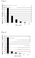
- Fig. 1 shows the results of measurement of the amounts of fibronectin produced in a case in which dipyridamole, which is a platelet aggregation inhibitor, was added to the cell culture system of the present invention, followed by culture.
- Fig. 2 shows the results of measurement of the amounts of fibronectin produced in a case in which limaprost alfadex, which is a platelet aggregation inhibitor, was added to the cell culture system of the present invention, followed by culture.
- The present invention is described in detail below.
- The present invention relates to a method for producing a cell culture system containing aggregated platelets, which comprises culturing platelets in the presence of one or more types of cells that form vascular wall lesion sites or the cell components thereof. Preferably, in accordance with the method of the present invention, platelets and one or more types of cells that form vascular wall lesion sites are cultured in a single culture vessel.
- Cells used in the present invention may be primary culture cells or established subculture cell lines. However, primary culture cells are preferable. Any types of mammals can be used as animals from which culture cells are derived. However, particular examples thereof include humans, canines, felines, swines, miniswines, rabbits, hamsters, rats, mice, and the like. However, the origin of cells used in the present invention is not limited to such animal species.
- Two ore more types of different cells that are cultured in a single cell culture vessel in the present invention may be derived from the same animal species or different animal species. However, such cells are preferably derived from the same animal species.
- Preferably, cells that form vascular wall lesion sites, which are used in the present invention, are cells that form arteriosclerotic lesions. Preferred examples thereof include vascular endothelial cells, vascular smooth muscle cells, fat cells (e.g., mast cells), and macrophages. Particularly preferably, platelets are cultured in the presence of vascular endothelial cells. Cells that form vascular wall lesion sites may be primary culture cells or established subculture cell lines. However, primary culture cells are preferable.
- In addition, in accordance with the present invention, platelets may be cultured in the presence of the cell components of one or more types of cells that form vascular wall lesion sites. The cell components used herein are not particularly limited as long as they contain components derived from cells that form vascular wall lesion sites. Examples thereof include, but are not limited to, pulverized cells, cell extracts, and cell fractions.
- The types of culture vessels used for cell culture in the present invention are not particularly limited. However, preferred examples thereof are culture flasks, culture tubes, petri dishes, and microplates. Of these, petri dishes and microplates are more preferable. Material used for a culture vessel is not particularly limited. However, preferred examples thereof are hard glass, polystyrene, polypropylene, polyethylene, and polyvinyl chloride plastic. Of these, polystyrene and polypropylene are particularly preferable.
- In the particularly preferable embodiment of the present invention, insert cells can be used as culture vessels. The insert cells that are referred to as culture vessels are used by inserting them into microplate wells. They are made of PET (polyethylene terephthalate) and the like. The bottom of an insert cell is made up of a porous membrane. The porous membrane does not allow platelets or cells used, which form vascular wall lesion sites (e.g., vascular endothelial cells), to pass therethrough but allows proteins and albumin to readily pass therethrough. Pore sizes of such membrane are preferably 0.2 to 2 µm. The pore sizes may be selected depending on the types of cells used. Specific examples of versatile insert cells that can be used for 12-well microplates include FALCON (No. 3180), SUMIRON (No. 5123), Iwaki Glass (No. 12-001-01), and TERUMO (No. 001-12).
- Types of media used for cell culture are not particularly limited. However, preferred examples thereof include Eagle's MEM medium, Dulbecco's modified Eagle's MEM medium, RPMI1640 medium, F-10 medium, F-12 medium, McCoy's medium, NCTC-109 medium, and William's medium. Of these, Eagle's MEM medium, and Dulbecco's modified Eagle's MEM medium are preferable. Preferred examples of serum include calf serum, FBS, murine serum, rat serum, rabbit serum, human serum, and horse serum. Of these, calf serum and FBS are particularly preferable.
- In the case of the most preferable combination of conditions upon cell culture in accordance with the present invention, polystyrene-made microplates are used as culture vessels, and Eagle's MEM medium containing FBS is used as a medium. However, such combination is not particularly limited to this example.
- The cell number of platelets to be introduced and the number of cells to be introduced (which form vascular wall lesion sites (e.g., vascular endothelial cells)) are not particularly limited. However, preferably, the cell number of platelets is 0.5 to 7 x 10⁶ cells/well. Also, preferably, the cell number of cells that form vascular wall lesion sites is 0.5 to 8 x 10⁴ cells/well. More preferably, the cell number of platelets is 1 to 4 x 10⁶ cells/well. Also, more preferably, the cell number of cells that form vascular wall lesion sites is 0.8 to 4 x 10⁴ cells/well.
- Platelets and cells that form vascular wall lesion sites (e.g., vascular endothelial cells) may be introduced into insert cells in a manner such that they form the upper or lower layer in each insert cell. Preferably, cells that form vascular wall lesion sites are first introduced into insert cells, and then platelets are introduced into the upper layers that have reached a certain level of confluence. For instance, cells that form vascular wall lesion sites are introduced preferably 1 to 4 days prior to introduction of platelets and more preferably 2 to 3 days prior to introduction of platelets.
- In the most preferable case, cells that form vascular wall lesion sites (1 to 2 x 10⁴ cells/well) are first introduced into a cell filter, followed by culture for 3 days. Next, platelets (1.8 to 2.2 x 10⁶ cells/well) are introduced thereinto. Thus, a culture system can be produced.
- Culture of platelets and cells that form vascular wall lesion sites can be carried out under the conditions which are used for general culture of animal cells. For instance, culture can be carried out at 37°C in the presence of 5% CO2 for several hours to several days and preferably for 1 day to 10 days or thereabout.
- Preferably, after a certain period of culture of platelets and cells that form vascular wall lesion sites, a platelet-activating factor (PAF) can be added to the medium. Note that no particular theory is mentioned herein. Platelets and cells that form vascular wall lesion sites are introduced into insert cells, followed by incubation in a single culture vessel and addition of PAF. Thus, the platelets adhere to collagen components of the cells that form vascular wall lesion sites, such as vascular endothelial cells, followed by platelet aggregation. Thus, growth-activating substances derived from the platelets act on the cells that form vascular wall lesion sites, resulting in induction of cell growth. Therefore, states of vascular wall lesion sites of vascular diseases such as arteriosclerosis and post-PTCA restenosis can be reproduced in vitro.
- Further, in accordance with the present invention, it is possible to evaluate effects of a test substance upon vascular wall lesion by administering the test substance to the aforementioned cell culture system containing aggregated platelets that can be obtained by the present invention and evaluating the effects of the test substance upon platelet aggregation.
- Test substances to be used herein are not particularly limited and any substances can be used. Examples of such test substances include peptides, proteins, non-peptidic compounds, synthetic low molecular weight compounds, fermentation products, cell extracts, plant extracts, and animal tissue extracts. These compounds may be novel compounds or known compounds. In addition, libraries containing many molecules, such as peptide libraries or compound libraries, can also be used as test substances.
- Preferably, platelets and cells that form vascular wall lesion sites (e.g., vascular endothelial cells) are cultured in insert cells, such that the effects of a test substance upon platelets and vascular endothelial cells that have aggregated with the addition of PAF can be determined. Thus, it is possible to evaluate transfer of the test substance or PAF to lesions of vascular diseases. In accordance with the present invention, it is possible to screen for medicaments that are highly effective for vascular diseases such as arteriosclerosis and post-PTCA restenosis by examining uptake of a test substance or PAF into platelets and proliferated cells that form vascular wall lesion sites.
- Examples of a test substance (medicament) screened for by the method of the present invention include, but are not limited to, substances promoting platelet aggregation, substances inhibiting platelet aggregation, substances promoting transfer of PAF to platelets, and substances inhibiting transfer of PAF to platelets.
- The present invention will be further specifically described in the following examples. However, the examples are not intended to limit the scope of the present invention.
- Vascular endothelial cells were separated from murine aortic endothelium (Tissue Culture Method (10th edition) (Soshiki-baiyo-ho), Kodansha, 1998, the Japanese Tissue Culture Association, eds.). The thus separated vascular endothelial cells were suspended in Eagle's MEM medium (No. 11095-080, GIBCO) containing 10% FBS. The resultant was introduced into insert cells (No. 3180, FALCON) for 12-well microplates. The number of cells in each well was adjusted to 10,000 cells. The cells were cultured at 37°C in the presence of 5% CO2 for 3 days.
- Next, in accordance with the method described in "Biochimica Biophysica Acta" (vol. 1213, pp. 127-134 (1994)), platelets were prepared from rabbit whole blood. The platelets (2,000,000 cells) were separated and introduced into upper part of wells of the insert cells, in each bottom of which endothelial cells had been cultured. Thereafter, the insert cells were placed on 12-well microplates (No. 3503, FALCON) and a medium was added thereto, followed by culture at 37°C in the presence of 5% CO2 for 5 days. After culture, 10 µg/well of PAF (No. 301-001-M001, Funakoshi) was added thereto, followed by culture at 37°C in the presence of 5% CO2 for 24 hours.
- During culture, it was possible to observe ADP (which is an index of platelet aggregation) that was assumed to be released from dense granules of platelets in the medium. In addition, it was possible to detect fibronectin (which is an index of platelet aggregation) that was observed with the presence of intravascular wall lesion.
- As with the case of Example 1 above, vascular endothelial cells and platelets were cultured in insert cells. Specifically, vascular endothelial cells were separated from murine aortic endothelium (Tissue Culture Method (10th edition) (Soshiki-baiyo-ho), Kodansha, 1998, the Japanese Tissue Culture Association, eds.). The thus separated vascular endothelial cells were suspended in Eagle's MEM medium (No. 11095-080, GIBCO) containing 10% FBS. The resultant was introduced into insert cells for 12-well microplates (No. 3180, FALCON). The number of cells in each well was adjusted to 10,000 cells. The cells were cultured at 37°C in the presence of 5% CO2 for 3 days.
- Next, in accordance with the method described in "Biochimica Biophysica Acta" (vol. 1213, pp. 127-134 (1994)), platelets were prepared from rabbit whole blood. The platelets (2,000,000 cells) were separated and introduced into upper parts of wells of the insert cells, in each bottom of which endothelial cells had been cultured. Thereafter, the insert cells were placed on 12-well microplates (No. 3503, FALCON) and a medium was added thereto, followed by culture at 37°C in the presence of 5% CO2 for 5 days. After culture, 1, 5, 10, 15, and 20 µg/well of dipyridamole (Boehringer), which is a platelet aggregation inhibitor, and 1, 5, 10, 15, and 20 µg/well of limaprost alfadex (Dainippon Sumitomo Pharma), which is also a platelet aggregation inhibitor, were separately added thereto. 20 minutes after the addition, 10 µg/well of PAF (No. 301-001-M001, Funakoshi) was added to each well, followed by culture at 37°C in the presence of 5% CO2 for 24 hours. Figs. 1 and 2 show the results of measurement of the amount of fibronectin produced in each well.
- In general, limaprost alfadex is said to have stronger platelet aggregation inhibitory effects than dipyridamole. The results shown in figs. 1 and 2 support such hypothesis. It has been demonstrated that the cell culture system of the present invention can be used as a test system for searching for platelet aggregation agonists and for predicting of the effects of such agonists in animals.
- In accordance with the present invention, it is possible to provide a cell culture system in which platelet aggregation that is found at lesions of arteriosclerosis, restenosis, or the like is reproduced. In addition, with the use of a cell culture system of the present invention, it is possible to provide a method for evaluating medicaments for vascular diseases.
Claims (9)
- A method for producing a cell culture system containing aggregated platelets, which comprises culturing platelets in the presence of one or more types of cells that form vascular wall lesion sites or the cell components thereof.
- The method according to claim 1 wherein one or more types of cells that form vascular wall lesion sites are primary culture cells that have been separated from organisms.
- The method according to claim 1 wherein one or more types of cells that form vascular wall lesion sites are selected from the group consisting of vascular endothelial cells, vascular smooth muscle cells, fat cells, and macrophages.
- The method according to claim 1 wherein one or more types of cells that form vascular wall lesion sites are vascular endothelial cells.
- The method according to claim 1 wherein, with the use of insert cells, platelets are cultured in the presence of one or more types of cells that form vascular wall lesion sites or the cell components thereof.
- The method according to claim 1 which comprises activating platelets with the addition of PAF (platelet-activating factor) to a medium.
- A method for producing a vascular wall lesion model, which comprises reproducing vascular wall lesion in a culture vessel with the use of the cell culture system produced by the method of claim 1.
- A cell culture system containing aggregated platelets that is obtained by the method of claim 1.
- A method for evaluating effects of a test substance upon vascular wall lesion, which comprises administering the test substance to a cell culture system containing aggregated platelets that is obtained by the method of claim 1, and evaluating the effects of the test substance upon platelet aggregation.
Applications Claiming Priority (1)
| Application Number | Priority Date | Filing Date | Title |
|---|---|---|---|
| JP2006000527A JP2007181414A (en) | 2006-01-05 | 2006-01-05 | How to make vascular wall lesions with platelets |
Publications (1)
| Publication Number | Publication Date |
|---|---|
| EP1806397A1 true EP1806397A1 (en) | 2007-07-11 |
Family
ID=37899376
Family Applications (1)
| Application Number | Title | Priority Date | Filing Date |
|---|---|---|---|
| EP07000075A Withdrawn EP1806397A1 (en) | 2006-01-05 | 2007-01-03 | Method for producing a model of vascular wall lesion using platelets |
Country Status (3)
| Country | Link |
|---|---|
| US (1) | US20070196814A1 (en) |
| EP (1) | EP1806397A1 (en) |
| JP (1) | JP2007181414A (en) |
Families Citing this family (1)
| Publication number | Priority date | Publication date | Assignee | Title |
|---|---|---|---|---|
| JP2018058768A (en) * | 2015-02-10 | 2018-04-12 | 国立大学法人京都大学 | Composition for maintaining and/or enhancing functions of platelet |
Citations (1)
| Publication number | Priority date | Publication date | Assignee | Title |
|---|---|---|---|---|
| WO2001008694A2 (en) * | 1999-07-30 | 2001-02-08 | Massachusetts Institute Of Technology | Direct arterial infiltration for production of vascular pathology |
Family Cites Families (1)
| Publication number | Priority date | Publication date | Assignee | Title |
|---|---|---|---|---|
| US5624946A (en) * | 1994-07-05 | 1997-04-29 | Williams; James | Use of leflunomide to control and reverse chronic allograft rejection |
-
2006
- 2006-01-05 JP JP2006000527A patent/JP2007181414A/en not_active Abandoned
-
2007
- 2007-01-03 EP EP07000075A patent/EP1806397A1/en not_active Withdrawn
- 2007-01-04 US US11/649,231 patent/US20070196814A1/en not_active Abandoned
Patent Citations (1)
| Publication number | Priority date | Publication date | Assignee | Title |
|---|---|---|---|---|
| WO2001008694A2 (en) * | 1999-07-30 | 2001-02-08 | Massachusetts Institute Of Technology | Direct arterial infiltration for production of vascular pathology |
Non-Patent Citations (3)
| Title |
|---|
| GRAHAM D J ET AL: "Aortic endothelial and smooth muscle cell co-culture: an in vitro model of the arterial wall.", JOURNAL OF INVESTIGATIVE SURGERY, vol. 4, no. 4, 1991, pages 487 - 494, XP000613222, ISSN: 0894-1939 * |
| SUWANNAPRAPHA P ET AL: "Improvement of function and morphology of tumor necrosis factor-alpha treated endothelial cells with 17-beta estradiol - A preliminary study for a feasible simple model for atherosclerosis", CIRCULATION JOURNAL, vol. 69, no. 6, June 2005 (2005-06-01), pages 730 - 738, XP002429807, ISSN: 1346-9843, Retrieved from the Internet <URL:http://www.jstage.jst.go.jp/article/circj/69/6/730/_pdf> * |
| TULL S P ET AL: "Cellular pathology of atherosclerosis - Smooth muscle cells promote adhesion of platelets to cocultured endothelial cells", CIRCULATION RESEARCH, vol. 98, no. 1, January 2006 (2006-01-01), pages 98 - 104, XP002429806, ISSN: 0009-7330, Retrieved from the Internet <URL:http://circres.ahajournals.org/cgi/content/abstract/98/1/98> * |
Also Published As
| Publication number | Publication date |
|---|---|
| US20070196814A1 (en) | 2007-08-23 |
| JP2007181414A (en) | 2007-07-19 |
Similar Documents
| Publication | Publication Date | Title |
|---|---|---|
| Frodermann et al. | Macrophages and cardiovascular health | |
| Pusceddu et al. | ST2 and the ST2/IL-33 signalling pathway–biochemistry and pathophysiology in animal models and humans | |
| Li et al. | Hematopoietic stem cell repopulating ability can be maintained in vitro by some primary endothelial cells | |
| Galvez et al. | Complete repair of dystrophic skeletal muscle by mesoangioblasts with enhanced migration ability | |
| Yoshida et al. | IL-7 receptor α+ CD3–cells in the embryonic intestine induces the organizing center of Peyer's patches | |
| Shehadah et al. | Human placenta-derived adherent cell treatment of experimental stroke promotes functional recovery after stroke in young adult and older rats | |
| CN109072276A (en) | The evaluation method of anticancer agent | |
| JPWO2003027281A1 (en) | Skeletal muscle stroma-derived multipotent stem cells | |
| Boehm et al. | Bone marrow–derived immune cells regulate vascular disease through a p27 Kip1-dependent mechanism | |
| Viti et al. | Multi-disciplinary insights from the first European forum on visceral myopathy 2022 meeting | |
| Yang et al. | Various cell populations within the mononuclear fraction of bone marrow contribute to the beneficial effects of autologous bone marrow cell therapy in a rodent stroke model | |
| Gran et al. | Imaging, myeloid precursor immortalization, and genome editing for defining mechanisms of leukocyte recruitment in vivo | |
| Stratton et al. | Macrophages and associated ligands in the aged injured nerve: a defective dynamic that contributes to reduced axonal regrowth | |
| Li et al. | Nexilin regulates oligodendrocyte progenitor cell migration and remyelination and is negatively regulated by protease-activated receptor 1/Ras-Proximate-1 signaling following subarachnoid hemorrhage | |
| Deutsch et al. | Reactivation of the Nkx2. 5 cardiac enhancer after myocardial infarction does not presage myogenesis | |
| Molnár et al. | The blood-brain barrier in primary CNS lymphomas: ultrastructural evidence of endothelial cell death | |
| Biery et al. | A protocol for the generation of treatment-naïve biopsy-derived diffuse intrinsic pontine glioma and diffuse midline glioma models | |
| Takahashi et al. | Establishment of lacZ-transgenic rats: a tool for regenerative research in myocardium | |
| JP2002536459A (en) | Inhibition of the formation of atherosclerotic lesions | |
| Halpage et al. | Prolonged tamoxifen‐enriched diet is associated with cardiomyopathy and nutritional frailty in mice | |
| Wiseman et al. | A macro-transection model of brain trauma for neuromaterial testing with functional electrophysiological readouts | |
| EP1806397A1 (en) | Method for producing a model of vascular wall lesion using platelets | |
| Rodemer et al. | PTPσ knockdown in lampreys impairs reticulospinal axon regeneration and neuronal survival after spinal cord injury | |
| Tremblay et al. | In vitro colony assays for characterizing tri-potent progenitor cells isolated from the adult murine pancreas | |
| CN110093311B (en) | ICAM-1 Marking and Its Application |
Legal Events
| Date | Code | Title | Description |
|---|---|---|---|
| PUAI | Public reference made under article 153(3) epc to a published international application that has entered the european phase |
Free format text: ORIGINAL CODE: 0009012 |
|
| AK | Designated contracting states |
Kind code of ref document: A1 Designated state(s): AT BE BG CH CY CZ DE DK EE ES FI FR GB GR HU IE IS IT LI LT LU LV MC NL PL PT RO SE SI SK TR |
|
| AX | Request for extension of the european patent |
Extension state: AL BA HR MK YU |
|
| 17P | Request for examination filed |
Effective date: 20080102 |
|
| 17Q | First examination report despatched |
Effective date: 20080214 |
|
| AKX | Designation fees paid |
Designated state(s): AT BE BG CH CY CZ DE DK EE ES FI FR GB GR HU IE IS IT LI LT LU LV MC NL PL PT RO SE SI SK TR |
|
| STAA | Information on the status of an ep patent application or granted ep patent |
Free format text: STATUS: THE APPLICATION IS DEEMED TO BE WITHDRAWN |
|
| 18D | Application deemed to be withdrawn |
Effective date: 20101116 |