EP1802313B1 - Therapeutic agents trageting the ncca-atp channel and methods of use thereof - Google Patents
Therapeutic agents trageting the ncca-atp channel and methods of use thereof Download PDFInfo
- Publication number
- EP1802313B1 EP1802313B1 EP05805849A EP05805849A EP1802313B1 EP 1802313 B1 EP1802313 B1 EP 1802313B1 EP 05805849 A EP05805849 A EP 05805849A EP 05805849 A EP05805849 A EP 05805849A EP 1802313 B1 EP1802313 B1 EP 1802313B1
- Authority
- EP
- European Patent Office
- Prior art keywords
- cell
- sur1
- treatment
- compound
- subject
- Prior art date
- Legal status (The legal status is an assumption and is not a legal conclusion. Google has not performed a legal analysis and makes no representation as to the accuracy of the status listed.)
- Expired - Lifetime
Links
Images












Classifications
-
- A—HUMAN NECESSITIES
- A61—MEDICAL OR VETERINARY SCIENCE; HYGIENE
- A61K—PREPARATIONS FOR MEDICAL, DENTAL OR TOILETRY PURPOSES
- A61K31/00—Medicinal preparations containing organic active ingredients
-
- A—HUMAN NECESSITIES
- A61—MEDICAL OR VETERINARY SCIENCE; HYGIENE
- A61K—PREPARATIONS FOR MEDICAL, DENTAL OR TOILETRY PURPOSES
- A61K31/00—Medicinal preparations containing organic active ingredients
- A61K31/16—Amides, e.g. hydroxamic acids
- A61K31/165—Amides, e.g. hydroxamic acids having aromatic rings, e.g. colchicine, atenolol, progabide
-
- A—HUMAN NECESSITIES
- A61—MEDICAL OR VETERINARY SCIENCE; HYGIENE
- A61K—PREPARATIONS FOR MEDICAL, DENTAL OR TOILETRY PURPOSES
- A61K31/00—Medicinal preparations containing organic active ingredients
- A61K31/16—Amides, e.g. hydroxamic acids
- A61K31/17—Amides, e.g. hydroxamic acids having the group >N—C(O)—N< or >N—C(S)—N<, e.g. urea, thiourea, carmustine
- A61K31/175—Amides, e.g. hydroxamic acids having the group >N—C(O)—N< or >N—C(S)—N<, e.g. urea, thiourea, carmustine having the group, >N—C(O)—N=N— or, e.g. carbonohydrazides, carbazones, semicarbazides, semicarbazones; Thioanalogues thereof
-
- A—HUMAN NECESSITIES
- A61—MEDICAL OR VETERINARY SCIENCE; HYGIENE
- A61K—PREPARATIONS FOR MEDICAL, DENTAL OR TOILETRY PURPOSES
- A61K31/00—Medicinal preparations containing organic active ingredients
- A61K31/33—Heterocyclic compounds
- A61K31/335—Heterocyclic compounds having oxygen as the only ring hetero atom, e.g. fungichromin
- A61K31/365—Lactones
- A61K31/366—Lactones having six-membered rings, e.g. delta-lactones
- A61K31/37—Coumarins, e.g. psoralen
-
- A—HUMAN NECESSITIES
- A61—MEDICAL OR VETERINARY SCIENCE; HYGIENE
- A61K—PREPARATIONS FOR MEDICAL, DENTAL OR TOILETRY PURPOSES
- A61K31/00—Medicinal preparations containing organic active ingredients
- A61K31/33—Heterocyclic compounds
- A61K31/395—Heterocyclic compounds having nitrogen as a ring hetero atom, e.g. guanethidine or rifamycins
- A61K31/40—Heterocyclic compounds having nitrogen as a ring hetero atom, e.g. guanethidine or rifamycins having five-membered rings with one nitrogen as the only ring hetero atom, e.g. sulpiride, succinimide, tolmetin, buflomedil
-
- A—HUMAN NECESSITIES
- A61—MEDICAL OR VETERINARY SCIENCE; HYGIENE
- A61K—PREPARATIONS FOR MEDICAL, DENTAL OR TOILETRY PURPOSES
- A61K31/00—Medicinal preparations containing organic active ingredients
- A61K31/33—Heterocyclic compounds
- A61K31/395—Heterocyclic compounds having nitrogen as a ring hetero atom, e.g. guanethidine or rifamycins
- A61K31/435—Heterocyclic compounds having nitrogen as a ring hetero atom, e.g. guanethidine or rifamycins having six-membered rings with one nitrogen as the only ring hetero atom
- A61K31/44—Non condensed pyridines; Hydrogenated derivatives thereof
-
- A—HUMAN NECESSITIES
- A61—MEDICAL OR VETERINARY SCIENCE; HYGIENE
- A61K—PREPARATIONS FOR MEDICAL, DENTAL OR TOILETRY PURPOSES
- A61K31/00—Medicinal preparations containing organic active ingredients
- A61K31/33—Heterocyclic compounds
- A61K31/395—Heterocyclic compounds having nitrogen as a ring hetero atom, e.g. guanethidine or rifamycins
- A61K31/54—Heterocyclic compounds having nitrogen as a ring hetero atom, e.g. guanethidine or rifamycins having six-membered rings with at least one nitrogen and one sulfur as the ring hetero atoms, e.g. sulthiame
- A61K31/549—Heterocyclic compounds having nitrogen as a ring hetero atom, e.g. guanethidine or rifamycins having six-membered rings with at least one nitrogen and one sulfur as the ring hetero atoms, e.g. sulthiame having two or more nitrogen atoms in the same ring, e.g. hydrochlorothiazide
-
- A—HUMAN NECESSITIES
- A61—MEDICAL OR VETERINARY SCIENCE; HYGIENE
- A61K—PREPARATIONS FOR MEDICAL, DENTAL OR TOILETRY PURPOSES
- A61K31/00—Medicinal preparations containing organic active ingredients
- A61K31/56—Compounds containing cyclopenta[a]hydrophenanthrene ring systems; Derivatives thereof, e.g. steroids
- A61K31/565—Compounds containing cyclopenta[a]hydrophenanthrene ring systems; Derivatives thereof, e.g. steroids not substituted in position 17 beta by a carbon atom, e.g. estrane, estradiol
-
- A—HUMAN NECESSITIES
- A61—MEDICAL OR VETERINARY SCIENCE; HYGIENE
- A61K—PREPARATIONS FOR MEDICAL, DENTAL OR TOILETRY PURPOSES
- A61K31/00—Medicinal preparations containing organic active ingredients
- A61K31/56—Compounds containing cyclopenta[a]hydrophenanthrene ring systems; Derivatives thereof, e.g. steroids
- A61K31/565—Compounds containing cyclopenta[a]hydrophenanthrene ring systems; Derivatives thereof, e.g. steroids not substituted in position 17 beta by a carbon atom, e.g. estrane, estradiol
- A61K31/566—Compounds containing cyclopenta[a]hydrophenanthrene ring systems; Derivatives thereof, e.g. steroids not substituted in position 17 beta by a carbon atom, e.g. estrane, estradiol having an oxo group in position 17, e.g. estrone
-
- A—HUMAN NECESSITIES
- A61—MEDICAL OR VETERINARY SCIENCE; HYGIENE
- A61K—PREPARATIONS FOR MEDICAL, DENTAL OR TOILETRY PURPOSES
- A61K31/00—Medicinal preparations containing organic active ingredients
- A61K31/64—Sulfonylureas, e.g. glibenclamide, tolbutamide, chlorpropamide
-
- A—HUMAN NECESSITIES
- A61—MEDICAL OR VETERINARY SCIENCE; HYGIENE
- A61K—PREPARATIONS FOR MEDICAL, DENTAL OR TOILETRY PURPOSES
- A61K38/00—Medicinal preparations containing peptides
- A61K38/16—Peptides having more than 20 amino acids; Gastrins; Somatostatins; Melanotropins; Derivatives thereof
- A61K38/164—Peptides having more than 20 amino acids; Gastrins; Somatostatins; Melanotropins; Derivatives thereof from bacteria
- A61K38/166—Streptokinase
-
- A—HUMAN NECESSITIES
- A61—MEDICAL OR VETERINARY SCIENCE; HYGIENE
- A61K—PREPARATIONS FOR MEDICAL, DENTAL OR TOILETRY PURPOSES
- A61K38/00—Medicinal preparations containing peptides
- A61K38/16—Peptides having more than 20 amino acids; Gastrins; Somatostatins; Melanotropins; Derivatives thereof
- A61K38/17—Peptides having more than 20 amino acids; Gastrins; Somatostatins; Melanotropins; Derivatives thereof from animals; from humans
- A61K38/177—Receptors; Cell surface antigens; Cell surface determinants
-
- A—HUMAN NECESSITIES
- A61—MEDICAL OR VETERINARY SCIENCE; HYGIENE
- A61K—PREPARATIONS FOR MEDICAL, DENTAL OR TOILETRY PURPOSES
- A61K38/00—Medicinal preparations containing peptides
- A61K38/16—Peptides having more than 20 amino acids; Gastrins; Somatostatins; Melanotropins; Derivatives thereof
- A61K38/43—Enzymes; Proenzymes; Derivatives thereof
- A61K38/46—Hydrolases (3)
- A61K38/48—Hydrolases (3) acting on peptide bonds (3.4)
- A61K38/482—Serine endopeptidases (3.4.21)
-
- A—HUMAN NECESSITIES
- A61—MEDICAL OR VETERINARY SCIENCE; HYGIENE
- A61K—PREPARATIONS FOR MEDICAL, DENTAL OR TOILETRY PURPOSES
- A61K38/00—Medicinal preparations containing peptides
- A61K38/16—Peptides having more than 20 amino acids; Gastrins; Somatostatins; Melanotropins; Derivatives thereof
- A61K38/43—Enzymes; Proenzymes; Derivatives thereof
- A61K38/46—Hydrolases (3)
- A61K38/48—Hydrolases (3) acting on peptide bonds (3.4)
- A61K38/49—Urokinase; Tissue plasminogen activator
-
- A—HUMAN NECESSITIES
- A61—MEDICAL OR VETERINARY SCIENCE; HYGIENE
- A61K—PREPARATIONS FOR MEDICAL, DENTAL OR TOILETRY PURPOSES
- A61K45/00—Medicinal preparations containing active ingredients not provided for in groups A61K31/00 - A61K41/00
- A61K45/06—Mixtures of active ingredients without chemical characterisation, e.g. antiphlogistics and cardiaca
-
- A—HUMAN NECESSITIES
- A61—MEDICAL OR VETERINARY SCIENCE; HYGIENE
- A61P—SPECIFIC THERAPEUTIC ACTIVITY OF CHEMICAL COMPOUNDS OR MEDICINAL PREPARATIONS
- A61P25/00—Drugs for disorders of the nervous system
-
- A—HUMAN NECESSITIES
- A61—MEDICAL OR VETERINARY SCIENCE; HYGIENE
- A61P—SPECIFIC THERAPEUTIC ACTIVITY OF CHEMICAL COMPOUNDS OR MEDICINAL PREPARATIONS
- A61P35/00—Antineoplastic agents
-
- A—HUMAN NECESSITIES
- A61—MEDICAL OR VETERINARY SCIENCE; HYGIENE
- A61P—SPECIFIC THERAPEUTIC ACTIVITY OF CHEMICAL COMPOUNDS OR MEDICINAL PREPARATIONS
- A61P35/00—Antineoplastic agents
- A61P35/04—Antineoplastic agents specific for metastasis
-
- A—HUMAN NECESSITIES
- A61—MEDICAL OR VETERINARY SCIENCE; HYGIENE
- A61P—SPECIFIC THERAPEUTIC ACTIVITY OF CHEMICAL COMPOUNDS OR MEDICINAL PREPARATIONS
- A61P43/00—Drugs for specific purposes, not provided for in groups A61P1/00-A61P41/00
-
- A—HUMAN NECESSITIES
- A61—MEDICAL OR VETERINARY SCIENCE; HYGIENE
- A61P—SPECIFIC THERAPEUTIC ACTIVITY OF CHEMICAL COMPOUNDS OR MEDICINAL PREPARATIONS
- A61P9/00—Drugs for disorders of the cardiovascular system
- A61P9/10—Drugs for disorders of the cardiovascular system for treating ischaemic or atherosclerotic diseases, e.g. antianginal drugs, coronary vasodilators, drugs for myocardial infarction, retinopathy, cerebrovascula insufficiency, renal arteriosclerosis
-
- C—CHEMISTRY; METALLURGY
- C12—BIOCHEMISTRY; BEER; SPIRITS; WINE; VINEGAR; MICROBIOLOGY; ENZYMOLOGY; MUTATION OR GENETIC ENGINEERING
- C12Y—ENZYMES
- C12Y304/00—Hydrolases acting on peptide bonds, i.e. peptidases (3.4)
- C12Y304/21—Serine endopeptidases (3.4.21)
- C12Y304/21068—Tissue plasminogen activator (3.4.21.68), i.e. tPA
-
- C—CHEMISTRY; METALLURGY
- C12—BIOCHEMISTRY; BEER; SPIRITS; WINE; VINEGAR; MICROBIOLOGY; ENZYMOLOGY; MUTATION OR GENETIC ENGINEERING
- C12Y—ENZYMES
- C12Y304/00—Hydrolases acting on peptide bonds, i.e. peptidases (3.4)
- C12Y304/21—Serine endopeptidases (3.4.21)
- C12Y304/21073—Serine endopeptidases (3.4.21) u-Plasminogen activator (3.4.21.73), i.e. urokinase
-
- C—CHEMISTRY; METALLURGY
- C07—ORGANIC CHEMISTRY
- C07K—PEPTIDES
- C07K14/00—Peptides having more than 20 amino acids; Gastrins; Somatostatins; Melanotropins; Derivatives thereof
- C07K14/435—Peptides having more than 20 amino acids; Gastrins; Somatostatins; Melanotropins; Derivatives thereof from animals; from humans
- C07K14/46—Peptides having more than 20 amino acids; Gastrins; Somatostatins; Melanotropins; Derivatives thereof from animals; from humans from vertebrates
- C07K14/47—Peptides having more than 20 amino acids; Gastrins; Somatostatins; Melanotropins; Derivatives thereof from animals; from humans from vertebrates from mammals
- C07K14/4701—Peptides having more than 20 amino acids; Gastrins; Somatostatins; Melanotropins; Derivatives thereof from animals; from humans from vertebrates from mammals not used
- C07K14/4702—Regulators; Modulating activity
- C07K14/4705—Regulators; Modulating activity stimulating, promoting or activating activity
Definitions
- the present invention is directed to fields of cell biology, physiology and medicine. More specifically, the present invention addresses novel methods of treating a patient comprising administering a therapeutic compound that targets a unique non-selective cation channel activated by intracellular calcium and blocked by intracellular ATP (NC Ca-ATP channel).
- the therapeutic compound is an antagonist, and uses thereof in therapies, such as treatment of cerebral ischemia or edema, benefiting from blocking and/or inhibiting the NC Ca-ATP channel.
- Compositions comprising antagonists of the NC Ca-ATP channel are also contemplated.
- NC Ca-ATP channel A unique non-selective monovalent cationic ATP-senstive channel (NC Ca-ATP channel) was identified first in native reactive astrocytes (NRAs) and later, as described herein, in neurons and capillary endothelial cells after stroke or traumatic brain injury (See, International application WO 03/079987 to Simard et al ., and Chen and Simard, 2001, each incorporated by reference herein in its entirety).
- the NC CaATP channel is thought to be a heteromultimer structure comprised of sulfonylurea receptor type 1 (SUR1) regulatory subunits and pore-forming subunits, similar to the K ATP channel in pancreatic ⁇ cells (Chen et al ., 2003). The pore-forming subunits of the NC Ca-ATP channel remain uncharacterized.
- SUR1 sulfonylurea receptor type 1
- SUR imparts sensitivity to antidiabetic sulfonylureas such as glibenclamide and tolbutamide, and is responsible for activation by a chemically diverse group of agents termed "K + channel openers" such as diazoxide, pinacidil and cromakalin (Aguilar-Bryan et al., 1995; Inagaki et al ., 1996; Isomoto et al ., 1996; Nichols et al ., 1996; Shyng et al ., 1997).
- K + channel openers such as diazoxide, pinacidil and cromakalin
- the K ATP channel in pancreatic ⁇ cells is formed from SUR1 linked with Kir6.2, whereas the cardiac and smooth muscle K ATP channels are formed from SUR2A and SUR2B linked with Kir6.2 and Kir6.1, respectively (Fujita et al ., 2000).
- the NC Ca-ATP channel is also sensitive to sulfonylurea compounds.
- the NC Ca-ATP channel conducts sodium ions, potassium ions, cesium ions and other monovalent cations with near equal facility (Chen and Simard, 2001) suggesting further that the characterization, and consequently the affinity to certain compounds, of the NC Ca-ATP channel differs from the K ATP channel.
- NC Ca-ATP channel expressed and found in astrocytes differs physiologically from the other channels with respect to calcium sensitivity and adenine nucleotide sensitivity (Chen et al ., 2001).
- the gliotic capsule that forms around a "foreign body" in the brain is an important, albeit neglected, biological system.
- the gliotic capsule represents the response of the brain to an injurious stimulus -- an attempt by the brain to wall off, isolate, dispose of, and otherwise protect itself from the foreign body.
- the gliotic capsule forms a potentially harmful mass of tissue from which originates edema fluid that contributes to brain swelling, and whose constituent cells undergo cytotoxic edema, which adds further to brain swelling.
- the gliotic capsule protects foreign cells from immunologic surveillance.
- gliotic capsule The essential elements involved in formation of a gliotic capsule appear to be uniform in many types of CNS pathology, be it a traumatically implanted foreign body, a metastatic tumor, a brain abscess, or infarcted necrotic tissue following a stroke.
- microglia and astrocytes become activated near the site of injury, with large, stellate-shaped GFAP-positive reactive astrocytes forming the most prominent cellular component of the response.
- the foreign nature of the entity is recognized, and the response is initiated to surround and contain it.
- the interface between the foreign body and the gliotic capsule referred to as the inner zone of the gliotic capsule, appears to be of great importance in determining the overall response to injury.
- the gliotic capsule forms a potentially harmful mass of tissue that contributes to brain swelling and mass effect, and that may shelter foreign cells from surveillance by the immune system.
- Applicants are the first to determine that, in a variety pathological conditions in both rats and humans, reactive astrocytes (R1 astrocytes) in the inner zone of the gliotic capsule express a novel SUR1 -regulated cation channel, the NC Ca-ATP channel, and that this channel directly controls cell viability: opening the channel is associated with necrotic cell death and closing the channel is associated with protection from cell death induced by energy (ATP) depletion.
- R1 astrocytes reactive astrocytes in the inner zone of the gliotic capsule express a novel SUR1 -regulated cation channel, the NC Ca-ATP channel, and that this channel directly controls cell viability: opening the channel is associated with necrotic cell death and closing the channel is associated with protection from cell death induced by energy (ATP) depletion.
- the present invention relates to a unique non-selective cation channel activated by intracellular calcium and blocked by intracellular ATP (NC Ca-ATP channel) that can be expressed in neuronal cells, neuroglia cells (e.g., astrocyte, ependymal cell, oligodentrocyte and microglia) or neural endothelial cells (e.g., capillary endothelial cells) in which the cells have been or are exposed to a traumatic insult, for example, an acute neuronal insult (e.g., hypoxia, ischemia, cerebral edema or cell swelling), toxic compounds or metabolites, an acute injury, cancer, brain abscess, etc.
- an acute neuronal insult e.g., hypoxia, ischemia, cerebral edema or cell swelling
- toxic compounds or metabolites e.g., an acute injury, cancer, brain abscess, etc.
- the present invention relates to the regulation and/or modulation of this NC Ca-ATP channel and how its modulation can be used to treat various diseases and/or conditions, for example acute neuronal insults (e.g., stroke, an ischemic/hypoxic insult). Yet further, the present invention relates to the regulation and/or modulation of this NC Ca-ATP channel and its role in maintaining or disrupting the integrity of the gliotic capsule.
- acute neuronal insults e.g., stroke, an ischemic/hypoxic insult.
- the present invention relates to the regulation and/or modulation of this NC Ca-ATP channel and its role in maintaining or disrupting the integrity of the gliotic capsule.
- the modulation and/or regulation of the channel results from administration of an antagonist or inhibitor of the channel.
- a composition an antagonist or inhibitor
- a composition is administered to block or inhibit the channel to prevent cell death, for example to treat cerebral edema that results from ischemia due to tissue trauma or to increased tissue pressure.
- the channel is blocked to prevent or reduce or modulate depolarization of the cells.
- the antagonist that is employed in the present invention is a compound which inhibits a NC Ca-ATP channel or is a pharmaceutically acceptable salt thereof.
- the compound or salt is administered in combination with a thrombolyotic agent or pharmaceutically acceptable salt thereof. Accordingly, the present invention provides:
- composition(s) of the present invention may be delivered alimentary or parenterally.
- alimentary administration include, but are not limited to orally, buccally, rectally, or sublingually.
- Parenteral administration can include, but are not limited to intramuscularly, subcutaneously, intraperitoneally, intravenously, intratumorally, intraarterially, intraventricularly, intracavity, intravesical, intrathecal, or intrapleural.
- Other modes of administration may also include topically, mucosally, transdermally, direct injection into the brain parenchyma.
- An effective amount of an antagonist of NC Ca-ATP channel that may be administered to a cell includes a dose of about 0.0001 nM to about 2000 ⁇ M.
- an effective amount of an antagonist of the NC Ca-ATP channel or related-compounds thereof as a treatment varies depending upon the host treated and the particular mode of administration.
- the dose range of the antagonist of the NC Ca-ATP channel or related-compounds thereof will be about 0.01 ⁇ g/kg body weight to about 20,000 ⁇ g/kg body weight.
- body weight is applicable when an animal is being treated. When isolated cells are being treated, " body weight " as used herein should read to mean “total cell body weight ". The term “total body weight” may be used to apply to both isolated cell and animal treatment.
- a variety of different dosage levels will be of use, for example, 0.0001 ⁇ g/kg, 0.0002 ⁇ g/kg, 0.0003 ⁇ g/kg, 0.0004 ⁇ g/kg, 0.005 ⁇ g/kg, 0.0007 ⁇ g/kg, 0.001 ⁇ g/kg, 0.1 ⁇ g/kg, 1.0 ⁇ g/kg, 1.5 ⁇ g/kg, 2.0 ⁇ g/kg, 5.0 ⁇ g/kg, 10.0 ⁇ g/kg, 15.0 ⁇ g/kg, 30.0 ⁇ g/kg, 50 ⁇ g/kg, 75 ⁇ g/kg, 80 ⁇ g/kg, 90 ⁇ g/kg, 100 ⁇ g/kg, 120 ⁇ g/kg, 140 ⁇ g/kg, 150 ⁇ g/kg, 160 ⁇ g/kg, 180 ⁇ g/kg, 200 ⁇ g/kg, 225 ⁇ g/kg, 250 ⁇ g/kg, 275 ⁇ g/kg, 300 ⁇ g/kg,
- the NC Ca-ATP channel is blocked by antagonists of type 1 sulfonylurea receptor (SUR1) and opened by SUR1 activators. More specifically, the antagonists of type 1 sulfonylurea receptor (SUR1) include blockers of K ATP channels and the SUR1 activators include activators of K ATP channels. More specifically, the NC Ca-ATP channel of the present invention has a single-channel conductance to potassium ion (K + ) between 20 and 50 pS. The NC Ca-ATP channel is also stimulated by Ca 2+ on the cytoplasmic side of the cell membrane in a physiological concentration range, where concentration range is from 10 -8 to 10 -5 M.
- the NC Ca-ATP channel is also inhibited by cytoplasmic ATP in a physiological concentration range, where the concentration range is from 10 -1 to 10 M.
- the NC Ca-ATP channel is also permeable to the following cations; K + , Cs + , Li + , Na + ; to the extent that the permeability ratio between any two of the cations is greater than 0.5 and less than 2.
- the present invention comprises a pharmaceutical composition
- a pharmaceutical composition comprising a thrombolytic agent (e.g ., tissue plasminogen activator (tPA), urokinase, prourokinase, streptokinase, anistreplase, reteplase, tenecteplase) or a pharmaceutically acceptable salt thereof and a compound that inhibits a NC Ca-ATP channel or a pharmaceutically acceptable salt thereof.
- a thrombolytic agent e.g ., tissue plasminogen activator (tPA), urokinase, prourokinase, streptokinase, anistreplase, reteplase, tenecteplase
- This pharmaceutical composition can be considered neuroprotective.
- the pharmaceutical composition comprising a combination of the thrombolytic agent and a compound that inhibits a NC Ca-ATP channel is neuroprotective because it increases the therapeutic window of for the administration of the thrombolytic agent by several hours, for example the therapeutic window for administration of thrombolytic agents may be increased by several hours (4-8 hrs) by co-administering antagonist of the NC Ca-ATP channel.
- the channel can be inhibited by an NC Ca-ATP channel inhibitor, an NC CaATP channel blocker, a type 1 sulfonylurea receptor (SUR1) antagonist, SUR1 inhibitor, or a compound capable of reducing the magnitude of membrane current through the channel.
- SUR1 antagonist is selected from the group consisting of glibenclamide, tolbutamide, repaglinide, nateglinide, meglitinide, LY397364, LY389382, glyclazide, glimepiride, and compounds known to inhibit or block K ATP channels.
- MgADP can also be used to inhibit the channel.
- K ATP channels include, but are not limited to tolbutamide, glyburide (1[p-2[5-chloro-O-anisamido)ethyl] phenyl] sulfonyl] -3-cyclohexyl-3-urea); chlopropamide (1-[[(p-chlorophenyl)sulfonyl]-3-propylurea; glipizide (1-cyclohexyl-3[[p-[2(5-methylpyrazine carboxamido)ethyl] phenyl] sulfonyl] urea); or tolazamide(benzenesulfonamide-N-[[(hexahydro-1H-azepin-lyl)amino] carbonyl] -4-methyl).
- the compound that inhibits the NC Ca-ATP channel is thus administered in combination with a thrombolytic agent (e.g., tissue plasminogen activator (tPA), urokinase, prourokinase, streptokinase, anistreplase, reteplase, tenecteplase).
- a thrombolytic agent e.g., tissue plasminogen activator (tPA), urokinase, prourokinase, streptokinase, anistreplase, reteplase, tenecteplase.
- the thrombolytic agent is a tissue plasminogen activator (tPA), urokinase, prourokinase, streptokinase, anistreplase, reteplase, tenecteplase or any combination thereof.
- tPA tissue plasminogen activator
- the SUR1 antagonist can be administered by any standard parenteral or alimentary route, for example the SUR1 antagonist may be administered as a bolus injection or as an infusion or a combination thereof.
- the channel is expressed on neuronal cells, neuroglia cells, neural epithelial cells or a combination thereof.
- the inhibitor blocks the influx of Na + into the cells thereby preventing depolarization of the cells. Inhibition of the influx of Na + into the cells thereby prevents cytotoxic edema and reduces hemorrhagic conversion. Thus, this treatment reduces cell death or necrotic death of neuronal and/or neural endothelial cells.
- the amount of the SUR1 antagonist administered to the subject is in the range of about 0.0001 ⁇ g/kg/day to about 20 mg/kg/day, about 0.01 ⁇ g/kg/day to about 100 ⁇ g/kg/day, or about 100 ⁇ g/kg/day to about 20 mg/kg/day.
- the SUR1 antagonist may be administered to the subject in the from of a treatment in which the treatment may comprise the amount of the SUR1 antagonist or the dose of the SUR1 antagonist that is administered per day (1, 2, 3, 4, etc.), week (1, 2, 3, 4, 5, etc.), month (1, 2, 3, 4, 5, etc.), etc.
- Treatments may be administered such that the amount of SUR1 antagonist administered to the subject is in the range of about 0.0001 ⁇ g/kg/treatment to about 20 mg/kg/treatment, about 0.01 ⁇ g/kg/treatment to about 100 ⁇ g/kg/treatment, or about 100 ⁇ g/kg/treatment to about 20 mg/kg/treatment.
- Another embodiment comprises a neuroprotective infusion kit comprising a compound that inhibits a NC Ca-ATP channel in a neuronal cell, a neuroglia cell, a neural endothelial cell or a combination thereof and an IV solution.
- the compound and solution are contained within the same container or within different containers. More specifically, the compound is contained within the container of solution.
- the kit may further comprise a neuroprotective bolus kit, wherein the bolus kit comprises a pre-loaded syringe of a compound inhibits a NC Ca-ATP channel in a neuronal cell, a neuroglia cell, a neural endothelial cell or a combination thereof.
- the bolus kit comprises a pre-loaded syringe of a compound inhibits a NC Ca-ATP channel in a neuronal cell, a neuroglia cell, a neural endothelial cell or a combination thereof.
- another embodiment comprises a neuroprotective kit comprising a compound that inhibits NC Ca-ATP channel in a neuronal cell, a neuroglia cell, an endothelium cell or a combination thereof and a thrombolytic agent (e.g. , tPA), an anticoagulant (e . g ., warfarin or coumadin), an antiplatelet ( e.g. , aspirin), a diuretic ( e . g ., mannitol), a statin, or a vasodilator ( e.g. , nitroglycerin).
- a thrombolytic agent e.g. , tPA
- an anticoagulant e. g ., warfarin or coumadin
- an antiplatelet e.g. , aspirin
- diuretic e . g ., mannitol
- statin e.g. , nitroglycerin
- FIG. 1 shows that addition of exogenous phosphatidylinositol-4,5-bisphosphate (PIP 2 ) causes activation of the NC CaATP channel, despite the presence of ATP in the bath solution.
- PIP 2 exogenous phosphatidylinositol-4,5-bisphosphate
- FIG. 2 shows that the NC Ca-ATP channel in an R1 astrocyte is inhibited by estrogen.
- the initial portion of the record shows brisk activity from a number of superimposed channels, recorded in a cell attached patch of membrane from an R1 astrocyte obtained from a female. Addition of 10 nM estrogen to the bath promptly resulted in strong inhibition of channel activity.
- the mechanism involved is believed to be related to estrogen receptor mediated activation of phospholipase C (PLC), resulting in depletion of PIP 2 from the membrane, and reflecting an apparent increase in affinity for ATP.
- PLC phospholipase C
- FIGS. 3A-3B show Western blots demonstrating that R1 astrocytes from both males and females express estrogen receptors and SUR1, a marker of the NC Ca-ATP channel.
- Cell lysates were obtained from gelatin sponge implants from males (M) and females (F) and studied at two dilutions (4x and 1x), with lysates from uterus used as controls.
- FIG. 3A was developed using antibodies directed against estrogen receptors (ER), demonstrating that both ER ⁇ and ER ⁇ are expressed in astrocytes from both genders.
- Western blots showed that SUR1 is also expressed by cells from both genders, with pancreatic tissue used as control ( FIG. 3B ).
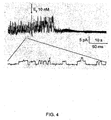
- FIG. 4 shows that the NC Ca-ATP channel in an R1 astrocyte from a male is inhibited by estrogen.
- the initial portion of the record shows brisk activity from a number of superimposed channels, recorded in a cell attached patch of membrane from an R1 astrocyte obtained from a male. Addition of 10 nM estrogen to the bath promptly resulted in strong inhibition of channel activity.
- FIGS 5A-5D shows the gliotic capsule.
- FIG. 5A shows a coronal section of a rat brain sectioned though the site of implantation of a large gelatin sponge; the sponge (innermost dark region) is encapsulated by a gliotic capsule (light area), outside of which is found a region of vasogenic edema (outer dark area), identified by pre-mortem administration of methylene blue.
- FIG. 5B and 5C show low power and high power views, respectively, of the gliotic capsule immunolabeled for GFAP.
- FIG. 5D shows a high power view of GFAP-labeled cells inside of the gelatin sponge implant.
- FIGS. 6A-6H show immunolabeled astrocytes.
- FIGS. 6A, 6C, 6E show freshly-isolated large phase-bright R1 astrocytes immunolabeled for GFAP ( FIG. 6C ) and vimentin (FIG. 6E).
- FIG. 6B,D,F show freshly-isolated small phase-dark R2 astrocytes immunolabeled for GFAP ( FIG. 6D ) and vimentin ( FIG. 6F).
- FIG. 6G shows primary cultures of astrocytes isolated from a gliotic capsule, with R1 astrocytes developing into large polygonal cells ( FIG. 6Gb ), and R2 astrocytes developing into small bipolar cells ( FIG. 6Ga).
- FIG. 6H shows that R2 astrocytes, but not R1 astrocytes, are labeled with fluorescein tagged chlorotoxin derived from the scorpion, Leiurus quinquestriatus .
- FIGS. 7A-7D show that the inner zone of the gliotic capsule expresses SUR1 but not SUR2. Immunolabling for SUR1 ( FIG. 7A ) showed prominent expression in cells adjacent to the gelatin sponge (gf), whereas immunolabeling for SUR2 showed no expression ( FIG. 7B ). A single cell enzymatically isolated from a gelatin sponge implant and immunolabeled for SUR1 is shown ( FIG. 7C). FIG. 7D shown RT-PCR for SUR1 in control insulinoma cells (lane 2) and in isolated R1 astrocytes (lane 3), and for SUR2 in control cardiac cells (lane 4), but not in isolated R1 astrocytes (lane 5).
- FIGS. 8A-8I show various features of the gliotic capsule.
- the gliotic capsule is characterized by GFAP-positive cells that are several cell-layers thick ( FIG. 8A ). Only the inner zone of the gliotic capsule is hypoxic, as demonstrated by pimonidozole labeling ( FIG. 8B ) and by immunolabeling for HIF1 ⁇ ( FIG. 8C ). Also, only the inner zone is immunolabeled for SUR1 ( FIG. 8D ), and for the tight junction proteins, ZO-1 ( FIG. 8E ) and occludens ( FIG. 8F).
- FIGS. 8G-I show that pimonidazole, HIF1 ⁇ and occludens all localize to GFAP-positive astrocytes that form the inner zone of the gliotic capsule.
- FIGS. 9A-B show effects of NC Ca-ATP channel inhibition ( FIG. 9A ) and NC Ca-ATP channel activation ( FIG. 9B ) on the gliotic capsule.
- Animals with gelatin sponge implants were treated with glibenclamide infusion ( FIG. 9A ) or diazoxide infusion ( FIG. 9B ) via osmotic mini-pumps that delivered the compounds directly into the area of the gelatin sponge.
- Immunolabeling for GFAP showed that channel inhibition with glibenclamide resulted in formation of a well defined gliotic capsule ( FIG. 9A ), whereas channel activation with diazoxide resulted in formation of a broader, ill-defined capsule ( FIG. 9B ), due to diazoxide-induced necrotic death of inner zone cells.
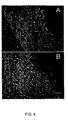
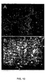
- FIGS. 10A-B show that infusion of diazoxide into the area around the gelatin sponge resulted in a heavy infiltration of polymorphonuclear leukocytes (PMNs).
- Nuclear labeling with DAPI showed densely packed small cells in the vicinity of the gelatin sponge ( FIG. 10A ), with immunolabeling using the PMN-specific marker, MMP-8, demonstrating that these cells were PMNs ( FIG. 10B ). It is believed that the strong inflammatory response represented by the infiltrating PMNs was due to disruption of the barrier between brain and foreign body (gelatin sponge) normally formed by the inner zone of the gliotic capsule.
- FIGS. 11A-11L show that R1 astrocytes in the inner zone of the gliotic capsule typically express SUR1, a marker for the NC Ca-ATP channel.
- SUR1 a marker for the NC Ca-ATP channel.
- the inner zones of the gliotic capsules in rats with gelatin sponge implants ( FIGS. 11A-11C ), in rats with cerebral abscess (FIGS. 11D-11F ), and in humans with metastatic tumor ( FIGS. 11J-11L ) are shown.
- the area of reactive gloss adjacent to a stroke in the rat FIGS. 11G-11I ) resulting from occlusion of the middle cerebral artery.
- a field of cells is labeled for GFAP and co-labelled for SUR1, as indicated. Examples of single cells at high power are also shown for each condition.
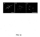
- FIGS. 12A-12C shows that stellate astrocytes near the edge of a stroke up-regulate SUR1 (FIG. 12A ), a marker of the NC Ca-ATP channel.
- SUR1 FIG. 12A
- FIG. 12B,12C shows that cells with altered morphology including blebbing are also immunolabeled for SUR1 ( FIG. 12B,12C) .
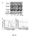
- FIGS. 13A-13C show that glibenclamide protects from Na azide-induced channel opening and necrotic cell death.
- FIG. 13A shows phase contrast images of 4 different freshly isolated R1 astrocytes observed over the course of 30 min each.
- the cell exposed to vehicle solution alone remained phase bright with no pathological deterioration (control).
- the cell depleted of ATP by exposure to Na azide (1 mM) developed progressive blebbing consistent with cytotoxic edema.
- the cell exposed to the NC Ca-ATP channel opener, diazoxide developed progressive blebbing consistent with cytotoxic edema.
- the cell exposed to Na azide in the presence of glibenclamide remained phase bright with no pathological deterioration.
- FIGS. 13A shows phase contrast images of 4 different freshly isolated R1 astrocytes observed over the course of 30 min each.
- the cell exposed to vehicle solution alone remained phase bright with no pathological deterioration (control).
- FIG. 13B and 13C show cell death of isolated R1 astrocytes induced by ATP depletion in vitro .
- Freshly isolated R1 astrocytes were labeled for necrotic death with propidium iodide (PI) ( FIG. 13B ), or for apoptotic death with annexin V ( FIG. 13C ), under control conditions, after exposure to Na azide (1 mM), or after exposure to Na azide in the presence of glibenclamide (1 ⁇ M). Exposure to Na azide resulted mostly in necrotic death that was largely prevented by glibenclamide.
- PI propidium iodide
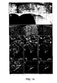
- FIG. 14A-14L shows that SUR1 is up-regulated in MCA stroke.
- Immunofluorescence images showing SUR1 at 3 hr in the core of the stroke in cells FIG. 14D
- FIG. 14E double-labeled for the neuronal marker, NeuN
- FIG. 14G, 14J double-labeled for the astrocytic marker, GFAP (FIG. 14H) , and the endothelial cell marker, von Willebrand factor (FIG. 14K ).
- FIG. 14F, 14I, and 14L Superimposed images of double-labeled fields are shown ( FIG. 14F, 14I, and 14L) .
- FIGS. 15A-15G show that SUR1 but not Kir6.1 or Kir6.2 is transcriptionally up-regulated in MCA stroke.
- FIGS 16A-16D show patch clamp recordings of NC CaATP channel in neuron-like cells in stroke.
- FIG. 16A shows phase-contrast image of large neuron-like cells enzymatically isolated from ischemic region 3 hr following MCAO.
- FIG. 16B shows recording of inside-out patch using Cs + as the charge carrier; channel activity was blocked by glibenclamide given as indicated (arrow); a and b show expanded records of the portions indicated.
- FIG. 16C shows recordings at potentials indicated of inside-out patch using K + as the charge carrier; channel activity was blocked by glibenclamide.
- FIG. 16D shows a plot of single channel amplitudes at different voltages showing single channel slope conductance of 34 pS.
- FIGS. 17A-17E show that glibenclamide reduces mortality, edema and stroke size in MCA stroke.
- FIG. 17A Mortality was assessed during 7 days after MCA stroke [double occlusion model with malignant cerebral edema (MCE)] in two treatment groups, each comprised of 19 female and 10 male rats, treated with either saline (empty symbols) or glibenclamide (filled symbols); mortality at 7 days was significantly different. Subgroup analyses for males and females showed similar results.
- MCA stroke double occlusion model with malignant cerebral edema (MCE)
- 17B edema was assessed 8 hr after MCA stroke (MCE model) in two treatment groups, each comprised of 6 male rats treated with either saline or glibenclamide; tissues were first processed with TTC to allow separation into TTC(+) and TTC(-) portions of the involved hemisphere and contralateral hemisphere, prior to determining wet/dry weights; values in TTC(+) regions were statistically different.
- MCA stroke MCA stroke
- stroke size was assessed 48 hr after MCA stroke [thromboembolic (TE) model] in two treatment groups, each comprised of 10 male rats, treated with either saline or glibenclamide; images of TTC-stained coronal sections following MCA stroke (TE model) in an animal treated with saline ( FIG. 17C ) and another treated with glibenclamide ( FIG. 17D ), showing cortical sparing often associated with glibenclamide treatment; values of stroke size, expressed as percent of hemisphere volume (FIG. 17E ).
- TE thromboembolic
- FIGS 18A-18D show that tissue distribution of BODIPY-glibenclamide in MCA stroke.
- a-c Fluorescence images of brain sections in an animal 6 hr after MCA stroke (MCE model) and administration of BODIPY-glibenclamide; fluorescent labeling was evident in cells, microvessels ( FIG. 18A ) and capillaries ( FIG. 18C ) from ischemic regions, but not in the contralateral hemisphere ( FIG. 18B ); the images in ( FIGS. 18A, 18B ) are from the same animal, taken with the same exposure time; in ( FIG. 18C ), the single layer of nuclei confirms that the structure brightly labeled by BODIPY-glibenclamide is a capillary.
- FIG. 18D immunofluorescence image of a brain section from an animal 6 hr after MCA stroke (MCE model) labeled with anti-SUR1 antibody showing strong labeling in a capillary and in adjacent neuron-like cells.
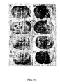
- FIGS. 19A-9H show that glibenclamide reduces hemorrhagic conversion.
- FIGS 19A-19D are from animals co-treated with saline;
- FIGS. 19E-19H are from animals co-treated with glibenclamide.
- the left column of photographs of coronal sections shows, in rows 1-2 only, intraventricular hemorrhage, plus large areas of hemorrhagic conversion in ischemic cortical/subcortical regions (red areas on the right side of pictures; arrows).
- the right column of photographs of TTC-processed sections from the same animals show the areas of infarction.
- FIGS 20A-20B show zymography showing gelatinase activity of matrix metalloproteinases (MMP's) in stroke, and absence of direct MMP inhibition by glibenclamide.
- FIG. 20A shows activation of MMP-9 & MMP-2 in stroke tissue compared to control; activity of recombinant MMP-9 & MMP-2 shown at left.
- FIG. 20B shows gelatinase activity of recombinant enzyme and stroke tissue under control conditions (CTR), in presence of glibenclamide (10 ⁇ M), and in presence of MMP inhibitor II (300 nM; Calbiochem).
- CTR gelatinase activity of recombinant enzyme and stroke tissue under control conditions
- FIG. 21 shows phase contrast photomicrograph of cerebral capillaries freshly isolated from normal brain, after enzymatic cleaning in preparation for patch clamping.
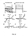
- FIGS. 22A-22F show that freshly isolated cerebral endothelial and smooth muscle cells are readily distinguished electrophysiologically.
- FIGS. 22A and 22B show superimposed macroscopic currents recorded during 200 ms depolarizing pulses from -120 mV to +120 mV in 20 mV steps in an endothelial cell ( FIG. 22A ) and in an elongated smooth muscle cell ( FIG. 22B) ; holding potential, -60 mV; nystatin perforated patch technique; bath solution, standard Krebs with 2 mM Ca 2+ ; pipette solution, 145 mM K + .
- FIGS. 22C and 22D show current-voltage curves computed from average (mean ⁇ SE) currents at the end of 200-ms test pulses recorded in 9 endothelial cells ( FIG. 22C ) and 7 smooth muscle cells ( FIG. 22D ); same holding potential, technique and solutions as in FIGS. 22A and 22B.
- FIGS. 22E and 22F show current voltage curves recorded during ramp pulses (0.45 mV/ms, holding potential, -60 mV) in an endothelial cell ( FIG. 22E ) and in a smooth muscle cell ( FIG. 22F ); same holding potential, technique and bath solution as in FIGS. 22A and 22B , but with pipette solution containing 145 mM Cs + instead of K + .
- FIG. 23 shows real time RT-PCR showing up-regulation of SUR1-mRNA in stroke.
- FIGS. 24A-24E show SUR1 knock down (SUR1KD) in R1 astrocytes protects from ATP-depletion-induced depolarization.
- FIGS. 24A and 24B show Western blot ( FIG. 24A ) and quantification of Western blots ( FIG. 24B ) of R1 cell lysates confirmed knock down of SUR1 expression by antisense.
- FIGS. 24C-24E show Na azide caused large depolarizations in cells exposed to SCR-ODN ( FIG. 24C, 24E ) but little or no depolarization in cells exposed to AS-ODN ( FIG. 24D, 24E ).
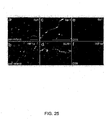
- FIGS. 25A-25F show transcription factors in stroke.
- the peri-infarct region showed up-regulation of both transcription factors, Sp1 ( FIGS. 25A, 25C ) and HIF1 ⁇ ( FIG. 25B ) in neuron-like cells and capillaries, as well as SUR1 in capillaries (FIG. 25D ).
- Control tissues showed little Sp1 and no HIF1 ⁇ ( FIGS. 25E and 25F ).
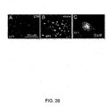
- FIGS. 26A-26C show an increase in nuclear localization of the transcription factor, SP1, and SP1 co-localization with SUR1 in stroke.
- Immunofluorescence images showing increase of nuclear SP1 labeling in ischemic area 3-hr after MCAO ( FIG. 26B ), compared to contralateral side ( FIG. 26A).
- FIG. 26C double labeling of large neuron-like cell showing nuclear SP1 (green) and cytoplasmic /plasmalemmal SUR1 (red) in the same cell.
- FIGS. 27A-27D show regulation of SUR1 expression by the transcription factor, HIF1 ⁇ .
- FIG. 27A and 27C show Western blot analysis of HIF1 ⁇ protein in R1 astrocytes from gelfoam implant model of control (CTR) and HIF1 ⁇ knock-down (KD).
- FIG. 27B and 27C show SUR1 protein in the same cell lysates.
- the term "acute” refers to the onset of a health effect, usually the effect is a rapid onset that is considered brief, not prolonged.
- acute cerebral ischemia refers to a cerebral ischemic event that has a rapid onset and is not prolonged.
- acute cerebral ischemia and “stroke” can be used interchangeably.”
- the term "agonist” refers to a biological or chemical agent that combines with a receptor on a cell and initiates the same or equivalent reaction or activity produced by the binding of an endogenous substance.
- the agonist combines, binds, and/or associates with a NC Ca-ATP channel of a neuronal cell, a neuroglial cell, or a neural endothelial cell, such that the NC Ca-ATP channel is opened (activated).
- the agonist combines, binds and/or associates with a regulatory subunit of the NC Ca-ATP channel, particularly a SUR1.
- the agonist combines, binds, and/or associates with a pore-forming subunit of the NC Ca-ATP channel, such that the NC Ca-ATP channel is opened (activated).
- the terms agonist and/or activator can be used interchangeably.
- the term "antagonist" refers to a biological or chemical agent that acts within the body to reduce the physiological activity of another chemical or biological substance.
- the antagonist blocks, inhibits, reduces and/or decreases the activity of a NC Ca-ATP channel of a neuronal cell, a neuroglia cell or a neural endothelial cell ( e.g ., capillary endothelial cells).
- the antagonist combines, binds, associates with a NC Ca-ATP channel of neuronal cell, a neuroglia cell or a neural endothelial cell ( e .
- the antagonist combines, binds and/or associates with a regulatory subunit of the NC Ca-ATP channel, particularly a SUR1.
- the antagonist combines, binds, and/or associates with a pore-forming subunit of the NC Ca-ATP channel, such that the NC Ca-ATP channel is closed (deactivated).
- the terms antagonist or inhibitor can be used interchangeably.
- brain abscess or “cerebral abscess” refer to a circumscribed collection of purulent exudate that is typically associated with swelling.
- blood brain barrier refers the barrier between brain blood vessels and brain tissues whose effect is to restrict what may pass from the blood into the brain.
- Cerebral ischemia refers to a lack of adequate blood flow to an area, for example a lack of adequate blood flow to the brain, which may be the result of a blood clot, blood vessel constriction, a hemorrhage or tissue compression from an expanding mass.
- depolarization refers to an increase in the permeability of the cell membrane to sodium ions wherein the electrical potential difference across the cell membrane is reduced or eliminated.
- the terms "effective amount” or “therapeutically effective amount” are interchangeable and refer to an amount that results in an improvement or remediation of the symptoms of the disease or condition. Those of skill in the art understand that the effective amount may improve the patient's or subject's condition, but may not be a complete cure of the disease and/or condition.
- endothelium refers a layer of cells that line the inside surfaces of body cavities, blood vessels, and lymph vessels or that form capillaries.
- endothelial cell refers to a cell of the endothelium or a cell that lines the surfaces of body cavities, for example, blood or lymph vessels or capillaries.
- endothelial cell refers to a neural endothelial cell or an endothelial cell that is part of the nervous system, for example the central nervous system or the brain.
- the term "gliotic capsule” refers to a physical barrier surrounding, in whole or in part, a foreign body, including a metastatic tumor, a cerebral abscess or other mass not normally found in brain except under pathological conditions.
- the gliotic capsule comprises an inner zone comprising neuronal cells, neuroglial cells (e.g ., astrocytes) and/or endothelial cells expressing a NC Ca-ATP channel.
- morbidity is the state of being diseased. Yet further, morbidity can also refer to the disease rate or the ratio of sick subjects or cases of disease in to a given population.
- mortality is the state of being mortal or causing death. Yet further, mortality can also refer to the death rate or the ratio of number of deaths to a given population.
- neuronal cell refers to a cell that is a morphologic and functional unit of the nervous system.
- the cell comprises a nerve cell body, the dendrites, and the axon.
- the terms neuron, nerve cell, neuronal, neurone, and neurocyte can be used interchangeably.
- Neuronal cell types can include, but are not limited to a typical nerve cell body showing internal structure, a horizontal cell (of Cajal) from cerebral cortex; Martinottic cell, biopolar cell, unipolar cell, Pukinje cell, and a pyramidal cell of motor area of cerebral cortex.
- neural refers to anything associated with the nervous system.
- neuroglia or “neuroglial cell” refers to a cell that is a non-neuronal cellular element of the nervous system.
- the terms neuroglia, neurogliacyte, and neuroglial cell can be used interchangeably.
- Neuroglial cells can include, but are not limited to ependymal cells, astrocytes, oligodendrocytes, or microglia.
- preventing refers to minimizing, reducing or suppressing the risk of developing a disease state or parameters relating to the disease state or progression or other abnormal or deleterious conditions.
- stroke refers to any acute, clinical event related to the impairment of cerebral circulation.
- acute cerebral ischemia and “stroke” can be used interchangeably.
- tumor-brain barrier refers to a biochemical barrier between a foreign body in the brain and the surrounding tissue of the brain.
- the tumor-brain barrier is interchangeably referred to herein as TBB.
- treating and “treatment” as used herein refer to administering to a subject a therapeutically effective amount of a composition so that the subject has an improvement in the disease or condition.
- the improvement is any observable or measurable improvement.
- Treating may also comprise treating subjects at risk of developing a disease and/or condition.
- the therapeutic compound of the present invention comprises an antagonist of a NC Ca-ATP channel of a neuronal cell, a neuroglial cell, a neural endothelial cell or a combination thereof.
- Antagonists are contemplated for use in treating adverse conditions associated with hypoxia and/or ischemia that result in increased intracranial pressure and/or cytotoxic edema of the central nervous system. Such conditions include trauma, ischemic brain injury, namely secondary neuronal injury, and hemorrhagic infarction.
- Antagonists protect the cells expressing the NC Ca-ATP channel, which is desirable for clinical treatment in which gliotic capsule integrity is important and must be maintained to prevent the spread of infection, such as with a brain abscess. The protection via inhibition of the NC Ca-ATP channel is associated with a reduction in cerebral edema.
- the NC Ca-ATP channel is blocked, inhibited, or otherwise is decreased in activity.
- an antagonist of the NC Ca-ATP channel is administered and/or applied.
- the antagonist modulates the NC Ca-ATP channel such that flux through the channel is reduced, ceased, decreased and/or stopped.
- the antagonist may have a reversible or an irreversible activity with respect to the activity of the NC Ca-ATP channel of the neuronal cell, neuroglial cell, endothelial cell or a combination thereof.
- the antagonist may prevent or lessen the depolarization of the cells thereby lessening cell swelling due to osmotic changes that can result from depolarization of the cells.
- inhibition of the NC Ca-ATP channel can reduce cytotoxic edema and death of endothelial cells.
- Subjects that can be treated with the therapeutic composition of the present invention include, but are not limited subjects suffering from or at risk of developing conditions associated hypoxia and/or ischemia that result in increased intracranial pressure and/or with cytotoxic edema of the central nervous system (CNS).
- Such conditions include, but are not limited to trauma (e.g ., traumatic brain injury (TBI), concussion) ischemic brain injury, hemorrhagic infarction, stroke, atrial fibrillations, clotting disorders, pulmonary emboli, arteriovenous malformations, mass-occupying lesions (e.g ., hematomas), etc .
- trauma e.g traumatic brain injury (TBI), concussion) ischemic brain injury, hemorrhagic infarction, stroke, atrial fibrillations, clotting disorders, pulmonary emboli, arteriovenous malformations, mass-occupying lesions (e.g ., hematomas), etc .
- Still further subjects at risk of developing such conditions can include subjects undergoing treatments that increase the risk of stroke, for example, surgery (vascular or neurological), treatment of myocardial infarction with thrombolytics, cerebral/endovascular treatments, stent placements, angiography, etc.
- an antagonist of the NC Ca-ATP channel is co-administered with a thrombolytic agent.
- Co-administration of these two compounds increase the therapeutic window of the thrombolytic agent by reducing hemorrhagic conversion.
- the therapeutic window for thrombolytic agents may be increased by several (4-8) hours by co-administering antagonist of the NC Ca-ATP channel.
- thrombolytic agent In addition to a thrombolytic agent, other agents can be used in combination with the antagonist of the present invention, for example, but not limited to antiplatelets, anticoagulants, vasodilators, statins, diuretics, etc .
- compositions of the present invention can be used to produce neuroprotective kits that are used to treat subjects at risk or suffering from conditions that are associated with cytotoxic cerebral edema.
- the invention is based, in part, on the discovery of a specific channel, the NC Ca-ATP channel, defined as a channel on astrocytes in US Application Publication No. 20030215889 , which is incorporated herein by reference in its entirety. More specifically, the present invention has further defined that this channel is not only expressed on astrocytes, it is expressed on neural cells, neuroglial cells, and/or neural endothelial cells after brain trauma, for example, an hypoxic event, an ischemic event, or other secondary neuronal injuries relating to these events.
- this channel is not only expressed on astrocytes, it is expressed on neural cells, neuroglial cells, and/or neural endothelial cells after brain trauma, for example, an hypoxic event, an ischemic event, or other secondary neuronal injuries relating to these events.
- the NC Ca-ATP channel is activated by calcium ions (Ca 2+ ) and is sensitive to ATP.
- this channel is a non-selective cation channel activated by intracellular Ca 2+ and blocked by intracellular ATP.
- this channel When opened by depletion of intracellular ATP, this channel is responsible for complete depolarization due to massive Na + influx, which creates an electrical gradient for Cl - and an osmotic gradient for H 2 O, resulting in cytotoxic edema and cell death.
- massive Na + does not occur thereby preventing cytotoxic edema.
- NC Ca-ATP channel is a non-selective cation channels that readily allows passage of Na + , K + and other monovalent cations; 2) it is activated by an increase in intracellular calcium, and/or by a decrease in intracellular ATP; 3) it is regulated by sulfonylurea receptor type 1 (SUR1), which heretofore had been considered to be associated exclusively with K ATP channels such as those found in pancreatic ⁇ cells.
- SUR1 sulfonylurea receptor type 1
- the NC Ca-ATP channel of the present invention has a single-channel conductance to potassium ion (K + ) between 20 and 50 pS.
- the NC Ca-ATP channel is also stimulated by Ca 2+ on the cytoplasmic side of the cell membrane in a physiological concentration range, where concentration range is from 10 -8 to 10 -5 M.
- the NC Ca-ATP channel is also inhibited by cytoplasmic ATP in a physiological concentration range, where the concentration range is from 10 -1 to 10 M.
- the NC Ca-ATP channel is also permeable to the following cations; K + , Cs + , Li + , Na + ; to the extent that the permeability ratio between any two of the cations is greater than 0.5 and less than 2.
- the present invention comprises modulators of the channel, in particular an antagonist of the channel.
- modulators of the channel in particular an antagonist of the channel.
- Examples of an antagonist are identified in US Application Publication No. 20030215889 , which is incorporated herein by reference in its entirety.
- One of skill in the art is aware that the NC Ca-ATP channel is comprised to two subunits, the regulatory subunit, SUR1, and the pore forming subunit.
- antagonists to sulfonylurea receptor-1 are suitable for blocking the channel.
- suitable SUR1 antagonists include, but are not limited to glibenclamide, tolbutamide, repaglinide, nateglinide, meglitinide, LY397364, LY389382, glyclazide, glimepiride, and combinations thereof.
- the SUR1 antagonists is selected from the group consisting of glibenclamide and tolbutamide.
- another antagonist can be MgADP.
- K ATP channels include blockers of K ATP channels, for example, but not limited to tolbutamide, glyburide (1[p-2[5-chloro-O-anisamido)ethyl] phenyl] sulfonyl] -3-cyclohexyl-3-urea); chlopropamide (1-[[(p-chlorophenyl)sulfonyl] -3-propylurea; glipizide (1-cyclohexyl-3[[p-[2(5-methylpyrazine carboxamido) ethyl] phenyl] sulfonyl] urea); or tolazamide(benzenesulfonamide-N-[[(hexahydro-1H-azepin-lyl)amino] carbonyl] -4-methyl).
- tolbutamide glyburide (1[p-2[5-chloro-O-anisamido)ethy
- Modulators of the NC Ca-ATP channel in particular antagonists, that modify the activity and/or expression can be identified.
- Assays may comprise random screening of large libraries of candidate substances; alternatively, the assays may be used to focus on particular classes of compounds selected with an eye towards structural attributes that are believed to make them more likely to modulate the function or activity or expression of the NC Ca-ATP channel.
- the compounds for screening in accordance with the invention include, but are not limited to natural or synthetic organic compounds, peptides, antibodies and fragments thereof, peptidomimetics, that bind to the NC Ca-ATP channel and block the channel (e.g ., antagonists).
- compounds that block the channel are preferred.
- libraries of known compounds can be screened, including natural products or synthetic chemicals, and biologically active materials, including proteins, for compounds which are inhibitors or activators.
- a compound is an NC Ca-ATP antagonist, which includes an NC Ca-ATP channel inhibitor, an NC Ca-ATP channel blocker, a SUR1 antagonist, SUR1 inhibitor, and/or a compound capable of reducing the magnitude of membrane current through the channel.
- Compounds may include, but are not limited to, small organic or inorganic molecules, compounds available in compound libraries, peptides such as, for example, soluble peptides, including but not limited to members of random peptide libraries; (see, e.g ., Lam, K. S. et al., 1991, Nature 354: 82-84 ; Houghten, R. et a/., 1991, Nature 354: 84- 86 ), and combinatorial chemistry-derived molecular library made of D- and/or L- configuration amino acids, phosphopeptides (including, but not limited to, members of random or partially degenerate, directed phosphopeptide libraries; see, e.g., Songyang, Z.
- peptides such as, for example, soluble peptides, including but not limited to members of random peptide libraries; (see, e.g ., Lam, K. S. et al., 1991, Nature 354: 82-84 ; Hough
- antibodies including, but not limited to, polyclonal, monoclonal, humanized, anti-idiotypic, chimeric or single chain antibodies, and FAb, F(ab') 2 and FAb expression library fragments, and epitope-binding fragments thereof).
- Other compounds which can be screened in accordance with the invention include but are not limited to small organic molecules that are able to cross the blood-brain barrier, gain entry into an appropriate neural cell and affect the expression of the NC Ca-ATP channel gene or some other gene involved in the NC Ca-ATP channel activity ( e . g ., by interacting with the regulatory region or transcription factors involved in gene expression); or such compounds that affect the activity of the NC Ca-ATP channel or the activity of some other intracellular factor involved in the NC Ca-ATP channel activity.
- an inhibitor or antagonist is defined as any substance that down-regulates, reduces, inhibits, blocks or decreases the NC Ca-ATP channel expression or activity.
- An activator or agonist is defined as any substance that up-regulates, enhances, activates, increases or opens the NC Ca-ATP channel.
- a method may generally comprise:
- an alternative assessing step can be assessing the ability of the candidate substance to bind specifically to the NC Ca-ATP channel in vitro or in vivo ;
- the NC Ca-ATP channel may be provided in a cell or a cell free system and the NC Ca-ATP channel may be contacted with the candidate substance.
- the modulator is selected by assessing the effect of the candidate substance on the NC Ca-ATP channel activity or expression.
- the method may further provide manufacturing of the modulator.
- the therapeutic compound of the present invention comprises an antagonist of a NC CA-ATP channel of a neuronal cell, a neuroglial cell, a neural endothelial cell or a combination thereof.
- Antagonists are contemplated for use in treating adverse conditions associated with intracranial pressure and/or cytotoxic edema of the central nervous system. Such conditions include trauma (e.g ., traumatic brain injury (TBI)), ischemic brain injury, primary and secondary neuronal injury, stroke, arteriovenous malformations (AVM), mass-occupying lesion ( e.g ., hematoma), and hemorrhagic infarction.
- TBI traumatic brain injury
- AVM arteriovenous malformations
- mass-occupying lesion e.g ., hematoma
- hemorrhagic infarction e.g hematoma
- Antagonists protect the cells expressing the NC CA-ATP channel, which is desirable for clinical treatment in which ionic or cytotoxic edema is formed, in which capillary integrity is lost following ischemia, and in which gliotic capsule integrity is important and must be maintained to prevent the spread of infection, such as with a brain abscess.
- a brain abscess is a completely enclosed and results in cerebral swelling.
- the protection via inhibition of the NC CA-ATP channel is associated with a reduction in cerebral ionic and cytotoxic edema.
- the compound that inhibits the NC CA-ATP channel is neuroprotective.
- the NC CA-ATP channel is blocked, inhibited, or otherwise is decreased in activity.
- an antagonist of the NC CA-ATP channel is administered and/or applied.
- the antagonist modulates the NC CA-ATP channel such that flux (ion and/or water) through the channel is reduced, ceased, decreased and/or stopped.
- the antagonist may have a reversible or an irreversible activity with respect to the activity of the NC CA-ATP channel of the neuronal cell, neuroglial cell, a neural endothelial cell or a combination thereof.
- inhibition of the NC CA-ATP channel can reduce cytotoxic edema and death of endothelial cells which are associated with formation of ionic edema and with hemorrhagic conversion.
- the present invention is useful in the treatment or alleviation of acute cerebral ischemia.
- the administration of effective amounts of the active compound can block the channel, which if remained open leads to neuronal cell swelling and cell death.
- a variety of antagonists to SUR1 are suitable for blocking the channel.
- suitable SUR1 antagonists include, but are not limited to glibenclamide, tolbutamide, repaglinide, nateglinide, meglitinide, LY397364, LY389382, glyclazide, glimepiride, and combinations thereof.
- the SUR1 antagonists is selected from the group consisting of glibenclamide and tolbutamide.
- Another antagonist that can be used is MgADP.
- Still other therapeutic "strategies" for preventing neural cell swelling and cell death can be adopted including, but not limited to methods that maintain the neural cell in a polarized state and methods that prevent strong depolarization.
- inhibitors or antagonist of the NC CA-ATP channel can be used to reduce or alleviate or abrogate hemorrhagic conversion.
- the pathological sequence that takes place in capillaries after ischemia can be divided into 3 stages, based on the principal constituents that move from the intravascular compartment into brain parenchyma (Ayata 2002; Betz, 1996; Betz 1989).
- the first stage is characterized by formation of "ionic" edema, during which the BBB remains intact, with movement of electrolytes (Na + , Cl - ) plus water into brain parenchyma.
- the second stage is characterized by formation of "vasogenic" edema, due to breakdown of the BBB, during which macromolecules plus water enter into brain parenchyma.
- the third stage is characterized by hemorrhagic conversion, due to catastrophic failure of capillaries, during which all constituents of blood extravasate into brain parenchyma.
- hemorrhagic conversion due to catastrophic failure of capillaries, during which all constituents of blood extravasate into brain parenchyma.
- understanding these phases requires that 2 things be identified: (i) the driving force that "pushes" things into parenchyma; and (ii) the permeability pore that allows passage of these things into parenchyma.
- the use of the antagonist thereof can reduce the mortality of a subject suffering from a stroke and/or rescue the penumbra area or prevent damage in the penumbra area which comprises areas of tissue that are at risk of becoming irreversibly damaged.
- endothelial cell depolarization is abrogated, slowed, reduced or inhibited due to the opening of the NC Ca-ATP channel.
- abrogation of cell depolarization results in abrogation or inhibition of Na + influx, which prevents a change in osmotic gradient thereby preventing an influx of water into the endothelial cell and stopping cell swelling, blebbing and cytotoxic edema.
- preventing or inhibiting or attenuating endothelial cell depolarization can prevent or reduce hemorrhagic conversion.
- Neuronal cells in which the antagonist of the NC Ca-ATP channel may be administered may include any cell that expresses SUR1, for example any neuronal cell, neuroglial cell or a neural endothelia cell.
- Subjects that may be treated with the antagonist or related-compound thereof include those that are suffering from or at risk of developing trauma (e.g ., traumatic brain injury (TBI)), ischemic brain injury, primary and secondary neuronal injury, stroke, arteriovenous malformations (AVM), brain abscess, mass-occupying lesion, hemorrhagic infarction, or any other condition associated with cerebral hypoxia or cerebral ischemia resulting in cerebral edema and/or increased intracranial pressure, for example, but not limited to brain mass, brain edema, hematoma, end stage cerebral edema, encephalopathies, etc .
- the antagonist can be a therapeutic treatment in which the therapeutic treatment includes prophylaxis or a prophylactic treatment.
- the antagonist or related-compounds thereof are neuroprotective.
- An effective amount of an antagonist of the NC Ca-ATP channel that may be administered to a cell includes a dose of about 0.0001 nM to about 2000 ⁇ M. More specifically, doses of an agonist to be administered are from about 0.01 nM to about 2000 ⁇ M; about 0.01 ⁇ M to about 0.05 ⁇ M; about 0.05 ⁇ M to about 1.0 ⁇ M; about 1.0 ⁇ M to about 1.5 ⁇ M; about 1.5 ⁇ M to about 2.0 ⁇ M; about 2.0 ⁇ M to about 3.0 ⁇ M; about 3.0 ⁇ M to about 4.0 ⁇ M; about 4.0 ⁇ M to about 5.0 ⁇ M; about 5.0 ⁇ M to about 10 ⁇ M; about 10 ⁇ M to about 50 ⁇ M; about 50 ⁇ M to about 100 ⁇ M; about 100 ⁇ M to about 200 ⁇ M; about 200 ⁇ M to about 300 ⁇ M; about 300 ⁇ M to about 500 ⁇ M; about 500 ⁇ M to about 1000 ⁇ M; about 1000 ⁇ M to about 1500 ⁇ M
- the antagonist or related-compound thereof can be administered parenterally or alimentary.
- Parenteral administrations include, but are not limited to intravenously, intradermally, intramuscularly, intraarterially, intrathecally, subcutaneous, or intraperitoneally U.S. Pat. Nos. 6,613,308 , 5,466,468 , 5,543,158 ; 5,641,515 ; and 5,399,363 (each specifically incorporated herein by reference in its entirety).
- Alimentary administrations include, but are not limited to orally, buccally, rectally, or sublingually.
- the administration of the therapeutic compounds and/or the therapies of the present invention may include systemic, local and/or regional administrations, for example, topically (dermally, transdermally), via catheters, implantable pumps, etc .
- routes of administration are also contemplated such as, for example, arterial perfusion, intracavitary, intraperitoneal, intrapleural, intraventricular and/or intrathecal.
- the skilled artisan is aware of determining the appropriate administration route using standard methods and procedures. Other routes of administration are discussed elsewhere in the specification and are incorporated herein by reference.
- Treatment methods will involve treating an individual with an effective amount of a composition containing an antagonist of NC Ca-ATP channel or related-compound thereof.
- An effective amount is described, generally, as that amount sufficient to detectably and repeatedly to ameliorate, reduce, minimize or limit the extent of a disease or its symptoms. More specifically, it is envisioned that the treatment with the an antagonist of NC Ca-ATP channel or related-compounds thereof will inhibit cell depolarization, inhibit Na + influx, inhibit an osmotic gradient change, inhibit water influx into the cell, inhibit cytotoxic cell edema, decrease stroke size, inhibit hemorrhagic conversion, and decrease mortality of the subject.
- the effective amount of an antagonist of NC Ca-ATP channel or related-compounds thereof to be used are those amounts effective to produce beneficial results, particularly with respect to stroke treatment, in the recipient animal or patient. Such amounts may be initially determined by reviewing the published literature, by conducting in vitro tests or by conducting metabolic studies in healthy experimental animals. Before use in a clinical setting, it may be beneficial to conduct confirmatory studies in an animal model, preferably a widely accepted animal model of the particular disease to be treated.
- Preferred animal models for use in certain embodiments are rodent models, which are preferred because they are economical to use and, particularly, because the results gained are widely accepted as predictive of clinical value.
- a specific dose level of active compounds such as an antagonist of the NC Ca-ATP channel or related-compounds thereof for any particular patient depends upon a variety of factors including the activity of the specific compound employed, the age, body weight, general health, sex, diet, time of administration, route of administration, rate of excretion, drug combination, and the severity of the particular disease undergoing therapy. The person responsible for administration will determine the appropriate dose for the individual subject. Moreover, for human administration, preparations should meet sterility, pyrogenicity, general safety and purity standards as required by FDA Office of Biologics standards.
- the effective amount of the antagonist or related-compound thereof can be the amount that is required to achieve the desired result: reduction in the risk of stroke, reduction in intracranial pressure, reduction in cell death, reduction in stroke size, etc .
- This amount also is an amount that maintains a reasonable level of blood glucose in the patient, for example, the amount of the antagonist maintains a blood glucose level of at least 60 mmol/l, more preferably, the blood glucose level is maintain in the range of about 60 mmol/l to about 150 mmol/l.
- the amounts prevents the subject from becoming hypoglycemic. If glucose levels are not normal, then one of skill in the art would administer either insulin or glucose, depending upon if the patient is hypoglycemic or hyperglycemic.
- an effective amount of an antagonist of the NC Ca-ATP channel or related-compounds thereof as a treatment varies depending upon the host treated and the particular mode of administration.
- the dose range of the antagonist of the NC Ca-ATP channel or related-compounds thereof will be about 0.01 ⁇ g/kg body weight to about 20,000 ⁇ g/kg body weight.
- body weight is applicable when an animal is being treated. When isolated cells are being treated, " body weight " as used herein should read to mean “total cell body weight ". The term “total body weight” may be used to apply to both isolated cell and animal treatment.
- a variety of different dosage levels will be of use, for example, 0.0001 ⁇ g/kg, 0.0002 ⁇ g/kg, 0.0003 ⁇ g/kg, 0.0004 ⁇ g/kg, 0.005 ⁇ g/kg, 0.0007 ⁇ g/kg, 0.001 ⁇ g/kg, 0.1 ⁇ g/kg, 1.0 ⁇ g/kg, 1.5 ⁇ g/kg, 2.0 ⁇ g/kg, 5.0 ⁇ g/kg, 10.0 ⁇ g/kg, 15.0 ⁇ g/kg, 30.0 ⁇ g/kg, 50 ⁇ g/kg, 75 ⁇ g/kg, 80 ⁇ g/kg, 90 ⁇ g/kg, 100 ⁇ g/kg, 120 ⁇ g/kg, 140 ⁇ g/kg, 150 ⁇ g/kg, 160 ⁇ g/kg, 180 ⁇ g/kg, 200 ⁇ g/kg, 225 ⁇ g/kg, 250 ⁇ g/kg, 275 ⁇ g/kg, 300 ⁇ g/kg,
- Administration of the therapeutic antagonist of NC Ca-ATP channel composition of the present invention to a patient or subject will follow general protocols for the administration of therapies used in stroke treatment, such as thrombolytics, taking into account the toxicity, if any, of the antagonist of the NC Ca-ATP channel. It is expected that the treatment cycles would be repeated as necessary. It also is contemplated that various standard therapies, as well as surgical intervention, may be applied in combination with the described therapy.
- the treatments may include various "unit doses.”
- Unit dose is defined as containing a predetermined quantity of the therapeutic composition (an antagonist of the NC Ca-ATP channel or its related-compounds thereof) calculated to produce the desired responses in association with its administration, e . g ., the appropriate route and treatment regimen.
- the quantity to be administered, and the particular route and formulation, are within the skill of those in the clinical arts.
- Also of import is the subject to be treated, in particular, the state of the subject and the protection desired.
- a unit dose need not be administered as a single injection but may comprise continuous infusion over a set period of time.
- an antagonist of the NC Ca-ATP channel or related-compounds thereof is used in combination with an additional therapeutic agent to more effectively treat a cerebral ischemic event, and/or decrease intracranial pressure.
- the antagonist is used in combination with an antithrombotic/fibrinolytic agent.
- Thromoblytics that are used can include, but are not limited to prourokinase, streptokinase, and tissue plasminogen activator (tPA).
- the present invention comprises co-administration of an antagonist of the NC Ca-ATP channel with a thrombolytic agent. Co-administration of these two compounds will increase the therapeutic window of the thrombolytic agent.
- suitable thrombolytic agents that can be employed in the methods and pharmaceutical compositions of this invention are prourokinase, streptokinase, and tissue plasminogen activator (tPA).
- the effective amounts of the additional therapeutic agent may simply be defined as that amount effective to reduce cerebral edema when administered to an animal in combination with an agonist of NC Ca-ATP channel or related-compounds thereof. This may be easily determined by monitoring the animal or patient and measuring those physical and biochemical parameters of health and disease that are indicative of the success of a given treatment. Such methods are routine in animal testing and clinical practice.
- a cell with antagonist of NC Ca-ATP channel or related-compounds thereof in combination with an additional therapeutic agent, such as tPA, aspirin, statins, diuretics, warfarin, coumadin, mannitol, etc .
- additional therapeutic agent such as tPA, aspirin, statins, diuretics, warfarin, coumadin, mannitol, etc .
- These compositions would be provided in a combined amount effective to inhibit hemorrhagic conversion, cell swelling and edema.
- This process may involve contacting the cells with agonist of NC Ca-ATP channel or related-compounds thereof in combination with an additional therapeutic agent or factor(s) at the same time.
- This may be achieved by contacting the cell with a single composition or pharmacological formulation that includes both agents, or by contacting the cell with two distinct compositions or formulations, at the same time, wherein one composition includes an antagonist of the N Ca-ATP channel or derivatives thereof and the other includes the additional agent.
- treatment with an antagonist of NC Ca-ATP channel or related-compounds thereof may precede or follow the additional agent treatment by intervals ranging from minutes to hours to weeks to months.
- additional agent is applied separately to the cell, one would generally ensure that a significant period of time did not expire between the time of each delivery, such that the agent would still be able to exert an advantageously combined effect on the cell.
- thrombolytic agent typically, for maximum benefit of the thrombolytic agent, or therapy must be started within three hours of the onset of stroke symptoms, making rapid diagnosis and differentiation of stroke and stroke type critical.
- administration of the NC Ca-ATP channel with a thrombolytic agent increases this therapeutic window.
- the therapeutic window for thrombolytic agents may be increased by several (4-8) hours by co-administering antagonist of the NC Ca-ATP channel.
- the combination of the antagonist and tPA results in a decrease or prevention of hemorrhagic conversion following reperfusion.
- Hemorrhagic conversion is the transformation of a bland infarct into a hemorrhagic infarct after restoration of circulation. It is generally accepted that these complications of stroke and of reperfusion are attributable to capillary endothelial cell dysfunction that worsens as ischemia progresses.
- the present invention is protective of the endothelial cell dysfunction that occurs as a result of an ischemic event.
- Endothelial cell dysfunction comprises three phases. Phase one is characterized by formation of ionic edema with the blood brain barrier still intact. The second phase is characterized by formation of vasogenic edema in which the blood brain barrier is no longer intact. Phase three is characterized by hemorrhagic conversion due to failure of capillary integrity during which all constituents of blood, including erythrocytes, extravasate into brain parenchyma. Disruption of BBB involves ischemia-induced activation of endothelial cells that results in expression and release of MMPs, specifically, MMP-2 (gelatinase A) and MMP-9 (gelatinase B).
- hemorrhagic conversion increases mortality of the patient, it is essential that these patients receive treatment in an urgent manner. For example, it is known that hemorrhagic conversion typically results in patients if reperfusion and tPA treatment is delayed beyond 3 hr or more after thrombotic stroke.
- the administration of the antagonist of the present invention will reduce necrotic death of ischemic endothelial cells, and will thereby prolong the therapeutic window for tPA, thereby decreasing mortality of the patient.
- compositions of the present invention comprise an effective amount of one or more modulators of NC Ca-ATP channel (antagonist) and a thombolytic agent dissolved or dispersed in a pharmaceutically acceptable carrier.
- pharmaceutically acceptable refers to molecular entities and compositions that do not produce an adverse, allergic or other untoward reaction when administered to an animal, such as, for example, a human, as appropriate.
- the preparation of a pharmaceutical composition will be known to those of skill in the art in light of the present disclosure, as exemplified by Remington's Pharmaceutical Sciences, 18th Ed. Mack Printing Company, 1990 , incorporated herein by reference.
- preparations should meet sterility, pyrogenicity, general safety and purity standards as required by FDA Office of Biological Standards.
- pharmaceutically acceptable carrier includes any and all solvents, dispersion media, coatings, surfactants, antioxidants, preservatives (e . g. , antibacterial agents, antifungal agents), isotonic agents, absorption delaying agents, salts, preservatives, drugs, drug stabilizers, gels, binders, excipients, disintegration agents, lubricants, sweetening agents, flavoring agents, dyes, such like materials and combinations thereof, as would be known to one of ordinary skill in the art (see, for example, Remington's Pharmaceutical Sciences, 18th Ed. Mack Printing Company, 1990, pp. 1289-1329 , incorporated herein by reference). Except insofar as any conventional carrier is incompatible with the active ingredient, its use in the pharmaceutical compositions is contemplated.
- the modulators of NC CaATP channel may comprise different types of carriers depending on whether it is to be administered in solid, liquid or aerosol form, and whether it need to be sterile for such routes of administration as injection.
- the present invention can be administered intravenously, intradermally, transdermally, intrathecally, intraventricularly, intraarterially, intraperitoneally, intranasally, intravaginally, intrarectally, topically, intramuscularly, subcutaneously, mucosally, orally, topically, locally, inhalation ( e .
- the modulators of NC Ca-ATP channel may be formulated into a composition in a free base, neutral or salt form.
- Pharmaceutically acceptable salts include the acid addition salts, e.g ., those formed with the free amino groups of a proteinaceous composition, or which are formed with inorganic acids such as for example, hydrochloric or phosphoric acids, or such organic acids as acetic, oxalic, tartaric or mandelic acid.
- Salts formed with the free carboxyl groups can also be derived from inorganic bases such as for example, sodium, potassium, ammonium, calcium or ferric hydroxides; or such organic bases as isopropylamine, trimethylamine, histidine or procaine.
- solutions Upon formulation, solutions will be administered in a manner compatible with the dosage formulation and in such amount as is therapeutically effective.
- the formulations are easily administered in a variety of dosage forms such as formulated for parenteral administrations such as injectable solutions, or aerosols for delivery to the lungs, or formulated for alimentary administrations such as drug release capsules and the like.
- the composition of the present invention suitable for administration is provided in a pharmaceutically acceptable carrier with or without an inert diluent.
- the carrier should be assimilable and includes liquid, semi-solid, i.e., pastes, or solid carriers. Except insofar as any conventional media, agent, diluent or carrier is detrimental to the recipient or to the therapeutic effectiveness of a the composition contained therein, its use in administrable composition for use in practicing the methods of the present invention is appropriate.
- carriers or diluents include fats, oils, water, saline solutions, lipids, liposomes, resins, binders, fillers and the like, or combinations thereof.
- composition may also comprise various antioxidants to retard oxidation of one or more component. Additionally, the prevention of the action of microorganisms can be brought about by preservatives such as various antibacterial and antifungal agents, including but not limited to parabens (e.g ., methylparabens, propylparabens), chlorobutanol, phenol, sorbic acid, thimerosal or combinations thereof.
- various antibacterial and antifungal agents including but not limited to parabens (e.g ., methylparabens, propylparabens), chlorobutanol, phenol, sorbic acid, thimerosal or combinations thereof.
- the composition is combined with the carrier in any convenient and practical manner, i.e., by solution, suspension, emulsification, admixture, encapsulation, absorption and the like. Such procedures are routine for those skilled in the art.
- the composition is combined or mixed thoroughly with a semi-solid or solid carrier.
- the mixing can be carried out in any convenient manner such as grinding.
- Stabilizing agents can be also added in the mixing process in order to protect the composition from loss of therapeutic activity, i.e., denaturation in the stomach.
- stabilizers for use in an the composition include buffers, amino acids such as glycine and lysine, carbohydrates such as dextrose, mannose, galactose, fructose, lactose, sucrose, maltose, sorbitol, mannitol, etc .
- the present invention may concern the use of a pharmaceutical lipid vehicle compositions that include modulators of NC CaATP channel (antagonist) one or more lipids, and an aqueous solvent.
- lipid will be defined to include any of a broad range of substances that is characteristically insoluble in water and extractable with an organic solvent. This broad class of compounds are well known to those of skill in the art, and as the term "lipid” is used herein, it is not limited to any particular structure. Examples include compounds which contain long-chain aliphatic hydrocarbons and their derivatives. A lipid may be naturally occurring or synthetic (i.e., designed or produced by man). However, a lipid is usually a biological substance.
- Biological lipids are well known in the art, and include for example, neutral fats, phospholipids, phosphoglycerides, steroids, terpenes, lysolipids, glycosphingolipids, glycolipids, sulphatides, lipids with ether and ester-linked fatty acids and polymerizable lipids, and combinations thereof.
- neutral fats phospholipids, phosphoglycerides, steroids, terpenes, lysolipids, glycosphingolipids, glycolipids, sulphatides, lipids with ether and ester-linked fatty acids and polymerizable lipids, and combinations thereof.
- lipids are also encompassed by the compositions and methods of the present invention.
- the modulators of NC CaATP channel may be dispersed in a solution containing a lipid, dissolved with a lipid, emulsified with a lipid, mixed with a lipid, combined with a lipid, covalently bonded to a lipid, contained as a suspension in a lipid, contained or complexed with a micelle or liposome, or otherwise associated with a lipid or lipid structure by any means known to those of ordinary skill in the art.
- the dispersion may or may not result in the formation of liposomes.
- the actual dosage amount of a composition of the present invention administered to an animal patient can be determined by physical and physiological factors such as body weight, severity of condition, the type of disease being treated, previous or concurrent therapeutic and/or prophylatic interventions, idiopathy of the patient and on the route of administration. Depending upon the dosage and the route of administration, the number of administrations of a preferred dosage and/or an effective amount may vary according tot he response of the subject. The practitioner responsible for administration will, in any event, determine the concentration of active ingredient(s) in a composition and appropriate dose(s) for the individual subject.
- compositions may comprise, for example, at least about 0.1% of an active compound.
- the an active compound may comprise between about 2% to about 75% of the weight of the unit, or between about 25% to about 60%, for example, and any range derivable therein.
- the amount of active compound(s) in each therapeutically useful composition may be prepared is such a way that a suitable dosage will be obtained in any given unit dose of the compound. Factors such as solubility, bioavailability, biological half-life, route of administration, product shelf life, as well as other pharmacological considerations will be contemplated by one skilled in the art of preparing such pharmaceutical formulations, and as such, a variety of dosages and treatment regimens may be desirable.
- the modulators of NC CaATP channel are formulated to be administered via an alimentary route.
- Alimentary routes include all possible routes of administration in which the composition is in direct contact with the alimentary tract.
- the pharmaceutical compositions disclosed herein may be administered orally, buccally, rectally, or sublingually.
- these compositions may be formulated with an inert diluent or with an assimilable edible carrier, or they may be enclosed in hard- or soft- shell gelatin capsule, or they may be compressed into tablets, or they may be incorporated directly with the food of the diet.
- the active compounds may be incorporated with excipients and used in the form of ingestible tablets, buccal tables, troches, capsules, elixirs, suspensions, syrups, wafers, and the like (Mathiowitz et al., 1997; Hwang et al., 1998; U.S. Pat. Nos. 5,641,515 ; 5,580,579 and 5,792, 451 , each specifically incorporated herein by reference in its entirety).
- the tablets, troches, pills, capsules and the like may also contain the following: a binder, such as, for example, gum tragacanth, acacia, cornstarch, gelatin or combinations thereof; an excipient, such as, for example, dicalcium phosphate, mannitol, lactose, starch, magnesium stearate, sodium saccharine, cellulose, magnesium carbonate or combinations thereof; a disintegrating agent, such as, for example, corn starch, potato starch, alginic acid or combinations thereof; a lubricant, such as, for example, magnesium stearate; a sweetening agent, such as, for example, sucrose, lactose, saccharin or combinations thereof; a flavoring agent, such as, for example peppermint, oil of wintergreen, cherry flavoring, orange flavoring, etc .
- a binder such as, for example, gum tragacanth, acacia, cornstarch, gelatin or combinations thereof
- an excipient such as,
- the dosage unit form When the dosage unit form is a capsule, it may contain, in addition to materials of the above type, a liquid carrier. Various other materials may be present as coatings or to otherwise modify the physical form of the dosage unit. For instance, tablets, pills, or capsules may be coated with shellac, sugar, or both. When the dosage form is a capsule, it may contain, in addition to materials of the above type, carriers such as a liquid carrier. Gelatin capsules, tablets, or pills may be enterically coated. Enteric coatings prevent denaturation of the composition in the stomach or upper bowel where the pH is acidic. See, e.g ., U.S. Pat. No. 5,629,001 .
- the basic pH therein dissolves the coating and permits the composition to be released and absorbed by specialized cells, e.g ., epithelial enterocytes and Peyer's patch M cells.
- a syrup of elixir may contain the active compound sucrose as a sweetening agent methyl and propylparabens as preservatives, a dye and flavoring, such as cherry or orange flavor.
- any material used in preparing any dosage unit form should be pharmaceutically pure and substantially non-toxic in the amounts employed.
- the active compounds may be incorporated into sustained-release preparation and formulations.
- compositions of the present invention may alternatively be incorporated with one or more excipients in the form of a mouthwash, dentifrice, buccal tablet, oral spray, or sublingual orally- administered formulation.
- a mouthwash may be prepared incorporating the active ingredient in the required amount in an appropriate solvent, such as a sodium borate solution (Dobell's Solution).
- the active ingredient may be incorporated into an oral solution such as one containing sodium borate, glycerin and potassium bicarbonate, or dispersed in a dentifrice, or added in a therapeutically-effective amount to a composition that may include water, binders, abrasives, flavoring agents, foaming agents, and humectants.
- the compositions may be fashioned into a tablet or solution form that may be placed under the tongue or otherwise dissolved in the mouth.
- suppositories are solid dosage forms of various weights and shapes, usually medicated, for insertion into the rectum. After insertion, suppositories soften, melt or dissolve in the cavity fluids.
- traditional carriers may include, for example, polyalkylene glycols, triglycerides or combinations thereof.
- suppositories may be formed from mixtures containing, for example, the active ingredient in the range of about 0.5% to about 10%, and preferably about 1% to about 2%.
- modulators of NC CaATP channel may be administered via a parenteral route.
- parenteral includes routes that bypass the alimentary tract.
- the pharmaceutical compositions disclosed herein may be administered for example, but not limited to intravenously, intradermally, intramuscularly, intraarterially, intraventricularly, intrathecally, subcutaneous, or intraperitoneally U.S. Pat. Nos. 6,7537,514 , 6,613,308 , 5,466,468 , 5,543,158 ; 5,641,515 ; and 5,399,363 (each specifically incorporated herein by reference in its entirety).
- Solutions of the active compounds as free base or pharmacologically acceptable salts may be prepared in water suitably mixed with a surfactant, such as hydroxypropylcellulose. Dispersions may also be prepared in glycerol, liquid polyethylene glycols, and mixtures thereof and in oils. Under ordinary conditions of storage and use, these preparations contain a preservative to prevent the growth of microorganisms.
- the pharmaceutical forms suitable for injectable use include sterile aqueous solutions or dispersions and sterile powders for the extemporaneous preparation of sterile injectable solutions or dispersions ( U.S. Patent 5,466,468 , specifically incorporated herein by reference in its entirety). In all cases the form must be sterile and must be fluid to the extent that easy injectability exists.
- the carrier can be a solvent or dispersion medium containing, for example, water, ethanol, DMSO, polyol (i.e., glycerol, propylene glycol, and liquid polyethylene glycol, and the like), suitable mixtures thereof, and/or vegetable oils.
- a coating such as lecithin
- surfactants for example, water, alcohol, glycol, and water.
- the prevention of the action of microorganisms can be brought about by various antibacterial and antifungal agents, for example, parabens, chlorobutanol, phenol, sorbic acid, thimerosal, and the like.
- isotonic agents for example, sugars or sodium chloride.
- Prolonged absorption of the injectable compositions can be brought about by the use in the compositions of agents delaying absorption, for example, aluminum monostearate and gelatin.
- aqueous solutions For parenteral administration in an aqueous solution, for example, the solution should be suitably buffered if necessary and the liquid diluent first rendered isotonic with sufficient saline or glucose.
- aqueous solutions are especially suitable for intravenous, intramuscular, subcutaneous, and intraperitoneal administration.
- sterile aqueous media that can be employed will be known to those of skill in the art in light of the present disclosure.
- one dosage may be dissolved in 1 ml of isotonic NaCl solution and either added to 1000 ml of hypodermoclysis fluid or injected at the proposed site of infusion, (see for example, " Remington's Pharmaceutical Sciences” 15th Edition, pages 1035-1038 and 1570-1580 ).
- Sterile injectable solutions are prepared by incorporating the active compounds in the required amount in the appropriate solvent with various of the other ingredients enumerated above, as required, followed by filtered sterilization.
- dispersions are prepared by incorporating the various sterilized active ingredients into a sterile vehicle which contains the basic dispersion medium and the required other ingredients from those enumerated above.
- the preferred methods of preparation are vacuum-drying and freeze-drying techniques which yield a powder of the active ingredient plus any additional desired ingredient from a previously sterile-filtered solution thereof.
- a powdered composition is combined with a liquid carrier such as, e.g ., water or a saline solution, with or without a stabilizing agent.
- the active compound modulators of NC CaATP channel may be formulated for administration via various miscellaneous routes, for example, topical (i.e., transdermal) administration, mucosal administration (intranasal, vaginal, etc .) and/or inhalation.
- compositions for topical administration may include the active compound formulated for a medicated application such as an ointment, paste, cream or powder.
- Ointments include all oleaginous, adsorption, emulsion and water-solubly based compositions for topical application, while creams and lotions are those compositions that include an emulsion base only.
- Topically administered medications may contain a penetration enhancer to facilitate adsorption of the active ingredients through the skin. Suitable penetration enhancers include glycerin, alcohols, alkyl methyl sulfoxides, pyrrolidones and luarocapram.
- compositions for topical application include polyethylene glycol, lanolin, cold cream and petrolatum as well as any other suitable absorption, emulsion or water-soluble ointment base.
- Topical preparations may also include emulsifiers, gelling agents, and antimicrobial preservatives as necessary to preserve the active ingredient and provide for a homogeneous mixture.
- Transdermal administration of the present invention may also comprise the use of a "patch".
- the patch may supply one or more active substances at a predetermined rate and in a continuous manner over a fixed period of time.
- the pharmaceutical compositions may be delivered by eye drops, intranasal sprays, inhalation, and/or other aerosol delivery vehicles.
- Methods for delivering compositions directly to the lungs via nasal aerosol sprays has been described e.g ., in U.S. Pat. Nos. 5,756,353 and 5,804,212 (each specifically incorporated herein by reference in its entirety).
- the delivery of drugs using intranasal microparticle resins Takenaga et al., 1998) and lysophosphatidyl-glycerol compounds ( U.S. Pat. No. 5,725, 871 , specifically incorporated herein by reference in its entirety) are also well-known in the pharmaceutical arts.
- transmucosal drug delivery in the form of a polytetrafluoroetheylene support matrix is described in U.S. Pat. No. 5,780,045 (specifically incorporated herein by reference in its entirety).
- aerosol refers to a colloidal system of finely divided solid of liquid particles dispersed in a liquefied or pressurized gas propellant.
- the typical aerosol of the present invention for inhalation will consist of a suspension of active ingredients in liquid propellant or a mixture of liquid propellant and a suitable solvent.
- Suitable propellants include hydrocarbons and hydrocarbon ethers.
- Suitable containers will vary according to the pressure requirements of the propellant.
- Administration of the aerosol will vary according to subject's age, weight and the severity and response of the symptoms.
- compositions described herein may be comprised in a kit.
- kits of the present invention are kits comprising an antagonist.
- the kit may comprise an SUR1 antagonist to block and/or inhibit the NC CaATP channel.
- Such kits will generally contain, in suitable container means, a pharmaceutically acceptable formulation of SUR1 antagonist.
- the kit may have a single container means, and/or it may have distinct container means for each compound.
- the liquid solution is an aqueous solution, with a sterile aqueous solution being particularly preferred.
- the SUR1 antagonist may also be formulated into a syringeable composition.
- the container means may itself be a syringe, pipette, and/or other such like apparatus, from which the formulation may be applied to an infected area of the body, injected into an animal, and/or even applied to and/or mixed with the other components of the kit.
- aqueous solutions include, but are not limited to ethanol, DMSO and/or Ringer's solution.
- concentration of DMSO or ethanol that is used is no greater than 0.1% or (1 ml/1000 L).
- the components of the kit may be provided as dried powder(s).
- the powder can be reconstituted by the addition of a suitable solvent. It is envisioned that the solvent may also be provided in another container means.
- the container means will generally include at least one vial, test tube, flask, bottle, syringe and/or other container means, into which the SUR1 antagonist is suitably allocated.
- the kits may also comprise a second container means for containing a sterile, pharmaceutically acceptable buffer and/or other diluent.
- kits of the present invention will also typically include a means for containing the vials in close confinement for commercial sale, such as, e.g ., injection and/or blow-molded plastic containers into which the desired vials are retained.
- a means for containing the vials in close confinement for commercial sale such as, e.g ., injection and/or blow-molded plastic containers into which the desired vials are retained.
- kits of the invention may also comprise, and/or be packaged with, an instrument for assisting with the injection/administration and/or placement of the SUR1 antagonist within the body of an animal.
- an instrument may be a syringe, pipette, forceps, and/or any such medically approved delivery vehicle.
- kits also include a second active ingredient.
- the second active ingredient is a thrombolytic agent.
- the second active ingredient may be combined in the same vial as the SUR1 antagonist or may be contained in a separate vial.
- kits of the present invention can be assembled such that an IV bag comprises a septum or chamber which can be opened or broken to release the compound into the IV bag.
- kits may include a bolus kit in which the bolus kit comprises a pre-loaded syringe or similar easy to use, rapidly administrable device.
- An infusion kit may comprise the vials or ampoules and an IV solution (e.g ., Ringer's solution) for the vials or ampoules to be added prior to infusion.
- the infusion kit may also comprise a bolus kit for a bolus/loading dose to be administered to the subject prior, during or after the infusion.
- K ATP channels A characteristic feature of K ATP channels (Kir6.1, Kir6.2) is that channel affinity for ATP is modulated by the presence of the membrane lipid, PIP 2 .
- the open-state stability of K ATP channels is increased by application of PIP 2 to the cytoplasmic side of the membrane (Ashcroft, 1998; Baukrowitz et al., 1998; Rohacs et al., 1999).
- An increase in the open-state stability is manifested as an increase in the channel open probability in the absence of ATP, and in a corresponding decrease in sensitivity to inhibition by ATP (Enkvetchakul et al., 2000; Haruna et al., 2000; Koster et al., 1999; and Larsson et al., 2000).
- the standard model involved placing a stab injury into the parietal lobe of an anesthetized rat and implanting a sterile foreign body (gelatin sponge; Gelfoam®) into the stab wound.
- Variants of the standard model included impregnating the sponge with a substance (e.g ., lipopolysaccharide, LPS) or infusing a substance continuously in vivo using an osmotic mini-pump with the delivery catheter placed directly into the sponge.
- a substance e.g ., lipopolysaccharide, LPS
- LPS lipopolysaccharide
- the sponge was assayed for protein (e.g ., Western) or mRNA (RT-PCR), or it was enzymatically dissociated to yield constituent cells for electrophysiological or other single-cell measurements.
- protein e.g ., Western
- mRNA RT-PCR
- the gliotic capsule was well developed 7-10 days after injury.
- the gliotic capsule was visualized in coronal sections by perfusing the animal with Evans Blue prior to perfusion-fixation of the brain ( FIG. 5A ).
- a region of edema (dark) was seen to outline the avascular gliotic capsule (light) that surrounded the gelatin sponge (dark).
- Immunohistochemical examination with anti-GFAP antibodies showed that the brain parenchyma in the vicinity of the sponge harbors many GFAP-positive reactive astrocytes ( FIG. 5B ; arrow showed where the gelatin sponge was).
- FIG. 5C arrow showed where the gelatin sponge was.
- these intraparenchymal GFAP-positive cells were shown to be large and to bear many prominent cell processes ( FIG. 5C , arrow).
- Examining the gelatin sponge itself showed GFAP-positive reactive astrocytes that migrated into the interstices of the sponge ( FIG. 5D , arrow).
- Microglia were not prominent in the inner zone of the gliotic capsule itself, as indicated by sparse labeling for OX-42.
- Cells of the inner zone of the gliotic capsule were negative for the O2A progenitor marker, A2B5, and the fibroblast marker, prolyl 4-hydroxylase (Dalton et al., 2003).
- R1 astrocytes Electrophysiological studies on R1 astrocytes showed that they express Kir2.3 inward rectifier channels that are regulated by TGF ⁇ 1 via PKC ⁇ (Perillan et al., 2002; Perillan et al., 2000). When freshly isolated but not after culturing, R1 astrocytes also expressd a novel SUR1-regulated NC Ca-ATP channel (Chen et al., 2003; Chen et al., 2001).
- Glibenclamide binds to sulfonylurea receptors, SUR1 and SUR2, with higher affinity for SUR1.
- Immunofluorescence studies were performed using anti-SURx antibodies.
- the inner zone of the gliotic capsule immediately outside of the gelatin sponge (gf in FIG. 7 ) was strongly labeled with anti-SUR1 antibody ( FIG. 8A ) but not with anti-SUR2 antibody ( FIG. 7B ). Although individual cells were not discerned at low magnification, higher magnification showed that SUR1 label was uniformly distributed in individual cells after isolation ( FIG. 7C ).
- the SUR1 response was always located precisely at the interface with the foreign body, in the innermost zone of the gliotic capsule.
- Cells that were SUR1 positive were always GFAP positive. It was evident from this experiment that cells clinging to the gelatin sponge and that were harvested with it were likeliest to express SUR1. Also, it was clear that R1 astrocytes in this innermost region comprised a unique subpopulation of reactive astrocytes. From this observation emerged the concept of the "inner zone" of the gliotic capsule as being a unique entity, distinct from the remainder of the gliotic capsule.
- the histochemical marker pimonidazole
- pimonidazole which at pO 2 ⁇ 10 mm Hg, forms irreversible covalent adducts with cellular proteins that can be detected immunohistochemically (Arteel et al, 1998; Hale et al., 2002; Kennedy et al., 1997).
- rats were prepared with a stab injury and implantation of a gelatin sponge. Rats were allowed to survive 1 week. Pimonidazole was administered prior to death, and cryosections were processed for immunofluorescence study using the appropriate antibody to detect pimonidazole adducts. Cryosections were double labeled for GFAP.
- This experiment confirmed the presence of hypoxic conditions restricted to the SUR1-positive inner zone of the gliotic capsule, with the most prominent pimonidazole labeling extending only 20-50 ⁇ m deep ( FIG. 8B ; GFAP not shown but the depth of the GFAP response resembled that in FIG. 8A ). High resolution imaging showed that pimonidazole labeling ( FIG. 8G , upper right) was present in large GFAP-positive astrocytes ( FIG. 8G , lower left).
- HIF-1 hypoxia-responsive transcription factor
- the inner zone of the gliotic capsule with its R1 astrocytes that express SUR1-regulated NC Ca-ATP channels and tight junction proteins, may be acting as an important barrier between the foreign body and the brain, e . g ., a foreign body-brain barrier (FbBB). If true, one would expect that breaching the barrier might significantly affect the overall response to injury.
- FbBB foreign body-brain barrier
- Rats were prepared with a stab injury and implantation of a gelatin sponge according to our usual protocol and were allowed to survive 1 week. At time of surgery, rats were also implanted with osmotic mini-pumps subcutaneously with the delivery catheter placed in the brain at the site of injury. Animals received pumps with either glibenclamide (1 ⁇ M at 0.5 ⁇ l/hr x 7 days) or diazoxide (10 ⁇ M at 0.5 ⁇ l/hr x 7 days). No systemic toxicity was observed, neurological behavior was not impaired, and animals appeared healthy and were not febrile.
- FIG. 9A gelatin sponge to the right
- FIG. 9B gelatin sponge to the right
- Tissues were obtained from the 3 rat models (trauma, abscess and stroke) and from human metastatic tumor, and double immunolabeling was performed with antibodies directed against GFAP and SUR1.
- Low power views showed a layer of tissue adjacent to the gelatin sponge implant with positive immunolabeling for GFAP that coincided with positive immunolabeling for SUR1 ( FIG. 11A,B ).
- Examination of individual cells at high power showed that the SUR1 immunolabel was present in large stellate-shaped astrocytes, confirming the presence of SUR1-positive R1 astrocytes in the inner zone of the gliotic capsule surrounding a foreign body implant ( FIG. 11C ).
- a brain abscess model in the rat was studied.
- the abscess was produced by implanting an autologous fecal pellet subcortically under general anesthesia. These animals survived quite well, although they showed evidence of mild weight loss.
- a purulent cavity was found surrounded by a gliotic capsule.
- Low power views of the gliotic capsule adjacent to the area of puss showed cells with positive immunolabeling for GFAP that coincided with positive immunolabeling for SUR1 ( FIG. 11D,E ).
- a standard stoke model in the rat was studied.
- the stroke was produced by intra-carotid insertion of a thread up to the bifurcation of the internal carotid artery, placed under general anesthesia. Animals surviving the stroke were sacrificed at 1 week and the brain was examined. Low power views of tissues adjacent to the area of stroke showed cells with positive immunolabeling for GFAP that coincided with positive immunolabeling for SUR1 ( FIG. 11G,H ). Examination of individual cells at high power showed that the SUR1 immunolabel was present in large stellate-shaped astrocytes, confirming the presence of SUR1-positive R1 astrocytes in the gliotic capsule surrounding stroke ( FIG. 11I ).
- Tissue was obtained from humans undergoing surgery for resection of metastatic brain tumors.
- the gliotic capsule that surrounds the metastasis is readily distinguished from the tumor itself and from edematous white matter.
- Low power views of the gliotic capsule adjacent to the metastasis showed cells with positive immunolabeling for GFAP that coincided with positive immunolabeling for SUR1 ( FIG. 11J,K ).
- Examination of individual cells at high power showed that the SUR1 immunolabel was present in large stellate-shaped astrocytes with multiple well-developed processes, confirming the presence of SUR1-positive R1 astrocytes in the gliotic capsule surrounding metastatic brain tumor in humans ( FIG. 11L ).
- NC Ca-ATP channels were studied in a rodent model of stroke.
- SUR1 labeling was found in stellate-shaped cells ( FIG. 12A ) that were also GFAP-positive.
- stellate cells were absent, but SUR1 labeling was found in round cells exhibiting a bleb-like appearance ( FIG. 12B,C ) that were also GFAP-positive (not shown).
- the round cells with blebbing in situ resembled reactive astrocytes in vitro undergoing necrotic death after exposure to Na azide. The effect of glibenclamide vs. saline was determined.
- Glibenclamide or saline was administered via subcutaneously-implanted osmotic mini-pump (1 ⁇ M at 0.5 ( ⁇ l/hr).
- 3-day mortality after stroke was 68%
- SUR1 was also studied in a rodent model of trauma.
- the effect of direct infusion of drugs into the site of trauma was examined using an implanted osmotic mini-pump.
- the channel inhibitor, glibenclamide was used to reduce death of reactive astrocytes, and the channel activator, diazoxide, to promote astrocyte death.
- Glibenclamide infusion reduced the overall injury response, stabilized the gliotic capsule around the foreign body implant, and minimized the inflammatory response compared to control.
- NC Ca-ATP channel plays a critical role in the injury response, and they strongly support the hypothesis that inflammation is closely linked to activity of the NC Ca-ATP channel and necrotic death of reactive astrocytes.
- thromboembolic (TE) stroke 7-8 allogeneic clots, 1.5 mm long, were embolized. Allogeneic, thrombin-induced, fibrin-rich blood clots were prepared (Toomy et al ., 2002).
- MCA strokes with malignant cerebral edema the inventors first embolized microparticles (Nakabayashi et al ., 1997) [polyvinyl alcohol (PVA) particles; Target Therapeutics, Fremont CA; 150-250 ⁇ m diameter, 600 ⁇ g in 1.5 ml heparinized-saline], followed by standard permanent intraluminal suture occlusion (Kawamura et al ., 1991) using a monofilament suture (4-0 nylon, rounded at the tip and coated with poly-L-lysine) advanced up to the ICA bifurcation and secured in place with a ligature.
- PVA polyvinyl alcohol
- mice After stroke, animals are given 10 ml glucose-free normal saline by dermoclysis. Rectal temperature was maintained at ⁇ 37° C using a servo-controlled warming blanket until animals awoke from anesthesia. Blood gases and serum glucose at the time of stroke were: pO 2 , 94 ⁇ 5 mm Hg; pCO 2 , 36 ⁇ 5 mm Hg; pH, 7.33 ⁇ 0.01; glucose 142 ⁇ 6 mg/dl in controls and pO 2 , 93 ⁇ 3 mm Hg; pCO 2 , 38 ⁇ 2 mm Hg; pH, 7.34 ⁇ 0.01; glucose 152 ⁇ 7 mg/dl in glibenclamide-treated animals.
- mini-osmotic pumps (Alzet 2002, Durect Corporation, Cupertino, CA) were implanted subcutaneously that delivered either saline or glibenclamide (Sigma, St. Louis, MO; 300 ⁇ M or 148 ⁇ g/ml, 0.5 ⁇ l/hr subcutaneously, no loading dose).
- Stroke size (TE model), measured as the volume of TTC(-) tissue in consecutive 2 mm thick slices and expressed as the percent of hemisphere volume, was compared 48 after stroke in 2 treatment groups, each comprised of 10 male rats, treated with either saline or glibenclamide.
- Mortality was compared during the first week after stroke in 2 treatment groups, each comprised of 29 rats (19 female plus 10 male), treated with either saline or glibenclamide.
- Edema was compared at 8 hr after stroke in 2 treatment groups, each comprised of 11 male rats, treated with either saline or glibenclamide; rats in each of these 2 treatment groups were subdivided into 2 subgroups, with the first of these being used to analyze water in the entire involved hemisphere (no TTC processing), and the second being used to analyze water in the TTC(+) vs. TTC(-) portions of the involved hemisphere.
- BODIPY-FL-glyburide BODIPY-FL-glyburide, Molecular Probes, Eugene, OR; 300 ⁇ M or 235 ⁇ Lg/ml, 0.5 ⁇ l/hr subcutaneously, no loading dose.
- 15 rats were used for validation of drug action (mortality, tissue water and glucose) and 5 rats were used for determination of drug distribution.
- Tissue water was quantified by the wet/dry weight method (Hua et al ., 2003). Tissue samples were blotted to remove small quantities of adsorbed fluid. Samples were weighed with a precision scale to obtain the wet weight (WW), dried to constant weight at 80° C and low vacuum, and then reweighed to obtain the dry weight (WD). The percent H 2 O of each tissue sample was then calculated as (WW-WD) ⁇ 100/WW.
- Tissues lysates and gels were prepared (Perillan et al ., 2002). Membranes were developed for SUR1 (SC-5789; Santa Cruz Biotechnology), Kir6.1 (Santa Cruz) or Kir6.2 (Santa Cruz). Membranes were stripped and re-blotted for ⁇ -actin (1:5000; Sigma, St. Louis, MO), which was used to normalize the primary data. Detection was carried out using the ECL system (Amersham Biosciences, Inc.) with routine imaging and quantification (Fuji LAS-3000).
- Non-radioactive digoxigenin-labeled probes were made according to the manufacturer's protocol (Roche) using SP6 or T7 RNA polymerase.
- RNA dig-labeled probes (sense and anti-sense) were generated from pGEM-T easy plasmids (Promega) with the SUR1 insert (613 bp) flanked by the primers: 5' AAGCACGTCAACGCCCT 3' (forward); 5' GAAGCTTTTCCGGCTTGTC 3' (reverse).
- Fresh-frozen (10 ⁇ m) or paraffin-embedded (4 ⁇ m) sections of rat brain (3, 6, 8 hours after MCA stroke) were used for in situ hybridization (Anisimov et al ., 2002).
- Example 10 To assess if other causes of hypoxia, for example arterial occlusion, resulted in up-regulation of SUR1, two rodent models of permanent focal cerebral ischemia described in Example 10 were used.
- the MCE model was used to evaluate SUR1 protein and mRNA, and to assess effects of SUR1 inhibition on edema and survival, while the TE model was used to measure effects of SUR1 inhibition on stroke size. Absence of perfusion ( FIG. 14A ), TTC staining (Mathews et al ., 2000) (FIG. 14B ) and GFAP immunolabeling were used to distinguish infarct from peri-infarct regions.
- SUR1 expression increased transiently in the core of the infarct.
- an increase in SUR1 became evident as early as 2-3 hr after MCA occlusion ( FIG. 14D ), well before onset of necrosis, and later disappeared as necrosis set in ( FIG. 14C , right side of figure).
- SUR1 was very prominent in neurons that co-labeled with NeuN ( FIG. 14D-F ).
- SUR1 expression increased later than in the core but was sustained.
- SUR1 expression sharply demarcated infarct and peri-infarct areas ( FIG. 14C ).
- SUR1 expression was found in neurons, astrocytes and capillary endothelial cells, as shown by co-labeling with NeuN, GFAP ( FIG. 14G-I ) and von Willebrand factor ( FIG. 14J-L ), respectively.
- SUR1 is not normally expressed in such abundance in these cortical and subcortical areas (Treherne & Ashford, 1991; Karschin et al ., 1997) as is evident in contralateral tissues ( FIG. 14C , left side of figure).
- FIG. 15A-G of the showed that SUR1 was significantly up-regulated in stroke. It also showed that the pore-forming subunits, Kir6.1 and Kir6.2, were not up-regulated in stroke, suggesting that K ATP channels were not involved.
- SUR1 up-regulation is due to NC Ca-ATP channels and not to K ATP channels.
- patch clamp recordings of neurons and endothelial cells from ischemic regions were performed. Large neuron-like cells were enxymatically isolated 3-hr ( FIG. 16A ) and 6 -hr after stroke. Patch clamp study was carried out using Cs + in the bath and pipette, to block all K + channels including K ATP channels.
- glibenclamide had been shown to ameliorate cytotoxic edema of astrocytes induced by energy depletion (Chen et al ., 2003), it was reasoned that the beneficial effect on mortality was related to edema.
- the effect of glibenclamide on the formation of edema 8 hr after induction of stroke (MCE model) was examined. This is a time that preceded death of any animal in the mortality study..In the first of two experiments, water content in the involved and uninvolved hemispheres was measured using the methods described in Example 14. For the control hemisphere, water was 77.9 ⁇ 0.2%.
- TE non-lethal thromboembolic
- FIG. 17C-E glibenclamide treatment resulted in a highly significant reduction in stroke volume, compared to saline controls (32.5 ⁇ 4.9% vs. 15,5 ⁇ 2.3%; p ⁇ 0.01).
- saline controls 32.5 ⁇ 4.9% vs. 15,5 ⁇ 2.3%; p ⁇ 0.01
- FIG. 17C-D a phenomenon previously reported with decompressive craniectomy.
- cortical sparing may reflect improved leptomeningeal collateral blood flow due to reduced cerebral edema and reduced intracranial pressure.
- the fluorescent derivative, BODIPY-glibenclamide was used to label tissues in vivo following stroke (MCE model).
- the low systemic dose of drug used yielded minimal labeling in the uninvolved hemisphere ( FIG. 18B ) and pancreas, and none in the unperfused core of the stroke.
- FIG. 18A cells in peri-infarct regions were clearly labeled, with well-defined labeling of large neuron-like cells and of microvessels (FIG. 18A ), including capillaries ( FIG. 18C ), that showed prominent expression of SUR1 ( FIG. 18D ).
- Preferential cellular labeling in ischemic brain likely reflected not only an increase in glibenclamide binding sites, but also an increase in uptake, possibly due to alteration of the blood brain barrier.
- NC Ca-ATP channels were found in capillary endothelium and neurons in addition to their previously described presence in astrocytes (Chen et al ., 2001; Chen et al ., 2003).
- Additional patch clamp experiments on neurons and microvessels isolated from ischemic cortex 1-6 hr after MCA occlusion (MCE model) confirmed the presence of NC Ca-ATP channels, showing a non-selective cation channel of around 30-35 pS conductance, that was easily recorded with Cs + as the charge carrier, and that was blocked by glibenclamide. This channel was not present in cells from non-ischemic cerebral tissues.
- a rodent model of thromboembolic stroke was used (Aoki et al ., 2002; Kijkhuizen et al ., 2001; Kano et al ., 2000; Sumii et al ., 2002; Tejima et al ., 2001). Briefly, male spontaneously hypertensive rats that have been fasted overnight are anesthetized using halothane (1-1.5% in a 70/30 mixture of N 2 O/O 2 ) with spontaneous respiration (Lee et al ., 2004; Sumii et al ., 2002). Rectal temperature was maintained at ⁇ 37°C with a thermostat-controlled heating pad.
- the right femoral artery was cannulated, and physiological parameters, including temperature, mean blood pressure, pH, pO 2 , and pCO 2 , glucose were monitored.
- Temporary focal ischemia was obtained with an embolic model that used allogeneic clots to occlude the MCA. Allogeneic, thrombin-induced, fibrin-rich blood clots were prepared using methods adapted from Niessen et al. (Asahi et al ., 2000; Niessen et al ., 2003; Sumii et al ., 2002). Seven clots, 1.5 mm long, were used for embolizing.
- mice were given glucose-free normal saline, 10 ml total, by dermoclysis. Temperature was maintained until animals were awake and were moving about.
- a mini-osmotic pump (Alzet 2002, Durect Corporation, Cupertino, CA) was implanted subcutaneously that delivered either saline or glibenclamide (300 ⁇ M or 148 ⁇ g/ml, 0.5 ⁇ l/hr s.q.).
- Physiological parameters including temperature, mean blood pressure (tail cuff plethysmography), blood gases and glucose were monitored.
- Animals were euthanized 24 hr following stroke and brains were perfused to remove blood from the intravascular compartment. Coronal sections of the fresh brains were prepared and photographed, following which sections were processed for TTC staining to identify areas of infarction.
- the method was adapted in part from Harder et al. (1994) with modifications as previously reported (Seidel, 1991). Briefly, a rat was deeply anesthetized, the descending aorta was ligated, the right atrium was opened and the left ventricle was cannulated to allow perfusion of 50 ml of a physiological solution containing a 1% suspension of iron oxide particles (particle size, 10 ⁇ m; Aldrich Chemical Co.). The brain was removed, the pia and pial vessels were stripped away and the cortical mantel is minced into pieces 1-2 mm 3 with razor blades. The tissue pieces were incubated with trypsin plus DNAse and then sieved through nylon mesh (210 ⁇ m).
- microvessels were resuspended in collagenase, agitated and incubated at 37° C for an additional 10 min. To terminate the digestion, microvessels were adhered to the side of the container with a magnet and washed repeatedly to remove enzyme and cellular debris.
- Isolated endothelial cells were studied using freshly isolated endothelial cells using a nystatin-perforated patch technique. With physiological solutions, the cells exhibited a prominent, strongly rectifying inward current at negative potentials, and a modest outward current at positive potentials ( FIG. 22A ), yielding a characteristic current-voltage curve with near-zero current at intermediate potentials ( FIG. 22C ), similar to previous observations in freshly isolated endothelial cells (Hogg et al ., 2002). When K + in the pipette solution was replaced with Cs + , K + channel currents were completely blocked. In endothelial cells, this yielded a current-voltage curve that was linear ( FIG. 22E ). These data demonstrated that voltage
- the supernatants underwent a gelatinase purification process with gelatin-Sepharose 4B (Pharmacia), and Zymography was performed on the purified supernatants in sodium dodecyl sulfate gels containing gelatin (Rosenberg, 1994). Dried gels were scanned with a transparency scanner, and images were analyzed by densitometry. The relative lysis of an individual sample was expressed as the integrated density value of its band and divided by the protein content of the sample.
- FIG. 20A Zymography confirmed that gelatinase activity was increased after stroke ( FIG. 20A ), and showed that gelatinase activity assayed in the presence of glibenclamide ( FIG. 20B , Glibenclamide) was the same as that assayed without ( FIG. 20B , CTR), although gelatinase activity was strongly inhibited by commercially available MMP inhibitor II ( FIG. 20B , MMP-2/MMP-9 inhibitor).
- MMP inhibitor II MMP-2/MMP-9 inhibitor
- CTR corthelial tissues contralateral
- STROKE ipsilateral to MCAO
- cDNA was synthesized with 4 ⁇ g of total RNA per 50 ⁇ l of reaction mixture using TaqMan RT kit (Applied Biosystems). Relative values of SUR1-mRNA were obtained by normalizing to H1f0 (histone 1 member 0).
- SUR1 forward GAGTCGGACTTCTCGCCCT
- SUR1 reverse CCTTGACAGTGGCCGAACC
- SUR1 TaqMan Probe 6-FAM-TTCCACATCCTGGTCACACCGCTGTTAMRA
- H1f0 forward CGGACCACCCCAAGTATTCA
- H1f0 reverse GCCGGCACGGTTCTTCT
- H1f0 TaqMan Probe 6-FAM-CATGATCGTGGCTGCTA TCCAGGCA-TAMRA.
- SUR1 expression was "knocked down" in situ by infusing oligodeoxynucleotide (ODN) for 14 days using a mini-osmotic pump, with the delivery catheter placed in the gelfoam implantation site in the brain, in the otherwise standard model we use for R1 astrocyte isolation (Perillan et al ., 1980, Perillan et al ., 2002, Perillan et al ., 2000, Perillan et al., 1999).
- ODN oligodeoxynucleotide
- SUR1KD Knockdown of SUR1 expression was achieved using antisense (AS; 5'-GGCCGAGTGGTTCTCGGT-3') (Yokoshiki et al ., 1999) oligodeoxynucleotide (ODN), with scrambled (SCR; 5'-TGCCTGAGGCGTGGCTGT-3') ODN being used as control.
- Immunoblots of gliotic capsule showed significant reduction in SUR1 expression in SUR1 knockdown (SUR1KD) tissues compared to controls receiving scrambled sequence ODN ( FIG. 24A and 24B ).
- the inventors enzymatically isolated single cells from SUR1KD and controls using a standard cell isolation protocols described above (Chen et al ., 2003) to assess functional responses to ATP depletion induced by Na azide.
- Na azide (1 mM) caused rapid depolarization due to Na + influx attributable to activation of NC Ca-ATP channels ( FIG. 24C ).
- this depolarizing response was opposite the hyperpolarizing response observed when K ATP channels were activated.
- Na azide had little effect on resting membrane potential ( FIG. 24D ).
- application of Na azide resulted in depolarization of 64 ⁇ 3.7 mV, whereas in cells for SUR1KD, depolarization was only 8.7 ⁇ 1.7 mV ( FIG. 24E ).
- SP1 transcription factor binding sites have been identified in the proximal SUR1 promoter region that are considered to be important for activation of SUR1 transcriptional activity (Ashfield et al ., 1998; Hilali et al ., 2004). Notably, SP1 has essentially not been studied in stroke (Salminen et al ., 1995). dependent channels in freshly isolated endothelial cells are exclusively K + channels that do not carry Na + .
- Neurons were isolated from vibratome sections. Immunolabeling experiments indicated that ischemic NeuN-positive neurons expressed SUR1 within 2-3 hr after MCAO, before necrosis was evident. Therefore, tissues were prepared at 2-3 hr after MCAO. The brain was divided coronally at the level of the bregma, and cryosections were prepared from one half and vibratome sections were prepared from the other half.
- Cryosections (10 ⁇ m) were used for TTC staining (Mathews et al ., 2000) or alternatively, high-contrast silver infarct staining (SIS), (Vogel et al ., 1999) to identify the region of ischemia, and for immunolabeling, to verify SUR1 up-regulation in neurons double labeled for NeuN.
- Vibratome sections 300 ⁇ m were processed (Hainsworth et al ., 2000; Kay et al ., 1986; Moyer et al ., 1998) to obtain single neurons for patch clamping. Selected portions of coronal slices were incubated at 35° C in HBSS bubbled with air.
- the pieces were transferred to HBSS containing 1.5 mg/ml protease XIV (Sigma). After 30-40 min of protease treatment, the pieces were rinsed in enzyme-free HBSS and mechanically triturated.
- enzyme-free HBSS For controls, cells from mirror-image cortical areas in the uninvolved hemisphere were used. Cells were allowed to settle in HBSS for 10-12 min in a plastic Petri dish mounted on the stage of an inverted microscope. Large and medium-sized pyramidal-shaped neurons were selected for recordings. At this early time of 2-3 hr, only neurons and capillaries, not astrocytes, show up-regulation of SUR1.
- MMP-9 & MMP-2 Activation of MMP-9 & MMP-2 in stroke tissue was compared to controls. Briefly, gelatinase activity of recombinant enzyme and stroke tissue under control conditions (CTR), in presence of glibenclamide (10 ⁇ M), and in presence of MMP-inhibitor II (300 nM; Calbiochem).
- the ischemic peri-infarct tissues was immunolabeled for SP1, which is important for SUR1 expression, for HIF1 ⁇ , which is widely recognized to be up-regulated in cerebral ischemia (Semenza 2001; Sharp et al ., 2000) and for SUR1 itself.
- SP1 was prominently expressed in large neuron-like cells and in capillaries ( FIG. 25A, 25C ) in regions confirmed to be ischemic by virtue of expression of HIF1 ⁇ ( FIG. 25B ).
- capillaries that expressed SP1 also showed prominent expression of SUR1 (FIG. 25C, 25D ).
- Contralateral control tissues showed little immunolabeling for SP1 and none for HIF1 ⁇ ( FIG. 25E, 25F ).
- Nuclear SP1 localization was significantly augmented early-on in stroke ( FIG. 26A, 26B ), and nuclear SP1 was found in large neuron-like cells that express SUR1 following MCAO ( FIG. 26C ).
- HIF1 ⁇ knock-down animals were obtained by infusion of antisense oligodeoxynucleotide at the site of gelfoam implant.
- FIG. 27 confirms the HIF1 ⁇ knock-down animals results in a significant decrease in SUR1 expression ( FIG. 27B, 27D ), providing strong evidence that not only SP1 but also HIF1 ⁇ is likely to be an important regulator of SUR1 expression.
Landscapes
- Health & Medical Sciences (AREA)
- Life Sciences & Earth Sciences (AREA)
- Chemical & Material Sciences (AREA)
- General Health & Medical Sciences (AREA)
- Public Health (AREA)
- Veterinary Medicine (AREA)
- Animal Behavior & Ethology (AREA)
- Medicinal Chemistry (AREA)
- Pharmacology & Pharmacy (AREA)
- Epidemiology (AREA)
- Engineering & Computer Science (AREA)
- Bioinformatics & Cheminformatics (AREA)
- Organic Chemistry (AREA)
- Immunology (AREA)
- Proteomics, Peptides & Aminoacids (AREA)
- Gastroenterology & Hepatology (AREA)
- Nuclear Medicine, Radiotherapy & Molecular Imaging (AREA)
- General Chemical & Material Sciences (AREA)
- Chemical Kinetics & Catalysis (AREA)
- Biomedical Technology (AREA)
- Zoology (AREA)
- Cardiology (AREA)
- Urology & Nephrology (AREA)
- Vascular Medicine (AREA)
- Heart & Thoracic Surgery (AREA)
- General Engineering & Computer Science (AREA)
- Biochemistry (AREA)
- Wood Science & Technology (AREA)
- Genetics & Genomics (AREA)
- Cell Biology (AREA)
- Neurosurgery (AREA)
- Neurology (AREA)
- Oncology (AREA)
- Pharmaceuticals Containing Other Organic And Inorganic Compounds (AREA)
- Medicines That Contain Protein Lipid Enzymes And Other Medicines (AREA)
- Acyclic And Carbocyclic Compounds In Medicinal Compositions (AREA)
- Steroid Compounds (AREA)
- Measuring Or Testing Involving Enzymes Or Micro-Organisms (AREA)
Abstract
Description
- This Application claims priority to
U.S. Provisional Application No. 60/610,758 filed September 18, 2004 - This invention was made in part with government support under Grant No. NS048260 awarded by the National Institutes of Health and a Merit Review grant from the United States Department of Veterans Affairs. The United States Government may have certain rights in the invention.
- The present invention is directed to fields of cell biology, physiology and medicine. More specifically, the present invention addresses novel methods of treating a patient comprising administering a therapeutic compound that targets a unique non-selective cation channel activated by intracellular calcium and blocked by intracellular ATP (NCCa-ATP channel). In specific embodiments, the therapeutic compound is an antagonist, and uses thereof in therapies, such as treatment of cerebral ischemia or edema, benefiting from blocking and/or inhibiting the NCCa-ATP channel. Compositions comprising antagonists of the NCCa-ATP channel are also contemplated.
- A unique non-selective monovalent cationic ATP-senstive channel (NCCa-ATP channel) was identified first in native reactive astrocytes (NRAs) and later, as described herein, in neurons and capillary endothelial cells after stroke or traumatic brain injury (See, International application
WO 03/079987 to Simard et al - SUR imparts sensitivity to antidiabetic sulfonylureas such as glibenclamide and tolbutamide, and is responsible for activation by a chemically diverse group of agents termed "K+ channel openers" such as diazoxide, pinacidil and cromakalin (Aguilar-Bryan et al., 1995; Inagaki et al., 1996; Isomoto et al., 1996; Nichols et al., 1996; Shyng et al., 1997). In various tissues, molecularly distinct SURs are coupled to distinct pore-forming subunits to form different KATP channels with distinguishable physiological and pharmacological characteristics. The KATP channel in pancreatic β cells is formed from SUR1 linked with Kir6.2, whereas the cardiac and smooth muscle KATP channels are formed from SUR2A and SUR2B linked with Kir6.2 and Kir6.1, respectively (Fujita et al., 2000). Despite being made up of distinctly different pore-forming subunits, the NCCa-ATP channel is also sensitive to sulfonylurea compounds.
- Also, unlike the KATP channel, the NCCa-ATP channel conducts sodium ions, potassium ions, cesium ions and other monovalent cations with near equal facility (Chen and Simard, 2001) suggesting further that the characterization, and consequently the affinity to certain compounds, of the NCCa-ATP channel differs from the KATP channel.
- Other nonselective cation channels that are activated by intracellular Ca2+ and inhibited by intracellular ATP have been identified but not in astrocytes. Further, the NCCa-ATP channel expressed and found in astrocytes differs physiologically from the other channels with respect to calcium sensitivity and adenine nucleotide sensitivity (Chen et al., 2001).
- The gliotic capsule that forms around a "foreign body" in the brain is an important, albeit neglected, biological system. On the one hand, the gliotic capsule represents the response of the brain to an injurious stimulus -- an attempt by the brain to wall off, isolate, dispose of, and otherwise protect itself from the foreign body. On the other hand, the gliotic capsule forms a potentially harmful mass of tissue from which originates edema fluid that contributes to brain swelling, and whose constituent cells undergo cytotoxic edema, which adds further to brain swelling. Also, the gliotic capsule protects foreign cells from immunologic surveillance.
- The essential elements involved in formation of a gliotic capsule appear to be uniform in many types of CNS pathology, be it a traumatically implanted foreign body, a metastatic tumor, a brain abscess, or infarcted necrotic tissue following a stroke. First, microglia and astrocytes become activated near the site of injury, with large, stellate-shaped GFAP-positive reactive astrocytes forming the most prominent cellular component of the response. Secondly, the foreign nature of the entity is recognized, and the response is initiated to surround and contain it. Although the concept of "foreign body" encompasses a large variety of pathological conditions, the responses in most cases bear a great deal of similarity to one another.
- The interface between the foreign body and the gliotic capsule, referred to as the inner zone of the gliotic capsule, appears to be of great importance in determining the overall response to injury.
- Despite the overall benefits, the gliotic capsule forms a potentially harmful mass of tissue that contributes to brain swelling and mass effect, and that may shelter foreign cells from surveillance by the immune system. Applicants are the first to determine that, in a variety pathological conditions in both rats and humans, reactive astrocytes (R1 astrocytes) in the inner zone of the gliotic capsule express a novel SUR1 -regulated cation channel, the NCCa-ATP channel, and that this channel directly controls cell viability: opening the channel is associated with necrotic cell death and closing the channel is associated with protection from cell death induced by energy (ATP) depletion.
- The present invention relates to a unique non-selective cation channel activated by intracellular calcium and blocked by intracellular ATP (NCCa-ATP channel) that can be expressed in neuronal cells, neuroglia cells (e.g., astrocyte, ependymal cell, oligodentrocyte and microglia) or neural endothelial cells (e.g., capillary endothelial cells) in which the cells have been or are exposed to a traumatic insult, for example, an acute neuronal insult (e.g., hypoxia, ischemia, cerebral edema or cell swelling), toxic compounds or metabolites, an acute injury, cancer, brain abscess, etc. More particularly, the present invention relates to the regulation and/or modulation of this NCCa-ATP channel and how its modulation can be used to treat various diseases and/or conditions, for example acute neuronal insults (e.g., stroke, an ischemic/hypoxic insult). Yet further, the present invention relates to the regulation and/or modulation of this NCCa-ATP channel and its role in maintaining or disrupting the integrity of the gliotic capsule.
- The modulation and/or regulation of the channel results from administration of an antagonist or inhibitor of the channel. Thus, depending upon the disease, a composition (an antagonist or inhibitor) is administered to block or inhibit the channel to prevent cell death, for example to treat cerebral edema that results from ischemia due to tissue trauma or to increased tissue pressure. In these instances the channel is blocked to prevent or reduce or modulate depolarization of the cells.
- The antagonist that is employed in the present invention is a compound which inhibits a NCCa-ATP channel or is a pharmaceutically acceptable salt thereof. The compound or salt is administered in combination with a thrombolyotic agent or pharmaceutically acceptable salt thereof. Accordingly, the present invention provides:
- A pharmaceutical composition comprising a thrombolytic agent or a pharmaceutically acceptable salt thereof, and a compound that inhibits a NCCa-ATP channel or a pharmaceutically acceptable salt thereof.
- A compound that inhibits a NCCa-ATP channel, or a pharmaceutically acceptable salt thereof, for use in the treatment of an acute cerebral ischemia in a subject in combination with a thrombolytic agent or a pharmaceutically acceptable salt thereof.
- A thrombolytic agent, or a pharmaceutically acceptable salt thereof, for use in the treatment of an acute cerebral ischemia in a subject in combination with a compound that inhibits a NCCa-ATP channel or a pharmaceutically acceptable salt thereof.
- Use of a compound that inhibits a NCCa-ATP channel, or a pharmaceutically acceptable salt thereof, for the manufacture of a medicament for treating an acute cerebral ischemia in a subject in combination with a thrombolytic agent or a pharmaceutically acceptable salt thereof.
- Use of a thrombolytic agent, or a pharmaceutically acceptable salt thereof, for the manufacture of a medicament for treating an acute cerebral ischemia in a subject in combination with a compound that inhibits a NCCa-ATP channel or a pharmaceutically acceptable salt thereof.
- A compound effective to inhibit a NCCa-ATP channel in a neuronal cell, a neuroglia cell, a neural endothelial cell or a combination thereof, for the treatment of a subject at risk for developing a stroke, wherein the subject is undergoing treatment with a thrombolytic agent for myocardial infarction.
- A compound effective to inhibit a NCCa-ATP channel in a neuronal cell, a neuroglia cell, a neural endothelial cell or a combination thereof, for use in the treatment of a subject at risk for developing cerebral edema in combination with a thrombolytic agent or a pharmaceutically acceptable salt thereof, wherein the subject is:
- suffering from an arterior-venous malformation or a mass occupying lesion, or
- involved in activities that have an increased risk of brain trauma.
- A neuroprotective infusion kit comprising (i) a compound that inhibits a NCCa-ATP channel in a neuronal cell, a neuroglia cell, a neural endothelial cell or a combination thereof, and (ii) thrombolytic agent or a pharmaceutically acceptable salt thereof, and (iii) an IV solution.
- A neuroprotective kit comprising (i) a compound that inhibits NCca-ATP channel in a neuronal cell, a neuroglia cell, a neural endothelial cell or a combination thereof and (ii) a thrombolytic agent.
- The composition(s) of the present invention may be delivered alimentary or parenterally. Examples of alimentary administration include, but are not limited to orally, buccally, rectally, or sublingually. Parenteral administration can include, but are not limited to intramuscularly, subcutaneously, intraperitoneally, intravenously, intratumorally, intraarterially, intraventricularly, intracavity, intravesical, intrathecal, or intrapleural. Other modes of administration may also include topically, mucosally, transdermally, direct injection into the brain parenchyma.
- An effective amount of an antagonist of NCCa-ATP channel that may be administered to a cell includes a dose of about 0.0001 nM to about 2000µM.
- An effective amount of an antagonist of the NCCa-ATP channel or related-compounds thereof as a treatment varies depending upon the host treated and the particular mode of administration. In one embodiment of the invention the dose range of the antagonist of the NCCa-ATP channel or related-compounds thereof will be about 0.01 µg/kg body weight to about 20,000 µg/kg body weight. The term " body weight " is applicable when an animal is being treated. When isolated cells are being treated, " body weight " as used herein should read to mean "total cell body weight ". The term "total body weight" may be used to apply to both isolated cell and animal treatment. All concentrations and treatment levels are expressed as "body weight" or simply "kg" in this application are also considered to cover the analogous "total cell body weight" and "total body weight" concentrations. However, those of skill will recognize the utility of a variety of dosage range, for example, 0.01 µg/kg body weight to 20,000 µg/kg body weight, 0.02 µg/kg body weight to 15,000 µg/kg body weight, 0.03 µg/kg body weight to 10,000 µg/kg body weight, 0.04 µg/kg body weight to 5,000 µg/kg body weight, 0.05 µg/kg body weight to 2,500 µg/kg body weight, 0.06 µg/kg body weight to 1,000 µg/kg body weight, 0.07 µg/kg body weight to 500 µg/kg body weight, 0.08 µg/kg body weight to 400 µg/kg body weight, 0.09 µg/kg body weight to 200 µg/kg body weight or 0.1 µg/kg body weight to 100 µg/kg body weight. Further, those of skill will recognize that a variety of different dosage levels will be of use, for example, 0.0001 µg/kg, 0.0002 µg/kg, 0.0003 µg/kg, 0.0004 µg/kg, 0.005 µg/kg, 0.0007 µg/kg, 0.001 µg/kg, 0.1 µg/kg, 1.0 µg/kg, 1.5 µg/kg, 2.0 µg/kg, 5.0 µg/kg, 10.0 µg/kg, 15.0 µg/kg, 30.0 µg/kg, 50 µg/kg, 75 µg/kg, 80 µg/kg, 90 µg/kg, 100 µg/kg, 120 µg/kg, 140 µg/kg, 150 µg/kg, 160 µg/kg, 180 µg/kg, 200 µg/kg, 225 µg/kg, 250 µg/kg, 275 µg/kg, 300 µg/kg, 325 µg/kg, 350 µg/kg, 375 µg/kg, 400 µg/kg, 450 µg/kg, 500 µg/kg, 550 µg/kg, 600 µg/kg, 700 µg/kg, 750 µg/kg, 800 µg/kg, 900 µg/kg, 1 mg/kg, 5 mg/kg, 10 mg/kg, 12 mg/kg, 15 mg/kg, 20 mg/kg, and/or 30 mg/kg. Of course, all of these dosages are exemplary, and any dosage in-between these points is also expected to be of use in the invention. Any of the above dosage ranges or dosage levels may be employed for an antagonist of NCCa-ATP channel or related-compounds thereof.
- The NCCa-ATP channel is blocked by antagonists of
type 1 sulfonylurea receptor (SUR1) and opened by SUR1 activators. More specifically, the antagonists oftype 1 sulfonylurea receptor (SUR1) include blockers of KATP channels and the SUR1 activators include activators of KATP channels. More specifically, the NCCa-ATP channel of the present invention has a single-channel conductance to potassium ion (K+) between 20 and 50 pS. The NCCa-ATP channel is also stimulated by Ca2+ on the cytoplasmic side of the cell membrane in a physiological concentration range, where concentration range is from 10-8 to 10-5 M. The NCCa-ATP channel is also inhibited by cytoplasmic ATP in a physiological concentration range, where the concentration range is from 10-1 to 10 M. The NCCa-ATP channel is also permeable to the following cations; K+, Cs+, Li+, Na+; to the extent that the permeability ratio between any two of the cations is greater than 0.5 and less than 2. - As noted above, the present invention comprises a pharmaceutical composition comprising a thrombolytic agent (e.g., tissue plasminogen activator (tPA), urokinase, prourokinase, streptokinase, anistreplase, reteplase, tenecteplase) or a pharmaceutically acceptable salt thereof and a compound that inhibits a NCCa-ATP channel or a pharmaceutically acceptable salt thereof. This pharmaceutical composition can be considered neuroprotective. For example, the pharmaceutical composition comprising a combination of the thrombolytic agent and a compound that inhibits a NCCa-ATP channel is neuroprotective because it increases the therapeutic window of for the administration of the thrombolytic agent by several hours, for example the therapeutic window for administration of thrombolytic agents may be increased by several hours (4-8 hrs) by co-administering antagonist of the NCCa-ATP channel.
- The channel can be inhibited by an NCCa-ATP channel inhibitor, an NCCaATP channel blocker, a
type 1 sulfonylurea receptor (SUR1) antagonist, SUR1 inhibitor, or a compound capable of reducing the magnitude of membrane current through the channel. More specifically, the SUR1 antagonist is selected from the group consisting of glibenclamide, tolbutamide, repaglinide, nateglinide, meglitinide, LY397364, LY389382, glyclazide, glimepiride, and compounds known to inhibit or block KATP channels. MgADP can also be used to inhibit the channel. Other compounds that can be used to block or inhibit KATP channels include, but are not limited to tolbutamide, glyburide (1[p-2[5-chloro-O-anisamido)ethyl] phenyl] sulfonyl] -3-cyclohexyl-3-urea); chlopropamide (1-[[(p-chlorophenyl)sulfonyl]-3-propylurea; glipizide (1-cyclohexyl-3[[p-[2(5-methylpyrazine carboxamido)ethyl] phenyl] sulfonyl] urea); or tolazamide(benzenesulfonamide-N-[[(hexahydro-1H-azepin-lyl)amino] carbonyl] -4-methyl). - The compound that inhibits the NCCa-ATP channel is thus administered in combination with a thrombolytic agent (e.g., tissue plasminogen activator (tPA), urokinase, prourokinase, streptokinase, anistreplase, reteplase, tenecteplase). An acute cerebral ischemia in a subject can therefore be treated by a method comprising administering to a subject an amount of a thrombolytic agent or a pharmaceutically acceptable salt thereof in combination with an amount of a compound that inhibits a NCCa-ATP channel or a pharmaceutically acceptable salt thereof. In certain embodiments, the thrombolytic agent is a tissue plasminogen activator (tPA), urokinase, prourokinase, streptokinase, anistreplase, reteplase, tenecteplase or any combination thereof. The SUR1 antagonist can be administered by any standard parenteral or alimentary route, for example the SUR1 antagonist may be administered as a bolus injection or as an infusion or a combination thereof.
- The channel is expressed on neuronal cells, neuroglia cells, neural epithelial cells or a combination thereof. The inhibitor blocks the influx of Na+ into the cells thereby preventing depolarization of the cells. Inhibition of the influx of Na+ into the cells thereby prevents cytotoxic edema and reduces hemorrhagic conversion. Thus, this treatment reduces cell death or necrotic death of neuronal and/or neural endothelial cells.
- In certain embodiments, the amount of the SUR1 antagonist administered to the subject is in the range of about 0.0001 µg/kg/day to about 20 mg/kg/day, about 0.01µg/kg/day to about 100 µg/kg/day, or about 100 µg/kg/day to about 20 mg/kg/day. Still further, the SUR1 antagonist may be administered to the subject in the from of a treatment in which the treatment may comprise the amount of the SUR1 antagonist or the dose of the SUR1 antagonist that is administered per day (1, 2, 3, 4, etc.), week (1, 2, 3, 4, 5, etc.), month (1, 2, 3, 4, 5, etc.), etc. Treatments may be administered such that the amount of SUR1 antagonist administered to the subject is in the range of about 0.0001 µg/kg/treatment to about 20 mg/kg/treatment, about 0.01 µg/kg/treatment to about 100 µg/kg/treatment, or about 100 µg/kg/treatment to about 20 mg/kg/treatment.
- Another embodiment comprises a neuroprotective infusion kit comprising a compound that inhibits a NCCa-ATP channel in a neuronal cell, a neuroglia cell, a neural endothelial cell or a combination thereof and an IV solution. The compound and solution are contained within the same container or within different containers. More specifically, the compound is contained within the container of solution.
- The kit may further comprise a neuroprotective bolus kit, wherein the bolus kit comprises a pre-loaded syringe of a compound inhibits a NCCa-ATP channel in a neuronal cell, a neuroglia cell, a neural endothelial cell or a combination thereof.
- Still further, another embodiment comprises a neuroprotective kit comprising a compound that inhibits NCCa-ATP channel in a neuronal cell, a neuroglia cell, an endothelium cell or a combination thereof and a thrombolytic agent (e.g., tPA), an anticoagulant (e.g., warfarin or coumadin), an antiplatelet (e.g., aspirin), a diuretic (e.g., mannitol), a statin, or a vasodilator (e.g., nitroglycerin).
- The foregoing has outlined rather broadly the features and technical advantages of the present invention in order that the detailed description of the invention that follows may be better understood. Additional features and advantages of the invention will be described hereinafter which form the subject of the claims of the invention. It should be appreciated by those skilled in the art that the conception and specific embodiment disclosed may be readily utilized as a basis for modifying or designing other structures for carrying out the same purposes of the present invention. It should also be realized by those skilled in the art that such equivalent constructions do not depart from the spirit and scope of the invention as set forth in the appended claims. The novel features which are believed to be characteristic of the invention, both as to its organization and method of operation, together with further objects and advantages will be better understood from the following description when considered in connection with the accompanying figures. It is to be expressly understood, however, that each of the figures is provided for the purpose of illustration and description only and is not intended as a definition of the limits of the present invention.
- For a more complete understanding of the present invention, reference is now made to the following descriptions taken in conjunction with the accompanying drawing, in which:
-
FIG. 1 shows that addition of exogenous phosphatidylinositol-4,5-bisphosphate (PIP2) causes activation of the NCCaATP channel, despite the presence of ATP in the bath solution. Initially, channel activity was recorded in an inside-out patch of membrane from an R1 astrocyte, with a bath solution containing 1 µM Ca2+ and 10 µM ATP, which was sufficient to block channel activity. Addition of 50 µM PIP2 resulted in channel activation, reflecting an apparent decrease in affinity of the channel for ATP. -
FIG. 2 shows that the NCCa-ATP channel in an R1 astrocyte is inhibited by estrogen. The initial portion of the record shows brisk activity from a number of superimposed channels, recorded in a cell attached patch of membrane from an R1 astrocyte obtained from a female. Addition of 10 nM estrogen to the bath promptly resulted in strong inhibition of channel activity. The mechanism involved is believed to be related to estrogen receptor mediated activation of phospholipase C (PLC), resulting in depletion of PIP2 from the membrane, and reflecting an apparent increase in affinity for ATP. -
FIGS. 3A-3B show Western blots demonstrating that R1 astrocytes from both males and females express estrogen receptors and SUR1, a marker of the NCCa-ATP channel. Cell lysates were obtained from gelatin sponge implants from males (M) and females (F) and studied at two dilutions (4x and 1x), with lysates from uterus used as controls.FIG. 3A was developed using antibodies directed against estrogen receptors (ER), demonstrating that both ERα and ERβ are expressed in astrocytes from both genders. Western blots showed that SUR1 is also expressed by cells from both genders, with pancreatic tissue used as control (FIG. 3B ). -
FIG. 4 shows that the NCCa-ATP channel in an R1 astrocyte from a male is inhibited by estrogen. The initial portion of the record shows brisk activity from a number of superimposed channels, recorded in a cell attached patch of membrane from an R1 astrocyte obtained from a male. Addition of 10 nM estrogen to the bath promptly resulted in strong inhibition of channel activity. -
FIGS 5A-5D shows the gliotic capsule.FIG. 5A shows a coronal section of a rat brain sectioned though the site of implantation of a large gelatin sponge; the sponge (innermost dark region) is encapsulated by a gliotic capsule (light area), outside of which is found a region of vasogenic edema (outer dark area), identified by pre-mortem administration of methylene blue.FIG. 5B and 5C show low power and high power views, respectively, of the gliotic capsule immunolabeled for GFAP.FIG. 5D shows a high power view of GFAP-labeled cells inside of the gelatin sponge implant. -
FIGS. 6A-6H show immunolabeled astrocytes.FIGS. 6A, 6C, 6E show freshly-isolated large phase-bright R1 astrocytes immunolabeled for GFAP (FIG. 6C ) and vimentin(FIG. 6E). FIG. 6B,D,F show freshly-isolated small phase-dark R2 astrocytes immunolabeled for GFAP (FIG. 6D ) and vimentin (FIG. 6F). FIG. 6G shows primary cultures of astrocytes isolated from a gliotic capsule, with R1 astrocytes developing into large polygonal cells (FIG. 6Gb ), and R2 astrocytes developing into small bipolar cells (FIG. 6Ga). FIG. 6H shows that R2 astrocytes, but not R1 astrocytes, are labeled with fluorescein tagged chlorotoxin derived from the scorpion, Leiurus quinquestriatus. -
FIGS. 7A-7D show that the inner zone of the gliotic capsule expresses SUR1 but not SUR2. Immunolabling for SUR1 (FIG. 7A ) showed prominent expression in cells adjacent to the gelatin sponge (gf), whereas immunolabeling for SUR2 showed no expression (FIG. 7B ). A single cell enzymatically isolated from a gelatin sponge implant and immunolabeled for SUR1 is shown (FIG. 7C). FIG. 7D shown RT-PCR for SUR1 in control insulinoma cells (lane 2) and in isolated R1 astrocytes (lane 3), and for SUR2 in control cardiac cells (lane 4), but not in isolated R1 astrocytes (lane 5). -
FIGS. 8A-8I show various features of the gliotic capsule. The gliotic capsule is characterized by GFAP-positive cells that are several cell-layers thick (FIG. 8A ). Only the inner zone of the gliotic capsule is hypoxic, as demonstrated by pimonidozole labeling (FIG. 8B ) and by immunolabeling for HIF1α (FIG. 8C ). Also, only the inner zone is immunolabeled for SUR1 (FIG. 8D ), and for the tight junction proteins, ZO-1 (FIG. 8E ) and occludens (FIG. 8F). FIGS. 8G-I show that pimonidazole, HIF1α and occludens all localize to GFAP-positive astrocytes that form the inner zone of the gliotic capsule. -
FIGS. 9A-B show effects of NCCa-ATP channel inhibition (FIG. 9A ) and NCCa-ATP channel activation (FIG. 9B ) on the gliotic capsule. Animals with gelatin sponge implants were treated with glibenclamide infusion (FIG. 9A ) or diazoxide infusion (FIG. 9B ) via osmotic mini-pumps that delivered the compounds directly into the area of the gelatin sponge. Immunolabeling for GFAP showed that channel inhibition with glibenclamide resulted in formation of a well defined gliotic capsule (FIG. 9A ), whereas channel activation with diazoxide resulted in formation of a broader, ill-defined capsule (FIG. 9B ), due to diazoxide-induced necrotic death of inner zone cells. -
FIGS. 10A-B show that infusion of diazoxide into the area around the gelatin sponge resulted in a heavy infiltration of polymorphonuclear leukocytes (PMNs). Nuclear labeling with DAPI showed densely packed small cells in the vicinity of the gelatin sponge (FIG. 10A ), with immunolabeling using the PMN-specific marker, MMP-8, demonstrating that these cells were PMNs (FIG. 10B ). It is believed that the strong inflammatory response represented by the infiltrating PMNs was due to disruption of the barrier between brain and foreign body (gelatin sponge) normally formed by the inner zone of the gliotic capsule. -
FIGS. 11A-11L show that R1 astrocytes in the inner zone of the gliotic capsule typically express SUR1, a marker for the NCCa-ATP channel. The inner zones of the gliotic capsules in rats with gelatin sponge implants (FIGS. 11A-11C ), in rats with cerebral abscess(FIGS. 11D-11F ), and in humans with metastatic tumor (FIGS. 11J-11L ) are shown. Also shown is the area of reactive gloss adjacent to a stroke in the rat (FIGS. 11G-11I ) resulting from occlusion of the middle cerebral artery. In all cases, a field of cells is labeled for GFAP and co-labelled for SUR1, as indicated. Examples of single cells at high power are also shown for each condition. -
FIGS. 12A-12C shows that stellate astrocytes near the edge of a stroke up-regulate SUR1(FIG. 12A ), a marker of the NCCa-ATP channel. In the middle of the stroke, cells with altered morphology including blebbing are also immunolabeled for SUR1 (FIG. 12B,12C) . -
FIGS. 13A-13C show that glibenclamide protects from Na azide-induced channel opening and necrotic cell death.FIG. 13A shows phase contrast images of 4 different freshly isolated R1 astrocytes observed over the course of 30 min each. The cell exposed to vehicle solution alone remained phase bright with no pathological deterioration (control). The cell depleted of ATP by exposure to Na azide (1 mM) developed progressive blebbing consistent with cytotoxic edema. Similarly, the cell exposed to the NCCa-ATP channel opener, diazoxide, developed progressive blebbing consistent with cytotoxic edema. The cell exposed to Na azide in the presence of glibenclamide remained phase bright with no pathological deterioration.FIGS. 13B and13C show cell death of isolated R1 astrocytes induced by ATP depletion in vitro. Freshly isolated R1 astrocytes were labeled for necrotic death with propidium iodide (PI) (FIG. 13B ), or for apoptotic death with annexin V (FIG. 13C ), under control conditions, after exposure to Na azide (1 mM), or after exposure to Na azide in the presence of glibenclamide (1 µM). Exposure to Na azide resulted mostly in necrotic death that was largely prevented by glibenclamide. -
FIG. 14A-14L shows that SUR1 is up-regulated in MCA stroke. Watershed area between MCA-ACA in 3 different animals 8-16 hr after MCA stroke, identified by pre-mortem administration of Evans blue and postmortem perfusion with India ink (FIG. 14A ), by TTC staining (FIG. 14B ) and by immunofluorescence imaging for SUR1 (FIG. 14C). Immunofluorescence images showing SUR1 at 3 hr in the core of the stroke in cells (FIG. 14D) double-labeled for the neuronal marker, NeuN(FIG. 14E ), and showing SUR1 at 8 hr in the peri-infarct region in cells (FIG. 14G, 14J ) double-labeled for the astrocytic marker, GFAP(FIG. 14H) , and the endothelial cell marker, von Willebrand factor(FIG. 14K ). Superimposed images of double-labeled fields are shown (FIG. 14F, 14I, and 14L) . -
FIGS. 15A-15G show that SUR1 but not Kir6.1 or Kir6.2 is transcriptionally up-regulated in MCA stroke. Western blots for SUR1 (≈180 kDa) at different times (FIG. 15A ) and in different locations (FIG. 15B ) after MCA stroke; in (FIG. 15A ), lysates were all from TTC(+) peri-infarct regions of the involved hemisphere, obtained at the times indicated; in(FIG. 15B) , lysates were all obtained 8 hr after MCA stoke from the regions indicated; each individual lane in a and b is from a single animal. Quantification of the data from(FIG. 15A) and (FIG. 15B) , respectively, combined with comparable data for Kir6.1 and Kir6.2; for each individual blot, data were normalized to values of β-actin and to the control data for that blot and analyzed separately; **, p<0.01. In situ hybridization for SUR1, 3 hr after MCA stroke; paraffin sections showed that large neuron-like cells (FIG. 15E ) and capillaries (FIG. 15F ) in the ischemic zone were labeled, whereas tissues from the same areas on the control side were not (FIG. 15G ). -
FIGS 16A-16D show patch clamp recordings of NCCaATP channel in neuron-like cells in stroke.FIG. 16A shows phase-contrast image of large neuron-like cells enzymatically isolated fromischemic region 3 hr following MCAO.FIG. 16B shows recording of inside-out patch using Cs+ as the charge carrier; channel activity was blocked by glibenclamide given as indicated (arrow); a and b show expanded records of the portions indicated.FIG. 16C shows recordings at potentials indicated of inside-out patch using K+ as the charge carrier; channel activity was blocked by glibenclamide.FIG. 16D shows a plot of single channel amplitudes at different voltages showing single channel slope conductance of 34 pS. -
FIGS. 17A-17E show that glibenclamide reduces mortality, edema and stroke size in MCA stroke. InFIG. 17A , Mortality was assessed during 7 days after MCA stroke [double occlusion model with malignant cerebral edema (MCE)] in two treatment groups, each comprised of 19 female and 10 male rats, treated with either saline (empty symbols) or glibenclamide (filled symbols); mortality at 7 days was significantly different. Subgroup analyses for males and females showed similar results. InFIG. 17B edema was assessed 8 hr after MCA stroke (MCE model) in two treatment groups, each comprised of 6 male rats treated with either saline or glibenclamide; tissues were first processed with TTC to allow separation into TTC(+) and TTC(-) portions of the involved hemisphere and contralateral hemisphere, prior to determining wet/dry weights; values in TTC(+) regions were statistically different. InFIGS. 17C-17E , stroke size was assessed 48 hr after MCA stroke [thromboembolic (TE) model] in two treatment groups, each comprised of 10 male rats, treated with either saline or glibenclamide; images of TTC-stained coronal sections following MCA stroke (TE model) in an animal treated with saline (FIG. 17C ) and another treated with glibenclamide (FIG. 17D ), showing cortical sparing often associated with glibenclamide treatment; values of stroke size, expressed as percent of hemisphere volume(FIG. 17E ). -
FIGS 18A-18D show that tissue distribution of BODIPY-glibenclamide in MCA stroke. a-c, Fluorescence images of brain sections in ananimal 6 hr after MCA stroke (MCE model) and administration of BODIPY-glibenclamide; fluorescent labeling was evident in cells, microvessels (FIG. 18A ) and capillaries (FIG. 18C ) from ischemic regions, but not in the contralateral hemisphere (FIG. 18B ); the images in (FIGS. 18A, 18B ) are from the same animal, taken with the same exposure time; in (FIG. 18C ), the single layer of nuclei confirms that the structure brightly labeled by BODIPY-glibenclamide is a capillary. InFIG. 18D , immunofluorescence image of a brain section from ananimal 6 hr after MCA stroke (MCE model) labeled with anti-SUR1 antibody showing strong labeling in a capillary and in adjacent neuron-like cells. -
FIGS. 19A-9H show that glibenclamide reduces hemorrhagic conversion.FIGS 19A-19D are from animals co-treated with saline;FIGS. 19E-19H are from animals co-treated with glibenclamide. The left column of photographs of coronal sections shows, in rows 1-2 only, intraventricular hemorrhage, plus large areas of hemorrhagic conversion in ischemic cortical/subcortical regions (red areas on the right side of pictures; arrows). The right column of photographs of TTC-processed sections from the same animals show the areas of infarction. -
FIGS 20A-20B show zymography showing gelatinase activity of matrix metalloproteinases (MMP's) in stroke, and absence of direct MMP inhibition by glibenclamide.FIG. 20A shows activation of MMP-9 & MMP-2 in stroke tissue compared to control; activity of recombinant MMP-9 & MMP-2 shown at left.FIG. 20B shows gelatinase activity of recombinant enzyme and stroke tissue under control conditions (CTR), in presence of glibenclamide (10 µM), and in presence of MMP inhibitor II (300 nM; Calbiochem). -
FIG. 21 shows phase contrast photomicrograph of cerebral capillaries freshly isolated from normal brain, after enzymatic cleaning in preparation for patch clamping. -
FIGS. 22A-22F show that freshly isolated cerebral endothelial and smooth muscle cells are readily distinguished electrophysiologically.FIGS. 22A and 22B show superimposed macroscopic currents recorded during 200 ms depolarizing pulses from -120 mV to +120 mV in 20 mV steps in an endothelial cell (FIG. 22A ) and in an elongated smooth muscle cell (FIG. 22B) ; holding potential, -60 mV; nystatin perforated patch technique; bath solution, standard Krebs with 2 mM Ca2+; pipette solution, 145 mM K+.FIGS. 22C and 22D show current-voltage curves computed from average (mean ± SE) currents at the end of 200-ms test pulses recorded in 9 endothelial cells (FIG. 22C ) and7 smooth muscle cells (FIG. 22D ); same holding potential, technique and solutions as inFIGS. 22A and 22B. FIGS. 22E and 22F show current voltage curves recorded during ramp pulses (0.45 mV/ms, holding potential, -60 mV) in an endothelial cell (FIG. 22E ) and in a smooth muscle cell (FIG. 22F ); same holding potential, technique and bath solution as inFIGS. 22A and 22B , but with pipette solution containing 145 mM Cs+ instead of K+. -
FIG. 23 shows real time RT-PCR showing up-regulation of SUR1-mRNA in stroke. -
FIGS. 24A-24E show SUR1 knock down (SUR1KD) in R1 astrocytes protects from ATP-depletion-induced depolarization.FIGS. 24A and 24B show Western blot (FIG. 24A ) and quantification of Western blots (FIG. 24B ) of R1 cell lysates confirmed knock down of SUR1 expression by antisense.FIGS. 24C-24E show Na azide caused large depolarizations in cells exposed to SCR-ODN (FIG. 24C, 24E ) but little or no depolarization in cells exposed to AS-ODN (FIG. 24D, 24E ). -
FIGS. 25A-25F show transcription factors in stroke. Immunofluorescence images of subcortical watershed region between ACA and MCA territories, from ipsilateralperi-infarct tissues 8 hr after MCAO (FIGS. 25A-D ) and from contralateral control tissues(FIGS. 25E, 25F ). The peri-infarct region showed up-regulation of both transcription factors, Sp1 (FIGS. 25A, 25C ) and HIF1α (FIG. 25B ) in neuron-like cells and capillaries, as well as SUR1 in capillaries(FIG. 25D ). Control tissues showed little Sp1 and no HIF1α (FIGS. 25E and 25F ). -
FIGS. 26A-26C show an increase in nuclear localization of the transcription factor, SP1, and SP1 co-localization with SUR1 in stroke. Immunofluorescence images showing increase of nuclear SP1 labeling in ischemic area 3-hr after MCAO (FIG. 26B ), compared to contralateral side (FIG. 26A). FIG. 26C double labeling of large neuron-like cell showing nuclear SP1 (green) and cytoplasmic /plasmalemmal SUR1 (red) in the same cell. -
FIGS. 27A-27D show regulation of SUR1 expression by the transcription factor, HIF1α.FIG. 27A and 27C show Western blot analysis of HIF1α protein in R1 astrocytes from gelfoam implant model of control (CTR) and HIF1α knock-down (KD).FIG. 27B and 27C show SUR1 protein in the same cell lysates. - It is readily apparent to one skilled in the art that various embodiments and modifications can be made to the invention disclosed in this Application without departing from the scope and spirit of the invention.
- The use of the word "a" or "an" when used in conjunction with the term "comprising" in the claims and/or the specification may mean "one," but it is also consistent with the meaning of "one or more," "at least one," and "one or more than one." The use of the term "or" in the claims is used to mean "and/or" unless explicitly indicated to refer to alternatives only or the alternative are mutually exclusive, although the disclosure supports a definition that refers to only alternatives and "and/or."
- As used herein, the term "acute" refers to the onset of a health effect, usually the effect is a rapid onset that is considered brief, not prolonged.
- As used herein, the term "acute cerebral ischemia" refers to a cerebral ischemic event that has a rapid onset and is not prolonged. The terms "acute cerebral ischemia" and "stroke" can be used interchangeably."
- As used herein, the term "agonist" refers to a biological or chemical agent that combines with a receptor on a cell and initiates the same or equivalent reaction or activity produced by the binding of an endogenous substance. In the present invention, the agonist combines, binds, and/or associates with a NCCa-ATP channel of a neuronal cell, a neuroglial cell, or a neural endothelial cell, such that the NCCa-ATP channel is opened (activated). In certain embodiments, the agonist combines, binds and/or associates with a regulatory subunit of the NCCa-ATP channel, particularly a SUR1. Alternatively, the agonist combines, binds, and/or associates with a pore-forming subunit of the NCCa-ATP channel, such that the NCCa-ATP channel is opened (activated). The terms agonist and/or activator can be used interchangeably.
- As used herein, the term "antagonist" refers to a biological or chemical agent that acts within the body to reduce the physiological activity of another chemical or biological substance. In the present invention, the antagonist blocks, inhibits, reduces and/or decreases the activity of a NCCa-ATP channel of a neuronal cell, a neuroglia cell or a neural endothelial cell (e.g., capillary endothelial cells). In the present invention, the antagonist combines, binds, associates with a NCCa-ATP channel of neuronal cell, a neuroglia cell or a neural endothelial cell (e.g., capillary endothelial cells), such that the NCCa-ATP channel is closed (deactivated), meaning reduced biological activity with respect to the biological activity in the diseased state. In certain embodiments, the antagonist combines, binds and/or associates with a regulatory subunit of the NCCa-ATP channel, particularly a SUR1. Alternatively, the antagonist combines, binds, and/or associates with a pore-forming subunit of the NCCa-ATP channel, such that the NCCa-ATP channel is closed (deactivated). The terms antagonist or inhibitor can be used interchangeably.
- As used herein, the terms "brain abscess" or "cerebral abscess" refer to a circumscribed collection of purulent exudate that is typically associated with swelling.
- As used herein, the terms "blood brain barrier" or "BBB" refer the barrier between brain blood vessels and brain tissues whose effect is to restrict what may pass from the blood into the brain.
- As used herein, the term "cerebral ischemia" refers to a lack of adequate blood flow to an area, for example a lack of adequate blood flow to the brain, which may be the result of a blood clot, blood vessel constriction, a hemorrhage or tissue compression from an expanding mass.
- As used herein, the term "depolarization" refers to an increase in the permeability of the cell membrane to sodium ions wherein the electrical potential difference across the cell membrane is reduced or eliminated.
- As used herein, the terms "effective amount" or "therapeutically effective amount" are interchangeable and refer to an amount that results in an improvement or remediation of the symptoms of the disease or condition. Those of skill in the art understand that the effective amount may improve the patient's or subject's condition, but may not be a complete cure of the disease and/or condition.
- As used herein, the term "endothelium" refers a layer of cells that line the inside surfaces of body cavities, blood vessels, and lymph vessels or that form capillaries.
- As used herein, the term "endothelial cell" refers to a cell of the endothelium or a cell that lines the surfaces of body cavities, for example, blood or lymph vessels or capillaries. In certain embodiments, the term endothelial cell refers to a neural endothelial cell or an endothelial cell that is part of the nervous system, for example the central nervous system or the brain.
- As used herein, the term "gliotic capsule" refers to a physical barrier surrounding, in whole or in part, a foreign body, including a metastatic tumor, a cerebral abscess or other mass not normally found in brain except under pathological conditions. In certain embodiments, the gliotic capsule comprises an inner zone comprising neuronal cells, neuroglial cells (e.g., astrocytes) and/or endothelial cells expressing a NCCa-ATP channel.
- The term "morbidity" as used herein is the state of being diseased. Yet further, morbidity can also refer to the disease rate or the ratio of sick subjects or cases of disease in to a given population.
- The term "mortality" as used herein is the state of being mortal or causing death. Yet further, mortality can also refer to the death rate or the ratio of number of deaths to a given population.
- As used herein, the term "neuronal cell" refers to a cell that is a morphologic and functional unit of the nervous system. The cell comprises a nerve cell body, the dendrites, and the axon. The terms neuron, nerve cell, neuronal, neurone, and neurocyte can be used interchangeably. Neuronal cell types can include, but are not limited to a typical nerve cell body showing internal structure, a horizontal cell (of Cajal) from cerebral cortex; Martinottic cell, biopolar cell, unipolar cell, Pukinje cell, and a pyramidal cell of motor area of cerebral cortex.
- As used herein, the term "neural" refers to anything associated with the nervous system.
- As used herein, the terms "neuroglia" or "neuroglial cell" refers to a cell that is a non-neuronal cellular element of the nervous system. The terms neuroglia, neurogliacyte, and neuroglial cell can be used interchangeably. Neuroglial cells can include, but are not limited to ependymal cells, astrocytes, oligodendrocytes, or microglia.
- The term "preventing" as used herein refers to minimizing, reducing or suppressing the risk of developing a disease state or parameters relating to the disease state or progression or other abnormal or deleterious conditions.
- As used herein, the term "stroke" refers to any acute, clinical event related to the impairment of cerebral circulation. The terms "acute cerebral ischemia" and "stroke" can be used interchangeably."
- As used herein, the term "tumor-brain barrier" refers to a biochemical barrier between a foreign body in the brain and the surrounding tissue of the brain. The tumor-brain barrier is interchangeably referred to herein as TBB.
- The terms "treating" and "treatment" as used herein refer to administering to a subject a therapeutically effective amount of a composition so that the subject has an improvement in the disease or condition. The improvement is any observable or measurable improvement. Thus, one of skill in the art realizes that a treatment may improve the patient's condition, but may not be a complete cure of the disease. Treating may also comprise treating subjects at risk of developing a disease and/or condition.
- The therapeutic compound of the present invention comprises an antagonist of a NCCa-ATP channel of a neuronal cell, a neuroglial cell, a neural endothelial cell or a combination thereof. Antagonists are contemplated for use in treating adverse conditions associated with hypoxia and/or ischemia that result in increased intracranial pressure and/or cytotoxic edema of the central nervous system. Such conditions include trauma, ischemic brain injury, namely secondary neuronal injury, and hemorrhagic infarction. Antagonists protect the cells expressing the NCCa-ATP channel, which is desirable for clinical treatment in which gliotic capsule integrity is important and must be maintained to prevent the spread of infection, such as with a brain abscess. The protection via inhibition of the NCCa-ATP channel is associated with a reduction in cerebral edema.
- In one aspect, the NCCa-ATP channel is blocked, inhibited, or otherwise is decreased in activity. In such examples, an antagonist of the NCCa-ATP channel is administered and/or applied. The antagonist modulates the NCCa-ATP channel such that flux through the channel is reduced, ceased, decreased and/or stopped. The antagonist may have a reversible or an irreversible activity with respect to the activity of the NCCa-ATP channel of the neuronal cell, neuroglial cell, endothelial cell or a combination thereof. The antagonist may prevent or lessen the depolarization of the cells thereby lessening cell swelling due to osmotic changes that can result from depolarization of the cells. Thus, inhibition of the NCCa-ATP channel can reduce cytotoxic edema and death of endothelial cells.
- Subjects that can be treated with the therapeutic composition of the present invention include, but are not limited subjects suffering from or at risk of developing conditions associated hypoxia and/or ischemia that result in increased intracranial pressure and/or with cytotoxic edema of the central nervous system (CNS). Such conditions include, but are not limited to trauma (e.g., traumatic brain injury (TBI), concussion) ischemic brain injury, hemorrhagic infarction, stroke, atrial fibrillations, clotting disorders, pulmonary emboli, arteriovenous malformations, mass-occupying lesions (e.g., hematomas), etc. Still further subjects at risk of developing such conditions can include subjects undergoing treatments that increase the risk of stroke, for example, surgery (vascular or neurological), treatment of myocardial infarction with thrombolytics, cerebral/endovascular treatments, stent placements, angiography, etc.
- According to the present invention an antagonist of the NCCa-ATP channel is co-administered with a thrombolytic agent. Co-administration of these two compounds increase the therapeutic window of the thrombolytic agent by reducing hemorrhagic conversion. The therapeutic window for thrombolytic agents may be increased by several (4-8) hours by co-administering antagonist of the NCCa-ATP channel.
- In addition to a thrombolytic agent, other agents can be used in combination with the antagonist of the present invention, for example, but not limited to antiplatelets, anticoagulants, vasodilators, statins, diuretics, etc.
- Yet further, the compositions of the present invention can be used to produce neuroprotective kits that are used to treat subjects at risk or suffering from conditions that are associated with cytotoxic cerebral edema.
- The invention is based, in part, on the discovery of a specific channel, the NCCa-ATP channel, defined as a channel on astrocytes in
US Application Publication No. 20030215889 , which is incorporated herein by reference in its entirety. More specifically, the present invention has further defined that this channel is not only expressed on astrocytes, it is expressed on neural cells, neuroglial cells, and/or neural endothelial cells after brain trauma, for example, an hypoxic event, an ischemic event, or other secondary neuronal injuries relating to these events. - The NCCa-ATP channel is activated by calcium ions (Ca2+) and is sensitive to ATP. Thus, this channel is a non-selective cation channel activated by intracellular Ca2+ and blocked by intracellular ATP. When opened by depletion of intracellular ATP, this channel is responsible for complete depolarization due to massive Na+ influx, which creates an electrical gradient for Cl- and an osmotic gradient for H2O, resulting in cytotoxic edema and cell death. When the channel is blocked or inhibited, massive Na+ does not occur thereby preventing cytotoxic edema.
- Certain functional characteristics distinguishes the NCCa-ATP channel from other known ion channels. These characteristics can include, but are not limited to 1) it is a non-selective cation channels that readily allows passage of Na+, K+ and other monovalent cations; 2) it is activated by an increase in intracellular calcium, and/or by a decrease in intracellular ATP; 3) it is regulated by sulfonylurea receptor type 1 (SUR1), which heretofore had been considered to be associated exclusively with KATP channels such as those found in pancreatic β cells.
- More specifically, the NCCa-ATP channel of the present invention has a single-channel conductance to potassium ion (K+) between 20 and 50 pS. The NCCa-ATP channel is also stimulated by Ca2+ on the cytoplasmic side of the cell membrane in a physiological concentration range, where concentration range is from 10-8 to 10-5 M. The NCCa-ATP channel is also inhibited by cytoplasmic ATP in a physiological concentration range, where the concentration range is from 10-1 to 10 M. The NCCa-ATP channel is also permeable to the following cations; K+, Cs+, Li+, Na+; to the extent that the permeability ratio between any two of the cations is greater than 0.5 and less than 2.
- The present invention comprises modulators of the channel, in particular an antagonist of the channel. Examples of an antagonist are identified in
US Application Publication No. 20030215889 , which is incorporated herein by reference in its entirety. One of skill in the art is aware that the NCCa-ATP channel is comprised to two subunits, the regulatory subunit, SUR1, and the pore forming subunit. - In certain embodiments, antagonists to sulfonylurea receptor-1 (SUR1) are suitable for blocking the channel. Examples of suitable SUR1 antagonists include, but are not limited to glibenclamide, tolbutamide, repaglinide, nateglinide, meglitinide, LY397364, LY389382, glyclazide, glimepiride, and combinations thereof. In a preferred embodiment of the invention the SUR1 antagonists is selected from the group consisting of glibenclamide and tolbutamide. Yet further, another antagonist can be MgADP. Other antagonist include blockers of KATP channels, for example, but not limited to tolbutamide, glyburide (1[p-2[5-chloro-O-anisamido)ethyl] phenyl] sulfonyl] -3-cyclohexyl-3-urea); chlopropamide (1-[[(p-chlorophenyl)sulfonyl] -3-propylurea; glipizide (1-cyclohexyl-3[[p-[2(5-methylpyrazine carboxamido) ethyl] phenyl] sulfonyl] urea); or tolazamide(benzenesulfonamide-N-[[(hexahydro-1H-azepin-lyl)amino] carbonyl] -4-methyl).
- Modulators of the NCCa-ATP channel, in particular antagonists, that modify the activity and/or expression can be identified. Assays may comprise random screening of large libraries of candidate substances; alternatively, the assays may be used to focus on particular classes of compounds selected with an eye towards structural attributes that are believed to make them more likely to modulate the function or activity or expression of the NCCa-ATP channel.
- By function, it is meant that one may assay for mRNA expression, protein expression, protein activity, or channel activity, more specifically, the ability of the modulator to inhibit or block the NCCa-ATP channel. Thus, the compounds for screening in accordance with the invention include, but are not limited to natural or synthetic organic compounds, peptides, antibodies and fragments thereof, peptidomimetics, that bind to the NCCa-ATP channel and block the channel (e.g., antagonists). For use in the treatment of neural cell swelling or brain swelling, compounds that block the channel are preferred.
- With reference to screening of compounds that affect the NCCa-ATP channel, libraries of known compounds can be screened, including natural products or synthetic chemicals, and biologically active materials, including proteins, for compounds which are inhibitors or activators. Preferably, such a compound is an NCCa-ATP antagonist, which includes an NCCa-ATP channel inhibitor, an NCCa-ATP channel blocker, a SUR1 antagonist, SUR1 inhibitor, and/or a compound capable of reducing the magnitude of membrane current through the channel.
- Compounds may include, but are not limited to, small organic or inorganic molecules, compounds available in compound libraries, peptides such as, for example, soluble peptides, including but not limited to members of random peptide libraries; (see, e.g., Lam, K. S. et al., 1991, Nature 354: 82-84; Houghten, R. et a/., 1991, Nature 354: 84- 86), and combinatorial chemistry-derived molecular library made of D- and/or L- configuration amino acids, phosphopeptides (including, but not limited to, members of random or partially degenerate, directed phosphopeptide libraries; see, e.g., Songyang, Z. et al., 1993, Cell 72: 767-778), antibodies (including, but not limited to, polyclonal, monoclonal, humanized, anti-idiotypic, chimeric or single chain antibodies, and FAb, F(ab')2 and FAb expression library fragments, and epitope-binding fragments thereof).
- Other compounds which can be screened in accordance with the invention include but are not limited to small organic molecules that are able to cross the blood-brain barrier, gain entry into an appropriate neural cell and affect the expression of the NCCa-ATP channel gene or some other gene involved in the NCCa-ATP channel activity (e.g., by interacting with the regulatory region or transcription factors involved in gene expression); or such compounds that affect the activity of the NCCa-ATP channel or the activity of some other intracellular factor involved in the NCCa-ATP channel activity.
- To identify, make, generate, provide, manufacture or obtain modulator, one generally will determine the activity of the NCCa-ATP channel in the presence, absence, or both of the candidate substance, wherein an inhibitor or antagonist is defined as any substance that down-regulates, reduces, inhibits, blocks or decreases the NCCa-ATP channel expression or activity. An activator or agonist is defined as any substance that up-regulates, enhances, activates, increases or opens the NCCa-ATP channel. For example, a method may generally comprise:
- (a) providing a candidate substance suspected of activating or inhibiting the NCCa-ATP channel expression or activity in vitro or in vivo;
- (b) assessing the ability of the candidate substance to activate or inhibit the NCCa-ATP channel expression or activity in vitro or in vivo;
- (c) selecting an modulator; and
- (d) manufacturing the modulator.
- In certain embodiments, an alternative assessing step can be assessing the ability of the candidate substance to bind specifically to the NCCa-ATP channel in vitro or in vivo;
- In further embodiments, the NCCa-ATP channel may be provided in a cell or a cell free system and the NCCa-ATP channel may be contacted with the candidate substance. Next, the modulator is selected by assessing the effect of the candidate substance on the NCCa-ATP channel activity or expression. Upon identification of the modulator, the method may further provide manufacturing of the modulator.
- The therapeutic compound of the present invention comprises an antagonist of a NCCA-ATP channel of a neuronal cell, a neuroglial cell, a neural endothelial cell or a combination thereof. Antagonists are contemplated for use in treating adverse conditions associated with intracranial pressure and/or cytotoxic edema of the central nervous system. Such conditions include trauma (e.g., traumatic brain injury (TBI)), ischemic brain injury, primary and secondary neuronal injury, stroke, arteriovenous malformations (AVM), mass-occupying lesion (e.g., hematoma), and hemorrhagic infarction. Antagonists protect the cells expressing the NCCA-ATP channel, which is desirable for clinical treatment in which ionic or cytotoxic edema is formed, in which capillary integrity is lost following ischemia, and in which gliotic capsule integrity is important and must be maintained to prevent the spread of infection, such as with a brain abscess. Those of skill in the art realize that a brain abscess is a completely enclosed and results in cerebral swelling. The protection via inhibition of the NCCA-ATP channel is associated with a reduction in cerebral ionic and cytotoxic edema. Thus, the compound that inhibits the NCCA-ATP channel is neuroprotective.
- In one aspect, the NCCA-ATP channel is blocked, inhibited, or otherwise is decreased in activity. In such examples, an antagonist of the NCCA-ATP channel is administered and/or applied. The antagonist modulates the NCCA-ATP channel such that flux (ion and/or water) through the channel is reduced, ceased, decreased and/or stopped. The antagonist may have a reversible or an irreversible activity with respect to the activity of the NCCA-ATP channel of the neuronal cell, neuroglial cell, a neural endothelial cell or a combination thereof. Thus, inhibition of the NCCA-ATP channel can reduce cytotoxic edema and death of endothelial cells which are associated with formation of ionic edema and with hemorrhagic conversion.
- Accordingly, the present invention is useful in the treatment or alleviation of acute cerebral ischemia. According to a specific embodiment of the present invention the administration of effective amounts of the active compound can block the channel, which if remained open leads to neuronal cell swelling and cell death. A variety of antagonists to SUR1 are suitable for blocking the channel. Examples of suitable SUR1 antagonists include, but are not limited to glibenclamide, tolbutamide, repaglinide, nateglinide, meglitinide, LY397364, LY389382, glyclazide, glimepiride, and combinations thereof. In a preferred embodiment of the invention the SUR1 antagonists is selected from the group consisting of glibenclamide and tolbutamide. Another antagonist that can be used is MgADP. Still other therapeutic "strategies" for preventing neural cell swelling and cell death can be adopted including, but not limited to methods that maintain the neural cell in a polarized state and methods that prevent strong depolarization.
- In further embodiments, inhibitors or antagonist of the NCCA-ATP channel can be used to reduce or alleviate or abrogate hemorrhagic conversion. The pathological sequence that takes place in capillaries after ischemia can be divided into 3 stages, based on the principal constituents that move from the intravascular compartment into brain parenchyma (Ayata 2002; Betz, 1996; Betz 1989). The first stage is characterized by formation of "ionic" edema, during which the BBB remains intact, with movement of electrolytes (Na+, Cl-) plus water into brain parenchyma. The second stage is characterized by formation of "vasogenic" edema, due to breakdown of the BBB, during which macromolecules plus water enter into brain parenchyma. The third stage is characterized by hemorrhagic conversion, due to catastrophic failure of capillaries, during which all constituents of blood extravasate into brain parenchyma. In accordance with Starling's law, understanding these phases requires that 2 things be identified: (i) the driving force that "pushes" things into parenchyma; and (ii) the permeability pore that allows passage of these things into parenchyma.
- Thus, the use of the antagonist thereof can reduce the mortality of a subject suffering from a stroke and/or rescue the penumbra area or prevent damage in the penumbra area which comprises areas of tissue that are at risk of becoming irreversibly damaged.
- With the administration of an antagonist of the NCCa-ATP channel, endothelial cell depolarization is abrogated, slowed, reduced or inhibited due to the opening of the NCCa-ATP channel. Thus, abrogation of cell depolarization results in abrogation or inhibition of Na+ influx, which prevents a change in osmotic gradient thereby preventing an influx of water into the endothelial cell and stopping cell swelling, blebbing and cytotoxic edema. Thus, preventing or inhibiting or attenuating endothelial cell depolarization can prevent or reduce hemorrhagic conversion.
- Neuronal cells in which the antagonist of the NCCa-ATP channel may be administered may include any cell that expresses SUR1, for example any neuronal cell, neuroglial cell or a neural endothelia cell.
- Subjects that may be treated with the antagonist or related-compound thereof include those that are suffering from or at risk of developing trauma (e.g., traumatic brain injury (TBI)), ischemic brain injury, primary and secondary neuronal injury, stroke, arteriovenous malformations (AVM), brain abscess, mass-occupying lesion, hemorrhagic infarction, or any other condition associated with cerebral hypoxia or cerebral ischemia resulting in cerebral edema and/or increased intracranial pressure, for example, but not limited to brain mass, brain edema, hematoma, end stage cerebral edema, encephalopathies, etc. Thus, the antagonist can be a therapeutic treatment in which the therapeutic treatment includes prophylaxis or a prophylactic treatment. The antagonist or related-compounds thereof are neuroprotective.
- An effective amount of an antagonist of the NCCa-ATP channel that may be administered to a cell includes a dose of about 0.0001 nM to about 2000µM. More specifically, doses of an agonist to be administered are from about 0.01 nM to about 2000µM; about 0.01 µM to about 0.05 µM; about 0.05 µM to about 1.0 µM; about 1.0 µM to about 1.5 µM; about 1.5 µM to about 2.0 µM; about 2.0 µM to about 3.0 µM; about 3.0 µM to about 4.0 µM; about 4.0 µM to about 5.0 µM; about 5.0 µM to about 10 µM; about 10 µM to about 50 µM; about 50 µM to about 100 µM; about 100 µM to about 200 µM; about 200 µM to about 300 µM; about 300 µM to about 500µM; about 500 µM to about 1000 µM; about 1000 µM to about 1500 µM and about 1500 µM to about 2000 µM. Of course, all of these amounts are exemplary, and any amount in-between these points is also expected to be of use in the invention.
- The antagonist or related-compound thereof can be administered parenterally or alimentary. Parenteral administrations include, but are not limited to intravenously, intradermally, intramuscularly, intraarterially, intrathecally, subcutaneous, or intraperitoneally
U.S. Pat. Nos. 6,613,308 ,5,466,468 ,5,543,158 ;5,641,515 ; and5,399,363 (each specifically incorporated herein by reference in its entirety). Alimentary administrations include, but are not limited to orally, buccally, rectally, or sublingually. - The administration of the therapeutic compounds and/or the therapies of the present invention may include systemic, local and/or regional administrations, for example, topically (dermally, transdermally), via catheters, implantable pumps, etc. Alternatively, other routes of administration are also contemplated such as, for example, arterial perfusion, intracavitary, intraperitoneal, intrapleural, intraventricular and/or intrathecal. The skilled artisan is aware of determining the appropriate administration route using standard methods and procedures. Other routes of administration are discussed elsewhere in the specification and are incorporated herein by reference.
- Treatment methods will involve treating an individual with an effective amount of a composition containing an antagonist of NCCa-ATP channel or related-compound thereof. An effective amount is described, generally, as that amount sufficient to detectably and repeatedly to ameliorate, reduce, minimize or limit the extent of a disease or its symptoms. More specifically, it is envisioned that the treatment with the an antagonist of NCCa-ATP channel or related-compounds thereof will inhibit cell depolarization, inhibit Na+ influx, inhibit an osmotic gradient change, inhibit water influx into the cell, inhibit cytotoxic cell edema, decrease stroke size, inhibit hemorrhagic conversion, and decrease mortality of the subject.
- The effective amount of an antagonist of NCCa-ATP channel or related-compounds thereof to be used are those amounts effective to produce beneficial results, particularly with respect to stroke treatment, in the recipient animal or patient. Such amounts may be initially determined by reviewing the published literature, by conducting in vitro tests or by conducting metabolic studies in healthy experimental animals. Before use in a clinical setting, it may be beneficial to conduct confirmatory studies in an animal model, preferably a widely accepted animal model of the particular disease to be treated. Preferred animal models for use in certain embodiments are rodent models, which are preferred because they are economical to use and, particularly, because the results gained are widely accepted as predictive of clinical value.
- As is well known in the art, a specific dose level of active compounds such as an antagonist of the NCCa-ATP channel or related-compounds thereof for any particular patient depends upon a variety of factors including the activity of the specific compound employed, the age, body weight, general health, sex, diet, time of administration, route of administration, rate of excretion, drug combination, and the severity of the particular disease undergoing therapy. The person responsible for administration will determine the appropriate dose for the individual subject. Moreover, for human administration, preparations should meet sterility, pyrogenicity, general safety and purity standards as required by FDA Office of Biologics standards.
- One of skill in the art realizes that the effective amount of the antagonist or related-compound thereof can be the amount that is required to achieve the desired result: reduction in the risk of stroke, reduction in intracranial pressure, reduction in cell death, reduction in stroke size, etc. This amount also is an amount that maintains a reasonable level of blood glucose in the patient, for example, the amount of the antagonist maintains a blood glucose level of at least 60 mmol/l, more preferably, the blood glucose level is maintain in the range of about 60 mmol/l to about 150 mmol/l. Thus, the amounts prevents the subject from becoming hypoglycemic. If glucose levels are not normal, then one of skill in the art would administer either insulin or glucose, depending upon if the patient is hypoglycemic or hyperglycemic.
- An effective amount of an antagonist of the NCCa-ATP channel or related-compounds thereof as a treatment varies depending upon the host treated and the particular mode of administration. In one embodiment of the invention the dose range of the antagonist of the NCCa-ATP channel or related-compounds thereof will be about 0.01 µg/kg body weight to about 20,000 µg/kg body weight. The term " body weight " is applicable when an animal is being treated. When isolated cells are being treated, " body weight " as used herein should read to mean "total cell body weight ". The term "total body weight" may be used to apply to both isolated cell and animal treatment. All concentrations and treatment levels are expressed as "body weight" or simply "kg" in this application are also considered to cover the analogous "total cell body weight" and "total body weight" concentrations. However, those of skill will recognize the utility of a variety of dosage range, for example, 0.01 µg/kg body weight to 20,000 µg/kg body weight, 0.02 µg/kg body weight to 15,000 µg/kg body weight, 0.03 µg/kg body weight to 10,000 µg/kg body weight, 0.04 µg/kg body weight to 5,000 µg/kg body weight, 0.05 µg/kg body weight to 2,500 µg/kg body weight, 0.06 µg/kg body weight to 1,000 µg/kg body weight, 0.07 µg/kg body weight to 500 µg/kg body weight, 0.08 µg/kg body weight to 400 µg/kg body weight, 0.09 µg/kg body weight to 200 µg/kg body weight or 0.1 µg/kg body weight to 100 µg/kg body weight. Further, those of skill will recognize that a variety of different dosage levels will be of use, for example, 0.0001 µg/kg, 0.0002 µg/kg, 0.0003 µg/kg, 0.0004 µg/kg, 0.005 µg/kg, 0.0007 µg/kg, 0.001 µg/kg, 0.1 µg/kg, 1.0 µg/kg, 1.5 µg/kg, 2.0 µg/kg, 5.0 µg/kg, 10.0 µg/kg, 15.0 µg/kg, 30.0 µg/kg, 50 µg/kg, 75 µg/kg, 80 µg/kg, 90 µg/kg, 100 µg/kg, 120 µg/kg, 140 µg/kg, 150 µg/kg, 160 µg/kg, 180 µg/kg, 200 µg/kg, 225 µg/kg, 250 µg/kg, 275 µg/kg, 300 µg/kg, 325 µg/kg, 350 µg/kg, 375 µg/kg, 400 µg/kg, 450 µg/kg, 500 µg/kg, 550 µg/kg, 600 µg/kg, 700 µg/kg, 750 µg/kg, 800 µg/kg, 900 µg/kg, 1 mg/kg, 5 mg/kg, 10 mg/kg, 12 mg/kg, 15 mg/kg, 20 mg/kg, and/or 30 mg/kg. Of course, all of these dosages are exemplary, and any dosage in-between these points is also expected to be of use in the invention. Any of the above dosage ranges or dosage levels may be employed for an antagonist of NCCa-ATP channel or related-compounds thereof.
- Administration of the therapeutic antagonist of NCCa-ATP channel composition of the present invention to a patient or subject will follow general protocols for the administration of therapies used in stroke treatment, such as thrombolytics, taking into account the toxicity, if any, of the antagonist of the NCCa-ATP channel. It is expected that the treatment cycles would be repeated as necessary. It also is contemplated that various standard therapies, as well as surgical intervention, may be applied in combination with the described therapy.
- The treatments may include various "unit doses." Unit dose is defined as containing a predetermined quantity of the therapeutic composition (an antagonist of the NCCa-ATP channel or its related-compounds thereof) calculated to produce the desired responses in association with its administration, e.g., the appropriate route and treatment regimen. The quantity to be administered, and the particular route and formulation, are within the skill of those in the clinical arts. Also of import is the subject to be treated, in particular, the state of the subject and the protection desired. A unit dose need not be administered as a single injection but may comprise continuous infusion over a set period of time.
- In the context of the present invention, it is contemplated that an antagonist of the NCCa-ATP channel or related-compounds thereof is used in combination with an additional therapeutic agent to more effectively treat a cerebral ischemic event, and/or decrease intracranial pressure. In particular, the antagonist is used in combination with an antithrombotic/fibrinolytic agent.
Thromoblytics that are used can include, but are not limited to prourokinase, streptokinase, and tissue plasminogen activator (tPA). - Thus, in certain embodiments, the present invention comprises co-administration of an antagonist of the NCCa-ATP channel with a thrombolytic agent. Co-administration of these two compounds will increase the therapeutic window of the thrombolytic agent. Examples of suitable thrombolytic agents that can be employed in the methods and pharmaceutical compositions of this invention are prourokinase, streptokinase, and tissue plasminogen activator (tPA).
- When an additional therapeutic agent, as long as the dose of the additional therapeutic agent does not exceed previously quoted toxicity levels, the effective amounts of the additional therapeutic agent may simply be defined as that amount effective to reduce cerebral edema when administered to an animal in combination with an agonist of NCCa-ATP channel or related-compounds thereof. This may be easily determined by monitoring the animal or patient and measuring those physical and biochemical parameters of health and disease that are indicative of the success of a given treatment. Such methods are routine in animal testing and clinical practice.
- To inhibit hemorrhagic conversion, reduce cell swelling, etc., using the present invention, one would generally contact a cell with antagonist of NCCa-ATP channel or related-compounds thereof in combination with an additional therapeutic agent, such as tPA, aspirin, statins, diuretics, warfarin, coumadin, mannitol, etc. These compositions would be provided in a combined amount effective to inhibit hemorrhagic conversion, cell swelling and edema. This process may involve contacting the cells with agonist of NCCa-ATP channel or related-compounds thereof in combination with an additional therapeutic agent or factor(s) at the same time. This may be achieved by contacting the cell with a single composition or pharmacological formulation that includes both agents, or by contacting the cell with two distinct compositions or formulations, at the same time, wherein one composition includes an antagonist of the NCa-ATP channel or derivatives thereof and the other includes the additional agent.
- Alternatively, treatment with an antagonist of NCCa-ATP channel or related-compounds thereof may precede or follow the additional agent treatment by intervals ranging from minutes to hours to weeks to months. In embodiments where the additional agent is applied separately to the cell, one would generally ensure that a significant period of time did not expire between the time of each delivery, such that the agent would still be able to exert an advantageously combined effect on the cell. In such instances, it is contemplated that one would contact the cell with both modalities within about 1-24 hr of each other and, more preferably, within about 6-12 hr of each other.
- Typically, for maximum benefit of the thrombolytic agent, or therapy must be started within three hours of the onset of stroke symptoms, making rapid diagnosis and differentiation of stroke and stroke type critical. However, in the present invention, administration of the NCCa-ATP channel with a thrombolytic agent increases this therapeutic window. The therapeutic window for thrombolytic agents may be increased by several (4-8) hours by co-administering antagonist of the NCCa-ATP channel.
- Yet further, the combination of the antagonist and tPA results in a decrease or prevention of hemorrhagic conversion following reperfusion. Hemorrhagic conversion is the transformation of a bland infarct into a hemorrhagic infarct after restoration of circulation. It is generally accepted that these complications of stroke and of reperfusion are attributable to capillary endothelial cell dysfunction that worsens as ischemia progresses. Thus, the present invention is protective of the endothelial cell dysfunction that occurs as a result of an ischemic event.
- Endothelial cell dysfunction comprises three phases. Phase one is characterized by formation of ionic edema with the blood brain barrier still intact. The second phase is characterized by formation of vasogenic edema in which the blood brain barrier is no longer intact. Phase three is characterized by hemorrhagic conversion due to failure of capillary integrity during which all constituents of blood, including erythrocytes, extravasate into brain parenchyma. Disruption of BBB involves ischemia-induced activation of endothelial cells that results in expression and release of MMPs, specifically, MMP-2 (gelatinase A) and MMP-9 (gelatinase B).
- Since hemorrhagic conversion increases mortality of the patient, it is essential that these patients receive treatment in an urgent manner. For example, it is known that hemorrhagic conversion typically results in patients if reperfusion and tPA treatment is delayed beyond 3 hr or more after thrombotic stroke. Thus, the administration of the antagonist of the present invention will reduce necrotic death of ischemic endothelial cells, and will thereby prolong the therapeutic window for tPA, thereby decreasing mortality of the patient.
- Pharmaceutical compositions of the present invention comprise an effective amount of one or more modulators of NCCa-ATP channel (antagonist) and a thombolytic agent dissolved or dispersed in a pharmaceutically acceptable carrier. The phrases "pharmaceutical or pharmacologically acceptable" refers to molecular entities and compositions that do not produce an adverse, allergic or other untoward reaction when administered to an animal, such as, for example, a human, as appropriate. The preparation of a pharmaceutical composition
will be known to those of skill in the art in light of the present disclosure, as exemplified by Remington's Pharmaceutical Sciences, 18th Ed. Mack Printing Company, 1990, incorporated herein by reference. Moreover, for animal (e.g., human) administration, it will be understood that preparations should meet sterility, pyrogenicity, general safety and purity standards as required by FDA Office of Biological Standards. - As used herein, "pharmaceutically acceptable carrier" includes any and all solvents, dispersion media, coatings, surfactants, antioxidants, preservatives (e.g., antibacterial agents, antifungal agents), isotonic agents, absorption delaying agents, salts, preservatives, drugs, drug stabilizers, gels, binders, excipients, disintegration agents, lubricants, sweetening agents, flavoring agents, dyes, such like materials and combinations thereof, as would be known to one of ordinary skill in the art (see, for example, Remington's Pharmaceutical Sciences, 18th Ed. Mack Printing Company, 1990, pp. 1289-1329, incorporated herein by reference). Except insofar as any conventional carrier is incompatible with the active ingredient, its use in the pharmaceutical compositions is contemplated.
- The modulators of NCCaATP channel (antagonist) may comprise different types of carriers depending on whether it is to be administered in solid, liquid or aerosol form, and whether it need to be sterile for such routes of administration as injection. The present invention can be administered intravenously, intradermally, transdermally, intrathecally, intraventricularly, intraarterially, intraperitoneally, intranasally, intravaginally, intrarectally, topically, intramuscularly, subcutaneously, mucosally, orally, topically, locally, inhalation (e.g., aerosol inhalation), injection, infusion, continuous infusion, localized perfusion bathing target cells directly, via a catheter, via a lavage, in cremes, in lipid compositions (e.g., liposomes), or by other method or any combination of the forgoing as would be known to one of ordinary skill in the art (see, for example, Remington's Pharmaceutical Sciences, 18th Ed. Mack Printing Company, 1990, incorporated herein by reference).
- The modulators of NCCa-ATP channel (antagonist) may be formulated into a composition in a free base, neutral or salt form. Pharmaceutically acceptable salts, include the acid addition salts, e.g., those formed with the free amino groups of a proteinaceous composition, or which are formed with inorganic acids such as for example, hydrochloric or phosphoric acids, or such organic acids as acetic, oxalic, tartaric or mandelic acid. Salts formed with the free carboxyl groups can also be derived from inorganic bases such as for example, sodium, potassium, ammonium, calcium or ferric hydroxides; or such organic bases as isopropylamine, trimethylamine, histidine or procaine. Upon formulation, solutions will be administered in a manner compatible with the dosage formulation and in such amount as is therapeutically effective. The formulations are easily administered in a variety of dosage forms such as formulated for parenteral administrations such as injectable solutions, or aerosols for delivery to the lungs, or formulated for alimentary administrations such as drug release capsules and the like.
- Further in accordance with the present invention, the composition of the present invention suitable for administration is provided in a pharmaceutically acceptable carrier with or without an inert diluent. The carrier should be assimilable and includes liquid, semi-solid, i.e., pastes, or solid carriers. Except insofar as any conventional media, agent, diluent or carrier is detrimental to the recipient or to the therapeutic effectiveness of a the composition contained therein, its use in administrable composition for use in practicing the methods of the present invention is appropriate. Examples of carriers or diluents include fats, oils, water, saline solutions, lipids, liposomes, resins, binders, fillers and the like, or combinations thereof. The composition may also comprise various antioxidants to retard oxidation of one or more component. Additionally, the prevention of the action of microorganisms can be brought about by preservatives such as various antibacterial and antifungal agents, including but not limited to parabens (e.g., methylparabens, propylparabens), chlorobutanol, phenol, sorbic acid, thimerosal or combinations thereof.
- In accordance with the present invention, the composition is combined with the carrier in any convenient and practical manner, i.e., by solution, suspension, emulsification, admixture, encapsulation, absorption and the like. Such procedures are routine for those skilled in the art.
- In a specific embodiment of the present invention, the composition is combined or mixed thoroughly with a semi-solid or solid carrier. The mixing can be carried out in any convenient manner such as grinding. Stabilizing agents can be also added in the mixing process in order to protect the composition from loss of therapeutic activity, i.e., denaturation in the stomach. Examples of stabilizers for use in an the composition include buffers, amino acids such as glycine and lysine, carbohydrates such as dextrose, mannose, galactose, fructose, lactose, sucrose, maltose, sorbitol, mannitol, etc.
- In further embodiments, the present invention may concern the use of a pharmaceutical lipid vehicle compositions that include modulators of NCCaATP channel (antagonist) one or more lipids, and an aqueous solvent. As used herein, the term "lipid" will be defined to include any of a broad range of substances that is characteristically insoluble in water and extractable with an organic solvent. This broad class of compounds are well known to those of skill in the art, and as the term "lipid" is used herein, it is not limited to any particular structure. Examples include compounds which contain long-chain aliphatic hydrocarbons and their derivatives. A lipid may be naturally occurring or synthetic (i.e., designed or produced by man). However, a lipid is usually a biological substance. Biological lipids are well known in the art, and include for example, neutral fats, phospholipids, phosphoglycerides, steroids, terpenes, lysolipids, glycosphingolipids, glycolipids, sulphatides, lipids with ether and ester-linked fatty acids and polymerizable lipids, and combinations thereof. Of course, compounds other than those specifically described herein that are understood by one of skill in the art as lipids are also encompassed by the compositions and methods of the present invention.
- One of ordinary skill in the art would be familiar with the range of techniques that can be employed for dispersing a composition in a lipid vehicle. For example, the modulators of NCCaATP channel (antagonist) may be dispersed in a solution containing a lipid, dissolved with a lipid, emulsified with a lipid, mixed with a lipid, combined with a lipid, covalently bonded to a lipid, contained as a suspension in a lipid, contained or complexed with a micelle or liposome, or otherwise associated with a lipid or lipid structure by any means known to those of ordinary skill in the art. The dispersion may or may not result in the formation of liposomes.
- The actual dosage amount of a composition of the present invention administered to an animal patient can be determined by physical and physiological factors such as body weight, severity of condition, the type of disease being treated, previous or concurrent therapeutic and/or prophylatic interventions, idiopathy of the patient and on the route of administration. Depending upon the dosage and the route of administration, the number of administrations of a preferred dosage and/or an effective amount may vary according tot he response of the subject. The practitioner responsible for administration will, in any event, determine the concentration of active ingredient(s) in a composition and appropriate dose(s) for the individual subject.
- In certain embodiments, pharmaceutical compositions may comprise, for example, at least about 0.1% of an active compound. In other embodiments, the an active compound may comprise between about 2% to about 75% of the weight of the unit, or between about 25% to about 60%, for example, and any range derivable therein. Naturally, the amount of active compound(s) in each therapeutically useful composition may be prepared is such a way that a suitable dosage will be obtained in any given unit dose of the compound. Factors such as solubility, bioavailability, biological half-life, route of administration, product shelf life, as well as other pharmacological considerations will be contemplated by one skilled in the art of preparing such pharmaceutical formulations, and as such, a variety of dosages and treatment regimens may be desirable.
- In preferred embodiments of the present invention, the modulators of NCCaATP channel (antagonist) are formulated to be administered via an alimentary route. Alimentary routes include all possible routes of administration in which the composition is in direct contact with the alimentary tract. Specifically, the pharmaceutical compositions disclosed herein may be administered orally, buccally, rectally, or sublingually. As such, these compositions may be formulated with an inert diluent or with an assimilable edible carrier, or they may be enclosed in hard- or soft- shell gelatin capsule, or they may be compressed into tablets, or they may be incorporated directly with the food of the diet.
- In certain embodiments, the active compounds may be incorporated with excipients and used in the form of ingestible tablets, buccal tables, troches, capsules, elixirs, suspensions, syrups, wafers, and the like (Mathiowitz et al., 1997; Hwang et al., 1998;
U.S. Pat. Nos. 5,641,515 ;5,580,579 and5,792, 451 , each specifically incorporated herein by reference in its entirety). The tablets, troches, pills, capsules and the like may also contain the following: a binder, such as, for example, gum tragacanth, acacia, cornstarch, gelatin or combinations thereof; an excipient, such as, for example, dicalcium phosphate, mannitol, lactose, starch, magnesium stearate, sodium saccharine, cellulose, magnesium carbonate or combinations thereof; a disintegrating agent, such as, for example, corn starch, potato starch, alginic acid or combinations thereof; a lubricant, such as, for example, magnesium stearate; a sweetening agent, such as, for example, sucrose, lactose, saccharin or combinations thereof; a flavoring agent, such as, for example peppermint, oil of wintergreen, cherry flavoring, orange flavoring, etc. When the dosage unit form is a capsule, it may contain, in addition to materials of the above type, a liquid carrier. Various other materials may be present as coatings or to otherwise modify the physical form of the dosage unit. For instance, tablets, pills, or capsules may be coated with shellac, sugar, or both. When the dosage form is a capsule, it may contain, in addition to materials of the above type, carriers such as a liquid carrier. Gelatin capsules, tablets, or pills may be enterically coated. Enteric coatings prevent denaturation of the composition in the stomach or upper bowel where the pH is acidic. See, e.g.,U.S. Pat. No. 5,629,001 . Upon reaching the small intestines, the basic pH therein dissolves the coating and permits the composition to be released and absorbed by specialized cells, e.g., epithelial enterocytes and Peyer's patch M cells. A syrup of elixir may contain the active compound sucrose as a sweetening agent methyl and propylparabens as preservatives, a dye and flavoring, such as cherry or orange flavor. Of course, any material used in preparing any dosage unit form should be pharmaceutically pure and substantially non-toxic in the amounts employed. In addition, the active compounds may be incorporated into sustained-release preparation and formulations. - For oral administration the compositions of the present invention may alternatively be incorporated with one or more excipients in the form of a mouthwash, dentifrice, buccal tablet, oral spray, or sublingual orally- administered formulation. For example, a mouthwash may be prepared incorporating the active ingredient in the required amount in an appropriate solvent, such as a sodium borate solution (Dobell's Solution). Alternatively, the active ingredient may be incorporated into an oral solution such as one containing sodium borate, glycerin and potassium bicarbonate, or dispersed in a dentifrice, or added in a therapeutically-effective amount to a composition that may include water, binders, abrasives, flavoring agents, foaming agents, and humectants. Alternatively the compositions may be fashioned into a tablet or solution form that may be placed under the tongue or otherwise dissolved in the mouth.
- Additional formulations which are suitable for other modes of alimentary administration include suppositories. Suppositories are solid dosage forms of various weights and shapes, usually medicated, for insertion into the rectum. After insertion, suppositories soften, melt or dissolve in the cavity fluids. In general, for suppositories, traditional carriers may include, for example, polyalkylene glycols, triglycerides or combinations thereof. In certain embodiments, suppositories may be formed from mixtures containing, for example, the active ingredient in the range of about 0.5% to about 10%, and preferably about 1% to about 2%.
- In further embodiments, modulators of NCCaATP channel (antagonist ) may be administered via a parenteral route. As used herein, the term "parenteral" includes routes that bypass the alimentary tract. Specifically, the pharmaceutical compositions disclosed herein may be administered for example, but not limited to intravenously, intradermally, intramuscularly, intraarterially, intraventricularly, intrathecally, subcutaneous, or intraperitoneally
U.S. Pat. Nos. 6,7537,514 6,613,308 ,5,466,468 ,5,543,158 ;5,641,515 ; and5,399,363 (each specifically incorporated herein by reference in its entirety). - Solutions of the active compounds as free base or pharmacologically acceptable salts may be prepared in water suitably mixed with a surfactant, such as hydroxypropylcellulose. Dispersions may also be prepared in glycerol, liquid polyethylene glycols, and mixtures thereof and in oils. Under ordinary conditions of storage and use, these preparations contain a preservative to prevent the growth of microorganisms. The pharmaceutical forms suitable for injectable use include sterile aqueous solutions or dispersions and sterile powders for the extemporaneous preparation of sterile injectable solutions or dispersions (
U.S. Patent 5,466,468 , specifically incorporated herein by reference in its entirety). In all cases the form must be sterile and must be fluid to the extent that easy injectability exists. It must be stable under the conditions of manufacture and storage and must be preserved against the contaminating action of microorganisms, such as bacteria and fungi. The carrier can be a solvent or dispersion medium containing, for example, water, ethanol, DMSO, polyol (i.e., glycerol, propylene glycol, and liquid polyethylene glycol, and the like), suitable mixtures thereof, and/or vegetable oils. Proper fluidity may be maintained, for example, by the use of a coating, such as lecithin, by the maintenance of the required particle size in the case of dispersion and by the use of surfactants. The prevention of the action of microorganisms can be brought about by various antibacterial and antifungal agents, for example, parabens, chlorobutanol, phenol, sorbic acid, thimerosal, and the like. In many cases, it will be preferable to include isotonic agents, for example, sugars or sodium chloride. Prolonged absorption of the injectable compositions can be brought about by the use in the compositions of agents delaying absorption, for example, aluminum monostearate and gelatin. - For parenteral administration in an aqueous solution, for example, the solution should be suitably buffered if necessary and the liquid diluent first rendered isotonic with sufficient saline or glucose. These particular aqueous solutions are especially suitable for intravenous, intramuscular, subcutaneous, and intraperitoneal administration. In this connection, sterile aqueous media that can be employed will be known to those of skill in the art in light of the present disclosure. For example, one dosage may be dissolved in 1 ml of isotonic NaCl solution and either added to 1000 ml of hypodermoclysis fluid or injected at the proposed site of infusion, (see for example, "Remington's Pharmaceutical Sciences" 15th Edition, pages 1035-1038 and 1570-1580). Some variation in dosage will necessarily occur depending on the condition of the subject being treated. The person responsible for administration will, in any event, determine the appropriate dose for the individual subject. Moreover, for human administration, preparations should meet sterility, pyrogenicity, general safety and purity standards as required by FDA Office of Biologics standards.
- Sterile injectable solutions are prepared by incorporating the active compounds in the required amount in the appropriate solvent with various of the other ingredients enumerated above, as required, followed by filtered sterilization. Generally, dispersions are prepared by incorporating the various sterilized active ingredients into a sterile vehicle which contains the basic dispersion medium and the required other ingredients from those enumerated above. In the case of sterile powders for the preparation of sterile injectable solutions, the preferred methods of preparation are vacuum-drying and freeze-drying techniques which yield a powder of the active ingredient plus any additional desired ingredient from a previously sterile-filtered solution thereof. A powdered composition is combined with a liquid carrier such as, e.g., water or a saline solution, with or without a stabilizing agent.
- In other preferred embodiments of the invention, the active compound modulators of NCCaATP channel (antagonist) may be formulated for administration via various miscellaneous routes, for example, topical (i.e., transdermal) administration, mucosal administration (intranasal, vaginal, etc.) and/or inhalation.
- Pharmaceutical compositions for topical administration may include the active compound formulated for a medicated application such as an ointment, paste, cream or powder. Ointments include all oleaginous, adsorption, emulsion and water-solubly based compositions for topical application, while creams and lotions are those compositions that include an emulsion base only. Topically administered medications may contain a penetration enhancer to facilitate adsorption of the active ingredients through the skin. Suitable penetration enhancers include glycerin, alcohols, alkyl methyl sulfoxides, pyrrolidones and luarocapram. Possible bases for compositions for topical application include polyethylene glycol, lanolin, cold cream and petrolatum as well as any other suitable absorption, emulsion or water-soluble ointment base. Topical preparations may also include emulsifiers, gelling agents, and antimicrobial preservatives as necessary to preserve the active ingredient and provide for a homogeneous mixture. Transdermal administration of the present invention may also comprise the use of a "patch". For example, the patch may supply one or more active substances at a predetermined rate and in a continuous manner over a fixed period of time.
- In certain embodiments, the pharmaceutical compositions may be delivered by eye drops, intranasal sprays, inhalation, and/or other aerosol delivery vehicles. Methods for delivering compositions directly to the lungs via nasal aerosol sprays has been described e.g., in
U.S. Pat. Nos. 5,756,353 and5,804,212 (each specifically incorporated herein by reference in its entirety). Likewise, the delivery of drugs using intranasal microparticle resins (Takenaga et al., 1998) and lysophosphatidyl-glycerol compounds (U.S. Pat. No. 5,725, 871 , specifically incorporated herein by reference in its entirety) are also well-known in the pharmaceutical arts. Likewise, transmucosal drug delivery in the form of a polytetrafluoroetheylene support matrix is described inU.S. Pat. No. 5,780,045 (specifically incorporated herein by reference in its entirety). - The term aerosol refers to a colloidal system of finely divided solid of liquid particles dispersed in a liquefied or pressurized gas propellant. The typical aerosol of the present invention for inhalation will consist of a suspension of active ingredients in liquid propellant or a mixture of liquid propellant and a suitable solvent. Suitable propellants include hydrocarbons and hydrocarbon ethers. Suitable containers will vary according to the pressure requirements of the propellant. Administration of the aerosol will vary according to subject's age, weight and the severity and response of the symptoms.
- Any of the compositions described herein may be comprised in a kit.
- Therapeutic kits of the present invention are kits comprising an antagonist. Depending upon the condition and/or disease that is being treated, the kit may comprise an SUR1 antagonist to block and/or inhibit the NCCaATP channel. Such kits will generally contain, in suitable container means, a pharmaceutically acceptable formulation of SUR1 antagonist. The kit may have a single container means, and/or it may have distinct container means for each compound.
- When the components of the kit are provided in one and/or more liquid solutions, the liquid solution is an aqueous solution, with a sterile aqueous solution being particularly preferred. The SUR1 antagonist may also be formulated into a syringeable composition. In which case, the container means may itself be a syringe, pipette, and/or other such like apparatus, from which the formulation may be applied to an infected area of the body, injected into an animal, and/or even applied to and/or mixed with the other components of the kit.
- Examples of aqueous solutions include, but are not limited to ethanol, DMSO and/or Ringer's solution. In certain embodiments, the concentration of DMSO or ethanol that is used is no greater than 0.1% or (1 ml/1000 L).
- However, the components of the kit may be provided as dried powder(s). When reagents and/or components are provided as a dry powder, the powder can be reconstituted by the addition of a suitable solvent. It is envisioned that the solvent may also be provided in another container means.
- The container means will generally include at least one vial, test tube, flask, bottle, syringe and/or other container means, into which the SUR1 antagonist is suitably allocated. The kits may also comprise a second container means for containing a sterile, pharmaceutically acceptable buffer and/or other diluent.
- The kits of the present invention will also typically include a means for containing the vials in close confinement for commercial sale, such as, e.g., injection and/or blow-molded plastic containers into which the desired vials are retained.
- Irrespective of the number and/or type of containers, the kits of the invention may also comprise, and/or be packaged with, an instrument for assisting with the injection/administration and/or placement of the SUR1 antagonist within the body of an animal. Such an instrument may be a syringe, pipette, forceps, and/or any such medically approved delivery vehicle.
- In addition to the SUR1 antagonist kits also include a second active ingredient. The second active ingredient is a thrombolytic agent. The second active ingredient may be combined in the same vial as the SUR1 antagonist or may be contained in a separate vial.
- In addition to the above kits, the therapeutic kits of the present invention can be assembled such that an IV bag comprises a septum or chamber which can be opened or broken to release the compound into the IV bag. Another type of kit may include a bolus kit in which the bolus kit comprises a pre-loaded syringe or similar easy to use, rapidly administrable device. An infusion kit may comprise the vials or ampoules and an IV solution (e.g., Ringer's solution) for the vials or ampoules to be added prior to infusion. The infusion kit may also comprise a bolus kit for a bolus/loading dose to be administered to the subject prior, during or after the infusion.
- The following examples are included to demonstrate preferred embodiments of the invention. It should be appreciated by those of skill in the art that the techniques disclosed in the examples which follow represent techniques discovered by the inventor to function well in the practice of the invention, and thus can be considered to constitute preferred modes for its practice. However, those of skill in the art should, in light of the present disclosure, appreciate that many changes can be made in the specific embodiments which are disclosed and still obtain a like or similar result without departing from the spirit and scope of the invention.
- A characteristic feature of KATP channels (Kir6.1, Kir6.2) is that channel affinity for ATP is modulated by the presence of the membrane lipid, PIP2. The open-state stability of KATP channels is increased by application of PIP2 to the cytoplasmic side of the membrane (Ashcroft, 1998; Baukrowitz et al., 1998; Rohacs et al., 1999). An increase in the open-state stability is manifested as an increase in the channel open probability in the absence of ATP, and in a corresponding decrease in sensitivity to inhibition by ATP (Enkvetchakul et al., 2000; Haruna et al., 2000; Koster et al., 1999; and Larsson et al., 2000).
- Given the numerous similarities between the KATP channel and the NCCa-ATP channel, the inventors postulated that ATP-sensitivity of the NCCa-ATP channel would respond to PIP2 in the same way. This was tested by studying NCCa-ATP channels in inside out patches with Cs+ as the charge carrier, and with 1 µM Ca2+ and 10 µM ATP in the bath, with the latter expected to fully block the channel. Under these conditions, only the NCCa-ATP channel was recorded in R1 astrocytes. When PIP2 (50 µM) was added to the bath, channel activity became prominent (
FIG. 1 ), as predicted by analogy to the effect of PIP2 on KATP channels. This channel activity was blocked by glibenclamide, confirming identity of the channel. - To determine if a receptor-mediated mechanism was involved in the modulation of NCCa-ATP channel activity, a well known phospholipase C (PLC) was used to study if PLC activation would cause degradation and consumption of PIP2 and thereby increase affinity for ATP, e.g., reduce channel opening. Estrogen is a well known PLC activator in brain as well as elsewhere (Beyer et al., 2002; Le Mellay et al., 1999; Qui et al., 2003). For this experiment, cell attached patches were studied to prevent alteration of intracellular signaling machinery. NCCa-ATP channel activity was produced by exposure to Na azide to cause depletion of cellular ATP (
FIG. 2 , initial part of the record). - When estrogen (E2; 10 nM) was applied to the bath, activity due to the NCCa-ATP channel was soon terminated (
FIG. 2 ). This suggested that estrogen exerted regulatory control over the NCCa-ATP channel, and suggested that an estrogen receptor capable of rapid (non-genomic) activation of signaling cascades was present on these cells. - Next, to determine whether estrogen receptors could be detected in R1 astrocytes from males and females. Gelatin sponge implants were harvested 7 days after implantation in a group of 3 female rats (F) and another group of 3 male rats (M). Pooled protein from each group was analyzed at 2 dilutions (4x=50 µg total protein; lx=12.5 µg total protein) by Western blotting, with protein from uterus being used as a control (
FIG. 3A ). Membranes were blotted with an antibody that recognized both a and β estrogen receptors. Both males and females showed prominent bands at the appropriate molecular weights for the α (66 kDa) and β (55 kDa) receptors (FIG. 3 ) (Hiroi et al., 1999). The same samples of protein from males and females were also used to confirm presence of SUR1, with protein from pancreas used as a positive control (FIG. 3B ). Notably, estrogen receptors have previously been reported in astrocytes from males and females (Choi et al., 2001). In cerebral cortex, the β isoform is reportedly more abundant (Guo et al., 2001) as suggested by the Western blot. - Next, the electrophysiological experiment of
FIG. 2 was repeated using R1 astrocytes harvested from male rats. As above, cell attached patches were studied in which NCCa-ATP channel activity was activated by depletion of intracellular ATP following exposure to Na azide (FIG. 4A ). Examination of the record at higher temporal resolution confirmed activity of a well defined channel of the appropriate conductance for the NCCa-ATP channel(FIG. 4B) . When estrogen was applied to the bath (FIG. 4 , E2, 10 nM, arrow), activity due to the NCCa-ATP channel was quickly terminated (FIG. 4 ). These data provided further evidence that estrogen exerted regulatory control over the NCCa-ATP channel, and suggested, in addition, that this response was equally robust in R1 astrocytes from males and females. - By analogy to the effects of estrogen, other mechanisms that deplete PIP2, including other receptor-mediated mechanism as well as more direct activators of PLC such as G-proteins etc., would be expected to have a similar inhibitory effect on activity of the NCCa-ATP channel and thereby exert a protective effect.
- The standard model involved placing a stab injury into the parietal lobe of an anesthetized rat and implanting a sterile foreign body (gelatin sponge; Gelfoam®) into the stab wound. Variants of the standard model included impregnating the sponge with a substance (e.g., lipopolysaccharide, LPS) or infusing a substance continuously in vivo using an osmotic mini-pump with the delivery catheter placed directly into the sponge. The injury procedure was well tolerated by the animals, with virtually no morbidity or mortality and minimal pain. After an appropriate time in vivo, the whole brain was harvested for histological or immunohistochemical study of tissue sections. Alternatively, if the sponge itself was gently removed from the brain, the inner zone of the gliotic capsule adheres to the sponge and was excised along with it. Thus, the sponge was assayed for protein (e.g., Western) or mRNA (RT-PCR), or it was enzymatically dissociated to yield constituent cells for electrophysiological or other single-cell measurements.
- The gliotic capsule was well developed 7-10 days after injury. The gliotic capsule was visualized in coronal sections by perfusing the animal with Evans Blue prior to perfusion-fixation of the brain (
FIG. 5A ). A region of edema (dark) was seen to outline the avascular gliotic capsule (light) that surrounded the gelatin sponge (dark). Immunohistochemical examination with anti-GFAP antibodies showed that the brain parenchyma in the vicinity of the sponge harbors many GFAP-positive reactive astrocytes (FIG. 5B ; arrow showed where the gelatin sponge was). At higher power, these intraparenchymal GFAP-positive cells were shown to be large and to bear many prominent cell processes (FIG. 5C , arrow). Examining the gelatin sponge itself showed GFAP-positive reactive astrocytes that migrated into the interstices of the sponge (FIG. 5D , arrow). - Phase contrast microscopy of cells freshly isolated by papain digestion of the inner zone of the gliotic capsule and gelatin sponge revealed three types of cells. Most of the cells (>90%) were large, round, have no cell processes and were phase-bright (
FIG. 6A ). A number of cells (3-5%) were small, round, have no cell processes and were phase-dark (FIG. 6B ). Occasionally, a cell was found that was intermediate in size, was phase-bright and had multiple processes that were more than one cell diameter in length (Chen et al., 2003). Immunofluorescence study showed that all of these cells were strongly positive for typical astrocyte markers, including GFAP (FIG. 6C,D ) and vimentin(FIG. 6E,F) . Microglia were not prominent in the inner zone of the gliotic capsule itself, as indicated by sparse labeling for OX-42. Cells of the inner zone of the gliotic capsule were negative for the O2A progenitor marker, A2B5, and the fibroblast marker, prolyl 4-hydroxylase (Dalton et al., 2003). - As with freshly isolated cells, three morphologically distinct types of cells were observed in primary culture. Most cells (>90%) were large polygonal cells (
FIG. 6Gb ), a few (3-5%) were small bipolar cells(FIG. 6Ga ), and only occasionally were process-bearing stellate-shaped cells observed (Perillan et al., 2000). All of these cells were strongly labeled with anti-GFAP antibodies (FIG. 6H ). Experiments in which cells obtained by enzymatic digestion were followed individually in primary culture showed that the large phase-bright cells develop into large polygonal cells (FIG. 6Gb ), and the small phase-dark cells developed into small bipolar cells (FIG. 6Ga ) (Dalton et al., 2003). - The three morphologically distinguishable types of GFAP-positive astrocytes from the inner zone of the gliotic capsule exhibited very different macroscopic whole cell electrophysiological profiles:
- (i) Electrophysiological studies on stellate astrocytes showed that they expressed Kir2.3 and Kir4.1 inward rectifier channels, and immunolabeling experiments suggested that they also expressed KATP channels comprised of SUR1 and Kir6.1 subunits (Chen et al., 2003; Perillan et al., 2000);
- (ii) Electrophysiological studies on R2 astrocytes showed that they expressed a novel Ca2+-activated Cl- channel that was sensitive to the polypeptide toxin from the scorpion, Leiurus quinquestriatus (Dalton et al., 2003). Only the R2 astrocyte expressed this channel.
- (iii) Electrophysiological studies on R1 astrocytes showed that they express Kir2.3 inward rectifier channels that are regulated by TGFβ1 via PKCδ (Perillan et al., 2002; Perillan et al., 2000). When freshly isolated but not after culturing, R1 astrocytes also expressd a novel SUR1-regulated NCCa-ATP channel (Chen et al., 2003; Chen et al., 2001).
- Glibenclamide binds to sulfonylurea receptors, SUR1 and SUR2, with higher affinity for SUR1. Immunofluorescence studies were performed using anti-SURx antibodies. The inner zone of the gliotic capsule immediately outside of the gelatin sponge (gf in
FIG. 7 ) was strongly labeled with anti-SUR1 antibody (FIG. 8A ) but not with anti-SUR2 antibody (FIG. 7B ). Although individual cells were not discerned at low magnification, higher magnification showed that SUR1 label was uniformly distributed in individual cells after isolation (FIG. 7C ). - Evidence for transcription of SUR1, but not SUR2 was also found in RT-PCR experiments run on mRNA from gelatin sponges isolated 7 days after implantation. The signal observed in astrocytes (
FIG. 7D , lane 3) was present at the appropriate position on the gel, similar to that from control insulinoma RIN-m5f cells (FIG. 7D , lane 2). By contrast, mRNA for SUR2 is not transcribed in reactive astrocytes (FIG. 7D , lane 5) although it is in cardiomyocytes used as control (FIG. 7D , lane 4). - To examine whether or not all GFAP-positive reactive astrocytes in the gliotic capsule are SUR1 positive, brains from rats that had been implanted 1 week earlier with a gelatin sponge, then perfusion-fixed and equilibrated in 40% sucrose in PBS x2 days were studied. Cryostat sections were double labeled with anti-GFAP and anti-SUR1 antibodies and studied with immunofluorescence. For this and other immunolabeling experiments, standard control protocol included use of the appropriate immunogenic peptide when available or omission of primary antibody.
- Five animals were sectioned and imaged with low power images. The images invariably showed that the depth (thickness) of the GFAP response from the edge of the gelatin sponge was several-fold greater than the depth of the SUR1 response. Measurements of the depth of the GFAP response yielded values of about 400-500 µm (
FIG. 8A ; inFIGS. 8A-8I , the location of the gelatin sponge implant was always to the left; bar inFIG. 8F equals 100 µm). By contrast, the prominent portion of the SUR1 response extended for a depth of only 25-50 µm (FIG. 8D ). Outside of the SUR1-positive zone was a wide region of GFAP-positive reactive astrocytes that were mostly SUR1 negative. The SUR1 response was always located precisely at the interface with the foreign body, in the innermost zone of the gliotic capsule. Cells that were SUR1 positive were always GFAP positive. It was evident from this experiment that cells clinging to the gelatin sponge and that were harvested with it were likeliest to express SUR1. Also, it was clear that R1 astrocytes in this innermost region comprised a unique subpopulation of reactive astrocytes. From this observation emerged the concept of the "inner zone" of the gliotic capsule as being a unique entity, distinct from the remainder of the gliotic capsule. - Other studies were performed to further evaluate the inner zone of the gliotic capsule. In previous experiments, it was found that primary culture of R1 astrocytes under normoxic culture conditions resulted in loss of the SUR1-regulated NCCa-ATP channel after 3 days, whereas cultured under hypoxic conditions resulted in continued expression of the channel (Chen et al., 2003). Thus, it was determined that expression of the channel required hypoxic conditions, and thus the inner zone of the gliotic capsule where SUR1 expressing R1 astrocytes were found might also be hypoxic. To evaluate this, the histochemical marker, pimonidazole, was used which at pO2 <10 mm Hg, forms irreversible covalent adducts with cellular proteins that can be detected immunohistochemically (Arteel et al, 1998; Hale et al., 2002; Kennedy et al., 1997).
- Briefly, rats were prepared with a stab injury and implantation of a gelatin sponge. Rats were allowed to survive 1 week. Pimonidazole was administered prior to death, and cryosections were processed for immunofluorescence study using the appropriate antibody to detect pimonidazole adducts. Cryosections were double labeled for GFAP. This experiment confirmed the presence of hypoxic conditions restricted to the SUR1-positive inner zone of the gliotic capsule, with the most prominent pimonidazole labeling extending only 20-50 µm deep (
FIG. 8B ; GFAP not shown but the depth of the GFAP response resembled that inFIG. 8A ). High resolution imaging showed that pimonidazole labeling (FIG. 8G , upper right) was present in large GFAP-positive astrocytes (FIG. 8G , lower left). - It was reasoned that hypoxia of the inner zone might lead to up-regulation/activation of the hypoxia-responsive transcription factor, HIF-1. To examine this, immunolabeling was performed of sections with anti-HIF-1α antibodies with co-labeling for GFAP. This experiment confirmed that HIF-1α labeling was mostly restricted to the SUR1-positive inner zone of the gliotic capsule, with labeling extending only 20-50 µm deep (
FIG. 8C ; GFAP not shown but the depth of the GFAP response resembled that inFIG. 8A ). High resolution imaging showed that HIF-1α labeling (FIG. 8H , upper right) was present in large GFAP-positive astrocytes (FIG. 8H , lower left). - Expression of tight junction proteins was also examined. Two tight junction proteins, ZO-1 and occludin-5, were studied, labeling alternate cryosections with antibodies directed against these proteins. Sections were double labeled for GFAP. Again, only the innermost layer 20-50 µm deep was labeled for either ZO-1 or occludin-5 (
FIG. 8E and 8F ; GFAP not shown but the depth of the GFAP response resembled that inFIG. 8A ). High resolution imaging showed that occludin-5 labeling (FIG. 8I , upper right) was present in large GFAP-positive astrocytes (FIG. 8I , lower left). - Thus, the inner zone of the gliotic capsule, with its R1 astrocytes that express SUR1-regulated NCCa-ATP channels and tight junction proteins, may be acting as an important barrier between the foreign body and the brain, e.g., a foreign body-brain barrier (FbBB). If true, one would expect that breaching the barrier might significantly affect the overall response to injury.
- Rats were prepared with a stab injury and implantation of a gelatin sponge according to our usual protocol and were allowed to survive 1 week. At time of surgery, rats were also implanted with osmotic mini-pumps subcutaneously with the delivery catheter placed in the brain at the site of injury. Animals received pumps with either glibenclamide (1 µM at 0.5 µl/
hr x 7 days) or diazoxide (10 µM at 0.5 µl/hr x 7 days). No systemic toxicity was observed, neurological behavior was not impaired, and animals appeared healthy and were not febrile. - Cryosections of injured brains were examined for GFAP. In animals receiving glibenclamide, a well defined gliotic capsule was visualized that was sharply demarcated from surrounding brain, with the inner zone appearing to be densely populated by GFAP-positive cells (
FIG. 9A ; gelatin sponge to the right). By contrast, animals receiving diazoxide showed an expanded GFAP-positive response that extended farther from the foreign body, with an outer region that was poorly demarcated, and an inner zone that was loose and not compact (FIG. 9B ; gelatin sponge to the right). - Cryosections were also examined with the nuclear label, DAPI. In sections from glibenclamide-treated animals, most of the labeling was attributable to GFAP-positive astrocytes. However, in sections from diazoxide-treated animals, DAPI labeling showed "sheets" of small nucleated cells (dull spots in
FIG. 10A ). On inspection, these sheets of cells appeared to be polymorphonuclear leukocytes (PMNs, neutrophils). This was confirmed by labeling with MMP-8, a PMN-specific marker (FIG. 10B ). It is important to note that no evidence of infection was present, and microbiological cultures of explanted materials showed no bacterial growth, including aerobic and anaerobic cultures; indicating that the inflammatory response was not due to infection. - Thus, protecting inner zone R1 astrocytes with glibenclamide appeared to have restrained the overall GFAP-response to injury, whereas killing inner zone R1 astrocytes with diazoxide appeared to have caused an expansion of the overall GFAP-response and recruitment of tremendous numbers of neutrophils. These observations strongly reinforced the concept of the "inner zone" of the gliotic capsule as being a unique entity, with a critical function in determining the overall response to injury.
- Tissues were obtained from the 3 rat models (trauma, abscess and stroke) and from human metastatic tumor, and double immunolabeling was performed with antibodies directed against GFAP and SUR1. Low power views showed a layer of tissue adjacent to the gelatin sponge implant with positive immunolabeling for GFAP that coincided with positive immunolabeling for SUR1 (
FIG. 11A,B ). Examination of individual cells at high power showed that the SUR1 immunolabel was present in large stellate-shaped astrocytes, confirming the presence of SUR1-positive R1 astrocytes in the inner zone of the gliotic capsule surrounding a foreign body implant (FIG. 11C ). - A brain abscess model in the rat was studied. The abscess was produced by implanting an autologous fecal pellet subcortically under general anesthesia. These animals survived quite well, although they showed evidence of mild weight loss. When sacrificed 1 week after surgery, a purulent cavity was found surrounded by a gliotic capsule. Low power views of the gliotic capsule adjacent to the area of puss showed cells with positive immunolabeling for GFAP that coincided with positive immunolabeling for SUR1 (
FIG. 11D,E ). Examination of individual cells at high power showed that the SUR1 immunolabel was present in large stellate-shaped astrocytes, confirming the presence of SUR1-positive R1 astrocytes in the inner zone of the gliotic capsule surrounding brain abscess (FIG. 11F ). - A standard stoke model in the rat was studied. The stroke was produced by intra-carotid insertion of a thread up to the bifurcation of the internal carotid artery, placed under general anesthesia. Animals surviving the stroke were sacrificed at 1 week and the brain was examined. Low power views of tissues adjacent to the area of stroke showed cells with positive immunolabeling for GFAP that coincided with positive immunolabeling for SUR1 (
FIG. 11G,H ). Examination of individual cells at high power showed that the SUR1 immunolabel was present in large stellate-shaped astrocytes, confirming the presence of SUR1-positive R1 astrocytes in the gliotic capsule surrounding stroke (FIG. 11I ). - Tissue was obtained from humans undergoing surgery for resection of metastatic brain tumors. At surgery, the gliotic capsule that surrounds the metastasis is readily distinguished from the tumor itself and from edematous white matter. Low power views of the gliotic capsule adjacent to the metastasis showed cells with positive immunolabeling for GFAP that coincided with positive immunolabeling for SUR1 (
FIG. 11J,K ). Examination of individual cells at high power showed that the SUR1 immunolabel was present in large stellate-shaped astrocytes with multiple well-developed processes, confirming the presence of SUR1-positive R1 astrocytes in the gliotic capsule surrounding metastatic brain tumor in humans (FIG. 11L ). - These data show for the first time SUR1 up-regulation in reactive astrocytes at the site of formation of a gliotic capsule consistent with expression of SUR1-regulated NCCa-ATP channels in R1 astrocytes. The data indicate that SUR1 expression in R1 astrocytes in the gliotic capsule was a common phenomenon in numerous pathological conditions that affect the brain. These data highlight a unique opportunity to manipulate R1 astrocytes of the inner zone selectively by exploiting pharmacological agents that act at SUR1 and that can therefore determine death or survival of these cells.
- Overall, these observations strongly reinforced the concept of the "inner zone" of the gliotic capsule as being a unique entity, distinct from the remainder of the gliotic capsule.
- NCCa-ATP channels were studied in a rodent model of stroke. In the penumbra, SUR1 labeling was found in stellate-shaped cells (
FIG. 12A ) that were also GFAP-positive. In the middle of the stroke, stellate cells were absent, but SUR1 labeling was found in round cells exhibiting a bleb-like appearance (FIG. 12B,C ) that were also GFAP-positive (not shown). The round cells with blebbing in situ resembled reactive astrocytes in vitro undergoing necrotic death after exposure to Na azide. The effect of glibenclamide vs. saline was determined. Glibenclamide or saline was administered via subcutaneously-implanted osmotic mini-pump (1 µM at 0.5 (µl/hr). In saline treated rats, 3-day mortality after stroke was 68%, whereas in glibenclamide-treated rats, 3-day mortality was reduced to 28% (n=29 in each group; p<0.001, by χ2). In separate animals, the stroke hemisphere in glibenclamide-treated rats contained only half as much excess water as in saline-treated rats (n=5 in each group; p<0.01, by t-test), confirming an important role of the NCCa-ATP channel in edema formation. - SUR1 was also studied in a rodent model of trauma. The effect of direct infusion of drugs into the site of trauma was examined using an implanted osmotic mini-pump. The channel inhibitor, glibenclamide, was used to reduce death of reactive astrocytes, and the channel activator, diazoxide, to promote astrocyte death. Glibenclamide infusion reduced the overall injury response, stabilized the gliotic capsule around the foreign body implant, and minimized the inflammatory response compared to control.
- Conversely, diazoxide essentially destroyed the gliotic capsule and incited a huge inflammatory response, characterized by massive influx of polymorphonuclear cells (PMNs) (
FIG. 10A, B ). These data suggested that NCCa-ATP channel plays a critical role in the injury response, and they strongly support the hypothesis that inflammation is closely linked to activity of the NCCa-ATP channel and necrotic death of reactive astrocytes. - Adult male or female Wistar rats (275-350 gm) were fasted overnight then anesthetized (Ketamine, 60 mg/kg plus Xylazine, 7.5 mg/kg, i.p.). The right femoral artery was cannulated, and physiological parameters, including temperature, pH, pO2, pCO2 and glucose were monitored. Using a ventral cervical incision, the right external carotid and pterygopalatine arteries were ligated. The common carotid artery was ligated proximally and catheterized to allow embolization of the internal carotid artery.
- For thromboembolic (TE) stroke, 7-8 allogeneic clots, 1.5 mm long, were embolized. Allogeneic, thrombin-induced, fibrin-rich blood clots were prepared (Toomy et al., 2002).
- For large MCA strokes with malignant cerebral edema (MCE), the inventors first embolized microparticles (Nakabayashi et al., 1997) [polyvinyl alcohol (PVA) particles; Target Therapeutics, Fremont CA; 150-250 µm diameter, 600 µg in 1.5 ml heparinized-saline], followed by standard permanent intraluminal suture occlusion (Kawamura et al., 1991) using a monofilament suture (4-0 nylon, rounded at the tip and coated with poly-L-lysine) advanced up to the ICA bifurcation and secured in place with a ligature.
- After stroke, animals are given 10 ml glucose-free normal saline by dermoclysis. Rectal temperature was maintained at ≈37° C using a servo-controlled warming blanket until animals awoke from anesthesia. Blood gases and serum glucose at the time of stroke were: pO2, 94±5 mm Hg; pCO2, 36±5 mm Hg; pH, 7.33±0.01; glucose 142±6 mg/dl in controls and pO2, 93±3 mm Hg; pCO2, 38±2 mm Hg; pH, 7.34±0.01; glucose 152±7 mg/dl in glibenclamide-treated animals.
- With both models, animals awoke promptly from anesthesia and moved about, generally exhibited abnormal neurological function, typically circling behavior and hemiparesis. Mortality with the thromboembolic (TE) model was minimal, whereas with the malignant cerebral edema (MCE) model, animals exhibited delayed deterioration, often leading to death. Most deaths occurred 12-24 hr after MCA occlusion, with necropsies confirming that death was due to bland infarcts. Rarely, an animal died <6 hr after stroke and was found at necropsy to have a subarachnoid hemorrhage, in which case it was excluded from the study. Mortality in untreated animals with MCE and bland infarcts was 65%, similar to that in humans with large MCA strokes (Ayata & Ropper, 2002).
- After MCA occlusion (both TE and MCE models), mini-osmotic pumps (Alzet 2002, Durect Corporation, Cupertino, CA) were implanted subcutaneously that delivered either saline or glibenclamide (Sigma, St. Louis, MO; 300 µM or 148 µg/ml, 0.5 µl/hr subcutaneously, no loading dose). Stroke size (TE model), measured as the volume of TTC(-) tissue in consecutive 2 mm thick slices and expressed as the percent of hemisphere volume, was compared 48 after stroke in 2 treatment groups, each comprised of 10 male rats, treated with either saline or glibenclamide. Mortality (MCE model) was compared during the first week after stroke in 2 treatment groups, each comprised of 29 rats (19 female plus 10 male), treated with either saline or glibenclamide. Edema (MCE model) was compared at 8 hr after stroke in 2 treatment groups, each comprised of 11 male rats, treated with either saline or glibenclamide; rats in each of these 2 treatment groups were subdivided into 2 subgroups, with the first of these being used to analyze water in the entire involved hemisphere (no TTC processing), and the second being used to analyze water in the TTC(+) vs. TTC(-) portions of the involved hemisphere. For localization of fluorescent-tagged drug, 20 male rats were subjected to MCA stroke (MCE model) and were implanted with mini-osmotic pumps that delivered BODIPY-conjugated glibenclamide (BODIPY-FL-glyburide, Molecular Probes, Eugene, OR; 300 µM or 235 µLg/ml, 0.5 µl/hr subcutaneously, no loading dose). Of these, 15 rats were used for validation of drug action (mortality, tissue water and glucose) and 5 rats were used for determination of drug distribution.
- Brains were perfusion-fixed (4% paraformaldehyde) and cryoprotected (30% sucrose). Cryosections (10 µm) were prepared and immunolabeled using standard techniques (Chen et al., 2003). After permeabilizing (0.3% Triton X-100 for 10 min), sections were blocked (2% donkey serum for 1 hr; Sigma D-9663), then incubated with primary antibody directed against SUR1 (1:300; 1 hr at room temperature then 48 h at 4° C; SC-5789; Santa Cruz Biotechnology). After washing, sections were incubated with fluorescent secondary antibody (1:400; donkey anti-goat Alexa Fluor 555; Molecular Probes, OR). For co-labeling, primary antibodies directed against NeuN (1:100; MAB377; Chemicon, CA); GFAP (1:500; CY3 conjugated; C-9205; Sigma, St. Louis, MO) and vWf (1:200; F3520, Sigma) were used and tissues were processed according to manufacturers' recommendations. Species-appropriate fluorescent secondary antibodies were used as needed. Fluorescent signals were visualized using epifluorescence microscopy (Nikon Eclipse E1000).
- Freshly harvested brains were cut into 2-mm thick coronal sections, and slices were exposed to TTC (0.125% w/v in 62.5 mM Tris-HCl, 13 mM MgCl2, 1.5% dimethylformamide) for 30 min at 37° C. For stroke size, stained sections were photographed and images were analyzed (Scion Image) to determine the percent of the involved hemisphere occupied by TTC(-) tissue; no correction for edema was performed. For some determinations of water or SUR1 protein content, individual coronal sections were divided under magnification into 3 parts: (i) the non-involved, control hemisphere; (ii) the TTC(+) portion of the involved hemisphere; (iii) the TTC(-) portion of the involved hemisphere. For each animal, pooled tissues from the 3 parts were then processed for tissue water measurements or for Western blots.
- Tissue water was quantified by the wet/dry weight method (Hua et al., 2003). Tissue samples were blotted to remove small quantities of adsorbed fluid. Samples were weighed with a precision scale to obtain the wet weight (WW), dried to constant weight at 80° C and low vacuum, and then reweighed to obtain the dry weight (WD). The percent H2O of each tissue sample was then calculated as (WW-WD)×100/WW.
- Tissues lysates and gels were prepared (Perillan et al., 2002). Membranes were developed for SUR1 (SC-5789; Santa Cruz Biotechnology), Kir6.1 (Santa Cruz) or Kir6.2 (Santa Cruz). Membranes were stripped and re-blotted for β-actin (1:5000; Sigma, St. Louis, MO), which was used to normalize the primary data. Detection was carried out using the ECL system (Amersham Biosciences, Inc.) with routine imaging and quantification (Fuji LAS-3000).
- Non-radioactive digoxigenin-labeled probes were made according to the manufacturer's protocol (Roche) using SP6 or T7 RNA polymerase. RNA dig-labeled probes (sense and anti-sense) were generated from pGEM-T easy plasmids (Promega) with the SUR1 insert (613 bp) flanked by the primers: 5' AAGCACGTCAACGCCCT 3' (forward); 5' GAAGCTTTTCCGGCTTGTC 3' (reverse). Fresh-frozen (10 µm) or paraffin-embedded (4 µm) sections of rat brain (3, 6, 8 hours after MCA stroke) were used for in situ hybridization (Anisimov et al., 2002).
- To assess if other causes of hypoxia, for example arterial occlusion, resulted in up-regulation of SUR1, two rodent models of permanent focal cerebral ischemia described in Example 10 were used.
- The MCE model was used to evaluate SUR1 protein and mRNA, and to assess effects of SUR1 inhibition on edema and survival, while the TE model was used to measure effects of SUR1 inhibition on stroke size. Absence of perfusion (
FIG. 14A ), TTC staining (Mathews et al., 2000)(FIG. 14B ) and GFAP immunolabeling were used to distinguish infarct from peri-infarct regions. - SUR1 expression increased transiently in the core of the infarct. Here, an increase in SUR1 became evident as early as 2-3 hr after MCA occlusion (
FIG. 14D ), well before onset of necrosis, and later disappeared as necrosis set in (FIG. 14C , right side of figure). At these early times before necrosis, SUR1 was very prominent in neurons that co-labeled with NeuN (FIG. 14D-F ). - In peri-infarct regions, including the classical ischemic "watershed" zone between anterior cerebral artery (ACA) and MCA territories, SUR1 expression increased later than in the core but was sustained. By 6-12 hr, SUR1 expression sharply demarcated infarct and peri-infarct areas (
FIG. 14C ). Here, SUR1 expression was found in neurons, astrocytes and capillary endothelial cells, as shown by co-labeling with NeuN, GFAP (FIG. 14G-I ) and von Willebrand factor (FIG. 14J-L ), respectively. SUR1 is not normally expressed in such abundance in these cortical and subcortical areas (Treherne & Ashford, 1991; Karschin et al., 1997) as is evident in contralateral tissues (FIG. 14C , left side of figure). - Western blots showed an increase in expression of SUR1 protein, most prominently in peri-infarct regions (
FIG. 15A-D ). However, the pore-forming subunits of KATP channels, Kir6.1 or Kir6.2, were not up-regulated (FIG. 15C-D ). In situ hybridization showed SUR1 transcripts in neurons and capillaries from regions of ischemia that were not present in control tissues (FIG. 15E-G ), suggesting that SUR1, but not KATP channels, was transcriptionally up-regulated in cerebral ischemia. - Thus, these data suggest that SUR1, but not Kir6.1 or Kir6.2, is transcriptionally up-regulated in cerebral ischemia, first in regions that are destined to undergo necrosis, and later in peri-infarct regions.
-
FIG. 15A-G of the showed that SUR1 was significantly up-regulated in stroke. It also showed that the pore-forming subunits, Kir6.1 and Kir6.2, were not up-regulated in stroke, suggesting that KATP channels were not involved. To prove that SUR1 up-regulation is due to NCCa-ATP channels and not to KATP channels, patch clamp recordings of neurons and endothelial cells from ischemic regions were performed. Large neuron-like cells were enxymatically isolated 3-hr (FIG. 16A ) and6 -hr after stroke. Patch clamp study was carried out using Cs+ in the bath and pipette, to block all K+ channels including KATP channels. These experiments showed robust cation channel activity that was blocked by glibenclamide, as predicted for the NCCa-ATP channel (FIG. 16B ). In addition, when channel activity was recorded with K+, the slope conductance was 34 pS (FIG. 16C,D ), as previously reported in freshly isolated R1 astrocytes, and much less than the 70-75 pS reported for KATP channels. - To determine the function of SUR1 that was newly expressed in cerebral ischemia, the effects of glibenclamide, a highly selective inhibitor of SUR1 was studied. The effect of glibenclamide on mortality (MCE model) was studied. In a large group of animals, both male and female, treatment with glibenclamide resulted in a dramatic reduction in mortality compared to saline, from 65% to 24% (p<0.002;
FIG. 17A ). - Since glibenclamide had been shown to ameliorate cytotoxic edema of astrocytes induced by energy depletion (Chen et al., 2003), it was reasoned that the beneficial effect on mortality was related to edema. The effect of glibenclamide on the formation of
edema 8 hr after induction of stroke (MCE model) was examined. This is a time that preceded death of any animal in the mortality study..In the first of two experiments, water content in the involved and uninvolved hemispheres was measured using the methods described in Example 14. For the control hemisphere, water was 77.9±0.2%. For the involved hemisphere, water rose by 3.4%, to 81.3±0.5% for the group treated with saline, whereas it rose by only 2.0%, to 79.9±0.3%, for the group treated with glibenclamide. These values were significantly different (p<0.05), consistent with an important role of SUR1 in formation of edema. - Next, to better characterize the location of edema, the water content after dividing coronal brain sections into viable TTC(+) and non-viable TTC(-) parts was examined. Water in the uninvolved hemisphere was 78.0±0.1% (
FIG. 17B ), similar to the previous value of 77.9±0.2%, indicating that TTC processing had not altered water content. For the involved hemisphere, water in the TTC(+) tissue rose by 5.4%, to 83.4±1.1% for the group treated with saline, whereas it rose by only 2.5%, to 80.5±0.3%, for the group treated with glibenclamide (FIG. 17B ). These values were significantly different (p<0.05). By contrast, values for water in TTC(-) tissues, 78.7±1.0% and 78.6±0.4% with saline and with glibenclamide, respectively, were not different (p=0.97), and were only slightly higher than the value for the uninvolved hemisphere (78.0%), reflecting a need for ongoing blood flow to increase tissue water (FIG. 17B ) (Ayata & Ropper, 2002). - In these animals, serum glucose at 8 hr when edema was measured remained in a range unlikely to have an effect on ischemia-induced damage (Li et al., 1994; Wass & Lanier, 1996) (122±4 vs. 93±3 mg/dl for saline and glibenclamide-treated animals, respectively; 11 rats/group). Together, these data indicated that the edema was located almost entirely in viable peri-infarct (penumbral) tissue adjacent to the early core of the stroke, and that glibenclamide was highly effective in reducing it, consistent with an important role for SUR1 in formation of edema.
- Thus, the data with low-dose glibenclamide, which is highly selective for SUR1 (Gribble & Reimann, 2003; Meyer et al., 1999) provided compelling evidence of a critical role for SUR1 in formation of cerebral edema.
- A non-lethal thromboembolic (TE) model was used to assess stroke size 48 hr after induction of stroke.
- With the TE model, glibenclamide treatment resulted in a highly significant reduction in stroke volume, compared to saline controls (32.5±4.9% vs. 15,5±2.3%; p<0.01) (
FIG. 17C-E ). Essentially all animals, regardless of treatment group, suffered infarctions involving the basal ganglia, which were supplied by terminal lenticulostriate arterioles. However, reduced stroke volumes in the glibenclamide group were often associated with marked sparing of the cerebral cortex (FIG. 17C-D ), a phenomenon previously reported with decompressive craniectomy (Doerfler et al., 2001). With glibenclamide, cortical sparing may reflect improved leptomeningeal collateral blood flow due to reduced cerebral edema and reduced intracranial pressure. - The fluorescent derivative, BODIPY-glibenclamide, was used to label tissues in vivo following stroke (MCE model).
- When delivered in the same manner as the parent compound, the fluorescent derivative exhibited similar protective effects, but was less potent [7-day mortality, 40% (n=10); water in the TTC(+) portion of the involved hemisphere at 8 hr, 82.7±1.4% (n=5); serum glucose, 109±4 mg/dl], consistent with reduced efficacy of the labeled drug (Zunkler et al., 2004). The low systemic dose of drug used yielded minimal labeling in the uninvolved hemisphere (
FIG. 18B ) and pancreas, and none in the unperfused core of the stroke. However, cells in peri-infarct regions were clearly labeled, with well-defined labeling of large neuron-like cells and of microvessels(FIG. 18A ), including capillaries (FIG. 18C ), that showed prominent expression of SUR1 (FIG. 18D ). Preferential cellular labeling in ischemic brain likely reflected not only an increase in glibenclamide binding sites, but also an increase in uptake, possibly due to alteration of the blood brain barrier. - Thus, the data indicated the presence of NCCa-ATP channels in capillary endothelium and neurons in addition to their previously described presence in astrocytes (Chen et al., 2001; Chen et al., 2003). Additional patch clamp experiments on neurons and microvessels isolated from ischemic cortex 1-6 hr after MCA occlusion (MCE model) confirmed the presence of NCCa-ATP channels, showing a non-selective cation channel of around 30-35 pS conductance, that was easily recorded with Cs+ as the charge carrier, and that was blocked by glibenclamide. This channel was not present in cells from non-ischemic cerebral tissues.
- In view of the above, it is suggested that SUR1-regulated NCCa-ATP channels that are opened by ATP depletion and that are newly expressed in ischemic neurons, astrocytes and endothelial cells constitute an important, heretofore unidentified pathway for Na+ flux required for formation of cytotoxic and ionic edema. Together, these findings suggest a critical involvement of SUR1 in a new pathway that determines formation of edema following cerebral ischemia. Molecular therapies directed at SUR1 may provide important new avenues for treatment of many types of CNS injuries associated with ischemia.
- A rodent model of thromboembolic stroke was used (Aoki et al., 2002; Kijkhuizen et al., 2001; Kano et al., 2000; Sumii et al., 2002; Tejima et al., 2001). Briefly, male spontaneously hypertensive rats that have been fasted overnight are anesthetized using halothane (1-1.5% in a 70/30 mixture of N2O/O2) with spontaneous respiration (Lee et al., 2004; Sumii et al., 2002). Rectal temperature was maintained at ≈37°C with a thermostat-controlled heating pad. The right femoral artery was cannulated, and physiological parameters, including temperature, mean blood pressure, pH, pO2, and pCO2, glucose were monitored. Temporary focal ischemia was obtained with an embolic model that used allogeneic clots to occlude the MCA. Allogeneic, thrombin-induced, fibrin-rich blood clots were prepared using methods adapted from Niessen et al. (Asahi et al., 2000; Niessen et al., 2003; Sumii et al., 2002). Seven clots, 1.5 mm long, were used for embolizing.
- Using a ventral cervical incision, the internal and external carotid arteries were exposed. The external carotid artery and pterygopalatine arteries were ligated. Removable surgical clips were applied to the common and internal carotid arteries. The modified PE-50 catheter containing the clots was inserted retrograde into the external carotid artery and advanced up to the internal carotid artery. The temporary clips were removed, and the clots were injected. Incisions were closed.
- After stroke, animals were given glucose-free normal saline, 10 ml total, by dermoclysis. Temperature was maintained until animals were awake and were moving about.
- Just prior to the time designated for treatment (reperfusion), animals were re-anesthetized and the femoral vein was cannulated. At the time designated for treatment, saline, or a loading dose of glibenclamide (1.5 µg/kg, i.v., Sigma, St. Louis) was first administered. Then, reperfusion was achieved with i.v. administration of rtPA (10 mg/kg, Alteplase, Genentech; dissolved in 2 ml distilled water, given over 30 min) (Buesseb et al., 2002). Then, using a dorsal thoracic incision, a mini-osmotic pump (Alzet 2002, Durect Corporation, Cupertino, CA) was implanted subcutaneously that delivered either saline or glibenclamide (300 µM or 148µg/ml, 0.5 µl/hr s.q.). Physiological parameters, including temperature, mean blood pressure (tail cuff plethysmography), blood gases and glucose were monitored.
- At the same time of 6 hr, animals were co-treated with either saline or glibenclamide (loading dose of 1.5 µg/kg i.v. plus implantation of a mini-osmotic pump containing 148 µg/ml=300 µM delivered at ½ µl/hr). Animals were euthanized 24 hr following stroke and brains were perfused to remove blood from the intravascular compartment. Coronal sections of the fresh brains were prepared and photographed, following which sections were processed for TTC staining to identify areas of infarction.
- All animals (5/5) co-treated with saline showed large regions of hemorrhagic conversion in cortical and subcortical parenchymal areas of infarction, along with evidence of intraventricular hemorrhage (
FIG. 19A-D ). In contrast, only 1/5 animals co-treated with glibenclamide had hemorrhagic conversion, with 4/5 showing no evidence of hemorrhage (FIG. 19E-H ). - These data suggest that there was protection from hemorrhagic conversion with the administration of glibenclamide, as well as reduction in stroke size, ionic edema, and vasogenic edema.
- The method was adapted in part from Harder et al. (1994) with modifications as previously reported (Seidel, 1991). Briefly, a rat was deeply anesthetized, the descending aorta was ligated, the right atrium was opened and the left ventricle was cannulated to allow perfusion of 50 ml of a physiological solution containing a 1% suspension of iron oxide particles (particle size, 10 µm; Aldrich Chemical Co.). The brain was removed, the pia and pial vessels were stripped away and the cortical mantel is minced into pieces 1-2 mm3 with razor blades. The tissue pieces were incubated with trypsin plus DNAse and then sieved through nylon mesh (210 µm). Retained microvessels were resuspended in collagenase, agitated and incubated at 37° C for an additional 10 min. To terminate the digestion, microvessels were adhered to the side of the container with a magnet and washed repeatedly to remove enzyme and cellular debris.
- Using these methods yielded healthy-appearing microvascular structures that were suitable for further digestion to obtain single cells (
FIG. 21 ) for further experiments. - Isolated endothelial cells were studied using freshly isolated endothelial cells using a nystatin-perforated patch technique. With physiological solutions, the cells exhibited a prominent, strongly rectifying inward current at negative potentials, and a modest outward current at positive potentials (
FIG. 22A ), yielding a characteristic current-voltage curve with near-zero current at intermediate potentials (FIG. 22C ), similar to previous observations in freshly isolated endothelial cells (Hogg et al., 2002). When K+ in the pipette solution was replaced with Cs+, K+ channel currents were completely blocked. In endothelial cells, this yielded a current-voltage curve that was linear (FIG. 22E ). These data demonstrated that voltage - Next, the supernatants underwent a gelatinase purification process with gelatin-Sepharose 4B (Pharmacia), and Zymography was performed on the purified supernatants in sodium dodecyl sulfate gels containing gelatin (Rosenberg, 1994). Dried gels were scanned with a transparency scanner, and images were analyzed by densitometry. The relative lysis of an individual sample was expressed as the integrated density value of its band and divided by the protein content of the sample.
- Zymography confirmed that gelatinase activity was increased after stroke (
FIG. 20A ), and showed that gelatinase activity assayed in the presence of glibenclamide (FIG. 20B , Glibenclamide) was the same as that assayed without (FIG. 20B , CTR), although gelatinase activity was strongly inhibited by commercially available MMP inhibitor II (FIG. 20B , MMP-2/MMP-9 inhibitor). These data demonstrated that glibenclamide did not directly inhibit gelatinase activity, and suggested that the reduction of hemorrhagic conversion observed with glibenclamide likely came about due to a beneficial, protective effect of glibenclamide on ischemic endothelial cells. - Additional molecular evidence for involvement of SUR1 in stroke was obtained using quantitative RT-PCR.
- Total RNA was extracted and purified from samples of homogenized brain tissues contralateral (CTR) and ipsilateral to MCAO (STROKE) using guanidine isothyocyonatye. cDNA was synthesized with 4 µg of total RNA per 50 µl of reaction mixture using TaqMan RT kit (Applied Biosystems). Relative values of SUR1-mRNA were obtained by normalizing to H1f0 (
histone 1 member 0). The following probes were used SUR1 forward: GAGTCGGACTTCTCGCCCT; SUR1 reverse: CCTTGACAGTGGCCGAACC; SUR1 TaqMan Probe: 6-FAM-TTCCACATCCTGGTCACACCGCTGTTAMRA; H1f0 forward: CGGACCACCCCAAGTATTCA; H1f0 reverse: GCCGGCACGGTTCTTCT; H1f0 TaqMan Probe: 6-FAM-CATGATCGTGGCTGCTA TCCAGGCA-TAMRA. - These data showed that mRNA for SUR1 was significantly increased in the core region, 3 hr after MCAO (
FIG. 23 ). - To further test involvement of SUR1, SUR1 expression was "knocked down" in situ by infusing oligodeoxynucleotide (ODN) for 14 days using a mini-osmotic pump, with the delivery catheter placed in the gelfoam implantation site in the brain, in the otherwise standard model we use for R1 astrocyte isolation (Perillan et al., 1980, Perillan et al., 2002, Perillan et al., 2000, Perillan et al., 1999). Knockdown of SUR1 expression (SUR1KD) was achieved using antisense (AS; 5'-GGCCGAGTGGTTCTCGGT-3') (Yokoshiki et al., 1999) oligodeoxynucleotide (ODN), with scrambled (SCR; 5'-TGCCTGAGGCGTGGCTGT-3') ODN being used as control.
- Immunoblots of gliotic capsule showed significant reduction in SUR1 expression in SUR1 knockdown (SUR1KD) tissues compared to controls receiving scrambled sequence ODN (
FIG. 24A and 24B ). - The inventors enzymatically isolated single cells from SUR1KD and controls using a standard cell isolation protocols described above (Chen et al., 2003) to assess functional responses to ATP depletion induced by Na azide. In R1 astrocytes from control tissues, Na azide (1 mM) caused rapid depolarization due to Na+ influx attributable to activation of NCCa-ATP channels (
FIG. 24C ). Notably, this depolarizing response was opposite the hyperpolarizing response observed when KATP channels were activated. In R1 astrocytes from SUR1KD, however, Na azide had little effect on resting membrane potential (FIG. 24D ). In controls, application of Na azide resulted in depolarization of 64±3.7 mV, whereas in cells for SUR1KD, depolarization was only 8.7±1.7 mV (FIG. 24E ). - In addition, membrane blebbing that typically follows exposure to Na azide was not observed in cells from SUR1KD, confirming the role for SUR1 in cytotoxic edema of R1 astrocytes.
- Based on work in pancreatic β cells, a number of SP1 transcription factor binding sites have been identified in the proximal SUR1 promoter region that are considered to be important for activation of SUR1 transcriptional activity (Ashfield et al., 1998; Hilali et al., 2004). Notably, SP1 has essentially not been studied in stroke (Salminen et al., 1995). dependent channels in freshly isolated endothelial cells are exclusively K+ channels that do not carry Na+.
- Neurons were isolated from vibratome sections. Immunolabeling experiments indicated that ischemic NeuN-positive neurons expressed SUR1 within 2-3 hr after MCAO, before necrosis was evident. Therefore, tissues were prepared at 2-3 hr after MCAO. The brain was divided coronally at the level of the bregma, and cryosections were prepared from one half and vibratome sections were prepared from the other half. Cryosections (10 µm) were used for TTC staining (Mathews et al., 2000) or alternatively, high-contrast silver infarct staining (SIS), (Vogel et al., 1999) to identify the region of ischemia, and for immunolabeling, to verify SUR1 up-regulation in neurons double labeled for NeuN. Vibratome sections (300 µm) were processed (Hainsworth et al., 2000; Kay et al., 1986; Moyer et al., 1998) to obtain single neurons for patch clamping. Selected portions of coronal slices were incubated at 35° C in HBSS bubbled with air. After at least 30 min, the pieces were transferred to HBSS containing 1.5 mg/ml protease XIV (Sigma). After 30-40 min of protease treatment, the pieces were rinsed in enzyme-free HBSS and mechanically triturated. For controls, cells from mirror-image cortical areas in the uninvolved hemisphere were used. Cells were allowed to settle in HBSS for 10-12 min in a plastic Petri dish mounted on the stage of an inverted microscope. Large and medium-sized pyramidal-shaped neurons were selected for recordings. At this early time of 2-3 hr, only neurons and capillaries, not astrocytes, show up-regulation of SUR1.
- Once the cells were isolated patch clamp experiments using well known methods including whole-cell, inside-out, outside-out and perforated, patch were used (Chen et al., 2003; Chen et al., 2001; Perillan et al., 2002; Perillan et al., 2000; Perillan et al., 1999)
- Activation of MMP-9 & MMP-2 in stroke tissue was compared to controls. Briefly, gelatinase activity of recombinant enzyme and stroke tissue under control conditions (CTR), in presence of glibenclamide (10 µM), and in presence of MMP-inhibitor II (300 nM; Calbiochem).
- Briefly, the ischemic peri-infarct tissues was immunolabeled for SP1, which is important for SUR1 expression, for HIF1α, which is widely recognized to be up-regulated in cerebral ischemia (Semenza 2001; Sharp et al., 2000) and for SUR1 itself. SP1 was prominently expressed in large neuron-like cells and in capillaries (
FIG. 25A, 25C ) in regions confirmed to be ischemic by virtue of expression of HIF1α (FIG. 25B ). Notably, capillaries that expressed SP1 also showed prominent expression of SUR1(FIG. 25C, 25D ). Contralateral control tissues showed little immunolabeling for SP1 and none for HIF1α (FIG. 25E, 25F ). - Nuclear SP1 localization was significantly augmented early-on in stroke (
FIG. 26A, 26B ), and nuclear SP1 was found in large neuron-like cells that express SUR1 following MCAO (FIG. 26C ). - HIF1α knock-down animals were obtained by infusion of antisense oligodeoxynucleotide at the site of gelfoam implant.
FIG. 27 confirms the HIF1α knock-down animals results in a significant decrease in SUR1 expression (FIG. 27B, 27D ), providing strong evidence that not only SP1 but also HIF1α is likely to be an important regulator of SUR1 expression. - All patents and publications mentioned in the specifications are indicative of the levels of those skilled in the art to which the invention pertains. All patents and publications are herein incorporated by reference to the same extent as if each individual publication was specifically and individually indicated to be incorporated by reference.
- Aguilar-Bryan L, et al., Science. 1995;268:423-426.
- Ammala C, et al., Nature. 1996;379:545-548.
- Anisimov, S. V., et al., Mech. Dev. 117,25-74 (2002).
- Aoki K, et al., Acta Neuropathol (Berl). 2003;106:121-124.
- Arteel GE, et al., Eur J Biochem. 1998;253:743-750.
- Ashcroft FM. Science. 1998;282:1059-1060.
- Ayata, C. & Ropper, A. H. J. Clin. Neurosci. 9,113-124 (2002).
- Babenko AP, et al., Annu Rev Physiol. 1998;60:667-687.
- Ballanyi, K. J. Exp. Biol. 207, 3201-3212 (2004).
- Barclay J, et al., J Neurosci. 2002;22:8139-8147.
- Baukrowitz T, et al., Science. 1998;282:1141-1144.
- Becker JB, et al., Ann N Y Acad Sci. 2001;937:172-187.
- Beyer C, et al., J Steroid Biochem Mol Biol. 2002;81:319-325.
- Blurton-Jones M, et al., J Comp Neurol. 2001;433:115-123.
- Bussink J, et al., Radiat Res. 2000;154:547-555.
- Cevolani D, et al., Brain Res Bull. 2001;54:353-361.
- Chen M, et al., J Neurosci. 2003;23:8568-8577.
- Chen M, Simard JM. J Neurosci. 2001;21:6512-6521
- Chen H., et al., J. Neurol. Sci. 118, 109-6 (1993).
- Choi I, et al., Mol Cell Endocrinol. 2001;181:139-150.
- Cress AE. Biotechniques. 2000;29:776-781.
- Dalton S, et al., Glia. 2003;42:325-339.
- Dhandapani K, et al., Endocrine. 2003;21:59-66.
- Dhandapani KM, et al., Biol Reprod. 2002;67:1379-1385.
- Dhandapani KM, et al., BMC Neurosci. 2002;3:6.
- Diab A, et al., Infect Immun. 1999;67:2590-2601.
- Doerfler, A., et al., Stroke 32, 2675-2681 (2001).
- Drain P, et al., Proc Natl Acad Sci USA. 1998;95:13953-13958.
- Dubik D et al., Oncogene. 1992;7:1587-1594.
- El Ashry D, et al., J Steroid Biochem Mol Biol. 1996;59:261-269.
- Enkvetchakul D, et al., Biophys J. 2000;78:2334-2348.
- Falk EM, et al., Pharmacol Biochem Behav. 2002;72:617-622.
- Fischer S, et al., J Cell Physiol. 2004;198:359-369.
- Foy MR, et al., Brain Res. 1984;321:311-314.
- Fujita A, et al., Pharmacol Ther. 2000;85:39-53.
- Garcia-Estrada J, et al., Brain Res. 1993;628:271-278.
- Garcia-Ovejero D, et al., J Comp Neurol. 2002;450:256-271.
- Garcia-Segura LM, et al., Prog Neurobiol. 2001;63:29-60.
- Garlid KD, et al., Circ Res. 1997;81:1072-1082.
- Giaccia AJ, et al., Int J Radiat Oncol Biol Phys. 1992;23:891-897.
- Gribble, F. M. & Reimann, F. Diabetologia 46, 875-891 (2003).
- Grover GJ. Can J Physiol Pharmacol. 1997;75:309-315.
- Guo XZ, et al., Cell Res. 2001;11:321-324.
- Hainsworth et al., NeuropharMacology. 2001;40:784-791.
- Hale LP, et al., Am J Physiol Heart Circ Physiol. 2002;282:H1467-H1477.
- Halstead J, et al., J Biol Chem. 1995;270:13600-13603.
- Harder et al., Am J Physiol. 1994;266:H2098-H2107.Haruna T, et al., Pflugers Arch. 2000;441:200-207.
- Haug A, et al., Arch Toxicol. 1994;68:1-7.
- Higashijima T, et al., J Biol Chem. 1990;265:14176-14186.
- Higgins CF. Annu Rev Cell Biol. 1992;8:67-113.
- Hiroi H, et al., J Mol Endocrinol. 1999;22:37-44.
- Hobbs MV, et al., J Immunol. 1993;150:3602-3614.
- Hogg el al., FEBS Lett. 2002;522:125-129.
- Hogg et al., Lung. 2002;180:203-214.
- Hohenegger M, et al., Proc Natl Acad Sci USA. 1998;95:346-351.
- Honda K, et al., J Neurosci Res. 2000;60:321-327.
- Hossain MA, et al., J Biol Chem. 2000;275:27874-27882.
- Hua Y, et al., J Cereb Blood Flow Metab. 2003;23:1448-1454.
- Hunt RA, et al., Hypertension. 1999;34;603-608.
- Huovinen R, et al., Int J Cancer. 1993;55:685-691.
- Ignotz RA, et al., J Cell Biochem. 2000;78:588-594.
- Inagaki N, et al., Neuron. 1996;16:1011-1017.
- Isomoto S, et al., J Biol Chem. 1996;271:24321-24324.
- Jain, Sci. Amer. 271: 58-65, 1994.
- Jorgensen MB, et al., Exp Neurol. 1993;120:70-88.
- Jovanovic A, et al., Lab Invest. 1998;78:1101-1107.
- Kakinuma Y, et al., Clin Sci (Lond). 2002;103 Suppl 48:210S-214S.
- Kangas L. Cancer Chemother Pharmacol. 1990;27:8-12.
- Kangas L. J Steroid Biochem. 1990;36:191-195.
- Kanthasamy A, et al., Neuroscience. 2002;114:917-924.
- Karschin, C., et al., FEBS Lett. 401, 59-64 (1997).
- Kawamura, S., et al., Acta Neurochir. (Wien.) 109, 126-132 (1991).
- Kay et al., J Neurosci Methods. 1986;16:227-238.
- Ke C, et al., Neurosci Lett. 2001;301:21-24.
- Kelly MJ, et al., Steroids. 1999;64:64-75.
- Kennedy AS, et al., Int J Radiat Oncol Biol Phys. 1997;37:897-905.
- Kielian T, et al., J Immunol. 2001;166:4634-4643.
- Kimura D. Sci Am. 1992;267:118-125.
- Kohshi K, J Neurol Sci. 2003;209:115-117.
- Koster JC, J Gen Physiol. 1999;114:203-213.
- Kucich U, et al., Arch Biochem Biophys. 2000;374:313-324.
- Kuiper GG, et al., Endocrinology. 1997;138:863-870.
- Kuiper GG, et al., Proc Natl Acad Sci U S A. 1996;93:5925-5930.
- Larsson O et al., Diabetes. 2000;49:1409-1412.
- Lawson K. Kidney Int. 2000;57:838-845.
- Le Mellay V, et al., J Cell Biochem. 1999;75:138-146.
- Leaney JL, Tinker A. Proc Natl Acad Sci U S A. 2000;97:5651-5656.
- Li, P. A., et al., Neurosci. Lett. 177, 63-65 (1994).
- Lieberherr M, et al., J Cell Biochem. 1999;74:50-60.
- Liss B, Roeper J. Mol Membr Biol. 2001;18:117-127.
- Liu Y, et al., Circulation. 1998;97:2463-2469.
- Mateo J, et al., Biochem J. 2003;376:537-544.
- Mathews et al., J Neurosci Methods. 2000;102:43-51.
- McNally JG, et al., Methods. 1999;19:373-385.
- Meyer, M., et al., Br. J. Pharmacol. 128, 27-34 (1999).
- Moon RC, Constantinou AI. Breast Cancer Res Treat. 1997;46:181-189.
- Moyer et al., J Neurosci Methods. 1998;86:35-54.
- Munoz A, et al., Stroke. 2003;34:164-170.
- Murayama T, et al., J Cell Physiol. 1996;169:448-454.
- Murphy K, et al., Mol Pharmacol. 2003;, in press.
- Nakabayashi, K. et al. AJNR Am. J. Neuroradiol. 18,485-491 (1997).
- Nichols CG, et al., Science. 1996;272:1785-1787.
- Oehmichen M, et al., Exp Toxicol Pathol. 2000;52:348-352.
- Oehmichen M, et al., Neurotoxicology. 2001;22:99-107.
- Olive PL, et al., Br J Cancer. 2000;83:1525-1531.
- Paczynski RP, et al., Stroke. 2000;31:1702-1708.
- Paech K, et al., Science. 1997;277:1508-1510.
- Panten U, et al., Biochem Pharmacol. 1989;38:1217-1229.
- Papadopoulos MC, et al., Mt Sinai J Med. 2002;69:242-248.
- Perillan PR, et al., J Biol Chem. 2002;277:1974-1980.
- Perillan PR, et al., Glia. 1999;27:213-225.
- Perillan PR, et al., Glia. 2000;31:181-192.
- Phillips MI, Zhang YC. Methods Enzymol. 2000;313:46-56.
- Piiper A, et al., Am J Physiol. 1997;272:G135-G140.
- Pogue BW, et al., Radiat Res. 2001;155:15-25.
- Proks P, et al., J Physiol. 1999;514 ( Pt 1):19-25.
- Qiu J, et al., J Neurosci. 2003;23:9529-9540.
- Rama Rao KV, et al., J Neurosci Res. 2003;74:891-897.
- RamaRao KV, et al., Neuroreport. 2003;14:2379-2382.
- Ramirez VD, Zheng J. Front Neuroendocrinol. 1996;17:402-439.
- Raucher D, et al., Cell. 2000; 100:221-228.
- Robinson AP, et al., Immunology. 1986;57:239-247.
- Robinson SP, et al., Eur J Cancer Clin Oncol. 1988;24:1817-1821.
- Rohacs T, et al., J Biol Chem. 1999;274:36065-36072.
- Rossignol F, et al., Gene. 2002;299:135-140.
- Ruknudin A, et al., J Biol Chem. 1998;273:14165-14171.
- Ruscher K, et al., J Neurosci. 2002;22:10291-10301.
- Russo J, et al., IARC Sci Publ. 1990;47-78.
- Russo J, Russo IH. Lab Invest. 1987;57:112-137.
- Saadoun S, et al., Br J Cancer. 2002;87:621-623.
- Schubert P, et al., Ann N Y Acad Sci. 2000;903:24-33.
- Seidel et al., Cell Tissue Res. 1991;265:579-587.
- Seino, S. Annu. Rev. Physiol 61, 337-362 (1999).
- Semenza GL. Biochem Pharmacol. 2000;59:47-53.
- Shaywitz BA, et al., Nature. 1995;373:607-609.
- Shyng S, et al., J Gen Physiol. 1997;110:643-654.
- Singer CA, et al., J Neurosci. 1999;19:2455-2463.
- Singh M, et al., J Neurosci. 1999;19:1179-1188.
- Smith SS, et al., Brain Res. 1987;422:40-51.
- Smith SS, et al., Brain Res. 1988;475:272-282.
- Sohrabji F, et al., Proc Natl Acad Sci U S A. 1995;92:11110-11114.
- Stone DJ, et al., J Neurosci. 1998;18:3180-3185.
- Streit WJ, et al., Prog Neurobiol. 1999;57:563-581.
- Sun MC, et al., J Neurosurg. 2003;98:565-569.
- Sylvia VL, et al, J Steroid Biochem Mol Biol. 2000;73:211-224.
- Teixeira C, et al., Cancer Res. 1995;55:3902-3907.
- Thrash-Bingham CA, et al., J Natl Cancer Inst. 1999;91:143-151.
- Toker A. Curr Opin Cell Biol. 1998;10:254-261.
- Toomey, J. R et al. Stroke 33, 578-585 (2002).
- Toran-Allerand CD. J Steroid Biochem Mol Biol. 1996;56:169-178.
- Tomer L, et al., J Neurosci. 2001;21:3207-3214.
- Treherne, J. M. & Ashford, M. L. ).
- Tucker SJ, et al., EMBO J. 1998;17:3290-3296.
- Tucker SJ, et al., Nature. 1997;387:179-183.
-
U.S. Patent 5,637,085 -
U.S. Patent 6,391,911 - Vogel et al., Stroke. 1999;30:1134-1141.
- Wallace W, et al., Biotechniques. 2001;31:1076-8, 1080, 1082.
- Wang JY, et al., Glia. 2000;32:155-164.
- Wang YL. Methods Cell Biol. 1998;56:305-315.
- Wass, C. T. & Lanier, W. L. Mayo Clin. Proc. 71, 801-812 (1996).
- Wiesener MS, et al., FASEB J. 2003;17:271-273.
-
WO 03/079987 - Woolley CS. Curr Opin Neurobiol. 1999;9:349-354.
- Xie LH, et al., Proc Natl Acad Sci U S A. 1999;96:15292-15297.
- Yajima Y, et al., Endocrinology. 1997;138:1949-1958.
- Young, W. & Constantini, S. The Neurobiology of Central Nervous System Trauma. Salzman, S. K. & Faden, A. I. (eds.), pp. 123-130 (Oxford University Press, New York, 1994).
- Zhang L, et al., Brain Res Mol Brain Res. 2002;103:1-11.
- Zhang Y, et al., J Neurosci. 2001;21:RC176.
- Zheng J, Ramirez VD. J Steroid Biochem Mol Biol. 1997;62:327-336.
- Zunkler, B. J., et al., Biochem. Pharmacol. 67, 1437-1444 (2004).
- Although the present invention and its advantages have been described in detail, it should be understood that various changes, substitutions and alterations can be made herein without departing from the spirit and scope of the invention as defined by the appended claims. Moreover, the scope of the present application is not intended to be limited to the particular embodiments of the process, machine, manufacture, composition of matter, means, methods and steps described in the specification. As one of ordinary skill in the art will readily appreciate from the disclosure of the present invention, processes, machines, manufacture, compositions of matter, means, methods, or steps, presently existing or later to be developed that perform substantially the same function or achieve substantially the same result as the corresponding embodiments described herein may be utilized according to the present invention. Accordingly, the appended claims are intended to include within their scope such processes, machines, manufacture, compositions of matter, means, methods, or steps.
Claims (41)
- A pharmaceutical composition comprising a thrombolytic agent or a pharmaceutically acceptable salt thereof, and a compound that inhibits a NCCa-ATP channel or a pharmaceutically acceptable salt thereof.
- The pharmaceutical composition of claim 1, wherein the compound that inhibits a NCCa-ATP channel is a type 1 sulfonylurea receptor (SUR1) antagonist.
- The pharmaceutical composition of claim 2, wherein the SUR1 antagonist is selected from glibenclamide, tolbutamide, repaglinide, nateglinide, meglitinide, LY397364, LY389382, glyclazide and glimepiride.
- The pharmaceutical composition of any one of the preceding claims, wherein the thrombolytic agent is a tissue plasminogen activator (tPA), urokinase, prourokinase, streptokinase, anistreplase, reteplase, tenecteplase or any combination thereof.
- A compound that inhibits a NCCa-ATP channel, or a pharmaceutically acceptable salt thereof, for use in the treatment of an acute cerebral ischemia in a subject in combination with a thrombolytic agent or a pharmaceutically acceptable salt thereof.
- The compound or salt for use as defined in claim 5, wherein the channel is expressed on neuronal cells, neuroglia cells, neural endothelial cells or a combination thereof.
- The compound or salt for use as defined in claim 5 or 6, wherein the compound that inhibits NCCa-ATP channel is a type 1 sulfonylurea receptor (SUR1) antagonist.
- The compound or salt for use as defined in claim 7, wherein the SUR1 antagonist is glibenclamide, tolbutamide, repaglinide, nateglinide, meglitinide, LY397364, LY389382, glyclazide, or glimepiride.
- The compound or salt for use as defined in claim 7 or 8, wherein the amount of SUR1 antagonist administered to the subject is in the range of:(a) 0.0001 µg/kg/day to 20 mg/kg/day,(b) 0.01 µg/kg/day to 100 µg/kg/day, or(c) 100 µg/kg/day to 20 mg/kg/day.
- The compound or salt for use as defined in claim 7 or 8, wherein the SUR1 antagonist administered to the subject is in the range of:(a) 0.0001 µg/kg/treatment to 20 mg/kg/treatment,(b) 0.01 µg/kg/treatment to 100 µg/kg/treatment, or(c) 100 µg/kg/treatment to 20 mg/kg/treatment.
- The compound or salt for use as defined in any one of claims 7 to 10, wherein the SUR1 antagonist is administered as a bolus injection, as an infusion, or as a bolus injection in combination with an infusion.
- The compound or salt for use as defined in any one of claims 5 to 11, wherein the thrombolytic agent is a tissue plasminogen activator (tPA), urokinase, prourokinase, streptokinase, anistreplase, reteplase, tenecteplase or any combination thereof.
- The compound or salt for use as defined in any one of claims 5 to 12, for reducing hemorrhagic conversion or cell death of neuronal and/or endothelial cells.
- A thrombolytic agent, or a pharmaceutically acceptable salt thereof, for use in the treatment of an acute cerebral ischemia in a subject in combination with a compound that inhibits a NCCa-ATP channel or a pharmaceutically acceptable salt thereof.
- The agent or salt for use as defined in claim 14, wherein the channel is expressed on neuronal cells, neuroglia cells, neural endothelial cells or a combination thereof.
- The agent or salt for use as defined in claim 14 or 15, wherein the compound that inhibits a NCCa-ATP channel is as defined in any one of claims 7 to 11.
- The agent or salt for use as defined in any one of claims 14 to 16, wherein the thrombolytic agent is a tissue plasminogen activator (tPA), urokinase, prourokinase, streptokinase, anistreplase, reteplase, tenecteplase or any combination thereof.
- The agent or salt for use as defined in any one of claims 14 to 17, for reducing hemorrhagic conversion or cell death of neuronal and/or endothelial cells.
- Use of a compound that inhibits a NCCa-ATP channel, or a pharmaceutically acceptable salt thereof, for the manufacture of a medicament for treating an acute cerebral ischemia in a subject in combination with a thrombolytic agent or a pharmaceutically acceptable salt thereof.
- Use of a thrombolytic agent, or a pharmaceutically acceptable salt thereof, for the manufacture of a medicament for treating an acute cerebral ischemia in a subject in combination with a compound that inhibits a NcCa-ATP channel or a pharmaceutically acceptable salt thereof.
- A compound effective to inhibit a NCCa-ATP channel in a neuronal cell, a neuroglia cell, a neural endothelial cell or a combination thereof, for use in the treatment of a subject at risk for developing a stroke, wherein the subject is undergoing treatment with a thrombolytic agent for myocardial infarction.
- A compound effective to inhibit a NCCa-ATP channel in a neuronal cell, a neuroglia cell, a neural endothelial cell or a combination thereof, for use in the treatment of a subject at risk for developing cerebral edema in combination with a thrombolytic agent or a pharmaceutically acceptable salt thereof, wherein the subject is:- suffering from an arterior-venous malformation or a mass occupying lesion, or- involved in activities that have an increased risk of brain trauma.
- A neuroprotective infusion kit comprising (i) a compound that inhibits a NCCa-ATP channel in a neuronal cell, a neuroglia cell, a neural endothelial cell or a combination thereof, and (ii) thrombolytic agent or a pharmaceutically acceptable salt thereof, and (iii) an IV solution.
- The kit of claim 23, wherein the compound and solution are contained within the same container or within different containers.
- The kit of claim 24, wherein the container with the compound is contained within the container of solution.
- The kit of claim 23 further comprising a neuroprotective bolus kit, wherein the bolus kit comprises a pre-loaded syringe of a compound that inhibits a NCCa-ATP channel in a neuronal cell, a neuroglia cell, a neural endothelial cell or a combination thereof.
- A neuroprotective kit comprising (i) a compound that inhibits NCCa-ATP channel in a neuronal cell, a neuroglia cell, a neural endothelial cell or a combination thereof and (ii) a thrombolytic agent.
- The kit of claim 27, wherein the thrombolytic agent is tPA.
- A pharmaceutical composition according to claim 2 formulated to provide a SUR1 antagonist in an amount between 0.01 µg/kg and 20,000 µg/kg for use in the treatment of acute cerebral ischemia in a subject.
- A pharmaceutical composition according to claim 2 formulated to provide a SUR1 antagonist in an amount between 0.02 µg/kg and 15,000 µg/kg for use in the treatment of acute cerebral ischemia in a subject.
- A pharmaceutical composition according to claim 2 formulated to provide a SUR1 antagonist in an amount between 0.03 µg/kg and 10,000 µg/kg for use in the treatment of acute cerebral ischemia in a subject.
- A pharmaceutical composition according to claim 2 formulated to provide a SUR1 antagonist in an amount between 0.044 µg/kg and 5,000 µg/kg for use in the treatment of acute cerebral ischemia in a subject.
- A pharmaceutical composition according to claim 2 formulated to provide a SUR1 antagonist in an amount between 0.05 µg/kg and 2,500 µg/kg for use in the treatment of acute cerebral ischemia in a subject.
- A pharmaceutical composition according to claim 2 formulated to provide a SUR1 antagonist in an amount between 0.06 µg/kg and 1,000 µg/kg for use in the treatment of acute cerebral ischemia in a subject.
- A pharmaceutical composition according to claim 2 formulated to provide a SUR1 antagonist in an amount between 0.07 µg/kg and 500 µg/kg for use in the treatment of acute cerebral ischemia in a subject.
- A pharmaceutical composition according to claim 2 formulated to provide a SUR1 antagonist in an amount between 0.08 µg/kg and 400 µg/kg for use in the treatment of acute cerebral ischemia in a subject.
- A pharmaceutical composition according to claim 2 formulated to provide a SUR1 antagonist in an amount between 0.09 µg/kg and 200 µg/kg for use in the treatment of acute cerebral ischemia in a subject.
- A pharmaceutical composition according to claim 2 formulated to provide a SUR1 antagonist in an amount between 0.1 µg/kg and 100 µg/kg for use in the treatment of acute cerebral ischemia in a subject.
- A pharmaceutical composition according to claim 2 formulated to provide a SUR1 antagonist to a cell in a concentration between about 0.01 µM to about 0.05 µM for use in the treatment of acute cerebral ischemia in a subject.
- A pharmaceutical composition according to claim 2 formulated to provide a SUR1 antagonist to a cell in a concentration between about 0.05 µM to about 1.0 µM for use in the treatment of acute cerebral ischemia in a subject.
- A pharmaceutical composition according to claim 2 formulated to provide a SUR1 antagonist to a cell in a concentration between about 1.0 µM to about 1.5 µM for use in the treatment of acute cerebral ischemia in a subject.
Priority Applications (1)
| Application Number | Priority Date | Filing Date | Title |
|---|---|---|---|
| EP10010753A EP2359832A3 (en) | 2004-09-18 | 2005-07-25 | Therapeutic agents targeting the NCCA-ATP channel and methods of use thereof |
Applications Claiming Priority (2)
| Application Number | Priority Date | Filing Date | Title |
|---|---|---|---|
| US61075804P | 2004-09-18 | 2004-09-18 | |
| PCT/US2005/026455 WO2006036278A2 (en) | 2004-09-18 | 2005-07-25 | THERAPEUTIC AGENTS TARGETING THE NCCa-ATP CHANNEL AND METHODS OF USE THEREOF |
Publications (2)
| Publication Number | Publication Date |
|---|---|
| EP1802313A2 EP1802313A2 (en) | 2007-07-04 |
| EP1802313B1 true EP1802313B1 (en) | 2010-11-03 |
Family
ID=36119325
Family Applications (3)
| Application Number | Title | Priority Date | Filing Date |
|---|---|---|---|
| EP10010753A Withdrawn EP2359832A3 (en) | 2004-09-18 | 2005-07-25 | Therapeutic agents targeting the NCCA-ATP channel and methods of use thereof |
| EP05805849A Expired - Lifetime EP1802313B1 (en) | 2004-09-18 | 2005-07-25 | Therapeutic agents trageting the ncca-atp channel and methods of use thereof |
| EP10010752.3A Expired - Lifetime EP2382977B1 (en) | 2004-09-18 | 2005-09-16 | Therapeutic agents targeting the ncca-atp channel and methods of use thereof |
Family Applications Before (1)
| Application Number | Title | Priority Date | Filing Date |
|---|---|---|---|
| EP10010753A Withdrawn EP2359832A3 (en) | 2004-09-18 | 2005-07-25 | Therapeutic agents targeting the NCCA-ATP channel and methods of use thereof |
Family Applications After (1)
| Application Number | Title | Priority Date | Filing Date |
|---|---|---|---|
| EP10010752.3A Expired - Lifetime EP2382977B1 (en) | 2004-09-18 | 2005-09-16 | Therapeutic agents targeting the ncca-atp channel and methods of use thereof |
Country Status (10)
| Country | Link |
|---|---|
| US (2) | US10583094B2 (en) |
| EP (3) | EP2359832A3 (en) |
| JP (2) | JP5307397B2 (en) |
| CN (2) | CN101043891A (en) |
| AT (1) | ATE486606T1 (en) |
| AU (1) | AU2005290238A1 (en) |
| CA (1) | CA2583397C (en) |
| DE (1) | DE602005024597D1 (en) |
| IL (1) | IL181740A0 (en) |
| WO (1) | WO2006036278A2 (en) |
Families Citing this family (28)
| Publication number | Priority date | Publication date | Assignee | Title |
|---|---|---|---|---|
| US8980952B2 (en) * | 2002-03-20 | 2015-03-17 | University Of Maryland, Baltimore | Methods for treating brain swelling with a compound that blocks a non-selective cation channel |
| DK2438913T3 (en) | 2002-03-20 | 2020-06-22 | Univ Maryland | Non-selective cation channel in neural cells and connections blocking the channel for use in treating brain swelling |
| ATE486606T1 (en) | 2004-09-18 | 2010-11-15 | Univ Maryland | THERAPEUTIC AGENTS FOR TARGETING THE NCCA ATP CHANNEL AND METHOD OF USE THEREOF |
| EP1850663A4 (en) * | 2005-02-22 | 2008-03-19 | Cedars Sinai Medical Center | USE OF MINOXIDIL SULPHATE AS ANTI-TUMOR DRUG |
| US20100119592A1 (en) * | 2006-12-08 | 2010-05-13 | Bruce Michael Frankel | Cytarabine for treatment of glioma |
| WO2008089103A2 (en) | 2007-01-12 | 2008-07-24 | University Of Maryland, Baltimore | Targeting ncca-atp channel for organ protection following ischemic episode |
| WO2008098160A1 (en) * | 2007-02-09 | 2008-08-14 | University Of Maryland, Baltimore | Antagonists of a non-selective cation channel in neural cells |
| CA3065983C (en) * | 2007-06-22 | 2022-07-26 | The United States Of America As Represented By The Department Of Veterans Affairs | Inhibitors of ncca-atp channels for therapy |
| JP2009055796A (en) * | 2007-08-30 | 2009-03-19 | Shionogi & Co Ltd | Life extension evaluation model |
| MX2022000196A (en) | 2008-09-30 | 2022-03-02 | Shaw Ind Group Inc | Recycled polyethylene terephthalate compositions, fibers and articles produced therefrom, and methods for producing same. |
| MX2022000193A (en) | 2008-09-30 | 2022-03-02 | Shaw Ind Group Inc | Recycled polyethylene terephthalate compositions, fibers and articles produced therefrom, and methods for producing same. |
| KR20180123176A (en) | 2010-07-19 | 2018-11-14 | 바이오젠 체사피크 엘엘씨 | Methods of Intravenous Administration of glyburide and other Drugs |
| JP2012061053A (en) | 2010-09-14 | 2012-03-29 | Yuuki Kitaoka | Administration apparatus, method of operating the same, and administration method |
| CN104602682B (en) | 2012-05-08 | 2018-12-14 | 埃罗米克斯公司 | Compounds for the treatment of aquaporin-mediated diseases |
| US9116974B2 (en) * | 2013-03-15 | 2015-08-25 | Robert Bosch Gmbh | System and method for clustering data in input and output spaces |
| AU2014346682B2 (en) | 2013-11-06 | 2020-03-12 | Aeromics, Inc. | Novel formulations |
| AU2016271576B2 (en) * | 2015-05-29 | 2021-10-07 | Biogen Chesapeake, Llc | Methods of reducing or preventing intimal damage caused by mechanical stimulation of endothelial cells |
| CN115006534B (en) * | 2017-05-09 | 2023-11-21 | 浙江大学 | Use of potassium channel Kir4.1 inhibitors for treating depression and pharmaceutical compositions |
| CN108078999B (en) * | 2017-12-22 | 2020-08-14 | 沈阳药科大学 | Pharmaceutical composition for preventing and treating ischemic stroke, preparation method and use thereof |
| US11227692B2 (en) * | 2017-12-28 | 2022-01-18 | International Business Machines Corporation | Neuron model simulation |
| US20210046094A1 (en) * | 2018-02-27 | 2021-02-18 | The United States Government As Represented By The Department Of Veterans Affairs | Compositions and methods for treating pain |
| CN108310387A (en) * | 2018-03-23 | 2018-07-24 | 昆药集团股份有限公司 | A kind of combination product and its application for preventing and/or treating cerebral ischemia |
| CN110372874B (en) * | 2019-06-20 | 2021-06-11 | 东南大学 | Rare earth nano enzyme, preparation method thereof and application thereof in degrading and determining estrogen endocrine disruptors |
| US20220265776A1 (en) * | 2019-07-09 | 2022-08-25 | Genesis Pharma Sa | Combination |
| CN111474366A (en) * | 2020-04-02 | 2020-07-31 | 桂林医学院 | Application of Kir6.2-SUR1 as a drug target for the treatment of manganese-toxic Parkinson's disease |
| CN111568898A (en) * | 2020-04-23 | 2020-08-25 | 首都医科大学附属北京天坛医院 | Application of lovastatin or its pharmaceutically acceptable salt for preparing medicine for treating cerebral arteriovenous malformation |
| CN115836050B (en) | 2020-07-17 | 2024-12-20 | 上海森辉医药有限公司 | Sulfonylurea derivative and medical application thereof |
| WO2024129564A1 (en) * | 2022-12-13 | 2024-06-20 | The Children's Medical Center Corporation | Methods and compositions for the treatment of vascular anomalies |
Family Cites Families (92)
| Publication number | Priority date | Publication date | Assignee | Title |
|---|---|---|---|---|
| NL154598B (en) | 1970-11-10 | 1977-09-15 | Organon Nv | PROCEDURE FOR DETERMINING AND DETERMINING LOW MOLECULAR COMPOUNDS AND PROTEINS THAT CAN SPECIFICALLY BIND THESE COMPOUNDS AND TEST PACKAGING. |
| US3817837A (en) | 1971-05-14 | 1974-06-18 | Syva Corp | Enzyme amplification assay |
| US3939350A (en) | 1974-04-29 | 1976-02-17 | Board Of Trustees Of The Leland Stanford Junior University | Fluorescent immunoassay employing total reflection for activation |
| US3996345A (en) | 1974-08-12 | 1976-12-07 | Syva Company | Fluorescence quenching with immunological pairs in immunoassays |
| US4277437A (en) | 1978-04-05 | 1981-07-07 | Syva Company | Kit for carrying out chemically induced fluorescence immunoassay |
| US4275149A (en) | 1978-11-24 | 1981-06-23 | Syva Company | Macromolecular environment control in specific receptor assays |
| US4366241A (en) | 1980-08-07 | 1982-12-28 | Syva Company | Concentrating zone method in heterogeneous immunoassays |
| US4957939A (en) | 1981-07-24 | 1990-09-18 | Schering Aktiengesellschaft | Sterile pharmaceutical compositions of gadolinium chelates useful enhancing NMR imaging |
| US4472509A (en) | 1982-06-07 | 1984-09-18 | Gansow Otto A | Metal chelate conjugated monoclonal antibodies |
| US4938948A (en) | 1985-10-07 | 1990-07-03 | Cetus Corporation | Method for imaging breast tumors using labeled monoclonal anti-human breast cancer antibodies |
| US4987071A (en) | 1986-12-03 | 1991-01-22 | University Patents, Inc. | RNA ribozyme polymerases, dephosphorylases, restriction endoribonucleases and methods |
| GB8809205D0 (en) * | 1988-04-19 | 1988-05-25 | Nye P C G | Treatment of oedema |
| US5725871A (en) | 1989-08-18 | 1998-03-10 | Danbiosyst Uk Limited | Drug delivery compositions comprising lysophosphoglycerolipid |
| US5707644A (en) | 1989-11-04 | 1998-01-13 | Danbiosyst Uk Limited | Small particle compositions for intranasal drug delivery |
| US5166162A (en) * | 1990-03-02 | 1992-11-24 | Adir Et Compagnie | Pyridylsulfonylurea and pyridylsulfonylthiourea compounds |
| US5466468A (en) | 1990-04-03 | 1995-11-14 | Ciba-Geigy Corporation | Parenterally administrable liposome formulation comprising synthetic lipids |
| CA2044853C (en) * | 1990-07-19 | 2004-11-09 | Susan A. Greenfield | Method for treating parkinson's disease employing an atp-sensitive potassium channel blocker |
| US5236932A (en) * | 1990-07-19 | 1993-08-17 | E. R. Squibb & Sons, Inc. | Method for treating Parkinson's disease employing quinine |
| US5215985A (en) * | 1990-07-20 | 1993-06-01 | E. R. Squibb & Sons, Inc. | Method for treating ischemic insult to neurons employing an ATP-sensitive potassium channel blocker |
| CA2044855A1 (en) | 1990-07-20 | 1992-01-21 | Kerry P. S. J. Murphy | Method for treating insult to neurons prone to parkinson's degeneration employing an atp-sensitive potassium channel blocker |
| ATE199095T1 (en) * | 1990-10-16 | 2001-02-15 | Childrens Medical Center | HEPARIN BINDING MITOGEN WITH HOMOLOGY TO THE EPIDERMIS GROWTH FACTOR (EGF) |
| US5399363A (en) | 1991-01-25 | 1995-03-21 | Eastman Kodak Company | Surface modified anticancer nanoparticles |
| ES2109362T3 (en) | 1991-06-21 | 1998-01-16 | Univ Cincinnati | ADMINISTRABLE PROTEINS ORALLY AND METHOD TO MAKE THEM. |
| US5756353A (en) | 1991-12-17 | 1998-05-26 | The Regents Of The University Of California | Expression of cloned genes in the lung by aerosol-and liposome-based delivery |
| US5916871A (en) * | 1992-04-27 | 1999-06-29 | Kansas State University Research Foundation | Inhibitory factor |
| ES2105262T3 (en) | 1992-05-18 | 1997-10-16 | Minnesota Mining & Mfg | PHARMACY SUPPLY DEVICE THROUGH THE MUCOSAS. |
| US5792451A (en) | 1994-03-02 | 1998-08-11 | Emisphere Technologies, Inc. | Oral drug delivery compositions and methods |
| US6071970A (en) * | 1993-02-08 | 2000-06-06 | Nps Pharmaceuticals, Inc. | Compounds active at a novel site on receptor-operated calcium channels useful for treatment of neurological disorders and diseases |
| US5543158A (en) | 1993-07-23 | 1996-08-06 | Massachusetts Institute Of Technology | Biodegradable injectable nanoparticles |
| US6350739B1 (en) | 1999-08-11 | 2002-02-26 | University Of Florida Resarch Foundation, Inc. | Methods of prevention and treatment of ischemic damage |
| DE4430212A1 (en) * | 1994-08-28 | 1996-02-29 | Merck Patent Gmbh | Ortho-substituted benzoic acid derivatives |
| US5580579A (en) | 1995-02-15 | 1996-12-03 | Nano Systems L.L.C. | Site-specific adhesion within the GI tract using nanoparticles stabilized by high molecular weight, linear poly (ethylene oxide) polymers |
| US20010003751A1 (en) * | 1995-02-22 | 2001-06-14 | Terashita Zen-Ichi | Pharmaceutical composition for treating transient ischemic attack |
| AUPN193095A0 (en) * | 1995-03-24 | 1995-04-27 | Polychip Pharmaceuticals Pty Ltd | Potassium ion channel blockers |
| IE80468B1 (en) | 1995-04-04 | 1998-07-29 | Elan Corp Plc | Controlled release biodegradable nanoparticles containing insulin |
| US5545656A (en) * | 1995-04-05 | 1996-08-13 | Pfizer Inc. | 2-Oxidole-1-carboxamide pharmaceutical agents for the treatment of alzheimer's disease |
| US5637085A (en) | 1995-11-20 | 1997-06-10 | Cardinale; Robert M. | Method of cancer tumor treatment by slow release delivery of 1,2,4-benzotriazine oxides to tumor site |
| JPH09208562A (en) | 1996-01-26 | 1997-08-12 | Ono Pharmaceut Co Ltd | Nitric oxide synthase inhibitor |
| US7608262B2 (en) * | 1996-02-16 | 2009-10-27 | The Kennedy Institute Of Rheumatology | Methods of preventing or treating thrombosis with tumor necrosis factor antagonists |
| US5856360A (en) | 1996-05-03 | 1999-01-05 | Children's Hospital Medical Center | Pharmaceutical method for the treatment of severe blood loss and for the inhibition or treatment of hemorrhagic shock |
| US6184248B1 (en) * | 1996-09-05 | 2001-02-06 | Robert K. K. Lee | Compositions and methods for treatment of neurological disorders and neurodegenerative diseases |
| US6043224A (en) * | 1996-09-05 | 2000-03-28 | The Massachusetts Institute Of Technology | Compositions and methods for treatment of neurological disorders and neurodegenerative diseases |
| US20020016443A1 (en) * | 1996-10-04 | 2002-02-07 | Keay Susan K. | Antiproliferative factor |
| US5962645A (en) * | 1996-10-04 | 1999-10-05 | University Of Maryland | Antiproliferative factor from patients with interstitial cystitis |
| WO1999001738A2 (en) * | 1997-06-30 | 1999-01-14 | University Of Maryland, Baltimore | Heparin binding-epidermal growth factor in the diagnosis of interstitial cystitis |
| US6679859B1 (en) * | 1997-10-24 | 2004-01-20 | Alliance Pharmaceutical Corp. | Amelioration of ischemic damage using synthetic oxygen carriers |
| US6506559B1 (en) | 1997-12-23 | 2003-01-14 | Carnegie Institute Of Washington | Genetic inhibition by double-stranded RNA |
| AU1690399A (en) * | 1997-12-26 | 1999-07-19 | Mochida Pharmaceutical Co., Ltd. | Neovascularization inhibitor containing dienogest as the active ingredient |
| US6180671B1 (en) * | 1998-03-10 | 2001-01-30 | Beth Israel Deaconess Medical Center, Inc. | Methods for treating disorders in which docosahexaenoic acid (DHA) levels are affected |
| US6737514B1 (en) | 1998-12-22 | 2004-05-18 | Corixa Corporation | Compositions and methods for the therapy and diagnosis of lung cancer |
| US6100047A (en) * | 1999-04-07 | 2000-08-08 | Zen Bio, Inc. | Modulation of the sulfonylurea receptor and calcium in adipocytes for treatment of obesity/diabetes |
| WO1999054706A2 (en) * | 1998-04-17 | 1999-10-28 | Univ Maryland | Method of treating interstitial cystitis with recombinant hb-egf |
| US6613785B2 (en) * | 1998-07-21 | 2003-09-02 | Smithkline Beecham Plc | Use of glucose uptake enhancer for reducing post-ischemic injury of the heart |
| US6365577B1 (en) * | 1998-10-26 | 2002-04-02 | Avi Biopharma, Inc. | p53 antisense agent and method |
| US6469130B1 (en) * | 1998-12-24 | 2002-10-22 | Kimberly-Clark Worldwide, Inc. | Synthetic fiber nonwoven web and method |
| WO2000044914A1 (en) | 1999-01-28 | 2000-08-03 | Medical College Of Georgia Research Institute, Inc. | Composition and method for in vivo and in vitro attenuation of gene expression using double stranded rna |
| US6391911B1 (en) | 1999-02-26 | 2002-05-21 | Robert E. Bases | Coadministration of lucanthone and radiation for treatment of cancer |
| CZ20013593A3 (en) * | 1999-04-06 | 2002-03-13 | Sankyo Company, Limited | Alpha-substituted derivatives of carboxylic acids |
| WO2000069823A1 (en) * | 1999-05-12 | 2000-11-23 | Neurosearch A/S | Ion channel modulating agents |
| KR20020047198A (en) * | 1999-09-30 | 2002-06-21 | 해피 페너 ; 해리 에이치. 페너 2세 | CERTAIN ALKYLENE DIAMINE-SUBSTITUTED PYRAZOLO[1,5-a]-1,5-PYRIMIDINES AND PYRAZOLO[1,5-a]-1,3,5-TRIAZINES |
| GB9927444D0 (en) | 1999-11-19 | 2000-01-19 | Cancer Res Campaign Tech | Inhibiting gene expression |
| KR20020063260A (en) * | 1999-12-23 | 2002-08-01 | 노파르티스 아게 | Use of hypoglycemic agent for treating impaired glucose metabolism |
| EP1251840A2 (en) * | 2000-01-26 | 2002-10-30 | Cedars-Sinai Medical Center | Method for using potassium channel activation for delivering a medicant to an abnormal brain region and/or a malignant tumor |
| WO2001068836A2 (en) | 2000-03-16 | 2001-09-20 | Genetica, Inc. | Methods and compositions for rna interference |
| ATE266409T1 (en) * | 2000-04-13 | 2004-05-15 | Pfizer Prod Inc | SYNERGISTIC EFFECT OF GLYBURIDE AND MILRINONE |
| WO2001082916A2 (en) * | 2000-05-03 | 2001-11-08 | Tularik Inc. | Combination therapeutic compositions and methods of use |
| CN100339132C (en) * | 2000-05-15 | 2007-09-26 | 史密丝克莱恩比彻姆公司 | Antithrombotics |
| US6627636B2 (en) * | 2000-06-15 | 2003-09-30 | Bristol-Myers Squibb Company | HMG-CoA reductase inhibitors and method |
| US6613308B2 (en) | 2000-09-19 | 2003-09-02 | Advanced Inhalation Research, Inc. | Pulmonary delivery in treating disorders of the central nervous system |
| DE10054481A1 (en) * | 2000-11-03 | 2002-05-08 | Aventis Pharma Gmbh | Acylaminoalkyl-substituted benzenesulfonamide derivatives, their preparation, their use and pharmaceutical compositions containing them |
| ITMI20010450A1 (en) | 2001-03-05 | 2002-09-05 | Univ Ferrara | PROPHARMACIES DERIVED FROM ASCORBIC ACID AT THE PASSAGE OF THE BLOOD-ENCEPHALIC BARRIER |
| US6492339B1 (en) * | 2001-05-23 | 2002-12-10 | Insmed, Incorporated | Compositions comprising D-chiro inositol and sulfonylureas and methods of treatment thereof |
| KR100484371B1 (en) | 2001-10-25 | 2005-04-20 | 가부시키가이샤 아텍스 | Method of manufacturing the sheet with heating wire |
| AU2002364610A1 (en) | 2001-12-31 | 2003-07-24 | Algos Therapeutics, Inc. | Methods and materials for modulating trpc4 |
| US20030171407A1 (en) | 2002-03-07 | 2003-09-11 | Upsher-Smith Laboratories, Inc. | Composition for reducing blood glucose and cholesterol |
| US8980952B2 (en) * | 2002-03-20 | 2015-03-17 | University Of Maryland, Baltimore | Methods for treating brain swelling with a compound that blocks a non-selective cation channel |
| DK2438913T3 (en) * | 2002-03-20 | 2020-06-22 | Univ Maryland | Non-selective cation channel in neural cells and connections blocking the channel for use in treating brain swelling |
| US20040029275A1 (en) | 2002-08-10 | 2004-02-12 | David Brown | Methods and compositions for reducing target gene expression using cocktails of siRNAs or constructs expressing siRNAs |
| US20050009733A1 (en) * | 2003-04-22 | 2005-01-13 | Pharmacia Corporation | Compositions of a cyclooxygenase-2 selective inhibitor and a potassium ion channel modulator for the treatment of central nervous system damage |
| US7326706B2 (en) * | 2003-08-15 | 2008-02-05 | Bristol-Myers Squibb Company | Pyrazine modulators of cannabinoid receptors |
| WO2005041877A2 (en) | 2003-10-29 | 2005-05-12 | Children's Medical Center Corporation | Method of inhibiting rejection following organ transplantation |
| CA2571718A1 (en) | 2004-06-23 | 2006-01-05 | Josette-Nicole Mahy Gehenne | Compounds for the treatment of an acute injury to the central nervous system |
| AU2005287090A1 (en) * | 2004-09-18 | 2006-03-30 | Department Of Veterans Affairs | Therapeutic agents targeting the NCca-ATP channel and methods of use thereof |
| ATE486606T1 (en) | 2004-09-18 | 2010-11-15 | Univ Maryland | THERAPEUTIC AGENTS FOR TARGETING THE NCCA ATP CHANNEL AND METHOD OF USE THEREOF |
| WO2007011595A2 (en) | 2005-07-15 | 2007-01-25 | Neuren Pharmaceuticals Limited | Neural regeneration peptides and antioxidants protect neurons from degeneration |
| CA2614677A1 (en) | 2005-07-15 | 2007-01-25 | Children's Medical Center Corporation | Methods for treating and diagnosing complications of premature birth |
| WO2007058902A1 (en) | 2005-11-11 | 2007-05-24 | Aurogen Inc. | Method for treating disease or disorder of adult central nervous system associated with tissue shrinkage or atrophy by administration of insulin |
| WO2008089103A2 (en) | 2007-01-12 | 2008-07-24 | University Of Maryland, Baltimore | Targeting ncca-atp channel for organ protection following ischemic episode |
| WO2008098160A1 (en) | 2007-02-09 | 2008-08-14 | University Of Maryland, Baltimore | Antagonists of a non-selective cation channel in neural cells |
| CA3065983C (en) | 2007-06-22 | 2022-07-26 | The United States Of America As Represented By The Department Of Veterans Affairs | Inhibitors of ncca-atp channels for therapy |
| US7787809B2 (en) * | 2007-09-10 | 2010-08-31 | Kabushiki Kaisha Toshiba | Image forming apparatus, transfer unit thereof, and method of shifting transfer rollers thereof |
| US7813201B2 (en) * | 2008-07-08 | 2010-10-12 | Atmel Corporation | Differential sense amplifier |
-
2005
- 2005-07-25 AT AT05805849T patent/ATE486606T1/en not_active IP Right Cessation
- 2005-07-25 CA CA2583397A patent/CA2583397C/en not_active Expired - Lifetime
- 2005-07-25 JP JP2007532321A patent/JP5307397B2/en not_active Expired - Lifetime
- 2005-07-25 DE DE602005024597T patent/DE602005024597D1/en not_active Expired - Lifetime
- 2005-07-25 AU AU2005290238A patent/AU2005290238A1/en not_active Abandoned
- 2005-07-25 EP EP10010753A patent/EP2359832A3/en not_active Withdrawn
- 2005-07-25 CN CNA2005800359028A patent/CN101043891A/en active Pending
- 2005-07-25 EP EP05805849A patent/EP1802313B1/en not_active Expired - Lifetime
- 2005-07-25 WO PCT/US2005/026455 patent/WO2006036278A2/en not_active Ceased
- 2005-07-25 US US11/574,793 patent/US10583094B2/en active Active
- 2005-09-16 CN CN2005800360557A patent/CN101217962B/en not_active Expired - Lifetime
- 2005-09-16 EP EP10010752.3A patent/EP2382977B1/en not_active Expired - Lifetime
-
2007
- 2007-03-06 IL IL181740A patent/IL181740A0/en unknown
-
2011
- 2011-10-24 JP JP2011233404A patent/JP2012017346A/en active Pending
-
2020
- 2020-01-24 US US16/752,301 patent/US20200163902A1/en not_active Abandoned
Also Published As
| Publication number | Publication date |
|---|---|
| WO2006036278A2 (en) | 2006-04-06 |
| JP2008513447A (en) | 2008-05-01 |
| IL181740A0 (en) | 2008-04-13 |
| ATE486606T1 (en) | 2010-11-15 |
| JP5307397B2 (en) | 2013-10-02 |
| EP2359832A2 (en) | 2011-08-24 |
| CN101217962A (en) | 2008-07-09 |
| AU2005290238A1 (en) | 2006-04-06 |
| JP2012017346A (en) | 2012-01-26 |
| EP2382977B1 (en) | 2014-08-06 |
| CA2583397A1 (en) | 2006-04-06 |
| US10583094B2 (en) | 2020-03-10 |
| EP1802313A2 (en) | 2007-07-04 |
| US20090130083A1 (en) | 2009-05-21 |
| WO2006036278A8 (en) | 2007-05-18 |
| US20200163902A1 (en) | 2020-05-28 |
| CA2583397C (en) | 2013-05-21 |
| EP2382977A1 (en) | 2011-11-02 |
| DE602005024597D1 (en) | 2010-12-16 |
| CN101043891A (en) | 2007-09-26 |
| EP2359832A3 (en) | 2011-11-23 |
| WO2006036278A3 (en) | 2007-02-22 |
| CN101217962B (en) | 2011-05-11 |
Similar Documents
| Publication | Publication Date | Title |
|---|---|---|
| US20200163902A1 (en) | THERAPEUTIC AGENTS TARGETING THE NCCa-ATP CHANNEL AND METHODS OF USE THEREOF | |
| US8980952B2 (en) | Methods for treating brain swelling with a compound that blocks a non-selective cation channel | |
| US8569377B2 (en) | Methods for treating spinal cord injury with a compound that inhibits a NCCA-ATP channel | |
| US12121526B2 (en) | Targeting NCCA-ATP channel for organ protection following ischemic episode | |
| JP2008513447A5 (en) | ||
| HK1119391A (en) | Therapeutic agents targeting the ncca-atp channel and methods of use thereof | |
| HK1231415A1 (en) | Targetting ncca-atp channel for organ protection following ischemic episode |
Legal Events
| Date | Code | Title | Description |
|---|---|---|---|
| PUAI | Public reference made under article 153(3) epc to a published international application that has entered the european phase |
Free format text: ORIGINAL CODE: 0009012 |
|
| 17P | Request for examination filed |
Effective date: 20070413 |
|
| AK | Designated contracting states |
Kind code of ref document: A2 Designated state(s): AT BE BG CH CY CZ DE DK EE ES FI FR GB GR HU IE IS IT LI LT LU LV MC NL PL PT RO SE SI SK TR |
|
| AX | Request for extension of the european patent |
Extension state: AL BA HR MK YU |
|
| DAX | Request for extension of the european patent (deleted) | ||
| 17Q | First examination report despatched |
Effective date: 20080410 |
|
| GRAP | Despatch of communication of intention to grant a patent |
Free format text: ORIGINAL CODE: EPIDOSNIGR1 |
|
| GRAS | Grant fee paid |
Free format text: ORIGINAL CODE: EPIDOSNIGR3 |
|
| GRAA | (expected) grant |
Free format text: ORIGINAL CODE: 0009210 |
|
| AK | Designated contracting states |
Kind code of ref document: B1 Designated state(s): AT BE BG CH CY CZ DE DK EE ES FI FR GB GR HU IE IS IT LI LT LU LV MC NL PL PT RO SE SI SK TR |
|
| REG | Reference to a national code |
Ref country code: GB Ref legal event code: FG4D |
|
| REG | Reference to a national code |
Ref country code: CH Ref legal event code: EP |
|
| REG | Reference to a national code |
Ref country code: IE Ref legal event code: FG4D |
|
| REG | Reference to a national code |
Ref country code: SE Ref legal event code: TRGR |
|
| REF | Corresponds to: |
Ref document number: 602005024597 Country of ref document: DE Date of ref document: 20101216 Kind code of ref document: P |
|
| REG | Reference to a national code |
Ref country code: CH Ref legal event code: NV Representative=s name: PATENTANWAELTE SCHAAD, BALASS, MENZL & PARTNER AG |
|
| REG | Reference to a national code |
Ref country code: NL Ref legal event code: VDEP Effective date: 20101103 |
|
| LTIE | Lt: invalidation of european patent or patent extension |
Effective date: 20101103 |
|
| PG25 | Lapsed in a contracting state [announced via postgrant information from national office to epo] |
Ref country code: LT Free format text: LAPSE BECAUSE OF FAILURE TO SUBMIT A TRANSLATION OF THE DESCRIPTION OR TO PAY THE FEE WITHIN THE PRESCRIBED TIME-LIMIT Effective date: 20101103 |
|
| PG25 | Lapsed in a contracting state [announced via postgrant information from national office to epo] |
Ref country code: LV Free format text: LAPSE BECAUSE OF FAILURE TO SUBMIT A TRANSLATION OF THE DESCRIPTION OR TO PAY THE FEE WITHIN THE PRESCRIBED TIME-LIMIT Effective date: 20101103 Ref country code: NL Free format text: LAPSE BECAUSE OF FAILURE TO SUBMIT A TRANSLATION OF THE DESCRIPTION OR TO PAY THE FEE WITHIN THE PRESCRIBED TIME-LIMIT Effective date: 20101103 Ref country code: PT Free format text: LAPSE BECAUSE OF FAILURE TO SUBMIT A TRANSLATION OF THE DESCRIPTION OR TO PAY THE FEE WITHIN THE PRESCRIBED TIME-LIMIT Effective date: 20110303 Ref country code: IS Free format text: LAPSE BECAUSE OF FAILURE TO SUBMIT A TRANSLATION OF THE DESCRIPTION OR TO PAY THE FEE WITHIN THE PRESCRIBED TIME-LIMIT Effective date: 20110303 Ref country code: FI Free format text: LAPSE BECAUSE OF FAILURE TO SUBMIT A TRANSLATION OF THE DESCRIPTION OR TO PAY THE FEE WITHIN THE PRESCRIBED TIME-LIMIT Effective date: 20101103 Ref country code: AT Free format text: LAPSE BECAUSE OF FAILURE TO SUBMIT A TRANSLATION OF THE DESCRIPTION OR TO PAY THE FEE WITHIN THE PRESCRIBED TIME-LIMIT Effective date: 20101103 Ref country code: SI Free format text: LAPSE BECAUSE OF FAILURE TO SUBMIT A TRANSLATION OF THE DESCRIPTION OR TO PAY THE FEE WITHIN THE PRESCRIBED TIME-LIMIT Effective date: 20101103 Ref country code: BG Free format text: LAPSE BECAUSE OF FAILURE TO SUBMIT A TRANSLATION OF THE DESCRIPTION OR TO PAY THE FEE WITHIN THE PRESCRIBED TIME-LIMIT Effective date: 20110203 |
|
| PG25 | Lapsed in a contracting state [announced via postgrant information from national office to epo] |
Ref country code: GR Free format text: LAPSE BECAUSE OF FAILURE TO SUBMIT A TRANSLATION OF THE DESCRIPTION OR TO PAY THE FEE WITHIN THE PRESCRIBED TIME-LIMIT Effective date: 20110204 |
|
| PG25 | Lapsed in a contracting state [announced via postgrant information from national office to epo] |
Ref country code: ES Free format text: LAPSE BECAUSE OF FAILURE TO SUBMIT A TRANSLATION OF THE DESCRIPTION OR TO PAY THE FEE WITHIN THE PRESCRIBED TIME-LIMIT Effective date: 20110214 Ref country code: CZ Free format text: LAPSE BECAUSE OF FAILURE TO SUBMIT A TRANSLATION OF THE DESCRIPTION OR TO PAY THE FEE WITHIN THE PRESCRIBED TIME-LIMIT Effective date: 20101103 Ref country code: BE Free format text: LAPSE BECAUSE OF FAILURE TO SUBMIT A TRANSLATION OF THE DESCRIPTION OR TO PAY THE FEE WITHIN THE PRESCRIBED TIME-LIMIT Effective date: 20101103 Ref country code: EE Free format text: LAPSE BECAUSE OF FAILURE TO SUBMIT A TRANSLATION OF THE DESCRIPTION OR TO PAY THE FEE WITHIN THE PRESCRIBED TIME-LIMIT Effective date: 20101103 |
|
| PG25 | Lapsed in a contracting state [announced via postgrant information from national office to epo] |
Ref country code: RO Free format text: LAPSE BECAUSE OF FAILURE TO SUBMIT A TRANSLATION OF THE DESCRIPTION OR TO PAY THE FEE WITHIN THE PRESCRIBED TIME-LIMIT Effective date: 20101103 Ref country code: DK Free format text: LAPSE BECAUSE OF FAILURE TO SUBMIT A TRANSLATION OF THE DESCRIPTION OR TO PAY THE FEE WITHIN THE PRESCRIBED TIME-LIMIT Effective date: 20101103 Ref country code: SK Free format text: LAPSE BECAUSE OF FAILURE TO SUBMIT A TRANSLATION OF THE DESCRIPTION OR TO PAY THE FEE WITHIN THE PRESCRIBED TIME-LIMIT Effective date: 20101103 Ref country code: PL Free format text: LAPSE BECAUSE OF FAILURE TO SUBMIT A TRANSLATION OF THE DESCRIPTION OR TO PAY THE FEE WITHIN THE PRESCRIBED TIME-LIMIT Effective date: 20101103 |
|
| PLBE | No opposition filed within time limit |
Free format text: ORIGINAL CODE: 0009261 |
|
| STAA | Information on the status of an ep patent application or granted ep patent |
Free format text: STATUS: NO OPPOSITION FILED WITHIN TIME LIMIT |
|
| 26N | No opposition filed |
Effective date: 20110804 |
|
| REG | Reference to a national code |
Ref country code: DE Ref legal event code: R097 Ref document number: 602005024597 Country of ref document: DE Effective date: 20110804 |
|
| PG25 | Lapsed in a contracting state [announced via postgrant information from national office to epo] |
Ref country code: IT Free format text: LAPSE BECAUSE OF FAILURE TO SUBMIT A TRANSLATION OF THE DESCRIPTION OR TO PAY THE FEE WITHIN THE PRESCRIBED TIME-LIMIT Effective date: 20101103 |
|
| PG25 | Lapsed in a contracting state [announced via postgrant information from national office to epo] |
Ref country code: MC Free format text: LAPSE BECAUSE OF NON-PAYMENT OF DUE FEES Effective date: 20110731 |
|
| REG | Reference to a national code |
Ref country code: IE Ref legal event code: MM4A |
|
| PG25 | Lapsed in a contracting state [announced via postgrant information from national office to epo] |
Ref country code: IE Free format text: LAPSE BECAUSE OF NON-PAYMENT OF DUE FEES Effective date: 20110725 |
|
| PG25 | Lapsed in a contracting state [announced via postgrant information from national office to epo] |
Ref country code: LU Free format text: LAPSE BECAUSE OF NON-PAYMENT OF DUE FEES Effective date: 20110725 Ref country code: CY Free format text: LAPSE BECAUSE OF EXPIRATION OF PROTECTION Effective date: 20101103 |
|
| PG25 | Lapsed in a contracting state [announced via postgrant information from national office to epo] |
Ref country code: TR Free format text: LAPSE BECAUSE OF FAILURE TO SUBMIT A TRANSLATION OF THE DESCRIPTION OR TO PAY THE FEE WITHIN THE PRESCRIBED TIME-LIMIT Effective date: 20101103 |
|
| PG25 | Lapsed in a contracting state [announced via postgrant information from national office to epo] |
Ref country code: HU Free format text: LAPSE BECAUSE OF FAILURE TO SUBMIT A TRANSLATION OF THE DESCRIPTION OR TO PAY THE FEE WITHIN THE PRESCRIBED TIME-LIMIT Effective date: 20101103 |
|
| REG | Reference to a national code |
Ref country code: FR Ref legal event code: PLFP Year of fee payment: 12 |
|
| REG | Reference to a national code |
Ref country code: FR Ref legal event code: PLFP Year of fee payment: 13 |
|
| REG | Reference to a national code |
Ref country code: FR Ref legal event code: PLFP Year of fee payment: 14 |
|
| P01 | Opt-out of the competence of the unified patent court (upc) registered |
Effective date: 20230522 |
|
| PGFP | Annual fee paid to national office [announced via postgrant information from national office to epo] |
Ref country code: DE Payment date: 20240729 Year of fee payment: 20 |
|
| PGFP | Annual fee paid to national office [announced via postgrant information from national office to epo] |
Ref country code: GB Payment date: 20240729 Year of fee payment: 20 |
|
| PGFP | Annual fee paid to national office [announced via postgrant information from national office to epo] |
Ref country code: FR Payment date: 20240725 Year of fee payment: 20 |
|
| PGFP | Annual fee paid to national office [announced via postgrant information from national office to epo] |
Ref country code: CH Payment date: 20240801 Year of fee payment: 20 |
|
| PGFP | Annual fee paid to national office [announced via postgrant information from national office to epo] |
Ref country code: SE Payment date: 20240727 Year of fee payment: 20 |
|
| REG | Reference to a national code |
Ref country code: DE Ref legal event code: R071 Ref document number: 602005024597 Country of ref document: DE |
|
| REG | Reference to a national code |
Ref country code: CH Ref legal event code: PL |
|
| REG | Reference to a national code |
Ref country code: GB Ref legal event code: PE20 Expiry date: 20250724 |
|
| REG | Reference to a national code |
Ref country code: SE Ref legal event code: EUG |