EP1795213B1 - A method for producing implantable microbial cellulose materials for various medical applications - Google Patents
A method for producing implantable microbial cellulose materials for various medical applications Download PDFInfo
- Publication number
- EP1795213B1 EP1795213B1 EP06024913A EP06024913A EP1795213B1 EP 1795213 B1 EP1795213 B1 EP 1795213B1 EP 06024913 A EP06024913 A EP 06024913A EP 06024913 A EP06024913 A EP 06024913A EP 1795213 B1 EP1795213 B1 EP 1795213B1
- Authority
- EP
- European Patent Office
- Prior art keywords
- cellulose
- microbial cellulose
- implantable
- tissue
- suture
- Prior art date
- Legal status (The legal status is an assumption and is not a legal conclusion. Google has not performed a legal analysis and makes no representation as to the accuracy of the status listed.)
- Expired - Fee Related
Links
- 229920001340 Microbial cellulose Polymers 0.000 title claims description 68
- 238000004519 manufacturing process Methods 0.000 title claims description 7
- 239000000463 material Substances 0.000 title description 102
- 229920002678 cellulose Polymers 0.000 claims description 43
- 239000001913 cellulose Substances 0.000 claims description 43
- 238000001035 drying Methods 0.000 claims description 19
- 239000012530 fluid Substances 0.000 claims description 14
- 238000003825 pressing Methods 0.000 claims description 4
- 238000012360 testing method Methods 0.000 description 39
- 210000001519 tissue Anatomy 0.000 description 35
- 238000000034 method Methods 0.000 description 24
- 230000008439 repair process Effects 0.000 description 22
- OKKJLVBELUTLKV-UHFFFAOYSA-N Methanol Chemical compound OC OKKJLVBELUTLKV-UHFFFAOYSA-N 0.000 description 18
- 230000000052 comparative effect Effects 0.000 description 17
- 238000010521 absorption reaction Methods 0.000 description 13
- 239000003814 drug Substances 0.000 description 12
- 239000007943 implant Substances 0.000 description 12
- 239000000203 mixture Substances 0.000 description 12
- 239000007788 liquid Substances 0.000 description 11
- 210000004872 soft tissue Anatomy 0.000 description 11
- 239000000126 substance Substances 0.000 description 11
- 210000002435 tendon Anatomy 0.000 description 11
- 102000008186 Collagen Human genes 0.000 description 10
- 108010035532 Collagen Proteins 0.000 description 10
- 239000003795 chemical substances by application Substances 0.000 description 10
- 229920001436 collagen Polymers 0.000 description 10
- 229940079593 drug Drugs 0.000 description 10
- 230000002787 reinforcement Effects 0.000 description 10
- 230000017423 tissue regeneration Effects 0.000 description 10
- 239000000523 sample Substances 0.000 description 9
- CURLTUGMZLYLDI-UHFFFAOYSA-N Carbon dioxide Chemical compound O=C=O CURLTUGMZLYLDI-UHFFFAOYSA-N 0.000 description 8
- 210000000988 bone and bone Anatomy 0.000 description 8
- 230000014759 maintenance of location Effects 0.000 description 8
- 238000002210 supercritical carbon dioxide drying Methods 0.000 description 8
- 238000002513 implantation Methods 0.000 description 7
- 229920001343 polytetrafluoroethylene Polymers 0.000 description 7
- 230000008569 process Effects 0.000 description 7
- 238000012545 processing Methods 0.000 description 7
- 210000000513 rotator cuff Anatomy 0.000 description 7
- XLYOFNOQVPJJNP-UHFFFAOYSA-N water Substances O XLYOFNOQVPJJNP-UHFFFAOYSA-N 0.000 description 7
- HEMHJVSKTPXQMS-UHFFFAOYSA-M Sodium hydroxide Chemical compound [OH-].[Na+] HEMHJVSKTPXQMS-UHFFFAOYSA-M 0.000 description 6
- 239000004810 polytetrafluoroethylene Substances 0.000 description 6
- 239000002904 solvent Substances 0.000 description 6
- 230000001851 biosynthetic effect Effects 0.000 description 5
- 210000004027 cell Anatomy 0.000 description 5
- 239000010408 film Substances 0.000 description 5
- -1 polypropylene Polymers 0.000 description 5
- 239000000047 product Substances 0.000 description 5
- 229920002994 synthetic fiber Polymers 0.000 description 5
- 241000283690 Bos taurus Species 0.000 description 4
- 230000003416 augmentation Effects 0.000 description 4
- 230000004888 barrier function Effects 0.000 description 4
- 239000001569 carbon dioxide Substances 0.000 description 4
- 229910002092 carbon dioxide Inorganic materials 0.000 description 4
- 238000004140 cleaning Methods 0.000 description 4
- 231100000025 genetic toxicology Toxicity 0.000 description 4
- 230000001738 genotoxic effect Effects 0.000 description 4
- 238000001727 in vivo Methods 0.000 description 4
- 210000003516 pericardium Anatomy 0.000 description 4
- 239000004033 plastic Substances 0.000 description 4
- 229920003023 plastic Polymers 0.000 description 4
- 230000036316 preload Effects 0.000 description 4
- 239000004627 regenerated cellulose Substances 0.000 description 4
- 230000001954 sterilising effect Effects 0.000 description 4
- 238000004659 sterilization and disinfection Methods 0.000 description 4
- 238000000352 supercritical drying Methods 0.000 description 4
- 235000002837 Acetobacter xylinum Nutrition 0.000 description 3
- 241000894006 Bacteria Species 0.000 description 3
- PEDCQBHIVMGVHV-UHFFFAOYSA-N Glycerine Chemical compound OCC(O)CO PEDCQBHIVMGVHV-UHFFFAOYSA-N 0.000 description 3
- 241001136169 Komagataeibacter xylinus Species 0.000 description 3
- 241001465754 Metazoa Species 0.000 description 3
- 239000004743 Polypropylene Substances 0.000 description 3
- 239000012867 bioactive agent Substances 0.000 description 3
- 239000012620 biological material Substances 0.000 description 3
- 150000001720 carbohydrates Chemical class 0.000 description 3
- 235000014633 carbohydrates Nutrition 0.000 description 3
- 239000008367 deionised water Substances 0.000 description 3
- 229910021641 deionized water Inorganic materials 0.000 description 3
- 239000004744 fabric Substances 0.000 description 3
- 239000002874 hemostatic agent Substances 0.000 description 3
- 239000000017 hydrogel Substances 0.000 description 3
- 238000000338 in vitro Methods 0.000 description 3
- 230000007794 irritation Effects 0.000 description 3
- 239000002609 medium Substances 0.000 description 3
- 229920000642 polymer Polymers 0.000 description 3
- 229920001155 polypropylene Polymers 0.000 description 3
- 239000011148 porous material Substances 0.000 description 3
- 238000002360 preparation method Methods 0.000 description 3
- 239000002510 pyrogen Substances 0.000 description 3
- 238000001878 scanning electron micrograph Methods 0.000 description 3
- 238000007920 subcutaneous administration Methods 0.000 description 3
- 238000009864 tensile test Methods 0.000 description 3
- 229920001661 Chitosan Polymers 0.000 description 2
- 229920002307 Dextran Polymers 0.000 description 2
- 102000018233 Fibroblast Growth Factor Human genes 0.000 description 2
- 108050007372 Fibroblast Growth Factor Proteins 0.000 description 2
- MHAJPDPJQMAIIY-UHFFFAOYSA-N Hydrogen peroxide Chemical compound OO MHAJPDPJQMAIIY-UHFFFAOYSA-N 0.000 description 2
- 108090000723 Insulin-Like Growth Factor I Proteins 0.000 description 2
- 241000283973 Oryctolagus cuniculus Species 0.000 description 2
- 229920000954 Polyglycolide Polymers 0.000 description 2
- 239000004792 Prolene Substances 0.000 description 2
- 241000700159 Rattus Species 0.000 description 2
- 229920000297 Rayon Polymers 0.000 description 2
- 206010070834 Sensitisation Diseases 0.000 description 2
- FAPWRFPIFSIZLT-UHFFFAOYSA-M Sodium chloride Chemical compound [Na+].[Cl-] FAPWRFPIFSIZLT-UHFFFAOYSA-M 0.000 description 2
- 102000013275 Somatomedins Human genes 0.000 description 2
- 239000013543 active substance Substances 0.000 description 2
- 229940030225 antihemorrhagics Drugs 0.000 description 2
- 210000001188 articular cartilage Anatomy 0.000 description 2
- 230000003190 augmentative effect Effects 0.000 description 2
- 230000015556 catabolic process Effects 0.000 description 2
- 230000001413 cellular effect Effects 0.000 description 2
- 238000007906 compression Methods 0.000 description 2
- 230000006835 compression Effects 0.000 description 2
- 210000002808 connective tissue Anatomy 0.000 description 2
- 238000005520 cutting process Methods 0.000 description 2
- 230000003013 cytotoxicity Effects 0.000 description 2
- 231100000135 cytotoxicity Toxicity 0.000 description 2
- 230000006378 damage Effects 0.000 description 2
- 238000006731 degradation reaction Methods 0.000 description 2
- 230000018044 dehydration Effects 0.000 description 2
- 238000006297 dehydration reaction Methods 0.000 description 2
- 238000011082 depyrogenation Methods 0.000 description 2
- 230000004069 differentiation Effects 0.000 description 2
- 238000012377 drug delivery Methods 0.000 description 2
- 230000009977 dual effect Effects 0.000 description 2
- 230000000694 effects Effects 0.000 description 2
- 238000000855 fermentation Methods 0.000 description 2
- 230000004151 fermentation Effects 0.000 description 2
- 229940126864 fibroblast growth factor Drugs 0.000 description 2
- 239000012634 fragment Substances 0.000 description 2
- 238000004108 freeze drying Methods 0.000 description 2
- 230000012010 growth Effects 0.000 description 2
- 238000003306 harvesting Methods 0.000 description 2
- 230000000670 limiting effect Effects 0.000 description 2
- 239000011159 matrix material Substances 0.000 description 2
- 230000004048 modification Effects 0.000 description 2
- 238000012986 modification Methods 0.000 description 2
- 210000003205 muscle Anatomy 0.000 description 2
- 238000004806 packaging method and process Methods 0.000 description 2
- 239000005014 poly(hydroxyalkanoate) Substances 0.000 description 2
- 239000004633 polyglycolic acid Substances 0.000 description 2
- 102000004196 processed proteins & peptides Human genes 0.000 description 2
- 108090000765 processed proteins & peptides Proteins 0.000 description 2
- 102000004169 proteins and genes Human genes 0.000 description 2
- 108090000623 proteins and genes Proteins 0.000 description 2
- 239000002994 raw material Substances 0.000 description 2
- 238000011069 regeneration method Methods 0.000 description 2
- 238000011160 research Methods 0.000 description 2
- 150000003839 salts Chemical class 0.000 description 2
- 230000008313 sensitization Effects 0.000 description 2
- 238000002791 soaking Methods 0.000 description 2
- 241000894007 species Species 0.000 description 2
- 230000003068 static effect Effects 0.000 description 2
- 230000008961 swelling Effects 0.000 description 2
- 230000001225 therapeutic effect Effects 0.000 description 2
- 230000009772 tissue formation Effects 0.000 description 2
- 239000002407 tissue scaffold Substances 0.000 description 2
- 230000000699 topical effect Effects 0.000 description 2
- 230000002087 whitening effect Effects 0.000 description 2
- 102000007350 Bone Morphogenetic Proteins Human genes 0.000 description 1
- 108010007726 Bone Morphogenetic Proteins Proteins 0.000 description 1
- 208000031404 Chromosome Aberrations Diseases 0.000 description 1
- 102000029816 Collagenase Human genes 0.000 description 1
- 108060005980 Collagenase Proteins 0.000 description 1
- 102000004190 Enzymes Human genes 0.000 description 1
- 108090000790 Enzymes Proteins 0.000 description 1
- IAYPIBMASNFSPL-UHFFFAOYSA-N Ethylene oxide Chemical compound C1CO1 IAYPIBMASNFSPL-UHFFFAOYSA-N 0.000 description 1
- 206010063560 Excessive granulation tissue Diseases 0.000 description 1
- 102000016359 Fibronectins Human genes 0.000 description 1
- 108010067306 Fibronectins Proteins 0.000 description 1
- 229920000544 Gore-Tex Polymers 0.000 description 1
- 102000018997 Growth Hormone Human genes 0.000 description 1
- 108010051696 Growth Hormone Proteins 0.000 description 1
- 206010018910 Haemolysis Diseases 0.000 description 1
- 206010061218 Inflammation Diseases 0.000 description 1
- 241000239218 Limulus Species 0.000 description 1
- 238000011887 Necropsy Methods 0.000 description 1
- 102000004316 Oxidoreductases Human genes 0.000 description 1
- 108090000854 Oxidoreductases Proteins 0.000 description 1
- 102000035195 Peptidases Human genes 0.000 description 1
- 108091005804 Peptidases Proteins 0.000 description 1
- 239000002202 Polyethylene glycol Substances 0.000 description 1
- 241000542420 Sphyrna tudes Species 0.000 description 1
- 229930006000 Sucrose Natural products 0.000 description 1
- CZMRCDWAGMRECN-UGDNZRGBSA-N Sucrose Chemical compound O[C@H]1[C@H](O)[C@@H](CO)O[C@@]1(CO)O[C@@H]1[C@H](O)[C@@H](O)[C@H](O)[C@@H](CO)O1 CZMRCDWAGMRECN-UGDNZRGBSA-N 0.000 description 1
- 239000004809 Teflon Substances 0.000 description 1
- 229920006362 Teflon® Polymers 0.000 description 1
- 102000004887 Transforming Growth Factor beta Human genes 0.000 description 1
- 108090001012 Transforming Growth Factor beta Proteins 0.000 description 1
- 102000009618 Transforming Growth Factors Human genes 0.000 description 1
- 108010009583 Transforming Growth Factors Proteins 0.000 description 1
- 210000001361 achilles tendon Anatomy 0.000 description 1
- 239000002253 acid Substances 0.000 description 1
- 231100000899 acute systemic toxicity Toxicity 0.000 description 1
- 238000005273 aeration Methods 0.000 description 1
- XAGFODPZIPBFFR-UHFFFAOYSA-N aluminium Chemical compound [Al] XAGFODPZIPBFFR-UHFFFAOYSA-N 0.000 description 1
- 229910052782 aluminium Inorganic materials 0.000 description 1
- 150000001413 amino acids Chemical class 0.000 description 1
- 238000004873 anchoring Methods 0.000 description 1
- 230000002491 angiogenic effect Effects 0.000 description 1
- 230000002924 anti-infective effect Effects 0.000 description 1
- 230000000890 antigenic effect Effects 0.000 description 1
- 239000002246 antineoplastic agent Substances 0.000 description 1
- QVGXLLKOCUKJST-UHFFFAOYSA-N atomic oxygen Chemical compound [O] QVGXLLKOCUKJST-UHFFFAOYSA-N 0.000 description 1
- 239000005667 attractant Substances 0.000 description 1
- 230000001580 bacterial effect Effects 0.000 description 1
- 230000008901 benefit Effects 0.000 description 1
- 239000000227 bioadhesive Substances 0.000 description 1
- 238000006065 biodegradation reaction Methods 0.000 description 1
- 229960000074 biopharmaceutical Drugs 0.000 description 1
- 210000004204 blood vessel Anatomy 0.000 description 1
- 210000001185 bone marrow Anatomy 0.000 description 1
- 210000002798 bone marrow cell Anatomy 0.000 description 1
- 229940112869 bone morphogenetic protein Drugs 0.000 description 1
- 239000004067 bulking agent Substances 0.000 description 1
- 239000000969 carrier Substances 0.000 description 1
- 210000000845 cartilage Anatomy 0.000 description 1
- 230000008614 cellular interaction Effects 0.000 description 1
- 230000008859 change Effects 0.000 description 1
- 125000003636 chemical group Chemical group 0.000 description 1
- 238000006243 chemical reaction Methods 0.000 description 1
- 210000001612 chondrocyte Anatomy 0.000 description 1
- 231100000005 chromosome aberration Toxicity 0.000 description 1
- 230000007665 chronic toxicity Effects 0.000 description 1
- 231100000160 chronic toxicity Toxicity 0.000 description 1
- 229960002424 collagenase Drugs 0.000 description 1
- 238000009833 condensation Methods 0.000 description 1
- 230000005494 condensation Effects 0.000 description 1
- 238000010276 construction Methods 0.000 description 1
- 238000001816 cooling Methods 0.000 description 1
- 238000004132 cross linking Methods 0.000 description 1
- 230000003436 cytoskeletal effect Effects 0.000 description 1
- 231100000433 cytotoxic Toxicity 0.000 description 1
- 230000001472 cytotoxic effect Effects 0.000 description 1
- 230000003247 decreasing effect Effects 0.000 description 1
- 230000007547 defect Effects 0.000 description 1
- 239000007857 degradation product Substances 0.000 description 1
- 230000001419 dependent effect Effects 0.000 description 1
- 230000002124 endocrine Effects 0.000 description 1
- 239000002158 endotoxin Substances 0.000 description 1
- 108010072542 endotoxin binding proteins Proteins 0.000 description 1
- 238000005516 engineering process Methods 0.000 description 1
- 239000003623 enhancer Substances 0.000 description 1
- 229940088598 enzyme Drugs 0.000 description 1
- 150000002148 esters Chemical class 0.000 description 1
- 239000000284 extract Substances 0.000 description 1
- 239000000835 fiber Substances 0.000 description 1
- 239000012467 final product Substances 0.000 description 1
- 239000011888 foil Substances 0.000 description 1
- 239000012458 free base Substances 0.000 description 1
- 230000006870 function Effects 0.000 description 1
- 239000007789 gas Substances 0.000 description 1
- 150000004676 glycans Chemical class 0.000 description 1
- PCHJSUWPFVWCPO-UHFFFAOYSA-N gold Chemical compound [Au] PCHJSUWPFVWCPO-UHFFFAOYSA-N 0.000 description 1
- 210000001126 granulation tissue Anatomy 0.000 description 1
- 239000003102 growth factor Substances 0.000 description 1
- 239000000122 growth hormone Substances 0.000 description 1
- 239000001963 growth medium Substances 0.000 description 1
- 230000035876 healing Effects 0.000 description 1
- 230000008588 hemolysis Effects 0.000 description 1
- 239000005556 hormone Substances 0.000 description 1
- 229940088597 hormone Drugs 0.000 description 1
- 229960003444 immunosuppressant agent Drugs 0.000 description 1
- 230000001861 immunosuppressant effect Effects 0.000 description 1
- 239000003018 immunosuppressive agent Substances 0.000 description 1
- 238000010874 in vitro model Methods 0.000 description 1
- 208000015181 infectious disease Diseases 0.000 description 1
- 230000004054 inflammatory process Effects 0.000 description 1
- 238000002386 leaching Methods 0.000 description 1
- 210000003041 ligament Anatomy 0.000 description 1
- 238000012454 limulus amebocyte lysate test Methods 0.000 description 1
- 239000006193 liquid solution Substances 0.000 description 1
- 230000007774 longterm Effects 0.000 description 1
- 238000005259 measurement Methods 0.000 description 1
- 238000000691 measurement method Methods 0.000 description 1
- 230000007246 mechanism Effects 0.000 description 1
- 230000005541 medical transmission Effects 0.000 description 1
- 230000007721 medicinal effect Effects 0.000 description 1
- 210000002901 mesenchymal stem cell Anatomy 0.000 description 1
- GBMDVOWEEQVZKZ-UHFFFAOYSA-N methanol;hydrate Chemical compound O.OC GBMDVOWEEQVZKZ-UHFFFAOYSA-N 0.000 description 1
- 230000000813 microbial effect Effects 0.000 description 1
- 244000005700 microbiome Species 0.000 description 1
- 238000000465 moulding Methods 0.000 description 1
- 230000035772 mutation Effects 0.000 description 1
- 235000015097 nutrients Nutrition 0.000 description 1
- 230000000399 orthopedic effect Effects 0.000 description 1
- 239000001301 oxygen Substances 0.000 description 1
- 229910052760 oxygen Inorganic materials 0.000 description 1
- 210000004738 parenchymal cell Anatomy 0.000 description 1
- 229940053363 periguard Drugs 0.000 description 1
- 230000000704 physical effect Effects 0.000 description 1
- 229920001223 polyethylene glycol Polymers 0.000 description 1
- 229920000903 polyhydroxyalkanoate Polymers 0.000 description 1
- 238000006116 polymerization reaction Methods 0.000 description 1
- 229920001282 polysaccharide Polymers 0.000 description 1
- 239000005017 polysaccharide Substances 0.000 description 1
- 238000003672 processing method Methods 0.000 description 1
- 235000019833 protease Nutrition 0.000 description 1
- 238000001243 protein synthesis Methods 0.000 description 1
- 238000004080 punching Methods 0.000 description 1
- 230000002829 reductive effect Effects 0.000 description 1
- 230000008929 regeneration Effects 0.000 description 1
- 230000003014 reinforcing effect Effects 0.000 description 1
- 239000003488 releasing hormone Substances 0.000 description 1
- 230000002441 reversible effect Effects 0.000 description 1
- 238000009781 safety test method Methods 0.000 description 1
- 238000004626 scanning electron microscopy Methods 0.000 description 1
- 235000015170 shellfish Nutrition 0.000 description 1
- 239000007787 solid Substances 0.000 description 1
- 230000007666 subchronic toxicity Effects 0.000 description 1
- 231100000195 subchronic toxicity Toxicity 0.000 description 1
- 239000005720 sucrose Substances 0.000 description 1
- 230000009469 supplementation Effects 0.000 description 1
- 238000006557 surface reaction Methods 0.000 description 1
- 239000004094 surface-active agent Substances 0.000 description 1
- 238000001356 surgical procedure Methods 0.000 description 1
- 230000002459 sustained effect Effects 0.000 description 1
- 231100000057 systemic toxicity Toxicity 0.000 description 1
- ZRKFYGHZFMAOKI-QMGMOQQFSA-N tgfbeta Chemical compound C([C@H](NC(=O)[C@H](C(C)C)NC(=O)CNC(=O)[C@H](CCC(O)=O)NC(=O)[C@H](CCCNC(N)=N)NC(=O)[C@H](CC(N)=O)NC(=O)[C@H](CC(C)C)NC(=O)[C@H]([C@@H](C)O)NC(=O)[C@H](CCC(O)=O)NC(=O)[C@H]([C@@H](C)O)NC(=O)[C@H](CC(C)C)NC(=O)CNC(=O)[C@H](C)NC(=O)[C@H](CO)NC(=O)[C@H](CCC(N)=O)NC(=O)[C@@H](NC(=O)[C@H](C)NC(=O)[C@H](C)NC(=O)[C@@H](NC(=O)[C@H](CC(C)C)NC(=O)[C@@H](N)CCSC)C(C)C)[C@@H](C)CC)C(=O)N[C@@H]([C@@H](C)O)C(=O)N[C@@H](C(C)C)C(=O)N[C@@H](CC=1C=CC=CC=1)C(=O)N[C@@H](C)C(=O)N1[C@@H](CCC1)C(=O)N[C@@H]([C@@H](C)O)C(=O)N[C@@H](CC(N)=O)C(=O)N[C@@H](CCC(O)=O)C(=O)N[C@@H](C)C(=O)N[C@@H](CC=1C=CC=CC=1)C(=O)N[C@@H](CCCNC(N)=N)C(=O)N[C@@H](C)C(=O)N[C@@H](CC(C)C)C(=O)N1[C@@H](CCC1)C(=O)N1[C@@H](CCC1)C(=O)N[C@@H](CCCNC(N)=N)C(=O)N[C@@H](CCC(O)=O)C(=O)N[C@@H](CCCNC(N)=N)C(=O)N[C@@H](CO)C(=O)N[C@@H](CCCNC(N)=N)C(=O)N[C@@H](CC(C)C)C(=O)N[C@@H](CC(C)C)C(O)=O)C1=CC=C(O)C=C1 ZRKFYGHZFMAOKI-QMGMOQQFSA-N 0.000 description 1
- 238000002560 therapeutic procedure Methods 0.000 description 1
- 239000010409 thin film Substances 0.000 description 1
- 231100000041 toxicology testing Toxicity 0.000 description 1
- 230000014616 translation Effects 0.000 description 1
- 238000012762 unpaired Student’s t-test Methods 0.000 description 1
- 230000002792 vascular Effects 0.000 description 1
- 239000011782 vitamin Substances 0.000 description 1
- 235000013343 vitamin Nutrition 0.000 description 1
- 229940088594 vitamin Drugs 0.000 description 1
- 229930003231 vitamin Natural products 0.000 description 1
- 238000005406 washing Methods 0.000 description 1
- 238000002424 x-ray crystallography Methods 0.000 description 1
Images


Classifications
-
- C—CHEMISTRY; METALLURGY
- C08—ORGANIC MACROMOLECULAR COMPOUNDS; THEIR PREPARATION OR CHEMICAL WORKING-UP; COMPOSITIONS BASED THEREON
- C08L—COMPOSITIONS OF MACROMOLECULAR COMPOUNDS
- C08L1/00—Compositions of cellulose, modified cellulose or cellulose derivatives
- C08L1/02—Cellulose; Modified cellulose
-
- A—HUMAN NECESSITIES
- A61—MEDICAL OR VETERINARY SCIENCE; HYGIENE
- A61K—PREPARATIONS FOR MEDICAL, DENTAL OR TOILETRY PURPOSES
- A61K31/00—Medicinal preparations containing organic active ingredients
- A61K31/70—Carbohydrates; Sugars; Derivatives thereof
- A61K31/715—Polysaccharides, i.e. having more than five saccharide radicals attached to each other by glycosidic linkages; Derivatives thereof, e.g. ethers, esters
- A61K31/716—Glucans
- A61K31/717—Celluloses
-
- A—HUMAN NECESSITIES
- A61—MEDICAL OR VETERINARY SCIENCE; HYGIENE
- A61L—METHODS OR APPARATUS FOR STERILISING MATERIALS OR OBJECTS IN GENERAL; DISINFECTION, STERILISATION OR DEODORISATION OF AIR; CHEMICAL ASPECTS OF BANDAGES, DRESSINGS, ABSORBENT PADS OR SURGICAL ARTICLES; MATERIALS FOR BANDAGES, DRESSINGS, ABSORBENT PADS OR SURGICAL ARTICLES
- A61L27/00—Materials for grafts or prostheses or for coating grafts or prostheses
- A61L27/14—Macromolecular materials
- A61L27/20—Polysaccharides
-
- A—HUMAN NECESSITIES
- A61—MEDICAL OR VETERINARY SCIENCE; HYGIENE
- A61P—SPECIFIC THERAPEUTIC ACTIVITY OF CHEMICAL COMPOUNDS OR MEDICINAL PREPARATIONS
- A61P21/00—Drugs for disorders of the muscular or neuromuscular system
Definitions
- This invention relates to polysaccharide materials and more particularly to microbial cellulose having suitable implantation properties for repair or replacement of soft tissue.
- the invention also relates to the use of the implantable microbial cellulose as, tissue closure reinforcement, buttresses for reinforcement of the soft tissue, adhesion barriers, articular cartilage repair, pericardial patches, and as carrier vehicles for drug or other active agent delivery for repair or regeneration of tissue
- Biologic materials include autograft tissue (a patient's own tissue), allograft (tissue from another individual of the same species) and xenograft (tissue from another species). While autograft often remains the gold standard, the harvest of tissue from one part of a body to be implanted into another part carries a degree of morbidity; often the harvest site being more painful than the implant site. Allograft, as described in U.S. Patent Nos.
- Synthetic materials include polymers comprising of polylactic (PLA), polyglycolic acid (PGA) and polypropylene, which have long been used as surgical sutures. These synthetic materials have been fabricated into films, mesh and more complex three dimensional structures depending on intended applications as described in U.S. Patent Nos. 5,441,508 ; 5,830,493 ; 6,031,148 ; 6,852,330 ; 6,946,003 ; and Koh J.L., et al. Supplementation of Rotator Cuff Repair with a Bioresorbable Scaffold, Am. J. Sports Med. 30:410-413, 2002 . U.S. Patent Nos.
- 6,156,056 ; 6,245,081 ; 6,620,166 ; and 6,814,741 describe the use of polymer based suture buttresses that anchor into a bone tunnel and then attach to a suture.
- Another example of a widely used synthetic material is poly(tetrafluoroethylene) PTFE, which has been used in wide array of medical implantable articles including vascular grafts ( U.S. Patent Nos. 4,946,377 and 5,718,973 ), tissue repair sheets and patches ( U.S. Patent No. 5,433,996 ).
- the PTFE material has also been used as a surgical staple line reinforcement device as described in U.S. Patent 5,702,409 and 5,810,855 .
- Polymeric hydrogels have also been adapted for surgical implants ( U.S. Patent No. 4,836,884 ); finding uses such as soft tissue and blood vessel substitutes.
- synthetic materials possess certain physical characteristics that make them suitable as an implant material. Such properties include biocompatibility, strength, chemically stability, etc. which can be particularly important for a specific application.
- PTFE has the strength and interconnecting fibril structure that is critical in fabrication of tubular grafts.
- Synthetic hydrogels which have a superficial resemblance to living tissue due to high water content, display minimal irritation to surrounding tissues making them useful as prosthetic devices.
- these synthetic materials also have limitations and disadvantages such as a limited range of physical and biochemical properties, unfavorable degradation products and profiles, leaching of chemicals, and difficult handling properties. Thus, there remains a need to explore alternative materials more suitable for specific surgical applications.
- Biosynthetic materials have also been used for tissue repair and augmentation.
- Chitosan, dextran and polyhydroxyalkanoate (PHA) polymers U.S. Patent No. 6,867,247 ) can all be considered biosynthetic to mean produced by living organisms.
- Chitosan is produced by certain shellfish, while dextran and PHA have been synthesized from bacteria.
- These materials have been suggested for use in various medical implantable applications that include tissue repair patches, tacks and sutures, as well scaffolds for soft tissue regeneration. Other applications include the use of these materials as skin substitutes, wound dressing and hemostatic agent.
- Another biomaterial that has had extensive use for surgical applications is cellulose and the use of viscose or regenerated cellulose as implantable articles is known.
- the patent that describes the use of microbial cellulose obtained from Acetobacter xylinum as an implant is U.S. Patent No. 6,599,518 wherein a solvent dehydrated microbially derived cellulose material can be used specifically for tissue repair materials, tissue substitutes and bulking agents for plastic and reconstructive.
- the materials described by the '518 patent differ from the instant invention in that they possess physical characteristics such as minimal elongation, high rigidity and inability to absorb considerable amounts of fluid. These attributes render the implant material non-conformable and therefore not useful for particular surgical applications such as soft tissue augmentation or buttressing and musculoskeletal tissue reinforcement, repair, or replacement.
- the '518 patent does not specify other processing methods other than solvent dehydration at ambient pressure to produce implantable products.
- the salt remaining from the process and the solvent drying at ambient pressure serve to stiffen the material.
- the form of the material in '518 allows for only minimal absorption of liquid and therefore only minimal swelling while having high tensile strength.
- the materials in '518 have limited fluid absorption capability and are not capable of being loaded with liquid solutions limiting its ability to deliver liquids containing drugs, biologics and other bioactive agents.
- US 2005/0042250 describes a thermally modified microbial-derived cellulose material for use as an implantable material which is processed using super-critical carbon dioxide technology to dry the material.
- the present invention describes a novel combination of drying and compressing to obtain strong, yet more conformable implant materials.
- the instant invention can have a varied pore size depending on the amount of compression and can therefore absorb liquid, swell to fill a space or have minimal swelling, yet increased conformability over the '518 material.
- Another potential application is the use of the absorptive properties of the processed microbial cellulose to load a liquid soluble medicament into the microbial cellulose for use as an anti-infective or drug delivery system.
- microbial cellulose Potential applications of the microbial cellulose have not been mentioned in the '518 patent.
- microbial cellulose as a buttress material to reinforce tissue during rotator cuff surgery has not been previously disclosed.
- an acceptable implantable material comprising microbial cellulose for use in soft tissue repair, regeneration or replacement applications. Accordingly, there remains a need for an implantable material comprising microbial cellulose that is processed differently from previously described materials. This novel processing results in an implantable material with desirable properties and that can be used in a variety of surgical and drug delivery applications. Methods of implanting microbial cellulose such as open, laparoscopic, arthroscopic, endoscopic or percutaneous methods are also particularly desirable and attainable with more conformable microbial cellulose.
- the invention provides a new process for preparing microbial cellulose, comprising the steps of drying microbial cellulose by supercritical fluid drying and subsequently pressing the dried microbial cellulose.
- the material can be used for providing a tissue scaffold, a suture buttress composition, or a shoulder repair composition.
- the invention also provides an implantable composition comprising microbial cellulose and a medically useful substance.
- a new class of implantable materials utilizing microbial cellulose for medical and surgical applications of soft tissue repair and reinforcement (staple, suture, etc.) including rotator cuff repair.
- the present invention describes a method for producing an implantable material comprising microbial cellulose.
- the instant implantable material has those properties necessary for in vivo applications, for example, the implantable material of the instant invention can be adapted to a three-dimensional shape and possess desired fluid absorption and pliability characteristics.
- the implantable materials of the instant invention are comprised of microbial cellulose.
- microbial cellulose Those methods of preparing microbial cellulose are known to those of ordinary skill and are described, for example, in U.S. Patent Nos. 5,846,213 and 4,912,049 , which are incorporated herein by reference in their entirety.
- Any cellulose producing organism can be used in producing the raw biosynthetic cellulose material.
- biosynthetic cellulose produced from a static culture of Acetobacter xylinum is preferred.
- the microbial cellulose content of the raw material is dependent on the amount of media supplied to the A.x. bacteria.
- the raw material is physically and chemically processed so as to be a suitable implantable material for medical and surgical uses.
- the microbial cellulose is first processed and cleaned to remove all non-cellulose material embedded in the cellulose pad and then depyrogenated using chemicals such as sodium hydroxide. After depyrogenation, the cellulose may be cross-linked by irradiation or chemical means if its strength needs to be adjusted. Addition of other agents, such as glycerol and polyethylene glycol used to modify the cellulose surface can also be performed in order to control water absorption and pliability which are desirable properties for implantable materials.
- the material can remain wet, moist, partially dehydrated, or totally dehydrated by air, heat, lyophilization, freeze-drying or supercritical fluid drying.
- the microbial cellulose is dried, drying being a removal of liquid, which is different from the dehydration described in US 6,599,518 B2 .
- Supercritical fluid drying refers to a drying step using a fluid held at a pressure and temperature above its critical point.
- the critical point is the lowest pressure and temperature at which the fluid can co-exist as a gas and a liquid.
- the critical pressure and temperature of carbon dioxide are 72.8 atmospheres and 31.1°C, respectively.
- Carbon dioxide held at a pressure and temperature above its critical point is in a supercritical condition or state. According to the present invention, carbon dioxide is preferred as a fluid.
- the term "supercritical drying” thus refers to a process that uses one or more fluids, with at least one being under supercritical conditions part of the time, to displace and remove water within the matrix of microbial cellulose.
- the pressure of the supercritical fluid be reduced over a period of time of from about 1 minute to about 120 minutes to prevent rapid cooling which causes condensation.
- Supercritical drying of microbial cellulose is described in US 5,772,647 and US 5,580,348 . The methods described in these patents can also be used according to the present invention.
- the material may be further processed by physically modifying the cellulose, preferably by compressing to a thin film after drying by applying repeated or sustained force directly to the dried material.
- the pressing step is achieved by constant pressure.
- the pressing can be achieved by repeated hammering, and the material further sterilized for applications as medical implantable articles using standard sterilization methods such as gamma irradiation, e-beam irradiation, ethylene oxide or steam sterilization.
- the combination of supercritical drying and subsequent compressing provides a material that is particularly suitable for implant materials.
- the microbial cellulose content is 1 mg/cm 2 to 50 mg/cm 2 .
- the tensile strength is preferably at least 80 N, more preferably in the range of 90 to 200 N.
- the absorption capability is preferably at least 3 g, more preferably at least 3.2 g, most preferably in the range 4.5 to 10 g, per gram of cellulose..
- the measurement methods for determining the tensile strength and the absorption capability are described below.
- the microbial cellulose of the present invention has preferably a mean pore size of at least about 0.01, more preferably in the range of 0.1 to 500 ⁇ m.
- its bulk density is preferably in the range of 0.0001 to about 0.5 g/cm 3 . The pore size and the bulk density are determined as described in US 5,580,348 (see column 9, lines 6 to 63).
- the dried cellulose may be re-hydrated.
- the invention provides a method for preparing an implantable device for medical and surgical applications comprising the steps of providing a microbial cellulose material; and incorporating said material into an implantable device for medical and surgical applications.
- the microbial cellulose may be incorporated or fashioned into medical devices by commonly known methods such as molding, cross-linking, chemical surface reaction, dehydrating and/or drying, cutting or punching.
- Such medical devices include tissue substitutes or scaffolds for repair or reinforcement of damaged soft tissue.
- microbial cellulose Physical properties of microbial cellulose such as tensile strength, three dimensional structure, suture retention, absorption and conformability may be measured to show its characteristics by commonly utilized techniques such as scanning electron microscopy (SEM), mechanical testing or other standard physical tests. Chemical properties such as degree of crystallinity, active chemical groups and degree of polymerization can also be examined by techniques such as x-ray crystallography. Finally, the biocompatibility/safety properties of the implantable microbial cellulose in vitro and in vivo may be assessed.
- SEM scanning electron microscopy
- implantable microbial cellulose may be compared to a wide variety of implantable materials available including polypropylene mesh, PTFE, polymeric hydrogels, collagen, and human or animal derived tissue currently being used in the medical industry. Based on the results of these comparisons, including strength, conformability, and adhesion properties, a number of implantable microbial cellulose articles may be tailored for specific applications.
- the instant microbial cellulose may be used as a substitute or scaffold in tissue engineering.
- the cellulose acts as a scaffold or trellis on which new tissue forms, orients and matures.
- the invention provides a method of tissue reinforcement, comprising an implantable composition comprising microbial cellulose and implanting said composition into a subject in need thereof.
- the instant invention may be easily prepared as a dry or hydrated pad for direct application on a tissue through which staples, sutures or bone anchors are being added to ensure attachment of the tissue to its supporting structure.
- tissue that is being repaired is friable and sutures or staples alone result in cutting or tearing and re-opening of the wound.
- the staples and/or sutures pass through both cellulose and tissue, the cellulose acting to reinforce the tissue by creating a stronger backing for attachment.
- the material must be conformable so as to not cause damage to the tissue by rubbing, sharp edges, etc.
- the device may be a tissue scaffold, a surgical suture reinforcement device, a surgical staple reinforcement device, an adhesion barrier, a device used in shoulder repair, preferably shoulder repair involving the rotator cuff or involving the labrum, or a suture repair preferably including anchoring tissue to the bone.
- the device can be implanted through an open procedure or through an arthroscope.
- the invention also provides a kit comprised of a microbial cellulose device according to the present invention and a sterilizable closable container.
- the closable container may be a sealable pouch or a thermoformed tray with lid.
- the invention provides a method for repair of the rotator cuff and other shoulder related tears using the cellulose material.
- a method or process for fabricating such implantable materials will be cited in the examples accordingly.
- the material can be used for reinforcing tissue in and around the shoulder.
- the microbial cellulose described can be processed using the methods described above to create a sheet with multi-directional strength that can be used as a surgical device for rotator cuff repair. This may include both open and arthroscopic repair and include suture or staple reinforcement.
- the instant invention also contemplates an implantable composition comprising microbial cellulose and a medically useful agent dissolved in the liquid that is absorbed by the microbial cellulose material.
- the implant material is then able to have a dual function such as structural support to the body and as a reservoir for bioactive agents or drug for delivery to the site of implantation.
- the agent may be incorporated into the microbial cellulose to reduce the risk of infection or rejection of the implant.
- Any number of medically useful agents for tissue repair can be used in the invention by adding the substances to an implantable composition comprising the microbial cellulose carrier, either at any steps in the manufacturing process or directly to the final composition.
- a medically useful agent is one having therapeutic, healing, curative, restorative, or medicinal properties.
- Such medically useful agents include collagen and insoluble collagen derivatives and soluble solids and/or liquids dissolved therein. Also included are amino acids, peptides, vitamins, co-factors for protein synthesis; hormones; endocrine tissue or tissue fragments; synthesizers; enzymes such as collagenase, peptidases, oxidases; cell scaffolds with parenchymal cells; angiogenic drugs and polymeric carriers containing such drugs; collagen lattices; biocompatible surface active agents, antigenic agents; cytoskeletal agents; cartilage fragments, living cells such as chondrocytes, bone marrow cells, mesenchymal stem cells, natural extracts, tissue transplants, bioadhesives, transforming growth factor (TGF-beta) and associated family proteins (bone morphogenetic protein (BMP), growth and differentiation factors (GDF) etc.), fibroblast growth factor (FGF), insulin-like
- the drug can be in its free base or acid form, or in the form of salts, esters, or any other pharmacologically acceptable derivatives, enantomerically pure forms, tautomers or as components of molecular complexes.
- the amount of drug to be incorporated in the composition varies depending on the particular drug, the desired therapeutic effect, and the time span for which the device is to provide therapy. Generally, for purposes of the invention, the amount of drug in the system can vary from about 0.0001% to as much as 60%, preferably from about 0.01 wt.-% to about 50 wt.-%, more preferably from about 0.1 wt.-% to 10 wt.-%, with respect to the total weight of the composition
- the active agent may be used to reduce inflammation, increase cell attachment, recruit cells, and/or cause differentiation of the cells to repair the damaged tissue.
- implantable materials using microbial cellulose may be applied in a number of other useful areas, including, but not limited to other soft tissue substitutes or scaffolds.
- Acetobacter xylinum microorganisms were cultured in a bioreactor containing a liquid nutrient medium at 30 degrees Celsius at an initial pH of 3-6.
- the medium was based on sucrose or other carbohydrates.
- the bioreactor was composed of a plastic box fitted with an airtight cover. Dimensions of the bioreactor measured 3.5 in x 3.5 in. An aeration port was made in the bioreactor that allowed the proper oxygen tension to be achieved.
- the fermentation process under static conditions was allowed to progress for a period of about 10-14 days, during which the bacteria in the culture medium produced an intact cellulose pellicle. Once the media was expended, the fermentation was stopped and the pellicle removed from the bioreactor. This material was termed '250'.
- the excess medium contained in the pellicle was removed by mechanical compression prior to chemical cleaning and subsequent processing of the pellicle.
- the cellulose pellicle was subjected to a series of chemical wash steps to convert the raw cellulose film into a medical grade and nonpyrogenic implantable material. Processing started with an 8% sodium hydroxide solution at 70-75 degrees Celsius for 1 hour, followed by a rinse in deionized water and then a soak in 0.25% hydrogen peroxide at 70-75 degrees Celsius for 1 hour.
- the resulting films were tested for pyrogens and mechanical properties.
- the amount of cellular debris left in the cellulose pad after processing is measured by validated Limulus Amoebocyte Lysate (LAL) testing as outlined by the U.S. Food and Drug Administration (FDA) in 21 CFR10.90.
- LAL Limulus Amoebocyte Lysate
- FDA U.S. Food and Drug Administration
- the instant cleaning process outlined above provided a nonpyrogenic cellulose pad ( ⁇ 0.50 EU/ml).
- the steps of the LAL test are defined by the test kit manufacturer and can simply be followed to yield the pyrogen level in the cellulose film.
- the pellicles were mechanically pressed to reduce the water content.
- the materials were then soaked in 100% methanol for approximately 1 hour.
- the methanol water mixture was decanted and the samples soaked again in 100% methanol overnight.
- the methanol was changed at approximately 16 hours and again at 24 hours.
- the pellicle was repressed to reduce the methanol.
- Pellicles were then placed into a pressure vessel separated by polypropylene mesh and underwent supercritical carbon dioxide drying (SCD) at 2000 psi and 40 degrees Celsius until the methanol was removed and the material dry.
- SCD supercritical carbon dioxide drying
- the resulting dry pad is cut to shape, packaged in single or dual foil pouches and sterilized by gamma irradiation at 25-35 kGy.
- Comparative Example 1 Material was initially cleaned, whitened, and dried as in Comparative Example 2. It was further processed by subjecting it to mechanical pressure to create a thinner version termed '360P'. For this example the material was hammered with a plastic mallet to compress the cellulose into a thin wafer. This was then packaged and sterilized as in Comparative Example 1.
- Comparative Example 3 Material was initially cleaned, whitened, and dried as in Comparative Example 3. It was further processed by subjecting it to mechanical pressure to create a thin version termed '440P'. For this example the material was hammered with a plastic mallet to compress the cellulose into a thin wafer. This was then packaged and sterilized as in Comparative Example 1.
- samples for both tensile and suture (1cm x 4cm) were tested. Samples were prepared by soaking them in deionized water for 30-35 minutes prior to testing. Tensile tests were performed by placing the samples between two grips such that a 25mm gap was tested. A 1N preload was applied and the test performed at 300mm/minute until failure. Both tensile load and elongation at failure were recorded. The suture testing was performed by threading a single 2.0 Prolene suture through one end of the test sample. The sample was placed in the grips at one end and the suture placed in the grips at the other such that a gap length of 60mm was achieved. A 1N preload was applied and the test performed at 300mm/minute until failure.
- Young's Modulus was calculated from the tensile test results and sample measurements. Table 1: Average Mechanical Values for Samples Sample Tensile (N) Suture (N) Elongation (%) Young's Modulus (MPa) 250 45.73 4.75 30.09 156.67 360 62.92 9.03 24.76 184.77 440 107.74 10.83 17.62 517.62
- Table 1 and Figure 1 demonstrate the increased tensile strength, Young's Modulus and suture retention strength with increasing cellulose content. A decrease in elongation percent suggests that the material becomes stiffer with increasing cellulose.
- samples for both tensile and suture (1cm x 4cm) and samples for stiffness (4cm x 5cm) were tested.
- Mechanical test samples were prepared by soaking them in deionized water for 30-35 minutes prior to testing.
- Tensile tests were performed by placing the samples between two grips such that a 25mm gap was tested.
- a 1N preload was applied and the test performed at 300mm/minute until failure. Both tensile load and elongation at failure were recorded.
- the suture testing was performed by threading a single 2.0 Prolene suture through one end of the test sample. The sample was placed in the grips at one end and the suture placed in the grips at the other, such that a 60mm gap length was produced.
- a 1N preload was applied and the test performed at 300mm/minute until failure. Stiffness testing was performed by placing a 4cm x 5cm piece of test material onto a fabric stiffness testing rig. A 1.2cm diameter flat based probe was then lowered to the sample and the peak force needed to push the test material through a 2.5cm (OD), 2cm ID diameter hole with a 45-degree beveled edge was recorded.
- Table 2 and Figure 2 demonstrate the increased tensile strength of the compressed samples over uncompressed controls, but no change in the suture retention strength.
- Material in the 440 configurations was prepared as in Comparative Examples 3 (SCD Control) and Example 2 (SCD Pounded) as well as following US Patent '518 (Solvent Dehydrated).
- a fourth material was prepared as in Comparative Example 3, but was then pressed for 60 seconds under 80 PSI of pressure (SCD 60 sec Press). This resulted in an intermediate thickness material between SCD Control and SCD Pounded.
- Samples were either tested for tensile or suture strength after being hydrated for 30-35 minutes as in Example 4 or underwent absorption testing as follows. Samples measuring 4 x 5 cm were placed on top of a sponge that was soaked in a 0.9% saline solution and the testing chamber closed. Weights of the samples were taken prior to testing and at 24 hours after placement on the sponge.
- Figure 5 illustrates the comparison of absorption amount versus tensile strength for the various materials. Note that the stronger a material is in tension the less it absorbs. This demonstrates that the '518 material (solvent dehydrated) would not be useful as a delivery vehicle for a medically useful agent when compared to material processed using any of the SCD methods.
- Figure 6 illustrates that even over time the '518 (solvent dehydrated) never reaches the level of absorption observed with materials prepared as described in the current invention.
- FIGS. 3 and 4 show an SEM image of the implantable microbial cellulose materials. Note the interconnected fibers in Figure 3 and the laminar structure in Figure 4 .
- Biocompatibility testing and implantation studies were conducted to assess the implantable microbial cellulose safety profile.
- a battery of in vitro and animal biocompatibility tests including cytotoxicity, sensitization, intracutaneous irritation, systemic toxicity and genotoxicity have been conducted on microbial cellulose, with the results indicating that the material is biocompatible.
- Muscle implantation studies in rabbits up to 24 weeks have been performed and the histological and gross necropsy results showed no significant tissue reaction, minimal cellular interaction, and very low adhesion to tissue. Table 3 lists the tests and the results.
- the implantable microbial cellulose materials prepared in comparative examples 1-3 were compared with existing medical devices used for shoulder repair.
- the suture retention properties of collagen-based products (both human- and animal-derived) and PTFE materials were compared to the implantable microbial cellulose. These were tested in the following in vitro model.
- Chicken Achilles tendons were harvested from fresh legs and stored in isotonic saline before use. These specimens were approximately 5cm long, 1cm wide and 2mm thick. Test specimens were individually prepared and tested on an Instron Mini 44 machine.
- test fixture consisted of a 3-mm thick aluminum 'L' shaped plate with three 0.5mm holes (hole edges polished to prevent suture damage) spaced 2.5mm in a row on both the side and top
- the top holes were used for pull-off testing and the side holes for shear testing.
- No.2 Mersilene TM suture was introduced from the back of the plate and through the tendon and graft (when present) and returned through the tendon maintaining either a 2.5mm or 5mm suture gap and tied on the back of the plate with a square knot.
- the plate was held by one grip of the Instron and the free end of the tendon passed through a small eye bolt held by the other grip and constrained with a hemostat. Tension was applied at 1.0cm/min until failure. Failure load and mechanism of failure were determined.
- Test materials included PTFE-Teflon fabric (Gore-Tex ® Soft Tissue Patch), human derived cross-linked collagen (GRAFTJACKETTM), bovine pericardium (Peri-Guard), and implantable cellulose material of Examples 1 and 2. Test units were one centimeter square patches with 5-mm suture spacing.
- the present invention is directed to a class of implantable materials using microbial cellulose that can be used for medical applications and medical devices to repair and replace injured orthopedic soft tissue.
- the products maybe constructed in variety of forms (e.g. film, pad, hydrated, dry) and with varying physical and chemical properties. Additionally, the materials can be used in combination with other biomaterials such as collagen, proteins, and other bioactive agents to enhance its efficacy for a particular application.
- biomaterials such as collagen, proteins, and other bioactive agents
- Material was initially cleaned, whitened, and dried as in Comparative Example 3. It was further processed by subjecting it to perforation by placing it in a tissue 1/1 mesher. This made macroscopic holes in the material while maintaining strength in one dimension.
Landscapes
- Health & Medical Sciences (AREA)
- Chemical & Material Sciences (AREA)
- Medicinal Chemistry (AREA)
- Life Sciences & Earth Sciences (AREA)
- Veterinary Medicine (AREA)
- Public Health (AREA)
- General Health & Medical Sciences (AREA)
- Animal Behavior & Ethology (AREA)
- Epidemiology (AREA)
- Pharmacology & Pharmacy (AREA)
- Chemical Kinetics & Catalysis (AREA)
- Organic Chemistry (AREA)
- Dermatology (AREA)
- Molecular Biology (AREA)
- Transplantation (AREA)
- Oral & Maxillofacial Surgery (AREA)
- Polymers & Plastics (AREA)
- Neurology (AREA)
- Bioinformatics & Cheminformatics (AREA)
- Nuclear Medicine, Radiotherapy & Molecular Imaging (AREA)
- Engineering & Computer Science (AREA)
- Orthopedic Medicine & Surgery (AREA)
- Physical Education & Sports Medicine (AREA)
- General Chemical & Material Sciences (AREA)
- Materials For Medical Uses (AREA)
- Polysaccharides And Polysaccharide Derivatives (AREA)
- Prostheses (AREA)
- Preparation Of Compounds By Using Micro-Organisms (AREA)
- Medicines That Contain Protein Lipid Enzymes And Other Medicines (AREA)
Description
- This invention relates to polysaccharide materials and more particularly to microbial cellulose having suitable implantation properties for repair or replacement of soft tissue. The invention also relates to the use of the implantable microbial cellulose as, tissue closure reinforcement, buttresses for reinforcement of the soft tissue, adhesion barriers, articular cartilage repair, pericardial patches, and as carrier vehicles for drug or other active agent delivery for repair or regeneration of tissue
- Various materials used as implantable devices in the medical industry have been well documented and can be divided into biologic, synthetic and biosynthesized. Biologic materials include autograft tissue (a patient's own tissue), allograft (tissue from another individual of the same species) and xenograft (tissue from another species). While autograft often remains the gold standard, the harvest of tissue from one part of a body to be implanted into another part carries a degree of morbidity; often the harvest site being more painful than the implant site. Allograft, as described in
U.S. Patent Nos. 5,073,373 ;5,290,558 ;5,510,396 ;6,030,635 ; and6,755,863 , has been used as a medical implant for a variety of indications including as a bone graft substitute and in the repair of rotator cuff defects. Xenograft, including collagen, has been implanted as bone graft substitutes (U.S. Patent No. 5,830,493 ), tendon and ligament repair, surgical staple buttressing (U.S. Patent No. 5,810,855 ) and other tissue repair and replacement (U.S. Patent Nos. 6,179,872 and6,206,931 ). These tissues carry the risk of disease transmission from donor to host and are often cross-linked, using cytotoxic chemicals, to improve their mechanical strength and degradation profile. - Synthetic materials include polymers comprising of polylactic (PLA), polyglycolic acid (PGA) and polypropylene, which have long been used as surgical sutures. These synthetic materials have been fabricated into films, mesh and more complex three dimensional structures depending on intended applications as described in
U.S. Patent Nos. 5,441,508 ;5,830,493 ;6,031,148 ;6,852,330 ;6,946,003 ; and Koh J.L., et al. Supplementation of Rotator Cuff Repair with a Bioresorbable Scaffold, Am. J. Sports Med. 30:410-413, 2002.U.S. Patent Nos. 6,156,056 ;6,245,081 ;6,620,166 ; and6,814,741 describe the use of polymer based suture buttresses that anchor into a bone tunnel and then attach to a suture. Another example of a widely used synthetic material is poly(tetrafluoroethylene) PTFE, which has been used in wide array of medical implantable articles including vascular grafts (U.S. Patent Nos. 4,946,377 and5,718,973 ), tissue repair sheets and patches (U.S. Patent No. 5,433,996 ). The PTFE material has also been used as a surgical staple line reinforcement device as described inU.S. Patent 5,702,409 and5,810,855 . Polymeric hydrogels have also been adapted for surgical implants (U.S. Patent No. 4,836,884 ); finding uses such as soft tissue and blood vessel substitutes. - These synthetic materials possess certain physical characteristics that make them suitable as an implant material. Such properties include biocompatibility, strength, chemically stability, etc. which can be particularly important for a specific application. For example, PTFE has the strength and interconnecting fibril structure that is critical in fabrication of tubular grafts. Synthetic hydrogels, which have a superficial resemblance to living tissue due to high water content, display minimal irritation to surrounding tissues making them useful as prosthetic devices. However, these synthetic materials also have limitations and disadvantages such as a limited range of physical and biochemical properties, unfavorable degradation products and profiles, leaching of chemicals, and difficult handling properties. Thus, there remains a need to explore alternative materials more suitable for specific surgical applications.
- Biosynthetic materials have also been used for tissue repair and augmentation. Chitosan, dextran and polyhydroxyalkanoate (PHA) polymers (
U.S. Patent No. 6,867,247 ) can all be considered biosynthetic to mean produced by living organisms. Chitosan is produced by certain shellfish, while dextran and PHA have been synthesized from bacteria. These materials have been suggested for use in various medical implantable applications that include tissue repair patches, tacks and sutures, as well scaffolds for soft tissue regeneration. Other applications include the use of these materials as skin substitutes, wound dressing and hemostatic agent. Another biomaterial that has had extensive use for surgical applications is cellulose and the use of viscose or regenerated cellulose as implantable articles is known. Several investigators have studied tissue biocompatibility of cellulose and its derivatives (Miyamoto, T. et al., Tissue Biocompatibility of Cellulose and its derivatives. J. Biomed. Mat. Res., V. 23, 125-133 (1989)) as well as examined some specific applications for the material. The oxidized form of regenerated cellulose has long been used as a hemostatic agent and adhesion barrier (Dimitrijevich, S. D., et al. In vivo Degradation of Oxidized regenerated Cellulose. Carbohydrate Research, V. 198, 331-341 (1990), Dimitrijevich, S. D., et al. Biodegradation of Oxidized regenerated Cellulose Carbohydrate Research, V. 195, 247-256 (1990)) and are known to degrade much faster than the non-oxidized counterpart. A cellulose sponge studied by Martson, et al., showed excellent biocompatibility with bone and connective tissue formation during subcutaneous implantation (Martson, M., et al., Is Cellulose sponge degradable or stable as an implantation material? An in vivo subcutaneous study in rat. Biomaterials, V. 20, 1989-1995 (1999), Martson, M., et al., Connective Tissue formation in Subcutaneous Cellulose sponge Implants in rats. Eur. Surg. Res., V. 30, 419-425 (1998), Martson, M., et al., Biocompatibility of Cellulose Sponge with Bone. Eur. Surg. Res., V. 30, 426-432 (1998)). The authors surmised that cellulose material can be a viable long term stable implant. Other forms and derivatives of cellulose have also been investigated (Pajulo, O. et al. Viscose cellulose Sponge as an Implantable matrix: Changes in the structure increase production of granulation tissue. J. Biomed. Mat. Res., V. 32, 439-446 (1996), Mello, L. R., et al., Duraplasty with Biosynthetic Cellulose: An Experimental Study. Journal of Neurosurgery, V. 86, 143-150 (1997)). - However, the prior art mentions only limited applications of microbial cellulose. For example, the use of microbial cellulose in the medical industry has been described for liquid loaded pads (
U.S. Patent No. 4,588,400 ), skin graft or vulnerary covers (U.S. Patent 5,558,861 ), wound dressings (U.S. Patent No. 5,846,213 ) and topical applications (U.S. Patent No. 4,912,049 ). These patents have focused on the use of microbial cellulose for topical applications and have not cited its particular application as implantable materials. Mello et al. described above suggests the use of microbial cellulose in duraplasty, but describes a stretch drying method that does not produce a mechanically strong material. The patent that describes the use of microbial cellulose obtained from Acetobacter xylinum as an implant isU.S. Patent No. 6,599,518 wherein a solvent dehydrated microbially derived cellulose material can be used specifically for tissue repair materials, tissue substitutes and bulking agents for plastic and reconstructive. The materials described by the '518 patent differ from the instant invention in that they possess physical characteristics such as minimal elongation, high rigidity and inability to absorb considerable amounts of fluid. These attributes render the implant material non-conformable and therefore not useful for particular surgical applications such as soft tissue augmentation or buttressing and musculoskeletal tissue reinforcement, repair, or replacement. Also, the '518 patent does not specify other processing methods other than solvent dehydration at ambient pressure to produce implantable products. The salt remaining from the process and the solvent drying at ambient pressure serve to stiffen the material. The form of the material in '518 allows for only minimal absorption of liquid and therefore only minimal swelling while having high tensile strength. Thus, the materials in '518 have limited fluid absorption capability and are not capable of being loaded with liquid solutions limiting its ability to deliver liquids containing drugs, biologics and other bioactive agents. -
US 2005/0042250 describes a thermally modified microbial-derived cellulose material for use as an implantable material which is processed using super-critical carbon dioxide technology to dry the material. - The present invention describes a novel combination of drying and compressing to obtain strong, yet more conformable implant materials. The instant invention can have a varied pore size depending on the amount of compression and can therefore absorb liquid, swell to fill a space or have minimal swelling, yet increased conformability over the '518 material.
- Another potential application is the use of the absorptive properties of the processed microbial cellulose to load a liquid soluble medicament into the microbial cellulose for use as an anti-infective or drug delivery system.
- Potential applications of the microbial cellulose have not been mentioned in the '518 patent. For example, the used of microbial cellulose as a buttress material to reinforce tissue during rotator cuff surgery has not been previously disclosed. Other specific applications of the material in this patent including adhesion barriers, articular cartilage repair, and pericardial patches are also described.
- Accordingly, heretofore there has not been provided an acceptable implantable material comprising microbial cellulose for use in soft tissue repair, regeneration or replacement applications. Accordingly, there remains a need for an implantable material comprising microbial cellulose that is processed differently from previously described materials. This novel processing results in an implantable material with desirable properties and that can be used in a variety of surgical and drug delivery applications. Methods of implanting microbial cellulose such as open, laparoscopic, arthroscopic, endoscopic or percutaneous methods are also particularly desirable and attainable with more conformable microbial cellulose.
- The invention provides a new process for preparing microbial cellulose, comprising the steps of drying microbial cellulose by supercritical fluid drying and subsequently pressing the dried microbial cellulose.
- The material can be used for providing a tissue scaffold, a suture buttress composition, or a shoulder repair composition.
- The invention also provides an implantable composition comprising microbial cellulose and a medically useful substance.
- There is provided, in accordance with one preferred embodiment of the invention, a new class of implantable materials utilizing microbial cellulose for medical and surgical applications of soft tissue repair and reinforcement (staple, suture, etc.) including rotator cuff repair.
- There is provided, in accordance with another preferred embodiment of the invention, a process for the preparation of these aforementioned materials that will yield the desirable properties for particular product applications.
- There is provided, in accordance with another preferred embodiment of the invention, a process for the preparation of these aforementioned materials whereby a liquid medicament is absorbed (loaded) into the material and that yields the desirable properties for particular product applications.
-
-
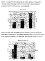
Figure 1 is a graph of the mechanical properties of three variations of implantable microbial cellulose illustrating the effect of cellulose content on the mechanical properties of tensile strength, elongation, suture retention strength and Young's Modulus. -
Figure 2 is a graph of the mechanical properties comparing control and compressed implantable microbial cellulose illustrating the effect of processing and cellulose content on the mechanical properties of tensile strength, elongation, suture retention strength and stiffness. -
Figure 3 is an SEM image taken in the horizontal plane of implantable microbial cellulose after supercritical fluid drying. -
Figure 4 is an SEM image taken in the vertical plane of implantable microbial cellulose after supercritical drying. -
Figure 5 is a graph of the tensile strength versus absorption amount comparing material from the '518 patent to those of the instant invention. -
Figure 6 is a graph of the absorption over time of the materials of the instant invention compared to that of material prepared using the method of '518. - The present invention describes a method for producing an implantable material comprising microbial cellulose. The instant implantable material has those properties necessary for in vivo applications, for example, the implantable material of the instant invention can be adapted to a three-dimensional shape and possess desired fluid absorption and pliability characteristics.
- The implantable materials of the instant invention are comprised of microbial cellulose. Those methods of preparing microbial cellulose are known to those of ordinary skill and are described, for example, in
U.S. Patent Nos. 5,846,213 and4,912,049 , which are incorporated herein by reference in their entirety. Any cellulose producing organism can be used in producing the raw biosynthetic cellulose material. However, biosynthetic cellulose produced from a static culture of Acetobacter xylinum is preferred. - The microbial cellulose content of the raw material is dependent on the amount of media supplied to the A.x. bacteria. Once the pellicle is harvested, the raw material is physically and chemically processed so as to be a suitable implantable material for medical and surgical uses. For example, the microbial cellulose is first processed and cleaned to remove all non-cellulose material embedded in the cellulose pad and then depyrogenated using chemicals such as sodium hydroxide. After depyrogenation, the cellulose may be cross-linked by irradiation or chemical means if its strength needs to be adjusted. Addition of other agents, such as glycerol and polyethylene glycol used to modify the cellulose surface can also be performed in order to control water absorption and pliability which are desirable properties for implantable materials. The material can remain wet, moist, partially dehydrated, or totally dehydrated by air, heat, lyophilization, freeze-drying or supercritical fluid drying. In a preferred embodiment, the microbial cellulose is dried, drying being a removal of liquid, which is different from the dehydration described in
US 6,599,518 B2 . - Supercritical fluid drying is preferred. Supercritical fluid drying refers to a drying step using a fluid held at a pressure and temperature above its critical point. The critical point is the lowest pressure and temperature at which the fluid can co-exist as a gas and a liquid. For example, the critical pressure and temperature of carbon dioxide are 72.8 atmospheres and 31.1°C, respectively. Carbon dioxide held at a pressure and temperature above its critical point is in a supercritical condition or state. According to the present invention, carbon dioxide is preferred as a fluid. The term "supercritical drying" thus refers to a process that uses one or more fluids, with at least one being under supercritical conditions part of the time, to displace and remove water within the matrix of microbial cellulose. It is generally desirable that the pressure of the supercritical fluid be reduced over a period of time of from about 1 minute to about 120 minutes to prevent rapid cooling which causes condensation. Supercritical drying of microbial cellulose is described in
US 5,772,647 andUS 5,580,348 . The methods described in these patents can also be used according to the present invention. - The material may be further processed by physically modifying the cellulose, preferably by compressing to a thin film after drying by applying repeated or sustained force directly to the dried material.
- In a preferred embodiment, the pressing step is achieved by constant pressure. Alternatively, the pressing can be achieved by repeated hammering, and the material further sterilized for applications as medical implantable articles using standard sterilization methods such as gamma irradiation, e-beam irradiation, ethylene oxide or steam sterilization.
- The combination of supercritical drying and subsequent compressing provides a material that is particularly suitable for implant materials. Preferably, the microbial cellulose content is 1 mg/cm2 to 50 mg/cm2. The tensile strength is preferably at least 80 N, more preferably in the range of 90 to 200 N. The absorption capability is preferably at least 3 g, more preferably at least 3.2 g, most preferably in the range 4.5 to 10 g, per gram of cellulose.. The measurement methods for determining the tensile strength and the absorption capability are described below. Moreover, the microbial cellulose of the present invention has preferably a mean pore size of at least about 0.01, more preferably in the range of 0.1 to 500 µm. Moreover, its bulk density is preferably in the range of 0.0001 to about 0.5 g/cm3. The pore size and the bulk density are determined as described in
US 5,580,348 (see column 9, lines 6 to 63). - Subsequent to the drying and physical modification step, the dried cellulose may be re-hydrated.
- In one preferred embodiment, the invention provides a method for preparing an implantable device for medical and surgical applications comprising the steps of providing a microbial cellulose material; and incorporating said material into an implantable device for medical and surgical applications. Once produced, the microbial cellulose may be incorporated or fashioned into medical devices by commonly known methods such as molding, cross-linking, chemical surface reaction, dehydrating and/or drying, cutting or punching. Such medical devices include tissue substitutes or scaffolds for repair or reinforcement of damaged soft tissue.
- Physical properties of microbial cellulose such as tensile strength, three dimensional structure, suture retention, absorption and conformability may be measured to show its characteristics by commonly utilized techniques such as scanning electron microscopy (SEM), mechanical testing or other standard physical tests. Chemical properties such as degree of crystallinity, active chemical groups and degree of polymerization can also be examined by techniques such as x-ray crystallography. Finally, the biocompatibility/safety properties of the implantable microbial cellulose in vitro and in vivo may be assessed.
- The properties of implantable microbial cellulose may be compared to a wide variety of implantable materials available including polypropylene mesh, PTFE, polymeric hydrogels, collagen, and human or animal derived tissue currently being used in the medical industry. Based on the results of these comparisons, including strength, conformability, and adhesion properties, a number of implantable microbial cellulose articles may be tailored for specific applications.
- The instant microbial cellulose may be used as a substitute or scaffold in tissue engineering. In this embodiment, the cellulose acts as a scaffold or trellis on which new tissue forms, orients and matures.
- In a preferred embodiment, the invention provides a method of tissue reinforcement, comprising an implantable composition comprising microbial cellulose and implanting said composition into a subject in need thereof. For example, the instant invention may be easily prepared as a dry or hydrated pad for direct application on a tissue through which staples, sutures or bone anchors are being added to ensure attachment of the tissue to its supporting structure. Often the tissue that is being repaired is friable and sutures or staples alone result in cutting or tearing and re-opening of the wound. In this embodiment the staples and/or sutures pass through both cellulose and tissue, the cellulose acting to reinforce the tissue by creating a stronger backing for attachment. For this application the material must be conformable so as to not cause damage to the tissue by rubbing, sharp edges, etc.
- The device may be a tissue scaffold, a surgical suture reinforcement device, a surgical staple reinforcement device, an adhesion barrier, a device used in shoulder repair, preferably shoulder repair involving the rotator cuff or involving the labrum, or a suture repair preferably including anchoring tissue to the bone. The device can be implanted through an open procedure or through an arthroscope.
- The invention also provides a kit comprised of a microbial cellulose device according to the present invention and a sterilizable closable container. The closable container may be a sealable pouch or a thermoformed tray with lid.
- In a preferred embodiment, the invention provides a method for repair of the rotator cuff and other shoulder related tears using the cellulose material. A method or process for fabricating such implantable materials will be cited in the examples accordingly.
- The material can be used for reinforcing tissue in and around the shoulder. The microbial cellulose described can be processed using the methods described above to create a sheet with multi-directional strength that can be used as a surgical device for rotator cuff repair. This may include both open and arthroscopic repair and include suture or staple reinforcement.
- The instant invention also contemplates an implantable composition comprising microbial cellulose and a medically useful agent dissolved in the liquid that is absorbed by the microbial cellulose material. The implant material is then able to have a dual function such as structural support to the body and as a reservoir for bioactive agents or drug for delivery to the site of implantation. Alternatively the agent may be incorporated into the microbial cellulose to reduce the risk of infection or rejection of the implant. Any number of medically useful agents for tissue repair can be used in the invention by adding the substances to an implantable composition comprising the microbial cellulose carrier, either at any steps in the manufacturing process or directly to the final composition.
- A medically useful agent is one having therapeutic, healing, curative, restorative, or medicinal properties. Such medically useful agents include collagen and insoluble collagen derivatives and soluble solids and/or liquids dissolved therein. Also included are amino acids, peptides, vitamins, co-factors for protein synthesis; hormones; endocrine tissue or tissue fragments; synthesizers; enzymes such as collagenase, peptidases, oxidases; cell scaffolds with parenchymal cells; angiogenic drugs and polymeric carriers containing such drugs; collagen lattices; biocompatible surface active agents, antigenic agents; cytoskeletal agents; cartilage fragments, living cells such as chondrocytes, bone marrow cells, mesenchymal stem cells, natural extracts, tissue transplants, bioadhesives, transforming growth factor (TGF-beta) and associated family proteins (bone morphogenetic protein (BMP), growth and differentiation factors (GDF) etc.), fibroblast growth factor (FGF), insulin-like growth factor (IGF-1) and other growth factors; growth hormones such as somatotropin; bone digestors; antitumor agents; fibronectin; cellular attractants and attachment agents; immuno-suppressants; permeation enhancers; and peptides, such as growth releasing factor, P-15 and the like.
- The drug can be in its free base or acid form, or in the form of salts, esters, or any other pharmacologically acceptable derivatives, enantomerically pure forms, tautomers or as components of molecular complexes. The amount of drug to be incorporated in the composition varies depending on the particular drug, the desired therapeutic effect, and the time span for which the device is to provide therapy. Generally, for purposes of the invention, the amount of drug in the system can vary from about 0.0001% to as much as 60%, preferably from about 0.01 wt.-% to about 50 wt.-%, more preferably from about 0.1 wt.-% to 10 wt.-%, with respect to the total weight of the composition
- The active agent may be used to reduce inflammation, increase cell attachment, recruit cells, and/or cause differentiation of the cells to repair the damaged tissue.
- In addition, implantable materials using microbial cellulose may be applied in a number of other useful areas, including, but not limited to other soft tissue substitutes or scaffolds.
- Other objects, features and advantages of the present invention will become apparent from the following examples. It should be understood, however, that the detailed description and the specific examples, while indicating preferred embodiments of the invention, are given by way of illustration only, since various changes and modifications within the spirit and scope of the invention will become apparent to those skilled in the art from this detailed description. The invention, thus generally described, will be understood more readily by reference to the following examples, which are provided by way of illustration and are not intended to be limiting of the present invention.
- To prepare the microbial cellulose of the invention, Acetobacter xylinum microorganisms were cultured in a bioreactor containing a liquid nutrient medium at 30 degrees Celsius at an initial pH of 3-6. The medium was based on sucrose or other carbohydrates.
- The bioreactor was composed of a plastic box fitted with an airtight cover. Dimensions of the bioreactor measured 3.5 in x 3.5 in. An aeration port was made in the bioreactor that allowed the proper oxygen tension to be achieved.
- The fermentation process under static conditions was allowed to progress for a period of about 10-14 days, during which the bacteria in the culture medium produced an intact cellulose pellicle. Once the media was expended, the fermentation was stopped and the pellicle removed from the bioreactor. This material was termed '250'.
- The excess medium contained in the pellicle was removed by mechanical compression prior to chemical cleaning and subsequent processing of the pellicle. The cellulose pellicle was subjected to a series of chemical wash steps to convert the raw cellulose film into a medical grade and nonpyrogenic implantable material. Processing started with an 8% sodium hydroxide solution at 70-75 degrees Celsius for 1 hour, followed by a rinse in deionized water and then a soak in 0.25% hydrogen peroxide at 70-75 degrees Celsius for 1 hour.
- The resulting films were tested for pyrogens and mechanical properties. The amount of cellular debris left in the cellulose pad after processing is measured by validated Limulus Amoebocyte Lysate (LAL) testing as outlined by the U.S. Food and Drug Administration (FDA) in 21 CFR10.90. The instant cleaning process outlined above provided a nonpyrogenic cellulose pad (≤0.50 EU/ml). The steps of the LAL test are defined by the test kit manufacturer and can simply be followed to yield the pyrogen level in the cellulose film.
- Once cleaned, the pellicles were mechanically pressed to reduce the water content. The materials were then soaked in 100% methanol for approximately 1 hour. The methanol water mixture was decanted and the samples soaked again in 100% methanol overnight. The methanol was changed at approximately 16 hours and again at 24 hours. Following the methanol exchange, the pellicle was repressed to reduce the methanol. Pellicles were then placed into a pressure vessel separated by polypropylene mesh and underwent supercritical carbon dioxide drying (SCD) at 2000 psi and 40 degrees Celsius until the methanol was removed and the material dry.
- The resulting dry pad is cut to shape, packaged in single or dual foil pouches and sterilized by gamma irradiation at 25-35 kGy.
- Material was prepared the same as in Comparative Example 1, however additional media was added at the start and the pellicle was allowed to grow 14-17 days. The resulting pellicle was termed '360'. The cleaning, whitening, drying, packaging and sterilization were identical to Comparative Example 1.
- Material was prepared the same as in Comparative Example 1, however additional media was added at the start and the pellicle was allowed to grow 21-25 days. The resulting pellicle was termed '440'. The cleaning, whitening, drying, packaging and sterilization were identical to Comparative Example 1.
- Material was initially cleaned, whitened, and dried as in Comparative Example 2. It was further processed by subjecting it to mechanical pressure to create a thinner version termed '360P'. For this example the material was hammered with a plastic mallet to compress the cellulose into a thin wafer. This was then packaged and sterilized as in Comparative Example 1.
- Material was initially cleaned, whitened, and dried as in Comparative Example 3. It was further processed by subjecting it to mechanical pressure to create a thin version termed '440P'. For this example the material was hammered with a plastic mallet to compress the cellulose into a thin wafer. This was then packaged and sterilized as in Comparative Example 1.
- Materials were processed as in Comparative Examples 1 - 3. The mechanical properties of these implantable microbial cellulose forms were analyzed using a United Tensile Tester (Model SSTM-2kN), including tensile strength, elongation, and suture retention.
- For each lot, samples for both tensile and suture (1cm x 4cm) were tested. Samples were prepared by soaking them in deionized water for 30-35 minutes prior to testing. Tensile tests were performed by placing the samples between two grips such that a 25mm gap was tested. A 1N preload was applied and the test performed at 300mm/minute until failure. Both tensile load and elongation at failure were recorded. The suture testing was performed by threading a single 2.0 Prolene suture through one end of the test sample. The sample was placed in the grips at one end and the suture placed in the grips at the other such that a gap length of 60mm was achieved. A 1N preload was applied and the test performed at 300mm/minute until failure. Young's Modulus was calculated from the tensile test results and sample measurements.
Table 1: Average Mechanical Values for Samples Sample Tensile (N) Suture (N) Elongation (%) Young's Modulus (MPa) 250 45.73 4.75 30.09 156.67 360 62.92 9.03 24.76 184.77 440 107.74 10.83 17.62 517.62 - Table 1 and
Figure 1 demonstrate the increased tensile strength, Young's Modulus and suture retention strength with increasing cellulose content. A decrease in elongation percent suggests that the material becomes stiffer with increasing cellulose. - Materials were processed as in comparative examples 2 and 3, and examples 1 and 2. The mechanical properties of these implantable microbial cellulose forms were performed using a United Tensile Tester (Model SSTM-2kN). Testing included tensile strength, elongation, suture retention and fabric stiffness.
- For each lot, samples for both tensile and suture (1cm x 4cm) and samples for stiffness (4cm x 5cm) were tested. Mechanical test samples were prepared by soaking them in deionized water for 30-35 minutes prior to testing. Tensile tests were performed by placing the samples between two grips such that a 25mm gap was tested. A 1N preload was applied and the test performed at 300mm/minute until failure. Both tensile load and elongation at failure were recorded. The suture testing was performed by threading a single 2.0 Prolene suture through one end of the test sample. The sample was placed in the grips at one end and the suture placed in the grips at the other, such that a 60mm gap length was produced. A 1N preload was applied and the test performed at 300mm/minute until failure. Stiffness testing was performed by placing a 4cm x 5cm piece of test material onto a fabric stiffness testing rig. A 1.2cm diameter flat based probe was then lowered to the sample and the peak force needed to push the test material through a 2.5cm (OD), 2cm ID diameter hole with a 45-degree beveled edge was recorded.
Table 2: Average Mechanical Values for Samples Sample Process Tensile (N) Suture (N) Elongation (%) Stiffness (N) 360 Control 67.49 9.34 24.97 23.60 360P Pressed 79.30 8.77 20.27 6.09 440 Control 78.73 15.25 29.16 42.00 440P Pressed 117.21 15.42 18.34 15.58 - Table 2 and
Figure 2 demonstrate the increased tensile strength of the compressed samples over uncompressed controls, but no change in the suture retention strength. - Material in the 440 configurations was prepared as in Comparative Examples 3 (SCD Control) and Example 2 (SCD Pounded) as well as following US Patent '518 (Solvent Dehydrated). A fourth material was prepared as in Comparative Example 3, but was then pressed for 60 seconds under 80 PSI of pressure (
SCD 60 sec Press). This resulted in an intermediate thickness material between SCD Control and SCD Pounded. Samples were either tested for tensile or suture strength after being hydrated for 30-35 minutes as in Example 4 or underwent absorption testing as follows. Samples measuring 4 x 5 cm were placed on top of a sponge that was soaked in a 0.9% saline solution and the testing chamber closed. Weights of the samples were taken prior to testing and at 24 hours after placement on the sponge. -
Figure 5 illustrates the comparison of absorption amount versus tensile strength for the various materials. Note that the stronger a material is in tension the less it absorbs. This demonstrates that the '518 material (solvent dehydrated) would not be useful as a delivery vehicle for a medically useful agent when compared to material processed using any of the SCD methods. -
Figure 6 illustrates that even over time the '518 (solvent dehydrated) never reaches the level of absorption observed with materials prepared as described in the current invention. - The physical characteristics of implantable microbial cellulose can be seen in
Figures 3 and 4 that show an SEM image of the implantable microbial cellulose materials. Note the interconnected fibers inFigure 3 and the laminar structure inFigure 4 . - Biocompatibility testing and implantation studies were conducted to assess the implantable microbial cellulose safety profile. A battery of in vitro and animal biocompatibility tests including cytotoxicity, sensitization, intracutaneous irritation, systemic toxicity and genotoxicity have been conducted on microbial cellulose, with the results indicating that the material is biocompatible. Muscle implantation studies in rabbits up to 24 weeks have been performed and the histological and gross necropsy results showed no significant tissue reaction, minimal cellular interaction, and very low adhesion to tissue. Table 3 lists the tests and the results.
Table 3: Biocompatibility testing and results of implantable microbial cellulose Test Results Cytotoxicity Pass Irritation Pass Acute Systemic Toxicity Pass Genotoxicity - Bacterial Reverse
MutationsPass Genotoxicity - In vitro
Chromosomal AberrationPass Genotoxicity - Mouse Bone Marrow
MicronucleusPass Sensitization Pass 4, 12, 18 and 26 week Rabbit
Muscle ImplantationPass Hemolysis Pass Subchronic Toxicity Pass Chronic Toxicity Pass Endotoxin (pyrogen level) Pass - The implantable microbial cellulose materials prepared in comparative examples 1-3 were compared with existing medical devices used for shoulder repair. The suture retention properties of collagen-based products (both human- and animal-derived) and PTFE materials were compared to the implantable microbial cellulose. These were tested in the following in vitro model.
- Chicken Achilles tendons were harvested from fresh legs and stored in isotonic saline before use. These specimens were approximately 5cm long, 1cm wide and 2mm thick. Test specimens were individually prepared and tested on an Instron Mini 44 machine.
- The test fixture consisted of a 3-mm thick aluminum 'L' shaped plate with three 0.5mm holes (hole edges polished to prevent suture damage) spaced 2.5mm in a row on both the side and top
- The top holes were used for pull-off testing and the side holes for shear testing. No.2 Mersilene™ suture was introduced from the back of the plate and through the tendon and graft (when present) and returned through the tendon maintaining either a 2.5mm or 5mm suture gap and tied on the back of the plate with a square knot. The plate was held by one grip of the Instron and the free end of the tendon passed through a small eye bolt held by the other grip and constrained with a hemostat. Tension was applied at 1.0cm/min until failure. Failure load and mechanism of failure were determined.
- Tendon with no graft and suture gaps of 2.5mm and 5mm served as controls. Test materials included PTFE-Teflon fabric (Gore-Tex® Soft Tissue Patch), human derived cross-linked collagen (GRAFTJACKET™), bovine pericardium (Peri-Guard), and implantable cellulose material of Examples 1 and 2. Test units were one centimeter square patches with 5-mm suture spacing.
- Twelve specimens were run for each test material and graft material. The two lowest failure load values were discarded (due to loosening of knot or tendon clamp) and the remaining ten averaged to determine failure strength. Averages were compared by unpaired Student's t-Tests.
- Results of the tests are given in Table 4. The failure loads occurred when the repairs first began to displace and these loads gradually decreased as the suture/graft passes through the tendon. The implantable microbial cellulose and bovine pericardium grafts were significantly (p< 0.05) stronger than the non-augmented suture for both tests. All non-augmented specimens failed by the suture slicing along the tendon (tear-out). Only the human collagen graft failed in a similar manner. The other grafts failed by various amounts of cut-out and tear-out with the bovine pericardium failing mainly by tear-out. However in all of the cut-out failures, the suture did not cut through any of the grafts but pulled them into and usually through the tendon. The graft/suture subsequently sliced along the tendon. These cut-out failures generally resulted in higher strengths.
Table 4 Strength of tendon augmentations in Newtons (SD). Augmentation Shear Pull-off None-2.5 mm suture gap 23 (8)
100%T27 (9)
100%TNone-5 mm suture gap 35 (5)
100%T36 (8)
100%TPTFE 39 (7)
100%T49 (8)
40%TBovine pericardium 56 (14)
60%T49 (19)
80%THuman collagen 41 (19)
100%T34 (9)
100%TImplantable
Cellulose48 (24)
40%T35 (14)
20%TT = percentage of tests that failed by tear-out. - As is apparent from the preceding description and examples, the present invention is directed to a class of implantable materials using microbial cellulose that can be used for medical applications and medical devices to repair and replace injured orthopedic soft tissue. The products maybe constructed in variety of forms (e.g. film, pad, hydrated, dry) and with varying physical and chemical properties. Additionally, the materials can be used in combination with other biomaterials such as collagen, proteins, and other bioactive agents to enhance its efficacy for a particular application. Many other variations and details of construction, composition and configuration will be apparent to those skilled in the art and such variations are contemplated within the scope of the present invention.
- Material was initially cleaned, whitened, and dried as in Comparative Example 3. It was further processed by subjecting it to perforation by placing it in a tissue 1/1 mesher. This made macroscopic holes in the material while maintaining strength in one dimension.
Claims (1)
- A method for producing a microbial cellulose, comprising the stepsa) drying the microbial cellulose, wherein the drying is carried out by supercritical fluid drying; and subsequentlyb) pressing the dried cellulose.
Priority Applications (1)
| Application Number | Priority Date | Filing Date | Title |
|---|---|---|---|
| EP09180581A EP2189168A1 (en) | 2005-12-02 | 2006-12-01 | Implantable microbial cellulose materials for various medical applications |
Applications Claiming Priority (1)
| Application Number | Priority Date | Filing Date | Title |
|---|---|---|---|
| US11/292,075 US20070128243A1 (en) | 2005-12-02 | 2005-12-02 | Implantable microbial cellulose materials for various medical applications |
Related Child Applications (1)
| Application Number | Title | Priority Date | Filing Date |
|---|---|---|---|
| EP09180581.2 Division-Into | 2009-12-23 |
Publications (3)
| Publication Number | Publication Date |
|---|---|
| EP1795213A2 EP1795213A2 (en) | 2007-06-13 |
| EP1795213A3 EP1795213A3 (en) | 2007-07-18 |
| EP1795213B1 true EP1795213B1 (en) | 2011-09-28 |
Family
ID=37888108
Family Applications (2)
| Application Number | Title | Priority Date | Filing Date |
|---|---|---|---|
| EP06024913A Expired - Fee Related EP1795213B1 (en) | 2005-12-02 | 2006-12-01 | A method for producing implantable microbial cellulose materials for various medical applications |
| EP09180581A Withdrawn EP2189168A1 (en) | 2005-12-02 | 2006-12-01 | Implantable microbial cellulose materials for various medical applications |
Family Applications After (1)
| Application Number | Title | Priority Date | Filing Date |
|---|---|---|---|
| EP09180581A Withdrawn EP2189168A1 (en) | 2005-12-02 | 2006-12-01 | Implantable microbial cellulose materials for various medical applications |
Country Status (5)
| Country | Link |
|---|---|
| US (1) | US20070128243A1 (en) |
| EP (2) | EP1795213B1 (en) |
| JP (2) | JP5700614B2 (en) |
| CA (1) | CA2632767A1 (en) |
| WO (2) | WO2007064772A2 (en) |
Families Citing this family (38)
| Publication number | Priority date | Publication date | Assignee | Title |
|---|---|---|---|---|
| WO2007146946A2 (en) * | 2006-06-13 | 2007-12-21 | Xylos Corporation | Implantable microbial cellulose materials for hard tissue repair and regeneration |
| PL381388A1 (en) * | 2006-12-24 | 2008-07-07 | Politechnika Łódzka | Biomaterial of microbiological cellulose for internal use, production method of biomaterial and application of bomaterial of microbiological cellulose in surgery of soft tissues |
| EP2070557A1 (en) * | 2007-12-12 | 2009-06-17 | Xylos Corporation | Implantable microbial cellulose materials for hard tissue repair and regeneration |
| US7832857B2 (en) * | 2008-08-18 | 2010-11-16 | Levinson Dennis J | Microbial cellulose contact lens |
| AU2009312480B2 (en) | 2008-11-07 | 2015-03-19 | Sofradim Production | Composite mesh including a 3D mesh and a non porous film of oxidized cellulose from bacterial cellulose origin |
| EP2346540B1 (en) * | 2008-11-07 | 2013-04-03 | Sofradim Production | Medical device including bacterial cellulose reinforced by resorbable reinforcing materials |
| AU2009312477B2 (en) * | 2008-11-07 | 2015-11-12 | Sofradim Production | Medical devices with definable porosity produced by bacterial polymer bio-synthesis |
| US20100159046A1 (en) * | 2008-12-19 | 2010-06-24 | Xylos Corporation | Minimal tissue attachment implantable materials |
| US20100158985A1 (en) * | 2008-12-19 | 2010-06-24 | Xylos Corporation | Porous structures of microbial-derived cellulose for in vivo implantation |
| US20100297239A1 (en) * | 2008-12-22 | 2010-11-25 | Paul Gatenholm | Osseointegrative meniscus and cartilage implants based on beta-glucan nanocomposites |
| DE102009024133A1 (en) | 2009-06-04 | 2010-12-09 | Jenpolymer Materials Ltd. & Co. Kg | Bacterial nanocellulose for cartilage regeneration |
| WO2011086788A1 (en) * | 2010-01-15 | 2011-07-21 | 国立大学法人島根大学 | Bone cement |
| WO2012091572A1 (en) * | 2010-12-30 | 2012-07-05 | Feyecon B.V. | Dehydration process that employs an ionic liquid choline salt |
| US8998059B2 (en) | 2011-08-01 | 2015-04-07 | Ethicon Endo-Surgery, Inc. | Adjunct therapy device having driver with cavity for hemostatic agent |
| US9492170B2 (en) | 2011-08-10 | 2016-11-15 | Ethicon Endo-Surgery, Inc. | Device for applying adjunct in endoscopic procedure |
| US9101359B2 (en) | 2011-09-13 | 2015-08-11 | Ethicon Endo-Surgery, Inc. | Surgical staple cartridge with self-dispensing staple buttress |
| US8998060B2 (en) | 2011-09-13 | 2015-04-07 | Ethicon Endo-Surgery, Inc. | Resistive heated surgical staple cartridge with phase change sealant |
| US9999408B2 (en) | 2011-09-14 | 2018-06-19 | Ethicon Endo-Surgery, Inc. | Surgical instrument with fluid fillable buttress |
| US9254180B2 (en) | 2011-09-15 | 2016-02-09 | Ethicon Endo-Surgery, Inc. | Surgical instrument with staple reinforcement clip |
| US9125649B2 (en) | 2011-09-15 | 2015-09-08 | Ethicon Endo-Surgery, Inc. | Surgical instrument with filled staple |
| US8814025B2 (en) | 2011-09-15 | 2014-08-26 | Ethicon Endo-Surgery, Inc. | Fibrin pad matrix with suspended heat activated beads of adhesive |
| US9198644B2 (en) | 2011-09-22 | 2015-12-01 | Ethicon Endo-Surgery, Inc. | Anvil cartridge for surgical fastening device |
| US9393018B2 (en) | 2011-09-22 | 2016-07-19 | Ethicon Endo-Surgery, Inc. | Surgical staple assembly with hemostatic feature |
| US8985429B2 (en) | 2011-09-23 | 2015-03-24 | Ethicon Endo-Surgery, Inc. | Surgical stapling device with adjunct material application feature |
| US8899464B2 (en) | 2011-10-03 | 2014-12-02 | Ethicon Endo-Surgery, Inc. | Attachment of surgical staple buttress to cartridge |
| US9089326B2 (en) | 2011-10-07 | 2015-07-28 | Ethicon Endo-Surgery, Inc. | Dual staple cartridge for surgical stapler |
| KR101371379B1 (en) * | 2012-01-26 | 2014-03-07 | (주) 유쎌 | Manifacturing method of dried bio-cellulose |
| DE102012003541A1 (en) | 2012-02-21 | 2013-08-22 | Jenpolymer Materials Ltd. & Co. Kg | Bacterial nanocellulose body useful e.g. as transplant for in situ cell colonization and tissue formation, comprises channel-like pore structure, interconnecting multidimensional structure, and channel-shaped cavities with open cell wall |
| JP6195851B2 (en) * | 2012-02-22 | 2017-09-13 | シンセス・ゲーエムベーハーSynthes GmbH | Absorbable cellulosic biomaterial and implant |
| US9421396B2 (en) | 2012-09-05 | 2016-08-23 | Mayo Foundation For Medical Education And Research | Brachytherapy and buttress element co-delivery |
| JP2014118498A (en) * | 2012-12-17 | 2014-06-30 | Chiba Flour Milling Co Ltd | Fermentation-derived cellulose or purification method of the same, purified fermentation-derived cellulose or pharmaceutical preparation of the same, and cosmetic, pharmaceutical and quasi-drug |
| WO2015181372A1 (en) | 2014-05-30 | 2015-12-03 | Sofradim Production | Implant comprising oxidized cellulose and method for preparing such an implant |
| US10258337B2 (en) | 2016-04-20 | 2019-04-16 | Ethicon Llc | Surgical staple cartridge with severed tissue edge adjunct |
| JP7010844B2 (en) * | 2016-05-06 | 2022-01-26 | エシコン・インコーポレイテッド | Surgical stapler / cutter and extended buttress |
| GB201800027D0 (en) | 2018-01-02 | 2018-02-14 | Keane Jennifer Killeen | Bacterial cellulose based hybrid materials |
| CN112138215B (en) * | 2020-09-26 | 2021-11-05 | 江苏大学 | Construction method and application of cell growth factor slow-release anisotropic scaffold based on nano-cellulose |
| CN114377201A (en) * | 2022-01-19 | 2022-04-22 | 福州市第二医院(福建省福州中西医结合医院、福州市职业病医院) | Rotator cuff patch based on cellulose gelatin coating and preparation method thereof |
| CN115444978A (en) * | 2022-09-14 | 2022-12-09 | 中国科学技术大学 | Bionic artificial ligament material and preparation method thereof |
Family Cites Families (47)
| Publication number | Priority date | Publication date | Assignee | Title |
|---|---|---|---|---|
| US2537979A (en) * | 1949-11-25 | 1951-01-16 | Ethicon Suture Lab Inc | Oxidized cellulose suture |
| US4429080A (en) * | 1982-07-01 | 1984-01-31 | American Cyanamid Company | Synthetic copolymer surgical articles and method of manufacturing the same |
| US4588400A (en) * | 1982-12-16 | 1986-05-13 | Johnson & Johnson Products, Inc. | Liquid loaded pad for medical applications |
| US4655758A (en) * | 1982-12-16 | 1987-04-07 | Johnson & Johnson Products, Inc. | Microbial polysaccharide articles and methods of production |
| BR8404937A (en) * | 1984-10-01 | 1986-05-06 | Bio Fill Ind E Comercio De Pro | PROCESS FOR PREPARING CELLULOSE FILM, CELLULOSE FILM OBTAINED BY THE SAME, ARTIFICIAL SKIN IMPLANT, INJURY TREATMENT PROCESS USING THE REFERRED CELLULOSE FILM AND USE |
| NZ214417A (en) * | 1984-12-21 | 1988-06-30 | Univ Texas | Microbially produced cellulose |
| CA1335266C (en) * | 1985-10-18 | 1995-04-18 | Arie Ben-Bassat | Reticulated cellulose product, sheets formed therefrom, methods and microorganisms for the production thereof |
| WO1987005038A1 (en) * | 1986-02-17 | 1987-08-27 | Commonwealth Scientific And Industrial Research Or | Implantable materials |
| US5863531A (en) * | 1986-04-18 | 1999-01-26 | Advanced Tissue Sciences, Inc. | In vitro preparation of tubular tissue structures by stromal cell culture on a three-dimensional framework |
| DE3783073T2 (en) * | 1986-04-22 | 1993-04-29 | Ajinomoto Kk | MODIFIED CELLULOSE GEL MADE BY MICROORGANISMS AND ITS COMPLEX WITH ANIMAL CELLS. |
| US5306311A (en) * | 1987-07-20 | 1994-04-26 | Regen Corporation | Prosthetic articular cartilage |
| US5273891A (en) * | 1988-01-06 | 1993-12-28 | Imperial Chemical Industries Plc | Process for the production of microbial cellulose |
| US5441508A (en) * | 1989-04-27 | 1995-08-15 | Gazielly; Dominique | Reinforcement and supporting device for the rotator cuff of a shoulder joint of a person |
| US5073373A (en) * | 1989-09-21 | 1991-12-17 | Osteotech, Inc. | Flowable demineralized bone powder composition and its use in bone repair |
| US5290558A (en) * | 1989-09-21 | 1994-03-01 | Osteotech, Inc. | Flowable demineralized bone powder composition and its use in bone repair |
| US4946377A (en) * | 1989-11-06 | 1990-08-07 | W. L. Gore & Associates, Inc. | Tissue repair device |
| EP0560934B2 (en) * | 1990-12-06 | 1999-11-10 | W.L. Gore & Associates, Inc. | Implantable bioabsorbable article |
| US5314476A (en) * | 1992-02-04 | 1994-05-24 | Osteotech, Inc. | Demineralized bone particles and flowable osteogenic composition containing same |
| US5281400A (en) * | 1992-09-30 | 1994-01-25 | Carr Metal Products | Plastic autoclave tray and lid combination |
| US5433996A (en) * | 1993-02-18 | 1995-07-18 | W. L. Gore & Associates, Inc. | Laminated patch tissue repair sheet material |
| AU6987594A (en) * | 1993-08-18 | 1995-03-14 | W.L. Gore & Associates, Inc. | A tubular intraluminal graft |
| US5580348A (en) * | 1994-05-10 | 1996-12-03 | Kimberly-Clark Corporation | Absorbent structure comprising a microbial polysaccharide and a process of making the same |
| TW369414B (en) * | 1994-09-30 | 1999-09-11 | Yamanouchi Pharma Co Ltd | Bone formation transplant |
| US5810855A (en) * | 1995-07-21 | 1998-09-22 | Gore Enterprise Holdings, Inc. | Endoscopic device and method for reinforcing surgical staples |
| US5702409A (en) * | 1995-07-21 | 1997-12-30 | W. L. Gore & Associates, Inc. | Device and method for reinforcing surgical staples |
| JP5053470B2 (en) * | 1996-08-23 | 2012-10-17 | クック バイオテク インコーポレイティド | Graft prosthesis and materials and methods thereof |
| US6503275B1 (en) * | 1996-11-15 | 2003-01-07 | Medevec Licensing, B.V. | Ophthalmic lens insertion instrument and package |
| US5846213A (en) * | 1997-06-16 | 1998-12-08 | The University Of Western Ontario | Cellulose membrane and method for manufacture thereof |
| US6620166B1 (en) * | 1998-01-09 | 2003-09-16 | Ethicon, Inc. | Suture buttress system |
| US6156056A (en) * | 1998-01-09 | 2000-12-05 | Ethicon, Inc. | Suture buttress |
| US6245081B1 (en) * | 1998-01-09 | 2001-06-12 | Steven M. Bowman | Suture buttress |
| JPH11241027A (en) * | 1998-02-26 | 1999-09-07 | Sony Corp | Polymer composite material and method for producing the same |
| US6030635A (en) * | 1998-02-27 | 2000-02-29 | Musculoskeletal Transplant Foundation | Malleable paste for filling bone defects |
| US6179872B1 (en) * | 1998-03-17 | 2001-01-30 | Tissue Engineering | Biopolymer matt for use in tissue repair and reconstruction |
| AU778081B2 (en) * | 1999-03-25 | 2004-11-11 | Tepha, Inc. | Medical devices and applications of polyhydroxyalkanoate polymers |
| GB9912240D0 (en) * | 1999-05-27 | 1999-07-28 | Smith & Nephew | Implantable medical devices |
| US6755863B2 (en) * | 1999-10-08 | 2004-06-29 | Bret A. Ferree | Rotator cuff repair using engineered tissues |
| US6599518B2 (en) * | 2000-11-21 | 2003-07-29 | Xylos Corporation | Solvent dehydrated microbially-derived cellulose for in vivo implantation |
| US6852330B2 (en) * | 2000-12-21 | 2005-02-08 | Depuy Mitek, Inc. | Reinforced foam implants with enhanced integrity for soft tissue repair and regeneration |
| DE10064314C1 (en) * | 2000-12-22 | 2002-08-08 | Skw Biogarde Gmbh & Co Kg | Microbially produced, physiologically compatible, permeable film, consisting of chitosan-containing cellulose |
| US6785863B2 (en) | 2002-09-18 | 2004-08-31 | Motorola, Inc. | Method and apparatus for generating parity-check bits from a symbol set |
| US20040142019A1 (en) * | 2003-01-16 | 2004-07-22 | Xylos Corporation | Microbial-derived cellulose amorphous hydrogel wound dressing |
| US8226715B2 (en) * | 2003-06-30 | 2012-07-24 | Depuy Mitek, Inc. | Scaffold for connective tissue repair |
| US20050037082A1 (en) * | 2003-08-13 | 2005-02-17 | Wan-Kei Wan | Poly(vinyl alcohol)-bacterial cellulose nanocomposite |
| US8198261B2 (en) * | 2003-08-22 | 2012-06-12 | Xylos Corporation | Thermally modified microbial-derived cellulose for in vivo implantation |
| US20080280360A1 (en) * | 2004-10-12 | 2008-11-13 | Trustees Of Tufts College | Method for Producing Biomaterial Scaffolds |
| WO2007027849A2 (en) * | 2005-08-31 | 2007-03-08 | Board Of Regents, The University Of Texas System | Multiribbon nanocellulose as a matrix for wound healing |
-
2005
- 2005-12-02 US US11/292,075 patent/US20070128243A1/en not_active Abandoned
-
2006
- 2006-11-30 WO PCT/US2006/045806 patent/WO2007064772A2/en not_active Ceased
- 2006-12-01 EP EP06024913A patent/EP1795213B1/en not_active Expired - Fee Related
- 2006-12-01 WO PCT/US2006/045992 patent/WO2007064881A2/en not_active Ceased
- 2006-12-01 EP EP09180581A patent/EP2189168A1/en not_active Withdrawn
- 2006-12-01 JP JP2008543483A patent/JP5700614B2/en not_active Expired - Fee Related
- 2006-12-01 CA CA002632767A patent/CA2632767A1/en not_active Abandoned
-
2014
- 2014-12-15 JP JP2014252604A patent/JP2015163679A/en active Pending
Also Published As
| Publication number | Publication date |
|---|---|
| WO2007064881A2 (en) | 2007-06-07 |
| WO2007064881A3 (en) | 2007-11-15 |
| JP2009518466A (en) | 2009-05-07 |
| EP1795213A2 (en) | 2007-06-13 |
| WO2007064772A2 (en) | 2007-06-07 |
| US20070128243A1 (en) | 2007-06-07 |
| CA2632767A1 (en) | 2007-06-07 |
| JP5700614B2 (en) | 2015-04-15 |
| EP2189168A1 (en) | 2010-05-26 |
| EP1795213A3 (en) | 2007-07-18 |
| JP2015163679A (en) | 2015-09-10 |
| WO2007064772A3 (en) | 2007-08-09 |
Similar Documents
| Publication | Publication Date | Title |
|---|---|---|
| EP1795213A2 (en) | Implantable microbial cellulose materials for various medical applications | |
| US8198261B2 (en) | Thermally modified microbial-derived cellulose for in vivo implantation | |
| AU2018354277A1 (en) | Biopolymer scaffold implants and methods for their production | |
| CN104271166B (en) | Biomaterial based on absorbent cellulosic and implant | |
| US20070286884A1 (en) | Implantable microbial cellulose materials for hard tissue repair and regeneration | |
| US20250049987A1 (en) | Method for Preparing a Three-Dimensional Scaffold for Medical Use | |
| US20260108658A1 (en) | A Three-Dimensional Scaffold for Medical Use Comprising Animal Collagen | |
| EP3539576A1 (en) | Hydrogel composites comprising chitosan and cellulose nanofibers | |
| EP1667600B1 (en) | Dura substiture and a process for producing the same | |
| WO2007146946A2 (en) | Implantable microbial cellulose materials for hard tissue repair and regeneration | |
| US20120157673A1 (en) | Minimal tissue attachment implantable materials | |
| EP2070557A1 (en) | Implantable microbial cellulose materials for hard tissue repair and regeneration | |
| US20230037708A1 (en) | Porous collagen/polymer matrix biocomposite material and use thereof as an implant for repairing meniscal lesions of the knee and/or for preventing or treating osteoarthritis of the knee | |
| WO2017205740A1 (en) | Process for preparing tissue regeneration matrix | |
| O’Brien | The development of novel scaffolds for tissue engineering with a range of structural and mechanical properties |
Legal Events
| Date | Code | Title | Description |
|---|---|---|---|
| PUAI | Public reference made under article 153(3) epc to a published international application that has entered the european phase |
Free format text: ORIGINAL CODE: 0009012 |
|
| AK | Designated contracting states |
Kind code of ref document: A2 Designated state(s): AT BE BG CH CY CZ DE DK EE ES FI FR GB GR HU IE IS IT LI LT LU LV MC NL PL PT RO SE SI SK TR |
|
| AX | Request for extension of the european patent |
Extension state: AL BA HR MK YU |
|
| PUAL | Search report despatched |
Free format text: ORIGINAL CODE: 0009013 |
|
| AK | Designated contracting states |
Kind code of ref document: A3 Designated state(s): AT BE BG CH CY CZ DE DK EE ES FI FR GB GR HU IE IS IT LI LT LU LV MC NL PL PT RO SE SI SK TR |
|
| AX | Request for extension of the european patent |
Extension state: AL BA HR MK YU |
|
| RIN1 | Information on inventor provided before grant (corrected) |
Inventor name: BEAM, HEATHER Inventor name: WRIGHT, FREDERIC S. Inventor name: DAMIEN, CHRIS Inventor name: SERAFICA, GONZALO |
|
| 17P | Request for examination filed |
Effective date: 20080117 |
|
| AKX | Designation fees paid |
Designated state(s): DE FR GB |
|
| 17Q | First examination report despatched |
Effective date: 20091013 |
|
| GRAP | Despatch of communication of intention to grant a patent |
Free format text: ORIGINAL CODE: EPIDOSNIGR1 |
|
| RTI1 | Title (correction) |
Free format text: A METHOD FOR PRODUCING IMPLANTABLE MICROBIAL CELLULOSE MATERIALS FOR VARIOUS MEDICAL APPLICATIONS |
|
| GRAS | Grant fee paid |
Free format text: ORIGINAL CODE: EPIDOSNIGR3 |
|
| GRAA | (expected) grant |
Free format text: ORIGINAL CODE: 0009210 |
|
| AK | Designated contracting states |
Kind code of ref document: B1 Designated state(s): DE FR GB |
|
| REG | Reference to a national code |
Ref country code: GB Ref legal event code: FG4D |
|
| 111Z | Information provided on other rights and legal means of execution |
Free format text: DE FR GB Effective date: 20110803 |
|
| REG | Reference to a national code |
Ref country code: DE Ref legal event code: R096 Ref document number: 602006024654 Country of ref document: DE Effective date: 20111124 |
|
| PGFP | Annual fee paid to national office [announced via postgrant information from national office to epo] |
Ref country code: FR Payment date: 20120105 Year of fee payment: 6 |
|
| PGFP | Annual fee paid to national office [announced via postgrant information from national office to epo] |
Ref country code: DE Payment date: 20111229 Year of fee payment: 6 |
|
| PLBE | No opposition filed within time limit |
Free format text: ORIGINAL CODE: 0009261 |
|
| STAA | Information on the status of an ep patent application or granted ep patent |
Free format text: STATUS: NO OPPOSITION FILED WITHIN TIME LIMIT |
|
| 26N | No opposition filed |
Effective date: 20120629 |
|
| REG | Reference to a national code |
Ref country code: DE Ref legal event code: R097 Ref document number: 602006024654 Country of ref document: DE Effective date: 20120629 |
|
| REG | Reference to a national code |
Ref country code: DE Ref legal event code: R082 Ref document number: 602006024654 Country of ref document: DE Representative=s name: HOFFMANN - EITLE, DE |
|
| REG | Reference to a national code |
Ref country code: DE Ref legal event code: R082 Ref document number: 602006024654 Country of ref document: DE Representative=s name: HOFFMANN - EITLE PATENT- UND RECHTSANWAELTE PA, DE Effective date: 20121204 Ref country code: DE Ref legal event code: R082 Ref document number: 602006024654 Country of ref document: DE Representative=s name: HOFFMANN - EITLE, DE Effective date: 20121204 Ref country code: DE Ref legal event code: R081 Ref document number: 602006024654 Country of ref document: DE Owner name: ELLIOT CAPITAL INVESTMENTS III, LTD., ROAD TOW, VG Free format text: FORMER OWNER: XYLOS CORP., LANGHORNE, PA., US Effective date: 20121204 Ref country code: DE Ref legal event code: R081 Ref document number: 602006024654 Country of ref document: DE Owner name: ELLIOT CAPITAL INVESTMENTS III, LTD., ROAD TOW, VG Free format text: FORMER OWNER: XYLOS CORP., LANGHORNE, PA., US Effective date: 20110930 Ref country code: DE Ref legal event code: R081 Ref document number: 602006024654 Country of ref document: DE Owner name: ELLIOT CAPITAL INVESTMENTS III, LTD., VG Free format text: FORMER OWNER: XYLOS CORP., LANGHORNE, US Effective date: 20121204 Ref country code: DE Ref legal event code: R081 Ref document number: 602006024654 Country of ref document: DE Owner name: ELLIOT CAPITAL INVESTMENTS III, LTD., VG Free format text: FORMER OWNER: XYLOS CORP., LANGHORNE, US Effective date: 20110930 |
|
| REG | Reference to a national code |
Ref country code: FR Ref legal event code: TP Owner name: ELLIOT CAPITAL INVESTMENTS III, LTD., VG Effective date: 20121227 |
|
| REG | Reference to a national code |
Ref country code: GB Ref legal event code: 732E Free format text: REGISTERED BETWEEN 20130124 AND 20130130 |
|
| GBPC | Gb: european patent ceased through non-payment of renewal fee |
Effective date: 20121201 |
|
| REG | Reference to a national code |
Ref country code: FR Ref legal event code: ST Effective date: 20130830 |
|
| PG25 | Lapsed in a contracting state [announced via postgrant information from national office to epo] |
Ref country code: DE Free format text: LAPSE BECAUSE OF NON-PAYMENT OF DUE FEES Effective date: 20130702 |
|
| REG | Reference to a national code |
Ref country code: DE Ref legal event code: R119 Ref document number: 602006024654 Country of ref document: DE Effective date: 20130702 |
|
| PG25 | Lapsed in a contracting state [announced via postgrant information from national office to epo] |
Ref country code: FR Free format text: LAPSE BECAUSE OF NON-PAYMENT OF DUE FEES Effective date: 20130102 Ref country code: GB Free format text: LAPSE BECAUSE OF NON-PAYMENT OF DUE FEES Effective date: 20121201 |