EP1788090A1 - Method for preparing artificial chromosomes which are free of host DNA - Google Patents
Method for preparing artificial chromosomes which are free of host DNA Download PDFInfo
- Publication number
- EP1788090A1 EP1788090A1 EP06020842A EP06020842A EP1788090A1 EP 1788090 A1 EP1788090 A1 EP 1788090A1 EP 06020842 A EP06020842 A EP 06020842A EP 06020842 A EP06020842 A EP 06020842A EP 1788090 A1 EP1788090 A1 EP 1788090A1
- Authority
- EP
- European Patent Office
- Prior art keywords
- dna
- artificial chromosomes
- cells
- microarray
- host
- Prior art date
- Legal status (The legal status is an assumption and is not a legal conclusion. Google has not performed a legal analysis and makes no representation as to the accuracy of the status listed.)
- Granted
Links
Images







Classifications
-
- C—CHEMISTRY; METALLURGY
- C12—BIOCHEMISTRY; BEER; SPIRITS; WINE; VINEGAR; MICROBIOLOGY; ENZYMOLOGY; MUTATION OR GENETIC ENGINEERING
- C12N—MICROORGANISMS OR ENZYMES; COMPOSITIONS THEREOF; PROPAGATING, PRESERVING, OR MAINTAINING MICROORGANISMS; MUTATION OR GENETIC ENGINEERING; CULTURE MEDIA
- C12N15/00—Mutation or genetic engineering; DNA or RNA concerning genetic engineering, vectors, e.g. plasmids, or their isolation, preparation or purification; Use of hosts therefor
- C12N15/09—Recombinant DNA-technology
- C12N15/10—Processes for the isolation, preparation or purification of DNA or RNA
- C12N15/1034—Isolating an individual clone by screening libraries
-
- C—CHEMISTRY; METALLURGY
- C12—BIOCHEMISTRY; BEER; SPIRITS; WINE; VINEGAR; MICROBIOLOGY; ENZYMOLOGY; MUTATION OR GENETIC ENGINEERING
- C12Q—MEASURING OR TESTING PROCESSES INVOLVING ENZYMES, NUCLEIC ACIDS OR MICROORGANISMS; COMPOSITIONS OR TEST PAPERS THEREFOR; PROCESSES OF PREPARING SUCH COMPOSITIONS; CONDITION-RESPONSIVE CONTROL IN MICROBIOLOGICAL OR ENZYMOLOGICAL PROCESSES
- C12Q1/00—Measuring or testing processes involving enzymes, nucleic acids or microorganisms; Compositions therefor; Processes of preparing such compositions
- C12Q1/68—Measuring or testing processes involving enzymes, nucleic acids or microorganisms; Compositions therefor; Processes of preparing such compositions involving nucleic acids
- C12Q1/6809—Methods for determination or identification of nucleic acids involving differential detection
Definitions
- the present invention relates to a novel method for isolating artificial chromosomes which are free of host DNA from a library, preferably BAC clones.
- the present invention further relates to various applications of said method in DNA analysis for medical, forensic, diagnostic or scientific purposes, e.g., ex vivo analyses of diseases like cancer being based on few or even single cells by, for example, microarray comparative genomic hybridization (CGH).
- CGH microarray comparative genomic hybridization
- the technical problem underlying the present invention is to provide methods for reliably analysing the genome of a small cell population or a single cell.
- the present invention allows to establish an array CGH protocol suited for numerous applications. Examples include the study of minute amounts of ex vivo DNA from premalignant lesions or very small tumors, from biopsies before and after systemic therapy, archived tumor samples of patients with known clinical outcome, analysis of single metastastic precursor cells or of circulating fetal cells in the maternal blood. Also, single cell analysis will be important in various fields studying the mechanisms of genetic instability of tumors or the genomic causes underlying human ageing 17,18 , because most of these changes are stochastic in nature and require single cell resolution. Therefore, single cell array CGH using artificial chromosomes purified according to the method of the present invention will enable novel insights into important biological and medical questions.
- the present invention relates to a method for isolating artificial chromosomes from a library of artificial chromosomes which are free of host DNA, said method comprising the following steps:
- step (a) of the method of the present invention Methods for preparing libraries of artificial chromosomes and suitable vectors which are useful in step (a) of the method of the present invention are known to the person skilled in the art and examples of such vectors are also provided in the appended examples.
- the term "preparing a library of artificial chromosomes” relates, accordingly, also to standard methods, as inter alia illustrated in Sambrook (2001) "Molecular Cloning: A Laboratory Manual", 3rd edition, Cold Spring Harbour Laboratory Press .
- already prepared libraries of artificial chromosomes may be employed.
- Such libraries comprise commercially available libraries care should be taken that the library used contains a recognition site for a restriction enzyme as defined in step (a) of the method of the invention.
- the recognition sequence for the restriction enzyme (x) is unique and digestion of the vector at this site does not interfere with important biological functions of the vector, e.g., as regards replication, selection markers etc..
- the term "free of host DNA” refers to isolated DNA, in particular artificial chromosomes that have been purified from host DNA contamination to such extent that it does not interfere with the methods of further analysis described in the sections below.
- the content of contaminating host DNA is less than 3%, more preferably less than 1,5%, more preferably less than 1.0%, more preferably less than 0.6% and even more preferably less than 0.4% of the total DNA.
- the obtained "host cell-free" DNA is particularly useful in the preparation of microaarrays as documented in the appended examples and described herein below.
- Preferred restriction enzymes (x) in the method of this invention are rare cutters like I-Ceul, PI-Scel, PI-Psal, I-Sal, Ascl and SgrAl with a sequence specificity of at least 9-10 or 11 bp, respectively.
- the restriction enzyme PI-Scel is particularly preferred.
- the library of artificial chromosomes is a library of BAC clones, e.g. the RPCI-11 library described in Osolgawa et al. 26 .
- the generation of BAC libraries is, inter alia, described in reference 27
- Any prokaryotic host cell can be used in the method of the invention, with bacterial host cells being preferred.
- Particularly preferred bacteria are E . coli, e.g., DH5a.
- the chromosomal DNA used for generating the library of artificial chromosomes is obtained from cells of a tissue sample, preferably from a human tissue sample.
- the method of the present invention requires the efficient separation of linear DNA from circular nucleic (e.g., undigested host DNA). Such methods are known to the person skilled in the art with Pulsed-Field-Gel-Electrophoresis (PFGE) being preferred.
- PFGE Pulsed-Field-Gel-Electrophoresis
- step (a) the chromsosomal DNA is isolated from cells which have no known numerical changes or imbalances in their DNA.
- the present invention also relates to various uses of the above described method involving DNA analysis of a sample, comprising prenatal diagnosis, forensic medicine, pathogenic analysis or biological/biochemical research.
- the isolated and linearized clones of the artificial chromosomes are presented as a microarray which can be prepared as, e.g., described in Example 1C.
- the method for DNA analysis comprises the following steps:
- step (a) the preparation of a microarray is described in Example 1 (C) or ref. 22,23 .
- the target DNA has been amplified in order to enhance sensitivity of the assay, e.g., by PCR.
- PCR was initially applied to amplify a single locus in target DNA, it is increasingly being used to amplify multiple loci simultaneously.
- primers for this general amplification of DNA are those based on repetitive sequences within the genome, which allow amplification of segments between suitable positioned repeats.
- Interspersed repetitive sequence PCR has been used to create human chromosome- and region-specific libraries.
- ISR-PCR Interspersed repetitive sequence PCR
- DOP-PCR degenerate oligonucleotide-primed PCR
- a more general amplification than with ISR-PCR can be achieved with "degenerate oligonucleotide-primed PCR” (DOP-PCR), with the additional advantage of species independence.
- DOP-PCR is based on the principle of priming from short sequences specified by the 3'-end of partially degenerate oligonucleotides used, during initial low annealing temperature cycles of the PCR protocol. Since these short sequences occur frequently, amplification of target DNA proceeds at multiple loci simultaneously.
- DOP-PCR can be applied for generating libraries containing a high level of single-copy sequences, provided pure and a substantial amount of DNA of interest can be obtained, e. g. flow-sorted chromosomes, microdissected chromosome bands or isolated yeast artificial chromosomes (YACs).
- a method called "primer-extension preamplification" (PEP) is directed to circumvent this problem by making multiple copies of the DNA sequences present in a single cell.
- PEP uses a random mixture of 15-base fully degenerated oligonucleotides as primers, thereby leading to amplification of DNA sequences from randomly distributed sites.
- RDA representational difference analysis
- the most preferred amplification method for the assay of the present invention is the adapter-linker PCR which is capable of substantially uniform and preferably complete amplification of genomic DNA, particularly from a single cell, which is described in 7,19,24 .
- the target DNA to be analysed has been obtained from a small cell population or a single cell, most preferably a single cell.
- the DNA which is amplified is the genome of a small cell population or a single cell or chromosomes or (a) fragment(s) thereof.
- the method of the present invention is particularly useful for the analysis of single cells such as disseminating tumor cells, cells obtained from a lymph node, peripheral blood cells, cells from bone marrow aspirates, cells from tumor biopsis, cells obtained from microdissected tissue, or the like.
- the inventive method is particularly useful for the assessment of clonal evolution events of genetic variants in complex (cell) populations, like, inter alia, the clonal evolution of single micro-metastatic cells isolated from peripheral blood, bone marrow, or the like.
- the methods provided herein or the uses of the artificial chromosomes are in particular useful for the study of minute amounts of ex vivo DNA from premalignant lesions or very small tumors, from biopsies before and after systemic therapy, archived tumor samples of patients with known clinical outcome, analysis of single metastastic precursor cells or of circulating fetal cells in the maternal blood. Accordingly, the methods/uses as provided herein also relate to the study of, e.g.
- biopsy samples also samples obtained from liquor and tissues (like liver, brain, tissue of the gastro-intestinal tract, colon, rectum, tissue of the reproductive tract, uterus, bladder, ovary, prostate gland, testis, tissue from the alimentary canal, oesophagus, stomach, duodenum, ileum and jejunum, and the like). Most preferably, single cells derived from these tissues or samples are analyzed. Accordingly, the uses as described herein, e.g.
- the use of the obtained, host-cell DNA-free artificial chromosomes may be employed in a method for DNA analysis in a sample, for example ex vivo DNA from any cell in question or a cell expected to have chromosomal or subchromosomal inbalances/umbalances or structural genome aberrations.
- the methods for DNA analysis are comparative genomic hybridization (CGH)-, representational difference analysis (RDA)-, analytical PCR-, restriction enzyme length polymorphism analysis (RFLP)-, single strand conformation polymorphism analysis (SSCP)-, DNA sequence-, "loss of heterozygosity” (LOH)-, fingerprint- and/or FISH-analysis, i.e. the transition of a "normal" cell to a tumor cell may, in accordance with the method of this invention and the corresponding uses be monitored.
- CGH comparative genomic hybridization
- RDA representational difference analysis
- RFLP restriction enzyme length polymorphism analysis
- SSCP single strand conformation polymorphism analysis
- LH loss of heterozygosity
- fingerprint- and/or FISH-analysis i.e. the transition of a "normal" cell to a tumor cell may, in accordance with the method of this invention and the corresponding uses be monitored.
- CGH is extremely useful in the analysis of the biological basis of the tumor progression process.
- the combination of the method/use of the present invention and CGH enables, e.g., the detection of DNA- copy number imbalances of single cells and thereby increases cellular resolution of array CGH at least 200 to 1000 fold.
- the method for DNA analysis is a method for detecting numerical changes and/or imbalances in DNA of a sample, wherein said target cells are cells suspected to have numerical changes and/or imbalances in their DNA.
- Preferred examples of such target cells are tumor cells.
- microarrays with artificial chromosomes which are obtainable by the method of the present invention.
- This microarray is, in contrast to microarrays of the prior art free of host cell DNA.
- microarrays can be prepared with the inventive method which are microarrays for, inter alia, comparative genomic hybridization (CGH).
- CGH comparative genomic hybridization
- Pinkel 4 1998 has prepared BAC chips.
- this method of preparation as documented in appended Figure 3d, this method of preparation (as described in Pinkel (1998) Nature Genetics 20, 207-211 is not suitable for the preparation of a single cell microarray/single cell array, i.e. single cell array CGH.
- the invention provides for novel microarrays which are free of host cell DNA.
- free of host cell DNA means that less than 3% of the total DNA on said microarray is host DNA, e.g. bacterial DNA (like E. coli DNA).
- microarrays which are, inter alia, useful in single cell CGH and which comprises less than 3%, preferably less than 1.5% more preferably less than 1%, more preferably less than 0.6% even more preferably less than 0.4% contaminating host DNA (versus total DNA on said microarray).
- BAC clones were grown in LB medium containing 20 ⁇ g/ml chloramphenicol. Standard preparation of BAC-DNA was performed by Qiagen midi columns, according to the manufacturer's recommendations. For preparation of BAC-DNA by pulsed-field electropheresis, bacterial agarose-inserts were prepared as described with minor modifications 12 and digested with PI-Scel.
- the bacteria were pelleted by centrifugation, embedded in 1% low melt agarose and lysed with lysozym (over night incubation at 37°C with 6 mM Tris-HCl pH 7.5, 1 M NaCl, 100 mM EDTA, 0.5% Brij-58, 0.2% deoxycholate, 0.5% lauryl sarkosine, 30 U/ml RNase (DNase free), 50.000 U/ml lysozym) followed by proteinase K digestion of the cellular proteins (over night incubation at 50°C with 0.5 M EDTA pH 9.0, 1% lauroyl-sarcosine, 1.5 U/ml proteinase K).
- Fragmented DNA was removed by gel-electrophoresis at 80 V for 2 h in 0.5 x TAE buffer.
- the BAC-DNA was digested with the restriction enzyme PI-Scel for 3 h at 37°C after giving the enzyme time to diffuse into the agarose-insert overnight on ice (1.5 U/100 ⁇ l PI-Scel; 100 mM KCI; 10 mM Tris-HCl pH 8.6; 10 mM MgCl 2 ; 1 mM dithiothreitol; 3.5 ⁇ g BSA). Subsequently, the proteinase K digest was repeated.
- agarose-inserts were equilibrated in TE, loaded on a 1% low melt agarose gel, and the DNA was separated by PFGE (Amersham Bioscience) according to manufacturers recommendations in 0.5 x TAE.
- the BAC-DNA was gel-extracted by an over night agarase digest (1 Unit agarase per 100 ⁇ l gel-volume in 30 mM Bis-Tris pH 4.5; 10 mM EDTA) and precipitated with isopropanol and Na-acetate (3 M) at 4°C. After washing with 70% ethanol, the DNA was resuspended in 50 ⁇ l TE.
- BAC-DNA preparations were amplified by the adapter-linker method with minor modifications 7,19 and by DOP-PCR as described 20 .
- different sequences of the adaptors were selected that were ligated to BAC DNA, i.e. 5'-CTGTGTCTGACGACTCAGTCT-3' and 5'-TAAGACTGAGTCdd-3'.
- Phi29 amplifications were performed with the TempliPhi Kit (Amersham Biosciences), according to manufacturer's recommendations.
- BAC probes were ethanol precipitated after PCR or rolling circle amplification and DNA concentration was adjusted to 0.4 ⁇ g/ ⁇ l in 3x SSC. Probes were applied in replicas of five to amino-silane coated slides (Corning, GAPS II) using an Omnigrid 100 spotting robot (Genomic Solutions, Ann Arbor, U.S.A.). DNA was immobilized by UV cross-linking followed by 30 min baking at 80°C.
- Adapter-linker amplified target and reference DNA were labeled by PCR in presence of Cy3 or Cy5 conjugated nucleotides, respectively.
- the 100 ⁇ l PCR mix comprised 13 mM Tris-HCI, pH 9.5, 3.25 mM MgCl 2 , 5 ⁇ M LIB1 primer 19 , 200 ⁇ M dATP and dGTP, 180 ⁇ M dTTP and dCTP, 20 ⁇ M Cy3/Cy5-dUTP (Amersham Biosciences) and Cy3/5-dCTP (Amersham Biosciences), 16 U ThermoSequenase (Amersham Biosciences), and 1 ⁇ l template DNA).
- the MJ-research thermocycler was programmed to nine cycles of 94°C 1 min, 60°C 30 sec, ramp to 72°C with 0.2°C/sec, 72°C 5 min with 60 sec extension/cycle.
- PCR adaptors were removed by Trul digestion (100 U Trul (Fermentas) and 10 ⁇ l of 10 x buffer containing 70 mM MgCl 2 , 1 M KCl and 1 mg/ml BSA). To remove unincorporated dye, DNA was applied to a Sephadex G50 column.
- Eluted labeled DNA was precipitated together with 80 ⁇ g C 0 t-1-DNA (Roche) and resolved in 50 ⁇ l master-mix (50% formamide, 2x SSC, 4% SDS and 0.1 g/ml dextrane sulphate (MW>500000; Fisher Biotech). Hybridization was done without cover slip at 42°C in a humidified chamber for two nights on a shaker as described 4 . Before hybridization of target DNA, the microarray was pre-hybridized over night at 42°C with 50 ⁇ l master-mix as above containing 250 ⁇ g Hering sperm DNA, 10 ⁇ g sonicated E. coli genomic DNA and 100 ⁇ g mock control DNA (adapter-linker PCR contamination product, amplified without addition of cellular DNA).
- Quantitative PCR was performed using the LightCycler System (Roche) and the LightCycler FastStart DNA Master SYBR Green I kit (Roche), according to the manufacturer's recommendation.
- the forward and reverse primer sequences for the amplification of E. coli TRPE genomic sequence and the pBACe3.6 vector sequence were taken from Foreman et.al. 11 , without the additional probe beacon.
- the crossing points of the samples that represent the threshold PCR cycle at which the exponential growth of the fluorescence signal was initially detected, were determined by the LightCycler Relative Quantification Software (Roche).
- the total amounts of vector and E. coli DNA in the samples were calculated in an exponential equation with the amplification efficacy of the vector or E. coli PCR reactions (1.92 and 1.93, respectively) as base to the power of the crossing points for each sample pair.
- the ratio of vector versus E. coli DNA was determined and expressed as percentage of contaminating E. coli DNA in the samples.
- Microarray CGH enabling the detection of DNA-copy number imbalances of single cells
- Such contaminating DNA could be introduced during sample preparation with the enzymes that are needed for the amplification of cellular DNA (proteinase K, Mse I, T4 DNA ligase and Taq polymerase).
- proteinase K, Mse I, T4 DNA ligase and Taq polymerase proteinsase K, Mse I, T4 DNA ligase and Taq polymerase.
- contaminating DNA from bacterial sources would not hybridize to human metaphases. This hypothesis was tested by hybridizing mock control samples that had been prepared without the addition of cellular DNA. Indeed, no signals were detected on metaphase spreads while the intensely fluorescent hybridization pattern on BAC arrays could hardly be distinguished from samples containing DNA amplified from human cells (data not shown).
- the RP-11 BAC clones used for this purpose contain a recognition site of the homing endonuclease PI- Sce I in the pBACe3.6 vector sequence which is not present in the E. coli genome 13 . Therefore, the exclusive linearization of the RP-11 BAC clones enables the physical separation from the E. coli genomic DNA during the gel run ( Figure 1), whereas circular BACs could not be separated under the applied conditions.
- the purification of the BAC DNA from E. coli DNA was verified by quantitative (q) PCR using primers for the E. coli TRPE gene 11 . While only traces of contaminating E.
- the DNA from 30 RP-11 clones (Table 1), either isolated by PFGE or by conventional Qiagen columns, was used for the generation of a microarray.
- the two DNA preparations of a BAC clone were amplified using adapter-linker-PCR, DOP-PCR and Phi29 rolling circle amplification, resulting in six different DNA samples obtained from each RP-11 clone.
- the applied amplification methods had been previously described for array preparation after conventional BAC preparation 6,10,14,15 . All DNA preparations of each BAC clone were spotted onto one glass slide (five replicas each) for a direct comparison of the resulting hybridization signals for each labeled DNA sample.
- a second generation of microarrays was constructed using a total of 94 RP11-BAC clones (Table 2) in order to verify reproducibility of the method with a different clone-set, independent amplification and spotting.
- Preparation and PCR-amplification of BAC-DNA was performed as described above.
- DNA was resolved in water following precipitation and adjusted to a concentration of 0.8 ⁇ g/ ⁇ l.
- a second generation chip could be generated allowing to reproduce results observed with the first generation chip, shown in Figure 5a and b.
- Hybridization of single tumor cell displaying deletion of chromosome 17 (TC-1) is shown in Figure 5a. Normal single cell DNA hybridized without detectable amplifications or deletions ( Figure 5b).
- a third generation of microarrays was constructed (3K array) containing a total of 2940 RP11-BAC clones, so that an average resolution of 1 Mb throughout the whole human genome was achieved.
- the preparation and PCR-amplification were performed as described above for the 30 BAC clone array.
- Spotting was performed in 1 x sciSPOT spotting buffer with a final DNA concentration of 0.4 ⁇ g/ ⁇ l. Labelling of probes as well as hybridization and washing were performed as described above.
- Data analysis was performed with CAPweb software (freely available at http://bioinfo-out.curie.fr/CAPweb/) and visualization was perfomed using VAMP. Only features with reference fluorescence intensity at least 2 fold over the background were taken into account. For all other parameters, default values of the software were used.
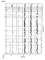
- Figure 6 a-c and e-f show hybridization of globally amplified DNA from single cells onto the 3K array.
- Panel and b show hybridization of two normal single cells, where throughout the whole array normal copy numbers are measured for all autosomal clones (except chromosome 19 which is not included into further analysis).
- the female gender of the probe is easily detected in the gonosomal clones.
- Figure 6 c shows an hybridization with the same single PBL from a patient with trisomy 21 as the one shown in fig. 3. The gain of genomic material on chromosome 21 is easily detected, the other clones measure a normal karyotype.
- Figure 6 d shows the hybridization profile of genomic DNA derived from T47D cell line according to standard protocols.
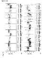
- the array CGH confers gain of information due to enhanced spatial resolution compared to metaphase CGH as demonstrated by the detection of two small inserted deletions on the q-arm of chromosome 11 (7 Mb and 500 kb of size; going from centromer to telomer) and the high-copy amplification on chromosome 14p (2.7 Mb of size) detectable as conserved imbalances in both single cells.
Landscapes
- Chemical & Material Sciences (AREA)
- Life Sciences & Earth Sciences (AREA)
- Organic Chemistry (AREA)
- Health & Medical Sciences (AREA)
- Engineering & Computer Science (AREA)
- Zoology (AREA)
- Wood Science & Technology (AREA)
- Genetics & Genomics (AREA)
- Bioinformatics & Cheminformatics (AREA)
- Biotechnology (AREA)
- General Engineering & Computer Science (AREA)
- General Health & Medical Sciences (AREA)
- Molecular Biology (AREA)
- Microbiology (AREA)
- Biophysics (AREA)
- Biomedical Technology (AREA)
- Biochemistry (AREA)
- Physics & Mathematics (AREA)
- Analytical Chemistry (AREA)
- Proteomics, Peptides & Aminoacids (AREA)
- Plant Pathology (AREA)
- Crystallography & Structural Chemistry (AREA)
- Immunology (AREA)
- Measuring Or Testing Involving Enzymes Or Micro-Organisms (AREA)
Abstract
Description
- The present invention relates to a novel method for isolating artificial chromosomes which are free of host DNA from a library, preferably BAC clones. The present invention further relates to various applications of said method in DNA analysis for medical, forensic, diagnostic or scientific purposes, e.g., ex vivo analyses of diseases like cancer being based on few or even single cells by, for example, microarray comparative genomic hybridization (CGH).
- The enormous heterogeneity of malignant cells within a tumor facilitates tumor progression and often is the underlying cause of treatment failure. Unfortunately, our understanding of small cell populations or even single cells that promote tumor growth and are selected during the natural course of disease or by therapy is still limited. For example, cancer stem cells have been identified that display infinite self-renewal capacity while the majority of tumor cells is not clonogenic1. Moreover, during metastatic progression significant differences of chromosomal aberrations between single disseminated tumor cells isolated from bone marrow of breast cancer patients and their matched primary tumors were uncovered2. Because disseminated tumor cells are thought to comprise the precursor cells of later arising metastases3, they are particularly important for the understanding of systemic cancer progression. These two examples may stand for an increasing number of clinical and scientific questions that require techniques for genomic analyses of single cells.
- Recently, microarrays have been developed, in which the metaphase chromosomes used for "Comparative Genomic Hybridization" (CGH) are replaced by clones of bacterial artificial chromosomes (BACs)4,5, and array CGH chips covering the complete human genome at tiling resolution have become available6. It could be shown that the higher resolution of genomic analysis confers novel information about genes amplified or deleted in human cancer. However, while physical resolution could be increased 100 fold from metaphase CGH to array CGH, cellular resolution still is very poor, requiring 1 to 50 ng of high-quality DNA for the detection of single-copy number imbalances6, which limits the application of the technique to large tumors and cell lines. However, the analysis of a single cell would require a 200 to 1000 fold increase in cellular resolution - a goal that could so far not be attained.
- Thus, the technical problem underlying the present invention is to provide methods for reliably analysing the genome of a small cell population or a single cell.
- The solution to said technical problem is achieved by providing the embodiments characterized in the claims. During the experiments leading to the present invention it was investigated whether existing protocols of array CGH may be used to analyze DNA amplified from single cells or microdissected archival tumor samples. Previously, the present inventors reported a method for the amplification of DNA from a single cell or from microdissected, formalin-fixed and paraffin-embedded tissues, which retains the copy number ratios of chromosomes as determined by metaphase CGH7,8,24. It was therefore tested how DNA amplified by this method can be analyzed on BAC arrays.
- Here the first protocol for the preparation of BAC arrays that enables array CGH with DNA from single cells or from microdissected, formalin-fixed and paraffin-embedded tissues is described. This protocol allows detecting even copy number ratios of 3:2 in samples from single cells. For these applications previously published protocols resulted in enormous variations of hybridization signals due to non-human DNA contamination. The preparation of large arrays using the protocol described below enables a wide range of applications that require a detailed characterization of rare cells in cancer research or in medical genetics.
- It was found that successful array CGH with DNA amplicons derived from single cells requires the DNA on the array to be essentially free of contaminating non-human DNA. Isolation of BAC DNA by pulsed field gel electrophoresis was the key to successful retrieval of array-CGH profiles from single cells. An additional effect of the subsequent amplification method was observed: adaptor-linker amplification was best suited for subsequent experiments. The protocol was extensively tested using single normal cells, single cells with known chromosomal aberrations (trisomy 21) and well-characterized single disseminated tumor cells isolated from the bone marrow of breast cancer patients. In addition, the purified BAC array was tested for the application of microdissected formalin-fixed paraffin-embedded tissues. All expected chromosomal aberrations could be detected by hybridization on PFGE-purified BACs while conventional arrays could only detect high copy number changes when single cells or minute amounts of DNA were analyzed.
- In summary, the present invention, inter alia, allows to establish an array CGH protocol suited for numerous applications. Examples include the study of minute amounts of ex vivo DNA from premalignant lesions or very small tumors, from biopsies before and after systemic therapy, archived tumor samples of patients with known clinical outcome, analysis of single metastastic precursor cells or of circulating fetal cells in the maternal blood. Also, single cell analysis will be important in various fields studying the mechanisms of genetic instability of tumors or the genomic causes underlying human ageing17,18, because most of these changes are stochastic in nature and require single cell resolution. Therefore, single cell array CGH using artificial chromosomes purified according to the method of the present invention will enable novel insights into important biological and medical questions.
- Accordingly, the present invention relates to a method for isolating artificial chromosomes from a library of artificial chromosomes which are free of host DNA, said method comprising the following steps:
- (a) preparing a library of artificial chromosomes with digested chromosomal DNA by use of a vector containing a recognition site for a restriction enzyme (x) which is not present within the genome of the host used for propagation of the artificial chromosomes;
- (b) digesting the DNAs prepared from the library of artificial chromosomes of step (a) with restriction enzyme (x) after propagation of the library; and
- (c) separating the digested linear artificial chromosomes of the library from the undigested circular host DNA by use of a separation method allowing to distinguish between linear and circular DNA.
- Methods for preparing libraries of artificial chromosomes and suitable vectors which are useful in step (a) of the method of the present invention are known to the person skilled in the art and examples of such vectors are also provided in the appended examples. The term "preparing a library of artificial chromosomes" relates, accordingly, also to standard methods, as inter alia illustrated in Sambrook (2001) "Molecular Cloning: A Laboratory Manual", 3rd edition, Cold Spring Harbour Laboratory Press. Furthermore, and in accordance with this invention, already prepared libraries of artificial chromosomes may be employed. Such libraries comprise commercially available libraries care should be taken that the library used contains a recognition site for a restriction enzyme as defined in step (a) of the method of the invention.
- Preferably, in the vectors useful in the method of the present invention, the recognition sequence for the restriction enzyme (x) is unique and digestion of the vector at this site does not interfere with important biological functions of the vector, e.g., as regards replication, selection markers etc..
- General methods for isolating clones representing artificial chromosomes and for the preparation of libraries of artificial chromosomes e.g., YACs, BACs etc., from host cells and digesting the DNAs of the clones representing artificial chromosomes are well known to the person skilled in the art and, e.g., described in Sambrook and Russell (Molecular Cloning; A Laboratory Manual, 3rd Edition, Cold Spring Harbour Laboratory Press, Cold Spring Harbour, NY (2001)); see also Example 1(A) below. An example of a prepared library of artificial chromosomes is "FISH-mapped clones" V1.3; human 32 k BAC array from CHORI, USA.
- Furthermore, the setting of conditions for the above described steps of the method of the present invention is well within the skill of the artisan and to be determined according to protocols described, for example in Sambrook et al, loc. cit., or in the appended examples.
- As used herein, the term "free of host DNA" refers to isolated DNA, in particular artificial chromosomes that have been purified from host DNA contamination to such extent that it does not interfere with the methods of further analysis described in the sections below. Preferably, the content of contaminating host DNA is less than 3%, more preferably less than 1,5%, more preferably less than 1.0%, more preferably less than 0.6% and even more preferably less than 0.4% of the total DNA. The obtained "host cell-free" DNA is particularly useful in the preparation of microaarrays as documented in the appended examples and described herein below.
- Preferred restriction enzymes (x) in the method of this invention are rare cutters like I-Ceul, PI-Scel, PI-Psal, I-Sal, Ascl and SgrAl with a sequence specificity of at least 9-10 or 11 bp, respectively. The restriction enzyme PI-Scel is particularly preferred. In a preferred embodiment of the method of the present invention, the library of artificial chromosomes is a library of BAC clones, e.g. the RPCI-11 library described in Osolgawa et al.26. The generation of BAC libraries is, inter alia, described in reference 27
- Any prokaryotic host cell can be used in the method of the invention, with bacterial host cells being preferred. Particularly preferred bacteria are E. coli, e.g., DH5a.
- In a further preferred embodiment of the method of the present invention, the chromosomal DNA used for generating the library of artificial chromosomes is obtained from cells of a tissue sample, preferably from a human tissue sample.
- The method of the present invention requires the efficient separation of linear DNA from circular nucleic (e.g., undigested host DNA). Such methods are known to the person skilled in the art with Pulsed-Field-Gel-Electrophoresis (PFGE) being preferred.
- In a further preferred embodiment of the method of the present invention, in step (a) the chromsosomal DNA is isolated from cells which have no known numerical changes or imbalances in their DNA.
- The present invention also relates to various uses of the above described method involving DNA analysis of a sample, comprising prenatal diagnosis, forensic medicine, pathogenic analysis or biological/biochemical research. Preferably, the isolated and linearized clones of the artificial chromosomes are presented as a microarray which can be prepared as, e.g., described in Example 1C.
- In a preferred use, the method for DNA analysis comprises the following steps:
- (a) preparing a microarray with the artificial chromosomes obtainable by the method of the present invention;
- (b) hybridizing DNA obtained from (a) target cell(s) with the artificial chromosomes of said microarray; and
- (c) evaluating the hybridization results.
- Methods for carrying out steps (a), (b) and (c) are well known to the person skilled in the art. For example, the preparation of a microarray is described in Example 1 (C) or ref.22,23. Preferably, prior to step (a) the target DNA has been amplified in order to enhance sensitivity of the assay, e.g., by PCR. Although PCR was initially applied to amplify a single locus in target DNA, it is increasingly being used to amplify multiple loci simultaneously. Frequently used primers for this general amplification of DNA are those based on repetitive sequences within the genome, which allow amplification of segments between suitable positioned repeats. Interspersed repetitive sequence PCR (IRS-PCR) has been used to create human chromosome- and region-specific libraries. A more general amplification than with ISR-PCR can be achieved with "degenerate oligonucleotide-primed PCR" (DOP-PCR), with the additional advantage of species independence. DOP-PCR is based on the principle of priming from short sequences specified by the 3'-end of partially degenerate oligonucleotides used, during initial low annealing temperature cycles of the PCR protocol. Since these short sequences occur frequently, amplification of target DNA proceeds at multiple loci simultaneously. DOP-PCR can be applied for generating libraries containing a high level of single-copy sequences, provided pure and a substantial amount of DNA of interest can be obtained, e. g. flow-sorted chromosomes, microdissected chromosome bands or isolated yeast artificial chromosomes (YACs). A method called "primer-extension preamplification" (PEP) is directed to circumvent this problem by making multiple copies of the DNA sequences present in a single cell. PEP uses a random mixture of 15-base fully degenerated oligonucleotides as primers, thereby leading to amplification of DNA sequences from randomly distributed sites. A method called representational difference analysis (RDA) is a subtractive DNA hybridization technique that discovers the differences between paired normal and tumor genomes. The minimal amount of DNA needed for RDA shown is 3 ng, corresponding to ≈ 1 x 10³ cells; see for discussion of these amplification methods and corresponding references ref.24. However, in the method of the present invention also single cells comprising about 6 pg DNA may be employed.
- However, the most preferred amplification method for the assay of the present invention is the adapter-linker PCR which is capable of substantially uniform and preferably complete amplification of genomic DNA, particularly from a single cell, which is described in7,19,24.
- In a further preferred use, the target DNA to be analysed has been obtained from a small cell population or a single cell, most preferably a single cell. Thus, the DNA which is amplified is the genome of a small cell population or a single cell or chromosomes or (a) fragment(s) thereof. It has surprisingly been found that the method of the present invention is particularly useful for the analysis of single cells such as disseminating tumor cells, cells obtained from a lymph node, peripheral blood cells, cells from bone marrow aspirates, cells from tumor biopsis, cells obtained from microdissected tissue, or the like. The inventive method is particularly useful for the assessment of clonal evolution events of genetic variants in complex (cell) populations, like, inter alia, the clonal evolution of single micro-metastatic cells isolated from peripheral blood, bone marrow, or the like.
- Accordingly, the methods provided herein or the uses of the artificial chromosomes (free of host cell DNA) are in particular useful for the study of minute amounts of ex vivo DNA from premalignant lesions or very small tumors, from biopsies before and after systemic therapy, archived tumor samples of patients with known clinical outcome, analysis of single metastastic precursor cells or of circulating fetal cells in the maternal blood. Accordingly, the methods/uses as provided herein also relate to the study of, e.g. biopsy samples, also samples obtained from liquor and tissues (like liver, brain, tissue of the gastro-intestinal tract, colon, rectum, tissue of the reproductive tract, uterus, bladder, ovary, prostate gland, testis, tissue from the alimentary canal, oesophagus, stomach, duodenum, ileum and jejunum, and the like). Most preferably, single cells derived from these tissues or samples are analyzed. Accordingly, the uses as described herein, e.g. the use of the obtained, host-cell DNA-free artificial chromosomes may be employed in a method for DNA analysis in a sample, for example ex vivo DNA from any cell in question or a cell expected to have chromosomal or subchromosomal inbalances/umbalances or structural genome aberrations.
- In a particularly preferred embodiment of the use of the present invention, the methods for DNA analysis are comparative genomic hybridization (CGH)-, representational difference analysis (RDA)-, analytical PCR-, restriction enzyme length polymorphism analysis (RFLP)-, single strand conformation polymorphism analysis (SSCP)-, DNA sequence-, "loss of heterozygosity" (LOH)-, fingerprint- and/or FISH-analysis, i.e. the transition of a "normal" cell to a tumor cell may, in accordance with the method of this invention and the corresponding uses be monitored.
- Analysis by CGH is the most preferred method. An important contribution of CGH to cancer research has been in pinpointing putative locations of cancer genes, especially at chromosomal sites undergoing DNA amplification. A large number of subregional chromosomal gains and DNA amplifications have been discovered by CGH in some cancers. Because oncogenes and drug-resistance genes are known to be upregulated by DNA amplifications, it has been speculated that DNA amplification sites in cancer could pinpoint locations of novel genes with important roles in cancer progression. Tumor progression implies the gradual transition of a localized, slow growing tumor to an invasive, metastatic and treatment refractory cancer. This progression is thought to be caused by a stepwise accumulation of genetic changes affecting critical genes. By providing genome-scale information of clonal genetic alternations, CGH is extremely useful in the analysis of the biological basis of the tumor progression process. The combination of the method/use of the present invention and CGH enables, e.g., the detection of DNA- copy number imbalances of single cells and thereby increases cellular resolution of array CGH at least 200 to 1000 fold.
- Thus, in a further particularly preferred use the method for DNA analysis is a method for detecting numerical changes and/or imbalances in DNA of a sample, wherein said target cells are cells suspected to have numerical changes and/or imbalances in their DNA. Preferred examples of such target cells are tumor cells.
- Finally, the present invention provides a microarray with artificial chromosomes which are obtainable by the method of the present invention. This microarray is, in contrast to microarrays of the prior art free of host cell DNA. As shown in the appended examples, microarrays can be prepared with the inventive method which are microarrays for, inter alia, comparative genomic hybridization (CGH). It has to be pointed out that also Pinkel4 (1998) has prepared BAC chips. However, as documented in appended Figure 3d, this method of preparation (as described in Pinkel (1998) is not suitable for the preparation of a single cell microarray/single cell array, i.e. single cell array CGH. In this invention it was successfully shown that this failure (based on the technique as described in Pinkel (1998)) was unexpectedly due to contamination of foreign host-DNA, i.e. bacterial DNA. In this invention it could be shown that said contaminating DNA precludes single cell array CGH. The method as provided herein for the isolation of artificial chromosomes and their corresponding use in e.g. microarrays overcomes these problems. As documented herein, the invention provides for novel microarrays which are free of host cell DNA. In context of this invention "free of host cell DNA" means that less than 3% of the total DNA on said microarray is host DNA, e.g. bacterial DNA (like E. coli DNA). That means the present invention provides for microarrays which are, inter alia, useful in single cell CGH and which comprises less than 3%, preferably less than 1.5% more preferably less than 1%, more preferably less than 0.6% even more preferably less than 0.4% contaminating host DNA (versus total DNA on said microarray).
-
-
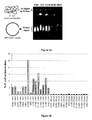
Figure 1: A Schematic illustration of the pulsed-field-gel-electrophoresis of PI-Sce digested RP11-BAC clones
- (A) The PI-Scel site is located in the pBACe3.6 vector sequence, while no restriction site is present in the genomic DNA of E. coli. The linearized BAC-DNA is recovered after gel-electrophoresis by gel extraction (BAC clones 86c20, 106m3).
- (B) E. coli contamination of BAC clone preparations determined by qPCR. The amount of contamination is given as percentage of the total DNA (conv., conventional Qiagen preparation; PFGE, pulsed field gel electrophoresis purification of BAC clones).
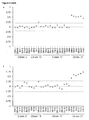
- Figure 2: Hybridization of BAC microarrays using 0.5 µg DNA from a patient with
trisomy 21
The six different DNA preparations spotted onto one array were simultaneously hybridized with Cy3 and Cy5 labeled reference and test DNA probes.Trisomy 21 was detected by all protocols. The log2 transformed ratios of test to reference DNA fluorescence intensities are plotted against the chromosomal locations of the BAC clones. Dashed gray and black lines represent the base line and the upper/lower significance thresholds defined by the three-times standard deviation.
a-c, PFGE purification of BAC clones; d-f, Qiagen preparation of BAC clone;. a and d, Adapter-linker PCR; b and e, DOP-PCR; c and f, Phi29 rolling circle amplification. - Figure 3: Hybridization of globally amplified DNA from a single PBL with
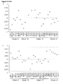
trisomy 21
The gain ofchromosome 21 is only revealed by the hybridization on the PFGE purified BAC clone sub-arrays. Furthermore, the adapter-linker PCR amplified BAC clones show superior hybridization characteristics compared to the DOP or Phi29 amplified targets.
a-c, PFGE purification of BAC clones; d-f, Qiagen preparation of BAC clones. a and d, Adapter-linker PCR; b and e, DOP-PCR; c and f, Phi29 rolling circle amplification. - Figure 4: Hybridization of adapter-linker amplified DNA from single tumor cells and from microdissected paraffin embedded tumor tissue
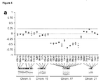
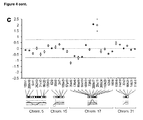
The hybridization profiles of the tumor samples on the PFGE purified, adapter-linker amplified BAC clone arrays is supplemented with the chromosomal locations of the probed BAC clones and the hybridization pattern of the same samples in metaphase CGH.
a and b, Hybridization of globally amplified DNA from the single tumor cells TC-1 and TC-2 derived from bone marrow samples of two patients with mammary carcinoma. A copy number gain ofchromosome 5 is detected in TC-2 and a deletion ofchromosome 17 in TC-1.
c, Hybridization of micro-dissected and globally amplified DNA from a formalin-fixed, paraffin-embedded pancreatic tumor sample. Note the Her2 amplification on chromosome 17q and the deletion of chromosome 17p. The log2 transformed test to reference DNA fluorescence intensities rations are plotted against the chromosomal locations of the BAC clones. Dashed grey and black lines represent the base line and the upper/lower three times standard deviation significance thresholds, respectively. The green and red dots indicate the variance of the hybridization in color switch experiments. - Figure 5: Hybridization of adapter-linker amplified DNA from normal single cells and single tumor cells
Below the graph, chromosomal localization is indicated.- a, Hybridization of globally amplified DNA from the single tumor cell TC-1 derived from bone marrow samples of one patient with mammary carcinoma. A deletion of
chromosome 17 is detected in TC-1. - b, Hybridization profile of two single cells derived from different normal donors. No deletions or amplifications can be detected. The log2 transformed test to reference DNA fluorescence intensities rations are plotted against the chromosomal locations of the BAC clones. Dashed grey and blue lines represent the base line and the upper/lower three times standard deviation significance thresholds, respectively.
- a, Hybridization of globally amplified DNA from the single tumor cell TC-1 derived from bone marrow samples of one patient with mammary carcinoma. A deletion of
- Figure 6. Hybridization of five different adapter-linker amplified single cell DNA samples versus male reference DNA on 3K single cell BAC array.
a, normal male single cell, b, normal female single cell, c, single PBL from patient with trisomy 21 (male), e and f, single cells from T47D cell line. d, hybridization of genomic DNA obtained from T47D cell line using a standard-protocol. Note the high degree of similarity when comparing single cell arrayCGH profiles and the genomic DNA of T47D hybridization. Note also the small differences between the two single cells and the genomic pooled DNA, caused by enhanced cellular resolution. The log2 transformed test to reference DNA fluorescence intensities ratios are plotted against the chromosomal locations of the BAC clones. Very light grey color of dots indicates normal clones, light shaded grey indicates loss and dark shaded grey indicates gain of chromosomal material. Black indicates high-copy amplifications. (data normalization and visualization was performed with CAPweb software). - Figure 7. Hybridization of two different adapter-linker amplified single tumor cell DNA samples.
These samples were generated from cells isolated from the bone marrow of one breast cancer patient, versus male reference DNA on 3K single cell BAC array. The metaphase CGH profile obtained by hybridizing the same DNA is plotted beneath the arrayCGH profile. Chromosome ideograms are placed horizontally, in a way that the genomic position on the metaphase and arrayCGH plot correspond to each another and enables a direct comparison a,single tumor cell 1, b,single tumor cell 2. Note the high correlation of arrayCGH and metaphase CGH profiles for both single tumor cells. Note also that more and smaller genomic imbalances are detected with the arrayCGH. For example 2 small inserted deletions on the q-arm of chromosome 11 (7 Mb and 500 kb in size; going from centromer to telomer) and the high-copy amplification on chromosome 14p (2.7 Mb) detectable as conserved imbalances in both single cells. The log2 transformed test to reference DNA fluorescence intensities ratios are plotted against the chromosomal locations of the BAC clones. Very light grey color of dots indicates normal clones, light shaded grey indicates loss and dark shaded grey indicates gain of chromosomal material. Black indicates high-copy amplifications. (data normalization and visualization was performed with CAPweb software). - BAC clones were grown in LB medium containing 20 µg/ml chloramphenicol. Standard preparation of BAC-DNA was performed by Qiagen midi columns, according to the manufacturer's recommendations. For preparation of BAC-DNA by pulsed-field electropheresis, bacterial agarose-inserts were prepared as described with minor modifications12 and digested with PI-Scel. Briefly, the bacteria were pelleted by centrifugation, embedded in 1% low melt agarose and lysed with lysozym (over night incubation at 37°C with 6 mM Tris-HCl pH 7.5, 1 M NaCl, 100 mM EDTA, 0.5% Brij-58, 0.2% deoxycholate, 0.5% lauryl sarkosine, 30 U/ml RNase (DNase free), 50.000 U/ml lysozym) followed by proteinase K digestion of the cellular proteins (over night incubation at 50°C with 0.5 M EDTA pH 9.0, 1% lauroyl-sarcosine, 1.5 U/ml proteinase K). Fragmented DNA was removed by gel-electrophoresis at 80 V for 2 h in 0.5 x TAE buffer. The BAC-DNA was digested with the restriction enzyme PI-Scel for 3 h at 37°C after giving the enzyme time to diffuse into the agarose-insert overnight on ice (1.5 U/100 µl PI-Scel; 100 mM KCI; 10 mM Tris-HCl pH 8.6; 10 mM MgCl2; 1 mM dithiothreitol; 3.5 µg BSA). Subsequently, the proteinase K digest was repeated. After inactivation of proteinase K by auto-hydrolysis, the agarose-inserts were equilibrated in TE, loaded on a 1% low melt agarose gel, and the DNA was separated by PFGE (Amersham Bioscience) according to manufacturers recommendations in 0.5 x TAE. The BAC-DNA was gel-extracted by an over night agarase digest (1 Unit agarase per 100 µl gel-volume in 30 mM Bis-Tris pH 4.5; 10 mM EDTA) and precipitated with isopropanol and Na-acetate (3 M) at 4°C. After washing with 70% ethanol, the DNA was resuspended in 50 µl TE.
- All BAC-DNA preparations were amplified by the adapter-linker method with minor modifications7,19 and by DOP-PCR as described20. To avoid cross-hybridization with the adaptors from the single cell PCR products, different sequences of the adaptors were selected that were ligated to BAC DNA, i.e. 5'-CTGTGTCTGACGACTCAGTCT-3' and 5'-TAAGACTGAGTCdd-3'. Phi29 amplifications were performed with the TempliPhi Kit (Amersham Biosciences), according to manufacturer's recommendations.
- BAC probes were ethanol precipitated after PCR or rolling circle amplification and DNA concentration was adjusted to 0.4 µg/µl in 3x SSC. Probes were applied in replicas of five to amino-silane coated slides (Corning, GAPS II) using an
Omnigrid 100 spotting robot (Genomic Solutions, Ann Arbor, U.S.A.). DNA was immobilized by UV cross-linking followed by 30 min baking at 80°C. - Adapter-linker amplified target and reference DNA were labeled by PCR in presence of Cy3 or Cy5 conjugated nucleotides, respectively. The 100 µl PCR mix comprised 13 mM Tris-HCI, pH 9.5, 3.25 mM MgCl2, 5 µM LIB1 primer19, 200 µM dATP and dGTP, 180µM dTTP and dCTP, 20µM Cy3/Cy5-dUTP (Amersham Biosciences) and Cy3/5-dCTP (Amersham Biosciences), 16 U ThermoSequenase (Amersham Biosciences), and 1 µl template DNA). The MJ-research thermocycler was programmed to nine cycles of 94°
C 1 min, 60°C 30 sec, ramp to 72°C with 0.2°C/sec, 72°C 5 min with 60 sec extension/cycle. After labeling, PCR adaptors were removed by Trul digestion (100 U Trul (Fermentas) and 10 µl of 10 x buffer containing 70 mM MgCl2, 1 M KCl and 1 mg/ml BSA). To remove unincorporated dye, DNA was applied to a Sephadex G50 column. Eluted labeled DNA was precipitated together with 80 µg C0t-1-DNA (Roche) and resolved in 50 µl master-mix (50% formamide, 2x SSC, 4% SDS and 0.1 g/ml dextrane sulphate (MW>500000; Fisher Biotech). Hybridization was done without cover slip at 42°C in a humidified chamber for two nights on a shaker as described4. Before hybridization of target DNA, the microarray was pre-hybridized over night at 42°C with 50 µl master-mix as above containing 250 µg Hering sperm DNA, 10 µg sonicated E. coli genomic DNA and 100 µg mock control DNA (adapter-linker PCR contamination product, amplified without addition of cellular DNA). - Single cell adapter-linker PCR and conventional-CGH was performed as published2,7,19.
- Data analysis was performed according to Quackenbush21. In brief, the geometric means of the BAC clone replicas were calculated and the fluorescence signal intensities of the Cy3 and Cy5 channel were globally normalized. BAC clones in chromosomal areas affected by copy number losses or gains were excluded when the signal intensity was normalized. For all BAC clones, the ratio of the normalized fluorescence intensity was log2 transformed and plotted against the chromosomal location. Copy number differences were assumed to be significant, if the signal intensity ratios of the affected BAC clones exceeded the upper or lower thresholds defined by the three-times standard deviation that was calculated from the baseline noise.
- Quantitative PCR was performed using the LightCycler System (Roche) and the LightCycler FastStart DNA Master SYBR Green I kit (Roche), according to the manufacturer's recommendation. The forward and reverse primer sequences for the amplification of E. coli TRPE genomic sequence and the pBACe3.6 vector sequence were taken from Foreman et.al.11, without the additional probe beacon. The crossing points of the samples that represent the threshold PCR cycle at which the exponential growth of the fluorescence signal was initially detected, were determined by the LightCycler Relative Quantification Software (Roche). The total amounts of vector and E. coli DNA in the samples were calculated in an exponential equation with the amplification efficacy of the vector or E. coli PCR reactions (1.92 and 1.93, respectively) as base to the power of the crossing points for each sample pair. The ratio of vector versus E. coli DNA was determined and expressed as percentage of contaminating E. coli DNA in the samples.
- First arrays were tested that had been successfully used to analyze large amounts of DNA from primary tumors9. By hybridizing DNA amplified from single cells of known karyotypes onto these arrays, however, the chromosome copy numbers of the test samples could not be correctly determined (data not shown). It was reasoned that bacterial or plasmid DNA contaminating the single cell preparations hybridizes to bacterial or plasmid DNA present on the BAC array causing large signal fluctuations.
- Such contaminating DNA could be introduced during sample preparation with the enzymes that are needed for the amplification of cellular DNA (proteinase K, Mse I, T4 DNA ligase and Taq polymerase). In standard chromosomal CGH contaminating DNA from bacterial sources would not hybridize to human metaphases. This hypothesis was tested by hybridizing mock control samples that had been prepared without the addition of cellular DNA. Indeed, no signals were detected on metaphase spreads while the intensely fluorescent hybridization pattern on BAC arrays could hardly be distinguished from samples containing DNA amplified from human cells (data not shown).
- Previously it was noted that non-human DNA on BAC arrays interferes with exact measurements in hybridization experiments10 and that the purity of plasmids containing the human DNA depends on how the BAC preparation is performed11. To reduce the amount of non-human DNA in the BAC preparations various approaches were tested, including long-range PCR, in vitro transcription of the BAC inserts, subtractive enrichment of the BAC-DNA, oligo-nucleotide based arrays and the purification of the BAC-clones by pulsed field gel electrophoresis (PFGE)12. The last method was clearly superior to all other strategies, because it enabled both highest recovery and best purification of the BAC DNA. The RP-11 BAC clones used for this purpose contain a recognition site of the homing endonuclease PI-SceI in the pBACe3.6 vector sequence which is not present in the E. coli genome13. Therefore, the exclusive linearization of the RP-11 BAC clones enables the physical separation from the E. coli genomic DNA during the gel run (Figure 1), whereas circular BACs could not be separated under the applied conditions. The purification of the BAC DNA from E. coli DNA was verified by quantitative (q) PCR using primers for the E. coli TRPE gene11. While only traces of contaminating E. coli DNA were detected in PFGE-purified samples (0.32% ± 0.11 SD), BAC clones conventionally prepared using Qiagen columns were contaminated to a varying degree (1.3-25.2%, average 8.0% ± 6.0 SD) (Figure 2). As the average purity of these Qiagen BAC preparations was similar to reported data (6-10%)11, they apparently represent those spotted on most of the current BAC arrays.
- The DNA from 30 RP-11 clones (Table 1), either isolated by PFGE or by conventional Qiagen columns, was used for the generation of a microarray. The two DNA preparations of a BAC clone were amplified using adapter-linker-PCR, DOP-PCR and Phi29 rolling circle amplification, resulting in six different DNA samples obtained from each RP-11 clone. The applied amplification methods had been previously described for array preparation after conventional BAC preparation6,10,14,15. All DNA preparations of each BAC clone were spotted onto one glass slide (five replicas each) for a direct comparison of the resulting hybridization signals for each labeled DNA sample.
- First, 500 ng unamplified DNA extracted from peripheral blood cells from a patient with
trisomy 21 were hybridized to the array. All BAC clones fromchromosome 21 displayed the expected copy number gain regardless which probe preparation and amplification method was used (Figure 2 a-f), verifying the suitability of both PFGE and conventional BAC DNA isolation methods for this application. However, when DNA from a single leukocyte of the same patient was amplified by adapter-linker PCR7 and subsequently hybridized onto the differently prepared BAC clone sets, thetrisomy 21 was only detected by PFGE purified clones (not shown; see below). Array-CGH with DNA from single leukocytes of healthy donors (n=3) showed normal ratios for all BAC clones (data not shown). When the effects of amplification methods on the ratios detected by PFGE isolated clones were compared, the reliability of measurements by adaptor-linker amplified DNA was superior to DOP-amplified DNA and Phi29-amplified DNA. This might be due to matched DNA fragments in probe and target, because both were generated by MseI digest. In addition, the hybridization noise originating from contaminating sequences was blocked by pre-hybridizing the array with unlabeled blocking DNA. As blocking DNA the mock control of the respective single cell amplification experiment was used, because it presumably contains similar contaminating DNA and is therefore best suited to increase signal to noise ratios. Interestingly, the blocking DNA improved only the quality of hybridization with the PFGE-purified samples on the array (Figure 3 a-c), while little or no effect could be observed for the conventional BAC preparations (Figure 3 d-f). Thus, the blocking of contaminating DNA before or during hybridization is successful only as long as the contamination does not exceed a certain threshold. - Finally, single disseminated tumor cells and DNA from formalin-fixed paraffin-embedded tissue isolated by laser microdissection were analysed. To this end tumor samples were selected for which previously chromosomal losses or gains by classical CGH within those regions that were covered by our BAC clones had been determined2,8. For the single cells the noise on any of the conventionally prepared BAC arrays prohibited an analysis while on the PFGE-purified arrays both cells could be evaluated. The first single tumor cell (TC-1) displayed a deletion of
chromosome 17 and the second (TC-2) a gain of chromosome 5 - as expected from the metaphase CGH profile (Figure 4 a and b). Similar results were obtained using formalin-fixed and paraffin-embedded tissues after microdissection and amplification. As before, gains and losses predicted by metaphase CGH onchromosome 17 were detected by microarray analysis on PFGE-purified and adaptor-linker-amplified DNA (Figure 4 c), while on BAC clones prepared by the standard BAC protocols only the high copy amplification of the HER2 gene on chromosome 17q21.2 could be detected. - A second generation of microarrays was constructed using a total of 94 RP11-BAC clones (Table 2) in order to verify reproducibility of the method with a different clone-set, independent amplification and spotting. Preparation and PCR-amplification of BAC-DNA was performed as described above. For spotting of samples, DNA was resolved in water following precipitation and adjusted to a concentration of 0.8 µg/µl. Addition of 2x concentrated sciSPOT spotting buffer for oligos (Scienion) diluted DNA to a final spotting concentration of 0.4µg/µl. After letting the slides dry, an aftertreatment was performed by the manufacturer with sciPROCESS buffer. Hereby slides were washed in 55°C sciPROCESS buffer for 15 minutes, then washed once in 55°C distilled water for 5 minutes and twice in 25°C distilled water for 25 minutes. Then the slides were dried by centrifugation at 900rpm. Spot quality was assessed by Sybr Green dyeing.
- It proved that good spot quality and morphology were dependent on spotting parameters, such as the spotting buffer, where ethylene glycol and DMSO produced spots that were not suited for hybridization, since DNA did not adhere to the slide surface. Scienion Oligo-spotting buffer finally turned out to be superior to all tested spotting buffers, yielding homogeneous spots. Additionally the procedure of PCR-cleanup played an important role, Microcon-column purification being inferior to ethanol-precipitation. Informative hybridization results could additionally only be achieved when BAC-DNA was spotted on GAPS II slides, whereas on CodeLink and Epoxy-slides hybridization results where of low quality.
- It was found that choice of reference DNA has a great impact on the hybridization. It was tested whether normal single cells yield better hybridization results than pools of 10, 100 or 1000 cells when cohybridized with sample DNA, due to the fact, that the amount of contamination in reference and probe would be comparable. Unexpectedly though, pools of 1000 cells were the optimal reference DNAs.
- Incorporating these findings, a second generation chip could be generated allowing to reproduce results observed with the first generation chip, shown in Figure 5a and b. Hybridization of single tumor cell displaying deletion of chromosome 17 (TC-1) is shown in Figure 5a. Normal single cell DNA hybridized without detectable amplifications or deletions (Figure 5b).
- A third generation of microarrays was constructed (3K array) containing a total of 2940 RP11-BAC clones, so that an average resolution of 1 Mb throughout the whole human genome was achieved. The preparation and PCR-amplification were performed as described above for the 30 BAC clone array. Spotting was performed in 1 x sciSPOT spotting buffer with a final DNA concentration of 0.4 µg/µl. Labelling of probes as well as hybridization and washing were performed as described above.
Data analysis was performed with CAPweb software (freely available at http://bioinfo-out.curie.fr/CAPweb/) and visualization was perfomed using VAMP. Only features with reference fluorescence intensity at least 2 fold over the background were taken into account. For all other parameters, default values of the software were used. - Figure 6 a-c and e-f show hybridization of globally amplified DNA from single cells onto the 3K array. Panel and b show hybridization of two normal single cells, where throughout the whole array normal copy numbers are measured for all autosomal clones (except
chromosome 19 which is not included into further analysis). In figure 6 b the female gender of the probe is easily detected in the gonosomal clones. Figure 6 c shows an hybridization with the same single PBL from a patient withtrisomy 21 as the one shown in fig. 3. The gain of genomic material onchromosome 21 is easily detected, the other clones measure a normal karyotype. Figure 6 d shows the hybridization profile of genomic DNA derived from T47D cell line according to standard protocols. In e and f single cell PCR products of the same cell line are hybridized and show a high degree of similarity but also subtle variances, due to the enhanced cellular resolution achieved by single cell array CGH.
In figure 7 a and b hybridization of single cell PCR-products of two tumor cells derived from the bone marrow of one breast cancer patient is shown in comparison to the hybridization profile generated by conventional single cell metaphase CGH. A high degree of similarity can be observed. The array CGH confers gain of information due to enhanced spatial resolution compared to metaphase CGH as demonstrated by the detection of two small inserted deletions on the q-arm of chromosome 11 (7 Mb and 500 kb of size; going from centromer to telomer) and the high-copy amplification on chromosome 14p (2.7 Mb of size) detectable as conserved imbalances in both single cells.Table 1 BAC clone Chromosome Map position FISH Accession RP11-103l11 5 3,875 K 5p15.33 AC092319.2 RP11-260e18 5 16,702 K 5p15.2 AC024588.4 RP11-5n11 5 31,730 K 5p13.3-5p14 AC022447.6 RP11-252i13 5 107,088 K 5q21 AC024587.5 RP11-21c10 5 134,022 K 5q31.1-5q31.2 AC106763.2 RP11-86c20 5 152,699 K 5q33 AC091991.2 RP11-170l13 5 155,253 K 5q33.2 AC114959.2 RP11-6219 15 37,310 K 15q14-15q15 AC027386.3 RP11-99l18 15 57,379 K 15q21-15q22 AC091748.3 RP11-2i17 15 70,146 K 15q23 AC020779.1 0 RP11-106m3 15 70,319 K 15q23 AC009690.1 7 RP11-57p19 15 92,187 K 15q26.2 AC009432.7 RP11-26n16 17 992 K 17p13.3 B86973 RP11-135n5 17 2,655 K 17p13.2 AC015799.2 3 RP11-89a15 17 8,624 K 17p12 AC135070.2 RP11-55c13 17 8,662 K 17p12-17p13 AC116169.5 RP11-100l12 17 28,824 K 17q11.2 AQ345666 RP11-73f15 17 29,287 K 17q11.2 AQ266525 RP11-268h17 17 39,404 K 17q21.2 AQ490954 RP11-29c11 17 40,020 K 17q21.1- B87496 RP11-266i24 17 41,501 K 17q21.2 17q21 AQ490757 RP11-110h20 17 47,416 K 17q21.3 AQ320062 RP11-52b5 17 65,437 K 17q24 AC027772.2 RP11-160f4 17 79,067 K 17q25.2 AQ377826.1 RP11-108h5 21 26,825 K 21q21 AQ348670 RP11-74d19 21 28,634 K 21q21 AQ266734 RP11-32a2 21 29,862 K 21q21.2- AP000239.2 21q21.3 RP11-96h21 21 30,958 K 21 q22.2 AQ314195 RP11-14b21 21 31,285 K 21q22.1 AP000245.3 RP11-114h1 21 41,228 K 21 q22.2- AQ383555 21 q22.3 Table 2 BAC clone Chromosome Map position FISH Marker RP11-94d19 3 60.8 Mb 3p14 AC016965 RP11-108a8 3 63.3 Mb 3p14 AC072020 RP11-180g14 3 59.5 Mb 3p14 AFMA072ZH RP11-114p15 3 62.6 Mb 3p14 AC048336 RP11-229a12 3 57.5 Mb 3p14.3(FHCRC) AC016923 RP11-111p21 3 45.4 Mb 3p21.2 AC063916 RP11-189h19 3 44.7 Mb 3p21.2 AC069071 RP11-122d19 3 54.1 Mb 3p21.2 AC018354 RP11-169g24 3 56.3 Mb 3p21.2 AC018920 RP11-3911 3 55.8 Mb 3p21.2 AQ029524 RP11-120c2 3 57.4 Mb 3p21.2 AC069438 RP11-121d3 3 14.4 Mb 3p21-22 AC016963 RP11-81N11 4 34.7 Mb 4p13 AQ281893 RP11-81H11 4 31.7 Mb 4p15.1 AQ281616 RP11-81N5 4 13.1 Mb 4p15.3 AQ281881 RP11-81L15 4 14.7 Mb 4p15.3 AQ281853 RP11-80H22 4 109.9 Mb 4q25-4q26 AQ284104 RP11-81J9 4 110.0 Mb 4q26 AQ281666 RP11-80P12 4 132.5 Mb 4q28 AQ284210 RP11-48F12 4 22.1 Mb 4q32.3 AQ200675 RP11-181G18 4 16.7 Mb 4q35/21q11.2 SHGC- 21147 RP11-9G14 5 41.3 Mb 5p12 AFM276VB1 RP11-5N11 5 31.7 Mb 5p13.3-5p14 AC022447 RP11-67P13 5 32.1 Mb 5p13-5p14 AQ203092 RP11-19I19 5 58.9 Mb 5q11.2g-5q11.2 AQ003242 RP11-15P2 5 66.3 Mb 5q12 AC063978 RP11-47N20 5 81.4 Mb 5q13-5q14 AQ194964 RP11-252I13 5 107.0 Mb 5q21 AC024587 RP11-115L24 5 103.2 Mb 5q21 AC009815 RP11-81 C5 5 115.3 Mb 5q22-5q23 AQ284841 RP11-15P5 5 120.4 Mb 5q23.1 B75415 RP11-86C20 5 152.7 Mb 5q33 AC091991 RP11-39C2 6 47.1 Mb 6p12 AL355518 RP11-79I2 6 43.5 Mb 6p21.1-6p21.2 AQ283086 RP11-79J17 6 33.0 Mb 6p21.2-6p21.3 AQ284306 RP11-78P9 6 108.6 Mb 6q21 AFMA309X RP11-28K19 6 119.6 Mb 6q22.2-6q22.3 B87366 RP11-18B17 15 58.6 Mb 15q21.3 SHGC- 11862 RP11-3017 15 54.3 Mb 15q21.3 B85180 RP11-99L18 15 57.4 Mb 15q21-15q22 AC091748 RP11-120011 15 57.7 Mb 15q22.1 AQ349760 RP11-13J23 15 58.7 Mb 15q22.2 B83398 RP11-64K10 15 68.7 Mb 15q23 AC010076 RP11-116L7 15 83.8 Mb 15q25 AQ345509 RP11-68P22 15 83.0 Mb 15q25 AQ241268 RP11-55C13 17 8.4 Mb 17p12-17p13 AC116169 RP11-135N5 17 2.4 Mb 17p13.2 AC015799 RP11-26N16 17 922.2 Kb 17p13.3 B86972 RP11-73F15 17 26.2 Mb 17q11.2 AQ266525 RP11-252N11 17 30.4 Mb 17q12 AQ504453 RP11-5808 17 31.0 Mb 17q12 AQ199256 RP11-50F16 17 55.6 Mb 17q12/17q23 AQ053427 RP11-266124 17 38.3 Mb 17q21 AQ490753 RP11-29C11 17 36.8 Mb 17q21.1- B87495 17q21.2 RP11-241 H2 17 39.0 Mb 17q21.1- AQ482539 17q21.2 RP11-268H17 17 36.4 Mb 17q21.2 AQ490951 RP11-42M14 17 48.1 Mb 17q22 AQ046621 RP11-118K23 17 55.1 Mb 17q23 SHGC- 36454 RP11-52B5 17 62.4 Mb 17q24 AC027772 RP11-76G4 17 70.8 Mb 17q25.1 AQ265611 RP11-160F4 17 76.0 Mb 17q25.2 AQ377826 RP11-84J3 17 74.6 Mb 17q25.2- SHGC- 17q25.3 11286 RP11-26B13 17 74.1 Mb 17q25.2- B86818 17q25.3 RP11-88I12 19 19.0 Mb 19p12-19p13.1 AFMA123XF RP11-84C17 19 7.8 Mb 19p13.3 AFMB299W RP11-46112 19 34.4 Mb 19q12-19q13.1 AQ201106 RP11-35J17 19 58.8 Mb 19q13.4 AQ045816 RP11-48M7 20 3.8 Mb 20p12 AQ198007 RP11-9G21 20 6.1 Mb 20p12.3 AFM317TE5 RP11-14907 20 5.5 Mb 20p12-20p13 AQ377575 RP11-153M12 20 989.4 Kb 20p13 AQ387835 RP11-235C20 20 5.1 Mb 20p13 AFMB352X D RP11-78P4 20 2.0 Mb 20p13 AQ281462 RP11-24E15 20 467.4 Kb 20p13 B86599 RP11-26F18 20 3.2 Mb 20p13 AC016217 RP11-107L14 20 2.6 Mb 20p13 AQ316350 RP11-61A21 21 15.4 Mb 21q11.1 AQ199973 RP11-31 B6 21 15.1 Mb 21q11.2-21q21 AQ046029 RP11-106K13 21 15.2 Mb 21q11.2-21q21 AQ315211 RP11-143A3 21 18.2 Mb 21q11.2-21q21 SHGC-2319 RP11-86121 21 21q21 AQ283088 RP11-53K11 21 25.8 Mb 21q21 AQ051878 RP11-49J9 21 21.1 Mb 21q21.1 AQ053086 RP11-617 21 26.9 Mb 21q21.2- B48248 21q21.3 RP11-83L17 21 26.9 Mb 21q21.3 AFM263XF5 RP11-276A17 21 31.4 Mb 21q21.3 AFM288YG5 RP11-147H 1 21 32.3 Mb 21q21.3 AQ373665 RP11-125H1 21 31.4 Mb 21q21.3 AQ348941 RP11-30N6 21 29.8 Mb 21q21.3 B85241 RP11-115H17 21 27.1 Mb 21q21.3- AQ346443 21q22.1 RP11-12M14 21 31.3 Mb 21q21-21q22.1 B75909 RP11-191I6 21 31.5 Mb 21q21-21q22.1 AFM288YG5 RP11-17020 21 35.3 Mb 21q22.1 B82827 RP11-54F16 21 32.8 Mb 21q22.1 AQ082133 -
- 1. Pardal, R., Clarke, M.F. & Morrison, S.J. Applying the principles of stem-cell biology to cancer. ).
- 2. Schmidt-Kittler, O. et al. From latent disseminated cells to overt metastasis: genetic analysis of systemic breast cancer progression. Proc Natl ).
- 3. Braun, S. et al. Cytokeratin-positive cells in the bone marrow and survival of patients with stage I, II, or III breast cancer. N Engl J Med 342, 525-33. (2000).
- 4. Pinkel, D. et al. High resolution analysis of DNA copy number variation using comparative genomic hybridization to microarrays. ).
- 5. Solinas-Toldo, S. et al. Matrix-based comparative genomic hybridization: biochips to screen for genomic imbalances. .
- 6. Ishkanian, A.S. et al. A tiling resolution DNA microarray with complete coverage of the human genome. Nat Genet 36, 299-303. Epub 2004 ).
- 7. Klein, C.A. et al. Comparative genomic hybridization, loss of heterozygosity, and DNA sequence analysis of single cells. Proc Natl Acad Sci U S A 96, 4494-9. (1999).
- 8. Stoecklein, N.H. et al. SCOMP is superior to degenerated oligonucleotide primed-polymerase chain reaction for global amplification of minute amounts of DNA from microdissected archival tissue samples. Am J Pathol 161, 43-51. (2002).
- 9. Fritz, B. et al. Microarray-based copy number and expression profiling in dedifferentiated and pleomorphic liposarcoma. Cancer Res 62, 2993-8. (2002).
- 10. Fiegler, H. et al. DNA microarrays for comparative genomic hybridization based on DOP-PCR amplification of BAC and PAC clones. Genes Chromosomes Cancer 36, 361-74 (2003).
- 11. Foreman, P.K. & Davis, R.W. Real-time PCR-based method for assaying the purity of bacterial artificial chromosome preparations. Biotechniques 29, 410-2 (2000).
- 12. Smith, C.L. & Cantor, C.R. Preparation and manipulation of large DNA molecules: advances and applications. Trends in .
- 13. Frengen, E. et al. A modular, positive selection bacterial artificial chromosome vector with multiple cloning sites. Genomics 58, 250-3 (1999).
- 14. Lage, J.M. et al. Whole genome analysis of genetic alterations in small DNA samples using hyperbranched strand displacement amplification and array-CGH. ).
- 15. Snijders, A.M. et al. Assembly of microarrays for genome-wide measurement of DNA copy number. Nat Genet 29, 263-4 (2001).
- 16. Klein, C.A. The systemic progression of human cancer: A focus on the individual disseminated cancer cell - the unit of selection. Adv Cancer Res 89, 35-67 (2003).
- 17. Hasty, Campisi, J., Hoeijmakers, J., van Steeg, H. & Vijg, J. Aging and genome maintenance: from the mouse? Science 299, 1355-9. (2003).
- 18. Lengauer, C., Kinzler, K.W. & Vogelstein, B. Genetic instability in colorectal cancers. Nature 386, 623-627 (1997).
- 19. Klein, C.A. et al. Genetic heterogeneity of single disseminated tumour cells in minimal residual cancer. Lancet 360, 683-9 (2002).
- 20. Telenius, H. et al. Degenerate oligonucleotide-primed PCR: general amplification of target DNA by a single degenerate primer. ).
- 21. Quackenbush, J. Microarray data normalization and transformation. Nat Genet 32, 496-501. (2002).
- 22.
US-Patent No. 6,210,878 B1 (2001 ). - 23. Solinas-Toldo S. et al. Matrix-based comparative genomic hybridization: biochips to screen for genomic imbalances. Genes Chromosomes Cancer 20(4), 399-407. (1997).
- 24.
EP- B1 1 109 938 - 25.
US-Patent No. 6,197,501 B1 (2001 ). - 26. Osolgawa, K. et al., Genome Research 11 :483-496 (2001).
- 27. Current Protocols in Molecular Biology, Vol. 1, 5.9.1, Supplement 55, "Consruction of Bacterial Artificial Chromosome (BAC/PAC) Libraries".
-

Claims (22)
- A method for isolating artificial chromosomes from a library of artificial chromosomes which are free of host DNA, said method comprising the following steps:(a) preparing a library of artificial chromosomes with digested chromosomal DNA by use of a vector containing a recognition site for a restriction enzyme (x) which is not present within the genome of the host used for propagation of the artificial chromosomes;(b) digesting the DNAs prepared from the library of artificial chromosomes of step (a) with restriction enzyme (x) after propagation of the library; and(c) separating the digested linear artificial chromosomes of the library from the undigested circular host DNA use of a separation method allowing to distinguish between linear and circular DNA.
- The method of claim 1. wherein said library of artificial chromosomes is a library of BAC clones.
- The method of claim 1 or 2, wherein said vector contains a single recognition site for the restriction enzyme (x).
- The method of any one of claims 1 to 3, wherein said restriction enzyme (x) is PI-Scel.
- The method of any one of claims 1 to 4, wherein said host is E.coli.
- The method of claim 5, wherein said chromosomal DNA is obtained from cells of a tissue sample.
- The method of any one of claims 1 to 6, wherein said chromosomal DNA is obtained from human cells.
- The method of any one of claims 1 to 7, wherein said method for separating the digested linear artificial chromosomes from the undigested circular host DNA is Pulsed-Field-Gel-Electrophoresis (PFGE).
- The method of any one of claims 1 to 8, wherein in step (a) the chromosomal DNA is isolated from cells which have no known numerical changes or imbalances in their DNA.
- Use of artificial chromosomes obtainable according to a method of any one of claims 1 to 9 in a method for DNA analysis of a sample.
- Use according to claim 10, wherein said method for DNA analysis comprises the following steps:(a) preparing a microarray with the artificial chromosomes;(b) hybridizing DNA obtained from (a) target cell(s) with the artificial chromosomes of said microarray; and(c) evaluating the hybridization results.
- The use according to claim 11, wherein prior to step (b) the target DNA has been amplified.
- The use according to claim 12, wherein said target DNA has been amplified by adapter-linker PCR.
- The use according to claim 12 or 13, wherein said DNA has been obtained from a small cell population or a single cell.
- The use according to any one of claims 11 to 14, wherein steps (b) and (c) are performed as "Comparative Genomic Hybridization" (CGH).
- The use according to any one of claims 10 to 15, wherein said method for DNA analysis is a method for detecting numerical changes and/or imbalances in DNA of a sample, wherein said target cells are cells suspected to have numerical changes and/or imbalances in their DNA.
- The use according to any one of claims 10 to 16 for the study of minute amounts of ex vivo DNA from premalignant lesions or very small tumors, from biopsies before and after systemic therapy, archived tumor samples of patients with known clinical outcome, analysis of single metastastic precursor cells or of circulating fetal cells in the maternal blood.
- The use according to claim 16, wherein said target are tumor cells.
- A microarray with artificial chromosomes obtainable by a method of any one of claims 1 to 9.
- The microarray of claim 19, whereby said microarray is free of host DNA.
- The microarray of claim 20, whereby said host DNA is E. coli DNA.
- The microarray of claim 20 and 21, whereby said microarray which is free of host DNA is a microarray that comprises less than 3% contamination of host DNA on said microarray.
Priority Applications (1)
| Application Number | Priority Date | Filing Date | Title |
|---|---|---|---|
| EP06020842A EP1788090B1 (en) | 2005-10-04 | 2006-10-04 | Method for preparing artificial chromosomes which are free of host DNA |
Applications Claiming Priority (2)
| Application Number | Priority Date | Filing Date | Title |
|---|---|---|---|
| EP05021667 | 2005-10-04 | ||
| EP06020842A EP1788090B1 (en) | 2005-10-04 | 2006-10-04 | Method for preparing artificial chromosomes which are free of host DNA |
Publications (2)
| Publication Number | Publication Date |
|---|---|
| EP1788090A1 true EP1788090A1 (en) | 2007-05-23 |
| EP1788090B1 EP1788090B1 (en) | 2012-04-25 |
Family
ID=35945307
Family Applications (1)
| Application Number | Title | Priority Date | Filing Date |
|---|---|---|---|
| EP06020842A Active EP1788090B1 (en) | 2005-10-04 | 2006-10-04 | Method for preparing artificial chromosomes which are free of host DNA |
Country Status (2)
| Country | Link |
|---|---|
| EP (1) | EP1788090B1 (en) |
| AT (1) | ATE555204T1 (en) |
Cited By (1)
| Publication number | Priority date | Publication date | Assignee | Title |
|---|---|---|---|---|
| CN105779572A (en) * | 2014-12-22 | 2016-07-20 | 深圳华大基因研究院 | Chip and method for capturing target sequences of tumor susceptibility genes, and mutation detection method |
-
2006
- 2006-10-04 AT AT06020842T patent/ATE555204T1/en active
- 2006-10-04 EP EP06020842A patent/EP1788090B1/en active Active
Non-Patent Citations (5)
| Title |
|---|
| ASHWORTH LINDA K ET AL: "Assembly of high-resolution bacterial artificial chromosome, P1-derived artificial chromosome, and cosmid contigs", ANALYTICAL BIOCHEMISTRY, vol. 224, no. 2, 1995, pages 564 - 571, XP002377945, ISSN: 0003-2697 * |
| FRENGEN E ET AL: "A Modular, Positive Selection Bacterial Artificial Chromosome Vector with Multiple Cloning Sites", GENOMICS, ACADEMIC PRESS, SAN DIEGO, US, vol. 58, no. 3, 15 June 1999 (1999-06-15), pages 250 - 253, XP004444891, ISSN: 0888-7543 * |
| PINKEL D ET AL: "HIGH RESOLUTION ANALYSIS OF DNA COPY NUMBER VARIATION USING COMPARATIVE GENOMIC HYBRIDIZATION TO MICROARRAYS", NATURE GENETICS, NEW YORK, NY, US, vol. 20, October 1998 (1998-10-01), pages 207 - 211, XP002925115, ISSN: 1061-4036 * |
| PINKEL, NATURE GENETICS, vol. 20, 1998, pages 207 - 211 |
| ZENG C ET AL: "Large-Insert BAC/YAC Libraries for Selective Re-isolation of Genomic Regions by Homologous Recombination in Yeast", GENOMICS, ACADEMIC PRESS, SAN DIEGO, US, vol. 77, no. 1-2, September 2001 (2001-09-01), pages 27 - 34, XP004432175, ISSN: 0888-7543 * |
Cited By (1)
| Publication number | Priority date | Publication date | Assignee | Title |
|---|---|---|---|---|
| CN105779572A (en) * | 2014-12-22 | 2016-07-20 | 深圳华大基因研究院 | Chip and method for capturing target sequences of tumor susceptibility genes, and mutation detection method |
Also Published As
| Publication number | Publication date |
|---|---|
| ATE555204T1 (en) | 2012-05-15 |
| EP1788090B1 (en) | 2012-04-25 |
Similar Documents
| Publication | Publication Date | Title |
|---|---|---|
| EP1941050B1 (en) | Methods and composition to generate unique sequence dna probes, iabeling of dna probes and the use of these probes | |
| Hughes et al. | The use of whole genome amplification in the study of human disease | |
| JP4475488B2 (en) | Single cell DNA amplification | |
| EP0981609B1 (en) | A method to clone mrnas and display of differentially expressed transcripts (dodet) | |
| US5827658A (en) | Isolation of amplified genes via cDNA subtractive hybridization | |
| US20100297641A1 (en) | Methylation altered dna sequences as markers associated with human cancer | |
| Arriola et al. | Evaluation of Phi29-based whole-genome amplification for microarray-based comparative genomic hybridisation | |
| CA2815076A1 (en) | Varietal counting of nucleic acids for obtaining genomic copy number information | |
| WO2000043531A2 (en) | Methods and kits for characterizing gc-rich nucleic acid sequences | |
| CN113122637A (en) | Reagent for detecting DNA methylation and application | |
| WO1997007244A1 (en) | ISOLATION OF AMPLIFIED GENES VIA cDNA SUBTRACTIVE HYBRIDIZATION | |
| KR101557975B1 (en) | Method for Amplification Nucleic Acid Using Aelle-Specific Reaction Primers | |
| CN101784673B (en) | Epigenetic methods | |
| CN112662764A (en) | Probe composition for detecting 11 cancers | |
| CN112662763A (en) | Probe composition for detecting common amphoteric cancers | |
| CN110791567B (en) | A single site DNA methylation detection kit | |
| EP1788090B1 (en) | Method for preparing artificial chromosomes which are free of host DNA | |
| Chan et al. | Progressive increase of genetic alteration in urinary bladder cancer by combined allelotyping analysis and comparative genomic hybridization | |
| CN113493835A (en) | Method and kit for screening large intestine tumor by detecting methylation state of BCAN gene region | |
| JP3965872B2 (en) | New quantitative polymorphism analysis method | |
| US6316192B1 (en) | Method for enrichment of unique DNA fragments through cyclical removal of PCR adapter attached to DNA fragments whose sequences are shared between two DNA pools | |
| WO2025096819A2 (en) | High-throughput dna methylation beadchip assay | |
| US20030157527A1 (en) | Identification of rearrangements in nucleic acid molecules | |
| Hoon et al. | [27] Assessment of genetic heterogeneity in tumors using laser capture microdissection | |
| RU2828103C1 (en) | Method of producing panel of dna probes for determining chromosomal translocations, deletions and amplifications |
Legal Events
| Date | Code | Title | Description |
|---|---|---|---|
| PUAI | Public reference made under article 153(3) epc to a published international application that has entered the european phase |
Free format text: ORIGINAL CODE: 0009012 |
|
| AK | Designated contracting states |
Kind code of ref document: A1 Designated state(s): AT BE BG CH CY CZ DE DK EE ES FI FR GB GR HU IE IS IT LI LT LU LV MC NL PL PT RO SE SI SK TR |
|
| AX | Request for extension of the european patent |
Extension state: AL BA HR MK YU |
|
| 17P | Request for examination filed |
Effective date: 20071123 |
|
| 17Q | First examination report despatched |
Effective date: 20071219 |
|
| AKX | Designation fees paid |
Designated state(s): AT BE BG CH CY CZ DE DK EE ES FI FR GB GR HU IE IS IT LI LT LU LV MC NL PL PT RO SE SI SK TR |
|
| AXX | Extension fees paid |
Extension state: HR Payment date: 20071123 Extension state: BA Payment date: 20071123 Extension state: MK Payment date: 20071123 Extension state: AL Payment date: 20071123 Extension state: YU Payment date: 20071123 |
|
| RAX | Requested extension states of the european patent have changed |
Extension state: RS Payment date: 20071123 Extension state: MK Payment date: 20071123 Extension state: HR Payment date: 20071123 Extension state: AL Payment date: 20071123 Extension state: BA Payment date: 20071123 |
|
| RAP1 | Party data changed (applicant data changed or rights of an application transferred) |
Owner name: UNIVERSITAET REGENSBURG |
|
| GRAP | Despatch of communication of intention to grant a patent |
Free format text: ORIGINAL CODE: EPIDOSNIGR1 |
|
| GRAS | Grant fee paid |
Free format text: ORIGINAL CODE: EPIDOSNIGR3 |
|
| GRAA | (expected) grant |
Free format text: ORIGINAL CODE: 0009210 |
|
| AK | Designated contracting states |
Kind code of ref document: B1 Designated state(s): AT BE BG CH CY CZ DE DK EE ES FI FR GB GR HU IE IS IT LI LT LU LV MC NL PL PT RO SE SI SK TR |
|
| AX | Request for extension of the european patent |
Extension state: AL BA HR MK RS |
|
| REG | Reference to a national code |
Ref country code: GB Ref legal event code: FG4D |
|
| REG | Reference to a national code |
Ref country code: CH Ref legal event code: EP |
|
| REG | Reference to a national code |
Ref country code: AT Ref legal event code: REF Ref document number: 555204 Country of ref document: AT Kind code of ref document: T Effective date: 20120515 |
|
| REG | Reference to a national code |
Ref country code: IE Ref legal event code: FG4D |
|
| REG | Reference to a national code |
Ref country code: DE Ref legal event code: R096 Ref document number: 602006029019 Country of ref document: DE Effective date: 20120614 |
|
| REG | Reference to a national code |
Ref country code: NL Ref legal event code: VDEP Effective date: 20120425 |
|
| REG | Reference to a national code |
Ref country code: CH Ref legal event code: NV Representative=s name: VOSSIUS & PARTNER |
|
| REG | Reference to a national code |
Ref country code: AT Ref legal event code: MK05 Ref document number: 555204 Country of ref document: AT Kind code of ref document: T Effective date: 20120425 |
|
| LTIE | Lt: invalidation of european patent or patent extension |
Effective date: 20120425 |
|
| PG25 | Lapsed in a contracting state [announced via postgrant information from national office to epo] |
Ref country code: SE Free format text: LAPSE BECAUSE OF FAILURE TO SUBMIT A TRANSLATION OF THE DESCRIPTION OR TO PAY THE FEE WITHIN THE PRESCRIBED TIME-LIMIT Effective date: 20120425 Ref country code: CY Free format text: LAPSE BECAUSE OF FAILURE TO SUBMIT A TRANSLATION OF THE DESCRIPTION OR TO PAY THE FEE WITHIN THE PRESCRIBED TIME-LIMIT Effective date: 20120425 Ref country code: LT Free format text: LAPSE BECAUSE OF FAILURE TO SUBMIT A TRANSLATION OF THE DESCRIPTION OR TO PAY THE FEE WITHIN THE PRESCRIBED TIME-LIMIT Effective date: 20120425 Ref country code: FI Free format text: LAPSE BECAUSE OF FAILURE TO SUBMIT A TRANSLATION OF THE DESCRIPTION OR TO PAY THE FEE WITHIN THE PRESCRIBED TIME-LIMIT Effective date: 20120425 Ref country code: IS Free format text: LAPSE BECAUSE OF FAILURE TO SUBMIT A TRANSLATION OF THE DESCRIPTION OR TO PAY THE FEE WITHIN THE PRESCRIBED TIME-LIMIT Effective date: 20120825 Ref country code: PL Free format text: LAPSE BECAUSE OF FAILURE TO SUBMIT A TRANSLATION OF THE DESCRIPTION OR TO PAY THE FEE WITHIN THE PRESCRIBED TIME-LIMIT Effective date: 20120425 |
|
| PGFP | Annual fee paid to national office [announced via postgrant information from national office to epo] |
Ref country code: IE Payment date: 20120917 Year of fee payment: 7 |
|
| PG25 | Lapsed in a contracting state [announced via postgrant information from national office to epo] |
Ref country code: LV Free format text: LAPSE BECAUSE OF FAILURE TO SUBMIT A TRANSLATION OF THE DESCRIPTION OR TO PAY THE FEE WITHIN THE PRESCRIBED TIME-LIMIT Effective date: 20120425 Ref country code: SI Free format text: LAPSE BECAUSE OF FAILURE TO SUBMIT A TRANSLATION OF THE DESCRIPTION OR TO PAY THE FEE WITHIN THE PRESCRIBED TIME-LIMIT Effective date: 20120425 Ref country code: GR Free format text: LAPSE BECAUSE OF FAILURE TO SUBMIT A TRANSLATION OF THE DESCRIPTION OR TO PAY THE FEE WITHIN THE PRESCRIBED TIME-LIMIT Effective date: 20120726 Ref country code: PT Free format text: LAPSE BECAUSE OF FAILURE TO SUBMIT A TRANSLATION OF THE DESCRIPTION OR TO PAY THE FEE WITHIN THE PRESCRIBED TIME-LIMIT Effective date: 20120827 |
|
| PG25 | Lapsed in a contracting state [announced via postgrant information from national office to epo] |
Ref country code: BE Free format text: LAPSE BECAUSE OF FAILURE TO SUBMIT A TRANSLATION OF THE DESCRIPTION OR TO PAY THE FEE WITHIN THE PRESCRIBED TIME-LIMIT Effective date: 20120425 |
|
| PG25 | Lapsed in a contracting state [announced via postgrant information from national office to epo] |
Ref country code: EE Free format text: LAPSE BECAUSE OF FAILURE TO SUBMIT A TRANSLATION OF THE DESCRIPTION OR TO PAY THE FEE WITHIN THE PRESCRIBED TIME-LIMIT Effective date: 20120425 Ref country code: NL Free format text: LAPSE BECAUSE OF FAILURE TO SUBMIT A TRANSLATION OF THE DESCRIPTION OR TO PAY THE FEE WITHIN THE PRESCRIBED TIME-LIMIT Effective date: 20120425 Ref country code: CZ Free format text: LAPSE BECAUSE OF FAILURE TO SUBMIT A TRANSLATION OF THE DESCRIPTION OR TO PAY THE FEE WITHIN THE PRESCRIBED TIME-LIMIT Effective date: 20120425 Ref country code: DK Free format text: LAPSE BECAUSE OF FAILURE TO SUBMIT A TRANSLATION OF THE DESCRIPTION OR TO PAY THE FEE WITHIN THE PRESCRIBED TIME-LIMIT Effective date: 20120425 Ref country code: AT Free format text: LAPSE BECAUSE OF FAILURE TO SUBMIT A TRANSLATION OF THE DESCRIPTION OR TO PAY THE FEE WITHIN THE PRESCRIBED TIME-LIMIT Effective date: 20120425 Ref country code: SK Free format text: LAPSE BECAUSE OF FAILURE TO SUBMIT A TRANSLATION OF THE DESCRIPTION OR TO PAY THE FEE WITHIN THE PRESCRIBED TIME-LIMIT Effective date: 20120425 Ref country code: RO Free format text: LAPSE BECAUSE OF FAILURE TO SUBMIT A TRANSLATION OF THE DESCRIPTION OR TO PAY THE FEE WITHIN THE PRESCRIBED TIME-LIMIT Effective date: 20120425 |
|
| PGFP | Annual fee paid to national office [announced via postgrant information from national office to epo] |
Ref country code: CH Payment date: 20121031 Year of fee payment: 7 Ref country code: DE Payment date: 20121130 Year of fee payment: 7 Ref country code: FR Payment date: 20121113 Year of fee payment: 7 |
|
| PG25 | Lapsed in a contracting state [announced via postgrant information from national office to epo] |
Ref country code: IT Free format text: LAPSE BECAUSE OF FAILURE TO SUBMIT A TRANSLATION OF THE DESCRIPTION OR TO PAY THE FEE WITHIN THE PRESCRIBED TIME-LIMIT Effective date: 20120425 |
|
| PGFP | Annual fee paid to national office [announced via postgrant information from national office to epo] |
Ref country code: GB Payment date: 20121031 Year of fee payment: 7 |
|
| PLBE | No opposition filed within time limit |
Free format text: ORIGINAL CODE: 0009261 |
|
| STAA | Information on the status of an ep patent application or granted ep patent |
Free format text: STATUS: NO OPPOSITION FILED WITHIN TIME LIMIT |
|
| 26N | No opposition filed |
Effective date: 20130128 |
|
| PG25 | Lapsed in a contracting state [announced via postgrant information from national office to epo] |
Ref country code: ES Free format text: LAPSE BECAUSE OF FAILURE TO SUBMIT A TRANSLATION OF THE DESCRIPTION OR TO PAY THE FEE WITHIN THE PRESCRIBED TIME-LIMIT Effective date: 20120805 |
|
| REG | Reference to a national code |
Ref country code: DE Ref legal event code: R097 Ref document number: 602006029019 Country of ref document: DE Effective date: 20130128 |
|
| PG25 | Lapsed in a contracting state [announced via postgrant information from national office to epo] |
Ref country code: MC Free format text: LAPSE BECAUSE OF NON-PAYMENT OF DUE FEES Effective date: 20121031 |
|
| PG25 | Lapsed in a contracting state [announced via postgrant information from national office to epo] |
Ref country code: BG Free format text: LAPSE BECAUSE OF FAILURE TO SUBMIT A TRANSLATION OF THE DESCRIPTION OR TO PAY THE FEE WITHIN THE PRESCRIBED TIME-LIMIT Effective date: 20120725 |
|
| PG25 | Lapsed in a contracting state [announced via postgrant information from national office to epo] |
Ref country code: TR Free format text: LAPSE BECAUSE OF FAILURE TO SUBMIT A TRANSLATION OF THE DESCRIPTION OR TO PAY THE FEE WITHIN THE PRESCRIBED TIME-LIMIT Effective date: 20120425 |
|
| PG25 | Lapsed in a contracting state [announced via postgrant information from national office to epo] |
Ref country code: LU Free format text: LAPSE BECAUSE OF NON-PAYMENT OF DUE FEES Effective date: 20121004 |
|
| REG | Reference to a national code |
Ref country code: CH Ref legal event code: PL |
|
| GBPC | Gb: european patent ceased through non-payment of renewal fee |
Effective date: 20131004 |
|
| REG | Reference to a national code |
Ref country code: IE Ref legal event code: MM4A |
|
| PG25 | Lapsed in a contracting state [announced via postgrant information from national office to epo] |
Ref country code: GB Free format text: LAPSE BECAUSE OF NON-PAYMENT OF DUE FEES Effective date: 20131004 Ref country code: LI Free format text: LAPSE BECAUSE OF NON-PAYMENT OF DUE FEES Effective date: 20131031 Ref country code: HU Free format text: LAPSE BECAUSE OF FAILURE TO SUBMIT A TRANSLATION OF THE DESCRIPTION OR TO PAY THE FEE WITHIN THE PRESCRIBED TIME-LIMIT Effective date: 20061004 Ref country code: CH Free format text: LAPSE BECAUSE OF NON-PAYMENT OF DUE FEES Effective date: 20131031 |
|
| REG | Reference to a national code |
Ref country code: DE Ref legal event code: R119 Ref document number: 602006029019 Country of ref document: DE Effective date: 20140501 |
|
| REG | Reference to a national code |
Ref country code: FR Ref legal event code: ST Effective date: 20140630 |
|
| PG25 | Lapsed in a contracting state [announced via postgrant information from national office to epo] |
Ref country code: FR Free format text: LAPSE BECAUSE OF NON-PAYMENT OF DUE FEES Effective date: 20131031 Ref country code: DE Free format text: LAPSE BECAUSE OF NON-PAYMENT OF DUE FEES Effective date: 20140501 |
|
| PG25 | Lapsed in a contracting state [announced via postgrant information from national office to epo] |
Ref country code: IE Free format text: LAPSE BECAUSE OF NON-PAYMENT OF DUE FEES Effective date: 20131004 |