EP1767646A1 - Probe for detection/determination of inositol-1,4,5-triphosphoric acid and method of detecting/determining inositol-1,4,5-triphosphoric acid with the same - Google Patents
Probe for detection/determination of inositol-1,4,5-triphosphoric acid and method of detecting/determining inositol-1,4,5-triphosphoric acid with the same Download PDFInfo
- Publication number
- EP1767646A1 EP1767646A1 EP05741379A EP05741379A EP1767646A1 EP 1767646 A1 EP1767646 A1 EP 1767646A1 EP 05741379 A EP05741379 A EP 05741379A EP 05741379 A EP05741379 A EP 05741379A EP 1767646 A1 EP1767646 A1 EP 1767646A1
- Authority
- EP
- European Patent Office
- Prior art keywords
- inositol
- probe
- trisphosphate
- cell
- polynucleotide expressing
- Prior art date
- Legal status (The legal status is an assumption and is not a legal conclusion. Google has not performed a legal analysis and makes no representation as to the accuracy of the status listed.)
- Ceased
Links
Images


Classifications
-
- C—CHEMISTRY; METALLURGY
- C07—ORGANIC CHEMISTRY
- C07K—PEPTIDES
- C07K7/00—Peptides having 5 to 20 amino acids in a fully defined sequence; Derivatives thereof
- C07K7/04—Linear peptides containing only normal peptide links
- C07K7/06—Linear peptides containing only normal peptide links having 5 to 11 amino acids
-
- C—CHEMISTRY; METALLURGY
- C07—ORGANIC CHEMISTRY
- C07K—PEPTIDES
- C07K14/00—Peptides having more than 20 amino acids; Gastrins; Somatostatins; Melanotropins; Derivatives thereof
- C07K14/435—Peptides having more than 20 amino acids; Gastrins; Somatostatins; Melanotropins; Derivatives thereof from animals; from humans
- C07K14/705—Receptors; Cell surface antigens; Cell surface determinants
-
- C—CHEMISTRY; METALLURGY
- C07—ORGANIC CHEMISTRY
- C07K—PEPTIDES
- C07K7/00—Peptides having 5 to 20 amino acids in a fully defined sequence; Derivatives thereof
- C07K7/04—Linear peptides containing only normal peptide links
- C07K7/08—Linear peptides containing only normal peptide links having 12 to 20 amino acids
-
- G—PHYSICS
- G01—MEASURING; TESTING
- G01N—INVESTIGATING OR ANALYSING MATERIALS BY DETERMINING THEIR CHEMICAL OR PHYSICAL PROPERTIES
- G01N33/00—Investigating or analysing materials by specific methods not covered by groups G01N1/00 - G01N31/00
- G01N33/48—Biological material, e.g. blood, urine; Haemocytometers
- G01N33/50—Chemical analysis of biological material, e.g. blood, urine; Testing involving biospecific ligand binding methods; Immunological testing
- G01N33/53—Immunoassay; Biospecific binding assay; Materials therefor
- G01N33/5308—Immunoassay; Biospecific binding assay; Materials therefor for analytes not provided for elsewhere, e.g. nucleic acids, uric acid, worms, mites
-
- G—PHYSICS
- G01—MEASURING; TESTING
- G01N—INVESTIGATING OR ANALYSING MATERIALS BY DETERMINING THEIR CHEMICAL OR PHYSICAL PROPERTIES
- G01N33/00—Investigating or analysing materials by specific methods not covered by groups G01N1/00 - G01N31/00
- G01N33/48—Biological material, e.g. blood, urine; Haemocytometers
- G01N33/50—Chemical analysis of biological material, e.g. blood, urine; Testing involving biospecific ligand binding methods; Immunological testing
- G01N33/53—Immunoassay; Biospecific binding assay; Materials therefor
- G01N33/536—Immunoassay; Biospecific binding assay; Materials therefor with immune complex formed in liquid phase
- G01N33/542—Immunoassay; Biospecific binding assay; Materials therefor with immune complex formed in liquid phase with steric inhibition or signal modification, e.g. fluorescent quenching
Definitions
- the invention of this application relates to a probe for spatiotemporal visualization of inositol-1,4,5-trisphosphate in a noninvasive manner, a method for detecting and quantifying ionositol-1,4,5-trisphosphate, a method for monitoring a stimulation effect on generation of inositol-1,4,5-trisphosphate in a cell, and a method for screening a substance inhibiting Ca 2+ release induced by inositol-1,4,5-trisphosphate by using the probe.
- Inositol-1,4,5-trisphosphate is a second messenger in living cells. Stimulations with hormones, growth factors, neurotransmitters, etc. activate phospholipase Cs, which then induces hydrolysis of phosphatidylinositol-4,5-biphosphate (PIP 2 ) to generate IP 3 . It induces release of Ca 2 + in a Ca 2+ store into cytoplasm upon binding to a Ca 2+ channel, an IP 3 receptor, on a smooth-surfaced endoplasmic reticulum or a sarcoplasmic reticulum membrane.
- PIP 2 phosphatidylinositol-4,5-biphosphate
- IP 3 is ubiquitously generated in tissues and organs of almost all animals, and it is one of the most important signaling molecules controlling a large number of biological responses such as fertilization, morphogenesis, angiogenesis and neural functions. Accordingly, it is expected to obtain knowledge on diverse biological responses by detecting where and when IP 3 is generated in living cells.
- Non-Patent Document 1 As a probe to measure the concentration of IP 3 in living cells, a fusion protein of green fluorescent protein (GFP) and pleckstrin homology domain (PH domain) was developed (Non-Patent Document 1). The fusion protein binds to both PIP 2 and IP 3 . The probe bound to PIP 2 at the plasma membrane translocates from the plasma membrane to the cytoplasm in response to an increased concentration of IP 3 . Simultaneous detection of fluorescence intensities of both the plasma membrane and cytoplasm with the probe can analyze IP 3 generation.
- GFP green fluorescent protein
- PH domain pleckstrin homology domain
- the said method has a shortcoming in quantitative capability since measurements might be influenced by a difference in PIP 2 concentration between cells or in shape of cells. It has been further problematic in that since axons and dendrites in neurons are very thin, it is difficult to measure fluorescence intensities in the plasma membrane and cytoplasm separately. Accordingly, a method for spatiotemporal visualization of IP 3 generation with high accuracy in a noninvasive manner has been currently in demand.
- the invention of this application aims to provide a probe for detecting and quantifying where and when inositol-1,4,5-trisphosphate generates in living cells with high accuracy in a noninvasive manner, and provide a method for detecting and quantifying inositol-1,4,5-trisphosphate using the probe.
- Non-Patent Document 1 Science, 284, 1527-1530, 1999
- Non-Patent Document 2 Proc. Natl. Acad. Sci. USA 77; 7380-7384, 1980
- the invention of this application first provides a probe for detection and quantification of inositol-1,4,5-trisphosphate, which comprises at least, inositol-1,4,5-trisphosphate recognition site containing an inositol-1,4,5-trisphosphate binding domain of inositol-1,4,5-trisphosphate receptor, and two reporter sites linked with each end of the inositol-1,4,5-trisphosphate recognition site, of which access imposes fluorescence resonance energy transfer, thereby a signal being detectable.
- the invention of this application second provides the probe wherein the two reporter sites are yellow fluorescent protein and cyan fluorescent protein, and third provides the probe wherein the polypeptide leading to conformational change upon specific binding to inositol-1,4,5-trisphosphate is an inositol-1,4,5-trisphosphate binding domain of inositol-1,4,5-trisphosphate receptor.
- the invention of this application fourth provides the probe wherein a nuclear localization sequence is linked with any one of the two reporter sites.
- the invention of this application fifth provides a method for detecting and quantifying inositol-1,4,5-trisphosphate, which comprises making the probe of any one of foregoing to coexist with inositol-1,4,5-trisphosphate, and measuring signal change with and without inositol-1,4,5-trisphosphate.
- the invention of this application sixth provides the method wherein the probe coexists with inositol-1,4,5-trisphosphate in a cell by introducing polynucleotide expressing the probe into the cell
- seventh provides the method wherein probe coexists with inositol-1,4,5-trisphosphate in all cells of a non-human animal or its progeny established by introducing polynucleotide expressing the probe into a non-human totipotent cell and developing the cell to individual.
- the invention of this application eighth provides a method for monitoring a stimulation effect on generation of inositol-1,4,5-trisphosphate in a cell, which comprises introducing a polynucleotide expressing the probe of any one of foregoing into the cell, stimulating the cell, and measuring signals before and after the stimulation
- ninth provides a method for monitoring a stimulation effect on generation of inositol-1,4,5-trisphosphate in a living individual, which comprises stimulating a non-human animal or its progeny established by introducing polynucleotide expressing the probe of any one of goregoing into a non-human totipotent cell and developing the cell to individual, and measuring signals before and after the stimulation.
- the invention of this application tenth provides a method for screening an inhibitor of Ca 2 + release induced by inositol-1,4,5-trisphosphate, which comprises making the probe of the third invention, inositol-1,4,5-trisphosphate and candidate substance to coexist, and measuring signal change with and without the candidate substance.
- the invention of this application eleventh provides the screening method, wherein the probe coexists with inositol-1,4,5-trisphosphate and the candidate substance in a cell by introducing polynucleotide expressing the probe into the cell, and twelfth provides the screening method, wherein the probe coexists with inositol-1,4,5-trisphosphate and the candidate substance in all cells of a non-human animal or its progeny established by introducing polynucleotide expressing the probe into a non-human totipotent cell and developing the cell to individual.
- the probe of the first to third inventions comprises at least inositol-1,4,5-trisphosphate recognition site and two reporter sites linked with each end of the recognition site, wherein the recognition site contains a polypeptide leading to conformational change upon the specific binding to inositol-1,4,5-trisphosphate thereby the access of two reporters is detectable.
- the polypeptide in the recognition site of the probe recognizes inositol-1,4,5-trisphosphate and specifically bind thereto, the conformational change of the polypeptide occurs and the distance and orientation between the two marker sites is changed. Consequently, inositol-1,4,5-trisphosphate can be detected and quantified by measuring the signal change from the marker sites.
- a nuclear localization sequence is further linked, whereby the probe can be localized in a nucleus. Accordingly, inositol-1,4,5-trisphosphate generation in the nucleus can be detected and quantified.
- the probe coexists with inositol-1,4,5-trisphosphate and the signal change is measured, whereby inositol-1,4,5-trisphosphate can be detected and quantified easily in a noninvasive manner.
- the probe in cells or in all cells of a non-human animal or its progeny, the probe can coexist with inositol-1,4,5-trisphosphate.
- a stimulation is applied to a cell into which a polynucleotide expressing the probe is introduced, or a non-human animal or its progeny established by introducing the polynucleotide expressing the probe into the non-human totipotent cell and developing the cell to individual.
- the stimulation effect on inositol-1,4,5-trisphosphate generation in cells or the living individual can be monitored by measuring the signal change before and after the stimulation.
- the probe and inositol-1,4,5-trisphosphate coexist, and the signal change is measured with and without a candidate substance.
- the probe for detection and quantification of inositol-1,4,5-trisphosphate of this invention comprises at least the following two portions having different functions:
- Principle of the probe of this invention is that binding the polypeptide in IP 3 recognition site with IP 3 induces a conformational change of the polypeptide, which then changes the steric configuration of the two reporters linked with each end of the IP 3 recognition site, thereby a signal change is appeared.
- IP 3 binding domain on the N terminal side of the IP 3 receptor is preferably exemplified, as the polypeptide leading to its conformational change upon specific binding to IP 3 .
- IP 3 receptor is a tetrameric complex, and subunit types 1, 2 and 3 are different in IP 3 -induced Ca 2+ release activity and expression distribution in cells and tissues.
- the IP 3 binding domain as the polypeptide may be any of the IP 3 receptor subunits, types 1 to 3. According to the studies of the present inventors, an especially good response is obtained when using a region from 224th amino acid to 579th amino acid of the type 1 IP 3 receptor as the IP 3 binding domain.
- such a polypeptide is not limited to the IP 3 binding domain, and all peptide chains of synthetic or natural can be used.
- any reporter sites are available so long as the signal change takes place with high accuracy in response to the conformational change of the polypeptide upon binding to IP 3 .
- fluorescent chromophores in the biochemical field as those quickly responding to the conformational change, there are chromophores changing fluorescence intensity ratio by occurrence of fluorescence resonance energy transfer (hereinafter referred to as FRET).
- FRET fluorescence resonance energy transfer
- the two marker sites two fluorescent chromophores different in fluorescence wavelength, specifically, a yellow fluorescent protein (YFP), a red-shifted mutant protein of green fluorescent protein (GFP) and a cyan fluorescent protein (CFP), a blue-shifted mutant protein of GFP may be employed.
- YFP yellow fluorescent protein
- GFP red-shifted mutant protein of green fluorescent protein
- CFP cyan fluorescent protein
- the two reporter sites various fluorescent proteins, split renilla luciferase, firefly luciferase, ( ⁇ -galactosidase, ⁇ -lactamase
- the probe of this invention may have other sites unless the binding to IP 3 and the access of the two marker sites are inhibited.
- a nuclear localization signal sequence may be linked with any one of the two reporter sites.
- NLS includes, for example, a sequence of SEQ ID NO: 1, or known NLS such as a nucleoplasmin-derived sequence (SEQ ID NO: 2) and a HIV-1 Rev-derived sequence (SEQ ID NO: 3), without limitation.
- SEQ ID NO: 2 a nucleoplasmin-derived sequence
- SEQ ID NO: 3 a HIV-1 Rev-derived sequence
- a linker sequence may be provided between the IP 3 recognition site and each marker site.
- the nuclear localization sequence and the marker sites may be linked directly or via the linker sequence.
- the probe of this invention binds to IP 3 to allow the signal change. Accordingly, making the probe to coexist with IP 3 and measuring the signal change by various chemical or biochemical analysis methods can detect IP 3 .
- IP 3 may be quantified by previously calibrating a relation of fluorescence intensities and IP 3 concentrations.
- using the probe can screen a substance inhibiting IP 3 -induced Ca 2+ release. That is, in a condition of the probe being coexisted with IP 3 , the signal change with and without the candidate substance is measured. The candidate substance that inhibits the binding of IP 3 with the probe is judged as IP 3 -induced Ca 2+ release inhibitor.
- the probe and a candidate substance may coexist with IP 3 in elute of disrupted cell.
- the probe may coexist with IP 3 in a cell by introducing an expression vector having a polynucleotide for the probe into each culture cell.
- the candidate substance can be introduced into the cell to make it to coexist with the probe.
- a method enables the IP 3 detection and quantification in vivo without disrupting the cell.
- a plasmid vector for animal cells is preferably used.
- known methods such as an electroporation method, a calcium phosphate method, a liposome method and a DEAE dextran method may be employed.
- the probe can coexist with IP 3 in all cells of a non-human animal or its progeny established by introducing a polynucleotide expressing the probe into a non-human totipotent cell and developing the cell to individual in accordance with a known method (for example, Non-Patent Document 2). Since such a transgenic non-human animal carries the probe in all body cells, introducing the candidate substance into the body thereof and measuring the IP 3 concentration in cells or tissues can screen the IP3-induced Ca 2+ release inhibitor.
- introducing the polynucleotide expressing the probe into the cell can monitor a stimulation effect on IP 3 generation in a cell, stimulating the cell and measuring the signal change before and after the stimulation. That is, the IP 3 generation amount can be estimated from the signal change of the stimulated cell, and from which the stimulation effect on the IP 3 generation can be known.
- the stimulation may be biochemical stimulation such as hormones and endocrine disrupters, or physical stimulation such as electricity, radiation, heat etc.
- a stimulation effect on IP 3 generation in a living individual and an effect of IP 3 generation on life activities can be also monitored.
- a non-human animal or its progeny which is established by introducing the polynucleotide expressing the probe into the non-human totipotent cell and developing the cell to individual, is stimulated, and the signal change before and after the stimulation is measured.
- cDNA encoding a probe for IP 3 detection and quantification (Fig. 1) was prepared by a genetic engineering method, and introduced into MDCK cell. Expression of the probe in the cytoplasm was confirmed since fluorescence was observed from the cells with a fluorescence microscope.
- IP 3 was introduced by a microinjection method. On each introduction of a small amount of IP 3 , CFP/YFP fluorescence intensity ratio was stepwise increased and saturated soon. It was clarified from this results that the probe recognizes IP 3 and FRET was inhibited dependent on IP 3 concentration.
- the probe was expressed in a neuron prepared from hippocampus of a rat embryo, and IP 3 kinetics in the neuron was observed. With 20 ⁇ M glutamate stimulation of the neuron, IP 3 concentration was temporally increased in not only the cell body but also the dendrites (Fig. 3).
- the probe was linked with the nuclear localization sequence of SEQ ID NO: 4 at the C terminal thereof, and expressed in MDCK cell. Since almost all fluorescences were observed from the nucleus, it was confirmed that the probe was localized in the nucleus. With 100 ⁇ M ATP stimulation of the MDCK cell, IP 3 concentration was temporally increased (Fig. 4). It has been reported that there are localized in nucleus an IP 3 receptor and an enzyme group which further phosphorylates IP 3 and produces second messengers such as IP 4 , IP 5 and IP 6 having different roles from IP 3 . However, it has not been clarified as to what extent IP 3 as a substrate of the enzymes exists in the nucleus, and whether IP 3 can permeate a nuclear pore. Using the probe of this invention, new knowledge that the IP 3 concentration is increased not only in the cytoplasm but also in the nucleus with extracellular stimulation has been obtained.
- this invention provides a probe for detection and quantification with high accuracy in the noninvasive manner as to where and when inositol-1,4,5-trisphosphate is produced in living cells. Further, this invention provides a method for detecting and quantifying inositol-1,4,5-trisphosphate, a method for monitoring a stimulation effect on inositol-1,4,5-trisphosphate generation in the cell and a method for screening a inhibitor for Ca 2 + release induced by inositol-1,4,5-trisphosphate, using the probe for detection and quantification of inositol-1,4,5-trisphosphate.
Landscapes
- Health & Medical Sciences (AREA)
- Life Sciences & Earth Sciences (AREA)
- Chemical & Material Sciences (AREA)
- Immunology (AREA)
- Molecular Biology (AREA)
- Engineering & Computer Science (AREA)
- Organic Chemistry (AREA)
- Medicinal Chemistry (AREA)
- General Health & Medical Sciences (AREA)
- Biochemistry (AREA)
- Urology & Nephrology (AREA)
- Hematology (AREA)
- Biomedical Technology (AREA)
- Biophysics (AREA)
- Proteomics, Peptides & Aminoacids (AREA)
- Genetics & Genomics (AREA)
- Cell Biology (AREA)
- Physics & Mathematics (AREA)
- Pathology (AREA)
- Biotechnology (AREA)
- General Physics & Mathematics (AREA)
- Microbiology (AREA)
- Analytical Chemistry (AREA)
- Food Science & Technology (AREA)
- Toxicology (AREA)
- Zoology (AREA)
- Gastroenterology & Hepatology (AREA)
- Tropical Medicine & Parasitology (AREA)
- Measuring Or Testing Involving Enzymes Or Micro-Organisms (AREA)
- Investigating Or Analysing Biological Materials (AREA)
- Investigating Or Analysing Materials By The Use Of Chemical Reactions (AREA)
- Peptides Or Proteins (AREA)
Abstract
Description
- A Probe for Detection and Quantification of Inositol-1,4,5-trisphosphate and A Method for Detecting and Quantifying Inositol-1,4,5-trisphosphate
- The invention of this application relates to a probe for spatiotemporal visualization of inositol-1,4,5-trisphosphate in a noninvasive manner, a method for detecting and quantifying ionositol-1,4,5-trisphosphate, a method for monitoring a stimulation effect on generation of inositol-1,4,5-trisphosphate in a cell, and a method for screening a substance inhibiting Ca2+ release induced by inositol-1,4,5-trisphosphate by using the probe.
- Inositol-1,4,5-trisphosphate (IP3) is a second messenger in living cells. Stimulations with hormones, growth factors, neurotransmitters, etc. activate phospholipase Cs, which then induces hydrolysis of phosphatidylinositol-4,5-biphosphate (PIP2) to generate IP3. It induces release of Ca2+ in a Ca2+ store into cytoplasm upon binding to a Ca2+ channel, an IP3 receptor, on a smooth-surfaced endoplasmic reticulum or a sarcoplasmic reticulum membrane.
- IP3 is ubiquitously generated in tissues and organs of almost all animals, and it is one of the most important signaling molecules controlling a large number of biological responses such as fertilization, morphogenesis, angiogenesis and neural functions. Accordingly, it is expected to obtain knowledge on diverse biological responses by detecting where and when IP3 is generated in living cells.
- As a probe to measure the concentration of IP3 in living cells, a fusion protein of green fluorescent protein (GFP) and pleckstrin homology domain (PH domain) was developed (Non-Patent Document 1). The fusion protein binds to both PIP2 and IP3. The probe bound to PIP2 at the plasma membrane translocates from the plasma membrane to the cytoplasm in response to an increased concentration of IP3. Simultaneous detection of fluorescence intensities of both the plasma membrane and cytoplasm with the probe can analyze IP3 generation.
- However, the said method has a shortcoming in quantitative capability since measurements might be influenced by a difference in PIP2 concentration between cells or in shape of cells. It has been further problematic in that since axons and dendrites in neurons are very thin, it is difficult to measure fluorescence intensities in the plasma membrane and cytoplasm separately. Accordingly, a method for spatiotemporal visualization of IP3 generation with high accuracy in a noninvasive manner has been currently in demand.
- Under these circumstances, the invention of this application aims to provide a probe for detecting and quantifying where and when inositol-1,4,5-trisphosphate generates in living cells with high accuracy in a noninvasive manner, and provide a method for detecting and quantifying inositol-1,4,5-trisphosphate using the probe.
- Non-Patent Document 1: Science, 284, 1527-1530, 1999
Non-Patent Document 2: Proc. Natl. Acad. Sci. USA 77; 7380-7384, 1980 - For solving the foregoing problems, the invention of this application first provides a probe for detection and quantification of inositol-1,4,5-trisphosphate, which comprises at least,
inositol-1,4,5-trisphosphate recognition site containing an inositol-1,4,5-trisphosphate binding domain of inositol-1,4,5-trisphosphate receptor, and
two reporter sites linked with each end of the inositol-1,4,5-trisphosphate recognition site, of which access imposes fluorescence resonance energy transfer, thereby a signal being detectable. - The invention of this application second provides the probe wherein the two reporter sites are yellow fluorescent protein and cyan fluorescent protein, and third provides the probe wherein the polypeptide leading to conformational change upon specific binding to inositol-1,4,5-trisphosphate is an inositol-1,4,5-trisphosphate binding domain of inositol-1,4,5-trisphosphate receptor.
- The invention of this application fourth provides the probe wherein a nuclear localization sequence is linked with any one of the two reporter sites.
- The invention of this application fifth provides a method for detecting and quantifying inositol-1,4,5-trisphosphate, which comprises making the probe of any one of foregoing to coexist with inositol-1,4,5-trisphosphate, and measuring signal change with and without inositol-1,4,5-trisphosphate.
- The invention of this application sixth provides the method wherein the probe coexists with inositol-1,4,5-trisphosphate in a cell by introducing polynucleotide expressing the probe into the cell, and seventh provides the method wherein probe coexists with inositol-1,4,5-trisphosphate in all cells of a non-human animal or its progeny established by introducing polynucleotide expressing the probe into a non-human totipotent cell and developing the cell to individual.
- The invention of this application eighth provides a method for monitoring a stimulation effect on generation of inositol-1,4,5-trisphosphate in a cell, which comprises introducing a polynucleotide expressing the probe of any one of foregoing into the cell, stimulating the cell, and measuring signals before and after the stimulation, and ninth provides a method for monitoring a stimulation effect on generation of inositol-1,4,5-trisphosphate in a living individual, which comprises stimulating a non-human animal or its progeny established by introducing polynucleotide expressing the probe of any one of goregoing into a non-human totipotent cell and developing the cell to individual, and measuring signals before and after the stimulation.
- The invention of this application tenth provides a method for screening an inhibitor of Ca2+ release induced by inositol-1,4,5-trisphosphate, which comprises making the probe of the third invention, inositol-1,4,5-trisphosphate and candidate substance to coexist, and measuring signal change with and without the candidate substance.
- The invention of this application eleventh provides the screening method, wherein the probe coexists with inositol-1,4,5-trisphosphate and the candidate substance in a cell by introducing polynucleotide expressing the probe into the cell, and twelfth provides the screening method, wherein the probe coexists with inositol-1,4,5-trisphosphate and the candidate substance in all cells of a non-human animal or its progeny established by introducing polynucleotide expressing the probe into a non-human totipotent cell and developing the cell to individual.
- The probe of the first to third inventions comprises at least inositol-1,4,5-trisphosphate recognition site and two reporter sites linked with each end of the recognition site, wherein the recognition site contains a polypeptide leading to conformational change upon the specific binding to inositol-1,4,5-trisphosphate thereby the access of two reporters is detectable. When the polypeptide in the recognition site of the probe recognizes inositol-1,4,5-trisphosphate and specifically bind thereto, the conformational change of the polypeptide occurs and the distance and orientation between the two marker sites is changed. Consequently, inositol-1,4,5-trisphosphate can be detected and quantified by measuring the signal change from the marker sites.
- In the probe of the fourth invention, a nuclear localization sequence is further linked, whereby the probe can be localized in a nucleus. Accordingly, inositol-1,4,5-trisphosphate generation in the nucleus can be detected and quantified.
- In the fifth to seventh inventions, the probe coexists with inositol-1,4,5-trisphosphate and the signal change is measured, whereby inositol-1,4,5-trisphosphate can be detected and quantified easily in a noninvasive manner. For example, in cells or in all cells of a non-human animal or its progeny, the probe can coexist with inositol-1,4,5-trisphosphate.
- In the eighth and ninth inventions, a stimulation is applied to a cell into which a polynucleotide expressing the probe is introduced, or a non-human animal or its progeny established by introducing the polynucleotide expressing the probe into the non-human totipotent cell and developing the cell to individual. The stimulation effect on inositol-1,4,5-trisphosphate generation in cells or the living individual can be monitored by measuring the signal change before and after the stimulation.
- In the tenth to twelfth inventions, the probe and inositol-1,4,5-trisphosphate coexist, and the signal change is measured with and without a candidate substance. This makes it possible to confirm whether or not the candidate substance inhibits the binding of inositol-1,4,5-trisphosphate with the inositol-1,4,5-trisphosphate receptor and to screen a substance inhibiting Ca2+ release induced by inositol-1,4,5-trisphosphate.
-
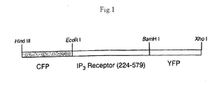
- Fig. 1 is a schematic view of the probe for detection and quantification of inositol-1,4,5-trisphosphate in the invention of this application.
- Fig. 2 is visualization of ATP-dependent IP3 generation in MDCK cells using the probe of this invention. (a), 100 µM ATP; (b), 10 µM ATP; and (c), 1 µM ATP.
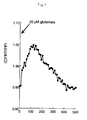
- Fig. 3 is visualization of IP3 concentration in neural dendrites using the probe of this invention.
- Fig. 4 is visualization of IP3 generation in the MDCK cell nucleus using the probe of this invention.
- The probe for detection and quantification of inositol-1,4,5-trisphosphate of this invention comprises at least the following two portions having different functions:
- (1) inositol-1,4,5-trisphosphate recognition site which contains a polypeptide specifically binding to inositol-1,4,5-trisphosphate and leading to conformational change upon the binding to inositol-1,4,5-trisphosphate, and
- (2) two marker sites linked with each end of the polypeptide, which access each other upon binding of the polypeptide with inositol-1,4,5-trisphosphate and emit a signal. Inositol-1,4,5-trisphosphate is sometimes referred to as "IP3" in the present specification.
- Principle of the probe of this invention is that binding the polypeptide in IP3 recognition site with IP3 induces a conformational change of the polypeptide, which then changes the steric configuration of the two reporters linked with each end of the IP3 recognition site, thereby a signal change is appeared.
- In the probe of this invention, IP3 binding domain on the N terminal side of the IP3 receptor is preferably exemplified, as the polypeptide leading to its conformational change upon specific binding to IP3.
- IP3 receptor is a tetrameric complex, and subunit types 1, 2 and 3 are different in IP3-induced Ca2+ release activity and expression distribution in cells and tissues. The IP3 binding domain as the polypeptide may be any of the IP3 receptor subunits, types 1 to 3. According to the studies of the present inventors, an especially good response is obtained when using a region from 224th amino acid to 579th amino acid of the type 1 IP3 receptor as the IP3 binding domain. Of course, such a polypeptide is not limited to the IP3 binding domain, and all peptide chains of synthetic or natural can be used.
- In the probe of this invention, any reporter sites are available so long as the signal change takes place with high accuracy in response to the conformational change of the polypeptide upon binding to IP3. Among various fluorescent chromophores in the biochemical field, as those quickly responding to the conformational change, there are chromophores changing fluorescence intensity ratio by occurrence of fluorescence resonance energy transfer (hereinafter referred to as FRET). Accordingly, as the two marker sites, two fluorescent chromophores different in fluorescence wavelength, specifically, a yellow fluorescent protein (YFP), a red-shifted mutant protein of green fluorescent protein (GFP) and a cyan fluorescent protein (CFP), a blue-shifted mutant protein of GFP may be employed. Of course, as the two reporter sites, various fluorescent proteins, split renilla luciferase, firefly luciferase, (β-galactosidase, β-lactamase etc. may be employed.
- The probe of this invention may have other sites unless the binding to IP3 and the access of the two marker sites are inhibited.
- For example, in the probe of this invention, a nuclear localization signal sequence (NLS) may be linked with any one of the two reporter sites. NLS includes, for example, a sequence of SEQ ID NO: 1, or known NLS such as a nucleoplasmin-derived sequence (SEQ ID NO: 2) and a HIV-1 Rev-derived sequence (SEQ ID NO: 3), without limitation. The probe is localized in the nucleus, IP3 transferred into the nucleus can be detected and IP3 concentration in the nucleus can be quantified.
- In order that the two reporter sites are sterically spaced-apart positions without IP3 and quickly approach upon binding of the polypeptide with IP3, a linker sequence may be provided between the IP3 recognition site and each marker site. The nuclear localization sequence and the marker sites may be linked directly or via the linker sequence.
- The probe of this invention binds to IP3 to allow the signal change. Accordingly, making the probe to coexist with IP3 and measuring the signal change by various chemical or biochemical analysis methods can detect IP3. IP3 may be quantified by previously calibrating a relation of fluorescence intensities and IP3 concentrations.
- Moreover, in the invention of this application, using the probe can screen a substance inhibiting IP3-induced Ca2+ release. That is, in a condition of the probe being coexisted with IP3, the signal change with and without the candidate substance is measured. The candidate substance that inhibits the binding of IP3 with the probe is judged as IP3-induced Ca2+ release inhibitor.
- In the method for IP3 detection and quantification and the method for screening the IP3-induced Ca2+ release inhibitor, various methods are employed to make the probe to coexist with IP3 or the candidate substance. For example, the probe and a candidate substance may coexist with IP3 in elute of disrupted cell.
- Alternatively, the probe may coexist with IP3 in a cell by introducing an expression vector having a polynucleotide for the probe into each culture cell. The candidate substance can be introduced into the cell to make it to coexist with the probe. Such a method enables the IP3 detection and quantification in vivo without disrupting the cell. As the expression vector, a plasmid vector for animal cells is preferably used. As a method for introducing such a plasmid vector into cells, known methods such as an electroporation method, a calcium phosphate method, a liposome method and a DEAE dextran method may be employed.
- In the invention of this application, the probe can coexist with IP3 in all cells of a non-human animal or its progeny established by introducing a polynucleotide expressing the probe into a non-human totipotent cell and developing the cell to individual in accordance with a known method (for example, Non-Patent Document 2). Since such a transgenic non-human animal carries the probe in all body cells, introducing the candidate substance into the body thereof and measuring the IP3 concentration in cells or tissues can screen the IP3-induced Ca2+ release inhibitor.
- In the invention of this application, introducing the polynucleotide expressing the probe into the cell can monitor a stimulation effect on IP3 generation in a cell, stimulating the cell and measuring the signal change before and after the stimulation. That is, the IP3 generation amount can be estimated from the signal change of the stimulated cell, and from which the stimulation effect on the IP3 generation can be known. The stimulation may be biochemical stimulation such as hormones and endocrine disrupters, or physical stimulation such as electricity, radiation, heat etc.
- A stimulation effect on IP3 generation in a living individual and an effect of IP3 generation on life activities can be also monitored. For this purpose, a non-human animal or its progeny, which is established by introducing the polynucleotide expressing the probe into the non-human totipotent cell and developing the cell to individual, is stimulated, and the signal change before and after the stimulation is measured.
- The embodiments of this invention are illustrated more specifically below by referring to Examples along the attached drawings. Of course, this invention is not limited to the following Examples, and it goes without saying that various modifications are possible for details.
- cDNA encoding a probe for IP3 detection and quantification (Fig. 1) was prepared by a genetic engineering method, and introduced into MDCK cell. Expression of the probe in the cytoplasm was confirmed since fluorescence was observed from the cells with a fluorescence microscope.
- Into the MDCK cell expressing the probe as obtained in Example 1, IP3 was introduced by a microinjection method. On each introduction of a small amount of IP3, CFP/YFP fluorescence intensity ratio was stepwise increased and saturated soon. It was clarified from this results that the probe recognizes IP3 and FRET was inhibited dependent on IP3 concentration.
- An increase in fluorescence intensity ratio (CFP/YFP) was observed by stimulating a purine receptor of the MDCK cell as obtained in Example 1 with 100 µM ATP (Fig. 2a). Even with 10 µM ATP and 1 µM ATP stimulations, a response of the probe was also observed depend on ATP concentration (Figs. 2b and 2c).
From this results, it has been confirmed that the temporal IP3 generation in the MDCK cell under physiological conditions can be visualized using the probe of this invention. - The probe was expressed in a neuron prepared from hippocampus of a rat embryo, and IP3 kinetics in the neuron was observed.
With 20 µM glutamate stimulation of the neuron, IP3 concentration was temporally increased in not only the cell body but also the dendrites (Fig. 3). - The probe was linked with the nuclear localization sequence of SEQ ID NO: 4 at the C terminal thereof, and expressed in MDCK cell. Since almost all fluorescences were observed from the nucleus, it was confirmed that the probe was localized in the nucleus. With 100 µM ATP stimulation of the MDCK cell, IP3 concentration was temporally increased (Fig. 4).
It has been reported that there are localized in nucleus an IP3 receptor and an enzyme group which further phosphorylates IP3 and produces second messengers such as IP4, IP5 and IP6 having different roles from IP3. However, it has not been clarified as to what extent IP3 as a substrate of the enzymes exists in the nucleus, and whether IP3 can permeate a nuclear pore.
Using the probe of this invention, new knowledge that the IP3 concentration is increased not only in the cytoplasm but also in the nucleus with extracellular stimulation has been obtained. - As has been thus far described in detail, this invention provides a probe for detection and quantification with high accuracy in the noninvasive manner as to where and when inositol-1,4,5-trisphosphate is produced in living cells. Further, this invention provides a method for detecting and quantifying inositol-1,4,5-trisphosphate, a method for monitoring a stimulation effect on inositol-1,4,5-trisphosphate generation in the cell and a method for screening a inhibitor for Ca2+ release induced by inositol-1,4,5-trisphosphate, using the probe for detection and quantification of inositol-1,4,5-trisphosphate.
Claims (12)
- A probe for detection and quantification of inositol-1,4,5-trisphosphate, which comprises at least,
inositol-1,4,5-trisphosphate recognition site containing an inositol-1,4,5-trisphosphate binding domain of inositol-1,4,5-trisphosphate receptor, and
two reporter sites linked with each end of the inositol-1,4,5-trisphosphate recognition site, of which access imposes fluorescence resonance energy transfer, thereby a signal being detectable. - The probe of claim 1, wherein the two reporter sites are yellow fluorescent protein and cyan fluorescent protein.
- The probe of claim 1 or 2, wherein the polypeptide leading to conformational change upon specific binding to inositol-1,4,5-trisphosphate is an inositol-1,4,5-trisphosphate binding domain of inositol-1,4,5-trisphosphate receptor.
- The probe of any one of claims 1 to 3, wherein a nuclear localization sequence is linked with any one of the two reporter sites.
- A method for detection and quantification of inositol-1,4,5-trisphosphate, which comprises making the probe of any one of claims 1 to 4 to coexist with inositol-1,4,5-trisphosphate, and measuring signal change with and without inositol-1,4,5-trisphosphate.
- The method according to claim 5, wherein the probe coexists with inositol-1,4,5-trisphosphate in a cell by introducing polynucleotide expressing the probe into the cell.
- The method according to claim 5, wherein the probe coexists with inositol-1,4,5-trisphosphate in all cells of a non-human animal or its progeny established by introducing polynucleotide expressing the probe into a non-human totipotent cell and developing the cell to individual.
- A method for monitoring a stimulation effect on generation of inositol-1,4,5-trisphosphate in a cell, which comprises introducing a polynucleotide expressing the probe of any one of claims 1 to 4 into the cell, stimulating the cell, and measuring signals before and after the stimulation.
- A method for monitoring a stimulation effect on generation of inositol-1,4,5-trisphosphate in a living individual, which comprises stimulating a non-human animal or its progeny established by introducing polynucleotide expressing the probe of any one of claims 1 to 4 into a non-human totipotent cell and developing the cell to individual, and measuring signals before and after the stimulation.
- A method for screening an inhibitor of Ca2+ release induced by inositol-1,4,5-trisphosphate, which comprises making the probe of claim 3, inositol-1,4,5-trisphosphate and candidate substance to coexist, and measuring signal change with and without the candidate substance.
- The method according to claim 10, wherein the probe coexists with inositol-1,4,5-trisphosphate and the candidate substance in a cell by introducing polynucleotide expressing the probe into the cell.
- The method according to claim 10, wherein the probe coexists with inositol-1,4,5-trisphosphate and the candidate substance in all cells of a non-human animal or its progeny established by introducing polynucleotide expressing the probe into a non-human totipotent cell and developing the cell to individual.
Applications Claiming Priority (2)
| Application Number | Priority Date | Filing Date | Title |
|---|---|---|---|
| JP2004152484 | 2004-05-21 | ||
| PCT/JP2005/009299 WO2005113792A1 (en) | 2004-05-21 | 2005-05-17 | Probe for detection/determination of inositol-1,4,5-triphosphoric acid and method of detecting/determining inositol-1,4,5-triphosphoric acid with the same |
Publications (2)
| Publication Number | Publication Date |
|---|---|
| EP1767646A1 true EP1767646A1 (en) | 2007-03-28 |
| EP1767646A4 EP1767646A4 (en) | 2008-03-19 |
Family
ID=35428414
Family Applications (1)
| Application Number | Title | Priority Date | Filing Date |
|---|---|---|---|
| EP05741379A Ceased EP1767646A4 (en) | 2004-05-21 | 2005-05-17 | PROBE FOR DETECTION / DETERMINATION OF THE PRESENCE OF INOSITOL-1,4,5-TRIPHOSPHORIC ACID AND METHOD FOR DETECTION / DETERMINATION OF THE PRESENCE OF INOSITOL-1,4,5-TRIPHOSPHORIC ACID USING THE SAME PROBE |
Country Status (5)
| Country | Link |
|---|---|
| US (1) | US20090208970A1 (en) |
| EP (1) | EP1767646A4 (en) |
| JP (1) | JP4717808B2 (en) |
| CA (1) | CA2567650A1 (en) |
| WO (1) | WO2005113792A1 (en) |
Families Citing this family (1)
| Publication number | Priority date | Publication date | Assignee | Title |
|---|---|---|---|---|
| CN120943969A (en) * | 2024-05-07 | 2025-11-14 | 华东理工大学 | An inositol optical probe, its preparation method and application |
Family Cites Families (6)
| Publication number | Priority date | Publication date | Assignee | Title |
|---|---|---|---|---|
| US5998204A (en) * | 1997-03-14 | 1999-12-07 | The Regents Of The University Of California | Fluorescent protein sensors for detection of analytes |
| US6469154B1 (en) * | 1999-05-21 | 2002-10-22 | The Regents Of The University Of California | Fluorescent protein indicators |
| JP3643520B2 (en) * | 2000-07-04 | 2005-04-27 | 独立行政法人科学技術振興機構 | cGMP visualization probe and cGMP detection / quantification method using the same |
| JP2002250730A (en) * | 2001-02-27 | 2002-09-06 | Shinwa Kako Kk | Molecular sensor and manufacturing method thereof |
| JP4267875B2 (en) * | 2002-07-24 | 2009-05-27 | 独立行政法人科学技術振興機構 | Inositol biosensor and its use |
| JP4803976B2 (en) * | 2003-07-09 | 2011-10-26 | 独立行政法人科学技術振興機構 | Molecular sensor for intracellular IP3 measurement |
-
2005
- 2005-05-17 EP EP05741379A patent/EP1767646A4/en not_active Ceased
- 2005-05-17 CA CA002567650A patent/CA2567650A1/en not_active Abandoned
- 2005-05-17 WO PCT/JP2005/009299 patent/WO2005113792A1/en not_active Ceased
- 2005-05-17 US US11/597,211 patent/US20090208970A1/en not_active Abandoned
- 2005-05-17 JP JP2006513747A patent/JP4717808B2/en not_active Expired - Fee Related
Also Published As
| Publication number | Publication date |
|---|---|
| WO2005113792A1 (en) | 2005-12-01 |
| EP1767646A4 (en) | 2008-03-19 |
| CA2567650A1 (en) | 2005-12-01 |
| JPWO2005113792A1 (en) | 2008-03-27 |
| JP4717808B2 (en) | 2011-07-06 |
| US20090208970A1 (en) | 2009-08-20 |
Similar Documents
| Publication | Publication Date | Title |
|---|---|---|
| Fukata et al. | Protein palmitoylation in neuronal development and synaptic plasticity | |
| Ortiz et al. | Synthesis of sulfonated carbofluoresceins for voltage imaging | |
| Caron et al. | Using FLIM-FRET to measure conformational changes of transglutaminase type 2 in live cells | |
| Morris | Fluorescent biosensors of intracellular targets from genetically encoded reporters to modular polypeptide probes | |
| Bae et al. | Distinct synaptic vesicle recycling in inhibitory nerve terminals is coordinated by SV2A | |
| EP1730499B1 (en) | Method for the detection of intracellular parameters with luminescent protein probes for the screening of molecules capable of altering said parameters | |
| EP2623514A1 (en) | Linker for unimolecular fret biosensor based on principle of fluorescence resonance energy transfer | |
| US20050059028A1 (en) | RNAi-based sensors, caged interfering RNAs, and methods of use thereof | |
| Kumar et al. | GloSensor assay for discovery of GPCR-selective ligands | |
| Hunter et al. | Real-time measurement of cannabinoid receptor-mediated cAMP signaling | |
| Robers et al. | A luminescent assay for real-time measurements of receptor endocytosis in living cells | |
| Abreu et al. | Optogenetic techniques for manipulating and sensing G protein-coupled receptor signaling | |
| Van Unen et al. | Quantitative single-cell analysis of signaling pathways activated immediately downstream of histamine receptor subtypes | |
| Sims et al. | Single-cell kinase assays: opening a window onto cell behavior | |
| WO2013151511A1 (en) | Modified Dual-Colour Protein | |
| Kumar et al. | Syndapin is dispensable for synaptic vesicle endocytosis at the Drosophila larval neuromuscular junction | |
| JP5688133B2 (en) | Tools for the identification of LINGO-1, LINGO-2, LINGO-3 and LINGO-4 ligands and their use | |
| JP2019216730A (en) | Human cellular models with biosensors | |
| JPWO2011090159A1 (en) | Probe reagent for measuring protein degradation activity | |
| US20090208970A1 (en) | Probe for detection and quantification of inositol-1,4,5-trisphosphate and a method for detecting and quantifying inositol-1,4,5-trisphosphate using the same | |
| Haruyama | Cellular biosensing: chemical and genetic approaches | |
| Oner et al. | Group II activators of G-protein signaling: monitoring the interaction of Gα with the G-protein regulatory motif in the intact cell | |
| Kimura et al. | Visualization of DNA methylation and histone modifications in living cells | |
| Maulucci et al. | Investigation of the spatial distribution of glutathione redox-balance in live cells by using Fluorescence Ratio Imaging Microscopy | |
| Tsutsui et al. | Engineering voltage sensing phosphatase (VSP) |
Legal Events
| Date | Code | Title | Description |
|---|---|---|---|
| PUAI | Public reference made under article 153(3) epc to a published international application that has entered the european phase |
Free format text: ORIGINAL CODE: 0009012 |
|
| 17P | Request for examination filed |
Effective date: 20061218 |
|
| AK | Designated contracting states |
Kind code of ref document: A1 Designated state(s): DE FR GB |
|
| DAX | Request for extension of the european patent (deleted) | ||
| RBV | Designated contracting states (corrected) |
Designated state(s): DE FR GB |
|
| A4 | Supplementary search report drawn up and despatched |
Effective date: 20080220 |
|
| RIC1 | Information provided on ipc code assigned before grant |
Ipc: C07K 19/00 20060101ALI20080211BHEP Ipc: C12Q 1/02 20060101AFI20051207BHEP Ipc: G01N 21/78 20060101ALI20080211BHEP Ipc: G01N 33/566 20060101ALI20080211BHEP Ipc: C07K 7/08 20060101ALI20080211BHEP Ipc: C07K 7/06 20060101ALI20080211BHEP Ipc: C12N 15/09 20060101ALI20080211BHEP Ipc: C07K 14/705 20060101ALI20080211BHEP Ipc: G01N 33/15 20060101ALI20080211BHEP Ipc: G01N 33/50 20060101ALI20080211BHEP |
|
| 17Q | First examination report despatched |
Effective date: 20090507 |
|
| REG | Reference to a national code |
Ref country code: DE Ref legal event code: R003 |
|
| STAA | Information on the status of an ep patent application or granted ep patent |
Free format text: STATUS: THE APPLICATION HAS BEEN REFUSED |
|
| 18R | Application refused |
Effective date: 20140327 |