EP1767511A1 - Macromolecular compound bond evaluating method - Google Patents
Macromolecular compound bond evaluating method Download PDFInfo
- Publication number
- EP1767511A1 EP1767511A1 EP05765321A EP05765321A EP1767511A1 EP 1767511 A1 EP1767511 A1 EP 1767511A1 EP 05765321 A EP05765321 A EP 05765321A EP 05765321 A EP05765321 A EP 05765321A EP 1767511 A1 EP1767511 A1 EP 1767511A1
- Authority
- EP
- European Patent Office
- Prior art keywords
- binding
- target molecule
- evaluation method
- drug candidate
- evaluation target
- Prior art date
- Legal status (The legal status is an assumption and is not a legal conclusion. Google has not performed a legal analysis and makes no representation as to the accuracy of the status listed.)
- Withdrawn
Links
Images


Classifications
-
- G—PHYSICS
- G16—INFORMATION AND COMMUNICATION TECHNOLOGY [ICT] SPECIALLY ADAPTED FOR SPECIFIC APPLICATION FIELDS
- G16B—BIOINFORMATICS, i.e. INFORMATION AND COMMUNICATION TECHNOLOGY [ICT] SPECIALLY ADAPTED FOR GENETIC OR PROTEIN-RELATED DATA PROCESSING IN COMPUTATIONAL MOLECULAR BIOLOGY
- G16B15/00—ICT specially adapted for analysing two-dimensional [2D] or three-dimensional [3D] molecular structures, e.g. structural or functional relations or structure alignment
-
- G—PHYSICS
- G16—INFORMATION AND COMMUNICATION TECHNOLOGY [ICT] SPECIALLY ADAPTED FOR SPECIFIC APPLICATION FIELDS
- G16B—BIOINFORMATICS, i.e. INFORMATION AND COMMUNICATION TECHNOLOGY [ICT] SPECIALLY ADAPTED FOR GENETIC OR PROTEIN-RELATED DATA PROCESSING IN COMPUTATIONAL MOLECULAR BIOLOGY
- G16B15/00—ICT specially adapted for analysing two-dimensional [2D] or three-dimensional [3D] molecular structures, e.g. structural or functional relations or structure alignment
- G16B15/30—Drug targeting using structural data; Docking or binding prediction
-
- G—PHYSICS
- G16—INFORMATION AND COMMUNICATION TECHNOLOGY [ICT] SPECIALLY ADAPTED FOR SPECIFIC APPLICATION FIELDS
- G16B—BIOINFORMATICS, i.e. INFORMATION AND COMMUNICATION TECHNOLOGY [ICT] SPECIALLY ADAPTED FOR GENETIC OR PROTEIN-RELATED DATA PROCESSING IN COMPUTATIONAL MOLECULAR BIOLOGY
- G16B20/00—ICT specially adapted for functional genomics or proteomics, e.g. genotype-phenotype associations
- G16B20/30—Detection of binding sites or motifs
-
- G—PHYSICS
- G16—INFORMATION AND COMMUNICATION TECHNOLOGY [ICT] SPECIALLY ADAPTED FOR SPECIFIC APPLICATION FIELDS
- G16B—BIOINFORMATICS, i.e. INFORMATION AND COMMUNICATION TECHNOLOGY [ICT] SPECIALLY ADAPTED FOR GENETIC OR PROTEIN-RELATED DATA PROCESSING IN COMPUTATIONAL MOLECULAR BIOLOGY
- G16B20/00—ICT specially adapted for functional genomics or proteomics, e.g. genotype-phenotype associations
Definitions
- the present invention relates to a binding evaluation method for a macromolecular compound.
- Priority is claimed on Japanese Patent Application No. 2004-205746, filed July 13, 2004 , the content of which is incorporated herein by reference.
- a drug having a low affinity with respect to a protein is not appropriate as a drug candidate agent, and such drugs having a low affinity are excluded from the drug candidate agent.
- affinity X-ray structural analysis or a classical molecular dynamics method is generally used.
- Non Patent Document 1 Takeshi Onda et al., J. Bio. Chem., 276 (2001) 47785-47793
- the binding site of adenylate cyclase to drug candidate agents and binding state thereof cannot be elucidated solely by the results.
- the binding site of forskolin with respect to the active site of adenylate cyclase is elucidated by substituting atoms or molecules, which are bound to the side chains of forskolin, with various atoms or molecules, this work is based on the experiments which require an extended period of time and vast amounts of labour and thus, is inefficient and not practical.
- affinity evaluation in the prior art document is one evaluating the internal energy of an analytical model where drug candidate agents are placed in the centre of the active site of adenylate cyclase
- the inventors of the present invention obtained calculation results showing that the internal energy is smaller in the analytical model where the position of forskolin is deviated from the centre of the active site of adenylate cyclase than that of the analytical model placing forskolin at the centre of the active site of adenylate cyclase.
- the present invention addresses the abovementioned problems, with objects shown below.
- the present invention adopts a solution which is a method evaluating the binding of an evaluation target molecule with respect to a macromolecular compound based on numerical calculations and which detects the binding sites of the evaluation target molecule with respect to the macromolecular compound as a site where electronic states in a predetermined restricted region including the evaluation target molecule and the part of the macromolecular compound, which is adjacent to the evaluation target molecule, are unstable.
- the binding site of the evaluation target molecule with respect to the macromolecular compound is detected by numerical calculations as a site where electronic states in a predetermined restricted region including the evaluation target molecule and the part of the macromolecular compound, which is adjacent to the evaluation target molecule, are unstable, it is possible to numerically analyze the analysis target including the macromolecular compound having an extremely large number of electrons efficiently. Accordingly, it is possible to dramatically improve the analysis efficiency of binding sites compared to the conventional analytical approach of binding sites based on experiments.
- A adenylate cyclase
- B forskolin
- G restricted region
- the present embodiment relates to an evaluation method for the binding between the active site of a protein as a macromolecular compound, more specifically adenylate cyclase (A), and a drug candidate agent as an evaluation target molecule, more specifically forskolin (B).
- A adenylate cyclase
- B a drug candidate agent as an evaluation target molecule
- FIG 1 is a flow chart showing a processing procedure of the present binding evaluation method.
- step S 1 shows a process for detecting binding positions as preprocessing and steps S2 to S9 show processes for detecting binding sites as actual processing.
- binding positions are identified by evaluating the internal energy (potential energy + kinetic energy) of analytical models formed from adenylate cyclase (A) and forskolin (B) based on the numerical analysis using a computer (step S 1).
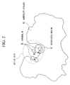
- FIG. 2 is a schematic diagram schematically showing an analytical model for identifying binding positions.
- forskolin (B) and adenylate cyclase (A) are bound, forskolin (B) binds to any place in the active site of adenylate cyclase (A) shown with broken lines.
- each internal energy of target models for analysis in a state where a plurality of positions of forskolin (B) with respect to the active site are set is numerically calculated using the classical molecular dynamics method and the position of forskolin (B) corresponding to the internal energy, which has the minimum value among the internal energies corresponding to each position, is identified as the binding position of forskolin (B) in the active site of adenylate cyclase (A).
- positions of forskolin (B) P 1 , P 2 , and P 3 shown in Fig. 2 are 3 places selected as representatives, internal energies of each target model for analysis where forskolin (B) is placed at each of positions P 1 , P 2 , and P 3 will be those below.
- Position P 1 -122477.433 kcal/mol
- Position P 2 -120889.463 kcal/mol
- Position P 3 : -124151.413 kcal/mol
- the internal energy is at its lowest when forskolin (B) is located at position P 3 among the 3 positions P 1 , P 2 , and P 3 .
- internal energies are determined for a plurality of positions other than these 3 positions P 1 , P 2 , and P 3 , as a result, internal energy showed the minimum value when forskolin (B) is located at position P 3 . That is, since forskolin (B) binds to the active site of adenylate cyclase (A) in the most internally energetic stabilized state when placed at position P 3 among the active sites, it is identified that the binding position of forskolin (B) with respect to the active site is position P 3 .
- steps S2 to S6 are processes determining the charge density ⁇ (r) of a predetermined restricted region (G) including forskolin (B) and the parts of adenylate cyclase (A), which is adjacent to forskolin (B), by determining the wave function ⁇ (r) regarding the restricted region (G) via numerical calculations using a computer (charge density calculating process). Note that in the wave function ⁇ (r) and charge density ⁇ (r), r is a variable showing the coordinates in a 3-dimensional space.
- the radial wave function R(r) shows, as is well-known, the existence probability of electrons and also in this graph, although the probability is "0" at the position of the atomic nucleus, it increases as the distance r increases and after reaching the maximum value at a certain distance, which is relatively close to the atomic nucleus, it gradually decreases and approach "0" asymptotically.
- the distance range where the radial wave function R(r) may take on significant values is limited to the vicinity of the atomic nucleus and in distant ranges outside this range, the function can be regarded as almost "0".
- Such variation characteristics of the radial wave function R(r) is found not only in the electron belonging to the hydrogen atom but also in electrons belonging to other atoms or other orbitals.
- the distance range where the wave function may take on significant values is set as the abovementioned restricted region (G).
- the range of variable r which represents 3-dimensional positions, is restricted to the region where the radial wave function R(r) may take on sufficiently large values and the charge density ⁇ (r) of the restricted region (G) is calculated by determining only the wave function in this restricted region (G).
- the distance range where the radial wave function R(r) may take on significant values differs to some extent depending on the type of atoms and electron orbitals, at the distance distant from the atomic nucleus by 10 to 20 ⁇ , the radial wave functions R(r) of almost all the atoms and electrons in electron orbitals can be considered to approach "0" asymptotically.
- a threshold value of 10 to 20 ⁇ is adopted to define the restricted region (G) and by this procedure and by taking the atomic nuclei of each atom constituting forskolin (B) as the centre, the 3-dimensional space within the radius of 10 to 20 ⁇ is set as the restricted region (G).
- This restricted region (G) is a 3-dimensional space including forskolin (B) and the parts of adenylate cyclase (A) adjacent to forskolin (B).
- each of the initial values of the wave function ⁇ (r) and effective potential V(r) regarding the restricted region (G) are set at the beginning (step S2) and the initial values of charge density ⁇ (r) based on all the electrons in the restricted region (G) are calculated by assigning the initial values of wave function ⁇ (r) to the formula (1) below (step S3).
- ⁇ r ⁇ ⁇ ⁇ * r ⁇ ⁇ r
- the wave function ⁇ (r) in the restricted region (G) is calculated (step S4). Note that in this Schrödinger wave equation (2), ⁇ is expected values of internal energy.
- Charge density ⁇ (r) is determined by assigning the wave function ⁇ (r) derived from the Schrödinger wave equation (2) to the formula (1) (step S5) and whether or not this charge density ⁇ (r) equals the charge density ⁇ (r) derived in the previous calculation is determined (step S6). If the two are not equal (when the result is "NO"), the procedures of steps S4 and S5 are repeated by taking the wave function ⁇ (r) and effective potential V(r) derived by solving the Schrödinger wave equation (2) in step S5 as new initial values to determine the new wave function ⁇ (r) and charge density ⁇ (r).
- step S6 When such a charge density ⁇ (r) is derived, the determination in step S6 will become "YES" and by assigning this charge density ⁇ (r) to Fukui functions (3) and (4) below, LUMOf + (r) and HOMOf(r) as values of Fukui functions (3) and (4) in the restricted region (G) are calculated (step S7).
- LUMOf + (r) and HOMOf(r) show the degree of instability of electronic states in the restricted region (G) and while LUMOf + (r) shows the degree of electron-accepting property (property to receive electrons), HOMOf(r) shows the degree of electron-donating property (property to release electrons).
- Such LUMOf + (r) and HOMOf(r) are displayed by images (step S8) and based on this displayed result, the binding site between forskolin (B) and adenylate cyclase (A) in the restricted region (G), that is, which atom constituting forskolin (B) binds to which atom constituting adenylate cyclase (A) is identified (step S9).
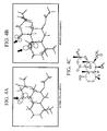
- Figs 4A to 4C are characteristic charts where LUMOf + (r) and HOMOf(r) in the forskolin vicinity-region inside the restricted region (G) are shown as "Isosurfaces". That is, in these characteristic charts, the part (spatial region) which corresponds to LUMOf + (r) and HOMOf(r) that are equivalent to 90% of the maximum value in this forskolin vicinity-region is set as "Isosurface” and this "Isosurface” is displayed by being made to correspond to the molecular structure of forskolin (B).
- FIG. 4A shows the abovementioned "Isosurface” regarding LUMOf + (r), as is apparent when this Fig. 4A is contrasted with Fig. 4C, which shows the molecular structure of forskolin (B), the arrow part displaying the "Isosurface” as a relatively large space in Fig. 4A is the part corresponding to an oxygen atom (O), which is one of the side chains of forskolin (B).
- O oxygen atom
- Fig. 4B shows the "Isosurface” regarding HOMOf(r), as is apparent when this Fig. 4B is contrasted with Fig. 4C, which shows the molecular structure of forskolin (B), the arrow part displaying the "Isosurface” as a relatively large space in Fig. 4B is the part corresponding to an OH group (OH), which is one of the side chains of forskolin (B).
- OH OH
- the binding between forskolin (B) and the active site of adenylate cyclase (A) is formed via covalent bonds of atoms, each of which belongs to the two substances, the abovementioned oxygen atom (O) and OH group (OH) of forskolin (B) are considered to be the sites which bind to the active site of adenylate cyclase (A) (binding sites).
- the present binding evaluation method which identifies the binding sites between the active site of adenylate cyclase (A) and forskolin (B) based on LUMOf + (r) and HOMOf(r) regarding forskolin (B) and the part of adenylate cyclase (A) that is adjacent to forskolin (B) in the restricted region (G), is an evaluation approach of binding sites which derives results that completely coincide with experimental results and which is sufficiently effective.
- the present binding evaluation method makes only atoms present in the restricted region (G) as targets for numerical analysis, it is possible to reduce the time required for calculating wave function ⁇ (r) and charge density ⁇ (r) to a large extent compared to the case where all the atoms constituting forskolin (B) and adenylate cyclase (A) are made targets for numerical analysis. Accordingly, it is possible to dramatically improve the analysis efficiency of binding sites compared to the conventional analytical approach of binding sites based on experiments.
- the present invention is not limited to the above embodiment and for example, modifications such as the following may be considered.
- (1) is one regarding the binding between forskolin (B) as an evaluation target molecule and adenylate cyclase (A) as one of macromolecular compounds, the present invention is not limited to this. That is, macromolecular compounds are not limited to adenylate cyclase (A) but may be other proteins or other macromolecular compounds and also the evaluation target molecules are not limited to forskolin (B).
- the restricted region (G) is set as the 3-dimensional spaces which have atomic nuclei of each atom constituting forskolin (B) as the centre and ranges within the radius of 10 to 20 ⁇ by adopting 10 to 20 ⁇ as a threshold value to define the restricted region (G), and by setting such a restricted region (G), analysis efficiency is dramatically improved by reducing computational complexity in the calculation of charge density ⁇ (r) required for the calculation of LUMOf + (r) and HOMOf(r) considerably.
- threshold value defining the restricted region (G) that is, the distance where the wave function ⁇ (r) is regarded to take on significant values, is not limited to 10 to 20 ⁇ .
- threshold values defining the restricted region (G) may be set for each type of constituting atoms or electron orbitals.
- the method for identifying the site where the electronic state is unstable is not limited to Fukui functions (3) and (4).
- the site where the electronic state is unstable may be identified using other evaluation functions.
- the binding site of an evaluation target molecule with respect to a macromolecular compound is detected by numerical calculations as a site where electronic states in a predetermined restricted region including the evaluation target molecule and the part of the macromolecular compound, which is adjacent to the evaluation target molecule, are unstable, it is possible to numerically analyze the analysis target including macromolecular compounds having an extremely large number of electrons efficiently. Accordingly, it is possible to dramatically improve the analysis efficiency of binding sites compared to the conventional analytical approach of binding sites based on experiments.
Landscapes
- Life Sciences & Earth Sciences (AREA)
- Physics & Mathematics (AREA)
- Engineering & Computer Science (AREA)
- Bioinformatics & Cheminformatics (AREA)
- Health & Medical Sciences (AREA)
- Spectroscopy & Molecular Physics (AREA)
- Chemical & Material Sciences (AREA)
- Medical Informatics (AREA)
- General Health & Medical Sciences (AREA)
- Theoretical Computer Science (AREA)
- Biophysics (AREA)
- Bioinformatics & Computational Biology (AREA)
- Biotechnology (AREA)
- Evolutionary Biology (AREA)
- Crystallography & Structural Chemistry (AREA)
- Pharmacology & Pharmacy (AREA)
- Medicinal Chemistry (AREA)
- Analytical Chemistry (AREA)
- Genetics & Genomics (AREA)
- Molecular Biology (AREA)
- Proteomics, Peptides & Aminoacids (AREA)
- Investigating Or Analysing Biological Materials (AREA)
- Organic Low-Molecular-Weight Compounds And Preparation Thereof (AREA)
Abstract
A method for evaluating the binding of an evaluation target molecule with respect to a macromolecular compound based on numerical calculations and which detects the binding sites of the evaluation target molecule with respect to the macromolecular compound as a site where electronic states in a predetermined restricted region including the evaluation target molecule and the part of the macromolecular compound, which is adjacent to the evaluation target molecule, are unstable.
Description
- The present invention relates to a binding evaluation method for a macromolecular compound.
Priority is claimed onJapanese Patent Application No. 2004-205746, filed July 13, 2004 - In the development of a drug, the binding between a protein as one of the macromolecular compounds constituting a living body and a drug candidate agent, is evaluated. That is, a drug having a low affinity with respect to a protein is not appropriate as a drug candidate agent, and such drugs having a low affinity are excluded from the drug candidate agent. Moreover, as an evaluation method for the abovementioned affinity, X-ray structural analysis or a classical molecular dynamics method is generally used.
- For example, in a prior art document below, there is disclosed study results of the binding mechanism between adenylate cyclase (type II) as a protein and forskolin as a drug candidate agent, as a case of affinity analysis using the classical molecular dynamics method. In this prior art document, by calculating each internal energy (potential energy + kinetic energy) of analytical models (systems) where various drug candidate agents such as forskolin are placed at the centre of the active site of adenylate cyclase using the classical molecular dynamics method, those having a relatively small internal energy (that is, those that stabilize the system energetically) are extracted as drug candidate agents having excellent affinity with respect to adenylate cyclase. At the same time, the document examines the binding site of forskolin, which is extracted as such and which is one of the drug candidate agents, with respect to adenylate cyclase.
[Non Patent Document 1]
Takeshi Onda et al., J. Bio. Chem., 276 (2001) 47785-47793 - Although the abovementioned classical molecular dynamics method provides preliminary results of evaluation regarding the affinity between the active site of adenylate cyclase and drug candidate agents, the binding site of adenylate cyclase to drug candidate agents and binding state thereof cannot be elucidated solely by the results.
For example, in the prior art document, although the binding site of forskolin with respect to the active site of adenylate cyclase is elucidated by substituting atoms or molecules, which are bound to the side chains of forskolin, with various atoms or molecules, this work is based on the experiments which require an extended period of time and vast amounts of labour and thus, is inefficient and not practical. - Moreover, although the approach of affinity evaluation in the prior art document is one evaluating the internal energy of an analytical model where drug candidate agents are placed in the centre of the active site of adenylate cyclase, there is a reliability problem and an evaluation approach with higher reliability is desired. That is, in the approach of affinity evaluation in the prior art document, since the effect which the position of drug candidate agents with respect to the active site of adenylate cyclase imposes on the internal energy is not considered, reliability thereof is low. The inventors of the present invention obtained calculation results showing that the internal energy is smaller in the analytical model where the position of forskolin is deviated from the centre of the active site of adenylate cyclase than that of the analytical model placing forskolin at the centre of the active site of adenylate cyclase.
- The present invention addresses the abovementioned problems, with objects shown below.
- (1) elucidate the binding site of a macromolecular compound with respect to evaluation target molecule more efficiently than in the past
- (2) improve the reliability of the binding position of a macromolecular compound with respect to evaluation target molecule more than that in the past
- In order to achieve the above object, the present invention adopts a solution which is a method evaluating the binding of an evaluation target molecule with respect to a macromolecular compound based on numerical calculations and which detects the binding sites of the evaluation target molecule with respect to the macromolecular compound as a site where electronic states in a predetermined restricted region including the evaluation target molecule and the part of the macromolecular compound, which is adjacent to the evaluation target molecule, are unstable.
- According to the present invention, since the binding site of the evaluation target molecule with respect to the macromolecular compound is detected by numerical calculations as a site where electronic states in a predetermined restricted region including the evaluation target molecule and the part of the macromolecular compound, which is adjacent to the evaluation target molecule, are unstable, it is possible to numerically analyze the analysis target including the macromolecular compound having an extremely large number of electrons efficiently. Accordingly, it is possible to dramatically improve the analysis efficiency of binding sites compared to the conventional analytical approach of binding sites based on experiments.
-
- FIG 1 is a flow chart showing a processing procedure of a binding evaluation method according to one embodiment of the present invention.
- FIG 2 is a schematic diagram of an analytical model for identifying the binding position of forskolin (B) with respect to the active site of adenylate cyclase (A) in one embodiment of the present invention.
- FIG 3 is a graph showing variations in wave function of electrons in the 1s orbital which belongs to a hydrogen atom as one example of the variation characteristics of wave function in the radial direction in one embodiment of the present invention.
- FIG 4A is a characteristic chart where LUMOf+(r) and HOMOf(r) in the forskolin vicinity-region are shown as isosurfaces in one embodiment of the present invention.
- FIG. 4B is a characteristic chart where LUMOf+(r) and HOMOf(r) in the forskolin vicinity-region are shown as isosurfaces in one embodiment of the present invention.
- FIG 4C is a molecular structure of forskolin derivatives.
- A: adenylate cyclase, B: forskolin, G: restricted region
- Hereunder is a description of one embodiment of the present invention, with reference to the drawings.
The present embodiment relates to an evaluation method for the binding between the active site of a protein as a macromolecular compound, more specifically adenylate cyclase (A), and a drug candidate agent as an evaluation target molecule, more specifically forskolin (B). - FIG 1 is a flow chart showing a processing procedure of the present binding evaluation method. In this processing procedure,
step S 1 shows a process for detecting binding positions as preprocessing and steps S2 to S9 show processes for detecting binding sites as actual processing. - In the process for detecting binding positions, binding positions are identified by evaluating the internal energy (potential energy + kinetic energy) of analytical models formed from adenylate cyclase (A) and forskolin (B) based on the numerical analysis using a computer (step S 1).
- FIG. 2 is a schematic diagram schematically showing an analytical model for identifying binding positions. When forskolin (B) and adenylate cyclase (A) are bound, forskolin (B) binds to any place in the active site of adenylate cyclase (A) shown with broken lines. In the present binding evaluation method, each internal energy of target models for analysis in a state where a plurality of positions of forskolin (B) with respect to the active site are set is numerically calculated using the classical molecular dynamics method and the position of forskolin (B) corresponding to the internal energy, which has the minimum value among the internal energies corresponding to each position, is identified as the binding position of forskolin (B) in the active site of adenylate cyclase (A).
- For example, although positions of forskolin (B) P1, P2, and P3 shown in Fig. 2 are 3 places selected as representatives, internal energies of each target model for analysis where forskolin (B) is placed at each of positions P1, P2, and P3 will be those below.
Position P1: -122477.433 kcal/mol
Position P2: -120889.463 kcal/mol
Position P3: -124151.413 kcal/mol - As is apparent from the intercomparison of these internal energies, the internal energy is at its lowest when forskolin (B) is located at position P3 among the 3 positions P1, P2, and P3. Although internal energies are determined for a plurality of positions other than these 3 positions P1, P2, and P3, as a result, internal energy showed the minimum value when forskolin (B) is located at position P3. That is, since forskolin (B) binds to the active site of adenylate cyclase (A) in the most internally energetic stabilized state when placed at position P3 among the active sites, it is identified that the binding position of forskolin (B) with respect to the active site is position P3.
- When the binding position of forskolin (B) is identified as described so far, the binding site of forskolin (B) with respect to adenylate cyclase (A) in the binding position is identified based on the processing of steps S2 to S9. Among these steps, steps S2 to S6 are processes determining the charge density ρ(r) of a predetermined restricted region (G) including forskolin (B) and the parts of adenylate cyclase (A), which is adjacent to forskolin (B), by determining the wave function Φ(r) regarding the restricted region (G) via numerical calculations using a computer (charge density calculating process). Note that in the wave function Φ (r) and charge density ρ(r), r is a variable showing the coordinates in a 3-dimensional space.
- The setting method of the abovementioned restricted region (G) will be described by referring also to Fig. 3. Fig. 3 is a graph where the wave function R(k) of the electron, which belongs to a hydrogen atom (the electron in the Is orbital), is numerically calculated by taking the distance r in the radial direction as a variable when setting the atomic nucleus as a base point. Note that in this graph, the vertical axis shows values where the radial wave function R(r) is multiplied by the distance r for convenience and the horizontal axis shows the distance from the atomic nucleus using the unit a.u. (1 a.u. = 0.529 Å).
- The radial wave function R(r) shows, as is well-known, the existence probability of electrons and also in this graph, although the probability is "0" at the position of the atomic nucleus, it increases as the distance r increases and after reaching the maximum value at a certain distance, which is relatively close to the atomic nucleus, it gradually decreases and approach "0" asymptotically. In other words, the distance range where the radial wave function R(r) may take on significant values is limited to the vicinity of the atomic nucleus and in distant ranges outside this range, the function can be regarded as almost "0". Such variation characteristics of the radial wave function R(r) is found not only in the electron belonging to the hydrogen atom but also in electrons belonging to other atoms or other orbitals.
- In the present embodiment, by taking such variation characteristics of the wave function into consideration, the distance range where the wave function may take on significant values is set as the abovementioned restricted region (G). In other words, the range of variable r, which represents 3-dimensional positions, is restricted to the region where the radial wave function R(r) may take on sufficiently large values and the charge density ρ(r) of the restricted region (G) is calculated by determining only the wave function in this restricted region (G). Although the distance range where the radial wave function R(r) may take on significant values differs to some extent depending on the type of atoms and electron orbitals, at the distance distant from the atomic nucleus by 10 to 20 Å, the radial wave functions R(r) of almost all the atoms and electrons in electron orbitals can be considered to approach "0" asymptotically.
- From the viewpoints above, in the present embodiment, a threshold value of 10 to 20Å is adopted to define the restricted region (G) and by this procedure and by taking the atomic nuclei of each atom constituting forskolin (B) as the centre, the 3-dimensional space within the radius of 10 to 20Å is set as the restricted region (G). This restricted region (G) is a 3-dimensional space including forskolin (B) and the parts of adenylate cyclase (A) adjacent to forskolin (B). By setting such a restricted region (G), computational complexity in the charge-density calculating process described below is reduced to a large extent compared to the case where the restriction region (G) is not provided.
- In the charge-density calculating process, each of the initial values of the wave function Φ(r) and effective potential V(r) regarding the restricted region (G) are set at the beginning (step S2) and the initial values of charge density ρ(r) based on all the electrons in the restricted region (G) are calculated by assigning the initial values of wave function Φ(r) to the formula (1) below (step S3).
-

- By solving the Schrödinger wave equation (2) below based on the abovementioned initial values of the effective potential V(r) and those of charge density p(r), the wave function Φ(r) in the restricted region (G) is calculated (step S4). Note that in this Schrödinger wave equation (2), ε is expected values of internal energy.
-

- Charge density ρ(r) is determined by assigning the wave function Φ(r) derived from the Schrödinger wave equation (2) to the formula (1) (step S5) and whether or not this charge density ρ(r) equals the charge density ρ(r) derived in the previous calculation is determined (step S6). If the two are not equal (when the result is "NO"), the procedures of steps S4 and S5 are repeated by taking the wave function Φ(r) and effective potential V(r) derived by solving the Schrödinger wave equation (2) in step S5 as new initial values to determine the new wave function Φ(r) and charge density ρ(r).
- In other words, by repeatedly solving the Schrödinger wave equation (2) until the charge density ρ(r) currently determined equals the charge density ρ(r) previously determined, the charge density ρ(r) which satisfies the Schrödinger wave equation (2), that is, which equals the previous charge density ρ(r) is determined.
- When such a charge density ρ(r) is derived, the determination in step S6 will become "YES" and by assigning this charge density ρ(r) to Fukui functions (3) and (4) below, LUMOf+(r) and HOMOf(r) as values of Fukui functions (3) and (4) in the restricted region (G) are calculated (step S7).
- LUMO: lowest unoccupied molecular orbital
(Nucleophilic)
- HOMO: highest occupied molecular orbital
(Electrophilic)
- These LUMOf+(r) and HOMOf(r) show the degree of instability of electronic states in the restricted region (G) and while LUMOf+(r) shows the degree of electron-accepting property (property to receive electrons), HOMOf(r) shows the degree of electron-donating property (property to release electrons). Such LUMOf+(r) and HOMOf(r) are displayed by images (step S8) and based on this displayed result, the binding site between forskolin (B) and adenylate cyclase (A) in the restricted region (G), that is, which atom constituting forskolin (B) binds to which atom constituting adenylate cyclase (A) is identified (step S9).
- Figs 4A to 4C are characteristic charts where LUMOf+(r) and HOMOf(r) in the forskolin vicinity-region inside the restricted region (G) are shown as "Isosurfaces". That is, in these characteristic charts, the part (spatial region) which corresponds to LUMOf+(r) and HOMOf(r) that are equivalent to 90% of the maximum value in this forskolin vicinity-region is set as "Isosurface" and this "Isosurface" is displayed by being made to correspond to the molecular structure of forskolin (B).
- Although Fig. 4A shows the abovementioned "Isosurface" regarding LUMOf+(r), as is apparent when this Fig. 4A is contrasted with Fig. 4C, which shows the molecular structure of forskolin (B), the arrow part displaying the "Isosurface" as a relatively large space in Fig. 4A is the part corresponding to an oxygen atom (O), which is one of the side chains of forskolin (B).
- On the other hand, although Fig. 4B shows the "Isosurface" regarding HOMOf(r), as is apparent when this Fig. 4B is contrasted with Fig. 4C, which shows the molecular structure of forskolin (B), the arrow part displaying the "Isosurface" as a relatively large space in Fig. 4B is the part corresponding to an OH group (OH), which is one of the side chains of forskolin (B).
- From this Fig. 4, it is clear that the oxygen atom (O), which is a side chain of forskolin (B), has a considerably high electron-accepting property among the respective atoms constituting forskolin (B) and the OH group (OH), which is a side chain of forskolin (B), has a considerably high electron-donating property among the respective atoms constituting forskolin (B). Since the binding between forskolin (B) and the active site of adenylate cyclase (A) is formed via covalent bonds of atoms, each of which belongs to the two substances, the abovementioned oxygen atom (O) and OH group (OH) of forskolin (B) are considered to be the sites which bind to the active site of adenylate cyclase (A) (binding sites).
- The result of the present binding evaluation method showing that these oxygen atom (O) and OH group (OH) in forskolin (B) are the binding sites with the active site of adenylate cyclase (A) completely coincide with the result (experimental result) in the prior art document, which is shown as a conventional art.
- On the other hand, by calculating LUMOf+(r) and HOMOf(r) regarding the part of adenylate cyclase, which is adjacent to forskolin (B), in the restricted region (G) based on the abovementioned Fukui functions (2) and (3) and also by displaying as "Isosurface", the sites (atoms) which have high electron-accepting property and electron-donating property in the part of adenylate cyclase, which is adjacent to forskolin (B), are identified. This result also completely coincides with the result (experimental result) in the prior art document, which is shown as a conventional art. Note that the characteristic chart showing LUMOf+(r) and HOMOf(r) regarding the part of adenylate cyclase (A), which is adjacent to forskolin (B), in the restricted region (G) as "Isosurface" is omitted because atoms are intricately present and are difficult to illustrate.
- In other words, the present binding evaluation method, which identifies the binding sites between the active site of adenylate cyclase (A) and forskolin (B) based on LUMOf+(r) and HOMOf(r) regarding forskolin (B) and the part of adenylate cyclase (A) that is adjacent to forskolin (B) in the restricted region (G), is an evaluation approach of binding sites which derives results that completely coincide with experimental results and which is sufficiently effective.
- Moreover, since the present binding evaluation method makes only atoms present in the restricted region (G) as targets for numerical analysis, it is possible to reduce the time required for calculating wave function Φ(r) and charge density ρ(r) to a large extent compared to the case where all the atoms constituting forskolin (B) and adenylate cyclase (A) are made targets for numerical analysis. Accordingly, it is possible to dramatically improve the analysis efficiency of binding sites compared to the conventional analytical approach of binding sites based on experiments.
- It should be noted that the present invention is not limited to the above embodiment and for example, modifications such as the following may be considered. (1) Although the above embodiment is one regarding the binding between forskolin (B) as an evaluation target molecule and adenylate cyclase (A) as one of macromolecular compounds, the present invention is not limited to this. That is, macromolecular compounds are not limited to adenylate cyclase (A) but may be other proteins or other macromolecular compounds and also the evaluation target molecules are not limited to forskolin (B).
- (2) In the above embodiment, the restricted region (G) is set as the 3-dimensional spaces which have atomic nuclei of each atom constituting forskolin (B) as the centre and ranges within the radius of 10 to 20 Å by adopting 10 to 20 Å as a threshold value to define the restricted region (G), and by setting such a restricted region (G), analysis efficiency is dramatically improved by reducing computational complexity in the calculation of charge density ρ(r) required for the calculation of LUMOf+(r) and HOMOf(r) considerably. Setting of such a restricted region (G) is based on the knowledge that it is possible to consider the wave function Φ(r) of electrons which belong to the atoms constituting forskolin, to approach "0" asymptotically in the distance which is 10 to 20 Å distant from atomic nuclei.
- However, the above threshold value defining the restricted region (G), that is, the distance where the wave function Φ(r) is regarded to take on significant values, is not limited to 10 to 20 Å. Moreover, since the variation characteristics of wave function Φ(r) differ depending on the type of constituting atoms or electron orbitals of forskolin, the distance where wave function Φ(r) takes on significant values differs in response to the type of constituting atoms or electron orbitals. Accordingly, threshold values defining the restricted region (G) may be set for each type of constituting atoms or electron orbitals.
- (3) Although the site where the electronic state is unstable is identified using Fukui functions (3) and (4) in the above embodiment, the method for identifying the site where the electronic state is unstable is not limited to Fukui functions (3) and (4). The site where the electronic state is unstable may be identified using other evaluation functions.
- According to the present invention, since the binding site of an evaluation target molecule with respect to a macromolecular compound is detected by numerical calculations as a site where electronic states in a predetermined restricted region including the evaluation target molecule and the part of the macromolecular compound, which is adjacent to the evaluation target molecule, are unstable, it is possible to numerically analyze the analysis target including macromolecular compounds having an extremely large number of electrons efficiently. Accordingly, it is possible to dramatically improve the analysis efficiency of binding sites compared to the conventional analytical approach of binding sites based on experiments.
Claims (48)
- A binding evaluation method comprising:evaluating of a binding of an evaluation target molecule with respect to a macromolecular compound based on numerical calculations, anddetecting a binding site between the evaluation target molecule and the macromolecular compound as a site where an electronic state in a predetermined restricted region including the evaluation target molecule and a part of the macromolecular compound, which is adjacent to the evaluation target molecule, is unstable.
- The binding evaluation method according to Claim 1, further comprising:detecting a position of the evaluation target molecule where internal energy of a targeted model for analysis formed from the macromolecular compound and the evaluation target molecule takes on a minimum value as a binding position with respect to an active site, as a preprocess.
- The binding evaluation method according to Claim 2, wherein the internal energy is calculated based on a classical molecular dynamics method.
- The binding evaluation method according to Claim 1, wherein the site where the electronic state is unstable is detected based on a Fukui function.
- The binding evaluation method according to Claim 2, wherein the site where the electronic state is unstable is detected based on a Fukui function.
- The binding evaluation method according to Claim 3, wherein the site where the electronic state is unstable is detected based on a Fukui function.
- The binding evaluation method according to Claim 1, wherein the restricted region is a region where a wave function of electrons in the evaluation target molecule takes on significant values.
- The binding evaluation method according to Claim 2, wherein the restricted region is a region where a wave function of electrons in the evaluation target molecule takes on significant values.
- The binding evaluation method according to Claim 3, wherein the restricted region is a region where a wave function of electrons in the evaluation target molecule takes on significant values.
- The binding evaluation method according to Claim 4, wherein the restricted region is a region where a wave function of electrons in the evaluation target molecule takes on significant values.
- The binding evaluation method according to Claim 5, wherein the restricted region is a region where a wave function of electrons in the evaluation target molecule takes on significant values.
- The binding evaluation method according to Claim 6, wherein the restricted region is a region where a wave function of electrons in the evaluation target molecule takes on significant values.
- The binding evaluation method according to Claim 1, wherein the macromolecular compound is a protein.
- The binding evaluation method according to Claim 2, wherein the macromolecular compound is a protein.
- The binding evaluation method according to Claim 3, wherein the macromolecular compound is a protein.
- The binding evaluation method according to Claim 4, wherein the macromolecular compound is a protein.
- The binding evaluation method according to Claim 5, wherein the macromolecular compound is a protein.
- The binding evaluation method according to Claim 6, wherein the macromolecular compound is a protein.
- The binding evaluation method according to Claim 7, wherein the macromolecular compound is a protein.
- The binding evaluation method according to Claim 8, wherein the macromolecular compound is a protein.
- The binding evaluation method according to Claim 9, wherein the macromolecular compound is a protein.
- The binding evaluation method according to Claim 10, wherein the macromolecular compound is a protein.
- The binding evaluation method according to Claim 11, wherein the macromolecular compound is a protein.
- The binding evaluation method according to Claim 12, wherein the macromolecular compound is a protein.
- The binding evaluation method according to Claim 1, wherein the evaluation target molecule is a drug candidate agent.
- The binding evaluation method according to Claim 2, wherein the evaluation target molecule is a drug candidate agent.
- The binding evaluation method according to Claim 3, wherein the evaluation target molecule is a drug candidate agent.
- The binding evaluation method according to Claim 4, wherein the evaluation target molecule is a drug candidate agent.
- The binding evaluation method according to Claim 5, wherein the evaluation target molecule is a drug candidate agent.
- The binding evaluation method according to Claim 6, wherein the evaluation target molecule is a drug candidate agent.
- The binding evaluation method according to Claim 7, wherein the evaluation target molecule is a drug candidate agent.
- The binding evaluation method according to Claim 8, wherein the evaluation target molecule is a drug candidate agent.
- The binding evaluation method according to Claim 9, wherein the evaluation target molecule is a drug candidate agent.
- The binding evaluation method according to Claim 10, wherein the evaluation target molecule is a drug candidate agent.
- The binding evaluation method according to Claim 11, wherein the evaluation target molecule is a drug candidate agent.
- The binding evaluation method according to Claim 12, wherein the evaluation target molecule is a drug candidate agent.
- The binding evaluation method according to Claim 13, wherein the evaluation target molecule is a drug candidate agent.
- The binding evaluation method according to Claim 14, wherein the evaluation target molecule is a drug candidate agent.
- The binding evaluation method according to Claim 15, wherein the evaluation target molecule is a drug candidate agent.
- The binding evaluation method according to Claim 16, wherein the evaluation target molecule is a drug candidate agent.
- The binding evaluation method according to Claim 17, wherein the evaluation target molecule is a drug candidate agent.
- The binding evaluation method according to Claim 18, wherein the evaluation target molecule is a drug candidate agent.
- The binding evaluation method according to Claim 19, wherein the evaluation target molecule is a drug candidate agent.
- The binding evaluation method according to Claim 20, wherein the evaluation target molecule is a drug candidate agent.
- The binding evaluation method according to Claim 21, wherein the evaluation target molecule is a drug candidate agent.
- The binding evaluation method according to Claim 22, wherein the evaluation target molecule is a drug candidate agent.
- The binding evaluation method according to Claim 23, wherein the evaluation target molecule is a drug candidate agent.
- The binding evaluation method according to Claim 24, wherein the evaluation target molecule is a drug candidate agent.
Applications Claiming Priority (2)
| Application Number | Priority Date | Filing Date | Title |
|---|---|---|---|
| JP2004205746A JP2006028038A (en) | 2004-07-13 | 2004-07-13 | Bond evaluation method for polymer compounds |
| PCT/JP2005/012200 WO2006006427A1 (en) | 2004-07-13 | 2005-07-01 | Macromolecular compound bond evaluating method |
Publications (2)
| Publication Number | Publication Date |
|---|---|
| EP1767511A1 true EP1767511A1 (en) | 2007-03-28 |
| EP1767511A4 EP1767511A4 (en) | 2009-02-11 |
Family
ID=35783770
Family Applications (1)
| Application Number | Title | Priority Date | Filing Date |
|---|---|---|---|
| EP05765321A Withdrawn EP1767511A4 (en) | 2004-07-13 | 2005-07-01 | Macromolecular compound bond evaluating method |
Country Status (4)
| Country | Link |
|---|---|
| US (1) | US8078432B2 (en) |
| EP (1) | EP1767511A4 (en) |
| JP (1) | JP2006028038A (en) |
| WO (1) | WO2006006427A1 (en) |
Families Citing this family (2)
| Publication number | Priority date | Publication date | Assignee | Title |
|---|---|---|---|---|
| JP2006282929A (en) * | 2005-04-04 | 2006-10-19 | Taiyo Nippon Sanso Corp | Molecular structure prediction method |
| WO2017013802A1 (en) * | 2015-07-23 | 2017-01-26 | 富士通株式会社 | Method for calculating binding free energy, calculation device, and program |
Family Cites Families (7)
| Publication number | Priority date | Publication date | Assignee | Title |
|---|---|---|---|---|
| WO1997024301A1 (en) * | 1995-12-28 | 1997-07-10 | Akiko Itai | Preparation method of physiologically active compound |
| US6549900B2 (en) * | 1995-12-28 | 2003-04-15 | Akiko Itai | Method for creating physiologically active compounds |
| JP4722325B2 (en) * | 2001-05-23 | 2011-07-13 | 日本化薬株式会社 | Subtype-selective adenylate cyclase activator |
| JP4054574B2 (en) * | 2001-12-25 | 2008-02-27 | セレスター・レキシコ・サイエンシズ株式会社 | Interaction site prediction apparatus, interaction site prediction method, program, and recording medium |
| JP2004002238A (en) * | 2002-05-31 | 2004-01-08 | Celestar Lexico-Sciences Inc | Active site prediction method, active site prediction device, program, and recording medium |
| JP4216277B2 (en) * | 2005-08-31 | 2009-01-28 | 株式会社Ihi | Method for evaluating drug activity of target compound |
| JP4279330B2 (en) * | 2005-08-31 | 2009-06-17 | 株式会社Ihi | Arithmetic device and local therapeutic agent |
-
2004
- 2004-07-13 JP JP2004205746A patent/JP2006028038A/en active Pending
-
2005
- 2005-07-01 US US11/571,968 patent/US8078432B2/en not_active Expired - Fee Related
- 2005-07-01 WO PCT/JP2005/012200 patent/WO2006006427A1/en not_active Ceased
- 2005-07-01 EP EP05765321A patent/EP1767511A4/en not_active Withdrawn
Non-Patent Citations (4)
| Title |
|---|
| DAMJANOVIC ANA ET AL: "Excitons in a photosynthetic light-harvesting system: a combined molecular dynamics, quantum chemistry, and polaron model study." PHYSICAL REVIEW. E, STATISTICAL, NONLINEAR, AND SOFT MATTER PHYSICS MAR 2002, vol. 65, no. 3 Pt 1, March 2002 (2002-03), page 31919, XP002508024 ISSN: 1539-3755 * |
| FUENTEALBA P. ET AL: "FUKUI FUNCTION IN CHEMISTRY"[Online] 8 August 2002 (2002-08-08), pages 1-43, XP002508025 Universidad de Chile Santiago, Chile Retrieved from the Internet: URL:http://www.nucleomileniocuantica.cl/personal/pfuentealba/parrvolu3.pdf> [retrieved on 2008-12-12] * |
| KHANDOGIN JANA ET AL: "Quantum descriptors for biological macromolecules from linear-scaling electronic structure methods." PROTEINS: STRUCTURE, FUNCTION, AND BIOINFORMATICS, vol. 56, no. 4, 14 May 2004 (2004-05-14), pages 724-737, XP002508023 ISSN: 1097-0134 * |
| See also references of WO2006006427A1 * |
Also Published As
| Publication number | Publication date |
|---|---|
| WO2006006427A1 (en) | 2006-01-19 |
| JP2006028038A (en) | 2006-02-02 |
| EP1767511A4 (en) | 2009-02-11 |
| US20080032419A1 (en) | 2008-02-07 |
| US8078432B2 (en) | 2011-12-13 |
Similar Documents
| Publication | Publication Date | Title |
|---|---|---|
| Chen et al. | Prediction of potential toxicity and side effect protein targets of a small molecule by a ligand–protein inverse docking approach | |
| Vanhaelen et al. | Design of efficient computational workflows for in silico drug repurposing | |
| Huang et al. | An iterative knowledge‐based scoring function to predict protein–ligand interactions: I. Derivation of interaction potentials | |
| Hewitt et al. | Evidence for ligandable sites in structured RNA throughout the Protein Data Bank | |
| Vorberg et al. | Modeling the biodegradability of chemical compounds using the online CHEmical modeling environment (OCHEM) | |
| Rohs et al. | Methylene blue binding to DNA with alternating AT base sequence: minor groove binding is favored over intercalation | |
| Ebbels et al. | Toxicity classification from metabonomic data using a density superposition approach:‘CLOUDS’ | |
| Ponce et al. | Non-stochastic and stochastic linear indices of the ‘molecular pseudograph’s atom adjacency matrix’: application to ‘in silico’studies for the rational discovery of new antimalarial compounds | |
| EP0943131A1 (en) | Method and means for synthesis-based simulation of chemicals having biological functions | |
| EP2782033B1 (en) | Calculation method of binding free energy, calculation device of binding free energy, program, screening method of compound | |
| Gajewicz-Skretna et al. | Aquatic toxicity (Pre) screening strategy for structurally diverse chemicals: global or local classification tree models? | |
| Feng et al. | Deep learning for peptide identification from metaproteomics datasets | |
| Stork et al. | Computational prediction of frequent hitters in target-based and cell-based assays | |
| Tekwe et al. | Application of survival analysis methodology to the quantitative analysis of LC-MS proteomics data | |
| US8078432B2 (en) | Binding evaluation method for macromolecular compound | |
| Keserû et al. | A neural network based virtual high throughput screening test for the prediction of CNS activity | |
| Veselinović et al. | Las Vegas algorithm in the prediction of intrinsic solubility of drug-like compounds | |
| Nassiri et al. | Addressing class imbalance in Bayesian classification through posterior probability adjustment | |
| Pyrkov et al. | Complementarity of hydrophobic properties in ATP‐protein binding: A new criterion to rank docking solutions | |
| Kuan et al. | A non-homogeneous hidden-state model on first order differences for automatic detection of nucleosome positions | |
| Bliznyuk et al. | Identification and energetic ranking of possible docking sites for pterin on dihydrofolate reductase | |
| Pizzi et al. | Fast search algorithms for position specific scoring matrices | |
| EP1492043A2 (en) | Pharmacophore model generation and use | |
| Smith et al. | Steiner minimal trees, twist angles, and the protein folding problem | |
| Gupta et al. | An alternative method for population pharmacokinetic data analysis under noncompliance |
Legal Events
| Date | Code | Title | Description |
|---|---|---|---|
| PUAI | Public reference made under article 153(3) epc to a published international application that has entered the european phase |
Free format text: ORIGINAL CODE: 0009012 |
|
| 17P | Request for examination filed |
Effective date: 20070116 |
|
| AK | Designated contracting states |
Kind code of ref document: A1 Designated state(s): CH DE FR GB LI |
|
| DAX | Request for extension of the european patent (deleted) | ||
| RBV | Designated contracting states (corrected) |
Designated state(s): CH DE FR GB LI |
|
| RAP1 | Party data changed (applicant data changed or rights of an application transferred) |
Owner name: IHI CORPORATION |
|
| A4 | Supplementary search report drawn up and despatched |
Effective date: 20090114 |
|
| 17Q | First examination report despatched |
Effective date: 20150529 |
|
| STAA | Information on the status of an ep patent application or granted ep patent |
Free format text: STATUS: THE APPLICATION IS DEEMED TO BE WITHDRAWN |
|
| 18D | Application deemed to be withdrawn |
Effective date: 20170201 |