EP1760158A1 - Method of assaying substance capable of changing mitochondrial membrane potential - Google Patents
Method of assaying substance capable of changing mitochondrial membrane potential Download PDFInfo
- Publication number
- EP1760158A1 EP1760158A1 EP05741434A EP05741434A EP1760158A1 EP 1760158 A1 EP1760158 A1 EP 1760158A1 EP 05741434 A EP05741434 A EP 05741434A EP 05741434 A EP05741434 A EP 05741434A EP 1760158 A1 EP1760158 A1 EP 1760158A1
- Authority
- EP
- European Patent Office
- Prior art keywords
- evaluation method
- regulator
- cell
- cultured cell
- ion
- Prior art date
- Legal status (The legal status is an assumption and is not a legal conclusion. Google has not performed a legal analysis and makes no representation as to the accuracy of the status listed.)
- Granted
Links
Images


Classifications
-
- G—PHYSICS
- G01—MEASURING; TESTING
- G01N—INVESTIGATING OR ANALYSING MATERIALS BY DETERMINING THEIR CHEMICAL OR PHYSICAL PROPERTIES
- G01N33/00—Investigating or analysing materials by specific methods not covered by groups G01N1/00 - G01N31/00
- G01N33/48—Biological material, e.g. blood, urine; Haemocytometers
- G01N33/50—Chemical analysis of biological material, e.g. blood, urine; Testing involving biospecific ligand binding methods; Immunological testing
- G01N33/5005—Chemical analysis of biological material, e.g. blood, urine; Testing involving biospecific ligand binding methods; Immunological testing involving human or animal cells
- G01N33/5008—Chemical analysis of biological material, e.g. blood, urine; Testing involving biospecific ligand binding methods; Immunological testing involving human or animal cells for testing or evaluating the effect of chemical or biological compounds, e.g. drugs, cosmetics
-
- G—PHYSICS
- G01—MEASURING; TESTING
- G01N—INVESTIGATING OR ANALYSING MATERIALS BY DETERMINING THEIR CHEMICAL OR PHYSICAL PROPERTIES
- G01N21/00—Investigating or analysing materials by the use of optical means, i.e. using sub-millimetre waves, infrared, visible or ultraviolet light
- G01N21/62—Systems in which the material investigated is excited whereby it emits light or causes a change in wavelength of the incident light
- G01N21/63—Systems in which the material investigated is excited whereby it emits light or causes a change in wavelength of the incident light optically excited
- G01N21/64—Fluorescence; Phosphorescence
- G01N21/6428—Measuring fluorescence of fluorescent products of reactions or of fluorochrome labelled reactive substances, e.g. measuring quenching effects, using measuring "optrodes"
-
- G—PHYSICS
- G01—MEASURING; TESTING
- G01N—INVESTIGATING OR ANALYSING MATERIALS BY DETERMINING THEIR CHEMICAL OR PHYSICAL PROPERTIES
- G01N33/00—Investigating or analysing materials by specific methods not covered by groups G01N1/00 - G01N31/00
- G01N33/48—Biological material, e.g. blood, urine; Haemocytometers
- G01N33/50—Chemical analysis of biological material, e.g. blood, urine; Testing involving biospecific ligand binding methods; Immunological testing
- G01N33/5005—Chemical analysis of biological material, e.g. blood, urine; Testing involving biospecific ligand binding methods; Immunological testing involving human or animal cells
- G01N33/5008—Chemical analysis of biological material, e.g. blood, urine; Testing involving biospecific ligand binding methods; Immunological testing involving human or animal cells for testing or evaluating the effect of chemical or biological compounds, e.g. drugs, cosmetics
- G01N33/5076—Chemical analysis of biological material, e.g. blood, urine; Testing involving biospecific ligand binding methods; Immunological testing involving human or animal cells for testing or evaluating the effect of chemical or biological compounds, e.g. drugs, cosmetics involving cell organelles, e.g. Golgi complex, endoplasmic reticulum
- G01N33/5079—Mitochondria
Definitions
- the present invention relates to an evaluation method of a substance capable of changing the mitochondrial membrane potential. Alternatively, it relates to an evaluation method of a substance inhibiting calcium influx into mitochondria. More particularly, the present invention relates to an evaluation method of an ATP-sensitive K + channel (mitochondrial ATP-sensitive K + channel, hereinafter to be referred to as mito K ATP channel) opener in the inner mitochondrial membrane.
- mito K ATP channel mitochondrial ATP-sensitive K + channel
- sarcK ATP channel Cellular membrane ATP-sensitive K+ channel
- sarcK ATP channel Cellular membrane ATP-sensitive K+ channel
- sarcK ATP channel is known to present in the myocardium cellular membrane, smooth muscle cellular membrane and pancreatic p cell membrane, and regulate the calcium concentration in the cytoplasm (see non-patent reference 1).
- openers of the K ATP channel in the myocardium cellular membrane and smooth muscle cellular membrane have been studied as therapeutic drugs for angina pectoris and hypertension.
- the K ATP channel blocker of the pancreatic ⁇ cellular membrane has been placed in the market as a therapeutic drug for diabetes.
- (2) Griardi cells were seeded on a 6- or 24-well culture plate, cultured for a given time in a culture medium containing mitochondrial membrane potential sensitive dye JC-1, and then in a culture medium containing a mitochondrial depolarizing agent CCCP, and the change in the intensity of red fluorescence, which is an index of the mitochondrial membrane potential, was examined by flow cytometry. As a result, decreased fluorescence intensity and depolarization of mitochondria were observed (see non-patent reference 5).
- the present invention aims to provide a convenient and high precision evaluation method of a pharmaceutical agent that changes the mitochondrial membrane potential, using the mitochondrial membrane potential of a cultured cell as an index.
- the present inventors cultured Girardi cells, stained mitochondria with mitochondrial membrane potential sensitive dye JC-1, determined the intensity ratio of JC-1 monomer fluorescence and J-aggregate fluorescence (monomer/J-aggregate), developed by the excitation light, by a fluorescence plate reader, and measured the mitochondrial membrane potential with the fluorescence intensity ratio as an index. As a result, they have found that a concurrent addition of KCl to ouabain results in a mitochondrial depolarization in a short time with good reproducibility.
- test substance i.e., whether a test substance has an activity as a mitoK ATP channel opener, as well as the intensity of the activity, and have further conducted intensive studies, which resulted in the completion of the present invention.
- the present invention relates to the evaluation methods of the following (1) to (27).
- a pharmaceutical agent that selectively acts on the mitoK ATP channel can be evaluated.
- a pharmaceutical agent that changes the mitochondrial membrane potential can be evaluated conveniently with high precision.
- a pharmaceutical agent that changes the mitochondrial membrane potential can be evaluated in a short time.
- the ability of a test substance to change the mitochondrial membrane potential is evaluated by using, as an index, the change in the mitochondrial membrane potential, which is produced by contacting an ion permeability regulator on a cellular membrane with a cultured cell, wherein the change is measured as a variation in the fluorescent intensity of a fluorescent dye.
- the present invention is preferably practiced under high potassium ion concentration conditions.
- a substance that changes the mitochondrial membrane potential can be selected, by which a substance capable of inhibiting cellular apoptosis, a substance capable of inhibiting calcium influx into mitochondria, and a substance capable of acting on the mitochondria ATP-sensitive K + channel can be selected.
- the cultured cell to be used in the present invention is preferably a cultured cell having high sensitivity to a regulator of cellular membrane ion permeability, particularly an Na/K ATPase inhibitor, more preferably a cultured cell having high sensitivity to ouabain.
- the cell having "high sensitivity" means a cell showing a sufficient change in the membrane potential by the addition of an ion permeability regulator, an Na/K ATPase inhibitor or ouabain and, for example, a cell showing a significantly different fluorescence intensity ratio by the addition of an ion permeability regulator, an Na/K ATPase inhibitor or ouabain, as compared to culture conditions free of the addition.
- the cultured cell to be used in the present invention means a cell line or a primary cultured cell.
- a preferable example thereof is a cell line, more preferably a human cell line.
- the "human cell line” means an established cell line derived from a human tissue, which has acquired an infinite proliferation ability. Of such human cell lines, one derived from a human organ or tissue such as the heart, brain, nerve, liver, pancreas and the like is more preferable.
- the "Girardi cell” to be used in the present invention means a Girardi cell (GIRARDI HEART, ECACC No. 9312082) derived from human heart, which is available from the European Collection of Cell Culture.
- the "test substance" to be used in the present invention is not particularly limited as long as it can be added to the evaluation system of the present invention and, for example, a cell extract, a cell culture supernatant, a fermentative microorganism product, a marine organism extract, a botanical extract, a purified or crudely-purified protein, a peptide, a non-peptidic compound, a nucleic acid, a synthesized low-molecular-weight compound, a natural compound, and the like can be mentioned.
- the "regulator of cellular membrane ion permeability" to be used in the present invention means a substance influential on the ion permeability of the cellular membrane.
- the regulator of cellular membrane ion permeability include an "ion transporter regulator", an “ion permeable channel regulator” and the like.
- these regulators include Na + /Ca ++ exchange system inhibitors (dichlorobenzamile etc.), Na + /H + exchange system inhibitors (amiloride, cariporide etc.), Ca ++ channel modulators (nifedipine, nicardipine, diltiazem, verapamil, Bay K 8644, maitotoxin etc.), Na + channel modulators (tetrodotoxin, veratridine, batrachotoxin, monensin etc.), K+ channel modulators (5-hydroxydecanoic acid, HMR-1098, levcromakalim, pinacidil, 4-aminopyridine, tetraethylammonium, valinomycin etc.), Na/K ATPase inhibitors (ouabain, digoxin, digitoxin etc.), and the like.
- the concentration when ouabain is used is preferably 0.1 ⁇ M - 3 mM, more preferably 100 ⁇
- the "contacting the cultured cell with a test substance” in the present invention is not particularly limited as long as the cultured cell can be brought into contact with a test substance and, for example, steps wherein a test substance is present in a cell culture medium, such as addition of a test substance to a cell culture medium under culture, cultivating cells in a cell culture medium adjusted in advance by the addition of a test substance and the like, and the like can be mentioned.
- the "contacting the cultured cell with a regulator of cellular membrane ion permeability" in the present invention is not particularly limited as long as the cultured cell can be brought into contact with a regulator of cellular membrane ion permeability and, for example, addition of a regulator of cellular membrane ion permeability to a cell culture medium under culture, cultivating cells in a cell culture medium adjusted in advance by the addition of a regulator of cellular membrane ion permeability and the like, and the like can be mentioned.
- an embodiment wherein a cell culture medium containing a test substance and a cell culture medium containing a regulator of cellular membrane ion permeability are separately prepared, and these cell culture media are added to cells for cultivation thereof, an embodiment wherein a cell culture medium containing a test substance and a regulator of cellular membrane ion permeability is prepared, which is added to cells for cultivation thereof can be mentioned.
- the "cell culture medium” to be used in the present invention is not particularly limited as long as the object cell can be cultured and any medium containing serum, serum-free medium, buffer and the like can be used.
- the "high potassium ion concentration" in the present invention means a concentration higher than that of a potassium ion (5-10 mM) generally used for cell culture, and is preferably 20-90 mM.
- the method for adjusting potassium ion is exemplified by, but not limited to, addition of a given amount of KCl.
- the "plate usable for an optical measurement method" in the present invention mainly refers to a 96-well clear bottom black plate or black plate. Besides these, as a cell culture plate measurable on a fluorescence plate reader, for example, 384-, 48-, 24-, 12- and 6-well plates and the like can be mentioned.
- the “fluorescent dye” in the present invention means a fluorescent dye sensitive to a change in the mitochondrial membrane potential and, for example, JC-1 can be mentioned.
- JC-1 (sometimes to be referred to as “mitochondrial membrane potential sensitive dye JC-1") is 5,5',6,6'-tetrachloro-1,1',3,3'-tetraethylbenzimidazolcarbocyanine iodide (Molecular Probes T-3168). This compound is taken up into cell, migrated into mitochondria, and forms an aggregate called J-aggregate when mitochondria is normal and potential difference from the outside is around -180 mV.
- the aggregate emits red fluorescence having a peak at 590-600 nm by the excitation at 488-490 nm.
- JC-1 in the cytoplasm becomes a monomer emitting green fluorescence having a peak at 527 nm.
- red fluorescence decreases and green fluorescence increases. Utilizing such property, the fluorescence intensity ratio of monomer/J-aggregate is detected to observe the mitochondrial membrane potential.
- Girardi cells are seeded on a plate usable for an optical measurement method, and cultured in a normal medium (MEM medium supplemented with additives, such as non-essential amino acid, fetal bovine serum and the like). Then, the medium is changed to a normal medium containing JC-1 to allow incorporation of JC-1 into the cell, the medium is changed to a normal medium containing a test substance, and the cells are cultured.
- MEM medium normal medium supplemented with additives, such as non-essential amino acid, fetal bovine serum and the like.
- a depolarization-inducing medium normal medium containing ouabain and KCl; in the evaluation method of the present invention, ouabain is used at a concentration of 0.1 ⁇ M - 3 mM, and KCl is used at a concentration of 20 mM - 90 mM).
- the test substance may be added along with or after addition of a depolarization-inducing medium following incorporation of JC-1 into the cell. After addition of the depolarization-inducing medium, the culture time can be 1-24 hr.
- the fluorescence of JC-1 monomer and the fluorescence of J-aggregate are measured on a fluorescence plate reader (capable of excitation at around 488-490 nm, which is the excitation wavelength of JC-1, and capable of fluorescence detection around 527 nm, which is the fluorescence wavelength peak of JC-1 monomer and around 590-600 nm, which is the fluorescence wavelength of J-aggregate), and the fluorescence intensity ratio (monomer fluorescence intensity/J-aggregate fluorescence intensity) is calculated.
- the following 4 kinds of wells are set on the same plate, and the activity is calculated as shown in the following.
- the pharmaceutical agents (compounds) evaluated by the present evaluation method are as follows. BMS-191095: synthesized according to J. Med. Chem., vol. 40, 24-34 (1997) and used for the experiment. levcromakalim (GlaxoSmithKline): compound 2 described in J. Med. Chem., vol. 29, 2194-2201 (1986) was synthesized and used for the experiment. diazoxide: SIGMA D9035 was purchased and used for the experiment. diltiazem hydrochloride: SIGMA D2521 was purchased and used for the experiment.
- Girardi cells suspended in a normal medium [(MEM (GIBCO 11095-098) supplemented with 1% non-essential amino acid (MEM Non-Essential Amino Acids Solution 10 mM 100X GIBCO 11140-050), and 10% fetal bovine serum (BIOFLUIDS DIVISION Lot No. 302030)] were seeded to a 96-well clear bottom black plate (MATRIX MT4940) at 200 ⁇ L/well and cultured.
- JC-1 (Molecular Probes T-3168, 1 mg/mL DMSO solution) was added to a phenol red-free normal medium [MEM (GIBCO 51200-038) supplemented with 1% non-essential amino acid (MEM Non-Essential Amino Acids Solution 10 mM 100X GIBCO 11140-050), 1% L-glutamine (L-Glutamine-200 mM 100X: GIBCO 25030-149) and 10% fetal bovine serum (BIOFLUIDS DIVISION: Lot No.
- each well was washed once with a phenol red-free normal medium (200 ⁇ L) and the same medium containing various concentrations of each test substance was dispensed at 50 ⁇ L/well.
- Each test substance was dissolved in DMSO (Dimethyl Sulfoxide: SIGMA D-8779) to the final DMSO concentration of 0.1%.
- DMSO Dimethyl Sulfoxide
- a medium containing 0.1% DMSO was dispensed to the wells of 'without treatment' and 'control' at 50 ⁇ L/well. The plate was placed back in a CO 2 incubator and incubated for 25 min.
- a depolarization-inducing medium containing 1 mM ouabain, 60 mM KCl, and a test substance at each concentration was dispensed to the well of 'test substance' by 50 uL.
- a normal medium containing 0.1% DMSO was dispensed to the well of 'without treatment', and a depolarization-inducing medium containing 0.1% DMSO was dispensed to the well of 'control' by 50 ⁇ L/well. The plate was placed back in a CO 2 incubator and incubated for 2 hr.
- the plate was set on a fluorescence plate reader (TECAN, SpectraFluor) heated to 37°C, and the J-aggregate fluorescence was measured at 595 nm using an excitation wavelength of 485 nm. Then, the monomer fluorescence was measured at 535 nm using an excitation wavelength of 485 nm. The measurement was performed at flash frequency of 20 times and 50 ⁇ sec intervals. Each group contained 4 wells.
- the average value of fluorescence intensity of the 'blank' well was subtracted from the value of each well of 'without treatment', 'control' and 'test substance' for each of monomer fluorescence and J-aggregate fluorescence, and the ratio of each fluorescence intensity, monomer/J-aggregate, was determined.
- the mitochondrial depolarization inhibitory action could be conveniently evaluated for BMS-191095 (Fig. 1) and diazoxide (Fig. 2) confirmed by other evaluation methods to have a mitoK ATP channel open action.
- levcromakalim (Fig. 3), which is a sarcK ATP channel opener
- diltiazem (Fig. 4), which is a calcium antagonist
- the evaluation method of the present invention can specifically detect a mitoK ATP channel opener by a convenient operation as compared to conventional evaluation methods.
- Girardi cells were suspended in a normal medium [(MEM (GIBCO 11095-098) supplemented with 1% non-essential amino acid (MEM Non-Essential Amino Acids Solution 10 mM 100X GIBCO 11140-050), and 10% fetal bovine serum (BIOFLUIDS DIVISION Lot No. 302030)]] to 1 ⁇ 10 5 cells/mL, seeded in a 96-well clear bottom black plate (MATRIX MT4940) at 200 ⁇ L/well, and cultured. For measurement of blank value of fluorescence for each plate, cells were not seeded in part of the wells.
- JC-1 (Molecular Probes T-3168, 1 mg/mL DMSO solution) was added to a phenol red-free normal medium [MEM (GIBCO 51200-038) supplemented with 1% non-essential amino acid (MEM Non-Essential Amino Acids Solution 10 mM 100X GIBCO 11140-050), 1% L-glutamine (L-Glutamine-200 mM 100X: GIBCO 25030-149) and 10% fetal bovine serum (BIOFLUIDS DIVISION: Lot No.
- a depolarization-inducing medium containing 1 mM ouabain, 60 mM KCl, and BMS-191095 at each concentration was dispensed to the well of 'test substance' by 100 uL.
- BMS-191095 was dissolved in DMSO (Dimethyl Sulfoxide: SIGMA D-8779) to the final DMSO concentration of 0.1%.
- DMSO Dimethyl Sulfoxide
- a medium containing 0.1% DMSO was dispensed to the wells of 'without treatment' and 'control' by 100 ⁇ L/well. The plate was placed back in a CO 2 incubator and incubated.
- a pharmaceutical agent that selectively acts on the mitoK ATP channel can be evaluated.
- a pharmaceutical agent that changes the mitochondrial membrane potential can be conveniently evaluated with high precision.
- a pharmaceutical agent that changes the mitochondrial membrane potential can be evaluated in a short time.
Landscapes
- Health & Medical Sciences (AREA)
- Life Sciences & Earth Sciences (AREA)
- Immunology (AREA)
- Engineering & Computer Science (AREA)
- Chemical & Material Sciences (AREA)
- Biomedical Technology (AREA)
- Physics & Mathematics (AREA)
- Hematology (AREA)
- Molecular Biology (AREA)
- Urology & Nephrology (AREA)
- Pathology (AREA)
- General Physics & Mathematics (AREA)
- General Health & Medical Sciences (AREA)
- Biochemistry (AREA)
- Analytical Chemistry (AREA)
- Microbiology (AREA)
- Tropical Medicine & Parasitology (AREA)
- Food Science & Technology (AREA)
- Bioinformatics & Cheminformatics (AREA)
- Toxicology (AREA)
- Cell Biology (AREA)
- Biotechnology (AREA)
- Medicinal Chemistry (AREA)
- Chemical Kinetics & Catalysis (AREA)
- Optics & Photonics (AREA)
- Nuclear Medicine, Radiotherapy & Molecular Imaging (AREA)
- Measuring Or Testing Involving Enzymes Or Micro-Organisms (AREA)
- Investigating Or Analysing Biological Materials (AREA)
- Investigating Or Analysing Materials By The Use Of Chemical Reactions (AREA)
Abstract
Description
- The present invention relates to an evaluation method of a substance capable of changing the mitochondrial membrane potential. Alternatively, it relates to an evaluation method of a substance inhibiting calcium influx into mitochondria. More particularly, the present invention relates to an evaluation method of an ATP-sensitive K+ channel (mitochondrial ATP-sensitive K+ channel, hereinafter to be referred to as mito KATP channel) opener in the inner mitochondrial membrane.
- Cellular membrane ATP-sensitive K+ channel (hereinafter to be referred to as sarcKATP channel) is known to present in the myocardium cellular membrane, smooth muscle cellular membrane and pancreatic p cell membrane, and regulate the calcium concentration in the cytoplasm (see non-patent reference 1). Conventionally, openers of the KATP channel in the myocardium cellular membrane and smooth muscle cellular membrane have been studied as therapeutic drugs for angina pectoris and hypertension. The KATP channel blocker of the pancreatic β cellular membrane has been placed in the market as a therapeutic drug for diabetes.
- In recent years, it has been pharmacologically and physiologically shown that the mitoKATP channel is present and ischemic injury of cell can be prevented by opening the channel (see non-patent reference 2).
- During ischemia, in the cell, Na/K ATPase activity on the cellular membrane decreases due to the cessation of energy production, and intracellular Na concentration increases. Upon reperfusion of blood flow, intracellular Na that increased during ischemia is exchanged with extracellular Ca to increase intracellular Ca. Ca influx into mitochondria increases intramitochondrial Ca concentration and depolarizes inner mitochondrial membrane. Moreover, the reactive oxygen species produced by reperfusion also promotes depolarization of the inner mitochondrial membrane.
- When the mitoKATP channel is previously opened, Ca influx into mitochondria can be inhibited during reperfusion, and depolarization of the inner mitochondrial membrane is also inhibited (see non-patent reference 3).
- For study of the relationship between the mitoKATP channel opening action and pathology, (1) an evaluation method using a cell line such as Girardi cell, PC12 cell, SHSY-5Y cell, human neuroblastoma cell, primary cultured rat cardiac muscle cell and the like, (2) an evaluation method using mitochondria isolated from a cell by centrifugation, (3) a method based on the evaluation of the effect on the isolated heart and the like are generally known.
- Furthermore, the following evaluation method using Griardi cells (GIRARDI HEART: ECACC No. 9312082) has been reported.
- (1) Griardi cells were seeded on a 24-well culture plate, adenosine or diazoxide (mitoKATP channel opener) was added, the cells were cultured for 3 hr in an acidified culture medium containing a metabolism inhibitor and the like under low oxygen conditions to reproduce an ischemic state, and further cultured for 1 hr in a normal culture medium at a normal oxygen concentration. Thereafter, to evaluate the level of apoptosis, a cell death indicator PI (propidium iodide) was added, and the cells were treated with trypsin and EDTA to be floated. The fluorescence at 565-640 nm emitted by the dead cells that had incorporated PI was measured by flow cytometry, and the cell number distribution was determined. In addition, without treating the cells with PI and trypsin, the concentration of lactic acid dehydrogenase in the culture supernatant was measured. As a result, adenosine strongly inhibited apoptosis, and the effect was clarified to have derived from a mitoKATP channel opening action via activation of the p38MAP kinase system (see non-patent reference 4).
- Alternatively, (2) Griardi cells were seeded on a 6- or 24-well culture plate, cultured for a given time in a culture medium containing mitochondrial membrane potential sensitive dye JC-1, and then in a culture medium containing a mitochondrial depolarizing agent CCCP, and the change in the intensity of red fluorescence, which is an index of the mitochondrial membrane potential, was examined by flow cytometry. As a result, decreased fluorescence intensity and depolarization of mitochondria were observed (see non-patent reference 5).
- Meanwhile, there is a report confirming a mitoKATP channel opening action of diazoxide in primary cultured rat neonatal cardiac muscle cell, utilizing the properties of ouabain, a cellular membrane Na/K ATPase inhibitor, to increase the intracellular Ca concentration and intramitochondrial Ca concentration (see non-patent reference 6).
- However, the conventional methods are associated with aspects yet to be improved.
- To be specific, in the aforementioned methods, (1) during cultivation under low oxygen conditions, the culture environment is difficult to maintain at a constant level and the levels of injury easily vary among wells; (2) cells need to be prepared in large amounts since cell injury is determined by flow cytometry or lactic acid dehydrogenase concentration measurement; (3) in flow cytometry, the operation up to a fluorescence detection is complicated; and (4) in the measurement of lactic acid dehydrogenase concentration, the measurement of absorbance is complicated.
- On the other hand, a screening method of a compound influential on the oxidization-reduction potential of mitochondria, which is based on the measurement of the fluorescence change, has been reported (patent reference 1). Nevertheless, even by the method described in patent reference 1, evaluation of one sample requires a complicated operation and a considerable amount of operation time.
- As described above, since conventional methods have difficulty in reproducing constant conditions, and since the time and labor necessary for the evaluation are enormous, the methods are not suitable for a simultaneous evaluation of a large number of pharmaceutical agents.
- patent reference 1:
WO99/53024 - non-patent reference 1: Kidney Int. 57, 838, 2000
- non-patent reference 2: Circ Res. 84, 973, 1999
- non-patent reference 3: Cardiovasc. Res. 55, 534, 2002
- non-patent reference 4: Basic Res. Cardiol. 95, 243, 2000
- non-patent reference 5: Cardiavasc. Res. 51, 691, 2001
- non-patent reference 6: Circ. Res. 89, 856, 2001
- The present invention aims to provide a convenient and high precision evaluation method of a pharmaceutical agent that changes the mitochondrial membrane potential, using the mitochondrial membrane potential of a cultured cell as an index.
- The present inventors cultured Girardi cells, stained mitochondria with mitochondrial membrane potential sensitive dye JC-1, determined the intensity ratio of JC-1 monomer fluorescence and J-aggregate fluorescence (monomer/J-aggregate), developed by the excitation light, by a fluorescence plate reader, and measured the mitochondrial membrane potential with the fluorescence intensity ratio as an index. As a result, they have found that a concurrent addition of KCl to ouabain results in a mitochondrial depolarization in a short time with good reproducibility. Furthermore, they have found that the addition of a test substance to the test system enables evaluation of mitochondrial depolarization-inhibiting activity, i.e., whether a test substance has an activity as a mitoKATP channel opener, as well as the intensity of the activity, and have further conducted intensive studies, which resulted in the completion of the present invention.
- Accordingly, the present invention relates to the evaluation methods of the following (1) to (27).
- (1) The present invention provides an evaluation method of a substance that changes the mitochondrial membrane potential, which comprises adding a regulator of cellular membrane ion permeability. Since the mitochondrial membrane potential can change due to calcium influx into mitochondria, the present invention also encompasses an evaluation method of a substance inhibiting calcium influx into mitochondria.
- (2) The present invention provides the evaluation method of the aforementioned (1), which comprises the following steps of:
-
- (a) contacting a cultured cell with a test substance,
- (b) contacting the cultured cell with a regulator of cellular membrane ion permeability, and
- (c) measuring the change in a mitochondrial membrane potential of the cultured cell.
- (3) The present invention provides the evaluation method of the aforementioned (2), wherein the aforementioned (a) to (c) further comprise (d) selecting a test substance that changes the mitochondrial membrane potential of the cultured cell.
- (4) The present invention provides the evaluation method of the aforementioned (3), wherein the substance in (d) inhibits cell apoptosis.
- (5) The present invention provides the evaluation method of the aforementioned (3) or (4), wherein the substance in (d) inhibits calcium influx into mitochondria.
- (6) The present invention provides the evaluation method of any one of the aforementioned (3) to (5), wherein the substance in (d) acts on the mitochondrial ATP-sensitive K+ channel.
- (7) The present invention provides the evaluation method of any one of the aforementioned (2) to (6), wherein the cultured cell has a high sensitivity to an ion permeability regulator of the cellular membrane.
- (8) The present invention provides the evaluation method of the aforementioned (7), wherein the cultured cell has a high sensitivity to an Na/K ATPase inhibitor.
- (9) The present invention provides the evaluation method of the aforementioned (8), wherein the cultured cell has a high sensitivity to ouabain.
- (10) The present invention provides the evaluation method of any one of the aforementioned (2) to (9), wherein the cultured cell is a cell line.
- (11) The present invention provides the evaluation method of the aforementioned (10), wherein the cultured cell is a human cell line.
- (12) The present invention provides the evaluation method of the aforementioned (11), wherein the cultured cell is derived from human heart, brain, nerve, liver or pancreas.
- (13) The present invention provides the evaluation method of the aforementioned (12), wherein the cultured cell is a Girardi cell.
- (14) The present invention provides the evaluation method of any one of the aforementioned (2) to (13), wherein the cultured cell is cultured on a plate usable for an optical measurement method.
- (15) The present invention provides the evaluation method of any one of the aforementioned (2) to (6), wherein the regulator of cellular membrane ion permeability induces the change in the mitochondrial membrane potential.
- (16) The present invention provides the evaluation method of the aforementioned (15), wherein the regulator of cellular membrane ion permeability is an ion transporter regulator or ion permeable channel regulator of a cellular membrane.
- (17) The present invention provides the evaluation method of the aforementioned (16), wherein the ion transporter regulator or ion permeable channel regulator is an Na+/Ca++ exchange system inhibitor, an Na+/H+ exchange system inhibitor, a Ca++ modulator or an Na/K ATPase inhibitor.
- (18) The present invention provides the evaluation method of the aforementioned (17), wherein the ion transporter regulator or ion permeable channel regulator is an Na/K ATPase inhibitor.
- (19) The present invention provides the evaluation method of the aforementioned (18), wherein the Na/K ATPase inhibitor is ouabain.
- (20) The present invention provides the evaluation method of any one of the aforementioned (2) to (6), wherein "(b) contacting the cultured cell with a regulator of cellular membrane ion permeability" is performed under a high potassium ion concentration.
- (21) The present invention provides the evaluation method of the aforementioned (20), wherein the potassium ion concentration is 20 - 90 mM.
- (22) The present invention provides the evaluation method of the aforementioned (20) or (21), wherein the regulator of cellular membrane ion permeability is ouabain.
- (23) The present invention provides the evaluation method of the aforementioned (22), wherein ouabain has a concentration of 0.1 µM - 3 mM.
- (24) The present invention provides the evaluation method of any one of the aforementioned (1) to (6), wherein the change in the membrane potential is measured by the fluorescent color of a fluorescent dye incorporated into the cell.
- (25) The present invention provides the evaluation method of the aforementioned (24), wherein the fluorescent dye develops two colors.
- (26) The present invention provides the evaluation method of the aforementioned (25), wherein the change in the membrane potential is measured by the ratio of the two fluorescent colors.
- (27) The present invention provides the evaluation method of any one of the aforementioned (24) to (26), wherein the fluorescent dye is JC-1.
- Using the evaluation method of the present invention, a pharmaceutical agent that selectively acts on the mitoKATP channel can be evaluated.
- Using the evaluation method of the present invention, a pharmaceutical agent that changes the mitochondrial membrane potential can be evaluated conveniently with high precision.
- Using the evaluation method of the present invention, a pharmaceutical agent that changes the mitochondrial membrane potential can be evaluated in a short time.
-
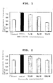
- Fig. 1 shows the evaluation of the mitochondrial depolarization action of BMS-191095, wherein the concentration of BMS-191095 and difference in the fluorescence intensity ratio were used as indices (Example 1).
- Fig. 2 shows the evaluation of the mitochondrial depolarization action of diazoxide, wherein the concentration of diazoxide and difference in the fluorescence intensity ratio were used as indices (Example 1).
- Fig. 3 shows the relationship between the concentration of levcromakalim and the fluorescence intensity ratio (Example 1).
- Fig. 4 shows the relationship between the concentration of diltiazem and the fluorescence intensity ratio (Example 1).
- Fig. 5 shows the evaluation of the mitochondrial depolarization action of BMS-191095, wherein the concentration of BMS-191095 and difference in the fluorescence intensity ratio were used as indices (Example 2).
- In the present invention, the ability of a test substance to change the mitochondrial membrane potential is evaluated by using, as an index, the change in the mitochondrial membrane potential, which is produced by contacting an ion permeability regulator on a cellular membrane with a cultured cell, wherein the change is measured as a variation in the fluorescent intensity of a fluorescent dye. Furthermore, the present invention is preferably practiced under high potassium ion concentration conditions.
- According to the present invention, a substance that changes the mitochondrial membrane potential can be selected, by which a substance capable of inhibiting cellular apoptosis, a substance capable of inhibiting calcium influx into mitochondria, and a substance capable of acting on the mitochondria ATP-sensitive K+ channel can be selected.
- The cultured cell to be used in the present invention is preferably a cultured cell having high sensitivity to a regulator of cellular membrane ion permeability, particularly an Na/K ATPase inhibitor, more preferably a cultured cell having high sensitivity to ouabain. The cell having "high sensitivity" means a cell showing a sufficient change in the membrane potential by the addition of an ion permeability regulator, an Na/K ATPase inhibitor or ouabain and, for example, a cell showing a significantly different fluorescence intensity ratio by the addition of an ion permeability regulator, an Na/K ATPase inhibitor or ouabain, as compared to culture conditions free of the addition.
- The cultured cell to be used in the present invention means a cell line or a primary cultured cell. A preferable example thereof is a cell line, more preferably a human cell line. The "human cell line" means an established cell line derived from a human tissue, which has acquired an infinite proliferation ability. Of such human cell lines, one derived from a human organ or tissue such as the heart, brain, nerve, liver, pancreas and the like is more preferable.
- The "Girardi cell" to be used in the present invention means a Girardi cell (GIRARDI HEART, ECACC No. 9312082) derived from human heart, which is available from the European Collection of Cell Culture.
The "test substance" to be used in the present invention is not particularly limited as long as it can be added to the evaluation system of the present invention and, for example, a cell extract, a cell culture supernatant, a fermentative microorganism product, a marine organism extract, a botanical extract, a purified or crudely-purified protein, a peptide, a non-peptidic compound, a nucleic acid, a synthesized low-molecular-weight compound, a natural compound, and the like can be mentioned. - The "regulator of cellular membrane ion permeability" to be used in the present invention means a substance influential on the ion permeability of the cellular membrane. Preferable specific examples of the regulator of cellular membrane ion permeability include an "ion transporter regulator", an "ion permeable channel regulator" and the like. Preferable specific examples of these regulators include Na+/Ca++ exchange system inhibitors (dichlorobenzamile etc.), Na+/H+ exchange system inhibitors (amiloride, cariporide etc.), Ca++ channel modulators (nifedipine, nicardipine, diltiazem, verapamil, Bay K 8644, maitotoxin etc.), Na+ channel modulators (tetrodotoxin, veratridine, batrachotoxin, monensin etc.), K+ channel modulators (5-hydroxydecanoic acid, HMR-1098, levcromakalim, pinacidil, 4-aminopyridine, tetraethylammonium, valinomycin etc.), Na/K ATPase inhibitors (ouabain, digoxin, digitoxin etc.), and the like. For example, the concentration when ouabain is used is preferably 0.1 µM - 3 mM, more preferably 100 µM - 2 mM.
- The "contacting the cultured cell with a test substance" in the present invention is not particularly limited as long as the cultured cell can be brought into contact with a test substance and, for example, steps wherein a test substance is present in a cell culture medium, such as addition of a test substance to a cell culture medium under culture, cultivating cells in a cell culture medium adjusted in advance by the addition of a test substance and the like, and the like can be mentioned.
- The "contacting the cultured cell with a regulator of cellular membrane ion permeability" in the present invention is not particularly limited as long as the cultured cell can be brought into contact with a regulator of cellular membrane ion permeability and, for example, addition of a regulator of cellular membrane ion permeability to a cell culture medium under culture, cultivating cells in a cell culture medium adjusted in advance by the addition of a regulator of cellular membrane ion permeability and the like, and the like can be mentioned.
- As the mode of embodiment of the present invention, an embodiment wherein a cell culture medium containing a test substance and a cell culture medium containing a regulator of cellular membrane ion permeability are separately prepared, and these cell culture media are added to cells for cultivation thereof, an embodiment wherein a cell culture medium containing a test substance and a regulator of cellular membrane ion permeability is prepared, which is added to cells for cultivation thereof can be mentioned.
- The "cell culture medium" to be used in the present invention is not particularly limited as long as the object cell can be cultured and any medium containing serum, serum-free medium, buffer and the like can be used.
- The "high potassium ion concentration" in the present invention means a concentration higher than that of a potassium ion (5-10 mM) generally used for cell culture, and is preferably 20-90 mM. The method for adjusting potassium ion is exemplified by, but not limited to, addition of a given amount of KCl.
- The "plate usable for an optical measurement method" in the present invention mainly refers to a 96-well clear bottom black plate or black plate. Besides these, as a cell culture plate measurable on a fluorescence plate reader, for example, 384-, 48-, 24-, 12- and 6-well plates and the like can be mentioned.
- The "fluorescent dye" in the present invention means a fluorescent dye sensitive to a change in the mitochondrial membrane potential and, for example, JC-1 can be mentioned.
"JC-1" (sometimes to be referred to as "mitochondrial membrane potential sensitive dye JC-1") is 5,5',6,6'-tetrachloro-1,1',3,3'-tetraethylbenzimidazolcarbocyanine iodide (Molecular Probes T-3168). This compound is taken up into cell, migrated into mitochondria, and forms an aggregate called J-aggregate when mitochondria is normal and potential difference from the outside is around -180 mV. The aggregate emits red fluorescence having a peak at 590-600 nm by the excitation at 488-490 nm. On the other hand, JC-1 in the cytoplasm becomes a monomer emitting green fluorescence having a peak at 527 nm. When mitochondria is depolarized, JC-1 cannot form J-aggregate, and therefore, red fluorescence decreases and green fluorescence increases. Utilizing such property, the fluorescence intensity ratio of monomer/J-aggregate is detected to observe the mitochondrial membrane potential. - The outline of the evaluation method of the present invention is shown in the following.
- First of all, Girardi cells are seeded on a plate usable for an optical measurement method, and cultured in a normal medium (MEM medium supplemented with additives, such as non-essential amino acid, fetal bovine serum and the like). Then, the medium is changed to a normal medium containing JC-1 to allow incorporation of JC-1 into the cell, the medium is changed to a normal medium containing a test substance, and the cells are cultured. Then, a depolarization-inducing medium is used (normal medium containing ouabain and KCl; in the evaluation method of the present invention, ouabain is used at a concentration of 0.1 µM - 3 mM, and KCl is used at a concentration of 20 mM - 90 mM). The test substance may be added along with or after addition of a depolarization-inducing medium following incorporation of JC-1 into the cell. After addition of the depolarization-inducing medium, the culture time can be 1-24 hr. Thereafter, the fluorescence of JC-1 monomer and the fluorescence of J-aggregate are measured on a fluorescence plate reader (capable of excitation at around 488-490 nm, which is the excitation wavelength of JC-1, and capable of fluorescence detection around 527 nm, which is the fluorescence wavelength peak of JC-1 monomer and around 590-600 nm, which is the fluorescence wavelength of J-aggregate), and the fluorescence intensity ratio (monomer fluorescence intensity/J-aggregate fluorescence intensity) is calculated.
The following 4 kinds of wells are set on the same plate, and the activity is calculated as shown in the following. - (1) non-treatment: well added with normal medium instead of depolarization-inducing medium,
- (2) control: well free of test substance,
- (3) test substance: well added with various kinds of test substances at each concentration,
- (4) blank: well free of JC-1. When the monomer fluorescence intensity and J-aggregate fluorescence intensity of the respective wells are shown as (1) m or (1) J,
- A: fluorescence intensity ratio of 'non-treatment' = [(1)m-(4)m]/[(1)J-(4)J]
- B: fluorescence intensity ratio of 'control' = [(2)m-(4)m]/[(2)J-(4)J]
- C: fluorescence intensity ratio of 'test substance' = [(3)m-(4)m]/[(3)J-(4)J]
- The pharmaceutical agents (compounds) evaluated by the present evaluation method are as follows.
BMS-191095: synthesized according to J. Med. Chem., vol. 40, 24-34 (1997) and used for the experiment.
levcromakalim (GlaxoSmithKline): compound 2 described in J. Med. Chem., vol. 29, 2194-2201 (1986) was synthesized and used for the experiment.
diazoxide: SIGMA D9035 was purchased and used for the experiment.
diltiazem hydrochloride: SIGMA D2521 was purchased and used for the experiment. - Girardi cells suspended in a normal medium [(MEM (GIBCO 11095-098) supplemented with 1% non-essential amino acid (MEM Non-Essential Amino Acids Solution 10 mM 100X GIBCO 11140-050), and 10% fetal bovine serum (BIOFLUIDS DIVISION Lot No. 302030))] were seeded to a 96-well clear bottom black plate (MATRIX MT4940) at 200 µL/well and cultured.
- JC-1 (Molecular Probes T-3168, 1 mg/mL DMSO solution) was added to a phenol red-free normal medium [MEM (GIBCO 51200-038) supplemented with 1% non-essential amino acid (MEM Non-Essential Amino Acids Solution 10 mM 100X GIBCO 11140-050), 1% L-glutamine (L-Glutamine-200 mM 100X: GIBCO 25030-149) and 10% fetal bovine serum (BIOFLUIDS DIVISION: Lot No. 302030)] at 10 µL/mL, the mixture was stirred with a vortex mixer and centrifuged at 3000 rpm for 1 min and the supernatant was filtered through a syringe filter (0.22 µm, MILLIPORE). The medium in the 96-well plate was removed and the JC-1 solution was dispensed to a cell-seeded well at 50 µL/well. A phenol red-free normal medium without JC-1 was dispensed to a blank well. The plate was placed back in a CO2 incubator and incubated for 20 min.
- Then, each well was washed once with a phenol red-free normal medium (200 µL) and the same medium containing various concentrations of each test substance was dispensed at 50 µL/well. Each test substance was dissolved in DMSO (Dimethyl Sulfoxide: SIGMA D-8779) to the final DMSO concentration of 0.1%.
A medium containing 0.1% DMSO was dispensed to the wells of 'without treatment' and 'control' at 50 µL/well. The plate was placed back in a CO2 incubator and incubated for 25 min. - Then, a depolarization-inducing medium containing 1 mM ouabain, 60 mM KCl, and a test substance at each concentration (prepared from phenol red-free normal medium as a base) was dispensed to the well of 'test substance' by 50 uL. A normal medium containing 0.1% DMSO was dispensed to the well of 'without treatment', and a depolarization-inducing medium containing 0.1% DMSO was dispensed to the well of 'control' by 50 µL/well.
The plate was placed back in a CO2 incubator and incubated for 2 hr. - After induction of depolarization, the plate was set on a fluorescence plate reader (TECAN, SpectraFluor) heated to 37°C, and the J-aggregate fluorescence was measured at 595 nm using an excitation wavelength of 485 nm. Then, the monomer fluorescence was measured at 535 nm using an excitation wavelength of 485 nm. The measurement was performed at flash frequency of 20 times and 50 µsec intervals. Each group contained 4 wells. The average value of fluorescence intensity of the 'blank' well was subtracted from the value of each well of 'without treatment', 'control' and 'test substance' for each of monomer fluorescence and J-aggregate fluorescence, and the ratio of each fluorescence intensity, monomer/J-aggregate, was determined.
- The results are shown in Fig. 1 to Fig. 4 (mean ± standard error, N=4).
- As a result of the experiment according to the evaluation method of the present invention, the mitochondrial depolarization inhibitory action could be conveniently evaluated for BMS-191095 (Fig. 1) and diazoxide (Fig. 2) confirmed by other evaluation methods to have a mitoKATP channel open action.
- In contrast, levcromakalim (Fig. 3), which is a sarcKATP channel opener, and diltiazem (Fig. 4), which is a calcium antagonist, did not prevent mitochondrial depolarization.
- From the above, it is appreciated that the evaluation method of the present invention can specifically detect a mitoKATP channel opener by a convenient operation as compared to conventional evaluation methods.
- Girardi cells were suspended in a normal medium [(MEM (GIBCO 11095-098) supplemented with 1% non-essential amino acid (MEM Non-Essential Amino Acids Solution 10 mM 100X GIBCO 11140-050), and 10% fetal bovine serum (BIOFLUIDS DIVISION Lot No. 302030))] to 1×10⁵ cells/mL, seeded in a 96-well clear bottom black plate (MATRIX MT4940) at 200 µL/well, and cultured.
For measurement of blank value of fluorescence for each plate, cells were not seeded in part of the wells. - JC-1 (Molecular Probes T-3168, 1 mg/mL DMSO solution) was added to a phenol red-free normal medium [MEM (GIBCO 51200-038) supplemented with 1% non-essential amino acid (MEM Non-Essential Amino Acids Solution 10 mM 100X GIBCO 11140-050), 1% L-glutamine (L-Glutamine-200 mM 100X: GIBCO 25030-149) and 10% fetal bovine serum (BIOFLUIDS DIVISION: Lot No. 302030)] at 10 µL/mL, the mixture was stirred with a vortex mixer and centrifuged at 3000 rpm for 2 min and the supernatant was filtered through a syringe filter (0.22 µm, MILLIPORE). The medium in the 96-well plate was removed and the JC-1 solution was dispensed to a cell-seeded well at 50 µL/well. A phenol red-free normal medium without JC-1 was dispensed to a blank well. The plate was placed back in a CO2 incubator and incubated for 20 min.
- Then, a depolarization-inducing medium containing 1 mM ouabain, 60 mM KCl, and BMS-191095 at each concentration (prepared from phenol red-free normal medium as a base) was dispensed to the well of 'test substance' by 100 uL.
- BMS-191095 was dissolved in DMSO (Dimethyl Sulfoxide: SIGMA D-8779) to the final DMSO concentration of 0.1%.
- A medium containing 0.1% DMSO was dispensed to the wells of 'without treatment' and 'control' by 100 µL/well. The plate was placed back in a CO2 incubator and incubated.
- After 2 hr, the plate was set on a fluorescence plate reader (Molecular Divices, SpectraMax GEMINI EM) heated to 37°C, and the measurement was performed under the conditions of Bottom read, Lm1 Ex: 485 nm Em: 530 nm, Lm2 Ex: 535 nm Em: 590 nm, Read/Well:6 times. Each group contained 4 wells. The average value of fluorescence intensity of the 'blank' well was subtracted from the value of each well of 'without treatment', 'control' and 'test substance' for each of monomer fluorescence and J-aggregate fluorescence, and the ratio of each fluorescence intensity, monomer/J-aggregate, was determined.
The results are shown in Fig. 5 (mean ± standard error, N=4). - Using the evaluation method of the present invention, a pharmaceutical agent that selectively acts on the mitoKATP channel can be evaluated. Using the evaluation method of the present invention, a pharmaceutical agent that changes the mitochondrial membrane potential can be conveniently evaluated with high precision. Using the evaluation method of the present invention, a pharmaceutical agent that changes the mitochondrial membrane potential can be evaluated in a short time.
- This application is based on a patent application No.
2004-147418 filed in Japan on May 18, 2004
The present invention is explained in more detail in the following by referring to Examples, which are not to be construed as limitative.
Claims (27)
- An evaluation method of a substance that changes the mitochondrial membrane potential, which comprises adding a regulator of cellular membrane ion permeability.
- The evaluation method of claim 1, which comprises the following steps of:(a) contacting a cultured cell with a test substance,(b) contacting the cultured cell with a regulator of cellular membrane ion permeability, and(c) measuring the change in a mitochondrial membrane potential of the cultured cell.
- The evaluation method of claim 2, wherein (a) to (c) further comprise (d) selecting a substance that changes the mitochondrial membrane potential of the cultured cell.
- The evaluation method of claim 3, wherein the substance in(d) inhibits cell apoptosis.
- The evaluation method of claim 3 or 4, wherein the substance in (d) inhibits calcium influx into mitochondria.
- The evaluation method of any one of claims 3 to 5, wherein the substance in (d) acts on the mitochondrial ATP-sensitive K+ channel.
- The evaluation method of any one of claims 2 to 6, wherein the cultured cell has a high sensitivity to an ion permeability regulator of the cellular membrane.
- The evaluation method of claim 7, wherein the cultured cell has a high sensitivity to an Na/K ATPase inhibitor.
- The evaluation method of claim 8, wherein the cultured cell has a high sensitivity to ouabain.
- The evaluation method of any one of claims 2 to 9, wherein the cultured cell is a cell line.
- The evaluation method of claim 10, wherein the cultured cell is a human cell line.
- The evaluation method of claim 11, wherein the cultured cell is derived from human heart, brain, nerve, liver or pancreas.
- The evaluation method of claim 12, wherein the cultured cell is a Girardi cell.
- The evaluation method of any one of claims 2 to 13, wherein the cultured cell is cultured on a plate usable for an optical measurement method.
- The evaluation method of any one of claims 2 to 6, wherein the regulator of cellular membrane ion permeability induces the change in the mitochondrial membrane potential.
- The evaluation method of claim 15, wherein the regulator of cellular membrane ion permeability is an ion transporter regulator or ion permeable channel regulator of the cellular membrane.
- The evaluation method of claim 16, wherein the ion transporter regulator or ion permeable channel regulator is an Na+/Ca++ exchange system inhibitor, an Na+/H+ exchange system inhibitor, a Ca++modulator or an Na/K ATPase inhibitor.
- The evaluation method of claim 17, wherein the ion transporter regulator or ion permeable channel regulator is an Na/K ATPase inhibitor.
- The evaluation method of claim 18, wherein the Na/K ATPase inhibitor is ouabain.
- The evaluation method of any one of claims 2 to 6, wherein "(b) contacting the cultured cell with a regulator of cellular membrane ion permeability" is performed under a high potassium ion concentration.
- The evaluation method of claim 20, wherein the potassium ion concentration is 20 - 90 mM.
- The evaluation method of claim 20 or 21, wherein the regulator of cellular membrane ion permeability is ouabain.
- The evaluation method of claim 22, wherein ouabain has a concentration of 0.1 µM - 3 mM.
- The evaluation method of any one of claims 1 to 6, wherein the change in the membrane potential is measured by the fluorescent color of a fluorescent dye incorporated into the cell.
- The evaluation method of claim 24, wherein the fluorescent dye develops two colors.
- The evaluation method of claim 25, wherein the change in the membrane potential is measured by the ratio of the two fluorescent colors.
- The evaluation method of any one of claims 24 to 26, wherein the fluorescent dye is JC-1.
Applications Claiming Priority (2)
| Application Number | Priority Date | Filing Date | Title |
|---|---|---|---|
| JP2004147418 | 2004-05-18 | ||
| PCT/JP2005/009066 WO2005111228A1 (en) | 2004-05-18 | 2005-05-18 | Method of assaying substance capable of changing mitochondrial membrane potential |
Publications (3)
| Publication Number | Publication Date |
|---|---|
| EP1760158A1 true EP1760158A1 (en) | 2007-03-07 |
| EP1760158A4 EP1760158A4 (en) | 2007-09-12 |
| EP1760158B1 EP1760158B1 (en) | 2013-07-17 |
Family
ID=35394167
Family Applications (1)
| Application Number | Title | Priority Date | Filing Date |
|---|---|---|---|
| EP05741434.4A Expired - Lifetime EP1760158B1 (en) | 2004-05-18 | 2005-05-18 | Method of assaying substance capable of changing mitochondrial membrane potential |
Country Status (5)
| Country | Link |
|---|---|
| US (1) | US7781184B2 (en) |
| EP (1) | EP1760158B1 (en) |
| JP (1) | JP4755587B2 (en) |
| ES (1) | ES2431523T3 (en) |
| WO (1) | WO2005111228A1 (en) |
Cited By (1)
| Publication number | Priority date | Publication date | Assignee | Title |
|---|---|---|---|---|
| WO2008110247A1 (en) | 2007-03-13 | 2008-09-18 | Grünenthal GmbH | Method for identifying the agonistic activity of a target compound on a potassium channel |
Families Citing this family (4)
| Publication number | Priority date | Publication date | Assignee | Title |
|---|---|---|---|---|
| WO2011019807A2 (en) * | 2009-08-11 | 2011-02-17 | The Johns Hopkins University | Measurement of mitochondrial membrane potential to assess organ dysfunction |
| CA2912676A1 (en) | 2013-05-16 | 2014-11-20 | Psychnostics, Llc | Methods for treating adhd and bipolar disorder using a membrane potential ratio test |
| CN113337453A (en) * | 2021-05-31 | 2021-09-03 | 中国科学院生态环境研究中心 | Method for detecting pulmonary toxicity of compounds |
| CN114530212B (en) * | 2022-01-11 | 2024-05-31 | 中国中医科学院中药研究所 | A method for predicting and evaluating the nephrotoxicity of chemical components in traditional Chinese medicine |
Family Cites Families (5)
| Publication number | Priority date | Publication date | Assignee | Title |
|---|---|---|---|---|
| US5169788A (en) * | 1989-06-09 | 1992-12-08 | New England Deaconess Hospital Corporation | Methods of measuring membrane potential using j-aggregate forming dyes |
| US7160687B1 (en) * | 1997-05-29 | 2007-01-09 | Cellomics, Inc. | Miniaturized cell array methods and apparatus for cell-based screening |
| US20030044776A1 (en) | 1998-09-25 | 2003-03-06 | James A. Dykens | Compositions and methods for identifying agents that alter mitochondrial permeability transition pores |
| WO2000068686A1 (en) | 1999-05-10 | 2000-11-16 | Tularik Inc. | High-throughput screening assays for modulators of mitochondrial membrane potential |
| WO2000079274A2 (en) | 1999-06-22 | 2000-12-28 | Mitokor | Methods for assaying subcellular conditions using energy transfer |
-
2005
- 2005-05-18 ES ES05741434T patent/ES2431523T3/en not_active Expired - Lifetime
- 2005-05-18 EP EP05741434.4A patent/EP1760158B1/en not_active Expired - Lifetime
- 2005-05-18 US US11/597,275 patent/US7781184B2/en not_active Expired - Fee Related
- 2005-05-18 WO PCT/JP2005/009066 patent/WO2005111228A1/en not_active Ceased
- 2005-05-18 JP JP2006513619A patent/JP4755587B2/en not_active Expired - Fee Related
Cited By (1)
| Publication number | Priority date | Publication date | Assignee | Title |
|---|---|---|---|---|
| WO2008110247A1 (en) | 2007-03-13 | 2008-09-18 | Grünenthal GmbH | Method for identifying the agonistic activity of a target compound on a potassium channel |
Also Published As
| Publication number | Publication date |
|---|---|
| ES2431523T3 (en) | 2013-11-26 |
| JPWO2005111228A1 (en) | 2008-03-27 |
| EP1760158A4 (en) | 2007-09-12 |
| EP1760158B1 (en) | 2013-07-17 |
| WO2005111228A1 (en) | 2005-11-24 |
| US20070202486A1 (en) | 2007-08-30 |
| JP4755587B2 (en) | 2011-08-24 |
| US7781184B2 (en) | 2010-08-24 |
Similar Documents
| Publication | Publication Date | Title |
|---|---|---|
| Guo et al. | A rapid and high content assay that measures cyto-ID-stained autophagic compartments and estimates autophagy flux with potential clinical applications | |
| Jones et al. | Sensitive determination of cell number using the CyQUANT® cell proliferation assay | |
| Ariazi et al. | Discovery and preclinical characterization of GSK1278863 (daprodustat), a small molecule hypoxia inducible factor–prolyl hydroxylase inhibitor for anemia | |
| Merrill et al. | A robust and economical pulse-chase protocol to measure the turnover of HaloTag fusion proteins | |
| Vázquez et al. | Assays to assess autophagy induction and fusion of autophagic vacuoles with a degradative compartment, using monodansylcadaverine (MDC) and DQ‐BSA | |
| Zwartsen et al. | Cardiotoxicity screening of illicit drugs and new psychoactive substances (NPS) in human iPSC-derived cardiomyocytes using microelectrode array (MEA) recordings | |
| Krogstad et al. | Antimalarials increase vesicle pH in Plasmodium falciparum. | |
| Duan et al. | Potent and selective inhibition of plasma membrane monoamine transporter by HIV protease inhibitors | |
| US20090263853A1 (en) | High-Throughput Cell-Based CFTR Assay | |
| Williams et al. | Quantitative high-throughput efficacy profiling of approved oncology drugs in inflammatory breast cancer models of acquired drug resistance and re-sensitization | |
| Bhosale et al. | Investigating the mitochondrial permeability transition pore in disease phenotypes and drug screening | |
| John et al. | Novel 384-well population patch clamp electrophysiology assays for Ca2+-activated K+ channels | |
| Pedemonte et al. | Structure-activity relationship of 1, 4-dihydropyridines as potentiators of the cystic fibrosis transmembrane conductance regulator chloride channel | |
| US7781184B2 (en) | Method of assaying substance capable of changing mitochondrial membrane potential | |
| Chen et al. | Observing protein interactions and their stoichiometry in living cells by brightness analysis of fluorescence fluctuation experiments | |
| Merugu et al. | Chemical probes and methods for single-cell detection and quantification of epichaperomes in hematologic malignancies | |
| Williams et al. | Generation and characterization of a stable MK2‐EGFP cell line and subsequent development of a high‐content imaging assay on the Cellomics ArrayScan platform to screen for p38 mitogen‐activated protein kinase inhibitors | |
| EP2447252B1 (en) | Theramutein modulators | |
| Darzynkiewicz | Acid-induced denaturation of DNA in situ as a probe of chromatin structure | |
| Governa et al. | Impairment of human polymorphonuclear leukocyte chemotaxis by 2, 5-hexanedione | |
| Manning et al. | The astrocytic zinc transporter ZIP12 is a synaptic protein that contributes to synaptic zinc levels in the mouse auditory cortex | |
| US7416893B2 (en) | Methods for diagnosing drug-resistant cancer cells or for identifying chemotherapeutic agents by measuring anomalous intracellular ion and/or second messenger dynamics | |
| Quesada et al. | Chemosensitization and drug accumulation assays as complementary methods for the screening of multidrug resistance reversal agents | |
| Perez et al. | A High-Throughput Flow Cytometry Assay for Identification of Inhibitors of 3′, 5′-Cyclic Adenosine Monophosphate Efflux | |
| AU2001276569B2 (en) | Quantitative fluorescent assay for determining the activity of transport proteins |
Legal Events
| Date | Code | Title | Description |
|---|---|---|---|
| PUAI | Public reference made under article 153(3) epc to a published international application that has entered the european phase |
Free format text: ORIGINAL CODE: 0009012 |
|
| 17P | Request for examination filed |
Effective date: 20061128 |
|
| AK | Designated contracting states |
Kind code of ref document: A1 Designated state(s): AT BE BG CH CY CZ DE DK EE ES FI FR GB GR HU IE IS IT LI LT LU MC NL PL PT RO SE SI SK TR |
|
| DAX | Request for extension of the european patent (deleted) | ||
| A4 | Supplementary search report drawn up and despatched |
Effective date: 20070810 |
|
| RAP1 | Party data changed (applicant data changed or rights of an application transferred) |
Owner name: MITSUBISHI TANABE PHARMA CORPORATION |
|
| 17Q | First examination report despatched |
Effective date: 20100119 |
|
| RIC1 | Information provided on ipc code assigned before grant |
Ipc: G01N 21/64 20060101ALN20120227BHEP Ipc: G01N 33/94 20060101ALN20120227BHEP Ipc: G01N 21/77 20060101ALN20120227BHEP Ipc: G01N 33/50 20060101AFI20120227BHEP Ipc: C12Q 1/02 20060101ALN20120227BHEP |
|
| RIC1 | Information provided on ipc code assigned before grant |
Ipc: G01N 21/64 20060101ALN20120312BHEP Ipc: G01N 33/50 20060101AFI20120312BHEP Ipc: G01N 21/77 20060101ALN20120312BHEP Ipc: G01N 33/94 20060101ALN20120312BHEP Ipc: C12Q 1/02 20060101ALN20120312BHEP |
|
| GRAP | Despatch of communication of intention to grant a patent |
Free format text: ORIGINAL CODE: EPIDOSNIGR1 |
|
| RIC1 | Information provided on ipc code assigned before grant |
Ipc: G01N 21/64 20060101ALN20120611BHEP Ipc: G01N 21/77 20060101ALN20120611BHEP Ipc: G01N 33/50 20060101AFI20120611BHEP Ipc: C12Q 1/02 20060101ALN20120611BHEP Ipc: G01N 33/94 20060101ALN20120611BHEP |
|
| RIN1 | Information on inventor provided before grant (corrected) |
Inventor name: UMEDA, ASAMI Inventor name: NAGAKI, SUMIKO |
|
| REG | Reference to a national code |
Ref country code: DE Ref legal event code: R079 Ref document number: 602005040436 Country of ref document: DE Free format text: PREVIOUS MAIN CLASS: C12Q0001020000 Ipc: G01N0033500000 |
|
| RIN1 | Information on inventor provided before grant (corrected) |
Inventor name: NAGAKI, SUMIKO Inventor name: UMEDA, ASAMI |
|
| RIC1 | Information provided on ipc code assigned before grant |
Ipc: G01N 33/50 20060101AFI20121112BHEP Ipc: G01N 21/64 20060101ALN20121112BHEP Ipc: C12Q 1/02 20060101ALN20121112BHEP Ipc: G01N 21/77 20060101ALN20121112BHEP Ipc: G01N 33/94 20060101ALN20121112BHEP |
|
| RAP1 | Party data changed (applicant data changed or rights of an application transferred) |
Owner name: MITSUBISHI TANABE PHARMA CORPORATION |
|
| GRAS | Grant fee paid |
Free format text: ORIGINAL CODE: EPIDOSNIGR3 |
|
| GRAP | Despatch of communication of intention to grant a patent |
Free format text: ORIGINAL CODE: EPIDOSNIGR1 |
|
| INTG | Intention to grant announced |
Effective date: 20130419 |
|
| GRAA | (expected) grant |
Free format text: ORIGINAL CODE: 0009210 |
|
| AK | Designated contracting states |
Kind code of ref document: B1 Designated state(s): AT BE BG CH CY CZ DE DK EE ES FI FR GB GR HU IE IS IT LI LT LU MC NL PL PT RO SE SI SK TR |
|
| REG | Reference to a national code |
Ref country code: GB Ref legal event code: FG4D |
|
| REG | Reference to a national code |
Ref country code: CH Ref legal event code: EP |
|
| REG | Reference to a national code |
Ref country code: IE Ref legal event code: FG4D |
|
| REG | Reference to a national code |
Ref country code: AT Ref legal event code: REF Ref document number: 622503 Country of ref document: AT Kind code of ref document: T Effective date: 20130815 |
|
| REG | Reference to a national code |
Ref country code: DE Ref legal event code: R096 Ref document number: 602005040436 Country of ref document: DE Effective date: 20130912 |
|
| REG | Reference to a national code |
Ref country code: ES Ref legal event code: FG2A Ref document number: 2431523 Country of ref document: ES Kind code of ref document: T3 Effective date: 20131126 |
|
| REG | Reference to a national code |
Ref country code: AT Ref legal event code: MK05 Ref document number: 622503 Country of ref document: AT Kind code of ref document: T Effective date: 20130717 |
|
| REG | Reference to a national code |
Ref country code: NL Ref legal event code: VDEP Effective date: 20130717 |
|
| REG | Reference to a national code |
Ref country code: LT Ref legal event code: MG4D |
|
| PG25 | Lapsed in a contracting state [announced via postgrant information from national office to epo] |
Ref country code: SE Free format text: LAPSE BECAUSE OF FAILURE TO SUBMIT A TRANSLATION OF THE DESCRIPTION OR TO PAY THE FEE WITHIN THE PRESCRIBED TIME-LIMIT Effective date: 20130717 Ref country code: CY Free format text: LAPSE BECAUSE OF FAILURE TO SUBMIT A TRANSLATION OF THE DESCRIPTION OR TO PAY THE FEE WITHIN THE PRESCRIBED TIME-LIMIT Effective date: 20130717 Ref country code: IS Free format text: LAPSE BECAUSE OF FAILURE TO SUBMIT A TRANSLATION OF THE DESCRIPTION OR TO PAY THE FEE WITHIN THE PRESCRIBED TIME-LIMIT Effective date: 20131117 Ref country code: LT Free format text: LAPSE BECAUSE OF FAILURE TO SUBMIT A TRANSLATION OF THE DESCRIPTION OR TO PAY THE FEE WITHIN THE PRESCRIBED TIME-LIMIT Effective date: 20130717 Ref country code: BE Free format text: LAPSE BECAUSE OF FAILURE TO SUBMIT A TRANSLATION OF THE DESCRIPTION OR TO PAY THE FEE WITHIN THE PRESCRIBED TIME-LIMIT Effective date: 20130717 Ref country code: PT Free format text: LAPSE BECAUSE OF FAILURE TO SUBMIT A TRANSLATION OF THE DESCRIPTION OR TO PAY THE FEE WITHIN THE PRESCRIBED TIME-LIMIT Effective date: 20131118 Ref country code: AT Free format text: LAPSE BECAUSE OF FAILURE TO SUBMIT A TRANSLATION OF THE DESCRIPTION OR TO PAY THE FEE WITHIN THE PRESCRIBED TIME-LIMIT Effective date: 20130717 |
|
| PG25 | Lapsed in a contracting state [announced via postgrant information from national office to epo] |
Ref country code: NL Free format text: LAPSE BECAUSE OF FAILURE TO SUBMIT A TRANSLATION OF THE DESCRIPTION OR TO PAY THE FEE WITHIN THE PRESCRIBED TIME-LIMIT Effective date: 20130717 Ref country code: FI Free format text: LAPSE BECAUSE OF FAILURE TO SUBMIT A TRANSLATION OF THE DESCRIPTION OR TO PAY THE FEE WITHIN THE PRESCRIBED TIME-LIMIT Effective date: 20130717 Ref country code: SI Free format text: LAPSE BECAUSE OF FAILURE TO SUBMIT A TRANSLATION OF THE DESCRIPTION OR TO PAY THE FEE WITHIN THE PRESCRIBED TIME-LIMIT Effective date: 20130717 Ref country code: GR Free format text: LAPSE BECAUSE OF FAILURE TO SUBMIT A TRANSLATION OF THE DESCRIPTION OR TO PAY THE FEE WITHIN THE PRESCRIBED TIME-LIMIT Effective date: 20131018 Ref country code: PL Free format text: LAPSE BECAUSE OF FAILURE TO SUBMIT A TRANSLATION OF THE DESCRIPTION OR TO PAY THE FEE WITHIN THE PRESCRIBED TIME-LIMIT Effective date: 20130717 |
|
| PG25 | Lapsed in a contracting state [announced via postgrant information from national office to epo] |
Ref country code: SK Free format text: LAPSE BECAUSE OF FAILURE TO SUBMIT A TRANSLATION OF THE DESCRIPTION OR TO PAY THE FEE WITHIN THE PRESCRIBED TIME-LIMIT Effective date: 20130717 Ref country code: EE Free format text: LAPSE BECAUSE OF FAILURE TO SUBMIT A TRANSLATION OF THE DESCRIPTION OR TO PAY THE FEE WITHIN THE PRESCRIBED TIME-LIMIT Effective date: 20130717 Ref country code: CZ Free format text: LAPSE BECAUSE OF FAILURE TO SUBMIT A TRANSLATION OF THE DESCRIPTION OR TO PAY THE FEE WITHIN THE PRESCRIBED TIME-LIMIT Effective date: 20130717 Ref country code: DK Free format text: LAPSE BECAUSE OF FAILURE TO SUBMIT A TRANSLATION OF THE DESCRIPTION OR TO PAY THE FEE WITHIN THE PRESCRIBED TIME-LIMIT Effective date: 20130717 Ref country code: RO Free format text: LAPSE BECAUSE OF FAILURE TO SUBMIT A TRANSLATION OF THE DESCRIPTION OR TO PAY THE FEE WITHIN THE PRESCRIBED TIME-LIMIT Effective date: 20130717 |
|
| PLBE | No opposition filed within time limit |
Free format text: ORIGINAL CODE: 0009261 |
|
| STAA | Information on the status of an ep patent application or granted ep patent |
Free format text: STATUS: NO OPPOSITION FILED WITHIN TIME LIMIT |
|
| 26N | No opposition filed |
Effective date: 20140422 |
|
| REG | Reference to a national code |
Ref country code: DE Ref legal event code: R097 Ref document number: 602005040436 Country of ref document: DE Effective date: 20140422 |
|
| PG25 | Lapsed in a contracting state [announced via postgrant information from national office to epo] |
Ref country code: LU Free format text: LAPSE BECAUSE OF FAILURE TO SUBMIT A TRANSLATION OF THE DESCRIPTION OR TO PAY THE FEE WITHIN THE PRESCRIBED TIME-LIMIT Effective date: 20140518 |
|
| REG | Reference to a national code |
Ref country code: CH Ref legal event code: PL |
|
| PG25 | Lapsed in a contracting state [announced via postgrant information from national office to epo] |
Ref country code: LI Free format text: LAPSE BECAUSE OF NON-PAYMENT OF DUE FEES Effective date: 20140531 Ref country code: MC Free format text: LAPSE BECAUSE OF FAILURE TO SUBMIT A TRANSLATION OF THE DESCRIPTION OR TO PAY THE FEE WITHIN THE PRESCRIBED TIME-LIMIT Effective date: 20130717 Ref country code: CH Free format text: LAPSE BECAUSE OF NON-PAYMENT OF DUE FEES Effective date: 20140531 |
|
| REG | Reference to a national code |
Ref country code: IE Ref legal event code: MM4A |
|
| PG25 | Lapsed in a contracting state [announced via postgrant information from national office to epo] |
Ref country code: IE Free format text: LAPSE BECAUSE OF NON-PAYMENT OF DUE FEES Effective date: 20140518 |
|
| REG | Reference to a national code |
Ref country code: FR Ref legal event code: PLFP Year of fee payment: 11 |
|
| PGFP | Annual fee paid to national office [announced via postgrant information from national office to epo] |
Ref country code: DE Payment date: 20150512 Year of fee payment: 11 Ref country code: GB Payment date: 20150513 Year of fee payment: 11 Ref country code: ES Payment date: 20150428 Year of fee payment: 11 |
|
| PGFP | Annual fee paid to national office [announced via postgrant information from national office to epo] |
Ref country code: FR Payment date: 20150508 Year of fee payment: 11 Ref country code: IT Payment date: 20150515 Year of fee payment: 11 |
|
| PG25 | Lapsed in a contracting state [announced via postgrant information from national office to epo] |
Ref country code: BG Free format text: LAPSE BECAUSE OF FAILURE TO SUBMIT A TRANSLATION OF THE DESCRIPTION OR TO PAY THE FEE WITHIN THE PRESCRIBED TIME-LIMIT Effective date: 20130717 |
|
| PG25 | Lapsed in a contracting state [announced via postgrant information from national office to epo] |
Ref country code: TR Free format text: LAPSE BECAUSE OF FAILURE TO SUBMIT A TRANSLATION OF THE DESCRIPTION OR TO PAY THE FEE WITHIN THE PRESCRIBED TIME-LIMIT Effective date: 20130717 Ref country code: HU Free format text: LAPSE BECAUSE OF FAILURE TO SUBMIT A TRANSLATION OF THE DESCRIPTION OR TO PAY THE FEE WITHIN THE PRESCRIBED TIME-LIMIT; INVALID AB INITIO Effective date: 20050518 |
|
| REG | Reference to a national code |
Ref country code: DE Ref legal event code: R119 Ref document number: 602005040436 Country of ref document: DE |
|
| GBPC | Gb: european patent ceased through non-payment of renewal fee |
Effective date: 20160518 |
|
| PG25 | Lapsed in a contracting state [announced via postgrant information from national office to epo] |
Ref country code: IT Free format text: LAPSE BECAUSE OF NON-PAYMENT OF DUE FEES Effective date: 20160518 |
|
| REG | Reference to a national code |
Ref country code: FR Ref legal event code: ST Effective date: 20170131 |
|
| PG25 | Lapsed in a contracting state [announced via postgrant information from national office to epo] |
Ref country code: DE Free format text: LAPSE BECAUSE OF NON-PAYMENT OF DUE FEES Effective date: 20161201 Ref country code: FR Free format text: LAPSE BECAUSE OF NON-PAYMENT OF DUE FEES Effective date: 20160531 |
|
| PG25 | Lapsed in a contracting state [announced via postgrant information from national office to epo] |
Ref country code: GB Free format text: LAPSE BECAUSE OF NON-PAYMENT OF DUE FEES Effective date: 20160518 |
|
| PG25 | Lapsed in a contracting state [announced via postgrant information from national office to epo] |
Ref country code: ES Free format text: LAPSE BECAUSE OF NON-PAYMENT OF DUE FEES Effective date: 20160519 |
|
| REG | Reference to a national code |
Ref country code: ES Ref legal event code: FD2A Effective date: 20181204 |